Animal Movement Across Scales. Edited by Lars-Anders Hansson and Susanne Åkesson. © Oxford University Press 2014. Published 2014 by Oxford University Press. CHAPTER 10 Sensory mechanisms of animal orientation and navigation Rachel Muheim, Jannika Boström, Susanne Åkesson, and Miriam Liedvogel Although questions such as ‘How do animals find their way, and how do they sense and process this information in the brain?’ have been asked for cen- turies, the field of animal orientation and naviga- tion has seen an immense leap forward in the past few decades. Moreover, our understanding has also expanded considerably regarding the molecular and physiological mechanisms of the different com- passes and cues used by animals for orientation and navigation (Åkesson et al., Chapter 9, and Svensson et al., Chapter 11). Most notable are the advances made in our understanding of how animals can sense information provided by the geomagnetic field and use this information for behavioural tasks, for example for compass orientation during migra- tion. But despite interdisciplinary and highly in- tegrative research over recent decades, we do not fully understand how animals perceive the Earth´s magnetic field. We know that animals use geomag- netic information for orientation tasks (see Åkesson et al., Chapter 9), but the receptor(s) remain to be identified. In this chapter, we review current knowl- edge in this area, outline challenges, and suggest fu- ture approaches to elucidate the sensory modalities used by animals for orientation and navigational tasks. 10.1 Magnetic sense Many hypotheses regarding how animals may sense the Earth’s magnetic field have been proposed. Three principally different mechanisms to achieve this could theoretically be used to sense the strength of the Earth’s magnetic field, including (1) induc- tion, (2) magnetic particles, and (3) magnetically sensitive biochemical reactions. The latter two possibilities have emerged as the most promising candidate magnetoreceptor mechanisms: A light- dependent process is thought to detect the align- ment of the geomagnetic field lines in space. This provides directional information that can be used for a magnetic compass (inclination compass, see Åkesson et al., Chapter 9). The other possibility is a detection process mediated by a ferromin- eral that reacts to very small changes in the direction and/or intensity of the magnetic field and, thereby, can be used as a magnetic com- pass and/or a magnetic positioning (map or signpost) sense (for reviews see Wiltschko and Wiltschko 1995a, 2005; Lohmann and Johnson 2000; Mouritsen and Ritz 2005). Both of the lat- ter two mechanisms are supported by behav- ioural and physiological data in a broad range of organisms (see also Åkesson et al., Chapter 9). In some animals, like newts and birds, the pres- ence of both mechanisms have independently been experimentally demonstrated to be present and used by the animals for different purposes, and thus are believed to be non-exclusive (Phil- lips 1986, Wiltschko and Wiltschko 1995b, and see 10.1.3). Here, we present the state-of-the-art knowledge of the sensory aspects of the two mag- netoreception mechanisms, and highlight recent advances and future challenges. 9780199677191-Hansson.indb 179 22/07/14 1:27 PM

Welcome message from author
This document is posted to help you gain knowledge. Please leave a comment to let me know what you think about it! Share it to your friends and learn new things together.
Transcript
Animal Movement Across Scales. Edited by Lars-Anders Hansson and Susanne Åkesson. © Oxford University Press 2014. Published 2014 by Oxford University Press.
Chapter 10
Sensory mechanisms of animal orientation and navigationRachel Muheim, Jannika Boström, Susanne Åkesson, and Miriam Liedvogel
Although questions such as ‘How do animals find their way, and how do they sense and process this information in the brain?’ have been asked for cen-turies, the field of animal orientation and naviga-tion has seen an immense leap forward in the past few decades. Moreover, our understanding has also expanded considerably regarding the molecular and physiological mechanisms of the different com-passes and cues used by animals for orientation and navigation (Åkesson et al., Chapter 9, and Svensson et al., Chapter 11). Most notable are the advances made in our understanding of how animals can sense information provided by the geomagnetic field and use this information for behavioural tasks, for example for compass orientation during migra-tion. But despite interdisciplinary and highly in-tegrative research over recent decades, we do not fully understand how animals perceive the Earth´s magnetic field. We know that animals use geomag-netic information for orientation tasks (see Åkesson et al., Chapter 9), but the receptor(s) remain to be identified. In this chapter, we review current knowl-edge in this area, outline challenges, and suggest fu-ture approaches to elucidate the sensory modalities used by animals for orientation and navigational tasks.
10.1 Magnetic sense
Many hypotheses regarding how animals may sense the Earth’s magnetic field have been proposed. Three principally different mechanisms to achieve
this could theoretically be used to sense the strength of the Earth’s magnetic field, including (1) induc-tion, (2) magnetic particles, and (3) magnetically sensitive biochemical reactions. The latter two possibilities have emerged as the most promising candidate magnetoreceptor mechanisms: A light-dependent process is thought to detect the align-ment of the geomagnetic field lines in space. This provides directional information that can be used for a magnetic compass (inclination compass, see Åkesson et al., Chapter 9). The other possibility is a detection process mediated by a ferromin-eral that reacts to very small changes in the direction and/or intensity of the magnetic field and, thereby, can be used as a magnetic com-pass and/or a magnetic positioning (map or signpost) sense (for reviews see Wiltschko and Wiltschko 1995a, 2005; Lohmann and Johnson 2000; Mouritsen and Ritz 2005). Both of the lat-ter two mechanisms are supported by behav-ioural and physiological data in a broad range of organisms (see also Åkesson et al., Chapter 9). In some animals, like newts and birds, the pres-ence of both mechanisms have independently been experimentally demonstrated to be present and used by the animals for different purposes, and thus are believed to be non-exclusive (Phil-lips 1986, Wiltschko and Wiltschko 1995b, and see 10.1.3). Here, we present the state-of-the-art knowledge of the sensory aspects of the two mag-netoreception mechanisms, and highlight recent advances and future challenges.
9780199677191-Hansson.indb 179 22/07/14 1:27 PM

180 A N I M A L M OV E M E N T AC R O S S S C A L E S
evidence that these particles function as magnetore-ceptors. Magnetite, maghemite, or other ferritin-like particles are found in a number of insect species, in-cluding bees, ants, and termites, but an actual mag-netoreceptor has not been located or described yet (reviewed by Wajnberg et al. 2010). The most con-vincing data supporting a magnetite-based receptor mechanism come from fish and birds, where there is both behavioural and electrophysiological evi-dence in favour of a magnetite-based magnetore-ception mechanism. Several behavioural studies with fish have reported alignment along magnetic fields or abilities to discriminate magnetic anoma-lies. For example, rainbow trout, Oncorhynchus mykiss, align along an external magnetic field, but become disoriented in a null magnetic field where no magnetic field is present; i.e. all dimensions of the magnetic field are cancelled out and the length of the magnetic vector is zero (Chew and Brown 1989). Trout are also able to discriminate between magnetic anomalies, but only when the conditional response allow movement and when the magnetic fields are spatially distinctive (Walker et al. 1997). Experiments with yellowfin tuna, Thunnus albac-ares, showed that these fish could sense differences in intensity of the magnetic field, but not differences in direction (Walker 1984). Neurophysiological re-cordings on rainbow trout supported these find-ings; specifically, single neurons in a side branch of the trigeminal nerve have been shown to respond to changes in intensity, but not to the direction of an imposed magnetic field (Walker et al. 1997). Single-domain magnetite particles have been re-ported in several locations in different fish species: near the basal lamina of the olfactory epithelium (the area innervated by the trigeminal nerve) and in the dermal bone cartilage of the skull (e.g. Walker et al. 1984, 1997). Based on the findings of iron-rich crystals in the olfactory lamellae in rainbow trout (Walker et al. 1997), Diebel et al. (2000) suggested a vertebrate magnetoreceptor in form of a multi-lobed cell, containing chains of magnetite particles located in the basal lamina of an olfactory lamella. Recently, these findings were supported by a new method developed to detect and characterize can-didate vertebrate magnetoreceptor cells (Eder et al. 2012). However, despite this exciting discovery, it remains to be shown how these magnetoreceptor
10.1.1 Magnetic sense based on ferromagnetic particles
The idea of a ferromineral-based magnetic sense originated from the findings of deposits of ferro-magnetic (magnetite and/or maghemite) particles in various organisms, including several animals that use the Earth’s magnetic field for behavioural tasks (Kirschvink et al. 1985, Williams and Wild 2001, Falkenberg et al. 2010, but see Treiber et al. 2012). However, we here want to stress that the mere presence of biogenic ferrominerals in any organism is the rule rather than the exception and does not automatically indicate the presence of a magnetic sense. These particles may be involved in many dif-ferent roles in the physiology of an organism; for example, magnetite synthesis seems to be a general way for organisms to deposit excess iron. With the exception of magnetotactic bacteria, where magnet-ite crystals act like a compass needle and enable the bacteria to passively align to the geomagnetic field (Blakemore 1975), the physiological mechanisms of magnetic field reception using ferromagnetic miner-als remain to be demonstrated for other organisms.
10.1.2 Biophysical and molecular mechanisms of ferromineral-based magnetoreception
Several conditions must be met for a magnetore-ception system based on ferromagnetic minerals to work (Kirschvink and Walker 1985, Kirschvink 1989, Winklhofer and Kirschvink 2010; for details see Box 10.1): (1) the ferromagnetic material must be biochemically precipitated by the organism itself, because incorporated external material is usually too contaminated; (2) the ferromagnetic particles must fulfil a variety of magnetophysical properties and have a specific magnetization and size, i.e. be single-domain (SD) or superparamagnetic (SPM) crystals; and (3) they must be mechanically coupled to nerve fibres or sensory organelles; otherwise, they cannot transmit any magnetic information to the brain.
10.1.3 Behavioural and physiological evidence for ferromineral-based magnetoreception
Despite numerous reports of ferromagnetic mate-rial in invertebrates, there is little direct behavioural
9780199677191-Hansson.indb 180 22/07/14 1:27 PM
S E N S O RY M E C H A N I S M S O F A N I M A L O R I E N TAT I O N A N D N AV I G AT I O N 181
Chapter 9). Pulse remagnetization experiments and treatment with zinc sulphide of the putative receptor organ (considered as diagnostic tool to test for the in-volvement of magnetite-based receptor mechanism; see Box 10.2) with migratory songbirds and pigeons, Columbia livia, have shown that experienced adult birds, but not inexperienced juveniles, are affected
cells function in detail, and how the information is transferred to a nerve signal.
Both birds and newts are believed to possess a dual magnetoreception mechanism, i.e. a light-dependent magnetic compass and a ( putatively in-dependent) magnetite-based magnetic map sense (Phillips 1986, Munro et al. 1997a; Åkesson et al.,
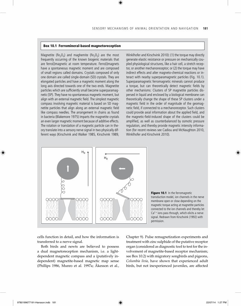
Magnetite (Fe3O4) and maghemite (Fe2O3) are the most frequently occurring of the known biogenic materials that are ferro(i)magnetic at room temperature. Ferro(i)magnets have a spontaneous magnetic moment and are composed of small regions called domains. Crystals composed of only one domain are called single-domain (SD) crystals. They are elongated particles and have a magnetic moment along the long axis directed towards one of the two ends. Magnetite particles which are sufficiently small become superparamag-netic (SP). They have no spontaneous magnetic moment, but align with an external magnetic field. The simplest magnetic compass involving magnetic material is based on SD mag-netite particles that align along an external magnetic field like compass needles. The arrangement in chains as found in bacteria (Blakemore 1975) imparts the magnetite crystals an even larger magnetic moment because of additive effects. The rotation or translation of a magnetic particle can in the-ory translate into a sensory nerve signal in two physically dif-ferent ways (Kirschvink and Walker 1985, Kirschvink 1989,
Winklhofer and Kirschvink 2010): (1) the torque may directly generate elastic resistance or pressure on mechanically cou-pled physiological structures, like a hair cell, a stretch recep-tor, or another mechanoreceptor; or (2) the torque may have indirect effects and alter magneto-chemical reactions or in-teract with nearby superparamagnetic particles (Fig. 10.1). Superparamagnetic ferromagnetic minerals cannot produce a torque, but can theoretically detect magnetic fields by other mechanisms: Clusters of SP magnetite particles dis-persed in liquid and enclosed by a biological membrane can theoretically change the shape of these SP clusters under a magnetic field in the order of magnitude of the geomag-netic field, if connected to a mechanoreceptor. Such clusters could provide axial information about the applied field, and the magnetic-field-induced shape of the clusters could be amplified, as well as counterbalanced by osmotic pressure regulation, and thereby provide magnetic intensity informa-tion (for recent reviews see Cadiou and McNaughton 2010, Winklhofer and Kirschvink 2010).
Box 10.1 Ferromineral-based magnetoreception
H0
H0
Figure 10.1 In the ferromagnetic transduction model, ion channels in the nerve membrane open or close depending on the magnetic torque acting at magnetite particles connected to the ion channels and thereby let Ca2 + ions pass through, which elicits a nerve signal. Redrawn from Kirschvink (1992) with permission.
9780199677191-Hansson.indb 181 22/07/14 1:27 PM
182 A N I M A L M OV E M E N T AC R O S S S C A L E S
Diagnostic tools used to test whether a magnetoreceptor based on ferrominerals is involved in a specific behaviour can be grouped into two categories: (1) tools that directly affect the magnetic particles, and (2) tools that affect the sensory receptor or the transduction pathways between the receptor and the brain.
(1) Pulse remagnetization is the application of a brief, strong, directional magnetic pulse (0.5 T for 4–5 ms). This procedure has been widely used to directly affect the fer-romagnetic particles in a putative magnetoreceptor (e.g. Wiltschko et al. 1994, 1995B; Beason et al. 1995):
• IfSDparticlesareinvolved,suchamagneticpulsewill permanently remagnetize the particles in the opposite direction.
• Ifthepulseisappliedanti-paralleltothemagneticmo-ment and with an intensity greater than the coercivity of SD particles, i.e. greater than the intensity required to reduce the magnetization of those particles to zero, this should result in a permanent reversal or change in orientation.
• IfclustersofSPmagnetiteareinvolved,suchastrongmagnetic pulse is expected to form agglomerations of clusters and impair the magnetoreceptor for a few days.
The downside of pulse remagnetization experiments is that it is difficult to predict the expected outcome of such a treatment as long as the exact structure and function of the receptor are not known. In addition, there is no proper
control experiment that allows distinguishing a true effect of the strong pulse on the magnetoreceptor from effects on other unrelated physiological processes.
(2) Tools in the second category aim to disrupt magneto-receptor function, and include (i) local anaesthetics blockade or treatment with zinc sulphide of the putative receptor or-gan (i.e. upper beak area in birds; e.g. Holland et al. 2009), and (ii) lesion studies, where either the transmitting nerve (i.e. trigeminal nerve system in birds; cf. Mora et al. 2004) or the putative brain areas involved in the processing of the information are lesioned (via mechanical cut or chemical le-sion, e.g. with ibotenic acid; Zapka et al. 2009). Note that studies using local anaesthesia should be treated with cau-tion, as no control experiments exist that can reliably work as treatment control. Further, nothing is known about the time span of efficacy of any such treatment for most mi-gratory animals. A further indirect indication for the involve-ment of a ferromineral-based mechanism is also directed orientation in total darkness, as this receptor mechanism is independent of light, but the radical-pair-based mechanism requires light to function (but it must be noted that energy for the radical-pair formation could in theory also be taken from chemical energy).
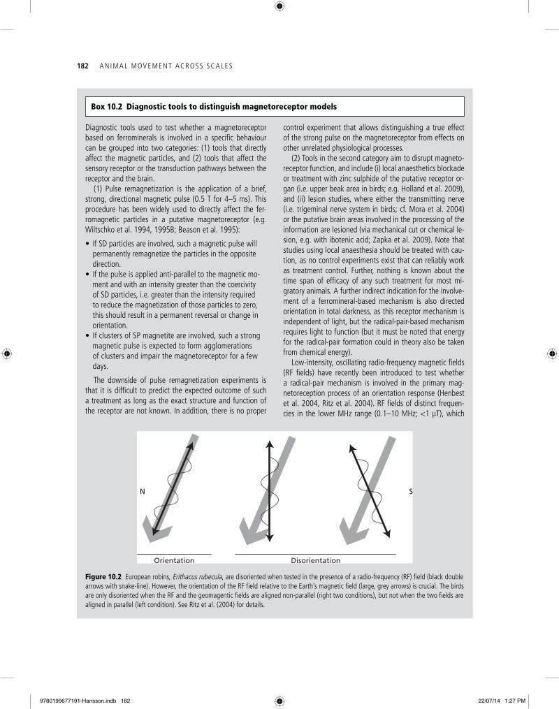
Low-intensity, oscillating radio-frequency magnetic fields (RF fields) have recently been introduced to test whether a radical-pair mechanism is involved in the primary mag-netoreception process of an orientation response (Henbest et al. 2004, Ritz et al. 2004). RF fields of distinct frequen-cies in the lower MHz range (0.1–10 MHz; <1 μT), which
Box 10.2 Diagnostic tools to distinguish magnetoreceptor models
N S
Orientation Disorientation
Figure 10.2 European robins, Erithacus rubecula, are disoriented when tested in the presence of a radio-frequency (RF) field (black double arrows with snake-line). However, the orientation of the RF field relative to the Earth’s magnetic field (large, grey arrows) is crucial. The birds are only disoriented when the RF and the geomagentic fields are aligned non-parallel (right two conditions), but not when the two fields are aligned in parallel (left condition). See Ritz et al. (2004) for details.
9780199677191-Hansson.indb 182 22/07/14 1:27 PM
S E N S O RY M E C H A N I S M S O F A N I M A L O R I E N TAT I O N A N D N AV I G AT I O N 183
other bird species, including migratory passerines (Falkenberg et al. 2010). The anatomical findings and theoretical assumptions were supported by behav-ioural experiments which demonstrated that birds are no longer able to detect magnetic stimuli when the trigeminal nerve, and thus the connection from the putative receptor to the brain, was cut (Mora et al. 2004; see also next section). Inactivation of the beak receptor in experiments (by lesioning the ophthal-mic branch of the trigeminal nerve) with free-flying birds, both migratory birds and homing pigeons, supported these findings, at least with respect to ex-perienced, adult individuals (Gagliardo et al. 2006, Holland et al. 2009).
Until very recently, such a ‘3-D magnetometer’ in the upper beak of birds was almost regarded as a fact. However, the findings were recently chal-lenged by a study demonstrating that these iron crystals were not located in nerve tissue, but rather in macrophages, and thus could not be the sen-sors providing the trigeminal nerve, and thereby the brain, with magnetic information (Treiber et al. 2012). Macrophages are immune cells known to contain ferritin proteins (which store iron in the cell), and their presence has been reported through-out the bird’s beak and other areas of the body, but nowhere associated to nerve tissue (Treiber et al. 2012). What consequences do these results have on our understanding of how birds may perceive the Earth’s magnetic field? They certainly challenge previous interpretations of behavioural experi-ments that concluded that the trigeminal nerve is the link that makes it possible to transfer informa-tion sensed via magnetosensors in the beak to the
by such a treatment (e.g. Wiltschko et al. 1994; Bea-son et al. 1995, 1997; Munro et al. 1997b; Holland et al. 2009). Presumably, juvenile birds exclusively use their light-dependent magnetic compass to de-termine their migratory direction during their first migration. Experienced adult birds likely also use their magnetic map sense, which they acquire dur-ing their first migration. Blocking the trigeminal nerve with a local anaesthetic eliminates the effects of pulse remagnetization (Beason and Semm 1996).
Where could these putative compass needles (single-domain or superparamagnetic magnetite crystals) be located in birds, and by which pathways could they transmit the magnetic information to the brain? Williams and Wild (2001) found trigeminally innervated structures in the nasal region of pigeons which they interpreted as single-domain magnetite. A few years later, Fleissner and colleagues (2003, 2007) found nerve-cell endings (branches of the trigeminal nerve) in the upper beak of pigeons. Those nerve-cell endings contained dense clusters of ferromagnetic structures at six distinct locations in the beak, which could sense the geomagnetic field and subsequently transmit this information to the brain. Depending on their alignment relative to the magnetic field, these clusters were suggested to become uniquely deformed by the Earth’s magnetic field, leading to an opening of mechano-sensitive ion channels and ultimately result in a nerve signal transmitted via the trigeminal nerve to the brain. The birds would there-by be able to measure the intensity, and consequently the polarity, of a magnetic field in all three dimen-sions, providing them with a ‘3-D magnetometer’. These structures were later confirmed for several
vary with the intensity of the ambient magnetic field and the magnetoreceptor molecule involved, interfere with the in-terconversion between the singlet and triplet excited states, and mask or alter the magnetic field effects produced by the Earth’s magnetic field (Fig. 10.2). Such RF disturbances can lead to either disorientation or change in orientation, depending on the amount and type of change, and how the animals integrate the information into a migratory direc-
tion. Magnetite-based magnetoreceptors are not affected by RF fields, since the rotation of magnetite particles as proposed in animal magnetoreceptors is too slow and the ferromagnetic resonance frequency is expected to be in the GHz rather than MHz range (Ritz et al. 2000, 2004; Henbest et al. 2004). This makes the application of RF fields a unique, diagnostic tool for studying the involvement of a radical-pair mechanism in magnetoreception (Fig. 10.2).
Box 10.2 Continued
9780199677191-Hansson.indb 183 22/07/14 1:27 PM
184 A N I M A L M OV E M E N T AC R O S S S C A L E S
trigeminal nerve of the bobolink, Dolichonyx ory-zivorus, a migratory songbird from North America. Changes in the rotation of the vertical or horizontal component of the geomagnetic field and changes in intensity of as little as 50–200 nT led to alter-ations in the action potentials in the trigeminal nerve (Beason and Semm 1987). Magnetic com-pass responses (choosing and maintaining a direc-tion) were not affected by blocking the ophthalmic branch of the trigeminal nerve (Beason and Semm 1996). All of these findings are suggestive of the in-volvement of the trigeminal nerve in the process-ing of magnetic information from the beak to the brain.
10.1.5 Conditioning experiments
Further support for an involvement of the trigemi-nal nerve system comes from conditioning experi-ments, which show that pigeons can discriminate between the presence and absence of a strong mag-netic anomaly (~100,000 nT; Mora et al. 2004). This ability disappeared when the ophthalmic branch of the trigeminal nerve was cut, but remained when the olfactory nerve was lesioned instead (Mora et al. 2004). Similarly, Pekin ducks, Anas platy-rhynchos domestica, trained to find a hidden food reward indicated by a magnetic anomaly, were not able to find the reward when the trigeminal nerve was anaesthetized by an injection of ligno-caine hydrochloride, strongly indicating a role of the trigeminal nerve in the detection of magnetic anomalies (Freire et al. 2012). In addition, both pigeons (Thalau et al. 2007) and domestic chicks (Denzau et al. 2011) were able to use a small, but strong, anomaly to find a hidden food source, al-though none of these studies tested for the involve-ment of the trigeminal nerve. Still, in order for a magnetic ‘map sense’ to work, birds must be able to detect naturally occurring local changes in the magnetic field strengths that are about five orders of magnitude smaller (~10 nT) than the magnetic anomalies used in the operant conditioning by Mora et al. (2004). Therefore, to function as a puta-tive biologically relevant map or signpost sense, it still remains to be demonstrated that the trigeminal magnetoreception circuit can detect biologically relevant magnetic anomalies.
brain. The new findings add no evidence to sup-port the existence of iron-containing nerve cells at the previously reported locations along the upper beak of birds, but do not exclude the possibility that there still are magnetoreceptors at some yet-to-be-identified location in the bird’s upper beak. Consequently, we need to carefully re-evaluate hy-potheses and conclusions that were drawn based on behavioural studies that have previously been interpreted to support the function of a magnetite-based receptor located in the upper beak of birds. One plausible scenario could be that the real mag-netoreceptors are located in the olfactory tissue, which is adjacent to the area where the supposed magnetic sense system was identified. This region is known to be connected to the nervous tissue, which was shown to transmit magnetic informa-tion to the brain in lesion experiments. This would be well supported by the finding of candidate magneto-sensitive cells in olfactory regions in trout (Walker et al. 1997; see earlier in this section).
In conclusion, the hypothesis proposing that magnetic information is perceived by a magnetore-ceptor in the beak and transmitted to the brain via the trigeminal nerve might still be valid, as lesion-ing this link resulted in birds no longer being able to perceive magnetic stimuli. However, we do not understand the detailed function of this putative neuronal link, and we do not know which magne-tosensor feeds information into the nerve. Recently, a putative third magnetoreceptor was suggested to be located in the lagena of the inner ear of pigeons and encode information on the direction and inten-sity of the magnetic field (Wu and Dickman 2011, 2012). However, it remains to be demonstrated whether the reported brain activities as a conse-quence of changes in the magnetic field originate from an actual new magnetoreceptor, or whether they are the results of the integration of vestibular and magnetic field information, which is necessary for measuring magnetic fields.
10.1.4 electrophysiological recordings and lesion experiments
The behavioural evidence for a magnetite-based map sensor is also supported by neurophysiologic-al recordings from the ophthalmic branch of the
9780199677191-Hansson.indb 184 22/07/14 1:27 PM
S E N S O RY M E C H A N I S M S O F A N I M A L O R I E N TAT I O N A N D N AV I G AT I O N 185
10.1.6 Chemical magnetoreception based on a radical pair mechanism
In 1978, the physicist Klaus Schulten proposed that magnetic sensing might have spin chemical origins (Schulten et al. 1978, Schulten 1982). In a theoretical paper, he suggested that the yield of a biochemi-cal reaction proceeding via a radical pair might be sensitive to the orientation of an external magnetic field. Electron spins are not strongly coupled to the thermal bath and therefore represent one of only a few molecular features that might plausibly be in-fluenced by the Earth’s magnetic field. The suggest-ed mechanism (Box 10.3) involves a light-induced electron transfer between two molecules (note: such processes can also be chemically induced). The elec-tron transfer results in the generation of a radical-pair intermediate that will either exist in a singlet or a triplet excited state, and subsequently decay in chemically different end products. Theoreti-cal calculations and in vitro experiments showed that the ratio between singlet and triplet products from radical- pair reactions can be modulated by an Earth-strength magnetic field during the intercon-version step, i.e. when singlet radical intermediates convert to triplets and vice versa (Maeda et al. 2008, 2012). This orientation-dependent interconversion step (and subsequently resulting differences in the ratio of singlet and triplet end products) could theoretically be used to encode directional informa-tion. In a revival of this idea, Ritz et al. (2000) pro-posed that the retina with its almost perfect sphere would be an ideal substrate for such a mechanism. He further speculated that the radical-pair inter-mediate might be involved in some kind of visual reception system, exploiting the highly efficient visual transduction signalling cascade. According to this suggestion, the reaction yield anisotropy of the receptor radical pair could govern the direction-al response and thus form the basis of a magnetic compass (for recent reviews see Ritz et al. 2010, Mouritsen and Hore 2012).
Possible receptor candidate molecules involved in the primary magnetoreception process and mediat-ing a light-dependent radical-pair mechanism must meet the following criteria: they need to be light-sensitive, need to be able to form radical pairs that persist long enough so that the radical-pair yields
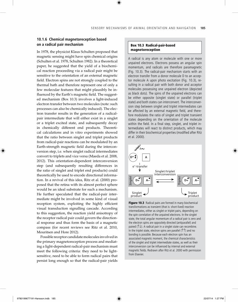
A radical is any atom or molecule with one or more unpaired electrons. Electrons possess an angular spin momentum, and radicals are therefore paramagnetic (Fig. 10.3). The radical-pair mechanism starts with an electron transfer from a donor molecule D to an accep-tor molecule A upon photo excitation (Fig. 10.3), re-sulting in a radical pair with both donor and acceptor molecules possessing one unpaired electron (depicted as black dots). The spins of the unpaired electrons can be either opposite (singlet state) or parallel (triplet state) and both states can interconvert. The interconver-sion step between singlet and triplet intermediates can be affected by an external magnetic field, and there-fore modulates the ratio of singlet and triplet transient states depending on the orientation of the molecule within the field. In a final step, singlet, and triplet in-termediates will react to distinct products, which may differ in their biochemical properties (modified after Ritz et al. 2000).
Box 10.3 Radical-pair-based magnetoreception
A−D+
AD*
Singlet /tripletinterconversion
Tripletproduct
Singletproduct
h•v
Singlet
A−D+Triplet
e−-transfer
Figure 10.3 Radical pairs are formed in many biochemical transformations as transient (that is: short-lived) reaction intermediates, either as singlet or triplet pairs, depending on the spin correlation of the unpaired electrons. In the singlet state, the total angular momentum of a radical pair is zero and the electron spins are oppositely directed (antiparallel) and paired (↑↓). A radical pair in a singlet state can recombine. In the triplet state, electron spins are parallel (↑↑) and no bonding is possible. Because each electron spin has an associated magnetic moment, the chemical characteristics of the singlet and triplet intermediate states, as well as their interconversion can be influenced by internal and external magnetic fields. Redrawn after Ritz et al. 2000 with permission from Elsevier.
9780199677191-Hansson.indb 185 22/07/14 1:27 PM
186 A N I M A L M OV E M E N T AC R O S S S C A L E S
laboratory mice (Muheim et al. 2006a), and zebra finches (Voss et al. 2007), has provided access to cryptochrome knockout animals. This has opened up a new and promising avenue in magnetorecep-tion research, allowing direct tests of the involve-ment of cryptochromes and other molecules possibly involved in the primary magnetoreception process. One recent study with Drosophila provided convinc-ing evidence that cryptochromes indeed are involved in light-dependent magnetoreception. In this study, adult Drosophila were able to discriminate a mag-netic field (although about 10 times stronger than the geomagnetic field) only when the cryptochrome gene was functionally intact, but not when it was genetically engineered to be dysfunctional (Gegear et al. 2008). Also, magnetic fields were shown to in-fluence cryptochrome-mediated effects of blue light on the free-running rhythm of the circadian clock in Drosophila (Yoshi et al. 2009). Magnetic field sensitivity could thus be an intrinsic property of cryptochrome-based photo-signalling systems, from which light-dependent magnetoreception might have evolved (Phillips et al. 2010a, b).
From a behavioural perspective, the putative magnetoreceptor must meet the following crite-ria to be a likely candidate for a light-dependent magnetic compass receptor (Liedvogel and Mour-itsen 2010, Phillips et al. 2010a, Ritz et al. 2010; Fig. 10.4): (1) the magnetoreceptor needs to be sensi-tive to light of specific spectral properties ( mainly wavelength), matching the receptor molecule(s) involved in the radical-pair process (see later dis-cussion); (2) the encoded information received by the animal needs to have axial properties and not allow to determine the polarity of the field lines, since the avian magnetic compass generally func-tions without using polarity, as first described by Wiltschko and Wiltschko (1972); (3) the directional information perceived by the animal must depend on the intensity of the magnetic field; thus exposure to magnetic field intensities never experienced be-fore can lead to disorientation, followed by a slow adaptation to the ‘new’ intensity (Wiltschko 1968, Wiltschko et al. 2006). A light-dependent magnetic compass has been demonstrated in both inverte-brates and vertebrates, including fruit flies, meal-worm beetles, bullfrogs, newts, homing pigeons, and several species of migratory birds (reviewed by Wiltschko and Wiltschko 1995a, 2005; Muheim
can be modified by an Earth-strength magnetic field, and should be localized in a spatially fixed re-lationship relative to each other criteria (reviewed by Rodgers and Hore 2009, Liedvogel and Mourit-sen 2010, Phillips et al. 2010a, Mouritsen and Hore 2012). Classical photopigments like the opsins do not form radical pairs, and thus are unlikely candi-dates for magnetoreception. Cryptochromes, a class of blue light photoreceptor molecules, have been suggested to be the most likely molecules involved in magnetoreception. They are the only known photopigments in the vertebrate eye that have the potential to form radical pairs. Cryptochromes form a multi-gene family of blue-green light pho-toreceptors known to be involved in circadian rhythm regulation (for a recent review see Chaves et al. 2011). They share high sequence homology with photolyases that form radical pair interme-diates persisting long enough for magnetic field effects to occur (Weber et al. 2002, Giovani et al. 2003). To date, cryptochromes from several taxa have been shown to also produce persistent, spin-correlated radical species upon photo-excitation in vitro (Liedvogel et al. 2007, Biskup et al. 2009, Schleicher et al. 2009). Cryptochromes are found in a large variety of species known to use a mag-netic compass, for example, in retinas of two mi-gratory bird species, European robins, Erithacus rubecula, and garden warblers, Sylvia borin, and two non-migratory species, chicken, Gallus gallus, and zebra finches, Taeniopygia guttatta (Liedvogel and Mouritsen 2010). A detailed neuroanatomical study of cryptochrome expression in robins and chicken recently revealed that the outer segments of the avian UV/V cones are most likely the primary sites of the light-dependent magnetoreceptor (Niessner et al. 2011). Niessner and colleagues found crypto-chrome expression in virtually every cone across the entire retina, which is one of the requirements for the radical-pair model to work. Still, more re-search is needed to unambiguously separate obser-vations of cryptochrome expression as a result of circadian rhythmicity and magnetoreception, and to describe the full details of the reception mecha-nism, including the neuronal pathways mediating the sensory information to the brain.
The development of reliable magnetic compass as-says in genetic model organisms, such as Drosophila (Phillips and Sayeed 1993, Gegear et al. 2008),
9780199677191-Hansson.indb 186 22/07/14 1:27 PM
S E N S O RY M E C H A N I S M S O F A N I M A L O R I E N TAT I O N A N D N AV I G AT I O N 187
oriented under UV to green light, and become dis-oriented or show shifted orientation under longer wavelengths (e.g. Wiltschko et al. 1993; Wiltschko and Wiltschko 1995b, 2001; Munro et al. 1997b; Muheim et al. 2002). European robins exposed to radio-frequency (RF) fields aligned nonparallel to the geomagnetic field vector become disoriented when tested under either a broadband RF field or distinct single frequencies of 1.375 or 7 MHz (Fig. 10.2; Ritz et al. 2004). The orientation of mole-rats, on the other hand, which is supposed to be mediated by a non-light-dependent magnetore-ceptor, is not impaired by an RF field, supporting
et al. 2002; Phillips et al. 2010a). There is also strong evidence from behavioural experiments for a light- dependent magnetic compass in C57BL/6J mice (Muheim et al. 2006a). A light-independent mag-netic compass rather seems to be the exception and has only been demonstrated in a few animals, such as subterranean mole-rats (Marhold et al. 1997) and sea turtles (Lohmann and Lohmann 1993), possibly as a result of an adaptation to the non-terrestrial lifestyle of these organisms.
In birds, the dependence of light of specific wavelengths for magnetic compass orientation has been studied extensively. In general, birds are well
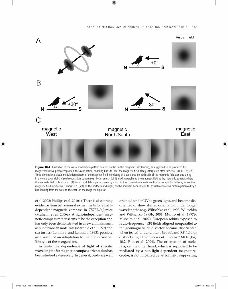
Figure 10.4 Illustration of the visual modulation pattern centred on the Earth’s magnetic field (arrow), as suggested to be produced by magnetosensitive photoreceptors in the avian retina, enabling birds to ‘see’ the magnetic field ( freely interpreted after Ritz et al. 2000). (A; left) Three-dimensional visual modulation pattern of the magnetic field, consisting of a dark area on each side of the magnetic field axis and a ring in the centre. (A; right) Visual modulation pattern seen by an animal (bird) looking parallel to the magnetic field at the magnetic equator, where the magnetic field is horizontal. (B) Visual modulation pattern seen by a bird looking towards magnetic south at a geographic latitude, where the magnetic field inclination is about 30°, (left) on the northern and (right) on the southern hemisphere. (C) Visual modulation pattern perceived by a bird looking from the west to the east (on the magnetic equator).
9780199677191-Hansson.indb 187 22/07/14 1:27 PM
188 A N I M A L M OV E M E N T AC R O S S S C A L E S
reptiles, and the frontal organ of amphibians, light-dependent photoreception could also take place in extra-retinal photoreceptors (for review see Phillips et al. 2010a). This has been nicely demonstrated in Eastern red-spotted newts, Nothophthalmus virides-cens, which show a 90° shifted response in magnetic compass orientation when tested under red light, compared to tests under blue or full-spectrum white light (Phillips and Borland 1992). Hence, newts trained to learn the shoreward side of a training tank with the top of their head, but not their eyes, covered with a red-light filter, showed the same 90° shifted response as control animals completely il-luminated by red light (Deutschlander et al. 1999). Thus, the light-sensitive-magnetosensitive recep-tors mediating shoreward orientation in newts are most likely located in extraocular photoreceptors in the pineal complex or hypothalamus (Deutschland-er et al. 1999). In anuran amphibians and lizards, single photoreceptors with two antagonistic pho-toreception mechanisms, like those proposed to underlie the light-dependent magnetic compass in newts, have been found in the pineal complex (Eldred and Nolte 1978, Solessio and Engbretson 1993).
10.2 Celestial compasses–sun, polarized light, and star compasses
10.2.1 physiological evidence for sun and star compass orientation
While the behavioural mechanisms of sun com-pass orientation are well understood in many species (see Åkesson et al., Chapter 9), we know little about how the sun compass works on the physiological level, especially in higher organisms. Similarly, it remains to be shown what key fea-tures from the rotating starry sky the birds use for orientation. Are birds taking ‘snapshot pictures’ at certain time intervals and overlaying these to identify the centre of rotation? Or are they able to take an overexposed picture, and thus can follow the movement of the stars by seeing their paths? We can currently only speculate on how the star compass in birds functions, and how this informa-tion is mediated by the brain and which brain areas are used for processing star compass orientation.
previous indications that their magnetic compass is based on magnetite (Thalau et al. 2006).
Questions that remain to be answered include: Where in the animals’ bodies are the light-dependent magnetoreceptors located, and by which neuronal pathway is the information transmitted from the receptor to the brain? As magnetic fields can trans-mit through all body material, the receptors could in theory be located just about anywhere. However, behavioural experiments suggest that light is nec-essary for light-dependent magnetic orientation to function; thus the receptors must be located at a pe-ripheral site of the animals’ body. In birds, and also mammals, the only locations where light can reach specialized photoreceptors are the eyes. Already in the 1980s, extracellular recordings provided the first evidence for the involvement of the visual cen-tre in light-dependent magnetoreception in birds. Cells in the nucleus of the basal optic root (nBOR) and in the optic tectum showed light-dependent magnetic responsiveness to changes in the direc-tion of a magnetic field and slow inversions of the vertical component of the magnetic field (Semm et al. 1984). Recent research on the neural basis of magnetoreception has largely substantiated these findings, and provided new insights into the neural pathways and brain areas involved in in-formation transfer and processing in both birds and mammals (e.g. Nemec et al. 2001, Heyers et al. 2007).
In birds, a brain structure in the visual Wulst, named ‘Cluster N’, and connected to the retina via the thalamofugal pathway, has been identified and suggested to be involved in the processing of light-dependent magnetic information in migratory birds during the night (Mouritsen et al. 2005, Heyers et al. 2007, Zapka et al. 2009). Migratory birds with a ( chemically) lesioned Cluster N are disorientated when tested for magnetic compass orientation, but their sunset or star compass remained intact and functional for orientation. These results strongly indicate that Cluster N is involved in processing magnetic compass information at low light levels (Zapka et al. 2009). However, not in all taxa is light-dependent magnetoreception necessarily limited to the retina. In animals that have special structures containing photoreceptors, like the parietal eyes of reptiles or the pineal complex of fish and some
9780199677191-Hansson.indb 188 22/07/14 1:27 PM
S E N S O RY M E C H A N I S M S O F A N I M A L O R I E N TAT I O N A N D N AV I G AT I O N 189
in an ommatidium that are often aligned at 90° to each other (Roberts et al. 2011). In insects, these polarization-sensitive photoreceptors are further concentrated in specialized regions of the eyes (in many cases in an area in the upper field of view,
Cleverly designed conditioning experiments could be one approach in order to get further insight into how birds are able to identify the centre of rotation as reference cue for star compass orientation. One possibility for identifying brain areas involved in processing star compass orientation would be ‘behavioural molecular mapping’, such as the method used to identify Cluster N as the brain area involved in magnetic compass orientation (Mouritsen et al. 2005). This requires an extremely well-designed experiment, including standardized control conditions and exclusion of any alternative variables that could putatively be used for com-pass orientation.
10.2.2 Behavioural and physiological evidence for polarized light sensitivity
The ability to perceive linearly polarized skylight and use it for orientation is common in the animal kingdom (Åkesson et al., Chapter 9). It has been extensively characterized, on both the behavioural and physiological level, first and foremost in in-vertebrates, such as arthropods (e.g. honeybees, crickets, ants, spiders, beetles, crabs, shrimp, cray-fish, water fleas) and cephalopods (e.g. squids and cuttlefish) (for reviews see Horváth and Varjú 2004, Roberts et al. 2011). However, also verte-brates seem to use this third dimension of light in various behavioural contexts, and polarized light sensitivity has been reported in fish (e.g. Cameron and Pugh 1991, Parkyn et al. 2003), amphibians (Adler and Taylor 1973), reptiles (Adler and Phil-lips 1985), and birds (e.g. Able 1982, Muheim et al. 2006b, and see below). To date, no evidence for polarization sensitivity in mammals has been pub-lished, so mammals may be an exception (but see polarization vision in humans, Box 10.4).
How can animals perceive polarized light? The fundamental prerequisites for a photoreceptor to be able to detect the electric vector of light are inherently polarization-sensitive pigment mol-ecules (dichroic chromophores). Some common chromophores, like retinal, fulfil this requirement. The exceptionally high polarization sensitivity found in many insects, crustaceans, and cephalo-pods is enhanced by the highly organized micro-villi of individual rhabdomeric photoreceptor cells
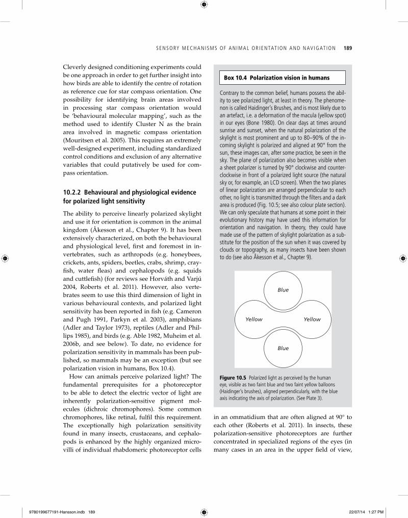
Contrary to the common belief, humans possess the abil-ity to see polarized light, at least in theory. The phenome-non is called Haidinger’s Brushes, and is most likely due to an artefact, i.e. a deformation of the macula (yellow spot) in our eyes (Bone 1980). On clear days at times around sunrise and sunset, when the natural polarization of the skylight is most prominent and up to 80–90% of the in-coming skylight is polarized and aligned at 90° from the sun, these images can, after some practice, be seen in the sky. The plane of polarization also becomes visible when a sheet polarizer is turned by 90° clockwise and counter-clockwise in front of a polarized light source (the natural sky or, for example, an LCD screen). When the two planes of linear polarization are arranged perpendicular to each other, no light is transmitted through the filters and a dark area is produced (Fig. 10.5; see also colour plate section). We can only speculate that humans at some point in their evolutionary history may have used this information for orientation and navigation. In theory, they could have made use of the pattern of skylight polarization as a sub-stitute for the position of the sun when it was covered by clouds or topography, as many insects have been shown to do (see also Åkesson et al., Chapter 9).
Box 10.4 Polarization vision in humans
Blue
YellowYellow
Blue
Figure 10.5 Polarized light as perceived by the human eye, visible as two faint blue and two faint yellow balloons (Haidinger’s brushes), aligned perpendicularly, with the blue axis indicating the axis of polarization. (See Plate 3).
9780199677191-Hansson.indb 189 22/07/14 1:27 PM
190 A N I M A L M OV E M E N T AC R O S S S C A L E S
2003). Still, there is strong experimental evidence that migratory birds use information from the skylight polarization pattern to determine their departure direction and calibrate their compasses (see Åkesson et al., Chapter 9). However, the (pu-tative) reception mechanism via which birds could perceive the pattern of polarized skylight remains a mystery. There are simply no obvious anatomical structures described in their eyes that could fulfil this job. The avian double cones have been pro-posed as polarized light receptors, since their ori-entation forms a cross pattern, with two opposite double cones facing each other, and the other two pointing away from each other (Young and Mar-tin 1984, Cameron and Pugh 1991). However, it is unclear whether they could act as polarized light receptors, and therefore, more research will be nec-essary to behaviourally characterize and identify the photoreceptors mediating polarized light sen-sitivity in birds.
10.3 Future perspectives
During the past decade, research on the sen-sory mechanisms of animal orientation and navigation has become highly integrative and in-terdisciplinary. The field has progressed from the observation- based study of mainly behavioural aspects of orientation and navigation, to include biophysical, neuroanatomical, and molecular tools. The introduction of new techniques from various neighbouring disciplines has led to signif-icant advances in our understanding of the senso-ry and cognitive mechanisms underlying animal orientation and navigation. Still, many open ques-tions remain to be answered. Where are the mag-netoreceptors located? How is the information processed in the brain? What is the neuronal basis of star compass orientation? Which brain areas are involved in processing celestial compass informa-tion from the sun, the stars, or polarized skylight cues? How do birds (and maybe other vertebrates as well) perceive polarized light cues and use this information for compass orientation? Given recent advances in technology and enhanced collabora-tive effort between disciplines, many of these questions will be addressed, and hopefully an-swered in the years to come.
so-called dorsal rim area), where they are aligned in ordered arrays to measure polarized light from different angles (Wehner 1989).
The insects’ compound eyes thus seem to be bet-ter suited to perceive polarized light than the ver-tebrate eye, and the question is then how higher animals can perceive polarized light. When we look up in the sky, we can see the sun and blue patches of sky, but most of us cannot see the e-vector of po-larized skylight, and thus the information provided by the skylight polarization pattern. This is mainly due to the fact that we have ciliary photoreceptors (unlike insects that have rhabdomeric photorecep-tors). Ciliary photoreceptors do not have the neces-sary ultrastructure and organization for polarized light reception (Roberts et al. 2011). Thus, the re-ceptor mechanism for perceiving polarized light via ciliary photoreceptors remains one of the big mysteries in sensory biology for most vertebrates. An exception are some fish species, like e.g. ancho-vies that have axially oriented cone photoreceptors enabling them to distinguish linearly polarized light from different angles, or salmonids that have double cones specialized for polarization sensitiv-ity (Flamarique et al. 1998, Kamermans and Haw-ryshyn 2011).
In birds, the perception of linearly polarized sky-light is still under debate. In his pioneering work, Kenneth Able first demonstrated the importance of polarized light for migratory birds (Able 1982, 1989; Able and Able 1993) by analysing the specific role of polarized light cues in migratory orientation. How-ever, most attempts to demonstrate polarized light sensitivity in birds in indoor settings have failed, mostly due to that carefully controlled condition-ing and discrimination experiments are extremely difficult to carry out. One of the problems is the differential reflection of polarized light on surfaces which can lead to light intensity artefacts (for re-view see Muheim 2011). Two early conditioning experiments successfully demonstrated polarized light sensitivity in homing pigeons (Kreithen and Keeton 1974, Delius et al. 1976), but other studies failed to confirm these findings. Also, discrimina-tion experiments with Japanese quails, Coturnix coturnix japonica, and European starlings, Sturnus vulgaris, were unsuccessful in demonstrating po-larized light sensitivity in birds (Greenwood et al.
9780199677191-Hansson.indb 190 22/07/14 1:27 PM
S E N S O RY M E C H A N I S M S O F A N I M A L O R I E N TAT I O N A N D N AV I G AT I O N 191
Delius, J. D., Perchard, R. J., and Emmerton, J. (1976). Polarized light discrimination by pigeons and an elec-troretinographic correlate. Journal of Comparative Physi-ology and Psychology, 90, 560–71.
Denzau, S., Kuriakose, D., Freire, R., Munro, U., and Wiltschko, W. (2011). Conditioning domestic chickens to a magnetic anomaly. Journal of Comparative Physiology A: Neuroethology, Sensory, Neural, and Behavioral Physiol-ogy, 197, 1137–41.
Deutschlander, M. E., Borland, S. C., and Phillips, J. B. (1999). Extraocular magnetic compass in newts. Nature, 400, 324–5.
Diebel, C. E., Proksch, R., Green, C. R., Neilson, P., and Walker, M. M. (2000). Magnetite defines a magnetore-ceptor. Nature, 406, 299–302.
Eder, S. H. K., Cadiou, H., Muhamad, A., McNaughton, P. A., Kirschvink, J. L., and Winklhofer, M. (2012). Magnet-ic characterization of isolated candidate vertebrate mag-netoreceptor cells. Proceedings of the National Academy of Sciences USA, 109, 12022–7.
Eldred, W. D., and Nolte, J. (1978). Pineal photorecep-tors: evidence for a vertebrate visual pigment with two physiologically active states. Vision Research, 18, 29–32.
Falkenberg, G., Fleissner, G., Schuchardt, K., et al. (2010). Avian magnetoreception: elaborate iron mineral con-taining dendrites in the upper beak seem to be a com-mon feature of birds. PLoS One, 5, e9231.
Flamarique, I. N., Hawryshyn, C. W., and Hárosi, F. I. (1998). Double-cone internal reflection as a basis for po-larization detection in fish. Journal of the Optical Society of America A, 15, 349–58.
Fleissner, G., Holtkamp-Rötzler, E., Hanzlik, M., et al. (2003). Ultrastructural analysis of a putative magneto-receptor in the beak of homing pigeons. Journal of Com-parative Neurology, 458, 350–60.
Fleissner, G., Stahl, B., Thalau, P., Falkenberg, G., and Fleissner, G. (2007). A novel concept of Fe-mineral-based magnetoreception: histological and physicochemical data from the upper beak of homing pigeons. Naturwis-senschaften, 94, 631–42.
Freire, R., Dunston, E., Fowler, E. M., McKenzie, G. L., Quinn, C. T., and Michelsen, J. (2012). Conditioned re-sponse to a magnetic anomaly in the Pekin duck (Anas platyrhynchos domestica) involves the trigeminal nerve. Journal of Experimental Biology, 215, 2399–404.
Gagliardo, A., Ioale, P., Savini, M., and Wild, J. M. (2006). Having the nerve to home: trigeminal magnetoreceptor versus olfactory mediation of homing in pigeons. Jour-nal of Experimental Biology, 209, 2888–92.
Gegear, R. J., Casselman, A., Waddell, S., and Reppert, S. M. (2008). Cryptochrome mediates light-dependent magnetosensitivity in Drosophila. Nature, 454, 1014–18.
References
Able, K. P. (1982). Skylight polarization patterns at dusk influence migratory orientation in birds. Nature, 299, 550–1.
Able, K. P. (1989). Skylight polarization patterns and the orientation of migratory birds. Journal of Experimental Biology, 141, 241–56.
Able, K. P., and Able, M. A. (1993). Daytime calibration of magnetic orientation in a migratory bird requires a view of skylight polarization. Nature, 364, 523–5.
Adler, K., and Phillips, J. B. (1985). Orientation in a desert lizard (Uma notata), time-compensated compass move-ment and polarotaxis. Journal of Comparative Physiology A: Neuroethology, Sensory, Neural, and Behavioral Physi-ology, 156, 547–52.
Adler, K., and Taylor, D. H. (1973). Extraocular perception of polarized light by orienting salamanders. Journal of Comparative Physiology A: Neuroethology, Sensory, Neural, and Behavioral Physiology, 87, 203–12.
Beason, R. C., Dussourd, N., and Deutschlander, M. E. (1995). Behavioural evidence for the use of magnetic material in magnetoreception by a migratory bird. Jour-nal of Experimental Biology, 198,141–6.
Beason, R. C., and Semm, P. (1987). Magnetic responses of the trigeminal nerve system of the bobolink, Dolichonyx oryzivorus. NeuroScience Letters, 80, 229–34.
Beason, R. C., and Semm, P. (1996). Does the avian oph-thalmic nerve carry magnetic navigational information? Journal of Experimental Biology, 199, 1241–4.
Beason, R. C., Wiltschko, R., and Wiltschko, W. (1997). Pi-geon homing: effects of magnetic pulses on initial orien-tation. Auk, 114, 405–15.
Biskup, T., Schleicher, E., Okafuji, A., et al. (2009). Direct observation of a photoinduced radical pair in a cryp-tochrome blue-light photoreceptor. Angewandte Chemie Internationale Edition, 48, 404–7.
Blakemore, R. P. (1975). Magnetotactic bacteria. Science, 190, 377–9.
Bone, R. A. (1980). The role of the macular pigment in the detection of polarized light. Vision Research, 20, 213–20.
Cadiou, H., and McNaughton, P. A. (2010). Avian magnetite- based magnetoreception: a physiologist’s perspective. Journal of the Royal Society Interface, 7, S193–S205.
Cameron, D. A., and Pugh, E. N. (1991). Double cones as a basis for a new type of polarisation vision in verte-brates. Nature, 353, 161–4.
Chaves, I., Pokorny, R., Byrdin, M., et al. (2011). The cryp-tochromes: blue light photoreceptors in plants and ani-mals. Annual Review of Plant Biology, 62, 335–64.
Chew, G. L., and Brown, G. E. (1989). Orientation of rain-bow trout (Salmo gairdneri) in normal and null magnetic fields. Canadian Journal of Zoology, 67, 641–3.
9780199677191-Hansson.indb 191 22/07/14 1:27 PM
192 A N I M A L M OV E M E N T AC R O S S S C A L E S
excited by blue light and forms long-lived radical-pairs. PLoS One, 2, e1106.
Liedvogel, M., and Mouritsen, H. (2010). Cryptochromes—a potential magnetoreceptor: what do we know and what do we want to know? Journal of the Royal Society Interface, 7, S147–62.
Lohmann, K. J., and Johnson, S. (2000). The neurobiology of magnetoreception in vertebrate animals. Trends in Neuroscience, 23, 153–9.
Lohmann, K. J., and Lohmann, C. M. (1993). A light- independent magnetic compass in the leatherback sea turtle. Biological Bulletins, 185, 149–51.
Maeda, K., Henbest, K. B., Cintolesi, F., et al. (2008). Chem-ical compass model of avian magnetoreception. Nature, 453, 387–90.
Maeda, K., Robinson, A. J., Henbest, K. B., et al. (2012). Magnetically sensitive light-induced reactions in cryp-tochrome are consistent with its proposed role as a magnetoreceptor. Proceedings of the National Academy of Sciences USA, 109, 4774–9.
Marhold, S., Wiltschko, W. and Burda, H. (1997). A mag-netic polarity compass for direction finding in a subter-ranean mammal. Naturwissenschaften, 84, 421–3.
Mora, C. V., Davison, M., Wild, J. M., and Walker, M. M. (2004). Magnetoreception and its trigeminal mediation in the homing pigeon. Nature, 432, 508–11.
Mouritsen, H., Feenders, G., Liedvogel, M., Wada, K. and Jarvis, E. D. (2005). Night-vision brain area in migratory songbirds. Proceedings of the National Academy of Sciences USA, 432, 8339–44.
Mouritsen, H., and Hore, P. (2012). The magnetic retina: light-dependent and trigeminal magnetoreception in mi-gratory birds. Current Opinion in Neurobiology, 22, 343–52.
Mouritsen, H., and Ritz, T. (2005). Magnetoreception and its use in bird navigation. Current Opinion in Neurobiol-ogy, 15, 406–14.
Muheim, R. (2011). Behavioural and physiological mech-anisms of polarized light sensitivity in birds. Philosophi-cal Transactions of the Royal Society B: Biological Sciences, 366, 763–71.
Muheim, R., Bäckman, J., and Åkesson, S. (2002). Magnetic compass orientation in European robins is dependent on both wavelength and intensity of light. Journal of Ex-perimental Biology, 205, 3845–56.
Muheim, R., Edgar, N. M., Sloan, K. S., and Phillips, J. B. (2006a). Magnetic compass orientation in C57BL/6 mice. Learning and Behaviour, 34, 366–73.
Muheim, R., Phillips, J. B., and Åkesson, S. (2006b). Polar-ized light cues underlie compass calibration in migra-tory songbirds. Science, 313, 837–9.
Munro, U., Munro, J. A., and Phillips, J. B. (1997a). Evi-dence for a magnetite-based navigational ‘map’ in birds. Naturwissenschaften, 84, 26–8.
Giovani, B., Byrdin, M., Ahmad, M., and Brettel, K. (2003). Light-induced electron-transfer in a cryptochrome blue-light photoreceptor. Nature Structural Biology, 10, 489–90.
Greenwood, V. J., Smith, E. L., Church, S. C., and Par-tridge, J. C. (2003). Behavioural investigation of polar-isation sensitivity in the Japanese quail, Coturnix cotur-nix japonica, and the European starling, Sturnus vulgaris. Journal of Experimental Biology, 206, 3201–10.
Henbest, K. B., Rodgers, C. T., Hore, P. J., and Timmel, C. R. (2004). Radio frequency magnetic field effects on a radical recombination reaction: a diagnostic test for the radical pair mechanism. Journal of the American Chemical Society, 126, 8102–3.
Heyers, D., Manns, M., Luksch, H., Güntürkün, O., and Mouritsen, H. (2007). A visual pathway links brain structures active during magnetic compass orientation in migratory birds. PLoS One, 2, e937.
Holland, R. A., Thorup, K., Gagliardo, A., et al. (2009). Testing the role of sensory systems in the migratory heading of a songbird. Journal of Experimental Biology, 212, 4065–71.
Horváth, G., and Varju, D. (2004). Polarized Light in Animal Vision: Polarization Patterns in Nature. Springer-Verlag, New York.
Kamermans, M., and Hawryshyn, C. (2011). Teleost polar-ization vision: how it might work and what it might be good for. Philosophical Transactions of the Royal Society B: Biological Sciences, 366, 742–56.
Kirschvink, J. L. (1989). Magnetite biomineralization and geomagnetic sensitivity in higher animals: an update and recommendations for future study. Bioelectromag-netics, 10, 239–59.
Kirschvink, J. L. (1992). Comment on ‘Constraints on bio-logical effects of weak extremely-low-frequency electro-magnetic fields’. Physical Review A, 46, 2178–84.
Kirschvink, J. L., and Walker, M. M. (1985). Particle-size considerations for magnetite-based magnetoreceptors. In K. L. Kirschvink, D. S. Jones, and B. J. Mcfadden (eds), Magnetite Biomineralization and Magnetoreception in Organisms: A New Biomagnetism, pp. 243–54. Plenum Press, New York/London.
Kirschvink, J. L., Walker, M. M., Chang, S.-B. R., Dizon, A. E., and Petersen, K. A. (1985). Chains of single-domain magnetite particles in chinook salmon, Oncorhynchus tshawytscha. Journal of Comparative Physiology A: Neuro-ethology, Sensory, Neural, and Behavioral Physiology, 157, 375–81.
Kreithen, M. L., and Keeton, W. T. (1974). Detection of po-larized light by the homing pigeon, Columba livia. Jour-nal of Comparative Physiology A: Neuroethology, Sensory, Neural, and Behavioral Physiology, 89, 83–92.
Liedvogel, M., Maeda, K., Henbest, K. B., et al. (2007). Chemical magnetoreception: bird cryptochrome 1a is
9780199677191-Hansson.indb 192 22/07/14 1:27 PM
S E N S O RY M E C H A N I S M S O F A N I M A L O R I E N TAT I O N A N D N AV I G AT I O N 193
Munro, U., Munro, J. A., Phillips, J. B., and Wiltschko, W. (1997b). Effect of wavelength of light and pulse magnetisation on different magnetoreception systems in a migratory bird. Australian Journal of Zoology, 45, 189–98.
Nemec, P., Altmann, J., Marhold, S., Burda, H., and Oels-chläger, H. A. (2001). Neuroanatomy of magnetorecep-tion: the superior colliculus involved in magnetic orien-tation in a mammal. Science, 294, 366–8.
Niessner, C., Denzau, S., Gross, J. C., et al. (2011). Avian ultraviolet/violet cones identified as probable magne-toreceptors. PLoS One, 6, e20091.
Parkyn, D. C., Austin, J. D. and Hawryshyn, C. W. (2003). Acquisition of polarized light orientation in salmonids under laboratory conditions. Animal Behaviour 65, 893–904.
Phillips, J. B. (1986). Two magnetoreception pathways in a migratory salamander. Science, 233,765–7.
Phillips, J. B., and Borland, S. C. (1992). Behavioural evidence for use of light-dependent magnetore-ception mechanism by a vertebrate. Nature, 359, 142–4.
Phillips, J. B., Jorge, P. E., and Muheim, R. (2010a). Light-dependent magnetic compass orientation in amphib-ians and insects: candidate receptors and candidate mo-lecular mechanisms. Journal of the Royal Society Interface, 7, S241–56.
Phillips, J. B., Muheim, R., and Jorge, P. E. (2010b). A be-havioural perspective on the biophysics of the light- dependent magnetic compass: a link between direction-al and spatial perception? Journal of Experimental Biology, 213, 3247–55.
Phillips, J. B., and Sayeed, O. (1993). Wavelength-dependent effects of light on magnetic compass orientation in Dros-ophila melanogaster. Journal of Comparative Physiology A: Neuroethology, Sensory, Neural, and Behavioral Physiology, 172, 303–8.
Ritz, T., Adem, S., and Schulten, K. (2000). A model for photoreceptor-based magnetoreception in birds. Bio-physical Journal, 78, 707–18.
Ritz, T., Ahmad, M., Mouritsen, H., Wiltschko, R., and Wiltschko, W. (2010). Photoreceptor-based magnetore-ception: optimal design of receptor molecules, cells, and neuronal processing. Journal of the Royal Society Interface, 7, S135–46.
Ritz, T., Thalau, P., Phillips, J. B., Wiltschko, R., and Wiltschko, W. (2004). Resonance effects indicate a radical- pair mechanism for avian magnetic compass. Nature, 429, 177–80.
Roberts, N. W., Porter, M. L., and Cronin, T. W. (2011). The molecular basis of mechanisms underlying polarisation vision. Philosophical Transactions of the Royal Society B: Biological Sciences, 366, 627–37.
Rodgers, C. T., and Hore, P. J. (2009). Chemical magnetore-ception in birds: the radical pair mechanism. Proceedings of the National Academy of Sciences USA, 106, 353–60.
Schleicher, E., Bittl, R., and Weber, S. (2009). New roles of flavoproteins in molecular cell biology: blue-light active flavoproteins studied by electron paramagnetic reson-ance. FEBS Journal, 276, 4290–303.
Schulten, K. (1982). Magnetic field effects in chemistry and biology. Advanced Solid State Physics, 22, 61–83.
Schulten, K., Swenberg, C., and Weller, A. (1978). A bio-magnetic sensory mechanism based on magnetic field modulated coherent electron spin motion. Zeischrift für Physikalische Chemie NF, 111, 1–5.
Semm, P., Nohr, D., Demaine, C., and Wiltschko, W. (1984). Neural basis of the magnetic compass: interactions of visual, magnetic and vestibular inputs in the pigeon’s brain. Journal of Comparative Physiology A: Neuroethology, Sensory, Neural, and Behavioral Physiology, 155, 283–8.
Solessio, E., and Engbretson, G. A. (1993). Antagonistic chromatic mechanisms in photoreceptors of the parietal eye of lizards. Nature, 364, 442–5.
Thalau, P., Holtkamp-Rötzler, E., Fleissner, G., and Wiltschko, W. (2007). Homing pigeons (Columba livia f. domestica) can use magnetic cues for locating food. Naturwissenschaften, 94, 813–19.
Thalau, P., Ritz, T., Burda, H., Wegner, R. E., and Wiltschko, R. (2006). The magnetic compass mechanisms of birds and rodents are based on different physical principles. Journal of the Royal Society Interface, 3, 583–7.
Treiber, C. D. Salzer, M. C., Riegler, J., et al. (2012). Clus-ters of iron-rich cells in the upper beak of pigeons are macrophages not magnetosensitive neurons. Nature, 484, 367–70.
Voss, J., Keary, N., and Bischof, H.-J. (2007). The use of the geomagnetic field for short distance orientation in zebra finches. NeuroReport, 18, 1053–7.
Wajnberg, E., Acosta-Avalos, D., Alves, O. C., De Oliveira, J. F., Srygley, R. B., and Esquivel, D. M. S. (2010). Magne-toreception in eusocial insects: an update. Journal of the Royal Society Interface, 7, S207–25.
Walker, M. M. (1984). Learned magnetic field discrimina-tion in yellowfin tuna, Thunnus albacares. Journal of Com-parative Physiology A: Neuroethology, Sensory, Neural, and Behavioral Physiology, 155, 673–9.
Walker, M. M., Diebel, C. E., Haugh, C. V., Pankhurst, P. M., Montgomery, J. C., and Green, C. R. (1997). Structure and function of the vertebrate magnetic sense. Nature, 390, 371–6.
Walker, M. M., Kirschvink, J. L., Chang, S.-B. R., and Di-zon, A. E. (1984). A candidate magnetic sense organ in the yellowfin tuna, Thunnus albacares. Science, 224, 751–3.
Weber, S., Kay, C. W. M., Mogling, H., Mobius, K., Hitomi, K., and Todo, T. (2002). Photoactivation of the flavin cofactor
9780199677191-Hansson.indb 193 22/07/14 1:27 PM
194 A N I M A L M OV E M E N T AC R O S S S C A L E S
Comparative Physiology A: Neuroethology, Sensory, Neural, and Behavioral Physiology, 177, 363–9.
Wiltschko, W., and Wiltschko, R. (2001). Light-dependent magnetoreception in birds: the behaviour of European robins, Erithacus rubecula, under monochromatic light of various wavelengths and intensities. Journal of Experi-mental Biology, 204, 3295–302.
Wiltschko, W., and Wiltschko, R. (2005). Magnetic orienta-tion and magnetoreception in birds and other animals. Journal of Comparative Physiology A: Neuroethology, Sen-sory, Neural, and Behavioral Physiology, 191, 675–93.
Winklhofer, M., and Kirschvink, J. L. (2010). A quantita-tive assessment of torque-transducer models for mag-netoreception. Journal of the Royal Society Interface, 7, S273–89.
Wu, L.-Q., and Dickman, J. D. (2011). Magnetoreception in an avian brain in part mediated by inner ear lagena. Current Biology, 21, 418–23.
Wu, L.-Q., and Dickman, J. D. (2012). Neural correlates of a magnetic sense. Science, 336, 1054–7.
Yoshii, T., Ahmad, M., and Helfrich-Förster, C. (2009). Cryptochrome mediates light-dependent magnetosen-sitivity of drosophila’s circadian clock. PLoS Biology, 7, e1000086.
Young, S. R., and Martin, G. R. (1984). Optics of retinal oil droplets: a model of light collection and polarization detection in the avian retina. Vision Research, 24, 129–37.
Zapka, M., Heyers, D., Hein, C.M., et al. (2009). Visual but not trigeminal mediation of magnetic compass informa-tion in a migratory bird. Nature, 461, 1274–7.
in Xenopus laevis (6–4) photolyase: observation of a transi-ent tyrosyl radical by time-resolved electron paramagnetic resonance. Proceedings of the National Academy of Sciences USA, 99, 1319–22.
Wehner, R. (1989). Neurobiology of polarization vision. Trends in Neurosciences, 12, 353–9.
Williams, M. N., and Wild, J. M. (2001). Trigeminally in-nervated iron-containing structures in the beak of hom-ing pigeons, and other birds. Brain Research, 889, 243–6.
Wiltschko, W. (1968). Über den Einfluss statischer Magnet-felder auf die Zugorientierung der Rotkehlchen, Eritha-cus rubecula. Zeitschrift für Tierpsychologie, 25, 537–58.
Wiltschko, W., Munro, U., Beason, R. C., Ford, H., and Wiltschko, R. (1994). A magnetic pulse leads to a tem-porary deflection in the orientation of migratory birds. Experientia, 50, 679–700.
Wiltschko, W., Munro, U., Ford, H., and Wiltschko, R. (1993). Red light disrupts magnetic orientation of mi-gratory birds. Nature, 364, 525–7.
Wiltschko, W., Stapput, K., Thalau, P., and Wiltschko, R. (2006). Avian magnetic compass: fast adjustment to in-tensities outside the normal functional window. Natur-wissenschaften, 93, 300–4.
Wiltschko, W., and Wiltschko, R. (1972). Magnetic com-pass of European robins. Science, 176, 62–4.
Wiltschko, R., and Wiltschko, W. (1995a). Magnetic Orienta-tion in Animals. Springer, New York.
Wiltschko, W., and Wiltschko R. (1995b). Migratory ori-entation of European robins is affected by the wave-length of light as well as by a magnetic pulse. Journal of
9780199677191-Hansson.indb 194 22/07/14 1:27 PM
Related Documents


