1 23 Journal of Cluster Science Including Nanoclusters and Nanoparticles ISSN 1040-7278 Volume 25 Number 4 J Clust Sci (2014) 25:1085-1098 DOI 10.1007/s10876-014-0691-7 Sensitivity of the Multiple Functional Moieties of Amino Acids for the Self- Assembly of Au Nanoparticles on Different Physicochemical Properties Anila Monga & Bonamali Pal

Welcome message from author
This document is posted to help you gain knowledge. Please leave a comment to let me know what you think about it! Share it to your friends and learn new things together.
Transcript
1 23
Journal of Cluster ScienceIncluding Nanoclusters andNanoparticles ISSN 1040-7278Volume 25Number 4 J Clust Sci (2014) 25:1085-1098DOI 10.1007/s10876-014-0691-7
Sensitivity of the Multiple FunctionalMoieties of Amino Acids for the Self-Assembly of Au Nanoparticles on DifferentPhysicochemical Properties
Anila Monga & Bonamali Pal
1 23
Your article is protected by copyright and all
rights are held exclusively by Springer Science
+Business Media New York. This e-offprint is
for personal use only and shall not be self-
archived in electronic repositories. If you wish
to self-archive your article, please use the
accepted manuscript version for posting on
your own website. You may further deposit
the accepted manuscript version in any
repository, provided it is only made publicly
available 12 months after official publication
or later and provided acknowledgement is
given to the original source of publication
and a link is inserted to the published article
on Springer's website. The link must be
accompanied by the following text: "The final
publication is available at link.springer.com”.
ORI GIN AL PA PER
Sensitivity of the Multiple Functional Moietiesof Amino Acids for the Self-Assembly of AuNanoparticles on Different Physicochemical Properties
Anila Monga • Bonamali Pal
Received: 24 August 2013 / Published online: 23 January 2014
� Springer Science+Business Media New York 2014
Abstract This paper investigates the extent of the self-assembly process of Au
nanoparticles, depending on the nature of structural and functional moieties of
various amino acids (L-cystine, glutathione, L-cysteine and N-acetyl cysteine) and
their influence on the plasmon sensitivity and electrokinetic parameters in corre-
lation with the catalysis of p-nitrophenol reduction. DLS particle size analysis
revealed that the hydrodynamic size 10–20 nm of Au nanospheres was increased to
135–550 nm, 100–460 nm and 130–240 nm after the addition of L-cystine, L-cys-
teine and glutathione, respectively, in contrast to no significant change of particle
size (15–60 nm) after N-acetyl cysteine addition. This difference in the extent of
aggregation as a function of structures of amino acids is further evidenced by
lengthy tubular arrays formation by glutathione as compared to branched chain like
morphology obtained by L-cystine through TEM. FTIR studies further confirmed the
binding of amino acids to Au nanospheres via –SH followed by linking of adjacent
nanoparticles through H-bonding. Due to the conformational diversity of amino
acids, the surface adsorbed –SH, –COO- and –NH3? species over assembled Au
nanoparticles led to the alteration of zeta potential and conductance, thus affected
the catalysis for the reduction of p-nitrophenol as compared to unmodified Au
nanoparticles.
Keywords Self-assemble Au nanospheres � Amino acid modified
Au nanoparticles � Electrokinetic parameters � Catalytic activity
Electronic supplementary material The online version of this article (doi:10.1007/s10876-014-0691-7)
contains supplementary material, which is available to authorized users.
A. Monga � B. Pal (&)
School of Chemistry and Biochemistry, Thapar University, Patiala 147004, Punjab, India
e-mail: [email protected]
123
J Clust Sci (2014) 25:1085–1098
DOI 10.1007/s10876-014-0691-7
Author's personal copy
Introduction
Self-assembly of nanoscale particles is the most promising route to create new
macroscopic hybrid materials which exhibit unique functionality. It is a reversible
process and has beneficial advantages [1–5] over random aggregation as it results in
lower Gibbs’s free energy [5, 6] and hence thermodynamically stable assembled-
structures than non-assembled structures. The close proximity of two or more
particles in assembled nanostructures gives rise to interaction of conduction electron
oscillations of each nanoparticle (NP) and displays rich optical, catalytic and
electrical characteristics that are distinctly different from individual particles [7–
14]. The interactions involved usually are non-covalent, such as electrostatic
interactions, hydrogen bonds, Vander Waals’ forces, coordination interactions and
solvophobic effects [15–17]. A variety of structures have been obtained by the
assembly process, including chains of nanorods (NRs), nanonecklaces, and
alternating bipyramid-nanosphere (NS) chains [18–24] for various applications.
AuNSs were organized into linear aggregates by ethanol due to dipole–dipole
interaction in the solution [25]. Sen and Patra [26] studied the optical properties of
chain-like assembled AuNPs using 3-mercaptopropionic acid and 2-mercap-
toethanol while Mirkin et al. [27] exploited the AuNPs assembly for colorimetric
sensing and sequencing strategies for DNA.
The AuNPs have high affinity for functional groups such as –SH, –COOH and
–NH2 present in amino acids (e.g. L-cystine and glutathione) where the coupling
arises from thiol (–SH) attachment to the NPs followed by cross-linking via two-
point electrostatic interactions of the exposed zwitterion functionalities (–COO-
and –NH3?) [18, 19]. These positively charged amines and negatively charged
carboxylates species on NPs surface imbalances the uniform charge present on Au
nanostructures suspension and develop a potential difference at the interface, which
is measured in terms of zeta potential (n) responsible for the stability of a colloidal
system [28, 29]. For instance, Kim et al. [29] observed that the zeta potential of
AuNSs (-52.72 mV) in water was reduced (-41.38 mV) by capping with benzyl
mercaptan. The reduction in n from ?47.6 ± 3.3 to ?10.9 ± 4.1 mV was observed
during addition of 10-5 M adipic acid in aqueous suspensions of AuNRs [30] due to
the electrostatic interactions between them. The ordered agglomeration is strongly
dependent upon the chemical structure of linking agents, for example; L-cystine is a
dimeric amino acid having disulfide and a pair of zwitterion groups while
glutathione is a tripeptide with one –SH and one zwitterion group as shown in
Scheme 1. This charge alteration of AuNPs after the assembly by these ionic species
affects the adsorption of reacting substrates over nanocatalyst’s surface, thereby
control the catalytic activity. Hence, this work demonstrates the nature of AuNSs
aggregation due to surface passivation with L-cystine and glutathione and
investigates their optical, electrokinetic and catalytic properties as compared to
unmodified AuNSs.
1086 A. Monga, B. Pal
123
Author's personal copy
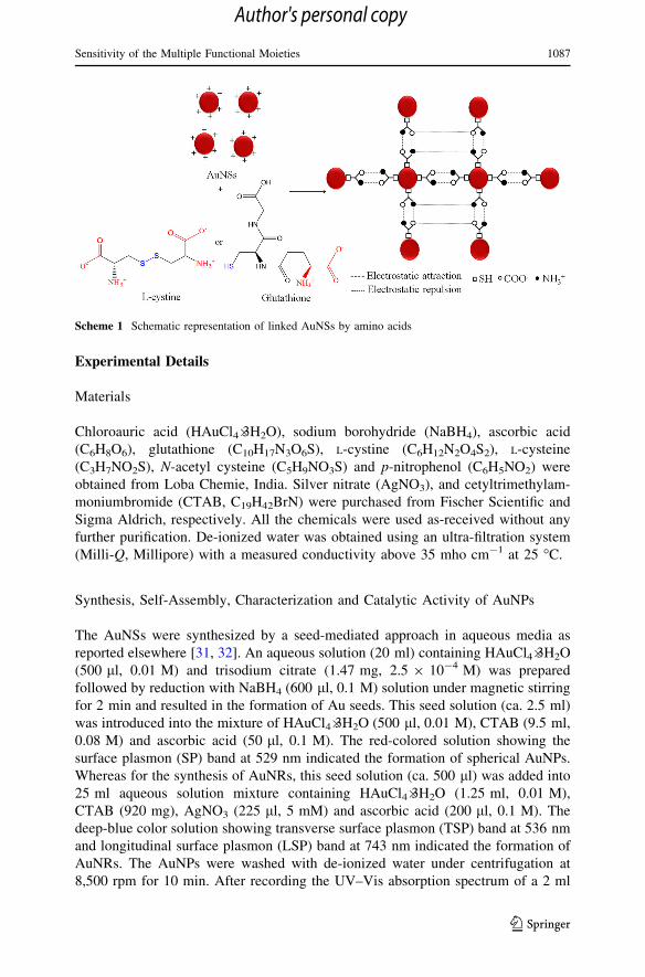
Experimental Details
Materials
Chloroauric acid (HAuCl4�3H2O), sodium borohydride (NaBH4), ascorbic acid
(C6H8O6), glutathione (C10H17N3O6S), L-cystine (C6H12N2O4S2), L-cysteine
(C3H7NO2S), N-acetyl cysteine (C5H9NO3S) and p-nitrophenol (C6H5NO2) were
obtained from Loba Chemie, India. Silver nitrate (AgNO3), and cetyltrimethylam-
moniumbromide (CTAB, C19H42BrN) were purchased from Fischer Scientific and
Sigma Aldrich, respectively. All the chemicals were used as-received without any
further purification. De-ionized water was obtained using an ultra-filtration system
(Milli-Q, Millipore) with a measured conductivity above 35 mho cm-1 at 25 �C.
Synthesis, Self-Assembly, Characterization and Catalytic Activity of AuNPs
The AuNSs were synthesized by a seed-mediated approach in aqueous media as
reported elsewhere [31, 32]. An aqueous solution (20 ml) containing HAuCl4�3H2O
(500 ll, 0.01 M) and trisodium citrate (1.47 mg, 2.5 9 10-4 M) was prepared
followed by reduction with NaBH4 (600 ll, 0.1 M) solution under magnetic stirring
for 2 min and resulted in the formation of Au seeds. This seed solution (ca. 2.5 ml)
was introduced into the mixture of HAuCl4�3H2O (500 ll, 0.01 M), CTAB (9.5 ml,
0.08 M) and ascorbic acid (50 ll, 0.1 M). The red-colored solution showing the
surface plasmon (SP) band at 529 nm indicated the formation of spherical AuNPs.
Whereas for the synthesis of AuNRs, this seed solution (ca. 500 ll) was added into
25 ml aqueous solution mixture containing HAuCl4�3H2O (1.25 ml, 0.01 M),
CTAB (920 mg), AgNO3 (225 ll, 5 mM) and ascorbic acid (200 ll, 0.1 M). The
deep-blue color solution showing transverse surface plasmon (TSP) band at 536 nm
and longitudinal surface plasmon (LSP) band at 743 nm indicated the formation of
AuNRs. The AuNPs were washed with de-ionized water under centrifugation at
8,500 rpm for 10 min. After recording the UV–Vis absorption spectrum of a 2 ml
Scheme 1 Schematic representation of linked AuNSs by amino acids
Sensitivity of the Multiple Functional Moieties 1087
123
Author's personal copy
(6.72 9 1017 atoms, see electronic supplementary info) of prepared AuNPs in
quartz cell, a certain amount of (100 ll, 100 mM) L-cystine (L-cys), glutathione
(glut), L-cysteine (cyt) and N-acetyl cysteine (N-acyt) were added into the AuNPs
solution and SP band absorption was recorded after a regular interval of time. The
obtained Au nanostructures capped with various amino acids were characterized by
UV–Vis absorption (Analytic Jena specord-205) spectrophotometer and TEM
(Hitachi 7500, 2 A, 120 kV). Fourier transform infrared (FTIR) spectra of pure
amino acid (i.e., glut) and glut–AuNS was recorded with Agilent Cary 630 FTIR
spectrometer. The solution containing 2 ml AuNSs (6.72 9 1017 atoms) and 100 ll
of amino acids (100 mM) were taken in a cuvette for the zeta potential and DLS
measurements by Brookhaven 7610 instruments. The catalytic reduction of
p-nitrophenol (PNP) was carried out by adding an ice-cold NaBH4 solution
(500 ll, 0.42 M) to PNP (5 ml, 0.2 mM), and stirred for 5 min at room temperature.
Then, the calculated amount of AuNSs [20 ll, 6.72 9 1015 atoms, ESI-(1)] was
added to initiate PNP reduction to the p-aminophenol (PAP) formation by
measuring the absorption spectra (kmax of PNP * 400 nm and PAP * 300 nm)
at regular intervals of time. The GC–MS analysis of reduction products was
conducted with Bruker GC-45X with Scion MS system equipped with RTX-5 MS
Sil column (15 m 9 0.25 mm 9 0.25 mm) and NMR spectrum was also taken with
Bruker Avance-II with a frequency of 400 MHz for 1H detection.
Results and Discussions
Optical Properties of Amino Acid Modified Au Nanostructures
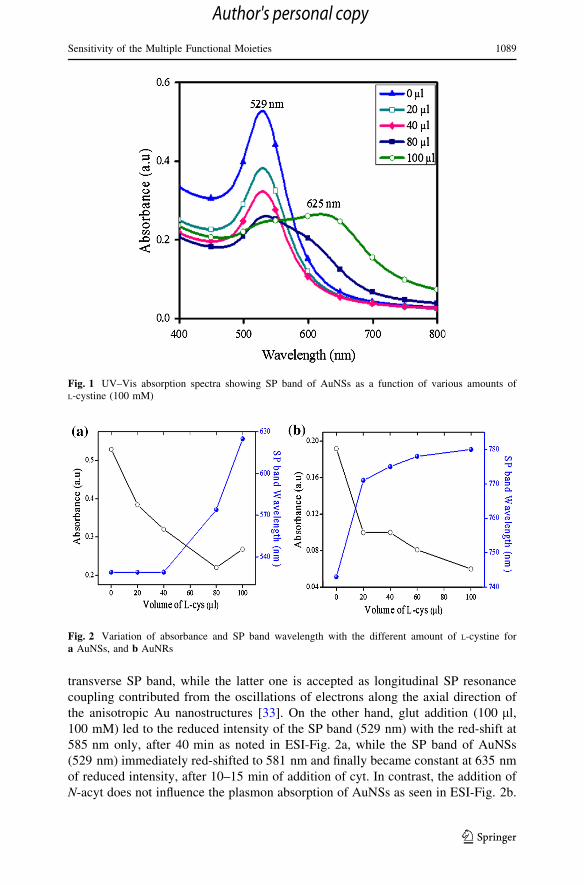
The effect of different amount (20–100 ll) of L-cys (100 mM) addition into an
aqueous suspension of AuNSs and AuNRs was studied at a regular interval of time.
The intensity of the SP absorption band of AuNSs at 529 nm is gradually decreased
with increasing amount of L-cys and is red-shifted to the longer wavelength
(625 nm) at higher concentration of L-cys (100 ll) as seen in Fig. 1. Similarly, in
AuNRs, the LSP band is red-shifted from 743 to 780 nm with decreased intensity on
adding a higher amount of L-cys, whereas the transverse band with reduced
absorption intensity is remained at 526 nm as shown in ESI-Fig. 1. The kinetics of
AuNSs and AuNRs surface passivation with the different amount of L-cys
(100 mM) clearly revealed (Fig. 2) that low concentration (20–60 ll) of L-cys
addition does not show any significant change in SP band except a linear decrease in
its intensity; however, beyond 80–100 ll, there is a remarkably bathochromic shift.
The variation in SP band absorbance and red-shifting arises in opposite trend
probably due to the electromagnetic coupling of SP electrons of adjacent AuNPs
linked by –SH, –COOH and –NH2 moieties of L-cys. As a result, the AuNPs exhibit
increased scattering and broadened SP peaks towards longer wavelengths.
Figure 3 shows the time course assembly of AuNSs with a fixed amount (100 ll,
100 mM) of L-cys, where the intensity of SP band (529 nm) is gradually reduced
with the simultaneous evolution of a new flat band at 643 nm, which shifted to
724 nm after 20 min of L-cys addition. The former band corresponds to the
1088 A. Monga, B. Pal
123
Author's personal copy
transverse SP band, while the latter one is accepted as longitudinal SP resonance
coupling contributed from the oscillations of electrons along the axial direction of
the anisotropic Au nanostructures [33]. On the other hand, glut addition (100 ll,
100 mM) led to the reduced intensity of the SP band (529 nm) with the red-shift at
585 nm only, after 40 min as noted in ESI-Fig. 2a, while the SP band of AuNSs
(529 nm) immediately red-shifted to 581 nm and finally became constant at 635 nm
of reduced intensity, after 10–15 min of addition of cyt. In contrast, the addition of
N-acyt does not influence the plasmon absorption of AuNSs as seen in ESI-Fig. 2b.
Fig. 1 UV–Vis absorption spectra showing SP band of AuNSs as a function of various amounts ofL-cystine (100 mM)
Fig. 2 Variation of absorbance and SP band wavelength with the different amount of L-cystine fora AuNSs, and b AuNRs
Sensitivity of the Multiple Functional Moieties 1089
123
Author's personal copy
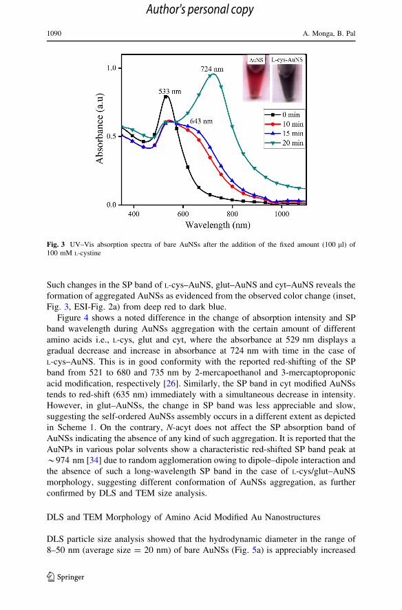
Such changes in the SP band of L-cys–AuNS, glut–AuNS and cyt–AuNS reveals the
formation of aggregated AuNSs as evidenced from the observed color change (inset,
Fig. 3, ESI-Fig. 2a) from deep red to dark blue.
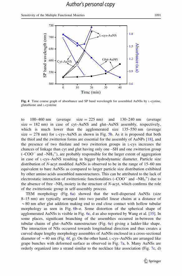
Figure 4 shows a noted difference in the change of absorption intensity and SP
band wavelength during AuNSs aggregation with the certain amount of different
amino acids i.e., L-cys, glut and cyt, where the absorbance at 529 nm displays a
gradual decrease and increase in absorbance at 724 nm with time in the case of
L-cys–AuNS. This is in good conformity with the reported red-shifting of the SP
band from 521 to 680 and 735 nm by 2-mercapoethanol and 3-mercaptoproponic
acid modification, respectively [26]. Similarly, the SP band in cyt modified AuNSs
tends to red-shift (635 nm) immediately with a simultaneous decrease in intensity.
However, in glut–AuNSs, the change in SP band was less appreciable and slow,
suggesting the self-ordered AuNSs assembly occurs in a different extent as depicted
in Scheme 1. On the contrary, N-acyt does not affect the SP absorption band of
AuNSs indicating the absence of any kind of such aggregation. It is reported that the
AuNPs in various polar solvents show a characteristic red-shifted SP band peak at
*974 nm [34] due to random agglomeration owing to dipole–dipole interaction and
the absence of such a long-wavelength SP band in the case of L-cys/glut–AuNS
morphology, suggesting different conformation of AuNSs aggregation, as further
confirmed by DLS and TEM size analysis.
DLS and TEM Morphology of Amino Acid Modified Au Nanostructures
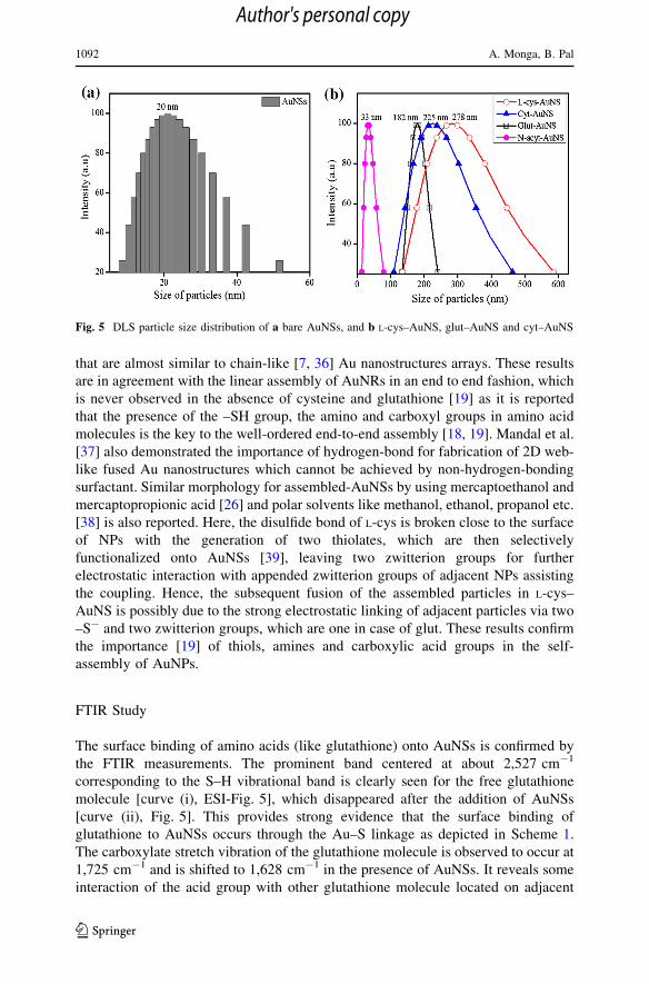
DLS particle size analysis showed that the hydrodynamic diameter in the range of
8–50 nm (average size = 20 nm) of bare AuNSs (Fig. 5a) is appreciably increased
Fig. 3 UV–Vis absorption spectra of bare AuNSs after the addition of the fixed amount (100 ll) of100 mM L-cystine
1090 A. Monga, B. Pal
123
Author's personal copy
to 100–460 nm (average size = 225 nm) and 130–240 nm (average
size = 182 nm) in case of cyt–AuNS and glut–AuNS assembly, respectively,
which is much lower than the agglomerated size 135–550 nm (average
size = 278 nm) for L-cys–AuNS as shown in Fig. 5b. As it is proposed that both
the thiol and the zwitterion forms are essential for the assembly of AuNPs [18], and
the presence of two thiolate and two zwitterion groups in L-cys increases the
chances of linkage than cyt and glut having only one –SH and one zwitterion group
(–COO- and –NH3?), are probably responsible for the larger extent of aggregation
in case of L-cys–AuNS resulting in bigger hydrodynamic diameter. Particle size
distribution of N-acyt modified AuNSs is observed to be in the range of 15–60 nm
equivalent to bare AuNSs as compared to larger particle size distribution exhibited
by other amino acids assembled nanostructures. This can be attributed to the lack of
electrostatic interaction of zwitterionic functionalities (–COO- and –NH3?) due to
the absence of free –NH2 moiety in the structure of N-acyt, which confirms the role
of the zwitterionic group in self-assembly process.
TEM morphology (Fig. 6a) showed that the well-dispersed AuNSs (size
8–15 nm) are typically arranged into two parallel linear chains at a distance of
*80 nm after glut addition making end to end close contact with hollow tubular
morphology as seen in Fig. 6b–e. Some distortion of the spherical shape of
agglomerated AuNSs is visible in Fig. 6c, d as also reported by Wang et al. [35]. In
some places, significant branching of the assemblies occurred in-between the
tubular chains of glut–AuNSs nanostructure (Fig. 6e) giving a ladder-like shape.
The interaction of NSs occurred towards longitudinal direction and thus creates a
curved shape lengthy morphology assembles of AuNSs enclosed in a cross-sectional
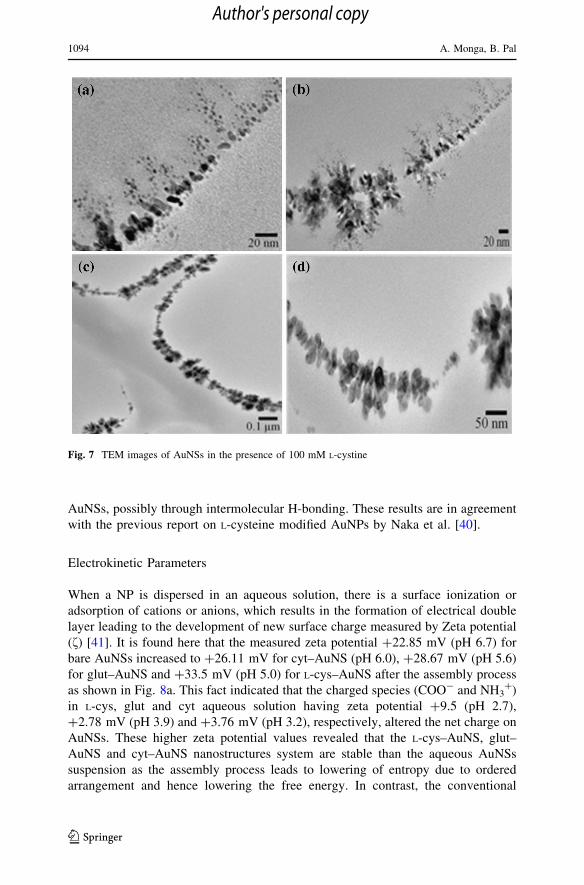
diameter of *40 nm (Fig. 6f, g). On the other hand, L-cys–AuNSs are arranged like
grape bunches with deformed surface as observed in Fig. 7a, b. Many AuNSs are
orderly organized into a strand similar to the necklace like association (Fig. 7c, d)
Fig. 4 Time course graph of absorbance and SP band wavelength for assembled AuNSs by L-cystine,glutathione and L-cysteine
Sensitivity of the Multiple Functional Moieties 1091
123
Author's personal copy
that are almost similar to chain-like [7, 36] Au nanostructures arrays. These results
are in agreement with the linear assembly of AuNRs in an end to end fashion, which
is never observed in the absence of cysteine and glutathione [19] as it is reported
that the presence of the –SH group, the amino and carboxyl groups in amino acid
molecules is the key to the well-ordered end-to-end assembly [18, 19]. Mandal et al.
[37] also demonstrated the importance of hydrogen-bond for fabrication of 2D web-
like fused Au nanostructures which cannot be achieved by non-hydrogen-bonding
surfactant. Similar morphology for assembled-AuNSs by using mercaptoethanol and
mercaptopropionic acid [26] and polar solvents like methanol, ethanol, propanol etc.
[38] is also reported. Here, the disulfide bond of L-cys is broken close to the surface
of NPs with the generation of two thiolates, which are then selectively
functionalized onto AuNSs [39], leaving two zwitterion groups for further
electrostatic interaction with appended zwitterion groups of adjacent NPs assisting
the coupling. Hence, the subsequent fusion of the assembled particles in L-cys–
AuNS is possibly due to the strong electrostatic linking of adjacent particles via two
–S- and two zwitterion groups, which are one in case of glut. These results confirm
the importance [19] of thiols, amines and carboxylic acid groups in the self-
assembly of AuNPs.
FTIR Study
The surface binding of amino acids (like glutathione) onto AuNSs is confirmed by
the FTIR measurements. The prominent band centered at about 2,527 cm-1
corresponding to the S–H vibrational band is clearly seen for the free glutathione
molecule [curve (i), ESI-Fig. 5], which disappeared after the addition of AuNSs
[curve (ii), Fig. 5]. This provides strong evidence that the surface binding of
glutathione to AuNSs occurs through the Au–S linkage as depicted in Scheme 1.
The carboxylate stretch vibration of the glutathione molecule is observed to occur at
1,725 cm-1 and is shifted to 1,628 cm-1 in the presence of AuNSs. It reveals some
interaction of the acid group with other glutathione molecule located on adjacent
Fig. 5 DLS particle size distribution of a bare AuNSs, and b L-cys–AuNS, glut–AuNS and cyt–AuNS
1092 A. Monga, B. Pal
123
Author's personal copy
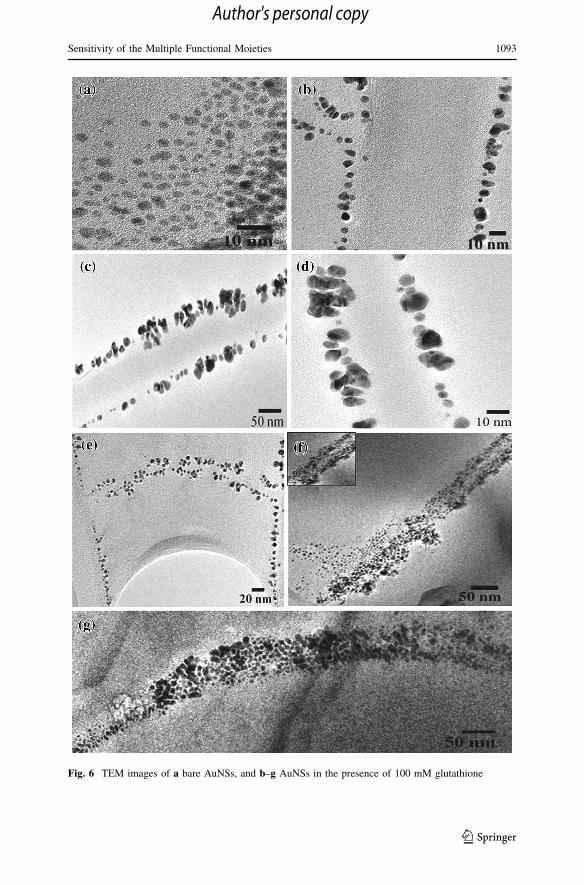
Fig. 6 TEM images of a bare AuNSs, and b–g AuNSs in the presence of 100 mM glutathione
Sensitivity of the Multiple Functional Moieties 1093
123
Author's personal copy
AuNSs, possibly through intermolecular H-bonding. These results are in agreement
with the previous report on L-cysteine modified AuNPs by Naka et al. [40].
Electrokinetic Parameters
When a NP is dispersed in an aqueous solution, there is a surface ionization or
adsorption of cations or anions, which results in the formation of electrical double
layer leading to the development of new surface charge measured by Zeta potential
(f) [41]. It is found here that the measured zeta potential ?22.85 mV (pH 6.7) for
bare AuNSs increased to ?26.11 mV for cyt–AuNS (pH 6.0), ?28.67 mV (pH 5.6)
for glut–AuNS and ?33.5 mV (pH 5.0) for L-cys–AuNS after the assembly process
as shown in Fig. 8a. This fact indicated that the charged species (COO- and NH3?)
in L-cys, glut and cyt aqueous solution having zeta potential ?9.5 (pH 2.7),
?2.78 mV (pH 3.9) and ?3.76 mV (pH 3.2), respectively, altered the net charge on
AuNSs. These higher zeta potential values revealed that the L-cys–AuNS, glut–
AuNS and cyt–AuNS nanostructures system are stable than the aqueous AuNSs
suspension as the assembly process leads to lowering of entropy due to ordered
arrangement and hence lowering the free energy. In contrast, the conventional
Fig. 7 TEM images of AuNSs in the presence of 100 mM L-cystine
1094 A. Monga, B. Pal
123
Author's personal copy
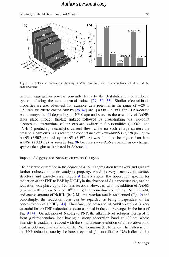
random aggregation process generally leads to the destabilization of colloidal
system reducing the zeta potential values [29, 30, 33]. Similar electrokinetic
properties are also observed; for example, zeta potential in the range of -29 to
-50 mV for citrate coated AuNPs [26, 42] and ?49 to ?71 mV for CTAB-coated
Au nanocrystals [6] depending on NP shape and size. As the assembly of AuNPs
takes place through thiolate linkage followed by cross-linking via two-point
electrostatic interactions of the exposed zwitterion functionalities (–COO- and
–NH3?) producing electrolytic current flow, while no such charge carriers are
present in bare ones. As a result, the conductance of L-cys–AuNS (22,728 lS), glut–
AuNS (5,902 lS) and cyt–AuNS (5,597 lS) was found to be higher than bare
AuNSs (2,323 lS) as seen in Fig. 8b because L-cys–AuNS contain more charged
species than glut as indicated in Scheme 1.
Impact of Aggregated Nanostructures on Catalysis
The observed difference in the degree of AuNPs aggregation from L-cys and glut are
further reflected in their catalysis property, which is very sensitive to surface
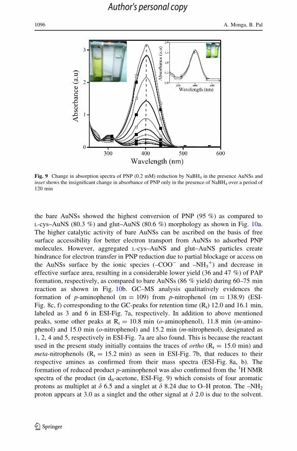
structure and particle size. Figure 9 (inset) shows the absorption spectra for
reduction of the PNP to PAP by NaBH4 in the absence of Au nanostructures, and no
reduction took place up to 120 min reaction. However, with the addition of AuNSs
(size = 8–10 nm, ca. 6.72 9 1015 atoms) to this mixture containing PNP (0.2 mM)
and excess amount of NaBH4 (0.42 M), the reaction rate is accelerated (Fig. 9) and
accordingly, the reduction rates can be regarded as being independent of the
concentration of NaBH4 [43]. Therefore, the presence of AuNPs catalyst is very
essential for the PNP reduction to occur as noted in the color changes in the inset of
Fig. 9 [44]. On addition of NaBH4 to PNP, the alkalinity of solution increased to
form p-nitrophenolate ions having a strong absorption band at 400 nm whose
intensity is gradually reduced with the simultaneous evolution of a new absorption
peak at 300 nm, characteristic of the PAP formation (ESI-Fig. 6). The difference in
the PNP reduction rate by the bare, L-cys and glut modified-AuNSs indicated that
Fig. 8 Electrokinetic parameters showing a Zeta potential, and b conductance of different Aunanostructures
Sensitivity of the Multiple Functional Moieties 1095
123
Author's personal copy
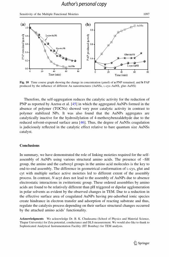
the bare AuNSs showed the highest conversion of PNP (95 %) as compared to
L-cys–AuNS (80.3 %) and glut–AuNS (80.6 %) morphology as shown in Fig. 10a.
The higher catalytic activity of bare AuNSs can be ascribed on the basis of free
surface accessibility for better electron transport from AuNSs to adsorbed PNP
molecules. However, aggregated L-cys–AuNS and glut–AuNS particles create
hindrance for electron transfer in PNP reduction due to partial blockage or access on
the AuNSs surface by the ionic species (–COO- and –NH3?) and decrease in
effective surface area, resulting in a considerable lower yield (36 and 47 %) of PAP
formation, respectively, as compared to bare AuNSs (86 % yield) during 60–75 min
reaction as shown in Fig. 10b. GC–MS analysis qualitatively evidences the
formation of p-aminophenol (m = 109) from p-nitrophenol (m = 138.9) (ESI-
Fig. 8c, f) corresponding to the GC-peaks for retention time (Rt) 12.0 and 16.1 min,
labeled as 3 and 6 in ESI-Fig. 7a, respectively. In addition to above mentioned
peaks, some other peaks at Rt = 10.8 min (o-aminophenol), 11.8 min (m-amino-
phenol) and 15.0 min (o-nitrophenol) and 15.2 min (m-nitrophenol), designated as
1, 2, 4 and 5, respectively in ESI-Fig. 7a are also found. This is because the reactant
used in the present study initially contains the traces of ortho (Rt = 15.0 min) and
meta-nitrophenols (Rt = 15.2 min) as seen in ESI-Fig. 7b, that reduces to their
respective amines as confirmed from their mass spectra (ESI-Fig. 8a, b). The
formation of reduced product p-aminophenol was also confirmed from the 1H NMR
spectra of the product (in d6-acetone, ESI-Fig. 9) which consists of four aromatic
protons as multiplet at d 6.5 and a singlet at d 8.24 due to O–H proton. The –NH2
proton appears at 3.0 as a singlet and the other signal at d 2.0 is due to the solvent.
Fig. 9 Change in absorption spectra of PNP (0.2 mM) reduction by NaBH4 in the presence AuNSs andinset shows the insignificant change in absorbance of PNP only in the presence of NaBH4 over a period of120 min
1096 A. Monga, B. Pal
123
Author's personal copy
Therefore, the self-aggregation reduces the catalytic activity for the reduction of
PNP as reported by Azetsu et al. [45] in which the aggregated AuNPs formed in the
absence of polymer (TOCNs) showed very poor catalytic activity in contrast to
polymer stabilized NPs. It was also found that the AuNPs aggregates are
catalytically inactive for the hydrosilylation of 4-methoxybenzaldehyde due to the
reduced solvent-exposed surface area [46]. Thus, the degree of AuNSs coagulation
is judiciously reflected in the catalytic effect relative to bare quantum size AuNSs
catalyst.
Conclusions
In summary, we have demonstrated the role of linking moieties required for the self-
assembly of AuNPs using various structural amino acids. The presence of –SH
group, the amino and the carboxyl groups in the amino acid molecules is the key to
end-to-end assembly. The difference in geometrical conformation of L-cys, glut and
cyt with multiple surface active moieties led to different extent of the assembly
process. In contrast, N-acyt does not lead to the assembly of AuNPs due to absence
electrostatic interactions in zwitterionic group. These ordered assemblies by amino
acids are found to be relatively different than pH triggered or dipolar agglomeration
in polar solvents as evident by the observed changes in TEM. Due to a reduction in
the effective surface area of coagulated AuNPs having pre-adsorbed ionic species
create hindrance in electron transfer and adsorption of reacting substrate and thus,
regulate the catalysis process depending on their surface structural changes occurred
by the attached amino acids’ functionality.
Acknowledgments We acknowledge Dr. B. K. Chudasama (School of Physics and Material Science,
Thapar University) for Zeta potential, conductance and DLS measurement. We would also like to thank to
Sophisticated Analytical Instrumentation Facility (IIT Bombay) for TEM analysis.
Fig. 10 Time course graph showing the change in concentration (lmol) of a PNP remained, and b PAPproduced by the influence of different Au nanostructures (AuNSs, L-cys–AuNS, glut–AuNS)
Sensitivity of the Multiple Functional Moieties 1097
123
Author's personal copy
References
1. G. M. Whitesides and M. Boncheva (2002). Proc. Natl Acad. Sci. USA 99, 4769.
2. M. Grzelczak, J. Vermant, E. M. Furst, and L. M. Liz-Marzan (2010). ACS Nano 4, 3591.
3. G. M. Whitesides and B. Grzybowski (2002). Science 295, 2418.
4. K. J. M. Bishop, C. E. Wilmer, S. Soh, and B. A. Grzybowski (2009). Small 5, 1600.
5. T. S. Sreeprasad and T. Pradeep (2011). Langmuir 27, 3381.
6. T. K. Sau and C. J. Murphy (2005). Langmuir 21, 2923.
7. G. Kawamura, Y. Yang, and M. Nogami (2008). J. Phys. Chem. C 112, 10632.
8. K. G. Thomas, S. Barazzouk, B. I. Ipe, S. T. S. Joseph, and P. V. Kamat (2004). J. Phys. Chem. B
108, 13066.
9. M. A. El-Sayed (2001). Acc. Chem. Res. 34, 257.
10. S. Nie and S. R. Emory (1997). Science 275, 1102.
11. Z. L. Wang (2000). J. Phys. Chem. B 104, 1153.
12. B. M. I. van der Zande, M. R. Boehmer, L. G. J. Fokkink, and C. Schoenenberger (2000). Langmuir
16, 451.
13. B. Nikoobakht, Z. L. Wang, and M. A. El-Sayed (2000). J. Phys. Chem. B 104, 8635.
14. X. Hu, W. Cheng, T. Wang, E. Wang, and S. Dong (2005). Nanotechnology 16, 2164.
15. I. W. Hamley (2003). Angew. Chem. Int. Ed. 42, 1692.
16. H. S. Park, A. Agarwal, N. A. Kotov, and O. D. Lavrentovich (2008). Langmuir 24, 13833.
17. H. Yan, S. H. Park, G. Finkelstein, J. H. Reif, and T. H. LaBean (2003). Science 301, 1882.
18. S. Zhang, X. Kou, Z. Yang, Q. Shi, G. D. Stucky, L. Sun, J. Wang and C. Yan (2007). Chem.
Commun. 1816.
19. P. K. Sudeep, S. T. S. Joseph, and K. G. Thomas (2005). J. Am. Chem. Soc. 127, 6516.
20. O. P. Khatri, K. Murase, and H. Sugimura (2008). Langmuir 24, 3787.
21. F. P. Zamborini, J. F. Hicks, and R. W. Murray (2000). J. Am. Chem. Soc. 122, 4514.
22. A. Sanchez-Iglesias, M. Grzelczak, J. Perez-Juste, and L. M. Liz-Marzan (2010). Angew. Chem. Int.
Ed. 49, 9985.
23. M. Sethi, G. Joung, and M. R. Knecht (2009). Langmuir 25, 1572.
24. X. Kou, S. Zhang, Z. Yang, C. K. Tsung, G. D. Stucky, L. Sun, J. Wang, and C. Yan (2007). J. Am.
Chem. Soc. 129, 6402.
25. J. Liao, Y. Zhang, W. Yu, L. Xu, C. Ge, J. Liu, and N. Gu (2003). Colloids Surf., A. 223, 177.
26. T. Sen and A. Patra (2009). J. Phys. Chem. C 113, 13125.
27. C. A. Mirkin, R. L. Letsinger, R. C. Mucic, and J. J. Storhoff (1996). Nature 382, 607.
28. B. Mukherjee and J. W. Weaver (2010). Environ. Sci. Technol. 44, 3332.
29. T. Kim, K. Lee, M. Gong, and S. W. Joo (2005). Langmuir 21, 9524.
30. C. J. Orendorff, P. L. Hankins, and C. J. Murphy (2005). Langmuir 21, 2022.
31. N. R. Jana, L. Gearheart, and C. J. Murphy (2001). Langmuir 17, 6782.
32. M. Eguchi, D. Mitsui, H. L. Wu, R. Sato, and T. Teranishi (2012). Langmuir 28, 9021.
33. D. F. Zhang, Q. Zhang, L. Y. Niu, L. Jiang, P. G. Yin, and L. Guo (2011). J. Nanopart. Res. 13, 3923.
34. R. Kaur and B. Pal (2012). J. Mol. Catal. A: Chem. 355, 39.
35. T. Wang, X. Hu, and S. Dong (2008). Chem. Commun. 4625.
36. A. N. Shipway, M. Lahav, R. Gabai, and I. Willner (2000). Langmuir 16, 8789.
37. S. Mandal, A. Shundo, S. Acharya, J. P. Hill, Q. Ji, and K. Ariga (2009). Chem. Asian J. 4, 1055.
38. R. Kaur and B. Pal (2014). Colloids and Surfaces A: Physicochem. Eng. Aspects 441, 155.
39. A. Ulman (1996). Chem. Rev. 96, 1533.
40. K. Naka, H. Itoh, Y. Tampo, and Y. Chujo (2003). Langmuir 19, 5546.
41. J. A. Davis, R. O. James, and J. O. Leckie (1978). J. Colloid Interface Sci. 63, 480.
42. K. Suttiponparnit, J. Jiang, M. Sahu, S. Suvachittanont, T. Charinpanitkul, and P. Biswas (2011).
Nanoscale Res. Lett. 6, 1.
43. K. Kuroda, T. Ishida, and M. Haruta (2009). J. Mol. Catal. A Chem. 298, 7.
44. S. Wunde, F. Polzer, Y. Lu, Y. Mei, and M. Ballauff (2010). J. Phys. Chem. C 114, 8814.
45. A. Azetsu, H. Koga, A. Isogai, and T. Kitaoka (2011). Catalysts 1, 83.
46. Y. Wei, S. Han, J. Kim, S. Soh, and B. A. Grzybowski (2010). J. Am. Chem. Soc. 132, 11018.
1098 A. Monga, B. Pal
123
Author's personal copy
Related Documents


![Contents Colloids and Surfaces A: Physicochemical and ...suspensions of solid nanoparticles (NPs) in a solvent are employed in a wide range of technological applications [1]. The properties](https://static.cupdf.com/doc/110x72/5f8372abb06fae482253c6c0/contents-colloids-and-surfaces-a-physicochemical-and-suspensions-of-solid-nanoparticles.jpg)
