Mutation Research 620 (2007) 103–113 Sensitivity of different endpoints for in vitro measurement of genotoxicity of extractable organic matter associated with ambient airborne particles (PM 10 ) Alena G´ abelov´ a a,∗ , Zuzana Valoviˇ cov´ a a , Gabriela Baˇ cov´ a a , Juraj L´ abaj a , Blanka Binkov´ a b , Jan Topinka b , Oksana Sevastyanova b , Radim J. ˇ Sr´ am b , Ivan Kalina c , Viera Habalov´ a c , Todor A. Popov d , Teodor Panev d , Peter B. Farmer e a Laboratory of Mutagenesis and Carcinogenesis, Cancer Research Institute SAS, Bratislava, Slovakia b Laboratory of Genetic Ecotoxicology, Institute of Experimental Medicine AS CR and Health Institute of Central Bohemia, Prague, Czech Republic c Department of Medical Biology, Medical Faculty, University of P.J. ˇ Saf´ arik, Koˇ sice, Slovakia d Department of Toxicology, National Center of Public Health Protection, Sofia, Bulgaria e Cancer Biomarkers and Prevention Group, Biocentre, University of Leicester, UK Available online 7 March 2007 Abstract Sensitivity and correlations among three endpoints were evaluated to assess the genotoxic potential of organic complex mixtures in vitro. This study was focused on DNA adduct formation, DNA single strand break induction and tumour suppressor p53 protein up- regulation produced by extractable organic matter (EOM) absorbed on respirable particulate matter PM 10 (particulate matter < 10 m) collected in three European cities (Prague, Sofia, Koˇ sice) during winter and summer period. To compare the sensitivity of particular endpoints for in vitro measurement of complex mixture genotoxicity, the metabolically competent human hepatoma cell line Hep G2 was treated with equivalent EOM concentration of 50 g/ml. Cell exposure to EOMs resulted in significant DNA adduct formation and DNA strand break induction, however, a lack of protein p53 up-regulation over the steady-state level was found. While the maximum of DNA strand breaks was determined after 2 h cell exposure to EOMs, 24 h treatment interval was optimal for DNA adduct determination. No substantial location- and season-related differences in EOM genotoxicity were detected using DNA strand break assessment. In agreement with these results no significant variation in DNA adduct levels were found in relation to the locality and season except for the monitoring site in Prague. The Prague EOM sample collected during summer period produced nearly three-fold lower DNA adduct level in comparison to the winter EOM sample. Comparable results were obtained when the ambient air genotoxicity, based on the concentration of carcinogenic PAHs in cubic meter of air (ng c-PAHs/m 3 ), was elicited using either DNA adduct or strand break determination. In general, at least six-fold higher genotoxicity of the winter air in comparison to the summer air was estimated by each particular endpoint. Moreover, the genotoxic potential of winter air revealed by DNA adduct assessment and DNA strand break measurement increased in the same order: Koˇ sice Prague < Sofia. Based on these data we suppose that two endpoints DNA breakage and DNA adduction are sensitive in vitro biomarkers for estimation of genotoxic activity of organic complex mixture associated with airborne particles. On the other hand, the measurement ∗ Corresponding author. Tel.: +421 259327512; fax: +421 259327506. E-mail address: [email protected] (A. G´ abelov´ a). 0027-5107/$ – see front matter © 2007 Elsevier B.V. All rights reserved. doi:10.1016/j.mrfmmm.2007.02.026

Welcome message from author
This document is posted to help you gain knowledge. Please leave a comment to let me know what you think about it! Share it to your friends and learn new things together.
Transcript

A
vrcewama
Ifa
mhgo
e
0
Mutation Research 620 (2007) 103–113
Sensitivity of different endpoints for in vitro measurement ofgenotoxicity of extractable organic matter associated
with ambient airborne particles (PM10)
Alena Gabelova a,∗, Zuzana Valovicova a, Gabriela Bacova a, Juraj Labaj a,Blanka Binkova b, Jan Topinka b, Oksana Sevastyanova b, Radim J. Sram b,
Ivan Kalina c, Viera Habalova c, Todor A. Popov d,Teodor Panev d, Peter B. Farmer e
a Laboratory of Mutagenesis and Carcinogenesis, Cancer Research Institute SAS, Bratislava, Slovakiab Laboratory of Genetic Ecotoxicology, Institute of Experimental Medicine AS CR and
Health Institute of Central Bohemia, Prague, Czech Republicc Department of Medical Biology, Medical Faculty, University of P.J. Safarik, Kosice, Slovakia
d Department of Toxicology, National Center of Public Health Protection, Sofia, Bulgariae Cancer Biomarkers and Prevention Group, Biocentre, University of Leicester, UK
Available online 7 March 2007
bstract
Sensitivity and correlations among three endpoints were evaluated to assess the genotoxic potential of organic complex mixtures initro. This study was focused on DNA adduct formation, DNA single strand break induction and tumour suppressor p53 protein up-egulation produced by extractable organic matter (EOM) absorbed on respirable particulate matter PM10 (particulate matter < 10 �m)ollected in three European cities (Prague, Sofia, Kosice) during winter and summer period. To compare the sensitivity of particularndpoints for in vitro measurement of complex mixture genotoxicity, the metabolically competent human hepatoma cell line Hep G2as treated with equivalent EOM concentration of 50 �g/ml. Cell exposure to EOMs resulted in significant DNA adduct formation
nd DNA strand break induction, however, a lack of protein p53 up-regulation over the steady-state level was found. While theaximum of DNA strand breaks was determined after 2 h cell exposure to EOMs, 24 h treatment interval was optimal for DNA
dduct determination.No substantial location- and season-related differences in EOM genotoxicity were detected using DNA strand break assessment.
n agreement with these results no significant variation in DNA adduct levels were found in relation to the locality and season exceptor the monitoring site in Prague. The Prague EOM sample collected during summer period produced nearly three-fold lower DNAdduct level in comparison to the winter EOM sample.
Comparable results were obtained when the ambient air genotoxicity, based on the concentration of carcinogenic PAHs in cubiceter of air (ng c-PAHs/m3), was elicited using either DNA adduct or strand break determination. In general, at least six-fold
igher genotoxicity of the winter air in comparison to the summer air was estimated by each particular endpoint. Moreover, theenotoxic potential of winter air revealed by DNA adduct assessment and DNA strand break measurement increased in the samerder: Kosice � Prague < Sofia.
Based on these data we suppose that two endpoints DNA breakage and DNA adduction are sensitive in vitro biomarkers forstimation of genotoxic activity of organic complex mixture associated with airborne particles. On the other hand, the measurement
∗ Corresponding author. Tel.: +421 259327512; fax: +421 259327506.E-mail address: [email protected] (A. Gabelova).
027-5107/$ – see front matter © 2007 Elsevier B.V. All rights reserved.doi:10.1016/j.mrfmmm.2007.02.026
104 A. Gabelova et al. / Mutation Research 620 (2007) 103–113
of protein p53 up-regulation manifested some limitations; therefore it cannot be used as a reliable endpoint for in vitro genotoxicityassessment.© 2007 Elsevier B.V. All rights reserved.
ocarbon
Keywords: Particulate matter; Carcinogenic polycyclic aromatic hydr1. Introduction
The in vitro short-term genotoxicity assays forman important part of risk assessment of potential car-cinogens. They are widely accepted around the worldas useful tools to reduce the occurrence of hazardouscompounds in the environment. Around 100 differenttypes of short-term tests on prokaryotic and eukaryoticorganisms have so far been developed to predict thepotential health hazard of new chemicals. Various end-points including gene mutations, clastogenic effects (e.g.chromosomal rearrangements, DNA breakage), DNAadducts or diverse cellular responses (e.g. DNA repair,apoptosis) are employed as biomarkers of genotoxic-ity. Among these endpoints, DNA adduct and DNAstrand break formation have been proposed as standardbiomarkers of DNA damage expression [1].
DNA adducts resulting from covalent binding ofchemicals to DNA are a critical event in the initiation ofcancer. Associations have been observed between DNAadduct formation, and mutagenesis [2], and tumorigene-sis [3]. Various analytical methods have been developedto detect covalent DNA adducts including immunoas-says, fluorescence and phosphorescence spectroscopy,mass spectrometry and 32P-postlabelling [4]. The 32P-postlabelling assay has been widely applied because ofits high sensitivity and the requirement for only smallamounts of DNA. This technique is able to detect a broadspectrum of hydrophobic compounds bound to DNA[5,6]; with some modifications, this method is addition-ally able to determine apurinic (AP) sites in DNA andoxidative DNA damage [7]. The 32P postlabelling assayis especially useful for detection of adducts produced bysingle (individual) chemicals [8,9] but it is frequentlyemployed also in human biomonitoring studies [10,11].Compared with other methods for DNA adduct analy-ses the 32P postlabelling assay has a unique potency fordetection of DNA adducts from complex mixtures withunknown structures [12–14].
DNA strand breaks are readily detected as cell-response to exposure; breaks are induced directly by the
agent or as a consequence of DNA damage removal in theprocess of DNA repair. A lot of biochemical techniqueshave been developed for DNA strand break estimation,based on different mechanisms and performed under dif-s; DNA adduct; Single strand break; In vitro genotoxicity
ferent conditions [15]. Among them, the single cell gelelectrophoresis (SCGE), known as the comet assay, isa sensitive, rapid and inexpensive fluorescence micro-scopic method measuring DNA damage at the level of asingle cell [16]. Depending on the experimental condi-tions, this technique is able to detect a broad spectrumof DNA damage including single and double strandbreaks, alkali labile sites, DNA–DNA/DNA–proteincross-linking and strand breaks associated with incom-plete excision repair [17,18]. In addition, the applicationof repair specific DNA endonucleases enables detectionof oxidative DNA damage and other base modificationsinduced by xenobiotics [19]. This technique has beensuccessfully used to estimate the genotoxic potential ofvarious chemical compounds [20–23] as well as organiccomplex mixtures [14,24–26].
The tumour suppressor protein p53 plays a key rolein maintaining genomic stability and homeostasis. Thisprotein regulates the expression of its downstream effec-tor genes, whose products are implicated in criticalcellular processes such as DNA repair [27], cell cyclecontrol [28] and apoptosis [29]. Induction of this pro-tein has been determined as the cellular response togenotoxic stress induced by multiple chemical DNAdamaging agents, ionizing or UV radiation [30,31]. Anup-regulation of p53 protein is triggered by damageaccumulating in DNA due to exposure to genotoxins[32,33]. Based on this phenomenon, Yang and Duerksen-Hughes [34] proposed the measurement of p53 inductionas a new promising in vitro approach for identifying thegenotoxic carcinogens.
A valuable tool for detection of compounds whichmight pose a health hazard to man are human-derivedmetabolically competent cell lines such as the humanhepatoma cell line Hep G2. This cell line has retainedcertain activities of various phase I and phase IIenzymes [22,35] which play a key role in the activation/detoxification of various promutagens/procarcinogens.The capacity of Hep G2 cells to detect the genotoxicityof both direct and indirect carcinogens has already beenwell documented [21,23].
The goal of this work, which is part of an EC-funded study [36], was to evaluate the sensitivity andcorrelation among three in vitro endpoints, DNA adductformation, DNA strand break induction and p53 protein

on Rese
ucePPSsts[
2
2
pNia1
2
soubFwepmap0ppbbsHrAs
2c
t(c[
A. Gabelova et al. / Mutati
p-regulation, to assess the genotoxic potential of theomplex mixture of organic compounds. EOMs werextracted uniformly from respirable particulate matterM10 (Ø < 10 �m) collected in three European cities:rague (Czech Republic), Kosice (Slovak Republic) andofia (Bulgaria) during the winter and summer sea-ons. All monitoring sites are classified according tohe European Environmental Register of monitoringtations as commercial, residential, traffic and urban37].
. Materials and methods
.1. Cell line
The human hepatoma cell line Hep G2 was generouslyrovided by Prof. Andrew R. Collins (University of Oslo,orway). Hep G2 cells were maintained in William’s mod-
fied medium supplemented with 10% fetal calf serum andntibiotics (penicillin 200 U/ml; streptomycin and kanamycin00 �g/ml) in humidified 5% CO2 atmosphere.
.2. Chemicals
B[a]P (CAS No. 50-32-8) was purchased from Sigma. Thetock solutions of B[a]P (2 mM in DMSO) and extractablerganic mixture (EOM, 50 mg/ml in DMSO) from individ-al samplings were kept at −20 ◦C and diluted immediatelyefore use. All chemicals used for cell cultivation (MEM,CS, antibiotics) and agarose LMP (low melting point)ere purchased from GIBCO BRL (Paisley, UK); agarose
lectrophoresis grade (NMP, normal melting point), spleenhosphodiesterase from ICN Biomedicals Inc., ethidium bro-ide, RNases A and T1, proteinase K, micrococcal nuclease
nd nuclease P1 from Sigma (Deisenhofen, Germany); T4olynucleotide kinase from USB (Cleveland, OH, USA).1 mm polyethylene-imine cellulose thin-layer chromatogra-hy (TLC) plates from Macherey-Nagel (Duren, Germany),rimary antibodies Ab-6 (clone DO-1) and p53 Westernlotting standard from Oncogene Research Products (Cam-ridge, MA, USA), secondary anti-mouse IgG (NA931),treptavidin–horseradish peroxidase conjugate (RPN1231),ybondTM C-pure membranes, chemiluminiscence detection
eagents and �-32P-ATP (3000 Ci/mmol, 10 �Ci/�l) frommersham (Buckinghamshire, UK). All other chemicals and
olvents were of HPLC or analytical grade.
.3. Air samples collection, extraction of EOM andhemical analysis
Details about the air particles PM10 (particulate mat-er < 10 �m) collection, extractions of organic complex mixtureEOM) from the filters with PM10 samples and the chemi-al analyses of PAHs have been already described elsewhere24,37]. The concentrations of selected PAHs, including
arch 620 (2007) 103–113 105
carcinogenic PAHs (c-PAH) in each organic extract were deter-mined by an HPLC method with fluorimetric detection.
2.4. Treatment of human cells
Human hepatoma cells Hep G2 were exposed to EOMsand positive control after reaching semi-confluency. The stocksolutions of individual EOMs (50 mg/ml) were diluted indimethylsulfoxide (DMSO) freshly before use, and addedto serum free medium or medium with 1% FCS in depen-dence of the treatment interval, to reach the final concentration50 �g/ml. The concentration of DMSO never exceeded 0.5%.Hep G2 cells were exposed to EOMs for 2 h (the comet assay)or for 24 h (DNA adduct detection and p53 induction). Con-trol cells (negative control) were exposed to 0.5% DMSO.The treatment was finished by rinsing off the cells twice withPBS buffer. Then, the cells were harvested and used for SCGE(the comet assay) or cells were re-suspended in 3 ml PBS anddivided into two aliquots (2 ml for DNA isolation; 1 ml forWestern blotting). Benzo[a]pyrene (BaP, 7.5 �M) was used asa positive control.
2.5. DNA isolation and 32P-postlabelling
The cell pellets were homogenised in a solution of 10 mMTris–HCl, 100 mM EDTA and 0.5% SDS, pH 8.0. DNA wasisolated using RNases A and T1 and proteinase K treatmentfollowed by phenol/chloroform/isoamylalcohol extraction andethanol precipitation [6]. DNA concentrations were estimatedspectrophotometrically by measuring of the UV absorbance at260 nm. DNA samples were stored at −80 ◦C until analysis.
32P-postlabelling analyses were performed as previouslydescribed [37]. Briefly, DNA samples (the exact amount ofDNA was 6 �g) were digested by a mixture of micrococ-cal nuclease and spleen phosphodiesterase for 4 h at 37 ◦C.The nuclease P1 procedure was used instead of the butanolextraction method for adduct enrichment. According to ourprevious experience, no substantial contribution to the totalDNA adduct levels might be expected from amino- and nitro-aromatics since the content of these PAH-derivatives in theextracts from ambient air particles is at least by three orderslower than that of PAHs. Adducted nucleotides were enzymat-ically labelled using �-32P-ATP and T4 polynucleotide kinaseand separated by multidirectional polyethylenimine–celluloseTLC (10 cm × 10 cm). The solvents used were as follows:D1, 1 M sodium phosphate, pH 6.8; D2, 3.8 M lithium for-mate, 8.5 M urea, pH 3.5; D3, 0.8 M lithium chloride, 0.5 MTris, 8.5 M urea, pH 8.0; D4 = D1, same direction as D3.After screen-enhanced autoradiography at −80 ◦C for 24 h, thedistinct DNA adduct spots were cut out and evaluated by mea-suring 32P-radioactivity using liquid scintillation spectroscopy.
To determine the exact amount of DNA in each sample, aliquotsof the enzymatic DNA digests (0.5 �g of DNA hydrolysate)were analyzed for nucleotide content by reverse-phase HPLCwith UV detection, which simultaneously allowed for control-ling the purity of the DNA. DNA adduct levels were expressed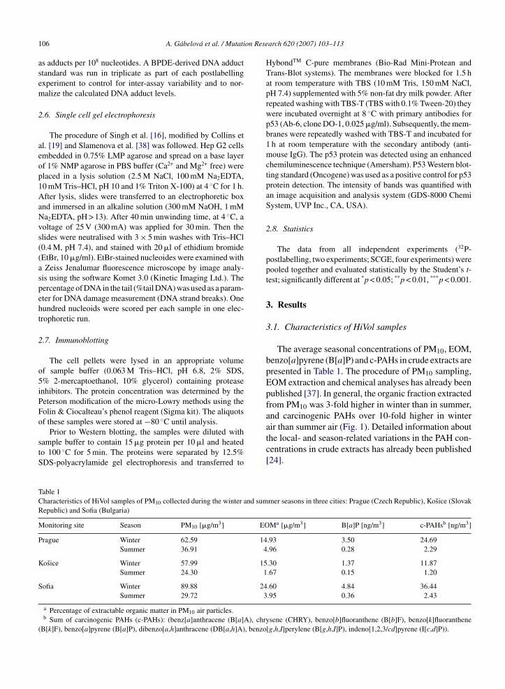
on Rese
106 A. Gabelova et al. / Mutatias adducts per 108 nucleotides. A BPDE-derived DNA adductstandard was run in triplicate as part of each postlabellingexperiment to control for inter-assay variability and to nor-malize the calculated DNA adduct levels.
2.6. Single cell gel electrophoresis
The procedure of Singh et al. [16], modified by Collins etal. [19] and Slamenova et al. [38] was followed. Hep G2 cellsembedded in 0.75% LMP agarose and spread on a base layerof 1% NMP agarose in PBS buffer (Ca2+ and Mg2+ free) wereplaced in a lysis solution (2.5 M NaCl, 100 mM Na2EDTA,10 mM Tris–HCl, pH 10 and 1% Triton X-100) at 4 ◦C for 1 h.After lysis, slides were transferred to an electrophoretic boxand immersed in an alkaline solution (300 mM NaOH, 1 mMNa2EDTA, pH > 13). After 40 min unwinding time, at 4 ◦C, avoltage of 25 V (300 mA) was applied for 30 min. Then theslides were neutralised with 3 × 5 min washes with Tris–HCl(0.4 M, pH 7.4), and stained with 20 �l of ethidium bromide(EtBr, 10 �g/ml). EtBr-stained nucleoides were examined witha Zeiss Jenalumar fluorescence microscope by image analy-sis using the software Komet 3.0 (Kinetic Imaging Ltd.). Thepercentage of DNA in the tail (%tail DNA) was used as a param-eter for DNA damage measurement (DNA strand breaks). Onehundred nucleoids were scored per each sample in one elec-trophoretic run.
2.7. Immunoblotting
The cell pellets were lysed in an appropriate volumeof sample buffer (0.063 M Tris–HCl, pH 6.8, 2% SDS,5% 2-mercaptoethanol, 10% glycerol) containing proteaseinhibitors. The protein concentration was determined by thePeterson modification of the micro-Lowry methods using theFolin & Ciocalteau’s phenol reagent (Sigma kit). The aliquots
◦
of these samples were stored at −80 C until analysis.Prior to Western blotting, the samples were diluted withsample buffer to contain 15 �g protein per 10 �l and heatedto 100 ◦C for 5 min. The proteins were separated by 12.5%SDS-polyacrylamide gel electrophoresis and transferred to
Table 1Characteristics of HiVol samples of PM10 collected during the winter and sumRepublic) and Sofia (Bulgaria)
Monitoring site Season PM10 [�g/m3] EO
Prague Winter 62.59 14Summer 36.91 4
Kosice Winter 57.99 15Summer 24.30 1
Sofia Winter 89.88 24Summer 29.72 3
a Percentage of extractable organic matter in PM10 air particles.b Sum of carcinogenic PAHs (c-PAHs): (benz[a]anthracene (B[a]A), chr
(B[k]F), benzo[a]pyrene (B[a]P), dibenzo[a,h]anthracene (DB[a,h]A), benzo
arch 620 (2007) 103–113
HybondTM C-pure membranes (Bio-Rad Mini-Protean andTrans-Blot systems). The membranes were blocked for 1.5 hat room temperature with TBS (10 mM Tris, 150 mM NaCl,pH 7.4) supplemented with 5% non-fat dry milk powder. Afterrepeated washing with TBS-T (TBS with 0.1% Tween-20) theywere incubated overnight at 8 ◦C with primary antibodies forp53 (Ab-6, clone DO-1, 0.025 �g/ml). Subsequently, the mem-branes were repeatedly washed with TBS-T and incubated for1 h at room temperature with the secondary antibody (anti-mouse IgG). The p53 protein was detected using an enhancedchemiluminescence technique (Amersham). P53 Western blot-ting standard (Oncogene) was used as a positive control for p53protein detection. The intensity of bands was quantified withan image acquisition and analysis system (GDS-8000 ChemiSystem, UVP Inc., CA, USA).
2.8. Statistics
The data from all independent experiments (32P-postlabelling, two experiments; SCGE, four experiments) werepooled together and evaluated statistically by the Student’s t-test; significantly different at *p < 0.05; **p < 0.01, ***p < 0.001.
3. Results
3.1. Characteristics of HiVol samples
The average seasonal concentrations of PM10, EOM,benzo[a]pyrene (B[a]P) and c-PAHs in crude extracts arepresented in Table 1. The procedure of PM10 sampling,EOM extraction and chemical analyses has already beenpublished [37]. In general, the organic fraction extractedfrom PM10 was 3-fold higher in winter than in summer,and carcinogenic PAHs over 10-fold higher in winter
air than summer air (Fig. 1). Detailed information aboutthe local- and season-related variations in the PAH con-centrations in crude extracts has already been published[24].mer seasons in three cities: Prague (Czech Republic), Kosice (Slovak
Ma [�g/m3] B[a]P [ng/m3] c-PAHsb [ng/m3]
.93 3.50 24.69
.96 0.28 2.29
.30 1.37 11.87
.67 0.15 1.20
.60 4.84 36.44
.95 0.36 2.43
ysene (CHRY), benzo[b]fluoranthene (B[b]F), benzo[k]fluoranthene[g,h,I]perylene (B[g,h,I]P), indeno[1,2,3/cd]pyrene (I[c,d]P)).
A. Gabelova et al. / Mutation Research 620 (2007) 103–113 107
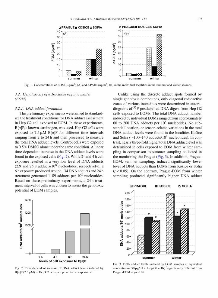
3) (B)
3(
3
iiBertttfe(6tBmp
FB
EOM, summer sampling, induced significantly lowerlevel of DNA adducts than EOMs from Kosice or Sofia(p < 0.05). On the contrary, Prague-EOM from winter
Fig. 1. Concentrations of EOM (�g/m3) (A) and c-PAHs (ng/m
.2. Genotoxicity of extractable organic matterEOM)
.2.1. DNA adduct formationThe preliminary experiments were aimed to standard-
ze the treatment conditions for DNA adduct assessmentn Hep G2 cell exposed to EOM. In these experiments,[a]P, a known carcinogen, was used. Hep G2 cells werexposed to 7.5 �M B[a]P for different time intervalsanging from 2 to 24 h and then processed to measurehe total DNA adduct levels. Control cells were exposedo 0.5% DMSO alone under the same condition. A linearime-dependent increase in the DNA adduct levels wereound in the exposed cells (Fig. 2). While 2- and 4 h cellxposure resulted in a very low level of DNA adducts2.9 and 25.8 adducts/108 nucleotides, respectively), ah exposure produced around 134 DNA adducts and 24 h
reatment generated 1189 adducts per 108 nucleotides.ased on these preliminary experiments, a 24 h treat-ent interval of cells was chosen to assess the genotoxic
otential of EOM samples.
ig. 2. Time-dependent increase of DNA adduct levels induced by[a]P (7.5 �M) in Hep G2 cells; a representative experiment.
in the individual localities in the summer and winter seasons.
Unlike using the discrete adduct spots formed bysingle genotoxic compounds, only diagonal radioactivezones of various intensities were determined in autora-diograms of 32P-postlabelled DNA digest from Hep G2cells exposed to EOMs. The total DNA adduct numberinduced by individual EOMs ranged from approximately60 to 200 DNA adducts per 108 nucleotides. No sub-stantial location- or season-related variations in the totalDNA adduct levels were found in the localities Kosiceand Sofia (∼100–140 adducts/108 nucleotides). In con-trast, nearly three-fold higher total DNA adduct level wasdetermined in cells exposed to EOM from winter sam-pling in comparison to summer sampling collected inthe monitoring site Prague (Fig. 3). In addition, Prague-
sampling produced significantly higher DNA adduct
Fig. 3. DNA adduct levels induced by EOM samples at equivalentconcentration 50 �g/ml in Hep G2 cells; *significantly different fromPrague-EOM at p < 0.05.

108 A. Gabelova et al. / Mutation Research 620 (2007) 103–113
Fig. 4. Time-dependent increase of DNA strand break levels in Hep G2cells exposed to B[a]P (7.5 �M); significantly different from untreatedcells; *p < 0.05, **p < 0.01, ***p < 0.001.
level in comparison to EOMs from the localities Kosiceand Sofia (p < 0.05).
3.2.2. Single cell gel electrophoresisSimilarly as for the DNA adduct experiments the pre-
liminary examinations were focused on standardizationof treatment conditions of Hep G2 cells. Semi-confluentcultures of cells were exposed to B[a]P (7.5 �M) fordifferent time intervals ranging from 20 min to 48 h.Control cells were exposed to 0.5% DMSO alone underthe same condition. A linear time-dependent increase ofDNA strand breaks was determined in B[a]P-treated HepG2 cells within 20–120 min time exposure interval, andthen the strand break level reached a plateau. Approxi-mately the same strand break levels were found after 24and 48 h exposure time interval (Fig. 4); at the same time,however, a notable decrease in cell viability was detected(data not shown). In agreement with this phenomenon,
no significant differences in cell response to EOM treat-ment in concentration range from 5 to 150 �g/ml werefound whether a 2, 24 or 48 h time exposure intervalwas used (Fig. 5A). Nearly the same DNA strand breakFig. 5. DNA strand breaks (A) and cytotoxicity (B) of EOM from Pr
Fig. 6. DNA strand break levels induced by EOM samples at equivalentconcentration 50 �g/ml in Hep G2 cells.
levels were determined in treated cells; however, a sub-stantial reduction of cell viability due to EOM toxicitywas detected at the same time (Fig. 5B).
Based on these preliminary experiments, an expo-sure time of 2 h was chosen to assess the genotoxicpotential of EOMs. Treatment of Hep G2 cells with indi-vidual EOMs resulted in approximately the same levelsof strand breaks expressed as ‘percentage of DNA inthe tail’ (25–30% of tail DNA). No significant local-or season-related differences in DNA strand break lev-els were determined among individual EOM samples(Fig. 6).
3.3. Air genotoxicity
Substantial location- and season-related differencesin the amount of both EOM and c-PAHs were esti-mated (Table 1). The highest c-PAH concentration was
ague, summer sampling after 2, 24 and 48 h of cell exposure.

A. Gabelova et al. / Mutation Research 620 (2007) 103–113 109
Fig. 7. Ambient air pollution genotoxicity expressed in terms of c-PAHs quantity per cubic meter air (ng c-PAH/m3) in individual monitoring sitesaccording to Table 1; comparison of the summer and winter seasons: (A) DNA adduct determination (DNA adducts/m3); (B) DNA strand breakdetermination (%tail DNA/m3).
Fig. 8. p53 protein levels in Hep G2 cells exposed for 24 h to different B[a]P concentrations, 0.1 �M DB[a,l]P (A) and 50 �g EOM/ml collectedi seasonse icrograr tandardp e cond
dtIttilapaaaiastiomTtK
n localities Kosice, Prague and Sofia during the summer and winterxposed cells in a fresh medium as described in Section 2.7. Fifteen mesolved by SDS-PAGE using 12.5% mini gel. P53 Western blotting srotein. (Control) Cells treated with 0.5% DMSO alone under the sam
etected in Sofia, winter sampling (36.44 ng/m3), whilehe lowest one in Kosice, summer sampling (1.2 ng/m3).t is reasonable to suppose that the higher the concen-ration of c-PAHs is in the ambient air, the higher ishe health risk for man. In order to be closer to then vivo situation DNA adduct and DNA strand breakevels were expressed in relative units based on themount of c-PAHs associated with particulate matterer unit volume of air (c-PAH, ng/m3). The ambientir genotoxicity is shown in Fig. 7A (DNA adducts)nd Fig. 7B (DNA strand breaks). Both DNA adductnd DNA strand break determination manifested signif-cantly higher genotoxicity of winter air than summerir. In general, the ambient air genotoxicity was at leastix-fold higher during the winter season as compared tohe summer season. While the summer air genotoxic-ty based on DNA adduct assessment increased in therder Kosice ∼ Prague < Sofia, using DNA strand break
easurement this order was Kosice ∼ Sofia < Prague.he winter air genotoxicity determined by each par-icular endpoint, however, increased in the same order:osice � Prague < Sofia.
(B). The p53 protein level was followed after 24 h postcultivation ofms of total protein from each sample in 10 �l of sample buffer werefrom Oncogene (cat. #WB21) was used as a positive control for p53
ition.
3.4. Expression of p53 protein
The p53 protein levels in EOM-treated cells wereanalyzed after 24 h postcultivation of exposed cells infresh medium. Cells were treated with individual EOMsfor 24 h. Control cells were exposed to 0.5% DMSOalone under the same condition. B[a]P (concentrationsranged from 0.1 to 40 �M) and DB[a,l]P (0.1 �M)were used as positive controls in these experiments.A significant up-regulation of p53 protein levels weredetermined only in cells exposed to B[a]P at concen-trations 7.5–40 �M (Fig. 8A), however, no substantialrise of p53 protein levels were detected in Hep G2 cellsexposed to EOMs regardless of the season and locality(Fig. 8B).
4. Discussion
Environmental air pollution is a matter of great inter-est because millions of people are chronically exposed tolow doses of potentially toxic and genotoxic chemicals,frequently present in the complex mixtures. Numerous
on Rese
110 A. Gabelova et al. / Mutatiepidemiological and experimental studies have con-sistently demonstrated that urban air pollution maybe responsible for increased incidence of respiratoryand cardiovascular mortality and morbidity [39,40],intrauterine growth retardation [41] and increased ratesof lung cancer [42–44]. Nearly 3000 chemical com-pounds have been identified in ambient air [45], manyof which are known or suspected human mutagens andcarcinogens [46,47]. Fractionation of the crude organiccomplex mixtures clearly demonstrated that the highestmutagenic potential is associated with fractions con-taining the PAHs and nitro-PAHs derivatives [37,48,49].Therefore the first step towards lowering the risk of ambi-ent air pollution on human health might be a biologicallybased characterization of complex mixtures using short-term in vitro assays, which are routinely used to measurethe effects of single chemicals. Such an approach allowsdirect measurement of a specific toxicological endpointin a biological system. In this respect, the selection ofreliable and sensitive biomarkers of toxic and genotoxicactivity is very important.
The major objective of this study was to compare thesensitivity and correlations of three endpoints to assessthe genotoxic potential of organic complex mixturesextracted from airborne particles collected in three Euro-pean cities, Prague (Czech Republic), Kosice (SlovakRepublic) and Sofia (Bulgaria) during the summer andwinter seasons. Different approaches, 32P-postlabelling,single cell gel electrophoresis and immunoblotting wereemployed in this study. Three endpoints, DNA adductformation, DNA strand break induction and protein p53up-regulation, were measured in Hep G2 cells exposedto EOMs at identical concentrations of 50 �g/ml.
Based on the preliminary experiments (Figs. 3 and 4),a 24 h cell treatment interval was chosen to assess thegenotoxic potential of EOM samples using the 32P-postlabelling technique and a 2 h time exposure usingthe SCGE. The total DNA adduct levels produced byEOMs ranged from 60 to 180 adducts per 108 nucleotides(Fig. 3); using the same treatment conditions B[a]P(7.5 �M) produced approximately 1200 adducts per 108
nucleotides (Fig. 2). If adjusted for the amount of B[a]P,the dose of 50 �g EOM/ml corresponded to the con-centration ∼0.05 �M B[a]P [50]. Although no local-and season-related differences in EOM genotoxicitywere determined between Kosice and Sofia, Prague-EOM, summer season, produced significantly lowerlevel of DNA adducts than EOMs from Kosice and Sofia
(Fig. 3). Opposite results were determined for EOMsfrom winter sampling; Prague-EOM generated substan-tially more DNA adducts than EOMs from Kosice andSofia (p < 0.05).arch 620 (2007) 103–113
Two-hour cell exposure to EOM resulted in a signif-icant rise of DNA strand breaks in EOM-treated cells(Fig. 6), however, no substantial location- or season-related differences in in EOM genotoxicity were found.In agreement with these results neither Cerna et al.[48] nor Binkova et al. [37,51] and Topinka et al. [49]found any significant differences in EOM genotoxicityin dependence on the locality and season. A lack of time-dependent increase of strand breaks due to B[a]P or EOMexposure (Figs. 4 and 5A) might be explained by sat-uration of DNA repair mechanisms involved in DNAdamage incision and removal at longer time treatmentintervals. This assumption is supported by the fact thata time-dependent increase of DNA strand breaks wasfound up to 120 min of cell exposure to B[a]P and thenthe strand break level reached a plateau (Fig. 4). Underthe same treatment conditions, a 2 and 4 h cell exposureto B[a]P resulted in very low levels of DNA adducts (2.9and 25.8 adducts/108 nucleotides, respectively) prob-ably due to efficient removal of damage from DNA.Extension of treatment intervals to 24 and 48 h mightlead to depletion of DNA repair enzymes which allowedaccumulation of damage in DNA. A time-dependentincrease of DNA adduct levels with a maximum after24 h exposure interval was found in B[a]P-treated cells(Fig. 2). The DNA damage accumulation in treated cellsresulted subsequently in decreased cell viability, a phe-nomenon observed in both B[a]P- and EOM-treated cells(Fig. 5B). However, other explanations including a satu-ration of activating enzymes or competition of individualcomponents (genotoxic and non-genotoxic) for the bio-transformation enzymes cannot be excluded.
Western blot analyses did not demonstrate any up-regulation of protein p53 due to cell exposure to EOMs(Fig. 8B). Lack of protein p53 induction over the basallevel due to exposure to artificial and environmentalmixtures was reported also by Binkova and Sram [50].Experiments with model carcinogens have suggestedthat a minimal DNA adduct level of ∼200 adducts per108 nucleotides is required to detect up-regulation ofp53 protein [9]. The DNA adduct levels determined inexposed Hep G2 cells did not reach these values; there-fore it is reasonable to suppose that the levels of p53protein in treated cells were below the limit of detection.
A very good correlation of ambient air genotoxicitywas found using DNA adduct determination and DNAstrand break measurement (Fig. 7A and B). Evaluationof the concentration of c-PAHs (ng/m3) in individual
monitoring sites was taken into account (Table 1). Over10-fold higher ambient air genotoxicity was determinedin winter air than summer air using both endpoints. Thisfinding is in agreement with observation of Topinka et
on Rese
aamaiaaibaso
pectbccoedsersscopDEldritwTrrPmDdd
batu
[
[
A. Gabelova et al. / Mutati
l. [49] reporting 10-fold seasonal variability of ambientir genotoxicity in different localities in Czech Republiceasured by DNA adduct analysis using primary rat hep-
tocytes. Moreover, quantitatively comparable seasonalncrease in DNA adduct levels found in Hep G2 cellsnd primary rat hepatocytes indicate that Hep G2 cellsre a relevant surrogate for primary hepatocyte culturesn terms of metabolic capacity. Finally, we found thatoth biomarkers of exposure used in this study (DNAdduct and strand break measurements) exhibited theame order of winter air genotoxicity which rose in therder: Kosice � Prague < Sofia.
The assessment of mutagenic/carcinogenic hazardsosed by organic complex mixtures of anthropogenicmissions is not a simple task. In contrast to a singlehemical, mixtures are likely to exhibit various interac-ions (additive or synergistic) that cannot be predictedy the concentration of any individual component. Aomprehensive assessment of genotoxic potential of theomplex organic mixtures requires the use of a batteryf reliable and sensitive assays measuring different rel-vant endpoints. Results obtained in this study clearlyemonstrated that both DNA adduct formation and DNAtrand break estimation are sensitive in vitro biomark-rs of complex mixture genotoxicity contributing to theisk assessment of ambient air pollution. However, onehould keep in mind that both methods as used in thistudy are not specific enough to identify the signifi-ance of individual components in the final genotoxicityf the complex environmental mixtures. While the 32P-ostlabelling technique measures the amount of covalentNA adducts (P1 nuclease modification) produced byOMs, the SCGE assay estimates DNA strand break
evels produced as a consequence of DNA repair orue to spontaneous release of unstable DNA adductsesulting in alkali labile sites. Based on this fact, thendividual approaches provide complementary informa-ion about the total genotoxic potency of the mixtures,hich is basic information to identify carcinogenic risk.he assessment of tumour suppressor protein p53 up-
egulation is a less efficient biomarker of genotoxicityequiring probably a threshold extent of DNA damage.rotein p53 up-regulation could be caused by severalechanisms including those which are independent fromNA damage induction, e.g. by inhibition of p53 degra-ation via ubiquitin pathway [52] or ribonucleotidylepletion [53].
The in vitro short-term assays such as DNA adducts
y 32P-postlabeling and DNA strand breaks by SCGEnalyses provide a valuable tool to evaluate the geno-oxic potential of complex mixtures. These assays can besed not only to assess the impact of air pollution on the[
arch 620 (2007) 103–113 111
human health but they contribute to a better understand-ing of the mechanisms underlying air pollution inducedadverse effects on biological systems.
Acknowledgements
The authors wish to thank Prof. Andrew R. Collinswho kindly offered the human hepatoma HepG2 cellsand Mrs. Alzbeta Vokalikova for an excellent technicalassistance.
The authors acknowledge the European Commission‘Quality of life and management of living resources’ pro-gramme (QLK4-CT-2000-00091) for financial support.
References
[1] C. Andreoli, D. Gigante, A. Nunziata, A review of in vitro meth-ods to assess the biological activity of tobacco smoke with theaim of reducing the toxicity of smoke, Toxicol. In Vitro 17 (2003)587–594.
[2] K. Hemminki, DNA adducts, mutations and cancer, Carcinogen-esis 14 (1993) 2007–2012.
[3] M. Otteneder, W.K. Lutz, Correlation of DNA adduct levels withtumor incidence: carcinogenic potency of DNA adducts, Mutat.Res. 424 (1999) 237–247.
[4] M.C. Poirier, R.M. Santella, A. Weston, Carcinogen macromolec-ular adducts and their measurement, Carcinogenesis 21 (2000)353–359.
[5] K. Randerath, E. Randerath, H.P. Agrawal, R.C. Gupta, M.E.Schurdak, M.V. Reddy, Postlabeling methods for carcinogen–DNA adduct analysis, Env. Health Perspect. 62 (1985) 57–65.
[6] R.C. Gupta, Enhanced sensitivity of 32P-postlabeling analysisof aromatic carcinogen:DNA adducts, Cancer Res. 45 (1985)5656–5662.
[7] D.H. Phillips, Detection of DNA modifications by the P-32-postlabelling assay, Mutat. Res. 378 (1997) 1–12.
[8] D. Renault, D. Brault, Y. Lossouarn, O. Perin-Roussel, D. Taras-Valero, F. Perin, V. Thybaud, Kinetics of DNA adduct formationand removal in mouse hepatocytes following in vivo exposureto 5,9-dimethyldibenzo[c,g]carbazole, Carcinogenesis 21 (2000)289–294.
[9] B. Binkova, Y. Giguere, P. Rossner Jr., M. Dostal, R.J. Sram,The effect of dibenzo[a,l]pyrene and benzo[a]pyrene on humandiploid lung fibroblasts: the induction of DNA adducts, expressionof p53 and p21(WAF1) proteins and cell cycle distribution, Mutat.Res. 471 (2000) 57–70.
10] R.J. Sram, B. Binkova, Molecular epidemiology studies onoccupational and environmental exposure to mutagens and car-cinogens, 1997–1999, Env. Health Perspect. 108 (Suppl. 1) (2000)57–70.
11] R.W. Godschalk, F.J. Van Schooten, H. Bartsch, A critical evalu-ation of DNA adducts as biological markers for human exposureto polycyclic aromatic compounds, J. Biochem. Mol. Biol. 36
(2003) 1–11.12] D.H. Phillips, P.B. Farmer, F.A. Beland, R.G. Nath, M.C.Poirier, M.V. Reddy, K.W. Turteltaub, Methods of DNA adductdetermination and their application to testing compounds forgenotoxicity, Env. Mol. Mutagen. 35 (2000) 222–233.

on Rese
[
[
[
[
[
[
[
[
[
[
[
[
[
[
[
[
[
[
[
[
[
[
[
[
[
[
[
[
[
[
112 A. Gabelova et al. / Mutati
13] H.L. Eriksson, M. Zeisig, L.G. Ekstrom, L. Moller, P-32-postlabeling of DNA adducts arising from complex mixtures:HPLC versus TLC separation applied to adducts from petroleumproducts, Arch. Toxicol. 78 (2004) 174–181.
14] F. Akcha, H.F. Vincent, A. Pfhol-Leszkowicz, Potential value ofthe comet assay and DNA adduct measurement in dab (Limandalimanda) for assessment of in situ exposure to genotoxic com-pounds, Mutat. Res. 534 (2003) 21–32.
15] A. Gabelova, D. Slamenova, L. Ruzekova, T. Farkasova, E.Horvathova, Measurement of DNA strand breakage and DNArepair induced with hydrogen peroxide using single cell gel elec-trophoresis, alkaline DNA unwinding and alkaline elution ofDNA, Neoplasma 44 (1997) 380–388.
16] N.P. Singh, M.T. McCoy, R.R. Tice, E.L. Schneider, A simpletechnique for low levels of DNA damage in individual cells, Exp.Cell Res. 175 (1988) 184–191.
17] R.R. Tice, E. Agurell, D. Anderson, B. Burlinson, A. Hartmann,H. Kobayashi, Y. Miyamae, E. Rojas, J.C. Ryu, Y.F. Sasaki, Singlecell gel/comet assay: guidelines for in vitro and in vivo genetictoxicology testing, Env. Mol. Mutagen. 35 (2000) 206–221.
18] A.R. Collins, The comet assay for DNA damage and repair: prin-ciples, applications, and limitations, Mol. Biotechnol. 26 (2004)249–261.
19] A.R. Collins, S.J. Duthie, V.L. Dobson, Direct enzymic detec-tion of endogenous oxidative base damage in human lymphocyteDNA, Carcinogenesis 14 (1993) 1733–1735.
20] S. Robichova, D. Slamenova, Study of N-nitrosomorpholine-induced DNA strand breaks in Caco-2 cells by the classical andmodified comet assay: influence of Vitamins E and C, Nutr. Can-cer 39 (2001) 267–272.
21] S. Knasmuller, W. Parzefall, R. Sanyal, S. Ecker, C. Schwab,M. Uhl, V. Mersch-Sundermann, G. Williamson, G. Hietsch, T.Langer, F. Darroudi, A.T. Natarajan, Use of metabolically com-petent human hepatoma cells for the detection of mutagens andantimutagens, Mutat. Res. 402 (1998) 185–202.
22] S. Knasmuller, V. Mersch-Sundermann, S. Kevekordes, F. Dar-roudi, W.W. Huber, C. Hoelzl, J. Bichler, B.J. Majer, Use ofhuman-derived liver cell lines for the detection of environmentaland dietary genotoxicants; current state of knowledge, Toxicology198 (2004) 315–328.
23] M. Uhl, C. Helma, S. Knasmuller, Evaluation of the single cellgel electrophoresis assay with human hepatoma (Hep G2) cells,Mutat. Res. 468 (2000) 213–225.
24] A. Gabelova, Z. Valovicova, E. Horvathova, D. Slamenova, B.Binkova, R.J. Sram, P.B. Farmer, Genotoxicity of environmentalair pollution in three European cities: Prague, Kosice and Sofia,Mutat. Res. 563 (2004) 49–59.
25] M. Lazarova, D. Slamenova, Genotoxic effects of a complex mix-ture adsorbed onto ambient air particles on human cells in vitro;the effects of Vitamins E and C, Mutat. Res. 557 (2004) 167–175.
26] C.A. Don Porto, P.H. Hoet, L. Verschaeve, G. Schoeters, B.Nemery, Genotoxic effects of carbon black particles, dieselexhaust particles, and urban air particulates and their extracts on ahuman alveolar epithelial cell line (A549) and a human monocyticcell line (THP-1), Env. Mol. Mutagen. 37 (2001) 155–163.
27] Q.E. Wang, Q.Z. Zhu, M.A. Wani, G. Wani, J.M. Chen, A.A.
Wani, Tumor suppressor p53 dependent recruitment of nucleotideexcision repair factors XPC and TFIIH to DNA damage, DNARepair 2 (2003) 483–499.28] L.H. Hartwell, M.B. Kastan, Cell cycle control and cancer, Sci-ence 266 (1994) 1821–1828.
[
arch 620 (2007) 103–113
29] S.W. Lowe, H.E. Ruley, T. Jacks, D.E. Housman, p53-dependentapoptosis modulates the cytotoxicity of anticancer agents, Cell74 (1993) 957–967.
30] M.B. Kastan, Q. Zhan, W.S. El Deiry, F. Carrier, T. Jacks, W.V.Walsh, B.S. Plunkett, B. Vogelstein, A.J. Fornace Jr., A mam-malian cell cycle checkpoint pathway utilizing p53 and GADD45is defective in ataxia-telangiectasia, Cell 71 (1992) 587–597.
31] Q. Zhan, U. Kontny, M. Iglesias, I. Alamo Jr., K. Yu, M.C. Hol-lander, C.D. Woodworth, A.J. Fornace Jr., Inhibitory effect ofBcl-2 on p53-mediated transactivation following genotoxic stress,Oncogene 18 (1999) 297–304.
32] R. Mirzayans, S. Bashir, D. Murray, M.C. Paterson, Inverse cor-relation between p53 protein levels and DNA repair efficiencyin human fibroblast strains treated with 4-nitroquinoline 1-oxide:evidence that lesions other than DNA strand breaks trigger thep53 response, Carcinogenesis 20 (1999) 941–946.
33] W.G. Nelson, M.B. Kastan, DNA strand breaks: the DNAtemplate alterations that trigger p53-dependent DNA damageresponse pathways, Mol. Cell. Biol. 14 (1994) 1815–1823.
34] J. Yang, P. Duerksen-Hughes, A new approach to identifyinggenotoxic carcinogens: p53 induction as an indicator of genotoxicdamage, Carcinogenesis 19 (1998) 1117–1125.
35] J. Rueff, C. Chiapella, J.K. Chipman, F. Darroudi, I.D. Silva, M.Duvergervanbogaert, E. Fonti, H.R. Glatt, P. Isern, A. Laires, A.Leonard, M. Llagostera, P. Mossesso, A.T. Natarajan, F. Palitti,A.S. Rodrigues, A. Schinoppi, G. Turchi, G. Werleschneider,Development and validation of alternative metabolic systems formutagenicity testing in short-term assays, Mutat. Res. 353 (1996)151–176.
36] P.B. Farmer, R. Singh, B. Kaur, R.J. Sram, B. Binkova, I. Kalina,T.A. Popov, S. Garte, E. Taioli, A. Gabelova, A. Cebulska-Wasilewska, Molecular epidemiology studies of carcinogenicenvironmental pollutants. Effects of polycyclic aromatic hydro-carbons (PAHs) in environmental pollution on exogenous andoxidative DNA damage, Mutat. Res. 544 (2003) 397–402.
37] B. Binkova, M. Cerna, A. Pastorkova, R. Jelinek, I. Benes, J.Novak, R.J. Sram, Biological activities of organic compoundsadsorbed onto ambient air particles: comparison between thecities of Teplice and Prague during the summer and winter seasons2000–2001, Mutat. Res. 525 (2003) 43–59.
38] D. Slamenova, A. Gabelova, L. Ruzekova, I. Chalupa, E. Hor-vathova, T. Farkasova, E. Bozsakyova, R. Stetina, Detection ofMNNG-induced DNA lesions in mammalian cells; validation ofcomet assay against DNA unwinding technique, alkaline elutionof DNA and chromosomal aberrations, Mutat. Res. 383 (1997)243–252.
39] N. Kunzli, I.B. Tager, Long-term health effects of particulate andother ambient air pollution: research can progress faster if wewant it to, Env. Health Perspect. 108 (2000) 915–918.
40] C.A. Pope III, R.T. Burnett, M.J. Thun, E.E. Calle, D. Krewski,K. Ito, G.D. Thurston, Lung cancer, cardiopulmonary mortality,and long-term exposure to fine particulate air pollution, JAMA287 (2002) 1132–1141.
41] J. Dejmek, I. Solansky, I. Benes, J. Lenicek, R.J. Sram, The impactof polycyclic aromatic hydrocarbons and fine particles on preg-nancy outcome, Env. Health Perspect. 108 (2000) 1159–1164.
42] F. Nyberg, P. Gustavsson, L. Jarup, T. Bellander, N. Berglind, R.
Jakobsson, G. Pershagen, Urban air pollution and lung cancer inStockholm, Epidemiology 11 (2000) 487–495.43] X. Zhao, Z. Wan, H. Zhu, R. Chen, The carcinogenic potentialof extractable organic matter from urban airborne particles inShanghai, China, Mutat. Res. 540 (2003) 107–117.
on Rese
[
[
[
[
[
[
[
[
[
A. Gabelova et al. / Mutati
44] P.M. Cury, A.J. Lichtenfels, M.S. Reymao, G.M. Conceicao, V.L.Capelozzi, P.H. Saldiva, Urban levels of air pollution modifies theprogression of urethane-induced lung tumours in mice, Pathol.Res. Pract. 196 (2000) 627–633.
45] J. Lewtas, Complex mixtures of air pollutants: characterizing thecancer risk of polycyclic organic matter, Env. Health Perspect.100 (1993) 211–218.
46] IARC, Polynuclear Aromatic Compounds, International Agencyfor Research into Cancer, Monogr. Eval. Carcinog. Risk Chem.Man (32), IARC, Lyon, France, 1983.
47] IARC, Certain Polycyclic Aromatic Hydrocarbons and Hete-rocyclic Compounds, International Agency for Research intoCancer, Monogr. Eval. Carcinog. Risk Chem. Man IARC(3),IACR, Lyon, France, 1973.
48] M. Cerna, D. Pochmanova, A. Pastorkova, I. Benes, J. Lenicek,J. Topinka, B. Binkova, Genotoxicity of urban air pollutants inthe Czech Republic. Part I. Bacterial mutagenic potencies oforganic compounds adsorbed on PM10 particulates, Mutat. Res.469 (2000) 71–82.
[
arch 620 (2007) 103–113 113
49] J. Topinka, L.R. Schwarz, F.J. Wiebel, M. Cerna, T. Wolff, Geno-toxicity of urban air pollutants in the Czech Republic. Part II.DNA adduct formation in mammalian cells by extractable organicmatter, Mutat. Res. 469 (2000) 83–93.
50] B. Binkova, R.J. Sram, The genotoxic effect of carcino-genic PAHs, their artificial and environmental mixtures (EOM)on human diploid lung fibroblasts, Mutat. Res. 547 (2004)109–121.
51] B. Binkova, D. Vesely, D. Vesela, R. Jelinek, R.J. Sram, Genotox-icity and embryotoxicity of urban air particulate matter collectedduring winter and summer period in two different districts of theCzech Republic, Mutat. Res. 440 (1999) 45–58.
52] U.G. Lopes, P. Erhardt, R.J. Yao, G.M. Cooper, P53-dependentinduction of apoptosis by proteasome inhibitors, J. Biol. Chem.
272 (1997) 12893–12896.53] S.P. Linke, K.C. Clarkin, A. Di Leonardo, A. Tsou, G.M. Wahl,A reversible, p53-dependent G0/G1 cell cycle arrest induced byribonucleotide depletion in the absence of detectable DNA dam-age, Genes Dev. 10 (1996) 934–947.
Related Documents