Molecular and Biochemical Parasitology, 33 (1989) 205-214 Elsevier 205 MBP 01107 Sensitive detection and schizodeme classification of Trypanosoma cruzi cells by amplification of kinetoplast minicircle DNA sequences: use in diagnosis of Chagas' disease Nancy R. Sturm', Wim Degrave^, Carlos Morel- and Larry Simpson' ^Department of Biology and Molecular Biology Institute, University of California, Los Angeles, CA, U.S.A. 'Department of • Biochemistry and Molecular Biology, Oswaldo Cruz Institute, FIOCRUZ, Rio de Janeiro, Brazil (Received 12 September 1988; accepted 21 October 1988) . Amplification of DNA sequences from the kinetoplast minicircle DNA was employed as a method for the detection and clas- sification of small numbers of Trypanosoma cruzi cells. Two overlapping fragments from the conserved 120 bp minirepeat regions of the minicircle DNA and one fragment covering the adjacent variable regions were amplified. The minimal amount of minicircle DNA required to detect a product by hybridization with an oligonucleotide probe was 0.015 fg. which represents approximately 10 molecules or 0.1% of the minicircle DNA component of a single cell. The amplification worked equally well with kDNA from several strains of T. cruzi and did not occur with kDNA from several other kinetoplastids. kDNA recovered from less than 10 trypanosomes in whole blood could be used as a template for amplification; the presence of a several billion fold excess of human DNA had no effect on the amplification process. Schizodeme analysis by hybridization with specific oligonucleotides or by direct restriction enzyme digestion could be performed on the amplified fragments representing the minicircle conserved region or var- iable regions. This method should prove useful as a rapid, specific and sensitive assay for Chagas' disease in chronic patients as well as for epidemiological studies of infected animals and insects. Key words: Trypanosoma cruzi; Minirepeat; Polymerase chain reaction; Minicircle DNA; Schizodeme Introduction Chagas' disease affects 12 million people in the Americas, with approximately 32 million people at risk of infection, and there is no generally use- ful chemotherapeutic treatment or vaccine. The disease is caused by the kinetoplastid protozoan, Trypanosoma cruzi, which is transmitted cycli- cally by insects of the Reduviid family or via blood transfusion from an infected donor. In the mam- malian host, after an initial acute phase, the par- asitemia may lapse into a long-lasting chronic phase, with ensuing multisymptomatic and poly- morphic syndromes. Due to the low abundance Correspondence address: L. Simpson, Department of Biol- ogy and Molecular Biology Institute, University of California, Los Angeles, CA 90024, U.S.A. Abbreviations: kDNA, kinetoplast DNA; PCR. polymerase chain reaction; SDS, sodium dodecylsulfatc. of circulating trypomastigotes in the blood, the most reliable method of direct parasitological de- tection, of this infection in the chronic state is xe- nodiagnosis [1], which entails allowing uninfected laboratory triatomids to take blood meals from the patient, and requires an incubation time of several weeks. Other methods include axenic cul- ture of blood and infection of susceptible labo- ratory animals. Identification and classification of the parasite into zymodemes [2,3] or schizo- demes [4] requires analysis of isoenzyme patterns and kinetoplast minicircle DNA restriction en- zyme digestion profiles. Both methods require more than lO** cells, which are obtained by out- growth of the parasites. However, the outgrowth of parasites in insects, culture or animals may in- cur selection of particular strains present in the original population, thus raising the possibility that the results obtained do not accurately reflect the original population of parasites in the patient [5,6]. . 0166-6851/89/$03.50 © 1989 Elsevier Science Publishers B.V. (Biomedical Division)

Welcome message from author
This document is posted to help you gain knowledge. Please leave a comment to let me know what you think about it! Share it to your friends and learn new things together.
Transcript
Molecular and Biochemical Parasitology, 33 (1989) 205-214 Elsevier
205
M B P 01107
Sensitive detection and schizodeme classification of Trypanosoma cruzi cells by amplification of kinetoplast minicircle DNA sequences: use in
diagnosis of Chagas' disease
Nancy R. Sturm', Wim Degrave^, Carlos Morel- and Larry Simpson' ^Department of Biology and Molecular Biology Institute, University of California, Los Angeles, CA, U.S.A. 'Department of
• Biochemistry and Molecular Biology, Oswaldo Cruz Institute, FIOCRUZ, Rio de Janeiro, Brazil
(Received 12 September 1988; accepted 21 October 1988)
. Amplification of DNA sequences from the kinetoplast minicircle DNA was employed as a method for the detection and classification of small numbers of Trypanosoma cruzi cells. Two overlapping fragments from the conserved 120 bp minirepeat regions of the minicircle DNA and one fragment covering the adjacent variable regions were amplified. The minimal amount of minicircle DNA required to detect a product by hybridization with an oligonucleotide probe was 0.015 fg. which represents approximately 10 molecules or 0.1% of the minicircle DNA component of a single cell. The amplification worked equally well with kDNA from several strains of T. cruzi and did not occur with kDNA from several other kinetoplastids. kDNA recovered from less than 10 trypanosomes in whole blood could be used as a template for amplification; the presence of a several billion fold excess of human DNA had no effect on the amplification process. Schizodeme analysis by hybridization with specific oligonucleotides or by direct restriction enzyme digestion could be performed on the amplified fragments representing the minicircle conserved region or variable regions. This method should prove useful as a rapid, specific and sensitive assay for Chagas' disease in chronic patients as well as for epidemiological studies of infected animals and insects.
Key words: Trypanosoma cruzi; Minirepeat; Polymerase chain reaction; Minicircle DNA; Schizodeme
Introduction
Chagas' disease affects 12 million people in the Americas, with approximately 32 million people at risk of infection, and there is no generally useful chemotherapeutic treatment or vaccine. The disease is caused by the kinetoplastid protozoan, Trypanosoma cruzi, which is transmitted cyclically by insects of the Reduviid family or via blood transfusion from an infected donor. In the mammalian host, after an initial acute phase, the parasitemia may lapse into a long-lasting chronic phase, with ensuing multisymptomatic and polymorphic syndromes. Due to the low abundance
Correspondence address: L. Simpson, Department of Biology and Molecular Biology Institute, University of California, Los Angeles, CA 90024, U.S.A.
Abbreviations: kDNA, kinetoplast DNA; PCR. polymerase chain reaction; SDS, sodium dodecylsulfatc.
of circulating trypomastigotes in the blood, the most reliable method of direct parasitological detection, of this infection in the chronic state is xe-nodiagnosis [1], which entails allowing uninfected laboratory triatomids to take blood meals from the patient, and requires an incubation time of several weeks. Other methods include axenic culture of blood and infection of susceptible laboratory animals. Identification and classification of the parasite into zymodemes [2,3] or schizodemes [4] requires analysis of isoenzyme patterns and kinetoplast minicircle DNA restriction enzyme digestion profiles. Both methods require more than lO** cells, which are obtained by outgrowth of the parasites. However, the outgrowth of parasites in insects, culture or animals may incur selection of particular strains present in the original population, thus raising the possibility that the results obtained do not accurately reflect the original population of parasites in the patient [5,6].
. 0166-6851/89/$03.50 © 1989 Elsevier Science Publishers B.V. (Biomedical Division)
206
T. cruzi cells, like other kinetoplastids, contain a complex network of catenated circular DNA molecules (termed kinetoplast DNA or kDNA) within their single mitochondrion [7]. Each network consists of 5-20 x lO-* minicircles of 1.42 kb and 20-50 maxicircles of 36 kb. The minicircles consist of multiple sequence classes, the exact number and frequency of which is uncertain. Each minicircle is organized into four 120 bp minirepeat conserved regions situated at 90° intervals and four non-repetitive variable regions characteristic of that sequence class [8]. In spite of the high copy number of kDNA minicircles per cell, the use of labeled oligonucleotide probes complementary to conserved regions of the minicircles to detect and classify T. cruzi cells into schizodemes proved impractical with low numbers of parasites (unpublished results). In this paper we show that the polymerase chain reaction (PCR) [9] can be used to amplify species- and strain-specific fragments of kDNA minicircles for detection and classification of small numbers of T. cruzi cells in the presence of a vast excess of human DNA.
Materials and Methods
DNA isolation. A cloned minicircle insert from the CI strain of T. cruzi (pTc-21) [8] was used as a homogeneous, linear template for template titration and animal blood lysate reconstructions. kDNA was prepared by a modification of the methods of Simpson [10] and Saucier et al. [11]. Cells were washed in 0.1 M EDTA, 0.15 M NaCl, 10 mM Tris-HCl (pH 7.9) and resuspended at 1.2 X 10'̂ cells ml"'. Sarkosyl was added to 3% and pronase to 0.5 mg ml"' and the lysate incubated at 60°C for 1-3 h. The lysate was then syringed through a #18 needle at 25 psi and centrifuged for 2 h at 24000 rpm in the SW 28 rotor. The crude kDNA pellet was dissolved in 10 mM Tris-HCl, 1 mM EDTA (pH 7.9) and layered on a step gradient of cesium chloride, prepared by layering 6 ml of cesium chloride with 100 jjig ml"' ethidium bromide (MR = 1.4040) under 24 ml of cesium chloride without dye (MQ = 1.3705) in an SW 28 polyallomer tube. The tube was centrifuged for 15 min at 20000 rpm and the kDNA'at the lower interface visualized by UV illumination and recovered by tube puncture. The dye was removed
by «-butanol extraction, and the kDNA precipitated with ethanol. For some experiments involving the use of extremely low template DNA concentrations, purified networks were briefly sonicated in a Braunsonic 1510 sonicator tp liberate minicircles. The kDNA from several other kinetoplastid genera was purified by the same method: Crithidia luciliae, Leptomonas collo-soma, Herpetomonas mariadeanei, Endotrypanum sp., Blastocrithidia culicis, and Leishmania torenro/ae. Whole cell DNA from cultured Trypanosoma rangeli was isolated by standard procedures [12].
PCR amplification of minicircle sequences. The kDNA samples (intact or sonicated) were used as templates for PCR using Thermus aquaticus (Taq) DNA polymerase [13] in an amplification buffer composed of 10 mM Tris-HCl (pH 8.8), 50 mM KCl, 1 mM dithiothreitol, and 1.5 mM of each deoxytrinucleotide. The 50 ,̂1 reactions were manually cycled through water baths in 400 fi.1 Eppendorf tubes as follows: The first cycle consists of a 2 min denaturation step at 94°C, 2 min annealing at 37°C, addition of 1-4 U of Taq DNA polymerase, and 1-2 min elongation at 70''C; subsequent cycles consist of 1 min at 93°C, 1 min at 37°C, and 1-2 min at 70°C. Elongation times were 1 min for conserved region amplification and 2 min for variable region amplifications. Samples were covered with 75 |xl light mineral oil. Prior to sampling, the denaturation step was omitted; reactions were cooled at 37°C for 1 min before drawing the sample. Samples (1/10 of the reaction volumes) were run on either 4% NuSieve agarose gels, 10% acrylamide gels, or 2.2% low 'melting agarose gels.
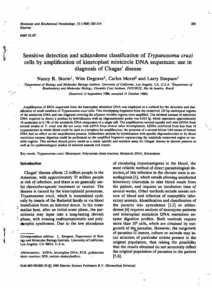
Oligonucleotides. Oligonucleotides were synthesized by standard phosphofamidite methods. The sequences and locations of the oligonucleotides used as primers and probes are shown in Fig. il. The Cll and Yl oligonucleotides cover polymorphic regions of the Cll minirepeat (region 4) and the YOl minirepeat (region 1) [8].
Hybridization of blots. Reaction products were blotted and fixed onto Nytran (Schleicher and Schucll) filters by cross-Unking with short wave-
207
A 10 20 30 40 50 60
C o n s e n s u s : AAAattGGGgNtNNGAAATTcNGGAAANTNTGGTTTTGGGAGGGGCGTTCAAaTTTtGGGgC S67:5'-TGGTTTTGGGAGGGG§§^TCAAfTTT
70 80 90 100 110 120 GgAAATTCATGCATCTCCCCCGTACATTATTTtGgCNAAAATGgGGATTTTTcaNGGGAGGT
S33 A: 5'-TCATGCATCTC JCCCGTACATTATTT S35: AGTACGTAGAGTGGGCATGTAATAAA-5'
Cll: 5'-TGCGGATTTTGGGATTTTT-ACGGGGAG Yl: 5'-GGTCGAAAATGGGG-TTGTTTAC
130 140 150 . 160 GGGGTTCGATTGGGGTTGGTGTAATATAGNNANtNNNNTGg
S34A: CCAAGCTAACCCCAACCACATTATAT-5' S 36:5'- GGGTTCGATTGGGGTTGGTGT
B
122bp: •
/S36
[..-• 330bp
Fig. 1. (A) The consensus nucleotide sequence of 20 minirepeat regions as determined by Degrave et al. (fig. 4 in ref. 8) for five sequenced minicircles from three strains of T. cruzi. Uppercase letters indicate a level of matches of more than 60%, lowercase letters indicate a level between 50 and 60%, and N (any nucleotide) indicates levels of less than 50%. The locations and sequences of the oligonucleotide primers used are indicated in their proper orientation. Degeneracies were chosen based on nucleotide polymorphisms in the aligned minirepeats [8|. (B) A schematic representation of the organization of the minirepeat regions on an individual minicircle with the primer sets and their amplification products indicated by dotted lines. The size of the minicircle is
approximately 1.42 kb. Note that only a single primer set and product is indicated for each minirepeat for simplification.
length ultraviolet light for 2 min. Hybridization and wash conditions used for ^^P-labeled oligonucleotide probes are as described in Wood et al. [14], modified by omitting dextran sulfate from the hybridization solution and using 1% sodium dodecylsulfatc (SDS) in the wash solution. The filters were routinely washed at 45°C and then at 65°C in 3.0 M tetramethylammonium chloride solution [14] with 1% SDS; this method eliminates base ratio effects and allows oligomer hybridizations to be performed at identical stringencies.
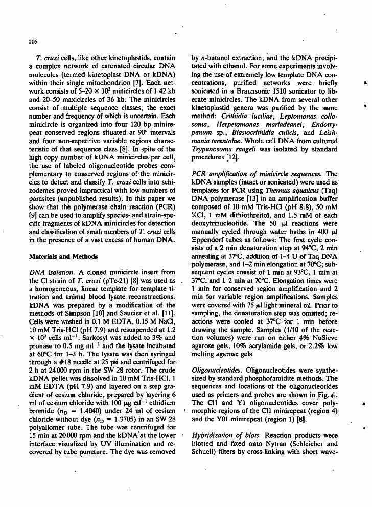
Isolation of DNA from blood. Total DNA from human blood, and from T. cruzi infected mouse blood was recovered by the high salt-detergent lysate method of Zolg et al. [15], followed by phenol/chloroform deproteinization and ethanol precipitation. 400 |xl of a mixture of water/1% Na sarkosinate ih 50 mM EDTA (pH 8.0)/cesium tri-fluoroacetate (2.04 g ml"', Pharmacia Fine Chemicals, Inc.) (2:1:1, v/v) was added to 100 \JA of blood. These lysed blood samples were stable at room temperature. The lysate was extracted with an equal volume of phenol/chloroform (1:1)
208
and the DNA was precipitated with ethanol from the aqueous phase. In the case of the infected mouse blood samples, half of the total DNA recovered from each 100 |xl sample was used as template for the PCR analysis.
Results
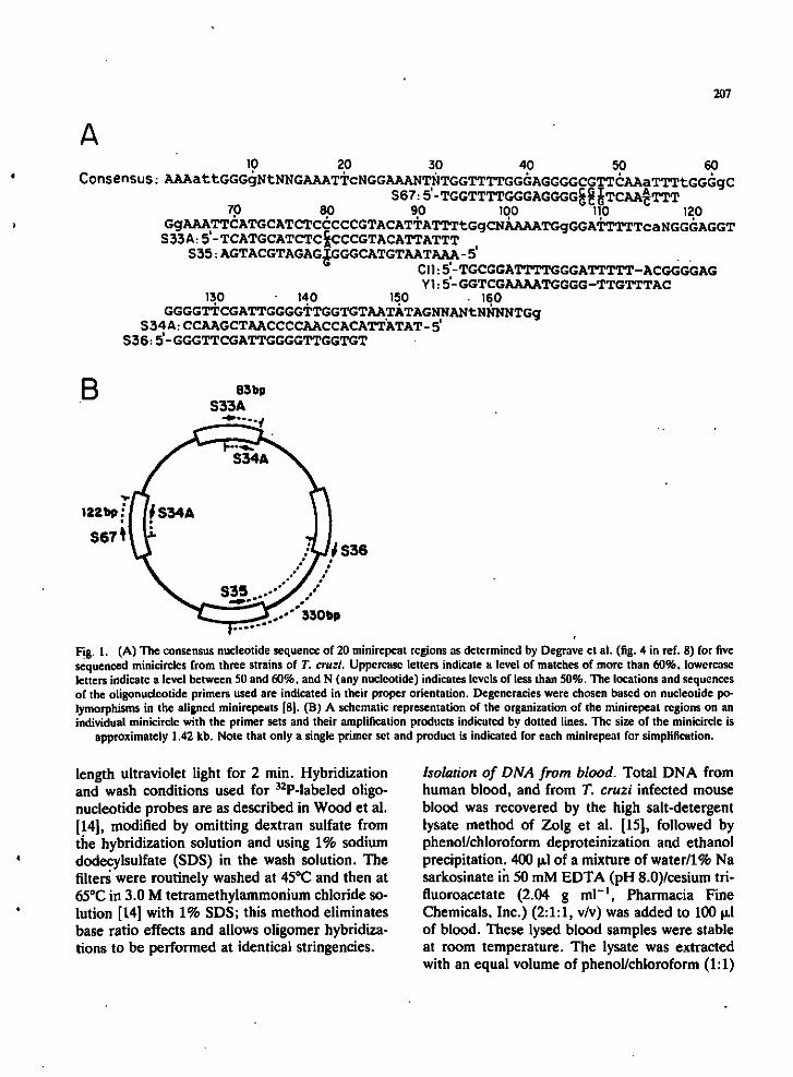
Selection of oligonucleotide primers for PCR. De-grave et al. (fig. 4 in ref. 8) aligned 2C) minirepeats from five minicircles which were cloned from the CL, Y and AWP strains of T. cruzi. On the basis of these alignments, and with the assumption that the observed conserved sequences within the minirepeats are present in most if not all minicircles in the kDNA networks, three conserved regions were selected for synthesis of oligonucleotide primers, as shown in Fig. lA. A schematic diagram of the three expected amplification products on a minicircle containing these conserved sequences is shown in Fig. IB. Note that the 83 and 122 bp products contain overiapping minirepeat sequences, whereas the 330 bp
product covers the adjacent variable region sequences in addition to a portion of the minirepeat. Since each variable region abuts a minirepeat, the 330 bp product should contain all variable region sequences from all minicircles in the network.
The expected 83, 122 and 330 bp products were obtained by amplification of T. cruzi kDNA with the appropriate primer sets, as shown in Fig. 2A and B. In addition, an approximately 50 bp ethidium bromide-stained band of variable intensity is present in the no-template DNA primer control lane 1 and also in lanes 2, 4 and 6. This band represents a primer hairpin artifact, the intensity of which is inversely proportional to the amount of template DNA used in the reaction (see Fig. 3 below). The 720 bp band in Fig. 2B is most likely also an artifact produced by hairpin-ning of the 330 bp product, since this band hybridizes to the S67 probe (Fig. 23) but not to the Cll probe (data not shown), as would be expected if this product represented the amplification of two adjacent variable regions.
B
1 2 3 4 5 6 7 M
•122 bp
•83 bp
CI Peru Y CI Peru Y
;-330bp
Fig. 2. (A) Amplification of the 83 and 122 bp fragments from three strains of T. cruzi. Lane 1. no template DNA, S34A/S67 primer control; lanes 2 and 3, CL strain kDNA template; lanes 4 and 5, Peru strain kDNA; lanes 6 and 7, Y strain kDNA; lane M, 123 bp ladder DNA marker. The amount of template DNA in each reaction was IS pg. The products were electrophoresed in a 4% NuSieve agarose gel which was stained with ethidium bromide. (B) Amplification of the 330 bp fragments from three strains of T. cruzi. The products were electrophoresed in a 2.2% low melting agarose gel which was blotted and probed with ^̂ P-
labelcd S67 oligonucleotide.
209
#minicircles: 10,0001000 100 50 10 1 0 M
492 bp 369 bp 246 bp
'i^'^$>P^^>^$^^§^'f§§l^ -83bp
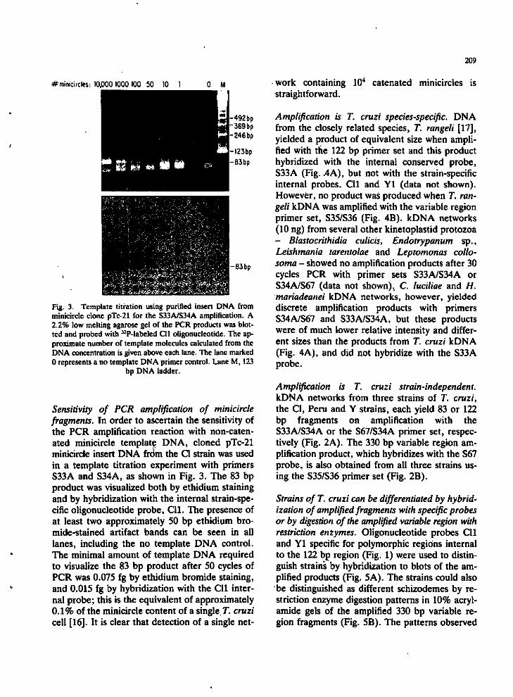
Fig. 3. Template tiifation using purified insert DNA from minicircle clone pTc-21 for the S33A/S34A amplification. A 2.2% low melting agarose gel of the PCR products was blotted and probed with ^'P-labeled Cll oligonucleotide. The approximate number of template molecules calculated from the DNA concentration is given above each lane. The lane marked 0 represents a no template DNA primer control. Lane M, 123
bp DNA ladder.
Sensitivity of PCR amplification of minicircle fragments. In order to ascertain the sensitivity of the PCR amplification reaction with non-catenated minicircle template DNA, cloned pTc-21 minicircle insert DNA frbm the CI strain was used in a template titration experiment with primers S33A and S34A, as shown in Fig. 3. The 83 bp product was visualized both by ethidium staining and by hybridization with the internal strain-specific oligonucleotide probe, Cll. The presence of at least two approximately 50 bp ethidium bromide-stained artifact bands can be seen in all lanes, including the no template DNA control. The minimal amount of template DNA required to visualize the 83 bp product after 50 cycles of PCR was 0.075 fg by ethidium bromide staining, and 0.015 fg by hybridization with the Cll internal probe; this is the equivalent of approximately 0.1% of the minicircle content of a single. T. cruzi cell [16]. It is clear that detection of a single net
work containing 10'* catenated minicircles is straightforward.
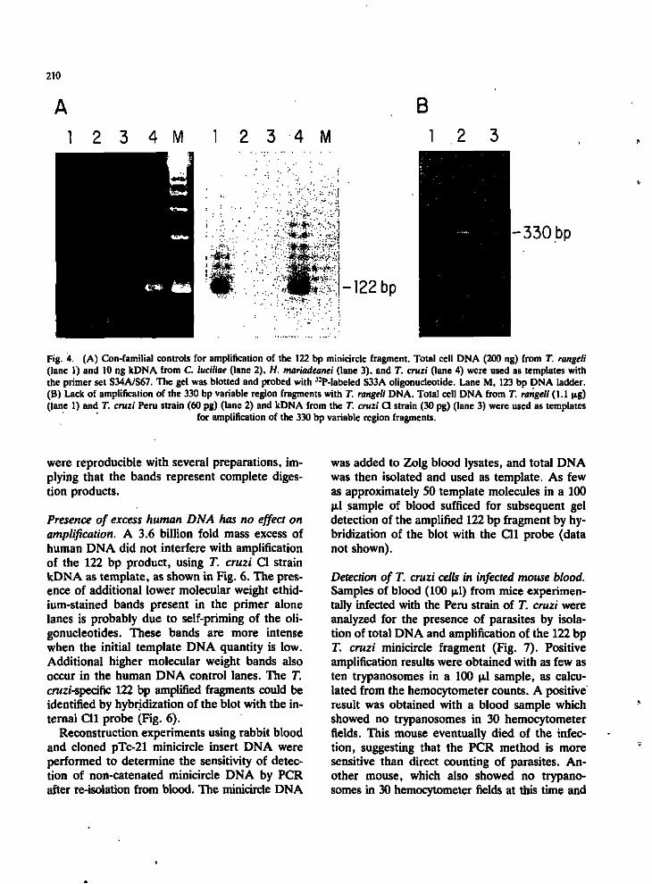
Amplification is T. cruzi species-specific. DNA from the closely related species, T. rangeli [17], yielded a product of equivalent size when amplified with the 122 bp primer set and this product hybridized with the internal conserved probe, S33A (Fig. .4A), but not with the strain-specific internal probes, Cll and Yl (data not shown). However, no product was produced when T. rangeli kDNA was amplified with the variable region primer set, S35/S36 (Fig. 4B). kDNA networks (10 ng) from several other kinetoplastid protozoa - Blastocrithidia culicis, Endotrypanum sp., Leishmania tarentolae and Leptomonas collo-soma - showed no amplification products after 30 cycles PCR with primer sets S33A/S34A or S34A/S67 (data not shown). C. luciliae and H. mariadeanei kDNA networks, however, yielded discrete amplification products with primers S34A/S67 and S33A/S34A, but these products were of much lower relative intensity and different sizes than the products from T. cruzi kDNA (Fig. 4A), and did not hybridize with the S33A probe.
Amplification is T. cruzi strain-independent. kDNA networks from three strains of T. cruzi, the CI, Peru and Y strains, each yield 83 or 122 bp fragments on amplification with the S33A/S34A or the S67/S34A primer set, respectively (Fig. 2A). The 330 bp variable region amplification product, which hybridizes with the S67 probe, is also obtained from all three strains using the S35/S36 primer set (Fig. 2B).
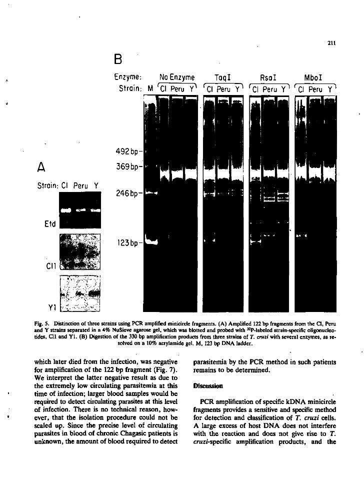
Strains of T. cruzi can be differentiated by hybridization of amplified fragments with specific probes or by digestion of the amplified variable region with restriction enzymes. Oligonucleotide probes Cll and Yl specific for polymorphic regions internal to the 122 bp region (Fig. 1) were used to distinguish strains by hybridization to blots of the amplified products (Fig. 5A). The strains could also be distinguished as different schizodemes by restriction enzyme digestion patterns in 10% acrylamide gels of the amplified 330 bp variable region fragments (Fig. 5B). The patterns observed
210
A B
1 2 3 4 M 1 2 3 4 M 1 2 3
-122 bp
-330bp
Fig. 4. (A) Con-familial controls for amplification of the 122 bp minicircle fragment. Total cell DNA (200 ng) from T. rangeli (lane 1) and 10 ng kDNA from C. luciliae (lane 2). H. mariadeanei (lane 3), and T. cruzi (lane 4) were used as templates with the primer set S34A/S67. The gel was blotted and probed with ^-P-labeled S33A oligonucleotide. Lane M, 123 bp DNA ladder. (B) Lack of amplification of the 330 bp variable region fragments with T. rangeli DNA. Total cell DNA from T. rangeli (1.1 pig) (lane 1) and T. cruzi Peru strain (60 pg) (lane 2) and kDNA from the T. cruzi CI strain (30 pg) (lane 3) were used as templates
for amplification of the 330 bp variable region fragments.
were reproducible with several preparations, implying that the bands represent complete digestion products.
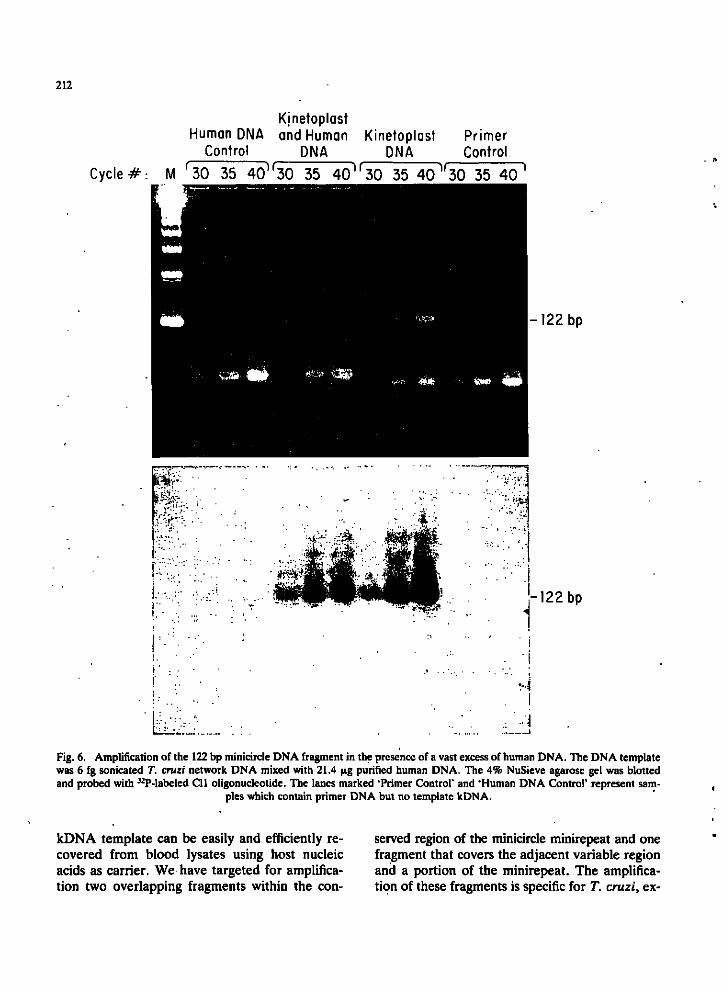
Presence of excess human DNA has no effect on amplification. A 3.6 billion fold mass excess of human DNA did not interfere with amplification of the 122 bp product, using T. cruzi CI strain kDNA as template, as shown in Fig. 6. The presence of additional lower molecular weight ethidium-stained bands present in the primer alone lanes is probably due to self-priming of the oligonucleotides. These bands are more intense when the initial template DNA quantity is low. Additional higher molecular weight bands also occur in the human DNA control lanes. The T. cruzi-specific 122 bp amplified fragments could be identified by hybridization of the blot with the internal Cll probe (Fig. 6).
Reconstruction experiments using rabbit blood and cloned pTc-21 minicircle insert DNA were performed to determine the sensitivity of detection of non-catenated minicircle DNA by PCR after re-isolation from blood. The minicircle DNA
was added to Zolg blood lysates, and total DNA was then isolated and used as template. As few as approximately 50 template molecules in a 100 1̂ sample of blood sufficed for subsequent gel
detection of the amplified 122 bp fragment by hybridization of the blot with the Cll probe (data not shown).
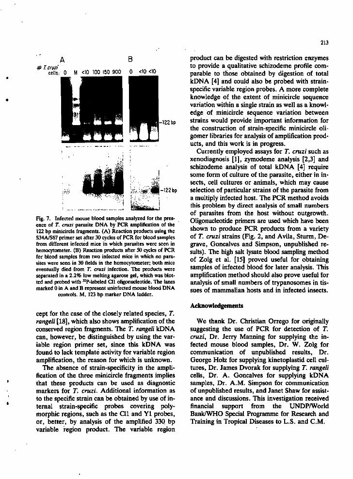
Detection of T. cruzi cells in infected mouse blood. Samples of blood (100 |xl) from mice experimentally infected with the Peru strain of T. cruzi were analyzed for the presence of parasites by isolation of total DNA and amplification of the 122 bp T. cruzi minicircle fragment (Fig. 7). Positive amplification results were obtained with as few as ten trypanosomes in a 100 |xl sample, as calculated from the hemocytometer counts. A positive result was obtained with a blood sample which showed no trypanosomes in 30 hemocytometer fields. This mouse eventually died of the infection, suggesting that the PCR method is more sensitive than direct counting of parasites. Another mouse, which also showed no trypanosomes in 30 hemocytometer fields at this time and
211
B Enzyme: No Enzyme TaqI Rsal Mbol
Strain: M 'Cl Peru Y^ ^Cl Peru Y^ ^Cl Peru Y^ ^ CI Peru Y^
A Strain: CI Peru Y
492 bp
369 bp
246 bp-
«o* mm
Et(j
123bp-
Fig. 5. Distinction of three strains using PCR amplified minicircle fragments. (A) Amplified 122 bp fragments from the CI, Peru and Y strains separated in a 4% NuSieve agarose gel, which was blotted and probed with ^^P-labeled strain-specific oligonucleotides, Cll and Yl . (B) Digestion of the 330 bp amplification products from three strains of T. cruzi with several enzymes, as re
solved on a 10% acrylamide gel. M, 123 bp DNA ladder.
which later died from the infection, was negative for amplification of the 122 bp fragment (Fig. 7). We interpret the latter negative result as due to the extremely low circulating parasitemia at this time of infection; larger blood samples would be required to detect circulating parasites at this level of infection. There is no technical reason, however, that the isolation procedure could not be scaled up. Since the precise level of circulating parasites in blood of chronic Chagasic patients is unknown, the amount of blood required to detect
parasitemia by the PCR method in such patients remains to be determined.
Discussion
PCR amplification of specific kDNA minicircle fragments provides a sensitive and specific method for detection and classification of T. cruzi cells. A large excess of host DNA does not interfere with the reaction and does not give rise to T. cruzi-specific amplification products, and the
212
Kinetoplast Human DNA and Human Kinetoplast Primer
Control DNA DNA Control
Cycle#: M '30 35 40^*30 35 40 ' ' 30 35 40^30 35 40 '
-122 bp
-122 bp
Fig. 6. Amplification of the 122 bp minicircle DNA fragment in the presence of a vast excess of human DNA. The DNA template was 6 fg sonicated T. cruzi network DNA mixed with 21.4 )jig purified human DNA. The 4% NuSieve agarose gel was blotted and probed with ^-P-labeled Cll oligonucleotide. The lanes marked 'Primer Control' and 'Human DNA Control' represent sam
ples which contain primer DNA but no template kDNA.
kDNA template can be easily and efficiently recovered from blood lysates using host nucleic acids as carrier. We have targeted for amplification two overlapping fragments within the con
served region of the minicircle minirepeat and one fragment that covers the adjacent variable region and a portion of the minirepeat. The amplification of these fragments is specific for T. cruzi, ex-
213
A B # T.cruii _ ,„ ,„
cells: 0 M <10 100 150 900 0 <10 <10
-122 bp
-122bp
Fig. 7. Infected mouse blood samples analyzed for the presence of T. cruzi parasite DNA by PCR amplification of the 122 bp minicircle fragments. (A) Reaction products using the S34A/S67 primer set after 30 cycles of PCR for blood samples from different infected mice in which parasites were seen in hemocytometer. (B) Reaction products after SO cycles of PCR for blood samples from two infected mice in which no parasites were seen in 30 fields in the hemocytometer; both mice eventually died from T. cruzi infection. The products were separated in a 2.2% low melting agarose gel, which was blotted and probed with ^-P-labcled Cll oligonucleotide. The lanes marked 0 in A and B represent uninfected mouse blood DNA
controls. M, 123 bp marker DNA ladder.
cept for the case of the closely related species, T. rangeli [18], which also shows amplification of the conserved region fragments. The T. rangeli kDNA can, however, be distinguished by using the variable region primer set, since this kDNA was found to lack template activity for variable region amplification, the reason for which is unknown.
The absence of strain-specificity in the amplification of the three minicircle fragments implies that these products can be used as diagnostic markers for T. cruzi. Additional information as to the specific strain can be obtained by use of internal strain-specific probes covering polymorphic regions, such as the Cll and Yl probes, or, better, by analysis of the amplified 330 bp variable i-egion product. The variable region
product can be digested with restriction enzymes to provide a qualitative schizodeme profile comparable to those obtained by digestion of total kDNA [4] and could also be probed with strain-specific variable region probes. A more complete knowledge of the extent of minicircle sequence variation within a single strain as well as a knowledge of minicircle sequence variation between strains would provide important information for the construction of strain-specific minicircle oligomer libraries for analysis of amplification products, and this work is in progress.
Currently employed assays for T. cruzi such as xenodiagnosis [1], zymodeme analysis [2,3] and schizodeme analysis of total kDNA [4] require some form of culture of the parasite, either in insects, cell cultures or animals, which may cause selection of particular strains of the parasite from a multiply infected host. The PCR method avoids this problem by direct analysis of small numbers of parasites from the host without outgrowth. Oligonucleotide primers are used which have been shown to produce PCR products from a variety of T. cruzi strains (Fig. 2, and Avila, Sturm, De-grave, Goncalves and Simpson, unpublished results). The high salt lysate blood sampling method of Zolg et al. [15] proved useful for obtaining samples of infected blood for later analysis. This amplification method should also prove useful for analysis of small numbers of trypanosomes in tissues of mammalian hosts and in infected insects.
Acl̂ nowiedgements
We thank Dr. Christian Orrego for originally suggesting the use of PCR for detection of T. cruzi. Dr. Jerry Manning for supplying the infected mouse blood samples. Dr. W. Zolg for communication of unpublished results, Dr. George Holz for supplying kinetoplastid cell cultures, Dr. James Dvorak for supplying T. rangeli cells, Dr. A. Goncalves for supplying kDNA samples, Dr. A.M. Simpson for communication of unpublished results, and Janet Shaw for assistance and discussions. This investigation received financial support from the UNDP/World Bank/WHO Special Programme for Research and Training in Tropical Diseases to L.S. and CM.
214
References
1 Schenone, H., Rojo, M., Rojas, A. and Concha, L. (1977) Positividad diurna y nocturna del xenodiagnostico en un paciente con infeccion chagasica cronica de parasitemia permanente. Bol. Chil. Parasitol. 32, 63-66.
2 Miles, M., Souza, A., Povoa, M., Shaw, J., Lainson, R. and Toye, P. (1978) Isozymic heterogeneity of Trypanosoma cruzi in the first autochthonous patients with Chagas' disease in Amazonian Brazil. Nature 272, 819-821.
3 Tibayrenc, M. and Ayala, F. (1988) Isoenzyme variability in Trypanosoma cruzi, the agent of Chagas' disease: Ge-netical, taxonomic and epidemiological significance. Evolution 42, 277-292.
4 Morel, C , Chiari, E., Camargo, E., Mattel, D., Romanha, A. and Simpson, L. (1980) Strains and clones of Trypanosoma cruzi can be characterized by restriction endonuclease fingerprinting of kinetoplast DNA minicircles. Proc. Natl. Acad. Sci. USA 77, 6810-6814.
5 Goncalves, A., Chiari, E., Cameiro, M., Romanha, A. and Morel, C. (1984) Schizodeme characterization of natural and artificial populations of Trypanosoma cruzi as a tool in the study of Chagas' disease. In: Application of Biochemical and Molecular Biology Techniques to Problems of Parasite and Vector Identification (Newton, B. and Michal, I., eds.), pp. 253-274, UNDP/WHO Special Programme for Research and Training in Tropical Diseases, Geneva, Switzerland.
6 Deane, M., Sousa, M., Pereira, N., Goncalves, A., Momen, H. and Morel, C. (1984) Tryparwsoma cruzi: Inoculation schedules and re-isolation methods select individual strains from doubly infected mice, as demonstrated by schizodeme and zymodeme. analyses. J. Protozool. 31, 276-280.
7 Simpson, L. (1987) The mitochondrial genome'of kinetoplastid protozoa: Genomic organization, transcription, replication and evolution. Annu. Rev. Microbiol. 41, 363-382.
8 Degrave, W., Fragoso, S., Britto, C , van Heuverswyn, H., Kidane, G., Cardoso, M., Mueller, R., Simpson, L. and Morel, C. (1988) Peculiar sequence organization of kinetoplast minicircles from Trypanosoma cruzi. Mol. Biochem. Parasitol. 27, 63-70.
9 Saiki, R., Scharf, S., Faloona, F., Mullis, K., Horn, G., Eriich, H. and Arnheim, N. (1985) Enzymatic amplification of beta-globin genomic sequences and restriction site analysis for diagnosis of sickle cell anemia. Science 230, 1350-1354.
10 Simpson, L. (1979) Isolation of maxicircle component of kinetoplast DNA from hemoflagellate protozoa. Proc. Natl. Acad. Sci. USA 76, 1585-1588.
11 Saucier, i. , Bevard, J., da Silva, J. and Riou. G. (1981) Occurrence of a kinetoplast DN A-protein complex in Trypanosoma cruzi. Biochem. Biophys. Res. Commun. 101, 988-994.
12 Simpson, L. and Beriiner, J. (1974) Isolation of the kinetoplast DNA of Leishmania tarentolae in the form of a network. J. Protozool. 21, 382-393.
13 Saiki, R., Gelfand. D., Stoffel, S., Scharf, S., Higuichi, R., Horn, G., Mullis, K. and Eriich, H. (1988) Primer-directed enzymatic amplification of DNA with a thermostable DNA polymerase. Science 239, 487-491.
14 Wood, W., Gitschier, J., Lasky, L. and Lawn, R. (1985) Base composition-independent hybridization in tetramethylammonium chloride: A method for oligonucleotide screening of highly complex gene libraries. Proc. Natl. Acad. Sci. USA 82, 1585-1588.
15 Zolg, J.W., Scott, E.D. and Wendlinger, M. (1988) High salt lysates: A simple method to store blood samples without refrigeration for subsequent use with DNA probes. Am. J. Trop. Med. Hyg. 39, 33-40.
16 Simpson, L. and da Silva, A. (1971) Isolation and characterization of kinetoplast DNA from Leishrrumia tarentolae. J. Mol. Biol. 56, 443-473.
17 D'Alessandro, A. (1976) Biology of Trypanosoma rangeli Tejera, 1920. In: Biology of the Kinetoplastlda, Vol. 1 (Lumsden, W. and Evans, D., eds.), pp. 327-403, Academic Press, London.
18 Frasch, A., Hajduk, S., Hoeijmakers, J., Borst, P., Brunei, F. and Davison, J. (1980) The kDNA of Trypanosoma equiperdum. Biochim. Biophys. Acta 607, 397-410.
Related Documents


