1 Sense Organ Jun Zhou ( 周周 ), M.D. & Ph.D. School of Medicine, Zhejiang Univ ersity 20131216

Sense Organ
Jan 01, 2016
Sense Organ. Jun Zhou ( 周俊 ), M.D. & Ph.D. School of Medicine, Zhejiang University. 20131216. LEARNING METHODS Listen attentively and think actively during the lecture. Preview and review the textbook and atlas as much as you can. - PowerPoint PPT Presentation
Welcome message from author
This document is posted to help you gain knowledge. Please leave a comment to let me know what you think about it! Share it to your friends and learn new things together.
Transcript
1
Sense Organ Jun Zhou ( 周俊 ), M.D. & Ph.D.
School of Medicine, Zhejiang University
20131216
2
LEARNING METHODS
•Listen attentively and think actively during the lecture.
•Preview and review the textbook and atlas as much as you can.
•NEVER passing by a word without knowing its definition.
•To understand the structure and function of each organ, not just memorize them.
•Email: [email protected]
3
Special sense receptors
Responsible for the five special senses: taste, smell, seeing, hearing, feeling
Tranduce stimuli from the environment into electrical impulses
4
Specialized diffuse receptors
Two important sensory organ The eye The ear
5
Specialized diffuse receptors
Free nerve terminals -- feel cold, hot, pain and slight touch Encapsulated nerve ending --have CT capsule Pacinian corpuscle Meissner corpuscle Proprioceptive receptors
6
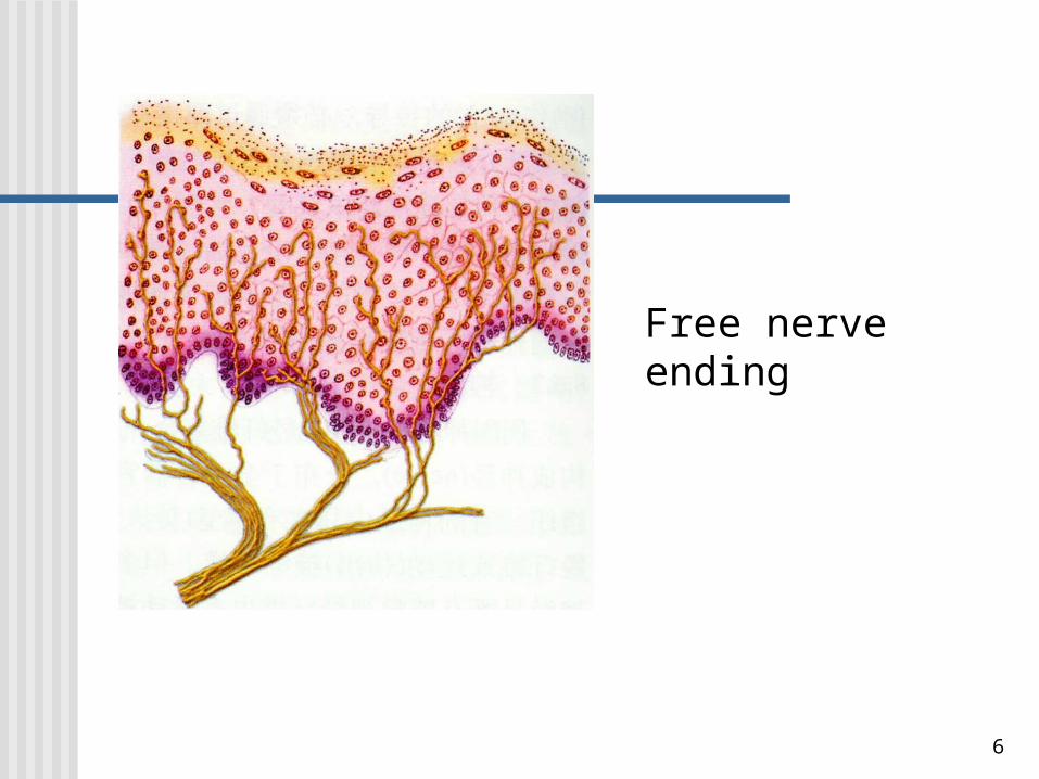
Free nerve ending
7
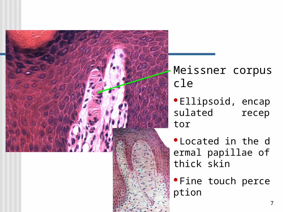
Meissner corpuscleEllipsoid, encapsulated receptor
Located in the dermal papillae of thick skin
Fine touch perception
8
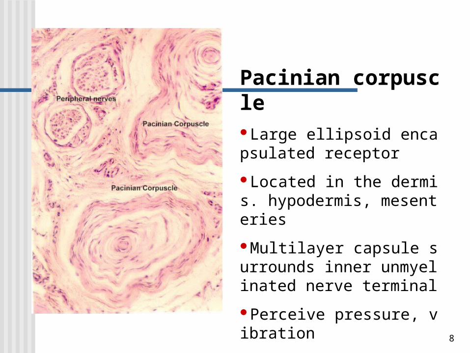
Pacinian corpuscleLarge ellipsoid encapsulated receptor
Located in the dermis. hypodermis, mesenteries
Multilayer capsule surrounds inner unmyelinated nerve terminal
Perceive pressure, vibration
9
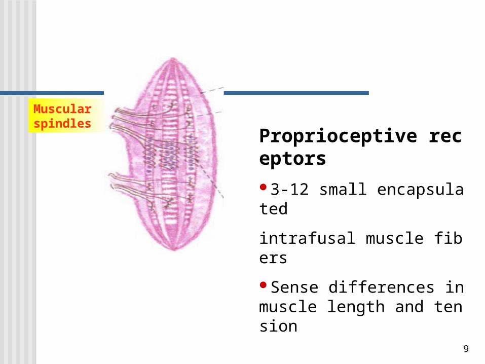
Muscular spindles
Proprioceptive receptors
3-12 small encapsulated
intrafusal muscle fibers
Sense differences in muscle length and tension
10
Two special sensory organsTwo special sensory organs
Eyes: visual organ
Ears: the organ of hearing and equilibrium.
11
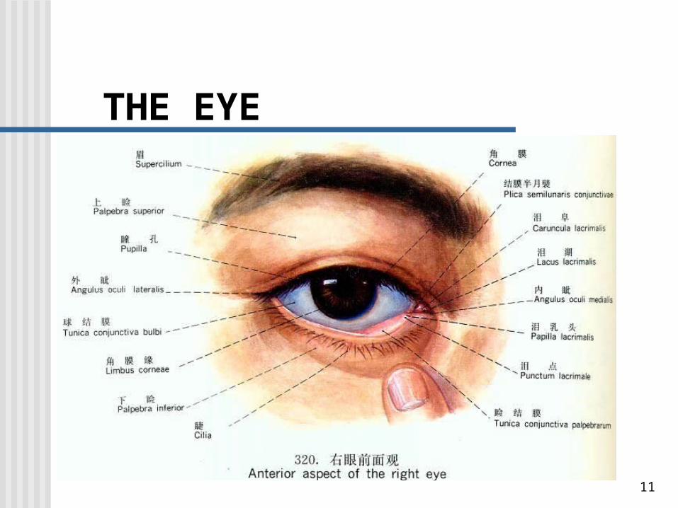
THE EYE
12
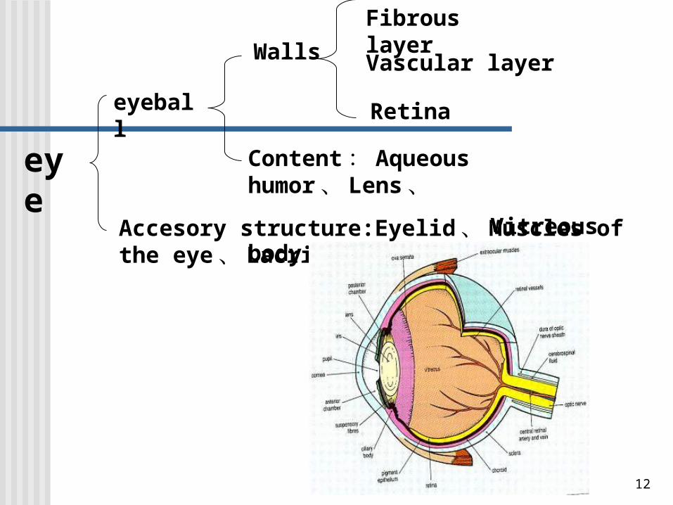
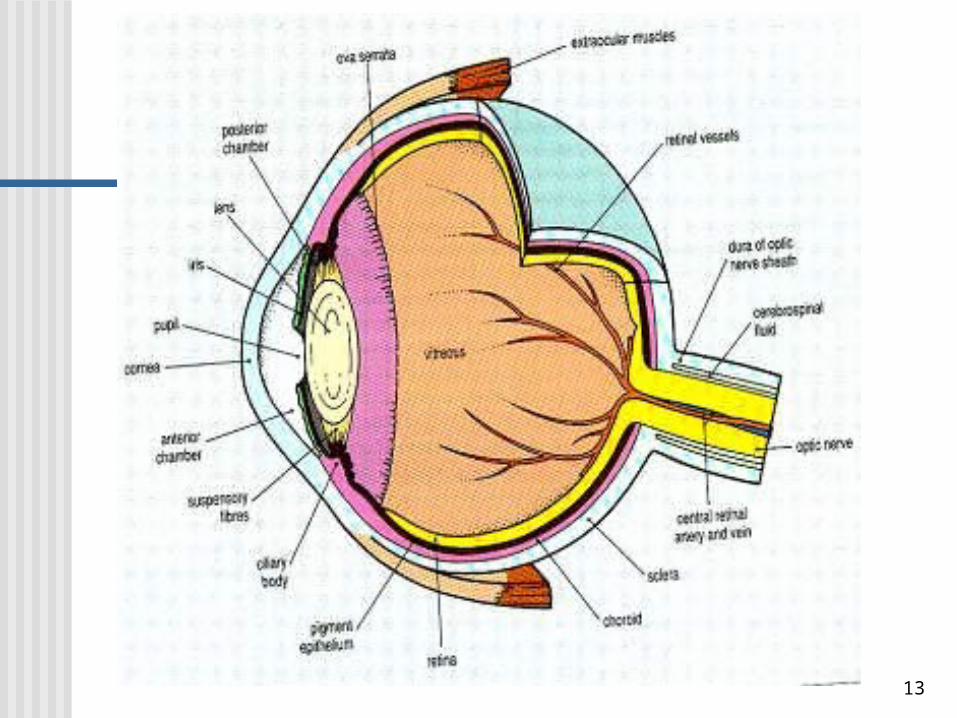
eyeball
Accesory structure:Eyelid 、 Muscles of the eye 、 Lacrimal gland
Walls
Content : Aqueous humor 、 Lens 、
Vitreous body
Fibrous layer
Vascular layer
Retina
eye
13
14
Ciliary body
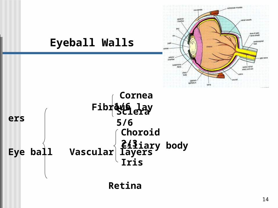
Eyeball Walls
Fibrous layers
Eye ball Vascular layers
Retina
Cornea 1/6
Sclera 5/6
Choroid 2/3
Iris
Fibrous layer, includes the cornea, covers the anterior one sixth of the eye,the transparent portion. It has a prominence or convexity.
The sclera is composed of dense fibrous connective tissue that provides attachment for the extrinsic muscles of the eye.The sclera has a slightly blue in children because of its thinness and is yellow in the eldly because of the accumulation of lipofuscin.
Blood vessels and melanin pigment give the choriod an intense dark-brown color.Provides nutrients to the retina.The anterior forms the ciliary body and iris.The ciliary body is a ringlike thickening that extends inward.The iris is a contractile diaphragm that extends over the anterior surface of the lens. The central circular aperture is the pupil.
The retina consists largely of photoreceptors cells,forming visual impulses along optic nerve.
15
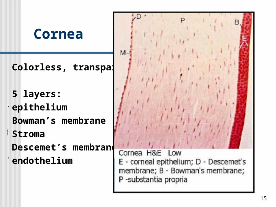
Cornea
Colorless, transparent
5 layers:
epithelium
Bowman’s membrane
Stroma
Descemet’s membrane
endothelium
16
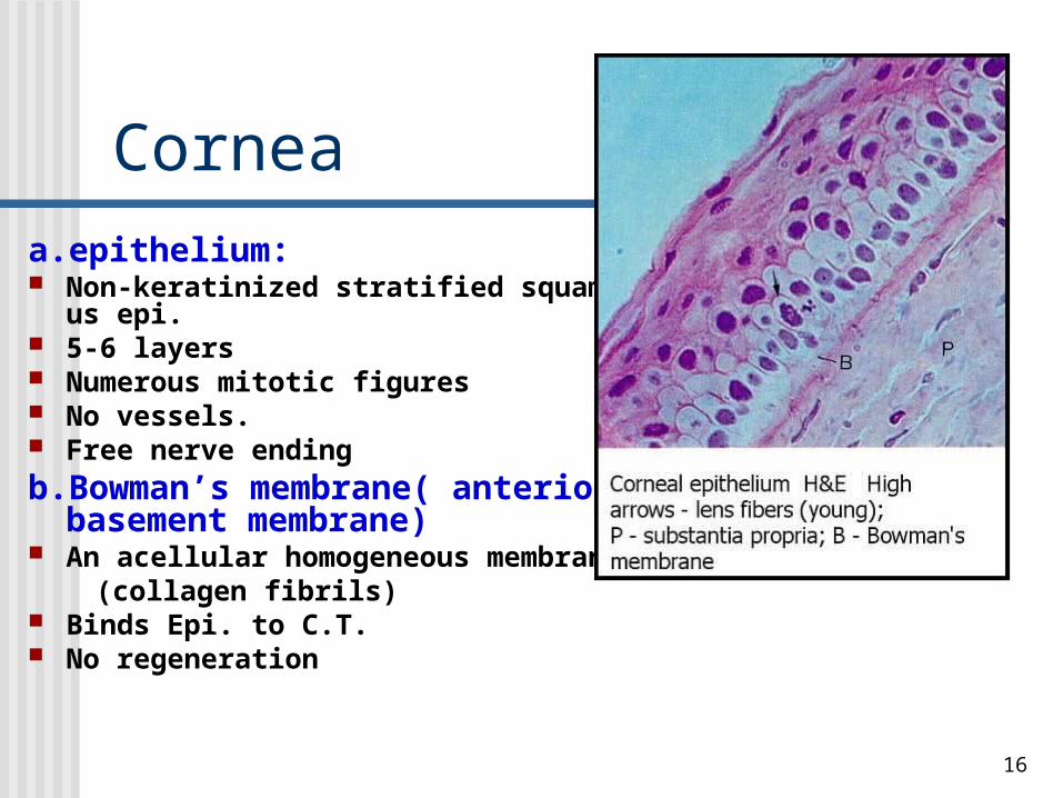
Corneaa.epithelium: Non-keratinized stratified squamous epi. 5-6 layers Numerous mitotic figures No vessels. Free nerve ending
b.Bowman’s membrane( anterior basement membrane)
An acellular homogeneous membrane (collagen fibrils) Binds Epi. to C.T. No regeneration
17
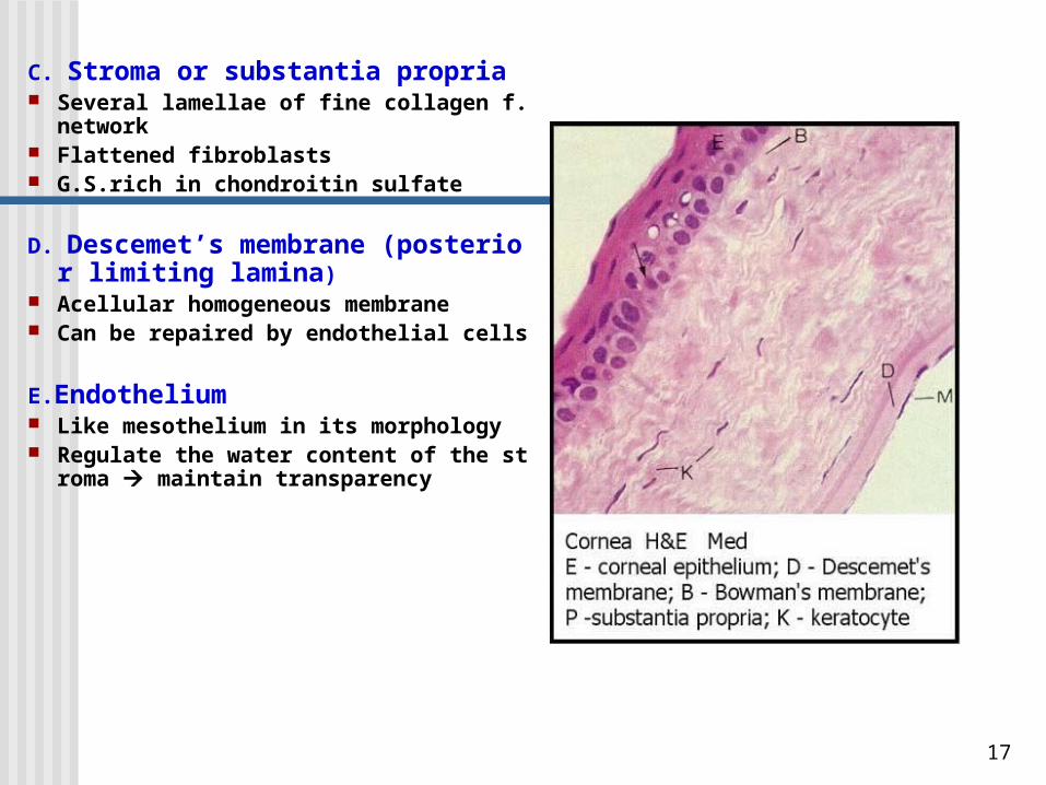
C. Stroma or substantia propria Several lamellae of fine collagen f.network Flattened fibroblasts G.S.rich in chondroitin sulfate
D. Descemet’s membrane (posterior limiting lamina)
Acellular homogeneous membrane Can be repaired by endothelial cells
E.Endothelium Like mesothelium in its morphology Regulate the water content of the stroma
maintain transparency
18
The reasons of cornea transparent
No blood vessels & pigments Basal membrane of epi. is plane Uniform spacing of collagen fibrils and lamellae
in stroma G.S. with transparent nature & maintains prop
er water

19
Retina
Two regions:The nonphotosensitive region
(nonvisual part)
Located anterior to the ora serrata, n
o photoreceptors.The photosensitive region (opti
c part)
Lines the inner surface of the eye posterior to the ora serrata (except the op
tic papilla)
20
Retina
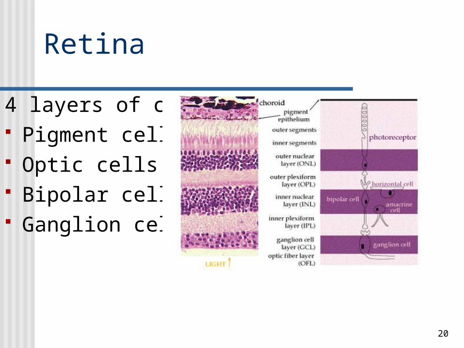
4 layers of cells: Pigment cells Optic cells Bipolar cells Ganglion cells
21
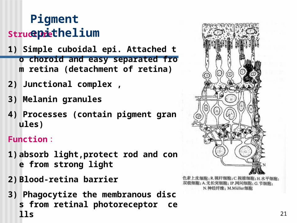
Structure :
1) Simple cuboidal epi. Attached to choroid and easy separated from retina (detachment of retina)
2) Junctional complex ,
3) Melanin granules
4) Processes (contain pigment granules)
Function :
1) absorb light,protect rod and cone from strong light
2) Blood-retina barrier
3) Phagocytize the membranous discs from retinal photoreceptor cells
4) Store vitamin A to assist in forming rhodopsin
Pigment epithelium
22
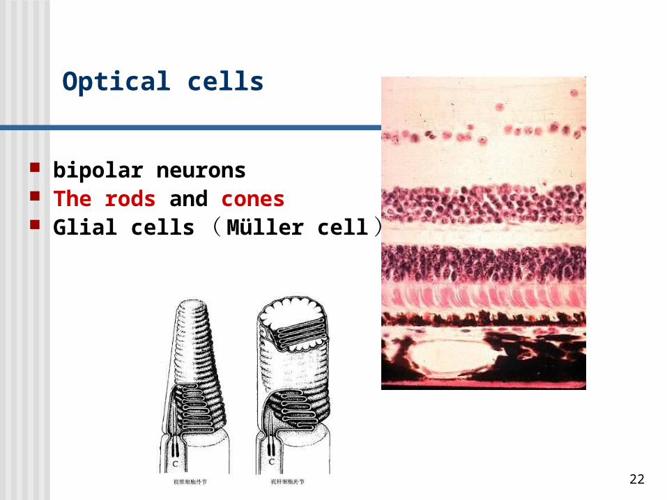
Optical cells
bipolar neurons The rods and cones Glial cells ( Müller cell )
23
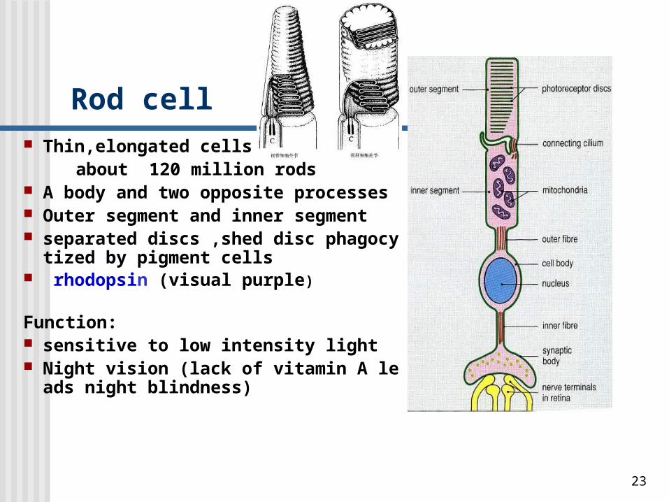
Rod cell Thin,elongated cells , about 120 million rods A body and two opposite processes Outer segment and inner segment separated discs ,shed disc phagocytized by pi
gment cells rhodopsin (visual purple) Function: sensitive to low intensity light Night vision (lack of vitamin A leads night bli
ndness)
24
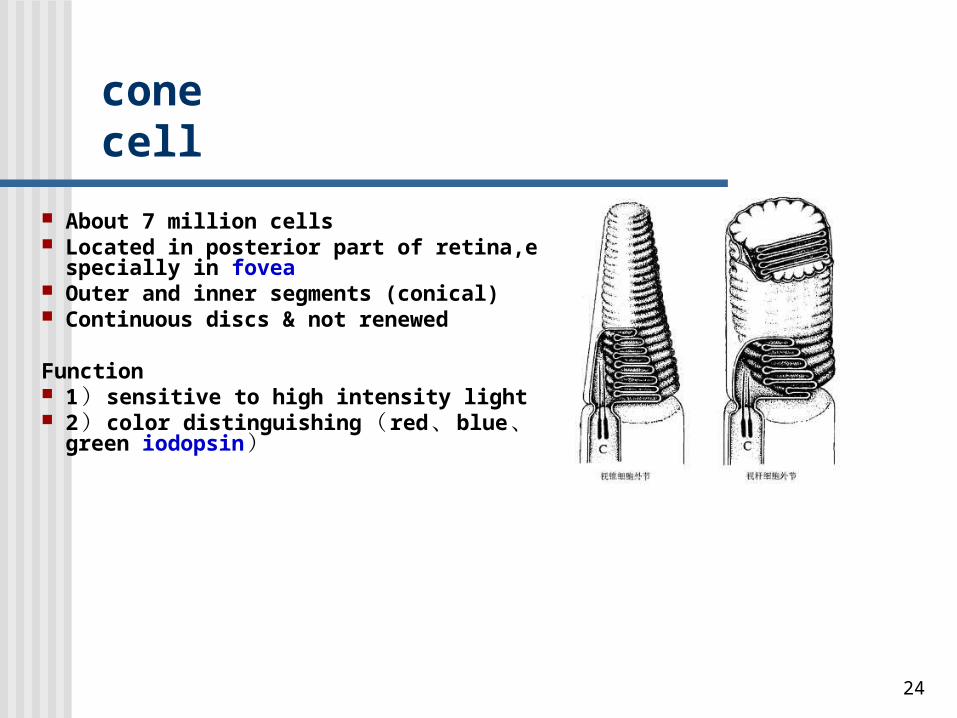
cone cell
About 7 million cells Located in posterior part of retina,especially in f
ovea Outer and inner segments (conical) Continuous discs & not renewed Function 1 ) sensitive to high intensity light 2 ) color distinguishing ( red 、 blue 、 green io
dopsin )
25
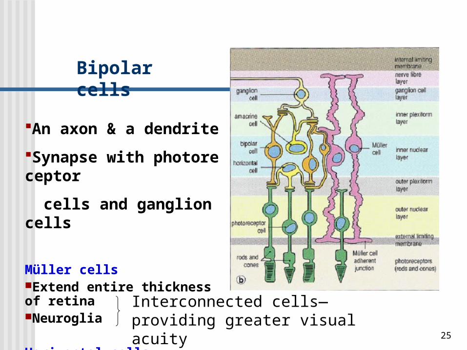
Bipolar cells
An axon & a dendrite
Synapse with photoreceptor
cells and ganglion cells
Müller cellsExtend entire thickness of retinaNeuroglia
Horizontal cellsAmacrine cells
Interconnected cells—providing greater visual acuity
26
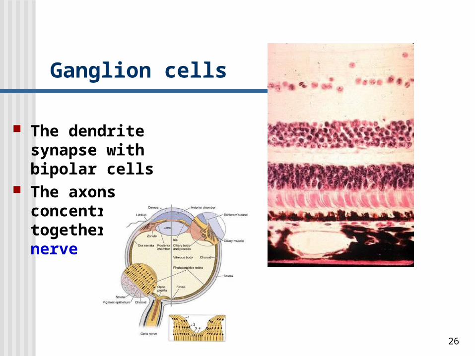
Ganglion cells
The dendrite synapse with bipolar cells
The axons concentrate together form optic nerve
27
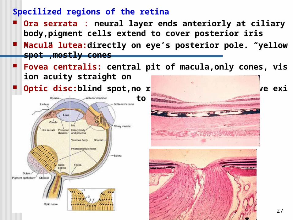
Specilized regions of the retina Ora serrata : neural layer ends anteriorly at ciliary body,pigment cells ext
end to cover posterior iris Macula lutea:directly on eye’s posterior pole. “yellow spot”,mostly cones Fovea centralis: central pit of macula,only cones, vision acuity straight on Optic disc:blind spot,no rods or cones,optic nerve exits,medial and inferior t
o fovea centralis.
28
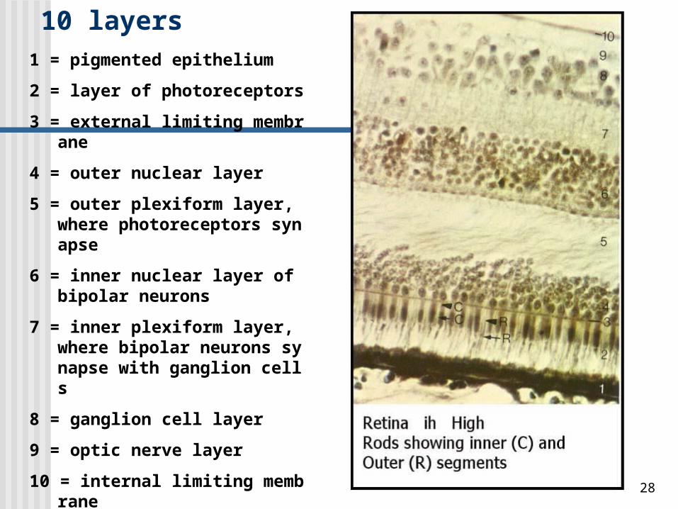
1 = pigmented epithelium
2 = layer of photoreceptors
3 = external limiting membrane
4 = outer nuclear layer
5 = outer plexiform layer, where photoreceptors synapse
6 = inner nuclear layer of bipolar neurons
7 = inner plexiform layer, where bipolar neurons synapse with ganglion cells
8 = ganglion cell layer
9 = optic nerve layer
10 = internal limiting membrane
10 layers
29
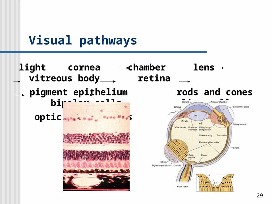
Visual pathways
light cornea chamber lens vitreous body retina
pigment epithelium rods and cones bipolar cells
ganglion cells optic nerve fibers
30
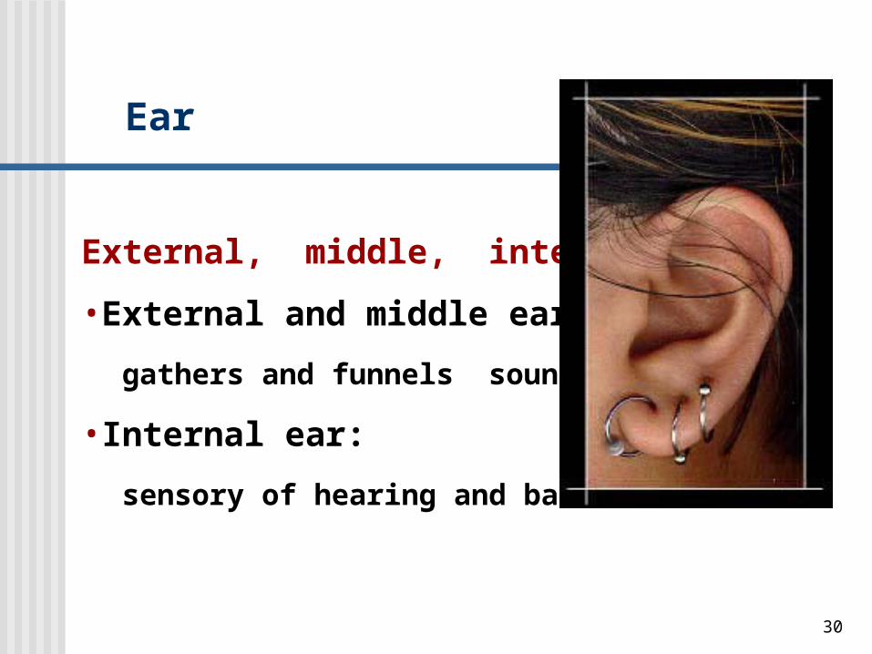
Ear
External, middle, internal ear
•External and middle ear:
gathers and funnels sound waves
•Internal ear:
sensory of hearing and balance
31
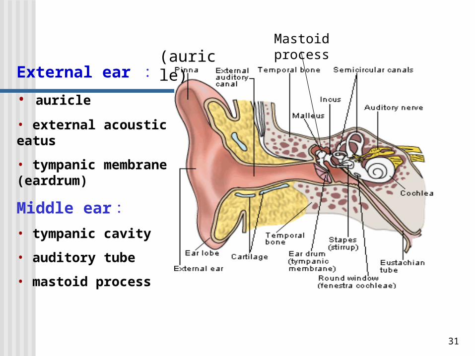
External ear :• auricle
• external acoustic meatus
• tympanic membrane (eardrum)
Middle ear :• tympanic cavity
• auditory tube
• mastoid process
(auricle)Mastoid process
32
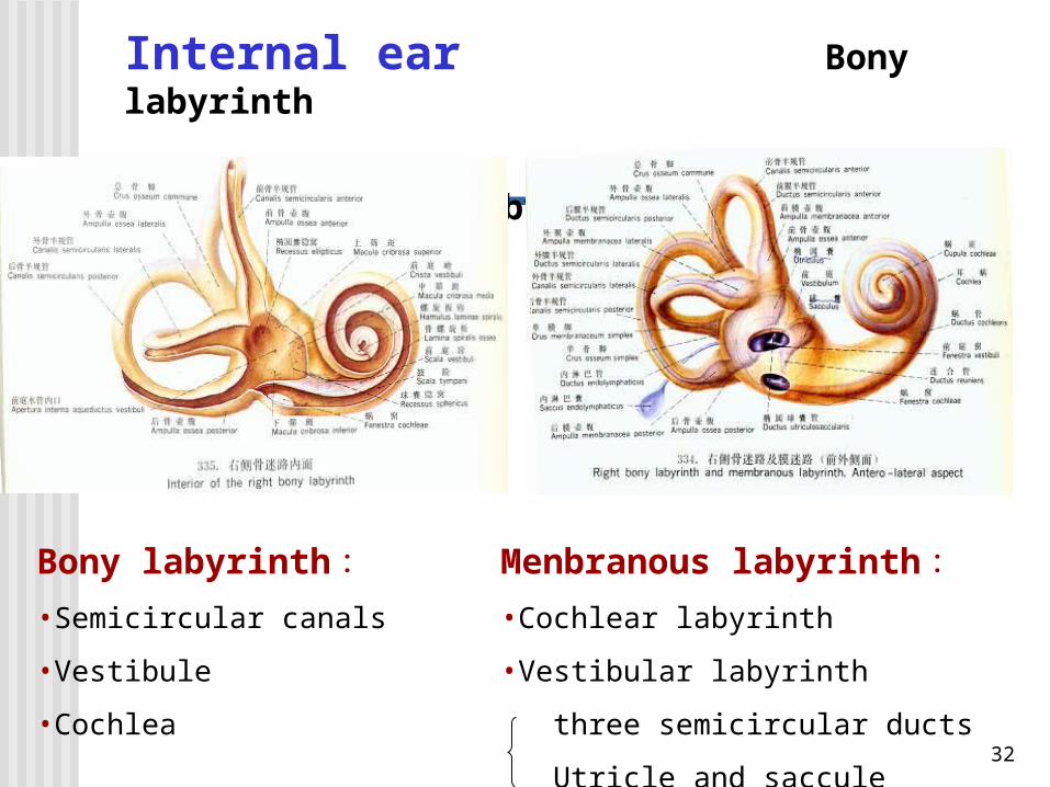
Internal ear Bony labyrinth
Membranous labyrinth
Bony labyrinth :•Semicircular canals
•Vestibule
•Cochlea
Menbranous labyrinth :•Cochlear labyrinth•Vestibular labyrinth
three semicircular ducts
Utricle and saccule
33
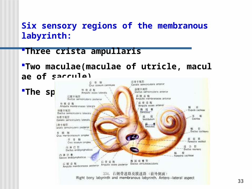
Six sensory regions of the membranous labyrinth:
Three crista ampullaris
Two maculae(maculae of utricle, maculae of saccule)
The spiral organ of Corti
34
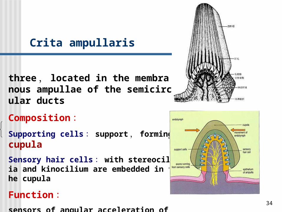
Crita ampullaris
three , located in the membranous ampullae of the semicircular ducts
Composition :
Supporting cells : support , forming cupula
Sensory hair cells : with stereocilia and kinocilium are embedded in the cupula
Function :sensors of angular acceleration of the head
35
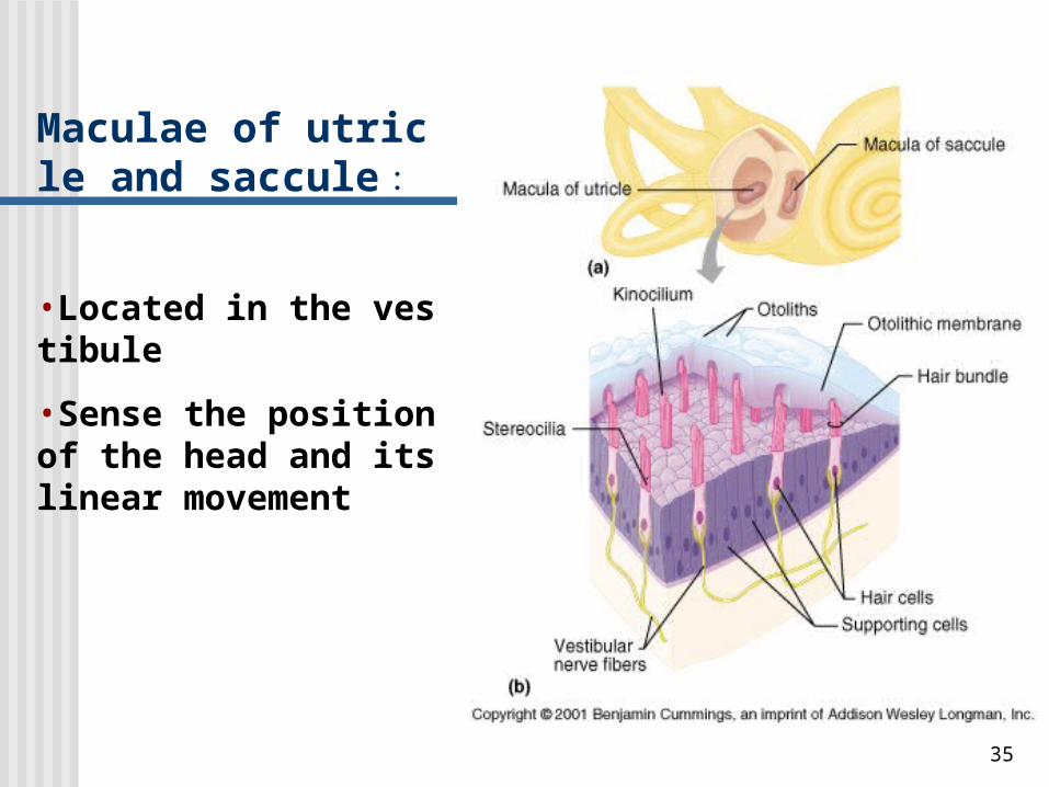
Maculae of utricle and saccule :
•Located in the vestibule
•Sense the position of the head and its linear movement
36
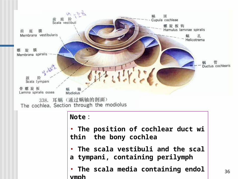
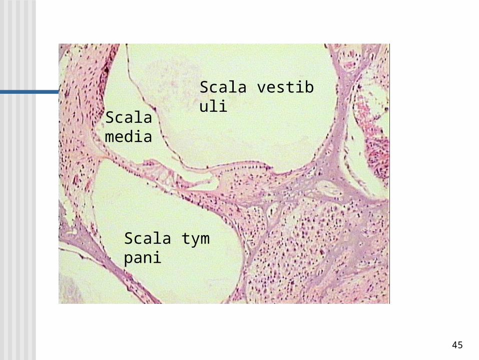
Note :• The position of cochlear duct within the bony cochlea
• The scala vestibuli and the scala tympani, containing perilymph
• The scala media containing endolymph
37
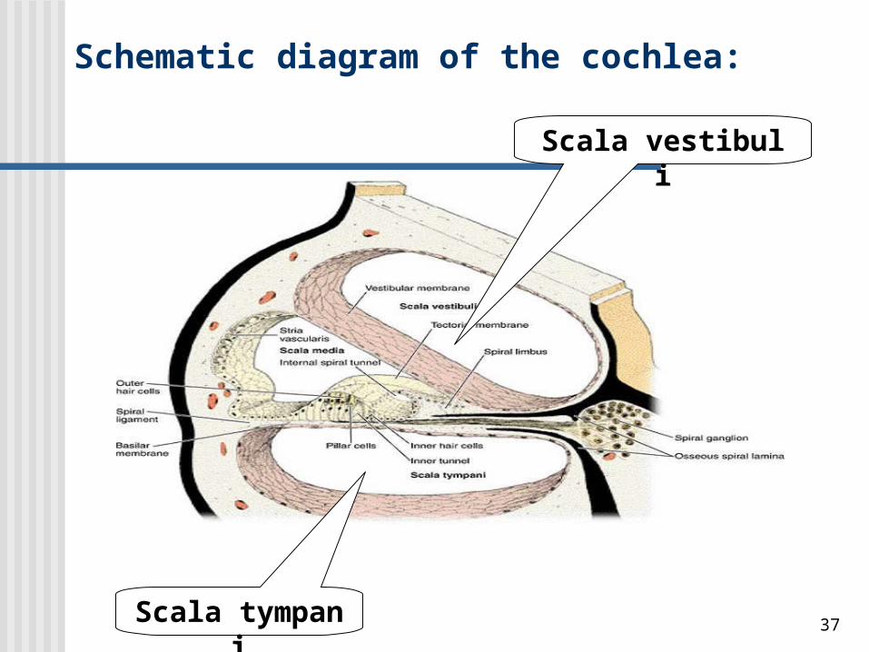
Schematic diagram of the cochlea:
Scala vestibuli
Scala tympani
38
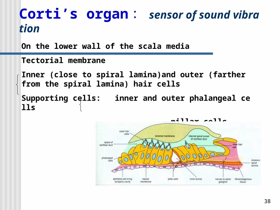
Corti’s organ : sensor of sound vibration
On the lower wall of the scala media
Tectorial membrane
Inner (close to spiral lamina)and outer (farther from the spiral lamina) hair cells
Supporting cells: inner and outer phalangeal cells
pillar cells
39
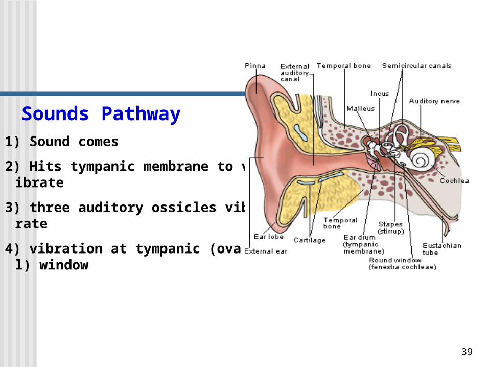
Sounds Pathway1) Sound comes
2) Hits tympanic membrane to vibrate
3) three auditory ossicles vibrate
4) vibration at tympanic (oval) window
40
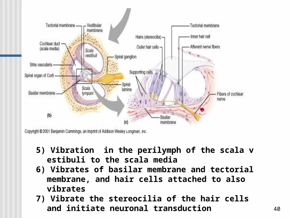
5) Vibration in the perilymph of the scala vestibuli to the scala media
6) Vibrates of basilar membrane and tectorial membrane, and hair cells attached to also vibrates
7) Vibrate the stereocilia of the hair cells and initiate neuronal transduction
41
Clinical Correlation
Vertigo: dysfunction of vestibular system
Causes: viral infections, certain drugs, tumors, excessive stimulation (seasickness, carsickness, or airsickness)
Hearing loss
1)Conductive hearing loss: sound waves are mechanically impeded from reaching the auditory sensory receptors within the internal ear, such as excessive accumulation of cerumen.
2)Sensorineural hearing impairment: injury to the auditory hair cells or the cochlea nerve. May be congenital or acquired. Causes include infections, trauma (exposure to excessive noise), administration of certain antibiotics, aging.
42
OBJECTIVES
• Know the general layers of the eye.
• Describe the structure of Cornea and its reason of transparency.
• Describe the structure of Retina and the function of pigment cell, rod cell and cone cell.
• Know the definition of Ora serrata, Macula lutea, Fovea centralis and Optic disc.
• Know the general structure of ear.
• Describe six sensory regions of the membranous labyrinth and their function.
43
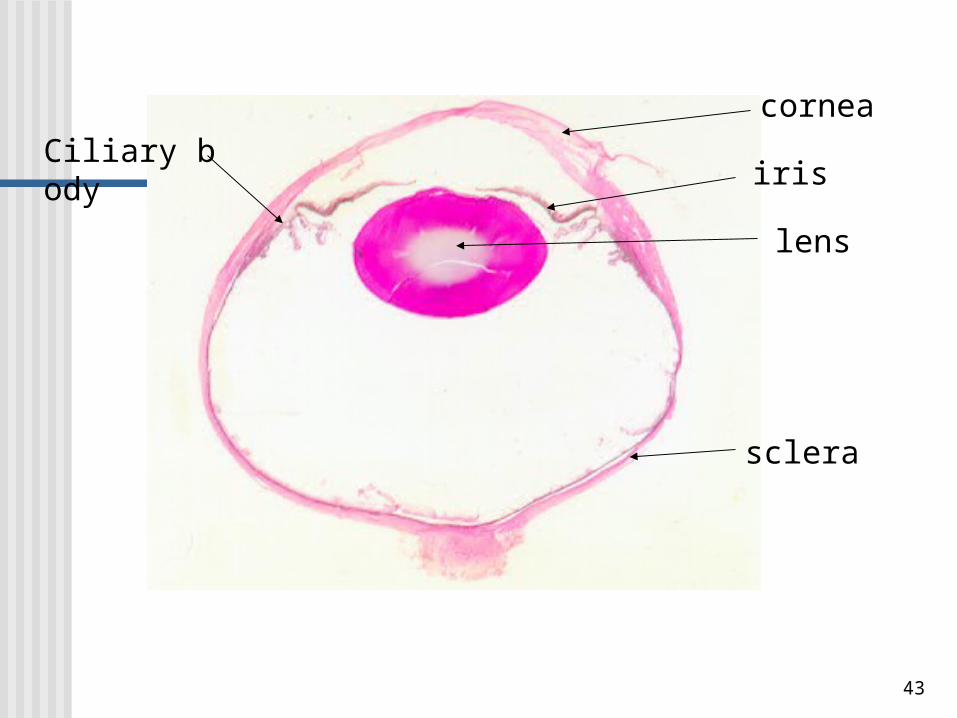
cornea
lens
irisCiliary body
sclera
44
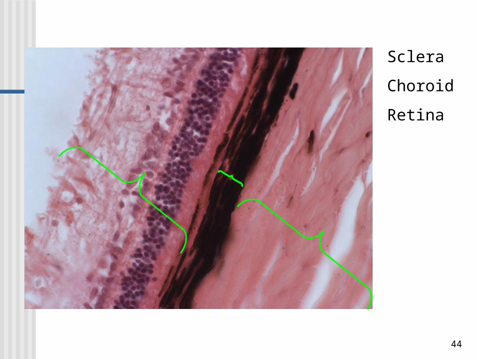
Sclera
Choroid
Retina
45
Scala vestibuli
Scala tympani
Scala media
46
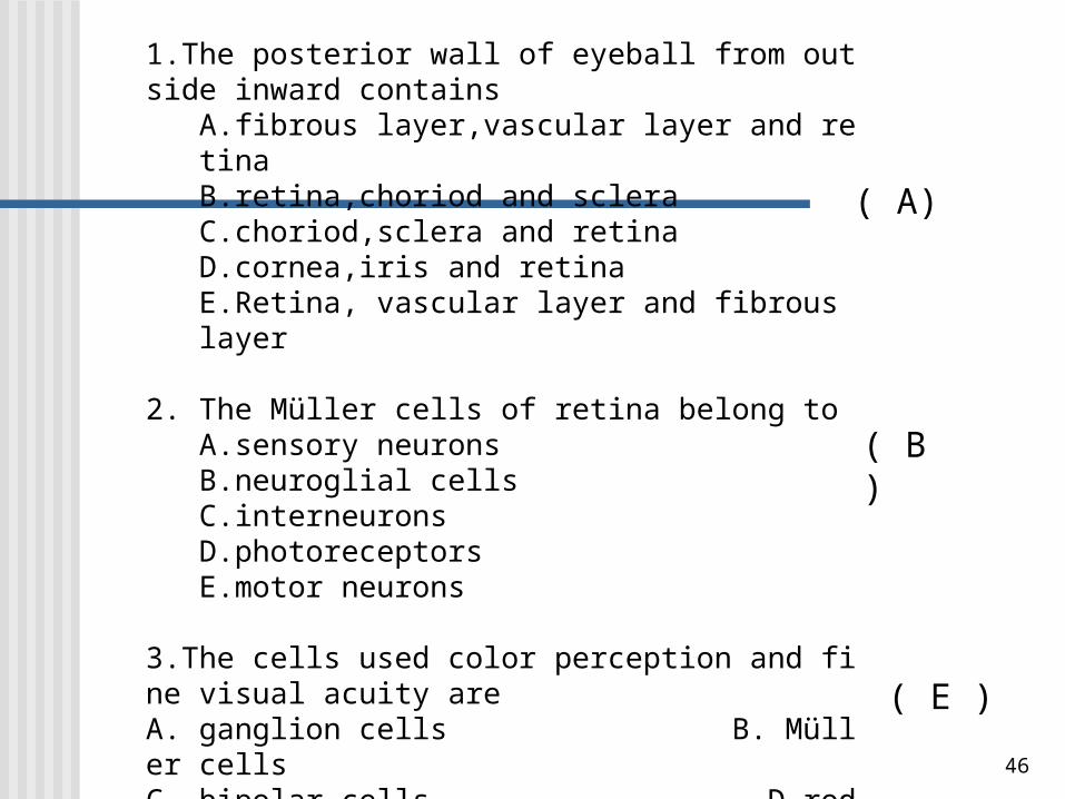
Choice: Select the single most appropriate answer.
1.The posterior wall of eyeball from outside inward containsA.fibrous layer,vascular layer and retinaB.retina,choriod and scleraC.choriod,sclera and retinaD.cornea,iris and retinaE.Retina, vascular layer and fibrous layer
2. The Müller cells of retina belong toA.sensory neuronsB.neuroglial cellsC.interneuronsD.photoreceptorsE.motor neurons
3.The cells used color perception and fine visual acuity areA. ganglion cells B. Müller cellsC. bipolar cells D.rodsE. cones
( A)
( B )
( E )
47
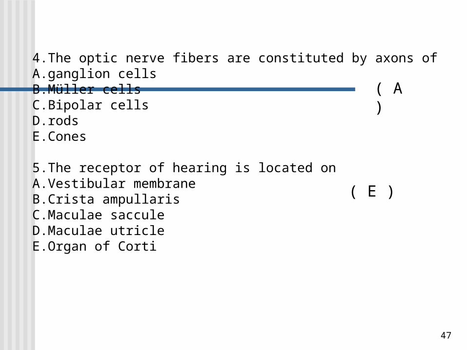
4.The optic nerve fibers are constituted by axons of A.ganglion cellsB.Müller cellsC.Bipolar cellsD.rodsE.Cones
5.The receptor of hearing is located onA.Vestibular membraneB.Crista ampullarisC.Maculae sacculeD.Maculae utricleE.Organ of Corti
( A )
( E )
48
Final score of Histology & Embryology (2013) : Attendance and picture drawing : 10% Quiz : 15% ( each quiz 5% ) LAB Test : 25% Final written examination : 50% Histology : 35-40% Embryology : 10-15%

49
Related Documents


