Case Report Corresponding author: Suba Ananthi Kumarasamy Department of Anatomy, Indira Gandhi Medical College and Research Institute, Kadirkamam, Puducherry 605009, India Tel: +91-9840717911, Fax: +91-0413-2257374, E-mail: subasara@yahoo. com, [email protected] This is an Open Access article distributed under the terms of the Creative Commons Attribution Non-Commercial License (http://creativecommons.org/licenses/by-nc/3.0/) which permits unrestricted non-commercial use, distribution, and reproduction in any medium, provided the original work is properly cited. Copyright © 2014. Anatomy & Cell Biology http://dx.doi.org/10.5115/acb.2014.47.3.207 pISSN 2093-3665 eISSN 2093-3673 Semimembranosus ganglion cyst Suba Ananthi Kumarasamy 1 , Bijun Sai Kannadath 1 , Sandosh Soundamourthy 1 , Aruna Subramanian 1 , Sankappa P. Sinhasan 2 , Ramachandra V. Bhat 2 Departments of 1 Anatomy and 2 Pathology, Indira Gandhi Medical College and Research Institute, Kadirkamam, Puducherry, India Abstract: Ganglion cysts are tumor-like lesions in the soft tissues, generated by mucoid degeneration of the joint capsule, tendon or tendon sheaths on the dorsum of hand, wrist and foot. However, an intratendinous origin for a ganglion cyst is extremely rare. During dissection of the popliteal fossa, a cyst of 2.5 cm×2 cm×0.5 cm was observed in the tendon of right semimembranosus, 3.5 cm above the insertion of the muscle. Contrast X-ray revealed the cyst as not communicating with the knee joint or any adjacent bursae. Histopathological examination confirmed the diagnosis of ganglion cyst. Key words: Ganglion, Knee, Semimembranosus, Tendon sheath, Degeneration Received Decemver 26, 2013; Revised February 17, 2014; Accepted February 27, 2014 semimembranosus, 3.5 cm above the insertion of the muscle (Fig. 1). A transparent gelatinous fluid was aspirated from the mass, indicating a cyst. Thickness of the smooth lining cyst wall was 0.2 cm. The stalk of the cyst was seen. To ascertain its communication with the knee joint, radio-opaque dye was injected. Contrast X-ray revealed it as non-communicating Introduction Ganglion cysts are mucin filled cystic lesions associated with the synovium of either a joint or a tendon sheath. It commonly occurs on the dorsum of the hand, wrist and foot. The cyst may have a single cavity or multilobulated. Cysts in the popliteal fossa which communicate with the knee joint by means of a stalk, are called Baker’s cysts. Non- communicating cysts are ganglion cysts, juxta articular my- xomas [1]. The following is the internationally second ever reported case of an intratendinous ganglion cyst arising from the semimembranosus tendon, with the first being reported in 2009 [2]. Case Report During the dissection of popliteal fossa in a cadaver of a 64-year-old male, a grey white cystic swelling of 2.5 cm×2 cm×0.5 cm was observed attached to the tendon of the right Fig. 1. Arrow showing the ganglion cyst on the tendon of right semimem branosus near its insertion in popliteal fossa with mag- nified image on right. MG, medial head of gastrocnemius; SM, semi- membranosus.

Welcome message from author
This document is posted to help you gain knowledge. Please leave a comment to let me know what you think about it! Share it to your friends and learn new things together.
Transcript
Case Report
Corresponding author: Suba Ananthi KumarasamyDepartment of Anatomy, Indira Gandhi Medical College and Research Institute, Kadirkamam, Puducherry 605009, IndiaTel: +91-9840717911, Fax: +91-0413-2257374, E-mail: [email protected], [email protected]
This is an Open Access article distributed under the terms of the Creative Commons Attribution Non-Commercial License (http://creativecommons.org/licenses/by-nc/3.0/) which permits unrestricted non-commercial use, distribution, and reproduction in any medium, provided the original work is properly cited.
Copyright © 2014. Anatomy & Cell Biology
http://dx.doi.org/10.5115/acb.2014.47.3.207pISSN 2093-3665 eISSN 2093-3673
Semimembranosus ganglion cystSuba Ananthi Kumarasamy1, Bijun Sai Kannadath1, Sandosh Soundamourthy1, Aruna Subramanian1, Sankappa P. Sinhasan2, Ramachandra V. Bhat2
Departments of 1Anatomy and 2Pathology, Indira Gandhi Medical College and Research Institute, Kadirkamam, Puducherry, India
Abstract: Ganglion cysts are tumor-like lesions in the soft tissues, generated by mucoid degeneration of the joint capsule, tendon or tendon sheaths on the dorsum of hand, wrist and foot. However, an intratendinous origin for a ganglion cyst is extremely rare. During dissection of the popliteal fossa, a cyst of 2.5 cm×2 cm×0.5 cm was observed in the tendon of right semimembranosus, 3.5 cm above the insertion of the muscle. Contrast X-ray revealed the cyst as not communicating with the knee joint or any adjacent bursae. Histopathological examination confirmed the diagnosis of ganglion cyst.
Key words: Ganglion, Knee, Semimembranosus, Tendon sheath, Degeneration
Received Decemver 26, 2013; Revised February 17, 2014; Accepted February 27, 2014
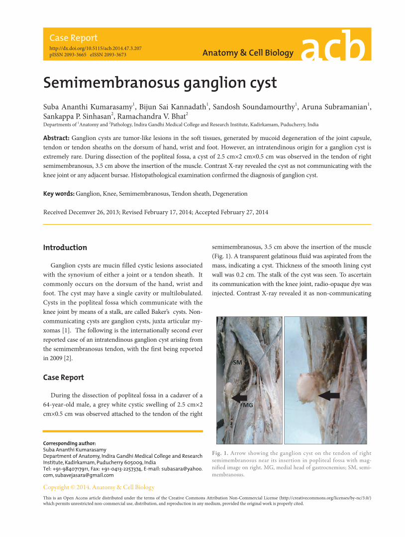
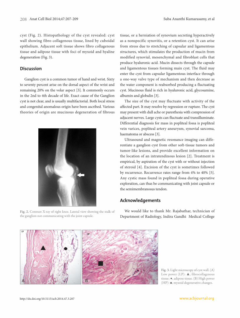
semimembranosus, 3.5 cm above the insertion of the muscle (Fig. 1). A transparent gelatinous fluid was aspirated from the mass, indicating a cyst. Thickness of the smooth lining cyst wall was 0.2 cm. The stalk of the cyst was seen. To ascertain its communication with the knee joint, radio-opaque dye was injected. Contrast X-ray revealed it as non-communicating
Introduction
Ganglion cysts are mucin filled cystic lesions associated with the synovium of either a joint or a tendon sheath. It commonly occurs on the dorsum of the hand, wrist and foot. The cyst may have a single cavity or multilobulated. Cysts in the popliteal fossa which communicate with the knee joint by means of a stalk, are called Baker’s cysts. Non-communicating cysts are ganglion cysts, juxta articular my-xomas [1]. The following is the internationally second ever reported case of an intratendinous ganglion cyst arising from the semimembranosus tendon, with the first being reported in 2009 [2].
Case Report
During the dissection of popliteal fossa in a cadaver of a 64-year-old male, a grey white cystic swelling of 2.5 cm×2 cm×0.5 cm was observed attached to the tendon of the right
Fig. 1. Arrow showing the ganglion cyst on the tendon of right semimem branosus near its insertion in popliteal fossa with magnified image on right. MG, medial head of gastrocnemius; SM, semimembranosus.
Anat Cell Biol 2014;47:207-209 Suba Ananthi Kumarasamy, et al208
www.acbjournal.orghttp://dx.doi.org/10.5115/acb.2014.47.3.207
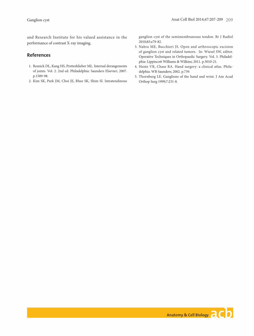
cyst (Fig. 2). Histopathology of the cyst revealed: cyst wall showing fibro collagenous tissue, lined by cuboidal epithelium. Adjacent soft tissue shows fibro collagenous tissue and adipose tissue with foci of myxoid and hyaline degeneration (Fig. 3).
Discussion
Ganglion cyst is a common tumor of hand and wrist. Sixty to seventy percent arise on the dorsal aspect of the wrist and remaining 20% on the volar aspect [3]. It commonly occurs in the 2nd to 4th decade of life. Exact cause of the Ganglion cyst is not clear, and is usually multifactorial. Both local stress and congenital anomalous origin have been ascribed. Various theories of origin are mucinous degeneration of fibrous
tissue, or a herniation of synovium secreting hyperactively as a nonspecific synovitis, or a retention cyst. It can arise from stress due to stretching of capsular and ligamentous structures, which stimulates the production of mucin from modified synovial, mesenchymal and fibroblast cells that produce hyaluronic acid. Mucin dissects through the capsule and ligamentous tissues forming main cyst. The fluid may enter the cyst from capsular ligamentous interface through a one-way valve type of mechanism and then decrease as the water component is reabsorbed producing a fluctuating cyst. Mucinous fluid is rich in hyaluronic acid, glycosamine, albumin and globulin [3].
The size of the cyst may fluctuate with activity of the affected part. It may resolve by regression or rupture. The cyst may present with dull ache or paresthesia with compression of adjacent nerves. Large cysts can fluctuate and transilluminate. Differential diagnosis for mass in popliteal fossa is popliteal vein varices, popliteal artery aneurysm, synovial sarcoma, haematoma or abscess [3].
Ultrasound and magnetic resonance imaging can diffe-rentiate a ganglion cyst from other soft-tissue tumors and tumor-like lesions, and provide excellent information on the location of an intratendinous lesion [2]. Treatment is empirical, by aspiration of the cyst with or without injection of steroid [4]. Excision of the cyst is sometimes followed by recurrence. Recurrence rates range from 4% to 40% [5]. Any cystic mass found in popliteal fossa during operative exploration, can thus be communicating with joint capsule or the semimembranosus tendon.
Acknowledgements
We would like to thank Mr. Rajabathar, technician of Department of Radiology, Indira Gandhi Medical College
Fig. 2. Contrast Xray of right knee. Lateral view showing the stalk of the ganglion not communicating with the joint capsule.
Fig. 3. Light microscopy of cyst wall. (A) Low power (LP): ▲, fibrocollagenous tis sue; ●, adipose tissue. (B) High power (HP): ■, my xoid degenerative changes.
Ganglion cyst
http://dx.doi.org/10.5115/acb.2014.47.3.207
Anat Cell Biol 2014;47:207-209 209
www.acbjournal.org
and Research Institute for his valued assistance in the performance of contrast X-ray imaging.
References
1. Resnick DL, Kang HS, Pretterklieber ML. Internal derangements of joints. Vol. 2. 2nd ed. Philadelphia: Saunders Elsevier; 2007. p.1589-98.
2. Kim SK, Park JM, Choi JE, Rhee SK, Shim SI. Intratendinous
ganglion cyst of the semimembranosus tendon. Br J Radiol 2010;83:e79-82.
3. Nahra ME, Bucchieri JS. Open and arthroscopic excision of ganglion cyst and related tumors. In: Wiesel SW, editor. Operative Techniques in Orthopaedic Surgery. Vol. 3. Philadel-phia: Lippincott Williams & Wilkins; 2011. p.3010-21.
4. Hentz VR, Chase RA. Hand surgery: a clinical atlas. Phila-delphia: WB Saunders; 2002. p.739.
5. Thornburg LE. Ganglions of the hand and wrist. J Am Acad Orthop Surg 1999;7:231-8.
Related Documents


![Ultrasound -WC 2017-KW.pptx [Read-Only]€¦ · Joint‐Elbow effusion, wrist ganglion cyst ... Microsoft PowerPoint - Ultrasound -WC 2017-KW.pptx [Read-Only] Author: akeeney Created](https://static.cupdf.com/doc/110x72/5ac89e287f8b9aa3298c441e/ultrasound-wc-2017-kwpptx-read-only-jointelbow-effusion-wrist-ganglion.jpg)