Decompression with fusion and decompression alone are the two treatment options for patients who have a lumbar spinal stenosis with degenerative spondylolisthesis. 1) Decompression with fusion can improve back pain, radiating pain, and neurogenic intermittent claudica- tion. In addition, it does not cause instability in patients with spondylolisthesis. 2) However, this surgery can cause adjacent segmental degeneration, pseudoarthrosis and large volume blood loss during the surgery. Additionally, the hospitalization period is long. 3,4) Recently, as the de- compression surgery technique without fusion has been developed, there have been some studies that found no significant increase in instability after spinal decompres- sion surgery without fusion through various types of mi- croexcision, in patients who have a lumbar spinal stenosis with degenerative spondylolisthesis. 5-7) Semi-Circumferential Decompression: Microsurgical Total en-bloc Ligamentum Flavectomy to Treat Lumbar Spinal Stenosis with Grade I Degenerative Spondylolisthesis Young Sang Lee, MD, Jun Cheol Choi, MD, Sang Hun Oh, MD, Sub Ri Park, MD, Sang Jun Park, MD, Nam Ik Cho, MD Department of Orthopedic Surgery, Bundang Jesaeng General Hospital, Daejin Medical Center, Seongnam, Korea Background: To describe and assess clinical outcomes of the semi-circumferential decompression technique for microsurgical en- bloc total ligamentum flavectomy with preservation of the facet joint to treat the patients who have a lumbar spinal stenosis with degenerative spondylolisthesis. Methods: We retrospectively analyzed the clinical and radiologic outcomes of 19 patients who have a spinal stenosis with Mey- erding grade I degenerative spondylolisthesis. They were treated using the “semi-circumferential decompression” method. We evaluated improvements in back and radiating pain using a visual analogue scale (VAS) and the Oswestry Disability Index (ODI). We also evaluated occurrence of spinal instability on radiological exam using percentage slip and slip angle. Results: The mean VAS score for back pain decreased significantly from 6.3 to 4.3, although some patients had residual back pain. The mean VAS for radiating pain decreased significantly from 8.3 to 2.5. The ODI score improved significantly from 25.3 pre- operatively to 10.8 postoperatively. No significant change in percentage slip was observed (10% preoperatively vs. 12.2% at the last follow-up). The dynamic percentage slip (gap in percentage slip between flexion and extension X-ray exams) did not change significantly (5.2% vs. 5.8%). Slip angle and dynamic slip angle did not change (3.2 o and 8.2 o vs. 3.6 o and 9.2 o , respectively). Conclusions: The results suggested that semi-circumferential decompression is a clinically recommendable procedure that can improve pain. This procedure does not cause spinal instability when treating patients who have a spinal stenosis with degenera- tive spondylolisthesis. Keywords: Degenerative spondylolisthesis, Semi-circumferential decompression, Total ligamentum flavectomy Copyright © 2015 by e Korean Orthopaedic Association is is an Open Access article distributed under the terms of the Creative Commons Attribution Non-Commercial License (http://creativecommons.org/licenses/by-nc/4.0) which permits unrestricted non-commercial use, distribution, and reproduction in any medium, provided the original work is properly cited. Clinics in Orthopedic Surgery • pISSN 2005-291X eISSN 2005-4408 Received March 16, 2015; Accepted August 17, 2015 Correspondence to: Young Sang Lee, MD Department of Orthopedic Surgery, Bundang Jesaeng General Hospital, Daejin Medical Center, 20 Seohyeon-ro 180beon-gil, Bundang-gu, Seongnam 13590, Korea Tel: +82-31-779-0175, Fax: +82-31-779-0179 E-mail: [email protected] Original Article Clinics in Orthopedic Surgery 2015;7:470-475 • http://dx.doi.org/10.4055/cios.2015.7.4.470

Welcome message from author
This document is posted to help you gain knowledge. Please leave a comment to let me know what you think about it! Share it to your friends and learn new things together.
Transcript
Decompression with fusion and decompression alone are the two treatment options for patients who have a lumbar spinal stenosis with degenerative spondylolisthesis.1)
Decompression with fusion can improve back pain,
radiating pain, and neurogenic intermittent claudica-tion. In addition, it does not cause instability in patients with spondylolisthesis.2) However, this surgery can cause adjacent segmental degeneration, pseudoarthrosis and large volume blood loss during the surgery. Additionally, the hospitalization period is long.3,4) Recently, as the de-compression surgery technique without fusion has been developed, there have been some studies that found no significant increase in instability after spinal decompres-sion surgery without fusion through various types of mi-croexcision, in patients who have a lumbar spinal stenosis with degenerative spondylolisthesis.5-7)
Semi-Circumferential Decompression: Microsurgical Total en-bloc Ligamentum
Flavectomy to Treat Lumbar Spinal Stenosis with Grade I Degenerative Spondylolisthesis
Young Sang Lee, MD, Jun Cheol Choi, MD, Sang Hun Oh, MD, Sub Ri Park, MD, Sang Jun Park, MD, Nam Ik Cho, MD
Department of Orthopedic Surgery, Bundang Jesaeng General Hospital, Daejin Medical Center, Seongnam, Korea
Background: To describe and assess clinical outcomes of the semi-circumferential decompression technique for microsurgical en-bloc total ligamentum flavectomy with preservation of the facet joint to treat the patients who have a lumbar spinal stenosis with degenerative spondylolisthesis. Methods: We retrospectively analyzed the clinical and radiologic outcomes of 19 patients who have a spinal stenosis with Mey-erding grade I degenerative spondylolisthesis. They were treated using the “semi-circumferential decompression” method. We evaluated improvements in back and radiating pain using a visual analogue scale (VAS) and the Oswestry Disability Index (ODI). We also evaluated occurrence of spinal instability on radiological exam using percentage slip and slip angle.Results: The mean VAS score for back pain decreased significantly from 6.3 to 4.3, although some patients had residual back pain. The mean VAS for radiating pain decreased significantly from 8.3 to 2.5. The ODI score improved significantly from 25.3 pre-operatively to 10.8 postoperatively. No significant change in percentage slip was observed (10% preoperatively vs. 12.2% at the last follow-up). The dynamic percentage slip (gap in percentage slip between flexion and extension X-ray exams) did not change significantly (5.2% vs. 5.8%). Slip angle and dynamic slip angle did not change (3.2o and 8.2o vs. 3.6o and 9.2o, respectively). Conclusions: The results suggested that semi-circumferential decompression is a clinically recommendable procedure that can improve pain. This procedure does not cause spinal instability when treating patients who have a spinal stenosis with degenera-tive spondylolisthesis.Keywords: Degenerative spondylolisthesis, Semi-circumferential decompression, Total ligamentum flavectomy
Copyright © 2015 by The Korean Orthopaedic AssociationThis is an Open Access article distributed under the terms of the Creative Commons Attribution Non-Commercial License (http://creativecommons.org/licenses/by-nc/4.0)
which permits unrestricted non-commercial use, distribution, and reproduction in any medium, provided the original work is properly cited.Clinics in Orthopedic Surgery • pISSN 2005-291X eISSN 2005-4408
Received March 16, 2015; Accepted August 17, 2015Correspondence to: Young Sang Lee, MDDepartment of Orthopedic Surgery, Bundang Jesaeng General Hospital, Daejin Medical Center, 20 Seohyeon-ro 180beon-gil, Bundang-gu, Seongnam 13590, KoreaTel: +82-31-779-0175, Fax: +82-31-779-0179E-mail: [email protected]
Original Article Clinics in Orthopedic Surgery 2015;7:470-475 • http://dx.doi.org/10.4055/cios.2015.7.4.470
471
Lee et al. Semi-Circumferential Decompression for Spinal Stenosis with Degenerative SpondylolisthesisClinics in Orthopedic Surgery • Vol. 7, No. 4, 2015 • www.ecios.org
We used the semi-circumferential decompression (SCD) method during surgeries for patients who had a lumbar spinal stenosis with degenerative spondylolisthe-sis for the last 4 years. SCD is a decompressive technique to perform a total en-bloc ligamentum flavectomy under a microscope that preserves the articular facet. We have observed and analyzed relief of symptoms and occurrence of instability. In this study, we suggest the clinical effective-ness of the SCD technique for posterior decompression to treat patients who have a lumbar spinal stenosis with degenerative spondylolisthesis.
METHODS
We retrospectively analyzed the outcomes of 19 patients (mean age, 67.9 years; 2 men and 17 women) who were treated using the SCD method for a lumbar spinal stenosis with degenerative spondylolisthesis from 2010 to 2013. We excluded patients who had bilateral foraminal stenosis. The average follow-up period was 37 months (range, 25 to 56 months). All patients had radiating pain and neurologic intermittent claudication (NIC) due to a spinal stenosis.
Magnetic resonance imaging showed central and lateral recess stenosis at the degenerative spondylolisthesis site in all patients. From 2010 to 2013, there was one patient with Meyerding grade II spondylolisthesis and no patient with Meyerding grade III spondylolisthesis. The follow-up pe-riod of patients with Meyerding grade II spondylolisthesis was too short (5 months). Hence, only patients with Mey-erding grade I spondylolisthesis were included. Single-level degenerative spondylolisthesis was observed at L4 on 5 in 13 of 19 patients, L3 on 4 in four patients, and L5 on S1 in one patient. Double-level degenerative spondylolis-thesis was observed at L3 on 4 and L4 on 5 in one patient. No instability was detected on a preoperative lumbar spine dynamic (flexion/extension) radiological study.
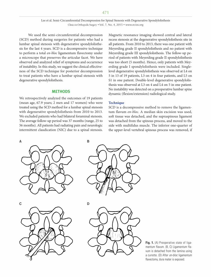
TechniqueSCD is a decompressive method to remove the ligamen-tum flavum en-bloc. A median skin excision was used, soft tissue was detached, and the supraspinous ligament was detached from the spinous process, and moved to the side with multifidus muscle. The inferior one-quarter of the upper-level vertebral spinous process was removed, if
Fig. 1. (A) Preoperative state of liga-mentum flavum. (B, C) Ligamentum fla-vum is detached from the lamina using a currette. (D) After en-bloc ligamentum flavectomy, dura mater is exposed.
A B
C D

472
Lee et al. Semi-Circumferential Decompression for Spinal Stenosis with Degenerative SpondylolisthesisClinics in Orthopedic Surgery • Vol. 7, No. 4, 2015 • www.ecios.org
needed, to enhance vision. The ligamentum flavum was detached from the inferior one-third of the lamina using a currette. After thinning the lamina with a high speed burr, a partial laminectomy was performed using a Kerrison rongeur. It is important that ligamentum flavum must not be removed before thinning the lamina with a high speed burr. By maintaining ligamentum flavum, we prevented damage to the dura mater while using the high speed burr. Finally, the total ligamentum flavum was removed en-bloc by detaching the ligamentum flavum from the inner side of the lamina to the inner side of the posterior facet (Figs. 1 and 2). The facet joint was preserved by leaving the superior articular facet unexcised. The detached supraspi-nous ligament was returned to its original position, and sutured with surrounding fascia. Ambulation was permit-ted beginning on postoperative day 1, and patients were encouraged to use a corset for 6 weeks.
Preoperative and postoperative symptom relief was estimated using the visual analogue scale (VAS) score and the Oswestry Disability Index (ODI) before surgery and at follow-up days. Patients were evaluated for VAS score and ODI after 3, 6 month post-surgery and every 1 year subse-
quently.We compared the preoperative and last follow-up X-
rays. All patients underwent dynamic (flexion/extension) X-rays. These results were used to estimate slip percent-age and slip angle and assess instability and progression of lumbar degenerative spondylolisthesis. Slip percentage and slip angle was estimated using Taillard’s and Boxall's methods, respectively. We estimated dynamic slip per-centage (preoperative and postoperative change in the slip percentage) and dynamic slip angle (preoperative and postoperative change in the slip angle), and analyzed the occurrence of vertebral instability. The Wilcoxon signed-rank test was performed to detect postoperative changes using IBM SPSS ver. 21.0 (IBM Co., Armonk, NY, USA). A p-value less than 0.05 was considered significant.
RESULTS
Clinical ResultThe mean VAS score of back pain decreased from 6.3 to 4.3, and the mean VAS score for lower leg radiating pain also decreased from 8.3 to 2.5 (p < 0.01). The average ODI
Fig. 2. The ligamentum flavum, which is in the shape of a butterfly, was removed en-bloc. (A) Dorsal surface, (B) ventral surface, (C) distal border, and (D) proximal border.
A B
C D

473
Lee et al. Semi-Circumferential Decompression for Spinal Stenosis with Degenerative SpondylolisthesisClinics in Orthopedic Surgery • Vol. 7, No. 4, 2015 • www.ecios.org
score (maximum, 45 points) improved significantly from 25.3 preoperatively to 10.3 postoperatively (p < 0.01).
Radiologic ResultThe change in slip percentage increased from 10% to 12.2% postoperatively, but the difference was not signifi-cant. The dynamic slip percentage did not show significant change postoperatively (5.2% vs. 5.8%). Slip angle in the SCD group also did not change (3.2° vs. 3.6°) at the last follow-up. The dynamic slip angle did not change (8.2° vs. 9.2°). These results indicated that the SCD method did not cause any vertebral instability.
DISCUSSION
Newman and Stone8) described degenerative spondylolis-thesis as a disease in which a vertebral body slips anteri-orly with no neural arch abnormality. Degenerative spon-dylolisthesis tends to be accompanied by spinal stenosis, and typically shows degeneration of the posterior facet or intervertebral disc, and ligament laxity. So far, decompres-sion with fusion is reportedly better than decompression-only surgery.9-11) In particular, a broad range of decom-pression techniques lead to poor outcomes, as compared
to decompression with fusion.12) According to the spinal degeneration theory suggested by Kirkaldy-Willis, spinal segment stability is maintained in patients with degenera-tive spondylolisthesis,13,14) and good surgical outcomes occur if sufficient decompression is performed preserving structures that affect posterior spinal stability. Thus, stud-ies on decompression-only surgery with minimal excision to treat patients with degenerative spondylolisthesis were recently conducted. Weiner et al.5) reported a surgical pro-cedure that restores the native position of the spinous pro-cess after a spinous process osteotomy and decompression to maintain stability by preserving the interspinous and supraspinous ligaments. Recently, various techniques us-ing unilateral approach and bilateral decompression have been used in an attempt to minimize postoperative verte-bral instability.6,7) The SCD technique has been introduced to prevent postoperative vertebral instability.15) Collective-ly, ours and previous results suggested that SCD technique could be applied for treatment of degenerative spondylo-listhesis without instability and also for spondylolisthesis with instability preoperatively. If spinal segment stability is maintained postoperatively and sufficient decompres-sion is achieved, surgical effect is well maintained. We car-ried out another study on the effect of SCD technique for
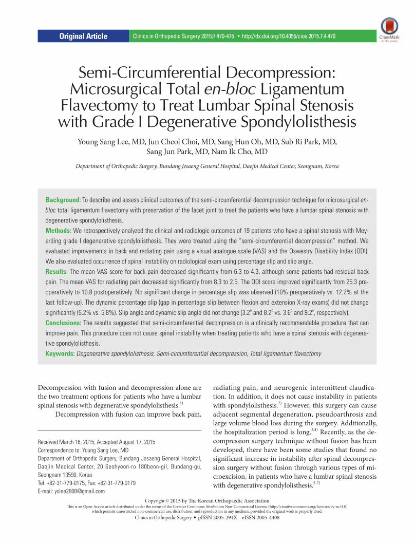
Fig. 3. Preoperative (A, C) and post operative (B, D) axial computed tomo graphy scans and magnetic reso nance images show the facet joint at the spon dylolisthesis level in a repre sen tative patient who underwent semi-circum ferential decompression preserving the facet joints. A bilateral laminotomy and total ligamentum flavectomy were performed, and the facet joint was com-pletely preserved.
A B
C D

474
Lee et al. Semi-Circumferential Decompression for Spinal Stenosis with Degenerative SpondylolisthesisClinics in Orthopedic Surgery • Vol. 7, No. 4, 2015 • www.ecios.org
spondylolisthesis with segmental instability. However, the follow-up period was short yet, studies to evaluate the ef-fect of SCD technique for spondylolisthesis with instability after sufficient follow-up period are currently ongoing.
Anatomical research about ligamentum flavum by Okuda et al.16) suggested that ligamentum flavum de-generate, ossify, and calcify in patients with degenerative spondylosis, and that these changes are more severe if spondylolisthesis is present. Another research by Okuda et al.17) showed that nerve root compression is most severe in the proximal portion of the ligamentum flavum because the ligamentum flavum is thickest in this area (Fig. 2). The proximal border of the ligamentum flavum is attached to upper level vertebra at the inner surface of lamina almost horizontally and just below the pedicle; thus nerve root is compressed continuously when the ligamentum flavum is not completely removed.17) However, we can determine whether decompression is sufficient by observing the liga-mentum flavum removed by en-bloc resection.
Zander et al.18) reported that a unilateral medial hemi-facetectomy can induce vertebral instability, and Hamasaki et al.19) reported that a bilateral medial facetectomy for the medial one-third of the posterior facet could induce ver-tebral instability. Actually, in patients who have spinal ste-nosis with degenerative spondylolisthesis, cause of nerve root compression is ligamentum flavum hypertrophy at lateral recess, and facet hypertrophy is not cause of nerve root compression. So medial facetectomy is unnecessary for decompression of nerve root, and SCD technique can decompress nerve root sufficiently, maintaining spinal segmental stability.
In addition, Abumi et al.20) conducted a biomechani-cal study demonstrating that spinal instability does not develop when the posterior facet is preserved with only the interspinous and supraspinous ligaments detached. According to these studies, the SCD technique, which de-compress posteriorly by total excision of the ligamentum flavum with preservation of the supraspinous ligament and posterior facet, improves clinical outcomes and does not lead to spinal instability. Thus, the SCD technique can be applied to patients who have a spinal stenosis with de-generative spondylolisthesis (Fig. 3).
Based on analysis of clinical improvement and change in slip percentage and slip angle, we demonstrated that decompression through a total ligamentum flavec-tomy preserving the facet joint results in clinical improve-ment without causing vertebral instability.
The limitation of SCD technique is that this tech-nique is not suitable for bilateral foraminal stenosis. There has been consensus that posterior lumbar interbody fusion is suitable for treatment of bilateral foraminal stenosis. However, the spinal stenosis with degenerative spondylo-listhesis and unilateral foraminal stenosis can be treated using SCD technique and lateral fenestration technique simultaneously.
Hence, degenerative spondylolisthesis without fo-raminal stenosis, in which especially NIC is main symp-tom, is an absolute indication for SCD, and degenerative spondylolisthesis with unilateral foraminal stenosis is a relative indication for SCD. Degenerative spondylolisthesis with severe bilateral foraminal stenosis is a contraindica-tion for SCD.
Decompression alone can reduce preoperative back pain but some preoperative back pain remains because it usually results from degeneration of facet joint degenera-tion or intervertebral disc. None of our patients had severe back pain that interfere with daily life activities.
Long term follow-up observations to assess instabil-ity and radiological exacerbations of spondylolisthesis are needed. Although a prospective randomized controlled clinical study is needed, our results showed that decom-pression surgery using the SCD technique was effective and less invasive for patients with spinal stenosis and de-generative spondylolisthesis.
In conclusion, the SCD technique, which decom-press posteriorly by en-bloc total ligamentum flavectomy and preserve posterior facet, was clinically effective and does not lead to postoperative spinal instability.
CONFLICT OF INTEREST
No potential conflict of interest relevant to this article was reported.
REFERENCES
1. Eismont FJ, Norton RP, Hirsch BP. Surgical management of lumbar degenerative spondylolisthesis. J Am Acad Orthop Surg. 2014;22(4):203-13.
2. Fischgrund JS, Mackay M, Herkowitz HN, Brower R, Mont-
gomery DM, Kurz LT. 1997 Volvo Award winner in clinical studies. Degenerative lumbar spondylolisthesis with spinal stenosis: a prospective, randomized study comparing de-compressive laminectomy and arthrodesis with and without spinal instrumentation. Spine (Phila Pa 1976). 1997;22(24):
475
Lee et al. Semi-Circumferential Decompression for Spinal Stenosis with Degenerative SpondylolisthesisClinics in Orthopedic Surgery • Vol. 7, No. 4, 2015 • www.ecios.org
2807-12.
3. Ragab AA, Fye MA, Bohlman HH. Surgery of the lumbar spine for spinal stenosis in 118 patients 70 years of age or older. Spine (Phila Pa 1976). 2003;28(4):348-53.
4. Carreon LY, Puno RM, Dimar JR 2nd, Glassman SD, John-son JR. Perioperative complications of posterior lumbar decompression and arthrodesis in older adults. J Bone Joint Surg Am. 2003;85(11):2089-92.
5. Weiner BK, Fraser RD, Peterson M. Spinous process oste-otomies to facilitate lumbar decompressive surgery. Spine (Phila Pa 1976). 1999;24(1):62-6.
6. Nakanishi K, Tanaka N, Fujimoto Y, et al. Medium-term clinical results of microsurgical lumbar flavectomy that pre-serves facet joints in cases of lumbar degenerative spondylo-listhesis: comparison of bilateral laminotomy with bilateral decompression by a unilateral approach. J Spinal Disord Tech. 2013;26(7):351-8.
7. Musluman AM, Cansever T, Yilmaz A, Cavusoglu H, Yuce I, Aydın Y. Midterm outcome after a microsurgical unilateral approach for bilateral decompression of lumbar degenera-tive spondylolisthesis. J Neurosurg Spine. 2012;16(1):68-76.
8. Newman PH, Stone KH. The etiology of spondylolisthesis. J Bone Joint Surg Br. 1963;45(1):39-59.
9. Sengupta DK, Herkowitz HN. Degenerative spondylolisthe-sis: review of current trends and controversies. Spine (Phila Pa 1976). 2005;30(6 Suppl):S71-81.
10. Herkowitz HN, Kurz LT. Degenerative lumbar spondylolis-thesis with spinal stenosis: a prospective study comparing decompression with decompression and intertransverse process arthrodesis. J Bone Joint Surg Am. 1991;73(6):802-8.
11. Mardjetko SM, Connolly PJ, Shott S. Degenerative lumbar spondylolisthesis: a meta-analysis of literature 1970-1993.
Spine (Phila Pa 1976). 1994;19(20 Suppl):2256S-2265S.
12. Chung NS, Jeon CH. Surgical treatment of degenerative and isthmic spondylolisthesis. J Korean Soc Spine Surg. 2009; 16(3):228-34.
13. Kirkaldy-Willis WH, Hill RJ. A more precise diagnosis for low-back pain. Spine (Phila Pa 1976). 1979;4(2):102-9.
14. Matsunaga S, Sakou T, Morizono Y, Masuda A, Demirtas AM. Natural history of degenerative spondylolisthesis: pathogenesis and natural course of the slippage. Spine (Phila Pa 1976). 1990;15(11):1204-10.
15. Baba I, Murakamu T. Treatment for degenerative spondylo-listhesis: the results of semi-circumferential decompression, surgical therapy without fusion. Rinsho Seikei Geka. 1997; 32:1303-9.
16. Okuda T, Baba I, Fujimoto Y, et al. The pathology of liga-mentum flavum in degenerative lumbar disease. Spine (Phila Pa 1976). 2004;29(15):1689-97.
17. Okuda T, Fujimoto Y, Tanaka N, Ishida O, Baba I, Ochi M. Morphological changes of the ligamentum flavum as a cause of nerve root compression. Eur Spine J. 2005;14(3):277-86.
18. Zander T, Rohlmann A, Klockner C, Bergmann G. Influ-ence of graded facetectomy and laminectomy on spinal bio-mechanics. Eur Spine J. 2003;12(4):427-34.
19. Hamasaki T, Tanaka N, Kim J, Okada M, Ochi M, Hut-ton WC. Biomechanical assessment of minimally invasive decompression for lumbar spinal canal stenosis: a cadaver study. J Spinal Disord Tech. 2009;22(7):486-91.
20. Abumi K, Panjabi MM, Kramer KM, Duranceau J, Oxland T, Crisco JJ. Biomechanical evaluation of lumbar spinal stability after graded facetectomies. Spine (Phila Pa 1976). 1990;15(11):1142-7.
Related Documents