11 Semen Analysis Alex C Varghese, Furquan Pathan, Ang Wen Jeat, Rakesh Sharma, Ashok Agarwal ABSTRACT Approximately 15% of the couples trying to conceive are clinically infertile. Male factor is involved in half of these cases. Semen analysis remains the cornerstone of evaluating male infertility. It is one of the first tests done to evaluate a man’s fertility. It is inexpensive but can help determine if there is a problem in sperm production or it is the quality of the sperm that is causing infertility. In this chapter, the basics of semen analysis are explained. The tests are simple but can be meaningful if performed by highly trained and specialized professional, in modern laboratories that are accredited to maintain the high standards and have strict quality controls. INTRODUCTION Approximately one in six couples is affected by infertility, a problem that can be caused by a number of factors, both male and female (Sigman and Jarow, 2007). The cause is attributed to the female in 30% of cases, to the male in 30%, to both in 30% and is unknown in 10% of cases. The methods for evaluation of male infertility typically have been limited to a semen analysis that evaluates sperm count, motility, and morphology. A semen analysis evaluates certain characteristics of a man’s semen and the sperm contained in the semen. It is an essential component of male infertility investigation, and interpretation of the results plays a vital role in the overall treatment of infertile couples. The basic aim of semen analysis is to evaluate descriptive parameters of the ejaculate (a mixture of spermatozoa suspended along with secretions from the testis, epididymis and other accessory glands). For a clinician, semen analysis results are predictive of potential fertility and the possible causes of infertility; for an epidemiologist, the results are the basis for assessing hazards in the environment, occupational exposure, or effects of drugs and chemicals. The sample must be obtained and transported to the clinical laboratory according to World Health

Welcome message from author
This document is posted to help you gain knowledge. Please leave a comment to let me know what you think about it! Share it to your friends and learn new things together.
Transcript
11
Semen Analysis
Alex C Varghese, Furquan Pathan, Ang Wen Jeat, Rakesh Sharma, Ashok Agarwal
ABSTRACT
Approximately 15% of the couples trying to conceive are clinically infertile. Malefactor is involved in half of these cases. Semen analysis remains the cornerstoneof evaluating male infertility. It is one of the first tests done to evaluate a man’sfertility. It is inexpensive but can help determine if there is a problem in spermproduction or it is the quality of the sperm that is causing infertility. In thischapter, the basics of semen analysis are explained. The tests are simple but canbe meaningful if performed by highly trained and specialized professional, inmodern laboratories that are accredited to maintain the high standards andhave strict quality controls.
INTRODUCTION
Approximately one in six couples is affected by infertility, a problem thatcan be caused by a number of factors, both male and female (Sigman andJarow, 2007). The cause is attributed to the female in 30% of cases, to themale in 30%, to both in 30% and is unknown in 10% of cases. The methodsfor evaluation of male infertility typically have been limited to a semenanalysis that evaluates sperm count, motility, and morphology.
A semen analysis evaluates certain characteristics of a man’s semen andthe sperm contained in the semen. It is an essential component of maleinfertility investigation, and interpretation of the results plays a vital rolein the overall treatment of infertile couples. The basic aim of semen analysisis to evaluate descriptive parameters of the ejaculate (a mixture ofspermatozoa suspended along with secretions from the testis, epididymisand other accessory glands).
For a clinician, semen analysis results are predictive of potential fertilityand the possible causes of infertility; for an epidemiologist, the results arethe basis for assessing hazards in the environment, occupational exposure,or effects of drugs and chemicals. The sample must be obtained andtransported to the clinical laboratory according to World Health
ANDROLOGY LABORATORY MANUAL182
Organization (WHO) guidelines (WHO, 1999), as semen analysis resultscan be significantly influenced by both the technique of semen collectionand the methods of laboratory analysis.
The WHO manual recommends obtaining two samples for initialevaluation at an interval of not less than 7 days or more than 3 weeks. If theresults from the two samples are distinctly different, additional sampleshave to be collected and examined.
A routine semen analysis includes the following important steps:
PATIENT INSTRUCTIONS FOR SAMPLE COLLECTION, SAFEHANDLING AND DELIVERY
1. Patient should be given clear and simple instructions explaining the needfor semen analysis and what is required for specimen collection.
2. Patient should be informed about the importance of abstinence time.Ejaculate must be collected after 3-5 days (but not more than 7 days) ofabstinence.
3. Samples should be obtained by masturbation and collected in a warm(20-40ºC), sterile, nontoxic plastic or glass wide-mouth container. Priorto sample collection, the patient must void and wash hands and genitalsto minimize the chances of contamination.
4. Use of lubricants and saliva should be avoided as their potential toxicitymight influence the result. Semen samples should be protected fromextremes of temperature (<20ºC or >40ºC) during transport to thelaboratory.
5. All sample containers are labeled with adequate information to eliminateany chances of error.
6. Regular condoms should not be used because of their spermicidal effect.Ideally, the samples must be collected close to the laboratory. If the
specimen cannot be produced close to the laboratory, it must be deliveredto the laboratory as soon as possible, certainly within 1 hour of collection.During this period, the sample has to be kept warm by carrying it next tothe body, and temperature extremes must be avoided.
INFORMATION ON THE SAMPLE CONTAINER AND PATIENT SHEET
Once the patient has collected the specimen, some preliminary informationabout the specimen should be obtained:1. Label the specimen clearly, indicating patient’s complete name, clinic
number, and collection date.2. Record collection date and time.3. Record abstinence time in days.4. Record time the sample is received at the laboratory.
SEMEN ANALYSIS 183
5. Record information about split ejaculate, noting when the sample waslost and whether the spill occurred at the beginning or after ejaculation.The samples always should be labeled as “Biohazard” and extreme
precaution should be taken. Follow safety guidelines and protocols inhandling the sample as the semen sample may contain infectious agents(e.g. hepatitis B, hepatitis C, HIV, herpes simplex).
PHYSICAL EXAMINATION
Macroscopic Examination
Semen Age
Record the time when the sample was received, to the liquefaction time.
Liquefaction
Incubation of the sample must be carried out at either ambient temperatureor by placing the specimen in an incubator at 37ºC. A normal sample usuallyliquefies within 60 minutes at room temperature, although usually thisoccurs within 15-20 minutes. It is determined by the time required for thegelatinous mass to liquefy. A normal sample might contain gel-likegelatinous corpuscles that do not liquefy. Exact liquefaction time is of nodiagnostic importance unless >2 hours elapse without any change. Thismay indicate poor prostatic secretion since the liquefying enzymes arederived from the prostate gland. On the other hand, absence of coagulationmay indicate ejaculatory duct obstruction or congenital absence of seminalvesicles.
For samples that do not liquefy, the sample can be mixed by rapidlymixing with a regular transfer pipette or, if the sample is viscous, viscositycan be broken by using a viscosity treatment system.
Color and Odor
It is important to note the color. Normal semen is homogeneously opaque,whitish grey or pearly white. The semen odor is unmistakable and pungentbecause of sperm oxidation. A yellowish tinge to the semen appears withan increase in the days of abstinence or probably due to carotene pigment.More pronounced yellow discoloration may indicate jaundice orcontamination of semen with urine (e.g. bladder neck dysfunction). Drugslike methylene blue and pyridium may also color the semen. Fresh blood(hematospermia) will give semen a reddish tinge, while old blood gives ita brownish tinge. This could be due to the presence of inflammation.Prostatic secretions give semen a strong distinctive odor. Absence oruncharacteristic odor could be associated with an infection.
ANDROLOGY LABORATORY MANUAL184
Volume
Volume of the ejaculate should be measured by transferring the liquefiedsample into a graduated 15 mL conical centrifuge tube. The normal volumeof ejaculate after 2-5 days of sexual abstinence is about 2-6 mL. Retrogradeejaculation, obstruction of lower urinary tract (urethra, congenital absenceof vas deferens, seminal vesicles) may yield low volume. According to theWHO laboratory manual, the reference value for semen volume is ≥ 2.0mL; however, for clinical purposes; semen volume is differentiated intothree categories to facilitate interpretation and diagnosis:
Aspermia: No semen produced after orgasm (seen in certain clinicalconditions).
Hypospermia: <0.5 mL of semen ejaculated (partial or complete retrogradeflow of semen, accessory glands impairment).
Hyperspermia: > 6 mL of semen ejaculated (long period of sexual abstinenceor overproduction of fluids from the accessory sex glands).
If the volume is <1 mL it is important to determine if the sample iscomplete. The highest sperm concentration is seen in the initial ejaculate.
Viscosity
Viscosity measures the seminal fluid’s resistance to flowing. It is measuredby the length of the ‘thread-lines’ or ‘spinnbarkeit.’ It can be estimated byusing a glass rod and observing the length of thread that forms on withdrawalof the rod. A normal sample leaves small, discrete drops; abnormal sampleswill form threads more than 2 cm long. High viscosity may interfere withdeterminations of sperm motility, concentration and antibody coating ofspermatozoa. Viscosity can be categorized as ‘normal’, ‘moderate’ or ‘high.’Viscous samples can be treated by a viscosity treatment system containing apremeasured vial of chymotrypsin (5 mg/vial (Conception Technologies,San Diego, CA). The sample can be swirled and left in the incubator foranother 10-15 min till viscosity is completely broken down and the sample issuitable for analysis.
pH
The pH of liquefied semen is determined by using pH test strips; pH 6.5 to10 has been found most suitable for this purpose. A drop of semen is spreadevenly onto the pH paper. After 30 seconds, the color of the impregnatedzone is compared with the calibrated strip.
Normal semen pH is in the range of 7.2 to 8.2, and it does tend to increasewith time after ejaculation. Any change in the normal range of pH may becaused by inflammation of the prostate or seminal vesicles.
SEMEN ANALYSIS 185
MICROSCOPIC EXAMINATION
Wet Preparation Examination
Load a 5 µL of well-mixed semen on a clean, warmed microscope slidewith a cover slip on top (18 × 18 mm). If a 22 × 22 mm cover slip is used, thesemen volume on the microscope slide should be 10 µL). This preparationhas a depth of approximately ~20 µm. A depth less than 20 µm will hamperthe rotational movements of the spermatozoa. Care should be taken to avoidformation of air bubbles that can be trapped between the cover slip andthe slide. It is important to wait for the drifting to cease/stabilize beforeexamination. In addition, a variety of other disposable two-well 20 µmcounting chambers are also available.
A phase contrast microscope is recommended for all examinations ofunstained preparations of fresh/washed semen. Initial examination is doneunder 100× total magnification (10× objective and 10× ocular), whichprovides an overview for determining mucus strands, sperm aggregation,and evenness of spread of spermatozoa on the slide. Subsequently, thesample should be examined for count and motility under 200×magnification.
Sperm Concentration
Determining accurate sperm concentration (million/mL of ejaculate) andtotal sperm count (million sperm per ejaculate) is important. The mostaccurate method of determining sperm concentration is volumetric dilutionand hemocytometry. Gently mixing the semen sample using a positivedisplacement pipette before the volume is withdrawn is essential for anaccurate determination of sperm concentration.
Hemocytometry
PrincipleA fixed volume of a liquefied semen aliquot is used and fixed sperm arecounted in a Neubauer hemocytometer chamber. Dilution of 1:19 is usuallyemployed. Dilutions may be made in small, clean, glass or plastic vials.Extreme care must be taken while making dilutions and preparing thehemocytometer.
Reagents
1. The diluent consists of 50 g sodium bicarbonate2. 10 ml of 35% of formaldehyde solution, and 0.25 g trypan blue dissolved
in reagent water up to 1 liter.
ANDROLOGY LABORATORY MANUAL186
Procedure1. Filter the solution through Whatman No.1 papers into a clean bottle
and store it at 4ºC.2. Add 50 µL liquefied semen to 950 µL diluent. Use a positive displacement
pipette to ensure accurate handling of the viscous semen.3. These dilutions can be stored for up to 4 weeks at 4ºC.4. Place the hemocytometer cover slip over the chamber.5. Vortex the diluent for 10 s. Transfer 10 µL to each chamber.6. Leave the hemocytometer in a humid chamber for 10 to 15 minutes for
the spermatozoa to settle down onto the counting grid.7. Count the spermatozoa using a 20× objective phase-contrast optics. The
central square of the grid in an improved Neubauer chamber contains25 large squares, each containing 16 small squares. The number ofsquares counted depends on the number of spermatozoa seen in thefirst large square as follows:
i. < 10 spermatozoa in the first large square - count the whole grid of25 large squares;.
ii. 10 - 40 spermatozoa per square - count only 10 large squares (twohorizontal or vertical rows); and
iii. >40 spermatozoa per square - count spermatozoa in the five largesquares (the four corners plus the center).
Results1. Counts of two hemocytometer chambers should be within 5% of their
average. If not, discard, remix sample, and prepare another sample tobe loaded on the hemocytometer, i.e. (higher value - lower value) mustbe < (sum of values/20) for the counts to be acceptable
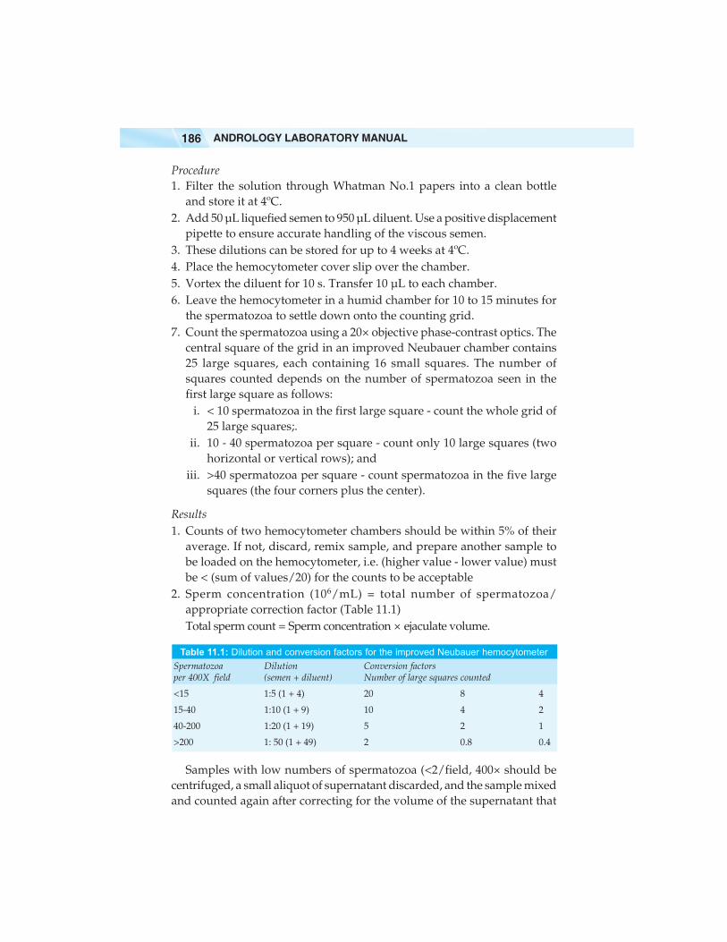
2. Sperm concentration (106/mL) = total number of spermatozoa/appropriate correction factor (Table 11.1)Total sperm count = Sperm concentration × ejaculate volume.
Table 11.1: Dilution and conversion factors for the improved Neubauer hemocytometerSpermatozoa Dilution Conversion factorsper 400X field (semen + diluent) Number of large squares counted
<15 1:5 (1 + 4) 20 8 4
15-40 1:10 (1 + 9) 10 4 2
40-200 1:20 (1 + 19) 5 2 1
>200 1: 50 (1 + 49) 2 0.8 0.4
Samples with low numbers of spermatozoa (<2/field, 400× should becentrifuged, a small aliquot of supernatant discarded, and the sample mixedand counted again after correcting for the volume of the supernatant that
SEMEN ANALYSIS 187
was removed. Samples in which no spermatozoa are seen must becentrifuged and examined for the presence of spermatozoa in the pellet.
Specialized Counting Chamber
a. Makler chamberThe Makler Counting Chamber (Sefi Medical Instruments, Heifa, Israel) iswidely used. It is only 10 microns deep, one-tenth the depth of an ordinaryhemocytometer, making it the shallowest of known chambers. It isconstructed from two pieces of optically flat glass; the upper layer servesas a cover glass, with a 1 sq. mm fine grid in the center subdivided into 100squares of 0.1 × 0.1 mm each. Spacing is firmly secured by four quartzpins.
A small number of uncalibrated drops from a well-mixed, undilutedspecimen are placed in the center of the chamber by means of a simple rodand immediately covered. A microscopic objective of × 20 is required.
b. Disposable counting chambers
Disposable counting chambers are available with multiple wells andchamber depths. These are available as Micro Cell (ConceptionTechnologies, San Diego, CA) or CellVU (Advanced Meditech International,Flushing, NY). These slides are easy to use. Each slide consists of twoseparate chambers or wells, each 20 µ in depth.
Loading the Chamber
1. Load an appropriate amount (5 µL) of well-mixed sample to one of theloading zones. The amount of sample will depend on the chamber depthand design. Do not overfill the chamber.
2. The counting chamber fills by capillary action.3. Wipe away any excess.4. Counting can be done with an eyepiece reticule consisting of a 10 × 10
box pattern.
Sperm Motility Assessment
Sperm motility is the ratio of the number of motile sperm to total numberof sperm in a given volume and is expressed as a percentage. Several scoringsystems exist for sperm motility assessments, but a simple grading systemis recommended. This provides an assessment of sperm motility withoutrequiring sophisticated equipments.
According to the WHO laboratory manual (WHO, 1999), five microscopicfields are assessed in a systematic way to classify 200 spermatozoa. Themotility of each spermatozoon is graded into one of four groups:
ANDROLOGY LABORATORY MANUAL188
a. Rapid progressive motility (i.e. > 25 µm/s at 37 ºC and > 20 µm/s at20ºC; note that 25 µm is approximately equal to 5 head lengths or half atail length).
b. Slow or sluggish progressive motilityc. Non-progressive motility (< 5 µm/s)d. Immotility
A normal semen analysis must contain at least 50% progressively motilespermatozoa.
Computer-assisted Semen Analysis (CASA)
Manual semen analysis lacks the ability to measure the kinematics of spermmotion. Of the several systems in use for automated semen analysis,computer-aided sperm analysis (CASA) is given much attention becauseof its potential benefits for analyzing sperm motion (sperm head andflagellar kinematics). Some of these motion characteris-tics have been shownto be related to IVF outcome. Some of the important kinematic parametersare:
i. Curvilinear velocity: Curvilinear velocity (VCL) is the measure of therate of travel of the centroid of the sperm head over a given timeperiod. This is calculated from the sum of the straight lines joiningthe sequential positions of the sperm along the sperm’s track. Valuesare reported as µm/s.
ii. Average path velocity: Average path velocity (VAP) is the velocity alongthe average path of the spermato-zoon. It is reported as µm/s.
iii. Straight-line velocity: Straight- line velocity (VSL) is the linear orprogressive velocity of the cell. It is also the straight-line distance betweenthe first and last centroid position for a given period of time. It is reportedas µm/s.
iv.Linearity: Linearity of forward progression (LIN) is the ratio of VSL toVCL and is expressed as percentage. A value of 100% represents cellsswimming in a perfectly straight line.
v. Amplitude of lateral head displacement: Amplitude of lateral headdisplacement (ALH) of the sperm head is calculated from theamplitude of its lateral deviation about the cells axis of progressionor average path. It is reported as µm.
A man is considered to be asthenozoospermic if the spermatozoa in hisejaculate show less than 50% forward progressive movement within 60minutes of ejaculation and necrozoospermic if all sperms are immotile.
Evaluation of Morphology Assessment
For a complete evaluation of a semen sample, the assessment of themorphological characteristics of the spermatozoa is important. The staining
SEMEN ANALYSIS 189
of a seminal smear allows the quantitative evaluation of normal andabnormal sperm forms in an ejaculate.
Smear Preparation
Slides should be pre-cleaned with 95% ethanol to allow firm attachment ofsmears. A small drop of semen, approximately a 5µL aliquot, is placed onthe slide. The fraction is then pulled out into a smear with a second slide;this is called the ‘feathering’ technique. This is done with minimum forceto ensure that the spermatozoa tails do not fall apart, and care is taken toguarantee that the smear is not too thick. Two smears are made from eachsample. If the sperm concentration is > 20 × 106/ mL, then 5 µL of semencan be used; if the sperm concentration is < 20 × 106/ mL, then 10-20 µL ofsemen is used. Smears are air-dried and fixed in 95% ethanol for 15 minutes.Air-dried smears can be batched prior to staining.
Staining Methods
The numerous staining techniques available include as the Papanicolaou,Giemsa, Shorr, modified Bryan-Leishman and Diff-Quik methods withPapanicolaou and Diff-Quik being the more common.
Papanicolaou Stain
Papanicolaou stain (WHO, 1999) is the most widely used. It is recommendedby the WHO laboratory manual because it gives a good staining tospermatozoa and other cells as it distinguishes basophilic cell componentsand acidophilic cell components. It allows a comprehensive examinationof nuclear chromatin pattern.
Reagentsi. Fixative: A freshly prepared solution of equal parts of analytical-
grade absolute ethanol and diethyl ether.ii. Graded ethanol (50%, 70%, 80%, 95%, and 99.5% (v/v)
Staining solutionsi. Hematoxylin: Orange G6 and EA-50 are commercially available.
ii. Acid ethanol: Prepared by mixing 300 mL of 095% (v/v) ethanol and2.0 mL concentrated hydrochloric acid (36% HCL) in 100 mL reagentwater.
iii. Scott’s solution: Prepare by dissolving 3.5 g NaHCO3 and 20.0 gMgSO4.7H2O in reagent water to a total volume of 1000 mL.
Procedure
Prepare the air-dried smear and fix as explained above. Proceed withstaining according to the sequence.
ANDROLOGY LABORATORY MANUAL190
1. Graded ethanol (80%, 70%, 50%) 10 dips each2. Running water 12-15 dips3. Hematoxylin 3 minutes4. Running water 3-5 dips5. Acid ethanol 2 dips6. Scott’s solution 4 min7. Distilled water 1 dip8. Graded ethanol 50%, 70%, 80% and 90% 10 dips9. Orange G6 2 min
10. Ethanol 95% 10 dips11. Ethanol 95% 10 dips12. EA-50 5 min13. Ethanol 95% (3 jars) 5 dips14. Ethanol 99.5% 2 min15. Xylene (3 staining jars) 1 min
With this stain, the head stains pale blue in the acrosomal region anddark blue in the post-acrosomal region. The midpiece may show some redstaining. The tail is stained bluish or reddish and the cytoplasmic dropletstains green.
Diff-Quik Staining
Reagents
Diff-Quik stain (Baxter Healthcare, Deerfield, IL) comprises fixative andtwo solutions – Diff-Quik I and II.1. Diff-Quik fixative: It contains 1.8 mg/L triarylmethane dye, 100% PDC
(pure dye content) in methyl alcohol.2. Diff-Quik solution I: It contains 1g/L xanthene dye 100% PDC, buffer
and sodium azide (0.01%) as preservative3. Diff-Quik solution II: It contains 1.25 g/L thiazine dye mixture, 100% PDC
(0.625 g/L azure A, and 0.625 g/L methylene blue) and buffer.4. Mounting agent: Accu-mount 60 media.
Procedure
i. Slide is prepared as described above and labeled with the accessionnumber, name, and date.
ii. Proceed with staining: Dip dry slide in Diff-Quik fixative solutioncontaining methanol, 5 times for 1 sec each time and allowing 1 secbetween dips.
iii. Allow slide to air dry for 15 min.

SEMEN ANALYSIS 191
iv. Dip dried fixed slide in Diff-Quik solution I (xanthene dye) 3 timesfor 1 sec each dip and allowing 1 sec between dips. Allow excessstain to drip off. Do not dry slide.
v. Dip slide into Diff-Quik solution II (thiazine dye) 5 times for 1 seceach dip and allow 1 sec between dips. Allow excess stain to dripoff. Do not dry slide.
vi. Rinse slide in deionized water gently and thoroughly to removeany excess stain.
vii. Allow stained slide to air dry in drying rack.Mount cover slip using Accumount on the dried stained slide. For
scoring, the slide can be viewed under oil immersion with magnificationof 1000 × using a high quality 100 × nonphase-contrast objective andcorrectly adjusted bright-field optics. About 200 spermatozoa are scoredfor various abnormalities. The xanthine stain produces the red tones, andthe thiazine increases the blue tones.
Scoring Sperm Morphology
Smears can be scored for morphology using the WHO classification (WHO,1999). Spermatozoa abnormalities are categorized as head, neck andmidpiece, and tail defects.a. Head defects: Large, small, tapered, pyriform, round, amorphous,
vacuolated (> 20% of the head area occupied by unstained vacuolarareas), heads with small acrosomal area (< 40% of head area), doubleheads, any combination of these.
b. Neck and midpiece defects: Bent neck; asymmetrical insertion of midpieceinto head; thick, irregular midpiece; abnormally thin midpiece; anycombination of these.
c. Tail defects: Short, multiple, hairpin, broken, bent, kinked, coiled tails, orany combination of these.
d. Cytoplasmic droplets: Greater than one-third of the area of a normal spermhead.For a spermatozoon to be normal, the head, neck, midpiece, and tail must
be normal. The head should be oval in shape. The length of the head should be4.0-5.0 µm and the width 2.5-3.5 µm. The length-to-width ratio should be 1.5-0 to 1.75. Length and width can be measured with an ocular micrometer. Theacrosome should be well defined and comprise 40-70% of the head area. Themidpiece should be slender, less than 1 µm in width, about one-and-a-halftimes the length of the head, and attached axially to the head. Cytoplasmicdroplets should be less than half the size of the normal head. The tail shouldbe approximately 45 µm long. Reference range is considered as greater than30% normal forms. In Kruger’s strict criteria classification, (Kruger et al. 1986),all ‘borderline’ forms are considered abnormal. Reference range includesspermatozoa with > 14% normal forms.
ANDROLOGY LABORATORY MANUAL192
Leukocytospermia Test
This test is performed on suspended cells in a liquefied semen specimenand quantitated by counting stained cells in a Makler counting chamber.Peroxidase-positive granulocytes (neutrophils, polynuclear leukocytes,macrophages) are identified by histochemical staining using the Endtz test.This test is often referred to as the myeloperoxidase test.
Preparation of Stock Solution (stable 6 months)
Reagentsi. Ethanol
ii. Benzidine 0.0625 giii. Distilled water 25 mL
Procedurei. Mix these chemicals in a clean 100 mL bottle. The solution should
be clear and yellow.ii. Cover the bottle with aluminum foil and store in the dark.
iii. Fresh stock solution should be prepared if it gets dark in color orforms a cloudy precipitate.
Benzidine is carcinogenic and should be handled carefully. Wear glovesand a face mask while weighing to avoid accidental contact or inhalation.The expired Endtz test solution should be discarded in concentrated bleachsolution.
Preparation of Working Solution
i. Mix 4.0 mL of stock solution and 50 µL of 3% H2O2 in a 10 mL tube(dilute 30% stock H2O2 10 times).
ii. Cover the bottle with aluminum foil and store in the dark.iii. Prepare fresh working solution from stock every week and discard
old solutions.
Equipment and Materials
1. Tyrode’s buffer2. Makler counting chamber3. Microcentrifuge tubes4. Eppendorf pipette and tips (5 µL, 20 µL, 40 µL)
Procedure
i. Measure 20 µL of liquefied semen specimen into a micro-centrifugetube; add 20 µL of Tyrode’s solution and 40 µL of working benzidenesolution. Mix and let sit at room temperature for 5 minutes.
ii. Load a Makler counting chamber with 5 µL of the above solutionand observe under 10 × magnification.
SEMEN ANALYSIS 193
iii. All granulocytes will stain dark brown in color and retain their roundshape.
iv. Count the cells in all the 100 squares of the Makler grid.v. Number of white blood cells (WBC) can be calculated by multiplying
total number of cells by 4 to correct for dilution factor. The totalWBC number will be 105/mL semen. This number should becorrected to million/ml by dividing by 10.
Report results as million/mL Endtz-positive cells. According to the WHOmanual, the normal concentration of WBC in semen is < 1 × 10⁶/mL,leukocytospermia is defined as the presence of >1 million/ml WBCs.
Reference range: 0.0-0.9 × 10⁶/mL (normal)
Panic value: Endtz test > 1 × 10⁶/mL (positive).
Quality Control
A weekly positive control should be run to check reagents. The resultsshould be greater than 1.0 × 10⁶/ml Endtz-positive cells.
Note: If semen specimen is not available, an EDTA anti-coagulated bloodspecimen may be used. Centrifuge the blood specimen to obtain the buffycoat. Remove the buffy layer containing WBC by using a transfer pipette.Dilute into 2 mL of Tyrode’s buffer and aliquot (0.1 mL). These aliquotsmay be used for approximately one month.
Sperm Viability
Sperm vitality is normally measured by testing cellular integrity, assessingthe ability of the sperm plasma membrane to exclude extracellularsubstances. The cytologically intact ‘live’ cells can be determined usingseveral vital staining techniques such as eosin Y and trypan blue. The hypo-osmotic swelling (HOS) test is also considered a test of sperm integrity.
Eosin-Nigrosin Stain
An eosin-nigrosin stain must be done on all specimens having a motility of30% or less. The stain must be performed immediately following the initialmotility examination (WHO, 1999).
Reagents
i. Eosin Y (1%): Weigh out 0.5 g of eosin Y and add it to 50 mL ofdeionized water. Dissolve this solution using gentle heat. Cool theliquid to room temperature and filter. This reagent is stable for 3months at room temperature.
ANDROLOGY LABORATORY MANUAL194
ii. Nigrosin (10%): Weigh out 5 g of nigrosin and add it to 50 mLdeionized water. Dissolve this solution using gentle heat. Cool theliquid to room temperature and filter. This reagent is stable for 3months at room temperature.
Procedure
i. Place one drop of well-mixed semen on a Boerner slide.ii. Add 2 drops of 1% aqueous eosin Y, stir with a wooden stirrer for 15
seconds.iii. Add 3 drops of 10% aqueous nigrosin. Mix with a wooden stirrer.iv. Immediately make two thin smears from this mixture by pipetting
10 µL onto each slide and air dry.v. Place a cover slip with Accu-Mount mounting media (Baxter).
vi. Count 100 sperm on each slide in duplicate using high power (×40).
vii. Calculate percentage of viable (unstained) and non-viable (stained)sperm.Viability should be ≥ motility in samples with < 30% motility.
Hypo-osmotic Swelling Test (HOS)
The hypo-osmotic swelling (HOS) test was originally described as a testfor sperm function (Jayendran, 1984, WHO, 1999). Now it is mostappropriately considered as an additional test for sperm vitality. The HOStest is based on the principle that live spermatozoa withstand moderatehypo-osmotic stress. Dead spermatozoa in which the plasma membrane isno longer intact do not swell, whereas senescent cells show uncontrolledswelling that eventually results in rupture of the overdistended plasmamembrane.
Reagents
i. Dissolve 0.735 g sodium citrate dihydrateii. 1.351g fructose
iii. Mix in 100 mL of distilled water.
Procedure
i. To 1 mL of HOS solution add 0.1 mL of liquefied semen and mixgently with the pipette.
ii. Incubate at 37ºC for 30-60 minutes.iii. Place a drop of semen mixture on a glass slide and place a cover
slip.iv. Examine under a phase contrast microscope.
SEMEN ANALYSIS 195
v. Observe for tail swelling at × 40 magnification.vi. Identify number of swollen cells in about 100 spermatozoa in
duplicate.Calculate the mean percentage of swollen cells:
Percent swelling =
Number of sperm with swollen tails× 100
Number of spermatozoa with swollen + non - swollen tails
Results
Normal values (fertile): > 60% spermatozoa with swollen tails
Abnormal values (infertile): < 50% spermatozoa with swollen tails.
HOS has a limited ability to predict male fertility, but it is useful inselecting non-motile but viable sperm for assisted reproductivetechnologies. An HOS result < 50% is associated with increased miscarriagerates.
Antisperm Antibodies
Immunological protection to sperm antigens is provided by the tightjunctions of Sertoli cells forming the blood-testis barrier. The spermatozoonevokes an immune response when exposed to the systemic immune defensesystem. In conditions in which this barrier gets disrupted, formation ofantisperm antibodies (ASA) may occur. Systemic and localimmunoregulatory mechanisms control the development of antispermaticimmunity, which may sometimes be overridden by genetic predispositions,non-physiological routes of inoculation, genital tract infections, etc., whichmay lead to ASA formation and sperm dysfunction.
Certain ASAs have a cytotoxic effect on the spermatozoa and can causecell death and immobilization of sperm cells. Other effects of ASAs includecreating agglutinated clumps of moving sperm in the semen sample,hampering passage of sperm through the cervical mucus, and zona bindingand passage.
Immunobead Test
Principle
Antibodies bound to the human sperm surface can be detected by otherantibodies that are against human IgG, IgA or IgM immunoglobulinmolecules (Bronson et al 1984; WHO, 1999; Mortimer, 1994a).
ANDROLOGY LABORATORY MANUAL196
Reagents
Immunobeads: Anti-IgG, -IgA and -IgM beads (Irvine Scientific, Santa Ana,CA). For screening, beads for total B-cell labeling can be used. Reconstitutethe immunobeads according to the manufacturer’s instructions. Beads canbe kept for several months at 4°C in the original buffer, which contains apreservative (azide).
Stock buffer: Tyrode’s solution or Dulbecco’s phosphate-buffered saline (PBS)can be used.
Buffer I (0.3% BSA): Buffer for bead washing (10 mL) and sperm washing(2 x 10 mL for each semen sample). Add 0.6 g bovine serum albumin (BSA;Cohn fraction V) to 200 mL stock buffer. 200 mL buffer is sufficient to washand run six unknown samples: one positive and one negative control andtwo sets of IgA and IgG beads.
Buffer II (5% BSA): Buffer for resuspension of beads and sperm pellets, 200 µLfor each specimen. Add 250 mg BSA to 5 ml stock buffer. A total of 2 mLbuffer II is needed for six samples, two controls and two sets of beads.1. Filter all solutions through 0.22 or 0.45 µm filters and warm to 25-35°C
before use.2. At least 200 motile sperm should be assessed for each test. A positive
(serum from a donor with high titers of antisperm antibodies) andnegative control should be included in each run.
Direct Immunobead Test
1. Add 0.2 mL of stock bead suspension to 10 mL of buffer I in separateconical centrifuge tubes. Repeat this for each immunobead type.
2. Determine the amount of semen to be used, transfer that volume toa tube and add up to 10 mL with buffer I.
3. Centrifuge all tubes at 500 g for 6 min. at room temperature.4. Tubes with sperm: Discard the supernatants. Resuspend the sperm
pellets in 10 ml of fresh buffer I and centrifuge again as above.5. Discard supernatants and resuspend sperm pellets in 200 µL of buffer
II.6. Tubes with beads: Discard the supernatants and resuspend the beads
in 200 µL of buffer.7. Add 5 µL droplets of each immunobead type on clean microscope
slides.8. Add 5 µL of washed sperm suspension to each droplet of beads and
mix well using a yellow pipette tip.9. Place a cover slip on each of the mixtures.
10. Leave the slides for 10 min at room temperature in a moist chamberand then assess under a 20 × phase contrast objective.
SEMEN ANALYSIS 197
Calculations and Results
Only motile sperm should be assessed. Calculate the percentage of spermsthat has two or more attached immunobeads. Those that have binding tothe tip of the tail should be ignored. Count at least 200 motile sperm induplicate for every preparation. Record the percentage of sperm carryingattached beads, the Ig class (IgG or IgA) and the site of binding (head,midpiece, and tail).
Indirect Immunobead Test
This is used to detect antisperm antibodies in heat-inactivated seminalplasma.
1. Wash normal donor sperm twice in buffer I as described above (Steps2-4).
2. Add 0.2 mL of stock bead suspension to 10 mL of buffer I in separateconical centrifuge tubes. Repeat this for each immunobead type.
3. Determine the amount of semen to be used, transfer this volume toa tube and add up to 20 mL with buffer I.
4. Centrifuge all tubes at 500 g for 6 min. at room temperature.5. Tubes with sperm: Discard the supernatants. Resuspend the sperm
pellets in 10 mL of fresh buffer I and centrifuge again as above.6. Discard supernatants and resuspend sperm pellets in 200 µL of buffer
II or prepare them initially by swim-up procedure or density gradientcentrifu-gation procedure followed by washing.
7. Adjust the washed sperm suspensions to a final motile spermconcentration of 50 × 106/mL in buffer II.
8. Dilute 10 µL of the fluid to be tested with 40 µL of buffer II and mixwith 50 µL of the washed donor sperm suspension. Incubate at 37°Cfor 60 min.
9. Wash the sperm twice as described above (Steps 2 – 4).10. Place 5 µL droplets of each immunobead type on clean microscope
slides.11. Add 5 µL of washed sperm suspension to each droplet of beads and
mix well using a yellow pipette tip.12. Place a cover slip on each of the mixtures.A positive and negative control should be included in each test run. A
positive control can be prepared by using serum from a donor (e.g., from avasectomized man) with high titers of serum sperm antibodies).
Limitations
Results are based on the analysis of motile sperm. Samples made usingsperm with poor motility may give false negative results. A positive finding
ANDROLOGY LABORATORY MANUAL198
of > 50% of motile sperm with attached beads is considered to be clinicallysignificant.
Normal Reference Values of Semen Variables
Each laboratory must determine its own reference range for each variable.According to the World Health Organization guidelines (WHO, 1999) thefollowing reference values for the semen sample are suggested:
Reference Value
Volume ≥ 2.0 mLpH ≥ 7.2Sperm concentration ≥ 20 × 106 spermatozoa/ mLMotility ≥ 50% motile (grades a + b) or> 25% with progressive motility (grade a) within 60minutes of ejaculationVitality > 75% aliveWhite blood cells < 1 × 10⁶/mLImmunobead test < 50% motile spermatozoa withbeads bound
SEMEN MICROBIOLOGY AND VIROLOGY
Infection of the male reproductive tract can directly or indirectly causeinfertility (Mortimer, 1994b). Inflammation caused by infection or variousdisorders can affect the secretory function of both the prostate and seminalvesicles. Asymptomatic infections of the prostate can cause partial orcomplete obstruction of the ejaculatory duct resulting in oligospermia andeven azoospermia. Infection of the seminal vesicles often causes substantialreduction in ejaculate volume and a low seminal fructose concentration.Microbiological examination of the semen is required to differentiate aspecific microbiological-induced pyospermia from other abnormalities thatcauses an increase in leukocytes.
Pyospermia
Pyospermia is a laboratory finding categorized as the abnormal presenceof leukocytes in human ejaculate. Pyospermia is established when theconcentration of seminal WBCs is in the range between 5 × 10⁵/mL and 5× 10⁶/mL seminal fluid during semen analysis (Anderson DJ, 1995).Numerous studies have demonstrated that leukocytes in ejaculate have aphysiological effect on sperm function, which may further impact maleinfertility (Wolff H, Anderson DJ, 1988).
The differential diagnosis of symptomatic pyospermia includes infection,autoimmune disease, and inflammation of the accessory sex glands and
SEMEN ANALYSIS 199
the lower male urogenital tract. Urogenital infections include acute andchronic prostatitis, seminal vesiculitis, epididymo-orchitis, cystitis,urethritis, urethral stricture, stone disease, foreign bodies, upper urinarytract infection, retrograde ejaculation, and localized sepsis of the adjacentlower gastrointestinal tract and asymptomatic bacteriuria. Chronicinfections that may result in pyospermia include fungal, mycobacterial,and congenital lesions that cause urogenital tract infection.
Collection of Semen Specimens
1. Hands must be washed thoroughly with antiseptic soap.2. Penis should be washed using antiseptic solution.3. The semen specimen should be collected by masturbating directly into
the sterile container.
Organisms Found in Semen
Many organisms found in semen are actually contaminants from thepatient’s skin or from the air at the time of collection. Not all are associatedwith pyospermia, hence caution must be exercised in interpretation ofpositive cultures.
Ureaplasma and Mycoplasma
Ureaplasma and mycoplasma species are common commensal inhabitantsof the lower genitourinary tract in adolescents and adult men and womenwho are sexually active. The organisms can be transmitted venereally andvertically from mother to offspring. Ureaplasma urealyticum and to a lesserextent Mycoplasma hominis are therefore often found in semen of infertilepatients. U. urealyticum is part of the normal genital flora of both men andwomen and is found in about 70% of sexually active humans. Their presenceis abnormal, and most authorities agree that the infection must be treatedwith antibiotics.
Chlamydia trachomatis
Chlamydia trachomatis is occasionally present in human semen. It is the causeof the most prevalent sexually transmitted bacterial disease worldwide andis responsible for an estimated 90 million infections. Considering the highworldwide prevalence of C. trachomatis infection, artificial inseminationby donor (AID) is a potential route for the spread of C. trachomatis and hasbeen reported as such. It cannot be cultured, since seminal plasma factor istoxic to the cell lines used for the culture. Treatment must extend to boththe partners.
ANDROLOGY LABORATORY MANUAL200
Neisseria gonorrhoeae
This organism causes severe symptoms in men. It can be identified in semenas gram-negative intracellular diplococci. Transmission is through sexualintercourse as well.
Trachomonas vaginalis and E. coli
In men with urinary tract infections, E.coli can colonize the prostate andcause production of IgA leading to sperm agglutination. Other organismsthat form the normal flora of the reproductive tract may commonly bepresent; these include enterococci such as S. faecalis and staphylococci suchas S. epidermidis. Mycobacterium tuberculosis maybe present in the absenceof pyospermia.
Viruses
Many viruses can be isolated from semen, but the most important is humanimmunodeficiency virus (HIV). Its presence can be detected by either cultureor polymerase chain reaction. Other viruses that could be present in semenare human T-cell lymphotrophic virus type I (HTLV-1), hepatitis B, hepatitisC, cytomegalovirus (CMV), and human papilloma virus (HPV). Genitalherpes virus often is isolated from semen in infected patients.
BIOCHEMICAL ASSESSMENT OF SEMINAL PLASMA, PROSTATE,EPIDIDYMIS AND SEMINAL VESICLES
The prostate, seminal vesicles, and epididymis produce components suchas zinc, citric acid and α-glucosidase that are uniquely specific to eachaccessory gland.
Measurement of Zinc
A colorimetric assay kit is used for determining the zinc content in the seminalplasma (Johnson and Eliasson, 1987, Mortimer, 1994c). It can be done in eithera -well plate or spectrophotometer cuvettes. The assay is based on the principlethat in the presence of zinc, 5-Br-PAPS (2-(5 bromo-2 - pyridylazo)-5-(N-propyl-N-sulphopropylamino)-phenol is converted to 5-Br-PAPS-Zn complex, whichabsorbs at 560 nm.
Reagents
1. Zinc kit: Chromogen solution is prepared by mixing color reagents Aand B in proportions of 4:1. It is stable for 1 week at 4oC.
2. Zinc standard: (0.1 mM).
SEMEN ANALYSIS 201
Specimens
1. Centrifuge semen at 1000 g for 15 minutes. 100 µL of semen is centrifugedto obtain 10 µL of neat seminal plasma.
2. A 10 µL aliquot of cell-free seminal plasma is diluted with 600 µL ofwater.
Procedure
1. Set absorbance readings on the spectrophotometer to 560 nm, and allowadequate time for stabilization.
2. Prepare a standard curve in duplicate (100 µM diluted to give 80, 60, 40,20 and 10 µM).
3. Set the spectrophotometer to zero with a cuvette containing reagentwater.
4. Add 2.5 mL working chromogen solution (mix 4 parts of color reagentA with 1 part of color reagent B). Add 20 µL of color reagent to 40 µL ofdiluted semen samples, standard, and blank.
5. Leave at room temperature for 5 minutes.6. Measure absorbance at 560 nm and calculate results.7. Calculation: Multiply by a dilution factor to obtain the concentration of
zinc (mM) in undiluted seminal plasma. Multiply by ejaculate volumeto obtain µmol/ejaculate.
Results
Zinc is a specific marker of prostatic function. Normal range of seminalzinc is 1.2-3.8 mmol/L or ≥ 2.4 µmol per ejaculate.
Measurement of Citric Acid in Seminal Plasma
Citric acid is an indicator of prostatic gland function. Decreased citric acidlevels may indicate either prostate dysfunction or prostatic duct obstruction.It can be measured using the Boehringer enzymatic, NADH –linked kit(Mortimer, 1994c).
Reagents
1. Boehringer Kit No. 130976: Contains Solution 1: 3 × Bottle1 (mainlyNADH), which is reconstituted by adding 12 mL reagent water andshaking well. 3 × Bottle 2 (citrate-lyase), reconstituted by adding 0.3 mLreagent water and shaking well.
2. Triethanolamine buffer (pH 7.7): Prepared by dissolving 14.9gtriethanolamine in 750 mL reagent water and adjusting the pH to 7.6 byadding 1 N HCl. Dissolve 0.027 g ZnCl2 in 250 mL reagent water and
ANDROLOGY LABORATORY MANUAL202
add it to the triethanolamine solution. Add 0.5 g of sodium azide andmix thoroughly.
3. Trichloroacetic acid (TCA, 15%): Dissolve 15 g trichloroacetic acid in 100mL reagent water).
4. NaOH (6 N): Dissolve 24 g NaOH pellets dissolved in 100 mL reagentwater.
5. Citric acid standard: 0.174 g citric acid in 10 mL reagent water. Make a 1 +57 dilution.
Specimens
1. Centrifuge 250 µL liquefied semen (cell- and protein-free) in anEppendrof tube at 1000 g for 15 minutes.
2. Add 100 µL of supernatant to 4.95 mL of 15% TCA in a small, cappedvial and shake well.
3. Add 0.75 mL NaOH (6 N) and adjust the pH to 7.0.4. Freeze three 0.5 mL aliquots of the extract in Eppendorf tubes at -20ºC.
Procedure
1. Set the spectrophotometer to 340 nm and allow adequate time forstabilization.
2. Mix 0.5 mL of solution 1, 2.3 mL TRA buffer and 0.2 mL sample, standard,or blank in a disposable cuvette. Prepare each set in duplicate.
3. Adjust the spectrophotometer reading to zero with a cuvette containingreagent water.
4. Measure initial absorbance (A1).5. Add 20 µL of solution 2. Shake it well, wait exactly 5 minutes, and
measure the absorbance again (A2).6. Calculate and analyze the results according to the formula:
∆A × (V ÷ V) × DF × MW ÷ E ÷ D + 100 = g/L whereDF = Specimen dilution factorV = final volume (3.02 mL)MW = molecular weight of the substance analyzed (192.1)E = absorption coefficient of NADH at 340 nm (6.3 cm3/µmol)D = light path (1 cm)V = sample volume (0.2 mL)∆A × 139.0 = mmol citric acid/ L.
Results
The normal range of seminal plasma citric acid concentration is 9.4-43.4mmol/L or ≥ 52 µmol per ejaculate. Decreased levels of citric acid mayindicate either prostatic dysfunction or duct obstruction. Further evaluationmust be done by physician.
SEMEN ANALYSIS 203
Measurement of Neutral ααααα-glucosidase in Seminal Plasma
Seminal plasma contains both neutral α-glucosidase isoenzyme thatoriginated from the epididymis and an acid isoenzyme contributed by theprostate. The latter can be selectively inhibited to allow measurement ofneutral α-glucosidase. P-nitrophenol α-glucopyranoside in the presenceof α- glucosidase is converted to p-nitrophenol, and the absorbance can beread at 405 nm (Cooper et al 1990, Mortimer, 1994c).
Reagents
1. Phosphate buffer (0.2 M, pH 6.8). Prepare 1% SDS in phosphate buffer.2. Color reagent 1 for stopping the reaction. 0.1 M sodium bicarbonate.3. Color reagent 2 for diluting the product. Prepare color reagent 1
containing 0.1% SDS.4. Substrate (p-nitrophenol glucopyranoside (PNPG, 5mg/mL in
phosphate buffer, pH 6.8). Prepared fresh.5. Glucosidase inhibitor for semen blanks Castanospermine (10 mM).
Prepare 1mM working solution. Freeze in aliquots at -20oC.6. 100 mM solution of sodium carbonate.7. Standard: 5 mM p-nitrophenol. Make fresh every time.
Specimen
Use sperm-free seminal plasma prepared by centrifuging an aliquot ofsemen at 1000 g for 15 minutes.
Procedure
1. Set a water bath at exactly 37°C for the incubation step below.2. Thaw specimens to be assayed and mix well.3. Prepare 100 µL of PNPG substrate solution in Eppendrof tube.4. Using a positive displacement pipette, add 10 µL specimen aliquots
in duplicate into the Eppendorf tube.5. Mix each tube and incubate at 37°C for 2 h.6. Include internal quality control samples consisting of high, medium,
and low activities of neutral α-glucosidase.7. To two high activity quality control semen pools. Add 8 mL of 1
mM castanospermine to provide semen blank value.8. Prepare PNP standard curve (160, 120, 80, 40 µm) with color reagent
2 (within an hour of incubation).9. Stop the reaction by adding 1.0 mL of color reagent 1 and mix.
10. Read absorbance of each sample at 405 nm against the blank (water).
ANDROLOGY LABORATORY MANUAL204
Results
1 unit of glucosidase activity is equal to the production of 1 µ mole product(PNP) per minute at 37oC. In this assay, the activity is derived from 15 µLof semen in a total volume of 1.115 mL over 120 minutes. Therefore thecorrection factor is 1115/15/120 or 0.6194.1. Read the concentration of PNP produced by the sample from the
standard curve (µM).2. Multiply by the correction factor (0.6194) to obtain the activity of neutral
glucosidase in undiluted seminal plasma (U/l).3. Subtract the activity of the castanospermine semen blank from each
sample to obtain the corrected (glucosidase-related) activity.4. Multiply the corrected activity by the ejaculate volume to obtain
glucosidase activity (mU) per ejaculate. α-glucosidase is a specificindicator for epipdymis function. Normal values are ≥ 20 mU/ ejaculate.
Quantitative Seminal Fructose
Sperm in semen sample are lysed, and addition of resorcinol and subsequentheating at 70oC results in a salmon-pink color, which is read at 420 nm(Davis and Gander, 1967, Moon and Bunge, 1968).
Reagents used
1. Concentrated HCl2. Deionized water.3. Fructose (0.32 mmole/L). Add 14.4 g of fructose to make 250 mL of
deionized water (5.56 mg/dL).4. Resorcinol 0.05%. Add 25 mg of resorcinol to 50 mL of ethanol (95%).
Freeze three aliquots (150 µL) aliquots of supernatant in 1.5 mLEppendorf tubes at – 20°C.
Procedure1. Turn on the 77oC water bath.2. Set spectrophotometer absorbance at 420 nm.3. Adjust the reading to zero with cuvette containing reagent water.4. Label three beakers for patient, positive control (pooled seminal
plasma from normal donors), and negative controls (no semenadded).
5. Treat with acid: To a clean beaker add 7.5 mL of deionized water +2.5 mL HCl and 50 µL of semen or seminal plasma. Mix each patientand control sample carefully (200-fold dilution).
6. Label beaker for each patient and control. Using a Whatman # 1filter paper, filter each patient and control mixture into appropriatelylabeled beaker.
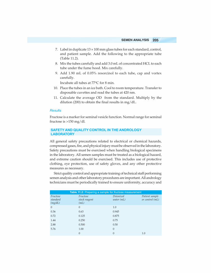
SEMEN ANALYSIS 205
7. Label in duplicate 13 × 100 mm glass tubes for each standard, control,and patient sample. Add the following to the appropriate tube(Table 11.2).
8. Mix the tubes carefully and add 3.0 mL of concentrated HCL to eachtube under the fume hood. Mix carefully.
9. Add 1.90 mL of 0.05% resorcinol to each tube, cap and vortexcarefully.Incubate all tubes at 77oC for 8 min.
10. Place the tubes in an ice bath. Cool to room temperature. Transfer todisposable cuvettes and read the tubes at 420 nm.
11. Calculate the average OD from the standard. Multiply by thedilution (200) to obtain the final results in mg/dL.
Results
Fructose is a marker for seminal vesicle function. Normal range for seminalfructose is >150 mg/dl.
SAFETY AND QUALITY CONTROL IN THE ANDROLOGYLABORATORY
All general safety precautions related to electrical or chemical hazards,compressed gases, fire, and physical injury must be observed in the laboratory.Safety precautions must be exercised when handling biological specimensin the laboratory. All semen samples must be treated as a biological hazard,and extreme caution should be exercised. This includes use of protectiveclothing, eye protection, use of safety gloves, and any other protectivemeasures as necessary.
Strict quality control and appropriate training of technical staff performingsemen analysis and other laboratory procedures are important. All andrologytechnicians must be periodically trained to ensure uniformity, accuracy and
Table 11.2: Preparing a sample for fructose measurementFructose Fructose Deionized Patient samplestandard stock reagent water (mL) or control (mL)(mg/dL) (mL)
0 0 1.0
0.36 0.63 0.945
0.72 0.125 0.875
1.44 0.250 0.75
2.88 0.500 0.50
5.76 1.00 0
0 0 1.0
ANDROLOGY LABORATORY MANUAL206
reproducibility, especially in sperm count, motility and sperm morphology(Mortimer, 1994d). Internal quality controls, proficiency testing of slides forviability and morphology both by WHO and Kruger’s strict criteria areavailable, and laboratories can enroll in a proficiency testing program.Technicians score the slides in a blind fashion, and the results are mailed tothe proficiency testing program. Adequate remedial measures should be inplace to minimize inter- and intraobserver variations. In addition, strictquality control for monitoring all equipment in use is critical, and all solutionsand reagents must be subjected to strict quality control measures to optimizeperformance and accuracy of results.
CONCLUSION
Semen analysis is fairly simple and easy to perform and remains thecornerstone of the andrology laboratory even as newer tests are beingintroduced. Maintaining strict interlaboratory, inter-observer, and intraobserver quality control measures and making quality control materialsavailable is important for optimizing results.
BIBLIOGRAPHY
1. Anderson DJ. Should male infertility patients be tested for leukocytospermia?Fertil Steril. 1995;63:246-48.
2. Bronson R, Cooper G, Rosenfeld D. Sperm antibodies: their role in infertility.Fertil Steril 1984;42:171-83.
3. Cooper TG, Yeung CH, Nashan D, Jockenhövel F, Nieschlag E. Improvementin the assessment of human epididymal function by the use of inhibitors inthe assay of alpha-glucosidase in seminal plasma. Int J Androl. 1990;13:297-305.
4. Davis JS, Gander JE. A reevaluation of the Roe procedure for the determinationof fructose. Anal Biochem 1967;19:72-79.
5. Jeyendran RS, Van der Ven HH, Perez-Pelaez M, Crabo BG, Zaneveld LJD.Development of an assay to assess the functional integrity of the human spermmembrane and its relationship to other semen characteristics. J Reprod Fertil1984;70:219-28.
6. Johnsen O, Eliasson R. Evaluation of a commercially available kit for thecolorimetric determination of zinc in human seminal plasma. Int J Androl1987;10:435-40.
7. Kruger TF, Menkveld R, Stander FS, Lombard CJ, Van der Merwe JP, vanZyl JA, Smith K. Sperm morphologic features as a prognostic factor in invitro fertilization. Fertil Steril 1986;46:1118-23.
8. Moon KH, Bunge RG. Observations on the biochemistry of human semen. I.Fructose. Fertil Steril. 1968;19:186-91.
9. Mortimer, D. Antisperm Antibodies. In: Practical Laboratory Andrology.Oxford University Press, Oxford, 1994a;111-25.
10. Mortimer, D. Biochemistry of spermatozoa and seminal plasma. In: PracticalLaboratory Andrology. Oxford University Press, Oxford, 1994c;89-109.
SEMEN ANALYSIS 207
11. Mortimer, D. Semen microbiology and virology. In: Practical LaboratoryAndrology. Oxford University Press, Oxford, 1994b;127-33.
12. Mortimer D. Technician training and Quality control Aspects. In: PracticalLaboratory Andrology. Oxford University Press, Oxford, 1994d;337-47.
13. Sigman M, Jarow JP. Male infertility. In: Wein AJ eds. Campbell-WalshUrology. 9th edition. Sanders Elsevier. Philadelphia, PA.2007;609-53.
14. Wolff H, Anderson DJ. Immunohistologic characterization and quantitationof leukocyte subpopulations in human semen. Fertil Steril 1988;49:497-504.
15. World Health Organization. WHO laboratory manual for the examinationof human semen and sperm–cervical mucus interaction, 1999, 4th edn,Cambridge University Press, Cambridge.
Related Documents