Self-Calibrating Parallel Imaging With Automatic Coil Sensitivity Extraction Charles A. McKenzie, 1 * Ernest N. Yeh, 2 Michael A. Ohliger, 2 Mark D. Price, 2 and Daniel K. Sodickson 1,2 Calibration of the spatial sensitivity functions of coil arrays is a crucial element in parallel magnetic resonance imaging (PMRI). The most common approach has been to measure coil sensi- tivities directly using one or more low-resolution images ac- quired before or after accelerated data acquisition. However, since it is difficult to ensure that the patient and coil array will be in exactly the same positions during both calibration scans and accelerated imaging, this approach can introduce sensitivity miscalibration errors into PMRI reconstructions. This work shows that it is possible to extract sensitivity calibration images directly from a fully sampled central region of a variable-density k-space acquisition. These images have all the features of tradi- tional PMRI sensitivity calibrations and therefore may be used for any PMRI reconstruction technique without modification. Be- cause these calibration data are acquired simultaneously with the data to be reconstructed, errors due to sensitivity miscalibration are eliminated. In vivo implementations of self-calibrating parallel imaging using a flexible coil array are demonstrated in abdominal imaging and in real-time cardiac imaging studies. Magn Reson Med 47:529 –538, 2002. © 2002 Wiley-Liss, Inc. Key words: parallel MRI; sensitivity calibration; coil arrays; SMASH; SENSE Over the last few years, a number of parallel MRI (PMRI) techniques have been used for accelerated in vivo imaging (1–3). The techniques take a variety of approaches to the problem of producing a complete MR image from under- sampled data acquired with an array of RF receiver coils. One requirement these techniques have in common is the need to determine spatial sensitivity information for the coil array. Since accurate coil sensitivity information is required for accurate spatial encoding in PMRI reconstruc- tions, it may be argued that for successful parallel imaging, the choice of sensitivity calibration strategy is at least as important as the choice of reconstruction strategy. Unfor- tunately, the determination of sensitivity calibration infor- mation can be a cumbersome and error-prone procedure. The most common solution to the problem of coil sen- sitivity calibration has been to measure sensitivities di- rectly using one or more low-resolution calibration im- ages. This calibration step can prolong total examination time, partially counteracting the benefits of decreased ac- quisition time associated with PMRI. It also introduces a possible source of error into the PMRI reconstruction, as it is difficult to ensure that the patient and coil array will be in the same positions during both the calibration scans and the accelerated data acquisitions. Misregistrations or in- consistencies between the calibrated and the “true” sensi- tivities result in artifacts in the reconstructed images. This is particularly true of real-time imaging, for which motion of the coil array, as well as motion of the anatomy during data acquisition, can render a fixed sensitivity reference inappropriate for the desired reconstruction. PMRI tech- niques that do not require a separate sensitivity reference scan but derive the necessary information directly from the accelerated data itself offer clear advantages for such applications. One approach to eliminating the need for separate cali- bration scans— AUTO-calibrating SiMultaneous Acquisi- tion of Spatial Harmonics (AUTO-SMASH)—was intro- duced in 1998 (4). This technique satisfied sensitivity cal- ibration requirements by acquiring a small number of extra lines at the center of k-space. The additional acquired lines were used to determine the weights necessary to recon- struct missing k-space lines. The AUTO-SMASH concept was recently expanded in the variable-density AUTO- SMASH (VD-AUTO-SMASH) approach (5). In VD-AUTO- SMASH, larger numbers of central k-space lines were ac- quired, allowing overdetermination of the weights re- quired for image reconstruction, and greatly improving the robustness of the technique. The extra lines acquired at the center of k-space were used not only for sensitivity cali- bration, but also as independent components of image data. It was shown in Ref. 5 that this improves image quality in the low-resolution components of reconstructed images. However, neither AUTO-SMASH nor VD-AUTO- SMASH made optimal use of the fully acquired central lines. In particular, the linear combinations of component coil data used in both reconstruction algorithms could in some cases lead to signal losses in reconstructed images, as a result of cancellation of complex component coil sensi- tivities with differing phases (6). In addition, the use of calibration information in these reconstructions was ap- plicable only to SMASH-like reconstructions, and could not be applied in a straightforward manner to the wide variety of other PMRI reconstruction techniques currently available. We describe a different approach to using the central lines of a variable-density acquisition, which provides in- ternal coil sensitivity calibrations that can be used in any PMRI technique requiring such information. This ap- proach is used in combination with a generalized recon- struction algorithm that improves image quality by allow- ing optimal combinations of the additionally acquired data 1 Department of Medicine, Cardiovascular Division, Beth Israel Deaconess Medical Center and Harvard Medical School, Boston, Massachusetts. 2 Harvard-MIT Division of Health Sciences and Technology, Boston, Massa- chusetts. Grant sponsor: Whitaker Foundation; Grant sponsor: National Institutes of Health; Grant number: R29 HL60802. *Correspondence to: Charles A. McKenzie, Ph.D., Department of Radiology, Beth Israel Deaconess Medical Center, East Campus, Room AN-239, 330 Brookline Ave., Boston, MA 02215. E-mail: charles_mckenzie@ caregroup.harvard.edu Received 18 June 2001; revised 3 October 2001; accepted 24 October 2001. Magnetic Resonance in Medicine 47:529 –538 (2002) DOI 10.1002/mrm.10087 © 2002 Wiley-Liss, Inc. 529

Welcome message from author
This document is posted to help you gain knowledge. Please leave a comment to let me know what you think about it! Share it to your friends and learn new things together.
Transcript
Self-Calibrating Parallel Imaging With Automatic CoilSensitivity Extraction
Charles A. McKenzie,1* Ernest N. Yeh,2 Michael A. Ohliger,2
Mark D. Price,2 and Daniel K. Sodickson1,2
Calibration of the spatial sensitivity functions of coil arrays is acrucial element in parallel magnetic resonance imaging (PMRI).The most common approach has been to measure coil sensi-tivities directly using one or more low-resolution images ac-quired before or after accelerated data acquisition. However,since it is difficult to ensure that the patient and coil array will bein exactly the same positions during both calibration scans andaccelerated imaging, this approach can introduce sensitivitymiscalibration errors into PMRI reconstructions. This workshows that it is possible to extract sensitivity calibration imagesdirectly from a fully sampled central region of a variable-densityk-space acquisition. These images have all the features of tradi-tional PMRI sensitivity calibrations and therefore may be used forany PMRI reconstruction technique without modification. Be-cause these calibration data are acquired simultaneously with thedata to be reconstructed, errors due to sensitivity miscalibrationare eliminated. In vivo implementations of self-calibrating parallelimaging using a flexible coil array are demonstrated in abdominalimaging and in real-time cardiac imaging studies. Magn ResonMed 47:529–538, 2002. © 2002 Wiley-Liss, Inc.
Key words: parallel MRI; sensitivity calibration; coil arrays;SMASH; SENSE
Over the last few years, a number of parallel MRI (PMRI)techniques have been used for accelerated in vivo imaging(1–3). The techniques take a variety of approaches to theproblem of producing a complete MR image from under-sampled data acquired with an array of RF receiver coils.One requirement these techniques have in common is theneed to determine spatial sensitivity information for thecoil array. Since accurate coil sensitivity information isrequired for accurate spatial encoding in PMRI reconstruc-tions, it may be argued that for successful parallel imaging,the choice of sensitivity calibration strategy is at least asimportant as the choice of reconstruction strategy. Unfor-tunately, the determination of sensitivity calibration infor-mation can be a cumbersome and error-prone procedure.
The most common solution to the problem of coil sen-sitivity calibration has been to measure sensitivities di-rectly using one or more low-resolution calibration im-ages. This calibration step can prolong total examinationtime, partially counteracting the benefits of decreased ac-quisition time associated with PMRI. It also introduces a
possible source of error into the PMRI reconstruction, as itis difficult to ensure that the patient and coil array will bein the same positions during both the calibration scans andthe accelerated data acquisitions. Misregistrations or in-consistencies between the calibrated and the “true” sensi-tivities result in artifacts in the reconstructed images. Thisis particularly true of real-time imaging, for which motionof the coil array, as well as motion of the anatomy duringdata acquisition, can render a fixed sensitivity referenceinappropriate for the desired reconstruction. PMRI tech-niques that do not require a separate sensitivity referencescan but derive the necessary information directly fromthe accelerated data itself offer clear advantages for suchapplications.
One approach to eliminating the need for separate cali-bration scans— AUTO-calibrating SiMultaneous Acquisi-tion of Spatial Harmonics (AUTO-SMASH)—was intro-duced in 1998 (4). This technique satisfied sensitivity cal-ibration requirements by acquiring a small number of extralines at the center of k-space. The additional acquired lineswere used to determine the weights necessary to recon-struct missing k-space lines. The AUTO-SMASH conceptwas recently expanded in the variable-density AUTO-SMASH (VD-AUTO-SMASH) approach (5). In VD-AUTO-SMASH, larger numbers of central k-space lines were ac-quired, allowing overdetermination of the weights re-quired for image reconstruction, and greatly improving therobustness of the technique. The extra lines acquired at thecenter of k-space were used not only for sensitivity cali-bration, but also as independent components of imagedata. It was shown in Ref. 5 that this improves imagequality in the low-resolution components of reconstructedimages.
However, neither AUTO-SMASH nor VD-AUTO-SMASH made optimal use of the fully acquired centrallines. In particular, the linear combinations of componentcoil data used in both reconstruction algorithms could insome cases lead to signal losses in reconstructed images, asa result of cancellation of complex component coil sensi-tivities with differing phases (6). In addition, the use ofcalibration information in these reconstructions was ap-plicable only to SMASH-like reconstructions, and couldnot be applied in a straightforward manner to the widevariety of other PMRI reconstruction techniques currentlyavailable.
We describe a different approach to using the centrallines of a variable-density acquisition, which provides in-ternal coil sensitivity calibrations that can be used in anyPMRI technique requiring such information. This ap-proach is used in combination with a generalized recon-struction algorithm that improves image quality by allow-ing optimal combinations of the additionally acquired data
1Department of Medicine, Cardiovascular Division, Beth Israel DeaconessMedical Center and Harvard Medical School, Boston, Massachusetts.2Harvard-MIT Division of Health Sciences and Technology, Boston, Massa-chusetts.Grant sponsor: Whitaker Foundation; Grant sponsor: National Institutes ofHealth; Grant number: R29 HL60802.*Correspondence to: Charles A. McKenzie, Ph.D., Department of Radiology,Beth Israel Deaconess Medical Center, East Campus, Room AN-239,330 Brookline Ave., Boston, MA 02215. E-mail: [email protected] 18 June 2001; revised 3 October 2001; accepted 24 October 2001.
Magnetic Resonance in Medicine 47:529–538 (2002)DOI 10.1002/mrm.10087
© 2002 Wiley-Liss, Inc. 529
(7). The benefits of this self-calibration and generalizedreconstruction method are demonstrated for abdominalimaging and real-time cardiac imaging studies—two appli-cations in which substantial motion of coil arrays may beencountered.
THEORY AND METHODS
Sensitivity Extraction
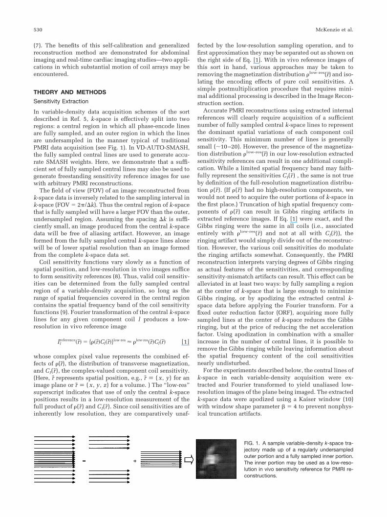
In variable-density data acquisition schemes of the sortdescribed in Ref. 5, k-space is effectively split into tworegions: a central region in which all phase-encode linesare fully sampled, and an outer region in which the linesare undersampled in the manner typical of traditionalPMRI data acquisition (see Fig. 1). In VD-AUTO-SMASH,the fully sampled central lines are used to generate accu-rate SMASH weights. Here, we demonstrate that a suffi-cient set of fully sampled central lines may also be used togenerate freestanding sensitivity reference images for usewith arbitrary PMRI reconstructions.
The field of view (FOV) of an image reconstructed fromk-space data is inversely related to the sampling interval ink-space (FOV � 2�/�k). Thus the central region of k-spacethat is fully sampled will have a larger FOV than the outer,undersampled region. Assuming the spacing �k is suffi-ciently small, an image produced from the central k-spacedata will be free of aliasing artifact. However, an imageformed from the fully sampled central k-space lines alonewill be of lower spatial resolution than an image formedfrom the complete k-space data set.
Coil sensitivity functions vary slowly as a function ofspatial position, and low-resolution in vivo images sufficeto form sensitivity references (8). Thus, valid coil sensitiv-ities can be determined from the fully sampled centralregion of a variable-density acquisition, so long as therange of spatial frequencies covered in the central regioncontains the spatial frequency band of the coil sensitivityfunctions (9). Fourier transformation of the central k-spacelines for any given component coil l produces a low-resolution in vivo reference image
Ilreference�r�� � ���r��Cl�r���low-res � �low-res�r��Cl�r�� [1]
whose complex pixel value represents the combined ef-fects of �(r�), the distribution of transverse magnetization,and Cl(r�), the complex-valued component coil sensitivity.(Here, r� represents spatial position, e.g., r� ( x, y) for animage plane or r� ( x, y, z) for a volume. ) The “low-res”superscript indicates that use of only the central k-spacepositions results in a low-resolution measurement of thefull product of �(r�) and Cl(r�). Since coil sensitivities are ofinherently low resolution, they are comparatively unaf-
fected by the low-resolution sampling operation, and tofirst approximation they may be separated out as shown onthe right side of Eq. [1]. With in vivo reference images ofthis sort in hand, various approaches may be taken toremoving the magnetization distribution �low-res(r�) and iso-lating the encoding effects of pure coil sensitivities. Asimple postmultiplication procedure that requires mini-mal additional processing is described in the Image Recon-struction section.
Accurate PMRI reconstructions using extracted internalreferences will clearly require acquisition of a sufficientnumber of fully sampled central k-space lines to representthe dominant spatial variations of each component coilsensitivity. This minimum number of lines is generallysmall (10–20). However, the presence of the magnetiza-tion distribution �low-res(r�) in our low-resolution extractedsensitivity references can result in one additional compli-cation. While a limited spatial frequency band may faith-fully represent the sensitivities Cl(r�) , the same is not trueby definition of the full-resolution magnetization distribu-tion �(r�). (If �(r�) had no high-resolution components, wewould not need to acquire the outer portions of k-space inthe first place.) Truncation of high spatial frequency com-ponents of �(r�) can result in Gibbs ringing artifacts inextracted reference images. If Eq. [1] were exact, and theGibbs ringing were the same in all coils (i.e., associatedentirely with �low-res(r�) and not at all with Cl(r�)), theringing artifact would simply divide out of the reconstruc-tion. However, the various coil sensitivities do modulatethe ringing artifacts somewhat. Consequently, the PMRIreconstruction interprets varying degrees of Gibbs ringingas actual features of the sensitivities, and correspondingsensitivity-mismatch artifacts can result. This effect can bealleviated in at least two ways: by fully sampling a regionat the center of k-space that is large enough to minimizeGibbs ringing, or by apodizing the extracted central k-space data before applying the Fourier transform. For afixed outer reduction factor (ORF), acquiring more fullysampled lines at the center of k-space reduces the Gibbsringing, but at the price of reducing the net accelerationfactor. Using apodization in combination with a smallerincrease in the number of central lines, it is possible toremove the Gibbs ringing while leaving information aboutthe spatial frequency content of the coil sensitivitiesnearly undisturbed.
For the experiments described below, the central lines ofk-space in each variable-density acquisition were ex-tracted and Fourier transformed to yield unaliased low-resolution images of the plane being imaged. The extractedk-space data were apodized using a Kaiser window (10)with window shape parameter � � 4 to prevent nonphys-ical truncation artifacts.
FIG. 1. A sample variable-density k-space tra-jectory made up of a regularly undersampledouter portion and a fully sampled inner portion.The inner portion may be used as a low-reso-lution in vivo sensitivity reference for PMRI re-constructions.
530 McKenzie et al.
Image Reconstruction
Internal sensitivities extracted as described above wereused in combination with a generalized encoding matrix(GEM) reconstruction, which takes advantage of the fullset of acquired k-space positions to fill in each missingk-space position. The GEM reconstruction is described indetail in Ref. 7. Essential features relevant to the currentwork will be summarized here.
The MR signal Sl detected in any given RF coil is theresult of a spatial integration of the distribution of trans-verse magnetization �(r�) against the sensitivity Cl(r�) ofthat coil, and against the sinusoidal spatial modulationsgenerated by encoding gradients:
Sl�k� � � � dr�Cl�r��exp��ik� � r����r��. [2]
Here, l � 1,2,. . .Ncoil is the index of any component coil inan Ncoil-element array, and k� contains the k-space indices(e.g., k� (kx,ky)) representing a total of Nacquired datapoints measured in the presence of various frequency- andphase-encoding gradients. In other words, the signal com-prises integrations or projections of the spin densityagainst Ncoil Nacquired distinct encoding functions
Bl�k� , r�� � Cl�r��exp��ik� � r��. [3]
With only a modest loss of generality (7), the integral in Eq.[2] may be approximated with a discrete sum:
Sl�k� � � �r�
Cl�r��exp��ik� � r����r�� � �r�
Bl�k�, r����r��. [4]
Here the vector index r� in the sum indicates a summationover all discrete pixel positions in the image. k� and l maybe grouped together into a single index k� l (k� ,l ), yieldingthe following matrix equation:
Sk� l � �r�
Bk� l,r��r�. [5]
or, in matrix notation,
S � B�. [6]
Inverting this equation yields an expression for the mag-netization distribution alone:
� � B�1S. [7]
Parallel image reconstruction in this formalism reducessimply to an inversion of the encoding matrix B. Arbitraryk-space trajectories, including variable-density trajectoriesused for self-calibration, may be treated in a straightfor-ward manner with this approach. First, a generalized en-coding matrix is formed as an ordered list of discretizedencoding functions made up of extracted coil sensitivitiesmultiplied by the known gradient modulation for eachacquired k-space position in turn. The encoding matrix
formed in this way will have dimension (Nacquired Ncoil) Nfull, with Nacquired measured data points in each coilbeing mapped into a total of Nfull target pixels. Providedthat the acceleration factor (Nfull/Nacquired) is less than orequal to the number of array elements Ncoil, this encodingmatrix may then be inverted with a suitable pseudoinverseprocedure. The use of a Moore-Penrose pseudoinverse(11,12), modified if necessary by a noise resistance matrix� (2,13), guarantees a least-squares solution with an opti-mal signal-to-noise ratio (SNR):
B�1 � �B†��1B��1B†��1. [8]
For fully sampled acquisitions, this formula naturally re-duces to the matched filter component coil combinationprocedure that has been shown to optimize SNR in tradi-tional phased-array imaging (13). For similar reasons, itwill make optimal use of all acquired lines in a variable-density trajectory. Once the encoding matrix has beenformed and inverted, the measured signals are groupedinto an ordered vector S that is multiplied by the inverse ofthe encoding matrix to yield the reconstructed image.
Raw reference images extracted from the central k-spacelines of a variable-density acquisition may be incorporateddirectly into this generalized reconstruction, without ex-plicit computation of the pure coil sensitivities. If the puresensitivities Cl(r�) are replaced by in vivo reference imagesIlreference(r�) � �low-res(r�)Cl(r�) from Eq. [1], the elements of
the modified encoding matrix B̃k� l,r�become
B̃k� l,r� � Ilreference�r��exp��ik� � r�� � �low-res�r��Cl�r��exp��ik� � r��
� �low-res�r��Bk� l,r� . [9]
Similarly, the elements of the modified encoding matrixinverse become
B̃ r�,k� l
�1 �Br�,k� l
�1
�low-res�r��. [10]
In Eqs. [9] and [10], the modified matrix elements at anygiven k� l and r� are obtained simply by multiplying or di-viding the original matrix elements by the appropriatevalue of �low-res(r�).
Eq. [7] then undergoes the following simple modifica-tion:
�̃ � B̃�1S
� ��low-res��1B�1S
� ��low-res��1� [11]
where (�low-res)�1 is a diagonal matrix containing the re-ciprocals (�low-res(r�))�1 along the diagonal. Equation [11] isequivalent to the following expression for the modifiedapparent magnetization �̃(r�):
�̃�r�� ���r��
�low-res�r��. [12]
Self-Calibrating Parallel Imaging 531
In other words, the apparent magnetization �̃(r�) obtainedby inversion of an encoding matrix constructed from un-corrected low-resolution in vivo reference images is justthe pixelwise quotient of the true full-resolution magnetiza-tion �(r�) with the low-resolution magnetization �low-res(r�).The final image may be corrected for �low-res(r�) throughpostmultiplication by any appropriate combination of ref-erence images, each of which itself contains �low-res(r�). Forexample, postmultiplication by a sum-of-squares combina-tion of reference images yields the desired magnetizationdistribution multiplied by the sum of squares of compo-nent coil sensitivities:
Ireconstructed�r�� � ��l�I l
reference�r���2�1/2
�̃�r��
� ��l��low-res�r��Cl�r���2�1/2 ��r��
�low-res�r��
� � �l�Cl �r��� 2� 1/2
��r��. [13]
(Here, Eq. [1] has been used to replace Ilreference(r�) with
�low-res(r�)Cl(r�).) In this way, spin-density variations in theextracted sensitivity reference divide out of the recon-struction, eliminating the need for extensive postprocess-ing of the sort described in Ref. 2.
When a regular Cartesian sampling pattern is used alongthe fully sampled frequency-encoding direction, as is thecase for the variable-density trajectories under discussion,it is straightforward to show that the encoding matrixattains a block diagonal form after Fourier transformation,with a discrete block for each position in the frequency-encoding direction (3,7). For the current work, this blockdiagonalization was exploited, and encoding matrix inver-sion was performed block by block, with consequent sav-ings in both memory utilization and reconstruction time.Further practical convenience was achieved by arrangingfor all unacquired lines in the undersampled outer regionof k-space to be filled with zeros in the raw data matricesfollowing acquisition. This allowed the missing k-spacepositions to be detected and the correct encoding matrix tobe formed automatically, without reference to the partic-ular acquisition sequence. For an irregular sampling gridthat is not zero-filled in this manner, information about thek-space position of each acquired raw data point would berequired for reconstruction.
Data Acquisition and Analysis
Variable-density acquisitions were performed on a Sie-mens 1.5 T Symphony scanner equipped with Quantumgradients. Data were acquired with a prototype coil array,furnished by Siemens Medical Systems, consisting of fourrigid elements placed below and four flexible elementspositioned above the volunteer or phantom. Since onlyfour independent receiver channels were available on oursystem, pairs of coil elements were combined in hardwareto yield a total of four inputs to the receiver chain. Except
where otherwise noted, coils immediately above and be-low each other were combined to yield four effective arrayelements distributed in the left–right direction.
Phantom Studies
Unaccelerated images of a standard resolution phantomwere obtained using a gradient-echo sequence (matrix �256 192, FOV � 315 236 mm, repetition time (TR) �190 ms, echo time (TE) � 4 ms, slice thickness � 6 mm) infour image plane orientations (coronal, 30° coronal to sag-ittal, 60° coronal to sagittal, and sagittal). The phase-en-code direction varied from left–right for the coronal planeto anterior–posterior for the sagittal plane.
To examine the effect of number of central lines, Ncenter,and the undersampling factor of outer lines (the outerreduction factor, or ORF) on the quality of image recon-struction, variable-density data sets were synthesized byreplacing selected lines in the fully gradient-encoded k-space data sets with zeros. This zero-filling decimationensured that the reference and variable-density data setswere identical, except for the acceleration, and alloweddirect comparison of reconstructed images. Various num-bers of central lines and ORFs were used to achieve netacceleration factors of 1.0–3.6. The net acceleration factoris calculated from Ncenter and ORF using the followingexpression:
Net acceleration factor �N full
N aquired
�N full
N center � �N full � N center�/ORF. [14]
After decimation of the fully encoded data, PMRI recon-structions were performed using the internal sensitivityextraction and GEM reconstruction procedures outlinedabove. Reconstructions were implemented in the MAT-LAB programming language (The Mathworks, Natick,MA).
To quantify the balance between Ncenter and ORF, SNRand artifact power (AP) were calculated for each recon-structed image. Pixel-by-pixel SNR values for the GEMreconstructions can be predicted by dividing the meanimage intensity Ireconstructed(r�) at each pixel position by theexpected standard deviation (SD) of noise at that position.Assuming that each signal point in the signal vector Scontains normally-distributed noise with SD �0, and usingthe fact that the variance of a linear sum is just the square-weighted sum of individual variances, one arrives at thefollowing expression for pixelwise SNR:
SNR�r�� �
��l�Cl�r���2�1/2
��r��
�0
�N acquired�1/2 ��l�I l
reference�r���2�1/2��k� l
�B̃r�,k� l
�1�2�1/2.
[15]
The numerator of Eq. [15] is just the expression forI reconstructed(r�) from Eq. [13]. The denominator is the
532 McKenzie et al.
underlying noise variance �0 normalized by a factor of(Nacquired)1/2 to account for the averaging of incoherentnoise from the acquired k-space data, and scaled by thesquare root of the sum of squares of all linear weightsmultiplying the signal vector S to yield I reconstructed(r�) .Equation [15] as it stands makes reference to the puresensitivities, which are never explicitly calculated in theself-calibrating reconstruction. However, for purposes ofcomparative evaluation, we are not primarily interested inpredicting the absolute SNR, which varies with the elec-tronic and geometrical properties of the coil array as wellas with the particular imaging sequence, and which mustgenerally be simulated or measured experimentally. In-stead, we are interested in determining SNR normalizedrelative to an optimal image reconstructed from the corre-sponding unaccelerated acquisition. For a fully sampledimage with an acceleration factor of 1, Eq. [15] becomes
SNR0�r�� ��N full�1/2
�0 ��l�Cl�r���2�1/2
��r�� [16]
yielding the following expression for normalized SNR:
SNRnormalised�r�� �SNR�r��SNR0�r��
�1
� N full
N acquired�1/2��l�I l
reference�r���2�1/2���k l
�B̃r�,�k l
�1�2�1/2. [17]
The geometrical noise multiplication factor, or “g-factor” described in Ref. 2, corresponds to the term��
l�Il
reference�r���2�1/ 2���kl
�B̃r�, �kl
�1 �2�1/ 2in the denominator of Eq. [17].
AP is a measure of the absolute difference between the“true” distribution of image intensity and the intensitydistribution in a reconstructed image. The unacceleratedimages I0(r�) were taken to be a measure of the “true” imageintensity, and AP was determined from the followingequation:
AP �
�r�
I 0�r��� � �I reconstructed�r��2
�r�
�I 0�r���2. [18]
Note that a higher value of AP represents increased artifactand reduced image quality. It should also be noted that theformula for AP in Eq. [18] does not measure residualaliasing alone, but will include any difference in imageintensity between I0 and Ireconstructed, including differ-ences solely due to noise. Thus, AP should be regarded asupper bound on the artifact level in the PMRI reconstruc-tions.
SNR and AP were calculated for a region of interest(ROI) selected from I reconstructed. The ROI was determinedby thresholding I reconstructed and including any pixels withimage intensity significantly above the noise floor. The sameROI was then used in both I reconstructed and I0. I reconstructed
was used for ROI selection to ensure that any residual
aliasing artifacts would be included in the AP calculation,while pixels containing only noise would be rejected.
In Vivo Studies
In vivo images were obtained from a total of four healthyadult volunteers. Informed consent was obtained fromthese volunteers in compliance with the regulations of theBeth Israel Deaconess Medical Center’s institutional re-view board.
Abdominal imaging was performed using an axial T1-weighted gradient-echo sequence (matrix � 96 256,FOV � 240 mm 320 mm, TR � 190 ms, TE � 4 ms,anterior-posterior phase-encode direction, slice thick-ness � 5 mm). To demonstrate the effects of sensitivitymiscalibration and the potential benefits of self-calibra-tion, distinct data sets were acquired during end-inspira-tory and end-expiratory breath-holds, respectively. Alongwith unaccelerated reference acquisitions in the twobreath-hold positions, an accelerated variable-density ac-quisition was performed at end inspiration with 32 centrallines and an ORF of 2 (net acceleration factor � 1.5). GEMreconstruction of the accelerated end-inspiratory data setwas performed using 1) an external sensitivity referencederived from the unaccelerated end-expiratory image set,and 2) an internal sensitivity reference extracted from theaccelerated variable-density data set itself.
Real-time cardiac imaging was also performed in threevolunteers using a real-time TrueFISP sequence (14,15)(matrix � 64 63, FOV � 350 350 mm, TR � 2.5 ms,TE � 1.25 ms, slice thickness � 10 mm, left–right phase-encode direction, view sharing, double-oblique short-axisimage plane). In these studies, only the four coils in theflexible top half of the array were used, to provide in-creased sensitivity in the image plane of interest and toeliminate unwanted signal from the back. Since only fourcoils were used, hardware combination of the coil outputswas not required. The flexible coils were conformed to thechest wall and centered on the position of the heart. Dur-ing real-time imaging the volunteers were allowed tobreathe freely. This resulted in significant motion of thechest wall, and therefore of the coil array, during dataacquisition. A variable-density trajectory with 21 centrallines and an ORF of 2 (net acceleration factor � 1.5) wasused to increase real-time frame rates to 15 frames persecond as compared with reference acquisitions at11 frames per second. To demonstrate the effects of sensi-tivity mismatches, accelerated images were first recon-structed using an external sensitivity reference derivedfrom one image of the unaccelerated real-time image set. Asecond GEM reconstruction was performed using a dis-tinct extracted internal sensitivity reference for each ac-celerated image.
RESULTS
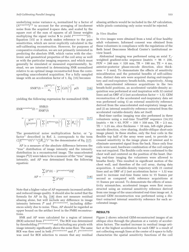
Figure 2 shows selected GEM-reconstructed images of anoblique slice through the phantom at a variety of acceler-ation factors. The marked increase in residual image arti-fact at the highest acceleration for each ORF is a result ofnot collecting enough lines at the center of k-space to fullycharacterize the spatial sensitivity functions of the coil
Self-Calibrating Parallel Imaging 533
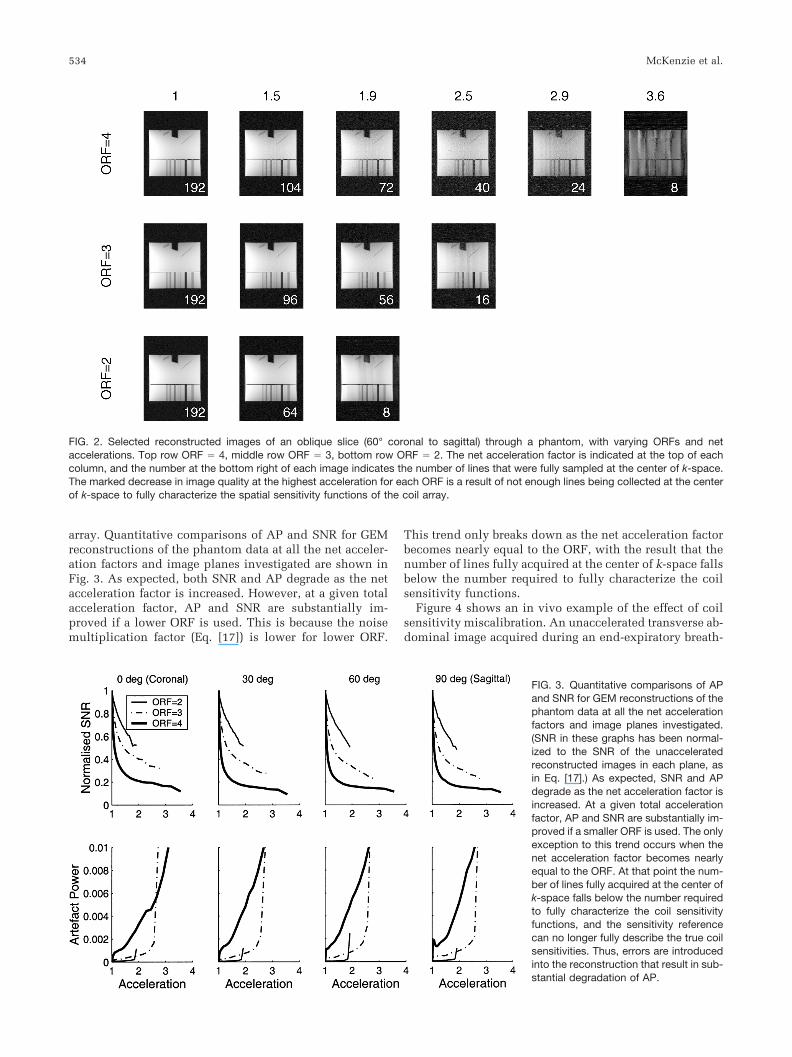
array. Quantitative comparisons of AP and SNR for GEMreconstructions of the phantom data at all the net acceler-ation factors and image planes investigated are shown inFig. 3. As expected, both SNR and AP degrade as the netacceleration factor is increased. However, at a given totalacceleration factor, AP and SNR are substantially im-proved if a lower ORF is used. This is because the noisemultiplication factor (Eq. [17]) is lower for lower ORF.
This trend only breaks down as the net acceleration factorbecomes nearly equal to the ORF, with the result that thenumber of lines fully acquired at the center of k-space fallsbelow the number required to fully characterize the coilsensitivity functions.
Figure 4 shows an in vivo example of the effect of coilsensitivity miscalibration. An unaccelerated transverse ab-dominal image acquired during an end-expiratory breath-
FIG. 2. Selected reconstructed images of an oblique slice (60° coronal to sagittal) through a phantom, with varying ORFs and netaccelerations. Top row ORF � 4, middle row ORF � 3, bottom row ORF � 2. The net acceleration factor is indicated at the top of eachcolumn, and the number at the bottom right of each image indicates the number of lines that were fully sampled at the center of k-space.The marked decrease in image quality at the highest acceleration for each ORF is a result of not enough lines being collected at the centerof k-space to fully characterize the spatial sensitivity functions of the coil array.
FIG. 3. Quantitative comparisons of APand SNR for GEM reconstructions of thephantom data at all the net accelerationfactors and image planes investigated.(SNR in these graphs has been normal-ized to the SNR of the unacceleratedreconstructed images in each plane, asin Eq. [17].) As expected, SNR and APdegrade as the net acceleration factor isincreased. At a given total accelerationfactor, AP and SNR are substantially im-proved if a smaller ORF is used. The onlyexception to this trend occurs when thenet acceleration factor becomes nearlyequal to the ORF. At that point the num-ber of lines fully acquired at the center ofk-space falls below the number requiredto fully characterize the coil sensitivityfunctions, and the sensitivity referencecan no longer fully describe the true coilsensitivities. Thus, errors are introducedinto the reconstruction that result in sub-stantial degradation of AP.
534 McKenzie et al.
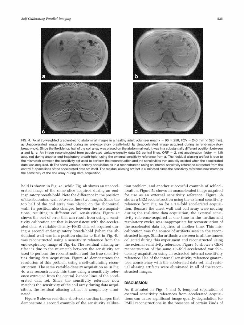
hold is shown in Fig. 4a, while Fig. 4b shows an unaccel-erated image of the same slice acquired during an end-inspiratory breath-hold. Note the difference in the positionof the abdominal wall between these two images. Since thetop half of the coil array was placed on the abdominalwall, its position also changed between the two acquisi-tions, resulting in different coil sensitivities. Figure 4cshows the sort of error that can result from using a sensi-tivity calibration set that is inconsistent with the acceler-ated data. A variable-density–PMRI data set acquired dur-ing a second end-inspiratory breath-hold (when the ab-dominal wall was in a position similar to that in Fig. 4b)was reconstructed using a sensitivity reference from theend-expiratory image of Fig. 4a. The residual aliasing ar-tifact is due to the mismatch between the sensitivity setused to perform the reconstruction and the true sensitivi-ties during data acquisition. Figure 4d demonstrates theresolution of this problem using a self-calibrating recon-struction. The same variable-density acquisition as in Fig.4c was reconstructed, this time using a sensitivity refer-ence extracted from the central k-space lines of the accel-erated data set. Since the sensitivity reference nowmatches the sensitivity of the coil array during data acqui-sition, the residual aliasing artifact is completely elimi-nated.
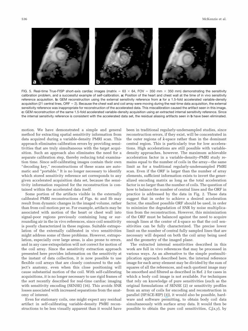
Figure 5 shows real-time short-axis cardiac images thatdemonstrate a second example of the sensitivity calibra-
tion problem, and another successful example of self-cal-ibration. Figure 5a shows an unaccelerated image acquiredfor use as an external sensitivity reference. Figure 5bshows a GEM reconstruction using the external sensitivityreference from Fig. 5a for a 1.5-fold accelerated acquisi-tion. Because the chest wall and coil array were movingduring the real-time data acquisition, the external sensi-tivity reference acquired at one time in the cardiac andrespiratory cycles was inappropriate for reconstruction ofthe accelerated data acquired at another time. This mis-calibration was the source of artifacts seen in the recon-structed image. Similar artifacts were seen in all the framescollected during this experiment and reconstructed usingthe external sensitivity reference. Figure 5c shows a GEMreconstruction of the same 1.5-fold accelerated variable-density acquisition using an extracted internal sensitivityreference. Use of the internal sensitivity reference guaran-teed consistency with the accelerated data set, and resid-ual aliasing artifacts were eliminated in all of the recon-structed images.
DISCUSSION
As illustrated in Figs. 4 and 5, temporal separation ofexternal sensitivity references from accelerated acquisi-tions can cause significant image quality degradation forPMRI reconstructions in the presence of certain kinds of
FIG. 4. Axial T1-weighted gradient-echo abdominal images in a healthy adult volunteer (matrix � 96 256, FOV � 240 mm 320 mm).a: Unaccelerated image acquired during an end-expiratory breath-hold. b: Unaccelerated image acquired during an end-inspiratorybreath-hold. Since the flexible top half of the coil array was placed on the abdominal wall, it was in a substantially different position betweena and b. c: An image reconstructed from accelerated variable-density data (32 central lines, ORF � 2, net acceleration factor � 1.5)acquired during another end-inspiratory breath-hold, using the external sensitivity reference from a. The residual aliasing artifact is due tothe mismatch between the sensitivity set used to perform the reconstruction and the sensitivities that actually existed when the accelerateddata was acquired. d: The same variable-density acquisition as in c reconstructed using an internal sensitivity reference extracted from thecentral k-space lines of the accelerated data set itself. The residual aliasing artifact is eliminated since the sensitivity reference now matchesthe sensitivity of the coil array during data acquisition.
Self-Calibrating Parallel Imaging 535
motion. We have demonstrated a simple and generalmethod for extracting spatial sensitivity information fromdata acquired during a variable-density PMRI scan. Thisapproach eliminates calibration errors by providing sensi-tivities that are truly simultaneous with the target acqui-sition. Such an approach also eliminates the need for aseparate calibration step, thereby reducing total examina-tion time. Since self-calibrating images contain their own“decoding key,” reconstructions of these scans are auto-matic and “portable.” It is no longer necessary to identifywhich stored sensitivity reference set corresponds to anygiven accelerated acquisition data set, because the sensi-tivity information required for the reconstruction is con-tained within the accelerated data itself.
Some portion of the artifacts visible in the externallycalibrated PMRI reconstructions of Figs. 4c and 5b mayresult from dynamic changes in the imaged volume, ratherthan from coil motion per se. For example, artifacts may beassociated with motion of the heart or chest wall intosignal-poor regions previously containing lung or sur-rounding air in the in vivo references, since coil sensitivityis poorly characterized in these regions. Suitable extrapo-lation of the externally calibrated in vivo sensitivitiesmight address some of these problems. However, extrapo-lation, especially over large areas, is also prone to errors,and in any case extrapolation will not correct for motion ofthe coil array. Since the sensitivity extraction approachpresented here provides information on the sensitivity atthe instant of data collection, it is now possible to useflexible coil arrays that are closely contoured to the sub-ject’s anatomy, even when this close positioning willcause substantial motion of the coil. With self-calibratingacquisitions, it is no longer necessary to use rigid frames ofthe sort recently described for real-time cardiac imagingwith sensitivity encoding (SENSE) (16). This avoids SNRlosses associated with increased separations from the anat-omy of interest.
Even for stationary coils, one might expect any residualartifact in self-calibrating variable-density PMRI recon-structions to be less visually apparent than it would have
been in traditional regularly-undersampled studies, sincereconstruction errors, if they exist, will be concentrated inthe outer regions of k-space rather than in the dominantcentral region. This is particularly true for low accelera-tions. High accelerations are still possible with variable-density approaches, however. The maximum achievableacceleration factor in a variable-density–PMRI study re-mains equal to the number of coils in the array—the samelimit as for a traditional regularly-undersampled PMRIscan. Even if the ORF is larger than the number of arrayelements, sufficient information exists to invert the gener-alized encoding matrix as long as the total accelerationfactor is no larger than the number of coils. The question ofhow to balance the number of central lines and the ORF inpractice is addressed by the data in Fig. 2. These datasuggest that in order to achieve a desired accelerationfactor, the smallest possible ORF should be used, in orderto minimize the degradation of SNR by noise multiplica-tion from the reconstruction. However, this minimizationof the ORF must be balanced against the need to acquireenough lines at the center of k-space so that the coil sen-sitivities can be fully characterized. The precise lowerlimit on the number of central fully sampled lines that arenecessary will depend on both the coil array being usedand the geometry of the imaged plane.
The extracted internal sensitivities described in thiswork are full in vivo references that may be processed invarious ways. As an alternative to the simple postmulti-plication approach described here, the internal referenceimage for each array element may be divided by the sum ofsquares of all the references, and each quotient image maybe smoothed and filtered as described in Ref. 2 for cases inwhich a body coil image is not available. For techniquesthat rely on knowledge of pure sensitivities (such as theoriginal formulations of SENSE (2) or sensitivity profilesfrom an array of coils for encoding and reconstruction inparallel (SPACE-RIP) (3)), it would also be possible, hard-ware and software permitting, to obtain body coil datasimultaneously with surface array data. It would then bepossible to obtain the pure coil sensitivities, Cl(x,y), by
FIG. 5. Real-time True-FISP short-axis cardiac images (matrix � 63 64, FOV � 350 mm 350 mm) demonstrating the sensitivitycalibration problem, and a successful example of self-calibration. a: Position of the heart and chest wall at the time of in vivo sensitivityreference acquisition. b: GEM reconstruction using the external sensitivity reference from a for a 1.5-fold accelerated variable-densityacquisition (21 central lines, ORF � 2). Because the chest wall and coil array were moving during the real-time data acquisition, the externalsensitivity reference was inappropriate for reconstruction of the accelerated data. This miscalibration caused the artifact seen in this image.c: GEM reconstruction of the same 1.5-fold accelerated variable-density acquisition using an extracted internal sensitivity reference. Sincethe internal sensitivity reference is consistent with the accelerated data set, the residual aliasing artifacts seen in b have been eliminated.
536 McKenzie et al.
using a suitable processing procedure (1,2). However,since the number of available receiver channels on a mag-net is limited, this would require sacrificing data acquisi-tion from one element in the surface coil array, in favor ofbody coil acquisition. While the spatial information fromthe body coil would contribute to the subsequent recon-struction, use of the body coil data would likely result in alower SNR than could have been achieved with an addi-tional surface coil.
Although in vivo sensitivity functions are more conve-nient to acquire and use than pure sensitivity functions,they must be used with care in some situations. For exam-ple, neither the sum of squares postmultiplication of Eq.[13] nor a sum of squares quotient as described in Ref.2 preserves phase information in the reconstructed image.For phase-sensitive experiments, such as flow velocitymeasurements, postmultiplication by a complex compo-nent coil reference image (or by each component coil ref-erence in turn) may be used, and phase information maybe extracted from the resulting reconstructed componentcoil images, as might be done for traditional phase-sensi-tive imaging with an array.
The generalized encoding matrix reconstruction usedhere provides a number of advantages for reconstructingvariable-density data. The encoding matrix is constructedusing knowledge of the k-space positions of all acquiredlines, including those that are also used to determine thecomponent coil sensitivities. Use of the Moore-Penrosepseudoinverse guarantees a matched filter combination ofall the acquired data. However, many other PMRI recon-struction techniques besides GEM are fully compatiblewith self-calibration as described here.
SMASH-like reconstructions are easily adapted to re-construction of variable-density data, as evidenced by theVD-AUTO-SMASH approach (5). Conventional SMASHreconstructions may also be used for reconstruction ofvariable-density data. Given knowledge of the extracted invivo spatial sensitivities, the weights required to generatevarious spatial harmonics may be formed in the usualmanner (17). SMASH reconstruction may then be accom-plished simply by convolving the vector of spatial har-monic weights with the undersampled k-space data. Awide range of algorithmic improvements may be appliedin these SMASH-like reconstructions. For example, therecently described generalized autocalibrating partiallyparallel acquisitions (GRAPPA) technique (18) improveson VD-AUTO-SMASH by performing spatial harmonic av-eraging in a manner similar to that in Ref. 17, and byproducing one image for each component coil of the re-ceiver array. Reconstructed component coil images arethen combined in sum-of-squares fashion, thereby elimi-nating the possibility of phase cancellation artifact, asdescribed in Ref. 6.
Since neither SMASH nor GRAPPA use all acquireddata to reconstruct the missing k-space lines, they do notmake optimal use of the acquired data in the sense that thereconstructions are not guaranteed to reconstruct imageswith the maximum possible SNR and minimum AP. How-ever, this sacrifice of some degree of theoretical exactnessimproves the robustness of these techniques in the face ofimperfect sensitivity calibration. These reconstructionsgenerate missing k-space lines from linear combinations of
a restricted set of nearby measured k-space lines, thuslimiting the propagation of sensitivity errors into the re-constructed image. As an added benefit, this restriction ofthe number of lines contributing to a reconstruction meansthat SMASH and GRAPPA reconstructions are much lesscomputationally intensive than a GEM reconstruction.
The image-domain PMRI reconstruction techniques arealso amenable to combination with the self-calibrationtechniques discussed here. However, image-domain ap-proaches such as the Cartesian formulation of SENSE (2)generally assume that k-space has been sampled in a reg-ular pattern to yield a coherent pattern of aliasing. Toperform an image-domain SENSE reconstruction, the extradata acquired at the center of k-space would have to bediscarded and the variable-density data set would have tobe decimated back down to a regularly undersampled dataset. This would be expected to result in some loss of imagequality and SNR efficiency, as compared to a full use of theadditionally acquired lines.
While the image-domain Cartesian SENSE approachwith decimation does not make full use of fully sampledcentral lines, a non-Cartesian formulation of SENSE maybe used instead (2). Indeed, GEM reconstruction bearssome similarity to the generalized SENSE reconstruction.The principal differences lie in the discretization proce-dure used to generate the encoding matrix, the use ofnumerical conditioning in encoding matrix inversion, andthe use of unprocessed in vivo sensitivity references withpostmultiplication (7). Hybrid approaches intermediatebetween SMASH and SENSE, as described in Ref. 7, arealso fully compatible with self-calibration as describedhere.
For the sake of ease of implementation, all the examplesof self-calibrating data acquisitions demonstrated hereused only two integral acceleration factors across k-space:no acceleration in the center of k-space, and a single inte-gral factor between 2 and 4 in the outer region of k-space.Since the results show that, for any given net accelerationfactor, the SNR and AP are dependent on the ORF used, itwill be interesting to investigate more complex undersam-pling schemes. For example, the use of variable ORFsacross the undersampled region, as was demonstrated us-ing external calibration with the SPACE-RIP technique (3),may also improve the quality of self-calibrating PMRI re-constructions.
Certain relatively common k-space sampling schemes,such as spiral and radial trajectories, are inherently self-calibrating, since they automatically sample the center ofk-space densely. Low-resolution internal sensitivity refer-ences may easily be generated from the densely sampledcenters of such acquisitions, and may be used with GEM ornon-Cartesian SENSE reconstructions. Applications suchas real-time spiral imaging would be expected to benefitsignificantly from such an approach.
CONCLUSIONS
We have demonstrated the use of a general self-calibratingPMRI approach for the elimination of artifacts due to ex-ternal coil sensitivity calibration. The particular sensitiv-ity extraction technique introduced here can be used incombination with most existing PMRI reconstruction tech-
Self-Calibrating Parallel Imaging 537
niques. Self-calibrating approaches hold particular prom-ise for real-time and/or interactive applications in whichcontinuously changing sensitivities necessitate frequentupdating of calibration information.
ACKNOWLEDGMENTS
The authors acknowledge Dr. O. Simonetti of SiemensMedical Systems for his assistance with modifying theTrue FISP sequence used for real-time cardiac imaging.Drs. Jianmin Wang and Daniel Driemel of Siemens MedicalSystems are acknowledged for their collaboration in the de-sign of the eight-element array, and for construction andtesting of the prototype array used in these experiments.
REFERENCES
1. Sodickson DK, Manning WJ. Simultaneous acquisition of spatial har-monics (SMASH): fast imaging with radiofrequency coil arrays. MagnReson Med 1997;38:591–603.
2. Pruessmann KP, Weiger M, Scheidegger MB, Boesiger P. SENSE: sen-sitivity encoding for fast MRI. Magn Reson Med 1999;42:952–962.
3. Kyriakos WE, Panych LP, Kacher DF, Westin CF, Bao SM, Mulkern RV,Jolesz FA. Sensitivity profiles from an array of coils for encoding andreconstruction in parallel (SPACE RIP). Magn Reson Med 2000;44:301–308.
4. Jakob PM, Griswold MA, Edelman RR, Sodickson DK. AUTO-SMASH:a self-calibrating technique for SMASH imaging. Simultaneous acqui-sition of spatial harmonics. MAGMA 1998;7:42–54.
5. Heidemann R, Griswold M, Haase A, Jakob PM. VD-AUTO-SMASHimaging. Magn Reson Med 2001;45:1066–1074.
6. McKenzie CA, Ohliger MA, Yeh EN, Price MD, Sodickson DK. Coil bycoil phased array image reconstruction with SMASH. Magn ResonImaging 2001;46:619–623.
7. Sodickson DK, McKenzie C. A generalized approach to parallel mag-netic resonance imaging. Med Phys 2001;28:1629–1643.
8. Sodickson DK. Tailored SMASH image reconstructions for robust invivo parallel MR imaging. Magn Reson Med 2000;44:243–251.
9. Wang Y. Description of parallel imaging in MRI using multiple coils.Magn Reson Med 2000;44:495–499.
10. Oppenheim AV, Schaefer R. Discrete time signal processing. Engle-wood Cliffs, NJ: Prentice Hall; 1989. 879 p.
11. Penrose R. A generalized inverse for matrices. Proc Camb Philol Soc1955;51:406–413.
12. Ben-Israel A, Greville TNE. Generalized inverses: theory and applica-tions. New York: Wiley; 1977.
13. Roemer PB, Edelstein WA, Hayes CE, Souza SP, Mueller OM. The NMRphased array. Magn Reson Med 1990;16:192–225.
14. Heid O. True FISP cardiac fluoroscopy. In: Proceedings of the 5thAnnual Meeting of ISMRM, Vancouver, Canada, 1997. p 320.
15. Bundy JM, Laub G, Kim R, Finn JP, Simonetti OP. Real-time dataacquisition for LV function. In: Proceedings of the 7th Annual Meetingof ISMRM, Philadelphia, 1999. p 386.
16. Weiger M, Pruessmann KP, Boesiger P. Cardiac real-time imaging usingSENSE. Magn Reson Med 2000;43:177–184.
17. McKenzie CA, Yeh EN, Sodickson DK. Improved spatial harmonicselection for SMASH image reconstructions. Magn Reson Med 2001;46:831–836.
18. Griswold M, Jakob PM, Heidemann R, Nittka M, Wang J, Kiefer B,Haase A. Push-button PPA reconstructions: generalized autocalibratingpartially parallel acquisitions (GRAPPA). In: Proceedings of the 9thAnnual Meeting of ISMRM, Glasgow, Scotland, 2001. p 8.
538 McKenzie et al.
Related Documents


