Welcome message from author
This document is posted to help you gain knowledge. Please leave a comment to let me know what you think about it! Share it to your friends and learn new things together.
Transcript
mojtaba
mojnia

Self Assessment & Review
MMUNOLOGY
mojtaba
mojnia

Self Assessment & Review
MMUNOLOGY
Rachna ChaurasiaMD Radiodiagnosis
MLB Medical College, Jhansi, India
Anshul JainMD Anaesthesia
MLB Medical College, Jhansi, India
the arora medical book publishers pvt. ltd.A Group of Jaypee Brothers Medical Publishers (P) Ltd.
4thEdition
Published by
Jitendar P VijJaypee Brothers Medical Publishers (P) LtdCorporate Office4838/24 Ansari Road, Daryaganj, New Delhi - 110002, India, Phone: +91-11-43574357Registered OfficeB-3 EMCA House, 23/23B Ansari Road, Daryaganj, New Delhi - 110 002, IndiaPhones: +91-11-23272143, +91-11-23272703, +91-11-23282021, +91-11-23245672Rel: +91-11-32558559, Fax: +91-11-23276490, +91-11-23245683e-mail: [email protected], Website: www.jaypeebrothers.com
Branches❑ 2/B, Akruti Society, Jodhpur Gam Road Satellite
Ahmedabad 380 015, Phones: +91-79-26926233, Rel: +91-79-32988717Fax: +91-79-26927094, e-mail: [email protected]
❑ 202 Batavia Chambers, 8 Kumara Krupa Road, Kumara Park EastBengaluru 560 001, Phones: +91-80-22285971, +91-80-2238295691-80-22372664, Rel: +91-80-32714073, Fax: +91-80-22281761e-mail: [email protected]
❑ 282 IIIrd Floor, Khaleel Shirazi Estate, Fountain Plaza, Pantheon RoadChennai 600 008, Phones: +91-44-28193265, +91-44-28194897Rel: +91-44-32972089, Fax: +91-44-28193231, e-mail: [email protected]
❑ 4-2-1067/1-3, 1st Floor, Balaji Building, Ramkote Cross Road,Hyderabad 500 095, Phones: +91-40-66610020, +91-40-24758498Rel:+91-40-32940929Fax:+91-40-24758499, e-mail: [email protected]
❑ No. 41/3098, B & B1, Kuruvi Building, St. Vincent RoadKochi 682 018, Kerala, Phones: +91-484-4036109, +91-484-2395739+91-484-2395740, e-mail: [email protected]
❑ 1-A Indian Mirror Street, Wellington SquareKolkata 700 013, Phones: +91-33-22651926, +91-33-22276404+91-33-22276415, Rel: +91-33-32901926, Fax: +91-33-22656075e-mail: [email protected]
❑ Lekhraj Market III, B-2, Sector-4, Faizabad Road, Indira NagarLucknow 226 016, Phones: +91-522-3040553, +91-522-3040554e-mail: [email protected]
❑ 106 Amit Industrial Estate, 61 Dr SS Rao Road, Near MGM Hospital, ParelMumbai 400 012, Phones: +91-22-24124863, +91-22-24104532,Rel: +91-22-32926896, Fax: +91-22-24160828, e-mail: [email protected]
❑ “KAMALPUSHPA” 38, Reshimbag, Opp. Mohota Science College, Umred RoadNagpur 440 009 (MS), Phone: Rel: +91-712-3245220, Fax: +91-712-2704275e-mail: [email protected]
USA Office1745, Pheasant Run Drive, Maryland Heights (Missouri), MO 63043, USA, Ph: 001-636-6279734e-mail: [email protected], [email protected]
Self Assessment & Review Microbiology Immunology© 2009, Jaypee Brothers Medical PublishersAll rights reserved. No part of this publication should be reproduced, stored in a retrieval system, or transmitted in any formor by any means: electronic, mechanical, photocopying, recording, or otherwise, without the prior written permission of theauthors and the publisher.
This book has been published in good faith that the material provided by authors is original. Every effort is made to ensureaccuracy of material, but the publisher, printer and author will not be held responsible for any inadvertent error(s). In caseof any dispute, all legal matters are to be settled under Delhi jurisdiction only.
Fourth Edition: 2009ISBN 978-81-8448-472-4Typeset at JPBMP typesetting unitPrinted at

Dedication
This book is dedicated toOur family members, teachers
and above all Godwho created us
mojtaba
mojnia


Preface
“First of all we want to thank all of the readers for their immense support—the key of success.”
The overwhelming success of the previous edition has encouraged us to carry this 4th edition a stepahead to revise and update the book, so as to match the pace of present PGMEE requirement of thisdifficult subject.
PGMEE is a battle field in which everyone fights for success but only few succeed because only fewknow the correct use of weapon of “Knowledge, hard work and guidance".
So, guys learn the golden words :
Success comes when, we do things right;When we learn how to make the best use of our time,
and how to deal with adversities
Why this book is different?Beside adding new questions, we have taken account of all mistakes to provide an error free text.This book provides important points of each topic in continuous manner not in parts as given in
previous guides.This book consists of theory of each topic followed by its questions so theory portion helps you to
solve new questions.This pattern of book allows you to revise whole infectious disease very clearly and quickly.This book provides sufficient matter which can be revised without any problem.
How to use this book?Read the theory first then do questions. You realize that this way of learning makes topic easy to
understand and easy to grasp for long-term memory. Try to complete each topic in one sitting.In the last we would like to say all the best for your PGMEE preparation and hope you will work hard
with positive attitude in your mind.
Always keep one thing in your mind:There's one thing, we cannot recycle and that's wasted time, so guys !
“Schedule a daily time for relaxing, reflecting, planning and brainstorming."

mojtaba
mojnia
Acknowledgments"To Bhagwan Mahavir and Ganesha whose blessing made our goal possible"
We will extremely grateful and obliged to our teachers• Dr. Ganesh Kumar (Principal, MLB Medical College, Jhansi)• Dr. A. K. Gupta• Dr. Mrs. Veena GuptaWe would always remain obliged to our life long teachers• Dr. B. C. Tiwari• Dr. Atish Sharma• Mr. Amod YadavWe would be extremely thankful to Dr. Vikas Chaurasia, Dr. Gaurav Tiwari whose inspiration makes thetask possible.We extend our sincere thanks and gratitude to our beloved family members for their encouragementand useful suggestionsFather : Dr. P. C. ChaurasiaMother : Mrs. Shakuntla ChaurasiaUncle & Aunty : Mr. Subhash Chandra Chaurasia & Mrs. Rashmi ChaurasiaBrother & Bhabhi : Dr. Vikas Chaurasia & Dr. Mrs. Anahita Chaurasia
Mr. Vishal Chaurasia & Mrs. Kavita ChaurasiaNephew & Niece : Priyanshu, Aashi and Paras
– Rachna ChaurasiaFather : Dr. D. B. JainMother : Mrs. Saroj JainSisters : Miss Ayusha Jain & Miss Ankita JainBrother : Mr. Kapil Jain
– Anshul JainWe offer cordial thanks to our seniors and colleagues for their support• Dr. Vinod Tiwari • Dr. Hazari M. Shukla• Dr. Rajendra Kr. Singh • Dr. R. K. Singh• Dr. Sachi Singhal • Dr. Surbhi Srivastav• Dr. Shekhar • Dr. Dinesh Dhiwan• Dr. Apoorva Abhinandan Mittal • Dr. Saurabh Jain• Dr. Deepak Singhal • Dr. Dushyant Agarwal• Dr. Balram • Dr. Monika Agarwal• Dr. Surendra Sahu • Dr. Mayank• Dr. Amit Gupta • Dr. Nidhi Singh• Dr. Vivek Agarwal • Dr. Rajesh Gupta

Self Assessment & Review Microbiology & Immunologyx
x
We cannot forget to acknowledge• Mr. Bishan Lal (Librarian, MLB Medical College, Jhansi) and All Staff.• Mr. C. P. Lavania (Librarian, SN Medical College, Agra) and All Staff.
We would like to acknowledge for their support in every part of our life.• Mr. Sandeep Tiwari• Mr. Adeep Tiwari• Mr. C. P. Lamba• Mr. Ankit Chaurasia• Mrs. Arti Rawat• Mrs. Renuka Singh
Last but not least, most important we gratefully acknowledge all readers, who will act as a guide toimprove and upgrading the content of this book.
mojtaba
mojnia
Contents
SECTION – A
REVISION AT A GLANCE1. Basics of Bacteriology 03 – 092. Basics of Virology 10 – 133. Basics of Mycology 14 – 154. Basics of Clinical Microbiology 16 – 185. Culture & Sterilisation 19 – 21
Questions & Answers 22 – 28
SECTION – B
Explanatory Series of Questions 1995 - 2008 "ALL INDIA, AIIMS & PGI"
UNIT – I BACTERIOLOGYGram Positive Cocci1. Staphylococci 31 – 442. Streptococci 45 – 67Gram Negative Cocci3. Neisseria 68 – 78Gram Positive Bacilli4. Clostridium 79 – 935. Corynebacterium 94 – 1056. Actinomycetes & Bacillus 106 – 1187. Listeria Monocytogenes 119 – 1258. Mycobacteria 126 – 146Gram Negative Bacilli9. Enterobacteriaceae 147 – 17110. Vibrio 172 – 18611. Pseudomonas & Yersinia 187 – 195Gram Negative Cocco - Bacilli12. Hemophilus, Bordetella & Brucella 196 – 20613. Campylobacter & Helicobacter 207 – 21414. Legionella 215 – 22015. Rickettsiaceae & Chlamydiae 221 – 23816. Spirochetes 239 – 25317. Mycoplasma 254 – 260
Self Assessment & Review Microbiology & Immunologyxii
xii
UNIT – II VIROLOGY1. DNA Virus 263 – 284
Herpes virus, Adeno & Pox, Parvo & Papovo2. RNA Virus 285 – 319
Picorna virus, Myxo virus, Rota & other viral gastroenteritis, Arbo, Rhabdoviruses3. Slow Virus Diseases 320 – 3254. Hepatitis Viruses 326 – 3435. HIV & Other Retroviruses 344 – 366
UNIT – III MYCOLOGY1. Dermatophytes 369 – 3722. Yeast & Yeast like Fungus 373 – 387
Cryptococcus, Candida, Pneumocystii carinii3. Aspergillus & Mucormycosis 388 – 3924. Dimorphic Fungi 393 – 399
UNIT – IV PARASITOLOGY1. Basics of Parasitology 403 – 4072. Protozoa 408 – 4313. Helminths 432 – 450
UNIT – V IMMUNOLOGY1. Basics of Immune System 453– 4572. Antigen & Antibody 458 – 4633. Hypersensitivity 464 – 494
MISCELLANEOUS 497 – 530Index 531
In this edition Ananthnarayan refers to Ananthanarayan and Panicker’s Textbook of Microbiology 7/e andPanicker refers to Panickers Textbook of Medical Parasitology 6/e.
SECTION – A
REVISION AT A GLANCE
mojtaba
mojnia

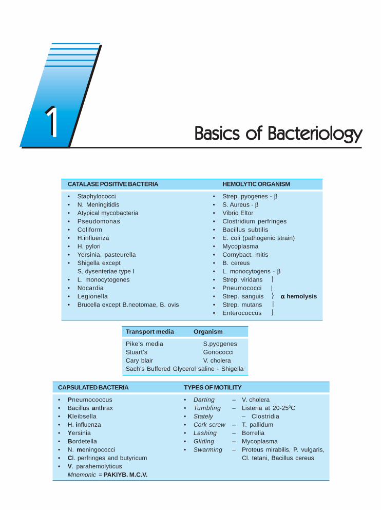
CATALASE POSITIVE BACTERIA HEMOLYTIC ORGANISM
• Staphylococci • Strep. pyogenes - β• N. Meningitidis • S. Aureus - β• Atypical mycobacteria • Vibrio Eltor• Pseudomonas • Clostridium perfringes• Coliform • Bacillus subtilis• H.influenza • E. coli (pathogenic strain)• H. pylori • Mycoplasma• Yersinia, pasteurella • Cornybact. mitis• Shigella except • B. cereus
S. dysenteriae type I • L. monocytogens - β• L. monocytogenes • Strep. viridans ⎫• Nocardia • Pneumococci |• Legionella • Strep. sanguis ⎬ ααααα hemolysis• Brucella except B.neotomae, B. ovis • Strep. mutans ⎥
• Enterococcus ⎭
Transport media Organism
Pike’s media S.pyogenesStuart’s GonococciCary blair V. choleraSach’s Buffered Glycerol saline - Shigella
CAPSULATED BACTERIA TYPES OF MOTILITY
• Pneumococcus • Darting – V. cholera• Bacillus anthrax • Tumbling – Listeria at 20-250C• Kleibsella • Stately – Clostridia• H. influenza • Cork screw – T. pallidum• Yersinia • Lashing – Borrelia• Bordetella • Gliding – Mycoplasma• N. meningococci • Swarming – Proteus mirabilis, P. vulgaris,• Cl. perfringes and butyricum Cl. tetani, Bacillus cereus• V. parahemolyticus
Mnemonic = PAKIYB. M.C.V.
Basics of BacteriologyBasics of BacteriologyBasics of BacteriologyBasics of BacteriologyBasics of Bacteriology11
Self Assessment & Review Microbiology & Immunology
SECTION – A
4
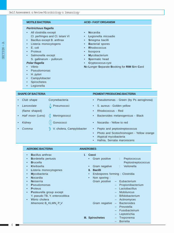
MOTILE BACTERIA ACID - FAST ORGANISM
Peritrichous flagella• All clostridia except • Nocardia
Cl. perfringes and Cl. tetani VI • Legionella micoadie• Bacillus except B. anthrax • Smegma bacilli• Listeria monocytogens • Bacterial spores• E. coli • Rhodococcus• Proteus • Isospora• Salmonella except • Mycobacterium
S. gallinarum - pullorum • Spermatic headPolar flagella • Cryptococcus cyst• Vibrio No Longer Separate Booking for RIM Sim Card• Pseudomonas• H. pylori• Campylobacter• Spirochetes• Legionella
SHAPE OF BACTERIA PIGMENT PRODUCING BACTERIA
• Club shape Corynebacteria • Pseudomonas - Green (by Ps aeroginosa)
• Lanceolate Pneumococi • S. aureus - Golden yellow
(flame shaped) • Rhodococcus - Red
• Half moon (Lens) Meningococci • Bacteroides melanogenicus - Black
• Kidney Gonococci • Nocardia - Yellow to red
• Comma V. cholera, Campylobacter • Pepto and peptostreptococcus• Photo and Scotochromogen - Yellow orange• Atypical mycobacteria• Hafnia, Serratia marcescens
AEROBIC BACTERIA ANAEROBES
• Bacillus anthrax I. Cocci• Bordetella pertusis • Gram positive : Peptococcus• Brucella Peptostreptococcus• Kleibsella • Gram negative : Veilonella• Listeria monocytogenes II. Bacilli• Mycobacteria • Endospores forming : Clostridia• Nocardia • Non sporing :• Neiserria Gram positive – Eubacterium• Pseudomonas – Propionibacterium• Proteus – Lactobacillus• Pasteurella group except – Mobiluncus
Y. pseudo TB, Y. enterocolitica – Bifidobacterium• Vibrio cholera – Actinomyces
Mnemonic B3 KLMN2 P3V Gram negative – Bacteroides– Prevotella– Fusobacteirum– Leptotrichia
III. Spirochetes – Treponema– Borrelia
Basics of Bacteriology
REVISION AT A GLANCE
5
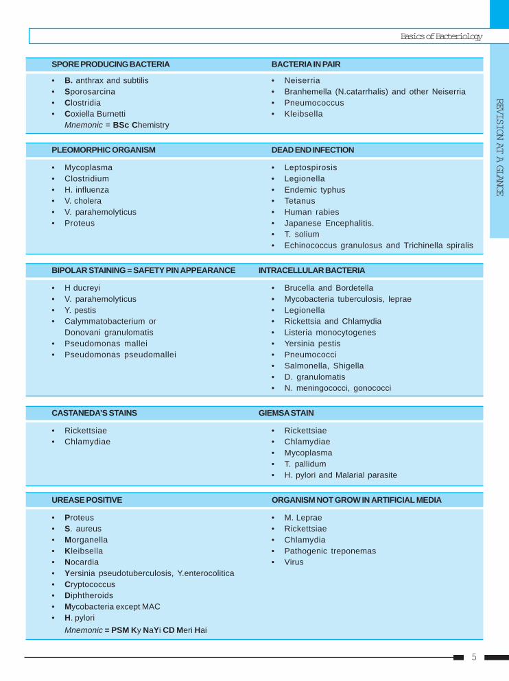
SPORE PRODUCING BACTERIA BACTERIA IN PAIR
• B. anthrax and subtilis • Neiserria• Sporosarcina • Branhemella (N.catarrhalis) and other Neiserria• Clostridia • Pneumococcus• Coxiella Burnetti • Kleibsella
Mnemonic = BSc Chemistry
PLEOMORPHIC ORGANISM DEAD END INFECTION
• Mycoplasma • Leptospirosis• Clostridium • Legionella• H. influenza • Endemic typhus• V. cholera • Tetanus• V. parahemolyticus • Human rabies• Proteus • Japanese Encephalitis.
• T. solium• Echinococcus granulosus and Trichinella spiralis
BIPOLAR STAINING = SAFETY PIN APPEARANCE INTRACELLULAR BACTERIA
• H ducreyi • Brucella and Bordetella• V. parahemolyticus • Mycobacteria tuberculosis, leprae• Y. pestis • Legionella• Calymmatobacterium or • Rickettsia and Chlamydia
Donovani granulomatis • Listeria monocytogenes• Pseudomonas mallei • Yersinia pestis• Pseudomonas pseudomallei • Pneumococci
• Salmonella, Shigella• D. granulomatis• N. meningococci, gonococci
CASTANEDA’S STAINS GIEMSA STAIN
• Rickettsiae • Rickettsiae• Chlamydiae • Chlamydiae
• Mycoplasma• T. pallidum• H. pylori and Malarial parasite
UREASE POSITIVE ORGANISM NOT GROW IN ARTIFICIAL MEDIA
• Proteus • M. Leprae• S. aureus • Rickettsiae• Morganella • Chlamydia• Kleibsella • Pathogenic treponemas• Nocardia • Virus• Yersinia pseudotuberculosis, Y.enterocolitica• Cryptococcus• Diphtheroids• Mycobacteria except MAC• H. pylori
Mnemonic = PSM Ky NaYi CD Meri Hai
Self Assessment & Review Microbiology & Immunology
SECTION – A
6
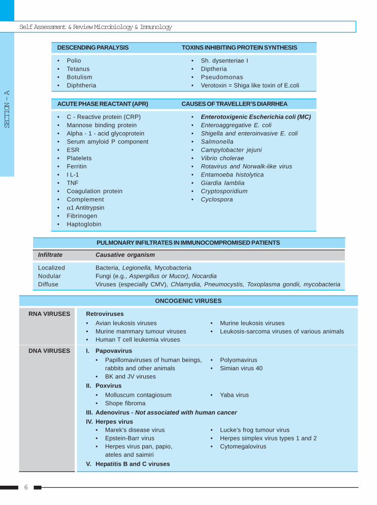
DESCENDING PARALYSIS TOXINS INHIBITING PROTEIN SYNTHESIS
• Polio • Sh. dysenteriae I• Tetanus • Diptheria• Botulism • Pseudomonas• Diphtheria • Verotoxin = Shiga like toxin of E.coli
ACUTE PHASE REACTANT (APR) CAUSES OF TRAVELLER’S DIARRHEA
• C - Reactive protein (CRP) • Enterotoxigenic Escherichia coli (MC)• Mannose binding protein • Enteroaggregative E. coli• Alpha - 1 - acid glycoprotein • Shigella and enteroinvasive E. coli• Serum amyloid P component • Salmonella• ESR • Campylobacter jejuni• Platelets • Vibrio cholerae• Ferritin • Rotavirus and Norwalk-like virus• I L-1 • Entamoeba histolytica• TNF • Giardia lamblia• Coagulation protein • Cryptosporidium• Complement • Cyclospora• α1 Antitrypsin• Fibrinogen• Haptoglobin
PULMONARY INFILTRATES IN IMMUNOCOMPROMISED PATIENTS
Infiltrate Causative organism
Localized Bacteria, Legionella, MycobacteriaNodular Fungi (e.g., Aspergillus or Mucor), NocardiaDiffuse Viruses (especially CMV), Chlamydia, Pneumocystis, Toxoplasma gondii, mycobacteria
ONCOGENIC VIRUSES
RNA VIRUSES Retroviruses• Avian leukosis viruses • Murine leukosis viruses• Murine mammary tumour viruses • Leukosis-sarcoma viruses of various animals• Human T cell leukemia viruses
DNA VIRUSES I. Papovavirus• Papillomaviruses of human beings, • Polyomavirus
rabbits and other animals • Simian virus 40• BK and JV viruses
II. Poxvirus• Molluscum contagiosum • Yaba virus• Shope fibroma
III. Adenovirus - Not associated with human cancerIV. Herpes virus
• Marek’s disease virus • Lucke’s frog tumour virus• Epstein-Barr virus • Herpes simplex virus types 1 and 2• Herpes virus pan, papio, • Cytomegalovirus
ateles and saimiriV. Hepatitis B and C viruses
Basics of Bacteriology
REVISION AT A GLANCE
7
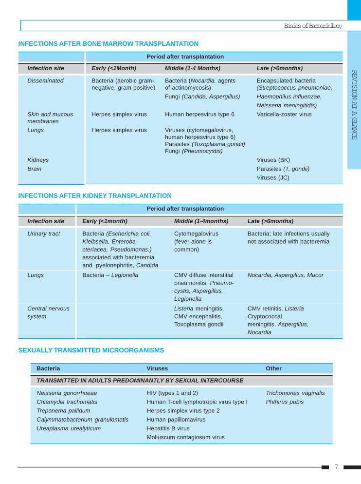
INFECTIONS AFTER BONE MARROW TRANSPLANTATION
Period after transplantation
Infection site Early (<1Month) Middle (1-4 Months) Late (>6months)
Disseminated Bacteria (aerobic gram- Bacteria (Nocardia, agents Encapsulated bacterianegative, gram-positive) of actinomycosis) (Streptococcus pneumoniae,
Fungi (Candida, Aspergillus) Haemophilus influenzae,Neisseria meningitidis)
Skin and mucous Herpes simplex virus Human herpesvirus type 6 Varicella-zoster virusmembranesLungs Herpes simplex virus Viruses (cytomegalovirus,
human herpesvirus type 6)Parasites (Toxoplasma gondii)Fungi (Pneumocystis)
Kidneys Viruses (BK)Brain Parasites (T. gondii)
Viruses (JC)
INFECTIONS AFTER KIDNEY TRANSPLANTATION
Period after transplantation
Infection site Early (<1month) Middle (1-4months) Late (>6months)
Urinary tract Bacteria (Escherichia coli, Cytomegalovirus Bacteria; late infections usuallyKleibsella, Enteroba- (fever alone is not associated with bacteremiacteriacea, Pseudomonas.) common)associated with bacteremiaand pyelonephritis, Candida
Lungs Bacteria – Legionella CMV diffuse interstitial Nocardia, Aspergillus, Mucorpneumonitis, Pneumo-cystis, Aspergillus,Legionella
Central nervous Listeria meningitis, CMV retinitis, Listeriasystem CMV encephalitis, Cryptococcal
Toxoplasma gondii meningitis, Aspergillus,Nocardia
SEXUALLY TRANSMITTED MICROORGANISMS
Bacteria Viruses Other
TRANSMITTED IN ADULTS PREDOMINANTLY BY SEXUAL INTERCOURSE
Neisseria gonorrhoeae HIV (types 1 and 2) Trichomonas vaginalisChlamydia trachomatis Human T-cell lymphotropic virus type I Phthirus pubisTreponema pallidum Herpes simplex virus type 2Calymmatobacterium granulomatis Human papillomavirusUreaplasma urealyticum Hepatitis B virus
Molluscum contagiosum virus
Self Assessment & Review Microbiology & Immunology
SECTION – A
8
Continue .....
SEXUAL TRANSMISSION REPEATEDLY DESCRIBED BUT NOT WELL DEFINED OR NOT PREDOMINANT MODE
Mycoplasma hominis Cytomegalovirus Candida albicansMycoplasma genitalium Human T-cell lymphotropic virus type II Sarcoptes scabieiGardenerella vaginalis and other Epstein-Barr virusvaginal bacteria Kaposi’s sarcoma - associated herpesvirusGroup B Streptococcus Transfusion - transmitted virusMobiluncus spp.
GASTROINTESTINAL PATHOGENS CAUSING ACUTE DIARRHEA
Mechanism Location Illness Stool findings Examples of pathogens involved
Noninflammatory Proximal Watery No fecal Vibrio cholerae, enterotoxigenic(enterotoxin) small diarrhea leukocytes; mild Escherichia coli (LT and ST),
bowel or no increase in Clostridium perfringens, Bacillusfecal lactoferrin cereus,Staphylococcus aureus,
Shigelloides, Rotavirus, Norwalk-likeviruses, Enteric adenoviruses,Giardia lamblia, Cryptosporidiumspp., Microsporidia
Inflammatory Colon or Dysentery Fecal Shigella spp., Salmonella spp.,(invasion or distal or infla- Polymorphonuclear Campylobacter jejuni,cytotoxin) small bowel mmatory leukocytes; Enterohemorrhagic E. coli, Enteroinv-
diarrhea substantial increase asive E. coli, Yersinia enterocolitica,in fecal lactoferrin Vibrio parahaemolyticus, Clostridium
difficile, Entamoeba histolytica
Penetrating Distal small Enteric Fecal mononuclear Salmonella typhi, Y. enterocolitica,bowel fever leukocytes Campylobacter fetus
NORMAL BACTERIAL FLORA
Skin – Staphylococcus epidermidis– Staphylococcus aureus– Micrococcus species– Nonpathogenic neisseria species– Alpha-hemolytic and nonhemolytic streptococci– Diphtheroids– Propoinibacterium species– Peptostreptococcus species– Candida species, acinetobacter species
Nasopharynx – Diphtheroids, Nonpathogenic neisseria species, α- hemolytic streptococci;– S.epidermidis, Nonhemolytic streptococci, Anaerobes– Yeasts, Haemophilus species, pneumococci, S aureus, Gram-negative rods, Neisseria
meningitidis
Gastrointestinal tract – Various Enterobacteriaceae except Salmonella, shigella; yersinia; Vibrio, andand rectum Campylobacter species
– Enterococci– Alpha-hemolytic and nonhemolytic streptococci
Basics of Bacteriology
REVISION AT A GLANCE
9
Continue .....
– Diphtheroids– S.aureus in small numbers– Yeasts in small numbers– Anaerobes in large numbers (MC Bacteroides)
Genitalia Any amount of the following :Corynebacterium species, Lactobacillus species, α-hemolytic and nonhemolyticstreptococci, nonpathogenic Neisseria species.
The following when mixed and not predominant :Enterococci, Enterobacteriaceae and other gram-negative rods, S epidermidis, Candidaalbicans and other yeasts.Anaerobes especially prevotella, clostridium and peptostreptococcus species.
TRANSPLACENTAL INFECTION
• Toxoplasmosis • Rubella • CMV (MC)• HSV • Syphilis • Varicella ZV• Parvo B-19 • Plasmodium • T.cruzi• HIV • Coxsackie virus • Enteroviruses• West Nile virus • Measles • Hepatitis B• HCV • TB • Lymphocytic choriomeningitis virus
ONCOGENIC MICROBES AND PARASITES
Organism Neoplasm
Human papilloma virus (papovaviridae) Cervical, vulvar, penile cancers, squamous cell carcinoma,oropharyngeal carcinoma
HSV type 2 Cervical carcinoma, B cell lymphoma
Hepatitis B virus (Hepadnaviridae) Hepatocellular carcinoma
Hepatitis C virus (Flaviviridae) Hepatocellular carcinoma, Lymphoplasmacytic lymphoma
HTLV - I (Retroviridae) Adult T cell leukemia / lymphoma
HTLV - II (Retroviridae) T cell variant of hairy cell leukemia
HTLV - III (Retroviridae) AIDS related malignancies, NHL, Kaposi sarcoma, SCC (esp of UGtract), Diffuse large B cell lymphoma, Burkitt’s lymphoma
Epstein barr virus (Herpesviridae) Mixed cellularity Hodgkin’s, Nasopharyngeal carcinoma (anaplastic),African Burkitt’s lymphoma, Post organ transplant lymphoma, PrimaryCNS diffuse large B cell lymphoma, Extranodal NK/T cell lymphoma(nasal type)
H. Pylori Gastric Malt lymphoma, Gastric cancer
Human Herpes virus 8 Primary effusion lymphoma, Multicentric castleman’s disease
Schistosoma hematobium Bladder cancer (squamous cell)
Clonorchis Cholongiocarcinoma
Opisthorchis Cholongiocarcinoma
DEFINITIONS
Virion – Extracellular infectious virus particle.Capsid – Protein coat that protects nucleic acid.Envelope – Lipoprotein coat which surrounds some virus particles. Lipid is of host cell origin while protein in
the form of peplomers is virus coded.Viroids – Subviral infectious agent which is protein free and consist of low molecular weight RNA (mostly
double stranded, small RNA). It is resistant to heat and organic solvents but sensitive to nucleases.Prion – Proteinaceous infectious particles causing chronic neurological degenerative disease of human.
• Virus is obligate intracellular parasite, without cellular organisation and contain only one type of nucleic acideither DNA or RNA but never both. So classified as :
CAPSID VIRION NUCLEIC ACID VIRUS FAMILY MEMBERS
DNA VIRUS
I. Icosahedral Naked SS(-ve) Parvoviridae B-19 parvovirusII. Icosahedral Naked ds circular Papovaviridae Papilloma virus,
(±) JC, BK virus, polyomavirusIII. Icosahedral Naked ds (±) Adenoviridae Human adenovirusIV. Icosahedral Enveloped ds with ss Hepadenoviridae HBV
(±) circularV. Icosahedral Enveloped ds (±) Herpesviridae VZ; HSV I, II; CMV; EBVVI. Complex Complex ds (±) Poxviridae Variola (small pox)
coats Molluscum contagiosum
RNA VIRUS
I. Icosahedral Naked SS (+) Picornaviridae Polio, coxsackie,entero,or (cubical) rhino, HAV
II. Icosahedral Naked SS AstroviridaeIII. Icosahedral Naked SS(+) Calcivridae HEV, NorwalkIV. Icosahedral Naked ds segmented (±) Reoviridae Rota, Reo, OrbivirusV. Icosahedral Enveloped SS (+) Togaviridae Rubella virusVI. Unknown Enveloped SS (+) Flaviviridae HCV, HGV, yellow
or complex fever, Dengue virus
Basics of VirologyBasics of VirologyBasics of VirologyBasics of VirologyBasics of Virology22
Basics of VirologyREVISION AT A GLANCE
11
Continue .....
VII. Unknown Enveloped SS (-) Arenaviridae Lassa fever virusor complex segmented (sandy appearance)
VIII. Unkonwn Enveloped SS (+) Coronaviridaeor complex
IX. Unknown Enveloped SS diploid (+) Retroviridae HIV 1, 2; HTLV I, II;or complex slow virus group
X. Helical Enveloped SS (-) Orthomyxoviridae Influenza A, B, Csegmented
XI. Helical SS (-) Bunyaviridae Hantavirus, sandflysegmented fever virus
XII.Helical Enveloped SS BornaviridaeXIII. Helical Enveloped SS (-) Rhabdoviridae Rabies virus,
Vesicular stomatitis virusXIV. Helical Enveloped SS (-) Paramyxoviridae Parainfluenza, RSV,
Mumps, rubeolaNew castle virus
XV. Helical Enveloped SS (-) Filoviridae Marburg virusEbola virus
Mnemonics• Segmented Nucleic acid = ‘PARBO virus’
= Picornaviruses, Arena, Reo, Bunya, Orthomyxovirus• Enveloped virus are sensitive to ether, chloroform, bile salts while non-enveloped are resistant• All RNA virus are enveloped except ‘PARC’
= Picorna, Astro, Reo, Calciviridae
• Viruses with both DNA and RNA – Retrovirus– Lentivirus– HBV
• Complex capsid – Pox– Bacteriophage
• Shapes- Bullet shaped – Rabies virus- Brick shaped – Pox virus- Rod shaped – Tobacco mosaic virus- Space vehicle – Adenovirus
• Smallest size virus – Parvovirus• Largest size virus – Filoviridae > Pox viridae
HEMATAGGLUTINATION (HA) - Is agglutination of erythrocytes by virus.• It is unstable in myxovirus because Neuraminidase (RDE- receptor destroying enzyme) cause reversal of
hemagglutination called as Elution. RDE also produced by cholera vibrios and many verterbrate cells.• In other viruses HA is stable.• In arbovirus, it is reversible by variation in pH and temperature.• HA measures total quantity of virus.• HA of human RBC is seen in - Reo, Influenza, Para-influenza, Entero and some cox and ECHO, Mumps.
Self Assessment & Review Microbiology & ImmunologySECTION – A
12
Mnemonic – RIPE Mango• HA also seen in measles, toga, rhino, rabies, pox, adenovirus.
POCK ASSAYUsed for quantitative infectivity assay of viruses [also by plaque assay] since each infectious virus particle can formone pock eg. variola, vaccinia, HSV, Pox (Monkey, Cow, Camel).
PHAGE ASSAYUsed for titrating number of viable bacteriophage and for purification of phages.
PHAGE TYPING• Used for typing and identification of bacteria eg.
Intraspecies typing of S. typhi (by using Vi antigen) and S. aureus; species specific bacteriophage of B.anthracis,MukerJee’s phage IV for classical V. cholerae.
VIRUS MULTIPLICATION• Critical step in viral biosynthesis is transcription of mRNA from viral nucleic acid.
• DNA virus synthesize nucleic acid in host cell nucleus except pox which synthesis all their components in hostcell cytoplasm.
• RNA virus synthesize nucleic acid in cytoplasm except orthomyxo, some paramyxo and retrovirus whichsynthesize partly in nucleus.
• Viral protein is synthesized only in cytoplasm.• Herpes and adeno assembled in nucleus while picoma and pox are assembled in cytoplasm.
ABNORMAL REPLICATIVE CYCLEVon Magnus Phenomenon : High hemagglutinin but low infectivity due to defective assembly or incomplete virusEg. Influenza virus.
Abortive Infection : Defect in the type of cell (non-permissive cell) not in the parental viruses lead to defectivematuration or assembly.
Defective virus : Genetically defective virus which are incapable of producing infectious daughter virions withoutthe helper activity of another virus Eg. Rous sarcoma virus, HDV, adeno associated satellite virus (dependovirus),Measles virus from SSPE etc.
VIRAL INTERACTION• Genetic Interaction – occur in virus by :
1. Mutation – Occur during every viral infection.Most mutation are lethal.
2. Recombination – occur when two different but related viruses (both active or both inactive or one active andone inactive) infect a cell simultaneously. It leads to cross reactivation / marker rescue; multiplicity reactivationand formation of pseudovirion.
• Non-Genetic Interaction1. Phenotyping mixing – transcapsidation occurs2. Genotyping mixing
Basics of VirologyREVISION AT A GLANCE
13
3. Complementation4. Interference – Infection of a cell by one virus inhibits simultaneous or subsequent infection by other virus. Most
important mediator is Interferon, a soluble cellular product.• It is applied in controlling polio outbreaks by introducing live attenuated polio vaccine.• It can be produced by receptor destruction as in myxo and enterovirus or by autointerference.
INCLUSION BODIESIt is the most charcateristic histological feature in virus infected cells. It is of following types :a. Intracytoplasmic eosinophilic inclusions :
Negri bodies – rabiesGuarnieri bodies – variola (small pox), vacciniaBollinger bodies – fowlpoxHenderson - peterson bodies – molluscum contagiosum
b. Intranuclear acidophilic inclusion bodies :Cowdry type A – herpes, chicken pox, CMV, yellow feverTorres bodies – yellow feverCowdry type B – polio virus
c. Both Nuclear and cytoplasmic :Warthin Finkeldey – measles
d. Intranuclear basophilic inclusion bodies :Cowdry type B – adenovirus
RESPIRATORY VIRUSES
Viruses Most frequent illness
Rhinoviruses Common coldCoronaviruses Common coldRespiratory syncytial virus Pneumonia and bronchiolitis in young childrenParainfluenza viruses Croup and lower respiratory tract disease in young childrenAdenoviruses Common cold and pharyngitis in childrenInfluenza A, B viruses InfluenzaEnteroviruses Acute undifferentiated febrile illnessesHerpes simplex viruses Gingivostomatitis in children; pharyngotonsillitis in adults
VIRUS CAUSING LATENT INFECTION
• Measles • Hepatitis B virus • Hepatitis C virus • Rabies virus• Human T-lymphotrophic virus • Herpes virus • Kuru • Oncogenic virus• Scrapie • Human immuno deficiency virus
REACTION TO PHYSICAL AND CHEMICAL AGENTS :• Stable at low temperature so for long term storage. They are kept by frozing at 700c, lyophilization or freeze
drying but poliovirus do not stand freeeze drying.• All virus are disrupted under alkaline pH. Enterovirus are very resistant to acid pH while rhinovirus are very
susceptible.• Most active antiviral disinfectants are oxidising agents such as H2O2, KMno4 and hypochlorides.• Chlorination kill most viruses except hepatitis virus, polio virus.
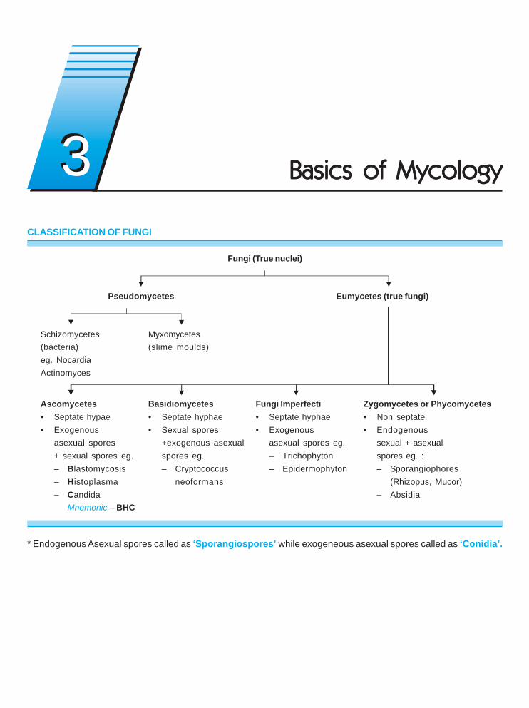
CLASSIFICATION OF FUNGI
Fungi (True nuclei)
Pseudomycetes Eumycetes (true fungi)
Schizomycetes Myxomycetes(bacteria) (slime moulds)eg. NocardiaActinomyces
Ascomycetes Basidiomycetes Fungi Imperfecti Zygomycetes or Phycomycetes• Septate hypae • Septate hyphae • Septate hyphae • Non septate• Exogenous • Sexual spores • Exogenous • Endogenous
asexual spores +exogenous asexual asexual spores eg. sexual + asexual+ sexual spores eg. spores eg. – Trichophyton spores eg. :– Blastomycosis – Cryptococcus – Epidermophyton – Sporangiophores– Histoplasma neoformans (Rhizopus, Mucor)– Candida – Absidia
Mnemonic – BHC
* Endogenous Asexual spores called as ‘Sporangiospores’ while exogeneous asexual spores called as ‘Conidia’.
Basics of MycologyBasics of MycologyBasics of MycologyBasics of MycologyBasics of Mycology33
Basics of MycologyREVISION AT A GLANCE
15
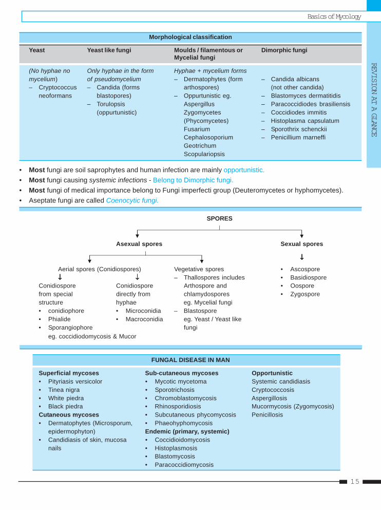
Morphological classification
Yeast Yeast like fungi Moulds / filamentous or Dimorphic fungiMycelial fungi
(No hyphae no Only hyphae in the form Hyphae + mycelium formsmycelium) of pseudomycelium – Dermatophytes (form – Candida albicans– Cryptococcus – Candida (forms arthospores) (not other candida)
neoformans blastopores) – Oppurtunistic eg. – Blastomyces dermatitidis– Torulopsis Aspergillus – Paracoccidiodes brasiliensis
(oppurtunistic) Zygomycetes – Coccidiodes immitis(Phycomycetes) – Histoplasma capsulatumFusarium – Sporothrix schenckiiCephalosoporium – Penicillium marneffiGeotrichumScopulariopsis
• Most fungi are soil saprophytes and human infection are mainly opportunistic.• Most fungi causing systemic infections - Belong to Dimorphic fungi.• Most fungi of medical importance belong to Fungi imperfecti group (Deuteromycetes or hyphomycetes).• Aseptate fungi are called Coenocytic fungi.
SPORES
Asexual spores Sexual spores
↓↓↓↓↓
Aerial spores (Conidiospores) Vegetative spores • Ascospore↓↓↓↓↓ ↓↓↓↓↓ – Thallospores includes • Basidiospore
Conidiospore Conidiospore Arthospore and • Oosporefrom special directly from chlamydospores • Zygosporestructure hyphae eg. Mycelial fungi• conidiophore • Microconidia – Blastospore• Phialide • Macroconidia eg. Yeast / Yeast like• Sporangiophore fungi
eg. coccidiodomycosis & Mucor
FUNGAL DISEASE IN MAN
Superficial mycoses Sub-cutaneous mycoses Opportunistic• Pityriasis versicolor • Mycotic mycetoma Systemic candidiasis• Tinea nigra • Sporotrichosis Cryptococcosis• White piedra • Chromoblastomycosis Aspergillosis• Black piedra • Rhinosporidiosis Mucormycosis (Zygomycosis)Cutaneous mycoses • Subcutaneous phycomycosis Penicillosis• Dermatophytes (Microsporum, • Phaeohyphomycosis
epidermophyton) Endemic (primary, systemic)• Candidiasis of skin, mucosa • Coccidioidomycosis
nails • Histoplasmosis• Blastomycosis• Paracoccidiomycosis
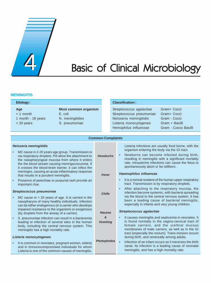
MENINGITIS
Etiology : Classification :
Age Most common organism Streptococcus agalactiae Gram+ Cocci< 1 month E. coli Streptococcus pneumoniae Gram+ Cocci1 month - 18 years N. meningitides Neisseria meningitidis Gram - Cocci> 20 years S. pneumoniae Listeria monocytogenes Gram + Bacilli
Hemophilus influenzae Gram - Cocco Bacilli
Common Complaints
Basic of Clinical MicrobiologyBasic of Clinical MicrobiologyBasic of Clinical MicrobiologyBasic of Clinical MicrobiologyBasic of Clinical Microbiology
Neisseria meningitidis
• MC cause in 2-20 years age group. Transmission isvia respiratory droplets. Pili allow the attachment tothe nasopharyngeal mucosa from where it entersthe the blood stream causing meningococcemia. Ifit crosses the blood-brain barrier, it can infect themeninges, causing an acute inflammatory responsethat results in a purulent meningitis.
• Presence of petechiae or purpurial rash provide animportant clue.
Streptococcus pneumoniae
• MC cause in > 20 years of age. It is carried in thenasopharynx of many healthy individuals. Infectioncan be either endogenous (in a carrier who developsimpaired resistance to the organism) or exogenous(by droplets from the airway of a carrier).
• S. pneumoniae infection can result in a bacteremialeading to infection of several sites in the humanbody, including the central nervous system. Thismeningitis has a high mortality rate.
Listeria monocytogenes
• It is common in neonates, pregnant women, elderlyand in immunocompromised individuals for whomListeria is one of the common causes of meningitis.
Listeria infections are usually food borne, with theorganism entering the body via the GI tract.
• Newborns can become infected during birth,resulting in meningitis with a significant mortalityrate, intrauterine infections can cause the fetus tospontaneously abort or be stillborn.
Haemophilus influenzae
• It is a normal resident of the human upper respiratorytract. Transmission is by respiratory droplets.
• After attaching to the respiratory mucosa, theinfection become systemic, with bacteria spreadingvia the blood to the central nervous system. It hasbeen a leading cause of bacterial meningitis,especially in infants and very young children.
Streptococcus agalactiae
• It causes meningitis and septicemia in neonates. Itis found normally in the vagino-cervical tract offemale carriers, and the urethral mucousmembranes of male carriers, as well as in the GItract (especially the rectum). Trans-mission occursduring birth, and venereally among adults.
• Infection of an infant occurs as it traverses the birthcanal. Its infection is a leading cause of neonatalmeningitis, and has a high mortality rate.
Headache
Fever
Chills
Nausea&
Vomiting
Photophobia
44
Basic of Clinical MicrobiologyREVISION AT A GLANCE
17
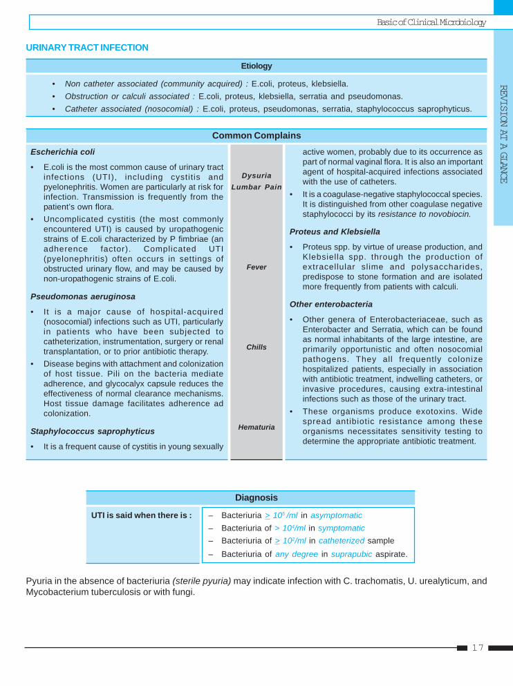
URINARY TRACT INFECTION
Etiology
• Non catheter associated (community acquired) : E.coli, proteus, klebsiella.• Obstruction or calculi associated : E.coli, proteus, klebsiella, serratia and pseudomonas.• Catheter associated (nosocomial) : E.coli, proteus, pseudomonas, serratia, staphylococcus saprophyticus.
Common Complains
Escherichia coli
• E.coli is the most common cause of urinary tractinfections (UTI), including cystit is andpyelonephritis. Women are particularly at risk forinfection. Transmission is frequently from thepatient’s own flora.
• Uncomplicated cystitis (the most commonlyencountered UTI) is caused by uropathogenicstrains of E.coli characterized by P fimbriae (anadherence factor). Complicated UTI(pyelonephritis) often occurs in settings ofobstructed urinary flow, and may be caused bynon-uropathogenic strains of E.coli.
Pseudomonas aeruginosa
• It is a major cause of hospital-acquired(nosocomial) infections such as UTI, particularlyin patients who have been subjected tocatheterization, instrumentation, surgery or renaltransplantation, or to prior antibiotic therapy.
• Disease begins with attachment and colonizationof host tissue. Pili on the bacteria mediateadherence, and glycocalyx capsule reduces theeffectiveness of normal clearance mechanisms.Host tissue damage facilitates adherence adcolonization.
Staphylococcus saprophyticus
• It is a frequent cause of cystitis in young sexually
active women, probably due to its occurrence aspart of normal vaginal flora. It is also an importantagent of hospital-acquired infections associatedwith the use of catheters.
• It is a coagulase-negative staphylococcal species.It is distinguished from other coagulase negativestaphylococci by its resistance to novobiocin.
Proteus and Klebsiella
• Proteus spp. by virtue of urease production, andKlebsiella spp. through the production ofextracellular slime and polysaccharides,predispose to stone formation and are isolatedmore frequently from patients with calculi.
Other enterobacteria
• Other genera of Enterobacteriaceae, such asEnterobacter and Serratia, which can be foundas normal inhabitants of the large intestine, areprimarily opportunistic and often nosocomialpathogens. They all frequently colonizehospitalized patients, especially in associationwith antibiotic treatment, indwelling catheters, orinvasive procedures, causing extra-intestinalinfections such as those of the urinary tract.
• These organisms produce exotoxins. Widespread antibiotic resistance among theseorganisms necessitates sensitivity testing todetermine the appropriate antibiotic treatment.
Diagnosis
UTI is said when there is : – Bacteriuria > 105 /ml in asymptomatic– Bacteriuria of > 104/ml in symptomatic– Bacteriuria of > 102/ml in catheterized sample– Bacteriuria of any degree in suprapubic aspirate.
Pyuria in the absence of bacteriuria (sterile pyuria) may indicate infection with C. trachomatis, U. urealyticum, andMycobacterium tuberculosis or with fungi.
DysuriaLumbar Pain
Fever
Chills
Hematuria
Self Assessment & Review Microbiology & ImmunologySECTION – A
1818
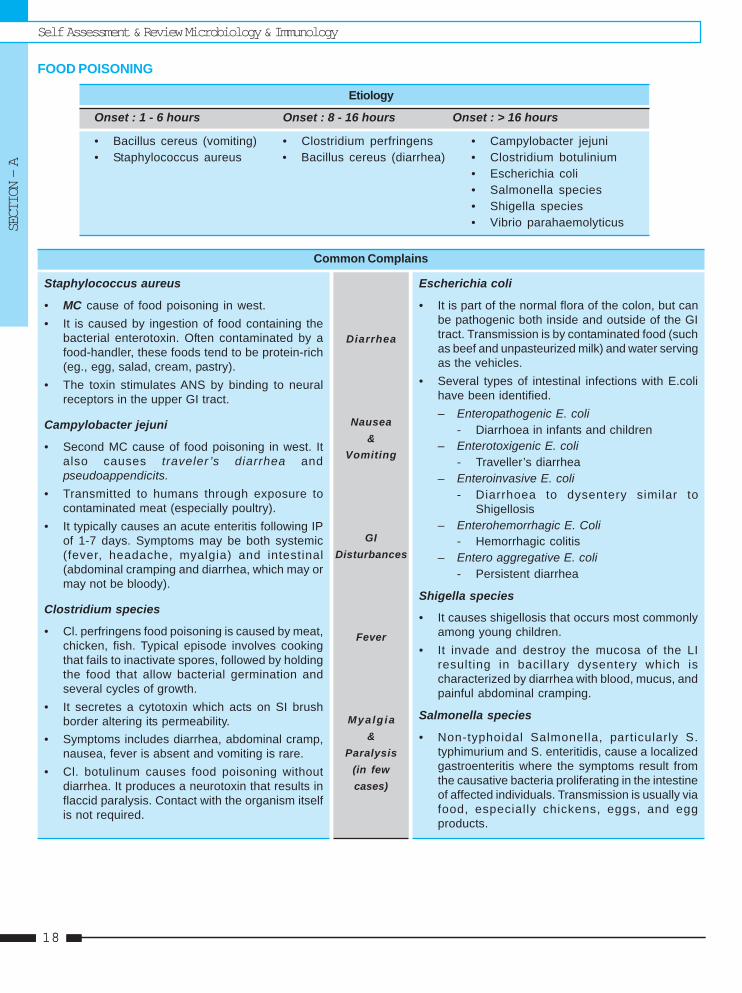
FOOD POISONING
Etiology
Onset : 1 - 6 hours Onset : 8 - 16 hours Onset : > 16 hours
• Bacillus cereus (vomiting) • Clostridium perfringens • Campylobacter jejuni• Staphylococcus aureus • Bacillus cereus (diarrhea) • Clostridium botulinium
• Escherichia coli• Salmonella species• Shigella species• Vibrio parahaemolyticus
Common Complains
Staphylococcus aureus
• MC cause of food poisoning in west.• It is caused by ingestion of food containing the
bacterial enterotoxin. Often contaminated by afood-handler, these foods tend to be protein-rich(eg., egg, salad, cream, pastry).
• The toxin stimulates ANS by binding to neuralreceptors in the upper GI tract.
Campylobacter jejuni
• Second MC cause of food poisoning in west. Italso causes traveler ’s diarrhea andpseudoappendicits.
• Transmitted to humans through exposure tocontaminated meat (especially poultry).
• It typically causes an acute enteritis following IPof 1-7 days. Symptoms may be both systemic(fever, headache, myalgia) and intestinal(abdominal cramping and diarrhea, which may ormay not be bloody).
Clostridium species
• Cl. perfringens food poisoning is caused by meat,chicken, fish. Typical episode involves cookingthat fails to inactivate spores, followed by holdingthe food that allow bacterial germination andseveral cycles of growth.
• It secretes a cytotoxin which acts on SI brushborder altering its permeability.
• Symptoms includes diarrhea, abdominal cramp,nausea, fever is absent and vomiting is rare.
• Cl. botulinum causes food poisoning withoutdiarrhea. It produces a neurotoxin that results inflaccid paralysis. Contact with the organism itselfis not required.
Escherichia coli
• It is part of the normal flora of the colon, but canbe pathogenic both inside and outside of the GItract. Transmission is by contaminated food (suchas beef and unpasteurized milk) and water servingas the vehicles.
• Several types of intestinal infections with E.colihave been identified.– Enteropathogenic E. coli
- Diarrhoea in infants and children– Enterotoxigenic E. coli
- Traveller’s diarrhea– Enteroinvasive E. coli
- Diarrhoea to dysentery similar toShigellosis
– Enterohemorrhagic E. Coli- Hemorrhagic colitis
– Entero aggregative E. coli- Persistent diarrhea
Shigella species
• It causes shigellosis that occurs most commonlyamong young children.
• It invade and destroy the mucosa of the LIresulting in bacil lary dysentery which ischaracterized by diarrhea with blood, mucus, andpainful abdominal cramping.
Salmonella species
• Non-typhoidal Salmonella, particularly S.typhimurium and S. enteritidis, cause a localizedgastroenteritis where the symptoms result fromthe causative bacteria proliferating in the intestineof affected individuals. Transmission is usually viafood, especially chickens, eggs, and eggproducts.
Diarrhea
Nausea&
Vomiting
GIDisturbances
Fever
Myalg ia&
Paralysis(in fewcases)
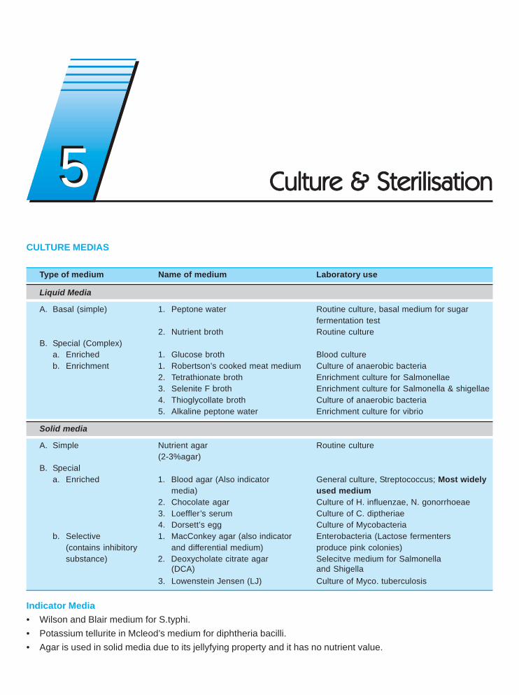
CULTURE MEDIAS
Type of medium Name of medium Laboratory use
Liquid Media
A. Basal (simple) 1. Peptone water Routine culture, basal medium for sugarfermentation test
2. Nutrient broth Routine cultureB. Special (Complex)
a. Enriched 1. Glucose broth Blood cultureb. Enrichment 1. Robertson’s cooked meat medium Culture of anaerobic bacteria
2. Tetrathionate broth Enrichment culture for Salmonellae3. Selenite F broth Enrichment culture for Salmonella & shigellae4. Thioglycollate broth Culture of anaerobic bacteria5. Alkaline peptone water Enrichment culture for vibrio
Solid media
A. Simple Nutrient agar Routine culture(2-3%agar)
B. Speciala. Enriched 1. Blood agar (Also indicator General culture, Streptococcus; Most widely
media) used medium2. Chocolate agar Culture of H. influenzae, N. gonorrhoeae3. Loeffler’s serum Culture of C. diptheriae4. Dorsett’s egg Culture of Mycobacteria
b. Selective 1. MacConkey agar (also indicator Enterobacteria (Lactose fermenters(contains inhibitory and differential medium) produce pink colonies)substance) 2. Deoxycholate citrate agar Selecitve medium for Salmonella
(DCA) and Shigella3. Lowenstein Jensen (LJ) Culture of Myco. tuberculosis
Indicator Media• Wilson and Blair medium for S.typhi.• Potassium tellurite in Mcleod’s medium for diphtheria bacilli.• Agar is used in solid media due to its jellyfying property and it has no nutrient value.
Culture & SterilisationCulture & SterilisationCulture & SterilisationCulture & SterilisationCulture & Sterilisation55
Self Assessment & Review Microbiology & Immunology
SECTION – A
2020
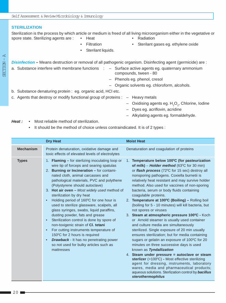
STERILIZATIONSterilization is the process by which article or medium is freed of all living microorganism either in the vegetative orspore state. Sterilizing agents are : • Heat • Radiation
• Filtration • Sterilant gases eg. ethylene oxide• Sterilant liquids.
Disinfection – Means destruction or removal of all pathogenic organism. Disinfecting agent (germicide) are :a. Substance interfere with membrane functions : – Surface active agents eg. quaternary ammonium
compounds, tween - 80– Phenols eg. phenol, cresol– Organic solvents eg. chloroform, alcohols.
b. Substance denaturing protein : eg. organic acid, HCl etc.c. Agents that destroy or modify functional group of proteins : – Heavy metals
– Oxidising agents eg. H2O2, Chlorine, Iodine– Dyes eg. acriflovin, acridine– Alkylating agents eg. formaldehyde.
Heat : • Most reliable method of sterilization.• It should be the method of choice unless contraindicated. It is of 2 types :
Dry Heat Moist Heat
Mechanism Protein denaturation, oxidative damage and Denaturation and coagulation of proteinstoxic effects of elevated levels of electrolytes
Types 1. Flaming – for sterlizing inoculating loop or 1. Temperature below 1000C (for pasteurizationwire tip of forceps and searing spatulas of milk) – Holder method (630C for 30 min)
2. Burning or Incineration – for contami- or flash process (720C for 15 sec) destroy allnated cloth, animal carcasses and nonsporing pathogens. Coxiella burnetii ispathological materials, PVC and polythene relatively heat resistant and may survive holder(Polystyrene should autoclave) method. Also used for vaccines of non-sporing
3. Hot air oven – Most widely used method of bacteria, serum or body fluids containingsterilization by dry heat coagulable proteins.
• Holding period of 1600C for one hour is 2. Temperature at 1000C (Boiling) – Rolling boilused to sterilize glassware, scalpels, all (boiling for 5 - 10 minutes) will kill bacteria, butglass syringes, swabs, liquid paraffins, not spores or virusesdusting powder, fats and grease 3. Steam at atmospheric pressure 1000C - Koch
• Sterilization control is done by spore of or Arnold steamer is usually used containernon-toxigenic strain of Cl. tetani and culture media are simultaneously
• For cutting instruments temperature of sterilized. Single exposure of 20 min usually1500C for 2 hours is required ensures sterilization; but for media containing
• Drawback - It has no penetrating power sugars or gelatin an exposure of 1000C for 20so not used for bulky articles such as minutes on three successive days is usedmattresses known as Tyndallization
4. Steam under pressure = autoclave or steamsterlizer (>1000C) - Most effective sterilizingagent for dressing, instruments, laboratorywares, media and pharmaceutical products;aqueous solutions. Sterlization control by bacillussterothermophilus
Culture & Sterilisation
REVISION AT A GLANCE
21
Filtration : Used to remove bacteria; virus isolation; testing water samples for vibrio cholera or typhoid bacilli; andobtaining bacterial toxins. Membrane filters is routinely used in water purification and analysis, sterlizationand sterility testing and for preparations of solutions for parenteral use. Most widely used pore diameteris 0.22 μm.
Radiation
Non ionization Ionizing radiation
1. Infrared radiation - Form of hot air – X-rays, gamma rays (Commonly used) and cosmic rayssterlization used for rapid mass sterlization referred to as cold Sterlizationof prepacked items such as syringes and – Used for plastics, syringes, swabs, catheters, animalscatheters feeds, cardboard, oils grease, fabrics, metal foils
2. Ultraviolet radiation - For entryways, operation – Most effective but very costlytheaters and laboratories
Alcohols• MC used are ethanol and isopropyl alcohol (better).• Both used as skin disinfectant in 70% concentration.• Not sporicidal but active against non sporing bacteria and viruses.• Isopropyl alcohol is used for disinfection of clinical thermometers.• Methyl alcohol effective against fungal spores and is used for treating cabinets and incubators.• Most effective skin antiseptic is alcoholic solution of chlorhexidine and iodine.Aldehydes• Formaldehyde : Sporicidal (Slow activity), bactericidal, virucidal. Used for sterlising instruments and heat sensitive
catheters and for fumigating wards, sick rooms and laboratories.• Glutarldehyde : Specially effective for tubercle bacilli, viruses and fungi.
– 2% solution called Cidex used for cystoscope and bronchoscopes.Dyes• Acridine and aniline dyes used as skin and wound antiseptic.• More active against Gram positive organism.Halogens• Iodine in aqueous and alcoholic solution is used widely as skin disinfectant.• Active against tubercle bacteria, viruses, spores (moderately).• Iodophores are compounds of iodine with non ionic wetting or surface active agents. They are more active.• Chlorine is used commonly as hypochlorites.Ethylene oxide• Highly penetrating and highly active against all microorganism including viruses and spores.• Specially used for sterilizing heart lung machine.Formaldehyde gas• For fumigation of operation theatres and other rooms.• Betapropiolactone is more efficient for fumigation than formaldehyde.Surface Active Agents• Most important antibacterial agents are cationic surface active agents.• No action on spores, tubercle bacilli and most viruses.• Most active at alkalines pH.• Soaps prepared from saturated fatty acids are more effective against G-ve bacilli while those prepared from
unsaturated fatty acids are more active against G+ve and neisseria group.
Self Assessment & Review Microbiology & Immunology
SECTION – A
2222
1. Which of the following is most resistant toanticeptics? [AI 08]a) Spore b) Prionc) Cyst d) Fungus
2. Which of the following statement is true :a) Solid media are enrichment media [AI 07]b) Nutrient broth is basal mediac) Agar adds nutrient to mediad) Chocolate agar is selective media
3. A chest physician performs bronchoscopy in theprocedure room of the out patient department.To make the instrument safe for use in the nextpatient waiting outside, the most appropriatemethod to disinfect the endoscope is by :a) 70% alcohol for 5 min [AI 03]b) 2% glutaraldehyde for 20 minc) 2% formaldheyde for 10 mind) 1% sodium hypochlorite for 15 min
4. Heat labile instruments for use in surgical pro-cedure can be best sterlized by : [AI 03]a) Absolute alcoholb) Ultra violet raysc) Chlorine releasing compoundsd) Ethylene oxide gas
5. Out of the following the true statement regard-ing sterlization is : [AI 97]a) Dry heat is the best method of sterilization of liq-
uid paraffinb) All glass syringes are best sterilized by boiling at
1000Cc) Bacterial vacines are best sterilized by ethylene
oxided) Pasteurization of milk by flash method is done
by heating at 630C for 30 minutes6. The operating temperature in an ethylene oxide
sterilization during warm cycle is : [AIIMS 04]a) 20-350Cb) 49-630Cc) 68-880Cd) 92-1100C
7. The sterilization method for the instrumentswhich are damaged by dry heat is : [AIIMS 95]a) Steamb) Radiation
c) Boilingd) Burning
8. Choose the correct ones for the decreasing or-der of resistance to sterilization : [PGI Dec. 07]a) Prions, bacterial spores, bacteriab) Bacterial spores, Bacteria, Prionsc) Bacteria, Prions, Bacterial sporesd) Prions, Bacteria, Bacterial sporese) Bacterial spores, prions, bacteria
9. Decreasing order of resistance to sterilization :a) Spores, prions, non-lipid of smll virusb) Prions, spores, enveloped viruses [PGI 07]c) Spores, mycobacteria, lipid or medium size vi-
rus10. Sterilising agents include : [PGI 02]
a) Dry heatb) Ethylene oxidec) Etherd) Alcohole) Chlorohexidine
11. Which of the following can be reliably used forhand washing : [PGI 00]a) Chlorhexidineb) Isopropyl alcoholc) Lysold) Cresole) Glutaraldehyde
12. Sporocidal disinfectant is following except :a) Glutaraldehyde [PGI 99]b) Formaldehydec) Ethylene oxided) Benzalkonium chloride
13. All are sporicidal except : [PGI 99]a) Lysolb) Glutaraldehydec) Ethylene dioxided) Formaldehyde
14. Sterlization of culture media containing serum isby : [PGI 98]a) Autoclavingb) Micropore filterc) Gamma radiationd) Centrifugation
QUESTIONS
Answer 1. b) Prion... 2. b) Nutrient ... 3. b) 2% ... 4. d) Ultra violet ... 5. a) Dry heat is ...6. b) 49-630C 7. b) Radiation 8. a) Prion ... 9. b and c 10. a and b
11. a, b and d 12. d) Benzalkoni ... 13. a) Lysol 14. a) Autoclaving
Questions & Answers
REVISION AT A GLANCE
23
1. Ans. is b i.e. Prion See below
Resistance of organism to antiseptics in decreasing order is as follows :• Prions• Coccidia• Spores• Mycobacteria• Cysts• Small non-enveloped virus• Trophozoites• Gram negative bacteria (non-sporulating)• Fungi• Large non-enveloped virus• Gram positive bacteria• Lipid enveloped / medium size virus (HIV, HBV)
2. Ans. is b i.e. Nutrient broth is basal media Ref. Ananthnarayan 7/e, p 37
Media
Simple media (basal media) Complex media Synthetic or defined media Special media
– Nutrient broth – Added ingredient – Prepared from chemicals – Enriched mediawith defined composition – Enrichment mediae.g., simple peptone water – Selective mediamedium – Indicator media
– Sugar media– Transport media
• Enriched media : Substance such as blood serum on egg are added to basal medium to promote growthe.g., blood agar, chocolate agar and egg media.
• Enrichment media : In mixed culture usually the nonpathogenic or commensal bacteria tends to overgrowthan pathogenic ones. In such conditions, substances which has stimulating effect on pathogenic one orinhibitory effect on unwanted one. These media are called enrichment media e.g., tetrathionate.
• Selective media : If the inhibitory substance is added to solid medium so as to supress the growth ofunwanted one; the media is called selective media e.g., desoxycholate citrate medium.
• Indicator media : Changes colour on growth of bacteria e.g., wilson blair media; Mc loed medium.• Differential media : To differntiate different bacteria on the basis of characteristics. Eg. Mac Conkey’s
medium.• Sugar media : – Here sugar means any fermentable substance.
– Usual sugar media consist of 1% of the sugar in peptone water along with an appropriateindicator.
• Transport media : For delicate organism e.g., stuart media.
EXPLANATIONS AND REFERENCES WITH ILLUSTRATIVE ANSWERS
Self Assessment & Review Microbiology & Immunology
SECTION – A
2424
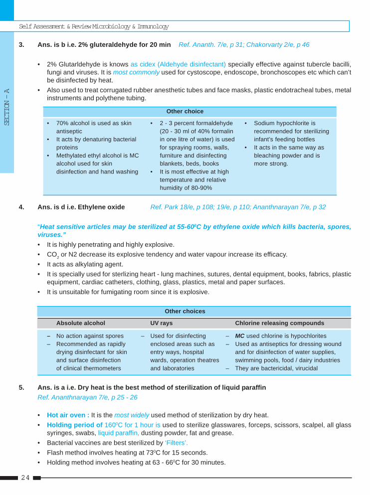
3. Ans. is b i.e. 2% gluteraldehyde for 20 min Ref. Ananth. 7/e, p 31; Chakorvarty 2/e, p 46
• 2% Glutarldehyde is knows as cidex (Aldehyde disinfectant) specially effective against tubercle bacilli,fungi and viruses. It is most commonly used for cystoscope, endoscope, bronchoscopes etc which can’tbe disinfected by heat.
• Also used to treat corrugated rubber anesthetic tubes and face masks, plastic endotracheal tubes, metalinstruments and polythene tubing.
Other choice
• 70% alcohol is used as skin • 2 - 3 percent formaldehyde • Sodium hypochlorite isantiseptic (20 - 30 ml of 40% formalin recommended for sterilizing
• It acts by denaturing bacterial in one litre of water) is used infant’s feeding bottlesproteins for spraying rooms, walls, • It acts in the same way as
• Methylated ethyl alcohol is MC furniture and disinfecting bleaching powder and isalcohol used for skin blankets, beds, books more strong.disinfection and hand washing • It is most effective at high
temperature and relativehumidity of 80-90%
4. Ans. is d i.e. Ethylene oxide Ref. Park 18/e, p 108; 19/e, p 110; Ananthnarayan 7/e, p 32
“Heat sensitive articles may be sterilized at 55-600C by ethylene oxide which kills bacteria, spores,viruses.”• It is highly penetrating and highly explosive.• CO2 or N2 decrease its explosive tendency and water vapour increase its efficacy.• It acts as alkylating agent.• It is specially used for sterlizing heart - lung machines, sutures, dental equipment, books, fabrics, plastic
equipment, cardiac catheters, clothing, glass, plastics, metal and paper surfaces.• It is unsuitable for fumigating room since it is explosive.
Other choices
Absolute alcohol UV rays Chlorine releasing compounds
– No action against spores – Used for disinfecting – MC used chlorine is hypochlorites– Recommended as rapidly enclosed areas such as – Used as antiseptics for dressing wound
drying disinfectant for skin entry ways, hospital and for disinfection of water supplies,and surface disinfection wards, operation theatres swimming pools, food / dairy industriesof clinical thermometers and laboratories – They are bactericidal, virucidal
5. Ans. is a i.e. Dry heat is the best method of sterilization of liquid paraffinRef. Ananthnarayan 7/e, p 25 - 26
• Hot air oven : It is the most widely used method of sterilization by dry heat.• Holding period of 1600C for 1 hour is used to sterilize glasswares, forceps, scissors, scalpel, all glass
syringes, swabs, liquid paraffin, dusting powder, fat and grease.• Bacterial vaccines are best sterilized by ‘Filters’.• Flash method involves heating at 730C for 15 seconds.• Holding method involves heating at 63 - 660C for 30 minutes.
Questions & Answers
REVISION AT A GLANCE
25
6. Ans. is b i.e. 49 - 630C Ref. Park 18/e, p 108; 19/e, p 110
Already explained, please see answer no. 4
7. Ans. is b i.e. Radiation Ref. Ananthanrayan 7/e, p 30; Chakraborty 2/e, p 45-46
Procedures of sterlisation of some important materials.
Materials Methods of sterilisation and disinfection
1. Glasswares - syringes, petridishes, Hot air oventest tubes, flasks, surgical instrument,oily fluids and powders
2. Serum, body fluids, bacterial vaccines Waterbath, vaccine bath3. Milk Pasteurisation, 630C x 30 min. or 720C x 20 sec.4. Cystoscope and endoscope Glutaraldehyde (Cidex-2%) or ethylene oxide5. Most of the culture media Autoclaving6. Culture media containing egg, serum Tyndallisation
or sugar7. Rubber, plastic and polythene tubes, Glutaradehyde, Ethylene oxide gas
disposable syringes8. Dressings, aprons, gloves, catheters Autoclaving
surgical instruments except sharpinstruments.
9. Sharp instruments 5% cresol10. Suture materials except catgut Autoclaving11. Catgut Ionising radiation12. Rubber or plastic disposable goods, Ionising radiation
disposable syringes, bone and tissuegraft, adhesive dressings
13. Faeces and urine, vomitus Bleaching powder, cresols, formalin, Burning, autoclavingSputum
14. Sterilisation of operation theatre Formaldehyde gas15. Wards and Laboratory or Operation Formaldehyde gas and cresols (Lysol)
theatre floor space16. Skin Tincture iodine, spirit (70% ethanol), savlon (phenol derivative)
8. Ans. is a i.e. Prions, bacterial spores, bacteriaAlready explained in Answer no. 1
9. Ans. is b and c i.e. Prions, spores, enveloped viruses; and Spores, mycobacteria, lipid or mediumsizeAlready explained in ans. no. 1
10. Ans. is a and b i.e. Dry heat; and Ethylene oxide Ref. Chakraoborty 2/e, p 35
Sterilization is the process by which an article, surface or medium is freed of all living microorganism eitherin vegetative or spore state.Sterlization agents are :
Self Assessment & Review Microbiology & Immunology
SECTION – A
2626
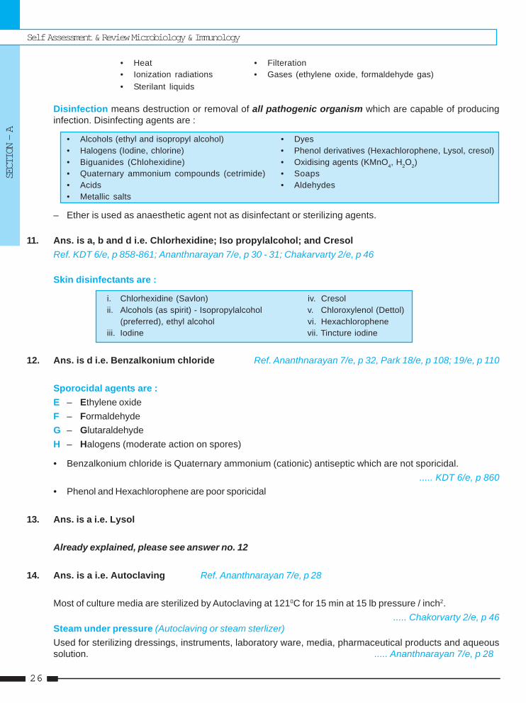
• Heat • Filteration• Ionization radiations • Gases (ethylene oxide, formaldehyde gas)• Sterilant liquids
Disinfection means destruction or removal of all pathogenic organism which are capable of producinginfection. Disinfecting agents are :
• Alcohols (ethyl and isopropyl alcohol) • Dyes• Halogens (Iodine, chlorine) • Phenol derivatives (Hexachlorophene, Lysol, cresol)• Biguanides (Chlohexidine) • Oxidising agents (KMnO4, H2O2)• Quaternary ammonium compounds (cetrimide) • Soaps• Acids • Aldehydes• Metallic salts
– Ether is used as anaesthetic agent not as disinfectant or sterilizing agents.
11. Ans. is a, b and d i.e. Chlorhexidine; Iso propylalcohol; and CresolRef. KDT 6/e, p 858-861; Ananthnarayan 7/e, p 30 - 31; Chakarvarty 2/e, p 46
Skin disinfectants are :
i. Chlorhexidine (Savlon) iv. Cresolii. Alcohols (as spirit) - Isopropylalcohol v. Chloroxylenol (Dettol)
(preferred), ethyl alcohol vi. Hexachloropheneiii. Iodine vii. Tincture iodine
12. Ans. is d i.e. Benzalkonium chloride Ref. Ananthnarayan 7/e, p 32, Park 18/e, p 108; 19/e, p 110
Sporocidal agents are :E – Ethylene oxideF – FormaldehydeG – GlutaraldehydeH – Halogens (moderate action on spores)
• Benzalkonium chloride is Quaternary ammonium (cationic) antiseptic which are not sporicidal...... KDT 6/e, p 860
• Phenol and Hexachlorophene are poor sporicidal
13. Ans. is a i.e. Lysol
Already explained, please see answer no. 12
14. Ans. is a i.e. Autoclaving Ref. Ananthnarayan 7/e, p 28
Most of culture media are sterilized by Autoclaving at 1210C for 15 min at 15 lb pressure / inch2...... Chakorvarty 2/e, p 46
Steam under pressure (Autoclaving or steam sterlizer)Used for sterilizing dressings, instruments, laboratory ware, media, pharmaceutical products and aqueoussolution. ..... Ananthnarayan 7/e, p 28
Questions & Answers
REVISION AT A GLANCE
27
1. b) Ethylene oxide 2. c) Steam at ... 3. a) Autoclaving 4. b) Hot air oven 5. a) 4-66. a) Absence of . 7. c) Differential ... 8. a) Syringes 9. d) Sterlization
Answer
Chapter Review••••• This section includes questions of VThis section includes questions of VThis section includes questions of VThis section includes questions of VThis section includes questions of Various Other PGMEES from 1990 – 2007.arious Other PGMEES from 1990 – 2007.arious Other PGMEES from 1990 – 2007.arious Other PGMEES from 1990 – 2007.arious Other PGMEES from 1990 – 2007.••••• Questions are arQuestions are arQuestions are arQuestions are arQuestions are ar ranged in decreasing order of yearranged in decreasing order of yearranged in decreasing order of yearranged in decreasing order of yearranged in decreasing order of yearwiz. This is done to makwiz. This is done to makwiz. This is done to makwiz. This is done to makwiz. This is done to make refere refere refere refere referralralralralral
system easier and uncomplicated to save the precious time of PGMEE Aspirant.system easier and uncomplicated to save the precious time of PGMEE Aspirant.system easier and uncomplicated to save the precious time of PGMEE Aspirant.system easier and uncomplicated to save the precious time of PGMEE Aspirant.system easier and uncomplicated to save the precious time of PGMEE Aspirant.
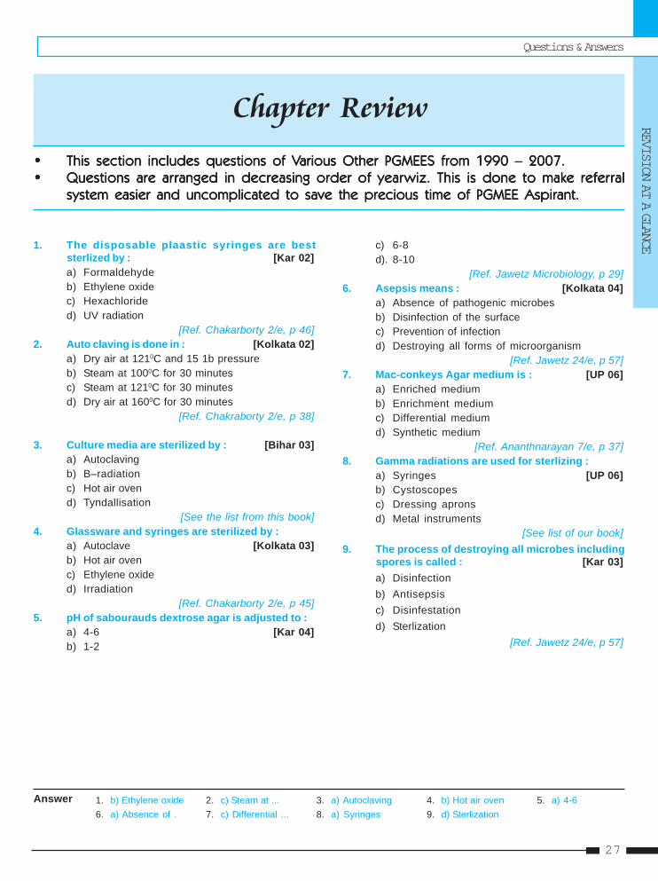
1. The disposable plaastic syringes are beststerlized by : [Kar 02]a) Formaldehydeb) Ethylene oxidec) Hexachlorided) UV radiation
[Ref. Chakarborty 2/e, p 46]2. Auto claving is done in : [Kolkata 02]
a) Dry air at 1210C and 15 1b pressureb) Steam at 1000C for 30 minutesc) Steam at 1210C for 30 minutesd) Dry air at 1600C for 30 minutes
[Ref. Chakraborty 2/e, p 38]
3. Culture media are sterilized by : [Bihar 03]a) Autoclavingb) B–radiationc) Hot air ovend) Tyndallisation
[See the list from this book]4. Glassware and syringes are sterilized by :
a) Autoclave [Kolkata 03]b) Hot air ovenc) Ethylene oxided) Irradiation
[Ref. Chakarborty 2/e, p 45]5. pH of sabourauds dextrose agar is adjusted to :
a) 4-6 [Kar 04]b) 1-2
c) 6-8d). 8-10
[Ref. Jawetz Microbiology, p 29]6. Asepsis means : [Kolkata 04]
a) Absence of pathogenic microbesb) Disinfection of the surfacec) Prevention of infectiond) Destroying all forms of microorganism
[Ref. Jawetz 24/e, p 57]7. Mac-conkeys Agar medium is : [UP 06]
a) Enriched mediumb) Enrichment mediumc) Differential mediumd) Synthetic medium
[Ref. Ananthnarayan 7/e, p 37]8. Gamma radiations are used for sterlizing :
a) Syringes [UP 06]b) Cystoscopesc) Dressing apronsd) Metal instruments
[See list of our book]9. The process of destroying all microbes including
spores is called : [Kar 03]a) Disinfectionb) Antisepsisc) Disinfestationd) Sterlization
[Ref. Jawetz 24/e, p 57]

Self Assessment & Review Microbiology & Immunology
SECTION – A
2828
NOTES
SECTION – BExplanatory Series of Questions 1995 - 2008 "ALL INDIA, AIIMS & PGI"
UNIT – I BACTERIOLOGY
Gram Positive Cocci1. Staphylococci 31 – 442. Streptococci 45 – 67Gram Negative Cocci3. Neisseria 68 – 78Gram Positive Bacilli4. Clostridium 79 – 935. Corynebacterium 94 – 1056. Actinomycetes & Bacillus 106 – 1187. Listeria Monocytogenes 119 – 1258. Mycobacteria 126 – 146Gram Negative Bacilli9. Enterobacteriaceae 147 – 17110. Vibrio 172 – 18611. Pseudomonas & Yersinia 187 – 195Gram Negative Cocco - Bacilli12. Hemophilus, Bordetella & Brucella 196 – 20613. Campylobacter & Helicobacter 207 – 21414. Legionella 215 – 22015. Rickettsiaceae & Chlamydiae 221 – 23816. Spirochetes 239 – 25317. Mycoplasma 254 – 260
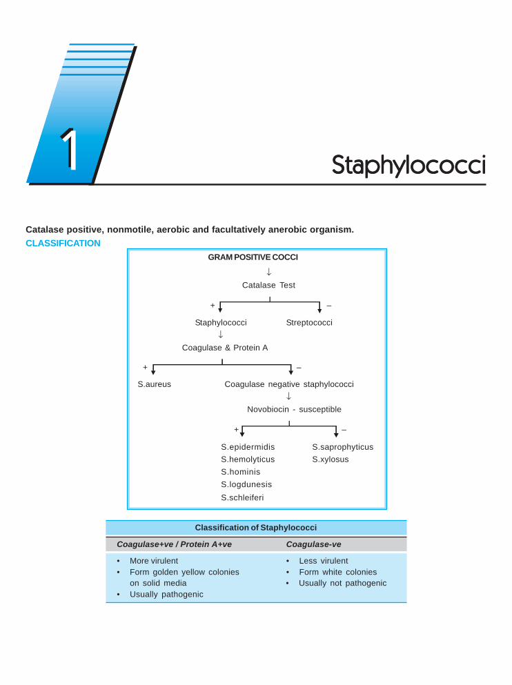
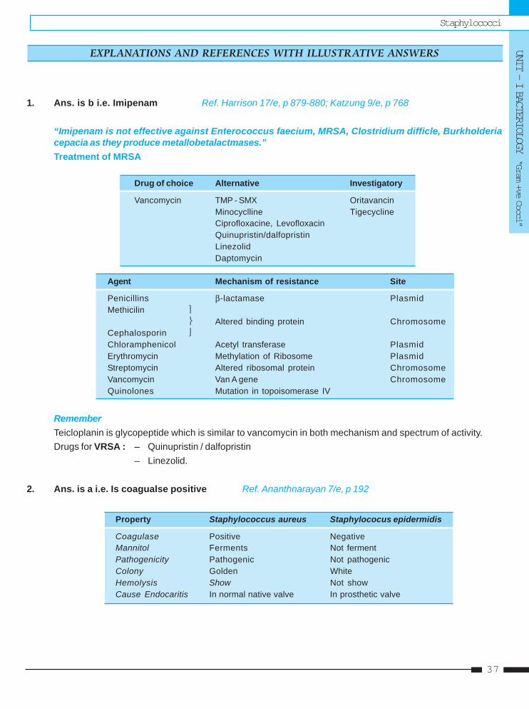
Catalase positive, nonmotile, aerobic and facultatively anerobic organism.CLASSIFICATION
GRAM POSITIVE COCCI
↓Catalase Test
+ –
Staphylococci Streptococci ↓
Coagulase & Protein A
+ –
S.aureus Coagulase negative staphylococci↓
Novobiocin - susceptible
+ –
S.epidermidis S.saprophyticusS.hemolyticus S.xylosusS.hominisS.logdunesisS.schleiferi
Classification of Staphylococci
Coagulase+ve / Protein A+ve Coagulase-ve
• More virulent • Less virulent• Form golden yellow colonies • Form white colonies
on solid media • Usually not pathogenic• Usually pathogenic
StaphylococciStaphylococciStaphylococciStaphylococciStaphylococci11
Self Assessment & Review Microbiology & ImmunologySECTION – B
3232
STAPHYLOCOCCI AUREUS• MC source of infection - Human patient and Carrier.• MC route of infection - Skin.• It is the MC cause of :
Acute endocarditis Acute osteomyelitisSpinal epidural abscess Nosocomial pneumonia ..... Harrison 16/e, p 1539Septic intracranial thrombophlebitis ParonychiaSkin, soft tissue infections Surgical wound infection
• S.aureus is MC cause of acute endocarditis except early and late prosthetic valve endocarditis which are causedby Coagulase-ve staph. and Streptococci viridans respectively.
• Recently methicillin resistant MRSA have been reported as primary cause of community acquired pneumonia..... Harrison 17/e, p 1621
Remember : Subacute Endocarditis is typically caused by Strep. viridans.
Culture• On nutrient agar, show characteristic oil paint appearance.• Show β hemolysis which is marked on rabbit or sheep blood and weak on horse blood agar.
Biochemical Reaction• Mannitol fermenter anaerobically (not by other species of Staphylococci)• Phosphatase reaction : – Gives prompt phosphatase reaction.
– Useful screening procedure as S.epidermidis is usually negative or only weakly positive.Virulence Factors• Most constant association of virulence is production of enzyme coagulase and to a lesser extent with mannitol
fermentation.a. Cell associated polymers :
• Cell wall polysaccharide peptidoglycan - Activates complement system. Induce release of cytokines.• Techoic acid - For adhesion and protection against complement mediated opsonization.• Capsular polysaccharide - Decrease opsonization.
b. Cell surface proteins :• Protein A - Responsible for coagglutination. Acts as an Fc receptor. Binds to Fc terminal of IgG 1, 2 and 4,
preventing opsonophagocytosis by PMNs.– Chemotactic, anticomplementory, antiphagocytic and B-cell mitogen.– Responsible for Co-agglutination.
• Clumping factor - Surface compound that is responsible for adherance of the organism to fibrinogen andfibrin. It is distinct from coagulase detected by slides test. ... Jawetz 24/e, p 226
c. Extracellular enzymes :1. Coagulase - Sufrace enzyme which converts fibrinogen to fibrin.
• It is a enzyme which requires presence of coagulase releasing factor (CRF) for its action.• Detected by tube test• It is of 8 types• Most human strain form type - A coagulase.• Coagulase test is standard criterion for S.aureus identification. In case of confusion tube test will be
deciding factor.
StaphylococciUNIT - I BACTERIOLOGY “Gram +ve Cocci”
33
Caution : Initially clumping factor is supposed to be bound form of coagulase. Now it is clear that it is totallydifferent, so, the concept of slide test for bound cogulase is wrong.2. Nuclease - A heat stable nuclease (DNAse) is characteristic of S.aureus3. Protein receptor - For mammalian proteins eg. fibronectin, fibrinogen, IgG, C1q. Facilitates adhesion.4. Lipase : Helps in infecting skin and subuctaneous tissue5. Hyaluronidase.6. Protease ⎫
⎬ Helps in spread of infection7. Fibrinolysin (Staphylokinase) ⎭α hemolysin
– Most important hemolysin– Protein inactivated at 700C, but reactivated aradoxically at 1000C.– Lyses rabbit erythrocyte but is less active against human erythrocytes.– Leucocidal, cytotoxic, dermonecrotic, neurotoxic and lethal.
ToxinA. Cytolytic Toxins :
• α Hemolysin : – Most important hemolysin– Protein inactivated at 700C, but reactivated aradoxically at 1000C.– Lyses rabbit erythrocyte but is less active against human erythrocytes.– Leucocidal, cytotoxic, dermonecrotic, neurotoxic and lethal.
• β Hemolysin - Shows ‘hot cold phenomenon’. Sphingomyelinase, hemolytic for sheep cells.• γ Hemolysin - Bi component protein.• δ Hemolysin - Detergent like effect on cell membranes.• Leucocidin (Panton valentine Toxin) - Bi component toxin associated with farunculosis.• Synergohymenotropic toxin : Bi component toxin such as Leucocidin and γ Hemolysin.
B. Enterotoxin : (A, B, C1-3, D, E and H)• Preformed, heat stable toxin, responsible for staphylococcal food poisoning which occur 2-6 hrs after consuming
meat and fish, milk or milk products.• Source - usually food handler which is carrier.• Mechanism - Toxin acts directly on autonomic nervous system (Vagal stimulation) and vomiting centre.• Type A toxin is responsible for most cases.
C. Toxic shock syndrome toxin (TSST) :Toxic shock syndrome is multisystem disease presenting with fever, hypotension, myalgia, vomiting, diarrhea,mucosal hyperemia, erythematous sunburn, rash, Disorientation or altered conciousness seen mostly inmenstruating women using highly absorbent vaginal tampons.• TSST-1 = Enterotoxin F = Pyrogenic Exotoxin C is responsible for most cases.• Vomiting is due to direct stimulation of ANS rather than local action.• Staph. Enterotoxin and TSST are super antigen leading to an excessive and non regulated immune response.
D. Exfoliative / Epidermatolytic Toxin / ET / Exfoliatin• Cause staphylococcal scalded skin syndrome (SSS).• Severe form is called Ritter’s disease in neonate and toxic epidermal necrolysis in elderly.• Milder form are pemphigus neonatorum and bullous impetigo.• There are two type : ETA and ETB, toxin possess serine protease activity which triggers exfoliation.
Self Assessment & Review Microbiology & ImmunologySECTION – B
3434
Typing• Staphylococci are typed on basis of their susceptibility to bacteriophage.• Phage typing is done by pattern method.
Lab diagnosis• Diagnosis is made by culture, specimen is plated on blood agar.• Smears are examined from culture and coagulase test is done.• Serological Test : Helpful in diagnosis of hidden deep infection.
– Antistaphylolysin (antialphalysin) titre of more than two unit is important specially when rising.• Polymerase chain raction (PCR) based assays have been applied for rapid diagnosis of S.aureus infection.
... Harrison 17/e, p 875Treatment
• If sensitive to penicillin →→→→→ Penicillin G• Penicillinase producing but sensitive to methicillin →→→→→ Naficillin or Oxacillin• Methicillin resistant Staph. Aureus (MRSA) →→→→→ Vancomycin• Vancomycin resistant Staph. Aureus (VRSA) →→→→→ Quinopristin, dalfopristin, linezolid• Emperical therapy →→→→→ Vancomycin
Special Cases :
• TSS →→→→→ Clindamycin (reduces toxin synthesis)• Food poisioning →→→→→ No antibiotic (as caused by preformed toxin)
COAGULASE (-) VE STAPHYLOCOCCI (CoNS)• MC pathogen complicating use of I.V. catheters, shunts and grafts, pacemaker wires, prosthetic valves, vascular
grafts, CSF shunts, dialyser.• Mnemonic - CoNS are MC source of infection on any exogenous implant.
Staph.epidermidis / albus• Normally present on human skin. Not pathogenic ordinarily.• Predilection for growth on implanted foreign bodies.• Common source of stich abscess.• S. sepidermidis is adapted to colonize these devices by its capacity to elaborate the extracellular polysaccharide
(glycocalyx or slime) that facilitates formation of protective biofilm on the device surface. This biofilm protectsbacteria from antibiotics and host defence.
• The attachment is also facilitated by autolysis (AtlE), fibrinogen binding protein, and accumulation-associatedprotein (AAP).
Staph. saprophyticus• Present on normal human skin and periurethral area.• Cause UTI in sexually active young women. This is due to its enhanced capacity to adhere to uroepithelial cells.
S. lugdunesis and S. schleiferi• Produces serious infections (native value endocarditis and osteomyelitis) than do other CoNS.
StaphylococciUNIT - I BACTERIOLOGY “Gram +ve Cocci”
35
1. A diabetic patient developed cellulitis due toS.aureus, which was found to be methicillin re-sistant on the antibiotic sensitivity testing. All thefollowing antibiotics will be appropriate except:a) Vancomycin [AI 06]b) Imipenemc) Teichoplanind) Linezolid
2. Staphylococcus aureus differs from staphylococ-cus epidermidis by : [AI 02]a) Is coagulase positiveb) Forms white coloniesc) A common cause of UTId) Causes endocarditis of prosthetic valve
3. True statement regarding non-coagulase staphy-lococci is : [AI 99]a) They are non-pathogenicb) They commonly infect indwelling prosthesisc) They may cause scarlet feverd) They are separated by gram’s staining
4. All of the following statement are true about Sta-phylococci except : [AIIMS 04]a) A majority of infection caused by coagulase (-)
ve Staph. are due to staph epidermidisb) β-Lactmase production is under plasmid con-
trolc) Expression of methicillin resistance in Staphy-
lococcus aureus increases when it is incubatedat 370C on blood agar
d) Methicillin resistance in Staph. aureus is inde-pendent of b-Lactmase production
5. Which one of the following Gram positive organ-ism is most common cause of UTI among sexu-ally active women : [AIIMS 04]a) Staphylococcus epidermidisb) Staphylococcus aureusc) Staphylococcus saprophyticusd) Enterococcus
6. The following is characteristic feature of staphy-lococcus food poisoning except : [AIIMS 04]a) Optimum temprature for toxin production is 370Cb) Intradietic toxin are responsible for intestinal
symptomsc) Toxin can be destroyed by boiling for 30 min-
utesd) Incubation period is 1-6 hours
7. A patient in an ICCU is on CVP line. His blood cul-ture shows growth of Gram (+) ve cocci whichare catalase positive and coagulase negative. Themost likely etiological agent is : [AIIMS 03]a) Staph. aureusb) Staph. epidermidisc) Streptococcus pyogenesd) Enterococcus faecalis
8. Staph. aureus causes vomiting in 6-8 hours. Themechanism of action is : [AIIMS 02]a) Stimulation of CAMPb) Vagal stimulationc) Stimulation of CGMPd) Acts through ganglioside GM receptor
9. A cook prepares sandwitches for 10 people go-ing for picnic. Eight out of them develop severegastroenteritis within 4-6 hrs of consumption ofthe sandwitches. It is likely that on investigationsthe cook is found to be carrier of : [AIIMS 02]a) Salmonella typhib) Vibrio choleraec) Entamoeba histolyticad) Staphylococcus aureus
10. A child after consuming food in a party complainsof diarrhea within 1-5 hours. The diagnosis is :a) S. aureus [AIIMS 01, 96, 95]b) Streptococcusc) Clostridium perfringensd) Clostridium botulinum
11. A 30 year old female is on antibiotics with pro-longed IV cannulation, has spike of fever, the likelycause is : [AIIMS 99]a) Pseudomonas aerugenosab) Coagulase negative staphylococcusc) Streptococcus agalactiaed) E. coli
12. All are true regarding staphylococcal toxin ex-cept : [AIIMS 97]a) Beta hemolysin shows hot cold phenomenonb) Mainly endotoxinc) Enterotoxin causes food poisoningd) Exfoliative toxin causes Ritter’s syndrome
QUESTIONS
Answer 1. b) Imipenem 2. a) Is coagul ... 3. b) They commonly ... 4. c) Expression ... 5. c) Staphyloc ...6. c) Toxin can ... 7. b) Staph. ... 8. d) Staphyloco ... 9. b) Vagal ... 10. a) S. aureus
11. b) Coagulase ... 12. b) Mainly ...
Self Assessment & Review Microbiology & ImmunologySECTION – B
3636
Answer 13. a) Staphyloco ... 14. a) Staphyloco ... 15. a, c and e 16. b and c 17. a and c18. c and d 19. a) 4-6 hrs
13. Which of the following organisms is implicated inthe causation of botryomycosis : [PGI 01]a) Staphylococcus aureusb) Staphylococcus albusc) Pseudomonas aeruginosad) Streptococcus pneumoniae) Streptococcus pyogenes
14. Staphylococcus in stool occurs in : [PGI 01, 00]a) Staphylococcal food poisoningb) Ischiorectal abscessc) Toxic shock syndromed) May be a normal findinge) Pseudomembranous colitis
15. Staphyloccus can cause : [PGI 01]a) Ecthymab) Erytharsmac) Furuncled) Impetigo contagioae) Sycosis barbae
16. Common source of staph in hospital : [PGI 99]a) IV fluids
b) Infective woundsc) Hands of hospital personneld) Bed linene) Instruments
17. Transfer of drug resistance in staphylococcus isby : [PGI 98]a) Transductionb) Transformationc) Conjugationd) Transfection
18. Pathogenicity of staphylococci is because of :a) Lecithinase [PGI 98]b) M-proteinc) Coagulased) Hyaluronidase
19. Incubation period of staphylococcal food poison-ing is : [PGI 95]a) 4-6 hrsb) 6-12 hrsc) 12-18 nrsd) 18-24 hrs
StaphylococciUNIT - I BACTERIOLOGY “Gram +ve Cocci”
37
1. Ans. is b i.e. Imipenam Ref. Harrison 17/e, p 879-880; Katzung 9/e, p 768
“Imipenam is not effective against Enterococcus faecium, MRSA, Clostridium difficle, Burkholderiacepacia as they produce metallobetalactmases.”Treatment of MRSA
Drug of choice Alternative Investigatory
Vancomycin TMP - SMX OritavancinMinocyclline TigecyclineCiprofloxacine, LevofloxacinQuinupristin/dalfopristinLinezolidDaptomycin
Agent Mechanism of resistance Site
Penicillins β-lactamase PlasmidMethicilin ⎫
⎬ Altered binding protein ChromosomeCephalosporin ⎭Chloramphenicol Acetyl transferase PlasmidErythromycin Methylation of Ribosome PlasmidStreptomycin Altered ribosomal protein ChromosomeVancomycin Van A gene ChromosomeQuinolones Mutation in topoisomerase IV
RememberTeicloplanin is glycopeptide which is similar to vancomycin in both mechanism and spectrum of activity.Drugs for VRSA : – Quinupristin / dalfopristin
– Linezolid.
2. Ans. is a i.e. Is coagualse positive Ref. Ananthnarayan 7/e, p 192
Property Staphylococcus aureus Staphylococus epidermidis
Coagulase Positive NegativeMannitol Ferments Not fermentPathogenicity Pathogenic Not pathogenicColony Golden WhiteHemolysis Show Not showCause Endocaritis In normal native valve In prosthetic valve
EXPLANATIONS AND REFERENCES WITH ILLUSTRATIVE ANSWERS
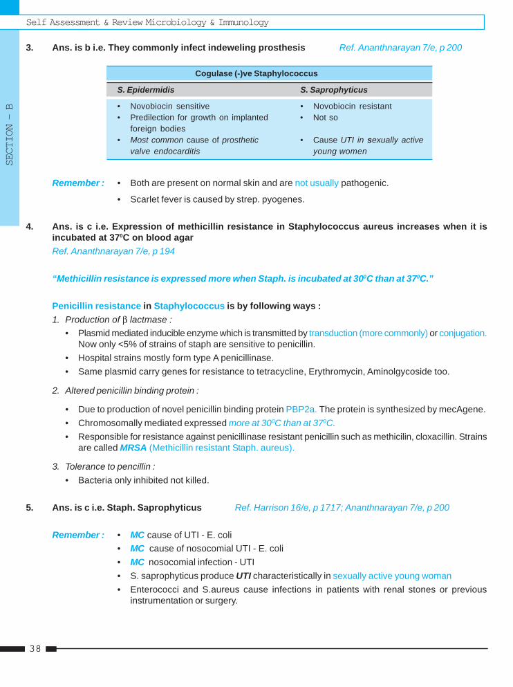
Self Assessment & Review Microbiology & ImmunologySECTION – B
3838
3. Ans. is b i.e. They commonly infect indeweling prosthesis Ref. Ananthnarayan 7/e, p 200
Cogulase (-)ve Staphylococcus
S. Epidermidis S. Saprophyticus
• Novobiocin sensitive • Novobiocin resistant• Predilection for growth on implanted • Not so
foreign bodies• Most common cause of prosthetic • Cause UTI in sexually active
valve endocarditis young women
Remember : • Both are present on normal skin and are not usually pathogenic.
• Scarlet fever is caused by strep. pyogenes.
4. Ans. is c i.e. Expression of methicillin resistance in Staphylococcus aureus increases when it isincubated at 370C on blood agarRef. Ananthnarayan 7/e, p 194
“Methicillin resistance is expressed more when Staph. is incubated at 300C than at 370C.”
Penicillin resistance in Staphylococcus is by following ways :1. Production of β lactmase :
• Plasmid mediated inducible enzyme which is transmitted by transduction (more commonly) or conjugation.Now only <5% of strains of staph are sensitive to penicillin.
• Hospital strains mostly form type A penicillinase.• Same plasmid carry genes for resistance to tetracycline, Erythromycin, Aminolgycoside too.
2. Altered penicillin binding protein :
• Due to production of novel penicillin binding protein PBP2a. The protein is synthesized by mecAgene.• Chromosomally mediated expressed more at 300C than at 370C.• Responsible for resistance against penicillinase resistant penicillin such as methicilin, cloxacillin. Strains
are called MRSA (Methicillin resistant Staph. aureus).
3. Tolerance to pencillin :• Bacteria only inhibited not killed.
5. Ans. is c i.e. Staph. Saprophyticus Ref. Harrison 16/e, p 1717; Ananthnarayan 7/e, p 200
Remember : • MC cause of UTI - E. coli• MC cause of nosocomial UTI - E. coli• MC nosocomial infection - UTI• S. saprophyticus produce UTI characteristically in sexually active young woman• Enterococci and S.aureus cause infections in patients with renal stones or previous
instrumentation or surgery.
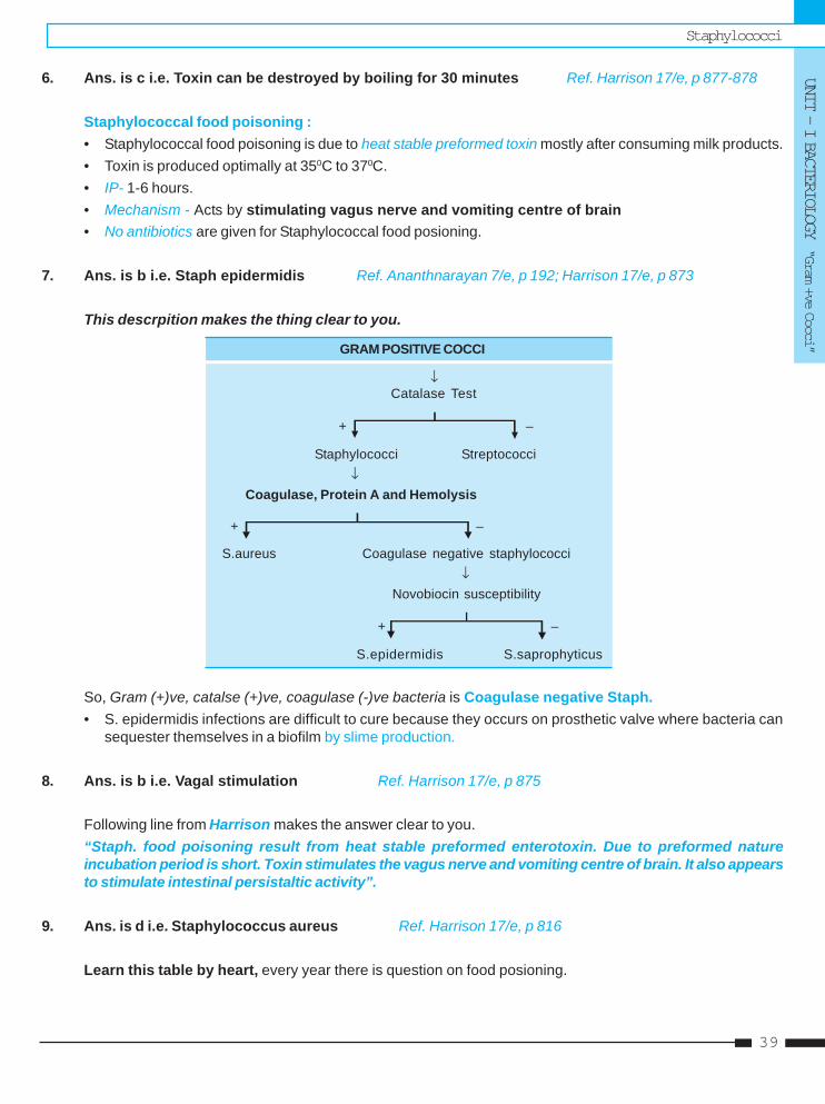
StaphylococciUNIT - I BACTERIOLOGY “Gram +ve Cocci”
39
6. Ans. is c i.e. Toxin can be destroyed by boiling for 30 minutes Ref. Harrison 17/e, p 877-878
Staphylococcal food poisoning :• Staphylococcal food poisoning is due to heat stable preformed toxin mostly after consuming milk products.• Toxin is produced optimally at 350C to 370C.• IP- 1-6 hours.• Mechanism - Acts by stimulating vagus nerve and vomiting centre of brain• No antibiotics are given for Staphylococcal food posioning.
7. Ans. is b i.e. Staph epidermidis Ref. Ananthnarayan 7/e, p 192; Harrison 17/e, p 873
This descrpition makes the thing clear to you.
GRAM POSITIVE COCCI
↓Catalase Test
+ –
Staphylococci Streptococci ↓
Coagulase, Protein A and Hemolysis
+ –
S.aureus Coagulase negative staphylococci↓
Novobiocin susceptibility
+ –
S.epidermidis S.saprophyticus
So, Gram (+)ve, catalse (+)ve, coagulase (-)ve bacteria is Coagulase negative Staph.• S. epidermidis infections are difficult to cure because they occurs on prosthetic valve where bacteria can
sequester themselves in a biofilm by slime production.
8. Ans. is b i.e. Vagal stimulation Ref. Harrison 17/e, p 875
Following line from Harrison makes the answer clear to you.“Staph. food poisoning result from heat stable preformed enterotoxin. Due to preformed natureincubation period is short. Toxin stimulates the vagus nerve and vomiting centre of brain. It also appearsto stimulate intestinal persistaltic activity”.
9. Ans. is d i.e. Staphylococcus aureus Ref. Harrison 17/e, p 816
Learn this table by heart, every year there is question on food posioning.
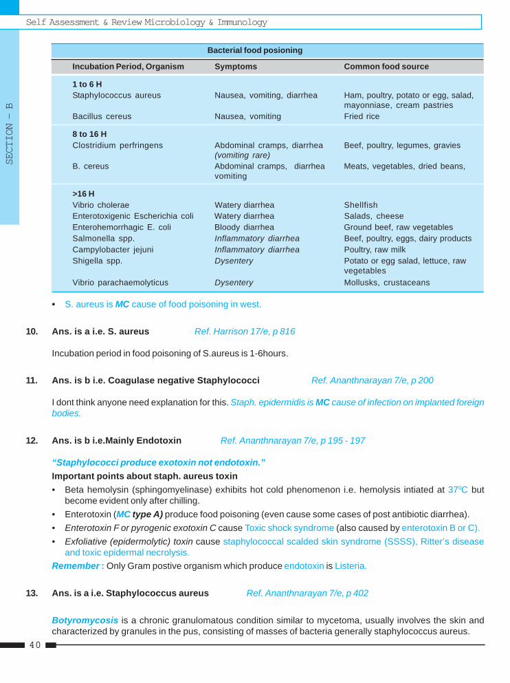
Self Assessment & Review Microbiology & ImmunologySECTION – B
4040
Bacterial food posioning
Incubation Period, Organism Symptoms Common food source
1 to 6 HStaphylococcus aureus Nausea, vomiting, diarrhea Ham, poultry, potato or egg, salad,
mayonniase, cream pastriesBacillus cereus Nausea, vomiting Fried rice
8 to 16 HClostridium perfringens Abdominal cramps, diarrhea Beef, poultry, legumes, gravies
(vomiting rare)B. cereus Abdominal cramps, diarrhea Meats, vegetables, dried beans,
vomiting
>16 HVibrio cholerae Watery diarrhea ShellfishEnterotoxigenic Escherichia coli Watery diarrhea Salads, cheeseEnterohemorrhagic E. coli Bloody diarrhea Ground beef, raw vegetablesSalmonella spp. Inflammatory diarrhea Beef, poultry, eggs, dairy productsCampylobacter jejuni Inflammatory diarrhea Poultry, raw milkShigella spp. Dysentery Potato or egg salad, lettuce, raw
vegetablesVibrio parachaemolyticus Dysentery Mollusks, crustaceans
• S. aureus is MC cause of food poisoning in west.
10. Ans. is a i.e. S. aureus Ref. Harrison 17/e, p 816
Incubation period in food poisoning of S.aureus is 1-6hours.
11. Ans. is b i.e. Coagulase negative Staphylococci Ref. Ananthnarayan 7/e, p 200
I dont think anyone need explanation for this. Staph. epidermidis is MC cause of infection on implanted foreignbodies.
12. Ans. is b i.e.Mainly Endotoxin Ref. Ananthnarayan 7/e, p 195 - 197
“Staphylococci produce exotoxin not endotoxin.”Important points about staph. aureus toxin• Beta hemolysin (sphingomyelinase) exhibits hot cold phenomenon i.e. hemolysis intiated at 370C but
become evident only after chilling.• Enterotoxin (MC type A) produce food poisoning (even cause some cases of post antibiotic diarrhea).• Enterotoxin F or pyrogenic exotoxin C cause Toxic shock syndrome (also caused by enterotoxin B or C).• Exfoliative (epidermolytic) toxin cause staphylococcal scalded skin syndrome (SSSS), Ritter’s disease
and toxic epidermal necrolysis.Remember : Only Gram postive organism which produce endotoxin is Listeria.
13. Ans. is a i.e. Staphylococcus aureus Ref. Ananthnarayan 7/e, p 402
Botyromycosis is a chronic granulomatous condition similar to mycetoma, usually involves the skin andcharacterized by granules in the pus, consisting of masses of bacteria generally staphylococcus aureus.
StaphylococciUNIT - I BACTERIOLOGY “Gram +ve Cocci”
41
14. Ans. is a i.e. Staphylococcal food poisoning Ref. Ananthnarayan 7/e, p 198
• In case of staphylococcal food poisoning food remnant and faeces are inoculated on selective medium likeludlam’s or salt milk agar or Roberson cooked meat medium containing 10% NaCl.
• TSS is due to systemic effect of absorbed toxin from site such as vagina, so no possibility of finding Staph.in case of TSS.
• Ischiorectal abscess is mostly due to E. coli.• Normal intestinal flora usually don’t contain Staph. aureus. So it is not a normal finding.• Pseudomembranous colitis is caused by clostridium difficle.
15. Ans. is a, c and e i.e. Ecthyma; Faruncle; and Sycobis barbaeRef. Harrison 17/e, p 875, 885; Dashore ‘Manual of skin disease’ 13-17; CMDT’ 08, p 119
Skin and soft tissue infection of S. aureus• Folliculitis – Superficial infection of ostia of hair follicle.• Faruncles (boils) – More extensive painful lesions that tends to occur in hairy moist region of body and
extend from hair follicle to become a true abscess, eg. buttock.• Carbuncle – Mostly located in lower neck and are even more severe and painful.• Acute paronychia – MC cause is S.aureus.• Bullous impetigo – Impetigo is most frequently diagnosed bacterial infection.• Ecthyma – It is deeper form of impetigo caused by Staph or Strep.• Cellulitis• Hiradenitis suppurativa - Recurrent follicular infections in region such as axilla.• Sycosis barbae is chronic folliculitis of beard hair follicle.
Remember :• S. aureus is MC cause of surgical wound infection. ..... Harrison 17/e, p 873• Non - bullous impetigo, cellulitis and erysipelas is caused by Strep. pyogenes more commonly than S.aureus.• Ecthyma gangreosum is caused by Ps. aeruginosa.• Erythma migrans is caused by Borrelia burgordeferi (Tick transmission).• Erythrasma is caused by corynebacterium minutissimus.• Impetigo contagiosa is usually caused by streptococci or mixed infections.
– Most cases of impetigo is caused by S.aureus.
16. Ans. is b and c i.e. Infective wounds; Hands of hospital personel Ref. Harrison 17/e, p 878
• MC mode of spread of S. aureus infection is via hands of health care workers.• S. aureus is MC cause of surgical wound infection and can cross infect wound of other patient too.• Most effective mode of prevention of nosocomial S. aureus infection is hand washing.
17. Ans. is a and c i.e. Transduction; and Conjugation Ref. Ananthnarayan 7/e, p 194
β lactamase mediated penicillin resistance is transmitted by transduction (more commonly) and conjugation.

Self Assessment & Review Microbiology & ImmunologySECTION – B
4242
18. Ans. is c and d i.e. Coagulase; and Hyaluronidase Ref. Ananthnarayan 7/e, p 195
• M protein, Lecithinase are not produced by Staphylococci.• Most important virulence factor is coagulase positivity.
Remember : Hyaluronidase - Helps in initiation and spread of infection.
19. Ans. is a i.e. 4 - 6 hours Ref. Harrison 17/e, p 816
Already explained, refer answer no. 8
StaphylococciUNIT - I BACTERIOLOGY “Gram +ve Cocci”
43
1. Phage typing is widely used for the intraspeciesclassification of one of the following bacteria :a) Staphylococci [Kar. 95]b) E. colic) Klebsiella pneumoniaed) Pseudomonas aeruginosa
[Ref. Ananthnarayan 7/e, p 197]2. The antibody marker in serum for staphylococal
endocarditis is : [AIIMS 90]a) ASLOb) Antitechoic acidc) Anti Lipopolysaccharided) Anti-M-protein
[Ref. Ananthnarayan 7/e, p 195]3. Toxic shock syndrome was first discovered in :
a) Tampoon users [JIPMER 91]b) Diabetic septicemiac) Drug addictsd) None
[Ref. Ananthnarayan 7/e, p 196]4. Phage typing is widely used for the intraspecies
classification of one of the following bacteria :a) Staphylococci [Karn 95]b) Escherichia colic) Klebsiella pneumoniaed) Pseudomonas aeruginosa
[Ref. Ananthnarayan 7/e, p 197]5. All are capsulated Bacteria except : [AP 96]
a) Step. Pneumoniab) Kleb. Pneunoniac) Vibriocholerad) H. influenza
[Ref. List of our book]
6. Pantone-valentine leucocidin is seen in infectionwith : [Kerala 97]a) Streptococcib) Staphylococcic) Gonococcid) Pnemococci
[Ref. Ananthnarayan 7/e, p 196]7. Bacteria is not shed in : [MP 98]
a) Carrier stateb) Latent injectionc) Incubation periodd) Subclinical infection
[Ref. Park 19/e, p 90]8. Gram positive cocci are : [UP 98]
a) Staphylococcusb) Streptococcusc) Pneumococcusd) All of the above
[Ref. Ananthnarayan 7/e, p 192, 202]9. Staphylococcal toxic syndrome is due to :
a) Enterotoxin A [Orrisa 2K]b) Enterbtoxin Bc) Enterotoxin Cd) Enterotoxin D
[Ref. Ananthnarayan 7/e, p 196]10. Staphylococcus Aureus remains in the skin for :
longer period because of : [MAHE 01]a) Catalaseb) Coagulasec) Hyaluronidased) None
[Ref. Ananthnarayan 7/e, p 195]
Chapter Review••••• This section includes questions of VThis section includes questions of VThis section includes questions of VThis section includes questions of VThis section includes questions of Various Other PGMEES from 1990 – 2007.arious Other PGMEES from 1990 – 2007.arious Other PGMEES from 1990 – 2007.arious Other PGMEES from 1990 – 2007.arious Other PGMEES from 1990 – 2007.••••• Questions are arQuestions are arQuestions are arQuestions are arQuestions are ar ranged in decreasing order of yearranged in decreasing order of yearranged in decreasing order of yearranged in decreasing order of yearranged in decreasing order of yearwiz. This is done to makwiz. This is done to makwiz. This is done to makwiz. This is done to makwiz. This is done to make refere refere refere refere refer ralralralralral
system easier and uncomplicated to save the precious time of PGMEE Aspirant.system easier and uncomplicated to save the precious time of PGMEE Aspirant.system easier and uncomplicated to save the precious time of PGMEE Aspirant.system easier and uncomplicated to save the precious time of PGMEE Aspirant.system easier and uncomplicated to save the precious time of PGMEE Aspirant.
1. a) Staphylococci 2. b) Antitechoic .. 3. a) Tampoon ... 4. a) Staphylococci 5. c) Vibriocholera6. b) Staphylococci 7. b) Latent injection 8. d) All of the ... 9. b and c 10. c) Hyaluronidase
Answer
Self Assessment & Review Microbiology & ImmunologySECTION – B
4444
11. Post operative parotitis is caused by : [Delhi 00]a) Staph aureusb) Streptococcusc) E. colid) Pneumococcus
[Ref. CSDT ‘12/e, p 28]12. Mohan comes from dinner he compain of diar-
rhoea, vomiting after 4 hours of meal. Most likelycausative agent : [UP 02]a) Stap aureusb) V. choleraec) Streptococcusd) E) coli
[Ref. Harrison, 17/e, p 816; Ananthnarayan, 7/e,p 196]
13. Quikest food poisoning (1-6) hour is :a) Staphylococcus [Kolkata 02]b) B. cereusc) Salmonellad) Vibrio cholera
[Ref. Harrison 17/e, p 816]14. Cutaneous mainfestation of staphylococci are all
except : [St Johns 02]a) Faruncleb) Follicular impetigoc) TSSd) TEN
[Ref. Ananthnarayan 7/e, p 197]15. Toxin of staphylococcus : [Bihar 03]
a) Hemolysinb) Leucocidinc) Enterotoxinsd) All
[Ref. Ananthnarayan 7/e, p 195]16. Toxic shock syndrome is due to :
a) Streptococcus pyogenes [Bihar 04]b) Staphylococcus aureusc) Strept albicansd) E. durans
[Ref. Ananthnarayan 7/e, p 196]
17. Staphylococcus secrete all, except :a) Lipase [Bihar 04]b) Cellulosec) Coagulased) Lecithinase
[Ref. Ananthnarayan 7/e, p 195]18. The most common mechanism of drug resistance
in staphylococcus is : [M.P. 04]a) Conjugationb) Plasmidsc) Transductiond) Translation
[Ref. Ananthnarayan 7/e, p 194]19. Hot cold phenomenon is seen due to which toxin
of staphylococci : [UP 04]a) Alpha lysinb) Beta lysinc) Gamma lysind) Theta lysin
[Ref. Ananthnarayan 7/e, p 196]20. Staphalococcus is differ from stretpococcus is by
a) Coagulase test [Jharkhand 05]b) Catalse testc) Phosphateased) Gram negative
[Ref. Harrison 17/e, p 873]21. Novobiocin susceptible staph is : [UP 06]
a) S. aureusb) S. epidermidisc) S. hemolyticusd) S. saprophyticus
[Ref. Harrison 17/e, p 873]22. Coagulase negative staphylococci includes all of
the following except : [UP 07]a) Staph. epidermidisb) Staph. haemolyticusc) Staph. intermediusd) Staph. saprophyticus
[Ref. Ananthnarayan 7/e, p 200]
11. a) Staph aureus 12. a) Stap aureus 13. a) Staphylococcus 14. c) TSS 15. d) All16. b) Staphyloco ... 17. b) Cellulose 18. c) Transduction 19. b) Beta lysin 20. a) Coagulase ...21. d) S. saprophy ... 22. c) Staph. ...
Answer
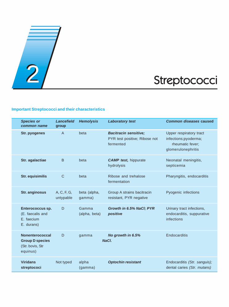
Important Streptococci and their characteristics
Species or Lancefield Hemolysis Laboratory test Common diseases causedcommon name group
Str. pyogenes A beta Bacitracin sensitive; Upper respiratory tractPYR test positive; Ribose not infections;pyoderma;fermented rheumatic fever;
glomerulonephritis
Str. agalactiae B beta CAMP test, hippurate Neonatal meningitis,hydrolysis septicemia
Str. equisimilis C beta Ribose and trehalose Pharyngitis, endocarditisfermentation
Str. anginosus A, C, F, G, beta (alpha, Group A strains bacitracin Pyogenic infectionsuntypable gamma) resistant, PYR negative
Enterococcus sp. D Gamma Growth in 6.5% NaCl; PYR Urinary tract infections,(E. faecalis and (alpha, beta) positive endocarditis, suppurativeE. faecium infectionsE. durans)
Nonenterococcal D gamma No growth in 6.5% EndocarditisGroup D species NaCl.(Str. bovis, Strequinus)
Viridans Not typed alpha Optochin resistant Endocarditis (Str. sanguis);streptococi (gamma) dental caries (Str. mutans)
StreptococciStreptococciStreptococciStreptococciStreptococci22
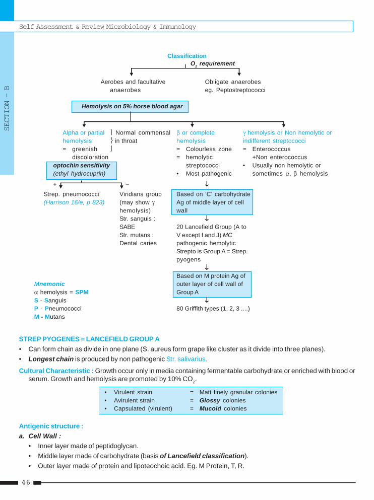
Self Assessment & Review Microbiology & Immunology
SECTION – B
4646
Classification
Aerobes and facultative Obligate anaerobesanaerobes eg. Peptostreptococci
Hemolysis on 5% horse blood agar
Alpha or partial ⎫ Normal commensal β or complete γ hemolysis or Non hemolytic orhemolysis ⎬ in throat hemolysis indifferent streptococci= greenish ⎭ = Colourless zone = Enterococcus
discoloration = hemolytic +Non enterococcusoptochin sensitivity streptococci • Usually non hemolytic or(ethyl hydrocuprin) • Most pathogenic sometimes α, β hemolysis
+ – ↓↓↓↓↓
Strep. pneumococci Viridians group Based on ‘C’ carbohydrate(Harrison 16/e, p 823) (may show γ Ag of middle layer of cell
hemolysis) wallStr. sanguis : ↓↓↓↓↓SABE 20 Lancefield Group (A toStr. mutans : V except I and J) MCDental caries pathogenic hemolytic
Strepto is Group A = Strep.pyogens
↓↓↓↓↓Based on M protein Ag of
Mnemonic outer layer of cell wall ofα hemolysis = SPM Group AS - Sanguis ↓↓↓↓↓P - Pneumococci 80 Griffith types (1, 2, 3 ....)M - Mutans
STREP PYOGENES = LANCEFIELD GROUP A• Can form chain as divide in one plane (S. aureus form grape like cluster as it divide into three planes).• Longest chain is produced by non pathogenic Str. salivarius.
Cultural Characteristic : Growth occur only in media containing fermentable carbohydrate or enriched with blood orserum. Growth and hemolysis are promoted by 10% CO2.
• Virulent strain = Matt finely granular colonies• Avirulent strain = Glossy colonies• Capsulated (virulent) = Mucoid colonies
Antigenic structure :a. Cell Wall :
• Inner layer made of peptidoglycan.• Middle layer made of carbohydrate (basis of Lancefield classification).• Outer layer made of protein and lipoteochoic acid. Eg. M Protein, T, R.
O2 requirement
Streptococci
UNIT - I BACTERIOLOGY “Gram +ve Cocci”
47
– M protein is basis of Griffith typing. Act as virulence factor by inhibiting phagocytosis. Antibody to M isprotective.
– T and R protein has no relation to virulence.b. Hair like pilli (fimbria) : Important for attachment to epithelial cells.Antigenic SimilaritySome antigens of streptococci are similar to normal human cells, because of which streptococal infection is associatedwith autoimmune disease like rheumatic fever.
Capsular Hyaluronic Acid → Synovial fluidCell wall protein → MyocardiumGp A carbohydrates → Cardiac ValvesCytoplasmic membrane → Vascular intimaPeptidoglycan (mucoprotein) → Skin antigen
Toxins and Virulence factorsa. Hemolysin - Streptolysin
O [Oxygen labile] S [Oxygen stable and serum soluble]
• Activity only on pour plate Hemolysis on surface• Antigenic specific Non antigenic• Cardiotoxic
• ASO titre → Anti Streptolysin O titre used in retrospective diagnosis; > 200 units is significant; inhibited bycholesterol (but not by Normal sera).
b. Pyrogenic Exotoxin = Erythrogenic = Dick = Scarlatinal Toxin :• This is superantigen causing TSS.• Used to identify children susceptible to scarlet fever by intradermal injection (=Dick test) and Schultz Charlton
Reaction.• Type A toxin is MC
c. Streptokinase (Fibrinolysin) : Facilitates spread of infection.d. Deoxyribo nuclease (Streptodornase; DNAase) - Responsible for thin serous character of strep exudates.e. Nictoniamide Adenine Dinucleotidase (NAD-ase).f. Hyaluronidase : Favour spread of infection.g. Serum Opacity Factor - Lipoproteinase.
Infection1. Respiratory :
• Sore throat is MC streptococcal infection.
2. Scarlet fever : Streptococcal pharyngitis, accompanied by rash made up of minute papules giving a characteristic“Sand paper” feel to skin.• Associated with circumoral pallor, straw berry tongue.• Rash reflect hypersentivity reaction to toxin.
3. Skin and Soft tissue infection :• Impetigo - Superficial infection of skin mainly by group A streptococci. Face and leg are MC site.
Bullous impetigo is caused by staph.
Self Assessment & Review Microbiology & Immunology
SECTION – B
4848
• Cellulitis - Diffuse spreading infection of skin and subcutaneous tissue especially of leg. Caused by Str.pyogenes (MC), Staph, Cl. perfringens, E.coli.
• Erysipelas - Superficial form of celluititis with bright red appearance of involved skin, seen classically oncheek. Skin assumes peau-d-orange texture due to involvement of superficial lymphatics.
• Necrotizing fascitis - Caused by Group A streptococci called as Hemolytic Streptococcal gangrene.• In skin infection ASO titre is not high and so ASO estimation has not much clinical significance.
4. Genital infection :• Anaerobic streptococci are most important cause of puerperal sepsis.
5. Bacteremia, pneumonia, toxic shock syndrome.6. Non suppurative complication : Develop 1-3 weeks after acute infection.
Acute Rheumatic fever Acute glomerulonephritis
• Develop after throat infection • Develop after either sore throat or skin infectionby any serotype of S. pyogenes by serotype 49, 53-55, 59-64, 1 and 12
• Repeated attacks common • No repeated attacks• Penicilin prophylaxis indicated • Not indicated• Course is progressive or static • Spontaneous resolution• ASO titre always raised • May or May not (after skin infection) raised• Marked immune response with no • Moderate immune response with decrease in
change in complement level complement level.Lab Diagnosis• In acute infection diagnosis is established by throat swab culture (diagnostic).
– Pike’s medium is used as transport media.– Sheep blood agar is recommended for isolation as it is inhibitory for H. hemolyticus.
• In Rheumatic fever and Glomerulonephritis retrospective diagnosis is needed.– ASO titre > 200 is indicative of prior streptococcal disease. After skin infection ASO titre is usually low. So
useful only in retrospective diagnosis of Rheumatic fever.– In acute glomerulonephritis and pyoderma, Anti DNAse and antihyaluronidase are used for retrospective
diagnosis.• Streptozyme test - Passive slide haemagglutination test. Sensitive and specific test for all types of streptococcal
infection.
Treatment of Group A Streptococci
• Pharyngitis, impetigo, erysipelas, cellulitis – Penicillin (Pn)• Necrotizing fascitis / myositis – Surgical debridement + Pn + Clindamycin• Pneumonia / Empyema – Pn + empyema drainage• Streptococcal TSS – Penicillin + Clindamycin + iv Ig
OTHER HEMOLYTIC STREPTOCOCCI(= Lancefield β Hemolytic Streptococci)Group B (Strep. agalactiae) : Virulence factor is polysaccharide capsule. (Group A have hyaluronic acid capsule).– S. agalactiae is the MC cause of neonatal meningitis in west.– It does not hydrolyse bile esculin agar, however hydrolyse sodium hippurate and are bacitracin resistant.– Identified by CAMP (Christie, Atkins and Munch - Peterson) reaction.– In infant it cause 2 type of infection :
• Early Onset : More common, acquired from maternal vagina during birth.– Essentially all infants are bacteremic presenting with lethargy respiratory distress and hypotension.
Streptococci
UNIT - I BACTERIOLOGY “Gram +ve Cocci”
49
• Late Onset Infection : Infection more often obtained from environment.– Meningitis is the MC manifestation and in most cases is associated with strain of capsular type III.
– TreatmentPenicillin is DOC for all group B streptococcal infection.
Group C - Strep. equisimilis• Predominant animal pathogen and is the source of streptokinase used for thrombolytic therapy.
Group D - StreptococciIncludes : • Fecal streptococci / Enterococcus.
• Non enterococcal group D streptococci.• Enterococci are now considered as separate genus on the basis of DNA homologus studies..
... Harrison 17/e, p 881Distinctive features of Enterococcus :– Grow in 40% bile– Grow in 6.5% NaCl at pH 9.6, 450C and in 0.1% methylene blue milk– Heat resistant, surviving 600C for 30 minutes– Present in intestine, genital tract and saliva– Cause wound infection, UTI nosocomal bacterimia in patient with IV catheters, endocarditis.– Usually resistant to Penicillin, cephalosporin etc– In case of Vancomycin and β lactam resistance - Linezolid (against all enterococci) or quinupristin dalfopristin
(against E.faecium only) are given.
Non Enterococcal Group D Streptococci– Grow in bile ..... Jawetz 23/e, p 235– Unable to grow in 6.5% NaCl and PYR negative– Penicillin susceptible.– Main species causing infection is S.bovis. S.bovis endocarditis is often associated with neoplasm of GIT, most
commonly colon.
Group F - Streptococci• Called as minute streptococci.• Includes Streptococcus MG.• Isolated from cases of atypical pneumonia.
Viridans streptococci• Heterogenous group of organism that are important agent of bacterial endocarditis. These are part of normal
flora of mouth.• Species include :
– S.sanguis : MC viridans streptococcus associated with endocarditis– S.mutans : Imporant in the causation of dental caries.– S.salivarus
PNEUMOCOCCUS• Gram positive Lanceolate diplococci.• It is MC cause of Lobar pneumonia, sinusitis, otitis media.
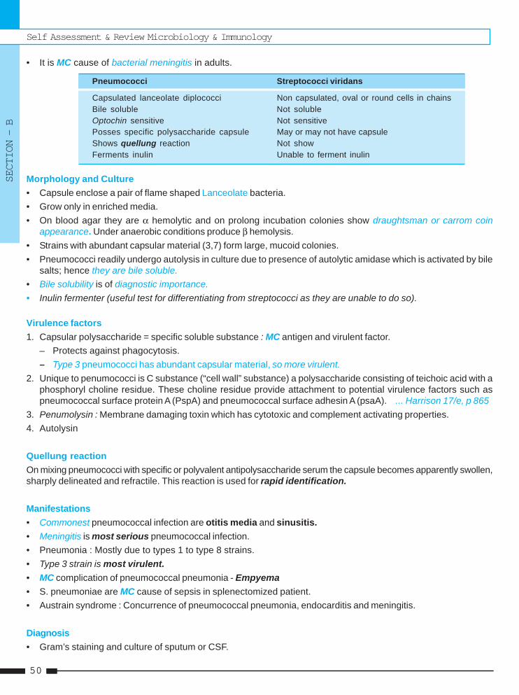
Self Assessment & Review Microbiology & Immunology
SECTION – B
5050
• It is MC cause of bacterial meningitis in adults.
Pneumococci Streptococci viridans
Capsulated lanceolate diplococci Non capsulated, oval or round cells in chainsBile soluble Not solubleOptochin sensitive Not sensitivePosses specific polysaccharide capsule May or may not have capsuleShows quellung reaction Not showFerments inulin Unable to ferment inulin
Morphology and Culture• Capsule enclose a pair of flame shaped Lanceolate bacteria.• Grow only in enriched media.• On blood agar they are α hemolytic and on prolong incubation colonies show draughtsman or carrom coin
appearance. Under anaerobic conditions produce β hemolysis.• Strains with abundant capsular material (3,7) form large, mucoid colonies.• Pneumococci readily undergo autolysis in culture due to presence of autolytic amidase which is activated by bile
salts; hence they are bile soluble.• Bile solubility is of diagnostic importance.• Inulin fermenter (useful test for differentiating from streptococci as they are unable to do so).
Virulence factors1. Capsular polysaccharide = specific soluble substance : MC antigen and virulent factor.
– Protects against phagocytosis.– Type 3 pneumococci has abundant capsular material, so more virulent.
2. Unique to penumococci is C substance (“cell wall” substance) a polysaccharide consisting of teichoic acid with aphosphoryl choline residue. These choline residue provide attachment to potential virulence factors such aspneumococcal surface protein A (PspA) and pneumococcal surface adhesin A (psaA). ... Harrison 17/e, p 865
3. Penumolysin : Membrane damaging toxin which has cytotoxic and complement activating properties.4. Autolysin
Quellung reactionOn mixing pneumococci with specific or polyvalent antipolysaccharide serum the capsule becomes apparently swollen,sharply delineated and refractile. This reaction is used for rapid identification.
Manifestations• Commonest pneumococcal infection are otitis media and sinusitis.• Meningitis is most serious pneumococcal infection.• Pneumonia : Mostly due to types 1 to type 8 strains.• Type 3 strain is most virulent.• MC complication of pneumococcal pneumonia - Empyema• S. pneumoniae are MC cause of sepsis in splenectomized patient.• Austrain syndrome : Concurrence of pneumococcal pneumonia, endocarditis and meningitis.
Diagnosis• Gram’s staining and culture of sputum or CSF.
Streptococci
UNIT - I BACTERIOLOGY “Gram +ve Cocci”
51
Treatment• Otitis media/Sinusitis/ Pneumonia - Amoxicillin.• Meningitis - Ceftriaxone plus vancomycin.• Endocarditis - Ceftriaxone or cefotaxime + vancomycin.• Penicillin resistance is not due to production of β lactmase but alteration in penicillin binding protein.
Pneumococcal VaccinePolyvalent polysaccharide vaccine containing capsular antigen of 23 most prevalent serotypes.Protection rate - 80 - 90%
Indication Contraindication
• In pateint with absent or dyfunctional spleen • < 2yr child and > 65 yr• Sickle cell disease • Lymphoreticular malignancies• Coeliac disease and immunosuppressive therapy• Chronic renal, lung, heart and liver disease • CSF leak• Diabetes mellitus • Alcoholic cirrhosis• Immunodeficiencies including HIV • Multiple myeloma
• Chronic glucocorticoid therapy• ‘Hodgkin’s’disease• Organ transplant recepient
Self Assessment & Review Microbiology & Immunology
SECTION – B
5252
1. Which is false regarding Gram positive cocci ?a. Staph. saprophyticus causes UTI in femalesb. Micrococci are oxidase positive [AI 08]c. Most enterococci are sensitive to penicillind. Pneumococci are capsulated
2. Which component of St. pyogenes has cross re-activity with synovium of human ? [AI 08]a. Capsular hyaluronic acidb. Cell proteinsc. Group A carbohydrate antigensd. Peptidoglycan
3. Which of the following is not true regarding en-terococcus ? [AI 08]a. Common species are E.faecalis and E.faeciumb. It is a cause for peritonitisc. It is universally susceptible to penicillinsd. Can cause intra-abdominal abscess
4. A patient admitted to an ICUs is on central venousline for the last one week; he is on ceftazidine andamikacin. After 7 days of antibiotics he developsa spike of fever and his blood culture is positivefor gram positive cocci in chains, which are cata-lase negative. Following this, vancomycin wasrestarted but the culture remained positive forsame organisms even after 2 weeks of therapy.The most likely organism causing infection is :a) Staph aureus [AI 07; AIIMS Nov. 06]b) Viridans streptococcic) Enterococus fecalisd) Coagulase positive staphylococcusd) Bile solubility
5. An infant had high grade fever and respiratory dis-tress at the time of presentation to the emergencyroom. The sample collected for blood culture wassubsequently positive showing growth of ααααα-haemolytic colonies. On Gram staining these weregram positive cocci. In the screening test for iden-tification, the suspected pathogen is likely to besusceptible to the following agent :a) Bacitracin [AI 07]b) Novobiocinc) Optochind) Cloxacillin
6. A person from village is complaining of develop-
ment of pustules. Extract from pus has showngram positive cocci, showing hemolysis, catalase–ve, identified as a group of streptococci. Follow-ing test is used : [AI 07; AIIMS Nov. 06]a) Bacitracin sensitivityb) Novobiocin sensitivityc) Optochin sensitivity
7. All are true about streptococcus except : [AI 01]a) M-protein is responsible for production of mu-
coid coloniesb) M- protein is responsible for virulencec) Mucoid colonies are virulentd) No resistance to penicillin has been reported
8. A beta hemolytic bacteria is resistant to vanco-mycin, shows growth in 6.5% NaCl, is non bile sen-sitive. It is likely to be : [AI 01]a) Strep. agalactiaeb) Strep. pneumoniac) Enterococcusd) Strep. Bovis
9. Toxin involved in the streptococcal toxic shocksyndrome is : [AI 01]a) Pyrogenic toxinb) Streptokinasec) Hemolysind) Neurotoxin
10. In a patient of orbital cellulitis, micro organism onculture show greenish colonies and optochin sen-sitivity. The most likely organism is : [AI 00]a) Strep. viridansb) Staphylococcusc) Pseudomonasd) Pneumococcus
11. True statement regarding Pneumococcus is :a) Virulence is due to polysaccharide capsuleb) Capsule is protein in nature [AI 99]c) Antibodies against capsule are not protectived) Resistance to penicillin has not yet been re-
ported
QUESTIONS
Answer 1. c) Most enterococci 2. a) Capsular... 3. c) It is universally... 4. c) Enterococus ... 5. c) Optochin ...6. a) Bacitracin ... 7. a) M-protein ... 8. c) Enterococcus 9. a) Pyrogenic ... 10. d) Pneumoco ...
11. a) Virulence ...
Streptococci
UNIT - I BACTERIOLOGY “Gram +ve Cocci”
53
12. True statements about Pneumococcus are allexcept : [AI 99]a) Pneumolysin a thiolactivated toxin, exerts a vari-
ety of effects on ciliary and PMN’s actionb) Autolysin may contribute to the pathogenesis of
pneumococcal disease by lysing bacteriac) Anticapsular ‘antibodies are serotype specificd) The virulence of pneumococci is dependent only
on the production of the capsular polysaccha-ride
13. True statement about Antistreptolysin ‘O’ titre is:a) In normal people the titre is <200 [AI 97]b) In acute glomerulonephritis the titre is lowc) ASO titre >200 indicate rheumatic feverd) Streptozyme test is an active haemagglutination
test14. One of the following infections is caused by
anaerobic gram positive cocci : [AI 95]a) Puerperal infectionb) Food poisoningc) Endocarditisd) Septicemia
15. Lance field grouping of streptococci is done byusing : [AIIMS 07]a. M proteinb. Group C peptidoglycan cell wallc. Group C carbohydrate antigend. M antigen
16. An infant had high grade fever and respiratory dis-tress at the time of presentation to the emergencyroom. The sample collected for blood culture wassubsequently positive showing growth of ααααα-hemolytic colonies. On Gram staining these weregram positive cocci. In the screening test for iden-tification, the suspected pathogen is likely to besusceptible to the following agent :a) Bacitracin [AIIMS 07, 06]b) Novobiocinc) Optochind) Oxacillin
17. A patient admitted to an ICU is on central venousline for the last one week. He is on Ceftazidimeand amikacin. After 7 days of antibiotics he devel-ops a spike of fever and his blood culture is posi-tive for gram positive cocci in chains which arecatalase - negative. Following this vancomycin wasstarted but the culture remained positive for sameorganism even after 2 weeks of therapy. The mostlikely organism causing infection is : [AIIMS 06]
a) Staphylococci aureusb) Viridans streptococcic) Enterococcus faecalisd) Coagulase negative staphylococcus
18. The sputum specimen of a 70 year old male wascultured on a 5% blood agar. The culture showedthe presence of a-haemolytic colonies next day.The further processing of this organism is mostlikely to yield : [AIIMS 05]a) Gram positive cocci in short chains, catalase
negative and bile resistantb) Gram positive cocci in pairs, catalase negative
and bile solublec) Gram positive cocci in clusters, catalase posi-
tive and coagulase positived) Gram negative coccobacilli, catalase positive
and oxidase positive19. Which of the following organism, when isolated
in the blood, requires the synergistic activity ofpenicillin + amino glycoside for appropriatetherapy : [AIIMS 04]a) Enterococcus faecalisb) Staph. aureusc) Streptococcus pneumoniaed) Bacteriodes fragilis
20. Most common causative organism for lobar pneu-monia is : [AIIMS 04]a) Staphylococcus aureusb) Streptococcus pyogenesc) Streptococcus pneumoniaed) Haemophilus influenzae
21. An outbreak of Streptococcal pharyngitis hasoccured in a remote village. In order to carry outthe epidemiological investigations it is necessoryto perform the culture of the throat swab of thepatient suffering from the disease. The transportmedia of choice would be : [AIIMS 02]a) Asalt manntol mediab) Pike’s mediac) Stuart mediad) Carry Blair media
22. Streptococcal toxic shock syndrome is due to thefollowing virulence factor : [AIIMS 03]a) M proteinb) Pyrogenic exotoxinc) Streptolysin Od) Carbohydrate cell wall
Answer 12. d) The virulence ... 13. b) In acute ... 14. a) Puerperal ... 15. c) Group c 16. c) Optochin17. c) Enterococcus ... 18. b) Gram ... 19. a) Enteroco ... 20. c) Streptoco ... 21. b) Pike’s ...22. b) Pyrogenic ...
Self Assessment & Review Microbiology & Immunology
SECTION – B
5454
23. The commonest organism causing cellulitis is :a) Streptococcus pyogenes [AIIMS 02]b) Streptococcus faecalisc) Streptococcus viridansd) Microaerophilic streptococi
24. 45 years old Ramlal has intra abdominal sepsis.The causative organism was found to be vanco-mycin, gentamycin and amplicillin resistant. Itgrows well in presence of 6.5% NaCl and arginine.Bile esculin hydrolysis is positive. Which of thefollowing is this organsim ? [AIIMS 01]a) Strept. agalactaeb) Enterococcus fecalisc) Streptococcus bovisd) Streptococcus pneumoneae
25. A patient of RHD developed infective endocarditisafter dental extraction. Most likely organism caus-ing this is : [AIIMS 01]a) Streptococcus viridansb) Streptococcus pneumoneaec) Streptococcus pyogenesd) S. aureus
26. Risk of Pneumococcal meningitis is seen in :a) Post splenectomy patient [AIIMS 99]b) Patient undergone neurosurgical interventionc) Patient following cardiac surgeryd) Patient with hypoplasia of lung
27. C-Carbohydrate in Streptococcus hemolyticus isimportant for : [AIIMS 98]a) Lancefield classificationb) Phagocytic inhibitionc) Toxin productiond) Haemolysis
28. Causative organism of late prosthetic valve en-docarditis is : [AIIMS 97]a) Streptococcus viridiansb) Proteus mirabilisc) Beta haemolytic streptococcid) Staphylococci
29. Lymphangitis is caused by : [AIIMS 95]a) Staphylococcusb) Streptococcic) Pneumococcid) Neisseria
30. Which of the following factor is mainly respon-sible for virulence in Streptococcus : [AIIMS 95]a) Carbohydrateb) Streptokinasec) Streptodornased) M protein
31. True about streptococcus : [PGI 03]a) Lancefield classification is based on M proteinb) Group ‘G’ not found in humanc) Group ‘B’ causes neonatal meningitisd) Group ‘C’ can be isolated from vaginal florae) Group ‘D’ causes urinary tract infection
32. After Splenectomy, most common infection :a) Pneumococcal [PGI 00]b) E. colic) Klebsiellad) Streptococcus
33. Streptococcal GN is best diagnosed by :a) ASO titre [PGI 99]b) Anti DNAsec) Anti hyaluronidased) Culture
34. Which toxin of streptococcus causes hemolysis:a) Streptolysin O [PGI 97]b) Sterptolysin Sc) Streptokinased) Sterptodornase
35. Streptococcus and pneumococcus are differen-tiated by : [PGI 97]a) Bile salt solubilityb) Growth charcteristicc) Gram stainingd) Inulin fermentation
36. Streptococci causing dental caries : [PGI 96]a) Streptococci equisimilisb) Streptococci mutansc) Streptococci pneumoniaed) Streptococci bovis
Answer 23. a) Streptoco ... 24. b) Enteroco ... 25. a) Streptoco ... 26. a) Post ... 27. a) Lancefield ...28. a) Streptoco ... 29. b) Streptococci 30. d) M protein 31. c and e 32. a) Pneumococcal33. b and c 34. b) Sterptolysin ... 35. a, b and d 36. b) Streptococci ...
Streptococci
UNIT - I BACTERIOLOGY “Gram +ve Cocci”
55
1. Ans. is c i.e. Most Enterococci are sensitive to penicillinRef. Harrison 17/e, p 888-889; Ananthnarayan 7/e, p 200
• Unlike streptococci enterococci are not reliably killed by penicillin or ampicillin alone at concentrationacheived clinically in the blood or tissues.• Antimicrobial susceptibility testing should be performed routinely on enterococcal isolates.Enterococci resistance to penicillin is via two mechanism :
Penicillin Resistance
Penicilase production Altered penicillin binxling protein
• Seen in E. faecalis • Common in E.faecum• Vancomycin, ampicillin / sulbactum, • Vancomycin plus gentamycin is used.
amoxicillin / clavulanate, imipenam maybe used in combinatioin with gentamycin
Other optionsOption ‘a’ • Staph. saprophyiticus specially causes UTI in sexually active young women.Option ‘b’ • Micrococci are catalase and oxidase positive Gr (+ve) cocci. They are strict aerobes and
are non pathogenic.• Micrococci are differentiated from Staphylococci by Hugh and deifson oxidation fermentation
test in which micrococci shows oxidative pattern while staphylococci show fermentive pattern.
2. Ans. is a i.e. Capsular hyaluronic acid Ref. Ananthnarayan 7/e, p 206; Harsh Mohan 5/e, p 330
Bacterial antigen Cross reacting human component
Capsular hyaluronic acid Human synovial fluidGroup A carbohydrate antigen Cardiac valvesCytoplasmic membrane antigen Vascular intimaCell wall protein MyocardiumPeptidoglycan Skin antigenMembrane antigens Sarcolemma of smooth and cardiac muscle, dermal fibroblasts
and neurons of caudate nucleus
3. Ans. is c i.e. It is universally succeptible to penicillins Ref. Harrison 17/e, p 888
Disease caused by Enterococci• UTI (particularly who are on antibiotic treatment)• Bacterial endocarditis• Liver abscess, intrabdominal abscess• Surgical wound infection.For more details, refer answer no. 1
EXPLANATIONS AND REFERENCES WITH ILLUSTRATIVE ANSWERS
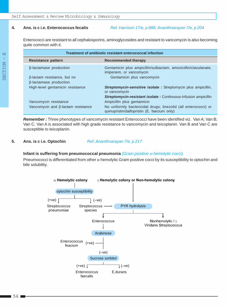
Self Assessment & Review Microbiology & Immunology
SECTION – B
5656
4. Ans. is c i.e. Enterococcus fecalis Ref. Harrison 17/e, p 888; Ananthnarayan 7/e, p 204
Enterococci are resistant to all cephalosporins, aminoglycosides and resistant to vancomycin is also becomingquite common with it.
Treatment of antibiotic resistant enterococcal infection
Resistance pattern Recommended therapy
β-lactamase production Gentamicin plus ampicillin/sulbactam, amoxicillin/clavulanate,imipenem, or vancomycin
β-lactam resistance, but no Gentamicin plus vancomycinβ-lactamase productionHigh-level gentamicin resistance Streptomycin-sensitive isolate : Streptomycin plus ampicillin,
or vancomycinStreptomycin-resistant isolate : Continuous-infusion ampicillin
Vancomycin resistance Ampicillin plus gentamicinVancomycin and β-lactam resistance No uniformly bactericidal drugs; linezolid (all enterococci) or
quinupristin/dalfopristin (E. faecium only)
Remember : Three phenotypes of vancomycin resistant Enterococci have been identified viz. Van A; Van B;Van C. Van A is associated with high grade resistance to vancomycin and teicoplanin. Van B and Van C aresusceptible to teicoplanin.
5. Ans. is c i.e. Optochin Ref. Ananthnarayan 7/e, p 217
Infant is suffering from pneumococcal pneumonia (Gram positive α-hemolytic cocci).Pneumococci is differentiated from other α hemolytic Gram positive cocci by its susceptibility to optochin andbile solubility.
PYR hydrolysis
Enterococcus
Enterococcus feacium
optochin susceptibility
(+ve) (–ve)Streptococcuspneumoniae
Streptococcusspecies
Arabinose
(+ve)
(–ve)Sucrose sorbitol
(+ve) (–ve)
Enterococcusfaecalis
E.durans
Streptococci
UNIT - I BACTERIOLOGY “Gram +ve Cocci”
57
6. Ans. is a i.e. Bacitracin sensitivity Ref. Ananthnarayan 7/e, p 205
“Sensitivity to bacitracin is employed as a convenient method for differentiating Str. pyogenes fromother hemolytic streptococci.”Pustule – A vesicle filled with leukocyte. MC cause is virus. MC bacterial cause is Strep. pyogenes.Remember : – Str. pyogenes is the etiologic agent in most of the streptococcal skin infection.
– Vancomycin is the drug of choice.– Optochin senstivity and bile solubility is used to differentiate pneumococci from Strep.
viridans group.
7. Ans. is a i.e. M-protein is responsible for production of mucoid coloniesRef. Ananthnarayan 7/e, p 205; Jawetz p 23/e, p 231
“Mucoid colonies are due to production of capsule of hyaluronic acid not due to M protein.”..... Harrison 17/e, p 882
Growth characteristic of Strep. pyogenes :• On blood agar virulent stains forms matt or mucoid colonies while avirulent form glossy colonies.• M, T, R are proteins found on outer part of cell wall and forms the basis of Griffith classification.
– M protein - Acts as virulence factor and antibody against it are protective.– T and R protein - No relation to virulence
• Streptococci (Except enterococci) are usually susceptible to penicillin so option “d” is not completelywrong. As option “a” is completely wrong, that would be the answer.
8. Ans. is c i.e. Enterococcus Ref. Ananthnarayan 7/e, p 204; Jawetz 24/e, p 243-244
Group D Streptococci
Enterococci Non enterococci
(E. faecalis, E, faecium) (Strep. bovis)• Able to grow in 6.5% NaCl • Not able• PYR positive • PYR negative• May shows α hemolysis • Non hemolytic• Penicilin resistant • Penicillin suscepitble
• Both enterococci and non enterococi group D sterptococci can grow in presence of bile and hydrolyzeesculin (bile esculin positive). ... Jawetz 24/e, p 243, 240
• Don’t confuse with Ananthnarayan 7/e, p 212 that non enterococci are inhibited with bile. If question comesgo with Jawetz.
9. Ans. is a i.e. Pyrogenic toxin Ref. Jawetz 24/e, p 238
“Streptococcal TSS is caused by pyrogenic exotoxin.”
Pyrogenic Exotoxin = Erythrogenic toxin = Dick = Scarlatinal Toxin• Produced by group A streptococci (type 1, 2, 3, 12 and 28)• There are 3 type of pyrogenic exotoxin : A, B, and C• A (MC cause of Streptococcal TSS) and C are coded by bacteriophage gene, while B is chromosomal
Self Assessment & Review Microbiology & Immunology
SECTION – B
5858
• Acts as superantigen• Associated with Streptococcal toxic shock syndrome and Scarlet fever• DOC for Streptococcal TSS is - Clindamycin ..... Harrison 17/e, p 886
10. Ans. is d i.e. Pneumococcus Ref. Ananthnarayan 7/e, p 216 - 217
“Pneumococci is optochin sensitive and forms green colonies on blood agar due to ααααα hemolysis.”
Characteristic features of Pneumococci :• Bile soluble, optochin sensitive• Inulin fermenter• Capsulated, shows quellung reaction• Colonies show central umbonation, appear as draughtsmann or carrom coin• MC causative agent of lobar Pneumonia, Adult meningitis, otitis media, sinusitis (orbital cellulitis is
complication of sinusitis).
11. Ans. is a i.e. Virulence is due to polysaccharide capsule Ref. Ananthnarayan 7/e, p 219
Virulence of Pneumococci depends on its capsule, toxin called pneumolysin and autolysin.
Virulence factors of Pneumococci
Capsule :
• Polysaccharide in nature• Protects against phagocytosis• Type 3 pneumococci has abundant capsular material and is more virulent• Antibody against capsule are type specific and protective.
Pneumolysin :
• Thial activated toxin, exterts variety of effects on ciliary cells and PMN ..... Harrison 16/e, p 807• Complement activating and cytotoxic properties• Immunogenic.
Autolysin :
• By lysing the bacteria and releasing bacterial components contributes to virulence.
IgA1 protease
• Cleaves IgA1 and hence decreases the function of this immunoglobulin.C-substance and peptidoglycans.
12. Ans. is d i.e. The virulence of pneumococci is dependent only on the production of the capsularpolysaccharide Ref. Ananthnarayan 7/e, p 219
Already explained, refer just above to find answer.

Streptococci
UNIT - I BACTERIOLOGY “Gram +ve Cocci”
59
13. Ans. is b i.e. Acute glomerulonephritis titre is low Ref. Ananthnarayan 7/e, p 210
Retrospective Diagnosis of Streptococcal infection• ASO (Anti Streptolysin ‘O’) titre :
– Estimation of antibody aganist streptolysin is a standard serological test for retrospective diagnosis ofstreptococcal infection.
– ASO titre > 200 are indicative of prior streptococcal infection.– Raised after throat infection only– Acute rheumatic fever :
- High level of ASO titre are usually found- Titres > 300 or 350 are taken as significant.
– Acute glomerulonephirits :- ASO titres are often low.
• Streptozyme test – Passive slide hemaglutination testBecomes positive after nearly all types of streptococcal infection whether of throat or skin.
• Anti DNA ase B and Antihyaluronidase – Useful for retrospective diagnosis of Streptococcal Pyodermaor for acute glomerulogephritis for which ASO titre is of much less value.
14. Ans. is a i.e. Puerperal infection Ref. Ananthnarayan 7/e, p 267
Anaerobic Cocci
Features Gram (+)ve Gram (–)ve
Organism – Peptostreptococci Veillonellae– Peptococcus
Inhabitation – Intestine, vagina and mouth Mouth, intestine and genital tract
Diseases – Puerperal sepsis No disease is identified till now.– Visceral abscess– UTI, wound infection– Gangrenous apendicitis
Treatment – Sensitive to penicillin, chloramphenicoland metronidazole
15. Ans. is c i.e. Group C carbohydrate antigen Ref. Ananthnarayan 7/e, p 203
Lancefield classificationClassification of β hemolytic streptococci into Group A to V (except I, J) on the basis of group specific Ccarbohydrate.
Griffith classificationSerological typing of Group A Streptococcus pyogenes on the basis of M proteins into types 1, 2, 3 etc.
Group C Carohydrate• Present in middle layer of cell wall.• This antigen is an integral part of cell wall.• Serological grouping of streptococci depend on C carbohydrate for which it has to be extracted from cell
wall.
Self Assessment & Review Microbiology & Immunology
SECTION – B
6060
• Method for extraction are :– Lancefield’s acid extraction method (organism are grown in Todd Hewitt broth)– Fuller’s method– Maxted’s method– Rantz and Randall’s method (Autoclaving).
16. Ans. is c i.e. Optochin Ref. Ananthnarayan 7/e, p 216 - 217
Already explained, please see answer no. 6
17. Ans. is c i.e. Enterococcus faecalis Ref. Harrison 17/e, p 888
• Enterococci are catalase negative and grow in chains and above all resistant to cephalosporins.• Enterococci is a frequent cause of nosocomial bacteremias and many of these enterococci are resistant to
vancomycin.• Enterococcal bacteremis is characterstically seen in ICU in patient taking cephalosporin as antibiotic.
Other options : • Staph. aureus and coagulase negative staph. are catalase positive.• Streptococci viridans are sensitive to vancomycin.
Remember : Whole streptococci group forms chains while Staphylococci group forms bunch similar tograpes.
18. Ans. is b i.e. Gram positive cocci in pairs, catalase negative and bile solubleRef. Ananthnarayan 7/e, p 204, 221; Jawetz 24/e, p 237
Bacteria which shows α or partial hemolysis includes :
Species Common disease caused
a. Strep. viridans includingS. sanguis SABES. mutans Dental caries
b. Pneumococci Lobar pneumonia, otitis mediac. S. anginosus Pyogenic infectionsd. Enterococcus including S.faecalis UTI, endocarditis
• As in quesion, sputum specimen is taken which is required in the diagnosis of respiratory tract infection(eg. pneumonia).
• So, it is clear that most probable answer is Pneumococci which is : diplococci; bile soluble; catalasenegative.
Remember : All streptococci is catalase negative while staphylococci is catalase positive :• Choice ‘a’ points towards S. viridans but it rarely cause pneumonia.• Choice ‘c’ points towards S. aureus which shows β hemolysis not α hemolysis.• Choice ‘d’ points towards Legionella. Though it cause pneumonia but it doesn’t shows
hemolysis.
Streptococci
UNIT - I BACTERIOLOGY “Gram +ve Cocci”
61
19. Ans. is a i.e. Enterococcus faecalis Ref. Harrison 17/e, p 888
See the following line.“Unlike streptococci, enterococci are not reliably killed by penicillin or ampicillin alone. Because invitro testing has shown evidence of synergistic killing by combination of penicillin or ampicillin withan amminoglycoside, combined therapy is recommended for enterococcal endocarditis and meningitis.”
• Enterococci are resistant to all cephalosporins
20. Ans. is c i.e. Streptococcus Pneumononiae Ref. Robbins 7/e, p 748
Remember : • MC - infection of pneumococci - Otitis media• Pneumococci is MC cause of Lobar Pneumonia, Acute sinusitis, Adult meningitis• MC cause of bronchopneumonia : Staphylococci• MC cause of atypical pneumonia : Mycoplasma pneumoniae.
21. Ans. is b i.e. Pike’s media Ref. Ananthnarayan 7/e, p 210
“Pike’s media is transport media for Streptococci.”
Diagnosis of Streptococcal infection
Acute infection Non-suppurative complication
• Gram’s staining • By detection of antibody through :• Culture – Streptozyme test
– Anti DNAase and anti hyaluronidase– ASO titre
Culture– Throat culture is diagnostic gold standard for pharyngitis.– Swab are either plated immmediately or sent to laboratory in Pike’s medium (used as transport media).– Specimen is plated on blood agar and incubated at 370C anaerobically or under 5-10% CO2.– Sheep blood agar is recommeded for primary isolation because it is inhibitory for Haemophilus.
Remember : • Stuart’s medium is transport media for Gonococci.• Cary - blair medium is transport media for V. cholera.
22. Ans. is b i.e. Pyrogenic extoxin Ref. Jawetz 24/e, p 238
Remember : • MC cause of Toxic shock syndrome - Staphylococci• MC cause of Streptococcal TSS - Pyrogenic exotoxin A = erythrogenic / Dick / Scarlatinal
toxin.• MC cause of Staphylococcal TSS - TSST=Pyrogenic exotoxin C = Enterotoxin type F.• DOC of TSS (both Streptococcal and staphylococal) is Clindamycin.
Self Assessment & Review Microbiology & Immunology
SECTION – B
6262
23. Ans. is a i.e. Streptococcus pyogenes Ref. Harrison 17/e, p 885
Cellulitis :– Diffuse spreading infection of skin (dermis and subcutaneous tissue) especially of lower leg.– Caused by Strep pyogenes (MC), Staph, Cl perfrigens, E.coli.– Major portal of entry for lower leg cellulitis is toe web tinea pedis with fissuring of skin.– Skin become peud orange in appearance; recurrent attack may sometimes affect lymphatic vessels producing
permanent swelling called as solid edema.– Streptococcus cellulitis tends to develop at sites where lymphatic drainage is disrupted.
24. Ans. is b i.e. Enterococcus faecalis Ref. Ananthnarayan 7/e, p 204
Growth in 6.5% NaCl is characteristic of Enterococci (E. faecalis, E. faecium, E. durans).
25. Ans. is a i.e. Streptococcus Viridans Ref. Harrison 17/e, p 789; Jawetz 23/e, p 237
It is case of Subacute bacterial endocarditis (SABE) as patient has Rheumatic heart disease (so damagedheart valves).
Remember : MC cause of SABE – S. viridansMC cause of Acute Bacterial endocarditis – S. aureus.MC cause of Prosthetic valve endocarditis – S. epidermidis.MC cause of Endocarditis in iv drug users – S. aureus
26. Ans. is a i.e. Post Splenectomy patient Ref. Harrison 17/e, p 866
Conditions predispose to pneumococcal infection
• Respiratory infection Inflammation • Defective complement function– Influenza, other viral respiratory infection • Defective clearance of pneumococcal bacterimia– Allergies – Congenital asplenia, hyposplenia– Cigarette smoking – Splenectomy– Chronic obstructive pulmonary disease – Sickle cell disease
• Anatomical disruption of meninges (dural tear) • Multifactorial conditions• HIV infection – Alcoholism• Defective antibody formation – Malnutrition
– Selective IgG subclass deficiency – Glucocorticoid treatment– Multiple myeloma – Cirrhosis of the liver– Chronic lymphocytic leukemia – Renal insufficiency– Lymphoma – Diabetes mellitus
– Anemia– Coronary artery disease
27. Ans. is a i.e. Lancefield classification Ref. Ananthnarayan 7/e, p 203 - 205
Already explained, Please refer ans. no. 15
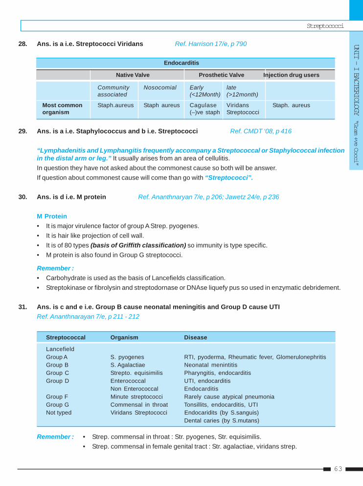
Streptococci
UNIT - I BACTERIOLOGY “Gram +ve Cocci”
63
28. Ans. is a i.e. Streptococci Viridans Ref. Harrison 17/e, p 790
Endocarditis
Native Valve Prosthetic Valve Injection drug users
Community Nosocomial Early lateassociated (<12Month) (>12month)
Most common Staph.aureus Staph aureus Cagulase Viridans Staph. aureusorganism (–)ve staph Streptococci
29. Ans. is a i.e. Staphylococcus and b i.e. Streptococci Ref. CMDT ‘08, p 416
“Lymphadenitis and Lymphangitis frequently accompany a Streptococcal or Staphylococcal infectionin the distal arm or leg.” It usually arises from an area of cellulitis.In question they have not asked about the commonest cause so both will be answer.If question about commonest cause will come than go with “Streptococci”.
30. Ans. is d i.e. M protein Ref. Ananthnaryan 7/e, p 206; Jawetz 24/e, p 236
M Protein• It is major virulence factor of group A Strep. pyogenes.• It is hair like projection of cell wall.• It is of 80 types (basis of Griffith classification) so immunity is type specific.• M protein is also found in Group G streptococci.
Remember :• Carbohydrate is used as the basis of Lancefields classification.• Streptokinase or fibrolysin and streptodornase or DNAse liquefy pus so used in enzymatic debridement.
31. Ans. is c and e i.e. Group B cause neonatal meningitis and Group D cause UTIRef. Ananthnarayan 7/e, p 211 - 212
Streptococcal Organism Disease
LancefieldGroup A S. pyogenes RTI, pyoderma, Rheumatic fever, GlomerulonephritisGroup B S. Agalactiae Neonatal menintitisGroup C Strepto. equisimilis Pharyngitis, endocarditisGroup D Enterococcal UTI, endocarditis
Non Enterococcal EndocarditisGroup F Minute streptococci Rarely cause atypical pneumoniaGroup G Commensal in throat Tonsillits, endocarditis, UTINot typed Viridans Streptococci Endocaridits (by S.sanguis)
Dental caries (by S.mutans)
Remember : • Strep. commensal in throat : Str. pyogenes, Str. equisimilis.• Strep. commensal in female genital tract : Str. agalactiae, viridans strep.
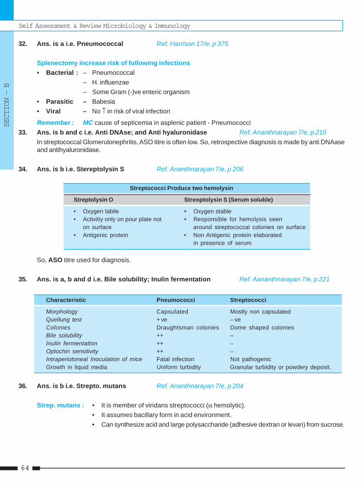
Self Assessment & Review Microbiology & Immunology
SECTION – B
6464
32. Ans. is a i.e. Pneumococcal Ref. Harrison 17/e, p 375
Splenectomy increase risk of following infections• Bacterial : – Pneumococcal
– H. influenzae– Some Gram (-)ve enteric organism
• Parasitic – Babesia• Viral – No ↑ in risk of viral infection
Remember : MC cause of septicemia in asplenic patient - Pneumococci33. Ans. is b and c i.e. Anti DNAse; and Anti hyaluronidase Ref. Ananthnarayan 7/e, p 210
In streptococcal Glomerulonephritis, ASO titre is often low. So, retrospective diagnosis is made by anti DNAaseand antihyaluronidase.
34. Ans. is b i.e. Stereptolysin S Ref. Ananthnarayan 7/e, p 206
Streptococci Produce two hemolysin
Streptolysin O Streoptolysin S (Serum soluble)
• Oxygen labile • Oxygen stable• Activitiy only on pour plate not • Responsible for hemolysis seen
on surface around streptococcal colonies on surface• Antigenic protein • Non Antigenic protein elaborated
in presence of serum
So, ASO titre used for diagnosis.
35. Ans. is a, b and d i.e. Bile solubility; Inulin fermentation Ref. Aananthnarayan 7/e, p 221
Characteristic Pneumococci Streptococci
Morphology Capsulated Mostly non capsulatedQuellung test + ve – veColonies Draughtsman colonies Dome shaped coloniesBile solubility ++ –Inulin fermentation ++ –Optochin sensitivity ++ –Intraperiotoneal Inoculation of mice Fatal infection Not pathogenicGrowth in liquid media Uniform turbidity Granular turbidity or powdery deposit.
36. Ans. is b i.e. Strepto. mutans Ref. Ananthnarayan 7/e, p 204
Strep. mutans : • It is member of viridans streptococci (α hemolytic).• It assumes bacillary form in acid environment.• Can synthesize acid and large polysaccharide (adhesive dextran or levan) from sucrose.
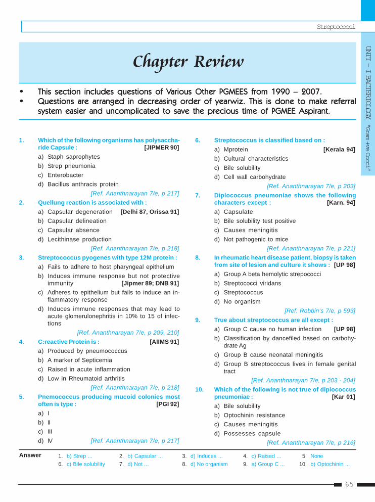
Streptococci
UNIT - I BACTERIOLOGY “Gram +ve Cocci”
65
1. b) Strep ... 2. b) Capsular ... 3. d) Induces ... 4. c) Raised ... 5. None6. c) Bile solubility 7. d) Not ... 8. d) No organism 9. a) Group C ... 10. b) Optochinin ...
1. Which of the following organisms has polysaccha-ride Capsule : [JIPMER 90]a) Staph saprophytesb) Strep pneumoniac) Enterobacterd) Bacillus anthracis protein
[Ref. Ananthnarayan 7/e, p 217]2. Quellung reaction is associated with :
a) Capsular degeneration [Delhi 87, Orissa 91]b) Capsular delineationc) Capsular absenced) Lecithinase production
[Ref. Ananthnarayan 7/e, p 218]3. Streptococcus pyogenes with type 12M protein :
a) Fails to adhere to host pharyngeal epitheliumb) Induces immune response but not protective
immunity [Jipmer 89; DNB 91]c) Adheres to epithelium but fails to induce an in-
flammatory responsed) Induces immune responses that may lead to
acute glomerulonephritis in 10% to 15 of infec-tions
[Ref. Ananthnarayan 7/e, p 209, 210]4. C:reactive Protein is : [AIIMS 91]
a) Produced by pneumococcusb) A marker of Septicemiac) Raised in acute inflammationd) Low in Rheumatoid arthritis
[Ref. Ananthnarayan 7/e, p 218]5. Pnemococcus producing mucoid colonies most
often is type : [PGI 92]a) Ib) IIc) IIId) IV [Ref. Ananthnarayan 7/e, p 217]
Answer
6. Streptococcus is classified based on :a) Mprotein [Kerala 94]b) Cultural characteristicsc) Bile solubilityd) Cell wall carbohydrate
[Ref. Ananthnarayan 7/e, p 203]7. Diplococcus pneumoniae shows the following
characters except : [Karn. 94]a) Capsulateb) Bile solubility test positivec) Causes meningitisd) Not pathogenic to mice
[Ref. Ananthnarayan 7/e, p 221]8. In rheumatic heart disease patient, biopsy is taken
from site of lesion and culture it shows : [UP 98]a) Group A beta hemolytic strepococcib) Streptococci viridansc) Streptococcusd) No organism
[Ref. Robbin’s 7/e, p 593]9. True about streptococcus are all except :
a) Group C cause no human infection [UP 98]b) Classification by dancefiled based on carbohy-
drate Agc) Group B cause neonatal meningitisd) Group B streptococcus lives in female genital
tract[Ref. Ananthnarayan 7/e, p 203 - 204]
10. Which of the following is not true of diplococcuspneumoniae : [Kar 01]a) Bile solubilityb) Optochinin resistancec) Causes meningitisd) Possesses capsule
[Ref. Ananthnarayan 7/e, p 216]
Chapter Review••••• This section includes questions of VThis section includes questions of VThis section includes questions of VThis section includes questions of VThis section includes questions of Various Other PGMEES from 1990 – 2007.arious Other PGMEES from 1990 – 2007.arious Other PGMEES from 1990 – 2007.arious Other PGMEES from 1990 – 2007.arious Other PGMEES from 1990 – 2007.••••• Questions are arQuestions are arQuestions are arQuestions are arQuestions are ar ranged in decreasing order of yearranged in decreasing order of yearranged in decreasing order of yearranged in decreasing order of yearranged in decreasing order of yearwiz. This is done to makwiz. This is done to makwiz. This is done to makwiz. This is done to makwiz. This is done to make refere refere refere refere referralralralralral
system easier and uncomplicated to save the precious time of PGMEE Aspirant.system easier and uncomplicated to save the precious time of PGMEE Aspirant.system easier and uncomplicated to save the precious time of PGMEE Aspirant.system easier and uncomplicated to save the precious time of PGMEE Aspirant.system easier and uncomplicated to save the precious time of PGMEE Aspirant.
Self Assessment & Review Microbiology & Immunology
SECTION – B
6666
11. Which of the following is not true of Diplococcuspneumoniae : [Kerala 01]a) Bile solubilityb) Optochin resistancec) Causes Meningitis’d) Possesses capsule
[Ref. Ananthnarayan 7/e, p 221]12. Streptococcal toxic shock syndrome is due to lib-
eration of : [Kerala 01]a) TSST-1b) Enterotoxinc) Pyrogenic exotoxind) Endotoxin
[Ref. Ananthnarayan 7/e, p 208]13. Most common cause of community acquired pneu-
monia: [UP 01]a) Strep pneumoniaeb) Kleb pneumoniaec) Vibrio cholerad) H. influenzae [Ref. Harrison 16/e, p 871]
14. True statement about streptococcus faecalis :a) Growns in 6.5% NaCl solution [Kolkata 02]b) Are lactose fermenterc) Easily destroyed at 600C for 300 minutesd) Classified on the basis of teichoic acid of cell
wall[Ref. Ananthnarayan 7/e, p 212]
15. Neonatal meningitis acquired through birth canalis due to : [TN 02]a) Streptococcus agalactiaeb) S.equisimilusc) S.pyogenesd) Pnemococci during passage
[Ref. Ananthnarayan 7/e, p 204]16. Dental caries is due to : [JIPMER 02; MP 05]
a) Streptococcus sanguisb) S.mitisc) S.mutansd) S.salivarius
[Ref. Ananthnarayan 7/e, p 204, 215]17. Quelling phenomenon is seen in: [Kolkata 03]
a) Pneumococcusb) Streptococcousc) Staphylococcusd) Hemophilus [Ref. Ananthnarayan 7/e, p 210]
18. Classification of pathogenic streptococci intogroup A, B, C, D and G is based on : [Kar 03]a) Color of colonies on blood agrb) Antigenicity of cell wall carbohydratec) Presence or absence of capsuled) Presence or absence of M protein
[Ref. Ananthnarayan 7/e, p 203]19. Impetigo contagiosa is caused by :
a) Group A beta hemolytic streptococcib) Staphylococcus [Jharkhand 03]c) H. Influenzaed) Psudomonas
[Ref. Harrison 17/e, p 799]20. Toxic shock syndrome is caused by :
a) Staphylococcus [Jharkhand 03]b) Psudomonasc) Streptococcid) bacteroids
[Ref. Ananthnarayan 7/e, p 196]21. Group-A streptococcus causes causes all, except
a) Scarlet fever [Bihar 04]b) Erysipelasc) Impetigod) Epidermolysis bullae
[Ref. Ananthnarayan 7/e, p 208]22. Quellung reaction is due to : [Bihar 04]
a) Capsular swellingb) Mitrochondrial swellingc) RBC swellingd) Ribosomal swelling
[Ref. Ananthnarayan 7/e, p 218]23. Virulence or pathogenicity of pneumococcus de-
pends on : [Kolkata 04]a) Capsular polysaccharidesb) Cell wall antigenc) Flagellar antigend) Fimbrial antigen
[Ref. Ananthnaryana 7/e, p 219]24. Griffith denonstrated biotransformation with :
a) H. influenzae [UP 03; 04]b) E. colic) Proteusd) Pneumococcus
[Ref. Ananthnarayan 7/e, p 55]
11. b) Optochin ... 12. c) Pyrogenic ... 13. a) Strep ... 14. a. Growns ... 15. a) Streptococc ...16. c) S.mutans 17. a) Pneumococcus 18. b) Antigenicity ... 19. a) Group ... 20. a) Staphyloco...21. d) Epidermolysis ... 22. a) Capsular ... 23. a) Capsular ... 24. d) Pneumo...
Answer
Streptococci
UNIT - I BACTERIOLOGY “Gram +ve Cocci”
67
25. Causative agent of SABE should be cultured in :a) Nutrient agar [UP 03]b) Blood agarc) L. J mediumd) Tellurite broth
[Ref. Ananthnarayan 7/e, p 204]26. An infant with neonatal meningitis has a positive
CAMP test, the causative agent is : [UP 03]a) Staphylococcusb) E.colic) Strept agalactiaed) Hemophiluis
[Ref. Ananthnarayan 7/e, p 204]27. Streptococcus pneumoniae true is : [SGPGI 05]
a) Vaccine is made from capsular polysaccharideb) Vaccine is routinely given to Indian childrenc) Catalase and oxidase positived) Bile insoluble and optochin sensitive
[Ref. Ananthnarayan 7/e, p 221]28. Draughtsman colonies are seen in : [UP 05]
a) Staphylococcusb) Salmonella
25. b) Blood ... 26. c) Strept ... 27. d) Bile insoluble ...28. c) Pneumococcus 29. c) Strep ... 30. c) Pneumococci31. a) Burkholderia ...
Answer
c) Pneumococcusd) Corynebacterium
[Ref. Ananthnarayan 7/e, p 217]29. Streptococcus causing Rheumatic heart is :
a) Streptococcus milleri [MP 05]b) Streptococcus mutansc) Streptococcus pyogensd Streptococcus equimilis
[Ref. Ananthnarayan 7/e, p 210]30. Which of the following microorganism is the most
common cause of lobar pneumonia : [MP 06]a) Klebsiella pneumoniaeb) Streptococcic) Pneumococcid) Staphylococci
[Ref. Ananthnarayan 7/e, p 216]31. Glanders disease is due to : [Bihar 06]
a) Burkholderia mallaeb) Diphtheriac) Mumpsd) Burkholderia psudomallae
[Ref. Ananthnarayan 7/e, p 321]
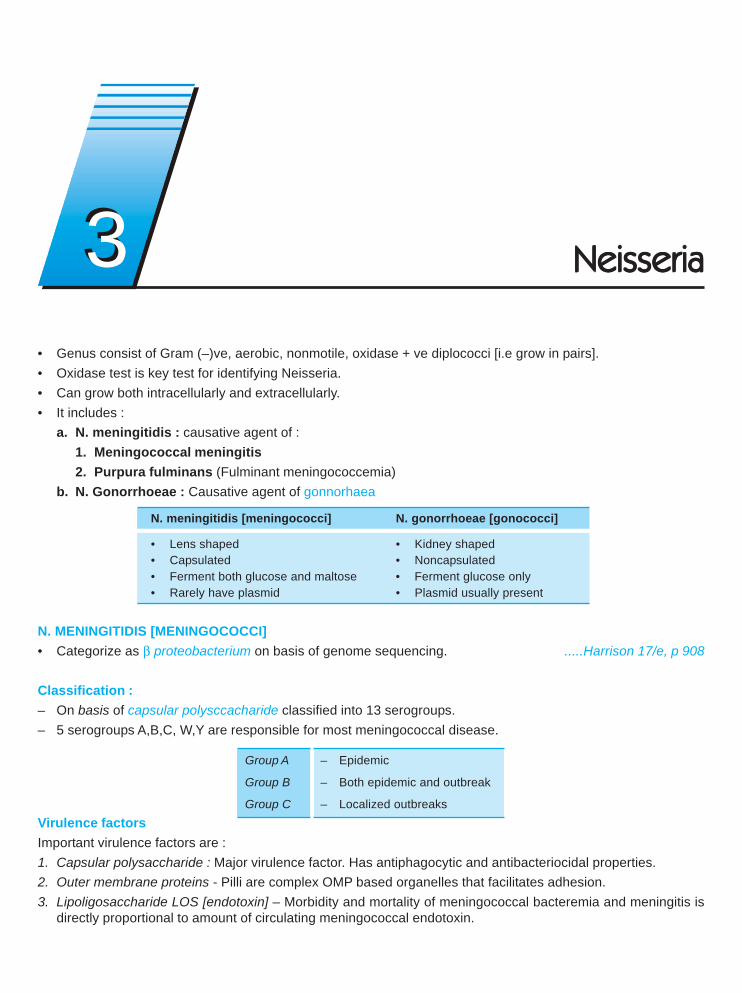
• Genus consist of Gram (–)ve, aerobic, nonmotile, oxidase + ve diplococci [i.e grow in pairs].• Oxidase test is key test for identifying Neisseria.• Can grow both intracellularly and extracellularly.• It includes :
a. N. meningitidis : causative agent of :1. Meningococcal meningitis2. Purpura fulminans (Fulminant meningococcemia)
b. N. Gonorrhoeae : Causative agent of gonnorhaea
N. meningitidis [meningococci] N. gonorrhoeae [gonococci]
• Lens shaped • Kidney shaped• Capsulated • Noncapsulated• Ferment both glucose and maltose • Ferment glucose only• Rarely have plasmid • Plasmid usually present
N. MENINGITIDIS [MENINGOCOCCI]• Categorize as β proteobacterium on basis of genome sequencing. .....Harrison 17/e, p 908
Classification :– On basis of capsular polysccacharide classified into 13 serogroups.– 5 serogroups A,B,C, W,Y are responsible for most meningococcal disease.
Group A – Epidemic
Group B – Both epidemic and outbreak
Group C – Localized outbreaksVirulence factorsImportant virulence factors are :1. Capsular polysaccharide : Major virulence factor. Has antiphagocytic and antibacteriocidal properties.2. Outer membrane proteins - Pilli are complex OMP based organelles that facilitates adhesion.3. Lipoligosaccharide LOS [endotoxin] – Morbidity and mortality of meningococcal bacteremia and meningitis is
directly proportional to amount of circulating meningococcal endotoxin.
NeisseriaNeisseriaNeisseriaNeisseriaNeisseria33
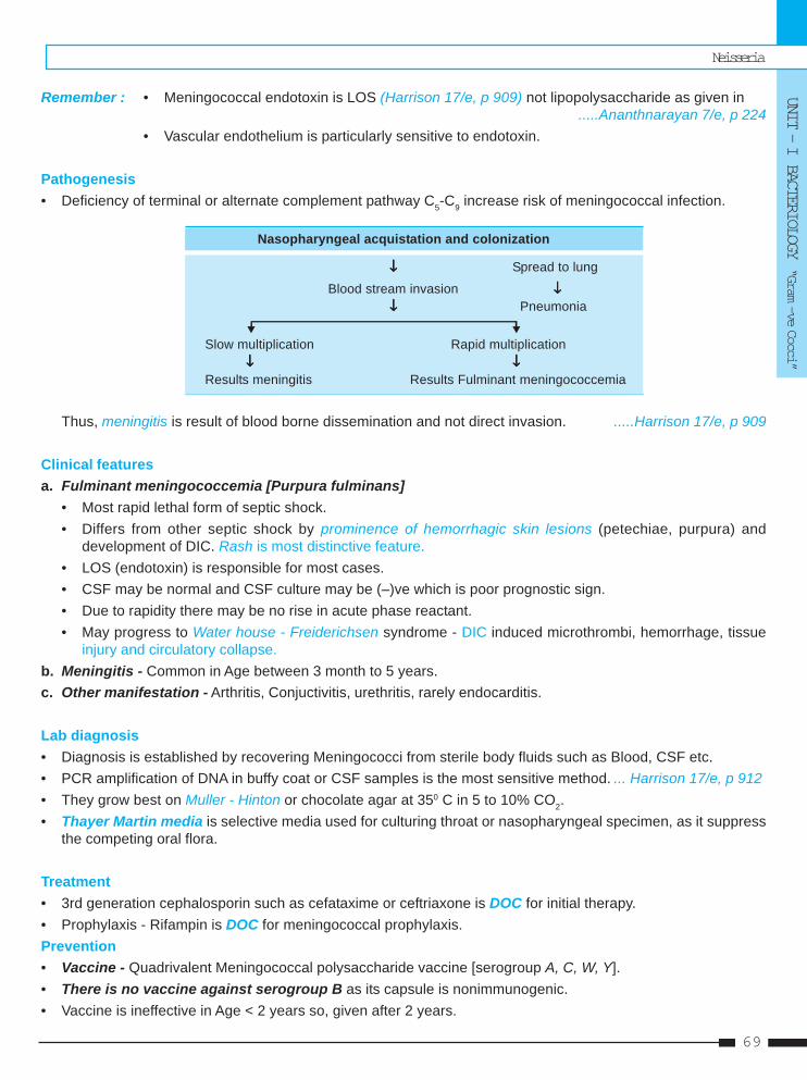
Neisseria
UNIT - I BACTERIOLOGY “Gram —ve Cocci”
69
Remember : • Meningococcal endotoxin is LOS (Harrison 17/e, p 909) not lipopolysaccharide as given in.....Ananthnarayan 7/e, p 224
• Vascular endothelium is particularly sensitive to endotoxin.
Pathogenesis• Deficiency of terminal or alternate complement pathway C5-C9 increase risk of meningococcal infection.
Nasopharyngeal acquistation and colonization
↓↓↓↓↓ Spread to lung
Blood stream invasion ↓↓↓↓↓↓↓↓↓↓ Pneumonia
Slow multiplication Rapid multiplication↓↓↓↓↓ ↓↓↓↓↓
Results meningitis Results Fulminant meningococcemia
Thus, meningitis is result of blood borne dissemination and not direct invasion. .....Harrison 17/e, p 909
Clinical featuresa. Fulminant meningococcemia [Purpura fulminans]
• Most rapid lethal form of septic shock.• Differs from other septic shock by prominence of hemorrhagic skin lesions (petechiae, purpura) and
development of DIC. Rash is most distinctive feature.• LOS (endotoxin) is responsible for most cases.• CSF may be normal and CSF culture may be (–)ve which is poor prognostic sign.• Due to rapidity there may be no rise in acute phase reactant.• May progress to Water house - Freiderichsen syndrome - DIC induced microthrombi, hemorrhage, tissue
injury and circulatory collapse.b. Meningitis - Common in Age between 3 month to 5 years.c. Other manifestation - Arthritis, Conjuctivitis, urethritis, rarely endocarditis.
Lab diagnosis• Diagnosis is established by recovering Meningococci from sterile body fluids such as Blood, CSF etc.• PCR amplification of DNA in buffy coat or CSF samples is the most sensitive method. ... Harrison 17/e, p 912• They grow best on Muller - Hinton or chocolate agar at 350 C in 5 to 10% CO2.• Thayer Martin media is selective media used for culturing throat or nasopharyngeal specimen, as it suppress
the competing oral flora.
Treatment• 3rd generation cephalosporin such as cefataxime or ceftriaxone is DOC for initial therapy.• Prophylaxis - Rifampin is DOC for meningococcal prophylaxis.Prevention• Vaccine - Quadrivalent Meningococcal polysaccharide vaccine [serogroup A, C, W, Y].• There is no vaccine against serogroup B as its capsule is nonimmunogenic.• Vaccine is ineffective in Age < 2 years so, given after 2 years.
Self Assessment & Review Microbiology & Immunology
SECTION – B
7070
Indication Contra indication
– Late complement or properdin deficiency – Pregnancy– Asplenia or splenectomy patient– Military Persons– Epidemic areas– Pilgrims on Haj– Individual travelling to subsaharan Africa
N. GONNORHOEAE [GONOCOCCI]• MC cause of PID worldwide [MC cause of PID in India is TB].• MC cause of septic arthritis in adult [MC joint knee].• Cause most severe type of ophthalmia neonatorum.
Culture : • More difficult to grow than meningococci • Essential to provide 5-10 % CO2
• Four types of colonies recognised - T1 to T4 • Type 1 and 2 = P+ and P++ respectively.• Type 3 and 4 = P– and are avirulent They are piliated and virulent.
Pathogenic factorsa. Outer-membrane protein :
• Pilli – Piliated strains adhere better to cells and are more virulent.• Opacity associated protein ( Protein II) – Important for adhesion.• Porin (Protein I, III) – Most abundant gonococcal surface protein.• H.8 Lipoprotein – Excellent target for antibody based diagnostic testing.• Transferrin and lactoferrin binding protein.• IgA1 protease (also produced by meningococci).Remember : IgA - 1 protease is also produced by S. pneumoniae, H. influenza; some streptococci.
....Jawetz 24/e, p 157b. Lipooligosaccharide = Endotoxin
• Resemble human glycosphingolipid, contributes to the local cytotoxic effects.c. Host factors, defeciency of terminal compliment components.
Clinical features• Mode of infection is almost exclusively veneral except ophthalmia neonatorum.• Terminal complement component [C5 - C9] deficiency predispose to gonococcal infection.• Higher incidence of gonorrhea occur in blood group B.• Gonococcal infection in males – Acute urethritis is MC clinical manifestation of gonorrhoea in males.• Gonococcal infection in females – Cervicitis is MC manifestation. Adult Vagina is resistant to gonococcal infection.• Gonorrhoea in pregnant woman – Salpingitis and PID can occur during 1st trim and can cause abortion.
In 2nd and 3rd trim, relative impermeability of cervical mucous prevent ascending infection.• Gonococcal infection in neonates – MC is opthalmia-neonatorum while septic arthritis is MC manifestation of
systemic infection.• In children Vulvovaginitis is MC gonococcal infection.
Neisseria
UNIT - I BACTERIOLOGY “Gram —ve Cocci”
71
Diagnosis• Rapid diagnosis by Gram’s staining of urethral exudates.• Part of sample should be inoculated on Thayer Martin Media. Detection of Gram –ve diplococci or monococci
is usually specific.• It is important to process all samples immediately because gonoccoi do not tolerate drying.• If processing is to occur within 6h, transport of specimens may be facilitated by the use of nonnutritive swab
transport systems such as Stuart or Amies medium.• For longer holding periods culture media with self contained CO2 generating systems (such as the JEM BEC or
Gono-Pak system may be used).
Treatment :• 3rd generation cephalosporins cefixime and ceftriaxone are DOC. [Penicillin is DOC]
..... Park 18/e, p 138; 19/e, p 141
Prevention and Control• Gonorrhea is transmitted from males to females more frequently than in opposite direction.• Condoms provide effective protection against transmission.
Self Assessment & Review Microbiology & Immunology
SECTION – B
7272
1. The following bacteria are most often associatedwith acute neonatal meningitis except : [AI 05]a) Escherichia colib) Streptococcus agalactiaec) Neisseria meningitidisd) Listeria monocytogenes
2. The following statements about meningococcalmeningitis are true except : [AI 03]a) The source of infection is mainly clinical casesb) The disease is more common in dry and cold
months of the yearc) Chemoprophylaxis of close contacts of cases is
recommendedd) The vaccines is not affective in children below 2
years of age3. Xavier and Yogender stay in the same hostel of
same university Xavier develops infection due togroup B meningococcus. After few daysYogender develops infection due to Group - Cmeningococcus. All the following are true state-ment except : [AI 02]a) Educate students about meningococcal transmis-
sion and take preventive measuresb) Chemoprophylaxis to all against both group B and
group Cc) Vaccine prophylaxis of contacts of Xavierd) Vaccine prophylaxis of contacts of Yogendra
4. Differentiation of N. gonorrhea and N. menin-gitidis can be done by : [AI 96]a) Glucose fermentationb) VP Reactionc) Maltose fermentationd) Indole test
5. The best site to obtain a swab in asymptomaticgonorrhea is : [AI 95]a) Endocervixb) Urethrac) Lateral vaginal walld) Posterior fornix
6. Conjugate vaccine are available for the preven-tion of invasive disease caused by all of the fol-lowing bacteria except : [AIIMS 04]a) H influenzaeb) Strep pneumoniae
c) Neisseria meningitidis (Group - C)d) Neisseria meningitidis (Group - B)
7. The virulence factor of Neisseria gonorrhoeaeincludes all of the following except : [AIIMS 03]a) Outer membrane proteinsb) IgA Proteasec) M Proteind) Pilli
8. All are true about Neisseria gonorrhea except :a) Gram positive cocci [AIIMS 01]b) Causes stricture urethrac) Involves seminal vesicles and spreads to epid-
idymisd) Drug of choice is ceftriaxone
9. Which of the following is true regarding Neisseriameningitidis infection : [AIIMS 00]a) It is the most common cause of meningitis in chil-
drenb) All strains are uniformly sensitive to sulfonamidesc) In children less than 2 years the vaccine is not
effectived) In India sero type B is most common cause
10. Least susceptible to gonococcal infection is :a) Anterior urethra [AIIMS 95]b) Testisc) Prostated) Epididymis
11. Which is the true statement regarding gonococ-cal urethritis : [PGI 06, 00]a) Symptoms are more severe in females than in
malesb) Rectum and prostate are resistant to gonococcic) Most patients present with symptoms of dysuriad) Single dose of ciprofloxacin is effective in treat-
mente) Commonly leads to arthritis
12. CSF in meningococcal meningitis shows :a) Gram (+) Diplococci, in pus cells [PGI 98]b) Gram (-) Diplococci in pus cellsc) Gram (-) bacillid) Gram (+) bacilli
QUESTIONS
Answer 1. c) Neisseria ... 2. a) The source ... 3. c) Vaccine ... 4. c) Maltose... 5. a) Endocervix6. d) Neisseria ... 7. c) M Protein 8. a) Gram ... 9. a and c 10. b) Testis
11. c and d 12. b) Gram (-) ...
Neisseria
UNIT - I BACTERIOLOGY “Gram —ve Cocci”
73
1. Ans. is c i.e. N. meningitidis Ref. Forfar & Arneils - Text book of Paedia, p 319, 1338
Causes of Meningitis :• Neonatal (< 1 month)
1. Bacterial causes :Organism : E. coli > Gp B streptococci > other Gram - ve bacilli > L. monocytogenesFrequency : 34% 30% 8% 6%Other bacterias are : Staph, Other Streptococci, Pneumococcus, Pseudomonas, Haemophilus,Meningococcus.
2. Viral and protozoal infections : TORCH, Varicella zoster and HIV.
3. Spirochetal and fungal infections
• 1 month - 11 month : N-meningitidis > Strep pneumoniae > H. influenza• 1 year - 20 years : N. meningitidis > Strep pneumoniae > H. influenza• More than 20 years : Strep pneumoniae (MC) ..... Harrison 17/e, p 2621
2. Ans. is a i.e. The source of infection is mainly clinical cases Ref. Park 18/e, p 138; 19/e, p 141
“Source of infection are carriers not cases.”
Meningococcal meningitis or Cerebrospinal fever.• It is caused by N. meningitides, a gram negative diplococci.• Fatality of typical untreated cases is about 80%.
Agent : Group A and C and to lesser extent Group B cause major epidemics.
Source : Carriers are the most important source of infection.Clinical cases present only a negligible source of infection.
Environmental Factors : Seasonal variation is well established. Outbreaks occur more frequently in the dryand cold months.
Mode of Transmission : Mainly by droplet infection.
Incubation Period : Usually 3 - 4 days
Diagnosis : Recovering Gram (–)ve diplococci from sterile body fluids such as CSF, blood
Treatment : Antibiotics save the lives of 95% of patients provided that it is started during the first 2 days ofillness.Cases : Penicillin is drug of choice. [3rd generation cephalosporins are DOC. ..... Harrison 17/e, p 912-913Carriers : Rifampicin
ProphylaxisChemoprophylaxis : Rifampicin (the DOC unless the organism is known to be sensitive to sulfadiazine).It is suggested for close contacts. Mass chemoprophylaxis is restricted to closed and medically supervisedcommunities.
EXPLANATIONS AND REFERENCES WITH ILLUSTRATIVE ANSWERS
Self Assessment & Review Microbiology & Immunology
SECTION – B
7474
Immunization : – Polysaccharide vaccines from Group A, C, Y and W 135– No vaccine is available for group B– Immunity last for 3 years– Not recommended for infants and children under 2 years– Contraindicated in pregnant women.
3. Ans. is c i.e. Vaccine prophylaxis of contacts of Xavier Ref. Park 19/e, p 142
For group B meningococcal infection no vaccine is available.
4. Ans. is c i.e. Maltose fermentation Ref. Ananthnarayan 7/e, p 226
N. meningitidis [meningoccci] N. gonorrhoeae [gonococci]
• Lens shaped • Kidney shaped• Capsulated • Noncapsulated• Ferment both glucose and maltose • Ferment glucose only
Remember : Indole and VP reaction is done to differentiate genera of Enterobacteriaceae.
5. Ans. is a i.e. Endocervix Ref. Harrison 17/e, p 919; Ananthnarayan 7/e, p 228
DIAGNOSIS OF GONOCOCCAL INFECTION
SPECIMEN
In Acute Gonorrhea In chronic gonorrhea
• Endocervical culture - 80 - 90% sensitivity • Morning drop secretion• Urethral discharge - 50% sensitivity • Exudate after prostatic massage• High vaginal swab are not satisfactory • Centrifuged deposits of urine when no• Rectal wall swab - if there is history of rectal sex urethral discharge
Microscopy : Fluorescent antibody technique
Transport Media : Stuart’s or Amies media processing is to occur within.
For longer holding period culture media with self CO2 generating system (such asJEMBEC or Gono- pak systems) may be used.
Culture Media : Acute → Chocolate agar or Muller - Hinton agar.Chronic → Selective media like Thayer Martin medium.
Serology : Done in chronic case or with metastatic lesion Eg CFT, Precipitation, passiveagglutination, Immunofluorescence, RIA.
Chemiluminescent DNA Probe : in high risk patient undergoing screening for STD's.Blood Culture (Eg. of synovial fluid) : in suspected cases of disseminated infection.• Preferred method for diagnosis of gonococcal infection in children is a standardized culture, from urethera
and cervix. However cervical specimen are not recommended in prepubertial girls unless necessary.
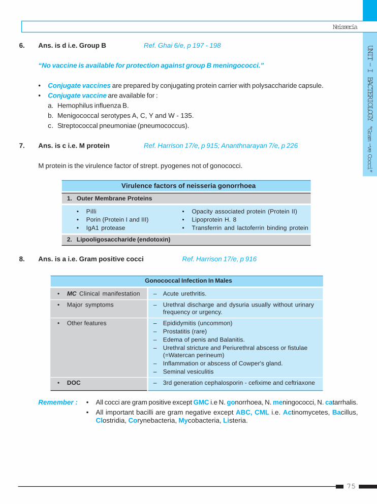
Neisseria
UNIT - I BACTERIOLOGY “Gram —ve Cocci”
75
6. Ans. is d i.e. Group B Ref. Ghai 6/e, p 197 - 198
“No vaccine is available for protection against group B meningococci.”
• Conjugate vaccines are prepared by conjugating protein carrier with polysaccharide capsule.• Conjugate vaccine are available for :
a. Hemophilus influenza B.b. Menigococcal serotypes A, C, Y and W - 135.c. Streptococcal pneumoniae (pneumococcus).
7. Ans. is c i.e. M protein Ref. Harrison 17/e, p 915; Ananthnarayan 7/e, p 226
M protein is the virulence factor of strept. pyogenes not of gonococci.
Virulence factors of neisseria gonorrhoea
1. Outer Membrane Proteins
• Pilli • Opacity associated protein (Protein II)• Porin (Protein I and III) • Lipoprotein H. 8• IgA1 protease • Transferrin and lactoferrin binding protein
2. Lipooligosaccharide (endotoxin)
8. Ans. is a i.e. Gram positive cocci Ref. Harrison 17/e, p 916
Gonococcal Infection In Males
• MC Clinical manifestation – Acute urethritis.
• Major symptoms – Urethral discharge and dysuria usually without urinaryfrequency or urgency.
• Other features – Epididymitis (uncommon)– Prostatitis (rare)– Edema of penis and Balanitis.– Urethral stricture and Periurethral abscess or fistulae
(=Watercan perineum)– Inflammation or abscess of Cowper's gland.– Seminal vesiculitis
• DOC – 3rd generation cephalosporin - cefixime and ceftriaxone
Remember : • All cocci are gram positive except GMC i.e N. gonorrhoea, N. meningococci, N. catarrhalis.• All important bacilli are gram negative except ABC, CML i.e. Actinomycetes, Bacillus,
Clostridia, Corynebacteria, Mycobacteria, Listeria.
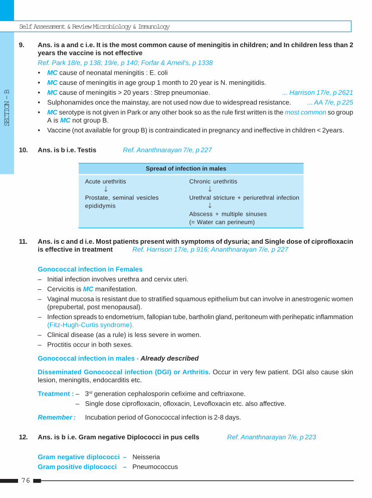
Self Assessment & Review Microbiology & Immunology
SECTION – B
7676
9. Ans. is a and c i.e. It is the most common cause of meningitis in children; and In children less than 2years the vaccine is not effectiveRef. Park 18/e, p 138; 19/e, p 140; Forfar & Arneil's, p 1338• MC cause of neonatal meningitis : E. coli• MC cause of meningitis in age group 1 month to 20 year is N. meningitidis.• MC cause of meningitis > 20 years : Strep pneumoniae. ... Harrison 17/e, p 2621• Sulphonamides once the mainstay, are not used now due to widespread resistance. ... AA 7/e, p 225• MC serotype is not given in Park or any other book so as the rule first written is the most common so group
A is MC not group B.• Vaccine (not available for group B) is contraindicated in pregnancy and ineffective in children < 2years.
10. Ans. is b i.e. Testis Ref. Ananthnarayan 7/e, p 227
Spread of infection in males
Acute urethritis Chronic urethritis↓ ↓
Prostate, seminal vesicles Urethral stricture + periurethral infectionepididymis ↓
Abscess + multiple sinuses(= Water can perineum)
11. Ans. is c and d i.e. Most patients present with symptoms of dysuria; and Single dose of ciprofloxacinis effective in treatment Ref. Harrison 17/e, p 916; Ananthnarayan 7/e, p 227
Gonococcal infection in Females– Initial infection involves urethra and cervix uteri.– Cervicitis is MC manifestation.– Vaginal mucosa is resistant due to stratified squamous epithelium but can involve in anestrogenic women
(prepubertal, post menopausal).– Infection spreads to endometrium, fallopian tube, bartholin gland, peritoneum with perihepatic inflammation
(Fitz-Hugh-Curtis syndrome).– Clinical disease (as a rule) is less severe in women.– Proctitis occur in both sexes.
Gonococcal infection in males - Already described
Disseminated Gonococcal infection (DGI) or Arthritis. Occur in very few patient. DGI also cause skinlesion, meningitis, endocarditis etc.
Treatment : – 3rd generation cephalosporin cefixime and ceftriaxone.– Single dose ciprofloxacin, ofloxacin, Levofloxacin etc. also affective.
Remember : Incubation period of Gonococcal infection is 2-8 days.
12. Ans. is b i.e. Gram negative Diplococci in pus cells Ref. Ananthnarayan 7/e, p 223
Gram negative diplococci – NeisseriaGram positive diplococci – Pneumococcus
Neisseria
UNIT - I BACTERIOLOGY “Gram —ve Cocci”
77
1. b) Maltose ... 2. b) Meningitis 3. None 4. None 5. b) Endotoxin6. c) Testis 7. b) Deficiency ... 8. a) Glucose 9. d) All of the ... 10. b) Possess ...
1. Differentiation of neisseria gonorrhoea andneisseria meningitidis is by : [AI 90]a) Glucose fermentationb) Maltose Fermentationc) V.-P. reactiond) Indol test
[Ref. Ananthnarayan 7/e, p 226]2. Which is not a metastatic complication of gono-
cocci : [JIPMER 91]a) Endocarditisb) Meningitisc) Nephritisd) Arthritis
[Ref. Ananthnarayan 7/e, p 227]3. Incubation period of gonorrhoea is :
a) Less than 24 hrs [JIPMER 92]b) 1 to 2 daysc) 2 to 15 daysd) 12 to 25 days
[Ref. Ananthnarayan 7/e, p 226]4. Gonococci in gram stained smears are seen in-
side the : [Kerala 94]a) Lymphocytesb) Neutrophilsc) Mast cellsd) Macrophages
[Ref. Ananthnarayan 7/e, p 225]5. Skin lesion in meningococcal meningitis is due to:
a) Exotoxin [Kerala 94]b) Endotoxinc) Allergic reactiond) Direct vascular Damage
[Ref. Ananthnarayan 7/e, p 224]
Answer
6. Which f the following is most resistance to gono-coccal infection : [Kerala 94]a) Prostateb) Epididymisc) Testisd) Urethra
[Ref. Robbins 7/e, p 1039]7. Neisseria infection are associated with :
a) Deficiency of early complements [CUPGEE 95]b) Deficiency of late complementsc) There is not such associationd) Any deficiency can be associated
[Ref. Ananthnarayan 7/e, p 224]8. Gonococcus ferments : [AP 97]
a) Glucoseb) Maltosec) Sucrosed) Fructose
[Ref. Ananthnarayan 7/e, p 226]9. The diagnosis of gonorrohea is established by :
a) Comliment fixation tests [Orissa 98]b) Pili agglutination testsc) Haem agglutination testsd) All of the above tests
[Ref. Ananthnarayan 7/e, p 228]10. Meningococci differ from gonococci in that they :
a) Are intra-cellular [ICS 98]b) Possess a capsulec) Causes fermentation of glucosed) Are oxidase positive
[Ref. Ananthnarayan 7/e, p 222]
Chapter Review••••• This section includes questions of VThis section includes questions of VThis section includes questions of VThis section includes questions of VThis section includes questions of Various Other PGMEES from 1990 – 2007.arious Other PGMEES from 1990 – 2007.arious Other PGMEES from 1990 – 2007.arious Other PGMEES from 1990 – 2007.arious Other PGMEES from 1990 – 2007.••••• Questions are arQuestions are arQuestions are arQuestions are arQuestions are ar ranged in decreasing order of yearranged in decreasing order of yearranged in decreasing order of yearranged in decreasing order of yearranged in decreasing order of yearwiz. This is done to makwiz. This is done to makwiz. This is done to makwiz. This is done to makwiz. This is done to make refere refere refere refere referralralralralral
system easier and uncomplicated to save the precious time of PGMEE Aspirant.system easier and uncomplicated to save the precious time of PGMEE Aspirant.system easier and uncomplicated to save the precious time of PGMEE Aspirant.system easier and uncomplicated to save the precious time of PGMEE Aspirant.system easier and uncomplicated to save the precious time of PGMEE Aspirant.
Self Assessment & Review Microbiology & Immunology
SECTION – B
7878
11. The following are gram-negative cocci except :a) Pneumococci [Kar 01]b) Meningococcic) Gonococcid) Veillonella
[Ref. Ananthnarayan 7/e, p 216]12. The vaccine against N:meningitides contains :
a) Whole bacteria [ICS 00]b) Capsular polysaccharidec) Somatic ‘0’ antigend) Lipo polysaccharide protein complex
[Ref. Ananthnarayan 7/e, p 225]
13. A pus culture on chocolate agar shows gram nega-tive cocci most likely organism is : [UP 04]a) Haemophilis ducreyib) Neisseria gonorrhoeac) Streptococcus pyogenesd) Streptococcus pneumoniae
[Ref. Ananthnarayan 7/e, p 225]14. Water can perineum” is caused by : [UP 06]
a) E. colib) Enterococcus faecalisc) Neiserria-gonorrhoead) Treponemia palladium
[Ref. Ananthnarayan 7/e, p 227]
11. a) Pneumococci 12. b) Capsular ...13. b) Neisseria ... 14. c) Neiserria ...
Answer
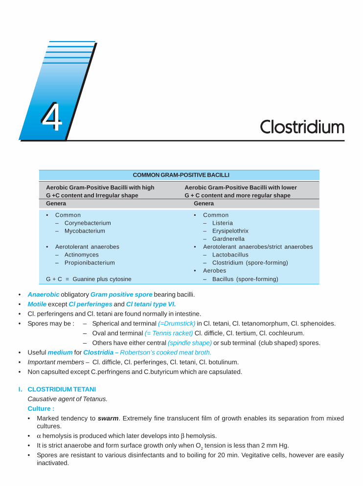
COMMON GRAM-POSITIVE BACILLI
Aerobic Gram-Positive Bacilli with high Aerobic Gram-Positive Bacilli with lowerG +C content and Irregular shape G + C content and more regular shapeGenera Genera
• Common • Common– Corynebacterium – Listeria– Mycobacterium – Erysipelothrix
– Gardnerella• Aerotolerant anaerobes • Aerotolerant anaerobes/strict anaerobes
– Actinomyces – Lactobacillus– Propionibacterium – Clostridium (spore-forming)
• AerobesG + C = Guanine plus cytosine – Bacillus (spore-forming)
• Anaerobic obligatory Gram positive spore bearing bacilli.• Motile except Cl perferinges and Cl tetani type VI.• Cl. perferingens and Cl. tetani are found normally in intestine.• Spores may be : – Spherical and terminal (=Drumstick) in Cl. tetani, Cl. tetanomorphum, Cl. sphenoides.
– Oval and terminal (= Tennis racket) Cl. difficle, Cl. tertium, Cl. cochleurum.– Others have either central (spindle shape) or sub terminal (club shaped) spores.
• Useful medium for Clostridia – Robertson’s cooked meat broth.• Important members – Cl. difficle, Cl. perferinges, Cl. tetani, Cl. botulinum.• Non capsulted except C.perfringens and C.butyricum which are capsulated.
I. CLOSTRIDIUM TETANICausative agent of Tetanus.Culture :• Marked tendency to swarm. Extremely fine translucent film of growth enables its separation from mixed
cultures.• α hemolysis is produced which later develops into β hemolysis.• It is strict anaerobe and form surface growth only when O2 tension is less than 2 mm Hg.• Spores are resistant to various disinfectants and to boiling for 20 min. Vegitative cells, however are easily
inactivated.
ClostridiumClostridiumClostridiumClostridiumClostridium44
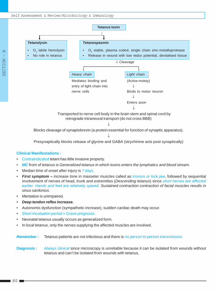
Self Assessment & Review Microbiology & Immunology
SECTION – B
8080
Tetanus toxin
Tetanolysin Tetanospasmin
• O2 labile hemolysin • O2 stable, plasma coded, single chain zinc-metalloprotease• No role in tetanus • Release in wound with low redox potential, devitalised tissue
↓ Cleavage
Heavy chain Light chain
Mediates binding and (Active moiety)entry of light chain into ↓nerve cells Binds to motor neuron
↓Enters axon
↓Transported to nerve cell body in the brain stem and spinal cord by
retrograde intraneural transport (do not cross BBB)↓
Blocks cleavage of synaptobrevin (a protein essential for function of synaptic apparatus).↓
Presynaptically blocks release of glycine and GABA (strychinine acts post synaptically)
Clinical Manifestations :• Contraindicated tetani has little invasive property.• MC from of tetanus is Generalized tetanus in which toxins enters the lymphatics and blood stream.• Median time of onset after injury is 7 days.• First symptom – increase tone in masseter muscles called as trismus or lock jaw, followed by sequential
involvement of nerves of head, trunk and extremities (Descending tetanus) since short nerves are affectedearlier. Hands and feet are relatively spared. Sustained contraction contraction of facial muscles results insinus sardonius.
• Mentation is unimpaired.• Deep tendon reflex increase.• Autonomic dysfunction (sympathetic increase), sudden cardiac death may occur.• Short Incubation period = Grave prognosis.• Neonatal tetanus usually occurs as generalized form.• In local tetanus, only the nerves supplying the affected muscles are involved.
Remember : Tetanus patients are not infectious and there is no person to person transmission.
Diagnosis : Always clinical since microscopy is unreliable because it can be isolated from wounds withouttetanus and can’t be isolated from wounds with tetanus.
Clostridium
UNIT - I BACTERIOLOGY “Gram +ve Bacilli”
81
Treatment :1. Antitoxin
• Best to give before wound manipulation.• Human tetanus immune globulin (TIG) is preparation of choice . T½ = 30 days. It decrease mortality.• Alternative is IV Ig• Management of wound is as follows :
All wound receive surgical toilet
↓↓↓↓↓ ↓ ↓ ↓ ↓ ↓Wound <6 hr, clean non penetrating Other woundsand with negligible tissue damage
↓↓↓↓↓ ↓ ↓ ↓ ↓ ↓ ↓↓↓↓↓ ↓ ↓ ↓ ↓ ↓
Immunity category Treatment Immunity category Treatment
A Nothing more required A Nothing more requiredB Toxoid one dose B Toxoid 1 doseC Toxoid one dose C Toxoid 1 dose + Human Tetanus IgD Toxoid complete course D Toxoid complete course
+ Human Tetanus Ig
A = Has had a complete course of toxoid or booster dose with in past 5 years.B = Has had a complete course of toxoid or booster dose with in past > 5 years and <10 years.C = Has had a complete course of toxoid or booster dose with in past >10 years.D = Has not had complete course of toxoid or immunity status is unknown.
2. Antibiotic : – Penicillin or metronidazole eradicate source of toxin i.e. vegetative cells.– Alternative clindamycin or erythromycin.– It is of no role if given > 6 hours.
Prevention :• Best prevented by active immunisation. Protective level of antitoxin > 0.01 IU/ml serum• Active immunisation – Monovalent Vaccine :
- 2 dose of Purified tetanus toxoid (TT) should be given at interval of 1-2 month- Ist booster after 1 year of 2nd dose.- 2nd booster after 5 year of 3rd dose.
– Combined Vaccine - DPT• Passive immunisation – Human tetanus hyperimmunoglobulin (TIG) is best prophylactic to use. Toxin already
bound to neural tissue is not affected.• Combined active and passive immunisation – Patient is given TIG in one arm and TT in other arm followed by
another dose of TT 6 weeks later and third dose 1 year later.
II. CL. BOTULINUM• Causative agent of botulism• It is agent of bioterrorism• Classified in to type A to G based on immunological difference in the toxins.

Self Assessment & Review Microbiology & Immunology
SECTION – B
8282
Toxin :• Botulinum toxin is most potent bacterial toxin known.• Produce 8 distinct toxin (A, B, C1, C2, D, E, F, G).• All toxins except C2 are neurotoxin. C2 is the cytotoxin of unknown clinical significance.• Acts on peripheral cholinergic nerve endings including Neuromuscular junctions, postganglionic parasympathetic
nerve endings and peripheral ganglion, CNS is not involved. It blocks release of Acetylcholine.• A, B, E and rarely F cause human disease. Type G (from C argentinense) has been associated with sudden
death. ..... Harrison 17/e, p 901• Type A toxin used for treatment of strabismus, blepharospasm, cervical dystonia.
Clinical Features :• Cause noninvasive botulism of three types :
1. Food borne botulism : Due to preformed toxin of canned food. With incubation period of 18-36 hr. Nauseavomiting, abdominal pain and characteristic symmetric descending paralysis occurs.• Symptoms begins with Cranial Nerve involvement producing diplopia, dysphagia, ptosis, diminished
pupillary reflex.• DTR decrease or normal, severe constipation no fever, no sensory finding, mentally intact.• Death is due to respiratory failure.
2. Wound botulism : Toxin is produced in wound. It resemble food borne illness except I.P is longer (~ 10days)and gastrointestinal symptoms are lacking.
3. Infant or intestinal botulism : – Due to ingestion of subterminal spores – Toxin is produced inside.– Source of infection is usually honey – Occur in infant < 6 months.
Remember :– Botulism is a food poisoning that do not cause diarrhea.– Infant botulism is most common form of botulismDiagnosis :• Demonstration of organism in serum by bio assay in mice is definitive.• Diagnosis must be considered in patient with symmetric descending paralyis who are mentally intact.
Treatment : • Food borne botulism – No role of antibiotics– Guanidine hydrochloride and Bivalent antitoxin given
• Wound botulism – Antitoxin• Infant botulism – Supportive care and human botulism immunoglobulin
III. CL. DIFFICLE ASSOCIATED DISEASE [CDAD]• MC diagnosed diarrheal illness acquired in the hospital, which results from ingestion of spores.• This colon infection is acquired almost exclusively in association with Antimicrobial use (MC clindamycin).• Risk factors for CDAD : old age, severe illness, use of electronic rectal thermometer, enteral tube feeding,
antacid treatment and gastrointestinal surgery.• Acquired exogenously and carried in stool of symptomatic and asymptomatic patients.• Asymptomatic fecal carriage is very common in healthy neonates.• It release toxin A (enterotoxin) and toxin B (cytotoxin), both glucosylate the GTP binding proteins and
disrupt cell cytoskeleton, so fliud is leaked leading to whitish yellow plaque formation over, colon, known asPseudomembranous colitis.
• MC symptom caused by Cl difficle - Diarrhoea• Complication - Toxic megacolon and sepsis.
Clostridium
UNIT - I BACTERIOLOGY “Gram +ve Bacilli”
83
Diagnosis :• Demonstrating toxin in feces by its characteristic effect on Hepatitis 2 and human diploid cell culture (Tissue
culture) is gold standard.• ELISA – Rapid and easy but has less sensitivity.
Treatment :• DOC – Metronidazole• DOC even for relapse is Metronidazole• If not respond for >48 hrs, give vancomycin• Rifampin, Bacitracin, Saccharomyces boulardii or lactobacillus GG, colonization with nontoxigenic strain of Cl
difficle, anion exchange binding resin cholestyramine, IV lg can also given.
IV. CL. PERFRINGES = CL. WELCHII• Causative agent of gas gangrene, enteritis necroticans (type C)• Toxigenic as well as invasive.• Capsulated, non-motile, shows Stormy fermentation• Absence of its central or subterminal spore in artificial media or pathological tissue is the characteristic
feature.• Classified on the basis of toxin, they produced.• It is the MC clostridial species isolated from tissue infection and bacteremia.• Clostridia are present in the normal colonic flora, Clostridia ramosum is the most abundant and is followed by
C.perfringens.
Toxins :• Produce twelve distinct toxin. Four major entero toxins are : α, β, ε and ι.• α Toxin = Phospholipase C= lecithinase : Associated with Gas gangrene.
– Hemolytic, Hot-cold variety toxin produced by all Cl perfringes but most abundantly by Type A.– Shows Nagler reaction in which zone of opacity is formed where there is no antitoxin.– Lecithinase also produced by Cl novyi, Cl. bifermentans, some vibrios.– It initiates muscle infection that may progress to gas gangrene.
• β and iota (i) toxin also have lethal and necrotizing properties. Increase capillary permeability.• θ (theta) toxin - Hemolysin, antigenically related to streptolysin O. Also known as Perfringolysin O.• Also produce neuraminidase, histamine - bursting factor etc.
Clinical features :It cause following diseases :1. Skin and soft tissue infection : It includes Gas gangrene = Clostridial myonecrosis.
• Most commonly cause by Cl. perfringes Type A. Also caused by Cl septicum, Cl novyi, Cl histolyticum etc.• Characteristic pathology – Near absence of PMNs despite extensive tissue destruction.• Essential factor - Trauma particularly deep muscle laceration.• Incubation period usually short : 10-48 hr with Cl perfringes, 2-3 days with Cl septicum, 5-6 days with Cl novyi.• Pain and crepitus present and death is due to circulatory failure.
Self Assessment & Review Microbiology & Immunology
SECTION – B
8484
• Diagnosis : – Frozen section biopsy of muscle.– Gram’s staining :
- Showing gram positive bacilli without spores - Cl perfringes- Showing citron bodies & boat or leaf shaped pleomorphic bacilli - Cl septicum- Showing large bacilli with oval or subterminal spores - Cl novyi.
• Treatment : – Surgery - mainstay of therapy– DOC Clindamycin + penicillin ..... Harrison 17/e, p 907– Hyperbaric O2 may also used.
2. Intestinal disorders :a. Food poisoning : Cl. perfringens (type A) is 2nd or 3rd MC cause of food poisoning mediated by Cytotoxin
which act on small bowel brush border and induces Ca2+ dependent alteration in permeability.• Usually caused by cold or warmed up meat dish after 8-24 hrs. It is self limited.
b. Enteritis necroticans (Pigbel) Caused by β toxin of type C strain.• Following ingestion of high protein meal with trypsin inhibitors (sweat potato) by host having limited
proteolytic activity of small intestine. Patient present with acute abdominal pain, bloody diarrhea, vomiting,signs of peritonitis.
c. Neurotropenic enterocolitis

Clostridium
UNIT - I BACTERIOLOGY “Gram +ve Bacilli”
85
1. The following statements are true regardingClostridium perfringes except : [AI 05]a) It is the commonest cause of gas gangreneb) It is normally present in human faecesc) The principal toxin of C. perfringens is the alpha
toxind) Gas gangrene producing strains of C. perfringes
produces heat resistant spores2. Regarding gas gangrene one of the following is
correct : [AI 04]a) It is due to Clostridium Botulinum infectionb) Clostridial species are gram-negative anaer-
obes forming sporesc) The clinical features are due to the release of
protein endotoxind) Gas is invariably present in the muscle com-
partments3. A person has received complete immunization
against tetanus 10 years ago. Now he presentswith a clean wound without any lacerations froman injury sustained 2.5 hours ago. He should nowbe given : [AI 01]a) Full course of tetanus toxoidb) Single dose of tetanus toxoidc) Human tet. globulind) Human tet. globulin and single dose of toxoid
4. True regarding pseudomembranous colitis are allexcept : [AI 00]a) It is caused by Clostridium difficleb) The organism is a normal commensal of gutc) It is due to production of phospholipase Ad) It is treated by vancomycin
5. Most common organism responsible for gas, gan-grene is : [AI 99]a) Clostridium-perfringensb) Clostridium-difficilec) Clostridium tetanid) Clostridium botulism
6. Most common organism, responsible for pseudomembranous colitis is : [AI 99]a) Clostridium difficleb) Clostridium botulism
c) Clostridium bifermentansd) Clostridium histolyticum
7. All of the following statements about Botulism aretrue except : [AI 97]a) Boutlism is caused by endotoxinb) Honey ingestion causes infant botulismc) Constipation is seend) Detection of antitoxin in the serum can aid in
diagnosis8. Swarming growth on culture is characteristic of
which Gram-negative organism : [AI 95]a) Clostridium welchib) Clostridium tetanic) Bacillus cereusd) Proteus mirabilis
9. An 18 year old male presented with acute onsetdescending paralysis of 3 days duration. There isalso a history of blurring of vision for the sameduration. On examination, the patient hasquadriparesis with areflexia. Both the pupils arenon-reactive. The most probable diagnosis is :a) Poliomyelitis [AIIMS 06]b) Botulismc) Diptheriad) Porphyria
10. A patient of Acute lymphocytic leukemia with fe-ver and neutropenia devlops diarrhoea after ad-ministration of amoxicillin therapy, which of thefollowing organism is most likely to be the caus-ative agent ? [AIIMS 05]a) Salmonella typhib) Clostridium difficlec) Clostridium perfringensd) Shigella flexneri
11. The following statement are true regarding botu-lism except : [AIIMS 03]a) Infant boutlism is caused by ingestion of per-
formed toxinb) Cl botulinum A, B, E and F cause human dis-
easec) The gene for botulinum toxin is coded by bacte-
riophaged) Cl. Bratii may cause botulism
QUESTIONS
Answer 1. d) Gas ... 2. d) Gas ... 3. b) Single ... 4. c) It is ... 5. a) Clostridium ...6. a) Clostridium ... 7. a) Boutlism ... 8. d) Proteus ... 9. b) Botulism 10. b) Clostridium ...
11. a) Infant ...
Self Assessment & Review Microbiology & Immunology
SECTION – B
8686
12. A 10 year old boy following a road traffic accidentpresents to the casualty with contaminatedwound over the left leg. He has received his com-plete primary immunization before preschool ageand received a booster of DT at school entry age.All of following can be done except :a) Injection of TT [AIIMS 01]b) Injection of human antiserumc) Broad spectrum antibioticsd) Wound debridement and cleaning
13. The most effective way of preventing tetanus is :a) Surgical debridement and toilet [AIIMS 01]b) Hyperbaric oxygenc) Antibioticsd) Tetanus toxoid
14. All occurs in botulism except : [AIIMS 97]a) Diplopiab) Diarrhoeac) Dysphagiad) Dysarthria
15. Not true about Botulinum toxin : [PGI 07]a. Short life spanb. Increased acetyl-choline releasec. Used for treatment in Blepharospasm, static anddyanmic wrinklesd. Effective for 3-4 monthse. Irreversibly decreases Ach in NM junction
16. Gastro-intestinal enteritis necroticans caused by:a. CI difficile [PGI 07]b. CI. perfringensc. Botulinumd. C jejunie. Pseudomonas
17. True about clostridium tetani : [PGI 03]a) It is gram +veb) Drum stick appearancec) Grows in aerobic environmentd) It is gram –vee) Produces endotoxin
18. Naeglers reaction is shown by : [PGI 00]a) Cl. Tetanib) Cl. Botulinumc) Cl. Perfringenesd) Cl. Septicum
Answer 12. b) Injection ... 13. d) Tetanus ... 14. b) Diarrhoea 15. a and b 16. b) CI. perfringens17. a and b 18. c) Cl. Perfrin ...

Clostridium
UNIT - I BACTERIOLOGY “Gram +ve Bacilli”
87
1. Ans. is d i.e. Gas gangrene producing strains of C. perfringes produces heat resistant sporesRef. Ananthnarayan 7/e, p 249 - 255
• Gas gangrene is caused by Cl perfringens (MC by type A), Cl novyi, Cl septicum, Cl. histolyticum.Others - Cl. sporogenes, Cl. fullax, Cl. bifermantans, Cl. Sordelli Cl. aerofoetidium and C. tertium.
• Most important toxin for gas gangrene is Alpha toxin = lecithinase which is responsible Naegler’s Reaction.• Cl. perfringes is found in feces and contaminates the skin of perineum, buttocks, thigh.• Its spores is used as remote indicator of fecal contamination of water.• Spores are usually destroyed within 5 minutes by boiling but those of food poisoning strains of Type A and
certain type C strain resist boiling for several hours.• Spores are destroyed by autoclaving at 1210C for 20 minutes.• Spores are resistant to antiseptics and disinfectants in common use.
2. Ans. is d i.e. Gas is invariably present in the muscle compartmentsRef. Ananthnarayan 7/e, p 249; Harrison 17/e, p 906
• Clostridia are Gram positive (so produce exotoxin as endotoxin are usually produced by gram negativebacteria) spore forming anaerobic bacilli.
• Gas gangrene clostridia are already mentioned (Cl. botulinum doesn’t cause gas gangrene).• Gas gangrene is characterized by rapid and extensive necrosis of muscle, accompanied by gas formation
(clinically as crepitations) and systemic toxicity.
3. Ans. is b i.e. Single dose of tetanus toxoid Ref. Park 18/e, p 252; 19/e, p 263
• Management of wound depend on nature of wound and immune status of person.• All wounds should receive surgical toilet.
Category Immunization status Clean wound of < 6 hrs. Other wounds eg.and with negligible tissue (contaminated wound)damage
A. Complete immunization Nothing Nothingwith in past 5yrs
B. Complete immunization 1 dose of toxoid Toxoid 1 dosewithin 5 - 10 years
C. Complete immunization Toxoid 1 dose Toxoid 1 dose + Humanmore than 10 years Tet. Ig
D. Has not had complete Toxoid complete course Toxoid complete courseimmunization or immunity + Human Tet Igstatus is unknown
Patient in question falls in category B.
EXPLANATIONS AND REFERENCES WITH ILLUSTRATIVE ANSWERS
Self Assessment & Review Microbiology & Immunology
SECTION – B
8888
4. Ans. is c i.e. It is due to production of phospholipase ARef. Harrison 17/e, p 819; Ananthnarayan 7/e, p 265
“Cl difficle cause pseudomembranous colitis (PMC) due to the production of toxin A (enterotoxin) andtoxin B (cytotoxin) not phospholipase A.”• Toxin A is potent neutrophil chemoattractant and both toxin A and B glucosylate the GTP binding protein of
Rho subfamily resulting in disruption of cytoskeleton causing loss of cell shape adherance with consiquentfluid leakage.
• Asymptomatic fecal carriage of Cl difficle in healthy neonates is very common. It also colonizes the colonof 3% of healthy adults. ..... CMDT’ 08, p 543
• For Cl difficle associated diarrhea (CDAD) three events are essential :
Event 1 : Antibiotics Hospitalization
Event 2 :CI. difficleacquistion
Asymptomatic C. difficle colonization(colonized patient activally have risk of subsequent CDAD)
C. difficle diarrhea
Inadequate anamnestic lgG hostresponse to toxin A of toxigenic C.difficleEvent 3 :
Diagnosis• Diagnosis of CAD is based a combination of clinical criteria :
– Diarrhea (> 3 unformed stools per 24 hours for > 2 days)– Toxin A or B detected by stool culture or pseudomembrane seen in colon.
• Endoscopy is a rapid diagnostic tool in seriously ill patient with suspected PMC but a negative result doesnot rule out CDAD.
• Treatment : Discontinue offending antibioticDOC – MetronidazoleDOC – For relapse Metronidazole
– If not respond > 48 hrs, give Vancomycin.
5. Ans. is a i.e. Cl. Perfringens Ref. Ananthnarayan 7/e, p 225
MC cause of gas gangrene is Cl. perfringes type A.
6. Ans. is a i.e. Cl difficle Ref. Ananthnarayan 7/e, p 265
Already explained, please see answer no. 4
7. Ans. is a i.e. Botulism is caused by Endotoxin Ref. Ananthnarayan 7/e, p 263
• Cl. botulinum produce exotoxin (since endotoxin is usually produced by gram negative bacteria) whichdiffers from other exotoxin in that it is produced intracellularly and appears in the medium only on death/autolysis of cell.
• Toxin production is determined by bacteriophage at least in types C and D toxin.
Clostridium
UNIT - I BACTERIOLOGY “Gram +ve Bacilli”
89
• All toxin are neurotoxin except C2 which is cytotoxin.• Toxin acts by blocking production or release of Ach. at synapse and Neuromuscular junction
(= parasympatholytic = effect of atropine) so cause constipation, paralysis etc.• Human disease is caused by type A, B, E and rarely F.• MC Type of botulism is ‘Infant botulism’ which is produced by eating of honey containing spores so toxin is
produced inside.• Food borne botulism and wound botulism are produced by preformed toxin.• Diagnosis may be confirmed by demonstration of bacillus or toxin in food or feces.• A retrospective diagnosis may be made by detection of antitoxin in the patient serum but it may not be seen
in all cases.
8. Ans. is d i.e. Proteus mirabilis Ref. Ananthnarayan 7/e, p 257, 283; Jawetz 24/e, p 207
Swarming growth is shown by :a. Cl. tetani, Bacillus cereus – Gram positive bacillib. Proteus mirabilis and Proteus vulgaris – Gram negative bacilli.
9. Ans. is b i.e. Botulism Ref. Harrison 17/e, p 902; Madell’s infectious disease 6/e, p 2824
Diagnosis of boutulism should be considered in patient with symmetric descending paralysis withbilateral cranial neuropathies in addition of following features :• Absent fever• Patient remain responsive• Heart rate normal or slow• Sensory deficit don’t occur except for blurred vision.
Remember : Neurologic signs and symptom of botulism :• Dysphagia • Dry mouth • Diplopia• Dysarthria • Upper limb weakness • Lower limb weakness• Blurred vision • Dyspnea
Also know : Polio present with asymmetric descending paralysis.
10. Ans. is b i.e. Clostridium difficle Ref. Harrison 17/e, p 818 - 820, 761; KDT 6/e, p 672 - 673
• It is a typical presentation of CDAD (Cl. difficle associated diarrhoea).• MC antibiotics causing CDAD - clindamycin, ampicillin and cephalosporins.• Antibiotics altered the normal intestinal flora which allows overgrowth of non-pathogenic organism. This
called as superinfection.• Superinfection also occur by candida albicans, resistant staphylococci, Proteus and Pseudomonas.• Superinfections are common when host defence is compromised as in steroid therapy, Leukemia and other
malignancies treated with anticancer drugs, AIDS, agranulocytosis, Diabetes.
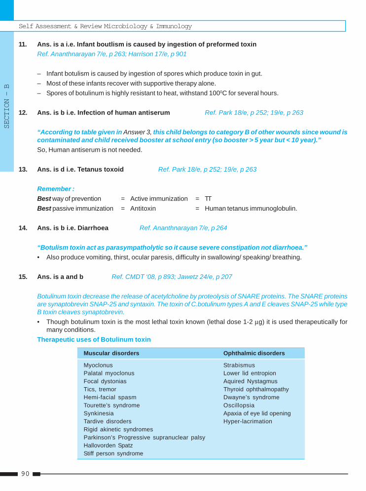
Self Assessment & Review Microbiology & Immunology
SECTION – B
9090
11. Ans. is a i.e. Infant boutlism is caused by ingestion of preformed toxinRef. Ananthnarayan 7/e, p 263; Harrison 17/e, p 901
– Infant botulism is caused by ingestion of spores which produce toxin in gut.– Most of these infants recover with supportive therapy alone.– Spores of botulinum is highly resistant to heat, withstand 1000C for several hours.
12. Ans. is b i.e. Infection of human antiserum Ref. Park 18/e, p 252; 19/e, p 263
“According to table given in Answer 3, this child belongs to category B of other wounds since wound iscontaminated and child received booster at school entry (so booster > 5 year but < 10 year).”So, Human antiserum is not needed.
13. Ans. is d i.e. Tetanus toxoid Ref. Park 18/e, p 252; 19/e, p 263
Remember :Best way of prevention = Active immunization = TTBest passive immunization = Antitoxin = Human tetanus immunoglobulin.
14. Ans. is b i.e. Diarrhoea Ref. Ananthnarayan 7/e, p 264
“Botulism toxin act as parasympatholytic so it cause severe constipation not diarrhoea.”• Also produce vomiting, thirst, ocular paresis, difficulty in swallowing/ speaking/ breathing.
15. Ans. is a and b Ref. CMDT ‘08, p 893; Jawetz 24/e, p 207
Botulinum toxin decrease the release of acetylcholine by proteolysis of SNARE proteins. The SNARE proteinsare synaptobrevin SNAP-25 and syntaxin. The toxin of C.botulinum types A and E cleaves SNAP-25 while typeB toxin cleaves synaptobrevin.• Though botulinum toxin is the most lethal toxin known (lethal dose 1-2 μg) it is used therapeutically for
many conditions.Therapeutic uses of Botulinum toxin
Muscular disorders Ophthalmic disorders
Myoclonus StrabismusPalatal myoclonus Lower lid entropionFocal dystonias Aquired NystagmusTics, tremor Thyroid ophthalmopathyHemi-facial spasm Dwayne’s syndromeTourette’s syndrome OscillopsiaSynkinesia Apaxia of eye lid openingTardive disroders Hyper-lacrimationRigid akinetic syndromesParkinson’s Progressive supranuclear palsyHallovorden SpatzStiff person syndrome
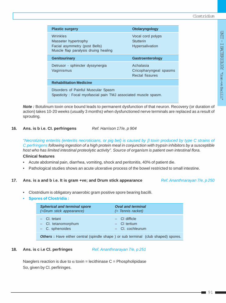
Clostridium
UNIT - I BACTERIOLOGY “Gram +ve Bacilli”
91
Plastic surgery Otolaryngology
Wrinkles Vocal cord polypsMasseter hypertrophy StutterinFacial asymmetry (post Bells) HypersalivationMuscle flap paralysis druing healing
Genitourinary Gastroenterology
Detrusor - sphincter dyssynergia AchalasiaVaginismus Cricopharyngeal spasms
Rectal fissures
Rehabilitation Medicine
Disorders of Painful Muscular SpasmSpasticity : Focal myofascial pain TMJ associated muscle spasm.
Note : Botulinum toxin once bound leads to permanent dysfunction of that neuron. Recovery (or duration ofaction) takes 10-20 weeks (usually 3 months) when dysfunctioned nerve terminals are replaced as a result ofsprouting.
16. Ans. is b i.e. CI. perfringens Ref. Harrison 17/e, p 904
“Necrotizing enteritis (enteritis necroticians, or pig bel) is caused by β toxin produced by type C strains ofC.perfringens following ingestion of a high protein meal in conjunction with trypsin inhibitors by a susceptiblehost who has limited intestinal proteolytic activity”. Source of organism is patient own intestinal flora.Clinical features• Acute abdominal pain, diarrhea, vomiting, shock and peritonitis, 40% of patient die.• Pathological studies shows an acute ulcerative process of the bowel restricted to small intestine.
17. Ans. is a and b i.e. It is gram +ve; and Drum stick appearance Ref. Ananthnarayan 7/e, p 250
• Clostridium is obligatory anaerobic gram positive spore bearing bacilli.• Spores of Clostridia :
Spherical and terminal spore Oval and terminal(=Drum stick appearance) (= Tennis racket)
– Cl. tetani – Cl difficle– Cl. tetanomorphum – Cl tertium– C. sphenoides – Cl. cochleurum
Others : Have either central (spindle shape ) or sub terminal (club shaped) spores.
18. Ans. is c i.e Cl. perfringes Ref. Ananthnarayan 7/e, p 251
Naeglers reaction is due to α toxin = lecithinase C = PhospholipidaseSo, given by Cl. perfiringes.
Self Assessment & Review Microbiology & Immunology
SECTION – B
9292
1. b) Cl. difficle 2. b) Parasympathetic 3. b) Cl. tetani 4. b) Lecithinase 5. c) Clostridiulm ...6. e) D 7. d) All of the ... 8. b) Clostridium ... 9. c) Clostridium ... 10. a) Cl. tertium
11. d) It is a gran ...
1. Pseudomembranous colitis is caused by :a) Cl. welchii [AI 91]b) Cl. difficlec) Yersinia enterocolitisd) Pseudomonas
[Ref. Ananthnarayan 7/e, p 251]2. Botulinum toxin acts on : [AI 92]
a) Sympathetic systemb) Parasympathetic systemc) Amygdalad) Motor cortex
[Ref. Ananthnarayan 7/e, p 263]3. Swarming growth is seen in which gram positive
bacillI : [AI 95]a) Cl. welchiib) Cl. tetanic) Proteusd) Bacillus cereus
4. Opacity around colonies of clostridium perfringesis due to : [JIPMER 95]a) Theta toxinb) Lecithinasec) Desmolased) Cytokinin
[Ref. Ananthnarayan 7/e, p 251]5. Crepitus in a wound is produced by : [AMU 97]
a) Staph aureusb) Clostridium tetanic) Clostridiulm-welchiid) Pseudomonas
[Ref. Ananthnarayan 7/e, p 255]6. Among the toxin produced by botulinum, the non
neurotoxic one is: [Kerala 00; AIIMS 92]a) Ab) B
Answer
c) Cl.d) C2e) D [Ref. Ananthnarayan 7/e, p 263]
7. Drumstick appearence is seen in :a) Cl. tetani [Keral 00, Orissa 00]b) Cl. tetanomorphumc) Cl. sphenoidsd) All of the above
[Ref. Ananthnarayan 7/e, 250]8. Foomy liver is caused by : [UP 00]
a) Clostridium tetanib) Clostridium welchiic) Pseudomonasd) Staph aureus
[Ref. Ananthnarayan 7/e, p 255]9. Gas gangrene is caused by : [Kar 01]
a) Clostridium tetanib) Clostridium difficlec) Clostridium perfringensd) Peptostreptococci
[Ref. Harrison 17/e, p 903]10. Oval bulging terminal spores seen in :
a) Cl. tertium [UP 04; M.P. 03]b) Cl. welchiic) Cl. perfringesd) Cl. histolyticeen
[Ref. Ananthnarayan 7/e, p 250]11. Each of the following statements concerning
Clostridium perfringes is correct except :a) It causes gas gangrene [SGPGI 04]b) It causes food poisoningc) It produces an exotoxind) It is a gran negative rod that does’not ferment
lactose[Ref. Ananthnarayan 7/e, p 250 - 252]
Chapter Review••••• This section includes questions of VThis section includes questions of VThis section includes questions of VThis section includes questions of VThis section includes questions of Various Other PGMEES from 1990 – 2007.arious Other PGMEES from 1990 – 2007.arious Other PGMEES from 1990 – 2007.arious Other PGMEES from 1990 – 2007.arious Other PGMEES from 1990 – 2007.••••• Questions are arQuestions are arQuestions are arQuestions are arQuestions are ar ranged in decreasing order of yearranged in decreasing order of yearranged in decreasing order of yearranged in decreasing order of yearranged in decreasing order of yearwiz. This is done to makwiz. This is done to makwiz. This is done to makwiz. This is done to makwiz. This is done to make refere refere refere refere refer ralralralralral
system easier and uncomplicated to save the precious time of PGMEE Aspirant.system easier and uncomplicated to save the precious time of PGMEE Aspirant.system easier and uncomplicated to save the precious time of PGMEE Aspirant.system easier and uncomplicated to save the precious time of PGMEE Aspirant.system easier and uncomplicated to save the precious time of PGMEE Aspirant.
Clostridium
UNIT - I BACTERIOLOGY “Gram +ve Bacilli”
93
12. b) Clinically 13. a) Clostridium ...14. a) Cl. perferingens 15. a) Presynaptic ...
Answer
12. Diagonsis of tetanus is made by : [MP 05]a) Culture of bacteria from woundb) Clinicallyc) Four fold raise in antibody against tetanus toxind) Gram staining of biopsy from wound
[Ref. Ananthnarayan 7/e, p 260]13. The organism causing pseudomembranous coli-
tis : [DNB 05]a) Clostridium difficileb) Clostridium perfringensc) Clostridium tetanid) Clostridium botulium
[Ref. Ananthnarayan 7/e, p 265]
14. Non motile clostridia is : [UP 06]a) Cl. perferingensb) Cl. novyic) Cl. botulismd) Cl. difficle
[Ref. Ananthnarayan 7/e, p 249]15. Site of action of tetanus toxin : [UP 07]
a) Presynaptic terminal of spinal cordb) Postsynaptic terminal of spinal cordc) Neuromuscular junctiond) Muscle fibres
[Ref. Ananthnarayan 7/e, p 259]
Gram positive non-motile rods with high G+C (guanine + cytosine) content.It includes : C. diphtheria, C. ulcerans and other coryneform bacteria like Arcanobacterium, Rhodococcus.
CORYNEBACTERIUM DIPHTHERIAE (KLEBS- LOFFLER BACILLUS)Morphology• Characteristically club shaped pleomorphic, non capsulated, nonsporing bacilli.• Arranged in pairs, palisades, clusters (chinese letter or cuneiform arrangement).• Contain Granules known as Babes Ernst or volutin granules which are composed of polymetaphosphate and are
more strongly Gram positive.• On staining with Loeffer’s methylene blue, granules show metachromatism.• Special stain to see granules clearly – Albert’s, Neisser’s and Ponder’s. Granules are also called as polar bodies
since arranged on poles.
Culture• Enrichment is necessary.• Selective medium : Cysteine - Tellurite blood agar (grow in 2 days) or Tinsdale medium.• For rapid growth (6 - 8 hours) - Loeffler’s serum slope used (also used for Mycobacteria TB).• Mcleod classified it into 3 types :
Feature C. gravis C. intermidius C. mitis
• Colony on tellurite Daisy head Frog’s Egg colony Poached egg colony• Hemolysis Variable Non hemolytic Usually hemolytic• Glycogen and starch Positive Negative Negative
fermentation• Most common Paralytic and Hemorrhagic Obstructive lesion
Complication Hemorrhagic in air passage
Diptheria Toxin• Polypeptide exotoxin, production depends on iron concentration.• Composed of 2 fragments : Fragment. B for binding and fragment A (enzymatic activity) for inactivating EF-2 in
presence of NAD. Hence, inhibits protein synthesis.• Toxin is inactive when it is released by bacteria.
CorCorCorCorCorynebacteriumynebacteriumynebacteriumynebacteriumynebacterium55
Corynebacterium
UNIT - I BACTERIOLOGY “Gram +ve Bacilli”
95
• Toxin acts both locally and systemically (mainly) while bacilli remain localized. Hence Diphtheria is toxemia notbacteremia.
• Corynephage controls production of toxin, Hence it shows lysogenic or phage conversion.• The strain almost universally used for toxin production is the ‘Park William 8’ Strain.• Exotoxin is also produced by C. ulcerans, C. pseudotuberculosis.• Both tox + and Tox - strains are infectious.
Clinical features• Cause diphtheria which is localized infection of mucous membrane or skin. It primarily involves respiratory tract.• Diphtherias are of following types :
1. Respiratory Diphtheria• MC type Tonsillo pharyngeal (Faucial)• MC symptoms - Fever, sore throat and weakness.• Also cause malignant or hypertoxic or bull neck appearance• Complications of Respiratory Diphtheria
– Mechanical complication (Asphyxia) due to membrane.– Systemic effects due to toxin eg myocarditis, peripheral polyneruopathy of descending type.– Risk is greater when involves larynx or tracheobroncial tree and in children (because of small airway
size).– Neurologic complication appear during first or second week of illness and begins with dysphagia.– Cardiac damage is permanent while recovery of nerve damage is the rule.– 1st muscle involve in paralysis - palatopharynges.– Cilliary paralysis occur but not pupillary paralysis i.e. blurred vision with preserved light reflex.
• Cause of death is circulatory failure.2. Cutaneous Diptheria – Punched out ulcers commonly caused by non-toxigenic strains (tox-).3. Invasive infection – (Rare) Risk factors are prexisting cardiac abnormality, IV drug abusers, alcoholic cirrhosis.
Diagnosis• Diagnosis of respiratory diphtheria is usually clinical while cutaneous diptheria requires lab confirmation.• Lab diagnosis can be either by demonstration of organism or demonstration of toxignicity by in vivo or in vitro
test.a. Demonstration of organism
– By Gram staing of throat swab.– Culture in specified media
b. Test for toxicigenicityIn vivo In vitro
• Done on guinea pigs can be intra- • Elek’s gel ppt. testcutaneous or subcutaneous • PCR for detection of toxgene
• ELISA• Immunochromatographic strip assay (fastest,with in hours)
... Jawetz 24/e, p 216Treatment• Most important element in treatment of respiratory diphtheria.
– Antitoxin but it does not prevents colonization nor eradicates carrier state.
Self Assessment & Review Microbiology & Immunology
SECTION – B
9696
• Antibiotics DOC Erythromycin or procaine pencillin G. Alternative is Rifampicin or clindamycin.• Sedatives or hypnotics are contraindicated.• Glucocorticoids do not reduce the risk of myocarditis or polyneuropathy.
Prevention• Active immunisation by Toxoid is best method to prevent diphtheria. Though active immunisation can prevent
manifestion of Diphtheria, it can not prevent carrier stage.• Active immunisation - Combined DPT is used most commonly.• Pertusis component in DPT increase potency of Diphtheria toxoid.• Toxoid of Diptheria shows Danysz phenomenon and Ehrlich phenomenon.
SCHICK TESTIntradermal test which provide information regarding :a. Immune statusb. Hyper senstivity to diptheria toxin.
In one arm toxin is injected, in other arm heat inactivated toxin is injected and following reaction may be seen.a. (–)ve reaction – No reaction in both arm Shows patient is immune to diphtheria.b. + ve reaction – Red flush of 10 - 50 mm with in 24 - 36 hours, reaching its maximum by 4th to 7th day. Control
arm shows no change. Patient is susceptible to diphtheria.c. Pseudo-positive reaction – Red flush equally on both arm, reaction fades very quickly. This is an allergic
type of reaction interpeted as Schik’s negative.d. Combined reaction – Test arm shows (+)ve, and control arm shows pseudo (+)ve. Dose of vaccine should
be reduced.
C.ULCERANS• Transmitted by cow’s milk, usually present as pharyngitis and can mimic respiratory diphtheria.
C. PSEUDOTUBERCULOSIS (=Preisz Nocard bacillus)• Typically present as suppurative granulamatous lymphadenitis.
C. MINUTISSIMUM• Cause Erythrasma and exhibits coral-red fluorescene under wood’s light.
C. PARVUM• Used as immunomodulator.
DIPHTHEROIDS• Normal commensals, stain more uniformly with few or no metachromatic granules eg. C.pseudodiphtheriticum.
Corynebacterium
UNIT - I BACTERIOLOGY “Gram +ve Bacilli”
97
1. The following statements are true about DPT vac-cine except : [AI 04]a) Aluminium salt has an adjuvant effectb) Whole killed bacteria of Bordetella pertussis has
an adjuvant effectc) Presence of acellular pertusis component in-
creases its immunogenecityd) Presence of H.influenza type B component in-
creases its immunogenecity2. Positive shick’s test indicates that person is :
a) Immune to diphtheria [AI 02]b) Hypersensitive to diptheriac) Susceptible to diptheriad) Carrier of diptheria
3. A child presents with a white patch over the ton-sils; diagnosis is made by culture in : [AI 01]a) Loeffler mediumb) LJ mediumc) Blood agard) Tellurite medium
4. True about corynebacterium diptheria are all ex-cept : [AI 98]a) Iron is required for toxin productionb) Toxin production is responsible for local reactionc) Nonsporing, noncapsular and non motiled) Toxin production is by lysogenic conversion
5. True statement regarding diptheria is : [AI 97]a) It can be diagnosed by demonstration of antibod-
ies by ELISAb) Immunization prevents carriers statec) Treatment of contacts is not indicatedd) Iron has critical value in the production of toxin
6. True about corynebacterium diptheria includesall of the following except : [AIIMS 07]a) Deep invasion is not seenb) Elek’s test is done for toxigenicityc) Metachromatic granules are seend) Toxigenicity is mediated by chromosomal change
7. Positive Shick’s test indicates that the person is:a) Immune to diptheria [AIIMS 07]b) Hypersensitive to diptheriac) Susceptible to diptheriad) Susceptible and hypersensitive to diphtheria
8. In a completely and adequately immunized childagainst Diptheria, the throat swab was collected.It showed the presence of C.diptheriae organismson Albert staining. These organisms can have oneof the following properties on further process-ing : [AIIMS 04]
a) It can grow on potassium tellurite mediab) It would show a + ve Elek’s gel precipitation testc) It can be pathogenic to experimental guinea pigd) It can produce cytotoxicity in tissue culture
9. A 12 year old child presents with fever and cervi-cal lymphadenopathy. Oral examination shows agrey membrane on the right tonsil extending tothe anterior pillar. Which of the following mediumwill be ideal for the culture of the throat swab fora rapid identification of the pathogen :a) Nutrient agar [AIIMS 02, 99]b) Blood agarc) Loeffler’s Serum sloped) LJ Medium
10. Regarding Schik’s test which of the following isfalse : [AIIMS 00]a) Erythematous reaction in both arms indicate
hypersenstivityb) Positive test means that person is immune to
diptheriac) Diptheria antitoxin is given intradermallyd) Test done to find out immune status against
diptheria11. A child with fever and pharyngitis which of the
following investigation should not to be done:a) Widal test [AIIMS 00]b) ASOc) Throat swab and cultured) Chest x-ray
12. Investigation of choice of diptheria carrier is :a) Throat swab culture [AIIMS 97]b) Gram’s stainc) Albert’s staind) Zeil Nelson’s stain
13. All are true regarding diptheria toxin except :a) Toxin blocks elongation of protein [AIIMS 97]b) Beta lysogenic strain produces toxinc) Iron is critical for toxin productiond) Toxin is necessary for local wound production
14. Which of the following is true about Diptheriaexcept : [PGI 05]a. Faucial diptheria is more dangerous than laryn-
geal diptheriab. Laryngeal diptheria mandates tracheostomyc. Child is infectious with faucial diptheriad. Myocarditis may be a complicatione. Palatal paralysis is irreversible
QUESTIONS
Answer 1. d) Presence ... 2. c) Susceptible ... 3. a) Loeffler ... 4. b) Toxin ... 5. d) Iron ...6. d) Toxigenicity ... 7. c) Susceptible ... 8. a) It can ... 9. c) Loeffler’s ... 10. b) Positive ...
11. a) Widal ... 12. a) Throat ... 13. d) Toxin ... 14. a and e
Self Assessment & Review Microbiology & Immunology
SECTION – B
9898
15. Metachromatic granules are found in : [PGI 00]a) Diptheriab) Mycoplasmac) Gardenerella vaginalisd) Chlamydiae) Staphylococcus
Answer 15. a) Diptheria 16. d) Loeffler’s ...
16. Corynebacterium diptheriae can be grown within6-8 hours on : [PGI 98]a) Potassium tellurite media with ironb) McConkey’s agarc) Dorset egg mediumd) Loeffler’s serum slope
Corynebacterium
UNIT - I BACTERIOLOGY “Gram +ve Bacilli”
99
1. Ans. is d i.e. Presence of H.influenza type B component increases its immunogenecity
Lets consider each choice one by one.Option ‘a’ : Two types of Diphtheria toxoid are in use now. ... Ananthnarayan 7/e, p 238
1. Fluid toxoid2. Adsorbed toxoid
Adsorbed toxoid is purified toxoid adsorbed into insoluble aluminium compounds usuallyaluminium phosphate less often the hydroxide.Adsorbed toxoid is much more immunogenic than fluid toxoid.
Option ‘b’ : Pertussis component in DPT (whole killed bacteria of bordetella pertusis) vaccine enhancesthe potency of the diptheria toxoid. ... Park 18/e, p 135Most serous complication of DPT is neurological which is primarily due to pertusis component.Duration of immunity after whole cell pertusis vaccination is short lived, with little protectionremaining after 10-12 years.
Option ‘c’ : To reduce complications of whole killed bacteria of pertusis in DPT, acellular vaccinedeveloped.• Protection against pertussis by vaccines correlated best with the production of antibody
to pertactin, fimbriae and pertussis toxin.• All acellular pertussis vaccines currently available contain pertussis toxoid.
Acellular vaccine is more immunogenic has less adverse effects. It is given as DTaP..... CMDT ‘06, p 1304
Option ‘d’ : Quadriple vaccine of DPT with H.influenza B is available in India but conjugate vaccine (eg.HIb vaccine) do not interfere with immunogenicity of simultaneously given other vaccines.
.... Ghai 6/e, p 197
2. Ans. is c i.e. Susceptible to diptheria Ref. Ananthnarayan 7/e, p 237; Park 19/e, p 137
Shick’s test• Intradermal test which provide information regarding :
a. Immune status, b. Hypersensitivity & susceptibility to diptheria toxin before giving active immunization.• In one arm toxin is injected (test arm) and in other arm heat inactivated toxin is injected (control arm).
Type of Reaction Observation Inference
i. Negative reaction No reaction in both arm (toxin No susceptibility. No hypersensitivityis neutralized by circulating antitoxin) Patient is immune to diptheria
ii. Positive reaction No change in control arm. Red flush No hypersensitivityin test arm that persist Susceptibility present
iii. Pseudo positive Red flush equally on both arm Hypersensitivity presentreaction (Schick’s that fades very quickly No susceptibilitynegative)
iv. Combined reaction Test arm shows positive and Hypersensitivity presentcontrol arm shows pseudo- Susceptibility presentpositive reaction Dose of vaccine should be reduced
EXPLANATIONS AND REFERENCES WITH ILLUSTRATIVE ANSWERS
Self Assessment & Review Microbiology & Immunology
SECTION – B
100100
Remember : • Antitoxin level of 0.01 unit or more per ml of blood is considered as index of immunity.• Shick’s test is no longer in use. The level of antitoxin, is assesed by passive
hemagglutination or by neutralization in cell culture.
3. Ans. is a i.e. Loeffler medium Ref. Ananthnarayan 7/e, p 231 - 232
In a child white patch over tonsils, probable diagnosis is of Diphtheria.
Culture media for corynebacterium are :i. Loefflers serum slope : Growth is very rapid and colonies seen in 6-8 hrs, before other bacteria grows. It
is also used for M. tuberculosis. Diptheria is emergency condition, so Loeffler’s slope is prefered media inthis child.
ii. Tellurite blood agar media : Selective media but growth is delayed and may take about 2 days to appear.
Remember : • LJ media, is for Mycobacteria TB.• Mcleods and Hoyle’s are modified Tellurite media.• Blood agar is used to differentiate Staph. and Sterpt. pharyngitis.
4. Ans. is b i.e. Toxin production is responsible for local reactionRef. Ananthnarayan 7/e, p 233 - 235; Jawetz 24/e, p 215
“Mechanical complications of diptheria are due to the membrane while the systemic effects are dueto the toxin.”• Toxin acts mainly systemically though there are partial local effects.• It has affinity for myocardium, adrenals and nerve endings.• Toxin acts by inactivating EF-2 thus inhibiting protein synthesis.• Toxin production is influenced by iron concentration in the medium. Toxin production is optimal at 0.14 μg/
ml and is suppressed at 0.5 μg/ml.• Toxigencity of diptheria bacullus depends on symbiotic bacteriophages so it shows lysogenic or phage
conversion i.e. nontoxigenic strain →→→→→ toxigenic strain by infecting with beta phage.Remember : • Corynebacteria are gram positive, non acid fast, non sporing, non capsulated, non motile
bacteria.• It contains polar bodies or volutin or babes-ernst or metachromatic granules of
polymetaphosphate which are more gram positive.• Granules are also stained by loeffler’s methylene blue, Albert’s, Neisser’s and ponder’s
stain.
5. Ans. is d i.e. Iron has critical value in the production of toxinRef. Ananthnarayan 7/e, p 234 - 236; Jawetz 24/e, p 216
Diagnosis of Diptheriai. Isolation by culture :
Swabs are inoculated on :– Loeffler’s serum slope (growth is rapid) : for rapid diagnosis.– Tellurite blood agar (growth is delayed but it is particularly important in isolation of bacilli from
Corynebacterium
UNIT - I BACTERIOLOGY “Gram +ve Bacilli”
101
convalescent, contacts, carriers) : Selective media.– Blood agar (for differentiating staphylococcal and streptococal pharyngitis).
ii. Demonstration of toxicity of isolated strain :In vivo test – done by infected broth emulsion of culture subacutaneously and intracutaneously into guineapigs.In vitro test – Elek’s gel precipitation test and tissue culture test on agar overlay of cell culture monolayer.
Control of Diptheriai. Cases – Antitoxin + penicillin or erythromycin ... Park 19/e, p 137ii. Carriers – Erythromyciniii. Contacts – a. When primary immunization or booster dose was received within the previous 2 years.
• No further treatment.b. When primary immunization or booster dose was received more than 2 years ago.
• Only a booster dose of diphtheria toxoid.c. Non immunized close contacts :
• Prophylatic penicillin or erythromycin• 1000 - 2000 units of diphtheria antitoxin• Active immunization against diphtheria.
iv. Community – Only effective control is by active immunization with diphtheria toxoid of all infants withsubsequent booster doses every 10 years thereafter.
Remember : Vaccine ‘being a toxoid’ is not directed against organism and hence immunization does notprevent carrier state which is due to organism not due to toxin.
6. Ans. is d i.e. Toxigenicity is mediated by chromosomal change Ref. Harrison 17/e, p 890
“Corynebacteriophage beta carries the structural gene (tox+) encoding diptheria toxin and a family ofclosely related corynebacteriophage are responsible for toxigenic conversion of tox– C.diptheria totox+ phenotype.”• Elek’s gel precipitation test is in vitro test for toxin detection.
7. Ans. is c i.e. Susceptible and hypersensitive to diphtheria Ref. Ananthnarayan 7/e, p 237
Already explained, refer answer no. 2
8. Ans. is a i.e. It can grow on potassium tellurite media Ref. Ananthnarayan 7/e, p 235
This child is carrier of diptheria.• Postassium tellurite is selective media for isolation of diptheria bacillus from convulescent contact, carriers.• Other three test are done for testing virulence only when isolated strain is C.diptheria.
9. Ans. is c i.e. Loeffler’s Serum slope Ref. Ananthnarayan 7/e, p 235
Already explained, refer see answer no. 3
Self Assessment & Review Microbiology & Immunology
SECTION – B
102102
10. Ans. is b i.e. Positive test means that person is immune to diptheriaRef. Park 19/e, p 137; Ananthnarayan 7/e, p 236
Already explained, refer asnwer no. 2
11. Ans. is a i.e. Widal test Ref. Ananthnarayan 7/e, p 235
Widal is test for diagnosing Typhoid which is not a cause of pharyngitis so there is no need to perform widal inthis child.
12. Ans. is a i.e. Throat swab culture Ref. Park 19/e, p 136
• Carriers can be detected only by cultural method.• Swab should be taken from both the nose and throat.
13. Ans. is d i.e. Toxin is necessary for local wound production Ref. Ananthnarayan 7/e, p 235 - 236
Already explained, refer answer no. 4
14. Ans. is a and e i.e. Faucial diptheria is more dangerous than laryngeal diptheria; and Palatal paralysisis irreversible Ref. Dhingra 3/e, p 348
Diptheria• Causative agent – Corynebacterium diptheriae (Gr+ve bacillus)• Incubation period – 2 - 6 days• Depending on site present as :
– Nasal diptheria - Mildest, Toxemia is minimal.– Faucial diptheria (most common) = Nasopharyngeal diptheria - More severe than nasal diptheria.– Laryngotracheal diptheria - Most severe, maximum obstructive symptom, tracheostomy may be
essential.• Complications :
– Myocarditis - Occurs towards the end of 1st or begining of 2nd week.– Peripheral neuropathy of descending type.– Renal failure.
Remember : Cardiac damage is permanent while recovery of nerve damage is rule.
15. Ans. is a i.e. Diptheria Ref. Ananthanrayan 7/e, p 231
Metachromatic granules• It is type of intracytoplasmic inclusions characteristically seen in diptheria bacilli.• Also known as volutin or metachromatic or Babes Ernst granules.• Strongly basophilic bodies consist of polymetaphosphate.• They are reservoir of energy and phosphate.
Corynebacterium
UNIT - I BACTERIOLOGY “Gram +ve Bacilli”
103
• They are most frequent in cells growing under nutritional deficient condition.
Remember : • Dorset egg media is for M. tuberculosis.• MacConkey is for differentiating between lactose and Non-lactose fermenters of
Enterobacteriacea.• Metachromatic granules also seen in B.pertusis.
16. Ans. is d i.e. Loeffler’s serum slope Ref. Ananthnarayan 7/e, p 231
Already explained, refer answer no. 3
Self Assessment & Review Microbiology & Immunology
SECTION – B
104104
1. a) Gram negative 2. c) Corynebac ... 3. d) Chalmydia 4. c) Pneumococcus 5. b) Diptheria6. b) Elecks’s ... 7. a) Loefflers ... 8. c) Laryngeal 9. c) S aureus 10. a) Anthrax
11. b) Treatment ... 12. a) Ponder’s ... 13. a) 100 MLD ... 14. a and c 15. a) Corynebac ...
1. One of the following about corynebacteriumdiptherias is wrong : [Kerala 91]a) Gram negativeb) Ernest babes granules seenc) Chinese letter patternd) Prevented by immunisation
[Ref. Ananthnarayan 7/e, p 231]2. Tellurite stimulates growth of : [Kerala 91]
a) E. coilb) Cl. tetanic) Corynebacterium diptheriaed) Salmonella
[Ref. Ananthnarayan 7/e, p 232]3. Growth on a cell-free artifical solid medium is pos-
sible for following except : [TN 92]a) Ureaplasma urealyticumb) Mycoplasma Pneumoniaec) C and L form of proteus vulgarisd) Chalmydia
[Ref. Ananthnarayan 7/e, p 423]4. Effective vaccine is available against :
a) Staphylococcus aureus [Kerla 94]b) Streptococcusc) Pneumococcusd) Gonococcus
[Ref. Ananthnarayan 7/e, p 221]5. Elecks gel precipitation test is for : [Delhi 96]
a) Gonococcusb) Diptheriac) H. influenzad) Anthrax
[Ref. Ananthnarayan 7/e, p 236]
Answer
6. True about diptheria is : [MP 98]a) Loffler’s serum is highly selective medium for C.
diphtheriab) Elecks’s Gel is a precipitation testc) Metachromatic granules is produced on stain
only by one strain of C. deptheriad) Gm -ve bacilli, non motile, non capsuhlated
[Ref. Ananthnarayan 7/e, p 236]7. Culture medium for corynebacterium diphtheria:
a) Loefflers serum slope [JIPMER 01]b) McConkeyc) Sabarauds agard) Lowenstein Jensen medium
[Ref. Ananthnarayan 7/e, p 235]8. The type of Diphtheria with highest mortality is :
a) Pharyngeal [JIPMER 00]b) Nasalc) Laryngeald) Conjuctival
[Ref. Harrison, 17/e, p 893]9. Erythrasma is caused by: [SRMC 02]
a) S.pyogenesb) Corynebacterium miniutisoniumc) S aureusd) Ricketessiae
[Ref. Ananthnarayan 7/e, p 240]10. Literally leather term is used for : [UP 03]
a) Anthraxb) Proteiusc) Diptheriad) Neiserria
[Ref. Ananthnarayan 7/e, p 234]
Chapter Review••••• This section includes questions of VThis section includes questions of VThis section includes questions of VThis section includes questions of VThis section includes questions of Various Other PGMEES from 1990 – 2007.arious Other PGMEES from 1990 – 2007.arious Other PGMEES from 1990 – 2007.arious Other PGMEES from 1990 – 2007.arious Other PGMEES from 1990 – 2007.••••• Questions are arQuestions are arQuestions are arQuestions are arQuestions are ar ranged in decreasing order of yearranged in decreasing order of yearranged in decreasing order of yearranged in decreasing order of yearranged in decreasing order of yearwiz. This is done to makwiz. This is done to makwiz. This is done to makwiz. This is done to makwiz. This is done to make refere refere refere refere refer ralralralralral
system easier and uncomplicated to save the precious time of PGMEE Aspirant.system easier and uncomplicated to save the precious time of PGMEE Aspirant.system easier and uncomplicated to save the precious time of PGMEE Aspirant.system easier and uncomplicated to save the precious time of PGMEE Aspirant.system easier and uncomplicated to save the precious time of PGMEE Aspirant.
Corynebacterium
UNIT - I BACTERIOLOGY “Gram +ve Bacilli”
105
11. True about diphtheria is : [SGPGI 03]a) Cause cranial nerve palsies in 2nd and 3rd weekb) Treatment with erythromycinc) It is Gram negative organismd) Passive immunization is harmful and should not
be tried[Ref. Harrison 17/e, p 890-893]
12. Metachromatic granules are stained by :a) Ponder’s stain [JIPMER 03]b) Negative stainc) Gram’s staind) Leishmans stain
[Ref. Ananthnarayan 7/e, p 231]13. One unit of diptheria antitoxin was defined as the
smallest amount of antitoxin required to neutrilise:a) 100 MLD of toxin [SGPGI 04]
b) 200 MLD of toxinc) 300 MLD of toxind) 400 MLD of toxin
[Ref. Ananthnarayan 7/e, p 238]14. Toxigenicity of C. diphtheria is determined by :
a) Elek’s gel ppt test [Bihar 04]b) Aselole’s reactionc) Nagler’s reactiond) All
[Ref. Ananthnarayan 7/e, p 236]15. KLB another name is : [Jharkhand 04]
a) Corynebacterium diphtheriab) Corynebacterium psudodiptheriaec) Clostridia tetanaed) Anthrax
[Ref. Ananthnarayan 7/e, p 231]
11. b) Treatment ... 12. a) Ponder’s ... 13. a) 100 MLD ...14. a and c 15. a) Corynebac ...
Answer
ACTINOMYCETES• Transitional forms between bacteria and fungi.• Gram positive, non motile, non sporing, non capsulated filaments.• It includes : a. Actinomyces
b. Nocardia.
Actinomyces• Anaerobic bacillus cause : Lumpy jaw (in cattle) and Actinomycosis in human.
Actinomycosis• It is endogenous infection since actinomyces normally present in mouth, intestine, vagina.• MC causative agent A. israelii.• Usually a cooperative disease.• Characterized by indurated swelling (mainly in connective tissue), suppuration, multiple sinsuses towards skin
with discharge of sulphur granules.
Types of Actinomycosis :• MC type cervico facial
– MC site of cervicofacial is lower jaw often adjacent to carious tooth. No lymph node involvement.• Abdominal – Usually around caecum.• Thoracic• Pelvic – In association with IUCD’s.• Mycetoma – Painless localized woody induration without systemic symptoms. Granulomatus involvement of
subcutaneous and deeper tissue induced by traumatic inoculation of saprophytic fungi or bacteria.– MC site foot, present as tumour with multiple discharging sinus called as Madura foot.– MC cause is fungi called as eumycotic mycetoma / Maduramycosis / Madura foot. Black granules, stout
filament seen on microscopy.– Fungal agents of mycetoma : - Pseudallescheria boydii - Exophiala jeanselmei
- Madurella mycetomatis - Acremonium falciforme- Madurella grisea
Actinomycetes & BacillusActinomycetes & BacillusActinomycetes & BacillusActinomycetes & BacillusActinomycetes & Bacillus66
Actinomycetes & Bacillus
UNIT - I BACTERIOLOGY “Gram +ve Bacilli”
107
– Bacterial mycetoma are usually caused by Actinomyces, Nocardia, Streptomyces, Nocardiopsis.– Even S. aureus and other pyogenic bacteria may cause mycetoma like lesion called Botryomycosis.– In actinomycotic mycetoma - granules are white to yellow and thin filaments seen on microscopy.– Treatment : - Actinomycetoma : Streptomycin + Dapsone or cotrimoxa-zole
- Eumycetoma of Madurella mycetoma : Keto / Itra-conazole- Other Eumycetoma : rarely responds to chemotherapy.
Diagnosis of Actinomycosis• Specimen – Sputum
– Shake it in test tube with saline - Sulphur granules seen.• Microscopy – Granules are infact, bacterial colonies
– Gram positive filaments in the form of radiating club shaped = sun ray appearance seen.– Club is formed due to antigen - antibody reaction– Culture on solid media - shows spidery colonies which later develop into “molar tooth” colonies.
Treatment• Penicillin is DOC.
NOCARDIA• Aerobic, partially urease positive, catalase positive, partially acid fast, causing exogenous infection,
Nocardiosis (which refers to invasive disease).• MC species associated with invasive disease N. asterodes.• MC risk factor - Contact with soil or vegetable matter.• Cell mediated immunity is important as neutrophil limit its growth but not kill them efficiently.• Nocardiosis is usually initiated by inhalation and their is no person to person transmission. ... Jawetz 24/e, p 219
Clinical Manifestation• MC manifestation is pneumonia and disseminated disease which follows inhalation of bacteria.• So, manifestation can be divided into :
a. Respiratory tract disease – MC is pneumonia. Prominent cough, small amount of thick purulent sputum thatis not malodorous.
b. Extrapulmonary Dissemination – MC site brain. Typical manifestation is subacute abscess usuallysupratentorial. ... Harrison 17/e, p 994
c. Disease Following Transcutaneous inoculation – Cellulitis, Lymphocutaneous syndrome (most casesassociated with N. brasiliensis), actinomycetoma (old Fistula disappear with appearance of new fistula).
Diagnosis• First step : examine sputum or pus for crooked, branching, beaded, gram positive, acid fast filaments. It also
takes silver stains.• Transtracheal aspiration should be avoided as it frequently leads to cellulitis in tissue around puncture wound.• Culture : It use paraffin as carbon source so paraffin baiting is used for isolation.
Treatment• DOC Trimethoprim - Sulfamethoxazole ... Harrison 17/e, p 995• Best alternative oral drug Minocycline• Best parenteral drug Amikacin.
Self Assessment & Review Microbiology & Immunology
SECTION – B
108108
BACILLUS• Genus consist of sporogenous aerobic gram positive bacilli.• They are generally motile with peritrichous flagella except anthrax bacilli.• Spores are heat resistant and constitute the commonest contaminants of bacteriological culture media.• Its important species are : 1. Bacillus anthracis
2. B. cereus.
1. BACILLUS ANTHRAX– First pathogenic bacteria to be observed under microscope.– First bacterium used for preparation of attenuated vaccine.– First communicable disease shown to be transmitted by inoculation of infected blood.– First bacillus to be isolated in pure culture and shown to possess spores.
Morphology :• Non motile, capsulated bacilli.• Capsule is polypeptide in nature (exception since usually capsule is of lipo polysaccharide in nature).• Bacilli arranged in BAMBOO STICK / BOX CAR LIKE APPEARANCE.• Colonies of B.anthracis are round and have a “cut glass” appearance in transmitted light.• On staining with polychrome methylene blue, it shows M’FADYEANS reaction; which represents capsular
material.• Spores – Used in biological warfare or bioterrorism. Formed in culture or soil but never in animal body. Not
stained by ordinary methods. Not cause bulging of vegetative cells (in comparison of clostridia spores).
Culture :• Selective medium : PLET medium.• On Agar plates – frosted glass appearance seen.• On microscopy it has Medusa Head Appearance.• On Gelatin stab – characteristic Inverted Fir Tree Appearance.• Solid Media with penicillin – String of pearl reaction.• Cut glass appearance – In transmitted light.
Resistance :• Bacilli remain viable in bone marrow for a week and in skin for 2 weeks.• Spores are destructed by oxidising agents H2O2, 4% KMnO4, formaldehyde (called as Duckering)
Virulence gactor :i. Capsular poly - D-glutamic acid : plasmid mediated, inhibit phagocytosis. Loss of plasmid cause loss of
virulence (basis of live attenuated anthrax spore vaccine).ii. Toxin : Plasmid coded, complex of three fractions :
– Factor I or Edema factor - activated only intracellularly leading increased CAMP.– Factor II or Protective antigen factor (PA) – Antibody to PA is protective because it blocks first step in toxin
activity i.e. binding to target cells.– Lethal factor or Factor III - cause cell death.
Disease :• Cause Anthrax which occurs primarily in herbivores (zoonosis).• Humans are more resistant to anthrax than herbivores.
Actinomycetes & Bacillus
UNIT - I BACTERIOLOGY “Gram +ve Bacilli”
109
• Transmitted by spores, by contact with infected animals or contaminated animal products, insects bites,ingestion or inhalation.
• Human anthrax is of 3 types :a. Cutaneous Anthrax (Hide Porter’s disease) : MC (95%); characterized by malignant pustule i.e. central
eschar surrounded by nonpitting edema.– Toxemia always present.– Smear of vesicle fluid is used to confirm diagnosis by culture and animal inoculation.
b. Pulmonary/Inhalational Anthrax / Wool Sorter’s Disease : Typically cause hemorrhagic mediastinitis.Characteristic X-ray finding is symmetric mediastinal widening.
c. Gastrointestinal anthrax : rare form
Laboratory diagnosis :• Microscopy : – Gram postive bacilli with M.Fadyean’s reaction - presumptive diagnosis made.
– Immunofluorescent microscopy can confirm identification.• If sample is putrid, Ascoli’s thermoprecipitin (ring precipitin) test is done which demonstrate anthrax
antigen in tissue extract.• Test for antibody by immunoassays are useful in confirming diagnosis.
Treatment :• DOC Penicillin• In Penicillin allergy – Ciprofloxacin, erythromycin, tetracycline or chloramphenicol.
2. BACILLUS CEREUS• Resembles B. anthrax except that it is motile (Swarming), non-capsulated, not susceptible to gamma
bacteriophage, not show ‘string of pearls’ reaction.• Isolated from feces and other sources on MYPA (mannitol egg yolk phenol red polymyxin agar).• It cause two type of food poisoning manifest by nausea, vomiting and abdominal cramps.
Features Diarrheal type Emetic Type
Incubation period 8-16 hrs 1-5 hrs
Food Cooked meat and vegetables Exclusively by rice
Enterotoxin Resemble heat labile toxin of E.coli Resemble heat stable toxin of S.aureusEither preformed or produced in Already preformed in riceintestine
Clinical Features Fever, Vomitting rare Diarrhea is not common
Serotype 2, 6, 8, 9, 10, 12 1, 3, 5
Remember : • Presence of B. cereus in patient stool is not sufficient for diagnosis since it may be present innormal stool specimen; concentration of 105 or more bacteria per gram of stool is considereddiagnostic. ... Jawetz 24/e, p 206
• It also cause eye infections; localized and systemic infections (occur in patient with medicaldevice or IV drug users).
Self Assessment & Review Microbiology & Immunology
SECTION – B
110110
1. An abattoir worker developed pustule which laterprogress to necrotic ulcer. Which of the follow-ing stain is useful demonstration of organismfrom smear made from pustule ?a) Polychromic methylene blue [AI 07; AIIMS 06]b) Chalkofluor whitec) Geimsad) Modified kinyon stain
2. A man, after skinning a dead animal, developed apustule on his hand. A smear prepared from thelesion showed the presence of Gram positive ba-cilli in long chains which were positive forMcFadyean’s reaction. The most likelyaetiological agent is : [AI 04]a) Clostridium tetanib) Listeria monocytogenesc) Bacillus anthracisd) Actinomyces sp
3. All of the following are true about anthraxexcept : [AI 98]a) Plasmid is responsible for toxin prodcutionb) Cutaneous anthrax generally resolve spontane-
ouslyc) Capsular polypeptide aids virulence by inhibiting
phagocytosisd) Toxin is a complex of two fractions
4. The causative organism of Mycetoma is :[AI 96]a) Nocardiab) Dimorphic fungusc) Aspergillusd) Dermatophytes
5. Which of the following is False aboutmycetoma : [AI 96]a) Can affect lower and upper extremitiesb) Caused by actinomycetes and filamentous fungic) Diagnosis is by examination of pusd) Uncommon in India
6. A discharging sinuses seen in : [AI 95]a) Sporotrichosisb) Cryptococcosisc) Histoplasmosisd) Myceotma
7. Which of the following is not true of Actinomyco-sis : [AI 95]a) Demonstration of filamentsb) Caused by actinomyces israeliic) Organism cannot be culturedd) Sulphur granules in pus
8. Nocardia is stained by : [AIIMS 08]a) Acid fast stainb) Kiram’s stainc) Alcian blued) Mucin stain
9. Which of the following is the most predominantconstituent of sulfur granules of Actinomycosisis : [AIIMS 04, 02]a) Organismsb) Neutrophils and monocytesc) Monocytes and lymphocytesd) Eosinophils
10. A clinical specimen was obtained from the woundof a patient diagnosed as Nocardiosis. For theselective isolation of Nocardia sp. which one ofthe following would be the best method :a) Paraffin bait technique [AIIMS 04]b) Castaneda’s culture methodc) Craige’s culture methodd) Hair bait technique
11. A malignant Pustule is a term used for :a) An infected malignant melanoma [AIIMS 03]b) A carbunclec) A rapidly spreading rodent ulcerd) Anthrax of skin
12. A farmer present with multiple discharging si-nuses in the leg not responding to antibiotics.Most likely diagnosis is : [AIIMS 02]a) Madurellab) Actino-mycetomac) Nocardiad) Sporothrix
13. True regarding anthrax is all except : [AIIMS 97]a) Caused by insect biteb) Caused by rubbing of skinc) Cutaneous type is rare nowdaysd) Pulmonary infection occurs by inhalation
QUESTIONS
Answer 1. a) Polychromic 2. c) Bacillus ... 3. d) Toxin is a ... 4. a) Nocardia 5. d) Uncommon ...6. d) Myceotma 7. c) Organism ... 8. None 9. a) Organisms 10. a) Paraffin ...
11. d) Anthrax ... 12. a) Madurella 13. c) Cutaneous ...
Actinomycetes & Bacillus
UNIT - I BACTERIOLOGY “Gram +ve Bacilli”
111
14. A patient present with vomiting he had eaten rice6 hour before. The most probable cause is :a) Bacillus cereus [PGI 07]b) Staph. aureusc) Cl. difficled) All
15. Actinomycotic mycetoma is caused by :a. Actinomyces [PGI 05]b. Nocardiasisc. Streptomycesd. Madura mycosise. Staphylococcus
16. Nocardia is differentiated from Actinomyces by :a) Gram stain [PGI 02]b) ZN Stainc) Nocardia causes mycetoma, Actinomyces do notd) Nocardia is faculative anaerobe
17. Which of the following is true regarding anthrax:a) M’Fadyean reaction shows capsule [PGI 01]b) Humans are usually resistant to infectionc) Less than 100 spores can cause pulmonary in-
fectiond) Gram stain shows organism with bulging sporese) Sputum microscopy helps in diagnosis
18. The most common cause of mycetoma in India :a) Nocardia braziliensis [PGI 97]b) Actinomadura madurec) Piedrad) Tinea cruris
Answer 14. a) Bacillus cereus 15. a, b and c 16. b) ZN Stain
17. a, b and e 18. a) Nocardia ...
Self Assessment & Review Microbiology & Immunology
SECTION – B
112112
1. Ans. is a i.e. Polychromic methylene blue Ref. Ananthnarayan 7/e, p 242 - 244
It is case of cutaneous anthrax in abattoir worker (slaughterhorse worker).• Cutaneous anthrax is also common in dock workers (who carry loads of hides and skin on their bare
backs), butchers, farmers, veterinarians, workers involved in meat packing.• Pulmonary anthrax is common in workers of wool facotries.• Intestinal anthrax occur in communities who eat carcasses of animals dying of anthrax.• Stains used in case of Anthrax :
– Gram’s stain– Sudan black B– Polychrome methylene blue (stains capsule = M’Fadyean reaction).
2. Ans. is c i.e. Bacillus anthrax Ref. Ananthnarayan 7/e, p 242 - 244; Harrison 17/e, p 1344
This is typical presentation of Cutaneous Anthrax.
Bacillus Anthrax is• Gram positive, Aerobic, Non-motile, Capsulated (polypeptide in nature), Spore forming bacilli.• Spore are formed in culture or in the soil but never in the animal body during life and do not cause bulging
of vegetative cell (In comparison of clostridium spores).• Chain of bacilli present bamboo stick or Box car like appearance.• When blood film containing anthrax bacilli is stained with polychrome methylene blue and then examined
under microscope, amorphous purplish material representing capsular material is noticed around bacilliThis is called M. ‘Fadyean’s reaction (characteristic of anthrax bacilli) and is used for presumptivediagnosis of anthrax in animals.
Cultural characteristic• Agar : frosted glass appearances• Microscopy : Medusa head appearance• Gelatin stab : Inverted fir tree appearance• String of perals reaction – For differentating B. anthrax from B. cereus and other aerobic spore
bearers.
Clinical features• Anthrax is zoonotic disease primarily of herbivores. Humans are more resistant than animals.• Human become infected when spores are introduced into body by contact with infected animal or
contaminated animal products, insect bites, ingestion, inhalation.
EXPLANATIONS AND REFERENCES WITH ILLUSTRATIVE ANSWERS
Actinomycetes & Bacillus
UNIT - I BACTERIOLOGY “Gram +ve Bacilli”
113
Types of Human Anthrax
Cutaneous anthrax Pulmonary anthrax Gastrointestinal(Hide porter’s disease) (Wool sorter’s disease) anthrax(= Malignant pustule) (= Inhalation anthrax)
• MC type of anthrax • Earliest symptom are • Rare form• Usual sites arms, hand, are typically viral like • High mortality rate
face, neck prodrome with fever, • Primary lesion is• Characterized by presence malaise, abdominal or most often located
of malignant pustule chest symptoms on tonsil– Central necrotic painless • 100% fatal though with
lesion covered by black prompt treatment, survivaleschar surrounded by is possiblesatellite lesions • Characteristic X-ray
• Generally resolved spontaneously mediastinal widenning,but 10-20% patients develop hemorrhagicfatal septicemia pleural effusion
• Also caused by shaving brushesof animal hair or occasionally byinsect bite
Remember : • 10,000 spores are required to produce lethal disease in 50% of animals exposed to thisdose, (LD50) though few as one to three spore may be adequate sometime.
• Incubation period of cutaneous anthrax is 1 to 7 days while that of pulmonary anthraxmay be as long as 6 weeks. ..... Jawetz 23/e, p 204
3. Ans. is d i.e. Toxin is a complex of two fractions Ref. Ananthnarayan 7/e, p 243; Jawetz 24/e, p 204
Toxin of B. anthrax is complex of three factors not two.Virulence factors of B. anthrax are :a. Capsular polypeptide : • Plasmid mediated
• Inhibit phagocytosis.b. Toxin : • Plasmid coded
• Complex of three fractions :i. Factors I or Edema factor/ EF (= Adenylate cyclase)ii. Factor II or protective antigen factor (PA)iii. Factor III or lethal factor / LFEF + PA = Edema toxinLF + PA = Lethal toxin = Major virulence factor and kill animals.
4. Ans. is a i.e. Nocardia Ref. Ananthnarayan 7/e, p 402; Jawetz 24/e, p 633
Mycetoma• Chronic granulomatous disease.• Involve subcutaneous and deeper tissues destructing the contagious bone and fascia.
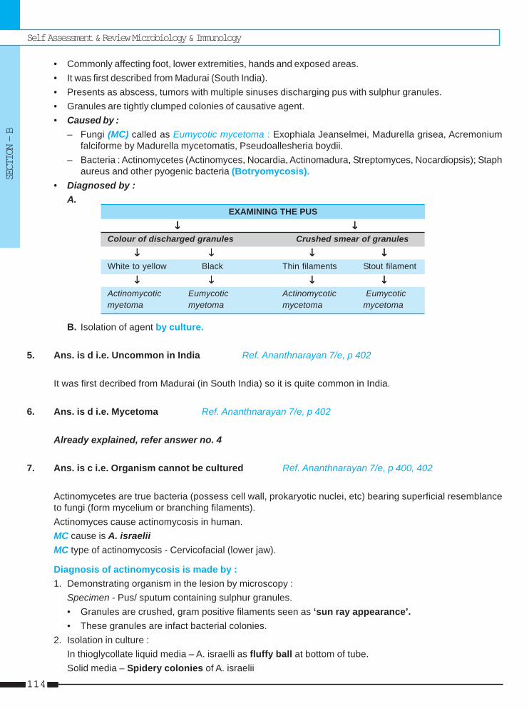
Self Assessment & Review Microbiology & Immunology
SECTION – B
114114
• Commonly affecting foot, lower extremities, hands and exposed areas.• It was first described from Madurai (South India).• Presents as abscess, tumors with multiple sinuses discharging pus with sulphur granules.• Granules are tightly clumped colonies of causative agent.• Caused by :
– Fungi (MC) called as Eumycotic mycetoma : Exophiala Jeanselmei, Madurella grisea, Acremoniumfalciforme by Madurella mycetomatis, Pseudoallesheria boydii.
– Bacteria : Actinomycetes (Actinomyces, Nocardia, Actinomadura, Streptomyces, Nocardiopsis); Staphaureus and other pyogenic bacteria (Botryomycosis).
• Diagnosed by :A.
EXAMINING THE PUS↓↓↓↓↓ ↓ ↓ ↓ ↓ ↓
Colour of discharged granules Crushed smear of granules↓↓↓↓↓ ↓ ↓ ↓ ↓ ↓ ↓↓↓↓↓ ↓ ↓ ↓ ↓ ↓
White to yellow Black Thin filaments Stout filament
↓↓↓↓↓ ↓ ↓ ↓ ↓ ↓ ↓↓↓↓↓ ↓ ↓ ↓ ↓ ↓Actinomycotic Eumycotic Actinomycotic Eumycoticmyetoma myetoma mycetoma mycetoma
B. Isolation of agent by culture.
5. Ans. is d i.e. Uncommon in India Ref. Ananthnarayan 7/e, p 402
It was first decribed from Madurai (in South India) so it is quite common in India.
6. Ans. is d i.e. Mycetoma Ref. Ananthnarayan 7/e, p 402
Already explained, refer answer no. 4
7. Ans. is c i.e. Organism cannot be cultured Ref. Ananthnarayan 7/e, p 400, 402
Actinomycetes are true bacteria (possess cell wall, prokaryotic nuclei, etc) bearing superficial resemblanceto fungi (form mycelium or branching filaments).Actinomyces cause actinomycosis in human.MC cause is A. israeliiMC type of actinomycosis - Cervicofacial (lower jaw).
Diagnosis of actinomycosis is made by :1. Demonstrating organism in the lesion by microscopy :
Specimen - Pus/ sputum containing sulphur granules.• Granules are crushed, gram positive filaments seen as ‘sun ray appearance’.• These granules are infact bacterial colonies.
2. Isolation in culture :In thioglycollate liquid media – A. israelli as fluffy ball at bottom of tube.Solid media – Spidery colonies of A. israelii
Actinomycetes & Bacillus
UNIT - I BACTERIOLOGY “Gram +ve Bacilli”
115
8. Ans. is a i.e. Acid fast Ref. Jawetz 24/e, p220
• No cardiac is urease positive, catalase positive partially acid fast organism.Stains for nocardia :Acid fast staining - Nocardia cell wall contains mycolic acid that are shorter chain than mycobacteria. If theyare stained with routine acid fast reagant (carbol-fuchsin) but decolorized with 1-4% sulfuric acid instead ofthe stronger acid decolorent, most isolate will stain acid fast.Silver stains.
9. Ans. is a i.e. Organism Ref. Ananthnarayan 7/e, p 401
Already explained, refer answer no. 7
10. Ans. is a i.e. Paraffin bait technique Ref. Harrison 17/e, p 994 - 995
Diagnosis of NocardiosisSpecimen – Sputum or pus or spinal fluid or urine or biopsy material.i. Microscopy – Crooked, branching, beaded, gram positive filament seen.ii. Stain – They are Acid fast and also take silver stains.iii. Isolation – Paraffin baiting mixed culture’s done as it use paraffin as carbon source.iv. In case of pneumonia, sampling done by bronchoscopy or lung aspiration but transtracheal aspiration
should be avoided as it frequently leads to nocardial cellulitis around the puncture wound.v. If brain involved – CT or MRIvi. In actinomycetoma – granules are examined.
Remember : – Castaneda culture is method of blood culture (eg. S. typhi, Brucella).– Craigie’s tube is used for the separation of motile from non-motile bacteria and also used
to obtain phase variants in salmonella species.
11. Ans. is d i.e. Anthrax of skin Ref. Ananthnarayan 7/e, p 244
Cutaneous anthrax is also known as Hide porter’s disease or malignant pustule.
Remember : Carbuncle is infection of 2 - 3 hair follicles.
12. Ans. is a i.e. Madurella Ref. Harrison 17/e, p 1266
Actinomycetoma, usually responds to antibiotics.
Treatment of Mycetoma
Actinomycetoma Eumycetoma
Prolonged combination chemotherapy eg. with Rarely responds to chemotherapy, some casesstreptomycin and either dapsone or caused by Madurella mycetomatic respond tocotrimoxazole Ketoconazole or itraconazole
Self Assessment & Review Microbiology & Immunology
SECTION – B
116116
13. Ans. is c i.e. Cutaneous type is rare nowdays Ref. Ananthnarayan 7/e, p 244
Cutaneous anthrax is MC type of anthrax.For more details, refer answer no. 2
14. Ans. is a i.e. Bacillus cereus Ref. Ananthnarayan 7/e, p 246
This is a characteristic presentation of B.cereus food poisoning (emetic type).
Features Diarrheal type Emetic Type
Incubation period 8-16 hrs 1-5 hrs
Food Cooked meat and vegetables Exclusively by rice
Enterotoxin Resemble heat labile toxin of E.coli Resemble heat stable toxin of S.aureusEither preformed or produced in Already preformed in riceintestine
Clinical Features Fever, Vomitting rare Diarrhea is not common
Serotype 2, 6, 8, 9, 10, 12 1, 3, 5
15. Ans. is a, b and c i.e. Actinomyces; Nocardiasis; and Streptomyces Ref. Jawetz 23/e, p 633
MycetomaLocalised chronic granulomatous involvment of the subcutaneous and deeper tissue.
Eumycotic mycetoma Actinomycotic mycetoma= Fungal (More common) = Bacterial
– Madurella mycetomatis – Actinomyces– Pseudollescheria boydii – Nocardia– Madurella grisea – Streptomyces– Acremonium falciforme – Nocardiposis– Exophiala jeanselmei
Remember : • Most common site of mycetoma - foot - (Called as Madura foot).• Staphylococcus is causative agent of botryomycosis.
16. Ans. is b i.e. ZN stain Ref. Ananthnarayan 7/e, p 401, 402; Harrison 17/e, p 994
Both are gram positive filaments causing mycetoma.
Features Actinomyces Nocardia
Morphology Non acid fast Acid fast (Ziehl and Neelsen stain); Fite Faraco Method
Growth in media Anaerobes Aerobic
Mode of infection Endogenous Exogenous
Diseases MC is cervicofacial MC is pneumonia and disseminated disease
Treatment Penicilin G Sulfonamides
Paraffin Can’t use Can use
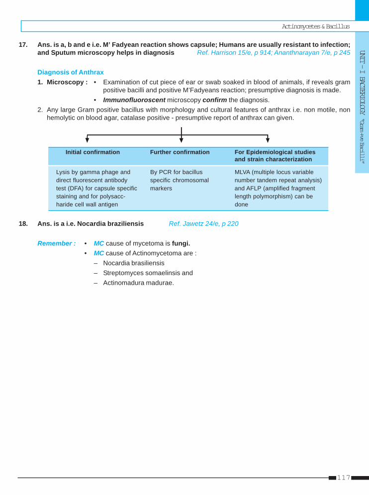
Actinomycetes & Bacillus
UNIT - I BACTERIOLOGY “Gram +ve Bacilli”
117
17. Ans. is a, b and e i.e. M’ Fadyean reaction shows capsule; Humans are usually resistant to infection;and Sputum microscopy helps in diagnosis Ref. Harrison 15/e, p 914; Ananthnarayan 7/e, p 245
Diagnosis of Anthrax1. Microscopy : • Examination of cut piece of ear or swab soaked in blood of animals, if reveals gram
positive bacilli and positive M’Fadyeans reaction; presumptive diagnosis is made.• Immunofluoroscent microscopy confirm the diagnosis.
2. Any large Gram positive bacillus with morphology and cultural features of anthrax i.e. non motile, nonhemolytic on blood agar, catalase positive - presumptive report of anthrax can given.
Initial confirmation Further confirmation For Epidemiological studiesand strain characterization
Lysis by gamma phage and By PCR for bacillus MLVA (multiple locus variabledirect fluorescent antibody specific chromosomal number tandem repeat analysis)test (DFA) for capsule specific markers and AFLP (amplified fragmentstaining and for polysacc- length polymorphism) can beharide cell wall antigen done
18. Ans. is a i.e. Nocardia braziliensis Ref. Jawetz 24/e, p 220
Remember : • MC cause of mycetoma is fungi.• MC cause of Actinomycetoma are :
– Nocardia brasiliensis– Streptomyces somaelinsis and– Actinomadura madurae.
Self Assessment & Review Microbiology & Immunology
SECTION – B
118118
1. b) Bacillus 2. d) Circumvento ... 3. a) Extremely ...4. a) Anthrax 5. a) Gram+ve ... 6. a) Bacillus ...7. c) Candida ... 8. c) Non-motile 9. d) Bacillus ...
1. Medussa head colony is found in : [UP 00]a) Clostridiumb) Bacillusc) Pneumococcusd) Staph. aureus
[Ref. Ananthnarayan 7/e, p 242]2. All are causes of madura mycosis except :
a) Streptomyces [UP 02]b) Nocardiac) Actino boydiid) Circumvento-geniculta
[Ref. Ananthnarayan 7/e, p 402]3. All are true about cutaneous anthrax except :
a) Extremely painful lesions [UP 02]b) The whole area is congested and edematousc) Central crustation with black eschard) Satellite nodule around inguinal region
[Ref. Ananthnarayan 7/e, p 244, Harrison 16/e, p115]
4. A wool cutter is suffering from fever with a cervi-cal lymphnode enlargement for last 15 days, mostlikely he is suffereing from : [UP 04]a) Anthraxb) Mycetomac) Sporotrichosisd) Coccidimycosis
[Ref. Ananthnarayan 7/e, p 244]5. Actinomycosis caused by : [Bihar 05]
a) Gram+ve organism
Answer
b) Gram–ve bacteriac) Anaerobic bacteriad) Fungus
[Ref. Ananthnarayan 7/e, p 400]6. Medusa head appearance : [Jharkhand 05]
a) Bacillus anthraxb) Bacillus subtulisc) Bacillus cereusd) Bacillus licheniformis
[Ref. Ananathnarayan 7/e, p 241]7. Sulphure granules are seen in :
a) Rhinoscleroma [UP 06]b) Actinomycetesc) Candida fungid) Listeria-mono
[Ref. Ananthnarayan 7/e, p 400-401]8. Anthrax bacilli differes from anthracoid bacilli by
being : [Kar 06]a) Non-capsulatedb) Strict aerobec) Non-motiled) Hameolytic colonies on blood
[Ref. Ananthnarayan 7/e, p 246]9. Malignant pustule is seen in infection :
a) Yersinia pestis [Kar 06]b) Bacillus cereusc) Clostridium welchiid) Bacillus anthracic
[Ref. Ananthnarayan 7/e, p 244]
Chapter Review••••• This section includes questions of VThis section includes questions of VThis section includes questions of VThis section includes questions of VThis section includes questions of Various Other PGMEES from 1990 – 2007.arious Other PGMEES from 1990 – 2007.arious Other PGMEES from 1990 – 2007.arious Other PGMEES from 1990 – 2007.arious Other PGMEES from 1990 – 2007.••••• Questions are arQuestions are arQuestions are arQuestions are arQuestions are ar ranged in decreasing order of yearranged in decreasing order of yearranged in decreasing order of yearranged in decreasing order of yearranged in decreasing order of yearwiz. This is done to makwiz. This is done to makwiz. This is done to makwiz. This is done to makwiz. This is done to make refere refere refere refere refer ralralralralral
system easier and uncomplicated to save the precious time of PGMEE Aspirant.system easier and uncomplicated to save the precious time of PGMEE Aspirant.system easier and uncomplicated to save the precious time of PGMEE Aspirant.system easier and uncomplicated to save the precious time of PGMEE Aspirant.system easier and uncomplicated to save the precious time of PGMEE Aspirant.

• Short coccoid, Catalase positive, Gram positive non-spore forming rod with tendency to occur in chains.• Shows slow tumbling motility at 200-250 C and is non motile at 370C.
Culture• Grows on ordinary media. (Muller - hinton agar).• Growth is improved if material is stored in tryptose phosphate or thioglycollate broth at 40 C called cold enrichment.
Listeria monocytogenes can be differentiated from other Listeria by :• β hemolysis on sheep blood agar.• Production of acid from glucose, mannose but not from D.xylose.
Listeria monocytogenes is divided into serotypes on the basis of somatic [O] or flagellar [H] antigen.• Most human infections are caused by 1/2a, 1/2b, 4b.• Human disease due to L.monocytogenes generally occurs in pregnancy or Immunosuppression.
Mode of TransmissionFood borne [Ready to eat food are most likely].
Pathogenesis• Intracellular pathogen [so, no role of humoral immunity] hence immunity is primarily cell - mediated.• Lack of gastric acidity increase risk.• Most important determinant of pathogenesis is Listerolysin O. ... Harrison 17/e, p 896• Life cycle of Listeria monocytogenes in host macrophages includes following steps :
The bacterial product listeriolysin O lyses
the phagolysosome,
allowing the escape of the
listeria.
1 2 3 4Listeria is
phagocytosed by a epithelial cell
and incorporated into a phagolysosome.
Listeria has cell surface protein
called internalin that interacts with
E.cadherin receptor on epithelial cells
promoting phagocytosis.
Listeria multiplies and assembles an actin
filament tail thatpushes the
bacterium to the surface of themacrophage.
5A filopods extension
forms, facilitating transfer
of the listeria into another
phagocyte.
Listeria MonocytogenesListeria MonocytogenesListeria MonocytogenesListeria MonocytogenesListeria Monocytogenes77
Self Assessment & Review Microbiology & Immunology
SECTION – B
120120
Clinical presentation1. Pregnancy Associated Listeriosis :
• Most infections detected in 3rd trimester.• Woman experience mild illness characterized by fever, myalgia, backache.• Transplacental spread results in chorioamnionitis, premature labor, intrauterine fetal death, stillbirth, early
onset disease of newborn, recurrent spontaneous abortion.2. Neonatal Listeriosis :
• Early onset - Before 7 days• Late onset - 7 - 21 days
A. Early onset disease : Most infants are symptomatic by 2nd day.• Transmitted by aspiration of infected amniotic fluid.• Present as : a. Intrauterine sepsis
b. Respiratory distressc. Skin lesionsd. Granulomatosis infantisepticum – Characterized by abscesses involving liver, spleen,
adrenal gland and other sites.• Mostly follow complicated labor.
B. Late onset disease :• Mostly present as meningitis• Born at term by uncomplicated labor• Transmitted during passage through birth canal.
3. Listeriosis not associated with pregnancy :• MC underlying condition are chronic glucocorticoid therapy, diabetes, solid and hematologic malignancy
(particularly fludarabine treated), liver disease, AIDS• Bacteremic infection without evident focus is MC clinical manifestation while infection in CNS ranks 2nd in
which meningitis is MC. It can directly invade brain parenchyna producing carebritis or focal abscess.• L.monocytogenes is MC cause of meningitis in kidney transplant patient after 1 month.• Listeria meningitis must be considered in chronically ill patient with aseptic meningitis particularly when
presentation is subacute.
Diagnosis• Invasive listeriosis is diagnosed when organism is cultured from Blood, CSF or amniotic fluid.• Antibody to listeriolysin O :
– For epidemiological purpose.– For diagnosis of culture negative CNS infection.
• Antons test – Instillation into rabbit eye cause conjuctivitis.
Treatment• IV administration of Ampicillin (DOC) or penicillin often in combination with aminoglycoside.• Cotrimoxazole in case of pencillin allergy.• Cephalosporins are not effective.
Listeria Monocytogenes
UNIT - I BACTERIOLOGY “Gram +ve Bacilli”
121
1. A 30 year old woman with a bad obstetric historypresents with fever. The blood culture from thepatient grows gram-positive small to mediumcoccobacilli that are pleomorphic, occurring inshort chains. Direct wet mount from the cultureshows tumbling motility. The most likely organ-ism is : [AI 04]a) Listeria monocytogenesb) Corynebacteriumsp.c) Enterococcus spd) Erysepelothrix rusiopathiae
2. All the following are true about Listeria except :a) Transmitted by contaminated milk [AI 02]b) Gram (-)ve bacteriac) Causes abortion in pregnancyd) Causes meningitis in neonates
3. A 3 week old child presented to the pediatricianwith meningitis A presumptive diagnosis of lateonset of perinatal infection was made . The CSFculture was positive for gram positive bacilliwhich of the following characteristic of this bac-teria would be helpful in differentiating it fromother bacterial agents : [AIIMS 05]
a) Ability to grow on blood agarb) Ability to produce catalasec) Fermentative attack on sugarsd) Motility at 250C
4. A major step in the pathogenesis of listeriosis is:a) The formation of antigen-antibody complex with
resultant complement activation and tissue dam-age [AIIMS 05]
b) The release of hyaluronidase by L. monocyto-genes, which contributes to its disseminationfrom local sites
c) The antiphagocytic activity of the L. monocyto-genes capsule
d) The survival and multiplication of L. mono-cytogenes within mononuclear phagocytes andhost epithelial cells
5. In patient with Listeria meningitidis who is aller-gic penicillin the treatment of choice is :a) Vancomycin [AIIMS 04]b) Gentamycinc) Trimethoprim - sulphome-thoxazoled) Ceftriaxone
QUESTIONS
Answer 1. a) Listeria ... 2. b) Gram (-)ve ... 3. d) Motility at ...
4. d) The survival ... 5. c) Trimethoprim ...
Self Assessment & Review Microbiology & Immunology
SECTION – B
122122
1. Ans. is a i.e. Listeria monocytogenes Ref. Ananthnarayan 7/e, p 403
Tumbling motility is characteristic of Listeria monocytogenes - (other three are non motile).
Listeria monocytogenes is :– Catalase positive, non-sporing gram positive, Cocco bacilli.– Tendency to occur in chains.– Characterisitics slow tumbling motility at 250C and at 370C is non-motile because peritrichous flagella are
produced optimally at 20 to 300C but only scantily or not at all at 370C.– Grows best between 300C and 370C (temperature range is 1 to 450C).– Aerobic or microaerophilic.– Intacellular as well as able for direct cell to cell spread so not eliminate by antibodies and cause infection
in deficient cell mediated immunity.
Mode of transmission of Listeriosis– Food borne by ready to eat foots (eg. pasteruized milk, cheese, pate, undercooked chicken).– Nosocomial transmission.
Forms of Human Listerosis
Pregnancy associated listeriosis Neonatal listeriosis Listeriosis not associatedwith pregnancy
• Most infections in 3rd a) Early onset - transmitted • MC underlying conditiontrimester by aspration of infected in non pregnant adults is
amniotic fluid chronic glucocorticoid• Transplacental spread b) Late onset - transmitted therapy
result in recurrent sponta during passage throughneous abortion, premature birth canallabour, IUD, chorioamnionitis Mostly present as meningitis
Remember :• E. rhusiopahtial is a-hemolytic non-motile Gram positive bacillus with tendency to form long filaments.• Its MC infection in humans is called erysipeloid = Seal finger = whole finger.
2. Ans. is b i.e. Gram –ve bacteria Ref. Harrison 17/e, p 895 - 897
Already explained, please see answer no. 1
3. Ans. is d i.e. Motility at 250 C Ref. Ananthnarayan 7/e, p 403
• This is a case of ‘Late onset neolnatal meningitis’ of Listeria monocytogenes as culture reveals grampositive bacillus.
EXPLANATIONS AND REFERENCES WITH ILLUSTRATIVE ANSWERS
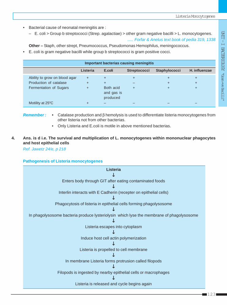
Listeria Monocytogenes
UNIT - I BACTERIOLOGY “Gram +ve Bacilli”
123
• Bacterial cause of neonatal meningitis are :– E. coli > Group b streptococci (Strep. agalactiae) > other gram negative bacilli > L. monocytogenes.
..... Forfar & Anelus text book of pedia 319, 1338Other – Staph, other strept, Pneumococcus, Pseudomonas Hemophilus, meningococcus.
• E. coli is gram negative bacilli while group b streptococci is gram positive cocci.
Important bacterias causing meningitis
Listeria E.coli Streptococci Staphylococci H. influenzae
Ability to grow on blood agar + + + + +Production of catalase + + – + +Fermentation of Sugars + Both acid + + +
and gas isproduced
Motility at 250C + – – – –
Remember : • Catalase production and β hemolysis is used to differentiate listeria monocytogenes fromother listeria not from other bacterias.
• Only Listeria and E.coli is motile in above mentioned bacterias.
4. Ans. is d i.e. The survival and multiplication of L. monocytogenes within mononuclear phagocytesand host epithelial cellsRef. Jawetz 24/e, p 218
Pathogenesis of Listeria monocytogenes
Listeria↓↓↓↓↓
Enters body through GIT after eating contaminated foods↓↓↓↓↓
Interlin interacts with E Cadherin (recepter on epithelial cells)↓↓↓↓↓
Phagocytosis of listeria in epithelial cells forming phagolysosome↓↓↓↓↓
In phagolysosome bacteria produce lysteriolysin which lyse the membrane of phagolysosome↓↓↓↓↓
Listeria escapes into cytoplasm↓↓↓↓↓
Induce host cell actin polymerization↓↓↓↓↓
Listeria is propelled to cell membrane↓↓↓↓↓
In membrane Listeria forms protrusion called filopods↓↓↓↓↓
Filopods is ingested by nearby epithelial cells or macrophages↓↓↓↓↓
Listeria is released and cycle begins again
Self Assessment & Review Microbiology & Immunology
SECTION – B
124124
So, in this way Listeria can move from cell to cell without being exposed to antibodies, complement or polymorphs.
Remember :• Iron is important virulence factor of Listeria• Shigella flexneri and rickettsia also use the host cell actin and contractile system to spread infection.
5. Ans. is c i.e. Trimethoprim - sulphomethoxazole Ref. Harrison 17/e, p 897
• DOC for listeriosis (non pregnant, neonate, pregnant) is ampicillin or penicillin often in combination withaminoglycosides.
• During last month of pregnancy (in case of penicillin allergy), may be treated with erythromycin.• Other wise in all cases of penicillin allergy DOC is trimethoprim-sulfamethoxazole.
Listeria Monocytogenes
UNIT - I BACTERIOLOGY “Gram +ve Bacilli”
125
3. d) Listeria
1. Tumbling motility is shown by : [DNB 04]a) Proteus vulgarisb) Proteus mirabilisc) Virbiod) Listeria
[Ref. Ananthnarayan 7/e, p 403]
Answer
Chapter Review••••• This section includes questions of VThis section includes questions of VThis section includes questions of VThis section includes questions of VThis section includes questions of Various Other PGMEES from 1990 – 2007.arious Other PGMEES from 1990 – 2007.arious Other PGMEES from 1990 – 2007.arious Other PGMEES from 1990 – 2007.arious Other PGMEES from 1990 – 2007.

Gram positive, Aerobic, acid fast, non motile, non capsulated and non sporing fungus like bacteria.Classification of mycobacteria is as follows :1. Tubercle bacilli - M.tuberculosis, M.bovis, M. africanum2. Lepra bacilli - M.leprae3. Mycobacteria causing skin ulcers - M.ulcerans, M.haemophilum, M.marinum or balnei4. Atypical mycobacteria = Nontuberculous = Paratubercle = MOTT
Group I Photochromogenes Eg. M.kansasii, M.marinum, M.simiae, M.asiaticum.
Group II Scotochromogens Eg. M.scrofulaceum, M.gordonae, M.szulgai, M. flavescens.
Group III Nonphotochromogens Eg. M.avium, M.intracellulare, M.xenopi, M.ulcerans, M.malmoense, M celatum,M. Hemophilum, M. gastri, M. genavense, M. shimoidei, M. trivale, M. terrae, M. nonchromogenicum.
Group IV Rapid growers – M.fortuitum, M.chelonae Chromogenic rapid growers are saprophytes Eg. M.smegmatic,M.phlei.
5. Johne’s bacillus - M.paratuberculosis
MYCOBACTERIA LEPRAEMorphology• Obligate intracellular bacilli with polar bodies and intracellular elements, resist decolourization of 5% H2SO4
(acid fast).• Live bacilli in tissue (solid and uniformly stained) - Morphological index (MI).• Dead bacilli – fragmented and granular appearance.• Bacteriological index (BI) – number of bacilli in tissue.• BI and MI suggests relapse/drug resistance if increase.• Bacilli arranged intracellularly in the form of globi, having ‘cigar bundle’ appearance.• Virchow’s lepra cells or foamy cells are large undifferentiated histiocytes in origin.• Strain variability is demonstrated recently.• (PGL-I) phenolic glycolipid act as virulence factor.
Culture• Unique in exhibiting dopa oxidase activity and acid fastness that is pyridine extractable.
MycobacteriaMycobacteriaMycobacteriaMycobacteriaMycobacteria88
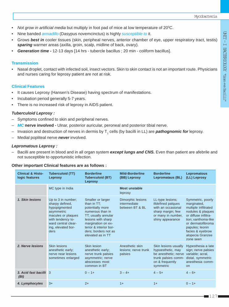
Mycobacteria
UNIT - I BACTERIOLOGY “Gram +ve Bacilli”
127
• Not grow in artificial media but multiply in foot pad of mice at low temperature of 200C.• Nine banded armadillo (Dasypus novemcinctus) is highly susceptible to it.• Grows best in cooler tissues (skin, peripheal nerves, anterior chamber of eye, upper respiratory tract, testis)
sparing warmer areas (axilla, groin, scalp, midline of back, ovary).• Generation time - 12-13 days [14 hrs - tubercle bacillus ; 20 min - coliform bacillus].
Transmission• Nasal droplet, contact with infected soil, insect vectors. Skin to skin contact is not an important route. Physicians
and nurses caring for leprosy patient are not at risk.
Clinical Features• It causes Leprosy (Hansen’s Disease) having spectrum of manifestations.• Incubation period generally 5-7 years.• There is no increased risk of leprosy in AIDS patient.
Tuberculoid Leprosy :– Symptoms confined to skin and peripheral nerves.– MC nerve involved - Ulnar, posterior auricular, peroneal and posterior tibial nerve.– Invasion and destruction of nerves in dermis by T1 cells (by bacilli in LL) are pathognomic for leprosy.– Medial popliteal nerve never involved.
Lepromatous Leprosy :– Bacilli are present in blood and in all organ system except lungs and CNS. Even than patient are afebrile and
not susceptible to opportunistic infection.
Other important Clinical features are as follows :
Clinical & Histo- Tuberculoid (TT) Borderline Mild-Borderline Borderline Lepromatouslogic features Leprosy Tuberculoid (BT) (BB) Leprosy Lepromataus (BL) (LL) Leprosy
Leprosy
MC type in India Most unstableleprosy
1. Skin lesions Up to 3 in number; Smaller or larger Dimorphic lesions LL-type lesions; Symmetric, poorlysharpy defined, than in TT; intermediate illdefined palques marginated,hypopigmented potentially more between BT & BL with an occasional multiple infiltratedasymmetric numerous than in sharp margin; few nodules & plaquesmacules or plaques TT; usually annular or many in number, or diffuse infiltra-with tendency to- lesions with sharp shiny appearance tion; xanthoma-likeward central clear- margination on ex- or dermatofibromaing, elevated bor- terior & interior bor- papules; leoninders ders; borders not as facies & eyebrow
elevated as in TT alopecia Granzeezone seen
2. Nerve lesions Skin lesions Skin lesion Anesthetic skin Skin lesions usually Hypesthesia a lateanesthetic early; anesthetic early; lesions; nerve trunk hypoesthetic, may sign; nerve palsiesnerve near lesions nerve trunk palsies palsies be anesthetic; nerve variable; acral,sometimes enlarged asymmetric; nerve trunk palsies comm- distal, symmetric
abscesses most on & frequently anesthesia comm-common in BT symmetric on
3. Acid fast bacilli 3 0 – 1+ 3 – 4+ 4 – 5+ 4 – 6+ (BI)
4. Lymphocytes 3+ 2+ 1+ 1+ 0 – 1+
Self Assessment & Review Microbiology & Immunology
SECTION – B
128128
Continue .....
5. Macrophage Epithelioid Epithelioid Epithelioid Usually undifferen- Foamy change the differentiation tiated; epithelioid rule; may be un-
foci sometimes differentiated inpresent; may show early lesionsfoamy change
6. Langhan’s 1 – 3+ 2+ – – – ginat Cells
7. Lepromin test +++ +++ – – –
8. Lymphocyte 95% 40% 10% 1 – 2% 1 – 2% transforma- tion test
9. CD.4 +/CD 8+T 1.35 1.11 NT 0.48 0.50 [CD-8/CD-4 = cell ratio in lesions
10. M. leprae 1 + (60%) 2+ 2+ 3+ 3+ (95%)
Reactional statesI. Type I Lepra reaction / Jopling Type I :
• Type IV hypersensitivity seen in Borderline leprosy not in polar form.• If precede therapy than termed down grading reaction i.e. towards LL.• If after therapy than termed Reversal reaction i.e. towards more tuberculoid.• Most characteristic microscopic feature of type I reaction is Edema.• It is associated with increased T cells bearing γ/δ receptors a unique feature of leprosy.• MC nerve trunk involved - Ulnar at elbow• Treatment : DOC glucocorticoids
Clofazimine also givenThalidomide - ineffective.
II. Type 2 lepra reaction – Erythema Nodosum Leproticum / Jopling Type II :• Type III hypersensitivity occurs exclusively in BL, LL• Usually follows therapy (sulfone syndrome) but may precede therapy.• MC feature - crops of painful erythematous papules that resolve spontaneously in a few day to weeks but
may recur.• Central role in pathobiology : TNF• Treatment :
– Mild - antipyretics alone– Moderate to severe – Ist drug to be used glucocorticords
DOC thalidomideClofazimine - More active than in Type I.
III. Lucio’s Phenomenon :• Type III Hypersentivity exclusively in diffuse lepromatosis form of LL, usually in untreated patient.• Treatment : – Neither glucocortiord nor thalidomide is effective.
– Wound care and therapy for bacteremia.
Mycobacteria
UNIT - I BACTERIOLOGY “Gram +ve Bacilli”
129
Complications• MC complication of leprous neuropathy is plantar ulceration particularly at metatarsal heads.• Nerve abscess : – MC site is ulnar nerve.
– Treatment is rapid surgical decompression.
Diagnosis• Biopsy of advancing edge of lesion in TT but in LL, biopsy of normal skin is also taken.• Hyperglobulinemia in LL.• Lepromin test – Type IV delayed hypersentivity which is biphasic.
Early reaction of Fernandez – read in 24 - 48 hours (analogous to tuberculin reaction).Late reaction of Mitsuda – peak in 4 weeks. More meaningful.It is of little diagnostic value but has more prognostic importance.
Treatment
Form of leprosy More Intensive regimen WHO recommended regime
i. Tuberculoid Dapsone 100 mg/d Dapsone 100 mg/d +(paucibacillary) X 5 years Rifampin 600 mg/month for 6 months
ii. Lepromatous Rifampin 600 mg/d Dapsone 100 mg/d +(Multibacillary) or for 3 years + dapsone Clofazimine 50 mg/d and Rifampin> 6 skin lesions 100 mg/d indefinitely 600 mg + clofazimine 300 mg monthly for 1year
• Single lesion paucibacillary leprosy - Single dose of ROM - rifampin, ofloxacin, minocycline.
MYCOBACTERIA TUBERCULOSISMorphology• Mammalian tubercle, isolated by Koch is stained by Ziehl - Neelsen method or by fluorescent dyes (auramine
O, rhodamine).• Resist decolourization by 20% H2SO4 and absolute alcohol for 10 minutes. Hence acid and alcohol fast.• Acid fastness is due to unsaponifiable wax (mycolic acid) or to a semipermeable membrane.• It has thick cell wall; shows spheroplast and L forms.
Culture• Generation time 14 – 15 hours.• Colonies appear in about 2 weeks (may take upto 8 weeks).• Grows luxuriantly in culture (Eugonic) and addition of 0.5% glycerol improves its growth but has no effect on
M.bovis (causative agent of bovine tuberculosis) which is dysgonic (grows sparsely).• Solid medium most widely employed for routine culture is Lowenstein - Jensen (LJ) medium without starch.• Liquid media are not generally used routinely, but used for senstivity testing, chemical analyses and preparation
of antigens and vaccines.• Virulent strain form long serpentine rods in liquid media while avirulent strain grow in dispersed manner. Though
cord factor itself is not a virulence factor but cord formation is coorelated with virulence. ... Jawets 24/e, p 322
Self Assessment & Review Microbiology & Immunology
SECTION – B
130130
BIOCHEMICAL REACTION POSITIVE IN NEGATIVE IN
Niacin Test : N Human tubercle Bovine tubercleAryl Sulphatase : A Only with Atypical mycobacteriaNeutral red test : N Virulent strain of tubercle Avirulent strainPeroxidase test : P Tubercle bacilli Atypical mycobacteriaCatalase test : C Most atypical mycobacteria Weakly positive in tubercleNitrate reduction text : N M.tuberculosis M. bovis
• Catalase and peroxidase activities are lost when tubercle bacilli become INH resistant.• Ureas test is positive in M. tuberculosis, M. bovis and most of the atypical mycobacteria except MAIC complex.
M.tuberculosis M.bovis
Morphology Curved long rod Straighter, shorter, stouter
Stain Less uniform more uniform
O2 requirement Obligate aerobe Microaerophilic
Culture Dry, rough, raised, irregular Flat, smooth, moist, break up easily
Growth Eugonic Dysgonic
Virulence factors :• Kat-G gene : encodes for oxidase, catalase enzyme.• rpoV : main sigma factor initiating transcription of several genes.• Erp gene : encodes for protein required for multiplication.• Strains of Beijing / w genotype family.
Antigenic Property• Group specificity is due to polysaccharide while type specificity is due to protein antigen.• Antibodies are not useful for diagnosis and immunity.
Pathogenicity• It is due to escape killing by macrophages and inducion of type IV hypersensitivity.• Following factors contribute in pathogenesis :
– Cord factor – Lipoarabinomannan– Complement system – M.TB heat shock protein.
• Risk of acquiring infection is determined mainly by exogenous factors while risk of developing disease dependslargely on endogenous factors.
• Most potent risk factors - HIV coinfection.
Clinical features• Divided into two categories : Pulmonary and extrapulmonary TB.
1. Pulmonary TB : Divided into two :a. Primary Disease : usually localized in middle and lower zones.
Primary focus is usually peripheral in subpleural region and is accompained by draining lymphatics, inflamedregional lymph nodes which are collectively called Primary complex/Ghon’s facus.Depending on the host immune response development of complex can follow healing by fibrosis/calcification; cavitation or progressive primary TB in form of consolidation; obstructive emphysema oratelactasis; TB bronchitis; miliary TB; occult hematogenous dissemination to apex of lung (Simons Focus).
Mycobacteria
UNIT - I BACTERIOLOGY “Gram +ve Bacilli”
131
b. Post primary disease (adult type or reactivation or secondary tuberculosis or chronic pulmonary TB).• Usually localized to apical and posterior segments of upper lobe due to high O2 concentration (Puhl’s
Lesion).• MC hematologic finding - mild anemia and leucocytosis• Infraclavicular lesion is called Assman’s Focus.
2. Extrapulmonary TB• MC site lymph node (MC cervical and supraclavicular).• Also involved-Pleura in the form of pleural effusion and empyema.
– Genitourinary tract (culture negative pyuria in acidic urine).– Skeletal TB (MC site spine, hip, knee).– TB meningitis (paresis of cranial nerves especially ocular, is frequent finding).– GI TB (MC site terminal ileum and caecum).– Tuberculous pericarditis (MC cause of chronic constrictive pericarditis).
DiagnosisSpecimen – sputum is best collected in the morning before any meal (3 sample).i. AFB microscopy : smear should be prepared from thick purulent part of sputum.
• Atleast 10000 AFB should be present per ml of sputum for demonstrating in direct smears. Positive reportcan be given only if >2 typical bacilli have seen.
• Fluorescent microscopy (stained with auramine phenol or auramine rhodamine fluorescent dye and examinedunder UV illumination) screened smear rapidly in comparison of Ziehl–Neelsen method.
• Concentration method for microscopy can also used eg. Petroff’s method using NaOH solution is widelyused.
ii. Culture :• Very sensitive diagnostic technique detecting 10 to 100 bacilli per ml.• LJ is standard solid media.• Negative report is given, if no growth occur after 8-12 weeks.• Slow growing, nonpigmented niacin positive AFB is taken as M.tuberculosis.• Liquid media with radiometric growth detection (eg. BACTEC 460) and nucleic acid probes, enables results
to be given in 2-3 weeks.
iii. Nucleic acid technology :• PCR and Ligase chain reaction are used as diagnostic technique.• RFLP and 15 fingerprinting used for epidemiological typing of strain.
iv. Immunodiagnosis :• Demonstation of hypersensitivity to tuberculo protein (tuberculin test / Montoux intradermal test) is a standard
procedure.• 1 purified protein derivative (PPD) = 50000 tuberculin units per milligram.• WHO advocates PPD tuberculin known as - RT 23 with Tween 80.• Routinely 1TU used.• Clinically 5 TU used.• Read after 72 hrs in which induration is measured in horizontal transverse diameter.• > 10 mm positive, < 5 mm negative.• < 6 and > 15 mm have more risk of developing TB.
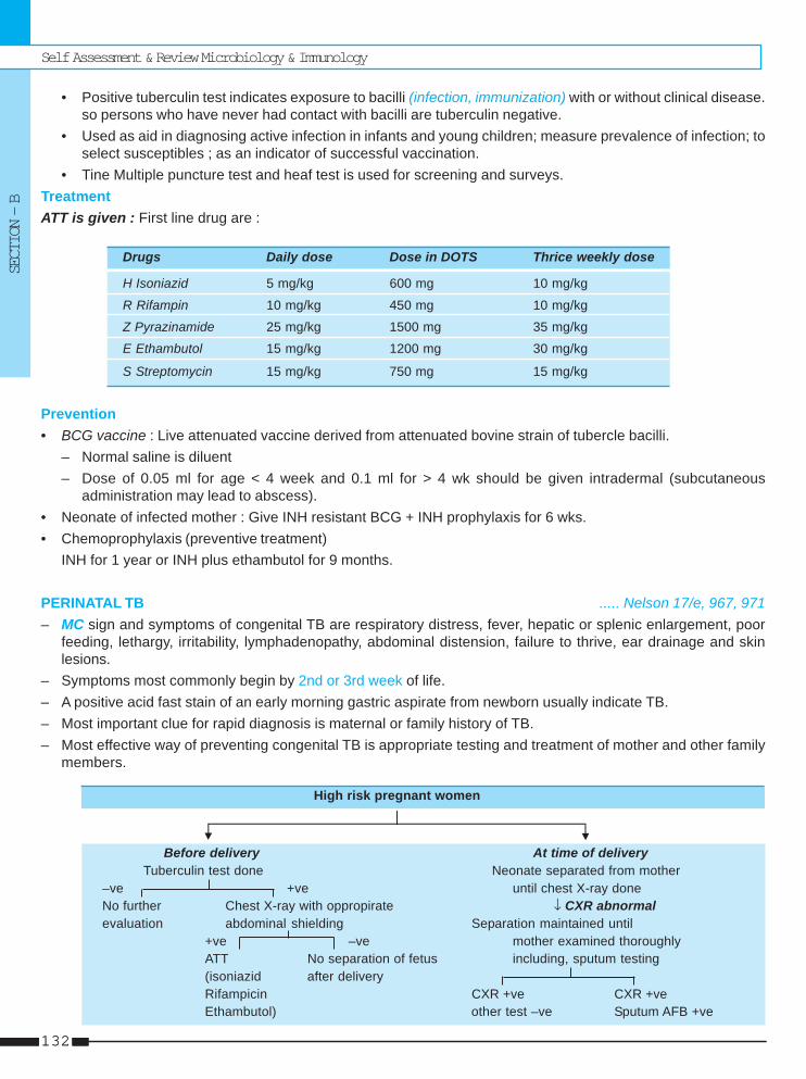
Self Assessment & Review Microbiology & Immunology
SECTION – B
132132
• Positive tuberculin test indicates exposure to bacilli (infection, immunization) with or without clinical disease.so persons who have never had contact with bacilli are tuberculin negative.
• Used as aid in diagnosing active infection in infants and young children; measure prevalence of infection; toselect susceptibles ; as an indicator of successful vaccination.
• Tine Multiple puncture test and heaf test is used for screening and surveys.TreatmentATT is given : First line drug are :
Drugs Daily dose Dose in DOTS Thrice weekly dose
H Isoniazid 5 mg/kg 600 mg 10 mg/kg
R Rifampin 10 mg/kg 450 mg 10 mg/kg
Z Pyrazinamide 25 mg/kg 1500 mg 35 mg/kg
E Ethambutol 15 mg/kg 1200 mg 30 mg/kg
S Streptomycin 15 mg/kg 750 mg 15 mg/kg
Prevention• BCG vaccine : Live attenuated vaccine derived from attenuated bovine strain of tubercle bacilli.
– Normal saline is diluent– Dose of 0.05 ml for age < 4 week and 0.1 ml for > 4 wk should be given intradermal (subcutaneous
administration may lead to abscess).• Neonate of infected mother : Give INH resistant BCG + INH prophylaxis for 6 wks.• Chemoprophylaxis (preventive treatment)
INH for 1 year or INH plus ethambutol for 9 months.
PERINATAL TB ..... Nelson 17/e, 967, 971– MC sign and symptoms of congenital TB are respiratory distress, fever, hepatic or splenic enlargement, poor
feeding, lethargy, irritability, lymphadenopathy, abdominal distension, failure to thrive, ear drainage and skinlesions.
– Symptoms most commonly begin by 2nd or 3rd week of life.– A positive acid fast stain of an early morning gastric aspirate from newborn usually indicate TB.– Most important clue for rapid diagnosis is maternal or family history of TB.– Most effective way of preventing congenital TB is appropriate testing and treatment of mother and other family
members.
High risk pregnant women
Before delivery At time of deliveryTuberculin test done Neonate separated from mother
–ve +ve until chest X-ray doneNo further Chest X-ray with oppropirate ↓ CXR abnormalevaluation abdominal shielding Separation maintained until
+ve –ve mother examined thoroughlyATT No separation of fetus including, sputum testing(isoniazid after deliveryRifampicin CXR +ve CXR +veEthambutol) other test –ve Sputum AFB +ve
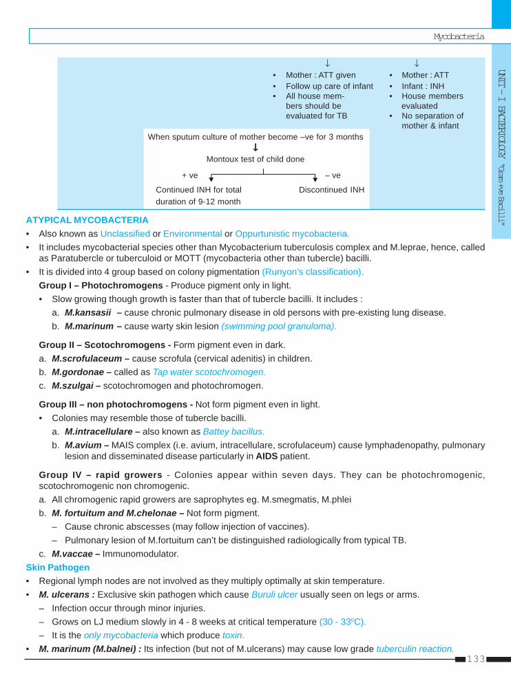
Mycobacteria
UNIT - I BACTERIOLOGY “Gram +ve Bacilli”
133
↓ ↓• Mother : ATT given • Mother : ATT• Follow up care of infant • Infant : INH• All house mem- • House members
bers should be evaluatedevaluated for TB • No separation of
mother & infantWhen sputum culture of mother become –ve for 3 months
↓↓↓↓↓Montoux test of child done
+ ve – ve
Continued INH for total Discontinued INHduration of 9-12 month
ATYPICAL MYCOBACTERIA• Also known as Unclassified or Environmental or Oppurtunistic mycobacteria.• It includes mycobacterial species other than Mycobacterium tuberculosis complex and M.leprae, hence, called
as Paratubercle or tuberculoid or MOTT (mycobacteria other than tubercle) bacilli.• It is divided into 4 group based on colony pigmentation (Runyon’s classification).
Group I – Photochromogens - Produce pigment only in light.• Slow growing though growth is faster than that of tubercle bacilli. It includes :
a. M.kansasii – cause chronic pulmonary disease in old persons with pre-existing lung disease.b. M.marinum – cause warty skin lesion (swimming pool granuloma).
Group II – Scotochromogens - Form pigment even in dark.a. M.scrofulaceum – cause scrofula (cervical adenitis) in children.b. M.gordonae – called as Tap water scotochromogen.c. M.szulgai – scotochromogen and photochromogen.
Group III – non photochromogens - Not form pigment even in light.• Colonies may resemble those of tubercle bacilli.
a. M.intracellulare – also known as Battey bacillus.b. M.avium – MAIS complex (i.e. avium, intracellulare, scrofulaceum) cause lymphadenopathy, pulmonary
lesion and disseminated disease particularly in AIDS patient.
Group IV – rapid growers - Colonies appear within seven days. They can be photochromogenic,scotochromogenic non chromogenic.a. All chromogenic rapid growers are saprophytes eg. M.smegmatis, M.phleib. M. fortuitum and M.chelonae – Not form pigment.
– Cause chronic abscesses (may follow injection of vaccines).– Pulmonary lesion of M.fortuitum can’t be distinguished radiologically from typical TB.
c. M.vaccae – Immunomodulator.Skin Pathogen• Regional lymph nodes are not involved as they multiply optimally at skin temperature.• M. ulcerans : Exclusive skin pathogen which cause Buruli ulcer usually seen on legs or arms.
– Infection occur through minor injuries.– Grows on LJ medium slowly in 4 - 8 weeks at critical temperature (30 - 330C).– It is the only mycobacteria which produce toxin.
• M. marinum (M.balnei) : Its infection (but not of M.ulcerans) may cause low grade tuberculin reaction.
Self Assessment & Review Microbiology & Immunology
SECTION – B
134134
1. Leprosy affects all the following except :a) Testes [AI 07]b) Ovariesc) Eyesd) Nerves
2. Which one of the following statement is true re-garding pathogenicity of Mycobacteria species:a) M. tuberculosis is more pathogenic than M.bovis
to humans [AI 06]b) M. Kansasii can cause disease indistinguishable
from tuberculosisc) M. africanum infection is acquired from environ-
mental sourced) M. marinum is responsible for tubercular lym-
phadenopathy3. In the management of leprosy, Lepromin test is
most useful for : [AI 03]a) Herd immunityb) Prognosisc) Treatmentd) Epidemological investigations
4. Which of the following is true regarding globi ina patient with lepromatous leprosy : [AI 02]a) Consists of lipid laden macrophagesb) Consist of macrophages filled with AFBc) Consists of neutrophils filled with bacteriad) Consists of activated lymphocytes
5. Basanti, 29 year aged female from Bihar presentwith active TB. She delivers baby. All of the fol-lowing are indicated except : [AI 01]a) Administer INH to the babyb) Withhold breast feedingc) Give ATT to mother for 2 yearsd) Ask mother to ensure proper disposal of sputum
6. Tuberculin test denotes : [AI 00]a) Previous or present sensitivity to tubercule pro-
teinsb) Patient is resistant to TBc) Person is susceptible to TBd) Protective immune status of individual against TB
7. The medium used for Mycobacterium tuberculo-sis is : [AI 96]a) Sabouraud’s meidumb) L J meidumc) Pick’s mediumd) NIH medium
8. Rapid examination of Tubercle bacilli is possiblewith : [AI 96]a) Ziel-nelson stainb) Kinyoun stainc) Auramine-Rhodamine staind) Giemsa stain
9. The commonsest focus of Scrofuloderma is :a) Lung [AI 96]b) Lymph nodec) Larynxd) Skin
10. For experimental work, Lepra bacilli are best cul-tured in : [AI 96]a) Armadillosb) Mouse foot padc) Guinea pigsd) Rabbit testes
11. True about mycobacterium other than tuberculo-sis : [AIIMS 08]a. Causes disseminated infectionb. Occurs in persons with normal immunityc. Causes decreased efficacy of BCG due to corss
immunityd. Person to person transmission is seen
12. The main cytokine, involved in erythema nodosumleprosum (ENL) reaction, is :a) Interleukin-2 [AIIMS 06]b) Interferon - gammac) Tumor necrosis factor - alphad) Macrophage colony stimulating factor
13. The following drug is not used for the treatmentof type II lepra reaction : [AIIMS 06]a) Chloroquinb) Thalidomidec) Cyclosporined) Corticosteroids
Answer 1. b) Ovaries 2. b) M. Kansasii ... 3. b) Prognosis 4. b) Consist ... 5. b) Withhold ...6. a) Previous ... 7. b) L J meidum 8. c) Auramine ... 9. b) Lymph ... 10. b) Mouse ...
11. c) Causes ... 12. c) Tumor ... 13. c) Cyclosporine
QUESTIONS
Mycobacteria
UNIT - I BACTERIOLOGY “Gram +ve Bacilli”
135
14. The following test is not used for diagnosis of lep-rosy : [AIIMS 06]a) Lepromin testb) Slit skin smearc) Fine needle aspiration cytologyd) Skin biopsy
15. Which of the following statement about lepromintest is not true : [AIIMS 07, 06]a) It is negative in most children in first 6 months of
lifeb) It is a diagnostic testc) It is an important aid to classify type of leprosy
diseased) BCG vaccination may convert lepra reaction from
negative to positive16. Under leprosy eradication programme the man-
agement of single lesion is : [AIIMS 02]a) Single dose of Rifampicin and Dapsoneb) Rifampicin and Dapsone for 6 monthsc) Rifampicin, Ofloxacin and Minocycline single
dosed) Rifampicin and Monocycline for 6 months
17. Collection of urine sample of a patient of TB kid-ney is done : [AIIMS 00]a) 24 hrs urineb) 12 hrs urinec) In early morningd) Any time
18. The characteristic finding in a case of leprosy is:a) Culture test is positive in 2-3 months in LJ mediab) Long contact with tuberculoid leprosy can trans-
mit the disease [AIIMS 98]c) CMI is seen in Lepromatous leprosyd) Macule lesion heals spontaneously
19. True about mantoux test : [PGI 03]a) <5mm always +veb) Usually -ve after treatmentc) Positive reaction in children <2 is not improtant
like in adultd) Usually read after 48 - 72 hourse) False +ve in post measles state
20. True regarding mycobacterium tuberculosis is :a) Produces visible colonies in 1 week time on
Lowenstein Jenson media [PGI 02]b) Decolorised by 20% sulphuric acid
c) Faculatative aerobed) Niacin positive
21. Which of the following are acid fast positive with20% sulphuric acid : [PGI 02]a) M. aviumb) M. lepraec) M. tuberculosisd) Nocardiae) Rhizopus
22. Selective media for TB bacilli is : [PGI 01]a) NNN mediab) Dorset mediac) LJ. mediad) Nutrient agare) Mac’Conkey media
23. Exacerbation of lesions in patients of borderlineleprosy is seen in : [PGI 01]a) ENL (erythema nodosum leprosum)b) Lepra reaction type 1c) Jarisch-Herxheimer reactiond) Resolving leprosy
24. Not easily culturable but well viable and used inepidemiology are : [PGI 00]a) Staphb) Mycobacterium TBc) E.Colid) Salmonella
25. True about tuberculin test are all except :a) Recent conversion in adult is an indication for
ATT [PGI 98]b) INH prophylaxis is started if the test is positivec) No risk in negatived) May be false negative in immunocompromised
patients26. Mycobacterium leprae can be grown on :
a) Culture media [PGI 98]b) Foot pad of mouse / Armadilloc) Liver of guinea pigd) Kidney of rabbit
27. In TB, immunity is provided by : [PGI 98]a) CD 4+b) CD 8+c) IgGd) IgM
Answer 14. a) Lepramin ... 15. b) It is a ... 16. c) Rifampicin ... 17. c) In early ... 18. b) Long ...19. d) Usually ... 20. d) Niacin ... 21. c) M. tuber ... 22. b and c 23. a and b24. b) Mycobacte ... 25. c) No risk ... 26. b) Foot pad ... 27. a) CD 4+
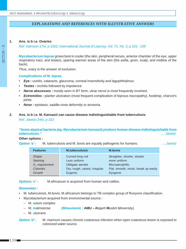
Self Assessment & Review Microbiology & Immunology
SECTION – B
136136
1. Ans. is b i.e. OvariesRef. Harrison 17/e, p 1022; International Journal of Leprosy, Vol. 71, No. 2, p 101 - 105
Mycobacterium leprae grows best in cooler (the skin, peripheral nerves, anterior chamber of the eye, upperrespiratory tract, and testes), sparing warmer areas of the skin (the axilla, groin, scalp, and midline of theback).Thus, ovary is the answer of exclusion.
Complications of M. leprae• Eye : uveitis, cataracts, glaucoma, corneal insensitivity and lagophthalmus.• Testes : orchitis followed by impotence.• Nerve abscesses : mostly seen in BT form, ulnar nerve is most frequently involved.• Extremities : planter ulceration (most frequent complication of leprous neuropathy), footdrop, charcot’s
joints.• Nose : epistaxis, saddle-nose deformity or anosmia.
2. Ans. is b i.e. M. Kansasii can cause disease indistinguishable from tuberculosisRef. Jawetz 24/e, p 322
“Some atypical bacteria (eg. Mycobacterium kansasii) produce human disease indistinguishable fromtuberculosis.” ..... JawetzOther options :Option ‘a’ : M. tuberculosis and M. bovis are equally pathogenic for humans. .....Jawetz
Features M.tuberculosis M.bovis
Shape Curved long rod Straighter, shorter, stouterStaining Less uniform more uniformO2 requirement Obligate aerobe MicroaerophilicColonies Dry, rough, raised, irregular Flat, smooth, moist, break up easilyGrowth Eugonic Dysgonic
Options ‘c’ : M.africanum is acquired from human and cattles.
Remember :• M. tuberculosis, M.bovis; M.africanum belongs to TB complex group of Runyons classification.• Mycobacterium acquired from environmental source :
– M. avium complex– M. malmoense (Mneumonic : AMU – Aligarh Muslim University)– M. ulcerans
Option ‘d’ : M. marinum causes chronic cutaneous infection when open cutaneous lesion is exposed tocolonized water source.
EXPLANATIONS AND REFERENCES WITH ILLUSTRATIVE ANSWERS
Mycobacteria
UNIT - I BACTERIOLOGY “Gram +ve Bacilli”
137
Remember : Atypical bacteria causes lymphadenitis are : M.avium, M. intracellulare, M. malmoense, M.scrofulaceum, M. Kansasii.M. Kansasii : Most pathogenic nontubercular mycobacteria.• 2nd MC cause of lung disease due to NTM.• Risk factors : COPD; Silicosis, Lung carcinoma, Prior tuberculosis.• Clinical features and treatment is similar to tuberculosis with rifampicin being the most
effective drug.
3. Ans. is b i.e. Prognosis Ref. Ananthnarayan 7/e, p 373 - 374
Lepromin test :• It is intradermal test which shows delayed type IV hypersensitivity.• It is biphasic : a. Early reaction of Fernandez : read in 24-48 hours (analgous to tuberculin reaction)
b. Late reaction of Mistuda – Peak in 4 weeks– It is more meaningful.
• It distinguishes betwen persons who can mount CMI against lepra bacillus antigens and those who cannot.• So, finally lepromin test is of little diagnostic value but has more prognostic value.• It is used to : – Classify the lesions of leprosy
– To assess prognosis and respose to treatment– To assess resistance of individual to leprosy– To verify the identity of candidate lepra bacilli.
• Antigen used in lepromin test :– Modern antigens contain 4x10⁷ lepra bacilli per ml.– Standard lepromins derived from armadillo derived lepra bacilli (lepromin A) replacing human derived
human lepromin H.
4. Ans. is b i.e. Consists of macrophages filled with AFB Ref. Ananthnarayan 7/e, p 370
• Acid fast lepra bacilli may lie extracelularly or intracellularly in large undifferentiated histiocytes (type ofmacrophage) called as Virchows ‘Lepra cells’ or ‘foamy cells’.
• Bacilli are bound together by lipid like substance the glia forming masses known as ‘Globi’.
5. Ans. is b i.e. Withhold breast feeding Ref. Nelson 17/e, p 971
If the mother is suspected of having active disease or detection of an acid fast bacilli in sputum showsevidence of current tuberculosis disease. Beside giving ATT to mother certain additional steps are necessaryto protect the infant. The most important :
a. INH therapy“INH threapy for newborns is so effective that separation of mother and infant is no longerconsidered mandatory.”• Separation should done (until mother become non-infectious) only if :
– Mother is so ill so as to require hospitalization.– She is expected to become non-adherent with her treatment.– There is strong suspicion that she has drug resistant tuberculosis.
Self Assessment & Review Microbiology & Immunology
SECTION – B
138138
• INH treatment of infant should be continued until the mother has been shown to be sputum culturenegative for at least 3 months.
b. Appropriate treatment of mother and other family members.• Though there is controversy in the question as according to ‘Some books’ Breast feeding is
contraindicated and isolation of infant from the mother having active TB should done.• But as all other three options are totally correct and as Indian child must have breast feeding, I have to
go with Nelson only.
6. Ans. is a i.e. Previous or present sensitivity to tubercule proteinsRef. Park 19/e, p 153; Ananthnarayan 7/e, p 361
• Tuberculin test denotes Type IV hypersensitivity to tuberculoprotein but not cellular immunity.• Positive tuberculin test indicates exposure to bacilli either in the form of infection or immunization with or
without clinical disease.• It does not indicate that active infection is present (except in infants and young children).• It does not indicate whether person is able to mount immune response against bacilli or not (as in lepromin
test).• It does not indicate resistance or susceptibility to TB (as in Schik’s test).
7. Ans. is b i.e. LJ medium Ref. Ananthnarayan 7/e, p 352
Media For M. Tuberculosis
Solid Liquid
• Lowenstein Jensen media (most widely used) • Dubos contain Tween 80• Dorset egg media • Middle brook’s• Loeffler’s media • Proskauer• Pawlowsky media
• Selective agent inhibiting other bacteria in LJ media is Malachite green.• Human tubercle bacilli do not grow in presence of para-nitrobenzoic acid.• Traces of fatty acid is toxic for tubercle bacilli in culture media.• Optimum pH for M. tuberculosis : 6.4 – 7.0.
Remember : • Sabouraud’s is culture media for fungi.• Pike’s media is transport media of S.pyogenes.• NIH swab is used for taking sample in pinworm infection.
8. Ans. is c i.e. Auramine Rhodamine stain Ref. Ananthnarayan 7/e, p 357
• Flourescent microscopy is preferred when several smears are to be examined daily (rapid screening).• In this, smears are stained with auramine phenol or auramine rhodamine fluorescent dyes and examined
under UV illumination.• Bacilli will appear as bright rods against dark backgound.
Remember : • Modified Ziehl-Neelsen stain is used for Actinomycetes and brucella.• Kinyoun stain also demonstrate AFB.• Giemsa stain demonstrate Negri bodeis, Tunga sp., spirochetes and protozoans.
Mycobacteria
UNIT - I BACTERIOLOGY “Gram +ve Bacilli”
139
9. Ans. is b i.e. Lymphnode Ref. Dashore Manual of Skin Disease, p 85
• Scrofuloderma is a type of cutaneous tuberculosis.• It results from direct extension of infections from underlying tuberculous focus i.e. infected lymph glands,
muscles or bones.• Patient’s immunity is poor or moderate.• Diagnosis of TB mainly depends on clinical examination.• Lab investigations – demonstration of bacilli in smears of biopsy material, culture.
10. Ans. is b i.e. Mouse foot pad Ref. Ananthnarayan 7/e, p 370 - 371; Harrison 17/e, p 1021
• Lepra bacilli can’t be cultivate in artificial culture media like viruses, rickettsia, chlamydia, pathogenictreponemes.
• It can multiply in the foot pads of mice kept at low temperature so this has become the standard procedurefor experimental work with the bacillus.
• Though nine banded armadillo is highly susceptible to infection with lepra bacilli, no book has mentionedthis is done experimentally or not.
11. Ans. is c i.e. Causes decreased efficacy of BCG due to cross immunity Ref. Park 19/e, p 161
Exposure to some non-tuberculous environmental mycobacteria (M.vacae, M.non-chromogenicum) may haveconferred partial immunity on the population and thus masked the potential benefit of BCG vaccination.There is also evidence that exposure to other species (M.kansasi, M.scrofuloceus) have an antagonisticaction against BCG.
MOTT (Mycobacteria Other Than Tuberculosis)MOTT are mycobacterial species that may cause human disease but do not cause tuberculosis.Spread : Unlike tuberculosis, which is spread from person to person, MOTT infections are not consideredcontagious. There is no evidence that the infection can be transmitted from one person to another. The modeof infection with MOTT is not clear.Clinical features : Like tuberculosis, a MOTT infection primarily affects the lungs and the symptoms aresimilar. Usually MOTT infections progress slowly.Symptoms include : Fever, Weight loss, Cough, Loss of appetite, Night sweats, Blood in the sputum.Diagnosis : MOTT infections can be more difficult to diagnose than tuberculosis. A diagnosis is generallybased on the following :• Medical history including your symptoms• Chest X-ray• Sputum culture : Several sputum cultures are often necessary.• Other procedures : More complicated diagnostic procedures (BAL) may be required in certain cases.Treatment : Many MOTT infections are benign with no need for treatment. MOTT infections are naturallyresistant to conventional antibiotics and it is necessary to use several ATT at the same time in order toovercome drug resistance.
12. Ans. is c i.e. Tumor necrosis factor - alpha Ref. Harrison 17/e, p 1023; KDT 6/e, p 756
TNF plays a central role in pathobiology of ENL.
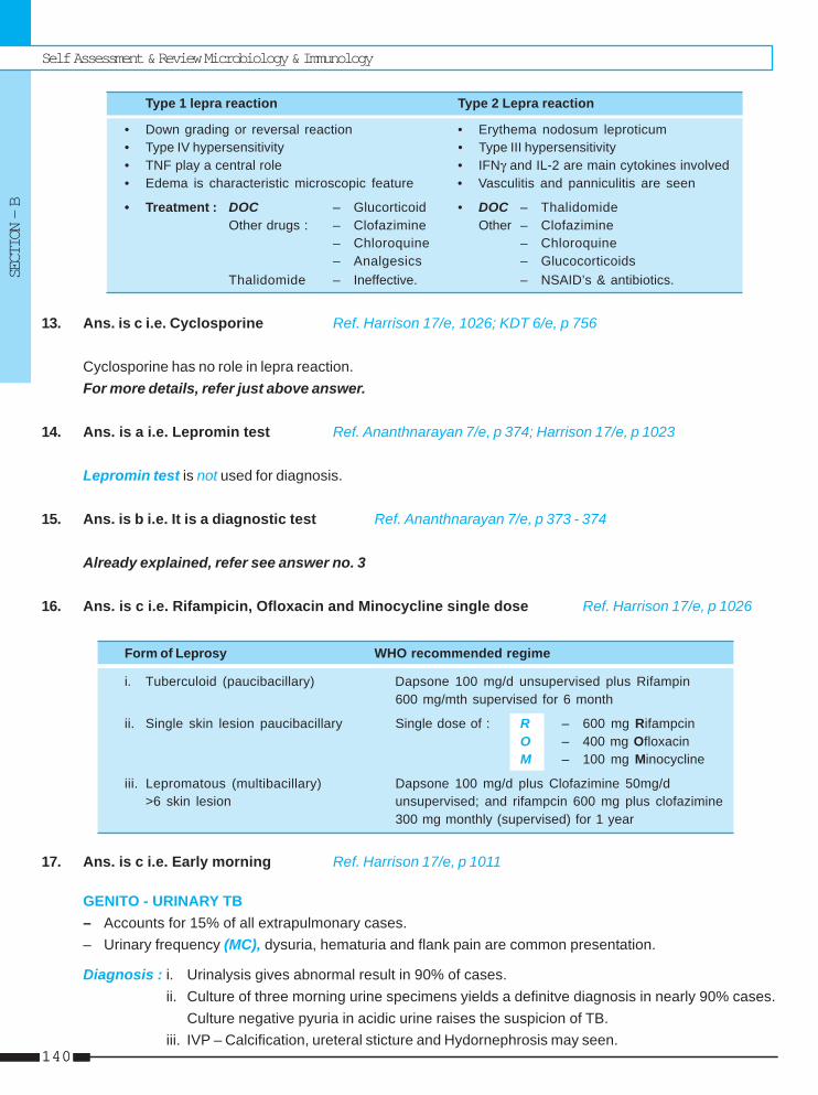
Self Assessment & Review Microbiology & Immunology
SECTION – B
140140
Type 1 lepra reaction Type 2 Lepra reaction
• Down grading or reversal reaction • Erythema nodosum leproticum• Type IV hypersensitivity • Type III hypersensitivity• TNF play a central role • IFNγ and IL-2 are main cytokines involved• Edema is characteristic microscopic feature • Vasculitis and panniculitis are seen
• Treatment : DOC – Glucorticoid • DOC – ThalidomideOther drugs : – Clofazimine Other – Clofazimine
– Chloroquine – Chloroquine– Analgesics – Glucocorticoids
Thalidomide – Ineffective. – NSAID’s & antibiotics.
13. Ans. is c i.e. Cyclosporine Ref. Harrison 17/e, 1026; KDT 6/e, p 756
Cyclosporine has no role in lepra reaction.For more details, refer just above answer.
14. Ans. is a i.e. Lepromin test Ref. Ananthnarayan 7/e, p 374; Harrison 17/e, p 1023
Lepromin test is not used for diagnosis.
15. Ans. is b i.e. It is a diagnostic test Ref. Ananthnarayan 7/e, p 373 - 374
Already explained, refer see answer no. 3
16. Ans. is c i.e. Rifampicin, Ofloxacin and Minocycline single dose Ref. Harrison 17/e, p 1026
Form of Leprosy WHO recommended regime
i. Tuberculoid (paucibacillary) Dapsone 100 mg/d unsupervised plus Rifampin600 mg/mth supervised for 6 month
ii. Single skin lesion paucibacillary Single dose of : R – 600 mg RifampcinO – 400 mg OfloxacinM – 100 mg Minocycline
iii. Lepromatous (multibacillary) Dapsone 100 mg/d plus Clofazimine 50mg/d>6 skin lesion unsupervised; and rifampcin 600 mg plus clofazimine
300 mg monthly (supervised) for 1 year
17. Ans. is c i.e. Early morning Ref. Harrison 17/e, p 1011
GENITO - URINARY TB– Accounts for 15% of all extrapulmonary cases.– Urinary frequency (MC), dysuria, hematuria and flank pain are common presentation.
Diagnosis : i. Urinalysis gives abnormal result in 90% of cases.ii. Culture of three morning urine specimens yields a definitve diagnosis in nearly 90% cases.
Culture negative pyuria in acidic urine raises the suspicion of TB.iii. IVP – Calcification, ureteral sticture and Hydornephrosis may seen.
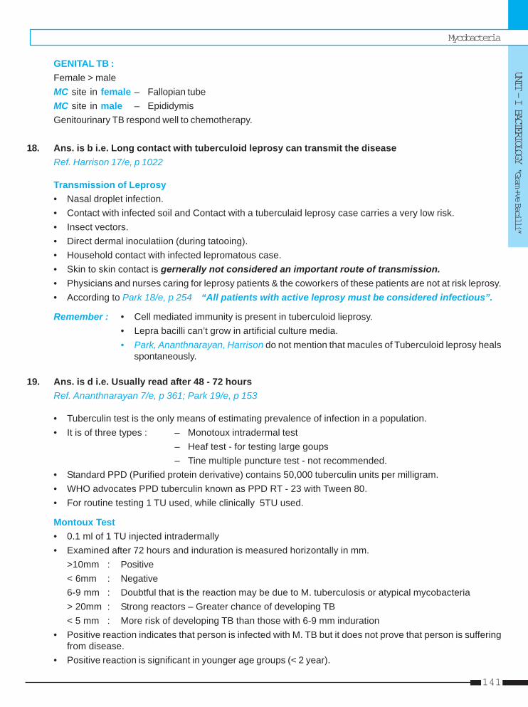
Mycobacteria
UNIT - I BACTERIOLOGY “Gram +ve Bacilli”
141
GENITAL TB :Female > maleMC site in female – Fallopian tubeMC site in male – EpididymisGenitourinary TB respond well to chemotherapy.
18. Ans. is b i.e. Long contact with tuberculoid leprosy can transmit the diseaseRef. Harrison 17/e, p 1022
Transmission of Leprosy• Nasal droplet infection.• Contact with infected soil and Contact with a tuberculaid leprosy case carries a very low risk.• Insect vectors.• Direct dermal inoculatiion (during tatooing).• Household contact with infected lepromatous case.• Skin to skin contact is gernerally not considered an important route of transmission.• Physicians and nurses caring for leprosy patients & the coworkers of these patients are not at risk leprosy.• According to Park 18/e, p 254 “All patients with active leprosy must be considered infectious”.
Remember : • Cell mediated immunity is present in tuberculoid lieprosy.• Lepra bacilli can’t grow in artificial culture media.• Park, Ananthnarayan, Harrison do not mention that macules of Tuberculoid leprosy heals
spontaneously.
19. Ans. is d i.e. Usually read after 48 - 72 hoursRef. Ananthnarayan 7/e, p 361; Park 19/e, p 153
• Tuberculin test is the only means of estimating prevalence of infection in a population.• It is of three types : – Monotoux intradermal test
– Heaf test - for testing large goups– Tine multiple puncture test - not recommended.
• Standard PPD (Purified protein derivative) contains 50,000 tuberculin units per milligram.• WHO advocates PPD tuberculin known as PPD RT - 23 with Tween 80.• For routine testing 1 TU used, while clinically 5TU used.
Montoux Test• 0.1 ml of 1 TU injected intradermally• Examined after 72 hours and induration is measured horizontally in mm.
>10mm : Positive< 6mm : Negative6-9 mm : Doubtful that is the reaction may be due to M. tuberculosis or atypical mycobacteria> 20mm : Strong reactors – Greater chance of developing TB< 5 mm : More risk of developing TB than those with 6-9 mm induration
• Positive reaction indicates that person is infected with M. TB but it does not prove that person is sufferingfrom disease.
• Positive reaction is significant in younger age groups (< 2 year).

Self Assessment & Review Microbiology & Immunology
SECTION – B
142142
• Negative test can’t taken as exclusion of TB since dermal Hypersensitivity to tuberculin can also be lostin immunosuppressive states which gives false -ve result eg. Malignancy, Hodgkin’s disease, post measlesstate, corticosteroid therapy.
• Repeat test may appear to be negative or exerts a booster effect.• Positive tubercullin test may occasionally revert to negative upon INH treatment.• After infection patient becomes tuberculin positve in 3-6 weeks (=I.P of TB).• After 8 weeks of BCG vaccination it becomes positive.
20. Ans. is d i.e. Niacin positive Ref. Ananthnarayan 7/e, p 351 - 353, 366
Mycobacteria tuberculosis is :– Gram positive, non motile, non capsulated, non sporing.– Obligate Aerobic.– Acid (resist decolorization by 20% H2SO4) and alcohol fast.– Generation time 14 - 15 hours.– Colonies appear slowly in about 2 weeks and may some time take upto 8 weeks.– Niacin, Neutral red, Nitrate reduction test positive (3N is positive).– Peroxidase and urease test is also positive.
21. Ans. is c i.e. M. tuberculosis Ref. Ananthnarayan 7/e, p 351, 370
Features M. tuberculosis M. leprae
Acid fastness Resist desoulourization by 20% H2SO4 Resist decolourization by 5% H2SO4
Alcohol fastness Present Absent
Culture Possible Not possible
Niacin Positive (also some strain of M. microti) Negative
Generation time 14 - 15 hours 12 - 13 days
Remember : Nocardia resist 1-4% of sulphuric acid (weakly acid fast).
22. Ans. is b and c i.e. Dorset media; LJ media Ref. Ananthnarayan 7/e, p 352
NNN media – for Leishmania donovaniNutrient agar – simple mediaMac Conkey media – differential as well as indicator media for lactose and non lactose fermenters.
23. Ans. is a and b i.e. ENL; Lepra reaction 1 Ref. Harrison 17/e, p 1023
Reactional state Type of Leprosy
i. Type I lepra reaction Borderline leprosy
ii. Type II lepra reaction (ENL) Exclusively in Borderline Lepromatous (BL) & lepromatoous leprosy
iii. Lucio’s phenomenon Diffuse lepromatous form of LL
• Jarisch Hexhimer reaction is seen in syphilis patient taking penicillin.
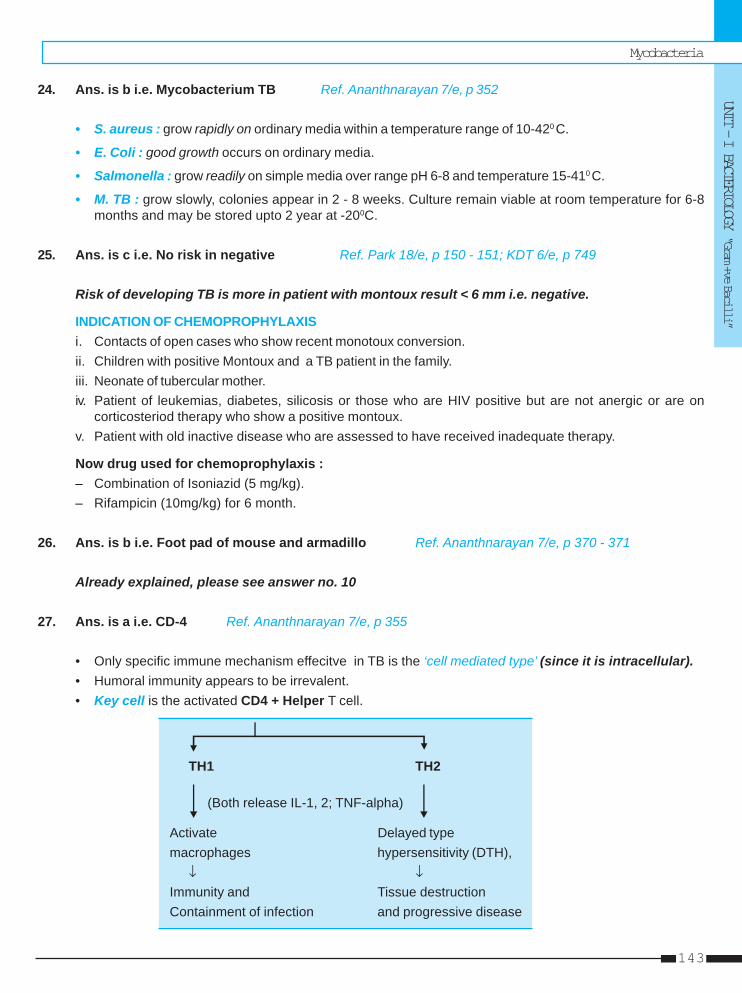
Mycobacteria
UNIT - I BACTERIOLOGY “Gram +ve Bacilli”
143
24. Ans. is b i.e. Mycobacterium TB Ref. Ananthnarayan 7/e, p 352
• S. aureus : grow rapidly on ordinary media within a temperature range of 10-420 C.
• E. Coli : good growth occurs on ordinary media.
• Salmonella : grow readily on simple media over range pH 6-8 and temperature 15-410 C.
• M. TB : grow slowly, colonies appear in 2 - 8 weeks. Culture remain viable at room temperature for 6-8months and may be stored upto 2 year at -200C.
25. Ans. is c i.e. No risk in negative Ref. Park 18/e, p 150 - 151; KDT 6/e, p 749
Risk of developing TB is more in patient with montoux result < 6 mm i.e. negative.
INDICATION OF CHEMOPROPHYLAXISi. Contacts of open cases who show recent monotoux conversion.ii. Children with positive Montoux and a TB patient in the family.iii. Neonate of tubercular mother.iv. Patient of leukemias, diabetes, silicosis or those who are HIV positive but are not anergic or are on
corticosteriod therapy who show a positive montoux.v. Patient with old inactive disease who are assessed to have received inadequate therapy.
Now drug used for chemoprophylaxis :– Combination of Isoniazid (5 mg/kg).– Rifampicin (10mg/kg) for 6 month.
26. Ans. is b i.e. Foot pad of mouse and armadillo Ref. Ananthnarayan 7/e, p 370 - 371
Already explained, please see answer no. 10
27. Ans. is a i.e. CD-4 Ref. Ananthnarayan 7/e, p 355
• Only specific immune mechanism effecitve in TB is the ‘cell mediated type’ (since it is intracellular).• Humoral immunity appears to be irrevalent.• Key cell is the activated CD4 + Helper T cell.
TH1 TH2
(Both release IL-1, 2; TNF-alpha)
Activate Delayed typemacrophages hypersensitivity (DTH),
↓ ↓Immunity and Tissue destructionContainment of infection and progressive disease
Self Assessment & Review Microbiology & Immunology
SECTION – B
144144
1. c) Non−photoc ... 2. a) Lungs 3. d) M. smegmatis 4. a) M. scrofulaceum 5. None6. c) Both of the ... 7. b) Scrofuloderma 8. a) Strict aerobes 10. b) TT 11. b) Armadillos
1. Battey bacillus or mycobacterium intracellulary isa) Photochromogen [JIPMER 90]b) Scotochromogenc) Non−photochromogend) Rapid grower
[Ref. Ananthnarayan 7/e, p 366]2. Reactivation tuberculosis is almost excusively a
disease of the : [JIPMER 90]a) Lungsb) Bonesc) Jointsd) Brain
[Ref. Ananthnarayan 7/e, p 355]3. Which of the following is not a pathogenic myco-
bacteria : [AI 91]a) M. Kansasiib) M. scrofulaceumc) M. cheolenid) M. smegmatis
[Ref. Ananthnarayan 7/e, p 351]4. Which of the following is scotochromogen :
a) M. scrofulaceum [JIPMER 91]b) M. ulceransc) M. Kansasiid) M. fortuitum
[Ref. Ananthnarayan 7/e, p 365]5. Cutaneous lesions may be produced by the fol-
lowing mycobacteria except : [Delhi 91]a) M. intracellulareb) M. Lepraec) M. marinumd) M. tuberculosise) M. ulcerans
[Ref. Ananthnarayan 7/e, p 367]
Answer
6. Acid fastness of tubercle bacilli is attributed to :a) Presence of mycolic acidb) Integrity of cellwall [AIIMS 91, 85; PGI 85]c) Both of the aboved) None of the above
[Ref. Ananthnarayan 7/e, p 352]7. In which type of Cutaneous Tuberculosis,
caseation is most commonly seen : [PGI 93]a) Papulonecroticb) Scrofulodermac) Lupus Vulgarisd) Erythema nodosum
8. True about M. tuberculosis is : [Delhi 93]a) Strict aerobesb) Gram negativec) Thin cell walld) Curved rod
[Ref. Ananthnarayan 7/e, p 351]10. Single skin lesion is seen in which type of Lep-
rosy: [AI 93]a) LLb) TTc) BLd) BT
[Ref. Harrison 17/e, p 1022]11. Lepra bacilli are best cultivated in : [AI 93]
a) Guinea pigb) Armadillosc) Mouse toot padd) Nutrient agar
[Ref. Ananthnarayan 7/e, p 370]
Chapter Review••••• This section includes questions of VThis section includes questions of VThis section includes questions of VThis section includes questions of VThis section includes questions of Various Other PGMEES from 1990 – 2007.arious Other PGMEES from 1990 – 2007.arious Other PGMEES from 1990 – 2007.arious Other PGMEES from 1990 – 2007.arious Other PGMEES from 1990 – 2007.••••• Questions are arQuestions are arQuestions are arQuestions are arQuestions are ar ranged in decreasing order of yearranged in decreasing order of yearranged in decreasing order of yearranged in decreasing order of yearranged in decreasing order of yearwiz. This is done to makwiz. This is done to makwiz. This is done to makwiz. This is done to makwiz. This is done to make refere refere refere refere refer ralralralralral
system easier and uncomplicated to save the precious time of PGMEE Aspirant.system easier and uncomplicated to save the precious time of PGMEE Aspirant.system easier and uncomplicated to save the precious time of PGMEE Aspirant.system easier and uncomplicated to save the precious time of PGMEE Aspirant.system easier and uncomplicated to save the precious time of PGMEE Aspirant.
Mycobacteria
UNIT - I BACTERIOLOGY “Gram +ve Bacilli”
145
12. Lepra cells found in lepromatous leprosy are :a) Neutophils [Kam 94]b) Lymphocytesc) Macrophagesd) Plasma cells
[Ref. Ananthnarayan 7/e, p 370]13. The animal model frequently used tor M. Leperae
is : [Karn 94]a) Miceb) Gninea pigc) Rabbitsd) Golden hamsters
[Ref. Ananthnarayan 7/e, p 370]14. Leprosy spreads by : [Kerala 95]
a) Skin to skin contactb) Blood transfussionc) Droplet spreadd) Ingestion
[Ref. Harrison, 17/e, p 267]15. Mycobacterium tuberculosis is differentiated from
other atypical mycobacteria by :a) Niacin test [SGPGI 95; AIIMS 91]b) AFB stainingc) PAS stainingd) None
[Ref. Ananthnarayan 7/e, p 353]16. Leprosy bacilli are cultured in : [AP 97]
a) LJ. mediumb) Foot pad of micec) Agar plated) None
[Ref. Ananthnarayan 7/e, p 370]17. The local lession in BCG is maximum in :
a) 2 days [Kerala 98]b) 7 daysc) 28 daysd) 1 year
[Ref. Park, 19/e, p 161]18. Mitsuda reaction is read after : [MP 98]
a) 3 daysb) 3 hoursc) 3 weeksd) 3 months
[Ref. Ananthnarayan 7/e, p 374]19. The factor which promotes, virulence of M. tuber-
culosis : [TN 98]
a) Wax Db) Cord factorc) Muramyl dipeptided) Mycolic acid
[Ref. Jawetz, 23/e, p 321]20. BCG differs from mantouxe test by : [UP 98]
a) Less sensitive and more specificb) More sensitive and more specificc) More sensitive and more specificd) Less sensitive and more specific
[Ref. Ghai 6/e, p 235, 192]21. Not a pathogenic mycobacterium to human :
a) M. paratuberculosis [TN 99]b) M. kansasiic) M. ulceransd) M. intracellular
[Ref. Ananthnarayan 6/e, p 324]22. Rapid diagnosis of tuberculosis is possible with:
a) Auramine rhodamine ∼ stain [ICS 2K]b) Zeil - Neelsen stainc) Geimsa staind) Leishman stain
[Ref. Ananthnarayan 7/e, p 357]23. Mycobacterium scrofulaceum which cause
scrofula, belongs to which groups of Atypical my-cobacteria: [Kerala 2K]a) Photochromogensb) Scotochromogensc) Non-photochromogensd) Rapid growerse) None of the above
[Ref. Ananthnarayan 7/e, p 365]24. Mycobacterium leprae can be grown in :
a) Teepol agar [Kar 2000]b) Tomato juice agarc) Foot pad of moused) LJ media
[Ref. Park 19/e, p 270]25. Photochromogenic strain of mycobacterium spe-
cies is : [Kar 2001]a) M. kansasiib) M. scrofulaceumc) M. avium intracellular/lared) M. smegmatis
[Ref. Ananthnarayan 7/e, p 365]
12. c) Macrophages 13. a) Mice 14. c) Droplet spread 15. a) Niacin test 16. b) Foot pad ...17. c) 28 days 18. c) 3 weeks 19. b) Cord factor 20. b) More sensitive 21. a) M. paratuber ...22. a) Auramine 23. b) Scotoch ... 24. c) Foot pad ... 25. a) M. kansasii
Answer
Self Assessment & Review Microbiology & Immunology
SECTION – B
146146
26. Mycobactium TB can be differentiated from othermycobacterium by : [Kerala 01]a) Production of niacinb) Arylsulphate testc) Coagulase testd) Bile solubility test
[Ref. Ananthnarayan 7/e, p 353]27. Mantoux test is an indicator of : [Kolkata 02]
a) Immediate of hypersensitivityb) Delayed hypersensitivityc) Cell-mediated cytotoxicityd) Of no interest
[Ref. Ananthnarayan 7/e, p 166]28. The best diagnostic procedure of M. tuberculosis
a) PCR [SGPGI 02]b) Auramine rhodamine stainc) Sputum cultured) Sputum examination
[Ref. Harrison, 17/e, p 1014]29. Drug resistance in tuberculosis is due to :
a) Transformation [Kar 2002]b) Transductionc) Conjugationd) Mutation
[Ref. Ananthnarayan 7/e, p 59; Jawetz 24/e, p326]
30. Subtype of leprosy has maximum number of TH:1cells: [UP 03]a) TTb) BBc) LLd) Borderline leprosy
[Ref. Harrison, 17/e, p 1023]31. All are rapid growers except : [SGPGI 03]
a) M. fotuitumb) M. cheloneic) M. auiuim intracellulared) M. smeqmatis
[Ref. Ananthnarayan 7/e, p 367]32. All of the following are acid fast bacteria except :
a) Cryptosporidia [SGPGI 03]b) Mycoplasmac) Mycobacteriad) Nocardia
[Learn the list from our book]
33. In a patient the lymphnodes show necrosis withpoor granuloma formation with plentiy of acid fastbacilli sugessts : [SGPGI 04]a) Tuberculosis in an immunocompromised pa-
tientb) HIV with tuberculsisc) Sarcoidosisd) Mycobacterium bovis infection
[Ref. Harrison, 17/e, p 1011]34. Culture of lepra bacilli done in : [Kolkata 05]
a) Nine banded armadillob) Tail of mousec) Foot pads of miced) nutrient agar
[Ref. Ananthnarayan 7/e, p 370-371]35. Mycobacterium non-pathogenic to man :
a. M.bovis [Kolkata 05]b. M.kansaic. M.aviun intracellulard. M.phlei
[Ref. Chakarborty 2/e, p 414]36. Swimming pool granuloma is caused by :
a) Myco kansasi [UP 05]b) Myco. avium intracellularec) Myco. scrofulaceumd) M. marinum
[Ref. Ananthnarayan 7/e, p 365]37. Most common organism in AIDS in TB, India :
a) M. tuberculosis [MP 06]b) M. avium intracellularc) M. bovisd) M. ulcerans
[Ref. Ananthnarayan 7/e, p 589; Park 19/e, p 291;Harrison 17/e, p 1013]
38. Stain for the diagnosis of tuberculosis : [MP 07]a) Auramine-rhodamineb) India inkc) Geimsa staind) Zeil - Neelsen stain
[Ref. Ananthnarayan 7/e, p 357]39. Globi is : [Jharkhand 05]
a) Histocyte containing acid-fast bacillusb) Lymphocyte containing acid-fast bacillusc) Nutrophill containing acid-fast bacillusd) Large lymphocyte containing acid-fast bacillus
[Ref. Ananthnaraya 7/e, p 370]
26. a) Production ... 27. b) Delayed ... 28. b) Auramine ... 29. None 30. a) TT31. c) M. auiuim ... 32. b) Mycoplasma 33. a and b 34. c) Foot pads ... 35. d) M.phlei36. d) M. marinum 37. a) M. tuberculosis 38. a) Auramine ... 39. a) Histocyte ...
Answer
• Enterobacteriaceae includes large group of Gram negative rods whose natural habitat is large intestine.• They are predominantly aerobic or facultative anaerobes, nonsporing and non acid fast.• They have following common characteristics :
– Grow well on Mac conkey media.– Catalase (+)ve; except Shigella dysentriae type 1.– Oxidase (-)ve; reduces nitrates to nitrite.– Urease (-)ve; except Proteus, Klebsiella, Morganella.– Motile by peritrichate flagella except shigella, klebsiella and salmonella gallinarum - pullorum.– Ferment glucose except shigella.
Classification :MC and oldest method to classify these bacteria is on basis of fermentation of lactose.
Lactose fermented rapidly Lactose fermented slowly Lactose not fermented= Coliforms = Late lactose fermenter = Paracolons = Mostly pathogenic
• Escherichia • Edwardsiellla • Shigella except S.sonnei• Klebsiella • Serratia • Salmonella• Enterobacter aerogenes • Citrobacter • Proteus
They forms pink colony on • ArizonaMac conkey's medium • Providenciaand are usually part of • Erwinianormal flora. • Shigella sonnei
Remember : Differential media which distinguishes lactose fermenting (coloured) from non-lactose fermentingcolonies (non pigmented) includes Eosin methylene blue (EMB), MacConkey or deoxycholate medium.
ESCHERICHIA COLIMorphologyMotile by peritrichate flagella, non sporing. Many pathogenic isolates are capsulated.
Culture and Biochemical characterstics
EnterobacteriaceaeEnterobacteriaceaeEnterobacteriaceaeEnterobacteriaceaeEnterobacteriaceae99
Self Assessment & Review Microbiology & Immunology
SECTION – B
148148
• Grows well on ordinary media.• On blood agar shows hemolysis.• Indole and Methyl red (+)ve while VP and citrate (-)ve [IMVC ++--].
Antigenic structure• Somatic antigen O – Lipopolysaccharide in nature, heat stable. Antibodies to O antigen are predominantly IgM.• Capsular antigen K – Heat labile responsible for attachments of bacteria to epithelial cells.
– Associated with virulence.• Flagellar antigen• Serotyping based on these three antigens is :
– Normal colon strains = commensal strains belong to early ‘O’ group (1, 2, 3, 4, etc). These strainsgenerally lack specialized virulent traits. However they may be involved in extraintestinal infection.
– Enteropathogenic strains : Belong to later 'O' group (25, 55, 86 etc).– Extraintestinal pathogenic E. coli [EXPEC] : MC cause of extraintestinal E.coli infection. Like commensal
E.coli they can be found in normal intestinal flora without causing gastroenteritis, but they have acquireddiverse virulence factor that enable them to live extraintestinally.
Virulence factorsSurface antigen :
• O antigen – It has endotoxic activities and also protects bacteria from phagocytosis.• K-antigen– Protect against phagocytosis.• Fimbriae – Plasmid coated fimbrae increase virulence, while chromosomally determined fimbriae has no
effect on virulence.
Toxin : E.coli produce two kind of exotoxin :• Hemolysins – No role in pathogenesis of dirrhoea.• Enterotoxins – Important in pathogenesis of diarrhoea.
• E.coli enterotoxins
Heat stable toxin [STA] Heat labile toxin [LT] Verocytotoxin [VT] = Shiga like toxin [SLT]
• Plasmid mediated • Plasmid mediated • Phage coded• Consist of 2 subunit • Belong to ribosome inactivating
protein [RIP] toxins• Activates cGMP A B
↓ ↓• Acts very rapidly Activates Binds GM1 • A subunit of SLT inhibit ribosme and ↓
CAMP gangliosides protein synthesis and shows cytotoxicity↓
Diarrhoea
Clinical findingsA. Diarrhoea – 5 types of diarrhoeagenic E.coli are recognised :
• Enteropathogenic = enteroadherant E.coli– Cause diarrhoea in infant and children.– Non toxigenic and non invasive.– Produce diarrhoea by disruption of brush border.
Enterobacteriaceae
UNIT - I BACTERIOLOGY “Gram —ve Bacilli”
149
– They are unable to ferment sorbitol.
• Enterotoxigenic E.coli (eg. 06, 08, 015, 025, 027, 0167)– Affect all age group.– MC cause of traveller’s diarrhoea (indistinguishable from cholera).– Produce either or both LT and STa.– Produce diarrhoea, only when it adheres to intestinal mucosa by fimbrial or colonization factor antigen and
produce toxin.
• Enteroinvasive E. coli– Called atypical E. coli as many strains don't ferment lactose or ferment it late.– Resemble Shigella flexneri except in fermenting dulcitol and forming alkali in litmus milk.– Cause illness resembling shigellosis.– Ability to penetrate cells is due to presence of plasmid which codes for outer membrane antigen called
virulence marker antigen [VMA].– For diagnosis of EIEC - Sereny test is used. Cell penetration of Hela or HEP-2 in culture is also diagnostic
test.
• Enterohemorrhagic E.coli [EHEC] = Verotoxingenic E.coli (VTEC)– Source of infection appears to be salad as washing only doesn't remove bacteria.– Produce Verocytotoxin VT whose major target is vascular endothelial cells.– Cause disease ranging from mild diarrhoea to fatal hemmorhagic colitis and hemolytic uremic syndrome
[HUS].– HUS : – Mainly caused by O157 H7 type which does not ferment sorbitol (Some 0157 and non 0157 strain
ferment sorbitol).– Antibiotics increase the incidence of HUS.
• Enteroaggregative E. coli - (EAEC)– Cause persistent diarrhoea.– Stacked brick' formation on Hep -2 cells.– Produce EAST – Enteroaggregative heat stable enterotoxin.
B. UTI• MC cause of both uncomplicated and nosocomial UTI.• UTI causing serotypes are those normally found in faeces.• Only one serotype is generally isolated from infected urine at a time while in diarrhea many serotypes are
present in a single culture.• Nephropathogenic E. coli typically produce Haemolysin.• Pyelonephritis is associated with specific type of pilus, P. pilus.• Diagnosis - UTI is said when there is : – Bacteriuria > 105 /ml in asymptomatic
– Bacteriuria of > 104/ml in symptomatic– Bacteriuria of > 102/ml in catheterized sample– Bacteriuria of any degree in suprapubic aspirate.
• Presumptive diagnosis of bacteriuria is made by : – Griess Nitrite test– Catalase Nitrite test– Triphenyl tetrazolium chloride test.
Self Assessment & Review Microbiology & Immunology
SECTION – B
150150
C. Pyogenic Infection – E.coli is MC Cause of intrabdominal abscess, peritonitis and cholangitis.
D. Meningitis – E.coli is MC cause of neonatal meningitis.
E. Pneumonia – E. coli is 2nd MC cause of nosocomial pnenumonia [1st being Staph aureus]
F. Bacteremia – UTI is MC source of E.coli bacteremia leading to septic shock
Treatment• UTI – Fluroquinolone are DOC.• Diarrhoea – Fluids and electrolyte correction with no antibiotics.• Drug resistance is plasmid mediated.
KLEBSIELLA• Non motile, capsulated rods (capsule seen as haloes around bacilli).• Classified into 3 species on the basis of biochemical characteristics and into serotypes on basis of K-
antigen (capsular antigen).
Klebsiella pneumoniae = Friedlander's bacillus• Rarely cause disease in normal person.
Clinical manifestation– Cause community acquired pneumonia in alcoholics, chronic bronchopulmonary disease or diabetes.– MC clinical syndromes it causes are pneumonia, UTI, abdominal infections, surgical site infection, soft tissue
infection and bacteremia.– Pneumonia : - Mainly Hospital acquired. Abscess are more common than in pneumococcal pneumonia.
- Cause classic lobar infiltrate with bulging fissure.- Sputum is red current jelly in character.
– UTI : Mainly in settings of prolonged catheterization.– Diarrhoea : By toxin similar to STA of E. coli.
Klebsiella Ozaenae : Causative agent of ozoena characterised by foul smelling nasal discharge.Klebsiella rhinoscleromatis : Causative agent of rhinoscleroma.
Treatment• Carbipenim (Imipenam) – Most active antibiotic against klebsiella.
SHIGELLA• Non motile, non capsulated, non lactose fermentar (except S. sonnei which ferments it late).• Classified on the basis of somatic O Antigen (LPS) and carbohydrate fermentation (mannitol) pattern.• Catalase is produced by all except Sh. dysnteriae I.• Mannitol is fermented by all except Sh. dysnteriae I.• MC Shigellosis worldwide – Sh. sonnei.• MC Shigellosis in India – Sh. flexneri.• Most clinically severe form of Shigellosis is caused by Sh. Dysenteriae type I while mildest form of bacillary
dysentery is by Sh. sonnei (may occur as food poisoning).• Only species that exist as single serotype – Sh. sonnei.
Enterobacteriaceae
UNIT - I BACTERIOLOGY “Gram —ve Bacilli”
151
• Pathogenic species of shigella are :
Present Designation Group & Type Mannitol Ornithine Decarboxylase
S dysenteriae A – –S flexneri B + –S boydii C + –S sonni D + +
Culture• Selective media : – Deoxycholate citrate agar (DCA).
– Hektoen enteric agar or Salmonella - Shigella agar. ..... Jawetz 24/e, p 257• Triple sugar iron (TSI) agar is used to differentiate salmonella and Shigella from other gram-negative rods in stool
cultures.
Pathogenesisa. Invasiveness (main) :
• Bacteria invade basolateral surface of colon epithelium →→→→→ intracellular replication and cell to cell spread withthe help of microbial protein Ics A (ATP-ase) and host protein cadherin L - CAM.
• This process present in all virulent shigellae as well as in EIEC. It is responsible for late dysentery. Hencenontoxic mutants can cause dysentery but non-invasive can’t produce dysentery.
b. Toxins :i. Endotoxin – LPS present in all shigella causing irritation of bowel.ii. Shigella Dysenteriae – I or Shiga bacillus exotoxin :
– It has neurotoxicity on blood vessel of CNS.• Enterotoxicity causing fluid accumulation in ligated rabbit ileal loop.• Cytotoxicity same as Verotoxin I or Shiga like toxin produced by some strain of EHEC including 0157:H7.
– Toxin has two peptide subunit. A unit (N-glycosidase) of cytotoxin hydrolyzes adenine form specific sites of60s RNA and thus inhibits protein synthesis. It contributes to fatal nature of S. dysenteriae infection.
– Toxins produce early, non bloody voluminous diarrhoea.
Clinical Features• Most cases of bacillary dysentery occur in < 10 year children.• Infective dose is in the order of 102 organisms (while in vibrio and Salmonella 105-108). ..... Jawetz 24/e, p 256• Transmitted faecorally generally direct person to person contact; anal-oral sex (esp. in Sh. sonnei).• Majority of lesion are in distal colon.• Complications – Toxic dilatation , Colonic perforation.
– Extraintestinal (esp. with S. dysenteriae and S. flexneri) like HUS, Thrombotic thrombocytopenicpurpura reactive arthritis, Reiter's syndrome.
Diagnosis• Specimen – Best to use mucus flakes of stool.• Transport media – Sach's buffered glycerol saline.• Specific diagnosis – Culture of shigella from stool.
Self Assessment & Review Microbiology & Immunology
SECTION – B
152152
Treatment• Mild to moderate dehydration – Oral rehydration therapy (No antibiotics).• Severe cases with bloody diarrhea – DOC Ampicilin or cotrimoxazole (Amoxicillin is not effective).• DOC for Multiresistant : Nalidixic acid.
SALMONELLA• Genus consist of bacilli that parasitise small intestine leading to enteric fever, gastroenteritis, septicemia.• They are motile with peritrichate flagella except S.gallinarum pullorum.• They are non-capsulated and non-sporing.
Culture and Biochemical characterstics• Grows readily on simple media.• On Wilson – Blair bismuth sulphite media S. typhi produce jet black colonies due to production of H2S.• Enrichment media – Selenite F and Tetrathionate broth.• Selective media – Salmonella-shigella agar, Deoxycholate citrate agar which promotes growth of salmonella over
other enterobacteria.• Salmonella ferment sugar producing acid and gas except S. typhi which is anaerogenic.• S. typhi need tryptphan as growth factor.• Boiling or chlorination of water and pasteurisation of milk destroy the bacilli.
Classification• Originally Salmonella was classified on the basis of O, H and Vi antigenic structure and biochemical reactions.
But now on the basis of DNA hybridization studies only 7 groups have been identified.• Nearly all the salmonella serotypes that infect human are in DNA hybridization group 1.
..... Jawetz 24/e, p 258Antigenic structure1. Flagellar antigen (H)
• Heat labile protein which is strongly immunogenic. When mixed with antisera, it rapidly produce-flufty clumps.2. Somatic antigen (O) (Biovin antigen)
Phospholipid polysaccharide complex identical with endotoxin. It remains unaffected by boiling. When mixed withantisera, forms chalky, granular clumps.
3. Vi antigen – Heat labile surface polysaccharide antigen enveloping O antigen. Found only in S.typhi and S.paratyphi.Poorly analogous to K antigen of coliforms. Heat-labile and inmmunogenic. Associated with Virulence.Remember : Order of immunogenic nature of antigen : H > O > Vi
Pathogenicity• On reaching gut, bacilli are phagocytosed.• Salmonella typhi resist intracellular killing by macrophages and polymorphs and enter mesentric lymph node and
from there via thoracic duct enter blood stream to produce bacteremia (Enteric fever).
Clinical ManifestationI. Enteric fever – Systemic disease characterized by fever and abdominal pain caused by S.typhi (called as
typhoid) and S.paratyphi. A, B and C.• I.P – 14 days.• Typical features – Step ladder pyrexia with relative bradycardia
– Rose spots (located primarily on chest) usually appear at the end of 1st week andresolve after 2 - 5 days.
Enterobacteriaceae
UNIT - I BACTERIOLOGY “Gram —ve Bacilli”
153
• Paratyphoid fever of S. paratyphi is generally milder.• Transmission - Transmitted through close contact with cases and carriers.
Carriers :• Those who excrete bacilli for more than a year are called as Chronic carrier.• Carrier rate is 3% (i.e. 3% of cases become chronic carrier).• Though cases occur more in males, carriers are common in females.• Faecal carrier are MC.• Urinary carrier signify abnormality in urinary tract.
Diagnosis (BASU)A. Cases
• Blood culture (B)– Positive in 1st week (Diagnostic gold standard).– Clot culture has higher rate of isolation, than blood culture.
• Agglutination (A) = Widal reaction– Becomes (+)ve by end of 1st week, peaks at 3rd week, decline afterwards.
• Stool culture (S) – 3rd week, particular valuable in patient on antibiotics in which blood culture is (-)veSample are plated directly on MacConkey, DCA, and Wilson - Blair media (highly selective).
• Urine culture (U) – (+)ve only in 2nd and 3rd weeks.
B. Carriers• Widal – No value in detection of carrier.• Demonstration of Vi agglutinin has been claimed to indicate carrier state and used as screening of carrier.
TreatmentCases :• First line – Ciprofloxacin, Ceftriaxone• Alternative – Azithromycin• Multidrug resistant [MDR] S. typhi - Ciprofloxacin is DOC. Multidrug resistant is due to R factor.Carriers : Amoxicillin, Cotrimoxazole, Ciprofloxacin are effective.
PreventionFollowing vaccines are available :
Vaccine Nature Dose Minimum age
Type 21a Live attenuated 4 oral dose 6 yearsViCPS Purified Vi polysacchride 1 Parenteral dose 2 yearsVirEPA Vi bound to recombinent protein 2 parenteral dose 6 month
Vir EPA is most effective.
II. Gastroenteritis = (Food poisoning)• MC caustive agent is S.typhimurium.• Most frequent sources are poultry, meat, milk and milk products.• Never caused by S. typhi.• IP-6-48 hours.• Clinical features – Nausea, vomiting, diarrhoea, Abdominal cramps and fever.
Self Assessment & Review Microbiology & Immunology
SECTION – B
154154
• Blood culture is negative.• Treatment – No antibiotics are given but for serious invasive cases, antibiotics is given.
III. Bacteremia and Endovascular infectionSalmonella serotypes S. cholerae-suis and S. dublin are frequently associated with sustained bacteremia.
PROTEUS BACILLI• Non lactose fermenter.• P. Mirablis which is responsible for 90% of proteus infection is a normal commensal.• Proteus differs from other enterobacteria by presece of enzyme phenyalanine deaminase (responsible for PPA
reaction).• Culture of proteus bacilli have characteristic fishy or seminal smell.• P mirablis and P. vulgaris swarms on solid culture media.• Antigenic structure – Proteus posses Flagelar (H) and somatic O antigen.• Infectious syndromes – UTI (20-30% of complicated UTI); Pneumonia (primarily in hospitalized patient);
Intraabdominal infections; soft tissue abscess.
Remember : Some strains of P. vulgaris called X strains are agglutinated by sera from typhus fever patient. Thisis due to sharing of carbohydrate hapten between Rickettsiae and proteus and forms basis of WeilFelix reaction.
Enterobacteriaceae
UNIT - I BACTERIOLOGY “Gram —ve Bacilli”
155
1. With reference to infection with Escherichia colithe following are true except : [AI 05]a) Enteroaggregative E. coli is associated with per-
sistent diarrhoeab) Enterohemorrhagic E. coli cause hemolytic ure-
mic syndromec) Entero-invasive E. coli produces a disease simi-
lar to salmonellosisd) Entero toxigenic E. coli is a common cause of
traveller’s diarrhoea2. A 20 year old man presented with hemorrhagic
colitis. The stool sample grow Escherichia coli inpure culture. The following serotype of E. coli islikely to be the causative agent : [AI 04]a) 0 157:H7b) 0 159:H7c) 0 107:H7d) 0 55:H7
3. A microbiologist wants to develop a vaccine forprevention of attachment of diarrhoeagenic E. colito the specific receptors in the gastro-intestinaltract. All of the following fimbrial adhesions wouldbe appropriate vaccine candidates except:a) CFA-1 [AI 04]b) Pi-Pilic) CS-2d) K88
4. In patient with typhoid, diagnosis after 15 days ofonset of fever is best done by : [AI 02]a) Blood cultureb) Widalc) Stool cutured) Urine culture
5. All are true about EHEC except : [AI 01]a) Sereny test is positiveb) Ferments sorbitolc) Causes HUSd) Elaborates Shiga like exotoxin
6. All of the following are true except : [AI 01]a) E.coli is an aerobe and facultative anaerobeb) Proteus forms uric acid stonesc) E.coli is motile by peritrichate flagellad) Proteus caused deamination of phenylalanine
to phenylpyrivic acid
7. E. coli attached to a surface with the help of :a) Fucose [AI 00]b) Concanavalinc) Phytohaemagglutinind) Lactin
8. True statement about Widal test in typhoid is :a) O-antigen titre remians positive for several
months and reaction to it is rapid [AI 99]b) H-antigen titre remains positive for several
months and reaction to it is rapidc) Both remains positive for several months and
reaction to both is rapidd) None
9. True statement about widal test in typhoid is:a) Widal test is confirmative in endemic areasb) Antibiotic treatment does not alter widal test re-
sultsc) Previous infection alters widal test [AI 99]d) Widal test does not alter with prior vaccination
10. Shigella can be differentiated from E. coli by all ofthe following features except : [AI 99]a) Shigella does not produce gas from glucoseb) Shigella does not ferment lactosec) Shigella does not ferment mannitold) Shigela has no flagella and is non motile
11. Which of the following statement regarding Shi-gella dysenteriae type I is true : [AI 99]a) It can lead to hemolytic uremic syndromeb) It produces an invasive enterotoxinc) It is an facultative aerobesd) It is MR negative
12. Shigella are be divided into sub group on the ba-sis of ability to ferment : [AI 97]a) Lactoseb) Maltosec) Fructosed) Mannitol
13. Which of the following is a true statement regard-ing Enterobacteriaceae : [AI 96]a) Motility is by polar flagellumb) Glucose is not fermented by all members of the
familyc) All members are oxidase positived) Nitrate reduction negative
QUESTIONS
Answer 1. c) Entero-... 2. a) 0 157:H7 3. b) Pi-Pili 4. b) Widal 5. a) Sereny ...6. b) Proteus ... 7. a) Fucose 8. b) H-antigen ... 9. c) Previous ... 10. c) Shigella ...
11. a) It can ... 12. d) Mannitol 13. b) Glucose ...
Self Assessment & Review Microbiology & Immunology
SECTION – B
156156
14. Most common organism causing UTI : [AI 96]a) E. colib) Proteusc) Klebsiellad) Staphylococci
15. Which of the following is true about Enteropatho-genic E. coli : [AI 96]a) Causes diarrhea in infantsb) Acts by invasion of intestinal epithelial cellsc) Adults are mostly affectedd) Affects immunocompromised host
16. Which of the following is true regarding Salmo-nella infection : [AI 96]a) Urine culture is +ve in 1st weekb) Stool culture is +ve in 1st weekc) Blood culture is +ve in 3-7 daysd) Widal test is +ve in 1st week
17. Which of the following produces enterotoxin:a) Sh. dysenteriae [AI 95]b) Sh. sonneic) Sh. flexnerid) Sh. boydi
18. Which antigen blocks the agglutination of salmo-nella by 0 antiserum : [AI 95]a) H.b) Fimbiraec) Vid) 0
19. Salmonella typhi is the causative agent of typhoidfever. The infective dose of S. typhi is :a) One bacillus [AIIMS 06]b) 108 - 1010 bacillic) 102 - 105 bacillid) 1 - 10 bacilli
20. A 20 year old male had pain in abdomen and mildfever followed by gastroenteritis. The stool ex-amination showed presence of pus cells andRBC’s on microscopy. The most likely etiologicalagent is : [AIIMS 03]a) Enteroinvasive E. colib) Enteropathogenic E. colic) Etnerotoxigenic E. colid) Enteroaggregative E. coli
21. There has been an outbreak of food borne salmo-nella gastroenteritis in the community and thestool sample is received in the laboratory. Whichis the enrichment medium of choice :
a) Cary-Blair medium [AIIMS 03]b) V - R mediumc) Selenite F mediumd) Thioglycholate medium
22. A 24 year cook in a hostel is suffering from en-teric fever 2 years back. The chronic carrier statein patient is diagnosed by : [AIIMS 02]a) Vi aglutination testb) Blood Culture in Brain Heart infusion brothc) Widal testd) C reactive protein
23. For typhoid endemic country like India, immuniza-tion of choice is : [AIIMS 01]a) TAB vaccineb) Typhoral 21A oral vaccinec) Monovalent vaccined) Any of these
24. Kallu, a 22 year old male had an outing with hisfriends and developed fever of 38.50C, diarrhoea,and vomiting following eating chicken salad, 24hours back. Two of his friends developed thesame symptoms. The diagnosis is : [AIIMS 01]a) Salmonela enteritis poisoningb) Bacillus cereusc) S. aureusd) Vibrio cholera
25. All are true about Shigella except : [AIIMS 99]a) Large dose is required for infectionb) Associated with hemolytic uremic syndromec) Causes bloody diarrhoea with mucusd) Gut pathology is due to toxin
26. A person returns to Delhi from Bangladesh after2 days and has diarrhoea. Stool examinationshows RBCs in stool. The likely organism caus-ing is :[AIIMS 99]a) Enteropathogenic E. colib) Enterotoxigenic E. colic) Salmonella typhid) Shigella dysenteriae
27. All are true about enterohemorrhagic E. coli ex-cept : [AIIMS 99]a) Sereny test positiveb) May cause diarrhoeac) Can cause hemolytic uremic syndomed) Verocytotoxin is produced
Answer 14. a) E. coli 15. a) Causes ... 16. c) Blood ... 17. a) Sh. dysente ... 18. c) Vi19. c) 102 - 105 ... 20. a) Enteroinva ... 21. c) Selenite F ... 22. a) Vi aglutinat ... 23. c) Monovalent ...24. a) Salmonela ... 25. a) Large ... 26. d) Shigella ... 27. a) Sereny ...

Enterobacteriaceae
UNIT - I BACTERIOLOGY “Gram —ve Bacilli”
157
28. All of the following are true regarding typhoid ex-cept : [AIIMS 99]a) Urinary carriers are more dangerousb) Vi ab is used for detecting carrierc) Vi is seen in normal populationd) Urine carrier is associated with anomalies
29. Which toxin is mediated by C-AMP except :a) V. cholera 01 [AIIMS 98]b) Heat stable E. coli toxinc) Heat labile E. coli toxind) V. cholera 0137
30. Traveller’s diarrhoea, is caused by : [AIIMS 97]a) Shigellab) E. colic) E. histolyticad) Giardiasis
31. HUS is caused by : [PGI 07]a. EIECb. Shigellac. Salmonellad. Cholerae. Klebsiella
32. Enterobacteriaecae all except : [PGI 06]a) Pseudomonasb) Klebsiellac) V. cholerad) Proteuse) E. Coli
33. True about salmonella gastroentritis is/are :a) Mainly diagnosed by serological tests [PGI 06]b) Blood & mucous are present in stoolc) Caused by animal productsd) Symptoms appear between 4 - 48 hourse) Features are mainly due to exotoxin
34. True about typhoid : [PGI 03]a) It is caused by S.typhib) Water can transmit the diseasec) Type 21 a is an oral vaccine
d) Chronic carrier is called when transmitted up to6 month
e) Widal test +ve in 1st week35. Which of these are true about E.coli : [PGI 02]
a) The L.T., (labile toxin), in ETEC acts via CAMPb) UTI causing E.coli attaches through pillic) The ST (Stable toxin) of ETEC is responsible for
causing hemolytic-uremic syndromed) EIEC invasiveness is under plasmid controle) In EPEC, the toxin helps in invasion
36. Salmonella gastroenteritis is : [PGI 02]a) Mainly diagnosed by serologyb) Blood and mucous present in bloodc) Caused by animal productsd) Symptoms appear by 4-48 hrsd) Features are due to exotoxin released
37. True about Salmonella typhi infection in intestineare : [PGI 01]a) Affects peyer’s patchesb) Common in mesenteric borderc) Erythrophagocytosis is characteristicd) Strictures are commone) Typhoid ulcer always bleed very much
38. M.C cause of liver abscess : [PGI 00]a) Streptococcusb) Staph’aureusc) E. colid) Staph pyogenes
39. Maximum urease is produced by : [PGI 00]a) H. pylorib) P. Mirabilisc) K. rhinomatisd) Ureaplasma
40. E. coli gives pink colour with : [PGI 99]a) Chocolate agarb) L J mediumc) MacConkey’s mediumd) Saline broth
Answer 28. c) Vi is seen ... 29. b) Heat ... 30. b) E. coli 31. b) Shigella 32. a and c33. c and d 34. a, b and c 35. a, b and d 36. c and d 37. a) Affects ...38. c) E. coli 39. b) P. Mirabilis 40. c) MacConkey’s ...
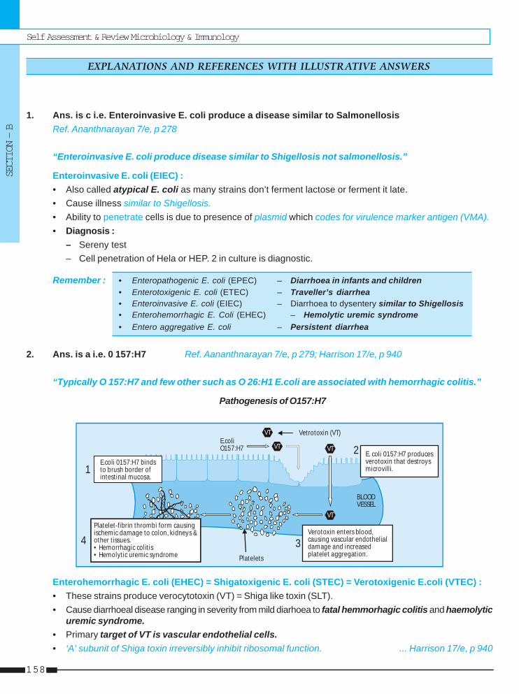
Self Assessment & Review Microbiology & Immunology
SECTION – B
158158
EXPLANATIONS AND REFERENCES WITH ILLUSTRATIVE ANSWERS
1. Ans. is c i.e. Enteroinvasive E. coli produce a disease similar to SalmonellosisRef. Ananthnarayan 7/e, p 278
“Enteroinvasive E. coli produce disease similar to Shigellosis not salmonellosis.”
Enteroinvasive E. coli (EIEC) :• Also called atypical E. coli as many strains don’t ferment lactose or ferment it late.• Cause illness similar to Shigellosis.• Ability to penetrate cells is due to presence of plasmid which codes for virulence marker antigen (VMA).• Diagnosis :
– Sereny test– Cell penetration of Hela or HEP. 2 in culture is diagnostic.
Remember : • Enteropathogenic E. coli (EPEC) – Diarrhoea in infants and children• Enterotoxigenic E. coli (ETEC) – Traveller’s diarrhea• Enteroinvasive E. coli (EIEC) – Diarrhoea to dysentery similar to Shigellosis• Enterohemorrhagic E. Coli (EHEC) – Hemolytic uremic syndrome• Entero aggregative E. coli – Persistent diarrhea
2. Ans. is a i.e. 0 157:H7 Ref. Aananthnarayan 7/e, p 279; Harrison 17/e, p 940
“Typically O 157:H7 and few other such as O 26:H1 E.coli are associated with hemorrhagic colitis.”
Pathogenesis of O157:H7
VT
VTVT
VT Vetrotoxin (VT)E.coliO157:H7
E.coli 0157:H7 bindsto brush border ofintestinal mucosa.
Platelet-fibrin thrombi form causingischemic damage to colon, kidneys &other t issues.• Hemorrhagic colit is• Hemolyt ic uremic syndrome
Verotoxin enters blood,causing vascular endothelialdamage and increasedplatelet aggregation.
BLOODVESSEL
E. coli 0157:H7 producesverotoxin that destroysmicrovilli.
Platelets
1
4 3
2
Enterohemorrhagic E. coli (EHEC) = Shigatoxigenic E. coli (STEC) = Verotoxigenic E.coli (VTEC) :• These strains produce verocytotoxin (VT) = Shiga like toxin (SLT).• Cause diarrhoeal disease ranging in severity from mild diarhoea to fatal hemmorhagic colitis and haemolytic
uremic syndrome.• Primary target of VT is vascular endothelial cells.• ‘A’ subunit of Shiga toxin irreversibly inhibit ribosomal function. ... Harrison 17/e, p 940

Enterobacteriaceae
UNIT - I BACTERIOLOGY “Gram —ve Bacilli”
159
• Typical EHEC is serotype 0 157:H7 and few others such as 0 26:H1.• Diagnosis of EHEC diarrhoea can be made by demonstration of bacilli or VT in feces.• Usually O 157:H7 serotype does not ferment sorbitol So, use of sorbitol Mac-Conkey medium helps in
screening of O:157 VTEC.
3. Ans. is b i.e. Pi Pilli Ref. Jawertz 24/e, p 252; Harrison 17/e, p 937
Correctly speaking guys we are unable to find all choices but Pi Pilli is related in pathogenesis ofpyelonephritis not GIT pathology.So, most probably that would be the answer.
4. Ans. is b i.e. Widal test Ref. Ananthnarayan 7/e, p 297 - 299
“ In 3rd week Widal test is investigation of choice.”
Diagnosis of TyphoidI. Cases (BASU)
Blood culture (B) :– Test of choice in first week (Diagnostic gold standard).– Becomes negative on treatment with antibiotics.– Clot culture : Yields higher rate of isolation than blood culture as bactericidal action of serum is obviated.Widal test :– Agglutinins usually appear by end of first week, Titre increase steadily till the 3rd or 4th week after which
it declines gradually.– Maximum titre is found in 3rd week.Stool culture (S) :– Salmonella are shed in faeces throughout the course of disease, even in convalescence. A positive
faecal culture occur in carriers also, so can’t differentiate between case and carrier.Urine culture :– Culture is positive in 25% of cases during 2nd or 3rd week.– Less useful than blood culture.
II. Carriers– Widal reaction – No value in detection of carriers.– Demonstration of Vi agglutinins indicate carrier state.
This is usefull screening test which is confirmed by culture.– Isolation of bacillus from faeces or bile. Cholagogue purgatives increase chance of isolation.– Tracing of carriers in cities is done by ‘Sewer Swab Technique’ or by filtration of sewage through millipore
membrane and culturing the membrane on Wilson and Blair media.
5. Ans. is a i.e. Sereny test is positive Ref. Ananthanrayan 7/e, p 279; Harrison 17/e, p 942
“Sereny test is positive in cases of EIEC not EHEC.”Mostly 0 157:H7 serotype of EHEC don’t ferment sorbitol but some 0157:H7 and non 0157 strain fermentsorbitol so, option B (Ferments sorbitol) is partly correct. ..... Harrison 17/e, p 942Sereny test – Installation of suspension of freshly isolated EIEC or Shigella in to the eyes of guinea pigs leadsto mucopurulent conjuctivitis.
Self Assessment & Review Microbiology & Immunology
SECTION – B
160160
6. Ans. is b i.e. Porteus forms uric acids stone Ref. Harrison 17/e, p 942-943
“Proteus results in formation of struvite stone not uric acid stone.”
• Uric acid stone forms in acidic urine in presence of hyperuricemia.• All members of enterobacteriacea are aerobes or facultative anaerobes.• Features of proteus bacilli :
– Non lactose fermenter– Differs from other enterobacteria by presence of enzyme phenylalanine deaminase which is responsible
for deamination of phenylalanine to phenyl pyruvic acid (PPA test).– By producing urease it degrades urea to NH3 and CO2, NH3 raises urinary pH. NH4
+ (formed from NH3)precipitate PO4
3- Mg2+ to form MgNH4PO4 (Struvite).The result is stone of struvite admixed with CaCO3
– Some strains (X-strains) are agglutinated by sera from typhus fever patient (weil felix reaction).
Remember : • Peritrichate flagella (flagella all around the cell) seen in :– Proteus – E.coli – Listeria– Clostridia – Typhoid bacilli – Bacillus
• PPA test is also seen in Morganella, Providencia.
7. Ans. is a i.e. Fucose Ref. Still searching
I am unable to find the Answer from any book. I am just mentioning the Answer which is taken as correct bymy seniors.Remember : Concanavalin A and phytohemagglutinin is mitogen that cause blast transformation of T cells
not of B cell.
8. Ans. is b i.e. H-antigen titre remains positive for several months and reaction to it is rapidRef. Ananthnarayan 7/e, p 299
Widal reaction :• Test for measurment of H and O agglutinins for typhoid and paratyphoid bacilli in patient serum.• Agglutinins usually appear by the end of 1st week. Titre ↑ steadily till the 3rd or 4th week.• O Agglutinins – O antigen is common between S. typhii and S. paratyphii, so doesn’t specify whether
infection is due to S. typhii or S. paratyphii.– It has better diagnostic value than H.– O antibodies disappear after infection.– O agglutination is seen as disc like pattern at bottom of tube.
• H agglutinins – H antigen are different for S. typhii or S. paratyphii so indicates type of infection– May be present due to prior disease, inapparent infection, thus it does not have good diagnostic
value.– Persist longer than O agglutinins.– Reaction to it, is rapid and leads to formation of cotton wooly clumps.
Remember : Order of disappearance of antibody in typhoid V1 → O → H.
Enterobacteriaceae
UNIT - I BACTERIOLOGY “Gram —ve Bacilli”
161
9. Ans. is c i.e. Previous infection alters widal test Ref. Ananthnarayan 7/e, p 297 - 299
“Widal test is false positive in case of prior infection and immunization.”• Agglutinins may be present on account of prior disease, innaparent infection or immunisation, so widal test
may be false positive and only +ve widal test should not be taken as proof of typhoid fever.• In cases where antibiotics are given early, agglutinins response is poor and Widal may be false –ve.• Widal test is never confirmatory. Only culture of blood is confirmatory for typhoid.
Remember : Typhoid investigation which are not affected by antibiotics : Stool culture and Bone marrowculture.
10. Ans. is c i.e. Shigella does not ferments mannitol Ref. Ananthanrayan 7/e, p 285
• Differentiating features between Shigella and E.coli :
Features Shigella E.coli
Motility — +Lactose fermentation — +Glucose fermentation — +Lysine decarboxylase — +
• Shigella ferment mannitol and this property is used for its classification.
Classification : Mannitol fermenters Non fermenters
S. flexnerii S. dysenteriaeS. boydiiS. sonnei
So, guys mannitol fermentation can’t diferentiate E. coli and Shigella as E.coli also ferments mannitol.
Remember : – S. sonnei is late lactose fermenter.– All members of Enterobacteriacea are motile except shigella, Klebsiella and salmonella
gallinarum-pallorum.– E.coli also ferments maltose.
11. Ans. is a i.e. It can lead to HUS Ref. Ananthnarayan 7/e, p 287; Harrison 17/e, p 963
“HUS occurs with S. dysenteriae type 1 infection.” ..... Harrison 17/e, p 963
Unique feature of S. dysenteriae :• Mannitol non fermenter (All other Shigella are mannitol fermenter).• Catalase –ve (All other Shigella are catalase +ve).• It produce exotoxin ( Remember : it is a Gram (-)ve bacteria producing exotoxin) which has less role
in dysentery, but has major role in pathogensis of :1. Microangiopathic complications2. Hemolytic uremic syndrome3. Thrombotic thrombocytopenic purpura.
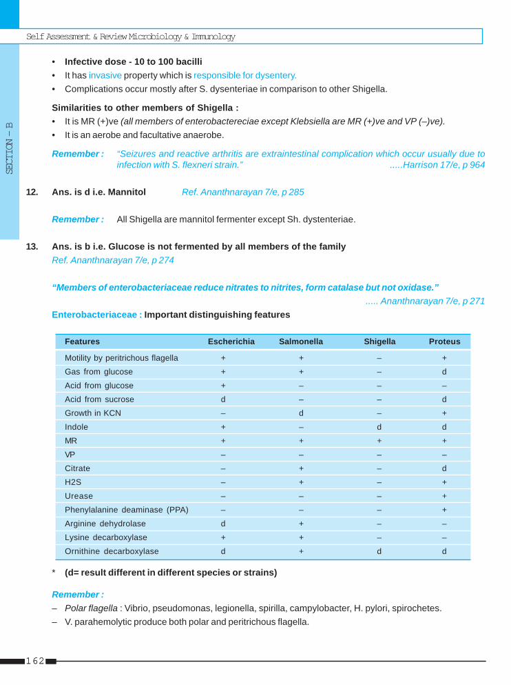
Self Assessment & Review Microbiology & Immunology
SECTION – B
162162
• Infective dose - 10 to 100 bacilli• It has invasive property which is responsible for dysentery.• Complications occur mostly after S. dysenteriae in comparison to other Shigella.
Similarities to other members of Shigella :• It is MR (+)ve (all members of enterobactereciae except Klebsiella are MR (+)ve and VP (–)ve).• It is an aerobe and facultative anaerobe.
Remember : “Seizures and reactive arthritis are extraintestinal complication which occur usually due toinfection with S. flexneri strain.” .....Harrison 17/e, p 964
12. Ans. is d i.e. Mannitol Ref. Ananthnarayan 7/e, p 285
Remember : All Shigella are mannitol fermenter except Sh. dystenteriae.
13. Ans. is b i.e. Glucose is not fermented by all members of the familyRef. Ananthnarayan 7/e, p 274
“Members of enterobacteriaceae reduce nitrates to nitrites, form catalase but not oxidase.”..... Ananthnarayan 7/e, p 271
Enterobacteriaceae : Important distinguishing features
Features Escherichia Salmonella Shigella Proteus
Motility by peritrichous flagella + + – +
Gas from glucose + + – d
Acid from glucose + – – –
Acid from sucrose d – – d
Growth in KCN – d – +
Indole + – d d
MR + + + +
VP – – – –
Citrate – + – d
H2S – + – +
Urease – – – +
Phenylalanine deaminase (PPA) – – – +
Arginine dehydrolase d + – –
Lysine decarboxylase + + – –
Ornithine decarboxylase d + d d
* (d= result different in different species or strains)
Remember :– Polar flagella : Vibrio, pseudomonas, legionella, spirilla, campylobacter, H. pylori, spirochetes.– V. parahemolytic produce both polar and peritrichous flagella.
Enterobacteriaceae
UNIT - I BACTERIOLOGY “Gram —ve Bacilli”
163
14. Ans. is a i.e. E. coli Ref. Harrison 17/e, p 1820
“E. coli cause 80% of acute UTI in patients with out catheterization.” .....Harrison 17/e, p 1820
Strains of E. coli causing UTI are called extraintestinal pathogenic strains of E.coli.
Remember :• E.coli is MC cause of catheter associate UTI too. .....Harrison 17/e, p 1823• UTI is MC nosocomial infection.• E. coli is MC cause of neonatal meningitis.• E.coli is MC cause of intraabdominal abscess.• Other gram negative rods causingUTI are : Proteus, klebsiella, serratia, pseudomonas, enterobacter.
15. Ans. is a i.e. Cause diarrhoea in infants Ref. Ananthnarayan 7/e, p 277
“Enteropathogenic E.coli have been associated with diarrhoea in infants and children usually occuringas institutional out breaks.”5 types of diarrhoeagenic E. coli are recognised :1. Enteropathogenic E. coli; (EPEC) = Entero adherant E. coli (EAEC)
– Cause diarrhea mainly in infants and children– Neither invasive, nor produce enterotoxin– Bacilli adheres to upper small intestine– Usually cause epidemic, but sporadic cases can also occur.
2. Enterotoxigenic E. coli (ETEC)– It is the MC cause of traveller’s diarrhoea– Produce either labile toxin (LT) or stable toxin (ST)– Affect all age groups– Most case occur as endemic.
3. Enteroinvasive E. coli (ETEC)– Similar to Shigellosis– Detected by Sereny’s test or by demonstration of penetration of Hep or Hela cells.
4. Enterohemmorhagic E.coli– These strains produce verocytotoxin (VT) = Shigalike toxin (ST)– Cause diarrhoeal disease ranging in severity from mild diarhoea to fatal hemmorhagic colitis and
hemolytic uremic syndrome.5. Enteroaggregative E. coli
– Appear aggregated as stacked brick formation on Hep - 2 cells or glass.– Produc low molecular weight heat stable enterotoxin (EAST1)– Cause persistent diarrhoea.
16. Ans. is c i.e. Blood culture is positive in 3 - 7days Ref. Ananthnarayan 7/e, p 297 - 299
Investigation for enteric fever : Test Timing
– Blood culture 1st week– Widal test 3rd week– Urine culture 2nd week
Self Assessment & Review Microbiology & Immunology
SECTION – B
164164
17. Ans. is a i.e. Shigella dysentriae Ref. Ananthnarayan 7/e, p 287; Jawetz 24/e, p 155
Exotoxins associated with diarrheal diseases are called as enterotoxins. They are produced by :
– Shigella dysenteriae I – Staph. aureus – B. cereus– Cl. perfringes – Y. enterocolitica – V. parahemolyticus– V. cholera – Kleibsella pneumonia – ETEC– Aeromonas
18. Ans. is c i.e. Vi Ref. Ananthnarayan 7/e, p 291
Vi antigen – Polysaccharide antigen enveloping the O antigen because of which many strains of S.typhi fails to agglutinate with the ‘O’ antiserum.
Antigens of Salmonella :
H antigen O antigen Vi. antigen
– Present on flagella – Integral part of cell wall – Envelops the ‘O’ antigen
– Heat labile protein – Phospholipid - protein - – Surface polysaccharidepolysaccharide complex
– Strongly immunogenic – Identical with endotoxin – Acts as virulence factorso, is less immunogenic and is poorly immunogenic
– Antibody formation is – Antibody formation is – Antibody production is slowrapid and in high titre slow and titre is low and titres is low
• Salmonella are classified into groups on basis of O antigen on the bacterial surface.• Vi antigen is used for epidemiological typing.
19. Ans. is c i.e. 102 - 105 bacilli Ref. Harrison 17/e, p 957; Jawetz 24/e, p 258
Infective dose of salmonella varies from 103 to 106 colony forming units.
Organism Infective dose
Shigella 10 - 1000Vibrio >1010 (if source of infection is water)
102 - 104 (if source of infection is food)Campylobacter jejuni 104
Yersinia enterocolitica 108 - 109
EHEC 10 - 100
20. Ans. is a i.e. Enteroinvasive E. coli Ref. Ananthnarayan 7/e, p 278; Harrison 17/e, p 941
EIEC cause illness resembling Shigellosis i.e. ranging from mild diarrhoea to frank dysentery.
21. Ans. is c i.e. Selenite ‘F’ medium Ref. Ananthnarayan 7/e, p 290, 303
“Selenite F and Tetrathionate broth are commonly employed enrichment media of salmonella.”

Enterobacteriaceae
UNIT - I BACTERIOLOGY “Gram —ve Bacilli”
165
Salmonella Gastroenteritis = Food poisoning• May be caused by any Salmonella except S. typhi• MC caused by S. typhimurium• Human infection occurs due to ingestion of contaminated foods.• Most frequent source – Poultry, meat, milk and milk products• IP – 16 - 48 hours• Clinical feature : Diarrhoea, vomiting, fever• Diagnosis : Isolation of salmonella from faeces• Treatment : No antibiotics
Remember :Differential media for salmonella :• Mac Conkey and Deoxycholate media : Form colourless colonies due to absence of lactose fermentation.• Wilson and Blair bismuth sulphite medium : Jet black colonies are formed due to production of H2S.• Selective media for salmonella : SS agar, Deoxycholate citrate agar.
22. Ans. is a i.e. Vi. agglutination test Ref. Ananthnarayan 7/e, p 300
Already explained, refer answer no. 4
23. Ans. is c i.e. Monovalent vaccine Ref. Park 18/e 189 - 190; 19/e, p 197
Since S. typhi is the major cause of typhoid fever in India, the vaccine of choice is the monovalent typhoidvaccine.
Antityphoid vaccine in India are :a. Monovalent antityphoid vaccine – Heat killed and phenol preserved.b. Bivalent antityphoid vaccine – Contains S. typhi and paratyphi A.c. TAB vaccine (WHO recommended that TAB vaccine should be discontinued).d. Live oral typhoid 21a vaccine (Typhoral)
– Enteric coated capsule of lyophilized vaccine containing not less than 10 viable organism of attenuatedS. typhi strain Ty 21a.
– It is indicated for immunization of adults and children aged more than 6 years.– Protection commences 2 weeks after taking last capsule and last for at least 3 years– Dose - 1 capsule on days 1, 3 and 5 one hour before meal with cold or luke warm milk or water.
24. Ans. is a i.e. Salmonella enteritis poisoningRef. Ananthnarayan 7/e, p 303; 6/e, p 279; Harrison 17/e, p 960
• I.P of S. aureus food poisoning – 1-6 hours• I.P of B. cereus food poisoning – Diarrhoea- 8 to 16 hours.
Vomiting - 1 to 6 hours.• I.P of V.cholera food poisoning – > 16 hours.
For more details, refer answer no. 21
Self Assessment & Review Microbiology & Immunology
SECTION – B
166166
25. Ans. is a i.e. Large dose is required for infectionRef. Ananthnarayan 7/e, p 287; Harrison 17/e, p 962
Correctly speaking 2 choices are wrong i.e. option “a” and option “d”.• Infective dose for bacillary dysentery is just 10 to 100 bacilli as Shigella survive gastric acidity better
than other enterobacteria.So, option “a” is clearly wrong.• Sh. dysenteraie forms enterotoxin (acts by inhibiting protein synthesis) which appears to be less
important in pathogenesis than invasive property.
So, option “d” is also wrong but not completely.
So, Answer would be clearly wrong option i.e. “a”
Remember : Small infective dose (10 - 100 bacilli) required in EHEC, entamoeba, giardia.
26. Ans. is d i.e. S. dysentriae Ref. See below
Causes of bloody diarrhoea :
Organism Incubation period
• Shigella >16h• EHEC > 16h• EIEC >16h• Campylobacter jejuni 2 - 6 days
Salmonella cause inflammatory diarrhoea with only WBC in stool not RBC and ETEC, EPEC causeswatery diarrhea.
Cause of Traveller’s diarrhea
Bacterial Viral Parasitic
ETEC (MC) Rotavirus (MC) Giardia (MC)V. cholera Norwalk virus Entamoeba histolyticaShigella CryptosporodiumSalmonella CyclosporaC. jejuni
27. Ans. is a i.e. Sereny test positive Ref. Ananthnarayan 7/e, p 279
Sereny test is positive in EIEC not in EHEC.28. Ans. is c i.e. Vi is seen in normal population Ref. Ananthnarayan 7/e, p 291; Park 18/e, p 189
“Vi antigen is seen in person harbouring the S.typhi (i.e. cases & carrier) not in normal individual.”
Chronic carriers of S. typhi :• Persons who excrete bacilli for more than a year after clinical attack.• Average carrier rate is 3% (i.e. 3% cases become chronic carriers).• Carrier state is more common in females (cases are more common in males).
Enterobacteriaceae
UNIT - I BACTERIOLOGY “Gram —ve Bacilli”
167
• Faecal carriers are more frequent than urinary carriers but urinary carriers are more dangerous.• Urinary carrier state is often associated with some abnormality of the urinary tract.• Faecal carrier stage is more common in patients with biliary abnormality and GI malignancy.• Diagnosis of carriers : – Demonstration of Vi antigen
– Isolation of S. typhi by sewer swab technique.• Treatment : – Ampicilin (4 - 6 g a day) together with probenecid for 6 week.
– Cholecystectomy with concomitant ampilcillin therapy has been regarded as thesuscessfull approach to treatment of carriers. ... Park 19/e, p 197
29. Ans. is b i.e. Heat stable E. coli toxin Ref. Ananthnarayan 7/e, p 274
Enterotoxins of E.coli
Stable Heat toxin Heat labile toxin Verocytotoxin = Shiga like toxin
• Acts through • Acts through • Inhibits protein synthesisactivation of CGMP activation of CAMP
Remember : Toxin of V. cholera also acts through activation of cAMP.
30. Ans. is b i.e. E.coli Ref. Ananthnarayan 7/e, p 278
All of them can cause travellers diarrhoea but MC cause is E.coli.For more detail, refer answer no. 26
31. Ans. is b i.e. Shigella Ref. Harrison 17/e, p 963
Already explained, refer answer no. 11
32. Ans. is a and c i.e. Pseudomonas; and V. cholera Ref. Ananthnarayan 7/e, p 272
Enterobacteriaceae
Tribe I Tribe II Tribe III Tribe IV
Escherichiae Klebsielleae Proteeae ErwinieaeGenus • Escherichia Genus • Klebsiella Genus • Proteus Genus • Erwinia
• Edwardsiella • Enterobacter • Morganella • Citrobacter • Hafnia • Providencia • Salmonella • Serratia • Shigella
33. Ans. is c and d i.e. Caused by animal products; and Symptoms appear between 4 - 48 hoursRef. Aananthnarayan 7/e, p 303
Already explained, refer answer no. 21
34. Ans. is a, b and c i.e. It is caused by S.typhi; Water can transmit the disease; and Type 21 a is an oralvaccine Ref. Ananthnarayan 7/e, p 295 - 300
• Typhoid is caused by S.typhi.• Water is mode of transmission.
Self Assessment & Review Microbiology & Immunology
SECTION – B
168168
• Ty 21a is a live oral vaccine.• Chronic carriers are those who excrete bacilli for more than a year (not 6 months).• Widal test gives negative results in 1st week.
35. Ans. is a, b and d i.e. The L.T., (labile toxin), in ETEC acts via CAMP; In types causing UTI theorganism attaches through pilli; and EIEC invasiveness is under plasmid controlRef. Ananthnarayan 7/e, p 274 - 279
• LT acts via cAMP and ST via cGMP. Mnemonic – Labile Toxin cAMP• Fimbriae (P fimbriae) or pilli binds to epthelium of urinary tract and helps in causing UTI.• HUS is caused by verocytotoxin = Shiga like toxin of EHEC not by ST of ETEC.• Invasiveness of EIEC is due to plasmid coated outer surface antigen called virulence marker antigen.• EPEC or Enteroadherent E.coli causes diarrhea by disruption of brush border not by toxin or invasion.
36. Ans. is c and d i.e. Caused by animal products; and Symptoms appear by 4 - 48 hrsRef. Ananthnarayan 7/e, p 303
Already explained, refer answer no. 21
37. Ans. is a i.e. Affects peyer’s patches Ref. Harrison 17/e, p 958
Enteric manifestations of S.typhi :• S. typhi invades peyer’s patches and form oval ulcer with their long axis along the length of bowel
(Tuberculosis result in transverse ulcer).• Peyer’s patches lie along antimesentric border so ulcer are common in antimesentric border.• Stricture is rare but perforation can occur (Stricture common in TB ulcer).
Remember : Erythrophagocytosis is feature of E. histolytica.
Neurologic manifestations of enteric fever :• Meningitis• GB syndrome• Neuropsychiatric symptoms (described as muttering delirium or coma vigil)
38. Ans. is c i.e. E. coli Ref. Harrison 17/e, p 938
Remember : E.coli is MC cause of : – Intraabdominal abscess – Neonatal meningitis– UTI.
39. Ans. is b i.e. P. mirabilis Ref. Ananthnarayan 7/e, p 282
Urease production (in order of decreasing) : – Proteus – Klebsiella – Citrobacter
40. Ans. is c i.e. MacConkey’s medium Ref. Ananthnarayan 7/e, p 271
Being Lactose fermenter E.coli give pink colour with MacConkey’s medium.
Enterobacteriaceae
UNIT - I BACTERIOLOGY “Gram —ve Bacilli”
169
1. a) Causes ... 2. a) High titre ... 3. a) S. typhi 4. a) Vi antigen 5. a) Antibody ...6. b) S. cholerae ... 7. a) All are ... 8. a) Bacteriostatic 9. c) M. Ulcerans 10. c) Enterotoxi ...
1. Enteropathogenic E. coli : [AIIMS 90]a) Causes acute gastroenteritis in infantsb) Invades intestinal epithiliumc) Produces cholera like toxind) It is Non-motile
[Ref. Ananthnarayan 7/e, p 277]2. Following are true of Widal test except : [AI 92]
a) High titre in first sample is diagnosticb) ‘H’ antigen is specific for serotypingc) Highly + ve H titre not diagnosticd) Maximum titre in third week
[Ref. Ananthnarayan 7/e, p 299]3. The following are gas producing salmonella ex-
cept : [AI 92]a) S. typhib) S. Enteritidisc) S. Cholerad) S. typhimurium
[Ref. Ananthnarayan 7/e, p 290]4. Agglutination with ‘O’ antigen of S. typhi is inhib-
ited by : [AIIMS 92]a) Vi antigenb) Pili antigenc) Flagellar antigend) All of the above
[Ref. Ananthnarayan 7/e, p 291]5. Which is true about Widal reaction :
a) Antibody to H Ag appears first and persistsb) Antibody to 0 Ag appears first and persistsc) Anitbodyes to H and 0 Ag appears simultaneously
and persists [PGI 92; AIIMS 90]d) None of the above
[Ref. Ananthnarayan 7/e, p 299]
Answer
6. Prolonged salmoella septicemia is caused by:a) S. enteritidis [PGI 93, 94]b) S. cholerae - suisc) S. typhimuriumd) S. typhi
[Ref. Ananthnarayan 7/e, p 303]7. Which is true of Entero bacteriacae : [AI 93]
a) All are Oxidase negativeb) Nitrate reduction negativec) Glucose not femented by alld) Motility bipolar flagellum
[Ref. Ananthnarayan 7/e, p 271]8. Agent which on addition to a colony inhibits its
growth and on removal the colony regrows is :a) Bacteriostatic [AI 93]b) Bactericidalc) Antibioticd) Antiseptic
[Ref. Jawartz 24/e, p 57]9. Buruli Ulcer is caused by : [JIPMER 93]
a) Streptococcusb) Spirillium minusc) M. Ulceransd) Brucella
[Ref. Ananthnarayan 7/e, p 367]10. Traveller’s diarrhoea is caused by: [PGI 94;
a) Enteropathogenic E. coli AIIMS 93; UP 01]b) Enterohaemorrhagic E. colic) Enterotoxigenic E. colid) Enteroinvasive E. coli
[Ref. Ananthnarayan 7/e, p 278]
Chapter Review••••• This section includes questions of VThis section includes questions of VThis section includes questions of VThis section includes questions of VThis section includes questions of Various Other PGMEES from 1990 – 2007.arious Other PGMEES from 1990 – 2007.arious Other PGMEES from 1990 – 2007.arious Other PGMEES from 1990 – 2007.arious Other PGMEES from 1990 – 2007.••••• Questions are arQuestions are arQuestions are arQuestions are arQuestions are ar ranged in decreasing order of yearranged in decreasing order of yearranged in decreasing order of yearranged in decreasing order of yearranged in decreasing order of yearwiz. This is done to makwiz. This is done to makwiz. This is done to makwiz. This is done to makwiz. This is done to make refere refere refere refere referralralralralral
system easier and uncomplicated to save the precious time of PGMEE Aspirant.system easier and uncomplicated to save the precious time of PGMEE Aspirant.system easier and uncomplicated to save the precious time of PGMEE Aspirant.system easier and uncomplicated to save the precious time of PGMEE Aspirant.system easier and uncomplicated to save the precious time of PGMEE Aspirant.
Self Assessment & Review Microbiology & Immunology
SECTION – B
170170
11. b and c 12. a) Adherance ... 13. a) Tryptophan 14. b) Only ‘O’ ... 15. a) Proteus16. b) Stool culture 17. b) Sereny’s ... 18. c) Intra gastrically ... 19. c) Previous ... 20. a) Enterotox21. d) Vibolyaccharide 22. None 23. b) It can cause ... 24. a) Typhoid ...
Answer
11. Hemolytic-uremic syndrome is due to :a) Enterotoxigenic E. coli [JIPMER 95]b) Enterohemorrhiagic E. colic) Shigellad) Salmonella
[Ref. Ananthnarayan 7/e, p 287]12. The machanism of action of Enteropathogenic E.
coli is : [Kerala 97]a) Adherance to enterocytesb) Stimulates adenyl cyclasec) Produces secretory diarrohead) None of the above
[Ref. Ananthnarayan 7/e, p 278]13. Growth factor needed for salmonella :
a) Tryptophan [Kerala 98]b) Niacinc) B-12d) Citrate
[Ref. Ananthnarayan 7/e, p 290]14. True about salmonella are all except : [UP 98]
a) Non lactose fermenterb) Only ‘O’ (somatic antigen) is used for classifica-
tion of salmonellac) Aerobic and facultatively anaerobicd) Selenite F and tetrathionate broth are used
[Ref. Ananthnarayan 7/e, p 293 - 294]15. Swarming growth is found in : [UP 98]
a) Proteusb) Bacillusc) Pneumococcusd) Snigella
[Ref. Ananthnarayan 7/e, p 283]16. Shigellosis is best diagnosis by :
a) Stool examination [AIIMS 89, UPSC 99]b) Stool culturec) Sigmoidoscopyd) Enzyme
[Ref. Ananthnarayan 7/e, p 288]17. Test showinng invasiveness of shigella is :
a) DICK’ stest [Orissa 00]b) Sereny’s testc) Shick’s test Cd) Rabbit ileal loop
[Ref. Ananthnarayan 7/e, p 278]
18. Labile toxin of E. coli can be detected by the fol-lowing methods of incubation except :a) Into infant rabbit bowel [Kerla 2K]b) Into adult rabbit’ skinc) Intra gastrically into infant Moused) Into Tissue culture of chinese hamster ovary cellse) Into YI Mouse adrenal cells
[Ref. Ananthnarayan 7/e, p 276]19. True about widal test is : [UP 00]
a) Widal test confirmative in endemic areab) Antibiotic treatment does’nt alter widal testc) Previous infection affects widal testd) Does not alter with prior vaccination
[Ref. Ananthnarayan 7/e, p 299 point 5]20. Traveller’s diarrhoea is caused by:
a) Enterotoxigenic E. coli [UP 00, 98]b) Enteropathogenic E. colic) Enteroin vasive E. colid) Eenterohemorrhagic E. coli
[Ref. Ananthnarayan 7/e, p 278]21. True about typhoid is : [UP 00]
a) Incubation period 3-6 weeksb) Chronic carrier is 10-15%c) Widal test is specificd) Vibolyaccharide of bacterial cell used for vacci-
nation[Ref. Ananthnarayan 7/e, p 302]
22. Correct statement about Widal test is :a) Only O antigen is used [Kolkata 02]b) Is a tube agglutinationc) Any antibody titre is diagnositicd) Antibody appears after 1 - 10 days of fever
[Ref. Ananthnarayan 7/e, p 99]23. True about shigella dysentriae except: [UP 03]
a) It invades the colonic mucoseb) It can cause hemolytic coremic syndrome
[Ref. Ananthnarayan 7/e, p 287]24. Widal test is done for : [Jharkhand 03]
a) Typhoid feverb) Salmonellac) Brucellosisd) All
[Ref. Ananthnarayan 7/e, p 299]
Enterobacteriaceae
UNIT - I BACTERIOLOGY “Gram —ve Bacilli”
171
25. a) Salmonella 26. a) Salmonella 27. c) S. gallinarum...28. b) Agglutination 29. a) Shiglla soneei 30. a) EAEC31. c) Decreasing ... 32. d) Widal test 33. d) EAEC34. b) Typhoid
Answer
25. Microorganisms that enter freshly laid eggs are :a) Salmonella [Kar 03]b) Brucellac) Shigellad) Vibrio cholerae
[Ref. Ananthnarayan 7/e, p 303]26. Microorganism that enter feshly laid eggs are :
a) Salmonella [Karn 03]b) Brucellac) Shigelad) Vibriocholara
[Ref. Ananthnarayan 7/e, p 303]27. All of the following Salmonella are motile except :
a) S. typhi [SGPGI 04]b) S. enteridisc) S. gallinarum pullorumd) S. Chester
[Ref. Ananthnarayan 7/e, p 290]28. Widal test is an example of : [DNB 05]
a) Flocculationb) Agglutinationc) Bothd) None
[Ref. Ananthnarayan 7/e, p 297]29. Non lactose fermenter includes all the following
except : [MP 07]a) Shiglla soneeib) Shigella dysenteriaec) Shigella flexnerid) Shigella boydii
[Ref. Ananthnarayan 7/e, p 271]
30. Persistant diarrhea is caused by : [UP 07]a) EAECb) EIECc) ETECd) EPEC
[Ref. Ananthnarayan 7/e, p 279]31. Verocytotoxin of E. coli acts by : [BHU 07]
a) Increasing CAMPb) Decreasing cAMPc) Decreasing protein synthesisd) Decreasing cGMP
[Ref. Harrison 17/e, p 940]32. Typhoid carries are detected by following except :
a) Isolation of bacteria from urine [UP 06]b) Isolation of the bacteria from bilec) Vi antigend) Widal test
[Ref. Park 19/e, p 197]33. Which strain associated with persistent diarrhea?
a) ETEC [Jharkhand 06]b) EPECc) EIECd) EAEC
[Ref. Anantharayan 7/e, p 279]34. “Pea-soup stool” is characteristically seen in :
a) Cholera [DNB 04]b) Typhoidc) Botulism diarrhoead) Traveller’s diarrhoeae) Salmonellosis
[Ref. Ananthnarayan 7/e, p 332]
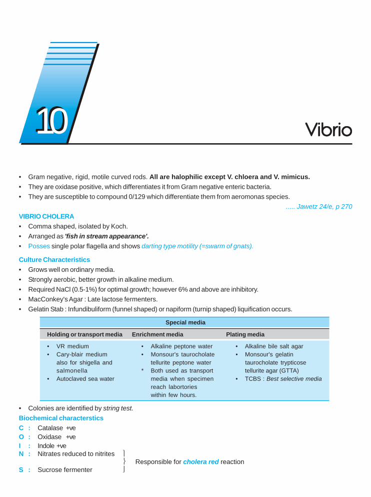
• Gram negative, rigid, motile curved rods. All are halophilic except V. chloera and V. mimicus.• They are oxidase positive, which differentiates it from Gram negative enteric bacteria.• They are susceptible to compound 0/129 which differentiate them from aeromonas species.
..... Jawetz 24/e, p 270VIBRIO CHOLERA• Comma shaped, isolated by Koch.• Arranged as 'fish in stream appearance'.• Posses single polar flagella and shows darting type motility (=swarm of gnats).
Culture Characteristics• Grows well on ordinary media.• Strongly aerobic, better growth in alkaline medium.• Required NaCl (0.5-1%) for optimal growth; however 6% and above are inhibitory.• MacConkey's Agar : Late lactose fermenters.• Gelatin Stab : Infundibuliform (funnel shaped) or napiform (turnip shaped) liquification occurs.
Special media
Holding or transport media Enrichment media Plating media
• VR medium • Alkaline peptone water • Alkaline bile salt agar• Cary-blair medium • Monsour's taurocholate • Monsour's gelatin
also for shigella and tellurite peptone water taurocholate trypticosesalmonella * Both used as transport tellurite agar (GTTA)
• Autoclaved sea water media when specimen • TCBS : Best selective mediareach labortorieswithin few hours.
• Colonies are identified by string test.Biochemical charactersticsC : Catalase +veO : Oxidase +veI : Indole +veN : Nitrates reduced to nitrites ⎫
⎬ Responsible for cholera red reactionS : Sucrose fermenter ⎭
VibrioVibrioVibrioVibrioVibrio1010
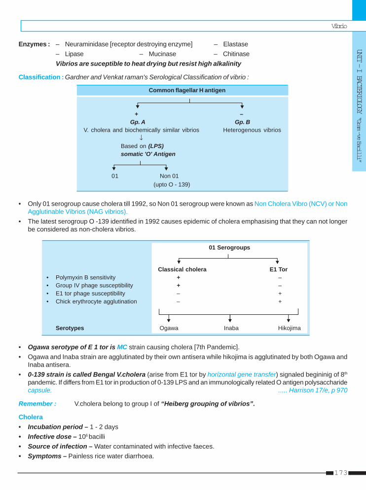
Vibrio
UNIT - I BACTERIOLOGY “Gram —ve Bacilli”
173
Enzymes : – Neuraminidase [receptor destroying enzyme] – Elastase– Lipase – Mucinase – ChitinaseVibrios are suceptible to heat drying but resist high alkalinity
Classification : Gardner and Venkat raman's Serological Classification of vibrio :
Common flagellar H antigen
+ –Gp. A Gp. B
V. cholera and biochemically similar vibrios Heterogenous vibrios↓
Based on (LPS)somatic 'O' Antigen
01 Non 01 (upto O - 139)
• Only 01 serogroup cause cholera till 1992, so Non 01 serogroup were known as Non Cholera Vibro (NCV) or NonAgglutinable Vibrios (NAG vibrios).
• The latest serogroup O -139 identified in 1992 causes epidemic of cholera emphasising that they can not longerbe considered as non-cholera vibrios.
01 Serogroups
Classical cholera E1 Tor• Polymyxin B sensitivity + –• Group IV phage susceptibility + –• E1 tor phage susceptibility – +• Chick erythrocyte agglutination – +
Serotypes Ogawa Inaba Hikojima
• Ogawa serotype of E 1 tor is MC strain causing cholera [7th Pandemic].• Ogawa and Inaba strain are agglutinated by their own antisera while hikojima is agglutinated by both Ogawa and
Inaba antisera.• 0-139 strain is called Bengal V.cholera (arise from E1 tor by horizontal gene transfer) signaled begininig of 8th
pandemic. If differs from E1 tor in production of 0-139 LPS and an immunologically related O antigen polysaccharidecapsule. ..... Harrison 17/e, p 970
Remember : V.cholera belong to group I of “Heiberg grouping of vibrios”.
Cholera• Incubation period – 1 - 2 days• Infective dose – 106 bacilli• Source of infection – Water contaminated with infective faeces.• Symptoms – Painless rice water diarrhoea.
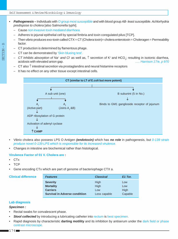
Self Assessment & Review Microbiology & Immunology
SECTION – B
174174
• Pathogenesis – Individuals with O group most susceptible and with blood group AB- least susceptible. Achlorhydriapredispose to cholera [also Salmonella typhi].– Cause non invasive toxin mediated diarrhoea.– Adheres to jejunal epithelial cell by special fimbria and toxin coregulated pilus [TCP].– Then vibrio produces exo-toxin called CTX = CT (Cholera toxin)= cholera enterotoxin = Choleragen = Permeability
factor.– CT production is determined by flamentous phage.– CT can be demonstrated by ‘Skin blueing test’.– CT Inhibits absorption of Na+ and Cl- as well as, ↑ secretion of K+ and HCO3; resulting in isotonic diarrhea,
acidosis with elevated anion gap. ..... Harrison 17/e, p 970– CT also ↑ intestinal secretion via prostaglandins and neural histamine receptors– It has no effect on any other tissue except intestinal cells.
CT (similar to LT of E.coli but more potent)
A sub unit (one) B subunint (5 in No.)
↓↓↓↓↓
A1 A2 Binds to GM1 ganglioside receptor of jejunum(Active part) (Joins A1 &B)
↓↓↓↓↓ADP ribosylation of G protein
↓↓↓↓↓Activation of adenyl cyclase
↓↓↓↓↓↑↑↑↑↑ CAMP
• Vibrio cholera also possess LPS O Antigen (endotoxin) which has no role in pathogenesis, but 0-139 strainproduce novel 0-139 LPS which is responsible for its increased virulence.
• Changes in intestine are biochemical rather than histological.
Virulence Factor of 01 V. Cholera are :• CTx• TCP• Gene encoding CTx which are part of genome of bacteriophage CTX φ.
Clinical difference Features Classical E1 Tor.
Severity High LowMortality High LowCarriers Low HighSurvival in Adverse conditiion Less capable Capable
Lab diagnosisSpecimen :• Rectal swabs for convalescent phase.• Stool collected by introducing a lubricating catheter into rectum is best specimen.• Rapid diagnosis by characteristic darting motility and its inhibition by antiserum under the dark field or phase
contrast microscope.
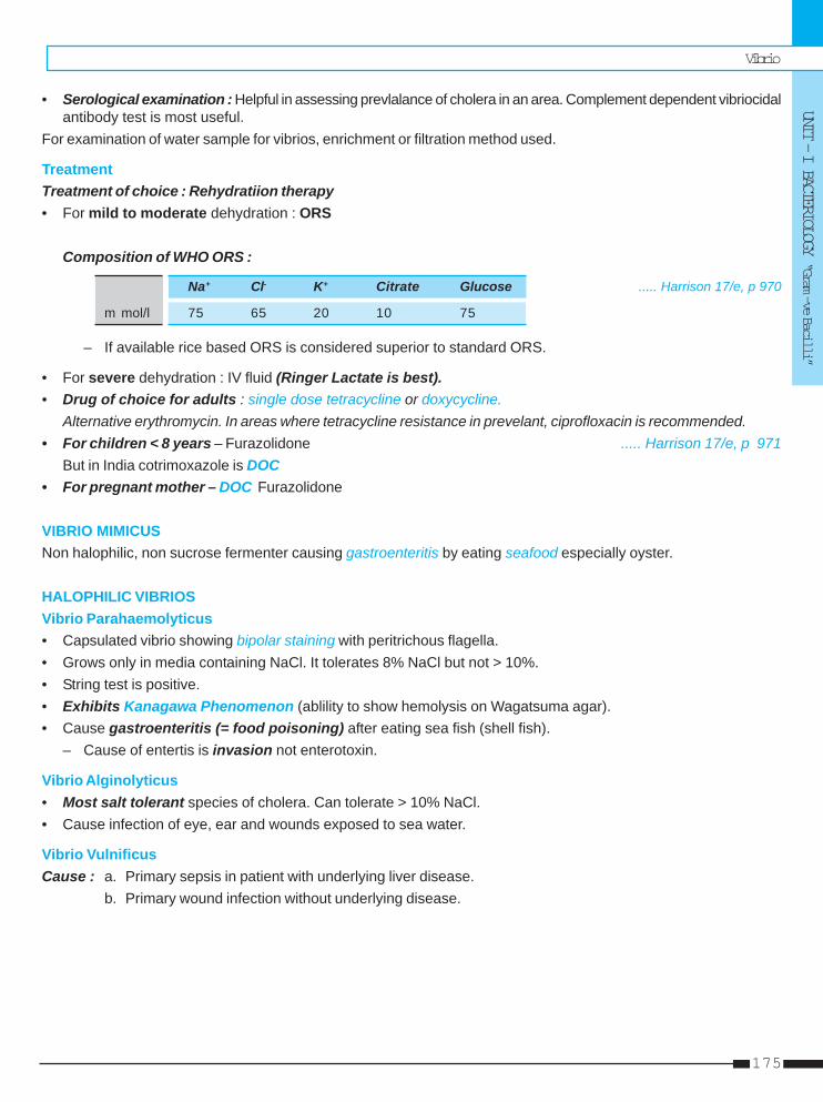
Vibrio
UNIT - I BACTERIOLOGY “Gram —ve Bacilli”
175
• Serological examination : Helpful in assessing prevlalance of cholera in an area. Complement dependent vibriocidalantibody test is most useful.
For examination of water sample for vibrios, enrichment or filtration method used.
TreatmentTreatment of choice : Rehydratiion therapy• For mild to moderate dehydration : ORS
Composition of WHO ORS :
Na+ Cl- K+ Citrate Glucose ..... Harrison 17/e, p 970
m mol/l 75 65 20 10 75
– If available rice based ORS is considered superior to standard ORS.
• For severe dehydration : IV fluid (Ringer Lactate is best).• Drug of choice for adults : single dose tetracycline or doxycycline.
Alternative erythromycin. In areas where tetracycline resistance in prevelant, ciprofloxacin is recommended.• For children < 8 years – Furazolidone ..... Harrison 17/e, p 971
But in India cotrimoxazole is DOC• For pregnant mother – DOC Furazolidone
VIBRIO MIMICUSNon halophilic, non sucrose fermenter causing gastroenteritis by eating seafood especially oyster.
HALOPHILIC VIBRIOSVibrio Parahaemolyticus• Capsulated vibrio showing bipolar staining with peritrichous flagella.• Grows only in media containing NaCl. It tolerates 8% NaCl but not > 10%.• String test is positive.• Exhibits Kanagawa Phenomenon (ablility to show hemolysis on Wagatsuma agar).• Cause gastroenteritis (= food poisoning) after eating sea fish (shell fish).
– Cause of entertis is invasion not enterotoxin.
Vibrio Alginolyticus• Most salt tolerant species of cholera. Can tolerate > 10% NaCl.• Cause infection of eye, ear and wounds exposed to sea water.
Vibrio VulnificusCause : a. Primary sepsis in patient with underlying liver disease.
b. Primary wound infection without underlying disease.
Self Assessment & Review Microbiology & Immunology
SECTION – B
176176
1. All of the folowing are true about V. cholera O139except : [AI 08]a) Clinical manifestations are similar to O1 E1 torb) First discovered in chennaic) Produces O139 lipopolysaccharided) Epidemiologically indistinguishable from
O1E1tor2. Which of the following bacteria acts by increasing
cAMP : [AI 07; AIIMS 06]a) Vibrio cholerab) Staphylococcus aureusc) E. coli heat stable toxind) Salmonella
3. About V. cholera all statements are true except :a) Non halophilic [AI 07]b) Can not grow in ordinary mediac) Can survive outside the intestined) Man is the only reservoir of cholera
4. Not true about vibrio O139 : [AI 07]a) Can cause disease indistinguishable from E-1 or
clinicallyb) First isolated in chennaic) Has O polysaccharide capsuled) Antibody to V. cholera is not protective against
O1395. All of the following vibrio species are halophilic
except : [AI 05]a) V. choleraeb) V. parahemolyticusc) V. alginolyticusd) V. flovialis
6. In the small intestine, cholera toxin acts by :a) ADP ribosylation of G regulatory protein [AI 05]b) Inhibition of adenyl cyclasec) Activation of GTPased) Active absorption of NaCl
7. Antibiotic treatment of choice for treating cholerain an adult is a single dose of : [AI 05]a) Tetracyclineb) Cotrimoxazolec) Doxycycline
d) Furazolidone8. Which of the following statement is true about :
Vibrio cholera : [AI 99]a) There is no natural reservoirb) Transported in alkaline peptone water mediumc) Halophilicd) Oxidase negative
9. All of the following statements about cholera aretrue except : [AI 97]a) O and H antigens measure carrier stateb) Culture medium is TCBS agarc) Produces indole and reduces nitrated) Synthesize neuraminidase
10. All of the following statements are true for El- torcholera except : [AI 97]a) Infection is mild and asymptomaticb) They are resistant to polymyxin-B unit discc) Chronic carriers are commond) Secondary attacks rate is high in families
11. Selective media for vibrio : [AIIMS 08]a) TCBSb) Stuartc) Skirrowsd) MYPA
12. The best sutied medium for Vibrio cholerae is :a) Thayer martin [AIIMS 07]b) TCBS mediumc) Scirrow mediumd) Loeffler’s medium
13. The endotoxin of the following gram-negative bac-teria does not play any part in the pathogenesisof the natural disease : [AIIMS 06]a) Escherichia colib) Klebsiella sp.c) Vibrio choleraed) Pseudomonas aeruginosa
14. Cholera toxin : [AIIMS 06]a) Increases the levels of intracellular cyclic GMPb) Acts through the receptor for opiatesc) Causes continued activation of adenylate cyclased) Inhibits the enzyme phosphodiesterase
QUESTIONS
Answer 1. b) First... 2. a) Vibrio ... 3. b) Can not ... 4. b) First isolated ... 5. a) V. cholerae6. a) ADP ... 7. c) Doxycycline 8. b) Transported ... 9. a) O and H ... 10. d) Secondary ...
11. a) TCBC 12. b) TCBS 13. c) Vibrio cholerae 14. c) Causes ...
Vibrio
UNIT - I BACTERIOLOGY “Gram —ve Bacilli”
177
15. The drug of choice for treating cholera in pregnantwomen is : [AIIMS 05]a) Tetracyclineb) Doxycyclinec) Furazolidoned) Cotrimoxazole
16. Which of the following is the drug of choice forchemoprophylaxis of cholera ? [AIIMS 05]a) Tetracyclineb) Doxycyclinec) Furazolidoned) Cotrimoxazole
17. True about V. cholerae is : [AIIMS 02]a) One attack of V. cholerae gives life-long immunityb) Affects adults and children with equal propensity
in non epidemic regionsc) In between epidemics, carrier states maintain the
organismd) Pathogenicity of 0-139 vibrio is due to 0 antigen
18. A 32 year old male, kallu who recently visited asea coast presented with ulcer over the left leg.The probable cause is : [AIIMS 01]a) Pasturella multocidab) Micrococcus halophillusc) Vibrio vulnificusd) Neisseria gonorrhea
19. True regarding cholera is : [AIIMS 95]a) Toxin acts on GM1 receptorb) Toxin action is CAMP mediatedc) Peritrichate flagellad) Utilises arginine, lysine
20. Which toxin acts by ADP ribosylation :a. Botulinum toxin [PGI 07]b. Shiga toxinc. V.choleraed. Diphtheria toxind. Pertusis
21. V. cholera able to stay in GIT because of :a) Acid resistance [PGI 06]b) Bile resistance
Answer 15. c) Furazolidone 16. a) Tetracycline 17. d) Pathogenicity ... 18. c) Vibrio vulnificus19. b) Toxin ...20. c) V.cholerae ... 21. c and d 22. a and c 23. d and e 24. c) Food can ...25. b) 0:139 26. a, b and e 27. a) Can tolerate ...
c) Motilityd) Binds to specific receptorse) Anaerobic potential
22. Cholera transmission by : [PGI 06]a) Food transmitsb) Vaccination gives 90% efficiencyc) Healthy carrierd) Chlorination is not effective
23. In patient presenting with diarrhoea due to vibriocholera, which of the following will be present :a) Abdominal pain [PGI 05, 01]b) Presence of leukocytes in stoolc) Feverd) Neutrophiliae) Occurence of many cases in the same locality
24. True about epidemiology of cholera : [PGI 03]a) Chemoprophylaxis is not effectiveb) Boiling of water cannot destroy the organismc) Food can transmit the diseased) Vaccination give 90% protection
25. Strain of V. cholerae in Bengal : [PGI 04]a) 0:037b) 0:139c) 0:17d) 0:40e) 0:149
26. In a patient presenting with diarrhea and pus cellsin stool, the causative organism can be all except:a) Non vibrio cholera 01 [PGI 01]b) Enterotoxigenic E. Colic) Enteroinvasive E. colid) Shigella dysenteriae 1e) Vibrio cholera
27. True about cholera vibrios is : [PGI 97]a) Can tolerate wide range of alkaline pHb) Non motile bacillic) Can’t be grown in mediad) NaCl stimulates growth
Self Assessment & Review Microbiology & Immunology
SECTION – B
178178
1. Ans. is b i.e. First discoverd in Chennai Ref. Harrison 17/e, p 969
• O139 Vibrio was isolated in Bengal in 1992.• The clinical manifestation and epidemiological features of the disease caused by V. cholera O139 are
indistinguishable from those of 01 cholera.• V. cholera is identical to E1 tor except for two important difference :
– Production of novel O - 139 Lipopolysaccharide.– Immunologically related O antigen polysaccharide capsule.
Remember :E1 tor biotype was first isolated at the E1 tor quarantine station in Egypt in 1905.
2. Ans. is a i.e. Vibrio cholera Ref. Harrison 17/e, p 970
Cholera toxin (CT)• Protein enterotoxin• Composed of 2 subunit : A – (Active monomeric moiety)
B – (Pentameric binding moiety)• Mechanism of action :
CT
A sub unit B subunint
↓ ↓ ↓ ↓ ↓
A1 A2 Binds to GM1 ganglioside receptor(Active part) (Joins A1 & B)
↓ADP ribosylation of G protein
↓Activation of adenyl cyclase
↓↑↑↑↑↑ CAMP
↓Inhibits absorption of Na+ and Cl- and result in diarrhoea.CT also ↑ intestinal secretion via prostaglandins and neural histamine receptors.
Remember :Mechansim of action of some important bacterial toxin.– Heat labile toxin of E. coli - ↑ cAMP– Heat stable toxin of E. coli - Increase cGMP– Botulism toxin - Inhibit release of acetylcholine from peripheral nerves.– Tetanus toxin - Inhibit release of glycine and GABA at presynaptic terminals.– Diptheria toxin - Inhibit protein synthesis by inactiviting EF-2.
EXPLANATIONS AND REFERENCES WITH ILLUSTRATIVE ANSWERS
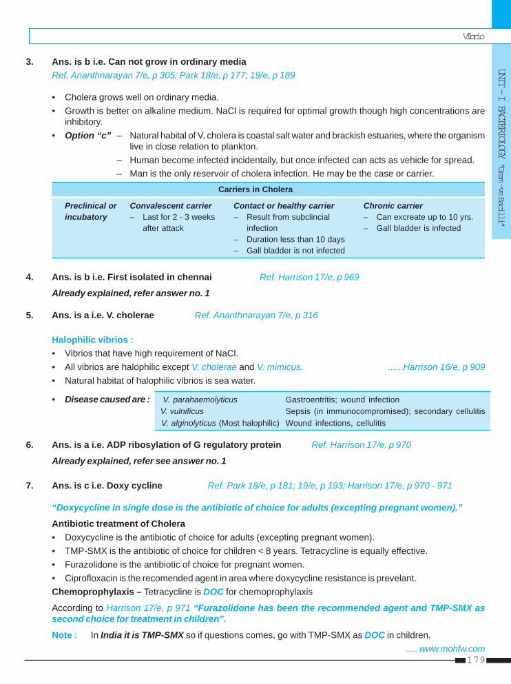
Vibrio
UNIT - I BACTERIOLOGY “Gram —ve Bacilli”
179
3. Ans. is b i.e. Can not grow in ordinary mediaRef. Ananthnarayan 7/e, p 305; Park 18/e, p 177; 19/e, p 189
• Cholera grows well on ordinary media.• Growth is better on alkaline medium. NaCl is required for optimal growth though high concentrations are
inhibitory.• Option “c” – Natural habital of V. cholera is coastal salt water and brackish estuaries, where the organism
live in close relation to plankton.– Human become infected incidentally, but once infected can acts as vehicle for spread.– Man is the only reservoir of cholera infection. He may be the case or carrier.
Carriers in Cholera
Preclinical or Convalescent carrier Contact or healthy carrier Chronic carrierincubatory – Last for 2 - 3 weeks – Result from subclincial – Can excreate up to 10 yrs.
after attack infection – Gall bladder is infected– Duration less than 10 days– Gall bladder is not infected
4. Ans. is b i.e. First isolated in chennai Ref. Harrison 17/e, p 969
Already explained, refer answer no. 1
5. Ans. is a i.e. V. cholerae Ref. Ananthnarayan 7/e, p 316
Halophilic vibrios :• Vibrios that have high requirement of NaCl.• All vibrios are halophilic except V. cholerae and V. mimicus. ......Harrison 16/e, p 909• Natural habitat of halophilic vibrios is sea water.
• Disease caused are : V. parahaemolyticus Gastroentritis; wound infection V. vulnificus Sepsis (in immunocompromised); secondary cellulitis V. alginolyticus (Most halophilic) Wound infections, cellulitis
6. Ans. is a i.e. ADP ribosylation of G regulatory protein Ref. Harrison 17/e, p 970
Already explained, refer see answer no. 1
7. Ans. is c i.e. Doxy cycline Ref. Park 18/e, p 181; 19/e, p 193; Harrison 17/e, p 970 - 971
“Doxycycline in single dose is the antibiotic of choice for adults (excepting pregnant women).”
Antibiotic treatment of Cholera• Doxycycline is the antibiotic of choice for adults (excepting pregnant women).• TMP-SMX is the antibiotic of choice for children < 8 years. Tetracycline is equally effective.• Furazolidone is the antibiotic of choice for pregnant women.• Ciprofloxacin is the recomended agent in area where doxycycline resistance is prevelant.Chemoprophylaxis – Tetracycline is DOC for chemoprophylaxis
According to Harrison 17/e, p 971 “Furazolidone has been the recommended agent and TMP-SMX assecond choice for treatment in children”.
Note : In India it is TMP-SMX so if questions comes, go with TMP-SMX as DOC in children...... www.mohfw.com
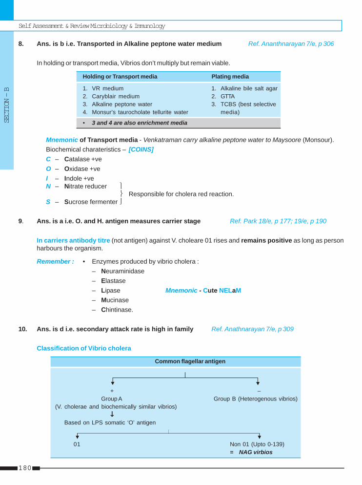
Self Assessment & Review Microbiology & Immunology
SECTION – B
180180
8. Ans. is b i.e. Transported in Alkaline peptone water medium Ref. Ananthnarayan 7/e, p 306
In holding or transport media, Vibrios don’t multiply but remain viable.
Holding or Transport media Plating media
1. VR medium 1. Alkaline bile salt agar2. Caryblair medium 2. GTTA3. Alkaline peptone water 3. TCBS (best selective4. Monsur’s taurocholate tellurite water media)
• 3 and 4 are also enrichment media
Mnemonic of Transport media - Venkatraman carry alkaline peptone water to Maysoore (Monsour).Biochemical charateristics – [COINS]C – Catalase +veO – Oxidase +veI – Indole +veN – Nitrate reducer ⎫
⎬ Responsible for cholera red reaction.S – Sucrose fermenter ⎭
9. Ans. is a i.e. O. and H. antigen measures carrier stage Ref. Park 18/e, p 177; 19/e, p 190
In carriers antibody titre (not antigen) against V. choleare 01 rises and remains positive as long as personharbours the organism.
Remember : • Enzymes produced by vibrio cholera :– Neuraminidase– Elastase– Lipase Mnemonic - Cute NELaM– Mucinase– Chintinase.
10. Ans. is d i.e. secondary attack rate is high in family Ref. Anathnarayan 7/e, p 309
Classification of Vibrio cholera
Common flagellar antigen
+ –Group A Group B (Heterogenous vibrios)
(V. cholerae and biochemically similar vibrios)↓↓↓↓↓
Based on LPS somatic ‘O’ antigen
01 Non 01 (Upto 0-139)= NAG virbios
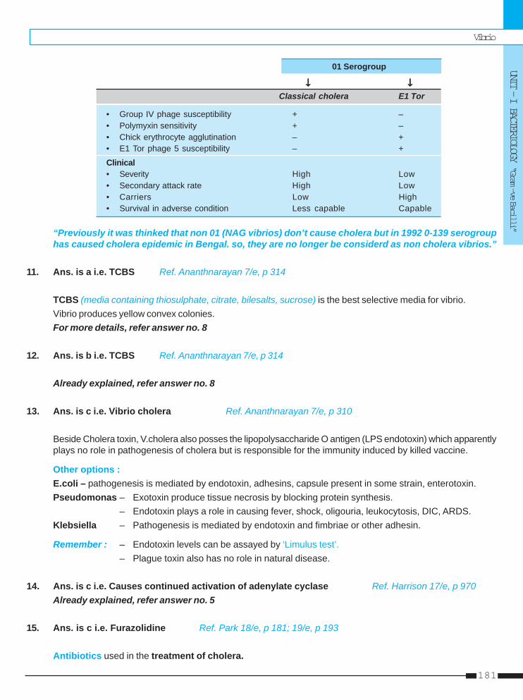
Vibrio
UNIT - I BACTERIOLOGY “Gram —ve Bacilli”
181
01 Serogroup
↓↓↓↓↓ ↓ ↓ ↓ ↓ ↓Classical cholera E1 Tor
• Group IV phage susceptibility + –• Polymyxin sensitivity + –• Chick erythrocyte agglutination – +• E1 Tor phage 5 susceptibility – +
Clinical• Severity High Low• Secondary attack rate High Low• Carriers Low High• Survival in adverse condition Less capable Capable
“Previously it was thinked that non 01 (NAG vibrios) don’t cause cholera but in 1992 0-139 serogrouphas caused cholera epidemic in Bengal. so, they are no longer be considerd as non cholera vibrios.”
11. Ans. is a i.e. TCBS Ref. Ananthnarayan 7/e, p 314
TCBS (media containing thiosulphate, citrate, bilesalts, sucrose) is the best selective media for vibrio.Vibrio produces yellow convex colonies.For more details, refer answer no. 8
12. Ans. is b i.e. TCBS Ref. Ananthnarayan 7/e, p 314
Already explained, refer answer no. 8
13. Ans. is c i.e. Vibrio cholera Ref. Ananthnarayan 7/e, p 310
Beside Cholera toxin, V.cholera also posses the lipopolysaccharide O antigen (LPS endotoxin) which apparentlyplays no role in pathogenesis of cholera but is responsible for the immunity induced by killed vaccine.
Other options :E.coli – pathogenesis is mediated by endotoxin, adhesins, capsule present in some strain, enterotoxin.Pseudomonas – Exotoxin produce tissue necrosis by blocking protein synthesis.
– Endotoxin plays a role in causing fever, shock, oligouria, leukocytosis, DIC, ARDS.Klebsiella – Pathogenesis is mediated by endotoxin and fimbriae or other adhesin.
Remember : – Endotoxin levels can be assayed by ‘Limulus test’.– Plague toxin also has no role in natural disease.
14. Ans. is c i.e. Causes continued activation of adenylate cyclase Ref. Harrison 17/e, p 970Already explained, refer answer no. 5
15. Ans. is c i.e. Furazolidine Ref. Park 18/e, p 181; 19/e, p 193
Antibiotics used in the treatment of cholera.
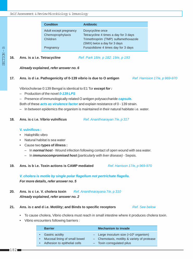
Self Assessment & Review Microbiology & Immunology
SECTION – B
182182
Condition Antibiotic
Adult except pregnancy Doxycycline onceChemoprophylaxis Tetracycline 4 times a day for 3 daysChildren Trimethroprim (TMP) sulfamethoxazole
(SMX) twice a day for 3 daysPregnancy Furazolidone 4 times day for 3 days
16. Ans. is a i.e. Tetracycline Ref. Park 18/e, p 182; 19/e, p 193
Already explained, refer answer no. 6
17. Ans. is d i.e. Pathogenicity of 0-139 vibrio is due to O antigen Ref. Harrision 17/e, p 969-970
Vibriocholerae 0:139 Bengal is identical to E1 Tor except for :– Production of the novel 0-139 LPS– Presence of immunologically related O antigen polysaccharide capsule.Both of these acts as virulence factor and explain resistance of 0 - 139 strain.– In between epidemics the organism is maintained in their natural habitate i.e. water.
18. Ans. is c i.e. Vibrio vulnificus Ref. Ananthnarayan 7/e, p 317
V. vulnificus :• Halophillic vibro• Natural habitat is sea water• Cause two types of illness :
– In normal host - Wound infection following contact of open wound with sea water.– In immunocompromised host (particularly with liver disease) - Sepsis.
19. Ans. is b i.e. Toxin actions is CAMP mediated Ref. Harrison 17/e, p 969-970
V. cholera is motile by single polar flagellum not pertrichate flagella.For more details, refer answer no. 5
20. Ans. is c i.e. V. cholera toxin Ref. Ananthnarayana 7/e, p 310Already explained, refer answer no. 2
21. Ans. is c and d i.e. Motility; and Binds to specific receptors Ref. See below
• To cause cholera, Vibrio cholera must reach in small intestine where it produces cholera toxin.• Vibrio encounters following barriers :
Barrier Mechanism to invade
• Gastric acidity – Large inoculum size (>106 organism)• Mucosal lining of small bowel – Chemotaxis, motility & variety of protease• Adhesion to epithelial cells – Toxin corregulated pilus
Vibrio
UNIT - I BACTERIOLOGY “Gram —ve Bacilli”
183
22. Ans. is a and c i.e. Food transmit; and Healthy carrierRef. Ananthnarayan 7/e, p 313; Park 18/e, p 177, 182; 19/e, p 191]Mode of transmission of cholera :– Faecally contaminated water (MC)– Contaminated food and drinks– Direct contact.
Cholera Vaccine
Parenteral vaccine Oral vaccines
Killed Killed (wc/rbs) or live (CVD 103 HgR)Protective value 50% Protective value 80% for live vaccine
So, no vaccine of cholera provide 90% protection.
Remember : • V. cholerae are killed with in 30 minutes by heating at 560 C or within a few secondsby boiling. Also killed by chlorination.
• DOC for chemoprophylaxis is tetracycline. Alternative is doxycycline.• Carriers in cholera includes preclinical or incubatory (1 - 5 days), convalenscent (2 - 3
weeks), contact or healthy (> 10 days) and chronic carrier.
23. Ans. is d and e i.e. Neutrophilia; and Occurence of many cases in the same localityRef. Park 18/e, p 177; 19/e, p 191; Harrison 17/e, p 970
• V. cholera cause non inflamatory (no WBC in stool) painless watery diarrhoea.• Neutrophilic leucocytosis occurs.• As secondary attack is high, many case occurs in same locality.
24. Ans. is c i.e. Food can transmit the disease Ref. Ananthnarayan 7/e, p 313
Already explained, refer answer no. 21
25. Ans. is b i.e. 0:139 Ref. Park 18/e, p 175; 19/e, p 189
0 - 139 posses novel 0 - 139 LPS which is responsible for its virulence. 0 : 139 has replaced E1 tor and ispredominant cause of cholera in areas where it had appeared.
26. Ans. is a, b and e i.e. Non vibrio cholera 01; Enterotoxigenic E. Coli; Vibrio choleraRef. Harrison 17/e, p 814
Causes of diarrhoea with pus cells / RBC’s in stool (inflammatary diarrhoea or dysentery). :
– Shigella – EIEC, EHEC– Campylobacter – Salmonella– Yersinia – Cl. difficle– Vibrio parahemolyticus
Self Assessment & Review Microbiology & Immunology
SECTION – B
184184
27. Ans. is a i.e. Can tolerate wide range of alkaline pH Ref. Ananthnarayan 7/e, p 305
• Vibrio cholera can grow in pH range 6.4-9.6 (Optimum - 8.2).• NaCl (0.5-1%) is required for optimal growth though high concentration (6% and above) is inhibitory.• Can be grown on media.• Motile by single polar flagellum.
Vibrio
UNIT - I BACTERIOLOGY “Gram —ve Bacilli”
185
1. d) Vibriocidal 2. a) Koch 3. c) Catfish ... 4. b) Robertkoch 5. b) Cary Blair6. a) Saline 7. a) Classical ... 8. b) Adenylate 9. c) E1-tor is more ... 10. a) VR medium
1. Which of the following about cholera is true :a) Invasive [AI 90]b) Endotoxin is releasedc) Recent infections in India are of classical typed) Vibriocidal antibody titre measures prevalence
[Ref. Ananthnarayan 7/e, p 314]2. Vibrio cholera was discovered by : [Kerala 94]
a) Kochb) Mekintoffc) John snowd) Virchow
[Ref. Ananthnarayan 7/e, p 305]3. Vibrio parahemolyticus food poisoning is caused
by ingestion of : [MP 96]a) Eggs and Paultry productsb) Raw vegetablec) Catfish, shellfish, seafoodd) Milk products
[Ref. Ananthnarayan 7/e, p 316-317]4. Vibrio cholera was discovered by : [Delhi 96]
a) Louis pastuerb) Robertkochc) Eberth and Gaffkyd) Bordet
[Ref. Ananthnarayan 7/e, p 305]5. The transport medium for stools sample in chol-
era is : [ICS 98]a) Thioglycolateb) Cary Blair mediumc) Dubos mediumd) Selenite ‘F’ broth
[Ref. Ananthnarayan 7/e, p 306]
Answer
6. The growth factor required for the growth of vibrioparahemolyticus is : [Kerala 98]a) Salineb) Tryptophanc) Biled) Citrate
[Ref. Ananthnarayan 7/e, p 316]7. In cholera the highest case fatality is observed in
case of : [Orissa 98]a) Classical cholera vibriob) Vibrio EL torc) Vibrio parahemolyticusd) NAG -vibrios
[Ref. Ananthnarayan 7/e, p 311]8 Cholera toxin acts by : [Kolkata 03]
a) Na+K+ATPase inhibitionb) Adenylate cyclase stimulationc) Opening of chloride channeld) Stimulation of Ca++channel
[Ref. Ananthnarayana 7/e, p 310]9. True about vibriocholera is : [SGPGI 03]
a) Disease more commonin womanb) Classical vibrio protect against development of
0.139 strain diseasec) E1-tor is more milder than classicald) Erythronycin is used in treatment
[Ref. Ananthnarayan 7/e, p 311]10. Transport media for cholera : [Jharkhand 04]
a) VR mediumb) LZ mdeiumc) Bile salt agard) TCBS
[Ref. Ananthnarayan 6/e, p 306]
Chapter Review••••• This section includes questions of VThis section includes questions of VThis section includes questions of VThis section includes questions of VThis section includes questions of Various Other PGMEES from 1990 – 2007.arious Other PGMEES from 1990 – 2007.arious Other PGMEES from 1990 – 2007.arious Other PGMEES from 1990 – 2007.arious Other PGMEES from 1990 – 2007.••••• Questions are arQuestions are arQuestions are arQuestions are arQuestions are ar ranged in decreasing order of yearranged in decreasing order of yearranged in decreasing order of yearranged in decreasing order of yearranged in decreasing order of yearwiz. This is done to makwiz. This is done to makwiz. This is done to makwiz. This is done to makwiz. This is done to make refere refere refere refere referralralralralral
system easier and uncomplicated to save the precious time of PGMEE Aspirant.system easier and uncomplicated to save the precious time of PGMEE Aspirant.system easier and uncomplicated to save the precious time of PGMEE Aspirant.system easier and uncomplicated to save the precious time of PGMEE Aspirant.system easier and uncomplicated to save the precious time of PGMEE Aspirant.
Self Assessment & Review Microbiology & Immunology
SECTION – B
186186
11. a) V R medium 12. b) V. cholera13. b) To bind GM1 ... 14. a) E1tor
Answer
11. Transport medium for cholera bacteria :a) V R medium [Jharkhand 05]b) L Z mediumc) Carry blair mediumd) Staruert medium
[Ref. Anantharayan 7/e, p 306]12. Non-Halophillic vibrios indudes all of the following
except : [SGPGI 07]a) V. vulnificusb) V. cholerac) V. parahaemolyticusd) V. alginolyticus
[Ref. Ananthnarayan 7/e, p 316]
13. The function of B subunit of cholera toxin is :a) ADP ribosylation of G protein [BHU 07]b) To bind GM1 ganglioside recepterc) To stablize cholera toxind) To increase cGMP
[Ref. Harrison 17/e, p 969]14. 7th pandemic of cholera is caused by : [UP 07]
a) E1torb) 0139 V. cholerac) Classical V. cholerad) V. mimicus
[Ref. Ananthnarayan 7/e, p 311]
PSEUDOMONASGram (–)ve aerobic motile bacilli with polar flagella.Pseudomonas aeruginosa the MC human pathogen in this group.
P. AERUGINOSA = P. PYOCYANEA• Obligate aerobic bacilli which is differentiated from enteric Gram (–)ve bacilli by its ability to oxidise indophenol
and inability to ferment lactose.• Non capsulated but many strains have mucoid slime layer of alginate particularly from patient of cystic fibrosis.
Culture and Growth characteristic• Grows well at 37-420C on ordinary media.• Growth at 420C helps differentiate it from other pseudomonas species.• Selective media – Cetrimide agar• Pigment :
a. Pyocyanin – Bluish green pigment produce only by P. aeruginsoa. It inhibits growth of many other bacteria.b. Fluorescin – Greenish yellow. Produce by all species of pseudomonas.
Classification• On the basis of difference of lipopolysaccharide.• Restriction endonuclease typing with pulsed gel electrophoresis is most reliable method.• Used for epidemiological purpose.
Pathogenicity and Resistance• MC and most serious cause of infection in burns.• MC infection outside hospital is suppurative otitis.• Causative agent of Shanghai fever.• Blue pus with characteristic fruity odour.• Pathognomic skin lesion termed ecthyma gangreosum which occur singly or in small number on the perineum,
buttocks and extremities• Resistant to common antiseptic and disinfectant such as dettol. Even may grow profusely in bottle of these
antiseptic.
Pseudomonas & YPseudomonas & YPseudomonas & YPseudomonas & YPseudomonas & Yersiniaersiniaersiniaersiniaersinia1111
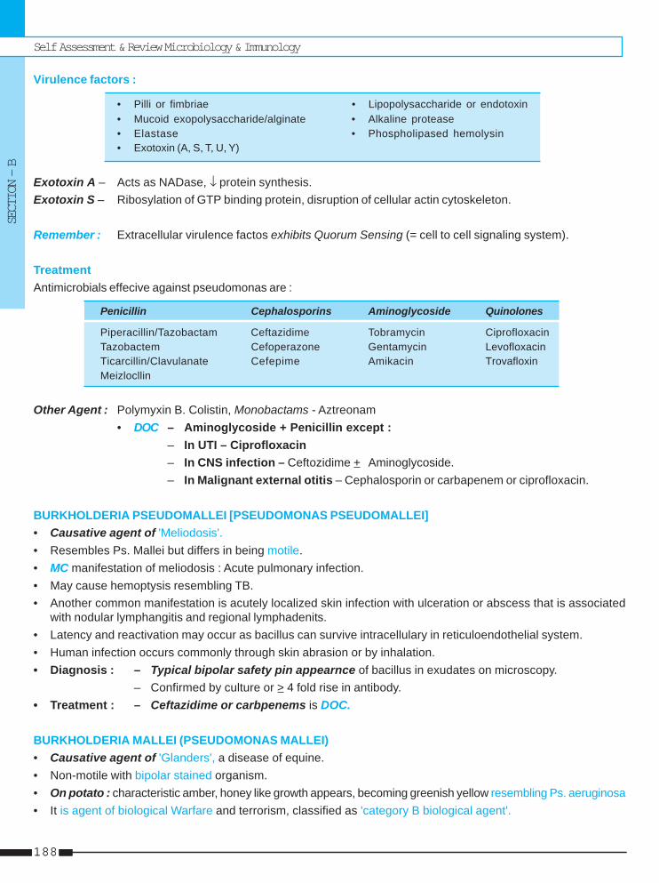
Self Assessment & Review Microbiology & Immunology
SECTION – B
188188
Virulence factors :
• Pilli or fimbriae • Lipopolysaccharide or endotoxin• Mucoid exopolysaccharide/alginate • Alkaline protease• Elastase • Phospholipased hemolysin• Exotoxin (A, S, T, U, Y)
Exotoxin A – Acts as NADase, ↓ protein synthesis.Exotoxin S – Ribosylation of GTP binding protein, disruption of cellular actin cytoskeleton.
Remember : Extracellular virulence factos exhibits Quorum Sensing (= cell to cell signaling system).
TreatmentAntimicrobials effecive against pseudomonas are :
Penicillin Cephalosporins Aminoglycoside Quinolones
Piperacillin/Tazobactam Ceftazidime Tobramycin CiprofloxacinTazobactem Cefoperazone Gentamycin LevofloxacinTicarcillin/Clavulanate Cefepime Amikacin TrovafloxinMeizlocllin
Other Agent : Polymyxin B. Colistin, Monobactams - Aztreonam• DOC – Aminoglycoside + Penicillin except :
– In UTI – Ciprofloxacin– In CNS infection – Ceftozidime + Aminoglycoside.– In Malignant external otitis – Cephalosporin or carbapenem or ciprofloxacin.
BURKHOLDERIA PSEUDOMALLEI [PSEUDOMONAS PSEUDOMALLEI]• Causative agent of 'Meliodosis'.• Resembles Ps. Mallei but differs in being motile.• MC manifestation of meliodosis : Acute pulmonary infection.• May cause hemoptysis resembling TB.• Another common manifestation is acutely localized skin infection with ulceration or abscess that is associated
with nodular lymphangitis and regional lymphadenits.• Latency and reactivation may occur as bacillus can survive intracellulary in reticuloendothelial system.• Human infection occurs commonly through skin abrasion or by inhalation.• Diagnosis : – Typical bipolar safety pin appearnce of bacillus in exudates on microscopy.
– Confirmed by culture or > 4 fold rise in antibody.• Treatment : – Ceftazidime or carbpenems is DOC.
BURKHOLDERIA MALLEI (PSEUDOMONAS MALLEI)• Causative agent of 'Glanders', a disease of equine.• Non-motile with bipolar stained organism.• On potato : characteristic amber, honey like growth appears, becoming greenish yellow resembling Ps. aeruginosa• It is agent of biological Warfare and terrorism, classified as 'category B biological agent'.
Pseudomonas & Yersinia
UNIT - I BACTERIOLOGY “Gram —ve Bacilli”
189
• In human it cause acute localized suppurative infection, acute pulmonary infection; acute septicemic infection andchronic suppurative infection.
• It induce 'straus reaction'.• Human infection is usually occupational.• Diagnosis : – Mallein test which is analogous to tuberculin test.
– Molecular methods for rapid identificatiion – 16s rRNA gene sequencing. Also distinguish itfrom B.pseudomallei.
• Treatment : – Same as meliodosis.
PASTEURELLAE• Group consist of gram negative, short, Pleomorphic bacilli that are primary pathogen of rodents.• It is divided into 3 genus :
a Yersinia : • includes plague bacillus (y.pestis); Y. pseudotuberculosis (primary pathogen of rodents);Y. enterocolitica.
• it is assigned in the family enterobacteriaceae.b. Pasteurella : • includes P. multiocida (non-motile, oxidase positive).c. Francisella : • includes F. Tularensis.
Remember : – Pasteurellae which Grow on MacConkey and are urease positive : Y.pseudotuberculosis, Y.enterocolitica.
– ‘a’ and ‘b’ are short pleomorphic, gram negative rods with bipolar staining. They are catalasepositive, oxidase negative except P. multiocida and are microaerophilic or facultative anaerobic.
..... Jawetz 24/e, p 294YERSINIA. PESTIS = PLAGUE BACILLUSMorphology• Non motile, Non sporing, microaerophilic, biochemically unreactive, pleomorphic bacilli/coccobacillus.• Characteristic bipolar (safety pin) appearance, with Wayson’s stain/Giemsa/Methylene.• It is noncapsulated but at > 300C it produce envelop (= F1 antigen) – a virulence factor that serves as the
principal immunodiagnostic marker of infection.• Serotypes do not exist.
Remember : Growth occur fastest at 300C and envelop develop best at 370C.
Cultural Characteristics• Optimum growth occurs at 270C and pH 7.2 (unlike most pathogen which usually usually grow at 370C).• Shows 'Stalactite growth' in ghee broth.
Biochemical reactions :• Based on fermentation of glycerol and reduction of nitrate, it is divided into 3 varieties.• Catalase and Aesculin positive; urease and oxidase negative.
Pathogenesis• From the site of flea bite (xenopsylla cheopis) it is carried to regional lymph nodes (=bubo) via lymphatic channel.• Monouclear phagocytes play role in dissemination of infection to distant sites (secondary pneumonia and
septicemia).• Primary pneumonia results by droplet infection of plague patient.• Primary septicemic plague consist of sepsis in absence of bubo while secondary septicemic plague is
complication of bubonic or pneumonic plague.• Toxin has no role.
Self Assessment & Review Microbiology & Immunology
SECTION – B
190190
Remember : It produce coagulase at 280C but not at 350C. ..... Jawetz 24/e, p 291
Clinical Features• Characterized by rapid onset of fever and other systemic manifestations of Gram –ve bacterial infections.• It is of 3 types :
a. Bubonic Plague : – I.P 2 - 7 days– MC type of plague and is almost always caused by bite of infected flea.– MC site of bubo femoral, inguinal region >axillary, cervical. ... Harrison 17/e, p 982– Distinguished from lymphadentis by its rapid onset, its extreme tenderness,
accompanying signs of toxemia and absence of cellulitis or obvious ascendinglymphadenitis.
– DIC is common and may lead to gangrene.– Y. pestis can also be cultured from blood of most bubonic plague patient.
b. Septicemic plague : – Often present with GI symptoms, DIC, multiorgan failure.– I.P 2 - 7 days
c. Pneumonic Plague : – Most infectious, most fatal type of plague with I.P of 1 - 3 days.• Primary pneumonia : Cyanosis is very prominent, with bloody mucoid sputum.• Secondary pneumonia : Diffuse interstitial pneumonia, less infectious.
Treatment : Streptomycin (DOC)
Prevention : • Prophylaxis of choice – Tetracycline.• Recombinant vaccine that use F1 and V antigen is available.
FRANCISELLA OR PASTEURELLA OR BRUCELLA TULARENSIS• Capsulated, non-motile, intracellular parasite which grows in special media such as Francis blood dextrose
cystine agar.• It resembles mycoplasma.• It cause tularemia, a disease of rabbits and other rodents which is transmitted by ticks.• In human it present as local ulceration with lymphadenitis, typhoid like fever with glandular enlargement or
influenza like respiratory infection.• Treatment : Streptomycin is DOC for adults and children.
YERSINOSIS• It denotes infection with yersinae other than Y.pestis (i.e. by enterocolitica and pseudotuberculosis).• They are motile at 250C but non-motile at 370C.• They show antigenic cross reaction with Y.pestis, Vibrio, Salmonella, Brucella.• For culture ‘cold enrichment’ is done.
Y.pseudotuberculosis : • Most human infection occur by serotype 01.
Y. Enterocolitca : • Most human infection occur by serotypes 03, 08, 09.• It causes gastroenteritis or enterocolitis; Mesentric adenitis or terminal ileitis; system
disease with bacteremia; erythema nodosum, reactive arthritis (in HLA - B 27).
Pseudomonas & Yersinia
UNIT - I BACTERIOLOGY “Gram —ve Bacilli”
191
1. A young boy had a flea bite while working in awheat grain godown. After 5 days he developedfever and had axillary lymphadenopathy. A smearwas sent to the laboratory to perform a specificstaining. Which one of the following stainingmethod would help in the identification of thesuspected pathogen : [AI 06]a) Albert stainingb) Zeihl-Nelson stainingc) McFadyean’s stainingd) Wayson staining
2. Which one of the following drugs is anantipseudomonal penicillin ? [AI 06]a) Cephalexinb) Cloxacillinc) Piperacillind) Dicloxacillin
3. The following statements are true regardingmeliodosis except : [AI 05]a) It is caused by Burkholderia malleib) The agent is a gram negative aerobic bacteriac) Bipolar staining of aetiological agent is seen with
methylene blued) The most common form of meliodosis is pulmo-
nary infection
4. An organism grown on agar shows green coloredcolonies; likely organism is : [AI 01]a) Staphylococcusb) E. Colic) Pseudomonasd) Peptostreptococcus
5. All of the following statement about plague arewrong except : [AIIMS 04]a) Domestic rat is main reservoirb) Bubonic is the most common varietyc) The causative bacillus can survive upto 10 years
in the soil of rodent burrows
d) Incubation period for pneumonic plague is oneto two weeks
6. A 50 year old chronic alcoholic male agricultureworker presented with high grade fever of oneweek with spells of chills and rigor. Examinationof the respiratory system reveals bilateral crepi-tation with scattered ronchi. Multiple subcutane-ous nodules were found on extensor surface ofleft forearm and left leg. Direct microscopy of thepus aspirate from the nodules reveals plenty ofGrams (–)ve bacilli with bipolar staining. Culturereveals distinct rough corrugated grey white colo-nies on blood agar. The organism were motileand oxidase positive. The most likely diagnosisis : [AIIMS 03]a) Plagueb) Meliodosisc) Bartonellosisd) Actinomycosis
7. The drug of choice for chemoprophylaxis in con-tacts of a patient of pneumonic plague is :a) Penicillin [AIIMS 02]b) Rifampicinc) Erythromycind) Tetracycline
8. True about Y. pestis : [PGI 06, 03]a) Gram +veb) Gram -vec) Motiled) Non-motilee) It is coccobacilli
9. True statement(s) about Y. pestis is/are :a) Gram positive [PGI 04]b) Non-motilec) Benzyl penicillin is given in prophylaxisd) Patients are kept isolated till 48 hrs. of treatmente) Repeated blood culture is diagnostic
QUESTIONS
Answer 1. d) Wayson ... 2. c) Piperacillin 3. a) It is caused by ... 4. c) Pseudomonas 5. b) Bubonic is ...
6. b) Meliodosis 7. d) Tetracycline 8. b, d and e 9. b and e
Self Assessment & Review Microbiology & Immunology
SECTION – B
192192
1. Ans. is d i.e. Wayson stainingRef. Park 19/e, p 244 - 246; Jawetz 24/e, p 292; Ananthnarayan 7/e, p 329
“It is typical clinical presentation of bubonic plague.”PLAGUE : • It is a zoonosis.
• Causative agent is Yersinia pestis which is :– Gram negative, Non motile, Coccobacilli– Exhibits bipolar staining with Wayson’s stain / Giemsa stain / Methylene blue.– Plague bacilli can survive and indeed multiply in the soil of rodent burrows where microclimate
and other conditions are favourable.Reservoir :• Wild rodents (eg. field mice) are natural reservoirs of plague.• In India wild rodent, Tatera indica has been incriminated as main reservoir, not the domestic rat.
Source of infection :• Infected rodents and fleas and case of pneumonic plague (not bubonic plague).Immunity : After recovery is relative.Vector : – Commonest vector is rat flea, (X-cheopis). Infected flea may live upto 4 year.• Human infection is most frequently contracted from bite of infected flea.• Basic cycle in epidemic bubonic plague is : Commensal rats → rat fleas → man• Incubation period : – Bubonic plague - 2 to 7 days
– Septicaemic Plague - 2 to 7 days– Pneumonic plague - 1 to 3 days
• MC type of human plague is Bubonic plague characterized by enlarged tender lymph nodes (Bubos).• MC site of bubo is femoral, inguinal, axillary and cervical nodes.• Pneumonic plague – Rare variety but most infectious variety of plague.• Septicaemic plague – Rare variety.
Diagnosis :– Specimens - Blood for culture.
- Aspirates of enlarged lymph nodes for smear and culture.– Smears - Stain with giemsa and specific immunofluorescent stains.
- Prime face diagnosis is by examination of smears which show characteristic bipolarappearance with Wayson’s stain.
– Culture - Blood culture are often positive in 24 hours.- Definite identification of culture is best done by immunofluorescence.
– Serology - Antibodies to F. 1 antigen may be detected by passive hemagglutination.
Treatment : • DOC – Streptomycin (Alternative tetracycline)
Chemoprophylaxis : • DOC – Tetracycline (Alternative sulfonamide)
EXPLANATIONS AND REFERENCES WITH ILLUSTRATIVE ANSWERS
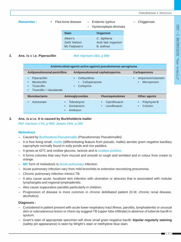
Pseudomonas & Yersinia
UNIT - I BACTERIOLOGY “Gram —ve Bacilli”
193
Remember : • Flea bone disease – Endemic typhus – Chiggerosis– Hymenoplepis diminata
Stain Organism
Albert’s C. diptheriaZeihl Nelson Acid fast organismMc Fadyean’s B. anthrax
2. Ans. is c i.e. Piperacillin Ref. Harrison 16/e, p 894
Antimicrobial agents active against pseudomonas aeruginosa
Antipseudomonal penicillins Antipseudomonal cephalosporins Carbapenems
• Piperacillin • Ceftazidime • Imipenem/cilastatin• Mezlocillin • Cefoperazone • Meropenem• Ticarcillin • Cefepime• Ticarcillin / clavulanate
Monobactams Aminoglycosides Fluoroquinolones Other agents
• Aztreonam • Tobramycin • Ciprofloxacin • Polymyxin B• Gentamicin • Levofloxacin • Colistin• Amikacin
3. Ans. is a i.e. It is caused by Burkholderia malleiRef. Harrison 17/e, p 955; Jawetz 24/e, p 265
Meliodosis– Caused by Burkhoderia Pseudomallei (Pseudomonas Pseudomallei).– It is free living small, motile (differentiating feature from pseudo. mallei) aerobic gram negative bacillary
saprophyte normally found in soily ponds and rice paddies.– It grows at 420C and oxidise glucose, lactose and is oxidase positive.– It forms colonies that vary from mucoid and smooth to rough and wrinkled and in colour from cream to
orange.– MC form of meliodosis is Acute pulmonary infection.– Acute pulmonary infection vary from mild bronchitis to extensive necrotizing pneumonia.– Chronic pulmonary infection mimics TB.– It also cause acute, localized skin infection with ulceration or abscess that is associated with nodular
lymphangitis and regional lymphadenitis.– Also cause suppurative parotitis particularly in children.– Progression of disease is more common in chronic debilitated patient (D.M, chronic renal disease,
alcoholics).
Diagnosis :– Considered in patient present with acute lower respiratory tract illness, parotitis, lymphadenitis or unusual
skin or subcutaneous lesion or chest ray suggest TB (upper lobe infiltrate) in absence of tubercle bacilli insputum.
– Gram’s stain of appropriate specimen will show small gram negative bacilli; bipolar regularly staining(safety pin appearance) is seen by Wright’s stain or methylene blue stain.
Self Assessment & Review Microbiology & Immunology
SECTION – B
194194
– Positive culture is diagnostic.– Positive serologic test is evidence of past infection.– X-ray - upper lobe infiltrate occasionally with thin walled cavities.Treatment : Ceftazidime or Carbepenems are DOC.
4. Ans. is c i.e. Pseudomonas Ref. Ananthnarayan 7/e, p 319
PseudomonasAerobic, nonsporing gram negative, motile bacilli, forms many pigments :a. Pyocyanin : – Bluish green pigment, produced only by Ps. aeruginosab. Fluorescin (Pyoverdin) :
– Greenish yellow pigment which oxidise in old culture to yellowish brown pigmentc. Pyrubin : – Redd. Pyomelanin : – Brown
Other pigment forming bacteria are : • S. aureus – Golden yellow pigment• B. melanogenicus – Black pigment• Rhodococcus – Red pigment• Nocardia – Yellow to red pigment• Pepto and peptostreptococcus• Photo and Scoto chromogen – Yellow orange pigment
5. Ans. is b i.e. Bubonic is the most common varietyRef. Jawetz 24/e, p 292; Ananthnarayan 7/e, p 329Already explained, refer answer no. 1
6. Ans. is b i.e. Melioidosis Ref. Harrison 17/e, p 955; Jawetz 24/e, p 265
• Actinomycosis is ruled out as it is gram positive bacilli.• Plague is ruled out as it is gram negative non motile coccobacilli.• Bartonellosis is ruled out as it does not exhibits bipolar staining.• Bacteria showing bipolar staining = safety pin appearance are :
– Calymmatobacterium granulomatis – Yersinia and Pasteurella – H. ducreyi– V. parahemolyticus – Ps. pseudomallei
7. Ans. is d i.e. Tetracycline Ref. Park 18/e, p 237; Jawetz 24/e, p 292; Ananthnarayan 7/e, p 329Already Explained, refer answer no. 1
8. Ans. is b, d and e i.e. Gram -ve; Non-motile; and It is coccobacilliRef. Jawetz 24/e, p 292; Ananthnarayan 7/e, p 329Yersinia is Gram –ve, non motile cocobacilli.
9. Ans. is b and e i.e Non - motile; and Repeated blood culture is diagnosticRef. Park 18/e, p 233, 237; Jawetz 24/e, p 292; Ananthnarayan 7/e, p 329Already Explained, refer answer no. 1

Pseudomonas & Yersinia
UNIT - I BACTERIOLOGY “Gram —ve Bacilli”
195
1. Granuloma with stellate abscess : [JIPMER 99]a) T.B.b) Tuleremiac) Sarcoidosisd) Staphylococcus
[Ref. Jawetz 24/e, p 288]
Chapter Review••••• This section includes questions of VThis section includes questions of VThis section includes questions of VThis section includes questions of VThis section includes questions of Various Other PGMEES from 1990 – 2007.arious Other PGMEES from 1990 – 2007.arious Other PGMEES from 1990 – 2007.arious Other PGMEES from 1990 – 2007.arious Other PGMEES from 1990 – 2007.
1. d) StaphylococcusAnswer
HEMOPHILUS• Genus comprised of non motile, N. sporing, oxidase positive Gram negative bacilli, characterized by requirement of
one or both of two accessory growth factors (X and V) in blood. Important species are H. influenza, H. aegypticusand H. ducreyi. These requirement are used to identify the bacteria.
• Both H.influenzae and H. haemolyticus have identical growth (factor X & V) requirement. So H. hemolyticus isdistinguished from H. influenzae by hemolysis on horse blood agar.
H. INFLUENZAE = PFEIFFER'S BACILLUSMorphology• Capsulated Coccobacilli shows pleomorphism.• Stained by Loeffler's methylene blue or Dilute carbol fuchsin.• On the basis of indole production, urease and ornithine decarboxylase activity, it is divided into eight biotypes.
Culture• Flides agar is best for primary isolation.• On Levinthal's medium – capsulated strain shows distinctive iridescence.• Require both X factor (heat stable haemin) and V factor (heat labile coenzyme present in RBC) so heated or
boiled blood agar (Chocolate agar) is superior to plain agar. X factor is not required for anaerobic growth.• Shows 'Satellitism' (dependnce on V factor) when S.aureus is streaked across blood agar.
Antigenic Properties• There are three major surface antigen - the capsular polysaccharide; the outer membrane protein (OMP) and
Lipo-oligosacharide.• Major antigenic determinant is capsular polysaccharide based on which, it is typed into six capsular types a to
while non-capsulated strains are non typable.• Most isolates from acute invasive infections belong to 'b'.• Type b capsule has unique structure containing pentose sugar (ribose and ribitol) in the form of Polyribosyl ribitol
phosphate (PRP) instead of hexoses and hexosamines as in other 5 serotypes. Hib PRP is used in vaccine.• H. influenza is first free living organism whose complete genome is sequenced.
Feature Type b Strains Nontypable Strains
Capsule Ribosyl-ribitol phosphate Unencapsulated
Pathogenesis Invasive infections due to hematogenous spread Mucosal infections due to contiguous spread
1212 Hemophilus, Bordetella & BrHemophilus, Bordetella & BrHemophilus, Bordetella & BrHemophilus, Bordetella & BrHemophilus, Bordetella & Brucellaucellaucellaucellaucella
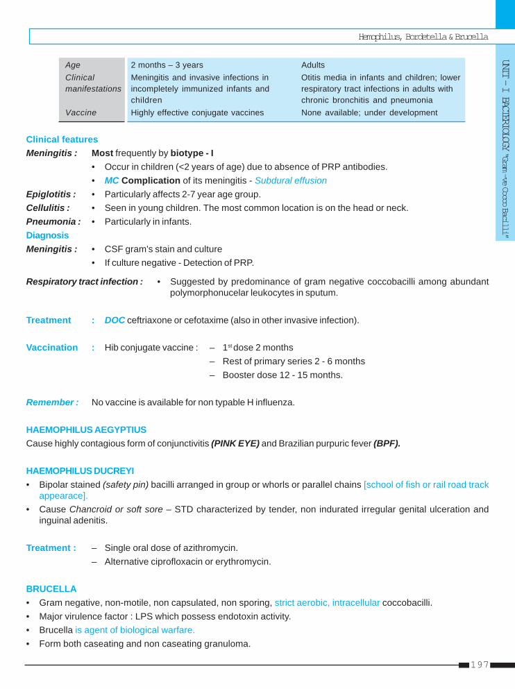
Hemophilus, Bordetella & Brucella
UNIT - I BACTERIOLOGY “Gram —ve Cocco Bacilli”
197
Age 2 months – 3 years AdultsClinical Meningitis and invasive infections in Otitis media in infants and children; lowermanifestations incompletely immunized infants and respiratory tract infections in adults with
children chronic bronchitis and pneumoniaVaccine Highly effective conjugate vaccines None available; under development
Clinical featuresMeningitis : Most frequently by biotype - I
• Occur in children (<2 years of age) due to absence of PRP antibodies.• MC Complication of its meningitis - Subdural effusion
Epiglotitis : • Particularly affects 2-7 year age group.Cellulitis : • Seen in young children. The most common location is on the head or neck.Pneumonia : • Particularly in infants.DiagnosisMeningitis : • CSF gram's stain and culture
• If culture negative - Detection of PRP.
Respiratory tract infection : • Suggested by predominance of gram negative coccobacilli among abundantpolymorphonucelar leukocytes in sputum.
Treatment : DOC ceftriaxone or cefotaxime (also in other invasive infection).
Vaccination : Hib conjugate vaccine : – 1st dose 2 months– Rest of primary series 2 - 6 months– Booster dose 12 - 15 months.
Remember : No vaccine is available for non typable H influenza.
HAEMOPHILUS AEGYPTIUSCause highly contagious form of conjunctivitis (PINK EYE) and Brazilian purpuric fever (BPF).
HAEMOPHILUS DUCREYI• Bipolar stained (safety pin) bacilli arranged in group or whorls or parallel chains [school of fish or rail road track
appearace].• Cause Chancroid or soft sore – STD characterized by tender, non indurated irregular genital ulceration and
inguinal adenitis.
Treatment : – Single oral dose of azithromycin.– Alternative ciprofloxacin or erythromycin.
BRUCELLA• Gram negative, non-motile, non capsulated, non sporing, strict aerobic, intracellular coccobacilli.• Major virulence factor : LPS which possess endotoxin activity.• Brucella is agent of biological warfare.• Form both caseating and non caseating granuloma.
Self Assessment & Review Microbiology & Immunology
SECTION – B
198
• Resist intracellular killing by suppression of myeloperoxide - hydrogen peroxide-halide system and production ofsuperoxide dismutase.
Clinical features• It cause Brucellosis, a zoonotic diseases, also know as mediterranean fever or Malta or undulant fever.• Source : Sheep, goats, camels.• Route of transmissiion : Occupational exposure, ingestion of untreated milk or milk products, raw meat (blood);
inhalation; transplacental; during breast feeding and during sexual activity.• MC and most virulent cause of brucellosis : B. melitensis.• MC symptoms are fever, chills, diaphoresis, headaches.
Pancarditis, Sleep disturbances, lymphadenopathy, Hepato splenomegaly, GBS syndrome, abortion or IUD duringpregnancy. are other important findings.
• MC focal feature is musculoskeletal pain and physical findings in the peripheral and axial skeleton.• Death is usually a consequence of cardiac involvement.• Immunity is mainly cell - mediated.
Diagnosis1. Most defenitive method -blood culture
– Castaneda method of blood culture is employed.– Preferred media are serum dextrose agar; trypticose soy agar.
2. PCR : More sensitive and more quicker than blood culture.3. Serological test :
a. Acute infection – Standard agglutination (SAT) test done– Shows prozone phenomenon– SAT also +ve in cholera, tularemia, yersinia, and immunization.
b. Chronic infection – Complement fixation test.c. For diagnosing animal infection – Rapid plate agglutination, rose bengal card and milk ring test done.
Treatment : • TB must always be excluded.• Gold standard treatment in adults : streptomycin + doxycycline. ... Harrison 17/e, p 976• Alternative rifampin + doxycycline.• In children, pregnant women or who can't tolerate tetracycline – Cotrimoxazole given.
BORDETELLA• Genus consist of Gram negative, strict aerobic coccobacilli which grows only on complex media.• Its two important members :
a. B. pertussis – Causative agent of pertussis (whooping cough = 100 day fever).b. B. parapertussis – Silent copy of pertussis toxin gene causing milder form of pertussis.
Morphology• B.Pertussis is pleomorphic, non motile, non sporing, capsulated, fimbriated coccobacilli, which show Bipolar
metachromatic granules on staining with toulidine blue.
Hemophilus, Bordetella & Brucella
UNIT - I BACTERIOLOGY “Gram —ve Cocco Bacilli”
199
Culture• Grows on enriched media like Regan towe or Bordet Gengou glycerine potato blood agar, forming colonies resembles
bisected pearls or Mercury drops or Aluminium paint appearance.• Culture films has 'Thumb print' appearance.• Charcoal containing media (as for legionella) is preferred.• Blood is required to neutralise the inhibitory materials formed during bacterial growth.
Virulent Factorsa. Most important is pertussis toxin (PT) – exotoxin protein consist of B - binding unit and A active unit having ADP
ribosylating activity of G protein (like of cholera toxin) B-parapertusis don’t express the gene coding for pertusistoxin. Also serves as adhesin, lymphocytosis producting factor, histamine sensitizer and islet activating protein.
b. Filamentous hemagglutinin (FHA) secreted protein.• Both PT and FHA hemagglutinin promotes secondary infection by coating H. influenza and pneumococci so
that they bind. This is known as 'PIRACY OF ADHESINS’.c. Surface adhesins (pertactin, Fimbriae), Adenylate cyclase, Hemolysin, tracheal cytotoxin, heat labile toxin LPS
endotoxin, pertactin agglutinogens are other virulence factor.Infection is initiated by attachment of the organism to the ciliated epithelial cells of the nasopharynx, attachmentis mediated by surface adhesions.
Pathogenesis• Local cellular invasion with intracelluar persistence (systemic dissemination not occur). Systemic manifestation
is due to toxin.• Both cellular and humoral immunity are important.• First defence is by antibody which prevents attacments of bacteria.• Neurological manifestation are due to hypoxia.
Clinical features• Incubation period - 7 - 14 days.• It has 3 stages : a. Catarrhal stage :
• Maximum infectivityb. Paroxysmal stage :
• Posttussive vomiting is frequent with mucus plug occassionally at end of episode.• Vomiting with cough is the best predictor of diagnosis of pertussis.• Episodes are often worsen at night and interfere with sleep.• Most complication occur during paroxymal stage.• Paroxysm is precipitated by noise, eating and physical contact
c. Convalescent stage
ComplicationSubconjunctival hemorrhages, abdominal and inguinal hernia, pneumothorax, petechiae, weight loss, apnea, pneumonia,seizures, encephalopathy.
Diagnosisi. Best specimen is obtained by nasopharyngeal aspiration ..... Harrison 17/e, p 935ii. Gold standard – Culture of nasopharyngeal secretioniii. Absolute lymphocytosis without ↑ in ESR
Self Assessment & Review Microbiology & Immunology
SECTION – B
200
iv. Most sensitive is PCRv. Serology – If symptoms > 4 weeks.
TreatmentDOC – Macrolide (Erythromycin, Clarithromycin Azithromycin)Alternative – Cotrimoxazoleβ agonist, Glucocorticoids and cough suppressants are not effective.
Prevention• Chemoprophylaxis – For house hold contact of cases.
– Erythromycin is DOC.
• Immunization – main stay of preventions is active immunization.Two types of vaccine are available :
a. Whole cell vaccine :– Associated with many adverse effects and may also cause - encephalopathy, sudden infant death syndrome,
and autism.– It is contraindicated in individual > 7 year age.
b. Acellular vaccine :– Less reactogenic and is recommended for routine immunization.– It contains pertussis toxoid.– Two component vaccine are more effective than monocomponent, since addition of pertactin increase
efficacy.– Protection against pertussis by vaccine correlated best with the production of antibody to pertactin, fimbriae,
and pertussis toxin.
Hemophilus, Bordetella & Brucella
UNIT - I BACTERIOLOGY “Gram —ve Cocco Bacilli”
201
1. A veterinary doctor had pyrexia of unknown ori-gin. His blood culture in special laboratory me-dia was positive for gram negative short bacilliwhich was oxidase positive. Which one of thefollowing is the likely organism grown in culture?a) Pasturella spp. [AI 06]b) Francisella spp.c) Bartonella spp.d) Brucella spp.
2. A farmer presenting with fever off-and on for thepast 4 years was diagnosed to be suffering fromchronic Brucellosis. All of the followingserolocical tests would be helpful in the diagno-sis at this state except : [AI 04]a) Standard Agglutination testb) 2 Mercapto-ethanol testc) Complement fixation testd) Coomb’s test
3. The following are true for Bordetella pertussisexcept : [AI 03]a) It is a strict human pathogenb) It can be cultured from the patient during
catarrahal stagec) It leads to invasion of the respiratory mucosad) Infection can be prevented by a acellular vac-
cine4. Malta fever is caused by : [AIIMS 08]
a. Legionellab. Borrelia burgdorferic. Brucella melitensisd. Pseudomonas
5. Brucellosis can be transmitted by all of the fol-lowing modes, except : [AIIMS 07, 06]a) Contact with infected placentab) Ingestion of raw vegetables from infected farmsc) Person to person transmissiond) Inhalation of infected dust or aerosol
6. Brucella is transmitted by all the following meansexcept : [AIIMS 06]a) Through placenta of animalsb) Person to person transmissionc) Aerosold) Eating uncooked food
7. The usual incubation period of pertusis is :a) 7 - 14 days [AIIMS 05]b) 3 - 5 daysc) 21 - 25 daysd) Less then 3 days
8. All the following are true about H. influenzae ex-cept : [AIIMS 03]a) It can be part of normal flora of some personsb) The serotyping is based on bacterial outer mem-
brane proteinc) It requires Haemin and NAD for growth in culture
mediumd) Type b is responsible for invasive disease
9. A 2 years old child is brought to the emergencywith history of fever and vomiting. On examina-tion he has neck rigidity. CSF examination showspolymorhphs more that 200/μμμμμl; protein 100mg/dland glucose 10mg/dl. the Grams stain shows thepresence of Gram negative coccobacilli. The cul-ture shows of bacteria only on chocolate. agarand not on blood agar. The causative agent is :a) Neisseria meningitides [AIIMS 02]b) Haemophilus influenzaec) Branhamella catarrhalisd) Legionella pneumophila
10. A farmer rearing sheep, presented with com-plaints of fever and weakness for the last onemonth. There is generalised lymphadenopathy.There was also associated hepatomegaly Biopsyof liver showed non-caseating granuloma. Thereare most likely due to infection with : [AIIMS 00]a) Yersinia pesitisb) Brucella canisc) Francisella tularensisd) Brucella melitenesis
11. True regarding pertussis vaccine is :a) 95% of vacinated are protected [AIIMS 00]b) Erythromycin should be given to contactsc) Neuroparalytic complication is seen in 1 in 15000d) Leucocytosis is diagnotic
12. Regarding Brucellosis, all of these are true ex-cept: [PGI 02]a) Man to man transmissionb) A zoonosisc) Blood cultures used in diagnosisd) Transmitted via animal products
ANSWERS WITH EXPLANATIONQUESTIONS
Answer 1. d) Brucella 2. a) Standard ... 3. d) Infection ... 4. c) Brucella 5. c) Person ...6. b) Person to ... 7. a) 7 - 14 ... 8. b) Serotyp ... 9. b) H. Influen .. 10. d) Brucella ...
11. b) Erythromy ... 12. a) Man to ...
Self Assessment & Review Microbiology & Immunology
SECTION – B
202
1. Ans. is d i.e. Brucella Ref. Ananthnarayan 7/e, p 346
Oxidase positive gram negative organism producing pyrexia of unknown origin in veterinary doctor is pointingtowards Brucella (coccobacilli or short rods).
• Brucella is the causative agent of Brucellosis (Malta fever, undulent fever).• Other options :Pasturella spp. (P. multocida)
– It is also short bacilli and oxidase positive, but it can grow over normal media and does not present withpyrexia of unknown origin. It is non-motile and produce indole.
– Manifestation of pasturella : Local suppuration, Meningitis, Respiratory tract infection.– Francisella (Casuative agent of Tularemia) : It is also short bacilli with fastidious growth requirement. But
it is oxidase negative.
Remember :– Brucella is oxidase and catalase positive except B.neotomae and B.ovis.– Risk factors for Brucellosis (Zoonotic disease).
- Occupational - Farmers, shepherds, Veterinanrians, Goats herds, Slaughter house workers.- Domestic - family members of individual in animal husbandry.- Laboratory workers - involved in handing cultures.- Travellars and Urban dwellers.
– Brucella can grow over normal media but growth is improved by addition of serum or liver extract.
2. Ans. is a i.e. Standard agglutination testRef. Ananthnarayan 7/e, p 349; Harrison 17/e, p 975; Jawetz 24/e, p 286
Diagnosis of Brucellosis1. Culture : • Blood culture is the most definitive method.
• Castaneda method of blood culture is recommended.2. Serology
a. Standard agglutination (tube agglutination) test (SAT) is performed most often. It identifies mainlythe IgM antibody.• Titre tends to decline after acute phase of the illness so not helpful in chronic brucellosis.• Positive agglutination test may be produced by cholera, tularemia or yersinia infection or immunisation.• Cholera induced agglutinins may be differentiated by agglutination absorption test and also as they
are removed by treatment with 2-mercapto ethanol.b. Blocking or non agglutinating antibodies – Most reliable method for obviating the blocking effect
and detecting the incomplete antibodies is antiglobulin (coombs) test.c. Complement fixation test – Detects both IgG and IgM so it is more useful in chronic cases.d. ELISA – Sesitive, specific and can detect IgM and IgG antibody separately so useful for differentiating
acute and chronic infections.e. PCR – To detect bacteremia, to predict relapse and to exclude chronic brucellosis. More sensitive and
quicker than blood culture.3. Hypersensitivity test – Not useful in diagnosing acute infections.
EXPLANATIONS AND REFERENCES WITH ILLUSTRATIVE ANSWERS
Hemophilus, Bordetella & Brucella
UNIT - I BACTERIOLOGY “Gram —ve Cocco Bacilli”
203
3. Ans. is d i.e. Infection can be prevented by an acellular vaccineRef. Ananthnarayan 6/e, p 342; Harrison 17/e, p 936
“All acellular vaccine curently available contain pertussis toxoid hence they only prevent disease notinfection.” ..... Harrison 17/e, p 936• Bordetella pertusis is an obligate human parasite.• Source of infection is case of pertussis.• Carriers and Subclinical infection are not found.• Infection is limited to respiratory tract; in advance conditions leads to desquamation of alveolar epithelium
(invasion of respiratory mucosa).• Bacilli do not invade blood stream.• Diagnosis :
– Culture of nasopharyngeal secretions remains the gold standard of diagnosis.– Best specimen is collected by nasopharyngeal aspiration.– Culture is positive in catarrhal and early proxysmal stage.– Media used is Bordet-Gengou or Regan-Lowe.
Remember : Regan-Lower charcoal medium – Transport media.
4. Ans. is c i.e. Brucella melitensis Ref. Ananthnarayana 7/e, p 346
Already explained, refer answer no. 1
5. Ans. is c i.e. Person to person transmission Ref. Park 19/e, p 242
Brucellosis is transmitted from infected animal to man and there is evidence of transmission from man to man.Modes of Transmission of Brucella
Contact infection (MC) Food borne infection Airborne infection
– Direct contact with infected – Ingestion of raw milk or – From aerosols intissue, blood, urine etc. dairy products cowshed and
– Mostly occupational – Water contaminated with slaughter houseexcreta of infected animal
6. Ans. is b i.e. Person to person transmission Ref. Park 19/e, p 242
Already explained, refer just above answer.
7. Ans. is a i.e. 7 - 14 days Ref. Park 19/e, p 139
Incubation period of pertusis is usually 7 to 14 days, but never exceed 3 weeks.
Important points about pertussis :• Caused by B.pertusis• Source of infection is case, there is no evidence of subclinical infection
Self Assessment & Review Microbiology & Immunology
SECTION – B
204
• Pertusis is most infectious in catarrhal stage• Infective period extends from a week after exposure to about 3 weeks after the onset of paraoxymal stage• Secondary attack rate is about 90%.
8. Ans. is b i.e. Serotyping is based on bacterial outer membrane proteinRef. Ananthnarayan 7/e, p 335
“H. influenzae strains have been classified on the basis of capsular polysaccharide not on the basis ofouter membrane protein.”
Important features of H. influenzae• Gram (–)ve coccobacilli• Require Factor V (nicotinamide nucleotide) and Factor X (haemin) for growth. So grow well on choclate
agar as factor V is free in choclate agar. (Growth is poor in blood agar).
Antigenic structure :a. Capsular antigen : – Basis of classification
– Most isolates of acute invasive infection belongs to type b.b. Bacterial outermembrane proteinc. Lipooligosaccharide
– Non capsulated strains of H. Influenzae can colonize the upper respiratory tract of childrens and adults.
9. Ans. is b i.e. H. Influenzae Ref. Ananthnarayan 7/e, p 332 - 337
See the morphology of asked bacteria, you will know the answer.
H.influenzae – Gram negative coccobacilliN. meningitides – Gram negative cocciLegionella – Gram negative coccobacilliB. catarrhalis – Gram negative cocci
10. Ans. is d i.e. Brucella melitensis Ref. Harrison 17/e, p 974
“History of contact with sheep and clinical feaures of fever, weakness, lymphadenopathy with noncaseating granuloma signifies Brucellosis.”• Brucella melitensis is most pathogenic and most common cause of brucellosis and usually infect sheep,
goats and camels.• Brucella canis usually infect dogs• Brucella abortus – Cattle or buffalo• Brucella suis – swine• No man to man transmission occurs.
Clinical features of Brucellosis :• Fever with profuse sweats, especially at night. If untreated fever follows undulating pattern i.e., fever →
afebrile period → fever.• Fever is associated with musculoskeletal symptoms i.e. pain.• Osteomyelitis commonly in lumbar and low thoracic vertebrae.
Hemophilus, Bordetella & Brucella
UNIT - I BACTERIOLOGY “Gram —ve Cocco Bacilli”
205
• Septic arthritis (MC knee)• Neurologic involvement with depression and lethargy• Endocarditis (MC in aortic valve)
11. Ans. is b i.e. Erythromycin should be given to contact Ref. Park 19/e, p 139 - 141
“Those known to have in contact with whooping cough should be given prophylactic erythromycin for10 days.”
Pertussis vaccine1. Killed whole cell vaccine : • Given in the form of DPT
• Protection 70 - 90%Adverse reaction :– Neurologic complication 1:170000
– Convulsions– Prolonged screaming– Hyporesponsible state.
Contraindication : – Epilepsy, convulsion or other CNS disorder.
2. Acellular vaccine :– Contain pertussis toxoid and filamentous hemagglutinin.– Same efficacy but less reactogenic– Two component (which contain pertacin and pertussis toxoid) are more effective than monocomponent
acellular vaccine. ..... Harrison 17/e, p 936
In pertusis lymphocytosis (not leucocytosis) is diagnostic.
12. Ans is a i.e. Man to man transmission Ref. Harrison 17/e, p 973
Already explained, refer answer no. 5
Self Assessment & Review Microbiology & Immunology
SECTION – B
206
1. a) Hemophilus 2. c) X and V factor 3. a) Brucellosis 4. c) Bordetella ... 5. a) Soft sore6. b) Chancroid 7. a) Hemophilus
1. Satellitism is seen in cultures of :a) Hemophilus [Nimhans 86, PGI 86,TN 95]b) Streptococcusc) Proteusd) Salmonella
[Ref. Ananthnarayan 7/e, p 333]2. Hemophilus needs : [TN 99]
a) X factorb) V factorc) X and V factord) VII fector
[Ref. Ananthnarayan 7/e, p 333]3. Milk ring test : [JIPMER 99]
a) Brucellosisb) Bacteroidesc) Tuberculosisd) Salmonellosis
[Ref. Ananthnarayan 7/e, p 350]4. Thumb print appearence in culture film smear is
seen : [Karnat 99]a) Bacillus antracisb) Brucella species
Answer
c) Bordetella pertussisd) Clostridum Welchii
[Ref. Ananthnarayan 7/e, p 339]
5. Hemophilus ducreyi is the causative agent is :a) Soft sore [Kar 01]b) Hard chancrec) Urethritisd) Granuloma inguinale
[Ref. Ananthnarayan 7/e, p 337]6. Haemophilus ducreyi causes : [Kolkata 04]
a) Lymphogranuoma venereumb) Chancroidc) Syphilisd) Psittacosis
[Ref. Ananthnarayan 7/e, p 337]7. Satellitism is seen in culture of : [DNB 05]
a) Hemophilusb) Streptococcusc) Klebsiellad) Proteuse) Salmonella
[Ref. Ananthnarayan 7/e, p 332]
Chapter Review••••• This section includes questions of VThis section includes questions of VThis section includes questions of VThis section includes questions of VThis section includes questions of Various Other PGMEES from 1990 – 2007.arious Other PGMEES from 1990 – 2007.arious Other PGMEES from 1990 – 2007.arious Other PGMEES from 1990 – 2007.arious Other PGMEES from 1990 – 2007.••••• Questions are arQuestions are arQuestions are arQuestions are arQuestions are ar ranged in decreasing order of yearranged in decreasing order of yearranged in decreasing order of yearranged in decreasing order of yearranged in decreasing order of yearwiz. This is done to makwiz. This is done to makwiz. This is done to makwiz. This is done to makwiz. This is done to make refere refere refere refere refer ralralralralral
system easier and uncomplicated to save the precious time of PGMEE Aspirant.system easier and uncomplicated to save the precious time of PGMEE Aspirant.system easier and uncomplicated to save the precious time of PGMEE Aspirant.system easier and uncomplicated to save the precious time of PGMEE Aspirant.system easier and uncomplicated to save the precious time of PGMEE Aspirant.
CAMPYLOBACTER• Motile, curved rods; non sporing (comma shaped on S shaped) shows darting motility.• Important pathogenic organisms are C.jejuni and C.fetus.
CAMPYLOBACTER JEJUNIComma’s or gull wing shaped bacteria motile with single polar flagellum.Very common cause of diarrhoea.
Culture• Growth occurs under microaerophillic condition 5% O2 optimal with added CO2 [10%]. Although C.jejuni grows
well at 36-370C, but incubation at 420C prevents most of other fecal bacteria thus helps in selective growth.• Selective media – Skirrow’s medium; Butzler’s media
Pathogensis and clinical feature• Infection is acquired by oral route. Source of infection is raw or under cooked food products from poultry, cattle
sheep, swine.• Organism multiply in small intestine, invade the epithelium and cause inflammation.• Usually present : with in 2-4 dayus• Clinically present as acute onset of crampy abdominal pain, profuse bloody diarrhoea.
Diagnosis• Faeces shows polymorphonuclear leucocytes.• Gram stained smear may show typical ‘S’ shaped bacteria.• Dark field or phase contrast microscopy may show darting motility.• Culture on selective media at 420C is definitive diagnostic test.
Treatment :• Erythromycin is DOC.
CAMPYLOBACTER FETUS• Oppurtunistic pathogen that cause systemic infection in immunocompromised.• Cause diarrhoeal illness in normal host.
Campylobacter & HelicobacterCampylobacter & HelicobacterCampylobacter & HelicobacterCampylobacter & HelicobacterCampylobacter & Helicobacter1313
Self Assessment & Review Microbiology & Immunology
SECTION – B
208
HELICOBACTER PYLORISpiral shaped gram (–)ve rod associated with gastritis and peptic ulcer.Motile with lopotrichous flagella.The sole source is human gastric mucosa.
Culture : • Grows well when incubated at 370C in micro aerophillic condition.• Media used include skirrow’s medium, chocolate medium.
Biochemical reaction : Catalase (+) ve, Oxidase + ve, Urease + ve
Pathogenesis1. Grows optimally at pH 6.0-7.0 and would be killed at pH with in the gastric lumen.2. But it survives as it is found deep in mucus layer near epithelial surface, without invading mucosa where physiologic
pH is present. It produce potent urease which provide ammonia to buffer acid.3. Major disease associated H.pylori virulence factor are vacuolating cytotoxin (Vac A) and group of genes termed
CagPaI.• H pylori colonization decrease somatostatin producing cells → ↑→ ↑→ ↑→ ↑→ ↑ Gastrin →→→→→ ↑ ↑ ↑ ↑ ↑ Acid →→→→→ Gastricmetaplasia in
duodenum →→→→→ Inflammation →→→→→ Ulceration.• Best characterized host determinent of disease is enhanced H. pylori stimulated secretion of IL - 1 β in
some peoples due to genetic polymorphism. ..... Harrison 17/e, p 9464. Colonization induce chronic superficial gastritis which includes both mononuclear and polymorphonuclear cell
infiltration. Gastric antrum is MC site of colonization.
Clinical Manifestation• 80% of duodenal ulcer and 60% of gastric ulcer are related to H.pylori.• Increase risk of gastric adenocarcinoma, Gastric MALT lymphoma.• Extragastrointestinal pathologies that are linked include ischemic heart disease and cerebrovascular disease.
Diagnosis
Invasive test : Non invasive tests :
• Requires upper GI endoscopic biopsy • Most consistently accurate test is urea breath test.• Most convenient biopsy based • Stool antigen test is less expensive and more
test is biopsy urease test. convenient but slightly less accurate• Microbiologic culture is • Simplest test are serologic assays measuring IgG
most specific but insensitive. levels in serum by ELISA or immunoblot.– Unlike serologic test urea breath tests can be
used to asses outcome of treatment 1 monthafter its completion. .... Harrison 17/p 947
TreatmentSymptomatic cases - First line :
Regimen 1 - O C A 7 days (Omeprazole , Clarithromycin, Amoxicilline)
Regimen 2 - O C M 7 days (Omeprazole, Clarithromycin, Metronidazole)
Second line : OBTM 14 days (Omeprazole, Bismuth, Tetracycline, Metronidazole)
No treatment is given for asymptomatic cases.
Remember : H. pylori and H. influenzae are bacteria whose complete genome has been mapped.
Campylobacter & Helicobacter
UNIT - I BACTERIOLOGY “Gram —ve Cocco Bacilli”
209
1. A child was diagnosed to be suffering from diar-rhoea due to Campylobacter Jejuni. Which of thefollowing will be the correct environmental con-ditions of incubation of culture plates of the stoolsample : [AI 05]a) Temperature of 420C and microaerophilicb) Temperature of 420C and 10% CO2
c) Temperature of 370C and microaerophilicd) Temperature of 370C and 10% CO2
2. All of the following are true about helicobactor-pylori except : [AI 98]a) About 50% of world population affectedb) 85% of population is affected, in some develop-
ing countiresc) All children in developing countries have immu-
nity by five years of aged) Infection is common in low socio-economic sta-
tus
3. True about helicobacter pylori is : [AI 98]a) Culture and gram staining of biopsy is the gold
standard investigationb) Controlled urea breath is negative with massive
infectionc) Anti urease antibody are produced only by inva-
sive strainsd) Ureas activity provides protective environment to
the bacilli
4. True about Helicobacter, pylori are all except :a) Urea, breath test is diagnostic [AI 98]b) Gram negative, flagellated bacillic) Risk factor for development of adenocarcinoma
of stomachd) It provides life ling immunity
5. A 35 year old patient complaint of abdominalcramps along with profuse diarrhoea. Treatingphysician wants to process the stool specimenfor isolation of Campylobacter jejuni. Which ofthe following is method of choice for culture ofstool : [AIIMS 04]
a) Culture on TCBS media incubated at 370C onaerobic condition
b) Culture on Skirrow’s medium incubated at 420Cunder microaerophillic condition
c) Culture on Mac conkeys medium incubated at420C under anaerobic condition
d) Culture on Wilson and Blairs medium at 370Cunder microaerophillic condition
6. Helicobacter pylori is not associated with :a) Gastrointestinal lymphoma [AIIMS 03]b) Gastric cancerc) Gastric leiomyomad) Peptic ulcer
7. Which of following is false regarding H. pylori :a) With chronic infection urease breath test become
negative [AIIMS 00]b) H. Pylori infection remains life long if untreatedc) Endoscopy is diagnosticd) Toxigenic strains usually cause ulcer
8. Which of the following correctly describe H.pylori:a) Gram negative cocci curved [PGI 05]b) Gram positive cocci straightc) Gram positive bacilli curvedd) Gram negative bacilli straighte) Gram negative bacilli curved rod
9. H. Pylori true about : [PGI 04]a) Gram +ve spiral organismb) It is a protozoac) Causes chronic gastirtis in adults due reinfectiond) Treatment prevents gastric Lymphomae) Duodenal mucosa normal
10. True about H. pylori : [PGI 04]a) It is flagellatedb) Involved in causation of peptic ulcer diseasec) Hypergastrinemia caused by itd) It is a Gram (–)ve organism
ANSWERS WITH EXPLANATIONQUESTIONS
Answer 1. a) Temperature ... 2. c) Anti urease... 3. d) Ureas ... 4. d) Urea, ... 5. b) Culture ...6. c) Gastric ... 7. a) With ... 8. e) Gram ... 9. c and d 10. a, b, c and d
Self Assessment & Review Microbiology & Immunology
SECTION – B
210
11. Regarding H. pylori, all are true except :a) Gram-negative bacillus [PGI 02]b) Strongly associated with duodenal ulcerc) Associated with lymphomad) C-14 urea breath test is used in diagnosise) It should be eradicated in all cases whenever
detected
12. True about H. pylori : [PGI 00]a) Seen in 85 to 90% cases of gastric ulcer
b) Seen in 20 to 25% cases of duodenal ulcerc) Transmitted from man to man, feco-orally and
by orogastric routed) Common in adults of developing countries
13. The most sensitive test for H. pylori is :a) Breath test [PGI 99]b) Rapid urease testc) Culture of biopsyd) Microscopy of biopsy
Answer 11. e) It should ... 12. c and d 13. b) Rapid ...
Campylobacter & Helicobacter
UNIT - I BACTERIOLOGY “Gram —ve Cocco Bacilli”
211
1. Ans. is a i.e. Temprature of 420 C and microarophilic Ref. Jawetz; 24/e, p 275
Diagnosis of C. jejuni diarrhoea :Specimen : StoolDirect microscopy : Dark field or phase contrast microscope shows gullwing shaped C. jejuni with charactersticdarting motility.Culture : Growth occurs under microaerophilic conditions with added CO2 (10%). Though C. jejuni grows wellat 36-370C but incubation at 420C prevents growth of other faecal bacteria and thus helps in selective growth.Selective media : Skirrows medium
Remember : C. jejuni cause inflammatory diarrhoea (i.e. presence of WBC in stool).
2. Ans. is c i.e. All children in developing countries have immunity by five years of ageRef. Harrison 16/e, p 886
Prevelance of H. pylori infection – 30% in developed countries– 80% in developing countries– 50% in total world population
Risk factors of H. pylori infection – Age - childhood - most infections are acquired in childhoodbut immunity does not develop.
– Low socioeconomic status– Crowding
3. Ans. is d i.e. Urease activity provides protective environment to the bacilliRef. Harrison 17/e, p 947
“Urease produced by H. pylori provide ammonia to buffer acid and helps in maintanance of infections.”
Diagnosis of H. pylori
Test Advantages Disadvantages
Invasive (based on endoscopic biopsy)
Biopsy urease test Quick, simple Not fully sensitive before 24hHistology May give additional histologic information Sensitivity dependent on
experience & use of special stainsCulture Permits determination of antibiotic Sensitivity dependent on experience
susceptibitlity
Noninvasive
Serology Inexpensive and convenient Cannot be used for early follow-up;13C or 14C urea Inexpensive and simpler than endoscopy; Low-dose irratation in 14C testbreath test useful for follow-up after treatmentStool antigen test Inexpensive & convenient; useful for follow-up New test; appears less accurate
after treatment; may be useful in children than urea breath test
Remember : Microbiologic culture are most specific but not gold standard
EXPLANATIONS AND REFERENCES WITH ILLUSTRATIVE ANSWERS
Self Assessment & Review Microbiology & Immunology
SECTION – B
212
4. Ans. is d Provides life long immunity Ref. Harrison 17/e, p 946
“Immune response include both production of antibody and cell mediated response but is ineffective inclearing the bacterium.”Important features of H. Pylori :• Gram (–)ve cocobacilli motile with lopotrichous flagella.• 80% of duodenal ulcer and 60% of gastric ulcer are related to H. pylori.• Increase the risk of : – Gastric adenocarcinoma – Gatric MALT lymphoma
– Reflux esophagitis – Ischemic heart disease– Cerebrovascular disease.
• Urea breath test is most consistently accurate test for diagnosis.• Microbiologic culture is most specific but insensitive.
5. Ans. is b i.e. Culture on Skirrow’s medium incubated at 420C under microaerophillic conditionRef. Jawetz 24/e, p 274-275
Already explained, refer answer no. 1
6. Ans. is c i.e. Gastric leiomyoma Ref. Harrison 17/e, p 947
Already explained, refer answer no. 4
7. Ans. is a i.e. With chronic infection urease breath test becomes negativeRef. Harrison 17/e, p 948
Urease breath test is most consistently accurate test for diagnosis of H. pylori.It becomes negative only after treatment and is used to asses outcome of treatment.
Remember : • Most specific test is microbiologic culture of specimen obtaining by upper GI endoscopicbiopsy.
• Major disease associated H. pylori virulence factor are vacuolating cytotoxin (VaCA) andgroup of genes called CagPal.
• Urea breath test, stool antigen test, and biopsy based tests can all be used to assess thesuccess of treatment.
8. Ans. is e i.e. Gram negative bacilli curved rod Ref. Ananthnarayan 7/e, p 407
“H.pylori is a Gram negative curved spiral rod motile by unipolar tuft of lopotrichous flagella.”Correctly speaking H. pylori is a coccobacilli.
Remember :
Other Gram (–)ve coccobacilli All important bacilli are gram positive except
– Hemophilus – Bordetella – Actinomycetes – Bacillus– Brucella – Campylobacter – Clostridium – Corynebacterium– Helicobacter – Legionella – Mycobacteria – Listeria– Rickettsiacea – Chlamydiae Mnemonic – ABC CML
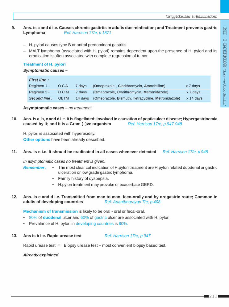
Campylobacter & Helicobacter
UNIT - I BACTERIOLOGY “Gram —ve Cocco Bacilli”
213
9. Ans. is c and d i.e. Causes chronic gastirtis in adults due reinfection; and Treatment prevents gastricLymphoma Ref. Harrison 17/e, p 1871
– H. pylori causes type B or antral predominant gastritis.– MALT lymphoma (associated with H. pylori) remains dependent upon the presence of H. pylori and its
eradication is often associated with complete regression of tumor.
Treatment of H. pyloriSymptomatic causes –
First line :Regimen 1 - O C A 7 days (Omeprazole , Clarithromycin, Amoxicilline) x 7 days
Regimen 2 - O C M 7 days (Omeprazole, Clarithromycin, Metronidazole) x 7 days
Second line : OBTM 14 days (Omeprazole, Bismuth, Tetracycline, Metronidazole) x 14 days
Asymptomatic cases – no treatment
10. Ans. is a, b, c and d i.e. It is flagellated; Involved in causation of peptic ulcer disease; Hypergastrinemiacaused by it; and It is a Gram (–)ve organism Ref. Harrison 17/e, p 947-948
H. pylori is associated with hyperacidity.Other options have been already described.
11. Ans. is e i.e. It should be eradicated in all cases whenever detected Ref. Harrison 17/e, p 948
In asymptomatic cases no treatment is given.Remember : • The most clear cut indication of H.pylori treatment are H.pylori related duodenal or gastric
ulceration or low grade gastric lymphoma.• Family history of dyspepsia.• H.pylori treatment may provoke or exacerbate GERD.
12. Ans. is c and d i.e. Transmitted from man to man, feco-orally and by orogastric route; Common inadults of developing countries Ref. Ananthnarayan 7/e, p 408
Mechanism of transmission is likely to be oral - oral or fecal-oral.• 80% of duodenal ulcer and 60% of gastric ulcer are associated with H. pylori.• Prevalance of H. pylori in developing countries is 80%.
13. Ans is b i.e. Rapid urease test Ref. Harrison 17/e, p 947
Rapid urease test = Biopsy urease test – most convenient biopsy based test.
Already explained.
Self Assessment & Review Microbiology & Immunology
SECTION – B
214
1. b) Uncommon ... 2. c) Helicobacter ...
1. About helicobacter Pylori, all are true except :a) Is a Gram -ve spiral bacteria [AIIMS 96]b) Uncommon undeveloping Asian countriesc) Urea breath test +ved) Associated with duodenal ulcer
[Ref. Harrison 17/e, p 946]
Answer
Chapter Review••••• This section includes questions of VThis section includes questions of VThis section includes questions of VThis section includes questions of VThis section includes questions of Various Other PGMEES from 1990 – 2007.arious Other PGMEES from 1990 – 2007.arious Other PGMEES from 1990 – 2007.arious Other PGMEES from 1990 – 2007.arious Other PGMEES from 1990 – 2007.••••• Questions are arQuestions are arQuestions are arQuestions are arQuestions are ar ranged in decreasing order of yearranged in decreasing order of yearranged in decreasing order of yearranged in decreasing order of yearranged in decreasing order of yearwiz. This is done to makwiz. This is done to makwiz. This is done to makwiz. This is done to makwiz. This is done to make refere refere refere refere refer ralralralralral
system easier and uncomplicated to save the precious time of PGMEE Aspirant.system easier and uncomplicated to save the precious time of PGMEE Aspirant.system easier and uncomplicated to save the precious time of PGMEE Aspirant.system easier and uncomplicated to save the precious time of PGMEE Aspirant.system easier and uncomplicated to save the precious time of PGMEE Aspirant.
2. Which of the following medically potent urease ac-tivity : [MP 06]a) Proteus mirabilisb) Brucella melitensisc) Helicobacter pylorid) Ureaplasma urealyticum
[Ref. Ananthnarayan 7/e, p 408]
• Gram negative non capsulated, intracellular cocobacilli.• They are motile, aerobic, catalase and oxidase positive.
Culture• Not grow on ordinary media.• Buffered charcoal yeast extract ‘BCYE‘ is selective medium used to grow legionella. It grows best at pH - 6.9
temperature 350C and 90% humidity.• MC species associated with human infection is L.pneumophilia, (MC with serogroup 1).• Other important species is L.micdadei (Pittsburgh pneumonia agent). It is partial acid fast (AFB).• Natural habitat is aquatic bodies including lakes and streams. Shows symbiotic relations with algae, amebas,
ciliated protozoa.• Factors enhancing colonisation are warm temperature, stagnation and sediments. It can form microcolonies
with in biofilms. Its eradication require disinfectants that can penetrate the biofilm.
Mode of transmission• Aspiration is predominant mode of transmission. Other modes include aerosolization, direct instillation in to
lungs.• No man to man transmission occurs.• Aerosolization by A.C., nebulizer, humidifier and direct instillation into lung are other modes.
Risk Factors and pathogenesis• Conditions that impair mucocilliary clearance predispose to legionaires disease most commonly cigarette smoking.• Hairy cell leukemia (not other leukemia) and immunocompromised state are other risk factor.• Bacteria attach host cell by type IV pili, HSP60 and major outermembrane protein. Legionella binds to CR1 and
CR3 integrin receptor.• Cell mediated immunity is primary mechanism of host defense (Role of neutrophill appears to be minimal).• Humoral immunity plays no role.
LegionellosisLegionella cause 2 clinical syndromes :A. Pontiac fever : • Acute febrile self limited illness. Airbone transmission with high attack rate.
• Pneumonia does not develop. MC symptom - malaise, fatigue and myalgia.
LegionellaLegionellaLegionellaLegionellaLegionella1414
Self Assessment & Review Microbiology & Immunology
SECTION – B
216
B. Legionnaires disease : Designation for pneumonia.• 4th MC cause of community acquired pneumonia (MC - S.pneumoniae 2nd MC - H.influenzae 3rd MC Chlamydia
pneumoneae). ..... Harrison 17/e, p 929• Cause atypical penumonia which is more serious than atypical pneumonia of other agents.• Clinical features suggestive of L. pneumonia :
– Diarrhoea – High fever– Hyponatremia – Proteinuria– Onset of symptom with in 10 days after discharge from hospital suggest nosocomial logionnaires disease.
• Mostly caused by serotype 1 but serotype 6 is associated more commonly with hospital acquired and haspoor prognosis.
• Relative bradycardia is useful diagnostic finding.
Extrapulmonary Legionellosis• Results from blood borne dissemination from lung.• MC extrapulmonary site is heart.• Most cases are hospital acquired.• MC neurological abnormality are confusion or changed mental status.
DiagnosisSpecimen: • Sensitivity of bronchoscopy specimens is approximately the same as that of sputum. Bronchoalveolar
lavage fluid gives higher yield than bronchial wash specimen.Staining : • Gram staining usually show numerous leucocytes but no organisms.
• DFA test is rapid and highly specific but less sensitive.Culture : • Definitive method of diagnosis.
• Requires 3-5 days to become grossly visible.• B’CYE media is used.
Antibody detection : • Requires 12 weeks, so used for retrospective diagnosis or epidemiologic studies.Urinary antigen : • Cheap, rapid, second in sensitivity and highly specific.
• Detectable with in 3 days.• The test is not affected by antibiotic administration.
Radiographic Abnormalities : • Pleural effusion• In immunosuppressed distinct round nodular opacity may be seen.
Treatment• Azithromycin and Respiratory tract quinolones (Levofloxacin, Gemifloxacin, Moxifloxacin) are DOC.• Quinoliones are preffered antibiotic in transplant recepients.
Prevention• Disinfection of water by :
– Superheat and flush method - Ideal for emergency situation– Copper and silver ionization method– Superchlorination is not effective.– Tap water filters particularly in transplant units.
Legionella
UNIT - I BACTERIOLOGY “Gram —ve Cocco Bacilli”
217
1. A 70 year old patient presents with high gradefever, dry cough and abdominal pain. Sputumsample collected from patient, shows Gram nega-tive organisms that are able to grow only on char-coal yeast extract medium. The most likely or-ganism is : [AI 07; AIIMS 06]a) H. influenzab) Leigonellac) Lesteria monocytogenesd) M. catarhalis
2. A 60-year old man is diagnosed to be sufferingfrom Legionnaires disease after he returns homefrom attending a convention. He could have ac-quired it : [AI 03]a) From a person suffereing from the infection
while traveling in the aero planeb) From a chronic carrier in the convention centerc) From inhalation of the aerosol in the air condi-
tioned room at convention centerd) By sharing an infected towel with a fellow del-
egate at the convention
3. Anju, a 28 year female, has diarrhoea, confusion,high grade fever with bilateral pneumonitis. Or-ganism causing this : [AI 00]a) Legionellab) Neisseria meningitisdisc) Streptococcus pneumoniaed) H.influenza
4. All of the following are true regarding Legionellaexcept : [AIIMS 04]a) Legionella can be grown on complex mediab) L. pneumophilia setogroup 1 is the most com-
mon serogroup isolated from humansc) Legionella are communicable from infected pa-
tient to othersd) L. pneumophila is not effectively killed by poly-
morphonuclear leukocyte
5. Which of the following is a good media to use fordiagnosis of legionnaires disease : [AIIMS 01]a) Theyer Martin mediab) BCYE agarc) Bordet Gengu mediad) Chocolate agar
6. Pontiac fever is caused by : [PGI Dec. 07]a. Legionellab. Listeriac. Scrub typhusd. Leptospirae. Ricktettsia
7. BCYE. medium is used to culture : [PGI 99]a) Mycoplasmab) T pallidumc) H. pylorid) Legionella
ANSWERS WITH EXPLANATIONQUESTIONS
Answer 1. b) Leigonella 2. c) From ... 3. a) Legionella4. c) Legionella ... 5. b) BCYE ... 6. a) Legionella7. a) Mycoplasma
Self Assessment & Review Microbiology & Immunology
SECTION – B
218
1. Ans. is b i.e. Legionella Ref. Harrison 17/e, p 929; Jawetz 24/e, p 313
Dry cough, high grade fever and growth on charcoal yeast medium suggest legionella.Legionella have fastidious requirements and grow on complex media such as buffered charcoal, yeast extract(BCYE) agar with L. cysteine and antibiotic supplements with 5% CO2 at pH 6.9, 350C and 90% humidity.
2. Ans. is c i.e. From inhalation of the aerosol in the air conditioned room at convention centerRef. Harrison 17/e, p 928
Important features of Legionella– Aerobic, Gram negative motile, non encapsulated bacilli .– Natural habitat are aquatic bodies such as stagnant water, mud, hot springs.– Out breaks are associated with contaminated water source such as air conditioning cooling to wers.– Multiple modes of transmission - Aspiration (MC), Aerosolization, direct instillation.– No man to man transmission, no animal reservoir.– It Causes :
Manifestations
Pneumonia Pontaic fever
– Atypical pneumonia – Acute febrile self limiting illness– Presents with high fever,
diarrhoea, pneumonia
• MC extrapulmonary site of Legionella is heart.– Selective media – Buffered charcoal yeast extract (BCYE) agar.– Treatment : - Macrolides and quinolones.
- β lactams are not effective.
3. Ans. is a i.e. Legionella Ref. Harrison 17/e, p 930
Clinical features suggestive of Legionnaire’s disease• Diarrhea• High fever (>400 C or >1040 F).• Numerous neutrophils but no organisms revealed by Gram’s staining of respiratory secretions.• Hyponatremia (serum sodium level of < 131 meq/L).• Failure to respond to β-lactam drugs (penicillins or cephalosporins) and aminoglycoside antibiotics.• Occurrence of illness in an environment in which the potable water supply is known to be contaminated
with Legionella.• Onset of symptoms within 10 days after discharge from the hospital. ......Harrison 17/e, p 930
Remember : MC extrapulmonary site in heart is which it causes myocarditis, pericarditis.
EXPLANATIONS AND REFERENCES WITH ILLUSTRATIVE ANSWERS
Legionella
UNIT - I BACTERIOLOGY “Gram —ve Cocco Bacilli”
219
4. Ans. is c i.e. Legionella are communicable from infected patient to othersRef. Harrison 17/e, p 929 - 930
“Their is no man to man transmission.”• Modes of transmission are aerosolization, aspiration and direct instilation of contaminated water into the
lung.• L. pneumophilia is most common legionella causing human disease.• Serogroup 1, 4 and 6 are most commonly implicated.• Cell mediated immunity is primary mechanism of host defence. Alveolar macrophage readily phagocytose
Legionella; many are killed but some proliferate intracellularly until cell rupture.
Risk factors for Legionnaires disease :
– Cigarette smoking and other condition that impair mucociliary clearance– Chronic lung disease– Advance age– Immunosuppression– Surgery is predisposing factor in nosocomial infection with transplant recepient at highest risk.
5. Ans. is b i.e. BCYE agar Ref. Jawetz 24/e, p 313
• BCYE agar is the medium used to grow Legionella.• This highly enhanched medium contains the amino acid L-cysteine which is an absolute growth
requirement for Legionella.
6. Ans. is a i.e. Legionella Ref. Harrison 17/e, p 928
Already explained, refer answer no. 2
7. Ans. is a i.e. Legionella Ref. Jawetz 24/e, p 313
Already explained, refer just above answer.
Self Assessment & Review Microbiology & Immunology
SECTION – B
220
1. b) Can be isolated ...
1. True about Legionella pneumoniae is :a) Acid fast [DNB 05]b) Can be isolated from lung biopsyc) Gram positived) Sputum shows polymorphonuclear leukocytes
with organisms[Ref. Harrison 17/e, p 931]
Answer
Chapter Review••••• This section includes questions of VThis section includes questions of VThis section includes questions of VThis section includes questions of VThis section includes questions of Various Other PGMEES from 1990 – 2007.arious Other PGMEES from 1990 – 2007.arious Other PGMEES from 1990 – 2007.arious Other PGMEES from 1990 – 2007.arious Other PGMEES from 1990 – 2007.
CHLAMYDIAE [PLT AGENT]• Obligate intracellular bacteria so unable to grow in cell free media.• Lacks enzyme of electron transport chain and require ATP from host cell so called energy parasites.• Peptidoglycan and N-acetylmuramic acid is absent from its cell wall. Its cell wall contain tetrapeptide linked
matrix and relatively high lipid content.• Show tropism for squamous epithelial cells and lymphnodes.
Growth cycleReplicate by binary fission without an eclipse phase.Chlamydiae occur in 2 forms :
Elementary Body (EB) Reticular Body (RB)
Extracellular Infective form Intracellular growing and replicative formContain rigid trilaminal cell wall Friable cell wall lacking peptidoglycanContain electron dense nucleoid No electron dense nucleoidDNA = RNA RNA > DNA about 4 times
Reticular body undergoes binary fission resulting in chlamydial microcolony called inclusion body. This whole cycletakes about 24 - 48 hours.
Effect on host cell :C. trachoma leave host cell with scar while. C. psiticosis leaves host cell severly damages followed by lysis.
ClassificationChlamydiae are divided into three species : i. C. trachomatis
ii. C. pneumoniaeiii. C. psittaci.
Features C. trachoma C. pneumoniae C.psittaci
Serovars 15 1 > 4Inclusion body Round vacuolar Round dense Large dense
called *HP bodies called *LCL bodiesGlycogen in inclusions + – –
RickRickRickRickRickettsiaceae & Chlamydiaeettsiaceae & Chlamydiaeettsiaceae & Chlamydiaeettsiaceae & Chlamydiaeettsiaceae & Chlamydiae1515
Self Assessment & Review Microbiology & Immunology
SECTION – B
222
Continue .....
Susceptibility to sulfonamide + – –Plasmid + – +Natural host Humans Humans BirdsTransmission Personto person Airborne person to Airborne bird
Mother to infant person excreta to humansElementary body morphology Round Pear shaped, round Round*HP = Halberstaedter Prowazek*LCL = Levinthal - cole - lillie
Antigen• Heatstable LPS – Genus (Group) specific
– Common to all chlamydia– Responsible for complement fixation test (CFT).
• Envelope surface – Species specific so classify chlamydiae into trachomatis, psittaci, pneumoniae and pecorum.• Major outer membrane protein (MOMP) – Used for intraspecies typing i.e. for serovar or serotypes.
– Responsible for Microimmunofluorescence.
Lab diagnosis• Microscopy
a. Staining – By Giemsa or castaneda or Ma chiavello particularly in neonatal inclusion conjuctivitis.b. Immunofluoroscence – Using monoclonal anti-body
– More sensitive and specific.• Isolation – Cell culture is preferred mode.
– Can also done by inoculation into embryonated eggs or experimental animals.• Demonstration of antigen
a. Microimmunofluoroscence – Commonly used method.b. ELISA – Preferred for screening.c. DNA probes and amplification texhniques (PCR and LCR) – More sensitive and specific.
• Detection of antibody – CFT : Can’t distinguish species as it is Group = Genus specific.– Micro IF : More useful for TRIC (inclusion conjunctivitis).
Remember : • High titre antibody are usually seen only in :– Infant pneumonia – Salpingitis – LGV.
• C.pneumoniae, grows better in HL and HEp - 2 cells than in HeLa 22q or McCoy cells.• McCoy cells are widely used to culture C. trachomatis.• Skin hypersensitivity in LGV can be demonstrated by Frie’s test.
CHLAMYDIAE TRACHOMATIS• MC cause of STD worldwide.• MC cause of opthalmia neonatorum.• 15 serotypes [A to K and L1, L2, L3] are known :
– A, B, Ba, C - Endemic blinding trachoma.– D to K - Inclusion conjuctivitis, Genital infection, Infant pneumonia.– L1, L2, L3 Lymphogranuloma venerum.
• C. trachomatis is MC cause of non gonococcal urethritis, post gonococcal urethritis.
Rickettsiaceae & Chlamydiae
UNIT - I BACTERIOLOGY “Gram —ve Cocco Bacilli”
223
• MC cause of epididymitis.• Inclusion conjuctivitis of Neonate is called inclusion blenorrhea while adult form is called as “Swimming
pool disease.”• LGV - Lymphogranuloma venerum - Most cases occur due to L2 serovar
– MC presentation in heterosexual man is painful lymphadenopathy called inguinal syndrome/Tropical bubo.– LGV strains are more invasive than the other serovars.– MC LN involved in woman - Intrapelvic and pararectal.– In women it cause rectal stricture and elephantiasis of vulva (esthiomene).– Elementary bodies are known as Miyagawa’s corpuscles.
• Reiter’s syndrome : – Conjuctivitis, urethritis, arthritis and characteristic mucocutaneous lesions– Associated with HLA B - 27.
Diagnosis : LCR and PCR are most sensitive chlamydial diagnostic test available. ..... Harrison 16/e, p 1014
Treatment : Azithromycin DOC for STD.
CHLAMYDIAE PNEUMONAIAE (TWAR STRAIN)• Distinguished from other 2 species on the basis of DNA morphology.• Only one serovar identified.• Associated with increased risk of atherosclerosis, asthma, sarcoidosis.• HL cells - Most effective cell line for isolation.
Treatment : Erythromycin/Tetracycline
CHLAMYDIAE PSITTACI• Primarily disease of parrots.• Acquired in humans by inhalation of dropping or nasal discharge.• Human infection mostly occupational.• Consumption of poultry products does not lead to infection.• Psiticosis is a septicemia, pneumonia is usual manifestation.
Treatment : • Tetracycline is DOC• Alternative Erythromycin.
RICKETTSIACEAE• This family consist of 3 genera - Rickettsia, Orientia and Ehrlichia.• These are Gram (–)ve obligate intracellular parasite so unable to grow in cell free media, except Rochalimaea,
quintana.• Transmitted by arthropod vectors.• In humans they infect vascular and reticuloendothelial cells.• Except for louse-borne typhus, humans are incidental hosts.• Coxella burnetti is notorious for its ability to survive outside reservoir or vector and for its extreme infectiousnes.
(Non arthropod air borne rickettsial disease).• Severity of Rickettsial disease are enhanced by sulphonamide. Penicillin is also ineffective in rickettsial disease.• Rickettsia are stained by Giemsa, Castaneda, Machiavello and Giemnez stains.• Rickettsia grow best in cells that are not metabolizing actively.• Rickettsia are non motile non capsulated, pleomorphic coccobacilli.
Self Assessment & Review Microbiology & Immunology
SECTION – B
224
Classification of Rickettsial Cell Disease
Disease Agent Vectors
Typhus group a. Epidemic typhus R. prowazekii Louseor Brill zinsser disease
b. Murine/Endemic typhus R.typhi (R. mooseri) Flea
Spotted fever group a. Rocky Mountain spotted R. rickettsii TickFever
b. R. pox R. akari Mitec. Fever boutonneuse R.conorii Tick
or Mediterranean spottedfever or Indian tick typhus
Other a. Q. fever Coxiella burnetii Nil [Air Borne]b. Trench fever / Five day Rochalimaea Louse
fever quintana (Bartonellaquintana)
c. Scrub typhus R. tsutsugamushi Mite(Chigger borne typhus) or Orientia tsutsuga
mushid. Ehrlichiosis Ehrlichiae Tick
• Don’t go into DETAILS of Individual disease as they are asked very rarely.
MNEMONIC TO LEARN THIS CONFUSING TABLE
Louseborne
Trenchfever
Epidemictyphus
L E T
P QR.prowazekii Rochalim.
quintana
Fleaborne
Endemic typhus
F E n
T
R. typhi
R. PoxMite
borneScrubtyphus
P S M
A T
R.akari O.tsutsugumushi
TICKS with Spotted Indian
Tickborne
RM spotted
fever
Indiantick
typhus
R C
R.ricketsi R.conori
• Most severe ricketssial disease is Rocky Mountain Spotted Fever.
• Most mild ricketssial disease Rickettsia pox.
• Rickettsial infections are characterized by fever, headache, malaise, prostraton, skin, rash (except in Q fever) andHepatosplenomegaly.
• Neil - Mooser or tunica reaction positive in R. typhi (R.mooseri), R. conori, R.akari and negative for R. prowazekii.
• Weil felix reaction - It is heterophile agglutination test. Based on sharing of alkali stable polysaccharide betweentyphus rickettsia and some strain of Proteus bacilli. (OX 19, OX - 2, OX K)
OX 19 - (+++) In Epidemic and Endemic typhus.
OX 19, OX - 2 - (++) In Rocky Mountain Spotted fever.
OXK - (+++) in Scrub typhus.
– No vlaue in : a. Q fever b. Trench fever c. Rickettsial pox.– False positive in Typhoid, liver disease, proteus infection, Pseudomonas, Borrelia, S.typhi.
Rickettsiaceae & Chlamydiae
UNIT - I BACTERIOLOGY “Gram —ve Cocco Bacilli”
225
1. A male patient with symptoms of uretheritis. Ex-amination reveals only pus cells without any or-ganism. Most likely cause is : [AI 08; AIIMS 07]a) Chlamydia trachomatisb) H. ducreyic) Treponema pallidumd) M. tuberculi
2. Lice are not the vectors of : [AI 07; AIMS 06]a) Relapsing fever4b) Q feverc) Trench feverd) Epidemic typhus
3. Which of the following is not true regardingchalmydia : [AI 07; AIIMS 06]a) Has biphasic lifeb) Elementary body is metabolically activec) Reticulate body undergoes binary fissiond) Once it invades into cell it abates
phagolysosomal fusion4. In a patient with UTI; on smear, no bacteria are
found on gram stain with abundant pus cells, todemonstrate organism, which of the following isuseful : [AI 07; AIIMS 06]a) Mccoy cell lineb) Thayer martin mediumc) L. J. mediumd) Acid fast staining
5. A patient complained of chills and fever followinga louse bite 2 weeks before. He had rashes allover the body and was delirious at the time ofpresentation to the hospital and subsequentlywent into coma. A provisional diagnosis of vascu-litis due to Rickettsial infection was made. Whichone of the following can be the causative agent :a) Rickettsia typhi [AI 05]b) Rickettsia rickettsiaec) Rickettsia prowazekiid) Rickettsia akari
6. Chalmydia trachomatis is associated with the fol-lowing except : [AI 05]a) Endemic trachomab) Inclusion conjuncitivitisc) LGVd) Community acquired pneumoia
7. Which one of the following statements is true re-garding Chlamydia pneumoniae : [AI 05]a) Fifteen serovars have been identified as human
pathogenb) Mode of transmission is by the air borne bird
excretac) The cytoplasmic inclusions presents in the spu-
tum specimen are rich in glycogend) The group specific antigen is responsible for
the production of complement fixing antibodies8. The most sensitive method for detecting cervical
Chlamydia trachomatis infection is : [AI 04]a) Direct fluorescent antibody testb) Enzyme immunoassayc) Polymerase chain reactiond) Culture on irradiated McConkey cells
9. All of the following statement are true regardingQ fever except : [AI 03]a) It is a zoonotic diseaseb) Human disease is characterized by an intersti-
tial pneumoniac) No rash is seend) Weil Felix reactions is very useful for diagnosis
10. Which of the following statements is true aboutendemic typhus : [AI 03]a) Is caused by R. rickettsiib) Is transmitted by bites of fleasc) Has no mammalian reservoird) Can be cultured in chemical defined culture
medium11. Chlamydia does not cause : [AI 00]
a) Q. feverb) Non gonococcal urethritisc) Trachomad) Salpingitis
12. Which of the following is an obligate parasite:a) Mycoplasma [AI 98]b) Chlamydia, trachomatisc) Gram -Ve bacillid) Gram +Ve cocci
ANSWERS WITH EXPLANATIONQUESTIONS
Answer 1. a) Chlamydia... 2. c) Trench fever 3. b) Elementary ... 4. a) Mccoy cell ... 5. c) Rickettsia ...6. d) Community ... 7. d) The group ... 8. c) Polymerase ... 9. d) Weil Felix ... 10. b) Is trans ...
11. a) Q. fever 12. b) Chlamydi ...
Self Assessment & Review Microbiology & Immunology
SECTION – B
226
c) They can grow in cell free culture mediad) The are obligate intracellular bacteria
20. Following is the etiological agent of Rocky Moun-tain spotted fever : [AIIMS 05]a) R. rickettsiib) Rochalimae quintanac) R. tsutsugamushid) Coxiella burnetti
21. Mode of transmission of Q-fever is : [AIIMS 04]a) Bite of infected louseb) Bite of infected tickc) Inhalation of aerosold) Bite of infected mice
22. A man present with fever, chills 2 weeks after alouse bite. There was maculopapular rash on thetrunk which spread peripherally. The cause of thisinfection can be : [AIIMS 03]a) Scrubtyphusb) Endemic typhusc) Rickettsiae poxd) Epidemic typhus
23. A man present to STD clinic with urethritis andurethetral discharge. Gram stain shows numer-ous pus cells but no microorganism. Culture isnegative on routine laboratory media. The mostlikely agent is : [AIIMS 02]a) Chlamydia trachomatisb) H. ducreyic) T. pallidumd) N. Gonorrhoeae
24. Chlamydia trachomatis serovars D-K cause :a) Arteriosclerosis [AIIMS 02]b) Trachomac) Lymphogranuloma venereumd) Urethritis
25. All are true about Rickettsia except : [AIIMS 99]a) Obligate intracellularb) Gram +ve bacillusc) Arthropods are vectord) Weil felix test used in diagnosis
26. Which is not transmitted by arthropod :a) Rickettsia prowazaki [AIIMS 97]b) Coxiella burmetic) Rickettsia akarid) Rickettsia rickettsi
13. Chalmydia causes all the following disease ex-cept : [AI 95]a) Non-gonococcal urethritisb) Pneumoniac) Trachomad) Parotitis
14. Scrub typhus is transmitted by : [AIIMS Nov. 07]a) Reduvid bugb) Trombiculid mitec) Enteric pathogensd) Cyclops
15. It is true regarding Endemic typhus that :a) Man is the only reservoir of infection[AIIMS 06]b) Flea is a vector of the diseasec) The rash developing into eschar is a character-
istic presentationd) Culture of the etiological agent in tissue culture
is a diagnostic16. All are true regarding chalmydia psittaci except:
a) Endemic in birds [AIMS 06]b) Cause non-gonococcal urethritisc) Grow in a specified laboratoryd) Tetracycline is trx of choice
17. A 45 year female complains of lower abdominalpain and vaginal discharge. On examination thereis cervicitis along with a mucopurulent cervicaldischarge. The gram smear of the dischargeshows presence of abundant pus cells but nobacteria. The best approach to isolate the pos-sible causative agent would be : [AIIMS 05]a) Culture on chocolatre agar supplemented with
Haeminb) Culture on McCoy cellsc) Culture on a bilayer human blood agard) Culture on vero cell lines
18. The following is not a method of isolation ofchlamydia from clinical specimes : [AIIMS 05]a) Yolk inoclationb) Enzyme immunoassayc) Tissue culture using irradiated McCoyd) Tissue culture using irridiated BHK cells
19. The following statements are true regardingChlamydia except : [AIIMS 05]a) Erythromycin is effective for therapy of
Chalmydial infectionsb) Their cell wall lacks a peptidoglycan layer
Answer 13. d) Parotitis 14. b) Trombiculid ... 15. b) Flea is a ... 16. b) Cause non ... 17. b) Culture ...18. b) Enzyme ... 19. c) They can ... 20. a) R. rickettsii 21. c) Inhalation ... 22. d) Epidemic ...23. a) Chlamydia ... 24. d) Urethritis 25. b) Gram +ve ... 26. b) Coxiella ...
Rickettsiaceae & Chlamydiae
UNIT - I BACTERIOLOGY “Gram —ve Cocco Bacilli”
227
27. Triad of Reiter’s syndrome : [PGI 07]a) Conjunctivitisb) Uveitisc) Polyarthritisd) Mucosal lesionse) Glaucoma
28. Which is caused by Rickettsia : [PGI 07]a) Weil’s diseaseb) Rocky mountain spotted feverc) Scrub typhusd) Lymes disease
29. Chalmydia grows in which of the following celllines : [PGI 01]a) HeLab) HeP2c) McCoyd) Human diploid fibroblast seriese) Vero cells
30. Chlamydia causes : [PGI 00]a) Infertilityb) Pneumothoraxc) Pelvic inflammatory diseased) Congenital malformation in fetus
31. Neill-Mooser reaction is used to diagnose :a) Rickettsiae [PGI 99]b) Chlamydiaec) Mycoplasmad) Herpes
32. Following grows in the cell free medium except :a) Rickettsia [PGI 99]b) M. lepraec) Bartonellad) Syphilis
Answer 27. a, c and d 28. b) Rocky .... 29. a, b and c30. a and c 31. a) Rickettsiae 32. a, b and d
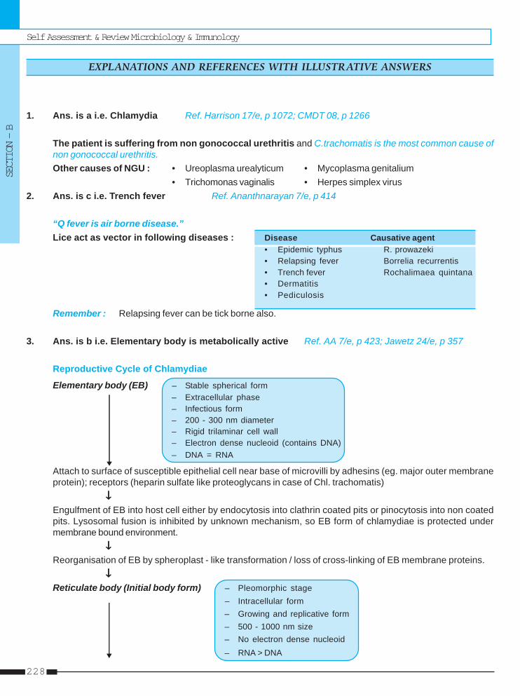
Self Assessment & Review Microbiology & Immunology
SECTION – B
228
1. Ans. is a i.e. Chlamydia Ref. Harrison 17/e, p 1072; CMDT 08, p 1266
The patient is suffering from non gonococcal urethritis and C.trachomatis is the most common cause ofnon gonococcal urethritis.Other causes of NGU : • Ureoplasma urealyticum • Mycoplasma genitalium
• Trichomonas vaginalis • Herpes simplex virus2. Ans. is c i.e. Trench fever Ref. Ananthnarayan 7/e, p 414
“Q fever is air borne disease.”Lice act as vector in following diseases : Disease Causative agent
• Epidemic typhus R. prowazeki• Relapsing fever Borrelia recurrentis• Trench fever Rochalimaea quintana• Dermatitis• Pediculosis
Remember : Relapsing fever can be tick borne also.
3. Ans. is b i.e. Elementary body is metabolically active Ref. AA 7/e, p 423; Jawetz 24/e, p 357
Reproductive Cycle of Chlamydiae
Elementary body (EB) – Stable spherical form– Extracellular phase– Infectious form– 200 - 300 nm diameter– Rigid trilaminar cell wall– Electron dense nucleoid (contains DNA)– DNA = RNA
Attach to surface of susceptible epithelial cell near base of microvilli by adhesins (eg. major outer membraneprotein); receptors (heparin sulfate like proteoglycans in case of Chl. trachomatis)
↓↓↓↓↓Engulfment of EB into host cell either by endocytosis into clathrin coated pits or pinocytosis into non coatedpits. Lysosomal fusion is inhibited by unknown mechanism, so EB form of chlamydiae is protected undermembrane bound environment.
↓↓↓↓↓Reorganisation of EB by spheroplast - like transformation / loss of cross-linking of EB membrane proteins.
↓↓↓↓↓Reticulate body (Initial body form) – Pleomorphic stage
– Intracellular form– Growing and replicative form– 500 - 1000 nm size– No electron dense nucleoid– RNA > DNA
EXPLANATIONS AND REFERENCES WITH ILLUSTRATIVE ANSWERS
Rickettsiaceae & Chlamydiae
UNIT - I BACTERIOLOGY “Gram —ve Cocco Bacilli”
229
Within membrane bound vacoule RB divides by binary fission repeatedly to form EB.↓↓↓↓↓
Cytoplasmic inclusion bodies form (EB filled vacoule)↓↓↓↓↓
This EB liberate from host cell to infect new cells.
4. Ans. is a i.e. Mccoy cell line Ref. Ananthnarayan 7/e, p 427; Jawetz 24/e, p 361
“Complant of urethritis with no result on gram staining signifies nongonococcal urethritis.”MC cause of NGU is chlamydia trachomatis urethritis is one of the manifestation of genital chlamydiasis.
Diagnosis of genital chalmydiasis (D-K serovars)I. Culture :
• Scarpe epithelial cells from 1-2cm deep into endocervix.• Dacron, cotton,rayon or calcium alginate on plastic shaft should be used to collect specimen.• Inoculum is centrifuged onto the monolayer of cycloheximde treated McCoy cells.• This is incubated at 35-370C for 48-72 hours.• Monolayers can be increased.• Examine monolayers by direct IF to see inclusion bodies.• This method is 80% sensitive but 100% specific.
Remember : HeLa cell culture can also used.
II. Direct cytologic examination (direct fluorescent antibody) and enzyme linked immunoassay.
III. Nucleic acid detection by PCR and LCR are test of choice to diagnose genital C.trachomatis infections.
IV. Serology – Serum antibodies are more common than trachoma because of more antigenicity in genitaltract.
Remember : LJ medium is for Mycobacteria tuberculosis. Thayer martin medium is for Neiserria.
5. Ans. is c i.e. Rickettsia prowazekii Ref. Ananthnarayan 7/e, p 414; Park 18/e, p 241; 19/e, p 253
Most important point in the given question is “louse borne rickettsial infection” which is only one i.e., Epidemictyphus which is caused by R. prowazekii.
Epidemic typhus– Also called as louse borne typhus or classical typhus or Gaol fever.– Usually seen among military and refugee populations and famine areas.– Humans are only natural vertebrate host.– Vector : Body or head louse (Pediculus humanus) not pubic louse.– Causative agent : R. prowazekii– Route of transmission : Louse feces is rubbed over abraded skin. So, infection is transmitted from man to
man by infected louse.– Incubation period : 5 to 15 days– Clinical feature : Fever, chills, rash (starts on trunk and spread over limbs sparing face, palms and soles),
stupor and delirium.– Rickettsia may become latent in lymphoid tissue causing recrudescent typhus or Brill - Zinsser disease.
Self Assessment & Review Microbiology & Immunology
SECTION – B
230
Remember : Trench fever is also transmitted by louse but causative agent is Rochalimaea or Bartonellaquintana which is excluded from Rickettsiacea, because it can grow in cell free media.
6. Ans. is d i.e. Community acquired pneumonia Ref. Harrison 17/e, p 1070-1073; AA 7/e, p 424
Disease caused by Chalmydia trachomatis
Serotype Disease
A, B, Ba, C • Endemic blinding trachomaDEFGHIJK • Inclusion conjunctivitis
• Genital chlamydiasis• Infant pneumonia
L1, L2, L3 • Lymphogranuloma venerum
Remember : • Four most common cause of community acquired pneumonia are :– S. pneumoniae– H. influenzae– Chlamydia pneumoniae– Legionella.
7. Ans. is d i.e. The group specific antigen is responsible for the production of complement fixingantibodies Ref. Jawetz 24/e 359; Ananthnarayan 7/e, p 424
“Genus or Group specific heat stable LPS antigen is responsible for CFT while serovar specific Majormembrane protein is responsible for Microimmunofluorescence.”Characteristic of chlamydiae
Feature C trachomatis C penumoniae C psittaci
Inclusion morphology Round, vacuolar Round, dense Large, variable shape, dense
Glycogen in inclusions Yes No No
Elementary body Round Pear-shaped, round Roundmorphology
Susceptible to Yes No Nosulfonamides
DNA homology to C <10% 100% <10%
pneumoniae
Plasmid Yes No yes
Serovars 15 1 > 4
Natural host Humans Humans Birds
Mode of trAns.mission Person to person, Airborne person to Airborne bird excreta tomother to infant person humans
Major diseases Trachoma, STDs, Pneumonia, Psittacosis, pneumonia,infants pneumonia, bronchitis, sinusitis fever of unexplained originLGV
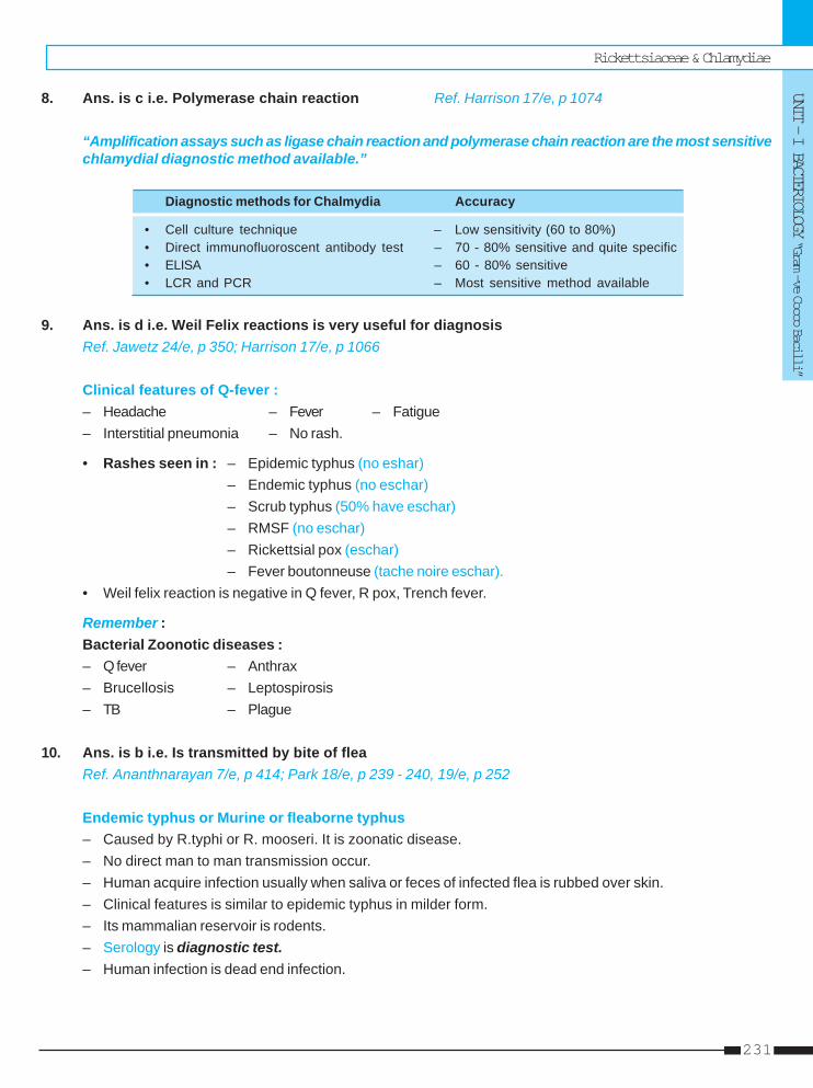
Rickettsiaceae & Chlamydiae
UNIT - I BACTERIOLOGY “Gram —ve Cocco Bacilli”
231
8. Ans. is c i.e. Polymerase chain reaction Ref. Harrison 17/e, p 1074
“Amplification assays such as ligase chain reaction and polymerase chain reaction are the most sensitivechlamydial diagnostic method available.”
Diagnostic methods for Chalmydia Accuracy
• Cell culture technique – Low sensitivity (60 to 80%)• Direct immunofluoroscent antibody test – 70 - 80% sensitive and quite specific• ELISA – 60 - 80% sensitive• LCR and PCR – Most sensitive method available
9. Ans. is d i.e. Weil Felix reactions is very useful for diagnosisRef. Jawetz 24/e, p 350; Harrison 17/e, p 1066
Clinical features of Q-fever :– Headache – Fever – Fatigue– Interstitial pneumonia – No rash.
• Rashes seen in : – Epidemic typhus (no eshar)– Endemic typhus (no eschar)– Scrub typhus (50% have eschar)– RMSF (no eschar)– Rickettsial pox (eschar)– Fever boutonneuse (tache noire eschar).
• Weil felix reaction is negative in Q fever, R pox, Trench fever.
Remember :Bacterial Zoonotic diseases :– Q fever – Anthrax– Brucellosis – Leptospirosis– TB – Plague
10. Ans. is b i.e. Is transmitted by bite of fleaRef. Ananthnarayan 7/e, p 414; Park 18/e, p 239 - 240, 19/e, p 252
Endemic typhus or Murine or fleaborne typhus– Caused by R.typhi or R. mooseri. It is zoonatic disease.– No direct man to man transmission occur.– Human acquire infection usually when saliva or feces of infected flea is rubbed over skin.– Clinical features is similar to epidemic typhus in milder form.– Its mammalian reservoir is rodents.– Serology is diagnostic test.– Human infection is dead end infection.
Self Assessment & Review Microbiology & Immunology
SECTION – B
232
.
Louseborne
Trenchfever
Epidemictyphus
L E T
P QR.prowazekii Rochalim.
quintana
Fleaborne
Endemic typhus
F E n
T
R. typhi
R. PoxMite
borneScrubtyphus
P S M
A T
R.akari O.tsutsugumushi
TICKS with Spotted Indian
Tickborne
RM spotted
fever
Indiantick
typhus
R C
R.ricketsi R.conori
Remember : Rickettsia can’t grow in artifical culture media.
11. Ans. is a i.e. Q. fever Ref. Ananthnarayan 7/e, p 424
Human disease caused by chlamydia
Species Serotype Disease
C. trachomatis A. B, Ba, C Endemic blinding trachomaC. trachomatis D - K Inclusion conjunctivitis, genital
chlamydiasis, infant pneumoniaC. trachomatis L1, L2, L3 Lymphogranuloma venerumC. psittaci Many serotype PsittacosisC. pneumoniae Only one serotype Acute respiratary disease
Remember : Inclusion conjunctivitis include inclusion blenorrhea or opthalmia - neonatorum and swimmingpool conjunctivitis.
12. Ans. is b i.e. Chlamydia trachomatis Ref. Ananthnarayan 7/e, p 423
“Chalmydiae are obligate intracellular bacterial parasite.” ..... Ananthnarayan p 422They can be isolated only in cell culture.McCoy and Hela cell lines are MC cell lines used to culture chlamydiae.
Remember : Other obligate parasite : • Rickettsiaceae and coxiella burnetti• M. Leprae• Pathogenic treponemes• Viruses.
13. Ans. is d i.e. Parotitis Ref. Ananthnarayan 7/e, p 424
Genital chlamydiasis :Men – Epididymitis, proctitis.Women – Bartholinitis, cervicitis, salpingitis, endometritis, PID, Perihepatitis (fitz Hugh syndrome),
Infertility, abortion, ectopic pregnancy.Either sex – Reiter’s syndrome, NGU, conjunctivitis.
Rickettsiaceae & Chlamydiae
UNIT - I BACTERIOLOGY “Gram —ve Cocco Bacilli”
233
14. Ans. is b i.e. Trombiculid mute Ref. Harrison 17/e, p 1064
Scrub typhus• Caused by O.tsutsugamushi• Transmitted by tromiculid mite which also shows transovarian spread• Clinical features : – Fever, headach, myalgia, cough and GI symptoms.
– Classic case includes an eschar, regional lymphadenopathy and a maculopappularrash.
Diagnosis :• Serologic assays (IFA, indirect immunoperoxidase and enzyme immunoassays) are main stay of diagnosis.Treatment : – Rifampin
– Azithromycin and clarithromycin
15. Ans. is b i.e. Flea is a vector of the disease Ref. Ananthnarayan 7/e, p 414
Already explained, refer answer no. 5
16. Ans. is b i.e. Cause non-gonococcal urethritis Ref. Ananthnarayan 7/e, p 428; Jawetz 24/e, p 364
C. Psittaci• Causes psittacoses in humans / birds, ornithosis in birds, meningo pneumonitis, feline pneumonitis and
other animal diseases.• Form diffuse intracytoplasmic inclusions that lack glycogen, not stained by I2 and not inhibited by
sulphadiazine or cycloserine. Heat stable group reactive. CF antigen resist proteolytic enzymes so seemsto be lipopolysaccharide.
• Psittacosis is disease of human acquired from contact with birds and also includes infection of psittacinebirds.
• Ornithosis is infection in all types of domestic birds.• Infection in birds is usually subclinical (carriers).• Human infection is usually occupational as is poultry workers, pigeon farmers, pet-shop owners, bird fencers
and veterinarian. Usually occur by inhalation of infected dried feces.• Incubation period about 10 days.• Manifest as influenza / atypical pneumonia / sepsis.• Antibodies to species specific antigen can neutralize toxicity and infectivity.• Diagnosis : Culture is dangerous and if done; than isolation should be attempted only in special laboratories
as laboratory infection is serious hazard detection of antigen by direct fluorescent antibody staining or byimmunoassay or PCR is preferred serology (confirmatory) by CFT or MIF.
• Treatment : – DOC is tetracycline.– Should be continued for 10 days after defervescence to prevent relapse.
17. Ans. is b i.e. Culture on McCoy cells Ref. Ananthnarayan 7/e, p 427; Jawetz 24/e, p 361
“Genital Chalmydiasis is suspected if Gram stained smear of urogenital exudates show significant numberof neutrophills (> 4/oil immersion field in urethritis, > 30 in cervicitis) in absence of gonococcal bacteria.”
For more details, refer answer no. 3
Self Assessment & Review Microbiology & Immunology
SECTION – B
234
18. Ans. is b i.e. Enzyme immunoassay Ref. Ananthnarayan 7/e, p 425
Isolation of Chlamydiae can done by :a. Inoculation into yolk sac/embryonated eggs of 6 - 8 day old chick embryo which may be pretreated by
streptomycin or polymyxin B.b. Inoculation into experimental animals (mice).c. Tissue / cell culture :
• Preferred mode• Commonly used cell lines are McCoy and HeLa cells.• Cell cultures are pretreated by irradiation or chemicals such as 5-iodo - 2 deoxyuridic or cycloheximide
to enhance replication and detection of inclusion bodies.• Pretreatment with DEAE dextrax or centrifugation after inoculation, promotes contact between chalmydiae
particles and cell monolayer.
19. Ans. is c i.e. They can grow in cell free culture media Ref. Ananthnarayan 7/e, p 422
– Chlamydia are obligate intracellular parasite so, they can not be grow in cell free media.– Gram negative coccobacilli.– Chalmydia don’t have peptidoglycan cell wall.– They lack enzymes of electron transport chain. So require ATP from host cells & are called energy parasites.– Drugs effecitve against chlamydiae : Doxycycline, Azithromycin, Erythromycin.
20. Ans. is a i.e. R. rickettsii Ref. Park 18/e, p 239, 19/e 252
Already explained, refer answer no. 10
21. Ans. is c i.e. Inhalation of aerosol Ref. Harrison 17/e, p 1066; Jawetz 24/e, p 351
“Q fever transmitted by inhalation of dust contaminated with rickettsial from placenta, dried feces,urine or milk or aerosols in slaughter houses.”
Q fever : • Caused by coxella burnetti• A zoonotic disease• Primary source of human infection are cattle, sheep and goats• Mode of transmission :
In infected female animal C. burnetti localises uterus↓
Infection reactivates during pregnancy↓
High concentration of coxella in placenta↓
Soil gets contaminated during parturation↓
Coxella aerosols generates during storms↓
Human infection
Rickettsiaceae & Chlamydiae
UNIT - I BACTERIOLOGY “Gram —ve Cocco Bacilli”
235
Remember : Q fever – Gives negative Weil-felix reaction.– Also transmitted by infected milk.
22. Ans. is d i.e. Epidemic typhus Ref. Park 18/e, p 239; 19/e, p 251
Already explained, refer answer no. 5
23. Ans. is a Chlamidia trachomatis Ref. Harrison 17/e, p 1072; Ananthnarayan 7/e, p 229
“Complaint of urethritis with no result on Gram staining signifies Non gonoccoccal urethritis.”
Cause of Non-gonococcal urethritis :
– Chlamydia trachomatis (MC) – Ureoplasma urealyticum– Trichomonas vaginalis – Herpes simplex virus– Mycoplasma hominis – CMV– Gardnerella vaginalis – Acinetobacteri Iwoffi, Ac calcoaceticus– Candida albicans
24. Ans. is d i.e. Urethritis Ref. Ananthnarayan 7/e, p 424
• Urethritis is one of the presentation.• Genital chlamydiasis, caused by serotype D-K of C. trachomatis.• C. pneumonia increased the risk of artherosclerosis, asthma, sarcoidosis.
25. Ans. is b i.e. Gram positive Ref. Ananthnarayan 7/e, p 423
Rickettsia are Gram negative coccobacilli.
26. Ans. is b i.e. Coxella burnetti Ref. Harrison 17/e, p 1066
“Coxella burnetti – Causative agent of Q fever is transmitted through aerosolization”
27. Ans. is a, c and d Ref. Harrison 17/e, p 1073
Reiter’s syndrome consists of conjunctivitis, urethritis, (or in female patient cervicitis) arthritis and characteristicmucocutaneous lesion.Pathogenesis is unknown. However more than 80% affected patient belong to HLA-B-27..
28. Ans. is b i.e. Rocky Mountain Ref. Harrison 17/e, p 1059
Family rickettsiae includes six genera :• Rickettsia • Ehrlichia• Orientia • Anaplasma• Neorickettisa • Coxiella
Self Assessment & Review Microbiology & Immunology
SECTION – B
236
As question is about disease by rickettsia answer is RMSF (which is caused by Rickettsia rickettsii) only asScrub typhus is caused by Orientia tsutsugamushi.
29. Ans. is a, b and c i.e. HeLa; HeP2; and McCoy Ref. Ananthnarayan 7/e, p 425
Cell lines for chlamydiae are : – McCoy– HeLa– HEp - 2, HL.
• Human diploid fibroblast series and vero cell lines are used for virus isolation.
30. Ans. is a and c i.e. Infertility; and Pelvic inflammatory disease Ref. Harrison 17/e, p 1071-1073
Chlamydial diseases are :• In Men – NGU, Epididymitis• In Women – Cervicitis, Salpingitis, Urethritis, PID, infertility• Either sex – Proctitis, LGV, Reiter syndrome• Neonates (As a result of perinatal infection) – Conjunctivitis, pneumonia
31. Ans. is a i.e. Rickettsiae Ref. Ananthnarayan 7/e, p 415 - 417
Neill - Mooser or Tunica reaction – Used to differentiate between R. typhi and R. prowazaki.– Male guinea pigs are inoculated intraperitoneally with blood from a case of endemic typhus or culture of R.
typhi, they develop fever and characteristic scrotal inflammation. This is called neill - Mooser reaction.– This is negative with R. Prowazaki but positive in R.typi, R.conori and R.akari.
32. Ans. is a, b and d i.e. Rickettsia; M. leprae; and Syphilis Ref. Ananthnarayan 7/e, p 423
Already explained, refer see answer no. 12
Rickettsiaceae & Chlamydiae
UNIT - I BACTERIOLOGY “Gram —ve Cocco Bacilli”
237
1. b) Endothelial ... 2. a) R. tsutsugamushi 3. a) Scrub typhus 4. a) Guinea worm ... 5. c) Cell culture6. d) Parotitis 7. d) C. burnetti 8. a) Giardia 9. None 10. c) Granuloma ...
11. a) Rickettsial ...
1. The primary site of multiplication of rickettsial or-ganisms is in the : [Kerala 90]a) Paranchymal cells of the liverb) Endothelial cells of small vesselsc) Media of arteriesd) Adventitia of all blood vessels
[Ref. Ananthnarayana 7/e, p 412]2. Weil felix reaction with OXK antigen indicates in-
fection with : [JIPMER 90]a) R. tsutsugamushib) R. Moseric) R. Quintanad) R. Akari
[Ref. Ananthnarayana 7/e, p 418]3. Mite transmits : [AI 91]
a) Scrub typhusb) Trench feverc) Endemic typhusd) Epidemic typhus
[Ref. Ananthnarayana 7/e, p 414]4. Anthro zoonosis are all except : [AI 93]
a) Guinea worm infectionb) Rabiesc) Plagued) Hydatid cyst
[Ref. Park 19/e, p 88]5. Most common diagnostic test in LGV is :
a) Fluorescent antibody [PGI 93]b) Complement fixation testc) Cell cultured) Frei’s test
[Ref. Ananthnarayana 7/e, p 425 - 428]6. Chlamydia cause all of the following except :
a) Trachoma [PGI 93]
Answer
b) Non-gonococcal urethritisc) Pneumoniad) Parotitis
[Ref. Ananthnarayana 7/e, p 424, 229]7. Q fever is caused by : [Kerala 94]
a) Rickettsia tsutsugamushib) R. prowozekiic) R. Quintanad) C. burnetti
[Ref. Ananthnarayana 7/e, p 419]8. Which is not a zoonotic disease : [UP 96]
a) Giardiab) Leptospirosisc) Brucellosisd) Epidemic typhus [Ref. Park 19/e, p 642]
9. The following are zoonotic disease except :a) R. Pox [UP 97]b) Scabiesc) Leptospirosisd) Brucellosis
[Ref. Park 19/e, p 642]10. Chalmydia cause all except : [UP 98]
a) Trachomab) Inclusion conjunctivitisc) Granuloma inguinaled) on specific urethritis
[Ref. Ananthnarayana 7/e, p 229, 424]11. Neil Mooser’s reaction is given by : [TN 02]
a) Rickettsial infectionb) Chalamydial infectionc) Mycoplasmad) Pnemococcai infection
[Ref. Ananthnarayan 7/e, p 415]
Chapter Review••••• This section includes questions of VThis section includes questions of VThis section includes questions of VThis section includes questions of VThis section includes questions of Various Other PGMEES from 1990 – 2007.arious Other PGMEES from 1990 – 2007.arious Other PGMEES from 1990 – 2007.arious Other PGMEES from 1990 – 2007.arious Other PGMEES from 1990 – 2007.••••• Questions are arQuestions are arQuestions are arQuestions are arQuestions are ar ranged in decreasing order of yearranged in decreasing order of yearranged in decreasing order of yearranged in decreasing order of yearranged in decreasing order of yearwiz. This is done to makwiz. This is done to makwiz. This is done to makwiz. This is done to makwiz. This is done to make refere refere refere refere referralralralralral
system easier and uncomplicated to save the precious time of PGMEE Aspirant.system easier and uncomplicated to save the precious time of PGMEE Aspirant.system easier and uncomplicated to save the precious time of PGMEE Aspirant.system easier and uncomplicated to save the precious time of PGMEE Aspirant.system easier and uncomplicated to save the precious time of PGMEE Aspirant.

Self Assessment & Review Microbiology & Immunology
SECTION – B
238
12. Micro organism that does not obey koch’s postu-lates : [Kerala 02, AI 89]a) M. tuberculosisb) Poliovirusc) M. lepraed) Streptococcus
[Ref. Ananthnarayan 7/e, p 3]13. Rickettsia prowazeki is transmitted by :
a) Fleas [SRMC 02]b) Mitesc) Tickd) Louse
[Ref. Ananthnarayan 7/e, p 414]14. All the following statement are true about rickett-
sial excepts : [UPSC 02]a) These are transmitted by arthropod vectorsb) Eschar is not seen in RMSFc) Well Felex reaction may be diagnosticd) Cephalosporin are drug of choice
[Ref. Ananthnarayan 7/e, p 416; Harrison 16/e, p1000
15. Chlamydia trachomatis, the causative agent fortrachoma : [Kar 2003]a) Is a yestb) Is an intracellular organismc) Forms extracellular bodies which are diagnosticd) Is never demonstrable in conjunctival scrapings
[Ref. Ananthnarayan 7/e, p 422, 424]16 Causative agent of Q fever : [Kolkata 03]
a) Ro.quintanab) R.prowazekiic) R.typhid) Coxiella burnetti
[Ref. Ananthnarayan 7/e, p 419]18. Reiter’s syndrome is due to : [Jharkhand 04]
a) Chlamydia
b) Nisseriac) Mycoplasmad) Herpes virus
[Ref. Ananthnarayan 6/e, p 229]19. For all of the following reaction Well Felix reaction
is diagnostic except : [SGPGI 05]a) Endemic typhusb) Scrub typhusc) Epidemic typhusd) Q fever
[Ref. Ananthnarayana 7/e, p 418 - 419]20. Endemic typhus is transmitted by : [DNB 05]
a) Rat fleab) Mitec) Tickd) None
[Ref. Ananthnarayana 7/e, p 414]21. All of the following can be grown in cell free cul-
ture media except : [SGPGI 05]a) Mycoplasmb) Rickettsiac) Salmoneliad) Campylobactor
[Ref. Ananthnarayan 7/e, p 413]22. Which rickettesiae are able to grow in cell free
media : [Jharkhand 05]a) R. Quintanab) R. rickettsic) R. typhid) R. Tsugatmushi
[Ref. Anantharayan 7/e, p 412]23. Infectious part of Chlamydia is :
a) Elementary body [Jharkhand 06]b) KP bodyc) Reticulate bodyd) None
[Ref. Anantharayan 7/e, p 422]
12. c) M. leprae 13. d) Louse 14. d) Cephalosporin ... 15. b) Is an intra ... 16. d) Coxiella ...18. a) Chlamydia 19. d) Q fever 20. a) Rat flea 21. b) Rickettsia 22. a) R. Quintana23. a) Elementary ...
Answer
Group comprising : Elongated, motile, flexible bacteria.Characteristic feature of spirochetes is presence of varying number of endoflagella which are polar flagella situatedbetween outer membrane and cell wall; unlike flagella of other bacteria they don’t protrude outside.Pathogenic spirochetes belong to genera : 1. Treponema
2. Borrelia3. Leptospira
TREPONEMA• Relatively short spirochetes with fine spiral and pointed or rounded ends.• Pathogenic treponemes have not been successfully cultivated in cell free media while the non-pathogenic
[which are commensals] can be cultivated.• Pathogenic treponemes include :
– T. pallidum (causative agent of endemic and veneral syphilis)– T. partenue (causative agent of yaws)– T. carateum (causative agent of pinta)They are identical in their morphology, antigenic structure and other biochemical features, differs only in clinicalfeature of disease they produce. But recently molecular signatures have been identified that can differentiate thethree subspeices of T.pallidum by PCR based methods. ... Harrison 17/e, p 1038
TREPONEMA PALLIDUM
Morphology• A thin spiral organism which is actively motile through endoflagella.• Seen by immunofluorescence staining or dark field illumination or phase contrast microscope.• Stained by silver impregnation methods - Fontana method useful for staining films and Levaditi method for
tissue sections.
Cultures and Growth• Grows only in tissue culture not in artificial culture media as it lack genes required for denovo synthesis ofaminoacids, nucleotides and lipids.• Reiter strain (T.phagedenis) - Non pathogenic. treponeme; shows morphological and antigenic similarities with
T. Pallidum; can grow in artificial culture.• T.pallidum is microaerophillic organism grows best in 1-4% O2.
SpirochetesSpirochetesSpirochetesSpirochetesSpirochetes1616
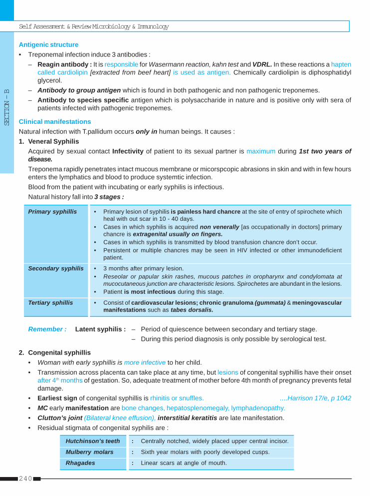
Self Assessment & Review Microbiology & Immunology
SECTION – B
240
Antigenic structure• Treponemal infection induce 3 antibodies :
– Reagin antibody : It is responsible for Wasermann reaction, kahn test and VDRL. In these reactions a haptencalled cardiolipin [extracted from beef heart] is used as antigen. Chemically cardiolipin is diphosphatidylglycerol.
– Antibody to group antigen which is found in both pathogenic and non pathogenic treponemes.– Antibody to species specific antigen which is polysaccharide in nature and is positive only with sera of
patients infected with pathogenic treponemes.
Clinical manifestationsNatural infection with T.pallidum occurs only in human beings. It causes :1. Veneral Syphilis
Acquired by sexual contact Infectivity of patient to its sexual partner is maximum during 1st two years ofdisease.Treponema rapidly penetrates intact mucous membrane or micorspcopic abrasions in skin and with in few hoursenters the lymphatics and blood to produce systemtic infection.Blood from the patient with incubating or early syphilis is infectious.Natural history fall into 3 stages :
Primary syphillis • Primary lesion of syphilis is painless hard chancre at the site of entry of spirochete whichheal with out scar in 10 - 40 days.
• Cases in which syphilis is acquired non venerally [as occupationally in doctors] primarychancre is extragenital usually on fingers.
• Cases in which syphilis is transmitted by blood transfusion chancre don’t occur.• Persistent or multiple chancres may be seen in HIV infected or other immunodeficient
patient.
Secondary syphilis • 3 months after primary lesion.• Reseolar or papular skin rashes, mucous patches in oropharynx and condylomata at
mucocutaneous junction are characteristic lesions. Spirochetes are abundant in the lesions.• Patient is most infectious during this stage.
Tertiary sphillis • Consist of cardiovascular lesions; chronic granuloma (gummata) & meningovascularmanifestations such as tabes dorsalis.
Remember : Latent syphilis : – Period of quiescence between secondary and tertiary stage.– During this period diagnosis is only possible by serological test.
2. Congenital syphillis• Woman with early syphillis is more infective to her child.• Transmission across placenta can take place at any time, but lesions of congenital syphillis have their onset
after 4th months of gestation. So, adequate treatment of mother before 4th month of pregnancy prevents fetaldamage.
• Earliest sign of congenital syphillis is rhinitis or snuffles. ....Harrison 17/e, p 1042• MC early manifestation are bone changes, hepatosplenomegaly, lymphadenopathy.• Clutton’s joint (Bilateral knee effusion), interstitial keratitis are late manifestation.• Residual stigmata of congenital syphilis are :
Hutchinson’s teeth : Centrally notched, widely placed upper central incisor.
Mulberry molars : Sixth year molars with poorly developed cusps.
Rhagades : Linear scars at angle of mouth.
Spirochetes
UNIT - I BACTERIOLOGY
241
Diagnosis1. Demonstration of Organism
• Darkfield microscopic examination of lesion exudate such as chancre of primary syphillis or more reliablyby immunofluoroscence or immunohistochemical method.
2. Serological test for syphilis
Nontreponemal test Treponemal test
Detect IgG or IgM against cardiolipin antigen Detect specific antibody against T. pallidum• Includes : RPR (Rapid plasma reagin) • Includes : Fluoroscent treponemal antibody
[test of choice for rapid diagnosis] absorption test [F-TAABS]• VDRL [test of choice for response – Agglutination assays (MHATP, TPHA, TPPA)
to therapy] – TPI (Treponemal pallidum immobilisation)• VDRL is type of slide flocculation test test
while kahn flocculation is tube test
Remember : – FTA-ABS, TPPA are most sensitive test.– TPI is most specific serological test.– TPPA has supplanted the FTA-ABS test as diagnostic test. ..... CMDT’ 06, p 1445
Diagnosis of neurosyphilis : Examination of CSF for pleocytosis, increase protein concentration, VDRL reactivity.Diagnosis of congenital syphilis : If both test i.e. VDRL and FTA- ABS IgM (specific) test are positive in theinfant than congenital syphilis should be strongly suspected and the child should be treated.
Treatment
Stage of syphilis DOC Patient with penicillin allergy
Primary secondaryor early latent Penicillin benzathine Tetracycline or DoxycyclineLate latent Penicllin G TetracyclineNeurosyphilis Aqueous penicillin G Desensitization and treatment with penicllinSyphilis in pregnancy According to stage Desensitization and treatment with pencillin
• Continued susceptibility of treponema to penicillin is due to highly conserved genome.
Jarisch Herxheimer reaction :– May occur after penicillin therapy.– Seen in primary; mostly after secondary; early latent syphilis; delayed in neurosyphilis.– Subsides itself.– Treatment : Symptomatic - Antibiotic should not be stopped. Steroids should not be given.
Evaluation of response to therapy :• Response to treatment is determined by monitoring VDRL or RPR titer.• Activity of neurosyphilis is best correlated with CSF pleocytosis.• Continued susceptibility of treponema to penicillin is due to highly conserved genome.
Self Assessment & Review Microbiology & Immunology
SECTION – B
242
NON VENERAL TREPONEMATOSISInfection usually transmitted by body to body contact. It includes :A. Endemic syphilis :
– Caused by T. pallidum subspecies endemicum– Disease is common is young children, primary chancre is not usually seen.– Treatment : Same as veneral syphillis.
B. Yaws :– Caused by T.pallidum subspecies T.pertenue which is morphologically and Immunologically identical to
T.pallidum. (Learn Py.)– Primary lesion is extragenital papule which enlarges and ulcerate to form an ulcerating granuloma.
C. Pinta :– Causative agent T.carateum (Learn Cap).– It is not identical but closely related to T.pallidum.– Primary lesion is extragenital papule which doesnot ulcerate but develop into lichenoid or psoriatic patch.
Remember : In non veneral treponematosis, yaws (always) and pinta (usually) serological test of syphilisare positive.
LEPTOSPIRAActively motile spirochetes possesing a large number of closely wound spirals and characterstic hooked ends.
Culture : Grow best under aerobic condition. For isolation EMJH media is used.
Pathogenesis and clinical manifestation :• Leptospirosis is a zoonosis with rodents being most important reservoir.• Transmission results from ingestion or contact with urine, blood or tissue from infected animal but not from bite.
Since leptospires are excreted in urine of infected rat, water is important vehicle. Human to human transmissiondon’t occur.
• Vasculitis is responsible for most clinical manifestation.• It mainly infects : Liver (centritobular necrosis is found)
Kidney (cause interstitial nephritis, tubular necrosis)• After formation of antibody, leptospires are eliminated from all sites except the eye, proximal renal tubules and
brain.• More than 40% of symptomatic person have mild and anticteric from of leptospirosis.• Severe leptospirosis is characterized by profound jaundice, renal dysfunction, hemorrhagic diathesis called
as Weil’s syndrome or Icterohemorrhagic fever.
Diagnosis1. Isolation of organism – From blood or CSF during 1st 10 days
– From urine after 1 week– For isolation EMJH medium is useful
2. Serology – Microscopic agglutination test [MAT]– ELISA
Treatment– Mild - Doxycycline or Ampicillin.– Moderate / severe - Penicillin or Erythromycin.
Spirochetes
UNIT - I BACTERIOLOGY
243
Chemoprophylaxis : DoxycyclineBORRELIA• Large, refractile spirochetes which can be stained by ordinary method and are Gram (–) ve.• Pathogenic species are :
B.burgodorferi - Causes Lyme’s disease
B.recurrentis - Causes Relapsing fever
B.vincenti - Causes Vincent angina
Lymes Disease• Causative agent B.burgodorferi.• MC vector born infection in Europe. Transmitted by bite of Ixodid ticks. .... Harrison 17/e, p 1055
• Clinical features– Stage I. (Localized infection)
• After incubation period of 3 to 32 days EM occurs at the site of bite. EM (Erythema migrans) is not painful.– Stage II. Disseminated infection
• Disseminate hematogenously to produce secondary annular skin lesion, meningitis, carditis (MC cardiacfinding is fluctuating degree of Atrioventricular heart block).
– Stage III. Persistent infection• Usually present as oligoarticular arthritis (MC knee), encephalopathy, polyneuropathy. Acrodermatitis
chronica atrophicans is late skin manifestation.
• Diagnosis– ELISA followed by western blot is best investigation.– Culture in BSK medium gives definitive diagnosis but not useful clinically.– PCR particularly in persistant infection.
• Treatment– For nervous manifestation and 30 heart block – Ceftriaxone is DOC. ..... Harrison 17/e, p 1058– For skin manifestation, arthritis 10 and 20 AV block – Doxycycline is DOC.
Relapsing Fever• Causative agent B.recurrentis.• It is of 2 types :
– Louse borne and Tick borne the, Borellia causing them are indistinguishable.– Louse borne relapsing fever occur as epidemic.– Tick borne occur as sporadic / endemic cases.
• Treatment : Erythromycin is DOC. ..... Harrison 17/e, p 1054
Vincent angina• Causative agent B.vincenti• Normal mouth commensal, but when associated with fusiform bacilli (Fusobacterium fusiform) causes ulcerative
gingivostomatitis or oropharyngitis called vincent angina.• Treatment : Penicillin and Metronidazole.
Self Assessment & Review Microbiology & Immunology
SECTION – B
244
1. Which is not true about Yaws ? [AI 08]a) Spread by sexual transmissionb) Caused by T.perteunec) Has cross immunity with syphillisd) Cannot be differentiated serologically from T.
pallidum
2. A bacterial disease with 3 ‘R’s i.e. rats, rice fieldsand rainfall is : [AI 05]a) Leptospirosisb) Plaguec) Melioidosisd) Rodent bite fever
3. A sweeper involved with repair-work of sewerswas admitted with fever, jaundice and renal fail-ure. The most appropriate test to diagnose in-fection of this patients : [AI 03]a) Weil Felix testb) Paul Bunnel testc) Microscopic agglutinatiion testd) Micro immunoflurescence test
4. The following are true regarding Lyme’s Disease,except : [AI 03]a) It is transmitted by ixodes tickb) Erythema chronicum migraines may be a clinical
featuresc) Borrelia recurrentis is the etiologic agentd) Rodents act as natural hosts
5. Which of the following is transmitted by Raturine ? [AI 02]a) Leptospirab) Listeriac) Legionellad) Mycoplasma
6. Congenital syphilis can be diagnosed by :a) IgM FTA abs [AI 01, 96]b) IgG FTA abs
c) VDRLd) TPI
7. False +ve VDRL is seen in : [AI 00, 95]a) Lepromatous leprosyb) Infectious mononucleosisc) HIVd) Pregnancy
8. All are true about FTA-ABS Syphilis, except :a) FTA-ABS becomes negative after treatmentb) Present in secondary syphilis [AI 00]c) It is a sensitive testd) May be positive in Lyme’s disease
9. The following statements are true regarding lep-tospirosis, except : [AIIMS 06]a) It is a zoonosisb) Man is the dead end hostc) Man is an accidental hostd) Lice act as reservoirs of infection
10. ‘Chancre redux’ is a clinical feature of :a) Early relapsing syphilis [AIIMS 06]b) Late syphilisc) Chancroidd) Recurrent herpes simplex infection
11. A 25-year old farmer presented with history ofhigh grade fever for 7 days and altered senso-rium for 2 day. On examination, he wascomatosed and had conjunctival hemorrhage.Urgent investgations showed a hemoglobin of 11gm/dl. Peripheral blood smear was negative formalarial parasite. What is the most likely diagno-sis ? [AIIMS 05]a) Brucellosisb) Weil’s diseasec) Acute viral hepatitisd) Q fever
ANSWERS WITH EXPLANATIONQUESTIONS
Answer 1. a) Spread by... 2. a) Leptospirosis 3. c) Microscopic ... 4. c) Borrelia ... 5. a) Leptospira6. a) IgM FTA ... 7. b) Infectious ... 8. a) FTA-ABS ... 9. d) Lice ... 10. ??
11. b) Weil’s ...
Spirochetes
UNIT - I BACTERIOLOGY
245
12. A 23 year old male had unprotected sexual inter-course with a commercial sex worker Two weekslater he developed a painless, indurated ulcer onthe glans that exuded clear serum on pressureinguinal lymph nodes in both groins were en-larged and non-tender Most appropirate diagnos-tic test is : [AIIMS 04]a) Gram’s stain of ulcer dischargeb) Dark field microscopy of ulcer dischargec) Giemsa stain of lymph node aspirated) ELISA for HIV infection
13. Which one of the following microorganisms usesantigenic variation as a major means of invadinghost defenses : [AIIMS 04]a) Streptococcus pneumoniab) Borrelia recurrentisc) Mycobacterium tuberculosisd) Listeria monocytogenes
14. 20 year old body has admitted with history of fe-ver, icterus, conjunctival suffusion and hematuriafor 20 days. Which of the following serologicaltest can be of diagnostic utility : [AIIMS 04]a) Widal testb) Microscopic agglutination testc) Paul Bunnel testd) Weil Felix reaction
15. Best indicator for monitoring of syphilis treat-ment: [AIIMS 02]a) VDRLb) FTA-ABSc) TPId) TPHA
16. Spirochaetes among following are : [PGI 06]a) Syphilisb) Leptospirac) Mycoplasmad) Brucellae) Borelia
17. Non venereal treponemas is/are : [PGI 04]a) T. Pertenueb) T. Carateumc) T. Palildumd) T. Cuniculi
18. Lyme disease caused by : [PGI 01]a) Leptospirab) Borreliac) Treponemad) Bordetellae) Arbovirus
19. Dark ground microscopy is used for : [PGI 98]a) TPIb) FTA-ABSc) Kahn’s testd) VDRL
20. Following is true of T. pallidum, except :a) Can be maintained in rabbit testis [PGI 98]b) Motile by peritrichate flagellac) To visualise, dark ground microscopy is usedd) TPI test is very useful
Answer 12. b) Dark field ... 13. b) Borrelia ... 14. b) Microscopic ... 15. a) VDRL 16. a, b and e17. a and b 18. b) Borrelia 19. a) TPI 20. b) Motile ...
Self Assessment & Review Microbiology & Immunology
SECTION – B
246
1. Ans. is a i.e. Spread by sexual transmission Ref. Harrison 17/e, p 1047
Yaws is caused by T. pallidum subspecies pertnue.• T.pertenue is antigenically identical to syphilitis.Mode of transmission : Infection is transmitted by direct contact with infectious lesions, often during play orgroup sleeping.Clinical features : After 3-4 weeks the first lesion begins as a papule usually on extremity, it is followed byappearance of multiple skin lesion.Treatment of choice : Benzathine penicililn.
2. Ans. is a i.e. Leptospirosis Ref. Ananthnarayan 7/e, p 389; Harrison 17/e, p 1048
Leptospirosis• Dead end infection of leptospira characterized by clinical manifestations ranging from inapparent infection
to fulminant icterohemmoragic fever (Weils’s syndrome).
Modes of transmission :• Rodents are most important reservoir.• Transmission occur by direct contact with urine, blood or tissue from an infected animal.• Water is an important vehicle of transmission.• Out breaks mostly occur as a result of heavy rainfall and consequent flooding.
So. guys Leptospirosis is associated with : – Rats– Rainfall– Rice - Rats usually live in rice farms.
Other options – Heavy rainfall tends to protect against plague and Rodent bite fever by damaging rodentburrows.
– Meliodosis is not associated with Rat.
3. Ans. is c i.e. Microscopic agglutination testRef. Harrison 17e, p 1050; Ananthnarayan 7/e, p 392; 6/e, p 362
• The patient is a case of Weil’s syndrome or icterohemmorhagic fever which is caused by Leptospiraand manifest as :– Fever– Jaundice– Renal failure (Hematuria)
• Human infection are usually due to occupational exposure to urine of infected animal eg.,– Farmers – Workers in rice field and sugarcane field– Workers in underground Sewers – Meat and animal handlers– Veterinarians
EXPLANATIONS AND REFERENCES WITH ILLUSTRATIVE ANSWERS
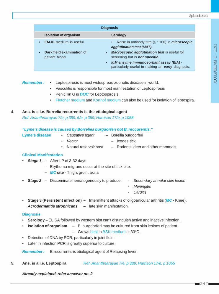
Spirochetes
UNIT - I BACTERIOLOGY
247
Diagnosis
Isolation of organism Serology
• EMJH medium is useful • Raise in antibody titre (> : 100) in microscopicagglutination test (MAT).
• Dark field examination of • Macroscopic agglutination test is useful forpatient blood screening but is not specific.
• IgM enzyme immunosorbant assay (EIA) -particularly useful in making an early diagnosis.
Remember : • Leptospirosis is most widespread zoonotic disease in world.• Vasculitis is responsible for most manifestation of Leptospirosis• Penicillin G is DOC for Leptospirosis.• Fletcher medium and Korthof medium can also be used for isolation of leptospira.
4. Ans. is c i.e. Borrelia recurrentis is the etiological agentRef. Ananthnarayan 7/e, p 389; 6/e, p 359; Harrison 17/e, p 1055
“Lyme’s disease is caused by Borreliea burgdorferi not B. reccurentis.”Lyme’s disease • Causative agent – Borellia burgdorferi
• Vector – Ixodes tick• Natural reservoir host – Rodents, deer and other mammals.
Clinical Manifestation• Stage 1 – After I.P of 3-32 days
– Erythema migrans occur at the site of tick bite.– MC site - Thigh, groin, axilla
• Stage 2 – Disseminate hematogenously to produce : - Secondary annular skin lesion- Meningitis- Carditis
• Stage 3 (Persistent infection) – Intermittent attacks of oligoarticular arthritis (MC - Knee).Acrodermatitis atrophicans – late skin manifestation.
Diagnosis• Serology – ELISA followed by western blot can’t distinguish active and inactive infection.• Isolation of organism – B. burgdorferi may be cultured from skin lesions of patient.
– Grows best in BSK medium at 330C.• Detection of DNA by PCR, particularly in joint fluid.• Later in infection PCR is greatly superior to culture.
Remember : B.recurrentis is etiological agent of Relapsing fever.
5. Ans. is a i.e. Leptospira Ref. Ananthnarayan 7/e, p 389; Harrison 17/e, p 1055
Already explained, refer ansewer no. 2
Self Assessment & Review Microbiology & Immunology
SECTION – B
248
6. Ans. is a i.e. IgM FTAbs Ref. Ananthnaryan 7/e, p 383; Harrison 16/e, p 984
“Newborn infant of mother with reactive VDRL or FTA-ABS shows (+) ve test irrespective of infectionbecause of transplacental transfer of maternal IgG antibody.”
As IgM antibody don’t cross placenta, neonatal IgM antibody can be detected in cord or neonatal serum withthe syphilis capita M or 195 IgM FTA-ABS test. ..... Harrison 16/e, p 984
Important points about Congenital syphilis• Transmission across placenta can take place any time, but lesion appear after 4 month of gestation.• Earliest sign of congenital syphilis – Rhinitis, snuffles• Residual stigmata of congenital syphilis – Hutchinson’s teeth, Mulberry molars, Rhagades• DOC of congenital syphilis – Penicillin G.
Caution : According to Harrison 17/e, p 1045 no commercially available IgM test is recomended for evaluationof infant with suspected cong. syphilis.
7. Ans. is b i.e. Infectious mononucleosis Ref. Harrison 16/e, p 985; CMDT ‘08, p 1269
The modern VDRL and RPR test are 97% to 99% specific and false psotive test are now limited tofollowing condition :
Causes of False Positive VDRL
Acute false positive reaction <6 months Chronic false positive reaction >6month
– Recent viral illness or immunization – Aging– Genital herpes – Auto immune disorders– HIV infection – SLE– Malaria – Rheumatoid arthritis– Parenteral drug use – Parenteral drug use
Infectious mononucelosis is acute infection of EBV and can give false (+)ve VDRL• In CMDT ‘08, p 1269 Leprosy is also mentioned as cause of false (+)ve VDRL: but with use of newer non-
lipoidal VDRL test, leprosy no longer give false positive result.• False negative VDRL.
Seen when very high antibody titre is present [Prozone phenomenon].
8. Ans. is a i.e. FTA-ABS becomes negative after treatment Ref. Harrison 17/e, p 1049
• Only VDRL and RPR for syphilis becomes negative after treatment and are recommended for evaluation oftherapy. VDRL or RPR titer progressively declines, becomes (–ve) by 12 months in 40-75% of primarycases and in 20-40% of secondary cases.
• FTA-ABS and agglutination test remains positive after treatment so these test are not useful in evaluatingthe response to therapy.
Diagnosis of syphilis1. Demonstration of organism :
• Darkfield microscopic examination of lesion exudate is useful in moist cutaneous lesion such aschancre of primary syphilis; condylomata of secondary syphilis.
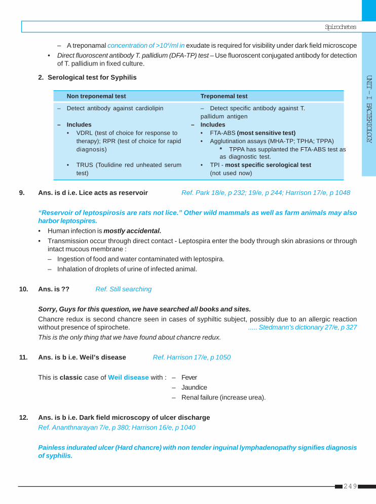
Spirochetes
UNIT - I BACTERIOLOGY
249
– A treponamal concentration of >104/ml in exudate is required for visibility under dark field microscope• Direct fluoroscent antibody T. pallidium (DFA-TP) test – Use fluoroscent conjugated antibody for detection
of T. pallidium in fixed culture.
2. Serological test for Syphilis
Non treponemal test Treponemal test
– Detect antibody against cardiolipin – Detect specific antibody against T.pallidum antigen
– Includes – Includes• VDRL (test of choice for response to • FTA-ABS (most sensitive test)
therapy); RPR (test of choice for rapid • Agglutination assays (MHA-TP; TPHA; TPPA)diagnosis) * TPPA has supplanted the FTA-ABS test as
as diagnostic test.• TRUS (Toulidine red unheated serum • TPI - most specific serological test
test) (not used now)
9. Ans. is d i.e. Lice acts as reservoir Ref. Park 18/e, p 232; 19/e, p 244; Harrison 17/e, p 1048
“Reservoir of leptospirosis are rats not lice.” Other wild mammals as well as farm animals may alsoharbor leptospires.• Human infection is mostly accidental.• Transmission occur through direct contact - Leptospira enter the body through skin abrasions or through
intact mucous membrane :– Ingestion of food and water contaminated with leptospira.– Inhalation of droplets of urine of infected animal.
10. Ans. is ?? Ref. Still searching
Sorry, Guys for this question, we have searched all books and sites.Chancre redux is second chancre seen in cases of syphiltic subject, possibly due to an allergic reactionwithout presence of spirochete. ..... Stedmann’s dictionary 27/e, p 327This is the only thing that we have found about chancre redux.
11. Ans. is b i.e. Weil’s disease Ref. Harrison 17/e, p 1050
This is classic case of Weil disease with : – Fever– Jaundice– Renal failure (increase urea).
12. Ans. is b i.e. Dark field microscopy of ulcer dischargeRef. Ananthnarayan 7/e, p 380; Harrison 16/e, p 1040
Painless indurated ulcer (Hard chancre) with non tender inguinal lymphadenopathy signifies diagnosisof syphilis.
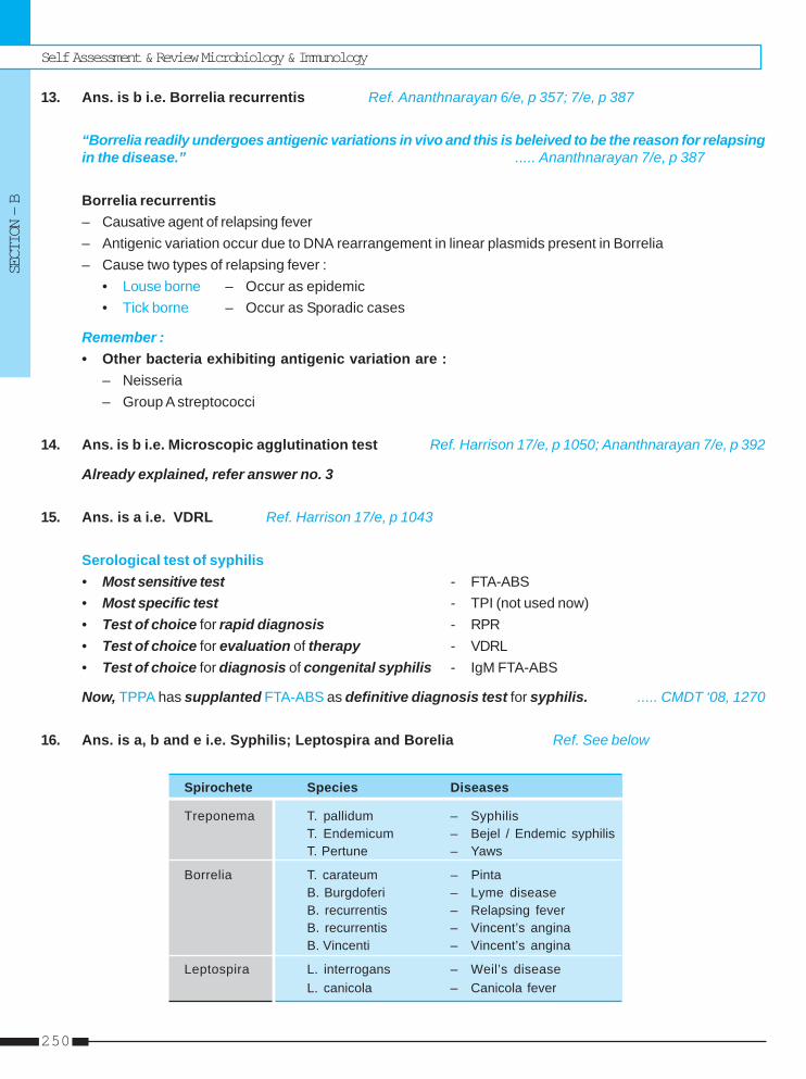
Self Assessment & Review Microbiology & Immunology
SECTION – B
250
13. Ans. is b i.e. Borrelia recurrentis Ref. Ananthnarayan 6/e, p 357; 7/e, p 387
“Borrelia readily undergoes antigenic variations in vivo and this is beleived to be the reason for relapsingin the disease.” ..... Ananthnarayan 7/e, p 387
Borrelia recurrentis– Causative agent of relapsing fever– Antigenic variation occur due to DNA rearrangement in linear plasmids present in Borrelia– Cause two types of relapsing fever :
• Louse borne – Occur as epidemic• Tick borne – Occur as Sporadic cases
Remember :• Other bacteria exhibiting antigenic variation are :
– Neisseria– Group A streptococci
14. Ans. is b i.e. Microscopic agglutination test Ref. Harrison 17/e, p 1050; Ananthnarayan 7/e, p 392
Already explained, refer answer no. 3
15. Ans. is a i.e. VDRL Ref. Harrison 17/e, p 1043
Serological test of syphilis• Most sensitive test - FTA-ABS• Most specific test - TPI (not used now)• Test of choice for rapid diagnosis - RPR• Test of choice for evaluation of therapy - VDRL• Test of choice for diagnosis of congenital syphilis - IgM FTA-ABS
Now, TPPA has supplanted FTA-ABS as definitive diagnosis test for syphilis. ..... CMDT ‘08, 1270
16. Ans. is a, b and e i.e. Syphilis; Leptospira and Borelia Ref. See below
Spirochete Species Diseases
Treponema T. pallidum – SyphilisT. Endemicum – Bejel / Endemic syphilisT. Pertune – Yaws
Borrelia T. carateum – PintaB. Burgdoferi – Lyme diseaseB. recurrentis – Relapsing feverB. recurrentis – Vincent’s anginaB. Vincenti – Vincent’s angina
Leptospira L. interrogans – Weil’s diseaseL. canicola – Canicola fever
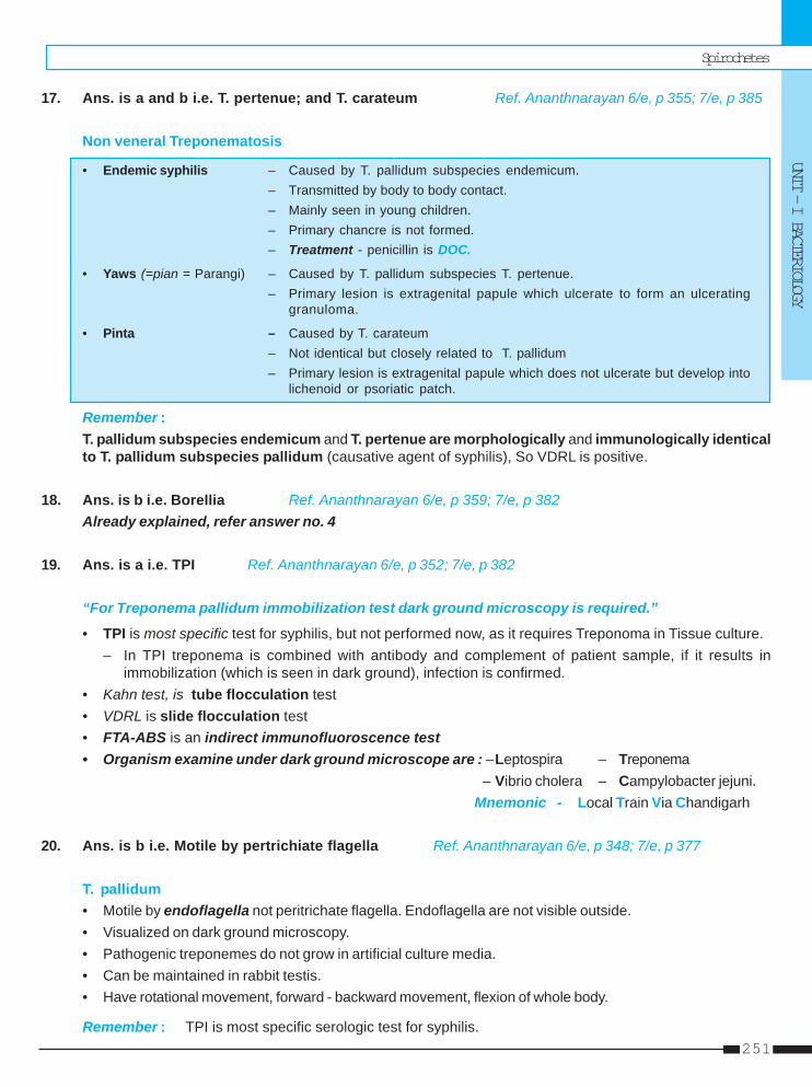
Spirochetes
UNIT - I BACTERIOLOGY
251
17. Ans. is a and b i.e. T. pertenue; and T. carateum Ref. Ananthnarayan 6/e, p 355; 7/e, p 385
Non veneral Treponematosis
• Endemic syphilis – Caused by T. pallidum subspecies endemicum.– Transmitted by body to body contact.– Mainly seen in young children.– Primary chancre is not formed.– Treatment - penicillin is DOC.
• Yaws (=pian = Parangi) – Caused by T. pallidum subspecies T. pertenue.– Primary lesion is extragenital papule which ulcerate to form an ulcerating
granuloma.
• Pinta – Caused by T. carateum– Not identical but closely related to T. pallidum– Primary lesion is extragenital papule which does not ulcerate but develop into
lichenoid or psoriatic patch.
Remember :T. pallidum subspecies endemicum and T. pertenue are morphologically and immunologically identicalto T. pallidum subspecies pallidum (causative agent of syphilis), So VDRL is positive.
18. Ans. is b i.e. Borellia Ref. Ananthnarayan 6/e, p 359; 7/e, p 382Already explained, refer answer no. 4
19. Ans. is a i.e. TPI Ref. Ananthnarayan 6/e, p 352; 7/e, p 382
“For Treponema pallidum immobilization test dark ground microscopy is required.”
• TPI is most specific test for syphilis, but not performed now, as it requires Treponoma in Tissue culture.– In TPI treponema is combined with antibody and complement of patient sample, if it results in
immobilization (which is seen in dark ground), infection is confirmed.• Kahn test, is tube flocculation test• VDRL is slide flocculation test• FTA-ABS is an indirect immunofluoroscence test• Organism examine under dark ground microscope are : –Leptospira – Treponema
– Vibrio cholera – Campylobacter jejuni.Mnemonic - Local Train Via Chandigarh
20. Ans. is b i.e. Motile by pertrichiate flagella Ref. Ananthnarayan 6/e, p 348; 7/e, p 377
T. pallidum• Motile by endoflagella not peritrichate flagella. Endoflagella are not visible outside.• Visualized on dark ground microscopy.• Pathogenic treponemes do not grow in artificial culture media.• Can be maintained in rabbit testis.• Have rotational movement, forward - backward movement, flexion of whole body.
Remember : TPI is most specific serologic test for syphilis.
Self Assessment & Review Microbiology & Immunology
SECTION – B
252
1. a) Painless ... 2. c) VDRL ... 3. b) TPHA is earliest ... 4. b) 4 weeks 5. b) T. pallidum ...6. None 7. d) Fluorescent 8. b) Treponema ... 9. a and d 10. a) Slide ...
1. Characteristic of primary chancre : [Kerla 97;a) Painless punched out ulcer [AIIMS 87, JIP. 87]b) Painless ulcer with over hanging edgesc) Pain less ulcer with irregular raised edgesd) Painfull punched out ulcer
[Ref. Ananthnarayana 7/e, p 379; Harrison 17/e,p 1040; Dasohre, p 105]
2. The following is not true of syphilis : [AIIMS 91]a) TPI is most specificb) VDRL is not specificc) VDRL is negative in secondary syphilisd) IgM test is specific for congenital syphilis
[Ref. Ananthnarayana 7/e, p 383; Harrison 17/e,p 1043]
3. Which is false about syphilis : [AI 92]a) TPI gives definite diagnosisb) TPHA is earliest to be +vec) VDRL is +ve 1 week after appearance of chancred) Serologically not different from yaws
[Ref. Ananthnarayana 7/e, p 382; Jawetz 24/e, p335]
4. In syphilis the semen is infective for :a) 2 weeks [JIPMER 92]b) 4 weeksc) 4 monthsd) 1 year
[Ref. Under consideration]5. Yaws is caused by : [Kerala 94; TN 89]
a) Pertunaeb) T. pallidum perterouec) T. caraetumd) Hemophilus
[Ref. Harrison 17/e, p 1046]
Answer
6. False +ve VDRL test is not seen in : [Kerala 94]a) Herpes infectionb) Leprosyc) Malariad) SLE
[Ref. Harrison 16/e, 982]7. Which of the following investigation is very spe-
cific for spirochaetal infection ? [Kar 03]a) Wasermanns testb) VDRL testc) Treponema pallidiumd) Fluorescent treponemal antibody absorption test
[Ref. Ananthnarayan 7/e, p 382]8. Organisms that has not been cultured success-
fully so far is : [J & K 01]a) Leptospirab) Treponema pallidumc) Bordetellad) Staphylococcus
[Ref. Ananthnarayan 7/e, p 377]9. Necrotizing granulomatous lymphadenopathy is
caused by : [JIPMER 95]a) Syphillisb) Granuloma inguinalec) Sarcoidosisd) Tuberculosis
[Ref. Taylor 3/e, p 77, table 5.2]10. VDRL is a : [TN 95]
a) Slide flocculation testb) Tube flocculation testc) Gel precipitation tesd) Indirect hemaglutination test
[Ref. Ananthnarayana 7/e, p 381, 95]
Chapter Review••••• This section includes questions of VThis section includes questions of VThis section includes questions of VThis section includes questions of VThis section includes questions of Various Other PGMEES from 1990 – 2007.arious Other PGMEES from 1990 – 2007.arious Other PGMEES from 1990 – 2007.arious Other PGMEES from 1990 – 2007.arious Other PGMEES from 1990 – 2007.••••• Questions are arQuestions are arQuestions are arQuestions are arQuestions are ar ranged in decreasing order of yearranged in decreasing order of yearranged in decreasing order of yearranged in decreasing order of yearranged in decreasing order of yearwiz. This is done to makwiz. This is done to makwiz. This is done to makwiz. This is done to makwiz. This is done to make refere refere refere refere refer ralralralralral
system easier and uncomplicated to save the precious time of PGMEE Aspirant.system easier and uncomplicated to save the precious time of PGMEE Aspirant.system easier and uncomplicated to save the precious time of PGMEE Aspirant.system easier and uncomplicated to save the precious time of PGMEE Aspirant.system easier and uncomplicated to save the precious time of PGMEE Aspirant.
Spirochetes
UNIT - I BACTERIOLOGY
253
11. Which of the following disease is not caused bytreponema : [Kar 00]a) Yawsb) Bejelc) Relapsing feverd) Syphilis
[Ref. Harrison 17/e, p 1052]12. Treponema palidum is very difficult to demonstrate
in : [JIPMER 02]a) Chancreb) Maculopapular lesionc) Gummad) Mucosal patch
[Ref. Ananthnarayana 7/e, p 380; Harrison 17/e,p 1042]
13. The most commonly performed test for the se-rodiagnosis of syphilis is : [Kar 2002]a) TPHAb) Wasermannc) TPId) VDRL
[Ref. Ananthnarayan 7/e, p 381]14. Syphilis was first identified by : [TN 02]
a) Fraenkelb) Nicolaicuc) Schaudinn and Hoffmand) Ogston
[Ref. Ananthnarayana 7/e, p 376]15. Syphilis is diagnosed all except : [UP 02]
a) TPIb) FTA-ABSc) Weil felixd) VDRL
[Ref. Ananthnarayana 7/e, p 352]16. Consider the following serological test :
a) TPI [UPSC 02]b) TPHAc) FTA-ABSd) VDRL
17. Which of these are specific for diagnostic syphi-lis? [Bihar 03]a) 1 onlyb) 2 and 4c) 3 and 4d) 1, 2, and 3
[Ref. Ananthnarayana 7/e, p 352]18. Most specific test for syphillis : [Kolkata 03]
a) FTA-ABSb) TPIc) VDRLd) ELISA
[Ref. Ananthnarayan 7/e, p 382]19. Test used for diagnosis of congenital syphilis :
a) IgMFTA [MP 05]b) VDRLc) TPId) Kahn flocculation test 23rd
[Ref. Ananthnarayana 7/e, p 384; Harrison 16/e,p 984]
20. Leptospira icterohemorrhagica infection is trans-mitted by : [TN 95]a) Batsb) Ratsc) Birdsd) Dogs
[Ref. Ananthnarayan 7/e, p 393]21. Antigenic variations seen in : [MP 06]
a) Borrelia recurrentisb) Borrelia burgdorferic) Borrelia vincentid) None
[Ref. Ananthnarayan 7/e, p 387]22. The causative agent of Lyme’s disease :
a) Borrelia burgdorferi [SGPGI 07; MP 05]b) Borrelia recurrentisc) Leptospira icterohemorrhagiaed) Clostridium difficile
[Ref. Ananthnarayana 7/e, p 389]
11. c) Relapsing ... 12. c) Gumma 13. d) VDRL 14. c) Schaudinn ... 15. c) Weil felix16. None 17. d) 1, 2, and 3 18. None 19. a) IgMFTA 20. b) Rats21. c) Borrelia ... 22. a) Borrelia ...
Answer
• Smallest free living bacteria, bounded by sterols containing soft trilaminar membrane.• Lack cell wall hence show polymorphism and resistance to cell wall active antimicrobial agent such as penicillin,
cephalosporin and lysozyme.• Also called as PPLO = Stable L forms.• Even cell precursors like muramic acid or diaminopimelic acid are absent.• Do not possess spores flagella or fimbria. Some species exhibit motility.
Morphology• Gram negative but better stained by Giemsa.• Can be grown on cell free media. Colony is typically biphasic with “Fried egg appearance” best studied after
staining by Dienes method.• Some species shows bulbous enlargement with a differentiated tip structure which aids in attachment of organism
to host cell carrying neuraminic acid receptor.
Mechanism of Pathogenicity• Adherence to host cell.• H2O2 production (as in M.pneumonia).• Ammonia production (as in M.hominis).• Urease activity to produce ammonia (as in U.urealyticum).• IgM autoantibodies that agglutinate human group O erythrocyte at 40C. This cold agglutinin produce anemia.
Classification• M. pneumoniae – Upper and lower respiratory tract infection.• M. genitalium and Ureaplasma Urealyticum – Urethritis and other genital condition.• M. hominis and U.urealyticum – Part of flora of bacterial vaginosis.
MYCOPLASMA PNEUMONIA = PRIMARY ATYPICAL PNEUMONIA = WALKING PNEUMONIA• Cause by M.pneumonia (= Eaton agent) in which pneumonia is classic presentation but non-pneumonic infection
is more common with prolonged incubation period.• Interstitial type of pneumonia characterized by paucity of respiratory signs on auscultation with striking radiological
abnormalty.• Extrapulmonary manifestations (eg. Erythema multiforme, anemia, coagulopathies) is due to autoantibodies
against brain, heart and muscle.
MycoplasmaMycoplasmaMycoplasmaMycoplasmaMycoplasma1717

Mycoplasma
UNIT - I BACTERIOLOGY
255
Diagnosisi. Isolation - Throat swab/respiratory secretions are inoculated into medium containing glucose and phenol.ii. Serological diagnosis Specific test - Immunofluorescence hemagglutination inhibition and metabolic inhibition
are most sensitive test.Non specific serological test are streptococcus MG (group F) and cold agglutination test.
GENITAL MYCOPLASMAS• Non-gonococcal Urethritis (NGU) - U.urealyticum and M.genitalium cause most of non-chlamydial cases of
NGU.• U.urealyticum called as T. strain/T. form of mycoplasma are able to hydrolyse urea. Hence Urea and cholesterol
is essential growth factor.
Treatment : Tetracycline [Doxycycline] is DOC for treatment of mycoplasma infection.
Cell wall defective bacteriaL phase variants (L forms)• Wall defective microbial forms that can replicate serially as non rigid cells and produce colonies on solid media.• Protoplasts are such forms usually derived from Gram +ve organisms. They are osmotically fragile.• Spheroplasts are cell wall defective form usually derived from Gram –ve bacteria. They retain outer membrane.• These L forms results from spontaneous mutation or by the effect of chemicals.• Reversion of L-form to the parental bacterial form is enhanced by growth in the presence of 15-30% gelatin or 2-5%
agar.
Self Assessment & Review Microbiology & Immunology
SECTION – B
256
1. Atypical pneumonia can be caused by the follow-ing microbial agents except : [AI 05]a) Mycoplasmab) Legionella pneumophiliac) Human corona virusd) Kleibsella pneumoniae
2. All are features of Ureplasma urealyticum except:a) Non gongococcal urethritis [AI 01]b) Salpingitisc) Epididymitisd) Bacterial vaginosis
3. The following statements are true with referencesto Mycoplasma except : [AIIMS 05]a) They are the smallest prokaryotic organisms that
can grow in cell free culture mediab) They are obligate intracellular organismsc) They lack a cell walld) They are resistant to Beta-lactam drugs
4. In reference to mycoplasmas, the following aretrue except : [AIIMS 05]a) They are inhibited by penicillinsb) They can reproduce in cell free mediac) They have an affinity for mammlian cell mem-
branesd) They can pass through fiters of 450 nm pore
size
5. The following is true about Mycoplasmas except:a) Multiply by binary fission [AIIMS 02, 96]b) Are sensitive to beta-lactam gourp of durgsc) Can grow in cell free mediad) Require sterols for their growth
6. Mycoplasma pneumonia is characterised by allexcept : [AIIMS 98]a) Diagnosed by serum cold antibodyb) Treatment is erythromycinc) Can not be cultured from sputumd) Raised ESR
7. True about mycoplasma is : [AIIMS 95]a) Causes lung infectionb) Penicillin is drug of choicec) Thick cell walld) Thallium acetate inhibits the growth
8. Diene’s method is used for : [PGI 99, 95]a) Mycoplasmab) Chlamydiaec) Plagued) Diptheria
9. Cell wall deficient organisms are : [PGI 99]a) Chlamydiab) Mycoplasmac) Streptococcusd) Anaerobes
ANSWERS WITH EXPLANATIONQUESTIONS
Answer 1. d) Kleibsella ... 2. b) Salpingitis 3. b) They are ... 4. b) They ... 5. a) They ...6. b) Are ... 7. c) Can ... 8. a) Causes ... 9. a) Mycoplasma
Mycoplasma
UNIT - I BACTERIOLOGY
257
1. Ans. is d i.e. Kleibsella pneumoniae Ref. Robbin’s 7/e, p 751
Atypical pneumonia is characterized by patchy inflammatory changes in the lung, largely confined toalveolar septa and pulmonary interstitium.
Causes of Atypical pneumonia :• Mycoplasma (MC) • Coxiella burnetti (Q fever)• Legionella pneumonia • Pneumocystii carnii• Francisella tularensis • H. capsulatum•. Chlamydia psittacosis, chlamydia pneumoniae • Coccidiodis immitis.• Viruses (influenzae A and B, RSV, Adeno, rhino, rubeola, varicella etc)
Kleibsella is associated with community acquired pneumonia classically in alcoholics (also in diabetics andchronic lung disease). It usually affects upper lobes producing expansion of lobes (bulging fissure) and Redcurrent jelly sputum.
Remember : Causes of community acquired pneumoniae in decreasing frequency Strep. pneumoniae >H. influenza > Chalmydia > Legionella.
2. Ans. is b i.e Salpingitis Ref. Harrison 17/e, p 1069; Ananthnarayan 7/e, p 398; CGDT 9/e, p 654
Ureoplasma urealyticum are T form mycoplasmas which are urease postive.
It causes :• Nongonococcal urethritis MC cause is Chalmydia trachomatis also caused by U. urealyticum and M.
genitalium.• Epididymitis (no role of M. homins). • Infertility in both men and women• Chorioamnionitis • Late abortion• Post partum fever • Low birth weight infant• Proctitis • Balanoposthitis• Reiter’s syndrome • Cervicitis and vaginitis• Acute salpingitis ... Not given in Harrison and CGDT• Pneumonia and chronic lung disease in VLBW infants• PID and bacterial vaginosis : by M hominsis and U.urealyticum• Bacterial vaginosis (Altered vaginal normal microbial flora) is associated with Gardenella and Hemophilus
vaginitis, in which clue cells are present and Amide test is positive.
If ‘none’ is given as option than it is more appropriate than ‘Salpingitis’ as salpingitis is given in‘Ananthnarayan’.
3. Ans. is b i.e. They are obligate intracellular organisms Ref. See below
• Mycoplasma is not obligate intracellular bacteria.
EXPLANATIONS AND REFERENCES WITH ILLUSTRATIVE ANSWERS
Self Assessment & Review Microbiology & Immunology
SECTION – B
258
• Obligate intracellular bacteria : – M.leprae– Chlamydia– Rickettsiaecae and coxiella burnetti– Pathogenic treponemes.
• Obligate intracellular bacteria can not grow in cell free media.
4. Ans. is a i.e They are inhibited by penicillins Ref. Ananthnarayan 7/e, p 395 - 397
• Mycoplasmas are devoid of cell walls (but bound by soft trilaminar unit membrane of sterols) and so theyare resistant to B. lactams (penicillin, cephalosporins, vancomycin, Bacitracin) and lysozmes thatact on cell wall.
• Due to lack of cell wall they are highly pleomorphic and pass through bacterial filters of 450 nm since sizevaries from 50-300 nm in diameter.
• Parasitic mycoplasma requires cholesterol or other sterols as an essential growth factor.• They have affinity for mammalian cell membrane. .....Jawertz 24/e, p 344• It typically colonizes mucosal surfaces of respiratory, GIT and genitourinary tracts.• Mycoplasma occur as granules and filaments (shows true branching).• They multiply by asynchronous binary fission producing budding forms and chains of beads.• Some species get attached to suitable host cells carrying neuraminic acid receptors by bulbous enlargement.• Mycoplasmas (and other organism) grows in cell free media.• Media of mycoplasma are enriched with 20% horse or human serum and yeast extract
– Penicillin and thallium are selective agents.– Colonies is typically biphasic with a fried egg appearance and are best studied after staining by
Dienes method.– Growth of mycoplasma is inhibited by specific antibody.
5. Ans. is b i.e. Are sensitive to beta-lactam gourp of durgs Ref. Ananthnarayan 7/e, p 395 - 397
Remember : Doxycycline is drug of choice for mycoplasma.
6. Ans. is c i.e. Can not be cultured from sputum Ref. Ananthnarayan 7/e, p 398; Harrison 17/e, p 1068
• Mycoplasma pneumoniae is MC cause of Atypical pneumonia which characterized by reticulonodular orinterstitial infiltration of lower lobes on X-ray with paucity of signs on auscultation.
Diagnosis• Specimen :
– Throat swabs, sputum or respiratory secretions.
• Microscopy :– It cannot be detected on Gram’s stain as it lack cell wall.– Gram’s stain of sputum shows leucocytes without predominance of any bacteria morphologic type.
• Culture :– It can be grown on artificial media but process is difficult as it requires special media and take more than
2 weeks so culture do not provide timely information.
Mycoplasma
UNIT - I BACTERIOLOGY
259
• Serology :a. Specific : Antibodies are detected by enzyme linked immunoassys, indirect immunofluorescence,
or complement fixation test.b. Nonspecific : Cold agglutinin aids in diagnosis since develops within 7-10 days of infection and can
be easily detected.– They are IgM autoantibodies which agglutinate human erythorcytes at 40C.– Cold agglutinin titre > 1:32 supports diagnosis of M. pneumoiae.– It can also performed at the bedside.
• Antigen Detection test :Include antigen capture, indirect enzyme immunoassays, DNA probing and Multiplex Nucleic acid amplificationtest.
Treatment Ambulatory patients with Hospitalized patients withcommunity acquired pneumonia community acquired pneumonia
• Oral Doxycycline • IV ceftriaxone or IV cefotaxime• Oral erythromycin• Oral clarithromycin, azithromycin;
levo, Gati, Moxifloxacin
7. Ans. is a i.e. Causes lung infection Ref. Ananthnarayan 7/e, p 395
Remember : Media for cultivating mycoplasma are enriched with 20% horse or human serum and yeastextract. Penicillin and thallium acetate are added as selective agent.
8. Ans. is a i.e. Mycoplasma Ref. Ananthnarayan 7/e, p 396
• Colonies of mycoplasma are typically biphasic, with a fried egg appearance.• Colonies are best studied after staining by Dienes method :
– Dienes method : A block of agar containing the colony is cut and placed on slide.– It is covered with a coverslip on which has been dried an alcoholic solution of methylene blue and azur.
• Colonies can’t be picked by platinum loops.• Most mycoplasma colonies are hemolytic.
9. Ans. is b i.e. Mycoplasma Ref. Ananthnaryan 7/e, p 12 - 13, 395 - 399
Cell wall deficient forms :a. L. forms (mycoplasma is stable L form).b. Protoplasts (by action of lysozyme on Gram positive bacteria).c. Spheroplasts (by action of lysozyme on Gram negative bacteria).
– Some cell wall material is retained in spheroplast as compared to protoplast.– Role of cell wall deficient forms of bacteria is persistence of chronic infections such as pyeloephritis
and recurrence of infection.– They do not initiate the infection.
Self Assessment & Review Microbiology & Immunology
SECTION – B
260
1. c) May be comm ... 2. b) L. Form is comm ...
1. True about Mycoplasma : [UP 97]a) CO2 is important for growthb) Does not cause hemolysisc) May be commensal in throatd) Acid fast bacillus
[Ref. Ananthnarayan 7/e, p 600]
Answer
Chapter Review••••• This section includes questions of VThis section includes questions of VThis section includes questions of VThis section includes questions of VThis section includes questions of Various Other PGMEES from 1990 – 2007.arious Other PGMEES from 1990 – 2007.arious Other PGMEES from 1990 – 2007.arious Other PGMEES from 1990 – 2007.arious Other PGMEES from 1990 – 2007.••••• Questions are arQuestions are arQuestions are arQuestions are arQuestions are ar ranged in decreasing order of yearranged in decreasing order of yearranged in decreasing order of yearranged in decreasing order of yearranged in decreasing order of yearwiz. This is done to makwiz. This is done to makwiz. This is done to makwiz. This is done to makwiz. This is done to make refere refere refere refere refer ralralralralral
system easier and uncomplicated to save the precious time of PGMEE Aspirant.system easier and uncomplicated to save the precious time of PGMEE Aspirant.system easier and uncomplicated to save the precious time of PGMEE Aspirant.system easier and uncomplicated to save the precious time of PGMEE Aspirant.system easier and uncomplicated to save the precious time of PGMEE Aspirant.
2. True about mcyoplasmas is : [MP 98]a) Not sensitive to antibioticsb) L. Form is commonestc) Culture in sarbaroudz mediad) Most common cause of nongonococcal urethri-
tis[Ref. Ananthnarayan 7/e, p 399]
Self Assessment & Review Microbiology & Immunologyxii
UNIT – II VIROLOGY
1. DNA Virus 263 – 284Herpes virus, Adeno & Pox, Parvo & Papovo
2. RNA Virus 285 – 319Picorna virus, Myxo virus, Rota & other viral gastroenteritis, Arbo, Rhabdoviruses
3. Slow Virus Diseases 320 – 3254. Hepatitis Viruses 326 – 3435. HIV & Other Retroviruses 344 – 366

HERPES VIRUSES
• Enveloped virus with linear double stranded genome.• Characterised by their ability to produce latent infection.• Replicate in host cell nucleus forming cowdry type A intranuclear (Lipshutz) inclusion bodies.
Classification of human herpes viruses
Species
Official name Common name Subfamily Cytopathology Site of latent infection
Human herpesvirus Herpes simplex virus alpha cytolytic neurons / gasserian ortype 1 type 1 trigeminal ganglia
Human herpesvirus Herpes simplex virus alpha cytolytic neurons / sacral gangliatype 2 type 2
Human herpesvirus Varicella zoster virus alpha cytolytic neurons / T3 - L3 (MC)type 3
Human herpesvirus Epstein–Barr virus gamma lymphoproliferative lymphoid tissue (B cells)type 4
Human herpesvirus Cytomegalovirus beta cytomegalic secretory glands Kidneys,type 5 (salivary glands and bowel)
Human herpesvirus Human B cell beta lymphoproliferative lymphoid tissuestype 6 lymphotropic virus
Human herpesvirus R K virus beta lymphoproliferative lymphoid tissuestype 7
Human herpesvirus gammatype 8
• HH V – 6 cause exanthem subitum / roseola infantum or sixth disease.• Different Herpes virus species don’t show any antigenic cross reaction except Herpes simplex type 1 and 2.• HHV-8 is associated with AIDS / Non AIDS Kaposi sarcoma.
DNA VirDNA VirDNA VirDNA VirDNA Virususususus
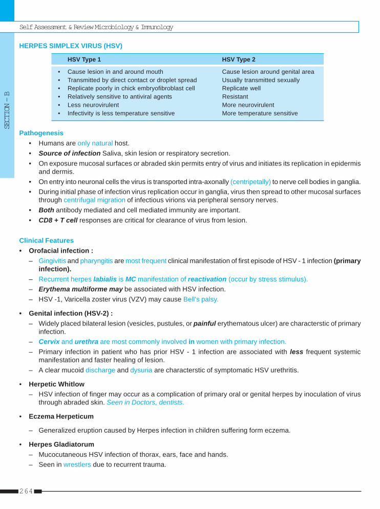
Self Assessment & Review Microbiology & Immunology
SECTION – B
264
HERPES SIMPLEX VIRUS (HSV)
HSV Type 1 HSV Type 2
• Cause lesion in and around mouth Cause lesion around genital area• Transmitted by direct contact or droplet spread Usually transmitted sexually• Replicate poorly in chick embryofibroblast cell Replicate well• Relatively sensitive to antiviral agents Resistant• Less neurovirulent More neurovirulent• Infectivity is less temperature sensitive More temperature sensitive
Pathogenesis• Humans are only natural host.• Source of infection Saliva, skin lesion or respiratory secretion.• On exposure mucosal surfaces or abraded skin permits entry of virus and initiates its replication in epidermis
and dermis.• On entry into neuronal cells the virus is transported intra-axonally (centripetally) to nerve cell bodies in ganglia.• During initial phase of infection virus replication occur in ganglia, virus then spread to other mucosal surfaces
through centrifugal migration of infectious virions via peripheral sensory nerves.• Both antibody mediated and cell mediated immunity are important.• CD8 + T cell responses are critical for clearance of virus from lesion.
Clinical Features• Orofacial infection :
– Gingivitis and pharyngitis are most frequent clinical manifestation of first episode of HSV - 1 infection (primaryinfection).
– Recurrent herpes labialis is MC manifestation of reactivation (occur by stress stimulus).– Erythema multiforme may be associated with HSV infection.– HSV -1, Varicella zoster virus (VZV) may cause Bell's palsy.
• Genital infection (HSV-2) :– Widely placed bilateral lesion (vesicles, pustules, or painful erythematous ulcer) are characterstic of primary
infection.– Cervix and urethra are most commonly involved in women with primary infection.– Primary infection in patient who has prior HSV - 1 infection are associated with less frequent systemic
manifestation and faster healing of lesion.– A clear mucoid discharge and dysuria are characterstic of symptomatic HSV urethritis.
• Herpetic Whitlow– HSV infection of finger may occur as a complication of primary oral or genital herpes by inoculation of virus
through abraded skin. Seen in Doctors, dentists.
• Eczema Herpeticum
– Generalized eruption caused by Herpes infection in children suffering form eczema.
• Herpes Gladiatorum– Mucocutaneous HSV infection of thorax, ears, face and hands.– Seen in wrestlers due to recurrent trauma.
DNA Virus
UNIT – II VIROLOGY
265
• Central and Peripheral nervous system manifestationA. Encephalitis – HSV -1 is MC cause of sporadic encephalitis.
– Present as acute onset of fever, focal neurologic signs especially of temporal lobe.– Most sensitive non invasive method for early diagnosis of HSV encephalitis is demonstration of HSV
DNA in CSF by PCR (Investigation of choice).• Demonstration in brain tissue by biopsy is over all most sensitive but invasive method.
B. Meningitis• HSV is MC cause of recurrent Lymphocytic meningitis (Mollaret's meningitis).• Diagnosed by demonstration of HSV DNA or HSV antibodies in CSF.
• Visceral infection– Oesophagitis MC site is distal oesophagus.– Endoscopically obtained secretion for cytology and culture provide most useful material for diagnosis.– Pneumonia in Immuno compromised.– Hepatitis – may lead to disseminated intravascular coagulation.
• Neonatal HSV infection– Infection is usually acquired perinatally at the time of delivery.– Of all age group, neonates have highest frequency of visceral or CNS infection.
Diagnosis• Both clinical and Laboratory.• Scrapings from base of characteristic lesion is taken and stained with Wright's, Giemsa (Tzanck Preparations) to
detect giant cell or intranuclear inclusions. Sensitivity is low and this can’t differentiate between VZV and HSV.• HSV infection is best confirmed (specific test) by isolation of virus in tissue culture.• PCR for detection of HSV DNA is most sensitive. ..... Harrison 17/e, p 1100
TreatmentAcyclovir is most frequently used drug.
VARICELLA - ZOSTERCausative agents of : • Varicella = Chicken pox (Primary infection)
• Herpes zoster = Shingles (Reactivation of latent infection).
Pathogenesis• Humans are only known reservoir of VZV.• Primary infection (Chicken pox) – Transmitted by respiratory route.• Recurrent infection – (Herpes zoster)- During primary infection virus infect dorsal root ganglia, where it remains
latent. When immunity wanes, virus reactivates and travel along sensory nerve to produce zoster lesion onmucosa, or skin supplied by it.
Clinical manifestationI. Chicken pox – Highly contagious disease (secondary attack rate 90%) affecting 5-9 year children most commonly.
• I.P – 10 to 21 days• Patient is infectious 48 hours prior to onset of vesicular rash to until all vesicles are crusted.• Skin lesions – The hall mark of infection includes maculopapules, vesicles and scabs in various stage of
evolution.• Immunocompromised have severe (often hemorrhagic) and long lasting lesion with higher rate of visceral
complication and fatality.
Self Assessment & Review Microbiology & Immunology
SECTION – B
266
• The MC site of extracutaneous involvement in children is CNS.• Visceral pneumonia is the most serious complication occuring mostly in adults.• Perinatal varicella is associated with high mortality when maternal disease develop with in 5 days before
delivery or within 48 hrs thereafter.
II. Herpes zoster (Shingles)• Reactivation of latent infection.• Age group – 60 and beyond.• Characterized by unilateral lesion within a dermatome associated with severe pain. The dermatome from T3 to
L3 is most frequently affected.• Patient with herpes zoster can transmit infection to seronegative individual which will develop chickenpox.• Zoster Opthalmicus – Due to reactivation in ophthalmic branch of trigeminal (gasserian) ganglia.• Ramsay Hunt syndrome – Due to reactivation in geniculate ganglion of facial nerve.• Most debiliating complication of Shingles is pain associated with acute neuritis and post herpetic neuralgia.• Patient with Hodgkin's disease and NHL are at greatest risk for progressive Herpes zoster.
Lab diagnosis• Isolation of VZV from vesicles in tissue culture cell lines or detection of DNA by PCR.• Serology : Most frequently used are :
– FAMA (fluoroscent antibody to membrane antigen) test– ELISA– Immuno adherant hemagglutination.
FAMA test and ELISA are most sensitive.
Treatment• No antiviral for immunocompetent child.• Aspirin should be avoided during episode of chicken pox as it increase risk of development of Reye's syndrome.• Acylovir therapy is recomonded for adolescent and adults with chicken pox of < 24 hours duration.• Herpes zoster – Famciclovir and valacyclovir are more effective than acyclovir.• Post herpetic neuralgia and acute neuritis : – Analgesics
– Gabapentin– Amitryptyline– Lidocaine patch– Glucocorticoid (with concomitent antiviral therapy).
• In immunocompromised patient IV acyclovir should be given.
CMV = SALIVARY GLAND VIRUS• Characterised by enlargement of infected cell and prominent intranuclear inclusion (Owl's eye appearance).• Largest virus of herpes family.• MC organism causing intrauterine infection.• MC pathogen complicating organ transplantation.Pathogenesis• Transmitted by sexual route, transplacental, blood transfusion, organ transplant.• Once infected, individual carry CMV for whole life.• Infection usually remains latent, reactivation may occur when cell mediated immunity is compromised.
DNA Virus
UNIT – II VIROLOGY
267
Clinical featureI. Congenital infection
• Petechiae, Hepatosplenomegaly, Jaundice are MC manifestation.• Intracerebral calcificatiion (usually periventricular) and chorioretinitis, deafness are other important findings.
II. Perinatal infection• Infection acquire through birth canal at the time of delivery or through breast milk.• Mostly asymptomatic.• Interstitial pneumonia in preterm infant.
III. Beyond neonatal period immunocompetant host• Heterophile antibody (-)ve infectious mononucleosis is MC manifestation.
IV. Immunocompromised hosta. Organ transplant patient : – Period of maximal risk of infection – Between 1 and 4 month after transplantation.
– Retinitis is late complication.– Transplanted organ is particulary vulnerable as a target for CMV infection eg.
CMV hepatitis in liver transplant holder.b. AIDS patient : – Cause retinitis (cottage and cheese appearance / pizza pie retinopathy) or disseminated
disease particulary when CD4 + < 50 - 100 / μl.
Diagnosis• Most sensitive method to detect CMV in blood is PCR.• Congenital infection is diagnosed by culture (best specimen saliva and urine); PCR.
Treatment• Ganciclovir or Valganciclovir is DOC.
EPSTEIN - BARR VIRUS• Causative agent of Heterophile (+)ve infectious mononucleosis (Kissing disease) = Glandular fever.• Associated with : – Nasopharyngeal Ca
– Burkitt's lymphoma– Hodgkin's disease (mixed cellularity type)– B cell lymphoma in patients with immunoficiency– CNS lymphoma in AIDS patient– Increase risk of Thymoma, tonsillar and gastric carcinoma.
Pathogenesis• Source of infection is usually salivary secretions so kissing is predominate mode of transmission.• The virus infect epithelium of oropharynx and the salivary gland; tonsillar crypts can also be infected directly. Virus
then spreads through the blood stream.• EBV receptor CD21 present on B cell is also receptor for CD3 component of complement; So EBV infection
immortalise B cell.• Memory B cell are reservoir of EBV in body.• Cellular immunity is more important than humoral immunity in controlling EBV infection.• If T cell immunity decreased, infected B cells begins to proliferate hence producing lymphoma.
Self Assessment & Review Microbiology & Immunology
SECTION – B
268
Clinical features• Most EBV infection in infant and young children are asymptomatic.• In adolescent, most infection present as infectious mononucleosis.
– IP : 4-8 weeks– MC symptom sore throat– MC sign - Lymphadenopathy (mostly of posterior cervical nodes)– Erythema nodosum, Erythema multiforme may also occur.
Diagnosis• Increase TLC; Lymphocytosis with > 10% atypical lymphocytes.
– Atypical lymphocyte are mainly CD8 + cells which have undergone blast transformation.• Neutropenia• Thrombocytopenia• Serological testing
– Heterophile test (Paul Bunnel test) is used for diagnosis of IM in children and adults.– EBV specific antibody test : IgM antibody to VCA is most useful for diagnosis of IM.
Complication• Most cases are self limited.• Most death which occur very rarely are due to CNS complications.• Coombs (+)ve autoimmune hemolytic anemia.• Acute EBV may be associated with Gullen Baire syndrome, CNpalsy (MC - facial nerve).
Other disease associated with EBV• Fatal lymphoproliferative disorder of Duncan’s disease.• Oral hairy leukoplakia is an early manifestation of EBV infection in AIDS patient.• Chronic fatigue syndrome.
POX VIRUSLargest pathogenic virus of vertebratesImportant pox virus are : – Variola (causative agent of small pox)
– Vaccinia (Artificial virus which was used as small pox vaccine)– Molluscum contagiousum.
Variola• Brick shaped virion• Elementary bodies are called paschen bodies.Vaccinia• It is an artificial virus whose genome can accomodate about 25000 foreign base pairs.• But it is not suitable as a vector for human use due to its pathogenic effects.• Properties are similar to variola.
DNA Virus
UNIT – II VIROLOGY
269
Small pox• On 8th May 1980 WHO anounced Global eradication of small pox.• In India last case was found in 1975.
– Disease had been eradicated, so clinical features etc are not going to be asked.
Cultivation of Pox virus• Both Variola and Vaccinia grow on CAM producing pocks.
– Variola pocks are small, shiny, white convex, non necrotic, non hemmorhagic lesions with ceiling temperature(highest temperature above which pocks are not produced) of 380 C.
– Vaccinia pocks are larger, irregular, flat, greyish, hemorrhagic and Necrotic with ceiling temperature of 410 C.• On tissue culture
– Cytopathic effect are produced by Vaccinia in 24 - 48 hours and more slowly by Variola.– Inclusion bodies called Guarnieri bodies can be seen.
Molluscum Contagiosum• Usually seen in children and young adults.• Characterised by pink or pearly white nodules on skin which show large inclusion bodies called Molluscum
bodies.• Virus can not be grown in eggs, tissues culture or animals.
ADENOVIRUS• Space vehicle (Hexagonal shape) shaped, non enveloped virus containing ds DNA.• They have capacity to carry DNA upto 7 kb so, are potential vectors of gene therapy.
Classification• There are about 50 serotype which are pathogenic to humans. ..... Ananthnarayan, p 486• Human adenovirus have been divided into 6 subgenera on the basis of DNA homology.
Clinical manifestation – MC is upper respiratory tract infection
Syndrome Principal serotype
Respiratory disease in children 1, 2, 5, 6Sore throat, febrile cold, pneumonia 3, 4, 7, 14, 21ARD in military recruits 4, 7, 21Follicular (swimming pool) conjunctivis 3, 7Epidemic keratoconjunctivitis (shipyard eye) 8, 19, 37Diarrhea (by enteric type adeno) 40, 41Hemorrhagic cystitis 11, 21Generalized exanthem, Mesentericadentitis and intussusception
Lab diagnosis• Isolation of virus from throat, eye, urine or feces.• It grow only in tissue cultures of human origin eg. human embryonic kidney, HeLA or HEP-3.• All mammalian adenovirus share a common complement fixing antigen which is detected by immunofluorescence
or ELISA.
Self Assessment & Review Microbiology & Immunology
SECTION – B
270
Adeno Associated virus – (Dependovirus)• These are virus which can multiply only in cells infected with adenovirus as they lack enough DNA.• It is classified under family parvoviridae.
PARVO VIRUS• Non enveloped SS DNA virus• Most parvo virus are pathogenic to Animals. Human pathogen is B-19, the causative agent of fifth disease.
Pathogenesis and Clinical Manifestation• Most of the severe manifestation of B19 viremia are due to its ability to lyse erythroid precursor.• Erythema Infectosum (fifth disease)
– MC manifestation of B19 infection.– Child present with facial rash (slapped cheek appearance) which is preceded by low grade fever.
• Arthropathy – In adults B-19 infection most commonly present as acute arthralgia and arthritis which is symmetricaland involves wrist most often.
• Transient aplastic crisis– B-19 infection is the MC cause of transient aplastic crisis in patient with chronic hemolytic disease.– Unlike patient with erythema infectosum or arthropathy, these patient can readily transmit B-19 infection to
other people.• Immunodeficient patient
– MC manifestation is chronic anemia.• Fetal and congenital infection
– Maternal B-19 infection usually do not adversely affect fetus but can rarely cause non immuno hydrops fetalisif infection occur in first 20 weeks of pregnancy.
Diagnosis• Most commonly relies on B-19 specific IgM and IgG antibodies.
PAPOVA VIRUS• Nonenveloped, Icosahedral DNA, tumor viruses• Family contains 2 genera : 1. Polyoma virus – which contains SV 40, polyoma viruses.
2. Papillomavirus – which contain human and animal papilloma virus.
HUMAN PAPILLOMA VIRUS (HPV)• HPV selectively infect the epithelium of skin and mucous membrane and may immortalize the keratinocyte
leading either asymptomatic infection or warts or neoplasia.• Genome consist of : – Early (E) region
– Late (L) region– Upstream regulatory region (URR).
• Product of E genes (E6; E7) are related to immortalization or malignant transformation of keratinocytes by interferingwith P53, Rb gene respectively.
• HPV infect only human skin and grows only in organ cultures of human skin.
DNA Virus
UNIT – II VIROLOGY
271
Clinical features• Replication of HPV begins with the infection of basal cells.• Koilocytes appear in granular cells.• It causes :
– Common Warts (verruca vulgaris) – Type 1, 2, 3, 4– Condyloma accuminata (anogenital warts) – Type 6, 11– Cervical intraepithelial neoplasia – Type 6, 11 (low risk)– Cervical cancer – 16,18,31, 33, 45 (high risk type)– Plantar warts (verruca plantaris)– Respiratory papillomatosis– Flat warts (verruca plana) – MC among children– Also associated with Sq cell carcinoma and dysplasia of penis, anus, vagina and vulva; epidermodysplasia
verruciformis (type 5, 8).
Diagnosis• The most sensitive and specific method of diagnosis is PCR or hybrid capture assay to detect HPV nucleic
acids and to identify specific virus type.
Treatment• Cryosurgery is initial treatment of choice for condyloma accuminatum.• Topically – Podophyllum, podofilox
– Interferon (IFN)– IFN inducer imiquimod.
Self Assessment & Review Microbiology & Immunology
SECTION – B
272
1. The following diseases are associated withEpstein - Barr virus infection, except : [AI 06]a) Infectious mononucleosisb) Epidermodysplasia veruciformisc) Nasopharyngeal carcinomad) Oral Hairy leukoplakia
2. Epstein Barr (EB) virus has been implicated inthe following malignancies except : [AI 04]a) Hodgkin’s diseaseb) Non-hodgkin’s lymphomac) Nasopharyngeal carcinomad) Multiple myeloma
3. Epstien Barr virus causes all the following except:a) Infectious mononucleosis [AI 02; AIIMS 99]b) Measlesc) Nasopharyngeal carcinomad) Non Hodgkins lymphoma
4. Infectivity of chicken pox last for :a) Till the last scab falls off [AI 02, AIIMS 00]b) 6 days after onset of rashc) 3 days after onset of rashd) Till the fever subsides
5. Virus causing hemorrhagic cystitis, diarrhea andconjunctivitis is : [AI 01]a) RSVb) Rhinovirusc) Adenovirusd) Rotavirus
6. A patient with sore throat has a positive PaulBunnel test. The causative organism is : [AI 00]a) EBVb) Herpes virusc) Adeno virusd) Cytomegalovirus
7. Herpes - zoster is caused by : [AI 99]a) Herpes-simplex type Ib) Herpes-simplex type IIc) Epstein-barr virusd) Varicella
8. Viral enterotoxin is detected as a possiblemechanism of pathogenesis in : [AI 98]a) Adeno virusb) Rota virusc) Calcivirusd) Astrovirus
9. All of the following are true about Herpes groupvirus excpt : [AI 98]
a) Ether-sensitiveb) May cause malignancyc) HSV II involves below diaphragmd) Burkitt’s lymphoma involves T-cells
10. Varicella are classified under : [AI 96]a) Enterovirusb) Retrovirusc) Poxvirusd) Herpes virus
11. EB virus belongs to which group : [AI 95]a) Retrovirusb) Herpes virusc) RNA virusd) Pox virus
12. All of the following are true about the papovavirusexcept : [AI 95]a) They are non-enveloped icosahedral virusesb) Produce papillomac) RNA virusd) SV-40 is oncogenic
13. Which of the following does not establish adiagnosis of congenital CMV infection in aneonate? [AI 95]a) Urine culture of CMVb) IgG CMV antibodies in bloodc) Intra-nuclear inclusion bodies in hepatocytesd) CMV viral DNA in blood by polymerase chain
reaction14. Parvovirus B19 does not cause : [AIIMS 08]
a. Roseola infantumb. Aplastic anemia in sickle cellc. Fetal hydropsd. Collapsing FSGS
15. Epstein Barr virus is associated with :[AIIMS 04]a) Carcinoma larynxb) Carcinoma bladderc) Carcinoma nasopharynxd) Carcinoma maxilla
16. All of the following statement are true regardingCNS infection except : [AIIMS 04]a) Measles virus is causative agent of subacute
sclerosing panecephalitisb) Cytomegalo virus cause bilateral temporal lobe
hemorrhagic infarctionc) Prion infection cause spongiform encephalopathyd) JC virus is causative agent of progressive
multifocal leucoencephalopathy
ANSWERS WITH EXPLANATIONQUESTIONS
Answer 1. b) Epidermodyspl ... 2. d) Multiple ... 3. b) Measles 4. b) 6 days ... 5. c) Adenovirus...6. a) EBV 7. d) Varicella 8. b) Rota ... 9. d) Burkitt’s ... 10. d) Herpes ...
11. b) Herpes ... 12. c) RNA ... 13. b) IgG ... 14. a) Roseola ... 15. c) Carcinoma ...16. b) Cytomegalo ....
DNA Virus
UNIT – II VIROLOGY
273
17. The most common cause of sporadic viralencephalitis is : [AIIMS 04]a) Japnese B encephalitisb) Herpes simplex encephalitisc) HIV encephalitisd) Rubeola encephalitis
18. A 40 year old man underwent kidneytransplantation. Two month after transplantation,he developed fever and feature suggestive ofbilateral diffuse interstitial pneumonia. Which ofthe following is most likely etiologic agent :a) Herpes simplex virus [AIIMS 03]b) Cytomegalovirusc) Epstein-barr virusd) Varicella - zoster virus
19. A neonate develops encephalitis without any skinlesions most probable causative organism is :a) HSV I [AIIMS 02]b) HSV IIc) Meningococcid) Streptococci
20. A patient has undergone a renal transplantation 2months back and now presented with difficultbreathing. X-ray showed bilateral diffuseinterstitial pneumonia. The Probable etiologicagent would be : [AIIMS 02]a) CMVb) Histoplasmac) Candidad) Pneumocystic carinii
21. Encephalitis is caused by : [AIIMS 98]a) HSV-Ib) EBVc) Infectious mononucleosisd) CMV
22. Parvo virus causes : [PGI 07]a. Aplastic anemiab. Erythema infectiosumc. Roseola infantumd. Arthritis
23. In parvovirus infection what is common in adult :a. Bone marrow [PGI 07]b. PRCAc. Erythema infectiosumd. Arthropathy
24. Which of the following pair is correct ? [PGI 05]a) RSV – Brochiolitisb) HHV5 – Infectious mononucleosisc) Parvovirus Exanthem subitumd) HHV - 6 - Kaposi sarcomae) VZV – Chicken pox
25. Which of the following statement is correct :a) Viral warts usually resolve spontaneouslyb) Plantar warts should not be excised [PGI 05]
c) Callosity are formed occupationallyd) Corn’s are viral in etiologye) Plantar warts are painless
26. Renal involvement is seen in which of the followinginfections : [PGI 03]a) Cytomegalovirusb) Polyoma virusc) Human papilloma virusd) HIVe) HBV
27. EBV associated with : [PGI 03]a) Ca tonsilb) Nasopharyngeal Cac) Anal Cad) Infectious mononucleosis
28. True about Herpes Virus : [PGI 03]a) HSV encephalopathy is treated with acyclovirb) Oropharyngeal involvement is common in HSV-
1c) Recurrent genital involvement is seen in HSV-2d) Recurrence is rare in HSV-1
29. Disease caused by Epstein Barr virus are :a) Infectious mononucleosis [PGI 02; 01]b) Burkitt’s lymphomac) Kaposi sarcomad) Nasopharyngeal Cae) Herpangina
30. Regarding HSV-2 infection True is / are : [PGI 02]a) Primary infection is usually widespreadb) Recurrent attacks are due to reactivation of latent
infectionc) Encephalitis can be caused by HSV-2d) Newborn may acquire infection via the birth
canal at the time of laboure) Treatment is with acylovir
31. Vaccine preparation requires which virus asvector: [PGI 01]a) Rhinovirusb) Vacciniac) Adenovirusd) Ebolae) Hepatitis B
32. EBV does not cause : [PGI 98]a) Nasopharyngeal Cab) Inverted papillomac) Burkitt’s lymphomad) Infectious mononucleosis
33. Immunocompromised patient due totransplantation is suffering from pyrexia andneutropenia. Most likely cause is : [PGI 97]a) HSVb) CMVc) Gram ‘-’ ve organismd) Gram ‘+’ ve organism
Answer 17. b) Herpes ... 18. b) Cytomega ... 19. b) HSV II 20. a) CMV 21. a) HSV-I22. a, b and d 23. d) Arthropathy 24. a, b and e 25. a, b and c 26. a, d and e27. a, b and d 28. a, b and c 29. a, b and d 30. a, b, c, d and e 31. b) Vaccinia32. b) Inverted ... 33. b) CMV
Self Assessment & Review Microbiology & Immunology
SECTION – B
274
1. Ans. is b i.e. Epidermodyplasia verruciformis Ref. Harrison 17/e, p 1107-1108
Epidermodyplasia verruciformis is a rare inherited disease with numerous flat warts on the hand and feet.These individuals have defect in cell mediated immunity and increased susceptibility to human papilloma virusinfection.
Disease caused by EBV
Neoplastic Non Neoplastic
– Burkitts lymphoma – Infectious mononucleosis - MC cause– Postorgan transplant lymphoma of infectious mononucleosis is EBV.– Primary CNS diffuse large B cell lymphoma – Oral Hairy leukoplakia - White corrugated
(type of NHL) lesion on tongue in HIV patient (early stage).– Hodgkins disease (particularly – Chronic fatigue syndrome.
mixed cellularity type)– Nasopharyngeal Ca– T-cell lymphoma– Gastric Ca ⎫
⎬ In some cases– Tonsilliar Ca ⎭
Remember : Multiple Myeloma is associated with Human herpes virus 8 in some cases not with EBV.
2. Ans. is d i.e. Multiple Myeloma Ref. Harrison 17/e, p 1107-1108
Already explained, refer answer no. 1
3. Ans. is b i.e. Measles Ref. Harrison 17/e, p 1107-1108
Already explained, refer answer no. 1
4. Ans. is b i.e. 6 days after rash Ref. Park 18/e, p 123; 19/e, p 125
“Period of communicability of varicella range from 1 to 2 days before the appreance of rash, and 4 to 5days there after.” ..... Park 18/e, p 123
The patient ceases to be infectious once the lesions have crusted.Scabs are not infectiveSo, option “a” is wrong.Important features of chicken poxCausative Agent - Varicella zoster virus (HHV type 3)Incubation period - 14 to 16 daysSecondary attack rate - 90%Rash
EXPLANATIONS AND REFERENCES WITH ILLUSTRATIVE ANSWERS
DNA Virus
UNIT – II VIROLOGY
275
Chicken pox Small pox
– Superficial Deep seated– Pleomorphic Only one stage of rash at 1 time– Centripetal Centrifugal– Unilocular Multilocular– Dew drop like appearance Umblicated– Inflammation (+)nt No area of inflammation around vesicle– Mostly flexor surface Mostly extensor surface
5. Ans. is c i.e. Adenovirus Ref. Ananthnarayan 7/e, p 488
Disease caused by AdenovirusDisease Principal Serotype
• Respiratory disease in children 1, 2, 5, 6• Acute respiratory disease in military recruits 4, 7, 21
(MC presentation in adult)• Epidemic keratoconjuctivitis (Shipyard eye) 8, 19, 37• Diarrhoea 40, 41• Pharyngoconjunctival fever 3, 7
6. Ans. is a i.e. EBV Ref. Ananthnarayan 7/e, p 484
• Paul Bunell test is the standard diagnostic procedure of infectious mononucleosis which is caused by EBV.• Paul Bunell test detects heterophile antibody.Paul Bunell test• In this test inactivated serum (560 C for 30 minutes) in doubling dilutions is mixed with equal volumes of a
1% suspension of sheep erythrocytes.• An agglutination titre of 100 or above is suggestive of infectious mononucleosis.• For confirmation, differential absorption of agglutinins with guineapig kidney and ox red cells is necessary• The Paul Bunell antibody develops early during the course of infectious mononucleosis, and disappears
within two months.• False positive : In patient with lymphoma hepatitis, malaria connective tissue disease
Remember : – MC cause of heterophile antibody (+)ve infectious mononucleosis is EBV.– MC cause of heterophile antibody (–) ve infectious mononucleosis is CMV.– Heterophile (–) ve IM also caused by toxoplasmosis, Listeria, non-infectious stimuli.
7. Ans. is d i.e. Varicella Ref. Ananthnaryan 7/e, p 480
“Herpes zoster is reactivation of latent chicken pox virus (varicella zoster virus) from the dorsal rootganglia.”Characteristic features of Herpes zoster– Unilateral vesicular eruption within a dermatome often associated with severe pain. The dermatome from
T3 to L3 are most frequently involved.– Ramsay hunt syndrome : Rare form of herpes zoster affecting the facial nerve with eruption on tympanic
membrane and external auditary canal with ipsilateral facial palsy.– Most debiliating complication of herpes zoster in both the normal and immunocompromised host is pain.
Self Assessment & Review Microbiology & Immunology
SECTION – B
276
8. Ans. is b i.e. Rotavirus Ref. Harrison 17/e, p 1207
Mechanism of Rotavirus diarrhea :a. Rotavirus destroy mature enterocytes of proximal small intestine.
• Loss of absorptive villi and proliferation of secretory crypt cells– Secretory diarrhea
• Brush border enzymes are reduced so unmetabolized disaccharides accumulates resulting.– Osmotic diarrhea
b. NSP4 = enterotoxin which alteres epithelial cell function and permeability causing secretory diarrhea.c. Virus evoke fluid secretion by the activation of enteric nervous system in intestinal wall.
9. Ans. is d i.e. Burkitt’s lymphoma involves T cells Ref. Ananthnarayan 7/e, p 474; Robbin’s 7/e, p 671
Burkitt’s lymphoma = ALL L3.• ALL are tumors of relatively mature B cells.
Feature of Herpes Virus– Enveloped Ds DNA virus, nucleocapsid is icosahedral.– As enveloped so are susceptible to fat solvent like ether, alcohol, bile salts.– Replicate in nucleus, forming cowdry type A intranuclelar (lipschutz) inclusion bodies.– Some herpes virus have been implicated with malignancies.– EBV - Mentioned in ans. 2– HSV - 2 - Carcinoma cervix– HHV-8 - Kaposi sarcoma, Multiple myeloma.
10. Ans. is d i.e. Herpes virus Ref. Ananthnarayan 7/e, p 475
Members of Human Herpes (HHV) family
Features Common name Disease
HHV-1 Herpes simplex virus type 1 Herpes (usually above the waist)
HHV-2 Herpes simplex virus type 2 Herpes (usually genital)HHV-3 Varicella zoster virus Chicken pox / Herpes zoster
HHV-4 Epstein-Barr virus Infectious mononucleosis and tumors
HHV-5 CMV Heterophile antibody (–)ve infectious mononucleosis
HHV-6 Human B-cell lymphotrophic virus Exanthum subitum (roseola infantum or sixth disease)HHV-7 RK virus No disease yet found
HHV-8 Kaposi sarcoma; multiple myeloma
11. Ans. is b i.e. Herpes virus Ref. Ananthnarayan 7/e, p 475
Already explained, refer see answer no. 10
DNA Virus
UNIT – II VIROLOGY
277
12. Ans. is c i.e. RNA virus Ref. Ananthnaryan 7/,e p 562
Papova virus are non enveloped; Icosahedral human virus containing Ds DNA as genetic material.Family Papova virus contain 2 generas :1. Papilloma virus – Contain humans and animal papilloma virus2. Polyoma virus – Contain SV-40 polyoma viruses
Other papova virus are :• JC virus – Isolated from patient of Hodgkin’s disease and progressive multifocal leucoencephalopathy.• BK virus – Isolated from urine of patient with kidney transplant.• SV 40 – Produce malignant tumors when inoculated into new born mice or hamsters.
13. Ans. is b i.e. IgG CMV antibodies in blood Ref. Nelson 17/e, p 1068
“IgG antibody test is of little diagnostic value as positive results also reflects maternal antibodies” although itsabsence exclude the diagnosis of congenital CMV infection. ..... Nelson
Diagnosis of congenital CMV infection• Virus isolation
– Definitive and best method.– Culture : Urine and saliva are best specimen for culture although it can be isolated from buffy coat
(blood), bronchoalveolar washing.– PCR
• Antibody assay– IgG test are not diagnostic as positive results reflects maternal antibodies.– IgM test lacks sensitivity and specificity and are unreliable for diagnosis of congenital infection.
• Detection in amniotic fluid– Fetal infection can be confirmed by viral isolation from amniotic fluid.– Detection of viral genome by PCR in amniotic fluid is equally sensitive (Viral genome > 105 genome is a
predictor of symptomatic congenital infection).
Remember : CMV infected cells contain large intranuclear and smaller intracytoplasmic inclusions whichare pathognomic for CMV infection.
14. Ans. is a i.e. Roseola infantum Ref. Harrison 17/e, p 1112, 1116
Roseola infantum or exanthem subitum is caused by HHV 6 and HHV-7 (rarely).Diseases caused by Parvo virus :• Erythema infectosum• Seronegative arthritis• Aplastic crisis in hemolytic anaemia• Fetal infection leads to non-immunohydrops fetalis• Chronic anaemia in immunocompromised• Hemophagocytic syndrome• Severe anaemia in malarial patients.
Self Assessment & Review Microbiology & Immunology
SECTION – B
278
15. Ans. is c i.e. Carcinoma Nasopharynx Ref. Harrison 17/e, p 1108
Already explained, refer see answer no. 1
16. Ans. is b i.e. CMV virus causes B/L temporal lobe infarction Ref. Harrison 17/e, p 1111
CNS manifestatin of CMV :– CMV rarely cause CNS infection.– Two forms of CMV encephalitis are seen :
1. Resemble HIV encephalitis and present as progressive dementia.2. Ventriculoencephalitis - Characterized by cranial nerve deficit, nystagmus and ventriculomegally.
– In immunocompromised patient CMV can also cause subacute progressive polyradiculopathy.No where is given that CMV can cause temporal lobe infarction : Hence answer
Other options :• Subacute sclerosing pan encephalitis
– It is a rare chronic progressive demyelinating disease of CNS associated with a chronic permissiveinfection of brain tissue with measles virus.
• Progressive Multifocal leucoencephalopathy– Progressive disorder characterised pathologically by multifocal areas of demyelination of varying size
distributing throughout the CNS caused by JC virus.• Spongiform encephalopathy
– Caused by prion infection; HIV infection.
17. Ans. is b i.e. Herpes simplex encephaitis Ref. Harrison 17/e, p 2630
“The most common virus causing sporadic cases of encephalitis in immunocompetant adult areHSV-1, VZV, EBV and less comonly enterovirus.”
Remember : Epidemic of encephalitis are caused by Arbovirus which include :1. Alphaviruses 2. Toga viruses 3. Bunyaviruses
Neurological manifestation of Herpes
CNS manifestation ANS manifestation PNS manifestation
Encephalitis Involving temporal • ANS dysfunction especially • Bell’s palsylobe especially of sacral region leading to • Cranial polyneuritis
Meningitis HSV is MC cause of numbness, tingiling of therecurrent lymphocytic buttocks or perineal areas.meningitis (mollaret’s • Urinary retention, constipation,meningitis) impotence.
• Gullen barre syndrome
18. Ans. is b i.e. Cytomegalovirus Ref. Harrison 17/e, p 1111
• It is a case of diffuse interstitial pneumonitis due to CMV.
Remember : • CMV is the MC infection complicating organ transplantation.• CMV is MC cause of intrauterine infection.• Risk of post transplant CMV infection is greatest 5-13 weeks after transplant.
DNA Virus
UNIT – II VIROLOGY
279
Infections after Kidney Transplantation
Period after transplanatation
Infection Site Early (< 1 month) Middle (1 - 4 months) Late (> 6 months)
Urinary tract Bacteria (Escherichia coli, Cytomegalovirus Bacteria; late infectionsKlebsiella, Enterobac- fever alone is common) usually not associaedteriaceae, Pseudomonas, with bacteremiaEnterococcus) associatedwith bacteremia andpyelonephritis, Candida
Lungs Legionella CMV diffuse interstitial Nocardia, Aspergillus,pneumonitis, Pneumocystis, MucorAspergillus, Legionella
Central nervous Listeria meningitis, CMV retinitis, Listeriasystem CMV encephalitis, meningitis, cryptococcal
Toxoplasma gondii meningitis, Aspergillus,Nocardia
19. Ans. is b i.e. HSV-II Ref. Harrison 17/e, p 1100
Although skin lesion are the most common recognized feature of disease, many infant do not developlesion until well into the course of disease.Most cases of neonatal infection are due to HSV-2.So, now it is clear that initially skin lesions may not be present in case of HSV -2 encephalitis.Meningococci do not cause encephalitis in neonates.
Remember : Of all HSV infected individual, neonates have highest frequency of visceral and / or CNSinfection.”Neonatal infection is usually acquired perinatally from contact with infected genital secretions at the time ofdelivery
20. Ans. is a i.e. CMV Ref. Harrison 17/e, p 1111
Already explained, refer see answer no. 18
21. Ans. is a i.e. HSV-1 Ref. Harrison 17/e, p 2630; 16/e, p 2480
Virus causing encephalitis• Common : – Arbovirus – Enterovirus – EBV
– HSV-1 – Mumps – VZV
• Less common : – CMV, HIV, measles.
• Rare : – Adenovirus, influenza virus, para influenza virus, rabies, rubella
So we can see : – HSV-1, EBV, CMV, all cause encephalitis– But commonest among these is HSV-1, Hence Answer.
Self Assessment & Review Microbiology & Immunology
SECTION – B
280
22. Ans. is a, b and d i.e. Aplastic anemia; Erythema infectiosum; and ArthritisRef. Harrison 17/e, p 1116
Already explained, refer answer no. 14
23. Ans. is d i.e. Arthropathy Ref. Harrison 17/e, p 1116
Arthropathy is commonest manifestation of parvo-virus in adults.• Erytheia infectosum is seen in infants• Bone marrow aplasia is seen in patients of chronic hemolytic anaemia.
24. Ans. is a, b and e i.e. RSV – Brochiolitis; HHV5 – Infectious mononucleosis; and VZV – Chicken poxRef. Ananthnarayan 7/e, p 475
Remember : RSV is most common cause of bronchiolitis.Other causes are : – Parainfluenza virus – Adenovirus
– Influenza virus – Mycoplasma pneumoniae
25. Ans. is a, b and c i.e. Viral warts usually resolve spontaneously; Plantar warts should not be excised;and Callosity are formed occupationallyRef. Harrison 17/e, p 1118; Short case by S. Das 2/e, p 20
Warts are patches of hyperkeratotic overgrow skin.• Three types of warts can be seen :
a. Common wartb. Veneral wartc. Senile wart
• Common wart can be :a. Verruca vulgaris = MC typeb. Verrucaplana = Flat wart - MC type in childrenc. Plantar wart = Verrucaplantaris - painfull
• HPV is the etiological agent of these warts.
Treatment– Most HPV lesion resolve spontaneously.– Cryosurgery is treatment of choice.– Surgical excision is not recommended as it leads to scarring and recurrence rate is quite high.
Other options• Callosity – Superficial circumscribed yellowish white flat thickened patch of hyperkerotic material.
Etiology is mostly occupational.• Corn – Localised hyperkeratosis of skin.
– Usually occurs at the site pressure eg. on sole, foot and toes.
DNA Virus
UNIT – II VIROLOGY
281
26. Ans. is a, d and e i.e. Cytomegalo virus; HIV and HBV Ref. Harrison 17/e, p 1789 - 1791, 1796
Virus causing Glomerular Disease
Disease Virus
• Focal segmental glomerulosclerosis HIV, HBV, Parvo virus, Cox sackie• Membrane proliferative glomerulonephritis HBV, HCV, HIV, CMV, EBV• Diffuse proliferative glomerulonephirits Cox sackie virus• Membraneous nephropathy HBV; HCV• Endocapillary proliferative GN Measles, Dengue• Mesangioproliferative GN Parvo virus, Mumps
Remember: Other infectious causes of Membraneous GN-Syphilis, Malaria, Schistosomiasis, Filariasis, Leprosy
27. Ans. is a, b and d i.e. Ca Tonsil; Nasopharyngeal Ca; and Infectious mononucleosisRef. Harrison 17/e, p 1108
Already explained, refer answer no 1
28. Ans. is a, b and c i.e. HSV encephalopathy is treated with acylovir; Oropharyngeal involvement iscommon in HSV -1; and Recurrent genital involvement is seen in HSV-2Ref. Harrison 17/e, p 1097 - 1098
Difference between HSV 1 and HSV 2 are as follows :
HSV Type 1 HSV Type 2
• Cause lesion in and around mouth Cause lesion around genital area• Transmitted by direct contact or droplet spread Usually transmitted sexually• Replicate poorly in chick embryofibroblast cell Replicate well• Relatively sensitive to antiviral agents Resistant• Less neurovirulent More neurovirulent• Infectivity is less temperature sensitive More temperature sensitive
Remember : – Genital HSV-2 infection is twice as likely to reactivate and recurs 8 to 10 times morefrequently than genital HSV-1 infection.
– Similarly oral - labial HSV-1 infection recurs more frequently than oral - labial HSV-2 infection.
Treatment of CNS infection : HSV encephalitis : IV acyclovir for 10 days.
29. Ans. is a, b and d i.e. Infectious mononeucleosis; Burkitt’s lymphoma; and Nasopharyngeal CaRef. Harrison 17/e, p 1107
Remember : Herpangina is caused by coxsackie virus A, coxsackie virus B and Echovirus.
For more details, refer answer no. 1
30. Ans. is a, b, c, d, e i.e. (All are correct options) Ref. Harrison 17/e, p 1097 - 1098
Characteristic of HSV-2 infection :• Usually (but not absolutely) cause lesion below waist.
Self Assessment & Review Microbiology & Immunology
SECTION – B
282
• First episode of primary genital herpes is characterised by fever, headache, malaise, myalgia.• Widely, spaced bilateral lesions on external genitalia are characteristic.• Cervical and urethral involvement seen in >80% woman in 1st episodes.• The 12 month recurrence rate among patient with first episode HSV-2 and HSV-1 infections are 90% and
55% respecitvely.• Neonatal HSV infection : Usually acquired perinatally at the time of delivery.• Treatment : Acyclovir is most commonly used drug.
31. Ans. is b i.e. Vaccinia Ref. Ananthnarayan 7/e, p 469
• Vaccinia virus is unique is that it is an artificial virus and does not occur in nature as such.• It is used as a vector for development of recombinant vaccines.• Its genome can accomodate 25,000 foreign base pairs.• Genes encoding antigens of HBV, HIV, rabies and neuropeptides are inserted in it.
32. Ans. is b i.e. Inverted papilloma Ref. Ananthnarayan 7/e, p 576
Note : Inverted papilloma is the MC neoplasm of nose and sinuses of unknown aetiology.
33. Ans. is b i.e. CMV Ref. Harrison 17/e, p 1111
• CMV is the MC pathogen complicating organ transplantation.• Period of maximum risk is between 1 and 4 month after transplantation.• In transplant recepient CMV produces :
– Fever– Hepatitis– Gastritis– Retinitis (late complication)– Leukopenia– Pneumonitis– Colitis.
DNA Virus
UNIT – II VIROLOGY
283
1. a) Monospot ... 2. b) Double ... 3. d) Chicken pox 4. None 5. d) Carcinoma ...
6. b) Syncytial ... 7. c) B cells 8. d) None of the ... 9. a) HSV-I cause ... 10. b) Herpes ...11. c) Herpes ...
1. Most sensitive test for diagnosis of infectiousmononucleosis : [Kerala 90]a) Monospot testb) Paul Bunnet testc) Lymphocytosis in peripheral smeard) Culture of the virus
[Ref. Harrison 17/e, p 1108]2. Herpes simplex virus is : [AI 91]
a) Single stranded DNAb) Double stranded DNAc) Single stranded RNAd) Double stranded RNA
[Ref. Ananthnarayan 7/e, p 446]3. Which of the following is not a pox virus :
a) Cow pox [AIIMS 92]b) Molluscum contagiosumc) Small poxd) Chicken pox
[Ref. Ananthnarayan 7/e, p 468, 473]4. Which is not a useful method of diagnosis of her-
pes infection : [JIPMER 93]a) DNA analysisb) Serologyc) Tissue cultured) Microscopy
[Ref. Ananthnarayan 7/e, p 477-478]5. Ebstein Barr virus causes all except : [Kerla 97]
a) Burkitt’s lymphomab) Infectious mononucleosisc) Nasopharyngeal carcinomad) Carcinoma cervix
[Ref. Ananthnarayan 7/e, p 483]6. Herpes virus infection causes : [UP 98]
a) Intracytoplasmic inclusion bodies
Answer
b) Syncytial formationc) Heat stabled) White shiny necrotic pocks
[Ref. Ananthnarayan 7/e, p 475]7. EB virus affects : [UP 99]
a) T cellsb) Monocytesc) B cellsd) Lymphocytes
[Ref. Ananthnarayan 7/e, p 483]8. Infectious mononucleosis, true is A/E : [UP 99]
a) Caused by EBVb) Also called kissing diseasec) Diagnosed by paul bunnel testd) None of the above
[Ref. Ananthnarayan 7/e, p 482-484]9. True about virus is : [UP 00]
a) HSV-I cause encephalitisb) EBV affects T lymphocytec) CMV is always symptomaticd) Herpeszoster is not reactivated
[Ref. Ananthnarayan 7/e, p 477]10. Which virus remains dormant but reactivate is :
a) Herpes simplex [UP 00]b) Herpes zosterc) EB virusd) CMV
[Ref. Ananthnarayan 7/e, p 478]11. Kaposi-sarcoma is caused by : [UP 01]
a) EBVb) Parvovirusc) Herpes virusd) Rotavirus
[Ref. Ananthnarayan 7/e, p 485]
Chapter Review••••• This section includes questions of VThis section includes questions of VThis section includes questions of VThis section includes questions of VThis section includes questions of Various Other PGMEES from 1990 – 2007.arious Other PGMEES from 1990 – 2007.arious Other PGMEES from 1990 – 2007.arious Other PGMEES from 1990 – 2007.arious Other PGMEES from 1990 – 2007.••••• Questions are arQuestions are arQuestions are arQuestions are arQuestions are ar ranged in decreasing order of yearranged in decreasing order of yearranged in decreasing order of yearranged in decreasing order of yearranged in decreasing order of yearwiz. This is done to makwiz. This is done to makwiz. This is done to makwiz. This is done to makwiz. This is done to make refere refere refere refere refer ralralralralral
system easier and uncomplicated to save the precious time of PGMEE Aspirant.system easier and uncomplicated to save the precious time of PGMEE Aspirant.system easier and uncomplicated to save the precious time of PGMEE Aspirant.system easier and uncomplicated to save the precious time of PGMEE Aspirant.system easier and uncomplicated to save the precious time of PGMEE Aspirant.
Self Assessment & Review Microbiology & Immunology
SECTION – B
284
12. Virus B 6-7 is causative agent in : [UP 01]a) Carcinoma cervixb) Carcinoma endometriumc) Clear cell carcinomad) Focal encephalitis
[Ref. Ananthnarayan 7/e, p 485]13. The causative organism for infectious mono-
nucleosis is : [Jharkhand 03]a) EB virusb) Coxasackie virusc) Varicella virusd) Parovo virus
[Ref. Ananthnarayan 7/e, p 483]14. Which virus reactivate and involves the eyes :
a) Herpes-Zoster [Jharkhand 04]b) CMVc) EM virusd) Enterovirus - 70
[Ref. Ananthnarayan 7/e, p 480]15. All are true regarding cytomegalovirus Except :
a. It is DNA virus [Kolkata 05]b. Most commonly infected in the last trimesterc. Diagnosed by increased IgA in fetal bloodd. Most common cause of congenital viral infection
[Ref. Ananthnarayan 7/e, p 299]16. The Epstein Barr Virus is implicated in all of the
following except : [DNB 05]a) Nasopharyngeal Cab) Burkitt’s lymphomac) Infectious mononucleosisd) Leukemia
[Ref. Harrison 17/e, p 1106]17. The following about epstien barr virus are true ex-
cept : [MP 06]a) It is a member of Herpes virus familyb) It infects epithelial cells of oropharynx
c) The main target of the virus is T-cells lympho-cytes
d) It is implicated in nasopharyngeal carcinoma[Ref. Harrison 17/e, p 1106]
18. Patient present in your clinic. On physical exami-nation, there is bilateral lymphadenopthy, which istender on palpation. He gave history of sexual con-tact. He is truck driver by profession. The prob-able causative agent is : [MP 06]a) Herpesb) LGVc) H. ducreyid) Treponema
[Ref. Harrison 17/e, p 832]19. Epstein barr virus causes all except : [MP 06]
a) Nasopharyngeal carcinomab) Burkitt’s lymphomac) Hodgkin’s diseased) Heterophile negative mononucleosis
[Ref. Harrison 17/e, p 1106-1108; Ananthnarayan7/e, p 484]
20. African burkitts lymphoma is cuased by :a. Cytomegala virus [UP 06]b. EB virusc. Herpes zosterd. Infectious mononucleosis
[Ref. Harrison 17/e, p 1106]21. Erythema infectosum is caused by : [Kar 06]
a. Human parvovirus is B19b. Papova virusc. Human herpes virus type 8d. Measles virus
22. Most common extra skin manifestation of varicellais involvement of : [UP 07; SGPGI 05]a) CNSb) Lungsc) Kidneysd) CVS
[Ref. Harrison 17/e, p 1103]
12. d) Focal ... 13. a) EB virus 14. a) Herpes ... 15. c) Diagnosed ... 16. d) Leukemia17. c) The main ... 18. a) Herpes 19. d) Heterophile ... 20. b) EB virus 21. a) Human ...22. a) CNS
Answer
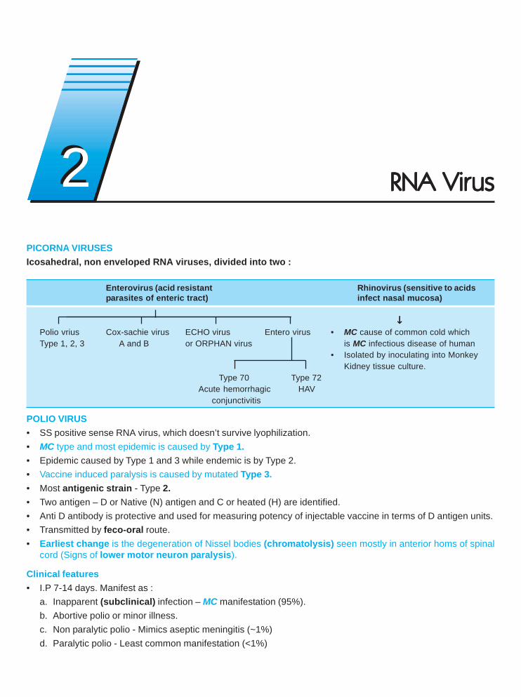
PICORNA VIRUSESIcosahedral, non enveloped RNA viruses, divided into two :
Enterovirus (acid resistant Rhinovirus (sensitive to acidsparasites of enteric tract) infect nasal mucosa)
↓↓↓↓↓Polio vrius Cox-sachie virus ECHO virus Entero virus • MC cause of common cold whichType 1, 2, 3 A and B or ORPHAN virus is MC infectious disease of human
• Isolated by inoculating into MonkeyKidney tissue culture.
Type 70 Type 72Acute hemorrhagic HAV
conjunctivitis
POLIO VIRUS• SS positive sense RNA virus, which doesn’t survive lyophilization.• MC type and most epidemic is caused by Type 1.• Epidemic caused by Type 1 and 3 while endemic is by Type 2.• Vaccine induced paralysis is caused by mutated Type 3.• Most antigenic strain - Type 2.• Two antigen – D or Native (N) antigen and C or heated (H) are identified.• Anti D antibody is protective and used for measuring potency of injectable vaccine in terms of D antigen units.• Transmitted by feco-oral route.• Earliest change is the degeneration of Nissel bodies (chromatolysis) seen mostly in anterior homs of spinal
cord (Signs of lower motor neuron paralysis).
Clinical features• I.P 7-14 days. Manifest as :
a. Inapparent (subclinical) infection – MC manifestation (95%).b. Abortive polio or minor illness.c. Non paralytic polio - Mimics aseptic meningitis (~1%)d. Paralytic polio - Least common manifestation (<1%)
22 RNA VirRNA VirRNA VirRNA VirRNA Virususususus
Self Assessment & Review Microbiology & Immunology
SECTION – B
286
• Predominant sign - Descending; asymmetric; proximal more than distal; flaccid paralysis of legs (MC), arms,abdominal, thoracic or bulbar muscles.
• Objective sensory testing usually yields normal results.• MC muscle affected - Quardriceps• MC muscle which undergoes complete paralysis - Tibialis anterior• MC muscle affected in hand - opponens pollicis• Common deformity at hip is flexion, abduction and external rotation.• At knee - flexion deformity is common but in severe cases triple deformity consist of flexion, posterior subluxation
and external rotation occurs.• Post polio syndrome is due to progressive dysfunction and loss of motor neurons that compensated for the lost
neurons during original infection (not due to persistent / reactivation of virus).• Risk of paralytic polio is increased by :
– Tonsillectomy – Intramuscular injection – Tooth extraction– Adenoidectomy – Strenous physical exercise – Fatigue– Cortisone adminstration
COX-SACKIE VIRUS, ECHO VIRUS AND OTHER ENTEROVIRUSES
• MC clinical manifestation of enterovirus infection - Non specific febrile illness. (Summer Grippe)• MC cause of aseptic meningitis – Enterovirus (ECHO is MC)• MC cause of rubelliform rash – Echovirus 9• Transplacental Transmission occur in coxsackie virus.
Serotype of Virus
Manifestation Cox sackievirus Enterovirus Echo
i. Acute hemorrhagic conjunctivitis (characteristic A-24 Ent. 70 –subconjuctival hemorrhage)
ii. Aseptic meningitis Most group A, all B Ent Eiii. Encephalitis A, B Ent Eiv. Exanthem A, B Ent Ev. Generalized disease of newborn B2-5 – Evi. Hand, foot and mouth disease A, B (commonly by A) Ent 71 –vii. Herpangina A, B (commonly by A) Ent 71 Eviii.Myocarditis, pericarditis A, B (Commonly B) – Eix. Paralysis A, B (commonly A) Ent Ex. Pleurodynia (Bornholm disease) A, B (commonly B) – Exi. Pneumonia A, B Ent Exii. Juvenile diabetes B-4 – –xiii.Orchitis Coxasackie – –xiv. Post viral fatigue syndrome B – –
Diagnosis of Enterovirus (Including polio)• MC procedure for diagnosis of infection - Isolation of enterovirus in cell culture.• Isolation of virus from nasopharyngeal, throat sample, stool is not specific but isolation from throat is more
specific than from stool.• Culture of CSF, serum, fluid from body cavities or tissues - less sensitive but specific.• If CSF culture is negative than stool culture is done within first 2 weeks after onset of symptoms to confirm
diagnosis.
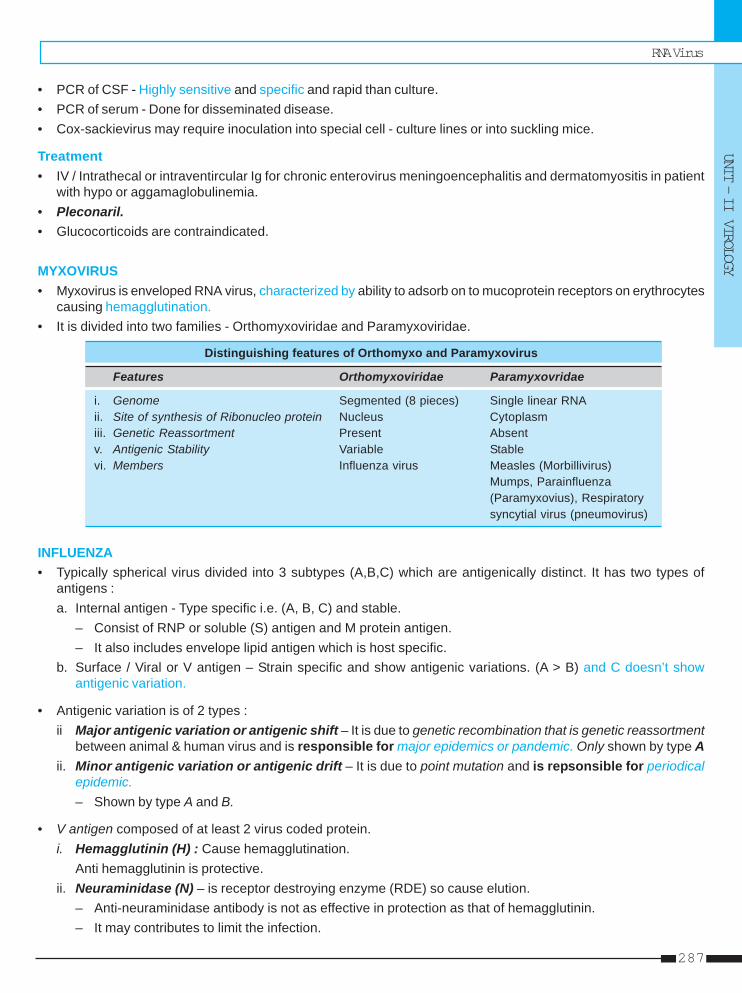
RNA Virus
UNIT – II VIROLOGY
287
• PCR of CSF - Highly sensitive and specific and rapid than culture.• PCR of serum - Done for disseminated disease.• Cox-sackievirus may require inoculation into special cell - culture lines or into suckling mice.
Treatment• IV / Intrathecal or intraventircular Ig for chronic enterovirus meningoencephalitis and dermatomyositis in patient
with hypo or aggamaglobulinemia.• Pleconaril.• Glucocorticoids are contraindicated.
MYXOVIRUS• Myxovirus is enveloped RNA virus, characterized by ability to adsorb on to mucoprotein receptors on erythrocytes
causing hemagglutination.• It is divided into two families - Orthomyxoviridae and Paramyxoviridae.
Distinguishing features of Orthomyxo and Paramyxovirus
Features Orthomyxoviridae Paramyxovridae
i. Genome Segmented (8 pieces) Single linear RNAii. Site of synthesis of Ribonucleo protein Nucleus Cytoplasmiii. Genetic Reassortment Present Absentv. Antigenic Stability Variable Stablevi. Members Influenza virus Measles (Morbillivirus)
Mumps, Parainfluenza(Paramyxovius), Respiratorysyncytial virus (pneumovirus)
INFLUENZA• Typically spherical virus divided into 3 subtypes (A,B,C) which are antigenically distinct. It has two types of
antigens :a. Internal antigen - Type specific i.e. (A, B, C) and stable.
– Consist of RNP or soluble (S) antigen and M protein antigen.– It also includes envelope lipid antigen which is host specific.
b. Surface / Viral or V antigen – Strain specific and show antigenic variations. (A > B) and C doesn’t showantigenic variation.
• Antigenic variation is of 2 types :ii Major antigenic variation or antigenic shift – It is due to genetic recombination that is genetic reassortment
between animal & human virus and is responsible for major epidemics or pandemic. Only shown by type Aii. Minor antigenic variation or antigenic drift – It is due to point mutation and is repsonsible for periodical
epidemic.– Shown by type A and B.
• V antigen composed of at least 2 virus coded protein.i. Hemagglutinin (H) : Cause hemagglutination.
Anti hemagglutinin is protective.ii. Neuraminidase (N) – is receptor destroying enzyme (RDE) so cause elution.
– Anti-neuraminidase antibody is not as effective in protection as that of hemagglutinin.– It may contributes to limit the infection.
Self Assessment & Review Microbiology & Immunology
SECTION – B
288
• In world, three types of influenza virus are circulating - A (H1N1), A(H3N2), and B viruses.• New influenza virus : A (H5N1), causative agent of bird flu.• Source of Infection – Usually a case or subclinical.• Portal of entry – Respiratory route• Incubation period – 18-72 hours.
Clinical Features• There is no viremia.• Respiratory symptoms are prominent but abdominal pain and vomiting may occur in type B infections.• MC complication is pneumonia.• Most serious complication of influenza B virus is Reye’s syndrome [also occur in influenza A and VZ virus].• Cardiac, neurological complication and gastric flu (with type B) may occur.• MC secondary bacterial pneumonia in influenza - Strep pneumoniae.
Lab-Diagnosis• Virus isolation – Best specimen is nasopharyngeal secretions
– Detected by Indirect fluorescent antibody.Prevention1. Best is immunization. Vaccine is recommended only in certain selected population. Vaccine is of following types.
a. Killed vaccine :Most commonly used vaccine :– Contains H, N antigens– Usually one dose given but in patient with no previous immunological experience 2 dose given– Immunity last for only 3 - 6 month.– Vaccine can produce very rarely Guillain Barre syndrome (ascending paralysis).
b. Live attenuated vaccines :Administered as nose drops so induce both local and systemic immunity
c. Newer vaccines :Split virus vaccine (sub -virion vaccine), Neuraminidase specific vaccine (sub-unit vaccine contain only N antigen);Recombinant vaccine
2. Antiviral drugs :– For type A virus : Amantadine and rimantidine (used in treatment also).– For both A and B : Zamamivir, Oseltamivir.
MEASLES (RUBEOLA)• RNA paramyxovirus, having only one serotype.• It cannot survive outside the human body.• Carriers are not known to occur. But subclinical cases occurs.• Secondary attack rate > 80%.• Immunity after vaccination and infection is life long.• Multinucleated giant cells with inclusion bodies in the nucleus and cytoplasm (Warthin - Finkeldey Cells) are
pathognomic for measles.
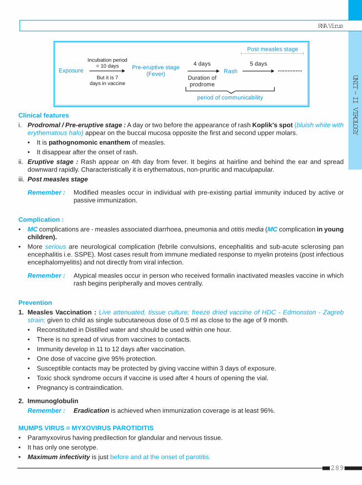
RNA Virus
UNIT – II VIROLOGY
289
Incubation period = 10 days
Exposure But it is 7
days in vaccine
Pre-eruptive stage(Fever) Rash
4 days 5 days
Post measles stage
...............Duration of prodrome
period of communicability
Clinical featuresi. Prodromal / Pre-eruptive stage : A day or two before the appearance of rash Koplik’s spot (bluish white with
erythematous halo) appear on the buccal mucosa opposite the first and second upper molars.• It is pathognomonic enanthem of measles.• It disappear after the onset of rash.
ii. Eruptive stage : Rash appear on 4th day from fever. It begins at hairline and behind the ear and spreaddownward rapidly. Characteristically it is erythematous, non-pruritic and maculpapular.
iii. Post measles stage
Remember : Modified measles occur in individual with pre-existing partial immunity induced by active orpassive immunization.
Complication :• MC complications are - measles associated diarrhoea, pneumonia and otitis media (MC complication in young
children).• More serious are neurological complication (febrile convulsions, encephalitis and sub-acute sclerosing pan
encephalitis i.e. SSPE). Most cases result from immune mediated response to myelin proteins (post infectiousencephalomyelitis) and not directly from viral infection.
Remember : Atypical measles occur in person who received formalin inactivated measles vaccine in whichrash begins peripherally and moves centrally.
Prevention1. Measles Vaccination : Live attenuated, tissue culture; freeze dried vaccine of HDC - Edmonston - Zagreb
strain; given to child as single subcutaneous dose of 0.5 ml as close to the age of 9 month.• Reconstituted in Distilled water and should be used within one hour.• There is no spread of virus from vaccines to contacts.• Immunity develop in 11 to 12 days after vaccination.• One dose of vaccine give 95% protection.• Susceptible contacts may be protected by giving vaccine within 3 days of exposure.• Toxic shock syndrome occurs if vaccine is used after 4 hours of opening the vial.• Pregnancy is contraindication.
2. ImmunoglobulinRemember : Eradication is achieved when immunization coverage is at least 96%.
MUMPS VIRUS = MYXOVIRUS PAROTIDITIS• Paramyxovirus having predilection for glandular and nervous tissue.• It has only one serotype.• Maximum infectivity is just before and at the onset of parotitis.
Self Assessment & Review Microbiology & Immunology
SECTION – B
290
• It spreads mainly by droplet infection.• One attack (clinical or subclinical) induce life long immunity.• Incubation Period – Usually 18 days.
Clinical Features• Mumps is the most frequent cause of parotitis in children in the age group 5-15 years.• Usually bilateral parotitis occurs. Submandibular and sublingual gland can also involved.• Some patient develops pre-sternal edema.• Other than parotitis, orchitis is the MC manifestation among post- pubertal males.• Aseptic meningitis is a common manifestation in both children and adults.• Glucose level in CSF may be abnormally low and this raise suspicion of bacterial meningitis.• In pregnancy, it does not lead to premature birth or fetal malformation.
Prevention• Single dose of live-attenuated vaccine primarily in susceptible adults especially males who have not had mumps.• Combined vaccine - MMR : at the age of 12-15 month and again at 4-12 years of age. ....Harrison 16/e, p 1155
PARAINFLUENZA VIRUS• First parainfluenza virus to be discovered - Sendai virus or Hemagglutinating virus of Japan (HVJ) or influenza
virus type D.• It ranks second only to RSV as cause of lower respiratory tract illness.• It has 4 types - 1, 2, 3, 4.
Clinical Features• Most serious clinical disease is croup (laryngotracheobronchitis).• Para-influenza type 1 is MC cause of croup in children.• Type 3 is important cause of lower respiratory disease (bronchitis, bronchiolitis and pneumonia) in infants.• In older children and adults, most frequent symptom - common cold or hoarsenss.• This virus confined to respiratory tract unlike mumps which is a systemic disease.
DiagnosisIsolation of virus from throat and nasal swabs by inoculating in primary monkey kidney cell cultures or continousmonkey kidney cell line (LLC-MK2) with trypsin.
RESPIRATORY SYNCYTIAL VIRUS (RSV)• MC cause of lower respiratory tract disease.• MC cause of bronchiolitis seen among infants between 1 and 6 month of age peaking between 2 and 3 months
of age.• MC manifestation in infants rhinorrhea.• In adults MC symptom are common cold with rhinorrhea, sore throat and cough.• Immunity is not long lasting.• Nasal IgA is more protective than serum antibody.• RSV is transmitted primarily by close contact with contaminated fingers or fomites.• It produce fine rales; rhonchi, emphysematous change (that is hyperexpansion on chest X-ray) and atelectasis.
RNA Virus
UNIT – II VIROLOGY
291
Diagnosis• Specific diagnosis – by isolation of RSV from respiratory secretions (sputum, throat swab, nasopharyngeal wash).
Treatment• Oxygen : mainstay of therapy.• Ribavirin : for infants who are severely ill or who are at high risk of complications of RSV infection (premature
infants and those with bronchopulmonary dysplasia, congenital heart disease and immunosuppression).
ROTAVIRUS AND OTHER VIRAL GASTROENTERITIS• MC cause of diarrheal disease in infants and children : Rotaviurs• MC agent among older children and adults : Norovirus (Norwalk like viruses)
ROTAVIRUS• Non enveloped, icosahedral virus with segmented ds RNA (Character of Reovirus family) so it exhibits genetic
reassortment.• VP-6 is major structural protein, is the target of commercial immunoassays and determines the group specificity
of rotaviruses.• Human illness is caused primarily by group A and to a much lesser extent by group B and C.• Adult diarrhea rota virus (ADRV) belong to group B while group C cause pediatric gastroenteritis.• 10 G serotypes of group A are identified in humans but 5 types (G1 through G4 and G9) are common.
Pathogenesis1. It infect and destroy mature enterocytes in villous epithelum of proximal small intestine causing :
a. Secretory diarrhea by ↓ villous epithelum + ↑ secretory crypt cells.b. Osmotic diarrhea – by ↓ brush border enzymes causing accumulation of unmetabolized disaccharides.
2. Secretory diarrhea also result by :a. Enterotoxin (=NSP4) which alter epithelial cell function and permeability.b. Activation of enteric nervous system in the intestinal wall.
Clinical features• It infects all children by 3-5 years. May also infect neonates.• Peak age - 4 to 23 months of age.• Occurs predominantly during the cooler fall and winter months.• Transmitted predominantly through fecao-oral route. Also transmitted by respiratory secretion, person to person,
contaminated environmental surface.• Severity of dehydration : Rotavirus > Norovirus > Sapovirus• Vomiting frequently precedes the diarrhea.• Stools are characteristically loose and watery and only infrequently contain red or white cells.• Rotaviurs associated with respiratory and neurologic features, sudden infant death syndrome, necrotizing
enterocolitis, intussusception and diabetes mellitus type I.• Its infection produces virus specific secretory IgA in intestine and IgA, IgM, IgG in serum which increase with
each reinfection, so severe disease is MC in young children with first or second infections.Diagnosis
Self Assessment & Review Microbiology & Immunology
SECTION – B
292
• As virus is shed in large quantities (107-1012/g) in stool, the diagnosis is confirmed by detecting viral antigen infeces by Enzyme immunoassays and by detecting viral RNA (by gel electrophoresis, probe hybridization orPCR).
• Human rotaviurs does not grow readily in cell culture.
Treatment• Rehydration therapy is given.• Antibiotics and antimotility agents avoided.• In immunocompromised children - oral immunoglobulin or colostrum given.
Prevention• Vaccine was withdrawn since it cause intussusception.
OTHER VIRAL GASTROENTRITIS1. Norwalk and Related human calciviruses
• Norovirus is the MC infectious agent of mild gastroenteritis in the community and affect all age groupswhereas sapoviruses primarily cause gastroenteritis in children.
• Noroviruses are the major cause of epidemics of gastroenteritis worldwide.
Pathogenesis• It is attached on carbohydrates (similar to human histoblood group antigens) of duodenal epithelium of
individuals with the secretor phenotype (genetic predisposition to illness).• Reversible lesion in upper jejunum e.g. broadening and blunting of villi, shortening of microvilli etc.• Malabsorption of carbohydrates and fats and decreased brush border enzymes.• Adenylate cyclase activity is not altered.• Gastric motor function is delayed but histological changes are absent in stomach and colon.
Clinical features• Transmission occurs predominantly by fecal-oral route but virus is also present in vomitus.• Also transmitted by aerosolization, contact with contaminated fomites, person to person contact.• Shellfish harvested from fecally contaminated water pose a special risk.• Vomiting is more common among children where adults usually develop diarrhea.• Constitutional symptoms are common.• Stool are characteristically loose and watery without blood, mucus or leukocytes.• There is paradoxic inverse association between level of antibody and protection from disease that is person
with higher level of pre-existing antibody are more susceptible to illness.
Diagnosis• PCR for detection of virus in stool and vomitus.• EIA (Enzyme immunoassays) for detection of virus in stool and serologic response to specific viral antigen.• It has not yet been propagated in cell cultures.
Treatment : Generally not required since it is self limited.
2. Adenovirus : Enteric adenovirus (40 and 41) are difficult to cultivate in cell lines.
3. Astrovirus : Serotype 1 is MC.
4. Torovirus : Cause less vomiting and more bloody diarrhea.
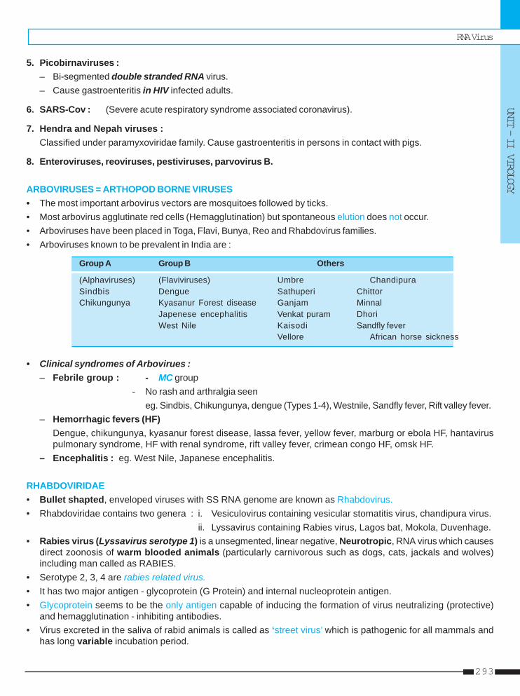
RNA Virus
UNIT – II VIROLOGY
293
5. Picobirnaviruses :– Bi-segmented double stranded RNA virus.– Cause gastroenteritis in HIV infected adults.
6. SARS-Cov : (Severe acute respiratory syndrome associated coronavirus).
7. Hendra and Nepah viruses :Classified under paramyxoviridae family. Cause gastroenteritis in persons in contact with pigs.
8. Enteroviruses, reoviruses, pestiviruses, parvovirus B.
ARBOVIRUSES = ARTHOPOD BORNE VIRUSES• The most important arbovirus vectors are mosquitoes followed by ticks.• Most arbovirus agglutinate red cells (Hemagglutination) but spontaneous elution does not occur.• Arboviruses have been placed in Toga, Flavi, Bunya, Reo and Rhabdovirus families.• Arboviruses known to be prevalent in India are :
Group A Group B Others
(Alphaviruses) (Flaviviruses) Umbre ChandipuraSindbis Dengue Sathuperi ChittorChikungunya Kyasanur Forest disease Ganjam Minnal
Japenese encephalitis Venkat puram DhoriWest Nile Kaisodi Sandfly fever
Vellore African horse sickness
• Clinical syndromes of Arbovirues :– Febrile group : - MC group
- No rash and arthralgia seeneg. Sindbis, Chikungunya, dengue (Types 1-4), Westnile, Sandfly fever, Rift valley fever.
– Hemorrhagic fevers (HF)Dengue, chikungunya, kyasanur forest disease, lassa fever, yellow fever, marburg or ebola HF, hantaviruspulmonary syndrome, HF with renal syndrome, rift valley fever, crimean congo HF, omsk HF.
– Encephalitis : eg. West Nile, Japanese encephalitis.
RHABDOVIRIDAE• Bullet shapted, enveloped viruses with SS RNA genome are known as Rhabdovirus.• Rhabdoviridae contains two genera : i. Vesiculovirus containing vesicular stomatitis virus, chandipura virus.
ii. Lyssavirus containing Rabies virus, Lagos bat, Mokola, Duvenhage.• Rabies virus (Lyssavirus serotype 1) is a unsegmented, linear negative, Neurotropic, RNA virus which causes
direct zoonosis of warm blooded animals (particularly carnivorous such as dogs, cats, jackals and wolves)including man called as RABIES.
• Serotype 2, 3, 4 are rabies related virus.• It has two major antigen - glycoprotein (G Protein) and internal nucleoprotein antigen.• Glycoprotein seems to be the only antigen capable of inducing the formation of virus neutralizing (protective)
and hemagglutination - inhibiting antibodies.• Virus excreted in the saliva of rabid animals is called as ‘street virus’ which is pathogenic for all mammals and
has long variable incubation period.
Self Assessment & Review Microbiology & Immunology
SECTION – B
294
• Serial brain - to - brain passage of Street Virus modifies it to convert into fixed virus which has following characteristics:– Short, fixed and reproducible incubation period.– Not form Negri bodies and not multiply in extraneural tissues.– Used in the preparation of anti - rabies vaccine.– It is pathogenic under certain conditions eg. when inadequately inactivated for vaccine production.
• Rabies is only communicable disease of man that is always fatal.
Remember : Rabies is dead end infection.
Type of Rabiesi. Urban rabies – maintained by the dog and is responsible for 99% of human cases in India.ii. Sylvatic or wild life rabies.iii. Bat rabies.
Remember : • In most of the world, dog is the most important vector.• Maldives is the only country which does not have human or animal rabies.• In India, rabies occur in all parts except Lakshadweep and Andman and Nicobar Islands• Most effective natural barrier to rabies - water.
Mode of transmission : • Animal bites - MC• Licks on abraded skin and abraded or unabraded mucosa.• Respiratory (aerosol) transmission.• Person to Person - rare• Also by corneal and organ transplants.
Incubation period : Highly variable depending on the site of bite (i.e. actual distance that the virus has to travel toreach to CNS), severity of bite etc.
Clinical features• Virus spreads centripetally from site of infection (striated muscle) than ascends through nerve associated
tissue space, and than spreads centrifugally in peripheral autonomic nerves to many tissues.• Salivary gland invasion is crucial for transmission of virus.• Most characteristic pathologic finding in CNS is the formation of cytoplasmic inclusions called Negri Bodies
(composed of finely fibrillar matrix and rabies virus particles) within neurons of Ammons horn, cerebral cortex,brain stem, hypothalmus, purkinje cells of the cerebellum and dorsal spinal ganglia.
• It has four stages :a. Prodromal period : Specific symptom is complaint of paresthesia / fasciculation at or around the site of
inoculation of virus.b. Encephalitic phase : Abnormalities of automatic nervous system. Aerophobia (pathognomic) and Hydrophobia
(pathognomic and absent in animals) may seen.c. Manifestation of brain stem dysfunction : The prominence of early brain stem dysfunction distinguish it
from other viral encephalitidis.d. Death or in rare cases recovery occurs.
Remember : It may be also present as ascending paralysis resembling GBS most frequently among personsgiven post-exposure prophylaxis after being bitten by vampire bats.
Diagnosis : Confirmed by antigen detection using immunofluorescence of infected tissue (corneal impressionsmear, skin biopsy or brain) and by virus isolation from saliva and other secretions.
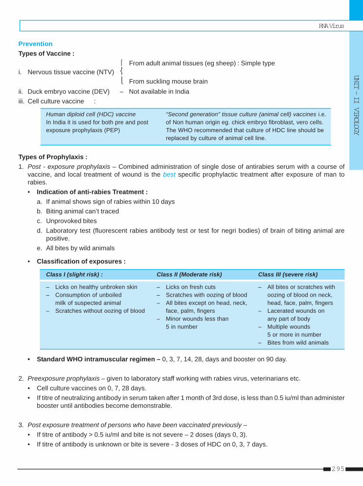
RNA Virus
UNIT – II VIROLOGY
295
PreventionTypes of Vaccine :
⎧ From adult animal tissues (eg sheep) : Simple typei. Nervous tissue vaccine (NTV) ⎨
⎩ From suckling mouse brainii. Duck embryo vaccine (DEV) – Not available in Indiaiii. Cell culture vaccine :
Human diploid cell (HDC) vaccine “Second generation” tissue culture (animal cell) vaccines i.e.In India it is used for both pre and post of Non human origin eg. chick embryo fibroblast, vero cells.exposure prophylaxis (PEP) The WHO recommended that culture of HDC line should be
replaced by culture of animal cell line.
Types of Prophylaxis :1. Post - exposure prophylaxis – Combined administration of single dose of antirabies serum with a course of
vaccine, and local treatment of wound is the best specific prophylactic treatment after exposure of man torabies.• Indication of anti-rabies Treatment :
a. If animal shows sign of rabies within 10 daysb. Biting animal can’t tracedc. Unprovoked bitesd. Laboratory test (fluorescent rabies antibody test or test for negri bodies) of brain of biting animal are
positive.e. All bites by wild animals
• Classification of exposures :
Class I (slight risk) : Class II (Moderate risk) Class III (severe risk)
– Licks on healthy unbroken skin – Licks on fresh cuts – All bites or scratches with– Consumption of unboiled – Scratches with oozing of blood oozing of blood on neck,
milk of suspected animal – All bites except on head, neck, head, face, palm, fingers– Scratches without oozing of blood face, palm, fingers – Lacerated wounds on
– Minor wounds less than any part of body5 in number – Multiple wounds
5 or more in number– Bites from wild animals
• Standard WHO intramuscular regimen – 0, 3, 7, 14, 28, days and booster on 90 day.
2. Preexposure prophylaxis – given to laboratory staff working with rabies virus, veterinarians etc.• Cell culture vaccines on 0, 7, 28 days.• If titre of neutralizing antibody in serum taken after 1 month of 3rd dose, is less than 0.5 iu/ml than administer
booster until antibodies become demonstrable.
3. Post exposure treatment of persons who have been vaccinated previously –• If titre of antibody > 0.5 iu/ml and bite is not severe – 2 doses (days 0, 3).• If titre of antibody is unknown or bite is severe - 3 doses of HDC on 0, 3, 7 days.
Self Assessment & Review Microbiology & Immunology
SECTION – B
296
1. Which of the following is not true about measles:a) High secondary attack [AI 08]b) Only one strain causes infectionc) Not infectious in prodromal periodd) Infections confer life long immunity
2. H5N1 is : [AI 08]a) Bird flu virusb) Vaccinefor HIVc) Causative agent of Japanese encpehalitisd) An eradicated virus
3. The most common etiological agent for acutebronchiolitis in infancy is : [AI 06]a) Influenza virusb) Parainfluenza virusc) Rhinovirusd) Respiratory syncytial virus
4. With reference to mumps which of the followingis true : [AI 06]a) Meningoencephalitis can precede parotitisb) Salivary gland involvement is limited to the
parotidsc) The patient is not infectious prior to clinical pa-
rotid enlargementd) Mumps orchitis frequently leads to infertility
5. All of the following statement are true about con-genital Rubella except : [AI 05]a) It is diagnosed when the infant has IgM antibod-
ies at birthb) It is diagnosed when IgG antibodies persist for
more than 6 monthc) MC congenital defects are deafness, cardiac
malformation and cataractd) Infection after 16 weeks of gestation result in
major congenital defects6. Laboratory diagnosis of viral respiratory tract in-
fections can be established by all of the followingtests except : [AI 04]a) Detection of virus specific IgM antibodies in
single serum specimenb) Demonstration of viral antigens by indirect im-
munofluorescence assay in nasopharyngealwashings
c) Isolation of viruses using centrifugation en-hanced culture
d) Detection of viral hemagglutination inhibiting(HAI) antibodies in a single serum specimen
7. All of the following statements are true regardingpoliovirus except : [AI 04]a) It is transmitted by feco-oral routeb) Asymptomatic infections are common in chil-
drenc) There is a single serotype causing infectiond) Live attenuated vaccine produces herd immu-
nity8. All of the following clinical features are associ-
ated with Enteroviruses except : [AI 04]a) Mycocarditisb) Pleurodyniac) Herpanginad) Hemorrhagic fever
9. Commonest complication of Mumps is : [AI 00]a) Orchitis and Oophritisb) Encephalitisc) Pneumoniad) Myocarditis
10. True statement about Influenza A virus : [AI 04]a) It has a double stranded segmented RNAb) Pandemics are caused by antigenic driftsc) Nucelocapsid antibody is not specificd) Hemaglutinin and neuraminidase are strain
specific11. Acute hemorrhagic conjunctivitis is caused by :
a) Enterovirus [AI 97]b) Adenovirusc) Poliovirusd) Hepadna virus
12. Reverse transcriptase polymerase chain reactioncan aid in diagnosis of all of the following viralinfection except : [AI 97]a) Adenovirusb) Astrovirusc) Rotavirusd) Poliovirus
13. All of the viruses cause pneumonia except :a) Cytomegalovirus [AI 95]b) Mumpsc) Measlesd) Retrovirus
Answer 1. c) Not .... 2. a) Bird flu .... 3. d) Respiratory ... 4. a) Meningoencep ... 5. d) Infection ...6. d) Detection ... 7. c) There is a ... 8. d) Hemorrhagic ... 9. a) Orchitis ... 10. d) Hemaglutinin ...
11. a) Enterovirus 12. a) Adenovirus 13. b) Mumps
ANSWERS WITH EXPLANATIONQUESTIONS
RNA Virus
UNIT – II VIROLOGY
297
14. True about polio : [AIIMS 08]a) Paralytic polio is most commonb) Only one type existc) Increased muscular activity leads to increased
paralysisd) Polio drop given only in <3 years
15. Regarding mumps which is true :a) Causes SSPE [AIIMS Nov. 07]b) Mumps causes aseptic meningitis in childrenc) Sublingual gland is invlved commonlyd) All
16. All are false regarding polio virus except :a) Most case are symptomatic [AIIMS Nov. 07]b) Inactivated vaccine given IMc) Inactivated polio vaccine are given to child less
than 3 year of aged) Only one type exists
17. Which of the following is the ‘Least common’ com-plication of measles ? [AIIMS 06]a) Dirrhoeab) Pneumoniac) Otitis mediad) SSPE
18. Risk of the damage of fetus by maternal rubella ismaximum if mother gets infected in :a) 6-12 weeks of pregnancy [AIIMS 05]b) 20-24 weeks of pregnancyc) 24-28 weeks of pregnancyd) 32-36 weeks of pregnancy
19. All are true about polio virus except : [AIIMS 02]a) Type 1 is responsible for most epidemicsb) Very difficult to eliminate type 1c) Type I is responsible for vaccine induced para-
lytic poliomyelitisd) Type I most commonly associated with paraly-sis
20. Rota virus is detected by : [AIIMS 02]a) Antigen in stoolb) Antibody in serumc) Demonstration of virusd) Stool culture
21. Enterovirus causes all except : [AIIMS 01]a) Hemorrhagic feverb) Pleurodyniac) Herpanginad) Aseptic meningitis
22. A 11 month old child presents with complaints ofrespiratory distress. On examination there is bi-lateral crepitation and wheezing. Which of the fol-lowing is the most likely cause : [AIIMS 00]a) Pneumoniab) Adenovirusc) Respiratory syncitial virusd) Rhinovirus
23. Rota virus is diagnosed by : [AIIMS 99]a) IgM specific antibody in stoolb) ELISA demonstrates antibody in stoolc) Immunoflorescence antigen in stoold) Culture of rota virus
24. Segmented double stranded RNA virus is seen in:a) Reovirus [AIIMS 98; PGI 97]b) Myxovirusc) Rabiesd) Parvo virus
25. Conjunctivitis is cause by all except :a) CMV [AIIMS 98]b) Enterovirus 70c) Coxsackie A 24d) Adenovirus
26. All are true about Rota virus except : [AIIMS 97]a) Causes diarrhoea in man and childrenb) Rota B can be grown in cell culturec) Rota C can cause diarrhoea in childrend) Culture can not be done
27. All are cultivable virus except : [AIIMS 97]a) Rota virusb) Entero virusc) ECHO virusd) Coxsackie virus
28. Segmented RNA is found in : [AIIMS 97, PGI 00]a) Influenza virusb) Rabies virusc) Herpes virusd) Molluscum contagiosum virus
29. Vaccination causing intussuception :a) Rota virus [PGI Dec. 07]b) Parvo virusc) Inactivated poliod) BCGe) Measles
Answer 14. c) Increased... 15. b) Mumps ... 16. b) Inactivated ... 17. d) SSPE 18. a) 6-12 ...19. c) Type I ... 20. a) Antigen ... 21. a) Hemorrhagic ... 22. c) Respiratory ... 23. c) Immunoflore ...24. a) Reovirus 25. a) CMV 26. b) Rota B ... 27. a) Rota ... 28. a) Influenza ...29. d) Rota virus
Self Assessment & Review Microbiology & Immunology
SECTION – B
298
30. Vaccines prepared by embryonated Hen’s egg are:a) Mealses [PGI 04]b) Rabiesc) Rubellad) Varicella
31. All are included in picorna group of viruses ex-cept: [PGI 04]a) Encephalo myocarditisb) HEVc) Foot and mouth virusd) Polio virus
32. Choose the correct matches : [PGI 02]a) Mumps-RA 27/3 strainb) Rubella-Jeryl-Lynn strainc) Measles - Edmonston zagreb strainc) BCG-Danish 1331 strain
33. Micro-Organism used as weapon in biological ter-rorism : [PGI 02]a) Small pox Virusb) Rabies Virusc) Ebola Virusd) Influenza C Viruse) Human parvovirus
34. Incubation period less than 10 days seen :a) Influenza [PGI 02]b) Cholerac) Plagued) Chickenpoxe) Rabies
35. Organism (s) causing, bronchiolitis in infant :a) RSV [PGI 00]b) Rhino virusc) Parainfluenzad) Influenzae) H. influenzae
36. Lipid envelope is found in which virus :a) Reo [PGI 98]b) Herpesc) Picornad) All of the above
ARBO AND RHABDO
37. Soft tick transmits : [AI 08]a) Relapsing feverb) KFDc) Tick typhusd) Tularemia
38. Negre body is seen in : [AI 07]a) CMVb) Rabiesc) Inclusion of herpes simplexd) EBV
39. Which of the following viral infections is transmit-ted by tick : [AI 05]a) Japanese encephalitisb) Dengue feverc) Kyasanur forest disease (KFD)d) Yellow fever
40. For the treatment of case of class III dog bite, allof the following are correct except : [AI 05]a) Give Ig for passive immunityb) Give ARVc) Immediately stitch wound under antibiotic cov-
eraged) Immediately wash wound with soap and water
41. Class II exposure in animal bites includes the fol-lowing : [AI 03]a) Scratches without oozing of bloodb) Licks on a fresh woundc) Scratch with oozing of blood on palmd) Bites from wild animals
42. Which of the following statement is true aboutrabies virus : [AI 03]a) It is double stranded - RNA virusb) Contains a DNA-dependent RNA polymerasec) RNA has a negative polarityd) Affects motor neurons
43. Which is true about arboviral disease : [AI 00]a) Yellow fever is endemic in Indiab) Dengue virus have only one serotypec) KFD is transmitted by ticksd) Japanese encphalitis is transmitted by Aedes
Answer 30. a) Mealses 31. b) HEV 32. c and d 33. a and c 34. a, b and c35. a, c and d 36. a) Reo 37. c) Relapsing ... 38. b) Rabies 39. c) Kyasanur ...40. c) Immediately ... 41. b) Licks on ... 42. c) RNA ... 43. c) KFD is ...
RNA Virus
UNIT – II VIROLOGY
299
44. Regarding Rabies, true is : [AI 00]a) Incubation period depends on the site of biteb) Diagnosis is by eosinophilic intranuclear inclu-
sionc) It is a DNA virusd) Caused only by dogs
45. All of the following rabies vaccines are commer-cially available except : [AI 99]a) Killed sheep brain vaccineb) Human diploid cell vaccinec) Vero continous cell vaccined) Recombinant glycoprotein
46. True statement about rabies is : [AI 97]a) Rabies infection cause life long immunityb) Rabies vaccine is always live attenuatedc) Rabies has various strainsd) Rabies is best diagnosed by immuno-flores-
cence study47. Negri bodes are found in : [AI 96]
a) Hypothalamusb) Hippocampusc) Midbraind) Medulla
48. Mark true in following : [AIIMS 08]a) Hanta virus pulmonary syndrome is caused by
inhalation of rodent urine and fecesb) Kyansur forest disese is caused by bite of wild
animalc) Lyssa virus is transmitted by ticksd) Chikugunya is caused by anophales
49. A 25 year old girl has admitted to hospital withprovisional diagnosis of rabies. The most suitableclinical sample that can confirm the antemortemdiagnosis is : [AIIMS 04]a) Serum for antivirus IgG antibodyb) Corneal impression smear for immuno-fluores-
cence stainc) CSF sample for viral cultured) Giemsa stain on smear prepared from salivary
secretions50. All are true regarding hantana virus except :
a) DNA virus [AIIMS 96]b) Carried by rodentsc) Causes recurrent respiratory infectiond) Hemorrhagic manifestation may occur
51. New infectious agents are :a) Nipah virus [PGI 07]b) Pneumocystis jeruvecic) Corona virusd) SARSe) Prion
52. Following are Arboviral disease : [PGI 03]a) KFDb) West Nile Feverc) Ganjam virusd) RSVe) Puumala virus
53. True about hanta virus : [PGI 02]a) Hantavirus pulmonary syndromeb) Transmitted by arthopodc) Transmitted by rodentsd) Hemorrhagic fever with renal failuree) Hantavirus pulmonary symdrome acquired from
person to person54. In Japanese Encephalitis pigs acts as : [PGI 00]
a) Amplifierb) Definitive hostc) Intermediate hostd) Any of the above
55. Rabies virus inactivated by : [PGI 97]a) Phenolb) UV radiationc) BPL (Beta propiolactone)d) All
56. Negri bodies are characteristic of viral infectionby : [PGI 97]a) Rabiesb) Toxoplasmosisc) Poliod) Herpes simpex infection
57. Hantan virus : [PGI 96]a) Is a DNA virusb) Causes Hemorrhagic fever with renal involve-
mentc) Belong to Retroviridae familyd) Person to person transmission
Answer 44. a) Incubation ... 45. d) Recombinant ... 46. d) Rabies is ... 47. b) Hippoca ... 48. a) Hanta virus ...49. b) Corneal ... 50. a) DNA ... 51. a, c and d 52. a, b, c and e 53. a, c and d54. a) Amplifier 55. d) All 56. a) Rabies 57. b) Causes ...
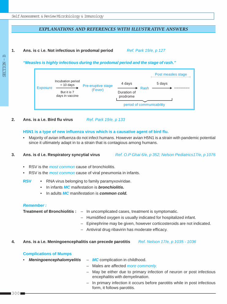
Self Assessment & Review Microbiology & Immunology
SECTION – B
300
1. Ans. is c i.e. Not infectious in prodomal period Ref. Park 19/e, p 127
“Measles is highly infectious during the prodomal period and the stage of rash.”
Incubation period = 10 days
Exposure But it is 7
days in vaccine
Pre-eruptive stage(Fever) Rash
4 days 5 days
Post measles stage
...............Duration of prodrome
period of communicability
2. Ans. is a i.e. Bird flu virus Ref. Park 19/e, p 133
H5N1 is a type of new influenza virus which is a causative agent of bird flu.• Majority of avian influenza do not infect humans. However avian H5N1 is a strain with pandemic potential
since it ultimately adapt in to a strain that is contagious among humans.
3. Ans. is d i.e. Respiratory syncytial virus Ref. O.P Ghai 6/e, p 352; Nelson Pediatrics17/e, p 1076
• RSV is the most common cause of bronchiolitis.• RSV is the most common cause of viral pneumonia in infants.
RSV • RNA virus belonging to family paramyxoviridae.• In infants MC maifestation is bronchiolitis.• In adults MC manifestation is common cold.
Remember :Treatment of Bronchiolitis : – In uncomplicated cases, treatment is symptomatic.
– Humidified oxygen is usually indicated for hospitalized infant.– Epinephrine may be given, however corticosteroids are not indicated.– Antiviral drug ribavirin has moderate efficacy.
4. Ans. is a i.e. Meningoencephalitis can precede parotitis Ref. Nelson 17/e, p 1035 - 1036
Complications of Mumps• Meningoencephalomyelitis – MC complication in childhood.
– Males are affected more commonly.– May be either due to primary infection of neuron or post infectious
encephalitis with demyelination.– In primary infection it occurs before parotitis while in post infectious
form, it follows parotitis.
EXPLANATIONS AND REFERENCES WITH ILLUSTRATIVE ANSWERS
RNA Virus
UNIT – II VIROLOGY
301
– Parotitis may be absent in some cases.– CSF shows lymphocytic pleocytosis.
• Orchitis and Epididymitis – These complication are rare in prepubscent age group, while commonin adolescent and adults.
– Infertility is rare even with bilateral orchitis.• Pancreatitis, myocarditis, arthritis, thyroiditis.• Measles associated deafness, Dacryoadenitis are other complication.
5. Ans. is d i.e. Infection after 16 weeks of gestation result in major congenital defectsRef. Park 18/e, p 128 - 129; 19/e, p 130 -131; Harrison 17/e, p 1218
Important features of Rubella virus• Rubella is RNA virus of togavirus family.• No known carrier state for postnatally acquired rubella.• Infectivity is greatest when rash is erupted.• Rubella (german measles) is mainly a disease of childhood particularly 3-10 years.• One attack results life long immunity• It cause two types of disease :
a. Postnatally Acquired Rubella• Virus is shed from pharynx during prodromal phase and continue for about a week after onset.• It is invariably self limited.• Foremost symptoms
– Posterior auricular, cervical and suboccipital lymphadenopathy; fever and rash (begins on faceand spreads down the body).
• Petechial enathem on soft plate called Forschheimer spots may seen.• Complications :
– Arthiritis (MC in fingers, wrist or knees) almost exclusively in women.– Hemorrhage due to thrombocytopenia and vascular damage.– Encephalitis.
• Diagnosis :– Throat swab culture for virus isolation– Serology : - Most widely used serological test is hemagglutination inhibition test (HAI).
- 4 fold rise in HI antibody titre in paired sera or presence of IgM in single seraobtained 2 weeks after the rash is diagnostic of recent rubella infection.
b. Congenital Rubella Syndrome• Infectivity as well as severity is more in early pregnancy (first trimester of <11 weeks).• Classic triad of patent ductus arteriosus (cardiac malformation), cataract and deafness is seen.• Infection in 2nd trimester - may be deafness only.• >16wk – no major abnormalities• Diagnosis
– Isolation of virus in cell cultures of throat samples, urine or other secretions.– Detection of IgM in single serum sample shortly after birth.– Persistance of Rubella IgG antibodies serum beyond 1 year or rising antibody titre anytime
during infancy in an unvaccinated child.
Self Assessment & Review Microbiology & Immunology
SECTION – B
302
– Biopsy of tissues/ blood / CSF fluid for viral antigen by monoclonal antibodies.– Detection of Rubella RNA by in situ hybridization and PCR.
• Prevention– Rubella vaccine : Live attenuated RA 27/3 vaccine– Strategy : - Immunize all infants at 12-15 mth with MMR and second dose in early childhood
- Also administer to anyone who is thought to be susceptible to infection and isnot pregnant.
– Contraindication : Pregnancy
Remember : Pregnancy should be avoided for atleast 3 months after rubella vaccination.
6. Ans. is d i.e. Detection of viral hemagglutination inhibiting (HAI) antibodies in a single serum specimenRef. Ananthnarayan 7/e, p 456; 457; Harrison 17/e, p 1079
Laboratory Diagnosis of Viral Disease• Microscopy : By electron microscopy (for viral diarrhea); Fluorescent antibody technique (eg Rabies) etc.• Demonstration of virus antigen : by counterimmunoelectrophoresis, radioimmunoassay, ELISA,
precipitation in gel Immunofluorescence.• Isolation of virus : by inoculation into animals, eggs or tissue culture.
As most viruses are heat labile, refrigeration is essential during transport.• Serological diagnosis : by neutralization, complement fixation, ELISA, Hemagglutination inhibition tests,
immunofluorescence.– It is essential to examine paired sera (acute and Convalescent).– Examination of single sample of serum for antibodes is meaningful only when IgM, specific test are
done.– Serological diagnosis is based on greater than fourfold rise in IgG in convalescent sera when acute
and convalescent serum are analyzed at the same time. A simultaneous fall in IgM confirms recentprimary viral infection.
– Paired sera with rising titres of antibody to virus specific antigens and shift from IgM to IgG are generallyaccepted as diagnostic of acute viral infection.
– Hemadsorption and hemagglutination assay measure the ability of serum antibodies to inhibit RNAvirus induced erythocyte adsorption or agglutination.
7. Ans. is c i.e. There is a single serotype causing infectionRef. Park 19/e, p 167-169; Ananthnarayan 7/e, p 493 - 496
Important features of Polio virus• Polio virus, causative agent of Polio, is SS positive sense RNA virus having 1, 2 and 3 serotypes.• MC serotype - Type 1• MC cause of epidemic - Type 1 (epidemics is also caused by Type 3 while endemics is by Type 2)• MC type associated with paralysis - Type 1• Most difficult to eradicate Type -1• Most antigenic - Type 2• MC cause of vaccine induced paralysis - mutated Type - 3

RNA Virus
UNIT – II VIROLOGY
303
• Modes of transmission– Main route : Faeco-oral route directly or indirectly.– Droplet infection : may occur in acute phase of disease.
• Clinical Spectrum– Most vulnerable age is between 6 month and 3 years.– M.C clinical presentation – Inapparent / subclinical infection.– Most rare clinical presentation – Paralytic polio.– Usual cause of death – respiratory insufficiency.– Progressive paralysis, coma or convulsions usually indicate cause other than polio.
• Prevention– Two type of vaccine :
i. Inactivated (salk) injectable polio vaccine.ii. Oral (sabin) live attenuated vaccine - It provides both local immunity (by producing intestinal IgA) and
systemic immunity.- Vaccine progeny is excreted in feces and secondary spread occurs to house hold contacts so
non immunized person are immunized by replacing wild strain by vaccine strain.- It results in herd immunity even if only about 66% of community is immunized.
Mnemonic = For Live Attenuated Vaccine (TIPS BYE C2MMR)
T I P S
Typhoid oral Influenza Plague Sabin
B Y E
BCG Yellow Fever Epidemic typhus
C2 M M R
Chicken pox Measles Mumps RubellaCholera
8. Ans. is d i.e. Hemorrhagic fever Ref. Ananthnarayan 7/e, p 499
See manifestation of enterovirus in theory of our book.
9. Ans. is a i.e. Orchitis and Oophoritis Ref. Harrison 17/e, p 1220; Park 19/e, p 132
• MC manifestation of mumps - bilateral parotitis.• It can also involve submaxillary and sublingual glands but never involved alone.• MC manifestation (other than parotitis) in post pubertal males is orchitis (sterility is rare). Seen in 20% of
cases.• Oophoritis is far less common than orchitis in males. It also not lead to sterility.• MC manifestation (other than parotitis) in children : Aseptic meningitis which may develop before, during
or in absence of parotitis.• Period of communicability – Usually 4-6 days before onset of symptoms and a week or more thereafter.
Period of maximum infectivity is just before and at onset of parotitis.
Self Assessment & Review Microbiology & Immunology
SECTION – B
304
• Complications :
– Encephalitis (rarely) – Cerebral ataxia– Facial palsy – Transverse myelitis– Gullen barre syndrome – Acqueductal stenosis (Hydrocephalous)– Pancreatitis
10. Ans. is d i.e. Hemagglutinin and neuraminidase are strain specificRef. Ananthnarayan 7/e, p 504 - 505
Antigenic structure of Influenza Virus
Surface / viral or V antigen Internal antigen
• Strain specific a. Envelop antigen (nucleocapsid) - Host specific• Shows antigenic variation b. Membrane (M) antigen - Type specific i.e. A, B or C• Two types c. Ribonucleoprotein (RNP) or soluble (s) antigen
a. Hemagglutination - Antibody • Type specificagainst this is protective • Stable i.e. not show antigenic variation
b. Neuraminidase - Antibodyagainst this is not protective
Antigenic variation
Antigneic drift or Antigenic shift orMinor antigenic variation Major antigenic variation
• Due to point mutation • Due to genetic reassortment• Responsible for epidemic • Responsible for pandemics• Shown by A, B • Only shown by type A
Remember : Ds RNA – Reoviridae family (reo, orbi, rota virus), PicobirnavirusesSS DNA – Parvoviridae
11. Ans. is a i.e. Enterovirus Ref. Ananthnarayan 7/e, p 499, Khurana Ophtha. 2/e, p 95
Acute epidemic hemorrhagic conjunctivitis or apollo conjunctivitis is caused by :• Pneumococci• Adeno virus• Coxsackie virus type A - 24• Enterovirus type 70 (MC) Mnemonic - PACE
12. Ans. is a i.e. Adenovirus Ref. See below
RT - PCR is used to detect RNA viruses. Adenovirus is a DNA Virus.So, it can not be detected RT - PCR.
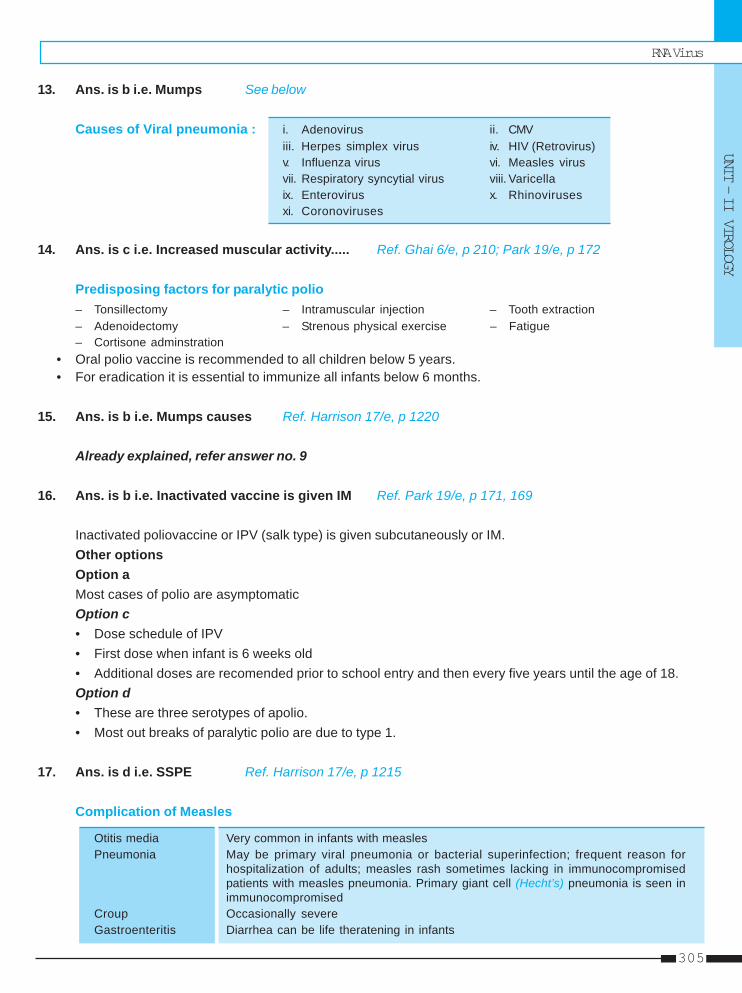
RNA Virus
UNIT – II VIROLOGY
305
13. Ans. is b i.e. Mumps See below
Causes of Viral pneumonia : i. Adenovirus ii. CMViii. Herpes simplex virus iv. HIV (Retrovirus)v. Influenza virus vi. Measles virusvii. Respiratory syncytial virus viii. Varicellaix. Enterovirus x. Rhinovirusesxi. Coronoviruses
14. Ans. is c i.e. Increased muscular activity..... Ref. Ghai 6/e, p 210; Park 19/e, p 172
Predisposing factors for paralytic polio– Tonsillectomy – Intramuscular injection – Tooth extraction– Adenoidectomy – Strenous physical exercise – Fatigue– Cortisone adminstration
• Oral polio vaccine is recommended to all children below 5 years.• For eradication it is essential to immunize all infants below 6 months.
15. Ans. is b i.e. Mumps causes Ref. Harrison 17/e, p 1220
Already explained, refer answer no. 9
16. Ans. is b i.e. Inactivated vaccine is given IM Ref. Park 19/e, p 171, 169
Inactivated poliovaccine or IPV (salk type) is given subcutaneously or IM.Other optionsOption aMost cases of polio are asymptomaticOption c• Dose schedule of IPV• First dose when infant is 6 weeks old• Additional doses are recomended prior to school entry and then every five years until the age of 18.Option d• These are three serotypes of apolio.• Most out breaks of paralytic polio are due to type 1.
17. Ans. is d i.e. SSPE Ref. Harrison 17/e, p 1215
Complication of Measles
Otitis media Very common in infants with measlesPneumonia May be primary viral pneumonia or bacterial superinfection; frequent reason for
hospitalization of adults; measles rash sometimes lacking in immunocompromisedpatients with measles pneumonia. Primary giant cell (Hecht’s) pneumonia is seen inimmunocompromised
Croup Occasionally severeGastroenteritis Diarrhea can be life theratening in infants

Self Assessment & Review Microbiology & Immunology
SECTION – B
306
Cervical adenitis Due to lymphoid hyperplasia as host response to virus; commonAcute encephalitits May be mild to severe / fatal; occurs in 1 in 1000 cases of meslesSubacute sclerosing In 1 in 100,000 cases of measles, usually when measles occured in infancy; seenpanencephalitis 5 - 10 years later.(SSPE)
18. Ans. is a i.e. 6-12 week of pregnancy Ref. Park 19/e, p 131; Harrison 17/e, p 1219
Already explained, refer see answer no. 3
19. Ans. is c i.e. Type I is responsible for vaccine induced paralytic polio myelitisRef. Park 19/e, p 169
Already explained, refer see answer no. 7
20. Ans. is a i.e. Antigen in stool Ref. Ananthnarayan 7/e, p 573; Harrison 17/e, p 1207
Diagnosis of Rotaviurs diarrheoa1. Stool examination
• Genotyping of rotavirus nucleic acid by PCR is most sensitive method.• As virus is shed in large quantities (107-1012g) in stool, diagnosis is confirmed by detecting virus in
faeces by enzyme immunoassay or viral RNA can be detected by gel electrophoresis, probe hybridization,or PCR.
• Viral shedding detectable by EIA usually subsides within a week but may persist for >30 days inimmunocompromised while PCR detect viral shedding for longer periods.
• Electron microscopy / immunoelectronmicroscopy is used to see virus in faeces.
2. Serology• IgM or IgG antibodies in the blood are increased.
21. Ans. is a i.e. Hemorrhagic fever Ref. Ananthnarayan 7/e, p 499
See manifestation of enterovirus in theory of our book.
22. Ans. is c i.e. Respiratory syncytial virus Ref. Ghai 6/e, p 352 - 353
“Child of 11 month (infant) with respiratory distress, bilateral crepitation and wheezing is a typicalpresentation of Bronchiolitis.”
• Cause of Bronchiolitis : – Respiratory syncytial virus (MC)– Parainfluenza 3, 1, 2– Adenovirus– Influenza virus– Mycoplasma pneumonia (Rarely). Mnemonic - My PAIR
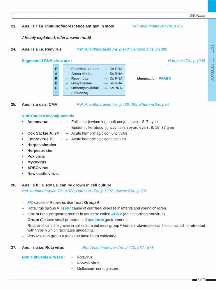
RNA Virus
UNIT – II VIROLOGY
307
23. Ans. is c i.e. Immunofluorescence antigen in stool Ref. Ananthnarayan 7/e, p 573
Already explained, refer answer no. 15
24. Ans. is a i.e. Reovirus Ref. Ananthnarayan 7/e, p 448; Harrison 17/e, p 1080
Segmented RNA virus are : ..... Harrison 17/e, p 1208
P – Picobirna viruses → Ds RNAA – Arena viridae → Ss RNAR – Reoviridae → Ds RNA Mnemonic = PARBOB – Bunyaviridae → Ss RNAO – Orthomyxoviridae → Ss RNA
(Influenza)
25. Ans. is a c i.e. CMV Ref. Ananthnarayan 7/e, p 488, 499; Khurana 2/e, p 94
Viral Causes of conjunctvitis• Adenovirus : – Follicular (swimming pool) conjunctivitis : 3, 7, type
– Epidemic keratoconjunctivitis (shipyard eye ) : 8, 19, 37 type• Cox Sackie A. 24 : – Acute hemorrhagic conjuncitivitis• Enterovirus 70 : – Acute hemorrhagic conjunctivitis• Herpes simplex• Herpes zoster• Pox virus• Myxovirus• ARBO virus• New castle virus.
26. Ans. is b i.e. Rota B can be grown in cell cultureRef. Ananthnarayan 7/e, p 572; Harrison 17/e, p 1207; Jawetz 23/e, p 507
– MC cause of Rotavirus diarrhea : Group A– Rotavirus (group A) is MC cause of diarrheal disease in infants and young children.– Group B cause gastroenteritis in adults so called ADRV (adult diarrhea rotavirus).– Group C cause small proportion of pediatric gastroenteritis.– Rota virus can’t be grown in cell culture but most group A human rotaviruses can be cultivated if pretreated
with trypsin which facilitates uncoating.– Very few non group A rotavirus have been cultivated.
27. Ans. is a i.e. Rota virus Ref. Ananthnarayan 7/e, p 473, 572 - 573
Non cultivable viruses : • Rotavirus• Norwalk virus• Mollascum contagiosum.
Self Assessment & Review Microbiology & Immunology
SECTION – B
308
28. Ans. is a i.e. Influenza virus Ref. Harrison 17/e, p 1080
Already explained, refer answer no. 24
29. Ans. is d i.e. Rota virus Ref. Harrison 17/e, p 1207
• The first rotavirus vaccine was introduced in 1998 and withdrawn because it was linked with intussuception.• In 2006 two new rotavirus vaccine have been introduced, one of this is a multivalent bovine human reassortant
rotavirus preparation. Second one is a single attenuated rotavirus strain.
30. Ans. is b i.e. Rabies Ref. Ananthnarayan 7/e, p 439, 458
• Vaccine that grow in embryonated eggs :– Influenza – Yellow fever (17 D strain)– Rabies (Flury strain) – Mumps ..... Ananthnarayan 7/e, p 514
• Varicella vaccine grown in chick embryo fibroblast culture.• Rubella – RA 27/3 vaccine produced in human diploid fibroblast. ..... Park 19/e, 131• No eggs culture vaccine measles are produced at all today. All are tissue culture vacciine, either check
embryo or human diploid cell line. ..... Park 19/e, p 120
31. Ans. is b i.e. HEV Ref. Ananthnarayan 7/e, p 447, 491
Picorna viruses
Enterovirus Rhinoviruses Hepatoviruses
↓↓↓↓↓ ↓↓↓↓↓ ↓↓↓↓↓ ↓↓↓↓↓ • HAV (enterovirus 72)Polio Coxasackie Echo Other enteroviurses
Foot and mouth disease is manifestation of entero virus and coxsackie.
32. Ans. is c and d i.e. Measles - Edmonston zogreb strain; and BCG-Danish 33 strainRef. Ananthnarayan 7/e, p 514
Vaccine Strain
Mumps Jeryl-Lynn strainChicken Pox OKA strainMeasles HDC-Edmonston Zagreb strainRubella RA 27/3BCG ‘Danis 1331’ StrainCholera CVD. 103 - HgR strainTyphoral Ty 21-a strainYellow fever 17-D Asibi strain
33. Ans. is a and c i.e. Small Pox Virus; and Ebola virus Ref. Harrison 17/e, p 1343
Bioterrorism agents• Category A :
– Anthrax (Bacillus anthracis)
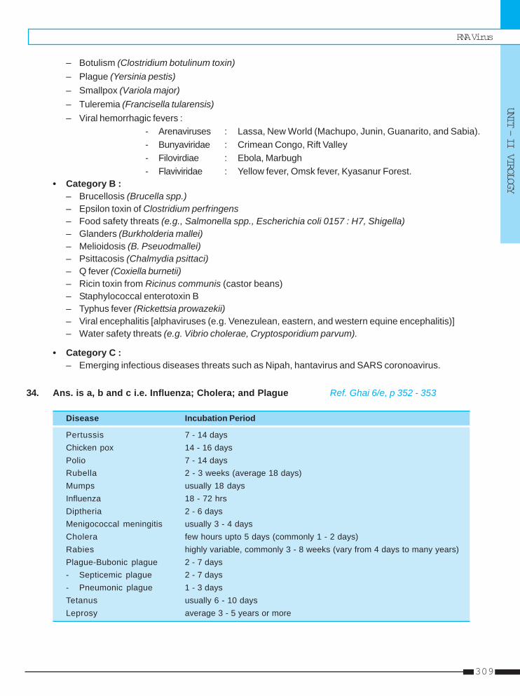
RNA Virus
UNIT – II VIROLOGY
309
– Botulism (Clostridium botulinum toxin)– Plague (Yersinia pestis)– Smallpox (Variola major)– Tuleremia (Francisella tularensis)– Viral hemorrhagic fevers :
- Arenaviruses : Lassa, New World (Machupo, Junin, Guanarito, and Sabia).- Bunyaviridae : Crimean Congo, Rift Valley- Filovirdiae : Ebola, Marbugh- Flaviviridae : Yellow fever, Omsk fever, Kyasanur Forest.
• Category B :– Brucellosis (Brucella spp.)– Epsilon toxin of Clostridium perfringens– Food safety threats (e.g., Salmonella spp., Escherichia coli 0157 : H7, Shigella)– Glanders (Burkholderia mallei)– Melioidosis (B. Pseuodmallei)– Psittacosis (Chalmydia psittaci)– Q fever (Coxiella burnetii)– Ricin toxin from Ricinus communis (castor beans)– Staphylococcal enterotoxin B– Typhus fever (Rickettsia prowazekii)– Viral encephalitis [alphaviruses (e.g. Venezulean, eastern, and western equine encephalitis)]– Water safety threats (e.g. Vibrio cholerae, Cryptosporidium parvum).
• Category C :– Emerging infectious diseases threats such as Nipah, hantavirus and SARS coronoavirus.
34. Ans. is a, b and c i.e. Influenza; Cholera; and Plague Ref. Ghai 6/e, p 352 - 353
Disease Incubation Period
Pertussis 7 - 14 daysChicken pox 14 - 16 daysPolio 7 - 14 daysRubella 2 - 3 weeks (average 18 days)Mumps usually 18 daysInfluenza 18 - 72 hrsDiptheria 2 - 6 daysMenigococcal meningitis usually 3 - 4 daysCholera few hours upto 5 days (commonly 1 - 2 days)Rabies highly variable, commonly 3 - 8 weeks (vary from 4 days to many years)Plague-Bubonic plague 2 - 7 days- Septicemic plague 2 - 7 days- Pneumonic plague 1 - 3 daysTetanus usually 6 - 10 daysLeprosy average 3 - 5 years or more
Self Assessment & Review Microbiology & Immunology
SECTION – B
310
35. Ans. is a, c and d i.e. RSV; Parainfluenza; and Influenza Ref. Ghai 6/e, p 352 - 353
Already explained, refer answer no. 17
36. Ans. is a i.e. Herpes Ref. Ananthnarayan 7/e, p 446
Enveloped DNA viruses : – Herpesviridae– Hepadnaviridae (HBV)
All RNA viruses are enveloped except PARC (Picorna, astro, Reo, Calciviridae).
ARBO AND RHABDO
37. Ans. is a i.e. Relapsing fever Ref. Park 19/e, p 635
Tick borne diseases are :Hard tick – Tick typhus – Viral encephalitis
– Tularemia – Tick paralysis– Human Babesiosis – Q fever (usually air borne disease)– Viral encephalitis (not Japenese encephalitis which is transmitted by culex)– Viral haemorrhagic fever (eg Kyasanur forest disease)
Soft tick – Relapsing fever
38. Ans. is b i.e. Rabies Ref. Harrison 17/e, p 1222
• Negri bodies are the intracytoplasmic inclusions of rabies virus in the CNS.• Negri bodies are distributed throughout the brain particularly in Ammon’s horn, the cerebral cortex, the
brain stem, the hypothalamus, purkinje cells of cerebellum and the dorsal root spinal ganglia.• They are not seen in about 20% cases of rabies and their absence does not rule out the diagnosis.
Inclusion BodiesIt is of following types :a. Intracytoplasmic eosinophilic inclusions :
Negri bodies – rabiesGuarnieri bodies – variola (small pox), vacciniaBollinger bodies – fowlpoxHenderson - peterson bodies – molluscum contagiosum
b. Intranuclear acidophilic inclusion bodies :Cowdry type A – herpes, chicken pox, CMV, yellow feverTorres bodies – yellow feverCowdry type B – polio virus
c. Both Nuclear and cytoplasmic :Warthin Finkeldey – measles
d. Intranuclear basophilic inclusion bodies :Cowdry type B – adenovirus
RNA Virus
UNIT – II VIROLOGY
311
39. Ans. is c i.e. Kyasanur Forest disease Ref. Park 19/e, p 622, 634
Already explained, refer answer no. 37
40. Ans. is c i.e. Immediately stitch wound under antibiotic coverage Ref. Park 19/e, p 229-230
“Immediate stitching is contraindicated.”• Combined administration of :
– Single dose of antirabies serum– Anti rabies (ARV) vaccine and;– Local treatment of wound is the best specific prophylactic treatment after exposure of man to rabies
(especially in all severe cases i.e. class III exposures and in all cases of unprovoked bites by wildanimals).
• Anti Rabies Serum (Passive immunity) should be given as promptly as possible after sensitivity test,irrespective of interval between exposure and begining of treatment.– It prolongs I.P if administered soon after exposure to rabies so it is particularly important in class III
bites.– It is given either as Horse antirabies serum (40 IU) or Human rabies immunoglobulin (20 IU) in which
part of the dose given around the wound and rest by IM in gluteal region.
• Local Treatment of wound :– It can reduce the chances of developing rabies by upto 80%. It consist of :
a. Cleansing with plenty of soap and water, preferably under a running tap for at least 5 minutes.b. Chemical treatment by virucidal agents either alcohol, tincture 0.01% of aqueous solution of iodine
or povidone iodine (not use savlon, cetavlon, carbolic or nitric acid).c. Suturing - Not done immediately but should be done 24 - 48 hours later.d. Antirabies serum.e. Observe the animal for 10 days.
41. Ans. is b i.e. Licks on Fresh wound Ref. Park 19/e, p 231
Classification of exposures
Class I (slight risk) : Class II (Moderate risk) Class III (severe risk)
– Licks on healthy unbroken skin – Licks on fresh cuts – All bites or scratches with– Consumption of unboiled – Scratches with oozing of blood oozing of blood on neck,
milk of suspected animal – All bites except on head, neck, head, face, palm, fingers– Scratches without oozing of blood face, palm, fingers – Lacerated wounds on
– Minor wounds less than any part of body5 in number – Multiple wounds
5 or more in number– Bites from wild animals
42. Ans. is c i.e. RNA has a negative polarity Ref. Ananthnarayan 7/e, p 535 - 536
Rabies virus are :• Bullet shaped
Self Assessment & Review Microbiology & Immunology
SECTION – B
312
• Belong to rhabdoviridae-serotype 1 (Lyssavirus type - 1) [Serotype 2, 3 and 4 are rabies related viruses].• Lipoprotein envelope carry glycoprotein spikes.• Core consist of helically arranged ribonucleoprotein and RNA dependent RNA transcriptase.• Genome is unsegmented, linear, negative sense RNA [Other -ve sense RNA viruses - Ortho,
paramyxoviridae].• Virus is inactivated by phenol, formalin, betapropioplactone, ultraviolet, irradiation, sunlight.• Virus has hemagglutinating activity due to spikes.• Virus spreads centripetally from site of inoculation (within striated muscle), than ascends throgh nerve
associated tissue space and than spreads centrifugally in peripheral autonomic nerves to many tissuesincluding salivary glands. Thus pathogenesis does not show that it has affinity for motor neurons.
43. Ans. is c i.e. KFD is transmitted by ticks Ref. Park 18/e, p 227 - 228, 602; 19/e, p 622
• List of tick transmitted disease is already mentioned in question no. 32• Yellow fever is an exotic disease for India i.e. disease which may be imported in India or India is yellow
fever ‘receptive’ area that is “an area in which yellow fever does not exist but where condition would permitits development if introduced”.
• Dengue virus has at least 4 serotypes not one.• Japanese encephalitis is transmitted by culex not Aedes.
Remember : Other culex transmitted diseases are : – Bancroftian filariasis– West Nile fever– Viral arthritis (epidemic / polyarthritis).
44. Ans. is a i.e. Incubation period depends on the site of biteRef. Park 18/e, p 218; 19/e, p 219; Ananthnarayan 7/e, p 449, 539
• Incubation period of Rabies depends on the :– Site of bite – Severity of bite– Number of wounds – Amount of virus injected– Species of biting animal – Protection provided by clothing and treatment undertaken.
• I.P is shorter in :– Severe exposures – Bites on face, head, neck and upper extremities– Bites by wild animals.
• In India most of human rabies cases have resulted from dog bites but it also occurs by bite of cat,monkey, horse, Jackals, fox-hyena etc.
• Also transmitted by licks on abraded skin and mucosa; aerosols; corneal and organ transplants.• Characteristic of rabies is Negri Body which is ovoid eosinophilic intracytoplasmic inclusion bodies.• Rabies is bullet shaped SS negative sense RNA virus.
45. Ans. is d i.e. Recombinant glycoprotein vaccine Ref. Park 18/e, p 219; 19/e, p 229
Rabies Vaccines• Vaccine currently in use are of three types
a. Nervous tissues vaccine : BPL inactivaed Adult sheep (Semple type) and Suckling mouse brain vaccine.
RNA Virus
UNIT – II VIROLOGY
313
b. Duck embryo vaccine : Not available in India.c. Cell culture vaccine -
– Human diploid cell vaccine (HDCV) used in India for both pre and post exposure immunzation.– Second generation tissue culture vaccine eg. Chick embryo fibroblast, vero continous cell vaccine
etc it is recommended by WHO.
46. Ans. is d i.e. Rabies is best diagnosed by immunoflorescence studyRef. Ananthnarayan 7/e, p 539; Park 18/e, p 217; 19/e, p 227
Diagnosis of Human Rabies– Specimen
Antemortem Corneal smears, skin biopsy from face or neck, saliva.Postmortem : Brain
– Method most commonly used for diagnosis is the demonstration of rabies virus antigens byimmunofluorescence (direct or using monoclonal antibodies).
– Demonstration of negri bodies in the brain or spinal cord.– Isolation of virus by intracerebral inoculation in mice; from the brain, CSF, saliva, urine.– Rapid isolation is done by tissue culture cell lines.– High titre rabies specific antibodies in CSF (Not seen after immunization) by Fluorescent antibody test.– Detection of rabies virus RNA in saliva by Reverse transcription PCR.
Remember : • Rabies infection terminates in death, not life long immunity.• Rabies virus has single serotype i.e. Lyssavirus type 1.• Rabies vaccine is killed inactivated vaccine.
– Inactivation is commonly done by phenol or Betapropiolactone.
47. Ans. is b i.e. Hippocampus Ref. Harrison 17/e, p 1222
Sites of Negri bodies• Cerebellum and hippocampus .....Ananthnarayan 7/e, p 539• Neurons of Ammon horn, cerebral cortex, brain stem, hypothalmus, purkinje cells of cerebellum and dorsal
spinal ganglia. .... Harrison 16/e, p 1157
48. Ans. is a i.e. Hanta virus pulmonary syndrome is caused by inhalation of rodent urine Ref.Jawetz 24/e, p 525; Park 19/e, p 240 - 241
Hantavirus are classified in the hantavirus genes of the Bunyaviridae family. It cause two serious and oftenfatal disease.– Hemorrhagic fever with renal syndrome.– Hanta virus pulmonary syndrome.• Hantavirus are natural pathogen of rodents. Viremia is present in infected rodents and the virus is shed in
urine, feces and saliva in high titres. Transmission from rodent to rodent and rodent to human is primarilyrespiratory by inhalation of virus contained in dried excreta.
Other options :• KFD is a febrile disease caused by an arbovirus and transmitted to man by bite of infective tick.• Chikugunya fever - Dengue like disease caused by chikugunya virus and transmitted by Aedes, Culex and
Mansonia mosquito.
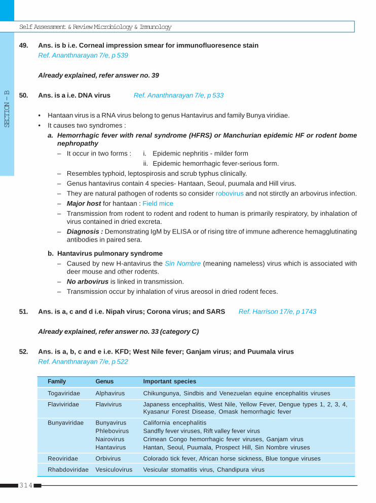
Self Assessment & Review Microbiology & Immunology
SECTION – B
314
49. Ans. is b i.e. Corneal impression smear for immunofluoresence stainRef. Ananthnarayan 7/e, p 539
Already explained, refer answer no. 39
50. Ans. is a i.e. DNA virus Ref. Ananthnarayan 7/e, p 533
• Hantaan virus is a RNA virus belong to genus Hantavirus and family Bunya viridiae.• It causes two syndromes :
a. Hemorrhagic fever with renal syndrome (HFRS) or Manchurian epidemic HF or rodent bomenephropathy– It occur in two forms : i. Epidemic nephritis - milder form
ii. Epidemic hemorrhagic fever-serious form.– Resembles typhoid, leptospirosis and scrub typhus clinically.– Genus hantavirus contain 4 species- Hantaan, Seoul, puumala and Hill virus.– They are natural pathogen of rodents so consider robovirus and not stirctly an arbovirus infection.– Major host for hantaan : Field mice– Transmission from rodent to rodent and rodent to human is primarily respiratory, by inhalation of
virus contained in dried excreta.– Diagnosis : Demonstrating IgM by ELISA or of rising titre of immune adherence hemagglutinating
antibodies in paired sera.
b. Hantavirus pulmonary syndrome– Caused by new H-antavirus the Sin Nombre (meaning nameless) virus which is associated with
deer mouse and other rodents.– No arbovirus is linked in transmission.– Transmission occur by inhalation of virus areosol in dried rodent feces.
51. Ans. is a, c and d i.e. Nipah virus; Corona virus; and SARS Ref. Harrison 17/e, p 1743
Already explained, refer answer no. 33 (category C)
52. Ans. is a, b, c and e i.e. KFD; West Nile fever; Ganjam virus; and Puumala virusRef. Ananthnarayan 7/e, p 522
Family Genus Important species
Togaviridae Alphavirus Chikungunya, Sindbis and Venezuelan equine encephalitis viruses
Flaviviridae Flavivirus Japaness encephalitis, West Nile, Yellow Fever, Dengue types 1, 2, 3, 4,Kyasanur Forest Disease, Omask hemorrhagic fever
Bunyaviridae Bunyavirus California encephalitisPhlebovirus Sandfly fever viruses, Rift valley fever virusNairovirus Crimean Congo hemorrhagic fever viruses, Ganjam virusHantavirus Hantan, Seoul, Puumala, Prospect Hill, Sin Nombre viruses
Reoviridae Orbivirus Colorado tick fever, African horse sickness, Blue tongue viruses
Rhabdoviridae Vesiculovirus Vesicular stomatitis virus, Chandipura virus
RNA Virus
UNIT – II VIROLOGY
315
53. Ans. is a, c and d i.e. Hantaviurs pulmonary syndrome; Transmitted by rodents; and Hemorrhagicfever with renal failure Ref. Ananthnarayan 7/e, p 533
Already explained, refer answer no. 50
54. Ans. is a i.e. Amplifier Ref. Park 18/e, p 228 - 229; 19/e, p 239
Japanese encephalitis• Vector : – Culex tritaneorhynchus (most important)
– C. vishnvi– C. gelidus.
• Man is an incidental ‘dead end’ host.• Animal host :
Pigs – major vertebrate host.– not manifest any symptoms so acts as as an amplifiers.
Cattles and buffaloes – act as mosquito attractants.Horses – only domestic animals which shows signs of encephalitis.
• Birds host – Pond herons (reservoir host), Cattle egrets, poultry and ducks.• Incubation period in man – 5 - 15 days.• Average period between onset of illnes and death is about 9 days.
Remember :Killed mouse brain vaccine is available by which immunity develops after one month of second dose.
55. Ans. is d i.e. All Ref. Ananthnarayan 7/e, p 536
• Inactivating agent of rabies virus :– Phenol – Formalin– Betapropiolactone – UV irradiaction– Sunlight.
• Thermal inactivation occurs in one hour at 500C and 5min at 600C.
56. Ans. is a i.e. Rabies Ref. Ananthnarayan 7/e, p 499; Harrison 17/e, p 1222
Already explained, refer answer no. 38
57. Ans. is b i.e. Causes Hemorrhagic fever with renal involvement Ref. Ananthnarayan 7/e, p 533
Already explained, refer answer no. 50
Self Assessment & Review Microbiology & Immunology
SECTION – B
316
1. c) Typhoid 2. a, b and d 3. a) Hippocampus 4. b and d 5. e) All 4 types6. c) Tick 7. b) Lassa ... 8. c) Semple ... 9. c) Enterovirus ... 10. c) Terminal ...
1. Man is the only reservoir of : [AIIMS 90]a) Rabiesb) Measlesc) Typhoidd) Japanese B encephalitis
[Ref. Park 19/e, p 127]2. Which of the following statemtn is/are true of all
paramyxoviruses : [DNB 90]a) They contain a single-standed RNA genome of
negative polarityb) Envelopes are dervied from the hostcells plasma
membranec) They have a cytoplasmic site of replicationd) They enter the body by the respiratory route
[Ref. Ananthnarayan 7/e, p 431, 437, 512]3. Negri Bodies are commonly seen in :
a) Hippocampus [AIIMS 90, 92; AI 88]b) Hypothalamusc) Mamillary bodiesd) Cerebrume) Pons
[Ref. Ananthnarayan 7/e, p 539]4. Which of the following vaccine [s) should not be
given to an immunodeficient patient : [DNB 90]a) Influenza vaccineb) Rubella vaccinec) Pneumococcal vaccined) Trivalent oral polio vaccine
[Ref. Park 19/e, p 97]5. In india, human infections has been reported den-
gue virus type : [AP 91]a) Types I and 1b) Types 1 and 3c) Types 2 and 4
Answer
Chapter Review••••• This section includes questions of VThis section includes questions of VThis section includes questions of VThis section includes questions of VThis section includes questions of Various Other PGMEES from 1990 – 2007.arious Other PGMEES from 1990 – 2007.arious Other PGMEES from 1990 – 2007.arious Other PGMEES from 1990 – 2007.arious Other PGMEES from 1990 – 2007.••••• Questions are arQuestions are arQuestions are arQuestions are arQuestions are ar ranged in decreasing order of yearranged in decreasing order of yearranged in decreasing order of yearranged in decreasing order of yearranged in decreasing order of yearwiz. This is done to makwiz. This is done to makwiz. This is done to makwiz. This is done to makwiz. This is done to make refere refere refere refere refer ralralralralral
system easier and uncomplicated to save the precious time of PGMEE Aspirant.system easier and uncomplicated to save the precious time of PGMEE Aspirant.system easier and uncomplicated to save the precious time of PGMEE Aspirant.system easier and uncomplicated to save the precious time of PGMEE Aspirant.system easier and uncomplicated to save the precious time of PGMEE Aspirant.
d) Type 1 onlye) All 4 types
[Ref. Park 19/e, p 206]6. KFD is transmitted by : [AI 91]
a) Fleasb) Mitec) Tickd) Mosquito
[Ref. Ananthnarayan 7/e, p 531]7. Which of the following is not common in India :
a) Japnese B encphalitis [AI 91]b) Lassa feverc) KFDd) Dengue
[Ref. Park 19/e, p 238]8. Neurological complications following Rabies vac-
cines is common with : [Jipmer 91]a) HDC Vaccineb) Chick embryo vaccinec) Semple vaccined) Duck egg vaccine
[Ref. Park 19/e, p 219]9. Acute epidemic keratoconjuctivitis is caused by :
a) Herpes virus [AI 91]b) Echo 51c) Enterovirus 70d) Enterovirus 72
[Ref. Ananthnarayan 7/e, p 499]10. The following is true of Rota virus : [AI 92]
a) Easily grown in cell cultureb) Double stranded DNAc) Terminal ileum villi destroyedd) Adult and old people account for 60% of infection
[Ref. Ananthnarayan 7/e, p 572; Harrison 17/e,p 1207]
RNA Virus
UNIT – II VIROLOGY
317
11. None 12. c) Neurons 13. c) Infects only ... 14. b) Decreased ... 15. b) Herpes ...16. c) Chicken ... 17. c) Culex ... 18. a) Rabies 19. b) Mumps 20. b) Chick ...21. d) Fluorescent 22. a and c 23. c) Measles ... 24. a) Herpangina
Answer
11. Which of the following does not cause conjuctivitis:a) Adeno virus [JIPMER 92]b) Entero virusc) Coxasackie virusd) Herpes virus
[Ref. Ananthnarayan 7/e, p 477, 488, 499]12. Negri bodies are located in : [Bihar 91; DNB 92]
a) Astrocytesb) Oliogodendrogliac) Neuronsd) Microglia
[Ref. Ananthnarayan 7/e, p 539]13. All are true of rabies Except : [AIIMS 92]
a) 100% mortalityb) Spreads from peripheryc) Infects only the braind) Prophylactic immunisation of people at Risk
[Ref. Ananthnarayan 7/e, p 537]14. Diarrrhoea in Rota virus infection is due to :
a) Increased intestinal hypermotility [PGI 93]b) Decreased absorption by villic) Increased Secretion by villid) None of the above
[Ref. Harrison 16/e, p 1142]15. Virus causing chicken pox belongs to : [AI 93]
a) Pox virusb) Herpes virusc) Coxsackie virusd) Adeno virus
[Ref. Ananthnarayan 7/e, p 479]16. Latent infection is seen in : [AI 93]
a) Small poxb) Adeno virusc) Chicken poxd) Measles
[Ref. Ananthnarayan 7/e, p 474]17. Japanese B encephalitis virus is transmitted by :
a) Aedes aegypti [JIPMER 86, NIMHANS 87,b) Culex fatigans Kar. 94]c) Culex tritaeniorynchusd) Hard ticke) Soft tick
[Ref. Ananthnarayan 7/e, p 526]
18. Negri bodies are seen in : [Kerala 94]a) Rabiesb) Small poxc) Trachomad) Lymphogranuloma venerum
[Ref. Ananthnarayan 7/e, p 539]19. Virus causing pneumonia are all except :
a) Cytomegalo virus [JIPMER 95]b) Mumpsc) Herpesd) Mealses
[Ref. Harrison 17/e, p 109]20. Which ARV has been recommended by WHO as
the most effective : [TN 95]a) Duck cell vaccineb) Chick fibroblast vaccinec) HDCVd) Sheep brain vaccine
[Ref. Park 19/e, p 229]21. The staining useful for antemorten diagnosis of
rabies is : [Karn. 95]a) Sellerb) Macchiavilloc) Giemsad) Fluorescent
[Ref. Ananthnarayan 7/e, p 539]22. Post exposure immunization is done for :
a) Measles [JIPMER 95]b) Polioc) Rabbiesd) Chicken pox
[Ref. Park 19/e, p 129, 229]23. Subacute sclerosing panencephalitis is associ-
ated with : [Delhi 96]a) Chicken poxb) Rabiesc) Measles virusd) Polio virus
[Ref. Ananthnarayan 7/e, p 568]24. In which of the following virus is shed in stool :
a) Herpangina [UP 96]b) Influenzac) Varicellad) Small pox
[Ref. Ananthnarayan 7/e, p 497]
Self Assessment & Review Microbiology & Immunology
SECTION – B
318
25. c and e 26. d) Edmonston ... 27. a) Bronchiolitis 28. d) Hexaminidase ... 29. d) Passive ...30. b) Reovirus 31. c) Culex ... 32. b) Genetically ... 33. a and d 34. b) Infantile ...35. d) Picorna ... 36. b and c 37. c) Girls ...
Answer
25. Which viral infection always causes clinical dis-ease in human beings : [Kerala 96, 97]a) Rubellab) Poliomyelitisc) Measlesd) Chicken poxe) Rabies
[Ref. park 19/e, p 127, 228]26. Strain used in measles vaccine is : [UP 99]
a) Salkb) Sabinc) TAFd) Edmonston jagreb
[Ref. Park 19/e, p 129]27. RSV causes : [UP 99]
a) Bronchiolitisb) Mumpsc) Croupd) Bronchitis
[Ref. Ananthnarayan 7/e, p 516]28. True about influenza A is : [UP 00]
a) Double strained segmented RNAb) Pandemic are caused by antigenic driftc) Nucleocapsid antibody is specificd) Hexaminidase and Neuraminidase is type spe-
cific[Ref. Ananthnarayan 7/e, p 504-505]
29. True about picorna virus are A/E : [UP 00]a) OPV provides herd immunityb) Immunity in poliomyelitis is type specificd) Best method of isolation is tissue cultured) Passive immunization of human immunoglobu-
lin has more value[Ref. Ananthnarayan 7/e, p 491 - 492; Park, 19/e,p 170]
30. Which of the following viruses is composed of twodistinct capsids enclosing the double strandedRNA? [Kar 00]a) Adenovirusb) Reovirusc). Herpes virusd) Myxovirus
[Ref. Jawetz 24/e, p 502]31. The vector for Japanese encephalitis is :
a) Lice [Kar 00]b) Tick
c) Culex mosquitod) Sandfly [Ref. Park 19/e, p 238]
32. Type of vaccine available commercially for rabiesare all except : [UP 00]a) Inactivated sheep brain vaccineb) Genetically engineered glycoprotein vaccinec) Duck embryo cultured vaccined) Human diploid cell vaccine
[Ref. Park 19/e, p 229]33. All enveloped helical RNA viruses belong to one
large group, which includes all of the following ex-cept : [Kar 01]a) Influenzab) Parainfluenzac) Mumpsd). Herpes
[Ref. Ananthnarayana 7/e, p 475, 501]34. Rotaviruses are responsible for : [Kar 01]
a) Acute nonbacterial gastroenteritis in adultsb) Infantile diarrheac) Teratogenic effectsd) Respiratory tract infection in immunocompro-
mised individuals[Ref. Harrison 17/e, p 1206]
35. Epidemic hemorrhagic conjunctivtis is caused :a) HSV [UP 02]b) HZVc) HIVd) Picorna virus
[Ref. Ananthnarayansa 7/e, p 499; Table 54.2]36. The congenital rubella syndrome : [DNB 02]
a) May be prevented by vaccination in early preg-nancy
b) Causes intra uterine growth retardationc) Causes cataractsd) Causes deafness only if acquired before 16
weeks of gestation[Ref. Park 19/e, p 130-131]
37. Rubella vaccination is contra indicated in all ex-cept : [JIPMER 02]a) Patient on immunosuppressantb) Girl with Leukemiac) Girls between 11-14 yearsd) Pregnancy
[Ref. Park 19/e, p 131]
RNA Virus
UNIT – II VIROLOGY
319
Answer 38. a) 3 doses 39. c) Arbo 40. None 41. d) HDCV 42. b) Multiple ...43. b) Enterovrus 44. a) A. hemorrhagic ... 45. c) Dengue 46. a) RSV 47. c) Primary ...48. a) RSV 49. b) Rabies ... 50. a) Louis ... 51. b) Aseptic...
38. Pre-exposure cell culture vaccine used in Rabiesa) 3 doses [UP 02; AI 90]b) 4 dosesc) 5 dosesd) 6 doses
[Ref. Park 19/e, p 233-234]39. Break bone fevers caused by which virus :
a) Variola [Bihar 03]b) Coxsackiec) Arbod) Adeno virus
[Ref. Anantharayan 7/e, p 530]40. Epidemic of polio is due to : [Kolkata 03]
a) Type I virusb) Type II virusc) Type III virusd) Combine of type II and type III virus
[Ref. Park 19/e, p 168]41. Recommended vaccines for rabies : [Kar 04]
a) DPTb) MMRc) BCGd) HDCV
[Ref. Park 19/e, p 229]42. About rabies true is : [SGPGI 05]
a) Vaccine causes life long immunityb) Multiple strains are foundc) CNS infention occurs through viremiad) Bullet-shaped nonenveloped, double stranded
RNA virus[Ref. Ananthnarayan 7/e, p 535 - 537]
43. Which of the following is associated with acutehemorrhagic conjunctivitis ? [Bihar 05]a) Rhabdovirusb) Enterovrusc) Calcivirusd) Echovirus
[Ref. Ananthnarayan 7/e, p 499]44. Enteroviruses cause : [Bihar 05]
a) A. hemorrhagic conjunctivitisb) Ac. follicular conjunctivitisc) Posterior follicular conjunctivitisd) Epidermic kerato conjunctivitis
[Ref. Ananthnaryan 7/e, p 499]
45. Break one fever is caused by which virus :a) Varioloa [Bihar 05]b) Coxsackiec) Dengued) Adenoa virus
[Ref. Anathnarayan 7/e, p 530]46. Commonest cause of Bronchiolitis is : [UP 05]
a) RSVb) Adenovirusc) Influenzad) Herpes virus
[Ref. Ghai 6/e, p 352]47. Which of the following is true regarding influenza:
a) It is caused by an enveloped DNA virusb) Laboratory studies may show neutropenia early
in the course of disease [MP 06]c) Primary infectious pneumonia is less common
than secondary bacteria pneumoniad) Antiviral agents is given early prevents compli-
cations[Ref. Harrison 17/e, p 1129]
48. Virus lacking hemagglutinin and nuraminidase buthave membrane fusion protein is :a) RSV [Jharkhand 06]b) CMVc) HSVd) Ebestein Barr virs
[Ref. Anantharayan 7/e, p 516]49. Negri bodies are seein in infections due to :
a) Polio virus [Kar 06]b) Rabies virusc) Herpes virusd) Aenovirus
[Ref. Ananthnarayan 7/e, p 539]50. A vaccine for rabies was first developed by :
a) Louis psteur [Kar 06]b) Robert kochc) Edward jennerd) Landsteiner
[Ref. Ananthnarayan 7/e, p 2]51. Coxsackie group A commonly causes : [TN 01]
a) Conjunctivitisb) Aseptic meningitisc) Hepatitisd) Myocarditis
[Ref. Ananthnarayan 7/e, p 497]
Slow virus infections have the following characteristics :– Incubation periods range from months to year.– Course of illness lasting for months or years with remissions and exacerbations.– Predilection for involvement of central nervous system.– Absence of immune response or an immune response that does not arrest the disease but may actually contribute
to pathogenesis.– Genetic predisposition.– Invariable fatal termination.
Remember : MC prion disorder in humans is sporadic form of CJD.
ClassificationI. Group A
• Infections of sheep caused by lentiviruses– Eg visna maedi
II. Group B = Subacute Spongioform viral encephalopathies• Comprise of prion (infectious protein) diseases.• These are chronic progressive degenerative diseases of CNS.• Pathology consists of progressive vacuolation in the dendritic and axonal process of neurons and extensive
astroglial hypertrophy and proliferations which leads to spongiform degeneration in the grey matter. There isno sign of any inflammation or immune response.
• Mechanism– Proliferation of an abnormal prion protein (PrPse) which is derived from the normal prion protein PrPe.– PrPse is not simply a misfolded protein but it is an alternatively folded molecule with a function.
Disease Mechanism of Pathogenesis
Human
a. Kuru (meaning tremor) Infection through ritualistic cannibalismb. Iatrogenic Creutzfeldt Jakob disease (CJD) Infection from prion contaminated human growth hormone,
duramater graft, corneal transplantc. Variant (CJD) Infection from bovine prion (Eating BSE infected beef)
Slow VirSlow VirSlow VirSlow VirSlow Virus Diseasesus Diseasesus Diseasesus Diseasesus Diseases33
Slow Virus Diseases
UNIT – II VIROLOGY
321
Continue ........
d. Familial CJD Germline mutation in PRNPe. Sporadic CJD Somatic mutation or spontaneous conversion of PrP
c into PrPsc
f. Fatal familial insomania Germline mutation in PRNPg. Gestmann strausster Scheinker Germline mutation in PRNP
Animal prion disease eg.a. Scrapieb. Mink encephalopathyc. Bovine spongiform encephalopathy (BSE, ‘mad cow disease).
III. Group C - eg.a. Subacute sclerosing panecephalitis (SSPE)
• It is delayed sequel to infection with defective measles virus.• Virus cannot be isolated in routine culture but only by co-cultivation of infected brain cells with susceptible
cells of non neural origin.• Antibody is regularly found in CSF and is pathognomonic.• SSPE also seen in rubella infection.
b. Progressive multifocal leucoencephalopathy (PML)• Seen in elderly persons whose immune proces is impaired by malignancy or HIV etc.• Caused by JC virus (papovavirus).
Remember : JC virus also cause Hodgkins disease of brain.
Self Assessment & Review Microbiology & Immunology
SECTION – B
322
1. Prion are : [AI 08]a) Infectious proteinsb) Made up of bacteria and virus particlesc) Nuclear materiald) Can be cultured in cell free media
2. Prions consists of : [AIIMS 07]
a) DNA and RNA
b) DNA, RNA and proteins
c) RNA and proteins
d) Only proteins
3. Which of the following is not prion associateddisease : [AIIMS 03]
a) Scarpie
b) Kuru
c) Creutzfedt - Jakob disease
d) Alzheimer disease
4. True about Prion disease is all except :
a) Myoclonus is seen in 10% of the patients
b) Caused by infectious protein [AIIMS 01]
c) Brain biopsy is diagnostic
d) Commonly manifests as dementia
5. Fatal familial insomnia is associated with :
a) Prion disease [AI 99]
b) Degenerative disease
c) Neoplastic disease
d) Vascular disease
6. Creutzfeld Jakob disease is caused by :
a) Prion [PGI 99]
b) JC virus
c) Genetic factors
d) Nutritional deficiency
QUESTIONS
Answer 1. a) Infectious ... 2. d) Only ... 3. d) Alzheimer ... 4. a) Myoclonus is ...5. a) Prion ... 6. a and c
Slow Virus Diseases
UNIT – II VIROLOGY
323
1. Ans. is a i.e. Infectious proetin Ref. Jawetz 24/e, p 2, 581
Prions are infectious protein devoid of nucleic acid.• Prions are usually resistant to standard means of inactivation. They are resistant to treatment with
formaldehyde, urea, dry heat, boiling. However they are sensitive to phenol, ether, autoclaving.• Prions infect and propagate by refolding abnormally into a structure which is able to convert normal
molecule of protein in to the abnormally structure form.
2. Ans. is d i.e. Only protein Ref. Jawetz 24/e, p 581
Already explained, refer answer no. 1
3. Ans. is d i.e. Alzheimer disease Ref. Harrison 17/e, p 2647
Various prion are as follows :
• Kuru • Creutzfeldt Jakob disease (CJD)• Fatal familial insomnia (FFI) • Gerstmann - Straussler - Scheinker disease (GSS)• Sporadic fatal insomnia (SFI) • Scrapie• Transmissible mink encephalopathy • Bovine spongiform encephalopathy (mad cow disease)• Chronic wasting disease • Feline spongiform encephalopathy• Exotic ungulate encephalopathy
4. Ans. is a i.e. Myoclonus is seen in 10% of the patients Ref. Harrison 17/e, p 2646 - 2650
• Prion is proteinaceous infectious particle that lacks nucleic acid and causes slow virus disease.• MC human prion disease is sporadic CJD.• Clinical Features of CJD :
– Most patient present with deficits in higher cortical function which almost always progress to dementia.– 90% patienty exhibit myoclonus which persist during sleep in comparison of other involuntary
movements.– Also present with visual impairment or cerebellar gait, coordination deficit, extrapyramidal dysfunction,
pyramidal signs, seizures.• Diagnosis :
1. Constellation of dementia, myoclonus and peirodic electrical burst in an afebrile 60 year old patientgenerally indicates CJD.
2. Only specific diagnostic test for CJD is measurement of PrPsc
3. In humans the diagnosis of CJD as established by brain biopsy if prpsc is detected.– There is no abnoramlity on gross examination of brain.– Pathologic hallmarks are spongiforms degeneration (in cerebral cortex, putamen etc) and Astrocytic
glycosis.– 10% of CJD patient have amyloid plaques.
EXPLANATIONS AND REFERENCES WITH ILLUSTRATIVE ANSWERS
Self Assessment & Review Microbiology & Immunology
SECTION – B
324
4. CT may be normal or show cortical atrophy.5. Sequencing the PRNP gene.6. CSF is nearly always normal but may show minimal protein elevation.
5. Ans. is a i.e. Prion disease Ref. Ananthnarayan 7/e, p 567
Already explained, refer answer no. 1
6. Ans. is a and c i.e. Prion; and Genetic factors Ref. Harrison 17/e, p 2649
• Genetic factors are involved in familial form of CJD.• Missense mutation and expansions in the octapeptide repeat region of the gene are responsible for
familial forms of prion disease.
Slow Virus Diseases
UNIT – II VIROLOGY
325
1. a) Slow virus 2. d) Kuru
1. Mad cow disease is due to :a) Slow virus [Culcutta 2K, UPGMEE 97]b) Mycoplasmac) Bacteriad) Fungus
[Ref. Ananthnarayan 7/e, p 567]
Answer
Chapter Review••••• This section includes questions of VThis section includes questions of VThis section includes questions of VThis section includes questions of VThis section includes questions of Various Other PGMEES from 1990 – 2007.arious Other PGMEES from 1990 – 2007.arious Other PGMEES from 1990 – 2007.arious Other PGMEES from 1990 – 2007.arious Other PGMEES from 1990 – 2007.••••• Questions are arQuestions are arQuestions are arQuestions are arQuestions are ar ranged in decreasing order of yearranged in decreasing order of yearranged in decreasing order of yearranged in decreasing order of yearranged in decreasing order of yearwiz. This is done to makwiz. This is done to makwiz. This is done to makwiz. This is done to makwiz. This is done to make refere refere refere refere referralralralralral
system easier and uncomplicated to save the precious time of PGMEE Aspirant.system easier and uncomplicated to save the precious time of PGMEE Aspirant.system easier and uncomplicated to save the precious time of PGMEE Aspirant.system easier and uncomplicated to save the precious time of PGMEE Aspirant.system easier and uncomplicated to save the precious time of PGMEE Aspirant.
2. “Human cannibalism” is associated with :a) Q fever [UP 06]b) Sleeping sicknessc) Trachomad) Kuru
[Ref. Ananthnaranayan 7/e, p 567]
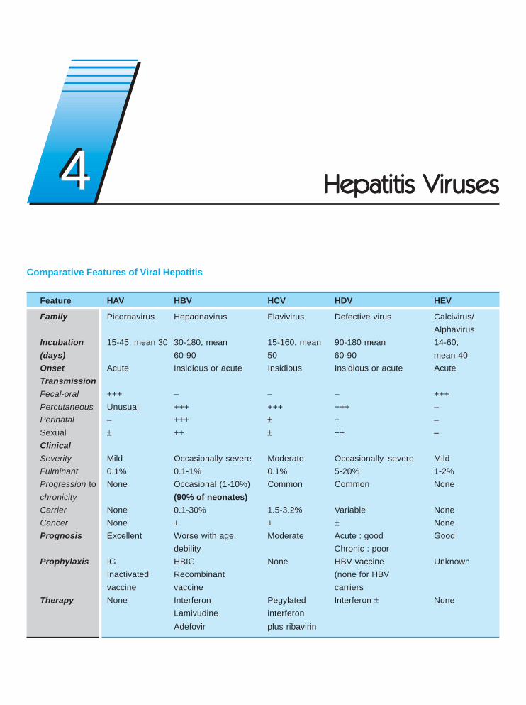
Hepatitis VirHepatitis VirHepatitis VirHepatitis VirHepatitis Virusesusesusesusesuses
Comparative Features of Viral Hepatitis
Feature HAV HBV HCV HDV HEV
Family Picornavirus Hepadnavirus Flavivirus Defective virus Calcivirus/Alphavirus
Incubation 15-45, mean 30 30-180, mean 15-160, mean 90-180 mean 14-60,(days) 60-90 50 60-90 mean 40Onset Acute Insidious or acute Insidious Insidious or acute AcuteTransmissionFecal-oral +++ – – – +++Percutaneous Unusual +++ +++ +++ –Perinatal – +++ ± + –Sexual ± ++ ± ++ –ClinicalSeverity Mild Occasionally severe Moderate Occasionally severe MildFulminant 0.1% 0.1-1% 0.1% 5-20% 1-2%Progression to None Occasional (1-10%) Common Common Nonechronicity (90% of neonates)Carrier None 0.1-30% 1.5-3.2% Variable NoneCancer None + + ± NonePrognosis Excellent Worse with age, Moderate Acute : good Good
debility Chronic : poorProphylaxis IG HBIG None HBV vaccine Unknown
Inactivated Recombinant (none for HBVvaccine vaccine carriers
Therapy None Interferon Pegylated Interferon ± NoneLamivudine interferonAdefovir plus ribavirin
44
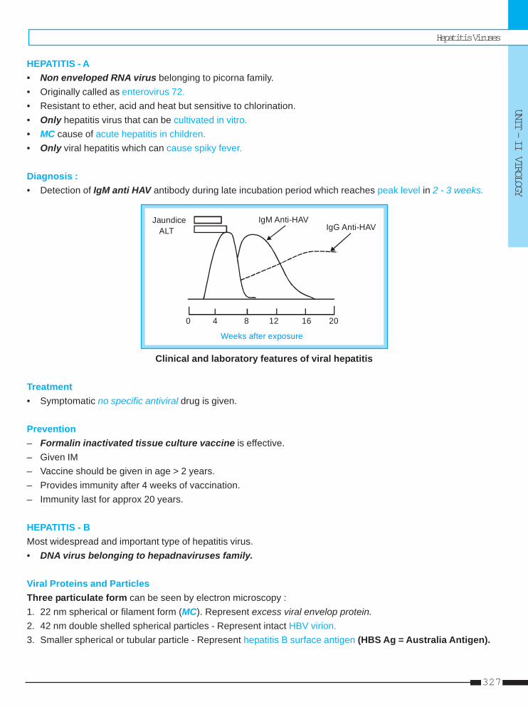
Hepatitis Viruses
UNIT – II VIROLOGY
327
HEPATITIS - A• Non enveloped RNA virus belonging to picorna family.• Originally called as enterovirus 72.• Resistant to ether, acid and heat but sensitive to chlorination.• Only hepatitis virus that can be cultivated in vitro.• MC cause of acute hepatitis in children.• Only viral hepatitis which can cause spiky fever.
Diagnosis :• Detection of IgM anti HAV antibody during late incubation period which reaches peak level in 2 - 3 weeks.
IgM Anti-HAVIgG Anti-HAV
Jaundice ALT
0 4 8 12 16 20
Weeks after exposure
Clinical and laboratory features of viral hepatitis
Treatment• Symptomatic no specific antiviral drug is given.
Prevention– Formalin inactivated tissue culture vaccine is effective.– Given IM– Vaccine should be given in age > 2 years.– Provides immunity after 4 weeks of vaccination.– Immunity last for approx 20 years.
HEPATITIS - BMost widespread and important type of hepatitis virus.• DNA virus belonging to hepadnaviruses family.
Viral Proteins and ParticlesThree particulate form can be seen by electron microscopy :1. 22 nm spherical or filament form (MC). Represent excess viral envelop protein.2. 42 nm double shelled spherical particles - Represent intact HBV virion.3. Smaller spherical or tubular particle - Represent hepatitis B surface antigen (HBS Ag = Australia Antigen).
Self Assessment & Review Microbiology & Immunology
SECTION – B
328
HBV Genes and Gene Products
Genes Regions Gene products
S S Major protein (S)S+Pre-S2 Middleprotein (M) HBs AgS+Pre-S1 and S2 Large protein (L)
C C HbcAgC+Pre-C HbeAg
P (Largest gene) DNA polymeraseX HBx Ag (contributes to carcinogenesis)
Mutants of HBVTwo types of mutant are found :• Hbe Ag Negative phenotype (MC mutant) : Results in severe chronic infection with detectable HBV DNA and
anti HBe Ag but HBe Ag is not detectable.• Escape mutant : Due to change in HBs Ag. Vaccine is not effective against them.
Pathogenesis and Clinical features1. Acute hepatitis
• Low grade fever, jaundice, splenomegaly and tender hepatomegaly is found in all acute viral hepatitis.• Non of hepatitis virus is known to be directly cytopathic to hepatocyte, it is immunologic response which
cause cell injury.
2. Chronic hepatitis• Persistence of HBeAg beyond 3 month or Persistance of HBsAg beyond 6 months after acute hepatitis.
3. Perinatal transmission• Occurs primarily in infants born to HBSAg carrier mothers or mothers with acute hepatitis B during third
trimester.• Likelihood of perinatal transmission of HBV correlates with presence of HBeAg; 90% of HBeAg positive
mothers but only 10 to 15% of anti HBe positive mother transmit infection.• Most infection are transmitted at the time of delivery and are not related to breast feeding.
..... Harrison, 17/e, p 1940• Mostly acute infection in neonate is clinically asymptomatic, but the child is very likely to become HBs Ag
carrier.
4. Extrahepatic manifestation of HBV• Glomerulonephritis• Polyarteritis nodosa• Essential mixed cryoglobulinemia.
5. Carrier stageCarriers are more common in patients of :• Down’s syndrome • Lepromatous leprosy• Leukemia • Hodgkins disease• Polyateritis nodosa • Chronic renal disease• Infection drug users– Carriers of HBs Ag particularly those infected in infancy have high risk of hepatocellular Ca.
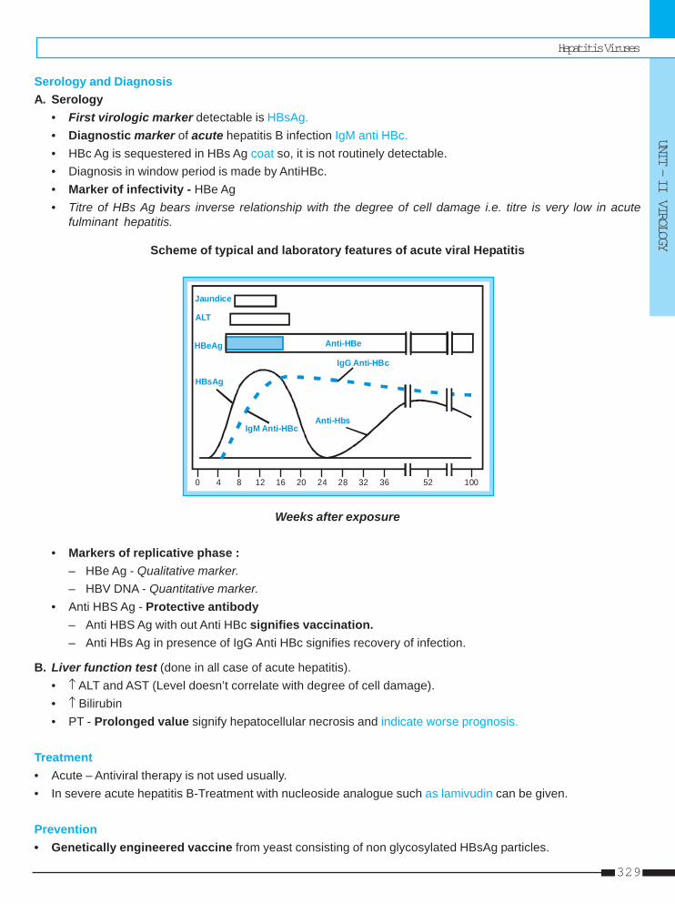
Hepatitis Viruses
UNIT – II VIROLOGY
329
Serology and DiagnosisA. Serology
• First virologic marker detectable is HBsAg.• Diagnostic marker of acute hepatitis B infection IgM anti HBc.• HBc Ag is sequestered in HBs Ag coat so, it is not routinely detectable.• Diagnosis in window period is made by AntiHBc.• Marker of infectivity - HBe Ag• Titre of HBs Ag bears inverse relationship with the degree of cell damage i.e. titre is very low in acute
fulminant hepatitis.
Scheme of typical and laboratory features of acute viral Hepatitis
0 4 8 12 16 20 24 28 32 36 52 100
Anti-HBe
Jaundice
ALT
HBeAg
HBsAg
IgM Anti-HBcAnti-Hbs
IgG Anti-HBc
Weeks after exposure
• Markers of replicative phase :– HBe Ag - Qualitative marker.– HBV DNA - Quantitative marker.
• Anti HBS Ag - Protective antibody– Anti HBS Ag with out Anti HBc signifies vaccination.– Anti HBs Ag in presence of IgG Anti HBc signifies recovery of infection.
B. Liver function test (done in all case of acute hepatitis).• ↑ ALT and AST (Level doesn’t correlate with degree of cell damage).• ↑ Bilirubin• PT - Prolonged value signify hepatocellular necrosis and indicate worse prognosis.
Treatment• Acute – Antiviral therapy is not used usually.• In severe acute hepatitis B-Treatment with nucleoside analogue such as lamivudin can be given.
Prevention• Genetically engineered vaccine from yeast consisting of non glycosylated HBsAg particles.
Self Assessment & Review Microbiology & Immunology
SECTION – B
330
Dose : – 3 IM dose (in deltoid not in gluteal)– Injections are recommended at O, I and 6 month.– Pregnancy is not contraindication to vaccine.
• Post exposure prophylaxis : Combination of HBIG and Hepatitis B vaccine.• Perinatal exposure :
– Single dose of HBIG 0.5 ml IM at birth followed by complete 3 dose of Hepatitis B vaccine.– First dose of vaccine should be given within 12 hours of life.
Remember : As Hepatitis B can lead to hepatocellular carcinoma, vaccination makes HCC the only humancancer which is vaccine preventable.
HEPATITIS D VIRUS = DELTA VIRUSDefective virus that require helper function of HBV for its replication.• HDV is formalin sensitive SSRNA virus.• Delta core of HDV is encapsidated by an outer envelop of HBsAg, so it require cooperative function of HBV.• Intracellular replication of HDV RNA can occur without HBV but liver injury require the presence of HBV.• HDV can cause either :
Features Superinfection Coinfection
Definition • In patient already infected with HBV • Infection simultaneously with HBV
Course • Grave course • Comparatively mild course• More chance of Fulminant hepatitis
and chronic infection
Serology IgM Anti HDV + Ig M Anti HDV +IgM Anti HBc Ig G Anti HBC
HDV antigen in liver and HDV RNA in serum and liver can be detected during HDV replication.
HEPATITIS C VIRUS• Linear SS RNA virus.• Belongs to gene hepacivirus of family flavivirus.• MC virus associated with transfusion related hepatitis.• Cause fatty change in liver.• HCV is associated with Cryoglobinemia; porphyra cutania tarda; Idiopathic pulmonary fibrosis; membrane
proliferative glomerulonephritis.
Genome• Envelope protein coded by hypervariable region varies from isolate to isolate and allow the virus to invade host
immunity.• Because of divergence of HCV isolates within a genotype or subtype and within the same host, these intragenotypic
differences are referred to as quasi-species.
Serology and Diagnosis• Assays of HCV RNA are the most sensitive test for HCV infection and represent the gold standard for diagnosis
of hepatitis C (HCV RNA detection has supplanted RIBA in most clinical settings).
Hepatitis Viruses
UNIT – II VIROLOGY
331
• 2 types of Amplification techniques can be used to detect HCV RNA.– Branched chain complementary DNA (b DNA) assay.– Reverse transcriptase PCR or TMA : It is more sensitive than b DNA assay.
• Anti HCV antibody not be detectable in acute phase and are not sensitive.• Anti HCV are not protective so chance of chronicity is very high.
Treatment• In typical case of hepatitis C recovery is rare; progression to chronic hepatitis is the rule.• Antiviral therapy with interferon alpha reduce the rate of chronicity.• Duration of infection is the single most important variable which determine interferon responsiveness.
CHRONIC HEPATITIS - CClinical features : • Fatigue is MC symptom Jaundice is rare.
• Extrahepatic manifestations may be seen.
Lab features : • Aminotransferase fluctuate between high to high normal value.• ALT and AST - increase with ALT > AST .• But when cirrhosis develop AST becomes greater than ALT• LKM1 - Antibody may be seen in cases of HCV.
Treatment : • Combination therapy of pegylated interferon plus Ribavirin is now standard therapy.• Liver transplantation - When cirrhosis develops.
Remember : • Chronic hepatitis C is MC indication of liver transplantation.• Best prognostic indicator in chronic Hepatitis C is liver histology.
..... Harrison, 17/e, p 1963
HEPATITIS - E VIRUS• Epidemic hepatitis or enterically transmitted non A non B hepatitis.• SS RNA virus belonging to Alpha virus family.• Hepatitis of hepatitis E. virus is characteristically associated with cholestasis.• Most important cause of fulminant hepatitis in pregnancy.
HEPATITIS G VIRUS• Blood borne RNA virus.
Important points about Chronic Hepatitis
Type of Hepatitis Diagnostic Test (s) Autoantibodies Treatment
Chronic hepatitis B HBsAg, IgG anti-HBc, HBeAg, HBV DNA Uncommon IFN-α, lamivudineChronic hepatitis C Anti-HCV, HCV RNA Anti-LKMI PEG-IFN-α Plus ribavirinChronic hepatitis D Anti-HDV, HDV RNA, HBsAg, IgG anti-HBc Anti-LKM3 IFN-α
Self Assessment & Review Microbiology & Immunology
SECTION – B
332
1. Serology of a young man shows HBsAg howeverHBeAg is negative with normal levels of AST andALT. He is asymptomatic. What is the next line ofmanagement ? [AI 08]a) Wait and watchb) Antivirusc) Immunoglobulinsd) Liver transplant
2. A young pregnant woman presents with fulminanthepatic failure. The most likely aetiological agentis : [AI 04]a) Hepatitis B virusb) Hepatitis C virusc) Hepatitis E virusd) Hepatitis A virus
3. Which of the following hepatitis virus havesignificant perinatal transmission : [AI 03]a) HEVb) HCVc) HBVd) HAV
4. A pregnant woman from Bihar presents withhepatic encephalopathy. The likely diagnosis :a) Hepatitis E [AI 01, 95]b) Hepatitis Bc) Sepsisd) Acute fatty liver of pregnancy
5. HCV is associated with : [AI 00]a) Anti LKM-1 antibodyb) Sclerodermac) Cryoglobulinemiad) Polyarteritis nodusa
6. Chronic liver disease is caused by : [AI 00]a) Hepatitis Bb) Hepatitis Ac) Hepatitis Cd) Hepatitis E
7. Non - parenteral hepatitis is : [AI 00, AIIMS 96]a) Hepatitis Ab) Hepatitis Bc) Hepatitis Cd) Hepatitis D
8. Reverse transcriptase of HBV is coded onfollowing gene : [AI 00]a) C geneb) S genec) P gened) X gene
9. A blood donor is not considered for safetransfusion, if he has : [AI 00]a) Anti HBS Ab+veb) Anti HBS Ab and HBc Ag+vec) HbS Ag +ve and IgM anti HBC+ved) Anti HBe+ve
10. During epidemic of hepatitis E, fatality is maximumin : [AI 00]a) Pregnant womenb) Infantsc) Malnourished maled) Adolescents
11. Acute infection with HBV is characterized by :a) HBs Ag [AI 99, AIIMS 96]b) Anti HBs Agc) IgM anti HBc Ag and HBs Agd) Anti HBc Ag
12. Marker for acute hepatitis B is : [AIIMS 07]a) HBV DNAb) IgM anti-HBcc) Core antgen (HbcAg)d) Anti-HbsAg
13. In a patient of active chronic hepatitis B all areseen except : [AIIMS 07]a) HbsAgb) IgM anti HbcAgc) HbeAgd) Anti-HbsAg
14. Which of the following Hepatitis virus is cultivable:a) Hepatitis A [AIIMS 07]b) Hepatitis Bc) Hepatitis Dd) Hepatitis E
QUESTIONS
Answer 1. a) Wait and watch 2. c) Hepatitis ... 3. c) HBV 4. a) Hepatitis E 5. a and c6. c) Hepatitis C 7. a) Hepatitis A 8. c) P gene 9. c) HbS ... 10. a) Pregnant ...
11. c) IgM ... 12. b) IgM anti... 13. b) IgM anti... 14. a) Hepatitis A
Hepatitis Viruses
UNIT – II VIROLOGY
333
15. With which of the following of viral hepatitisinfection in pregnancy, the maternal mortality thehighest : [AIIMS 06]a) Hepatitis Ab) Hepatitis Bc) Hepatitis Cd) Hepatitis E
16. A 30 year old patient presented with history ofjaundice for 10 days. His liver function testsshowed bilirubin of 10mg/dl, SGOT/SGPT - 1100/1450, serum alkaline phosphatase - 240 IU. Hewas positive for HbsAg. What should be theconfirmatory test to establish acute hepatitis Binfection ? [AIIMS 06]a) IgM Anti-HBc antibodyb) HbeAgc) HBV DNA by PCRd) Anti-HBc antibody
17. A 30 year old lady delivered a healthy baby at 37week of gestation. She was a known case ofchronic hepatitis B infection. She was positive forHBsAg but negative for HBcAg. Which of thefollowing is the most appropriate treatment forthe baby ? [AIIMS 05]a) Both active and passive immunization soon after
birthb) Passive immunization soon after birth and active
immunization at 1 year of agec) Only passive immunization soon after birthd) Only active immunization soon after birth
18. Hepatitis C virus is a : [AIIMS 04, PGI 98]a) Togavirusb) Flavi virusc) Filovirusd) Retrovirus
19. The best diagnostic test for recent hepatitis B is:a) HBs Ag [AIIMS 03, 01, 00b) IgM anti HBc Ag PGI 99, 97]c) Anti HBed) Anti HBs
20. Which of the following Hepatitis virus is a DNAvirus : [AIIMS 02]a) Hepatitis C virusb) Hepatitis B virusc) Delta agentd) Hepatitis E virus
21. A mother is HBs Ag positive at 32 weeks ofpregnancy. What should be given to the newbornto prevent neonatal infection : [AIIMS 02]a) Hepatitis B vaccine + immunoglobulinsb) Immunoglobulins onlyc) Hepatitis B vaccine onlyd) Immunoglobulins followed by vaccine 1 month
later22. A thirty year man presented with nausea, fever
and jaundice of 5 days duration. The biochemicaltests revealed a bilirubin of 6.7 mg/dl (conjugated5.0 mg/dl) with SGOT/SGPT (AST/ALT) of 1230/900IU/ml. The serological tests showed presence ofHBs Ag,Ig M anti HBc and HBe Ag. The most likelydiagnosis : [AIIMS-02]a) Chronic hepatitis B infection with high infectivityb) Acute hepatitis B infection with high infectivityc) Chronic hepatitis B infection with low infectivityd) Acute hepatitis B infection with low infectivity
23. Hepatitis B vaccination is given to a patient. Hisserum will reveal : [AIIMS 02]a) HBsAgb) Anti-HBsAgc) IgM Anti-HBc Ag and HBS Agd) IgM and IgG Anti-HBc Ag
24. Commonest hepatotropic virus causingincreased chronic carrier state is :[AIIMS 97, 01]a) HEVb) HAVc) HBVd) HCV
25. Acute hepatitis B can be earliest diagnosed by :a) IgM anti HBc ab [AIIMS 01, 95]b) HBs Agc) IgG anti HBc Abd) Anti HBs Ab
26. Which of the following is calcivirus : [AIIMS 01]a) HEVb) HBVc) HCVd) HAV
27. Best epidemiological tool for investigation ofhepatitis B is : [AIIMS 00; AI 97]a) Anti HBs Agb) Anti HBc Agc) Anti HBe Agd) HBc Ag
Answer 15. d) Hepatitis E 16. a) IgM ... 17. a) Both active... 18. b) Flavi ... 19. b) IgM ...20. b) Hepatitis ... 21. a) Hepatitis ... 22. b) Acute ... 23. b) Anti-HB... 24. d) HCV25. b) HBs Ag 26. a) HEV 27. b) Anti HBc ...
Self Assessment & Review Microbiology & Immunology
SECTION – B
334
28. In a patient only anti HBs Ag is positive in serumall other viral markers are negative. This indicatesa) Acute hepatitis [AIIMS 00]b) Chronic activec) Persistent carrierd) Hepatitis B vaccination
29. Hepatitis C virus true finding is : [AIIMS 98]a) Spreads along fecooral routeb) Antibody to HCV may not be seen in acute stagec) Does not cause chronic hepatitisd) It cannot be cultured
30. If a patient was immunzed with hepatitis B vaccine,which of the following is seen in serum:a) HbeAg [PGI 06]b) HbSAgc) anti Hbs antibodyd) Anti Hbe antibodye) Anti Hbc antibody
Answer 28. d) Hepatitis ... 29. b) Antibody ... 30. c) Anti Hbs...31. a) Enveloped ... 32. b and d 33. c and d
31. HCV is : [PGI 05]a) Enveloped RNA virusb) Unenveloped RNA virusc) Unenveloped positive strand RNAd) Unenveloped negative strand RNAe) DNA virus
32. Transmission of hepatitis A virus occurs :a) One week before and one week after onset of
symptom [PGI 99]b) 2 weeks before onset of symptomc) 2 week after onset of symptomd) 2 week after onset of symptom
33. HBV all true, except : [PGI 98]a) It is a DNA virusb) Spreads by blood transfusionsc) HBs Ag marker of infectivityd) Least chance of chronicity
Hepatitis Viruses
UNIT – II VIROLOGY
335
1. Ans. is a i.e. Wait and Watch Ref. Harrison 17/e, p 1946
In hepatitis B, among previously healthy adult who present with clinically apparent acute hepatitis recoveryocurs in 99% therefore, antiviral therapy is not likely to improve the rate of recovery and is not required.In this case patient is virtually asymptomatic, so no treatment is required.
2. Ans. is c i.e. Hepatitis E virus Ref. Harrison 17/e, p 1941; COGDT 9/e, p 439
Learn the following characteristics :
HAV – Cause spiky fever
HBV – Only hepatitis virus which is DNA virus, may cause cytopathic effect
HCV – Cause fatty change
HDV – Defective virus
HEV – Cause fulminant hepatitis in pregnant woman; Associated with cholestasis.
Remember :HEV – SS RNA virus belonging to Alpha virus family. Transmitted faeco-orally.
– Secondary person to person transmission is rare (C/F to other enteric born infection).
3. Ans. is c i.e. HBV Ref. Harrison 17/e, p 1940
Perinatal transmission of various hepatitis virus
HAV HBV HCV HDV HEV– +++ + + –
• HBV is MC hepatitis virus that is transmitted perinatally.• Most important factor that determine perinatal transmission is HBe Ag (risk 90%).• MC time of perinatal transmission is at the time of delivery.• MC presentation in neonate is asymptomatic HBs Ag carrier.• Most effective treatment of neonatal infection is HBIg immediately after delivery followed by complete 3
dose immunization by HBs Ag vaccine. First dose within first 12 hours of life.
4. Ans. is a i.e. Hepatitis. E Ref. COGDT 9/e, p 439
MC cause of fulminant hepatitis in pregnancy - Hepatitis E.MC cause of hepatic encephalopathy in pregnancy is - Hepatitis E.
EXPLANATIONS AND REFERENCES WITH ILLUSTRATIVE ANSWERS
Self Assessment & Review Microbiology & Immunology
SECTION – B
336
5. Ans. is a and c i.e. Anti LKM antibody; and Cryoglobulinemia Ref. CMDT ‘06, p 655
See, the following line.“HCV is a pathogenic factor in cryoglobulinemia and membranoproliferative GN. and may be related tolichen planus, Autoimmune thyroiditis; Lymphocytic sialedinitis; Idiopathic pulmonary fibrosis; Por-phyria cutanea tarda; Monoclonal gammopathies and lymphoma, Increase risk of Type II diabetes mel-litus.” .... CMDT’ 06, p 655Now see Harrison 17/e, p 1963“Some patient with serologically confirmable chronic hepatitis C have circulating anti LKM-1. Theoccurrence of anti LKM may result from partial sequence homology between the epitope recognizedby anti LKM -1and 2 segments of HCV polyprotein.”So, inspite of wasting time for 1 answer switch over to next question.
Remember : LKM 1 antibody – Hepatitis CLKM 2 antibody – Drug induced hepatitisLKM 3 antibody – Hepatitis D
6. Ans. is c i.e. Hepatitis C Ref. Harrison 17/e, p 1939
Decreasing order of progression to chronicityHCV > HDV > HBV > HAV = HEV
Frequency of 50-70% 5-20% 1-10% NoneChronicity 90% of neonates
Remember : • HCV is MC cause of chronic hepatitis.• Chronic hepatitis C is MC indication of liver transplantation.
7. Ans. is a i.e. Hepatitis A Ref. Harrison 17/e, p 1939
Transmission
Parenterally Faeco oral
Hepatitis B Hepatitis AHepatitis C Hepatitis EHepatitis DHepatitis G
• MC hepatitis associated with blood transfusion hepatitis C.
8. Ans. is c i.e. P gene Ref. Anantnarayan 7/e, p 552
HBV Genes and Gene Products
Genes Regions Gene products
S S Major protein (S)S+Pre-S2 Middleprotein (M) HBs AgS+Pre-S1 and S2 Large protein (L)
Hepatitis Viruses
UNIT – II VIROLOGY
337
C C HbcAgC+Pre-C HbeAg
P (Largest gene) DNA polymeraseX HBxAg
DNA polymerase of HBV has 2 activities : • DNA dependent DNA polymerase.• RNA dependent Reverse transcriptase.
9. Ans. is c i.e. HBs Ag + ve and Ig M anti HBc + ve Ref. Harrison 17/e, p 1943
• HBS Ag + ve and IgM anti HBc + ve suggest acute viral hepatitis.• So, transfusion from this donour contraindicated.
10. Ans. is a i.e. Pregnant women Ref. Jawetz p 24/e, p 471 ; COGDT 9/e, p 439
Already explained, refer answer no. 2
11. Ans. is c i.e. IgM anti HBc Ag and HBs Ag Ref. Harrison 17/e, p 1943
Types Serological Markers of HBV
Acute hepatitis B Ig M anti HBc Ag + ve ; HBs Ag + veChronic hepatitis B Ig G anti HBc Ag + ve ; HBs Ag + veMarker of vaccination Anti HBs Ag (+) ve AloneMarker of remote infection IgG Anti HBc(Used for epidemiological purposes)Marker of infectivity HBe Ag (If present indicate high infectivity)
12. Ans. is b i.e. IgM anti HBC Ref. Harrison 17/e, p 1934, 1942
• Diagnosis of acute hepatitis B infection is made by detection of HBsAg in serum. In cases where HBsAglevel are low, diagnosis can be established by presence of IgM anti HBc.
• Anti HBC of the IgM class predominates during the first 6 months after acute infection, where as IgG antiHBc beyond 6 months.
13. Ans. is b i.e IgM antiHBC Ref. Harrison 17/e, p 1943
IgM antiHBcAg is seen in acute hepatitis not in chronic active.Marker of chronic hepatitis : HBsAg+, IgG antiHBcAg, HBVDNA+, HBeAg+
14. Ans. is A i.e. Hepatitis A Ref. Harrison 17/e, p 1932
“HAV can be cultivated reproducibly in vitro.”
Remember : • HAV is the hepatitis virus : – That causes spiky fever– With no perinatal transmission
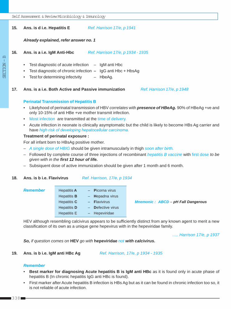
Self Assessment & Review Microbiology & Immunology
SECTION – B
338
15. Ans. is d i.e. Hepatitis E Ref. Harrison 17/e, p 1941
Already explained, refer answer no. 1
16. Ans. is a i.e. IgM Anti-Hbc Ref. Harrison 17/e, p 1934 - 1935
• Test diagnostic of acute infection – IgM anti Hbc• Test diagnostic of chronic infection – IgG anti Hbc + HbsAg• Test for determining infectvity – HbeAg.
17. Ans. is a i.e. Both Active and Passive immunization Ref. Harrison 17/e, p 1948
Perinatal Transmission of Hepatitis B• Likelyhood of perinatal transmission of HBV correlates with presence of HBeAg. 90% of HBeAg +ve and
only 10-15% of anti HBe +ve mother transmit infection.• Most infection are transmitted at the time of delivery.• Acute infection in neonate is clinically asymptomatic but the child is likely to become HBs Ag carrier and
have high risk of developing hepatocellular carcinoma.Treatment of perinatal exposure :For all infant born to HBsAg positive mother.– A single dose of HBIG should be given intramuscularly in thigh soon after birth.– Followed by complete course of three injections of recombinant hepatitis B vaccine with first dose to be
given with in the first 12 hour of life.– Subsiquent dose of active immunization should be given after 1 month and 6 month.
18. Ans. is b i.e. Flavivirus Ref. Harrison, 17/e, p 1934
Remember Hepatitis A – Picorna virusHepatitis B – Hepadna virusHepatitis C – Flavivirus Mnemonic : ABCD – pH Fall DangerousHepatitis D – Defective virusHepatitis E – Hepeviridae
HEV although resembling calcivirus appears to be sufficiently distinct from any known agent to merit a newclassification of its own as a unique gene hepevirus with in the hepeviridae family.
..... Harrison 17/e, p 1937So, if question comes on HEV go with hepeviridae not with calcivirus.
19. Ans. is b i.e. IgM anti HBc Ag Ref. Harrison, 17/e, p 1934 - 1935
Remember• Best marker for diagnosing Acute hepatitis B is IgM anti HBc as it is found only in acute phase of
hepatitis B (In chronic hepatitis IgG anti HBc is found).• First marker after Acute hepatitis B infection is HBs Ag but as it can be found in chronic infection too so, it
is not reliable of acute infection.
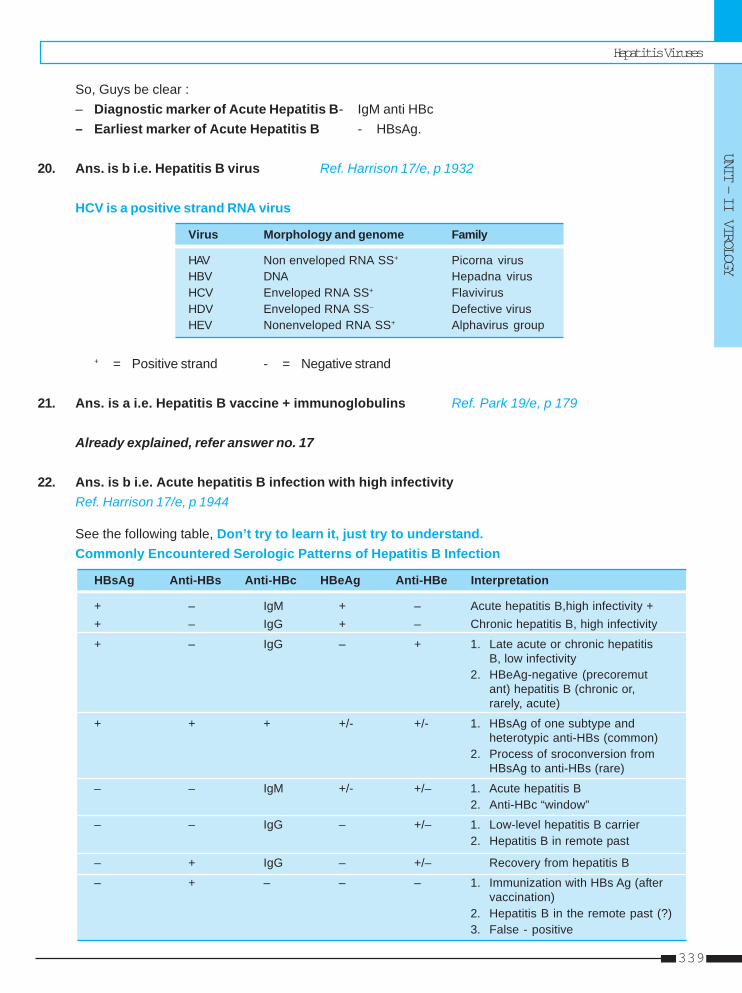
Hepatitis Viruses
UNIT – II VIROLOGY
339
So, Guys be clear :– Diagnostic marker of Acute Hepatitis B- IgM anti HBc– Earliest marker of Acute Hepatitis B - HBsAg.
20. Ans. is b i.e. Hepatitis B virus Ref. Harrison 17/e, p 1932
HCV is a positive strand RNA virus
Virus Morphology and genome Family
HAV Non enveloped RNA SS+ Picorna virusHBV DNA Hepadna virusHCV Enveloped RNA SS+ FlavivirusHDV Enveloped RNA SS– Defective virusHEV Nonenveloped RNA SS+ Alphavirus group
+ = Positive strand - = Negative strand
21. Ans. is a i.e. Hepatitis B vaccine + immunoglobulins Ref. Park 19/e, p 179
Already explained, refer answer no. 17
22. Ans. is b i.e. Acute hepatitis B infection with high infectivityRef. Harrison 17/e, p 1944
See the following table, Don’t try to learn it, just try to understand.Commonly Encountered Serologic Patterns of Hepatitis B Infection
HBsAg Anti-HBs Anti-HBc HBeAg Anti-HBe Interpretation
+ – IgM + – Acute hepatitis B,high infectivity ++ – IgG + – Chronic hepatitis B, high infectivity
+ – IgG – + 1. Late acute or chronic hepatitisB, low infectivity
2. HBeAg-negative (precoremutant) hepatitis B (chronic or,rarely, acute)
+ + + +/- +/- 1. HBsAg of one subtype andheterotypic anti-HBs (common)
2. Process of sroconversion fromHBsAg to anti-HBs (rare)
– – IgM +/- +/– 1. Acute hepatitis B2. Anti-HBc “window”
– – IgG – +/– 1. Low-level hepatitis B carrier2. Hepatitis B in remote past
– + IgG – +/– Recovery from hepatitis B
– + – – – 1. Immunization with HBs Ag (aftervaccination)
2. Hepatitis B in the remote past (?)3. False - positive
Self Assessment & Review Microbiology & Immunology
SECTION – B
340
23. Ans. is b i.e. Anti HbsAg Ref. Harrison 17/e, p 1944
Already explained, refer just above to find answer.
24. Ans. is d i.e. HCV Ref. Harrison 17/e, p 1939
Already explained, refer answer no. 5
25. Ans. is b i.e. HBs Ag Ref. Harrison 17/e, p 1933
Here question is about the earliest diagnostic marker, which is no doubt HBsAg.
• HBsAg in absence of IgM anti HBc or IgG anti HBc indicates early acute infection. ....Jawetz 24/e, p 480So, go with HBsAg.
Remember : • Dignostic marker of Acute hepatitis B – IgM anti HBC• Earliest marker of Acute hepatitis B – HBsAg.
26. Ans. is a i.e. HEV Ref. Ananthnarayan, 7/e, p 559
According to Ananthnarayan 7/e, p 557“HEV has been classified in genus hepesvirus under family calciviridae.”But according to Harrison 17/e, p 1937“HEV has classification of its own with in hepeviridae family.”In this question there is no dispute but if you have to choose among calciviridae and hepeviridae go withnepeviridae.
27. Ans. is b i.e. Anti HBc Ag Ref. Harrison 17/e, p 1934
Already explained, refer answer no. 11
28. Ans. is d i.e. Hepatitis B vaccination Ref. Harrison, 17/e, p 1944
Already explained, refer answer no. 22
29. Ans. is b i.e. Antibody to HCV may not Ref. Harrison, 17/e, p 1937, 1943
Serology of HCV : 3rd generation immunoassays which detect antibody against NS-5 region detect Anti HCVearlier than 1st and 2nd generation assay. These antibody may never been detectable in 5-10% of patient
.....Harrison, p 1943So, option “b” is correct.
Other options• Only hepatitis virus that can be cultured in vitro is HAV. .....Harrison, 17/e, p 1932
But other hepatitis virus can be cloned in E col.
Hepatitis Viruses
UNIT – II VIROLOGY
341
• In option “d” it is not clear whether they are asking culturability in vitro or in vivo (clonability) so, this optionmay be partially correct.
• HCV is transmitted parenterally.
30. Ans. is c i.e. AntiHbs Antibody Ref. Harrison 17/e, p 1944
Already explained, refer answer no. 22
31. Ans. is a i.e. Enveloped RNA virus Ref. Harrison 17/e, p 1934
Already explained, refer answer no. 20
32. Ans. is b and d i.e 2 week before onset of symptom; and 1 weeks after onset of symptomRef. Park, 19/e, p 174
“The risk of transmitting HAV is greatest from 2 week before to 1 week after the onset of Jaundice.”
33. Ans. is c and d i.e. HBs Ag marker of infectivity; and Least chance of chronicityRef. Harrison, 17/e, p 1934
• Marker of infectivity is HBeAg not HBsAg.• Least chance of chronicity are in Hepatitis A and Hepatitis E.
Self Assessment & Review Microbiology & Immunology
SECTION – B
342
1. a) Hepatitis C ... 2. a) Hepatitis C virus 3. a and b 4. c) Feco-oral ... 5. a) 0.5 ppm ...6. c) Enterically ... 7. b) HBsAg + ... 8. c) High infectivity 9. a) Resistance ... 10. b) Feco-oral
1. Which of the following acute viral hepatitis infec-tions has the highest risk of progression to chro-nicity ? [Kar 03]a) Hepatitis Cb) Hepatitis Bc) Hepatitis Ad) Hepatitis E
[Ref. Harrison 17/e, p 1939]2. Which hepatitis virus is notorious for causing a
chronic hepatitis evolving cirrhosis ? [Kar 04]a) Hepatitis C virusb) Hepatitis B virusc) Hepatitis E virusd) Cytomegalo virus
[Ref. Harrison 17/e, p 1973]3. A potent vaccine is available for : [AI 90]
a) Hepatitis Ab) Hepatitis Bc) Malariad) Respiratory syncitial virus
[Ref. Ananthnarayan 7/e, p 549, 556]4. Hepatitis A is transmitted by : [AI 90]
a) blood routeb) Inhalationc) Feco-oral routed) All
[Ref. Harrison 19/e, p 1539]5. HAV is not destroyed by : [AIIMS 91]
a) 0.5 ppm chlorineb) 1:4000 formalinc) UV radiationd) Biolling at 1000C for 5 minutes
[Ref. Ananthnarayan 7/e, p 548]
Answer
6. The commonest cause of viral hepatitis in India :a) Hepatitis type A virus [DNB 92]b) Hepatitis type B virusc) Enterically transmitted NANB virusd) Parenterally transmitted NANB viruse) Delta virus
[Ref. Ananthnarayan 7/e, p 559]7. The serological marker of accute Hepatitis B in-
fection is : [AI 92]a) HBSAg + HBeAgb) HBsAg + Core antibody (IgM)c) HBsAgd) HBcAG
[Ref. Ananthnarayan 7/e, p 1823]8. Presence of HBe Ag in patients with hepatitis indi-
cates : [AIIMS 92]a) Simple carriersb) Late convalescencec) High infectivityd) Carriers status
[Ref. Harrison 17/e, p 1942]9. Anti HBs Ab indicates : [AIIMS 92]
a) Resistance to Hepatits Bb) Acute infectionsc) Good prognosisd) Hepatocellular Carcinoma
[Ref. Harrison 17/e, p 1943]10. Most common route of spread of Hepatitis E is :
a) Sex [AI 93]b) Feco-oralc) Blood tranfusiond) IV injections
[Ref. Ananthnarayan 7/e, p 559]
Chapter Review••••• This section includes questions of VThis section includes questions of VThis section includes questions of VThis section includes questions of VThis section includes questions of Various Other PGMEES from 1990 – 2007.arious Other PGMEES from 1990 – 2007.arious Other PGMEES from 1990 – 2007.arious Other PGMEES from 1990 – 2007.arious Other PGMEES from 1990 – 2007.
Hepatitis Viruses
UNIT – II VIROLOGY
343
11. Hepatitis B is not transmitted by : [AIIMS 93]a) Blood transfusionb) Pasteurised albuminc) Cryoprecipitated) Sexual contact
[Ref. Harrison 17/e, p 709, Table (286.2)]12. Which of the following hepatitis has poor progno-
sis : [UP 05]a) Hepatitis Ab) Hepatitis Bc) Non-A Non-B typed) Hepatitis C
[Ref. Ananthnarayan 7/e, p 56]13. Carrier state does not axis for : [Kerala 99]
a) Hepatitis B virusb) Hepatitis A virusc) Non A non B Hepatitisd) Delta agent
[Ref. Harrison 17/e, p 1939, Table (285-2)]14. Chronic hepatitis is seen in : [Kerala 99]
a) Hepatitis Cb) Hepaitits Dc) Hepatitis Ad) Hepatitis E
[Ref. Harrison 17/e, p 1939]
15. HBV is associated with all of the following except:a) Hepatic cancer [SGPGI 05]b) Chronic hepatitisc) Hepatic adenomad) Cirrhosis
[Ref. Ananthnarayan 7/e, p 533]16. Route of transmission of Hepatitis E virus is :
a) Skin [DNB 04]b) Faeco oralc) Bloodd) Sexual contact
17. Serological markers of infectivity of Hepatitis B :a) HBsAg [Kerala 06]b) HBeAgc) IgM anti Hbcd) HbcAg
[Ref. Harrison 17/e, p 1942]18. Which hepatitis virus had been called as enterovi-
rus : [MP 07]a) Hepatitis Ab) Hepatitis Bc) Hepatitis Cd) Hepatitis E
[Ref. Ananthnarayan 7/e, p 548]
11. b) Pasteurised ... 12. b) Hepatitis B 13. b) Hepatitis A ... 14. a) Hepatitis C 15. c) Hepatic ...16. b) Faeco oral 17. b) HBeAg 18. a) Hepatitis A
Answer
Self Assessment & Review Microbiology & Immunology
SECTION – B
344
• Causative agent of AIDS = Slim disease.• Belongs to family retroviridae; subfamily lentiviridae.• Two types are found HIV 1 and HIV 2. HIV 1 is most common cause of AIDS in world.• Pan troglodytes troglodytes species of chimpanzees are natural reservoir of HIV-1
Morphology and Replication cycle• Spherical enveloped virus. Nucleocaspid has icosahedral structure.• Virus contain external spikes formed by the two major envelope protein - the external gp - 120 and transmembrane
gp - 41.• Genome is composed of two identical single stranded positive sense RNA copies.• Main genes are :
gag – Determines the core and shell of virus. Codes for precursor protein p55 which is cleaved into threeproteins p15, p18 and p24.
env – Determines the synthesis of envelope glycoprotein. gp- 160 which is cleaved in to gp 120 and gp 41.pol – Codes for polymerase reverse transcriptase and other viral enzymes.Other genes are : tat, rev, jej, vif, vpu, vpx, vpr, LTR.– The major difference between genomes of HIV 1 and HIV 2 is HIV 2 lacks vpu gene and has vpx gene which is
not present in HIV -1.• Replication begins with the high affinity binding of gp120 with CD-4 (Present on CD4+ T cells and monocyte
macrophage lineage cells). After binding with CD-4, gp120 undergoes conformational change that facilates bindingof corecepter. The major corcepter for HIV-1 are CCR-5 and CXCR-4.
• After fusion, HIV genome RNA is uncoated and internalized into target cell. The reverse transcriptase catalyze thereverse transcription of RNA to DNA. DNA so formed intergrates with host cell chromosome through the action ofvirus encoded enzyme integrase.
Antigenic variation and molecular heterogenity• HIV is highly mutable virus.• The variability of HIV is believed to be due to error prone nature of reverse transcription.• There are three groups of HIV -1.
Group M - (Most of infection), Group O and Group N.There are nine subtypes of M group. ..... Harrison 17/e, p 1141
55 HIV & Other RHIV & Other RHIV & Other RHIV & Other RHIV & Other Retroviretroviretroviretroviretrovirusesusesusesusesuses
HIV & Other Retroviruses
UNIT – II VIROLOGY
345
Subtype C is most prevelant world wide.In India and china subtype C is most prevelant.
Modes of TransmissionA. Sexual transmission
• The most common mode of transmission world wide is heterosexual transmission....Harrison 17/e, p 1142
• Chance of infection from male to female is twice as from female to male.• Anal intercourse has higher risk of transmission.
B. Transmission by blood and blood products :• Transmission of whole blood. Packed red cells, platelets, leucocytes and plasma are all capable of transmitting
HIV infection.• Hyperimmuno globulin, hepatitis B immunoglobulin, plasma derived heptatitis B vaccine and Rh immunoglobulin
have not been associated with transmission of HIV infection.• Risk of gettng HIV infection from transfusion of a unit of infected blood is > 95%.
C. Maternal fetal transmission - Risk is 30%• Occurs most commonly in perinatal period.• Cesarean section decrease risk of transmission.• Risk of infection is high if the mother is newly infected or if she has already developed AIDS.• Vitamin A deficiency increase risk of transmission.• Exclusive breast feeding carries lower risk of transmission than mixed feeding. ..... Harrison 17/e, p 1146• Presence of mastitis, low maternal CD4+ T cells counts and maternal vitamin A deficiency increase risk of
transmission.
D. Transmission by other body fluids :• HIV can be isolated in low titres from saliva, but saliva can not transmit HIV infection due to presence of
endogenous antiviral factors of which most important is secretory leucocyte protease inhibitor (SLPI).
Pathogenesis• The hallmark of HIV disease is a profound immunodeficiency due to quantitative and qualtitative deficiency of
helper or inducer T cells (CD4 - Tcells).
• Primary HIV infection and Intial Viremia :– Dendritic cells play an important role in the initiation of HIV infection due to presence of lectin called DC-SIGN
which binds with high affinity to HIV envelope.
• Chronic and Persistant Infection :– Establishment of chronic infection is due to the ability of virus to mutate.– Evolution of mutants that escape control by CD8 + cytolytic T lymphocytes is critical for progression of HIV
infection.– Another mechanism is the down regulation of HLA class 1 molecules resulting in lack of ability of CD8 + CTL
to kill the infected target cell.
• Cellular target of HIV :– CD 4 + = Helper = Inducer T cell (Primary target)– Monocye - Macrophages (10 - 15%)– Dendritic, langerhans cells– Few B cells (5 - 10%).
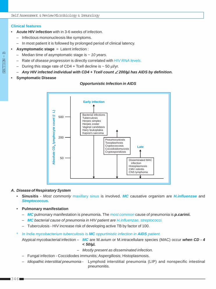
Self Assessment & Review Microbiology & Immunology
SECTION – B
346
Clinical features• Acute HIV infection with in 3-6 weeks of infection.
– Infectious mononucleosis like symptoms.– In most patient it is followed by prolonged period of clinical latency.
• Asymptomatic stage = Latent infection :– Median time of asymptomatic stage is ~ 10 years.– Rate of disease progression is directly correlated with HIV RNA levels.– During this stage rate of CD4 + Tcell decline is ~ 50 μl/yr.– Any HIV infected individual with CD4 + Tcell count ∠∠∠∠∠ 200/μμμμμl has AIDS by definition.
• Symptomatic DiseaseOpportunistic Infection in AIDS
Abs
olut
e C
D ly
mph
ocyt
e co
unt (
/L)
4
50
200
500Bacterial infectionsTuberculosisHerpes simplexHerpes zosterVaginal candidasisHairy leukoplakiaKaposi's sarcoma
PneumocystosisToxoplasmosisCryptococcosisCoccidioidomycosisCryptosporidiosis
Disseminated MAC infectionHistoplasmosisCMV retinitisCNS lymphoma
Early infection
Late
A. Disease of Respiratory System• Sinusitis - Most commonly maxillary sinus is involved. MC causative organism are H.influenzae and
Streptococcus.
• Pulmonary manifestation– MC pulmonary manifestation is pneumonia. The most common cause of pneumonia is p.carinii.– MC bacterial cause of pneumonia in HIV patient are H.influenzae, streptococci.– Tuberculosis - HIV increase risk of developing active TB by factor of 100.
* In India mycobacterium tuberculosis is MC oppurtinistic infection in AIDS patient.Atypical mycobacterial infection – MC are M.avium or M.intracellulare species (MAC) occur when CD - 4
< 50/μμμμμl.– Mostly present as disseminated infection.
– Fungal infection - Coccidiodes immunitis; Aspergillosis; Histoplasmosis.– Idiopathic interstitial pneumonia - Lymphoid interstitial pneumonia (LIP) and nonspecific intestinal
pneumonitis.
HIV & Other Retroviruses
UNIT – II VIROLOGY
347
B. Disease of CVSIt includes : • HIV associated cardiomyopathy - Dilated cardiomyopathy associated with CHF due to direct
consequence of HIV infection.• Lipodystrophy syndrome - Due to adverse effect of antiretroviral therapy.
C. Disease of Oropharynx and GITOral lesions • Thrush - Due to candida
• Hairy leukoplakia - Due to EBV (not premalignant condition)• Apthous ulcer.
Esophagitis - Causative organism are : – Candida– CMV - Single large ulcer– HSV - Multiple small ulcer.
Intestinal infection – Causes of diarrhoea in AIDS patient.• Bacterial – Salmonella, Shigella, Campylobacter jejuni.• Fungal – Histoplasmosis, Coccidiodomycosis and Pencillosis cause fever and diarrhoea.
D. Disease of Hematopoetic system• Anemia - MC hematologic abnormality.• Thrombocytopenia - Due to platelet specific antibody or as a direct result of HIV on megakaryocytes.• Lymphadenopathy and Leucopenia.
E. Dermatologic disease• Dermatologic problems occur in >90% of patients with HIV infection.• Folliculitis is the most prevalent dermatologic disorder in patients with HIV infection. Seen when CD4 + T cell
counts < 200 cells/μl.• Seborrheic dermatitis occurs in up to 50% of patient with HIV infection.• Psoriasis and icthyosis, though not increased in frequency, if occurs severity is more than normal population.
F. Neurologic ManifestationsMC opportunistic infection that involve CNS are Toxoplasmosis; Cryptococcus.• Meningitis - MC cause Cryptococcus neoformans.• HIV encephalopathy = AIDS dementia complex. Subcortical dementia due to direct effect of HIV.• Seizures - MC cause Toxoplasmosis.• Myelopathy - Associated with CMV infection.• Peripheral neuropathy.• Myopathy - Due to direct effect of HIV or due to drugs.
G. Ophthalmologic disease• MC fundoscopic finding is cotton wool spot due to retinal ischemia.• MC cause of blindness in HIV patient is CMV retinitis (charcteristic appearance - perivascular hemorrhage
and exudate) - occurs when CD-4 < 50/μl.• Acute retinal necrosis syndrome = Progressive Outer retinal necrosis (PORN) - Due to HSV and varicella
zoster.
H. Neoplastic Disease• Kaposi Sarcoma – Multicentric neoplasm of vascular origin appearing in skin, mucous membranes and viscera.• Non Hodgkin lymphoma : – Immunoblastic lymphoma (MC lymphoma in HIV patient)

Self Assessment & Review Microbiology & Immunology
SECTION – B
348
– Burkitt’s lymphoma– Primary CNS - Usually associated with EBV.
• AIDS increase incidence of : Hodgkin disease Multiple myeloma
Leukemia Melanoma Cervical CaBrain tumor Anal Ca Oral CaLung Ca Testicular Ca
Pediatric AIDS• Recurrent bacterial infections are more common in children.• Pneumonia caused by pneumocystis carinii is the MC AIDS defining diagnosis in childen with unrecognised HIV
infection.• Tumor most commonly associated are Non Hodgkin Lymphoma.
DIAGNOSIS OF HIV INFECTIONClinicalI. WHO case definition for AIDS surveillance :
Any individual (>12 years of age) is considered to have AIDS if at least 2 of the following major signs are presentin combination with at least 1 minor sign.Major signs :• Weight loss > 10% of body weight.
• Chronic diarrhoea for more than 1 months.• Prolonged fever for more than 1 month (intermittent or constant).
Minor signs :• Persistent cough for more than 1 month.• Generalized pruritic dermatitis.• History of herpes zoster.• Oropharyngeal candidiasis.• Chronic progressive or disseminated herpes simplex infection.• Generalized lymphadenopathy.
The presence of either generalized Kaposi sarcoma or cryptococcal meningitis is sufficient for the diagnosis ofAIDS for surveillance purposes.
II. Expanded WHO case definition for AIDS surveillance :For the purposes of AIDS surveillance and adult or adolescent (>12 years of age) is considered to have AIDS if atest for HIV antibody gives a positive result and one or more of the following conditions are present :• > 10% body weight loss or cachexia, with diarrhoea or fever, or both, intermittent or constant, for at least 1
month, not known to be due to a condition unrelated to HIV infection.• Cryptococcal meningitis.• Pulmonary or extra-pulmonary tuberculosis.• Neurological impairment that is sufficient to prevent independent daily activities, not known to be due to a
condition unrelated to HIV infection.• Candidiasis of the oesophagus.• Clinically diagnosed life-threatening or recurrent episodes of pneumonia, with or without aetiological confirmation.
HIV & Other Retroviruses
UNIT – II VIROLOGY
349
Laboratory diagnosis
Demonstration of Antibody Demonstration of HIV or its components
– ELISA (sensitive and best screening test) – Antigen detection p-24 is earliest virusmarker to appear in blood
– Western blot (specific) (demonstrate antibody – Virus isolation - By cultivation of patientto products of all major HIV gene) lymphocyte with uninfected lymphocyte
in present of IL-2– Modern 4th generation EIA kit combines – PCR - Gold standard for diagnosis in all
antibody detection with p24 antigen assay stages of HIV• DNA PCR; RNA PCR, RT - PCR -
RT PCR is most sensitive and best
* RT PCR is used as a diagnostic and prognostic tool and has become an technique for studies of sequencediversity and microbial resistance to antiretroviral agents.
* Nucleic acid amplification detection : Reverse transcriptase PCR, branched DNA (b DNA) and nucleic acidsequence based amplification.
Diagnosis of HIV infection in new born• The presence of anti HIV antibody (ELISA) is not diagnostic of infection until after 18 month of age.• Diagnosis during first few month of life is made by detection of HIV DNA with PCR (Best).• Other methods are : – p-24 antigen detection
– HIV culture.
Lab monitoring of patient with HIV infection• CD4 + Tcell count – Best indication of immediate state of immunologic competence of patient with HIV infection.• HIV RNA determination – By RT PCR and bDNA assay. Determine disease progression. Should be monitored
every 3-4 months.• HIV resistance testing.
TreatmentHIV - infected with viral load
> 5000 - 10.000 or CD < 500/ l4
AZT + 3TC ddl AZT + ddl AZT + ddCFirst linetreatment
Intolerence to regimen Progression of disease or viral load does not decrease by .5 log withinitiation of treatment or increase of
viral load by .5 log while on treatment
>
>Change to alternativeFirst-line treatment
Saquinavir + one or more nucleoside analogs
Ritonavir +one or morenucleosideanalogs
Indinavir +one or morenucleosideanalgos
d 4t + 3TC d4t + ddl
Progression of disease or viral load criteria as above
Saquinavir +Indinavir +one or morenucleosideanalogs
Nevirapine +AZT + ddl
Nelfinavir +one or morenucleosideanalogs
Second linetreatment
Third linetreatment
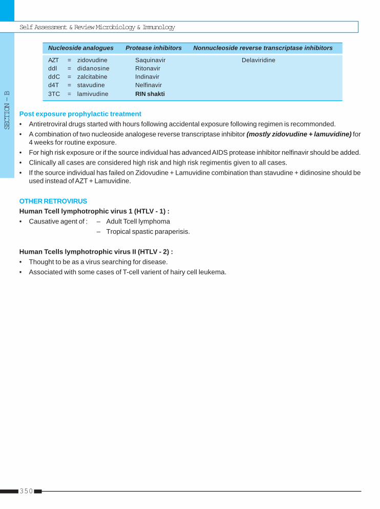
Self Assessment & Review Microbiology & Immunology
SECTION – B
350
Nucleoside analogues Protease inhibitors Nonnucleoside reverse transcriptase inhibitors
AZT = zidovudine Saquinavir Delaviridineddl = didanosine RitonavirddC = zalcitabine Indinavird4T = stavudine Nelfinavir3TC = lamivudine RIN shakti
Post exposure prophylactic treatment• Antiretroviral drugs started with hours following accidental exposure following regimen is recommonded.• A combination of two nucleoside analogese reverse transcriptase inhibitor (mostly zidovudine + lamuvidine) for
4 weeks for routine exposure.• For high risk exposure or if the source individual has advanced AIDS protease inhibitor nelfinavir should be added.• Clinically all cases are considered high risk and high risk regimentis given to all cases.• If the source individual has failed on Zidovudine + Lamuvidine combination than stavudine + didinosine should be
used instead of AZT + Lamuvidine.
OTHER RETROVIRUSHuman Tcell lymphotrophic virus 1 (HTLV - 1) :• Causative agent of : – Adult Tcell lymphoma
– Tropical spastic paraperisis.
Human Tcells lymphotrophic virus II (HTLV - 2) :• Thought to be as a virus searching for disease.• Associated with some cases of T-cell varient of hairy cell leukema.
HIV & Other Retroviruses
UNIT – II VIROLOGY
351
1. HIV can be detected and confirmed by : [AI 05]a) Polymerase chain reaction (PCR)b) Reverse transcriptase - PCRc) Real time PCRd) Mimic PCR
2. An HIV patient complains fo visual disturbances.Fundal examination shows bilateral retinal exu-dates and perivascular hemorrhages. Which ofthe following viruses are most likely to be respon-sible for this retinitis : [AI 04]a) Herpes simplex retinitisb) Human herpes virusc) Cytomegalovirusd) EBV
3. Which one of following is true regarding HIV in-fection : [AI 04]a) Following needle stick injury infectivity is re-
duced by administration of nucleoside ana-logues
b) CD 4 counts are the best predictors of diseaseprogression
c) Infected T cells survive for a month in infectedpatients
d) In latent phase HIV has minimal replication4. HIV virus has : [AI 02]
a) Single stranded DNAb) Single stranded RNAc) Double stranded DNAd) Double stranded RNA
5. Regarding HIV which of the following is not true:a) It is a DNA retrovirus [AI 02]b) Contains Reverse Transcriptasec) May infect host CD 4 cells other than T
lymphoctyesd) Causes a reduction in host CD 4 cells at late
stage of disease6. CMV retinitis in HIV occurs when the CD - 4 counts
fall below : [AI 02]a) 50b) 100c) 200d) 150
7. Regarding HIV infection, not true is : [AI 01]a) p24 is used for early diagnosisb) Lysis of infected CD4 cells is seenc) Dendritic cells do not support replicationd) Macrophage is a reservoir for the virus
8. Reverse transcriptase sequence in HIV is bestdescribed as : [AI 00]a) RNA - DNA - RNAb) DNA - RNAc) DNA - RNA - DNAd) RNA - DNA
9. Multifocal tumour of vascular origin in a patient ofAIDS : [AI 00]a) Kaposi sarcomab) Astrocytomac) Gastric carcinomad) Primary CNS lymphoma
10. A patient with HIV has diarrhoea with AFB +ve or-ganism in stool. The most likely organism is :a) Mycobacterium avium intracellulare [AI 00]b) Mycobacterium TBc) Mycobacterium lepraed) Mycoplasmas
11. A patient comes to hospital with a history of sorethroat, diarrhoea and sexual contact 2 weeksbefore the best investigation to rule out HIV is :a) p24 antigen assay [AI 00]b) ELISAc) Western blotd) Lymphnode biopsy
12. AIDS involves primarily : [AI 98]a) T-helper cellsb) T-suppressor cellsc) T-5 cytotoxic cellsd) B-cells
13. When compared to the Western blot technique,ELISA test is : [AI 96]a) More sensitive, less specificb) More sensitive, more specificc) Less sensitive, less specificd) Less sensitive, more specific
QUESTIONS
Answer 1. b) Reverse ... 2. c) Cytomega ... 3. a) Following ... 4. b) Single ... 5. a) It is a ...6. a) 50 7. c) Dendritic cells ... 8. a) RNA - ... 9. a) Kaposi ... 10. a) Mycobacterium ...
11. a) p24 antigen ... 12. a) T-helper ... 13. a) More sensitive ...
Self Assessment & Review Microbiology & Immunology
SECTION – B
352
14. All of the following viral genes associated withHIV infection code for structural proteins except:a) Gag gene [AI 95]b) Env genec) Pol gened) Tat gene
15. Which of the following is a marker of HIV infectionin blood : [AI 95]a) DNA polymeraseb) RNA polymerasec) Reverse transcriptased) DNA isomerase
16. All the following are true about HIV infection ex-cept : [AI 95]a) Caused by an enveloped RNA virusb) Rate of killing is directly proportional to T4 mol-
ecules on cell surfacec) Increased release of acid labile interferond) Decreased delayed hypersensitivity activity re-
action17. In HIV window period indicates : [AIIMS 07]
a. Time period between infection and onset ofsymptoms
b. Time period between infection and detection ofantibodies against HIV
c. Time between infection and treatmentd. Time between treatment and death
18. A known HIV positive patient is admitted in an iso-lation ward after an abdominal surgery followingan accident. The resident doctor who changedhis dressing the next day found it to be soaked inblood. Which of the following would be the rightmethod of choice of discarding the dressing :a) Pour 1% hypochlorite on the dressing material
and send it for incineration in a appropriate bagb) Pour 5% hypochlorite on the dressing material
and send it for incineration in a appropriate bagc) Put the dressing material directly in an appro-
priate bag and send for incineration [AIIMS 05]d) Pour 2% lysol on the dressing material and send
it for incineration in a appropriate bag19. Tissue of origin of Kaposi sarcoma is :
a) Lymphoid [AIIMS 05]b) Vascularc) Neurald) Muscular
20. Which of the following lesion is associated withHIV infection : [AIIMS 04]a) Hairy leukoplakiab) Erythroplakiac) Oral lichen planusd) Bullous pemphigoid
21. All of the following methods are used for the diag-nosis of HIV infection in a 2 month old child ex-cept: [AIIMS 03]a) DNA PCRb) Viral culturec) HIV ELISAd) p24 antigen assay
22. A resident doctor sustained a needle stick injurywhile sampling blood of patient who is HIV posi-tive. A decision is taken to often him postexpo-sure prophylaxis. Which one of the following wouldbe the best recommendation : [AIIMS 03]a) Zidovudine + Lamivudine for 4 weeksb) Zidovudine + Lamivudine Nevirapine for 4 weeksc) Zidovudine + Lamivudine + Indinavir for 4 weeksd) Zidovudine + Stavudine + nevirapine for 4 weeks
23. HIV infects most commonly : [AIIMS 00]a) CD 4 + helper cellsb) CD 8 + cellsc) Macrophaged) Neutrophil
24. A HIV mother delivers a baby. All are true except:a) Risk of HIV in the baby is up to 90%b) HIV infection cannot be diagnosed in the baby
with available methods [AIIMS 99]c) AIDS can be transmitted from mother to child
during deliveryd) Breast feeding can transmit AIDS
25. Which of the following is not seen in HIV Patientwith CD4 count less than 100/μμμμμl, who has non pro-ductive cough : [AIIMS 99]a) Mycobacterium tuberculosisb) Pneumocystis cariniic) Mycoplasma pneumonaed) Cryptococcal infection
26. Which infection is not common in HIV patients :a) Cryptosporidiosis [AIIMS 97]b) Atypical mycobacterial infectionc) Aspergillosisd) Candidiasis
Answer 14. d) Tat ... 15. c) Reverse ... 16. c) Increased ... 17. b) Time period ... 18. c) Put the ...19. b) Vascular 20. a) Hairy ... 21. c) HIV ... 22. c) Zidovudine ... 23. a) CD 4 + ...24. a) Risk of ... 25. c) Mycoplasma ... 26. c) Aspergillosis
HIV & Other Retroviruses
UNIT – II VIROLOGY
353
Answer 27. a and b 28. a, b, c and d 29. a, b, c and e 30. a and b 31. a, b, c, d and e32. a and c 33. c and e 34. c and d 35. None 36. d) Male to ...37. a) Myco. ... 38. a and b
27. Which of the following gene is present in HIV ge-nome : [PGI 06]a) Gagb) Tatc) p500d) Kinasee) P24
28. Fungal infection associated with AIDS patient area) Pueumocystis carini [PGI 03]b) Penicillium marneffeic) Candidad) Cryptococcuse) Cryptosporidium
29. HIV infection is associated with : [PGI 02]a) A glandular fever like illnessb) Generalized lymphadenopathyc) Gonococcal septicemiad) Sinus diseasee) Presenile dementia
30. Which of the following is HIV gene : [PGI 02]a) gagb) tatc) p2500d) kinasee) p24
31. Which of the following is an AIDS defining criteriaaccording to WHO: [PGI 01]a) Generalised lymphadenopathyb) Fever, weight loss and fatiguec) Pneumocystis carinii pneumoniad) Mycobacterium avium infectione) Persistent diarrhoea
32. In diagnosis of AIDS, criteria include the followingexcept : [PGI 01]a) CD 4 < 200b) CD 8 < 500c) CD 4 : CD 8 = 1
d) Presence of any of the opportunistic infectionstuberculosis, pneumocystis carinii, cytomega-lovirus
e) Western blot is positive33. Persistent diarrhoea in AIDS is caused by A\E :
a) Microsporidia [PGI 01]b) Cryptosporidium parvumc) Cryptococcusd) Isospora bellie) Giardia lambia
34. Important features of AIDS are : [PGI 01]a) Follicular tonsillitisb) Lichen planusc) Oral candidiasisd) Hairy leukoplakia
35. HIV gene is/are : [PGI 01]a) Gp73b) p24c) Gp120d) Gp5
36. True about HIV : [PGI 00]a) Not transmitted through semenb) More chances of transmission during cesarean
section than normal labourc) More infectious than hepatitis Bd) Male to female transmission > female to male
37. In India most common cause of T.B. in HIV :a) Myco. Tuberculosis [PGI 00]b) Myco. Avium intracellularec) M. bovisd) M. scrofulaceum
38. Oppurtunistic infection in HIV patient are :a) P. carrnii [PGI 96]b) M. aviumc) Pseudomonasd) Mycoplasma
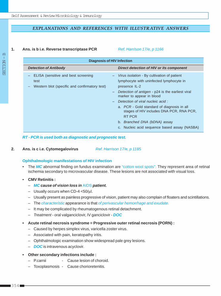
Self Assessment & Review Microbiology & Immunology
SECTION – B
354
1. Ans. is b i.e. Reverse transcriptase PCR Ref. Harrison 17/e, p 1166
Diagnosis of HIV Infection
Detection of Antibody Direct detection of HIV or its component
– ELISA (sensitive and best screening – Virus isolation - By cultivation of patienttest lymphocyte with uninfected lymphocyte in
– Western blot (specific and confirmatory test) presence IL-2– Detection of antigen - p24 is the earliest viral
marker to appear in blood– Detection of viral nucleic acid :
a. PCR - Gold standard of diagnosis in allstages of HIV includes DNA PCR, RNA PCR,RT PCR
b. Branched DNA (bDNA) assayc. Nucleic acid sequence based assay (NASBA)
RT - PCR is used both as diagnostic and prognostic test.
2. Ans. is c i.e. Cytomegalovirus Ref. Harrison 17/e, p 1185
Ophthalmologic manifestations of HIV infection• The MC abnormal finding on fundus examination are “cotton wool spots”. They represent area of retinal
ischemia secondary to microvascular disease. These lesions are not associated with visual loss.
• CMV Retinitis :– MC cause of vision loss in AIDS patient.– Usually occurs when CD-4 <50/μl.– Usually present as painless progressive of vision, patient may also complain of floaters and scintillations.– The characteristic appearance is that of perivascular hemorrhage and exudate.– It may be complicated by rheumatogenous retinal detachment.– Treatment - oral valganciclovir, IV ganciclovir - DOC
• Acute retinal necrosis syndrome = Progressive outer retinal necrosis (PORN) :– Caused by herpes simplex virus, varicella zoster virus.– Associated with pain, keratopathy iritis.– Ophthalmologic examination show widespread pale grey lesions.– DOC is intravenous acyclovir.
• Other secondary infections include :– P.carnii - Cause lesion of choroid.– Toxoplasmosis - Cause chorioretenitis.
EXPLANATIONS AND REFERENCES WITH ILLUSTR ATIVE ANSWERS

HIV & Other Retroviruses
UNIT – II VIROLOGY
355
3. Ans. is a i.e. Following needle stick injury infectivity is reduced by administration of nucleosideanaloguesRef. Harrison 17/e, p 1201; Park 18/e, p 279; 19/e, p 294
“Four week treatment with AZT monotherapy after needle stick exposure to HIV among health careworker decreases the chance of their becoming infected by 79%.” ..... Park 19/e, p 294
Guideliness for post exposure prophylaxis :• A combination of two nucleoside analogue reverse transcriptase inhibitors for 4 weeks for less severe
exposures.• A combination of two nucleoside analogue RT inhibitor plus a third drug given for 4 weeks for more severe
exposure.
Factor associated with increased risk of occupational transmission :• Deep injury.• Presence of visible blood on the instrument causing the exposure.• Injury with the device that has been placed in vein or artery of source patient.• Terminal illness of source patient.• Lack of post exposure antiretroviral therapy in exposed health are worker.
– Best Predictor to disease progression is HIV RNA estimation not CD 4 + count. ... H - 17/e, p 1167– CD 4 + count are the best indicator of immediate state of immunologic competence.
So, option ‘b’ is wrong.
• Clinical latency versus microbial latency– Clinical latency should not be confused with microbial latency since virus replication is present in most
patient.So, option ‘d’ is wrong.
Remember : • Test for monitoring of patient with HIV infection :– CD 4 + T cell count– HIV RNA determination - Best by RT - PCR– HIV resistance testing.
4. Ans. is b i.e. Single stranded RNA Ref. Ananthnarayan 7/e, p 583
Classification of HIV ( = HTLV III) Family – RetroviridaeSubfamily – Lenti virusGenome – SS RNA positive sense.
• Viron contain lipoprotein envelope and nucleocapsid is icosahedral in shape.• Characteristic feature of retrovirus is presence of reverse transcriptase enzyme.
– Viral RNA is transcribed by this enzyme first in to SS DNA then to ds DNA which gets integrated withhost cell.
– Thus in contrast to central dogma flow of information is RNA → DNA → RNA
Self Assessment & Review Microbiology & Immunology
SECTION – B
356
5. Ans. is a i.e. It is DNA retrovirus Ref. Ananthnarayan 7/e, p 583
HIV is RNA retrovirus (not DNA virus)• It primarily infect CD 4 + T cells but can infect other cells also which bear CD4 receptor on their surface
[Harrison 16/e, p 1093]. These include circulating dendritic cells; epidermal langerhan’s cells; monocytes.
6. Ans. is a i.e. 50 Ref. Harrison 17/e, p 1185
Already explained, refer answer no. 2
7. Ans. is c i.e. Dendritic cell do not support replicationRef. Ananthnarayan 7/e, p 586; Harrison 17/e, p 1157
• Cellular target of HIV– CD 4+ lymphocyte and CD 4 + cells of monocyte and macrophage lineage are principal target of HIV.– Circulating dendritic cells - play important role in initiation of HIV infection.– Epidermal langerhans cells.– 5 - 10% of B lymphocyte. ..... Ananthnarayan, 7/e, p 586
• Degree of cytopathicity of HIV for cells of the monocyte lineage is low, and HIV can replicate extensively incells of monocyte lineage. Hence play a role in dissemination of HIV in the body and can serve as reservoirof HIV infection. ..... Harrison, 17/e, p 1159
• p24 is the earliest virus marker to appear in blood and is the one tested for. ..... AA 7/e, p 590
8. Ans. is a i.e. RNA - DNA - RNA Ref. Ananthnarayan 7/e, p 583; Harrison 17/e, p 1132
Already explained, refer answer no. 4
9. Ans. is a i.e. Kaposi sarcoma Ref. Harrison 17/e, p 1162, 1186
“Kaposi sarcoma is a multicentric neoplasm of vascular origin consisting of multiple vascular nodulesappearing in skin, mucous membrane and viscera.”
Feature of Kaposi sarcoma :• Can develop at any stage of HIV infection, even in presence of normal CD-4 count.• It is a manifestation of excess proliferation of spindle cells that are believed to be vascular origin.
..... Harrison 17/e, p 1162• Development of KS is associated with Human herpes virus 8 or HHV - 8 is etiologic agent of KS.• Clinically KS often appear in sun exposed areas, particularly tip of nose. The initial lesion is a small raised
reddish purple nodule on skin. (MC appear as raised nodules).• LN involvement does not signify poor prognosis.
Treatment :• Observation and optimization of antiretroviral therapy.• Single or limited number of lesions : – Radiation
– Intralesional vinblastine and Cryotherapy.
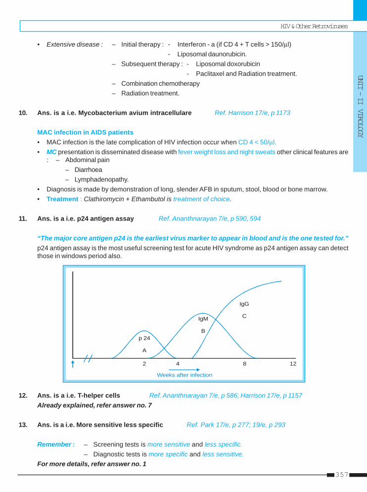
HIV & Other Retroviruses
UNIT – II VIROLOGY
357
• Extensive disease : – Initial therapy : - Interferon - a (if CD 4 + T cells > 150/μl)- Liposomal daunorubicin.
– Subsequent therapy : - Liposomal doxorubicin- Paclitaxel and Radiation treatment.
– Combination chemotherapy– Radiation treatment.
10. Ans. is a i.e. Mycobacterium avium intracellulare Ref. Harrison 17/e, p 1173
MAC infection in AIDS patients• MAC infection is the late complication of HIV infection occur when CD 4 < 50/μl.• MC presentation is disseminated disease with fever weight loss and night sweats other clinical features are
: – Abdominal pain– Diarrhoea– Lymphadenopathy.
• Diagnosis is made by demonstration of long, slender AFB in sputum, stool, blood or bone marrow.• Treatment : Clathiromycin + Ethambutol is treatment of choice.
11. Ans. is a i.e. p24 antigen assay Ref. Ananthnarayan 7/e, p 590, 594
“The major core antigen p24 is the earliest virus marker to appear in blood and is the one tested for.”p24 antigen assay is the most useful screening test for acute HIV syndrome as p24 antigen assay can detectthose in windows period also.
2 4 8 12
p 24
A
IgM
B
IgG
C
Weeks after infection
12. Ans. is a i.e. T-helper cells Ref. Ananthnarayan 7/e, p 586; Harrison 17/e, p 1157Already explained, refer answer no. 7
13. Ans. is a i.e. More sensitive less specific Ref. Park 17/e, p 277; 19/e, p 293
Remember : – Screening tests is more sensitive and less specific.– Diagnostic tests is more specific and less sensitive.
For more details, refer answer no. 1
Self Assessment & Review Microbiology & Immunology
SECTION – B
358
14. Ans. is d i.e. Tat gene Ref. Harrison 17/e, p 11140-1141; Ananthnarayan 7/e, p 584
GENES OF HIV
Genes coding for structural protein
• gag gene – determine the core and shell of virus• pol gene – codes for reverse transcriptase and other enzymes endonuclease• env gene – encodes the envelope glycoprotein
Regulatory gene
tat gene – enhance expression of all viral genenef gene – down regulating viral replicationrev gene – enhancing expression of structural proteinvif gene – influence infectivity of viral particlevpu gene – (present only in HIV - 1) ⎫
⎬ enhance maturation and release of progenyvpx gene – (present only in HIV - 2) ⎭vpr gene – stimulate promoter region of virusLTR sequence - Giving promoter, enhancer, integration signal
15. Ans. is c i.e. Reverse transcriptase Ref. Harrison 17/e, p 1132
Reverse transcriptase is characterstic enzyme of family retroviridae.
Retroviriae includes
• Oncoviridae (oncogenic viruses)– RNA tumor virus group (HTLV 1, HTLV 2 Rous sarcoma virus).
• Lentiviridae – HIV 1, HIV 2– Visna virus– Feline immunodeficiency virus
• Spumavirinae foamy virus – Simian foamy virus– Human foamy virus
16. Ans. is c i.e. Increased release of acid labile interferon Ref. Ananthnarayan 7/e, p 587 - 588
“Infected T-4 cells do not appear to release normal amount of IL-2, IFNγγγγγ and other lymphokines.”
γ Interferon is acid stable in contrast to other interferons.
Immunological abnormalities in HIV infection
I. Features that characterize AIDS
• Lymphopenia• Selective T cell deficiency - Reduction in number of T4 (CD4) cells, Inversion of T4 : T8 ratio.• Decreased delayed hypersensitivity on skin testing• Hypergammaglobulinemia - predominantly IgG and IgA; and IgM also in children.• Polyclonal activation of B cells and increased spontaneous secretion of Ig.

HIV & Other Retroviruses
UNIT – II VIROLOGY
359
Continue .....
II. Other consistently observed features
• Decreased in vitro lymphocyte proliferative response to mitogens and antigens.• Decreased cytotoxic response by T cells and NK cells• Decreased antibody response to new antigens.• Altered monocyte/macrophage function.• Elevated levels of immune complexes in serum.
17. Ans. is b i.e. Time period Ref. Harrison 17/e, p 1164
Window period is the period between infection to antibody detection.• This period is 3-4 weeks for antibody detection.• Detection by p24 antigen has decreased this to 16 days and subsequently to 12 days with nucleic acid
testing.
18. Ans. is c i.e. Put the dressing material directly in an appropriate bag and send for incinerationRef. Park 18/e, p 599; 19/e, p 648
This type of waste belongs to category 6 of biomedical waste Disposal method for this category is directincineration without chemical treatment.
Categories of Bio-Medical Waste in India
Option Waste Category Treatment and disposal
Category No.1 Human Anatomical Waste incineration2 / deep burial(Human tissues, organs body parts)
Category No. 2 Animal Waste Incineration2 / deep burial(animal tissue, organs, body parts blood andexperimental animals used in research, wastegenerated by veterinary hospitals colleges.
Category No. 3 Microbiology and Biotechnology Waste Local autoclaving / microwaving /(waste from laboratory cultures, stock or incinerationspecimens of micro-organisms, live or attenuatedvaccines, human and animal cell culture used inresearch and infectious agents from research andindustrial laboratories, waste from production ofbiologicals, toxins, dishes and devices and fortransfer of cultures)
Category No. 4 Waste sharps Disinefction (chemical treatment(needle, syringes,scar pels, blades, glass, etc. that @ autoclaving/microwaving andmay cause puncture and cuts. This includes both mutation / shredding)used and unused sharps)
Category No. 5 Discarded medicines and Cytotoxic drugs Incineration @ destruction and(wastes comprising of outdated, contaminated and drugs disposal in secured landfillsdiscarded medicines)
Self Assessment & Review Microbiology & Immunology
SECTION – B
360
Category No. 6 Solid waste Incineration @ autoclaving/(items contaminated with blood, and fluids including microwavingcotton, dressings, soiled plaster casts, linen,beddings, other material contaminated with blood)
Category No. 7 Solid waste Disinfection by chemical treatment(wastes generated from disposable items other @@ autoclaving /microwaving andthan the waste sharps such as tubings, catheters, mutilation / shredding ##intravenous sets etc.)
Category No. 8 Liquid waste Disinfection by chemical treatment(waste generated from laboratory and washing, @@ and discharge into drainscleaning, housekeeping and disinfecting activities)
Category No. 9 Incineration ash Disposal in municipal landfill(ash from incineration of any bio-medical waste)
Category No. 10 Chemicals used in production of biologicals, Chemical treatment @@ andchemicals used in disinfection, as insecticitdes, discharge into drains for liquidsetc. and secured landfill for solids.
@@ Chemical treatment using at least 1% hypochlorine solution or any other equipment chemical reagent.It must be ensured that chemical treatment ensures disinfection.
## Multilation / shredding must be such so as to prevent unauthorized reuse.
@ There will be no chemical pretreatment before incineration. Chlorinated plastics shall not be incinerated.
2 Deep burial shall be an option available only in towns with population less than lakhs & in rural areas.
Remember :Colour coding and type of container for disposal of bio-medical wastes.
Colour coding Type of container Waste category
Yellow Platic bag Cat. 1, Cat. 2, and Cat. 3, Cat. 6
Red Disinfected container/plastic bag Cat. 3, Cat. 6, Cat. 7
Blue / White Plastic bag / puncture Cat. 4, Cat. 7translucent proof container
Black Plastic bag Cat. 5 and Cat. 9 and, Cat. 10 (solid)
19. Ans. is b i.e. Vascular origin Ref. Harrison 17/e, p 1162
Already explained, refer answer no. 9
20. Ans. is a i.e. Hairy leukoplakia Ref. Harrison 17/e, p 1174
Oral lesions in AIDS patient
1. Hairy leukoplakia – Caused by EBV– White frond like lesion along lateral border of tongue– Not a premalignant condition– Treatment : Topical podophylin or systemic acyclovir.
HIV & Other Retroviruses
UNIT – II VIROLOGY
361
2. Thrush – Caused by Candida– White chessy exudate on erythematous mucosa in post. oropharynx– Most commonly seen on soft palate– Diagnosed by direct examination of scraping for pseudohyphal elements.
3. Apthous ulcer – Painful ulcer of unknown etiology on post oropharynx– Thalidomide is an effective treatment.
21. Ans. is c i.e. HIV ELISA Ref. CPDT 16/e, p 55
“HIV ELISA is not usefull for diagnosing HIV infection in newborn because IgG antibody of motherwhich has been transfered to neonate gives false positive result.”
Diagnosis of HIV in new born : 1. Detection of HIV DNA or RNA by PCR (Most effective)2. HIV culture3. HIV p24 antigen assay.
22. Ans. is c i.e. Zidovudine + Lamivudine + Indinavir for 4 weeks Ref. Harrison 17/e, p 1201
Post exposure prophylaxis• ↓ transmission by 79%.
Guide linesA. For routine exposure :
– Combination of two nucleoside analogue reverse transcriptase inhibitors daily for 4 weeks.B. For high risk or complicated exposure
– Combination of two nucleoside analogue reverse transcriptase inhibitor plus a protease inhibitor.• Most clinician administer the later regimen in all cases.So, the treatment is : Zidovudine + Lamuvidine + Indinavir
23. Ans. is a i.e. CD4 + Helper cells Ref. Harrison 17/e, p 1157; Ananthnarayan 7/e, p 586
Already explained, refer answer no. 7
24. Ans. is a i.e. Risk of HIV in baby is upto 90%Ref. Harrison 17/e, p 1145; Park 18/e, p 275; 19/e, p 289; Ghai 6/e, p 220
“In the absence of prophylactic antiretroviral treatment the probability of transmission of HIV frommother to fetus ranges from 15 to 25% in developed country and 25 to 35% in developing countries.”
..... Harrison, p 1145Relative proportion of mother to child transmission were :23 to 30% – In utero50 to 65% – During birth12 to 20% – Breast feeding.
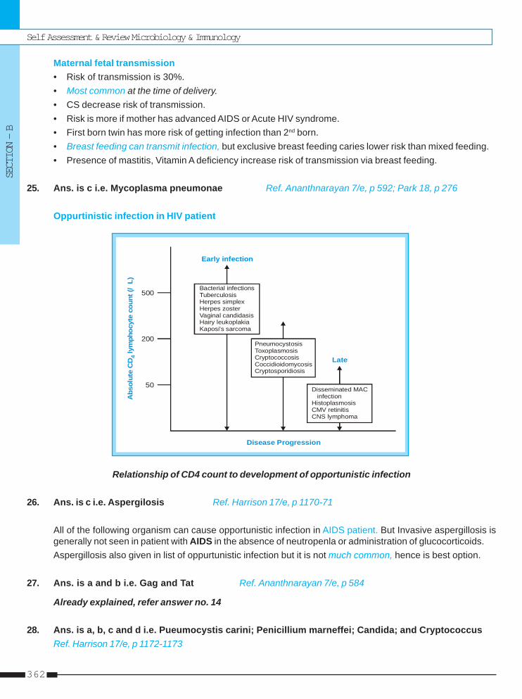
Self Assessment & Review Microbiology & Immunology
SECTION – B
362
Maternal fetal transmission• Risk of transmission is 30%.• Most common at the time of delivery.• CS decrease risk of transmission.• Risk is more if mother has advanced AIDS or Acute HIV syndrome.• First born twin has more risk of getting infection than 2nd born.• Breast feeding can transmit infection, but exclusive breast feeding caries lower risk than mixed feeding.• Presence of mastitis, Vitamin A deficiency increase risk of transmission via breast feeding.
25. Ans. is c i.e. Mycoplasma pneumonae Ref. Ananthnarayan 7/e, p 592; Park 18, p 276
Oppurtinistic infection in HIV patient
Abs
olut
e C
D ly
mph
ocyt
e co
unt (
/L)
4
50
200
500Bacterial infectionsTuberculosisHerpes simplexHerpes zosterVaginal candidasisHairy leukoplakiaKaposi's sarcoma
PneumocystosisToxoplasmosisCryptococcosisCoccidioidomycosisCryptosporidiosis
Disseminated MAC infectionHistoplasmosisCMV retinitisCNS lymphoma
Early infection
Late
Disease Progression
Relationship of CD4 count to development of opportunistic infection
26. Ans. is c i.e. Aspergilosis Ref. Harrison 17/e, p 1170-71
All of the following organism can cause opportunistic infection in AIDS patient. But Invasive aspergillosis isgenerally not seen in patient with AIDS in the absence of neutropenla or administration of glucocorticoids.Aspergillosis also given in list of oppurtunistic infection but it is not much common, hence is best option.
27. Ans. is a and b i.e. Gag and Tat Ref. Ananthnarayan 7/e, p 584
Already explained, refer answer no. 14
28. Ans. is a, b, c and d i.e. Pueumocystis carini; Penicillium marneffei; Candida; and CryptococcusRef. Harrison 17/e, p 1172-1173
HIV & Other Retroviruses
UNIT – II VIROLOGY
363
Fungal infection in AIDS patient are :– Pneumocytis carnii (MC oppurtinstic infection in HIV patient)– Cryptococcus neoformans (MC cause of meningitis in HIV patient)– Histoplasma– Penicillium marneffei– Coccidiodes immitis– Aspergillosis (pseudomembraneous bracheobronchitis in AIDS patient).– Sporothrix.
Note :Cryptosporidium is a parasite not a fungus.
29. Ans. is a, b, c and e i.e. A glandular fever like illness; Generalized lymphadenopathy; Gonococcalsepticemia; and Presenile dementiaSee below
Gonococcal septicemia is seen in terminal complement deficiency not in HIV patient.I do not think that I should explain whole clinical feature here again. Just revise them from theory portion.
30. Ans. is a and b i.e. Gag; and Tat Ref. Harrison 17/e, p 1140
Already explained, refer answer no. 14
31. Ans. is a, b, c, d and e i.e. All are correct options Ref. Park 18/e, p 277; 19/e, p 291
Guys, please see Diagnosis of AIDS from theory portion.
32. Ans. is b and c i.e. CD 8 < 500; and CD 4 : CD 8 = 1 Ref. Harrison 16/e, p 1104
Any HIV infected individual with a CD-4 + T cell count of < 200/μl has AIDS by definition regardless of symptomor oppurtinstic infection.
33. Ans. is c and e i.e. Cryptococcus; and Giardia lambia Ref. Harrison 17/e, p 1169
Cause of diarrhoea in HIV patient
Bacterial Fungal Parasitic
– Shigella – Histoplasmosis – Cryptosporidia– Salmonella – Penicillosis – Microsporidia– Campylobacter – Coccidioidomycosis – Isospora belli
34. Ans. is c and d i.e. Oral candidiasis; and Hairy leukoplakia Ref. Harrison 17/e, p 1174
Already explained, refer answer no. 20
35. Ans. is None Ref. Harrison 17/e, p 1140; Ananthnarayan 7/e, p 584
The options given are not gene but their protein products.
Self Assessment & Review Microbiology & Immunology
SECTION – B
364
36. Ans. is d i.e. Male to female transmission > female to male Ref. Harrison 17/e, p 1142
Modes of transmission of AIDS• Sexual Transmission :
– MC mode of HIV transmission is heterosexual transmission.– Chances of HIV infection from male to female is twice– Anal intercourse carries higher risk of transmission.– Presence of STD increase risk of transmission.– Adloscent girl and women above 45 are more prone to get HIV infection.
• Transmission by Blood and Blood products :– Transfusion of whole blood, packed RBC, platelets, leukocytes and plasma are all capable of transmitting
HIV infection.– Hyperimmune gamma globulin, hepatitis B immunoglobulin, plasma derived hepatitis B Vaccine and Rh
immunoglobulin have not been associated with transmission of HIV infection.
• Maternal fetal transmission– Risk of transmission is 30%.– Most common at the time of delivery.– CS decrease risk of transmission.– Risk is more if mother has advanced AIDS or Acute HIV syndrome.– First born twin has more risk of getting infection than 2nd born.– Breast feeding can transmit infection, but exclusive breast feeding caries lower risk than mixed feeding.– Presence of mastitis, Vitamin A deficiency increase risk of transmission via breast feeding.
37. Ans. is a i.e. Mycobacterium tuberculosis Ref. Harrison 17/e, p 1172
• HIV infection increase risk of developing active TB by a factor of 15 to 30.• In developing countries where M. tuberculosis infection is most frequent, HIV infected individual have primary
and secondary infection with the usual way.• Oppurtunistic infection with M.avium intra cellulare is MC oppurtunistic infection in west.
So, it is clear that in developing countries like India MC mycobacterial infection is M. tuberculosis.
38. Ans. is a and b i.e. P. carrnii; and M. avium Ref. Park 18/e, p 276; 19/e, p 292
Already explained, refer answer no. 2 and 10
HIV & Other Retroviruses
UNIT – II VIROLOGY
365
1. a) There is greater ... 2. a) M. tuberculosis 3. a) T-helper cells...4. a) Pol 5. b) Core antigen 6. d) C7. c) Retro virus 8. a) Reverse ... 9. a) From a male ...
10. a) GAG 11. c) Hetero ...
1. In the heterosexual trasmission (from ifective part-ner to non-infective partner) of HIV : [Kar 03]a) There is greater risk of transmission from man
to womanb) There is greater risk to transmission from woman
to manc) Risk is equal in neither waysd) HIV infection is not transmitted by heterosexual
act[Ref. Park 19/e, p 289]
2. In India, maximum cases of tuberculosis in AIDSpatients are due to : [Bihar 03]a) M.tuberculosisb) M. avium intracellularec) M.scrofulaceumd) M.akari
[Ref. Ananthnarayan 7/e, p 589]3. AIDS involves : [Jharkhand 04]
a) T-helper cellsb) T-suppressor cellsc) T-cytotoxic cellsd) B-cells
[Ref. Ananthnarayn 7/e, p 586]4. Which of the following gene is associate with en-
coding of reverse transcriptase ?a) Pol [Bihar 05]b) Envc) Gagd) p–24
[Ref. Ananthnarayan 7/e, p 584]5. Geg gene encodes for : [Bihar 05]
a) Reverse transcriptaseb) Core antigenc) Enveloped) Gene activation
[Ref. Ananthnarayan 7/e, p 584]
Answer
6. The most common HIV subtype in India is :a) D [Kar 06]b) Hc) Ed) C
[Ref. Harrison 17/e, p 1142; Fig 182-7]7. AIDS virus is : [Kerala 94]
a) RNA virusb) DNA virusc) Retro virusd) Entero virus
[Ref. Ananthnarayan 7/e, p 582]8. Following is the marker of HIV infection in blood :
a) Reverse Transcriptase [AIIMS 94]b) DNA polymerasec) RNA polymerased) None
[Ref. Ananthnarayan 7/e, p 583]9. Incidence of AIDS transmission from infective to
noninfective partner is more with : [NIMS 96]a) From a male to a female partner increased riskb) From a female to a male partner increased riskc) Equal risk to bothd) No risk of heterosexual transmission
[Ref. Park 19/e, p 289]10. The gene coding for core of HIV is : [Kerala 96]
a) GAGb) ENVc) POLd) TAT
[Ref. Ananthnarayan 7/e, p 584]11. Commonest mode of AIDS tranmission in India is
by : [Karn. 96]a) Vertical transmissionb) I.V. drug abusec) Heterosexual promiscuityd) Homosexual promiscuity
[Ref. Harrison 17/e, p 1142]
Chapter Review••••• This section includes questions of VThis section includes questions of VThis section includes questions of VThis section includes questions of VThis section includes questions of Various Other PGMEES from 1990 – 2007.arious Other PGMEES from 1990 – 2007.arious Other PGMEES from 1990 – 2007.arious Other PGMEES from 1990 – 2007.arious Other PGMEES from 1990 – 2007.
Self Assessment & Review Microbiology & Immunology
SECTION – B
366
12. The HIV virus can be destroyed in vitro by which ofthe following : [Karn. 96]a) Boilingb) Ethanolc) Cidexd) All of the above
[Ref. Ananthnarayan 7/e, p 585, 586]13. During the window period of patient with AIDS :
a) ELISA is -ve [NIMS 96]b) Western Blot is -vec) Both are -ved) PCR is -ve
[Ref. Ananthnarayan 7/e, p 591]14. Risk of HIV transmission is not seen with :
a) Whole blood [NIMS 96]b) Plateletsc) Plasma derived Hepatitis B vaccined) Leucocytes Vaccines
[Ref. Harrison 17/e, p 1144]15. AIDS is not transmitted by : [Kerala 97]
a) Blood transfusionb) Cryoprecipitatec) Breast milkd) Plasma
[Ref. Harrison 17/e, p 1144]16. All the following are true about HIV except :
a) A DNA virus belonging to lentivirus [TN 99]b) Attacks CD4 lymphocytesc) CD4 : CD8 ratio is reversedd) Mostly spread by heterosexual contact
[Ref. Ananthnarayans 7/e, p 582]17. Commonest helminthic infection in AIDS is :
a) Trichuris -Trichuria [UP 00]b) Strongyloidisc) Enterobiusd) Necator americana
[Ref. Ananthnarayan 7/e, p 592]18. Following cells are infected by HIV : [Karn. 02]
a) CD8 + T lymphocytesb) CD4 + T lymphocytesc) B Cellsd) Macrophages
[Ref. Harrison 17/e, p 1149]18. Which is not increased in HIV infection :
a) CMV [ST JOHN’S O2]b) Kaposi sarcomac) Mycobacterial infectiond) Pneumococcal infection
[Ref. Ananthnarayan 7/e, p 592 Table (62.5)]
20. Full blown imunodeficiency syndrome is :a) High viral titres with low CD4 count [SGPGI 03]b) Low viral titres with low CD4 countc) Low viral titres with high CD4 countd) High viral titres with high CD4 count
[Ref. Harrison 17/e, p 1169]21. All are true about AIDS except : [UP 03]
a) Seen in heterosexual onlyb) Caused by reterovirusc) Candidiases is also common featured) Retrovirus is thermolabile
[Ref. Ananthnarayan 7/e, p 586]22. Reverse Transcriptase is : [UP 04]
a) DNA polymeraseb) DNA dependant RNA polymerasec) RNA dependant DNA polymerased) None
[Ref. Ananthnarayan 7/e, p 583]23. Most common mode of transmission of HIV world
wide is : [SGPGI 04]a) Heterosexualb) Homosexualc) IV drug abused) Contaminated blood products
[Ref. Harrison 17/e, p 1142]24. Screening test for AIDS : [MP 05]
a) ELISAb) PCRc) Western blotd) CD-4 count
[Ref. Park 19/e, p 293]25. HIV virus has predilection for infecting :
a) CD4 + T cells [DNB 05]b) CD8 cellsc) Macrophagesd) Plasma cells
[Ref. Harrison 17/e, p 1149]26. Most frequent species of Mycobacterirum associ-
ated with HIV infection in India is : [MP 06]a) M. Avium intracelulareb) M. Fortuitumc) M. tuberclosisd) M. Bovis
27. Which of the following is not a HIV gene :a) Gag [Kerala 06]b) Oplc) Envd) Rb
[Ref. Ananthnarayan 7/e, p 584]
12. d) All of the ... 13. c) Both are -ve 14. c) Plasma ... 15. b) Cryoprecipitate 16. a) A DNA virus ...17. b) Strongyloidis 18. b) CD4 + T ... 19. d) Pneumo ... 20. a) High viral ... 21. a) Seen in ...22. c) RNA ... 23. a) Heterosexual 24. a) ELISA 25. a) CD4 + T cells 26. c) M. tuberclosis27. d) Rb
Answer
Self Assessment & Review Microbiology & Immunologyxii
UNIT – III MYCOLOGY
1. Dermatophytes 369 – 3722. Yeast & Yeast like Fungus 373 – 387
Cryptococcus, Candida, Pneumocystii carinii3. Aspergillus & Mucormycosis 388 – 3924. Dimorphic Fungi 393 – 399

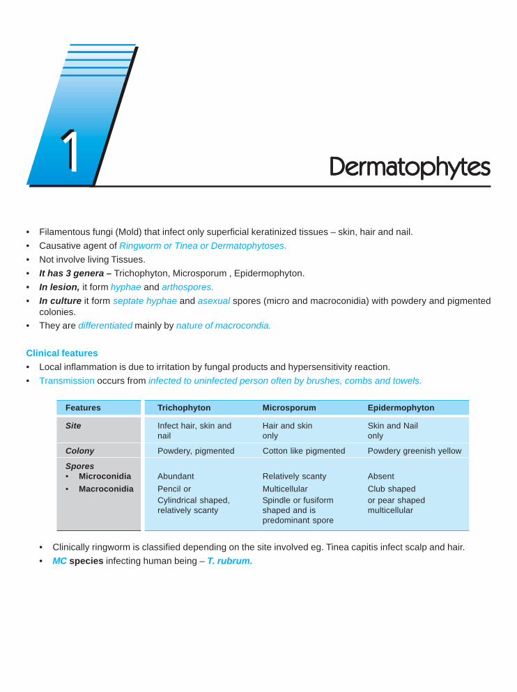
• Filamentous fungi (Mold) that infect only superficial keratinized tissues – skin, hair and nail.• Causative agent of Ringworm or Tinea or Dermatophytoses.• Not involve living Tissues.• It has 3 genera – Trichophyton, Microsporum , Epidermophyton.• In lesion, it form hyphae and arthospores.• In culture it form septate hyphae and asexual spores (micro and macroconidia) with powdery and pigmented
colonies.• They are differentiated mainly by nature of macrocondia.
Clinical features• Local inflammation is due to irritation by fungal products and hypersensitivity reaction.• Transmission occurs from infected to uninfected person often by brushes, combs and towels.
Features Trichophyton Microsporum Epidermophyton
Site Infect hair, skin and Hair and skin Skin and Nailnail only only
Colony Powdery, pigmented Cotton like pigmented Powdery greenish yellow
Spores• Microconidia Abundant Relatively scanty Absent• Macroconidia Pencil or Multicellular Club shaped
Cylindrical shaped, Spindle or fusiform or pear shapedrelatively scanty shaped and is multicellular
predominant spore
• Clinically ringworm is classified depending on the site involved eg. Tinea capitis infect scalp and hair.• MC species infecting human being – T. rubrum.
11 DerDerDerDerDermatophytesmatophytesmatophytesmatophytesmatophytes

Self Assessment & Review Microbiology & ImmunologySECTION – B
370
1. T. capitis (endothrix) is caused by : [PGI 00]a) Epidermophytonb) T. tonsuransc) T. violaceumd) Microsporum
2. Kerion is caused by : [PGI 98]a) Candidab) Streptococcusc) Dermatophytesd) Herpes
3. Tinea cruris is caused by : [PGI 97]a) Epidermophytonb) Trichophytonc) Microsporumd) Candida
QUESTIONS
Answer 1. b and c 2. c) Dermatophytes 3. a and b
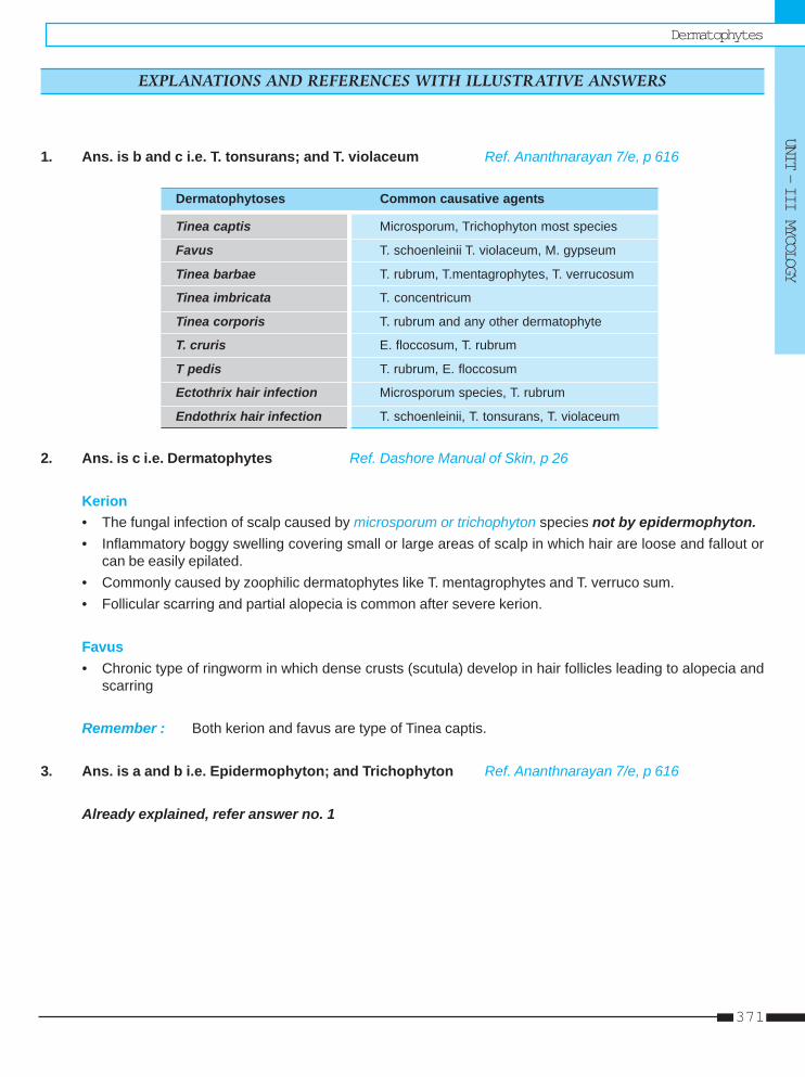
DermatophytesUNIT – III MYCOLOGY
371
1. Ans. is b and c i.e. T. tonsurans; and T. violaceum Ref. Ananthnarayan 7/e, p 616
Dermatophytoses Common causative agents
Tinea captis Microsporum, Trichophyton most species
Favus T. schoenleinii T. violaceum, M. gypseum
Tinea barbae T. rubrum, T.mentagrophytes, T. verrucosum
Tinea imbricata T. concentricum
Tinea corporis T. rubrum and any other dermatophyte
T. cruris E. floccosum, T. rubrum
T pedis T. rubrum, E. floccosum
Ectothrix hair infection Microsporum species, T. rubrum
Endothrix hair infection T. schoenleinii, T. tonsurans, T. violaceum
2. Ans. is c i.e. Dermatophytes Ref. Dashore Manual of Skin, p 26
Kerion• The fungal infection of scalp caused by microsporum or trichophyton species not by epidermophyton.• Inflammatory boggy swelling covering small or large areas of scalp in which hair are loose and fallout or
can be easily epilated.• Commonly caused by zoophilic dermatophytes like T. mentagrophytes and T. verruco sum.• Follicular scarring and partial alopecia is common after severe kerion.
Favus• Chronic type of ringworm in which dense crusts (scutula) develop in hair follicles leading to alopecia and
scarring
Remember : Both kerion and favus are type of Tinea captis.
3. Ans. is a and b i.e. Epidermophyton; and Trichophyton Ref. Ananthnarayan 7/e, p 616
Already explained, refer answer no. 1
EXPLANATIONS AND REFERENCES WITH ILLUSTRATIVE ANSWERS
Self Assessment & Review Microbiology & ImmunologySECTION – B
372
1. c) Nails, hair ... 2. c) Trichophyton
1. Dermatophytes are fungi infecting : [Delhi 93]a) Subcutaneous tissuesb) Systemic organsc) Nails, hair and skind) Superficial skin and deep tissue
[Ref. Ananthnarayan 7/e, p 613]
Answer
2. Dermatophytosis is caused by : [Delhi 02]a) Herpes simplexb) Papilloma virusc) Trichophytond) Candida
[Ref. Ananthnarayan 7/e, p 613]
Chapter Review••••• This section includes questions of VThis section includes questions of VThis section includes questions of VThis section includes questions of VThis section includes questions of Various Other PGMEES from 1990 – 2007.arious Other PGMEES from 1990 – 2007.arious Other PGMEES from 1990 – 2007.arious Other PGMEES from 1990 – 2007.arious Other PGMEES from 1990 – 2007.••••• Questions are arQuestions are arQuestions are arQuestions are arQuestions are ar ranged in decreasing order of yearranged in decreasing order of yearranged in decreasing order of yearranged in decreasing order of yearranged in decreasing order of yearwiz. This is done to makwiz. This is done to makwiz. This is done to makwiz. This is done to makwiz. This is done to make refere refere refere refere refer ralralralralral
system easier and uncomplicated to save the precious time of PGMEE Aspirant.system easier and uncomplicated to save the precious time of PGMEE Aspirant.system easier and uncomplicated to save the precious time of PGMEE Aspirant.system easier and uncomplicated to save the precious time of PGMEE Aspirant.system easier and uncomplicated to save the precious time of PGMEE Aspirant.
CRYPTOCOCCUS NEOFROMANSOnly yeast which is pathogenic. Also called as Eurpoean blastomycosis.Characterized by a thick polysaccharide capsule which is stained by India Ink.
Morphology• Cell wall is gram positive.• C. neoformans differs nonpathogenic species of Cryptococcus by its ability to grow at 370C and the production of
phenol oxidase and urease.There are four serotypes [A, B, C, D] and 3 varieties of C. neoformans :a. C. neoformans var grubii (serotype A)b C. neoformans var gatti (serotype B or C) : Cause disease is immunocompetent.c. C. neoformans var neoformans (serotype D).
Pathogenesis and Pathology :• MC predisposing factor for cryptococcosis : AIDS (risk increase when CD4 <200/ul).• Most infections in immunocompromised patient are caused by serotype A.• Infection is acquired by inhalation of the fungus in to lung which is frequently asymptomatic but in immuno-
compromised state there is hematogenous spread from lung to brain which leads to meningoencephalitis.• Cryptococci are best seen in tissue by staining with methenamine silver or periodic acid schiff.• Infection can occur by pigeon dropping or eucylyptus tree.
Clinical Manifestation• Most patient present with meningoencephalitis.• C.neoformis is MC cause of meningitis in AIDS patient.• C.meningitis may not show meningismus, which is seen in bacterial meningitis and it may be associated with
sudden vision loss.• Focal lesions called cryptococcomas are more common in previously normal patient.• These lesions are located MC at basal ganglia or the head of caudate nucleus. ..... Harrison 16/e, p 1184• Cryptococcomas are best seen with MRI.• Cryptococcus in AIDS patient has less sign and symptoms.• Headache is MC symptom.
22 YYYYYeast & Yeast & Yeast & Yeast & Yeast & Yeast Likeast Likeast Likeast Likeast Like Fe Fe Fe Fe Fungusungusungusungusungus
Self Assessment & Review Microbiology & Immunology
SECTION – B
374
Diagnosis• Lumbar puncture is single most useful diagnostic test.• Indian Ink preparation is the method of choice for detecting cryptococci in CSF.• Latex agglutination – Approx 90% of patient with cryptococcal meningitis including all AIDS patient, have capsular
antigen detectable in CSF. However this is less useful in diagnosis of pulmonary disease.
Treatment
Type of disease Preferred treatment Alternative
i. Disease in AIDS Patient Amphotericin B Itraconazoleii. Disease in non AIDS Patient
– Meningitis Amphotericin B Switch over to fluconazole when– Pulmonary disease patient condition has improved
• Immunocompromised Amphotericin B Switch over to Fluconazole whenPatient patient condition has improved
• Normal Fluconazole Itraconazole
CANDIDACandidiasis is MC systemic mycosis.• MC agent responsible is candida albicans.• All pathogenic candida are commensals of humans particularly in mouth, stool and vagina.
MorphologyGram positive fungiThey grow as budding yeast cells but they can also form pseudohypahe (except C. glabrata which doesn’t) both inculture and in tissues.• Candida albicans is dimorphic as it also forms true hyphae [other species of candida are not dimorphic].
... Jawetz 24/e, p 643• C. albicans is identified on basis of their ability to form of large thick walled spores (diagnostic) called
chlamydospores on nutritionally deficient media or corn meal agar at 200C.• C. albicans form germ tubes [Reynods Braude Phenomenon].• Accurate identification of Candida species other than C.albicans require biochemical tests.
Pathogenesis• Invasive candidiasis is usually preceded by ↑ colonization in mouth, vagina due to broad spectrum antibiotics.• Majority of non.albicans vagina speices enter the blood through intravascular catheterisation.
Clinical manifestationI. Cutaneous and Mucosal candidiasis -
• Risk factors association with superficial candidiasis are :– AIDS – Pregnancy – Diabetes (MC)– Infants and elders – Oral contraceptives – Trauma
• It includes -– Oral thrush :
- Painless discrete and confluent adherent white plaques on the oral and pharyngeal mucosa commonin AIDS at CD4 <50/μl.
- Cutaneous candidiasis may be intertriginous (erythematous scaling or moist lesion) or paronychial(seen in occupation that lead to frequent immersion of hands in water).
Yeast & Yeast Like Fungus
UNIT – III MYCOLOGY
375
– Esophageal candidiasis : Most lesion are in distal 3rd of oesophagus. Diagnosed by biopsy.– Vulvovaginal candidiasis : Mostly in 3rd trimester of pregnancy.– Chronic mucocutaneous candidiasis : Mostly onset in early childhood and association with cellular
immunodeficiencies and endocrinopathy. Present as hyperkeratotic skin lesion, partial alopecia, and bothoral and vaginal thrush.
II. Deeply Invasive (systemic) candidiasis :• Hematogenous seeding (candedemia) is common to retina, kidney, spleen, liver.• In immunocompetent patient – U/L or B/L white retinal exudates appear within 2 weeks of onset of
candedemia. Most cases of ocular involvement occur in non neutropenic patient.• Hepatosplenic candidiasis (Chronic disseminated candidiasis) usually occur in patient with acute leukaemia.
originates from intestinal seeding of portal and venous circulation. Mostly occur in neutropenic patient.• Candida can cause arthritis of knee in patient who have received chronic glucocorticoid injections in joint.• Endocarditis in previously damaged or prosthetic valve.
Diagnosisi. Superficial candidiasis : Demonstration of pseudohyphae or hyphae on wet smear with confirmation by culture or
staining (Gram’s, PAS, Methenamine silver).ii. Invasive candidiasis : Diagnosed by histologic section of biopsies or by culture of CSF, blood, joint fluid.
Treatment
Type Preferred Alternative
i. Mucocutaneous
• Cutaneous Topical azoles Topical nystatin• Vulvovaginal Azole cream or oral Fluconazole Nystatin suppository• Oropharyngeal Clotrimazole or Fluconazole Nystatin• Esophageal Fluconazole or itracon azole
ii. Deeply invasive
• Non neutropenic • Fluconazole or amphotericin B• Neutropenic • Amphotericin B
iii. Candida endophthalmitis • IV polyene + flucytosine
PNEUMOCYSTIS INFECTION• Opportunistic fungal pulmonary pathogen, lacks ergosterol so not susceptible to antifungal which inhibit ergosterol
synthesis.• Human isolate – P. jiroveci; P.carnii is found in rats. ... Harrison 17/e, p 1267• Most prominent antigen : Major surface glycoprotein which shows antigenic variation and facilitates its
adherence.• Other important antigen is 35-55kDa - which acts as marker of infection.
Pathogenesis and Pathology• Defects in CMI and humoral immunity predispose for its infection eg In HIV infection occur when CD4 + <200/μl.• Principal host effector cells – alveolar macrophage.• Transimission – Airborne; person to person.
Self Assessment & Review Microbiology & Immunology
SECTION – B
376
• In alveoli : – It attach to type I cells (extracellular), damage it.– Hypertrophy of type II cells occur causing, surfactant abnormalities.– Increase IL-8 and neutrophil in BAL fluid.– Alveoli filled with typical foamy vacuolated exudate.
• Severe disease :Mild mononuclear cell (ever plasma cell) interstitial infiltrate seen so called Interstitial plasma cell pneumoniae.
Risk factors for Pneumocystis Pneumonia– HIV– Immunosuppressive therapy particularly glucocorticoids for cancer– Organ transplantation and other disorders– Children with primary immunodeficiency disease and premature malnourished infants.
Clinical feature• Symptom – Dyspnea, fever, non productive cough.• Sign – Tachypnea tachycardia and cyanosis but lung auscultation reveals few abnormalities.• Risk for extrapulmonary spread increase with : Administration of aerosolized pentamidine.• MC extrapulmonary site : Lymph node, spleen, liver, bone marrow.• Most widely used prognostic factor is degree of hypoxemia.
Diagnosis1. Definitive diagnosis by histopathologic detection by :
a. Staining – Methenamine silver, toludine blue stain cell wall while Wright-Giemsa stain the nuceli.– Immunoflurescence with monoclonal antibodies; more sensitive.
b. DNA amplification by PCR – most sensitive
2. Specimen collectiona. Fiberoptic bronchoscopy with BAL which is more sensitive than sputum induction, is the mainstay of
diagnosis.b. Transbronchial biopsy and open lung biopsy – only when diagnosis can't be made by BAL.
3. CXR – Classic finding : - B/L diffuse infiltrate in perihilar region.- Nodular densities, cavitary lesion, Pneumothorax can also occur.- ↑ Frequency of upper lobe infiltrate in patient who take aerosolized pentamidine.
4. Reduced arterial O2 pressure (PaO2); increase alveolar arterial O2 gradient (PAO2-PaO2); respiratory alkalosis.
Treatment• DOC cotrimaxazole for all forms of pneumocystosis including extrapulmonary disease.• Alternative for mild to moderate case – Trimethoprim + dapsone and clindamycin + primaquine.• Alternative for moderate to severe – Pentamidine slow IV.• Adjunctive therapy – Glucocorticoid in HIV patient with moderate to severe pneumocystosis whose pulmonary
function deteriorates on taking anti pneumocystis drugs.
Prophylaxis• DOC for HIV primary and secondary (both HIV and Non HIV) prophylaxis is cotrimoxazole.• Alternative Dapsone• In HIV prophylaxis given when : – CD4 + <200/μl
– History of oropharyngeal candidiasis.
Yeast & Yeast Like Fungus
UNIT – III MYCOLOGY
377
1. Pneumocystis Jeroveci : [AI 08]a) Associated with CMVb) Diagnosis is by sputum microscopyc) Seen only in immunocompromised patientsd) Always associated with pneumatocele
2. The capsule of Cryptococcus neoformans in a CSFsample is best seen by : [AI 05]a) Gram’s stainb) Indian ink preparationc) Giemsa staind) Methanamine - silver stain
3. The M.C organism amongst the following thatcause acute meningitis in an AIDS patient is :a) Streptococcus pneumoniae [AI 05]b) Streptococcus agalactiaec) Cryptococcus neoformansd) Listeria monocytogenes
4. M.C fungal infection in febrile neutropenia is :a) Aspergillus niger [AI 01]b) Candidac) Mucormycosisd) Aspergillus fumigatus
5. Latex agglutination test of the antigen in CSF helpsin the diagnosis of : [AI 00]a) Cryptococcusb) Candidiasisc) Aspergillosisd) Histoplasmosis
6. Cryptococcus-neoformans is a : [AI 99]a) Protozoab) Fungusc) Parasited) Mycoplasma
7. True about cryptococcus neoform is all except :a) Capsular antigen is detected in CSF [AIIMS 96]b) Common in immuno compromised patientc) Anticapsular antibody prevents recurrenced) Strongly positive mucicarmine stain of the or-
ganism in tissue is diagnostic8. Which is false regarding Cryptococcus neo-
formans : [AI 95]
a) Grows at 50C and 370Cb) It has 4 serotypesc) Urease negatived) Causes superficial skin infection
9. Cryptococcus is least likely to cause infection of:a) Skin [AI 95]b) Bonec) Braind) Kidney
10. Which dye is most suitable for fungus demonstra-tion in biopsy : [AIIMS 06]a) Alizharian redb) Verihoff dyec) Mason’s trichomed) PAS
11. In HIV infected individual Gram stain of lung aspi-rate shows yeast like morphology. All of the fol-lowing are the most likely diagnosis except :a) Candida tropicalis [AIIMS 05]b) Cryptococcus neoformansc) Penicillium marneffid) Aspergillus fumigates
12. HIV positive female presents with an induratedulcer over the tongue Laboratory findings showgrowth in cornmeal agar at 20C, microscope byshowing hyphae and growth in human serum at370C show budding yeast Probable cause is :a) Candida albicans [AIIMS 01]b) Histoplasmosisc) Blastomycosisd) Coccidioidomycosis
13. Which fungal infection is commonest in neutro-penia : [AIIMS 99]a) Candidab) Histoplasmac) Aspergillus nigerd) Aspergillus fumigatus
14. All are yeast like fungus except : [AIIMS 97]a) Cryptococcusb) Candidac) Trichophytond) None of the above
Answer 1. b) Diagnosis is ... 2. b) Indian ... 3. c) Cryptococcus ... 4. b) Candida 5. a) Cryptococcus6. b) Fungus 7. c) Anticapsular ... 8. c) Urease ... 9. d) Kidney 10. d) PAS
11. d) Aspergillus ... 12. a) Candida ... 13. a) Candida 14. c) Trichophyton
QUESTIONS
Self Assessment & Review Microbiology & Immunology
SECTION – B
378
15. Candida infection is predisposed by all, except :a) Menstruation [AIIMS 96]b) Diabetesc) Mini pill usersd) Combined pill users
16. An HIV positive female has an indurated ulcer overthe tongue. Laboratory findings show growth incornmeal agar at 20 Degrees, microscopy show-ing hyphae and growth inhuman serum at 37 de-grees show budding yeasts. The probable causeis : [PGI 06]a) Candida albicansb) Histoplasmosisc) Blastomycosisd) Coccidiodomycosise) Mucormycosis
17. Endemic fungal infection is caused is by all of thefollowing except : [PGI 05]a) Coccidiodes immitusb) Cryptococcusc) Penicilliumd) Aspergilluse) Blastomyces
18. Which of the following are difficult to isolate fromculture : [PGI 03]a) Candidab) Dermatophytesc) Cryptococcusd) Malassezia furfure) Coccoidiodomycosis
19. Cryptococcus can be readily demonstrated by :a) Albert’s stain [PGI 02]b) India ink stainc) Giema’s staind) Gram’s staine) Z-N stain
20. Neurotrophic fungus is/are : [PGI 02]a) Cryptococcus neoformansb) Histoplasmosisc) Trichophytond) Candidae) Aspergillosis
21. Pneumocystis carini is a fungus because :a) rRNA, mitochondrial protein gene sequence and
presence thymiylate synthase [PGI 00]b) Cell wall contains glucansc) Antifungals are effective against P. carinid) Commonest infection in AIDS
22. Cryptoccoccal meningitis is common in :a) Renal transplant recipient [PGI 00]b) Agammaglobulinemiac) Neutropeniad) IgA deficiency
23. Eucalyptus camaldulensis is associated with thetransmission of : [PGI 99]a) Blastomyces dematitidisb) Histoplasmac) Cryptococcusd) Coccidiodes immitis
24. Fungi that possess a capsule is : [PGI 99]a) Candidab) Aspergillusc) Cryptococcusd) Mucor
25. Cryptococcus has predilection for : [PGI 98]a) Lungsb) Meningesc) Liverd) GIT
26. Fungi without sexual cycle are classified as :a) Fungi imperfecti [PGI 97]b) Phycomycesc) Ascomycetesd) Basidiomycetes
Answer 15. a) Menstruation 16. a) Candida albi... 17. b, c and d 18. d) Malassezia ... 19. b) India ...20. a, b, d and e 21. a and b 22. a) Renal ... 23. c) Cryptococcus 24. c) Cryptococcus25. a and b 26. a) Fungi ...
Yeast & Yeast Like Fungus
UNIT – III MYCOLOGY
379
1. Ans. is b i.e. Diagnosis Ref. Harrison 17/e, p 1267
Pneumocystis Jiroveci• Human isolate of Pneumocystis which is associated with severe pneumonia in immunocompromised state
particularly AIDS (PcP).• P.jiroveci is an extracellular pathogen. Growth in the lung is limited to surfactant layer above alveolar epithelium.• Serologic evidence suggest that most individuals are infected in early childhood (thus option “c” is wrong)
but the pneumonia is seen only in immunocompromised state.• Diagnosis is made by detection of organism in proper specimen.• Specimen for PcP
– Sputum - quick and non invasive.– Broncho-alveolar lavage (BAL) fluid - Main stay of pneumocystis diagnosis.– Transbronchial biopsy - If diagnosis can not be made by BAL.
• The overt infection is an acute interstitial plasma cell pneumonia that occurs with high frequency amongtwo groups :– As epidemic of primary infection among premature or debilated or marasmic infant of hospital wards in
underdeveloped countries.– As sporadic cases among older children and adults who have an abnormal cellular immune status.
2. Ans. is b i.e. Indian Ink preparation Ref. Ananthnarayan 7/e, p 621; Harrison 17/e, 1252
• Cryptococcus neoformans is the only pathogenic yeast.• Within the host and on certain culture media, it is surrounded by a large polysaccharide capsule.• Capsule stands out in India Ink preparation. ... Ananthnarayan 7/e, 621• Indian Ink smears remains the method of choice for detecting cryptococcus in “CSF”.
... Harrison 17/e, p 1252* Cryptococci are best seen in tissue by staining with methamine silver or periodic acid schiff (They stain the
fungus itself not capsule).* Muciramine staining of tissue is diagnostic but demonstrable only in few cases.
3. Ans is c i.e. Cryptococcus neoformans Ref. Harrison 17/e, p 1251 - 1252
• C. neoformans is MC cause of meningitis in AIDS patient.• Generally occurs when CD4+ T cell count <100/μμμμμl.• Diagnosis is made by identification of C. neoformans by India Ink or by detection of cryptococcal antigen by
latex agglutination test.• Strongly (+)ve result on muciarmine staining of tissue is diagnostic.
Other important features of cryptococcal meningitis :• MC cause of meningitis in AIDS patient• Predisposing factors : – HIV infection – Solid organ transplantation – Hodgkins disease
– Sarcoidosis – Corticosteroid therapy.
EXPLANATIONS AND REFERENCES WITH ILLUSTRATIVE ANSWERS
Self Assessment & Review Microbiology & Immunology
SECTION – B
380
• Intra cerebral mass lesion (cryptococcomas) and obstructive hydrocephalus may complicated the course.
Treatment : Patient with AIDS and high risk patient : I.V Amphotericin B followed by Fluconazole formaintainance.
4. Ans. is b i.e. Candida Ref. CMDT ‘08, p 1106
Neutropenia ↑↑↑↑↑ chance of Following Infection. ..... CMDT ‘08; p 1106
Bacterial Fungal Viral
Gram (-)ve enteric pathogens (MC) Candida (MC) Herpes zosterPseudomonas Aspergillus CMVGram (+)ve cocci (particularly Pneumocystis carnii Respiratory syncytial virusStaph. aureus; Staph epidermidis, Influenza virusand viridans streptococci)
Thus as candida is given before aspergillus it would be taken as more common.Organism cause infection in granulocytopenic Patients ..... Harrison 17/e, p 535
• Gram-positive cocci • Gram-negative bacilli– Staphylococcus epidermidis – Escherichia coli– Staphylococcus aureus – Klebsiella spp.– Viridans Streptococcus – Non-aeruginosa Pseudomonas spp.– Enterococcus faecalis – Enterobacter spp.– Streptococcus pneumoniae – Serratia spp.
• Gram-positive bacilli – Acinetobacter spp.– Diphtheroids – Citrobacter spp.– JK bacillus • Fungi
– Candida spp.
5. Ans. is a i.e. Cryptococcus Ref. Harrison 16/e, p 1184; CMDT ‘08, p 1334
“90% of patient with cryptococcal meningoencephalitis have capsular antigen detectable in CSF orserum by latex agglutination.” ... Harrison 16/e, p 1184Diagnosis of cryptococcal disease
Meningoencephalitis Pulmonary cryptococcus Cutaneous cryptococcus
– Lumbar puncture is most useful test – Mimics malignancy – Biopsy– India ink smear of CSF reveals – Biopsy is required for
encapsulated yeast in more diagnosisthan half of cases
– 90% of patient have capsular antigendetectable in CSF by latex agglutination
– CSF culture is definitive diagnostic test
6. Ans. is b i.e. Fungus Ref. Ananthnarayan 6/e, p 574; 7/e, p 620
Cryptococcus is the only pathogenic yeast.
Yeast & Yeast Like Fungus
UNIT – III MYCOLOGY
381
7. Ans. is c i.e. Anticapsular antibody prevents recurrence Ref. Harrison 17/e, p 1252
Cryptococcal InfectionAntibodies are neutralized rapidly by the large amount of capsular antigen released during infection so capsularantigen is not protective. Rather cellular immunity play role in protection.Remember : Virulence factors of cryptococcus
– Polysaccharide capsule– Ability to make melanin– Elaboration of enzymes (phospholipase, urease)
8. Ans. is c i.e. Urease negative Ref. Ananthnarayan 7/e, 621
Characteristic features of C. neoformans• It is Urease positive fungus.• Only pathogenic yeast.• Presence of polysaccharide capsule which is stained with India Ink.• Cell wall is gram positive.• Differs from non pathogenic C. neoformans by :
– Ability to grow at 370C– Production of phenol oxidase and urease.
• It has four serotypes A, B, C and D.– Most infection are caused by serotype A which is found in excreta of wild and domestic birds pigeon.– Serotype B occur around Eucalyptus tree. So, infection occur more in countries with large number of
Eucalyptus tree.
9. Ans. is d i.e. Kidney Ref. Harrison 17/e, p 1252
Manifestation of C. neoformans (in decreasing order of frequency).
Common Rare
• Menignoencephalitis (MC) • Prostitis• Pulmonary cryptococcusis • Endopthalmitis• Cutaneous cryptococcusis • Hepatitis• Osteolytic lesion (present as cold abscess) • Pericardititis
• Endocarditis• Renal abscess
10. Ans. is d i.e. PAS Ref. Ananthnarayan 7/e, p 611
“The periodic acid schiff (PAS) and methanamine silver are valuable methods for the demonstrationof fungal elements in tissue or biopsy section.”
Remember : – Sabauraud’s glucose agar (pH - 5.4), czapek - Dox medium and corn meal agar are mostcommonly used media in mycology.
– GMS (Gomorris methanamine silver) stain is best fungal stain for biopsy section.
Self Assessment & Review Microbiology & Immunology
SECTION – B
382
11. Ans. is d i.e. Aspergillus fumigates Ref. Harrison 17/e, p 1256, 1266
Aspergillus is a mould with septate hyphae (not have yeast like morphology).Another confusing option is option “c”.
Penicillum marneffi :• It is a dimorphic fungi, appearing as small yeast cells in tissue and as a mould in culture.• It is a leading cause of opportunistic infection in patients of late stage HIV.Candida is a group of yeast like fungus except C. albicans which is dimorphic.
Respiratory Infection in AIDS patients
Bacterial Fungal Viral
S. pneumoniae (MC) P.carnii (MC) CMVH.influenzae Penicillium marneffiM.tuberculosis AspergillusMycobacteria Histoplasmosisavium complex Candida and Cryptococcus
12. Ans. is a i.e. Candida Albicans Ref. Harrison 17/e, p 1254
This is a case of oral thrush due to candida albicans.Oral thrush mostly present as discrete and confluent white plaques on the oral and pharyngeal mucosa,particularly in the mouth and on the tongue.
Characteristic feature of Candida– Candidiasis is MC systemic mycosis.– Gram positive fungi characterized by their ability to form pseudohyphae.– Grows rapidly at 25 to 370C.– Candida albicans (not other species) can also form true hyphae so is dimorphic.– Candida albicans is identified on the basis of their ability to form germ tubes in serum or by formation of
thick walled spores called chlamydospores on nutritionally deficient media.
13. Ans. is a i.e. Candida Ref. CMDT ‘08; p 1106
Already explained, refer answer no. 4
14. Ans. is c i.e. Trichophyton Ref. Chakraborty 2/e, p 611, 622; Harrison 7/e, p 1266
Trichophyton is dermatophyton which comes under mould.
Morphological Classification of FungiYeast – Cryptococcus neoformans.Yeast like fungi (Grow partly as yeast and partly as chain of elongated budding cells forming pseudohyphae)Candida.
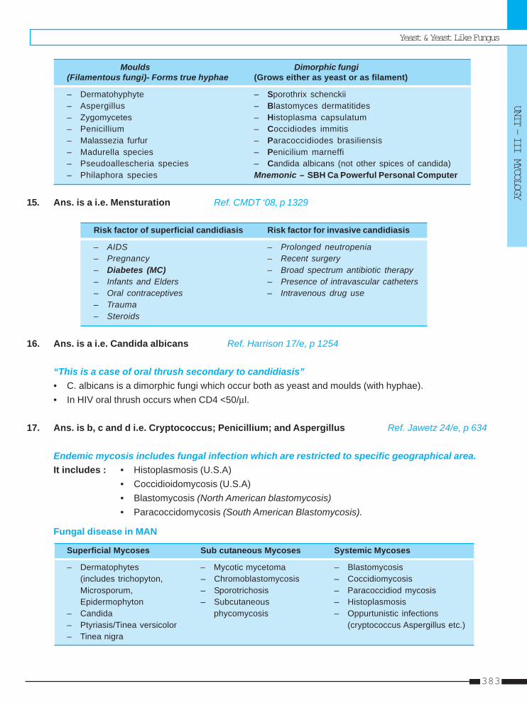
Yeast & Yeast Like Fungus
UNIT – III MYCOLOGY
383
Moulds Dimorphic fungi(Filamentous fungi)- Forms true hyphae (Grows either as yeast or as filament)
– Dermatohyphyte – Sporothrix schenckii– Aspergillus – Blastomyces dermatitides– Zygomycetes – Histoplasma capsulatum– Penicillium – Coccidiodes immitis– Malassezia furfur – Paracoccidiodes brasiliensis– Madurella species – Penicilium marneffi– Pseudoallescheria species – Candida albicans (not other spices of candida)– Philaphora species Mnemonic – SBH Ca Powerful Personal Computer
15. Ans. is a i.e. Mensturation Ref. CMDT ‘08, p 1329
Risk factor of superficial candidiasis Risk factor for invasive candidiasis
– AIDS – Prolonged neutropenia– Pregnancy – Recent surgery– Diabetes (MC) – Broad spectrum antibiotic therapy– Infants and Elders – Presence of intravascular catheters– Oral contraceptives – Intravenous drug use– Trauma– Steroids
16. Ans. is a i.e. Candida albicans Ref. Harrison 17/e, p 1254
“This is a case of oral thrush secondary to candidiasis”• C. albicans is a dimorphic fungi which occur both as yeast and moulds (with hyphae).• In HIV oral thrush occurs when CD4 <50/μl.
17. Ans. is b, c and d i.e. Cryptococcus; Penicillium; and Aspergillus Ref. Jawetz 24/e, p 634
Endemic mycosis includes fungal infection which are restricted to specific geographical area.It includes : • Histoplasmosis (U.S.A)
• Coccidioidomycosis (U.S.A)• Blastomycosis (North American blastomycosis)• Paracoccidomycosis (South American Blastomycosis).
Fungal disease in MAN
Superficial Mycoses Sub cutaneous Mycoses Systemic Mycoses
– Dermatophytes – Mycotic mycetoma – Blastomycosis(includes trichopyton, – Chromoblastomycosis – CoccidiomycosisMicrosporum, – Sporotrichosis – Paracoccidiod mycosisEpidermophyton – Subcutaneous – Histoplasmosis
– Candida phycomycosis – Oppurtunistic infections– Ptyriasis/Tinea versicolor (cryptococcus Aspergillus etc.)– Tinea nigra
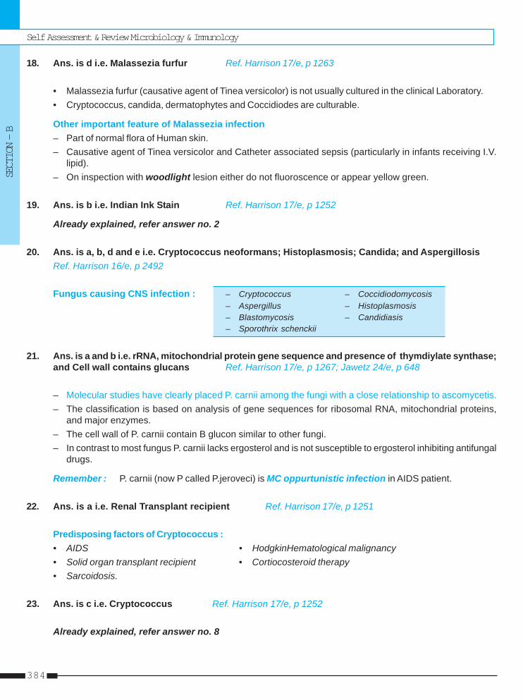
Self Assessment & Review Microbiology & Immunology
SECTION – B
384
18. Ans. is d i.e. Malassezia furfur Ref. Harrison 17/e, p 1263
• Malassezia furfur (causative agent of Tinea versicolor) is not usually cultured in the clinical Laboratory.• Cryptococcus, candida, dermatophytes and Coccidiodes are culturable.
Other important feature of Malassezia infection– Part of normal flora of Human skin.– Causative agent of Tinea versicolor and Catheter associated sepsis (particularly in infants receiving I.V.
lipid).– On inspection with woodlight lesion either do not fluoroscence or appear yellow green.
19. Ans. is b i.e. Indian Ink Stain Ref. Harrison 17/e, p 1252
Already explained, refer answer no. 2
20. Ans. is a, b, d and e i.e. Cryptococcus neoformans; Histoplasmosis; Candida; and AspergillosisRef. Harrison 16/e, p 2492
Fungus causing CNS infection : – Cryptococcus – Coccidiodomycosis– Aspergillus – Histoplasmosis– Blastomycosis – Candidiasis– Sporothrix schenckii
21. Ans. is a and b i.e. rRNA, mitochondrial protein gene sequence and presence of thymdiylate synthase;and Cell wall contains glucans Ref. Harrison 17/e, p 1267; Jawetz 24/e, p 648
– Molecular studies have clearly placed P. carnii among the fungi with a close relationship to ascomycetis.– The classification is based on analysis of gene sequences for ribosomal RNA, mitochondrial proteins,
and major enzymes.– The cell wall of P. carnii contain B glucon similar to other fungi.– In contrast to most fungus P. carnii lacks ergosterol and is not susceptible to ergosterol inhibiting antifungal
drugs.
Remember : P. carnii (now P called P.jeroveci) is MC oppurtunistic infection in AIDS patient.
22. Ans. is a i.e. Renal Transplant recipient Ref. Harrison 17/e, p 1251
Predisposing factors of Cryptococcus :• AIDS • HodgkinHematological malignancy• Solid organ transplant recipient • Cortiocosteroid therapy• Sarcoidosis.
23. Ans. is c i.e. Cryptococcus Ref. Harrison 17/e, p 1252
Already explained, refer answer no. 8
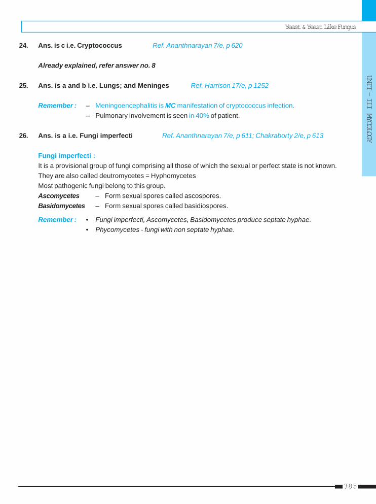
Yeast & Yeast Like Fungus
UNIT – III MYCOLOGY
385
24. Ans. is c i.e. Cryptococcus Ref. Ananthnarayan 7/e, p 620
Already explained, refer answer no. 8
25. Ans. is a and b i.e. Lungs; and Meninges Ref. Harrison 17/e, p 1252
Remember : – Meningoencephalitis is MC manifestation of cryptococcus infection.– Pulmonary involvement is seen in 40% of patient.
26. Ans. is a i.e. Fungi imperfecti Ref. Ananthnarayan 7/e, p 611; Chakraborty 2/e, p 613
Fungi imperfecti :It is a provisional group of fungi comprising all those of which the sexual or perfect state is not known.They are also called deutromycetes = HyphomycetesMost pathogenic fungi belong to this group.Ascomycetes – Form sexual spores called ascospores.Basidomycetes – Form sexual spores called basidiospores.
Remember : • Fungi imperfecti, Ascomycetes, Basidomycetes produce septate hyphae.• Phycomycetes - fungi with non septate hyphae.
Self Assessment & Review Microbiology & Immunology
SECTION – B
386
1. Germ tube test is diagnosis for : [Kar 04]a) Candida albicansb) Cryptococcus neoformansc) Histoplasma capsulumd) Cocciodiodomycosis
[Ref. Ananthnrayana 7/e, p 617]2. Pneumocystis carnii is : [SGPGI 03]
a) Bacteriab) Fungusc) Virusd) Parasite
[Ref. Harrison 17/e, p 1267]3. Pneumocystis carinii is diagnosed by :
a) Sputum examination for trophozoites and cystunder microscope [Kerala 01]
b) Culturec) Positive serologyd) Growth on artificial media
[Ref. Harrison 17/e, p 1268]4. All of the following are yeast like fungi except :
a) Canidida [AIIMS 91]b) Geotrichumc) Cryptococcusd) Trichophyton
[Ref. Chakorvarty 2/e, p 611]5. What is the probabale site of entry of a Aspergil-
lus: [DNB 91]a) Puncture woundb) Bloodc) Lungsd) Gastrointestinal tract
[Ref. Harrison 17/e, p 1257]6. Pneumocystis carinii is diagnosed by :
a) Silver nitrate staining [Kerala 91]
Chapter Review••••• This section includes questions of VThis section includes questions of VThis section includes questions of VThis section includes questions of VThis section includes questions of Various Other PGMEES from 1990 – 2007.arious Other PGMEES from 1990 – 2007.arious Other PGMEES from 1990 – 2007.arious Other PGMEES from 1990 – 2007.arious Other PGMEES from 1990 – 2007.••••• Questions are arQuestions are arQuestions are arQuestions are arQuestions are ar ranged in decreasing order of yearranged in decreasing order of yearranged in decreasing order of yearranged in decreasing order of yearranged in decreasing order of yearwiz. This is done to makwiz. This is done to makwiz. This is done to makwiz. This is done to makwiz. This is done to make refere refere refere refere refer ralralralralral
system easier and uncomplicated to save the precious time of PGMEE Aspirant.system easier and uncomplicated to save the precious time of PGMEE Aspirant.system easier and uncomplicated to save the precious time of PGMEE Aspirant.system easier and uncomplicated to save the precious time of PGMEE Aspirant.system easier and uncomplicated to save the precious time of PGMEE Aspirant.
b) Leishmann stainingc) Fontana stainingd) Acid fast staining
[Ref. Harrison 17/e, p 1268]7. Pneumocystis carinii is diagnosed by :
a) Silver nitrate staining [AIIMS 92]b) Leishmann stainingc) Fontana stainingd) Acid fast staining
[Ref. Harrison 17/e, p 1268]8. Blastomycosis is characterized by all except :
a) Yeast like fungus [Delhi 92]b) Commonly involves lung and skinc) Dimorphic fungusd) Common in South America
[Ref. Ananthnarayan 7/e, p 624]9. The following is not true of Candida albicans :
a) Yeastlike fungus [AI 92]b) Forms chlamydosoporesc) Blastomers seen in isolatesd) Causes meningtis in immuno compromised
[Ref. Ananthnarayan 7/e, p 616]10. Which is not true of cryptococcus neoformans :
a) Grows at 370C [JIPMER 92]b) Grows on sabourauds agarc) Polysaccharide capsuled) Urease negative
[Ref. Ananthnarayan 7/e, p 621]11. Candida is most often implicated in causation :
a) Conjunctivitis [Delhi 93]b) Tneacapitisc) Desert rheumatismd) Thrush
[Ref. Ananthnarayan 7/e, p 616]
1. a) Candida albicans 2. b) Fungus 3. a) Sputum ... 4. d) Trichophyton 5. c) Lungs6. a) Silver nitrate ... 7. a) Silver nitrate ... 8. d) Common in ... 9. c) Blastomers ... 10. d) Urease ...
11. d) Thrush
Answer
Yeast & Yeast Like Fungus
UNIT – III MYCOLOGY
387
12. c) Hydatidosis 13. b) IUCD userAnswer
12. Arc-C-5 in Countercurrent electrophoresis of Se-rum is diagnostic of : [JIPMER 93]a) Cysticercosisb) Cryptococcosisc) Hydatidosisd Brucellosis
[Ref. Paniker 6/e, p 154]
13. Candiasis is frequently associated with all except:a) OCP user [PGI 93]b) IUCD userc) Diabetesd) Pregnancy
[Ref. CMDT 08/e, p 1328]
ASPERGILLUS• Mold with septate hyphae with characterstic dichotomous branching and irregular outline.• MC cause of aspergillosis : A fumigatus.• Out of many species of Aspergillus, only those species that grow at 370C can cause invasive infection.
... Harrison 17/e, p 1256Types of AspergillosisA. Respiratory disease
1. In healthy person : Self limited pneumonitis by massive inhalation of spores.2. With underlying lung disease :
• Allergic bronchopulmonary aspergillosis in patient with asthma, cystic fibrosis : present with wheeze,central bronchiectasis etc.
• Endobronchial saprophytic pulmonary aspergillosis (Aspergilloma = fungus ball) in cyst or cavity of TB,sarcordosis, bronchiectasis, histoplasmosis.
• Often present with hemoptysis. There is no invasion.
B. Superficial infection• Sinusitis, otomycosis (usually by A.niger, A. fumigatus), keratitis etc.• Otomycosis is MC human disease caused by Aspergillus.
C. Disseminated (invasive) Aspergillosis• Lung invasion confined almost entirely to immunosuppresed patients : granulocyte count <500/ml [MC
risk factor : acute leukemia and recipients of tissue transplants].– Invasion in neutropenic is characterized by hypheal invasion of blood vessels, thrombosis, necrosis and
hemorhagic infarction.– Earliest CT finding : One or more small pulmonary nodules; Halo sign (Hazy rim around infarcted tissue),
Cresecent sign (seen when Bone marrow function recovers) can be seen.• In HIV patient, MC site of aspergilioisis : lung• Occur in HIV when CD4+<50/ml, characterized by B/L diffuse or focal infiltrate with a tendency to cavitate.
Aspergillus & MucorAspergillus & MucorAspergillus & MucorAspergillus & MucorAspergillus & Mucormycosismycosismycosismycosismycosis33
UNIT – III MYCOLOGY
Aspergillus & Mucormycosis
389
Diagnosis1. Microscopy :
Even a single isolate of Aspergillus in KOH mount of sputum of neutropenic patient or hematopoietic stem - celltransplant recipient with pneumonia particularly child or non-smoker suggest diagnosis of invasive disease.PAS stain biopsy of lung, nose, paranasal sinus or sites of dissemination can also be used.
2. Culture :Velvety to powdery surface of colony. Ability of A.fumigatus to grow at 450C helps to distinguish it from otherspecies.Culture may be negative or few colonies in aspergilloma or invasive disease.
Treatment
Type of disease Preferred treatment
• Fungus ball Lobectomy• Allergic bronchopulmonary aspergillosis Short course of glucocorticoids• Invasive aspergillosis Voriconazole, Amphotericin B
Self Assessment & Review Microbiology & Immunology
SECTION – B
390
1. In a patient, corneal scraping reveals narrowangled septate hyphae. Which of the following isthe likely etiologic agent : [AI 02]a) Mucorb) Aspergillusc) Histoplasmad) Candida
2. Which of the following is the most common etio-logical agent in paranasal sinus mycoses ?a) Aspergillus spp. [AIIMS 06]b) Histoplasmac) conidiobolus coronatusd) Candida albicans
3. An early diabetic has left sided orbital cellulitisCT scan of paranasal sinus shows evidence ofleft maxillary sinusitis. Gram stained smear of theorbital exudate shows irregularly branching sep-tate hyphae. The following is most likely etiologi-cal agent : [AIIMS 03]a) Aspergillusb) Rhizopus
c) Mucord) Candida
4. Branched septate hyphae found on corneal smearin a case of corneal ulcer is : [AIIMS 00]a) Candidab) Mucorc) Aspergillusd) Histoplasma
5. Mucor mycosis : [PGI 02]a) Angio-invasionb) Lymph invasionc) Septate hyphaed) Long term deferoxamine therapy is predisposing
factore) It may lead to blindness
6. Common fungus causing corneal ulcer : [PGI 01]a) Aspergillusb) Mucorc) Fusariumd) Sporthrix
Answer 1. b) Aspergillus 2. a) Aspergillus ... 3. a) Aspergillus
4. c) Aspergillus 5. a, d and e 6. a and c
QUESTIONS
UNIT – III MYCOLOGY
Aspergillus & Mucormycosis
391
1. Ans. is b i.e. Aspergillus Ref. Ananthnarayan 7/e, p 625; Harrison 17/e, p 1257
• Aspergillus is mold with septate hyphae with branching at 450.• Asexual conidia are arranged in chain, carried on elongated cells called sterigmata borne on expanded ends
of conidiophores.• Aspergillus fumigatus is MC cause of aspergillosis.• Commonest human disease caused by aspergillosis is otomycosis.• Aspergillus infection is characterized by hyphae invasion of blood vessel, thrombosis, necrosis, and
hemorrhagic infarction.
Other options : • Mucor – Broad nonseptate hyphae in tissue• Histoplasma – Dimorphic fungi• Candida – Characterised by presence of pseudohyphae.
Remember : Aspergillus is MC cause of fungal corneal ulcer.
2. Ans. is a i.e. Aspergillus Ref. Dhingra 3/e, p 241
Many different fungal species are found to involve the paranasal sinuses the more common being Asprgillus;Alternaria; Mucor or Rhizopus.
Varieties of fungal infection of sinuses.
• Fungus ball – Implantation of fungus in a healthy sinus– Maxillary sinus is most commonly involved– Treatment is surgical removal with no antifungal therapy.
• Allergic fungal sinusitis – There is no invasion of sinus mucosa by fungus– CT shows mucosal thickening with hyperdense area– Treatment is endoscopic surgical drainage with pre and post operative
systemic steroids.
• Chronic invasive – Fungus invades in to sinus mucosa– CT shows thickened mucosa with opacification of sinus and bone erosion– Treatment is endoscopic surgical removal followed by antifungal therapy.
• Fulminant fungal sinusitis – Acute presentation mostly seen in immunocompromised or diabeticindividuals
– Most common fungus associated are mucor and aspergillosis.
3. Ans. is a i.e. Aspergillus Ref. Ananthnarayan 7/e, p 625; Harrison 17/e, p 1257 - 1258
“Presence of branched septate hyphae in a patient of orbital cellulitis (occur as complication of sinusitis)suggest Aspergillus.”
EXPLANATIONS AND REFERENCES WITH ILLUSTRATIVE ANSWERS
Self Assessment & Review Microbiology & Immunology
SECTION – B
392
Aspergillus Sinusitis occur in three forms :1. Ball of hyphae may form in chronically obstructed paranasal sinus, without tissue invasion.2. A chronic fibrosing granulomatous inflammation begin in sinus and spread slowly to the orbit and brain.3. Allergic fungal sinusitis
• Mucor and Rhizopus belong to family Zygomycetes and have non septate hyphae.
4. Ans. is c i.e. Aspergillus Ref. Ananthnarayan 7/e, p 625; Harrison 17/e, p 1257
Already explained, refer answer no. 1
5. Ans. is a, d and e i.e. Angio-invasion; Long term deferoxamine therpay is predisposing factor; and Itmay lead to blindnessRef. Harrison 17/e, p 1261; CMDT ‘08; 1336
Causative agent of Mucormycosis : • Rhizopus• Rhizomucor• Cunninghamella.
They are molds and have non septate hyphae.Predisposing factors : • Recepient of organ transplant
• Hematologic malignancy• Who are receiving long term desferoxamine therapy• Diabetics• Treatment with steroid or cytotoxic drugs• Chronic renal failure.
Important features of Mucormycosis :• Vascular invasion by hyphae is a prominent feature.• Ischemic or hemorrhagic necrosis is foremost histologic findings.• Nose and paranasal sinus infection :
– Present with bloody nasal discharge with low grade fever and sinus pain followed in few days by doublevision.
– On examination nasal turbinate of involved side may be dusky red or necrotic.– Fungal invasion of ophthalmic artery or orbit may lead to blindness.– Coma may occur by direct invasion.
• Pulmonary mucormycosis :– Manifest as progressive severe pneumonia.– Hematogenous spread to other areas.
• GI invasion : Present as one or more ulcer which tends to perforate.
Treatment : Wide surgical debridement and intravenous amphotericin B is indicated.
5. Ans. is a and c i.e. Aspergillus; and Fusarium Ref. Khurana 2/e, p 123
Fungi causing corneal ulcer are : • Aspergillus (MC)• Candida• Fusarium.

• Dimorphic fungus occur in 2 forms :1. Yeast form = parasitic phase
In host tissues and on cultures at 370C (enriched agar).2. Spores and filamentous (mold) form = saprophytic phase
In soil and culture at 22-250C or Sabourads agar at room temperature.• Disease by all of them are restricted to specific areas of endemicity.• Dimorphic fungus are : Candida albicans, Histoplasma, Sporothrix schencku, B lastomycosis,
Coccidioidomycosis, Paracoccidiodomycosis, Penicillium marneffi.
Mnemonic = SBH Ca PC = Senior Boys Hostel Ca Powerful Personal Computer.
HISTOPLASMA CAPSULATUMNon capsulated intracellular (in macrophages) fungus with septate hyphae. Cause primarily a disease ofreticuloendothelial system.
Source of infectionInhalation of spores present in moist surface alkaline soil enriched by dropping of birds and bats.
PathologyIt forms 2 types of asexual spores large tuberculate macroconidia and smaller elliptical microconidia. Microcondiareach the alveoli and initiate granulomatous reaction.
Clinical features1. Majority of infections are asymptomatic or mild (acute primary pulmonary histoplasmosis).
• Cough, fever, malaise, and chest X-ray finding of hilar adenopathy due to caseation necrosis or calcification(which mimics TB) with or without 1 or more areas of pneumonitis are typical features.
2. In small proportion of patient (who lack history of acute primary pulmonary disease) develop progressive diseasein either form :• Chronic pulmonary histoplasmosis or chronic fibrocavitary pneumonia.• Disseminated disease : Among immunosuppresed and user of TNF α antagonist infliximab.
It mimics disseminated TB.
Dimorphic FDimorphic FDimorphic FDimorphic FDimorphic Fungiungiungiungiungi44
Self Assessment & Review Microbiology & Immunology
SECTION – B
394
DiagnosisPreffered method :Culture : • Tuberculate spore is diagnostic
• Sputum culture – For chronic pulmonary Histoplasmosis.• Culture of bone marrow, mucosal lesion, liver and BAL fluid are diagnostically usefull in disseminated
histoplasmosis.• Blood culture are best performed by lysis centrifugation method.
Treatment Disease Preferred treatment Alternative
Acute pulmonary NoneChronic pulmonary Itraconazole Amphotericin BDisseminated Itraconazole Amphotericin BSevere illness Amphotericin B• CNS involvement• Immunocompromised
BLASTOMYCOSIS (=NORTH AMERICAN BLASTOMYCOSIS)B.Dermatitides is Dimorphic fungi with septate hyphae forming conidiophores bearing single globose to piriformconidia and broad based yeast with single buds usually.
Source of infection : Inhalation of conidia from soil, decomposed vegetation or rotting wood.
Clinical features :It has marked predilection for lungs, skin and bone.• Majority of patient, has chronically progressive course and minority has self limited pneumonia.• Cutaneous disease is usually on the face or other exposed parts of the body in the form of elevated ulcerative
lesions.
Diagnosis : Demonstration of fungus in culture of sputum, pus or urine.
Treatment : • Rapid progression or severe illness → Amphotericin B• CNS disease → Amphotericin B• Mild to moderate and no CNS disease. → Itraconazole.
COCCIDIOIDOMYCOSIS• C. imimitis is Dimorphic fungi with septate hyphae forming barrel shaped (arthospores) or arthroconidia and non-
budding spherules with endospores (tissue form).• It is present in soil and rodents.• Infection is acquired by inhalation of dust containing arthospore.• Majority of person develop asymptomatic respiratory infection.• Some develop self limited influenza like fever known as valley fever or desert rheumatism.• Very few develops coccidiodal granuloma often with caseation necrosis.
Diagnosis :a. Sputum, urine and pus should be examined by wet smear and culture (arthospores are formed in chain from
alternate cells of septate hyphae).b. On biopsy appearance of mature spherule is diagnostic.
UNIT – III MYCOLOGY
Dimorphic Fungi
395
c. Serological test are very helpful.PARACOCCIDIODES BRASILLIENSIS– Dimorphic fungi with septate hyphae + rare globose conidia and chlamydospores.– Tissue form : Yeast with characteristic multiple budding.– Causative agent of 'South American Blastomycosis' in which pulmonary infection spreads hematogenously to
mucosa of mouth, nose; lymph node; skin and other internal organs (eg. adrenal, git).Ulcerative granuloma of buccal and nasal mucosa are prominent feature of disease.
Diagnosis : Culture of sputum, pus and mucosal lesion are often diagnostic.
Treatment : • Mild disease - Itraconazole• Advanced disease - Amphotericin B
SPOROTHORIXSporothrix schenckii is dimorphic fungi causing subcutaneous mycoses (not systemic like other dimorphic fungi).Colonies are blackish (variation in pigmentation) and shiny but becomes wrinkled and fuzzy with age.
Source of InfectionAcquired from thorn pricks of rose, sphagnum moss etc into subcutaneous tissue through minor trauma.
Clinical feature• Most cases occur in upper limb.• Usual site of infection : extremity (facial lesion in case of children).• Sporotrichosis is characterized by development on skin, subcutaneous tissue and in lymph nodes, of nodules
which soften and break to form indolent ulcers.• It is of following types :
a. Plaque sporotrichosis : Non tender lesion confined to site of inoculation.b. Lymphangitis sporotrichosis : It is MC manifestation in which there is painless red papule at a primary site
as well as along proximal lymphatic channels often with skip areas is quite distinctive. Nodules may ulcerateor exudate pus.
c. Extracutaneous sporotrichosis : Portal is probably lung. Pulmonary sporotrichosis is usually present assingle chronic cavitary upper lobe lesion.
Diagnosis• Culture (most reliable) of pus, joint fluid, sputum or skin biopsy in which septate hyphae carrying flower like
cluster of small conidia is seen.In tissue – Fungus is seen as 'cigar shaped yeast' yeast cell without mycelia.
• Sometimes 'Asteroid Bodies' can seen which is formed due to antigen-antibody reaction.
Treatment• Cutaneous sporotrichosis – DOC Itraconazole• Alternative potassium iodide• Extracutaneous sporotrichosis – IV Amphotericin B DOC• Alternative Itraconazole.
Self Assessment & Review Microbiology & Immunology
SECTION – B
396
1. All of the following are dimorphic fungi except :a) Sporotricum [AI 97]b) Blastomycetesc) Histoplasmad) Cryptococcus
2. What is true about Histoplasmosis : [AIIMS 08\a) In early stages it is indistinguishable from TBb) Blood culure is not diagnosticc) Hyphal forms are infectious formd) Person to person spread occurs by droplet in-
fection
3. A patient resident of Himachal Pradesh presentedwith a series of ulcers in a row, on his right leg.The biopsy from affected area is taken and cul-tured on sabauraud’s dextrose agar What wouldbe the most likely etiological agent :a) Sporothrix schenckii [AIIMS 03]b) Cladosporium spc) Pseudo allescheria boydiid) Nocardia brasilensis
3. The following fungi are thermally dimorphic ex-cept : [AIIMS 03]a) Sporothrix schenkiib) Cryptococcus neoformainsc) Blastomycosis dermatidisd) Histoplasma capsulatum
4. All are examples of dimorphic fungi except :a) Histoplasma capsulatum [AIIMS 97]b) Bastomycosis dermatitidisc) Cryptococcus neoformansd) Coccidiodes immitis
5. Dimorphic fungus are : [PGI 02]a) Candidab) Cryptococcusc) Blastomycosisd) Coccidiodimycosise) Sporotrichosis
Answer 1. d) Cryptococcus 2. a) In early stage ... 3. a) Sporothrix ...
4. b) Cryptococcus ... 5. c) Cryptococcus ... 6. c, d and e
QUESTIONS
UNIT – III MYCOLOGY
Dimorphic Fungi
397
1. Ans. is d i.e. Cryptococcus Ref. Jawtez 24/e, p 645, Chakraborty 2/e, p 611
Dimorphic fungus• Fungus which occur in two forms :
– Yeast form - in host tissue and on culture at 370C.– Filamentous (mold) from - In soil and culture at 22 - 250C or sabroud’s agar at room temperature.
Dimorphic fungus are :
– Sporothrix schenekii – Penicillium marneffi– Blastomyces – Histoplasma– Coccidiodomycosis – Paracoccidiodomycosis– Candida albicans (not other species of candida)
Mnemonic : SBH Ca Powerful Personal Computer
2. Ans. is a i.e. In early stages it is indistinguishable from TB Ref. Harrison 17/e, p 1244
Clinical manifestation of Histoplasma1. Majority of infections are asymptomatic or mild (acute primary pulmonary histoplasmosis).
• Cough, fever, malaise, and chest X-ray finding of hilar adenopathy due to caseation necrosis or calcification(which mimics TB) with or without 1 or more areas of pneumonitis are typical features.
2. In small proportion of patient (who lack history of acute primary pulmonary disease) develop progressivedisease in either form :• Chronic pulmonary histoplasmosis or chronic fibrocavitary pneumonia.• Disseminated disease : Among immunosuppresed and user of TNF α antagonist infliximab. It mimics
disseminated TB.Options options:• Fungal culture is the gold standard diagnostic test for histoplasmosis.• Mycelia are the naturally infectious forms.• Infection occurs through aerosolization of bird or fat droping.
3. Ans. is a i.e. Sporothrix schenckii Ref. Harrison 17/e, p 1265
Important features of sporothrixS. schenckii– Dimorphic fungus lives as saprophyte on plants.– Infection results from inoculation into subcutaneous tissue after minor trauma.– Nursery worker, florist, gardeners acquire the illness from roses, and other plants.– Most common manifestation is nearly painless red papule at the site of inoculation, over the next several
weeks similar nodules forms along proximal lymphatic channels. The nodules may ulcerate. Thus series ofulcer may form.
EXPLANATIONS AND REFERENCES WITH ILLUSTRATIVE ANSWERS
Self Assessment & Review Microbiology & Immunology
SECTION – B
398
Diagnosis : – In skin lesion the organism is hard to find .– Culture of pus or a skin biopsy is preferred method of diagnosis.
Treatment : – Potassium iodide– Itraconazole.
4. Ans. is b i.e. Cryptococcus neoformans Ref. Chakraborty 2/e, p 611
Don’t get confuse with term thermally dimorphic as it is same as dimorphic fungi i.e. fungi that occur asyeast at body temperature and Mould in soil ambient temperature.
5. Ans. is c i.e. Cryptococcus neoformans Ref. Chakraborty 2/e, p 611
Already explained, refer answer no. 1
6. Ans. is c, d and e i.e. Blastomycosis; Coccidiodimycosis; and SporotrichosisRef. Chakraborty 2/e, p 611; Jawetz 24/e, p 645
Remember : Candida as a whole is not dimorphic only candida albicans is dimorphic.
Fore more detail, refer answer no. 1
UNIT – III MYCOLOGY
Dimorphic Fungi
399
Answer 1. a) Blastomycosis ... 2. a) Very rare in patients ...
1. Which among are dimorphic fungi :a) Blastomycosis [Jharkhand 05]b) Paracoccidioidomycosisc) Histoplasmosisd) otomycosis
[Ref. Ananthnarayan 7/e, p 611]
Answer
2. All of the following regarding histoplasmosis aretrue except : [MP 06]a) Very rare in patients with AIDSb) Bone marrow is involvedc) Gomori methamine silver stain usedd) Dimorphic fungus
[Ref. Harrison 17/e, p 1245; AA 7/e, p 592;Robbins 7/e, p 754 - 755]
Chapter Review••••• This section includes questions of VThis section includes questions of VThis section includes questions of VThis section includes questions of VThis section includes questions of Various Other PGMEES from 1990 – 2007.arious Other PGMEES from 1990 – 2007.arious Other PGMEES from 1990 – 2007.arious Other PGMEES from 1990 – 2007.arious Other PGMEES from 1990 – 2007.••••• Questions are arQuestions are arQuestions are arQuestions are arQuestions are ar ranged in decreasing order of yearranged in decreasing order of yearranged in decreasing order of yearranged in decreasing order of yearranged in decreasing order of yearwiz. This is done to makwiz. This is done to makwiz. This is done to makwiz. This is done to makwiz. This is done to make refere refere refere refere referralralralralral
system easier and uncomplicated to save the precious time of PGMEE Aspirant.system easier and uncomplicated to save the precious time of PGMEE Aspirant.system easier and uncomplicated to save the precious time of PGMEE Aspirant.system easier and uncomplicated to save the precious time of PGMEE Aspirant.system easier and uncomplicated to save the precious time of PGMEE Aspirant.

Self Assessment & Review Microbiology & Immunology
SECTION – B
400
NOTES
Self Assessment & Review Microbiology & Immunologyxii
UNIT – IV PARASITOLOGY
1. Basics of Parasitology 403 – 4072. Protozoa 408 – 4313. Helminths 432 – 450

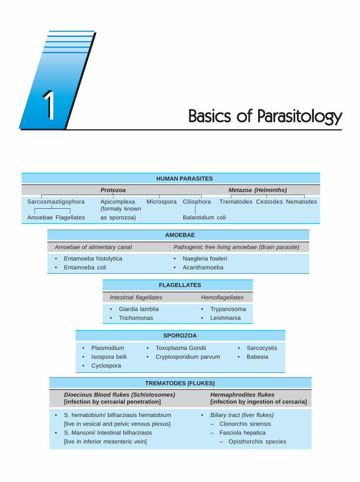
HUMAN PARASITES
Protozoa Metazoa (Helminths)
Sarcosmastigophora Apicomplexa Microspora Ciliophora Trematodes Cestodes Nematodes(formaly known
Amoebae Flagellates as sporozoa) Balantidium coli
AMOEBAE
Amoebae of alimentary canal Pathogenic free living amoebae (Brain parasite)
• Entamoeba histolytica • Naegleria fowleri• Entamoeba coli • Acanthamoeba
FLAGELLATES
Intestinal flagellates Hemoflagellates
• Giardia lamblia • Trypanosoma• Trichomonas • Leishmania
SPOROZOA
• Plasmodium • Toxoplasma Gondii • Sarcocystis• Isospora belli • Cryptosporidium parvum • Babesia• Cyclospora
TREMATODES (FLUKES)
Dioecious Blood flukes (Schistosomes) Hermaphrodites flukes[infection by cercarial penetration] [infection by ingestion of cercaria]
• S. hematobium/ bilharziasis hematobium • Biliary tract (liver flukes)[live in vesical and pelvic venous plexus] – Clonorchis sinensis
• S. Mansoni/ Intestinal bilharziasis – Fasciola hepatica[live in inferior mesenteric vein] – Opisthorchis species
11 Basics of PBasics of PBasics of PBasics of PBasics of Parasitologyarasitologyarasitologyarasitologyarasitology
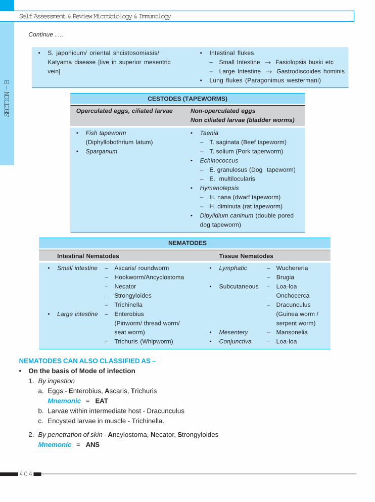
Self Assessment & Review Microbiology & ImmunologySECTION – B
404
Continue .....
• S. japonicum/ oriental shcistosomiasis/ • Intestinal flukesKatyama disease [live in superior mesentric – Small Intestine → Fasiolopsis buski etcvein] – Large Intestine → Gastrodiscoides hominis
• Lung flukes (Paragonimus westermani)
CESTODES (TAPEWORMS)
Operculated eggs, ciliated larvae Non-operculated eggsNon ciliated larvae (bladder worms)
• Fish tapeworm • Taenia(Diphyllobothrium latum) – T. saginata (Beef tapeworm)
• Sparganum – T. solium (Pork taperworm)• Echinococcus
– E. granulosus (Dog tapeworm)– E. multilocularis
• Hymenolepsis– H. nana (dwarf tapeworm)– H. diminuta (rat tapeworm)
• Dipylidium caninum (double poreddog tapeworm)
NEMATODES
Intestinal Nematodes Tissue Nematodes
• Small intestine – Ascaris/ roundworm • Lymphatic – Wuchereria– Hookworm/Ancyclostoma – Brugia– Necator • Subcutaneous – Loa-loa– Strongyloides – Onchocerca– Trichinella – Dracunculus
• Large intestine – Enterobius (Guinea worm /(Pinworm/ thread worm/ serpent worm)seat worm) • Mesentery – Mansonelia
– Trichuris (Whipworm) • Conjunctiva – Loa-loa
NEMATODES CAN ALSO CLASSIFIED AS –• On the basis of Mode of infection
1. By ingestiona. Eggs - Enterobius, Ascaris, Trichuris
Mnemonic = EATb. Larvae within intermediate host - Dracunculusc. Encysted larvae in muscle - Trichinella.
2. By penetration of skin - Ancylostoma, Necator, StrongyloidesMnemonic = ANS
Basics of ParasitologyUNIT – IV PARASITOLOGY
405
3. By blood sucking insects - Filariae4. By inhalation of Dust Containing eggs - Ascaris, Enterobius.
• Based on whether they lay eggs or larvae :1. Oviparous = Laying eggs
a Unsegmented eggs : Ascaris, Trichurisb. Segmented eggs : Ancylostoma, Necator
Mnemonic = ANSc. Eggs containing larvae: Enterobius
2. Viviparous = Producing larvae– Trichinella – Wuchereria– Brugia – Dracunculus.
3. Ovoviviparous (laying eggs containing fully formed larvae which hatch out immediately)– Strongyloides
Two Intermediate host are seen in :• Paragonimus Westerman (Lung fluke) • Diphyllobothrium latum (Fish tape worm)• Clonorchis sinesis (Chinese tapeworm) • Metagonimus yokogawai.
Man is intermediate (Secondary) host in :• Plasmodium • Toxoplasma gondii• Sarcocystis lindemanni • T. solium (man also act as definitive host).• Echinococcus granulo sus [dog tapeworm/ hydatid worm/ Taenia echinococcus]
Remember : In other parasitic infection, man act as definitive (primary) host.
Auto-Infection occur in : • H. nana• E. vermicularis Mnemonic = HETS• T. solium• Strongyloides stercoralis.
Eggs float (eggs can be demonstrated) in concentrated saturated solution :• E. granulosus • H. nana• All nematodes (but not unfertilized egg of Ascaris).
Charcot leyden crystal seen in :• E. histolytica (amoebic dysentery) • Whip worm dysentery• Ascaris pneumonia • Bronchial asthma.
Worms that crawl out :• Enterobius vermicularis • T. saginata.
Worms which does not multiply in host :• Ancylostoma duodenale • Enterobius vermicularis• W. bancrofti.
Self Assessment & Review Microbiology & ImmunologySECTION – B
406
Parasites associated with malignancy :• Clonorchis ⎫⎫⎫⎫⎫
⎬⎬⎬⎬⎬ Bile duct carcinoma• Opisthorchis ⎭⎭⎭⎭⎭• Schistosoma hematobium - bladder carcinoma.
Premunition (immunity to reinfection) seen in :• Syphilis • Cutaneous leishmaniasis.• Hyper/Holoendemic malarial area
Cystic stage is absent in :• Dientamoeba fragilis • Entamoeba gingivalis• Trichomonas vaginalis • Trichomonas intestinalis.
Undulating membrane seen in :• Trichomonas species • HemoflagellatesOnly ‘Protozoan’ parasite found in lumen of human small intestine - Giardia LambliaOnly ‘Ciliate protozoan’ Parasite of man - Balantidium coli
Parthenogenic worm (female is able to produce fertile eggs or larvae without meeting with males) : Strongyloidesstercoralis
Largest protozoa : Balantidum coli
Smallest intestinal amoeba : Dientamoeba fragilis
Smallest and commonest tapeworm found in human intestine : H. nana
Largest helminth T. saginata (beef tapeworm)
Largest liver fluke F. hepatica
Largest trematode infecting man - Fasciolopsis buski
Largest Nematode - Ascaris (roundworm)
Smallest Nematode - Trichinella.
MC protozoan parasite - Toxoplasma gondii
Dogs are responsible for transmission of :• Hydatid disease • Toxocara canis• L. donovani infantum.
Eggs needs development in soil :• Ancylostoma duodenale • Ascaris• Trichuris (whip-worm) • S. stercoralis.
Sputum examination done for :• Rhabditiform larva of Ascaris• Golden brown - eggs of paragonimus
Basics of ParasitologyUNIT – IV PARASITOLOGY
407
• Filariform larva of Strongyloides, Ancylostoma• Entamoeba histolytica (due to hepatobronchial fistula).
Cutaneous larva Migrans caused by :• Necator americanus • Sparganosis• Gnathostomiasis • Hypoderma and Gastrophilus.• Loa-loa and Dicrofilaria • Ancylostoma braziliense, A. caninum, A. duodenale• Fasciola and Paragonimus • Strongyloides stercoralis (larva currens/racing larvae).
Visceral Larva Migrans caused by :• Dog ascarid Toxocara canis (MC) • Cat ascarid T. cati• Anisakis (Large ascarid) • Gnathostoma.
Worms Pass through lung during its life cycle :• Schisto. hematobium (Lung act as 2nd filter) • Paragonimus• Echinococcus • Strong. sterocoralis• A. dudodenale • Ascaris.
Intracellular Parasites :• Leishmania (amastigote form) • Babesia• Plasmodium • Toxoplasma gondii• Sarcocystis. • Trypanosoma cruzi (amastigote form)
NEUROPARASITES
Protozoa Helminthes
Larvae of cestodes Nematodes Ecotopic ova of
• E. histolytica • T.solium • Visceral larva migrans • Schistosoma sp (hematobium)• Naegleria • E. granulosus • Ascaris lumbricoides • F. hepatica• Acarthamoeba • Multiceps sp. • Strongyloides stercoralis • Heterophyes hyterophyes• Trypanosoma • Gnathostoma spinigerum• P. falciparum• T. gondii
IMPORTANT POINTS ABOUT MALARIA• Infective forms for human is sporozoites in saliva of mosquito• Infective forms for mosquito is gametocytes in human blood. At least 12 gametocytes per cubic mm of blood
must be present to infect mosquito.– Gametocytes are maximum in number during the early stages of infections (may exceed 1000 per cubic mm
of blood).– Nonmotile zygote converted into motile ookinete in about 18 - 24 hours.
• Human reservoir is one who harbours the sexual forms (gametocytes) of the parasite.• Only animal reservoir is chimpanzees.
1. An anxious mother brought her 4 old daughter tothe pediatrician. The girl was passing loose bulkystools for the past 20 days. This was often asso-ciated with pain in abdomen. The pediatrician or-dered the stool examination, which showed thefollowing organism. Identify the organism :
a) Entamoeba histolytica [AI 03]b) Giardia lambliac) Cryptosporidiumd) E. coli
2. A patient present with diarrhoea. Analysis of stoolon wet mount shows mobile protozoa withoutRBCs and pus cells. the Diagnosis is : [AI 00]a) Balantidium colib) Giardiasis
c) Trichomonas hominisd) Entamoeba histolytica
3. All of the following statements about toxoplas-mosis are true except : [AI 97]a) Oocyst in freshly passed cat’s faeces is not in-
fectiveb) May spread by organ transplantationc) Maternal infection after 6 months has high risk of
transmissiond) Arthalgia, sore throat and abdominal pain are the
most common manifestation4. Plasmodium falciparum infection of man is char-
acterized by : [AI 97]a) The erythrocytes are increased in sizeb) All stages of erythrocytic schizogony are seen in
peripheral bloodc) Multiple infection of erythrocytes are seend) Each erythrocytic cycle lasts 72 hours
5. Which of the following is true about malaria :a) Size of RBC is enlarged in Vivax infectionb) Size of RBC is enlarged in Falciparum infectionc) Schuffner’s dots are seen in Malariae infectiond) Relapse is seen in Falciparum infection [AI 96]
Answer 1. b) Giardia lamblia 2. b) Giardiasis 3. d) Arthalgia ...4. c) Multiple ... 5. a) Size of RBC ...
QUESTIONS
22 ProtozoaProtozoaProtozoaProtozoaProtozoa
Protozoa
UNIT – IV PARASITOLOGY
409
6. Which of the following is true about P. falciparuma) James dots are seen [AI 96]b) Accole forms are seenc) Relapse are frequentd) Longest incubation period
7. Which of the following is true about. mature cystsof E. histolytica : [AI 96]a) Endoplasm and ectoplasm are clearly definedb) Eight chromatid bodiesc) Shows chromatid bodies and glycogen massd) Nuclear structure retains characteristic of tro-
phozoite8. Which of the following is true regarding the, Tro-
phozoite of E. histolytica : [AI 96]a) Has eccentric karyosomesb) Presence of bacteria insidec) Has four nucleid) Shows erythrophagocytosis
9. Acute primary amoebic meningoencephalitis trueis ? [AIIMS 08]a) Meningitis caused by acanthamoeba species
is acute in natureb) Diagnosed by trophozite in CSFc) Caused by feco oral transmissiond) More common in tropical climate
10. Vector of Kala-azar is : [AIIMS 07]a. Fleab. Tsetse flyc. Sand flyd. Mite
11. A patient presents with lower gastrointestinalbleed. Sigmoidoscopy shows ulcers in the sig-moid. Biopsy from this area shows flask-shapedulcers. Which of the following is the most appro-priate treatment ? [AIIMS 05]a) Intravenous ceftriaxoneb) Intravenous metronidazolec) Intravenous steroids and sulphasalazined) Hydrocortisone enemas
12. A 30 year old patient treated with features of acutemeningoencephalitis in casualty. His CSF on wetmount microscopy revealed motile unicellularmicroorganisms. The most likely organism is :a) Naegleria fowleri [AIIMS 05]b) Acanthamoeba castellani
c) Entamoeba histolyticad) Trypanosoma cruzi
13. Reduvid bug is a vector for the transmission of :a) Relapsing fever [AIIMS 05]b) Lyme’s diseasec) Scrub typhusd) Chaga’s disease
14. Which of the following infestations leads to mal-absorption : [AI 06; AIIIMS 04]a) Giardia lambliab) Ascaris lumbricoidesc) Necator americanad) Ancylostoma duodenale
15. Which one of the following is detected by the an-tigen detection test used for the diagnosis of P.falciparum malaria : [AIIMS 04]a) Circum sporozoite proteinb) Merozoite surface antigenc) Histidine rich protein I (HRPI)d) Histidine rich protein II (HRP II)
16. Which of the following statement is false :a) The presence of ingested erythrocytes is seen
only in Entamoeba histolytica [AIIMS 03]b) Young adult male of low socioenconomic status
are most commonly affected by invasive amoe-biasis
c) A low iron content in the diet predispose to inva-sive amoebiasis
d) The pathogenic and non pathogenic strains of E.histolytica can be differentiated by the electro-phoretic study of zymodemes
17. Toxoplasmosis in the foetus can be best con-firmed by : [AIIMS 02]a) IgM antibodies against Toxoplasma in the motherb) IgM antibodies against Toxoplasma in the foetusc) IgG antibodies against Toxoplasma in the motherd) IgG antibodies against Toxoplasma in the foe-
tus18. Invasive amoebiasis can be best diagnosed by :
a) ELISA [AIIMS 01]b) Counter current immunoelectrophoresisc) Indirect hemagglutination testd) Complement fixation test
Answer 6. b) Accole ... 7. d) Nuclear ... 8. d) Shows ... 9. b) Diagnosed by... 10. c) Sand fly11. b) Intravenous ... 12. a) Naegleria ... 13. d) Chaga’s ... 14. a) Giardia lamblia 15. d) Histidine ...16. c) A low ... 17. b) IgM antibodies ... 18. a) ELISA
Self Assessment & Review Microbiology & Immunology
SECTION – B
410
19. True about toxoplasmosis is all except :a) In adults toxoplasmosis is usually asymptom-
aticb) IgG antibodies are diagnostic in congenital toxo-
plasmosis [AIIMS 01]c) Is a anthroponotic diseased) Encephalitis is uncommon (rare) in immuno-
competent individuals20. A 35 year old male suffering from sudden onset
of high grade fever. On malarial slide examina-tion all stages of parasites are seen with sch-izonts of 20 microns size with 14-20 merozoitesper cell and yellow brown pigment. The diagno-sis is : [AIIMS 01]a) Plasmodium falciparumb) Plasmodium vivaxc) Plasmodium malariaed) Plasmodium ovale
21. Recurrent Giardiasis is associated with :a) Severe combined immunodeficiencyb) Common variable immunodeficiencyc) Digeorge syndrome [AIIMS 00; 97]d) C8 deficiency
22. Oocyst of toxoplasma is found in : [AIIMS 97]a) Catb) Dogc) Mosquitod) Cow
23. Parasitic encephalitis is caused by : [PGI 05]a) Ascarisb) Naegleriac) Acanthamoebad) Balamuthiae) Entamoeba
24. Which of the following is true about Giardia lamblia:a) Malabsorption commonly seen [PGI 05]b) Trophozoite form is binucleate pear shapedc) Diarrhea is seend) Jejunal wash fluid is diagnostice) Is a free living nematode
25. About microsporidia all of the following are falseexcept : [PGI 05]a) It is a fungusb) It is a protozoac) It is a bacteria
d) It is trematodae) It is associated with diarrhoea in HIV patients
26. Stages seen in peripheral smear of falciparummalaria : [PGI 05]a) Schizontsb) Gametocytesc) Accole trophozoited) Ring form
27. Parasite causing encephalitis is/are : [PGI 04]a) Entamoeba histolyticab) T. gondiic) Angiostrongyloid cantonensisd) T. cruzi
28. P. Falciparum causes : [PGI 05]a) Thrombocytopeniab) DICc) Hemolysisd) Haematemesis
29. True about toxoplasmosis : [PGI 03]a) Due to ingestion of sporocyst with meatb) Due to ingestion of oocyst from cat’s faecesc) Spiramycin given in pregnancyd) Due to bite of anopheles mosquitoe) Mostly symptomatic
30. True about Babesiosis : [PGI 03]a) Caused by Babesis microtib) Resides in RBCc) Resides in WBCd) Chloroquine is drug of choicee) It is a filarial parasite
31. True about Cryptosporidium parvum : [PGI 03]a) Affect only in immunocompromised patientb) It is one of the common opportunistic infection
in AIDSc) Cyst size 12-15 mmd) AFB +ve cyste) Treatment is metronidazole
32. True about amoebic colitis is : [PGI 02]a) Caused by E. histolyticab) Cyst contains 8 nucleic) Flask-shaped ulcers are presentd) Caecum is most commonly affectede) Is premalignant
Answer 19. b) IgG anti ... 20. b) Plasmodium ... 21. b) Common ... 22. a) Cat 23. b, c and d24. a, b, c and d 25. b ande 26. b, c and d 27. b, c and d 28. a, b, c and d29. b and c 30. a and b 31. b and d 32. a, c and d
Protozoa
UNIT – IV PARASITOLOGY
411
33. Chronic complication of malaria : [PGI 02]a) Splenomegalyb) Nephrotic syndromec) Pneumoniad) Hodgkin’s disease
34. Visceral Leishmaniasis : [PGI 00]a) Caused L. tropicab) Post leishmaniasis dermatitis is commonc) Antimonial are useful drugsd) Diagnosed by blood smeare) Vector is phlebotomus sargenti
35. “Amastigote forms” are seen in : [PGI 01]a) Leishmania donovanib) Toxoplasma gondiic) Leishmania majord) Entamoeba
36. Eosinophilic meningocencephalitis is caused by:a) Ganthostoma spiralis [PGI 00]b) Naegleriac) Toxocara canisd) Angiostrongylus cantonensis
37. True of malaria diagnosis : [PGI 00]a) Thick smear to identify parasiteb) ABER reveals positivity by 100c) All have same incubationd) Fluorescein Ab within 1 week
38. Stage of falciparum not seen in PBS is :a) Schizont [PGI 99]b) Gametocytec) Ring formd) Double ring
39. Mucocutaneous leishmaniasis is caused by :a) L-braziliensis [PGI 97]b) L.Tropicac) L-donovanid) L.orientalis
40. Amoebae not found in human intestine :a) E.histolytica [PGI 97]b) E. colic) E.nanad) E. gingivalis
41. Tachy-zoites are seen in : [PGI 97]a) Toxoplasmab) Toxocarac) Pulm eosinophiliad) Ascaris
42. In plasmodium falciparum following are seen inblood except : [PGI 97]a) Schizontsb) Mature trophozoitec) Mature gametocytesd) None
43. Cerebral malaria is caused by plasmodium :a) Falciparum [PGI 97]b) Ovalec) Malariad) Vivax
44. Which of the following is true about giardia :a) CFT is diagnostic [PGI 95]b) Trophozoites and cysts are seen in manc) Lives in lower intestined) Invades normal mucosa
Answer 33. a and b 34. c and d 35. a and c 36. b and d 37. a) Thick ...38. a) Schizont 39. a) L-braziliensis 40. d) E. gingivalis 41. a) Toxoplasma 42. a and b43. a) Falciparum 44. b) Trophozoites ...
Self Assessment & Review Microbiology & Immunology
SECTION – B
412
1. Ans. is b i.e. Giardia lamblia Ref. Paniker 6/e, p 37 - 38
It is typical figure of trophozoite and cyst of Giardia lamblia which is the MC intestinal protozoan parasite.
Trophozoite Cyst
• Pyriform / Heart/ Tennis or badminton racket shaped • Encystation occur in colon• Bilateral symmetrical and has large concave • Ovoid with hyaline cyst wall
sucking disc; 2 axostyles; 2 parabasal or median • Mature cyst hasbodies; 4 pairs of flagella; 2 nuclei with prominent 4 nuclei (= infective stage)central karyosomes • Passed in stool and is infectious
• Divides by longitudinal binary fission • Infective dose is as few as 10 cyst• Passed in stool but not infectious• Motility resemble “falling leaf”
2. Ans. is b i.e. Giardiasis Ref. Paniker 6/e, p 36 - 39; Jawetz 24/e, p 660 - 661
Let consider each option one by one.a. Balantidium coli – Live in large intestine causing balantidiasis (zoonosis).
– Occurs in 2 stages; trophozoite (binucleated) and cyst (= infective form).– Clinical disease results only when trophozoites burrow into mucosa causing ulcer and submucosal
abscesses resembling lesion of amoebiasis so it ‘resembles amoebiasis’ causing diarrhoea or frankdysentery so stool may contains pus cells.
– Treatment : Tetracyline / Metronidazole / Nitroimidazole
b. Giardia lamblia – Intestinal flagellate, the only protozoan parasite found in the lumen of human smallintestine (Duodenum and upper jejunum).• Infection is acquired by ingestion of cysts in contaminated food and water.• It does not invade tissue (so no RBC and pus cells in faeces) but remains attached to epithelial surface
by sucking disc which may cause abnormalities of villous architecture and loss of brush border enzymicactivities leading to self limited mucus diarrhoea; malabsorption like syndrome; weight loss; abdominalcramps. Occasionally cause biliary colic and jajundice. ... Paniker 6/e, p 38
• Diagnosis : Detection of cysts (encystation occur in colon) as well as trophozoite (die outside so notinfectious) in stool sample or in duodenal aspiration by enterotest if biliary symptoms predominate.
• Detection of antigen by ELISA or immunochromatographic strip test.• Giardiasis is a cause of traveller’s diarrhoea.
EXPLANATIONS AND REFERENCES WITH ILLUSTRATIVE ANSWERS
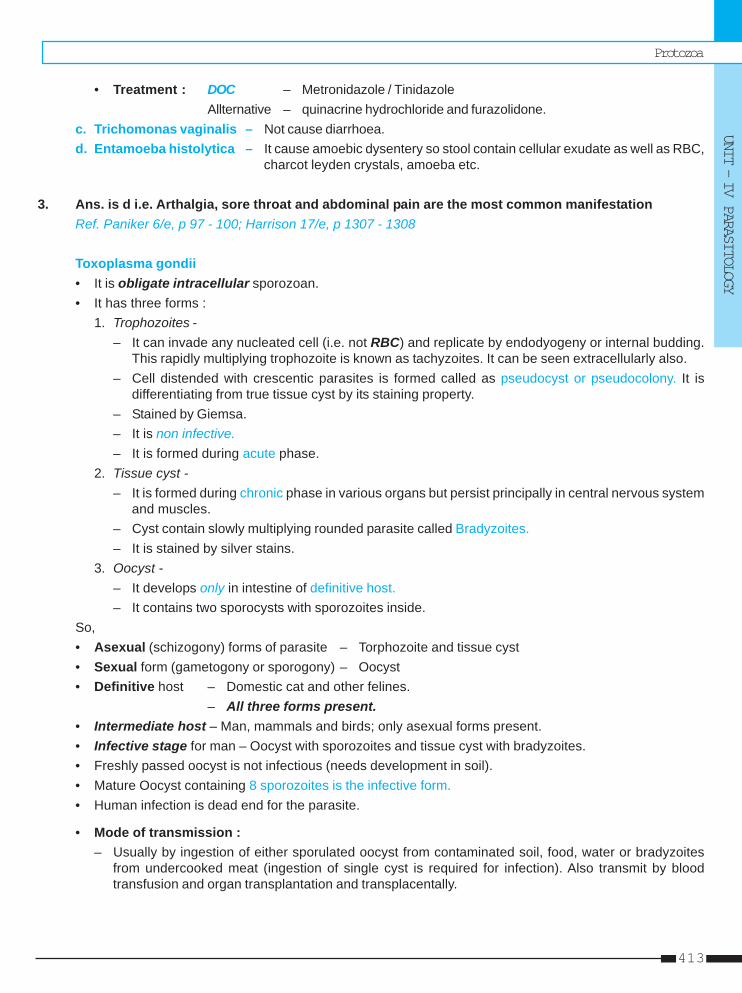
Protozoa
UNIT – IV PARASITOLOGY
413
• Treatment : DOC – Metronidazole / TinidazoleAllternative – quinacrine hydrochloride and furazolidone.
c. Trichomonas vaginalis – Not cause diarrhoea.d. Entamoeba histolytica – It cause amoebic dysentery so stool contain cellular exudate as well as RBC,
charcot leyden crystals, amoeba etc.
3. Ans. is d i.e. Arthalgia, sore throat and abdominal pain are the most common manifestationRef. Paniker 6/e, p 97 - 100; Harrison 17/e, p 1307 - 1308
Toxoplasma gondii• It is obligate intracellular sporozoan.• It has three forms :
1. Trophozoites -– It can invade any nucleated cell (i.e. not RBC) and replicate by endodyogeny or internal budding.
This rapidly multiplying trophozoite is known as tachyzoites. It can be seen extracellularly also.– Cell distended with crescentic parasites is formed called as pseudocyst or pseudocolony. It is
differentiating from true tissue cyst by its staining property.– Stained by Giemsa.– It is non infective.– It is formed during acute phase.
2. Tissue cyst -– It is formed during chronic phase in various organs but persist principally in central nervous system
and muscles.– Cyst contain slowly multiplying rounded parasite called Bradyzoites.– It is stained by silver stains.
3. Oocyst -– It develops only in intestine of definitive host.– It contains two sporocysts with sporozoites inside.
So,• Asexual (schizogony) forms of parasite – Torphozoite and tissue cyst• Sexual form (gametogony or sporogony) – Oocyst• Definitive host – Domestic cat and other felines.
– All three forms present.• Intermediate host – Man, mammals and birds; only asexual forms present.• Infective stage for man – Oocyst with sporozoites and tissue cyst with bradyzoites.• Freshly passed oocyst is not infectious (needs development in soil).• Mature Oocyst containing 8 sporozoites is the infective form.• Human infection is dead end for the parasite.
• Mode of transmission :– Usually by ingestion of either sporulated oocyst from contaminated soil, food, water or bradyzoites
from undercooked meat (ingestion of single cyst is required for infection). Also transmit by bloodtransfusion and organ transplantation and transplacentally.

Self Assessment & Review Microbiology & Immunology
SECTION – B
414
• Clinical features :– Human toxoplasmosis is zoonosis (ANTHROPO-ZOONOSES). It is of following types :
a. Toxoplasmosis in immunocompetent person : Mostly asymptomatic.– MC manifestation : Cervical lymphadenopathy which is generalized in 20-30%.– Headache, fever, myalgia, splenomegaly often present.– Meningoencephalitis, mycocarditis, pneumonitis, chorioretinitis are rare.
b. Toxoplasmosis in immuno compromised person : – MC site is CNS (usually brainstem).– MC symptom : Altered mental status.
c. Congenital toxoplasmosis– Occurs only when mother gets primary toxoplasmosis infection whether clinical or asymptomatic
during pregnancy or < 6months before conception (i.e. no risk if acquired > 6 months beforeconception).
– As gestational age is increased, risk of transmission to fetus increased i.e. max. in 3rd trimesterwhile severity of fetal damage is decreased i.e. infant is usually asymptomatic if infection transmitin 3rd trimester.
– It causes : - hydrocephalous - microencephaly- diffuse cerebral calcification - myocarditis- hepatosplenomegaly - chorioretinitis- mental retardation - multiorgan failure- myocarditis - pneumonitis
- lymphadenitis
4. Ans. is c i.e. Multiple infection of erythrocytes are seenRef. Harrison 17/e, p 1280; Paniker 6/e, p 75
Blood Smears of Plasmodium
Feature P. falciparum P. vivax P. malariae P. ovale
Features of red cells
Size All sizes / normal Large (young), pale Small (Old) / Normal Large (Young)
Shape Round may be Round or oval Round Round or pear-shaped,crenated fimbriated
Stippling Maurer’s clefts; Schuffner’s dots: None Schuffner’s dots, James dotLarge; red up to 20 numerous, small, red Occasionally, Zieman’sBasophilic stippling + stippling
Features of Parasite
Ring (early Theradlike, multiple Thicker Compact Compacttorphozoite) infections, double
chromatin dots,accole form orappluque
Mature / Late tropho Absent/occasionally Ameboid, may fill cell More regular, smaller; Less ameboid and smallerzoites (amoeboid form) seen Band form than those of P. vivax;
Schizonts Absent/occasionally 12 to 24 merozoites 8 to 12 merozoites, often 8 to 12 merozoites irregul-8-24 merozoites in grape like pattern rosetted around pigment larly arrangedgrape like pattern
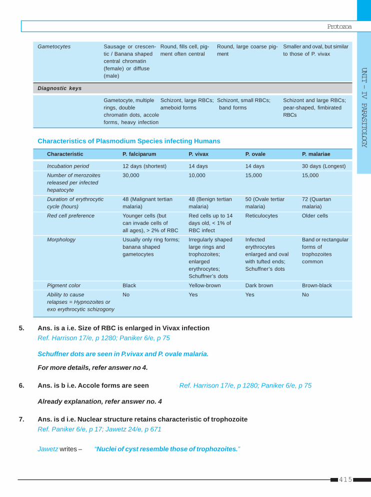
Protozoa
UNIT – IV PARASITOLOGY
415
Gametocytes Sausage or crescen- Round, fills cell, pig- Round, large coarse pig- Smaller and oval, but similartic / Banana shaped ment often central ment to those of P. vivaxcentral chromatin(female) or diffuse(male)
Diagnostic keys
Gametocyte, multiple Schizont, large RBCs; Schizont, small RBCs; Schizont and large RBCs;rings, double ameboid forms band forms pear-shaped, fimbiratedchromatin dots, accole RBCsforms, heavy infection
Characteristics of Plasmodium Species infecting Humans
Characteristic P. falciparum P. vivax P. ovale P. malariae
Incubation period 12 days (shortest) 14 days 14 days 30 days (Longest)
Number of merozoites 30,000 10,000 15,000 15,000released per infectedhepatocyte
Duration of erythrocytic 48 (Malignant tertian 48 (Benign tertian 50 (Ovale tertiar 72 (Quartancycle (hours) malaria) malaria) malaria) malaria)
Red cell preference Younger cells (but Red cells up to 14 Reticulocytes Older cellscan invade cells of days old, < 1% ofall ages), > 2% of RBC RBC infect
Morphology Usually only ring forms; Irregularly shaped Infected Band or rectangularbanana shaped large rings and erythrocytes forms ofgametocytes trophozoites; enlarged and oval trophozoites
enlarged with tufted ends; commonerythrocytes; Schuffner’s dotsSchuffner’s dots
Pigment color Black Yellow-brown Dark brown Brown-black
Ability to cause No Yes Yes Norelapses = Hypnozoites orexo erythrocytic schizogony
5. Ans. is a i.e. Size of RBC is enlarged in Vivax infectionRef. Harrison 17/e, p 1280; Paniker 6/e, p 75
Schuffner dots are seen in P.vivax and P. ovale malaria.
For more details, refer answer no 4.
6. Ans. is b i.e. Accole forms are seen Ref. Harrison 17/e, p 1280; Paniker 6/e, p 75
Already explanation, refer answer no. 4
7. Ans. is d i.e. Nuclear structure retains characteristic of trophozoiteRef. Paniker 6/e, p 17; Jawetz 24/e, p 671
Jawetz writes – “Nuclei of cyst resemble those of trophozoites.”
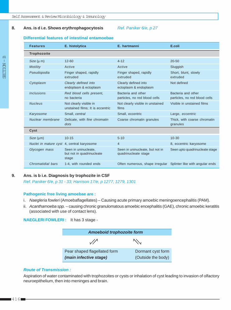
Self Assessment & Review Microbiology & Immunology
SECTION – B
416
8. Ans. is d i.e. Shows erythrophagocytosis Ref. Paniker 6/e, p 27
Differential features of intestinal entamoebae
Features E. histolytica E. hartmanni E.coli
Trophozoite
Size (μ m) 12-60 4-12 20-50
Motility Active Active Sluggish
Pseudopodia Finger shaped, rapidly Finger shaped, rapidly Short, blunt, slowlyextruded extruded extruded
Cytoplasm Clearly defined into Clearly defined into Not definedendoplasm & ectoplasm ectoplasm & endoplasm
Inclusions Red blood cells present, Bacteria and other Bacteria and otherno bacteria particles, no red blood cells particles, no red blood cells
Nucleus Not clearly visible in Not clearly visible in unstained Visible in unstained filmsunstained films; It is eccentric films
Karyosome Small, central Small, eccentric Large, eccentric
Nuclear membrane Delicate, with fine chromatin Coarse chromatin granules Thick, with coarse chromatindots granules
Cyst
Size (μm) 10-15 5-10 10-30
Nuclei in mature cyst 4, central karyosome 4 8, eccentric karyosome
Glycogen mass Seen in uninucleate, Seen in uninucleate, but not in Seen upto quadrinucleate stagebut not in quadrinucleate quadrinucleate stagestage
Chromatidial bars 1-4, with rounded ends Often numerous, shape irregular Splinter like with angular ends
9. Ans. is b i.e. Diagnosis by trophozite in CSFRef. Paniker 6/e, p 31 - 33; Harrison 17/e, p 1277, 1279, 1301
Pathogenic free living amoebae are :i. Naegleria fowleri (Amoebaflagellates) – Causing acute primary amoebic meningoencephalitis (PAM).ii. Acanthamoeba spp. – causing chronic granulomatous amoebic encephalitis (GAE), chronic amoebic keratitis
(associated with use of contact lens).
NAEGLERI FOWLERI : It has 3 stage -
Amoeboid trophozoite form
Pear shaped flagellated form Dormant cyst form(main infective stage) (Outside the body)
Route of Transmission :Aspiration of water contaminated with trophozoites or cysts or inhalation of cyst leading to invasion of olfactoryneuroepithelium, then into meninges and brain.
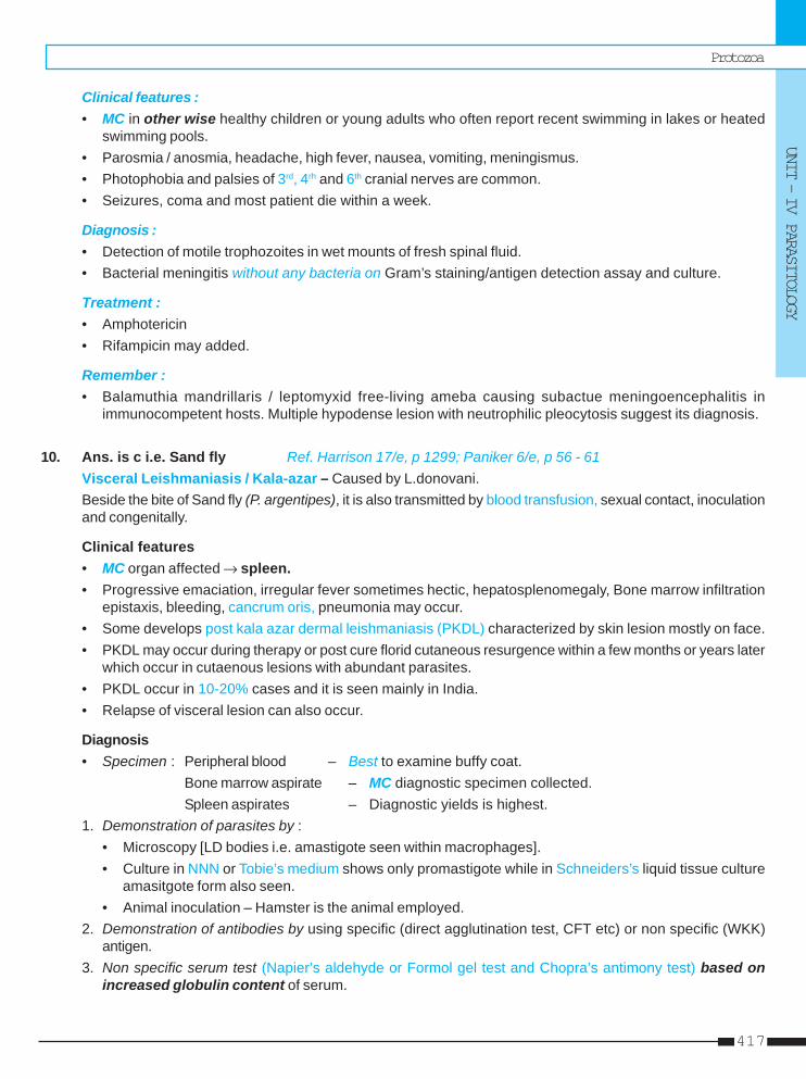
Protozoa
UNIT – IV PARASITOLOGY
417
Clinical features :• MC in other wise healthy children or young adults who often report recent swimming in lakes or heated
swimming pools.• Parosmia / anosmia, headache, high fever, nausea, vomiting, meningismus.• Photophobia and palsies of 3rd, 4rh and 6th cranial nerves are common.• Seizures, coma and most patient die within a week.
Diagnosis :• Detection of motile trophozoites in wet mounts of fresh spinal fluid.• Bacterial meningitis without any bacteria on Gram’s staining/antigen detection assay and culture.
Treatment :• Amphotericin• Rifampicin may added.
Remember :• Balamuthia mandrillaris / leptomyxid free-living ameba causing subactue meningoencephalitis in
immunocompetent hosts. Multiple hypodense lesion with neutrophilic pleocytosis suggest its diagnosis.
10. Ans. is c i.e. Sand fly Ref. Harrison 17/e, p 1299; Paniker 6/e, p 56 - 61Visceral Leishmaniasis / Kala-azar – Caused by L.donovani.Beside the bite of Sand fly (P. argentipes), it is also transmitted by blood transfusion, sexual contact, inoculationand congenitally.
Clinical features• MC organ affected → spleen.• Progressive emaciation, irregular fever sometimes hectic, hepatosplenomegaly, Bone marrow infiltration
epistaxis, bleeding, cancrum oris, pneumonia may occur.• Some develops post kala azar dermal leishmaniasis (PKDL) characterized by skin lesion mostly on face.• PKDL may occur during therapy or post cure florid cutaneous resurgence within a few months or years later
which occur in cutaenous lesions with abundant parasites.• PKDL occur in 10-20% cases and it is seen mainly in India.• Relapse of visceral lesion can also occur.
Diagnosis• Specimen : Peripheral blood – Best to examine buffy coat.
Bone marrow aspirate – MC diagnostic specimen collected.Spleen aspirates – Diagnostic yields is highest.
1. Demonstration of parasites by :• Microscopy [LD bodies i.e. amastigote seen within macrophages].• Culture in NNN or Tobie’s medium shows only promastigote while in Schneiders’s liquid tissue culture
amasitgote form also seen.• Animal inoculation – Hamster is the animal employed.
2. Demonstration of antibodies by using specific (direct agglutination test, CFT etc) or non specific (WKK)antigen.
3. Non specific serum test (Napier’s aldehyde or Formol gel test and Chopra’s antimony test) based onincreased globulin content of serum.
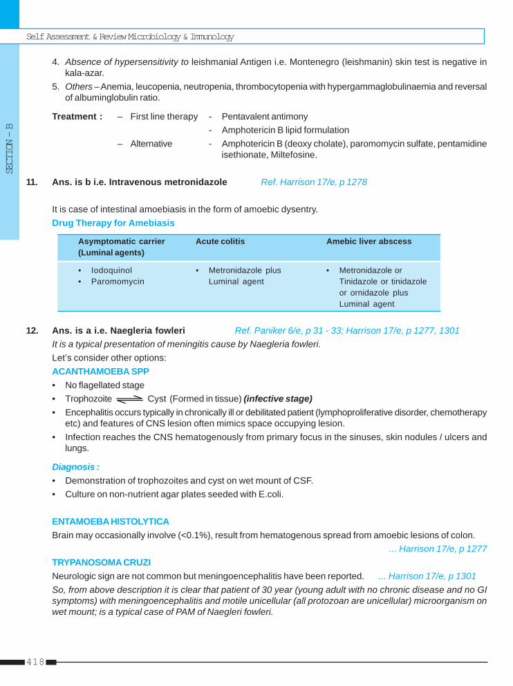
Self Assessment & Review Microbiology & Immunology
SECTION – B
418
4. Absence of hypersensitivity to leishmanial Antigen i.e. Montenegro (leishmanin) skin test is negative inkala-azar.
5. Others – Anemia, leucopenia, neutropenia, thrombocytopenia with hypergammaglobulinaemia and reversalof albuminglobulin ratio.
Treatment : – First line therapy - Pentavalent antimony- Amphotericin B lipid formulation
– Alternative - Amphotericin B (deoxy cholate), paromomycin sulfate, pentamidineisethionate, Miltefosine.
11. Ans. is b i.e. Intravenous metronidazole Ref. Harrison 17/e, p 1278
It is case of intestinal amoebiasis in the form of amoebic dysentry.Drug Therapy for Amebiasis
Asymptomatic carrier Acute colitis Amebic liver abscess(Luminal agents)
• Iodoquinol • Metronidazole plus • Metronidazole or• Paromomycin Luminal agent Tinidazole or tinidazole
or ornidazole plusLuminal agent
12. Ans. is a i.e. Naegleria fowleri Ref. Paniker 6/e, p 31 - 33; Harrison 17/e, p 1277, 1301It is a typical presentation of meningitis cause by Naegleria fowleri.Let’s consider other options:ACANTHAMOEBA SPP• No flagellated stage• Trophozoite Cyst (Formed in tissue) (infective stage)• Encephalitis occurs typically in chronically ill or debilitated patient (lymphoproliferative disorder, chemotherapy
etc) and features of CNS lesion often mimics space occupying lesion.• Infection reaches the CNS hematogenously from primary focus in the sinuses, skin nodules / ulcers and
lungs.
Diagnosis :• Demonstration of trophozoites and cyst on wet mount of CSF.• Culture on non-nutrient agar plates seeded with E.coli.
ENTAMOEBA HISTOLYTICABrain may occasionally involve (<0.1%), result from hematogenous spread from amoebic lesions of colon.
... Harrison 17/e, p 1277TRYPANOSOMA CRUZINeurologic sign are not common but meningoencephalitis have been reported. ... Harrison 17/e, p 1301So, from above description it is clear that patient of 30 year (young adult with no chronic disease and no GIsymptoms) with meningoencephalitis and motile unicellular (all protozoan are unicellular) microorganism onwet mount; is a typical case of PAM of Naegleri fowleri.
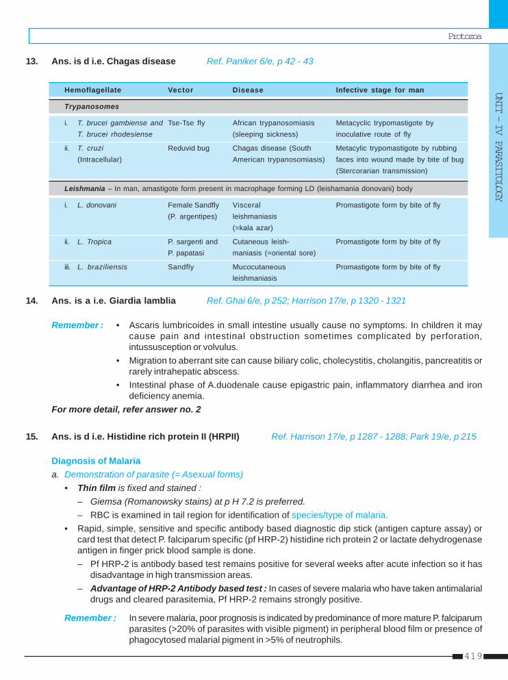
Protozoa
UNIT – IV PARASITOLOGY
419
13. Ans. is d i.e. Chagas disease Ref. Paniker 6/e, p 42 - 43
Hemoflagellate Vector Disease Infective stage for man
Trypanosomes
i. T. brucei gambiense and Tse-Tse fly African trypanosomiasis Metacyclic trypomastigote byT. brucei rhodesiense (sleeping sickness) inoculative route of fly
ii. T. cruzi Reduvid bug Chagas disease (South Metacylic trypomastigote by rubbing(Intracellular) American trypanosomiasis) faces into wound made by bite of bug
(Stercorarian transmission)
Leishmania – In man, amastigote form present in macrophage forming LD (leishamania donovani) body
i. L. donovani Female Sandfly Visceral Promastigote form by bite of fly(P. argentipes) leishmaniasis
(=kala azar)
ii. L. Tropica P. sargenti and Cutaneous leish- Promastigote form by bite of flyP. papatasi maniasis (=oriental sore)
iii. L. braziliensis Sandfly Mucocutaneous Promastigote form by bite of flyleishmaniasis
14. Ans. is a i.e. Giardia lamblia Ref. Ghai 6/e, p 252; Harrison 17/e, p 1320 - 1321
Remember : • Ascaris lumbricoides in small intestine usually cause no symptoms. In children it maycause pain and intestinal obstruction sometimes complicated by perforation,intussusception or volvulus.
• Migration to aberrant site can cause biliary colic, cholecystitis, cholangitis, pancreatitis orrarely intrahepatic abscess.
• Intestinal phase of A.duodenale cause epigastric pain, inflammatory diarrhea and irondeficiency anemia.
For more detail, refer answer no. 2
15. Ans. is d i.e. Histidine rich protein II (HRPII) Ref. Harrison 17/e, p 1287 - 1288; Park 19/e, p 215
Diagnosis of Malariaa. Demonstration of parasite (= Asexual forms)
• Thin film is fixed and stained :– Giemsa (Romanowsky stains) at p H 7.2 is preferred.– RBC is examined in tail region for identification of species/type of malaria.
• Rapid, simple, sensitive and specific antibody based diagnostic dip stick (antigen capture assay) orcard test that detect P. falciparum specific (pf HRP-2) histidine rich protein 2 or lactate dehydrogenaseantigen in finger prick blood sample is done.– Pf HRP-2 is antibody based test remains positive for several weeks after acute infection so it has
disadvantage in high transmission areas.– Advantage of HRP-2 Antibody based test : In cases of severe malaria who have taken antimalarial
drugs and cleared parasitemia, Pf HRP-2 remains strongly positive.
Remember : In severe malaria, poor prognosis is indicated by predominance of more mature P. falciparumparasites (>20% of parasites with visible pigment) in peripheral blood film or presence ofphagocytosed malarial pigment in >5% of neutrophils.
Self Assessment & Review Microbiology & Immunology
SECTION – B
420
• Thick blood film is stained without fixing for searching of parasites.
b. Malarial fluorescent antibody test is usually becomes positive 2 weeks or more after primary infection sopositive test is not necessarily an indication of current infection. It is of greatest value in epidemiologicalstudies and in determining whether a person has had malaria in the past.
16. Ans. is c i.e. A low iron content in the diet predispose to invasive amoebiasisRef. Park 19/e p 200-201; Paniker 6/e, p 23 - 28
Option “a’ : Trophozoites from acute dysenteric stool, often contain phagocytosed erythrocytes Thisfeatures is diagnostic for E. histolytica as phagycytosed red cells are not found in the minutaform or in other commensal intestinal amoebae. ...Paniker 6/e, p 23
Option “b” : Amoebiasis is more related to poor sanitation and socioeconomic status than to climate.....Park 19/e, 201
More common in adults than in children and in males than in females. ...Paniker 6/e, p 28Option “c” : Iron in diet is not related to invasive amoebiasis since it spreads by contaminated food and
water containing quadrinucleate mature cyst passed in feces of convalescent and carriers.Option “d” : Enzyme electrophoretic mobility analysis have so far identified 7 potentially pathogenic and
11 non pathogenic zymodemes. ...Park 19/e, p 200
Remember : Ordinary residual chlorination of water may not destroy cyst (but kill trophozoites) thoughsuper chlorination does. Tetraglycine hydroperiodide (Iodination) is best disinfectant.
17. Ans. is b i.e. IgM antibodies against Toxoplasma in the foetus Ref. Harrison 17/e, p 1309 - 1310
Diagnosis of Toxoplasmosisi. Tissue and body fluids : Demonstration of tachyzoites in lymphnodes establishes the diagnosis of acute
toxoplasmosis.
ii. Serology• Commonest method of laboratory diagnosis.• Diagnosis of acute infection can be established by detection of simultaneous presence of IgG (by
Sabin Feldman dye test, indirect fluorescent antibody test and ELISA) and IgM ( by double sandwitchIgM ELISA and IgM immunosorbent assay).
• Presence of IgA also favours diagnosis of acute infection. IgA ELISA is more sensitive than IgM ELISAfor detecting congenital infection.
iii. Molecular diagnosis• Real time PCR for either the B1 gene or the 529-bp sequence.
Immunocompetent Adult or childPatient with lymphadenopathy only, a positive IgM titre is an indication of acute infection and indication oftherapy.
Immunocompromised hosti. Person should be tested for IgG antibody to T. gondii soon after diagnosis of HIV infection.ii. IgM serum antibody is usually not detectable.
Congenital infectioni. Antibodies in neonate may be either due to congenital infection (IgG, IgM) or due to passive transfer of
antibodies from mother (IgG only).

Protozoa
UNIT – IV PARASITOLOGY
421
So detection of IgM specific antibody in fetus is helpful in diagnosing congenital toxoplasmosis since itdoes not cross placenta.Harrison writes : “Persistence of IgG antibody or positive IgM titer after first week of life – diagnosis ofcongenital Toxoplasmosis.”
ii. PCR of amniotic fluid to detect B1 gene of the parasite has replaced fetal blood sampling.
Ocular toxoplasmosisi. Positive IgG titer with typical eye lesions.ii. Antibody production is expressed in terms of Goldmann-Witmer coefficient.
18. Ans. is a i.e. ELISA Ref. Harrison 17/e, p 1277 - 1278; Jawetz 24/e, p 670 - 671; Paniker 6/e, p 19, 23
DIAGNOSIS OF AMOEBIASISSpecimen :• Fluid feces for examination of trophozoite.• Formed feces for cysts.• Scrapings and biopsies – most commonly by colonoscopy.• Liver abscess aspirate for trophozoites (as cyst are absent in tissues) from edge of abscess, not the
necrotic center.• Blood for serologic test and cell counts.
i. Fecal findings suggestive of amoebic colitis - positive test for heme, paucity of neutrophils, amoebiccyst or hematophagous trophozoite (definitive). Examine at least 3 fresh stool specimen.
ii. Culture - Dobells diphasic, Cleveland - Collier and Robinson’s media are used.iii. Trophozoites in biopsy specimen from colonic mass confirm the diagnosis of amoeboma.iv. Serology :
– Is primarily for extra-intestinal amoebiasis when stools are often negative.– Most commonly done by indirect hemagglutination assay (IHA) but it can’t distinguish recent from past
infection since it remains positive for as long as 10 year.– ELISA and agar gel diffusion assay are positive in more than 90% of patients with colitis, amoebomas
or liver abscess.So, best test for invasive amoebiasis - ELISAMC test done for invasive amoebiasis - IHA
– Antamoebic antibodies occur only with E. histolytica not with other entamoeba.– Enzymeba test is based on finding of histolysain (major cysteine protease of virulent form) in the
intestine (stool) plus circulating antibodies to histolysain after tissue invasion.– Enzymeba and ELISA helps to distinguish pathogenic from non pathogenic strain (E. dispar) in a stool
specimen.iv. Radiation methods – Hepatic abscess usually shows elevation of right dome of diaphragm by US, CT, MRI
etc.v. Liver enzyme are normal or minimally elevated (alkaline phosphatase most often elevated) even with large
liver abscess.
19. Ans. is b i.e. IgG antibodies are diagnostic in congenital toxoplasmosisRef. Harrison 17/e, p 1309 - 1310
Already explained, refer answer no. 3 and 17
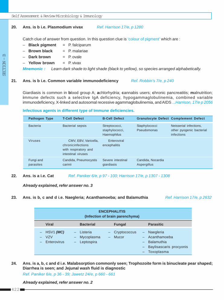
Self Assessment & Review Microbiology & Immunology
SECTION – B
422
20. Ans. is b i.e. Plasmodium vivax Ref. Harrison 17/e, p 1280
Catch clue of answer from question. In this question clue is ‘colour of pigment’ which are :– Black pigment = P. falciparum– Brown black = P. malariae– Dark brown = P. ovale– Yellow brown = P. vivaxMnemonic : Learn dark shade to light shade (black to yellow), so species arranged alphabetically.
21. Ans. is b i.e. Common variable immunodeficiency Ref. Robbin’s 7/e, p 240
Giardiasis is common in blood group A; achlorhydria; cannabis users; chronic pancreatitis; malnutrition;immune defects such a selective IgA deficiency, hypogammaglobulinemia, combined variableimmunodeficiency, X-linked and autosomal recessive agammaglobulinemia, and AIDS ...Harrison, 17/e p 2056
Infectious agents in different type of Immune deficiencies.
Pathogen Type T-Cell Defect B-Cell Defect Granulocyte Defect Complement Defect
Bacteria Bacterial sepsis Streptococci, Staphylococci Neisserial infections,staphylococci, Pseudomonas other pyogenic bacterialHaemophilus infections
Viruses CMV, EBV, Varicella, Enteroviralchronicinfections encephalitiswith respiratory andintestinal viruses
Fungi and Candida, Pneumocystis Severe intestinal Candida, Nocardiaparasites carinii giardiasis Aspergillus
22. Ans. is a i.e. Cat Ref. Paniker 6/e, p 97 - 100; Harrison 17/e, p 1307 - 1308
Already explained, refer answer no. 3
23. Ans. is b, c and d i.e. Naegleria; Acanthamoeba; and Balamuthia Ref. Harrison 17/e, p 2632
ENCEPHALITIS(Infection of brain parenchyma)
Viral Bacterial Fungal Parasitic
– HSV1 (MC) – Listeria – Cryptococcus – Naegleria– VZV – Mycoplasma – Mucor – Acanthamoeba– Enterovirus – Leptospira – Balamuthia
– Baylisascaris procyonis– Toxoplasma
24. Ans. is a, b, c and d i.e. Malabsorption commonly seen; Trophozoite form is binucleate pear shaped;Diarrhea is seen; and Jejunal wash fluid is diagnosticRef. Paniker 6/e, p 36 - 39; Jawetz 24/e, p 660 - 661
Already explained, refer answer no. 2
Protozoa
UNIT – IV PARASITOLOGY
423
25. Ans. is b and e i.e. It is a protozoa; and It is associated with diarrhoea in HIV patientsRef. Harrison 17/e, p 1314
• Microsporadia are gram positive, obligate intracellular, spore forming protozoa that cause disease inhumans especially as opportunistic pathogens in AIDS.
• In AIDS patient microsporidium cause diarrhoea.• Diagnosis is made by demonstration of spore in smear of faeces or duodenal aspirate by modified trichome
or chromotrope 2R based staining or Uveitx 2B or calcofluor fluoroscent staining.
26. Ans. is b, c and d i.e. Gametocytes; Accole trophozoite; and Ring formRef. Harrison 17/e, p 1280; 15/e p 1191
Already explained, refer answer no. 4
27. Ans. is b, c and d i.e. T. gondii; Angostrongyloid cantonensis; and T. cruziRef. Harrison 17/e, p 2632; 16/e, p 1239, 1245
Parasities causing encephalitis are :i. Toxoplasma gondii (rarely)ii. Trypanosoma cruzi (occasionally)iii. Angiostrongylyd cantonensis (eosinophilic meningoencephalitis)iv. Acanthamoeba (Granulomatous amoebic encephalitis)v. Naegleria fowleri (Primary amoebic meningoencephalitis)vi. Balamuthia.
28. Ans. is a, b, c and d i.e. Thrombocytopenia; DIC; Hemolysis; and HaematemesisRef. Harrison 17/e, p 1284
Manifestation of severe falciparum malaria :i. Cerebral malaria / unarousable coma :
• Coma is characteristic and omnious feature of falciparum malaria.• Manifest as diffuse symmetric encephalopathy, focal neurologic signs are unusual.
ii. Hypoglycemia – associated with poor prognosis.iii. Lactic acidosis – Plasma conc of HCO3– & Lactate are best biochemical prognosticators in severe malaria.iv. Non cardiogenic pulmonary edema.v. Renal impairment.vi. Hematologic abnormalities :
• Anemia due to ↑ RBC destruction, removal by spleen and ineffective erythropoiesis• Mild thrombocytopenia• Bleeding with DIC• Hematemesis due to stress ulceration or acute gastric erosion.
vii. Liver dysfunction – Associated with poor prognosis.viii. Convulsion, chest infection, catheter induced UTI, septicemia, salmonella bacteremia.
Self Assessment & Review Microbiology & Immunology
SECTION – B
424
29. Ans. is b and c i.e. Due to ingestion of oocyst from cat’s faeces; and Spiramycin given in pregnancyRef. Harrison 17/e, p 1310; Jawetz 24/e, p 684
Treatment of Toxoplasmosisi. Congenital infection : – Pyrimethamine and sulfadiazine
– Spiramycin + prednisoneii. Infection in immunocompetent :
– If only lymphadenopathy : No treatment unless have severe persistent symptoms.– If ocular toxoplasmosis : Pyrimethamine + sulfadiazine or clindamycin.
iii. Infection in pregnancy : Spiramycin (rovamycine) is DOC.iv. Infection in Immunocompromised :
– Prophylaxis for AIDS who are seropositive for T. gondii and have CD4 T cell <100/μl - Trimethoprim +sulfamethoxazole / Dapsone + Pyrimethamine.- Pyrimethamine + Sulfadiazine (preferred but not widely available).
Remember : Sporocyst, sporoblast, sporozoites are formed during sporulation of Oocyst in soil not in tissues.
30. Ans. is a and b i.e. Caused by Babesis microti; and Resides in RBCRef. Harrison 17/e, p 1295; Jawetz 24/e, p 680; Paniker 6/e, p 108
• Babesia (protozoan) is intraerythrocytic (blood) Parasite causing piroplasmosis/Texas fever/Acute hemolyticdisease/Babesiosis incidentally in human.
• Most human infection are caused by B. microti and B. divergens.• Vector Ixodid ticks (I. dammini or I. scapularis, I. Ricinus).• Mode of transmission – Tick bite and blood transfusion.• Clinical features :
– Mostly asymptomatic.– Characterized by malaise, fever, sweat, depression, myalgia, arthralgia which resembles malaria and
rickettsiosis.– Most severe illness is seen in immunosuppressed; splenectomized (usually infected by B. divergens –
may develop Jaundice, renal insufficiency) and elderly persons.– Rash is absent.
• Diagnosis :– Giemsa stained thick and thin film shows small intraerythrocytic parasites resembling malaria but it
does not form pigment, schizonts, or gametocytes and seen as tetrad forms infrequently.– ‘Maltese cross’ form in RBC without pigment or gametocyte is diagnostic.
• Treatment :
Organisms Adult / child
B.microti (mild) Atovaquone + azithromycinB. divergens and Others Quinine + Clindamycin + exchange transfusion
Protozoa
UNIT – IV PARASITOLOGY
425
31. Ans. is b and d i.e. It is one of the common opportunistic infection in AIDS; and AFB +ve cystRef. Harrison 17/e, p 1313; Jawetz 24/e, p 682; Paniker 6/e, p 105 - 107
Cryptosporidium• Cryposporidium is a Acid fast coccidian intracellular but extracytoplasmic parasite.• Completes its life cycle in one host (monoxenous).• Most human infection is caused by C. parvum.• Infective stage : Oocyst (contain 4 sporozoites) in feces which is infective immediately without further
developement so person to person transmission can occur.• Mode of infection : Acquired from infected animal or human feces or from feces contaminated food or
water.• Principal site of infection : Surface of villi of lower small bowel.• Clinical features
In immunocompetent In immunosuppressed AIDS patient
• Self limited watery non bloody diarrhoea • Chronic, persistent and profuse diarrhoea• Traveller’s diarrhoea can occur • Due to involvement of biliary tract papillary stenosis,
sclerosing cholangitis or cholecystitis can occur
• Diagnosis– Fecal examination for small oocyst of 4-5 μm in diameter.– Modified acid fast and Direct immunofluorescent stains and enzyme immunoassay (for fecal antigen).– Biopsy may show cryptosporidium at apical surfaces of intestinal epithelium.
• Treatment : – No chemotherapy is effective– Paromomycin - partially effective in HIV patient– Nitrazoxanide - in children– Spiramycin or combination therapy with azithromycin is also effective.
32. Ans. is a, c and d i.e. Caused by E. histolytica; Flask-shaped ulcers are present; and Caecum is mostcommonly affected Ref. Paniker 6/e, p 17; Jawetz 24/e, p 669 - 671
E. histolytica – It has 3 stages :a. Trophozoite or Vegetative or Amoeboid form
• It is the only form present in tissue.• It can’t initiate infection.
b. Pre-cystic stage• Encystment occur in intestinal lumen not in feces or not in tissues.• Not contain RBC and other ingested food particles.
c. Cystic stage• Mature Quadrinucleate cyst : Infective stage• It is present only in lumen of colon and in mushy or formed feces.• When stained with iodine, nuclear chromatin and karyosome appears bright yellow while chromidial
bars are unstained.Pathogenesis8 small trophozoites (=amoebulae or metacystic trophozoites) are released per infective cyst causing :
Self Assessment & Review Microbiology & Immunology
SECTION – B
426
Primary amoebiasis Secondary intestinal Chronic infection Invasive amoebiasislesion
• MC site caecum • MC in cecum, appendix • Occurs when organism • Extraintestinalor nearby ascending colon travel to ileocaecal infection is
valve and terminal ileum metastatic throughportal circulation
• Lesion with Pinhead • It occurs due to extension • Sigmoid colon and rectum • MC form is amebicsized center and raised from primary lesion are favoured site hepatitis or liveredges • Amoeboma may form abscess
• Flask shaped ulcers with narrow neck and broad base
• Mucosa surface between ulcer is normal
• Ulcer is not premalignant
• Intestinal amoebiasis present usually as amoebic dysentery.• Contents of amoebic abscess is called as anchovy paste.• MC complication of amoeba liver abscess is Pleuropulmonary involvement.
33. Ans. is a and b i.e. Splenomegaly; and Nephrotic syndrome Ref. Harrison 17/e, p 1285 - 1286
Chronic complication of malaria are :i. Tropical splenomegaly (Hyperreactive Malarial Splenomegaly).
– Produce hypergammaglobulinemia.– Normocytic normochromic anemia and splenomegaly.– Hepatomegaly with marked elevation of IgM, malarial antibody, hepatic sinusoidal lymphocytosis,
peripheral B cell lymphocytosis, formation of cryoglobulins (IgM aggregates and immune complexes),cytotoxic antibodes to suppressor CD-8 cells and increased vulnerability to respiratory and skin infection.
ii. Quartan malarial nephropathy of P. malariae causing neprotic syndrome, focal or segmentalglomerulonephritis.
iii. Burkitt’s lymphoma and EBV infection. Due to malaria related immunosuppression.
34. Ans. is c and d i.e. Antimonial are useful drugs; and Diagnosed by blood smearRef. Harrison 17/e, p 1299; Paniker 6/e, p 56 - 61
Already explained, refer answer no. 10
35. Ans. is a and c i.e. Leishmania donovani; Leishmania major Ref. Harrison 17/e, p 1296
Leshmania exist in 2 forms :a. Amastigote form : Non flagellated, occur in macrophage of man as leishmania donovani (LD) bodies.b. Promastigote form : Flagellated, occur in gut of sandfly and artificial culture.
36. Ans. is b and d i.e. Naegleria; and Angiostrongylus cantonensis Ref. Jawetz 23/e, p 688
Already explained, refer answer no. 27
Protozoa
UNIT – IV PARASITOLOGY
427
37. Ans. is a i.e. Thick smear to identify parasite Ref. Park 19/e p 215; Paniker 6/e, p 76, 90 - 92
No of slides examined• Annual blood examination rate, ABER = x 100
population• Incubation period of different malaria is different.
38. Ans. is a i.e. Schizont Ref. Harrison 15/e p 1191
Already explained, refer answer no. 4
39. Ans. is a i.e. L-brazillensis Ref. Paniker 6/e, p 42 - 43
Already explained, refer answer no. 13
40. Ans. is d i.e. E. gingivalis Ref. Paniker 6/e, p 14, 30
• Entamoeba gingivalis is present in the mouth, being found in large numbers when oral hygiene is poor.• It has no cystic stage so transmit by kissing, airborne droplet and by fomites.
Remember : • Cystic stage is absent in : – Dientamoeba fragilis– Trichomonas vaginalis– T. intestinalis.
• Entamoeba histolytica, entamoeba coli and Endolimax nana inhabit human intestine.
41. Ans. is a i.e. Toxoplasma Ref. Paniker 6/e, p 97 - 100; Harrison 17/e, p 1307 - 1308
Already explained, refer answer no. 3
42. Ans. is a and b i.e. Schizonts; and Mature trophozoite Ref. Harrison 15/e, p 1191
Already explained, refer answer no. 4
43. Ans. is a i.e. Falciparum Ref. Harrison 17/e, p 1284
Already explained, refer answer no. 28
44. Ans. is b i.e. Trophozoites and cysts are seen in manRef. Paniker 6/e, p 36 - 39; Jawetz 24/e, p 660 - 661
Already explained, refer answer no. 2
Self Assessment & Review Microbiology & Immunology
SECTION – B
428
1. a) Reduvid bug 2. d) P. malariae 3. a) Spleen 4. d) Full treatment ... 5. a) Indirect ...6. a) Duodenum ... 7. b) Trophozoites ... 8. a) Man 9. a) Cyst are 8 ... 10. a) Complement ...
11. c) Trichomonas
Answer
1. Vector fot T. cruzi is : [JIPMER 90]a) Reduvid bugb) Tse Tse flyc) Sand flyd) Hard tick
[Ref. Paniker 6/e, p 49]2. Band shaped torphozoites are seen in : [AI 90]
a) P. ovaleb) P. vivaxc) P. falciparumd) P. malariae
[Ref. Paniker 6/e, p 75]3. Which of the following is most severly affected in
Kala-azar : [BIHAR 90]a) Spleenb) Liverc) Adrenal glandd) Bone marrow
[Ref. Paniker 6/e, p 57]4. The following are true of kala-azar except :
a) Persistent hypergammaglo-bulinemia [TN 90]b) Pancytopeniac) Cancrum oris can occurd) Full treatment prevents post kala-azar dermal
leishmaniasis[Ref. Paniker 6/e, p 56-60]
5. Diagnostic test for amoebic hepatitis is : [AI 90]a) Indirect hemagglutination testb) Isolation from pusc) Isolation from wall of cavityd) Cyst in stools
[Ref. Harrison 17/e, p 1277]6. The normal habitat of giardia is : [AIIMS 90]
a) Duodenum and jejunum
b) Stomachc) Caecumd) Ileum
[Ref. Paniker 6/e, p 38]7. Amoebic liver abscess can be diagnosed by dem-
onstrating : [AI 91]a) Cysts in the sterile pusb) Trophozoites in the pusc) Cysts in the intestined) Trophozoites in the faces
[Ref. Paniker 6/e, p 27]8. The main reservoir for entamoeba histolytica is :
a) Man [AI 91]b) Dirty waterc) Soild) Ponds
[Ref. Park 19/e, p 201]9. All are true about Entamoeba Histolytica except :
a) Cyst are 8 nucleated [AI 91]b) Cyst are 4 nucleatedc) Trophozoites colonise in the colond) The chromatid bodies are our stained by iodides
[Ref. Paniker 6/e, p 17]10. The following is true of Giardiasis except :
a) Complement fixation test is diagnostic [AI 92]b) Stools contain only cystsc) Habitat is colond) Trophozoite and cysts are found in duredenum
[Ref. Paniker 6/e, p 38-39]11. The cystic form of all are seen in man except :
a) E. histolytica [AI 92]b) Girdiac) Trichomonasd) Toxoplasma [Ref. Paniker 6/e, p 40]
Chapter Review••••• This section includes questions of VThis section includes questions of VThis section includes questions of VThis section includes questions of VThis section includes questions of Various Other PGMEES from 1990 – 2007.arious Other PGMEES from 1990 – 2007.arious Other PGMEES from 1990 – 2007.arious Other PGMEES from 1990 – 2007.arious Other PGMEES from 1990 – 2007.••••• Questions are arQuestions are arQuestions are arQuestions are arQuestions are ar ranged in decreasing order of yearranged in decreasing order of yearranged in decreasing order of yearranged in decreasing order of yearranged in decreasing order of yearwiz. This is done to makwiz. This is done to makwiz. This is done to makwiz. This is done to makwiz. This is done to make refere refere refere refere refer ralralralralral
system easier and uncomplicated to save the precious time of PGMEE Aspirant.system easier and uncomplicated to save the precious time of PGMEE Aspirant.system easier and uncomplicated to save the precious time of PGMEE Aspirant.system easier and uncomplicated to save the precious time of PGMEE Aspirant.system easier and uncomplicated to save the precious time of PGMEE Aspirant.
Protozoa
UNIT – IV PARASITOLOGY
429
12. b) Intracellularity ... 13. b) RBC size is ... 14. b) Liver 15. c) Shows ... 16. a) Balantidium ...17. d) Refractile ... 18. a, c and d 19. a) Oocyst 20. d) Heart 21. None22. a) Acanthamoeba 23. c) P. argentipes 24. b) L. Brasilliensis 25. b) Erythrophago ...
Answer
12. Prolonged parasitism in malaria is due to :a) Antigenic variation [JIPMER 92]b) Intracellularity of parasitec) Immunosupressiond) Sequestration
[Ref. Robbin’s 7/e, p 401-402]13. Which is true of malaria : [AI 93]
a) Rods forms are seen in P. Malariaeb) RBC size is more in P. Vivaxc) Relapse seen in P. falciparumd) Male and Female mosquito transmit disease
[Ref. Harrison 15/e, p 1191]14. Commonest site of extraintestinal amoebiasis is :
a) Brain [Delhi 93]b) Liverc) Spleend) Lungs
[Ref. Paniker 6/e]15. Which is true of torphozoites of E. histolytica :
a) Has eccentric Karyosome [AI 93]b) Nuclear membrane with out chromatinc) Shows erythrophagocytosisd) Presence of bacteria inside cell
[Ref. Paniker 6/e, p 27]16. Which of the following organism is biggest :
a) Balantidium Coli [JIPMER 93]b) Entamoeba colic) Escherichia colid) Entamoeba Histolytica
[Ref. Paniker 6/e, p 111]17. All are seen in cyst of E. histolytica except :
a) Glycogen mass [Kerala 94]b) Chromatidial barsc) Eccenteric nucleusd) Refractile nucleus
[Ref. Paniker 6/e, p 27]18. Which is true about post infusion malaria :
a) Caused by plasmodium malaria [Bihar 94]b) Radical chemotherapy is requiredc) Transmitted by needlesd) Caused by P. ovale
[Ref. Paniker 6/e, p 87; Harrison 17/e, p 1285]19. The infective form of toxoplasma gondii is :
a) Oocyst [Karn. 94]
b) Bradycystc) Tachyzoited) All of the above
[Ref. Paniker 4/e, p 94]20. Plasmodium falciparum affects all organs except
a) Lung [Kerala 95]b) Liverc) Kidneyd) Heart
[Ref. Harrison 17/e, p 1285; Robbin’s 7/e, p 402,403]
21. The mature cyst of Entamoeaba histolytica differsfrom Entamoeba coli, in the following except :a) Size is 6 to 15 microns [Karnat 96]b) Nuclei are 1 to 4 in numberc) Karyosome is central in positiond) Chromatioid bars seen
[Ref. Paniker 6/e, p 27]22. Fulminant Amoeba meningoencephalitis caused
by : [AP 96]a) Acanthamoebab) N. folwleric) E. histolyticad) E. coli
[Ref. Paniker 6/e, p 31]23. Kala-azar in India is caused by : [AP 96]
a) P. papatasib) P. serpentic) P. argentipesd) Oncocercus
[Ref. Paniker 6/e, p 56]24. Mucocutaneous leishmaniasis is caused by :
a) L. tropica [PGI 97, Kerala 97]b) L. Brasilliensisc) L. mexicanad) None of the above
[Ref. Paniker 6/e, p 63]25. The most distinctive feature of pathogenic Entam-
oeba histolytica on fresh stool examination is :a) Presence of active pseudopodia [UP 97]b) Erythrophagocytosisc) Presence of intracytoplasmic vacuolesd) Presence of two nucleoli
[Ref. Paniker 6/e, p 23]
Self Assessment & Review Microbiology & Immunology
SECTION – B
430
26. a) Hyperglycaemia 27. b) RES 28. a) Disinfecting ... 29. a) Metronidazole ... 30. a) Nagleria ...31. c) One hour ... 32. c) A cyst may ... 33. c) Acute ... 34. a) Sporozoite 35. c) The pigment ...36. c) Schizont 37. a) Tick 38. c) Plasmodium ...
Answer
26. All of the following are seen in cerebral malariaexcept : [UPSC 97]a) Hyperglycaemiab) Thrombocytopaniac) Acute respiratory distress synsromed) Heavy parasitemia
[Ref. Harrison 17/e, p 1284]27. Amastigote form is seen in : [UP 98]
a) Macrophagesb) RESc) Lymphocytesd) RBC
[Ref. Paniker 6/e, p 53]28. Reidel-Walker co-efficient is related with :
a) Disinfecting power [Orrisa 98]b) Parasitic clearancec) Dietary requirementd) Statistical correlation
[Ref. KDT 5/e, p 805]29. Drugs of choice in Giardiasis is : [UP 99]
a) Metronidazole/Tinidazoleb) Albendazolec) Thiabendazoled) Diloxanide furoate
[Ref. Paniker 6/e, p 39]30. Primary amoebic meningoencephalitis is caused
by : [Kar 00]a) Nagleria fowlerib) Entamoeba hystoliticac) Endolimax nanad) Dientamoeba fragilis
[Ref. Paniker 6/e, p 31]31. Malarial parasites are easily detected if blood films
are taken and examined : [Kar 01]a) When the patient is febrileb) When the patient is having chillsc) One hour after the height of the paroxysmd) When the patient is afebrile
[Ref. Nelson, p 1050]32. Spot the incorrect statement regarding entamoeba
histolytica : [Kolkata 02]a) Cysts are necessary for the transmission of in-
fection from one host to anotherb) Cysts are necessary for the tranmission of infec-
tion from one host to anotehr
c) A cyst may be transferred into a trophozoite tocause invasion in the same
d) Cysts are found in the submucosa of the lowerintestinal wall
[Ref. Paniker 6/e, p 17-21]33. The most important reservoir of Leishmaniasis in
India is : [JIPMER 02]a) Dogsb) Rodentsc) Acute visceral leishmaniasisd) Case of postkalazar dermal leishmaniasis
[Ref. Park 19/e, p 256]34. The infective form of malarial parasite in the ver-
tebrate host is : [Kar 03]a) Sporozoiteb) Merozoitec) Schizontd) Gametocyte
[Ref. Paniker 6/e, p 69]35. In malaria the shuffner’s dots are due to :
a) The disintegrated parts of the parasiteb) The intracellular space not occupied by the para-
site [SGPGI 03]c) The pigment released from breakdown of he-
moglobind) The gametocytes
[Ref. Paniker 6/e, p 71]36. In falaciparum malaria all of the following are seen
except : [SGPGI 03]a) Crescent shaped gametesb) Maurer’s dotsc) Schizontd) Merozoites
[Ref. Harrison 15/e, 1191]37. Babesoisis is transmitted : [Kar 04]
a) Tickb) Mitesc) Flead) Mosquito
[Ref. Paniker 6/e, p 108]38. Complication in malaria are commonly with :
a) Plasmodium ovale [Kar 04]b) Plasmodium vivaxc) Plasmodium falciparumd) Plasmodium malariae
[Ref. Harrison 17/e, p 1283-1285]
Protozoa
UNIT – IV PARASITOLOGY
431
39. d) Clonorchis ... 40. b) Young RBC 41. c) E. gingivalis42. b) Balantidium ... 43. a) Blood 44. b and d45. d) P. malariae 46. d) None
Answer
39. Which of the following parasite passes throughthree hosts : [Kolkata 04]a) Fasciola hepaticab) Fasciola buskic) Schistosoma haematoboliumd) Clonorchis sinensis
[Ref. Paniker 6/e, p 127]40. P. vivax attacks with : [Jharkhand 04]
a) Reticulocytesb) Young RBCc) Old RBCd) Dead RBC
[Ref. Ananthnarayan 6/e, p 75]41. Entamoeba which is not found in gut : [MP 04]
a) E. colib) E. histolyticac) E. gingivalisd) E. nana
[Ref. Paniker 6/e, p 14]42. Largest intestinal protozoa is : [DNB 04]
a) Entamoeba colib) Balantidium colic) Giardia lambliad) Toxoplasma gondii
[Ref. Panikar 6/e, p 111]
43. Route of transmission of toxoplasma :a) Blood [DNB 04]b) Faecesc) Urined) None
[Ref. Harrison 17/e, p 1306]44. Most fatal amoebic encephalitis is caused by :
a) Entamoeba histolytica [Bihar 05]b) Naegleriac) Entamoeba dispard) Acanthamoea
[Ref. Harrison 17/e, p 1279]45. Nephrotic syndrome is casued by :
a) P. falciparum [Bihar 05]b) P.ovalec) P. vivaxd) P. malariae
[Ref. ww.pubmedcentral.nih.]46. In malaria reservoir, parasite remains as :
a) Merozoite [Bihar 06]b) Sporozoitec) Trophozoited) None
[Ref. Chaterjee Parasitology, p 113; Ghai 6/e, p241]
1. All of the following causes biliary obstructionexcept : [AI 08]a) Clonorchisb) Ascarisc) Ankylostoma duodenaled) Fasciola
2. Which of the following is not a neuroparasite :a) Taenia solium [AI 05; PGI 05]b) Acanthamoebac) Naegleriad) Trichinella spiralis
3. The organism most commonly causing genital fi-lariasis in most parts of Bihar and Easter U.P. isa) Wuchereria bancrofti [AI 03]b) Brugia malayic) Onchocerca volvulusd) Dirofilaria
4. In which stage of filariasis are microfilaria seenin peripheral blood : [AI 01]a) Tropical eosinophiliab) Early adenolymphangitis stagec) Late adenolymphangitis staged) Elephantiasis
5. Pancreatic Ca is caused by : [AI 01]a) Fasciolab) Clonorchis
c) Paragonimusd) None
6. Consumption of uncooked pork is likely to causewhich of the following helminthic disease :a) Taenia saginata [AI 01]b) Taenia soliumc) Hydatid cystd) Trichuris trichura
7. A child from Bihar comes with fever. Blood ex-amination shows sheathed microfilaria with nu-clei up to tail tip. The diagnosis is : [AI 00]a) B. malayib) W. bancroftic) Loa load) Oncicerca volvulus
8. Autoinfection is a mode of transmission in :a) Trichinella [AI 00]b) Cysticercosisc) Ancylostomad) Ascaris
9. Pigs are resevoir for : [AI 00]a) T.soliumb) T.saginatac) Trichinella spiralisd) Ancyclostoma
Answer 1. c) Ankylostoma ... 2. d) Trichinella ... 3. a) Wuchereria ... 4. b) Early adeno ... 5. d) None6. b) Taenia ... 7. a) B. malayi 8. b) Cysticercosis 9. a andc
QUESTIONS
HelminthsHelminthsHelminthsHelminthsHelminths33
Helminths
UNIT – IV PARASITOLOGY
433
10. Commonest parasite of CNS in India is : [AI 99]a) Schistosomiasisb) Cysticercosisc) Trichenella spiralisd) Hydatid cyst
11. Sputum examination is not useful in diagnosisof: [AI 98; AIIMS 96]a) Trichuriasis trichurab) Ancylostoma duodenalec) Paragonimusd) Strongyloides
12. Most common presenting symptom of threadworm infection amongst the following is :a) Abdominal pain [AI 97]b) Rectal prolapsec) Urticariad) Vaginitis
13. Which one of the following does not pass throughthe lungs : [AI 96]a) Hookwormb) Ascarisc) Strongyloidisd) Enterobius Vermicularis
14. Which of the following disease is transmitted byegg ingestion : [AI 95]a) Taeniasisb) Trichinosisc) Hydatidosisd) Strongyloidosis
15. All the following are True about Brugia malayiexcept : [AI 95]a) The intermediate hosts in the India are Mansonib) The tail tip is free from nucleic) Nuclei are blurred, so counting is difficultd) Adult worm is found in the lymphaitc system
16. Which of the following is the most common loca-tion of intracranial neurocysticerosis ?a) Brain parenchyma [AIIMS 05]b) Subarachnoid spacec) Spinal cordd) Orbit
17. Kalu, 30 year old man, presented withsubacutaneous itchy nodules over left iliac crest.On examination they are firm, non-tender, andmobile Skin scrappings contain microfilaria and
adult worms of : [AIIMS May 01]a) Loa-loab) Onchocerca volvulusc) Brugia malayid) Mansonella persetans
18. Autoinfection is seen with : [AIIMS 01]a) Ankylostomab) Enterobiusc) Echinococcusd) Ascariasis
19. On microscopic examination eggs are seen, buton saturation with salt solution no eggs are seen,The eggs are likely to be of : [AIIMS 99]a) Trichuris trichurab) Taenia soliumc) Ascaris lumbricoidesd) Ancylostoma duodenale
20. Larva found in muscle is : [AIIMS 98]a) Trichinella spiralisb) Ankylostoma duodenalec) Trichuris trichurad) Enterobius vermicularis
21. Which of the following parasite does not enterinto the body by skin penetration : [AIIMS 97]a) Dracunculusb) Necatar americanac) Ancylostoma duodenaled) Strongyloides
22. Liver is the target organ for : [AIIMS 97]a) Fasciola buskib) Paragonimus westermanic) Clonorchis sinensesd) Schistosoma Hematobium
23. All are true regarding filariasis except :a) Man is an intermediate host [AIIMS 97]b) Caused by Wuchereria bancroftic) Involves lymphatic systemd) DEC is used in treatment
24. Megaloblastic anaemia is caused by :[AIIMS 95]a) Diphyllobothrium latumb) Schistosoma hematobiumc) Echinococcus granulosusd) Taenia solium
Answer 10. b) Cysticercosis 11. a) Trichuriasis ... 12. a) Abdominal ... 13. d) Enterobius ... 14. c) Hydatidosis15. b) The tail ... 16. a) Brain ... 17. b) Onchocerca ... 18. b) Enterobius 19. b) Taenia ...20. a) Trichinella ... 21. a) Dracunculus 22. c) Clonorchis ... 23. a) Man is ... 24. a) Diphyllob ...
Self Assessment & Review Microbiology & Immunology
SECTION – B
434
25. All float in a saturated salt solution except :a) Clonorchis sinensis [AIIMS 95]b) Fertilised eggs of ascarisc) Larva of strongyloidisd) Trichuris trichura
26. Nematodes are differentiated from other wormsby : [PGI 05]a) Absent fragmentationb) Flat or fleshy leaf like wormsc) Separate sexesd) Cylindrical bodye) GIT is formed completely
27. Fish acts as intermediate host in : [PGI 04]a) D.Latumb) Clonorchis Sinensisc) H.Diminutad) H.Nana
28. Parasites causing lung infestation are : [PGI 03]a) H.nanab) Paragonimus westermanic) Taenia saginatad) E.granulosuse) E.multiocularis
29. Cholangiocarcinoma is caused by : [PGI 02]a) Fasciola infestationb) Clonorchis infestationc) Paragonimus infestationd) Ascaris infestatione) None of these
Answer 25. a) Clonorchis ... 26. a, c,d and e 27. a and b 28. b, d and e 29. b) Clonorchis ...30. b and e 31. a, b and d 32. a and b 33. a) Taenia ... 34. a) Not acquired ...
30. Hydatid disease of liver is caused : [PGI 01]a) Srongyloidesb) Echinococcus granulosusc) Taenia soliumd) Trichinella spiralise) Echinococcus multilocularis
31. Larval form in stool is found in : [PGI 01]a) Strongyloidesb) Ancylostoma duodenalec) Ascaris lumbricoidesd) Necator americanase) Trichuria
32. Parasites penetrating through skin for entry intothe body are : [PGI 99; 01]a) Ankylostoma duodenaleb) Srongyloidesc) Round wormd) Trichuris trichura
33. Cysticercosis is caused by larva of : [PGI 99]a) Taenia soliumb) T.saginatac) Echinococcusd) Ascaris
34. Neurocysticerosis, following are true except :a) Not acquired by eating contaminated vegetablesb) Caused by regurgitation of larva [PGI 98]c) Acquired by orofaecal routed) Acquired by eating pork
Helminths
UNIT – IV PARASITOLOGY
435
1. Ans. is c i.e. Ankylostoma Ref. Harrison 7/e p 1334, 1335, 2000Hepatobiliary Parasitism
– Common in Southern China– Biliary tract is infestated by adult helminths or their ova– Causes: Trematodes (Liver or biliary flukes) MC
• Clonorchis sinensis• Opisthorchis viverrini or O. felineus• Fasciola hepaticaNematode (Ascaris lumbricoides by intraductal migration of adult worm)Cestodes (Echinococcus spp by intrabiliary rupture of hydatid cyst)
– Clinical Features:• Chronic/recurrent pyogenic cholangitis ± multiple hepatic abscess• Ductal stones• Biliary obstruction
– Diagnosis: • Cholangiography• Characteristic ova on stool examination
– Treatment: If obstruction is present, TOC is laparotomy under antibiotic coverage with common duct explorationand biliary drainage procedure.
Helminths causing obstructive jaundice are : Ascaris, Clonorchis, Fasziola, Echinococcus.
2. Ans. is d i.e. Trichinella spiralis
Brain parasites
Protozoa Helminthes
↓↓↓↓↓ Larvae of cestodes Nematodes Ecotopic ova
– E. histolytica – Cysticercus – Visceral larva – F. hepatica– Naegleria cellulosae (T. solium) migrans – Heterophyes– Acanthamoeba – Hydatid cyst – Ascaris hyterophyes– Trypanosoma (E. granulosus) lumbricoides – Schitosoma– P. falciparum – Multiceps sp – Strongyloides (hematobium)– Toxoplasma gondii stercoralis
– Gnathosomaspinigerum
3. Ans. is a i.e. Wuchereria Bancrofti Ref. Paniker 6/e, p 196; Harrison 17/e, p 1324 - 1326
Filariasis• Definitive host - man.• Intermediate host - insects
EXPLANATIONS AND REFERENCES WITH ILLUSTRATIVE ANSWERS
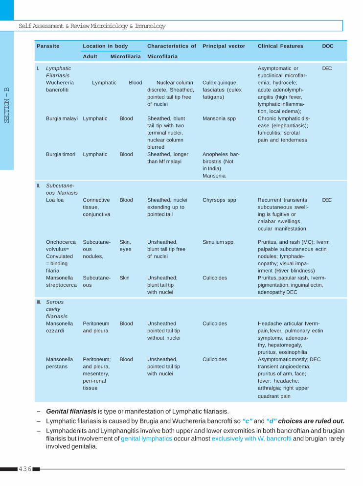
Self Assessment & Review Microbiology & Immunology
SECTION – B
436
Parasite Location in body Characteristics of Principal vector Clinical Features DOC
Adult Microfilaria Microfilaria
I. Lymphatic Asymptomatic or DECFilariasis subclinical microflar-Wuchereria Lymphatic Blood Nuclear column Culex quinque emia; hydrocele;bancrofiti discrete, Sheathed, fasciatus (culex acute adenolymph-
pointed tail tip free fatigans) angitis (high fever,of nuclei lymphatic inflamma-
tion, local edema);Burgia malayi Lymphatic Blood Sheathed, blunt Mansonia spp Chronic lymphatic dis-
tail tip with two ease (elephantiasis);terminal nuclei, funiculitis; scrotalnuclear column pain and tendernessblurred
Burgia timori Lymphatic Blood Sheathed, longer Anopheles bar-than Mf malayi birostris (Not
in India)Mansonia
II. Subcutane-ous filariasisLoa loa Connective Blood Sheathed, nuclei Chyrsops spp Recurrent transients DEC
tissue, extending up to subcutaneous swell-conjunctiva pointed tail ing is fugitive or
calabar swellings,ocular manifestation
Onchocerca Subcutane- Skin, Unsheathed, Simulium spp. Pruritus, and rash (MC); Ivermvolvulus= ous eyes blunt tail tip free palpable subcutaneous ectinConvulated nodules, of nuclei nodules; lymphade-= binding nopathy; visual impa-filaria irment (River blindness)Mansonella Subcutane- Skin Unsheathed; Culicoides Pruritus, papular rash, Iverm-streptocerca ous blunt tail tip pigmentation; inguinal ectin,
with nuclei adenopathy DEC
III. SerouscavityfilariasisMansonella Peritoneum Blood Unsheathed Culicoides Headache articular Iverm-ozzardi and pleura pointed tail tip pain, fever, pulmonary ectin
without nuclei symptoms, adenopa-thy, hepatomegaly,pruritus, eosinophilia
Mansonella Peritoneum; Blood Unsheathed, Culicoides Asymptomatic mostly; DECperstans and pleura, pointed tail tip transient angioedema;
mesentery, with nuclei pruritus of arm, face;peri-renal fever; headache;tissue arthralgia; right upper
quadrant pain
– Genital filariasis is type or manifestation of Lymphatic filariasis.– Lymphatic filariasis is caused by Brugia and Wuchereria bancrofti so “c” and “d” choices are ruled out.– Lymphadenits and Lymphangitis involve both upper and lower extremities in both bancroftian and brugian
filarisis but involvement of genital lymphatics occur almost exclusively with W. bancrofti and brugian rarelyinvolved genitalia.
Helminths
UNIT – IV PARASITOLOGY
437
4. Ans. is b i.e. Early adeno-lymphangitis stageRef. Park 19/e, 222; Paniker 6/e, p 200 - 204; Chatterjee 12/e, p 195 - 197
Clinical manifestations
Lymphatic filariasis
i. Asymptomatic amicrofilaraemia – No microfilarae and no clinical manifestation.
ii. Asymptomatic microfilaremia – Positive for Mf without any symptoms.
iii. Stage of acute manifestation – Recurrent episodes of acute inflammation in lymph glands andvessels characterized by lymphadenitis, lymphangitis, filarial fever,lymphangiovarix and lymphorrhagia. It is caused by adult wormbut blood may reveals microfilarae in early phase.
iv. Stage of chronic obstructive lesions – Caused by adult worm blocking lymph nodes and vessels eithermechanically or are commonly due to allergic inflammatoryreactions to worm antigens and secretions. Granuloma mayform.
– Main features are Hydrocele, elephantiasis (MC site is leg),Lymphadema (non pitting or brawny edema).
– Microfilaria in blood are absent either due to death or their failureto reach the systemic circulation due to lymphatic obstruction.
Occult filariasis
Meyers Kouwenaar syndrome – It is due to hypersenstivity reactions to filarial antigens It includesTropical pulmonary eosinophilia characterized by Nocturnalparoxysmal cough, wheeze and blood eosinophil count is above3000 per cmm.
– Microfilariae are not usually detectable in blood but lungbiopsies may show microfilariae
– Antifilarial antibody titres are characteristically elevated
5. Ans. is d i.e. None Ref. Paniker 6/e, p 7
Name of Parasite Carcinoma
• Clonorchis sinensis – Chloangiocarcinoma• Opisthorchis viverrini – Cholangiocarcinoma• Schistosoma hematobium – Bladder cancer
6. Ans. is b i.e. Taenia solium Ref. Paniker 6/e, p 144 - 145Taenia Solium – Causative agent of CYSTICERCOSIS.• Definitive host = Adult worm lives in jejunum of man.
Infective stage for man :– Pork’s flesh containing cysticercus cellulosae larvae or bladder worm.– Also by eggs either by ingestion in water/vegetables; Autoinfection by fingers contaminated with eggs
from perianal skin or feces and retrograde peristalsis.• Intermediate host = Pig
Infected stage for pig = Eggs containing hexacanth larvae so eggs are infective for both man & pig.
Remember : • Larvae is found in both man and pig.• But in man it is Dead end Infection.• Infection occur in both vegetarian and non vegetarian.
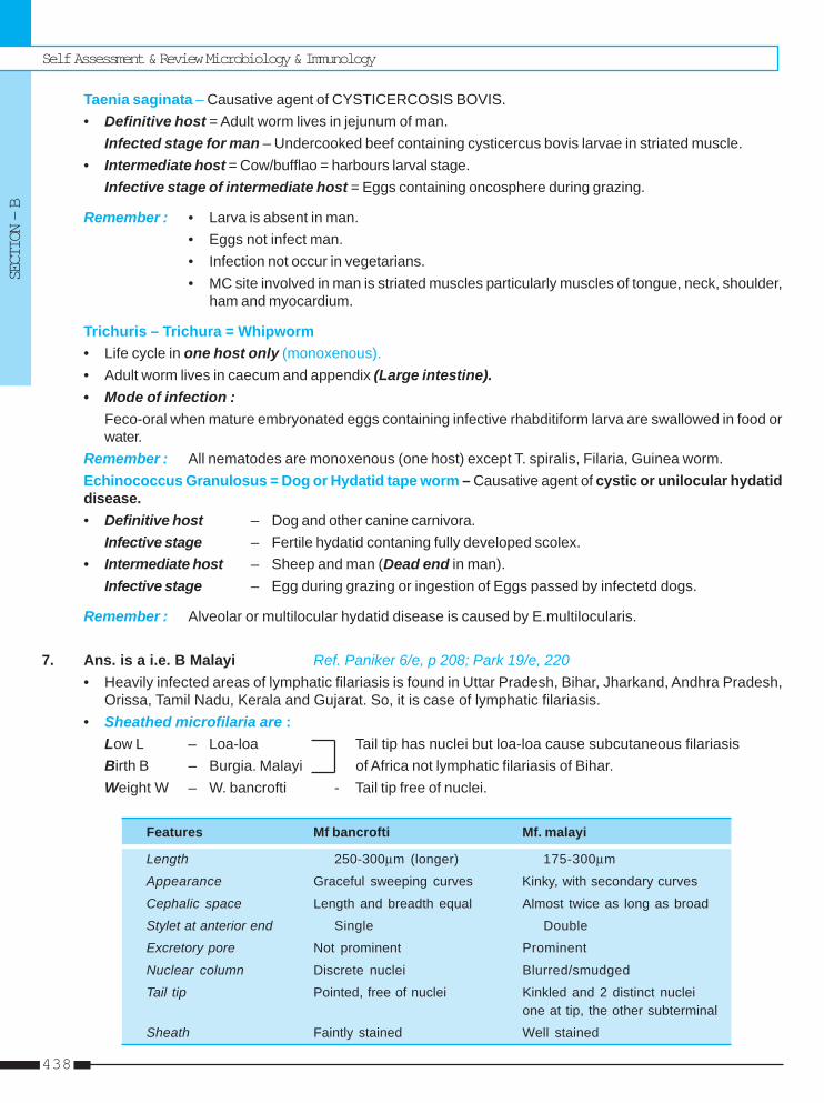
Self Assessment & Review Microbiology & Immunology
SECTION – B
438
Taenia saginata – Causative agent of CYSTICERCOSIS BOVIS.• Definitive host = Adult worm lives in jejunum of man.
Infected stage for man – Undercooked beef containing cysticercus bovis larvae in striated muscle.• Intermediate host = Cow/bufflao = harbours larval stage.
Infective stage of intermediate host = Eggs containing oncosphere during grazing.
Remember : • Larva is absent in man.• Eggs not infect man.• Infection not occur in vegetarians.• MC site involved in man is striated muscles particularly muscles of tongue, neck, shoulder,
ham and myocardium.
Trichuris – Trichura = Whipworm• Life cycle in one host only (monoxenous).• Adult worm lives in caecum and appendix (Large intestine).• Mode of infection :
Feco-oral when mature embryonated eggs containing infective rhabditiform larva are swallowed in food orwater.
Remember : All nematodes are monoxenous (one host) except T. spiralis, Filaria, Guinea worm.Echinococcus Granulosus = Dog or Hydatid tape worm – Causative agent of cystic or unilocular hydatiddisease.• Definitive host – Dog and other canine carnivora.
Infective stage – Fertile hydatid contaning fully developed scolex.• Intermediate host – Sheep and man (Dead end in man).
Infective stage – Egg during grazing or ingestion of Eggs passed by infectetd dogs.
Remember : Alveolar or multilocular hydatid disease is caused by E.multilocularis.
7. Ans. is a i.e. B Malayi Ref. Paniker 6/e, p 208; Park 19/e, 220• Heavily infected areas of lymphatic filariasis is found in Uttar Pradesh, Bihar, Jharkand, Andhra Pradesh,
Orissa, Tamil Nadu, Kerala and Gujarat. So, it is case of lymphatic filariasis.• Sheathed microfilaria are :
Low L – Loa-loa Tail tip has nuclei but loa-loa cause subcutaneous filariasisBirth B – Burgia. Malayi of Africa not lymphatic filariasis of Bihar.Weight W – W. bancrofti - Tail tip free of nuclei.
Features Mf bancrofti Mf. malayi
Length 250-300μm (longer) 175-300μm
Appearance Graceful sweeping curves Kinky, with secondary curves
Cephalic space Length and breadth equal Almost twice as long as broad
Stylet at anterior end Single Double
Excretory pore Not prominent Prominent
Nuclear column Discrete nuclei Blurred/smudged
Tail tip Pointed, free of nuclei Kinkled and 2 distinct nucleione at tip, the other subterminal
Sheath Faintly stained Well stained
Helminths
UNIT – IV PARASITOLOGY
439
8. Ans. is b i.e. Cysticercosis Ref. Paniker 6/e, p 149Autoinfection is seen in :H = Hymenolepis NanaE = Enterobius vermicularisT = Taenia solium (cysticercosis)S = Strongyloides sterocoralis
• Autoinfection in cysticercosis occur by finger contamination with the eggs from the perineal skin or faecesand by gravid segments reaching the stomach by retrograde peristalsis from jejunum.
9. Ans. is a and c i.e. T. solium; and Trichinella spiralis Ref. Paniker 6/e, p 149, 163TAENIA SOLIUM• Also known as pork taperworm.• Intermediate host is pig.
TRICHINELLA SPIRALIS• Primary host = Man – Mucosal epithelium of small intestine.• Infective form is the encysted larvae found in the muscles of pig and other animals (larvae is tissue
parasite).• Intermediate host = Pig• Mode of Infection – Uncooked pork meat containing encysted larvae.• Female worm is viviparous (eggs are absent).• Human infection is dead end infection.In this condition, you can only pray that you get only one correct option in every question.
10. Ans. is b i.e. Cysticercosis Ref. Harrison 17/e, p 1336; 16/e, p 2487
“Neurocysticercosis is the most common parasitic disease of CNS worldwide.”NEUROCYSTICERCOSIS = CYSTICERCOSIS OF CNS
Mode of transmission in T.solium
Ingestion of cysticercus Ingestion of eggs in water or vegetablescellulosae or bladder worm which develops in to larvae than into(larvae) in pork meat ↓
Adult worm
Autoinfection Autoinfection
by finger contamination by gravid segments reaching thewith eggs from perianal stomach by retrograde peristalsisskin or feces. from jejunum when they are digested
and thousands of eggs released.
• So man is both definitive and intermediate host for T. solium but in man it is Dead end infection.
Clinical features :• MC manifestation is new-onset partial seizures with or withour secondary generalization.
Site Symptoms
• Brain parenchyma (MC) • Seizures or focal neurological defects• Subarachnoid or ventricular space • Increased intra-cranial tension• Spinal cyst • Mimic intraspinal tumour
Self Assessment & Review Microbiology & Immunology
SECTION – B
440
Diagnosis : • By non contrast CT.• MC finding on neuroimaging is parenchymal brain calcification.
Treatment : Antiepileptic therapy + Albendazole or praziquantel.
Remember : Site of cysticercosis – CNS > Subcutaneous tissue > globe of eye.
11. Ans. is a i.e. Trichuris trichura Ref. Paniker 6/e, p 192
• Any parasite which pass through lungs at some or other stage during their life cycle, sputum examinationcan be done for them.
• These parasites are :– Rhabditiform larva of Ascaris– Golden brown eggs of Paragonimus Westermani (Lung fluke)– Filariform larvae of Strongyloides, Ancylostoma– Entamamoeba histolytica - Chocolate brown sputum due to hepatobronchial fistula.
12. Ans. is a i.e. Abdominal Pain Ref. Paniker 6/e, p 183 - 187; Harrison 17/e, p 1320
Enterobius vermicularis = Pinworm = thread worm = seat worm• Life cycle in one host only (man).• No intermediate host.• Adult worm live in caecum, appendix and adjacent part of ascending colon (i.e. in large intestine).• Mode of infection – By ingestion of egg containing infective larvae (=autoinfection) and retroinfection.
• Clinical features :– Mostly asymptomatic.– Cardinal symptoms : Perianal pruritus (It is nocturnal and cause excoriation + bacterial superinfection).– Abdominal pain and weight loss.– Vulvovaginitis and pelvic or peritoneal granuloma.
• Diagnosis :– Fecel examination is not useful.– Apply clear cellulose acetate tape to perianal region in the morning and see characteristic planoconvex,
non bile stained pin worm eggs (containing tadpole shaped coiled embryo) on microscopy.– Sampling can also done by NIH swab, scotch tape, glass pestle swab.
• Treatment :– Mebendazole / Albendazole / Pyrantel pamoate.
13. Ans. is d i.e. Enterobius vermicularis Ref. Paniker 6/e, p 184
Already explained, refer answer no. 11
14. Ans. is c i.e. Hydatidosis Ref. Paniker 6/e, p 159
Following parasitic infection occur by ingestion of eggs :• Echnicoccus granulosus• Hymenolepis Nana
Helminths
UNIT – IV PARASITOLOGY
441
• Ascaris Mnemonic - Isha HATE Tushar• Trichuris• Enterobius• Taenia solium (but mainly transmit by ingestion of undercooked pork meat containing cysticercus cellulose).
15. Ans. is b i.e. The tail tip is free from nuclei Ref. Paniker 6/e, p 196
Microfilaria tail free of nuclei – W.Bancrofti– O.Volvulus Mnemonic : BOO– M.Ozzardi
16. Ans. is a i.e. Brain parenchyma Ref. Harrison 17/e, p 1336; 16/e, p 2487
Already explained, refer answer no. 10
17. Ans. is b i.e. Onchocerca volvulus Ref. Pniker 6/e, p 208 - 211
• It is a case of subcutaneous filariasis so causes of lymphatic filariasis (Brugia) and serous cavity filariasis(M.perstans) are ruled out. So we left with only 2 options :
LOA LOA = EYE WORM : CAUSE LOIASIS / FUGITIVE SWELLINGS OR CALABAR SWELLINGS– Definitive host = Man (in subcutaneous tissue)– Intermediate host = Vector – day biting flies Chyrysops– Mode of trnsmission : Bite of infected Chyrysops– Microfilariae : Are sheathed and show diurnal periodicity and appear in blood only during day and
taken by chyrysops in which Mf develop into infective third stage larvae.– Clinical features : Is due to migration of adult worms causing fugitive swellings (which disappear in
few days only to reappear elsewhere); Ocular manifestations.
– Diagnosis : Demonstration of adult worm from scraping of skin or conjunctiva.
– Treatment : Surgery to remove worms; DEC; corticosteroids
ONCHOCERCA VOLVULUS :– Convulated or blinding filaria causing onchocerciasis or river blindness.– It is 2nd major cause of blindness in the world.
– Vector : Day biting female black flies (are pool feeders) Simulium which breed in fast flowing rivers.
– Microfilariae : Unsheathed; non periodic; found in skin, subctaneous lymphatics, conjunctiva and rarelyin blood.
– Clinical features : Subcutaneous nodule or onchocercoma which is circumscirbed, firm, non-tendertumor tend to occur over anatomical sites where bones are superficial such as scalp, scapula, ribs,elbow, iliac crest, sacrum, knees.
Lesions in skin (pruritus, pigmentation, atrophy, fibrosis) and eyes (photophobia to blindness, glaucoma,punctate or sclerosing keratitis, iridocyclitis, glaucoma) also seen.
– Diagnosis : Demonstrating microfilariae by slicing off a silver of skin; aspirating subcutaneous nodules,conjunctival biopsies.
– Treatment : Ivermectin; Enucleation of nodules; DEC (cause Mazotti reaction) and Suramin.
Self Assessment & Review Microbiology & Immunology
SECTION – B
442
18. Ans. is b i.e. Enterobius Ref. Paniker 6/e, p 149
Already explained, refer answer no. 8
19. Ans. is b i.e. Taenia solium
• Eggs float in or can be demonstrated in case of :i. Cestodes : a. Echinococcus
b. Hymenolepis nana.ii. All nematodes except unfertilized eggs of Ascaris.
• In this question all choices are nematodes except Taenia solium is cestode.• So guys, learn by heart ‘classification of parasites, given in our book in Section Basics of Parasitology.
20. Ans. is a i.e. Trichinella spiralis Ref. Paniker 6/e, p 159
Classification of Nematodes on the basis of Mode of infection.
Ingestion Penetration of skin By blood sucking insect Inhalation of dustcontaining eggs
a. Eggs - Ascaris • Ancylostoma • Filariae • Ascaris– Enterobius • Necator • Enterobius– Trichuris • Strongyloides
b. Larvae withinIntermediate hostor drinking watercontaining cyclops– Dracunculus Mnemonics : ANS
c. Encysted larvae i.e. Autonomicin muscle : Nervous systemTrichinella
For more details, refer answer no. 9
21. Ans. is a i.e. Dracunculus Ref. Paniker 6/e, p 159
Already explained, refer answer no. 20
22. Ans. is c i.e. Clonorchis sinensis Ref. Paniker 6/e, p 117
• All are trematodes (also known as flukes).• Man is definitive and snails are intermediate host usually.• They are classified as :
Helminths
UNIT – IV PARASITOLOGY
443
FLUKES HABITAT INTERMEDIATE HOST MODE OF TRANS.MISSION
a. Blood flukes
i. Schistosoma Vesical and pelvic Snail – Water borne disease(Bilharzia) vein plexuses which is infected – Man is infected byhematobium by miracidium bathing in contaminated
ii. S.mansoni Inferior mesentric water when cercaria penetratesvein (Intestinal unbroken skinbilharziasis) orSchistosomaldysentry
iii. S.japonicum Superior mesentricor oriental vein (katayamaschistosomiasis disease)
b. Liver Flukes
i. Clonorchis Biliary tract 1st intermediate Fish containingsinensis (associate with host : snail metacercariae are eaten raw(=Chinese liver Cholangio carci- 2nd intermediate or inadequately processedflukes) noma) host : fish
ii. Fasciola Biliary tract Primary host : Ingestion of watercress orhepatica man/sheep other water vegetation containing(=sheep liver Intermediate host : metacercariafluke) snails
c. Intestinal flukes
i. Fasciolopsis Duodenum Molluscum Ingestion of roots of lotus, bulb ofbuski (Giant or jejunum Snails water chesnut and other acquaticintestinal fluke) vegetations
ii. Heterophyes Small intestine Molluscum Ingestion of fishes since cercriaeSnails encyst on fishes
iii. Metagonimus Small intestine 1st intermediate Ingestion of raw fishYokogawai host : fresh water
snail; 2nd intermediate host : fish
v. Gastrodiscoides Large intestine Molluscum Ingestion of water plantshominis (only flukeinhabiting humanlarge intestine)
d. Lung Fluke
Paragonimus Cystic space of lung 1st intermediate Inadequately cooked crabs,Westermani host : snail cray fish= Oriental lung fluke 2nd intermediate
host : fresh watercray fish or crab fish
23. Ans. is a i.e. Man is an intermediate host
Already explained, refer answer no. 3
Self Assessment & Review Microbiology & Immunology
SECTION – B
444
24. Ans. is a i.e. Diphyllobothrium latum Ref. Paniker 6/e, p 139 - 143; Harrison 17/e, p 1340
Diphyllobothrum (Fish Tapeworm = Broad Tapeworm)• Definitive host – Adult worm lives in small intestine (usually in the ileum) of man, cat, dog and other fish
eating mammals.Infective stage for man – Plerocercoid larva / sparganum.
• Intermediate hostFirst – Cyclops infected by coracidium larva.Second – Freshwater fish infected by procercoid larvae.
• Eggs – Operculated, shed in feces but are not infective to man.• Mode of infection – Ingestion of imperfectly cooked infected fish containing plerocercoid larva.• Clinical features :
– Mechanical obstruction – Abdominal discomfort– Diarrhoea – Nausea– Anaemia (pernicious type called as Bothriocephalus Anaemia) which is due to vitamin B12 deficiency.
• Diagnosis : Demonstration of eggs in feces.• Treatment : Praziquantel / Niclosamide
25. Ans. is a i.e. Clonorchis sinensis
Already explained, refer answer no. 19
26. Ans. is a, c, d and e i.e. Absent fragmentation; Separate sexes; Cylindrical body; and GIT is formedcompletely Ref. Paniker 6/e, p 113 - 114
Features Nematodes Trematodes Cestodes
Shape Cylindrical/thread Flat or fleshy leaf like or flukes Tape worms
Segmentation Unsegmented Unsegmented Segmented
GIT Complete Incomplete Absent
Suckers Absent Present Present
Hooks Absent Absent May present
Sex Separate (Dioecious) Monoecious except MonoeciousSchistosomes
Number of host Monoxenous except Digenetic DigeneticTrichnella, filarial, except H.nanaDracunculusmedinesis
Body cavity Present Absent Absent
Remember : Nematodes may be viviparous or oviparous or Ovo-viviparous but other worms are oviparous.
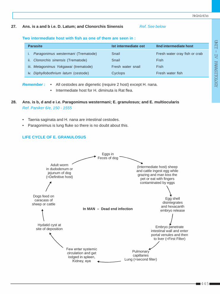
Helminths
UNIT – IV PARASITOLOGY
445
27. Ans. is a and b i.e. D. Latum; and Clonorchis Sinensis Ref. See below
Two intermediate host with fish as one of them are seen in :
Parasite Ist intermediate ost IInd intermediate host
i. Paragonimus westermani (Trematode) Snail Fresh water cray fish or crab
ii. Clonorchis sinensis (Trematode) Snail Fish
iii. Metagonimus Yokgawai (trematode) Fresh water snail Fish
iv. Diphyllobothrium latum (cestode) Cyclops Fresh water fish
Remember : • All cestodes are digenetic (require 2 host) except H. nana.• Intermediate host for H. diminuta is Rat flea.
28. Ans. is b, d and e i.e. Paragonimus westermani; E. granulosus; and E. multiocularisRef. Paniker 6/e, 150 - 1555
• Taenia saginata and H. nana are intestinal cestodes.• Paragonimus is lung fluke so there is no doubt about this.
LIFE CYCLE OF E. GRANULOSUS
Eggs inFeces of dog
(Intermediate host) sheep and cattle ingest egg whilegrazing and man kiss the
pet or eat with fingerscontaminated by eggs
Egg shell disintegrates
and hexacanth embryo release
Embryo penetrate intestinal wall and enter portal venules and then
to liver (=First Filter)
Pulmonarycapillaries
Lung (=second filter)
Hydatid site of deposition
cyst at
Dogs feed on caracass of
sheep or cattle
Adult wormin dudodenum orjejunum of dog
(=Definitive host)
In MAN – Dead end infection
Self Assessment & Review Microbiology & Immunology
SECTION – B
446
Clinical features• MC site of Hydatid cyst (E. granulosus typically develops unilocular cyst) : Liver; mostly in right lobe
presenting as Hepatomegaly, pain, obstructive jaundice.• Next common site is lung usually in the lower lobe of right lung – cough, hemoptysis, chest pain, dyspnea.• Hypersensitivity, fatal anaphylaxis if cyst ruptures.• Kidney - pain, hematuria.• Osseus hydatid.E. MULTILOCULARIS : Cause alveolar or multilocular hydatid disease in man.• Definitive host : Foxes, dogs, cats• Intermediate host : Rodents (main), man• Man infected by eating fruits or vegetables contaminated with feces.• Clinical features :
– MC site liver : Multilocular infiltrating lesion mistaken for malignant tumor.– Also metastasize to lungs and brain.
• Treatment : Resection is TOC.
29. Ans. is b i.e. Clonorchis infestation Ref. Paniker 6/e, p 7
Already explained, refer answer no. 5
30. Ans. is b and e i.e. Echinococcus granulosus; and Echinococcus multilocularisRef. Paniker 6/e, 150 - 155
Already explained, refer answer no. 28
31. Ans. is a, b and d i.e. Strongyloides; Ancylostoma duodenale; and Necator americanasRef. See below
Nematodes Fecal examination
i. Trichinella spiralis Adult worm
ii. Trichuris trichuria Eggs (barrel shaped)
iii. Strongyloides Rhabditiform larvae
iv. Ancylostoma duodenale and Egg which may hatched, so rhabditiformNecator anericanas larvae can also seen
v. Enterobius vermicularis Usually not useful
vi. Ascaris lumbricoides Eggs and adult worm(*Larvae in sputum or gastric washings)
vii. Filariasis No role
viii. Dracunculus medinensis No role (* tip of worm projects from base of ulcer)
32. Ans. is a and b i.e. Ankylostoma duodenale; and Strongyloides Ref. Paniker 6/e, p 159
Already explained, refer answer no. 20
Helminths
UNIT – IV PARASITOLOGY
447
33. Ans. is a i.e. Taenia solium Ref. Harrison 17/e, p 1336; 16/e, p 2487
Already explained, refer answer no. 10
34. Ans. is a i.e. Not acquired by eating contaminated vegetables Ref. Paniker 6/e, p 149
Already explained, refer answer no. 10
Self Assessment & Review Microbiology & Immunology
SECTION – B
448
1. Which is false about Wuchereria bancrofti :a) Causes filariasis [AI 90]b) Body is long and slenderc) Terminal nuclei absentd) Man and anopheles are host
[Ref. Paniker 6/e, p 196, 208]2. Schistosoma Japonicum resides in : [AI 92]
a) Vesical Plexusb) Splenic Veinc) Systemic Circulationd) Gall bladder
[Ref. Paniker 6/e, p 117]3. Definitive host for Guinea worm is : [AIIMS 93]
a) Manb) Cyclopsc) Snaild) Cyclops and man
[Ref. Paniker 6/e, p 214]4. Ankylostoma enters by human body by :
a) Ingestion [Kerala 94]b) Inhalationc) Penetration of skind) Inoculation
[Ref. Paniker 6/e, p 159]5. Cysticerocosis Cellulose is caused by :
a) T. Solium [Karn. 94]b) Echinococcus granulosusc) T. saginata-C. bovisd) H. nana
[Ref. Paniker 6/e, p 149]6. Habitat of hookworm is : [Kerala 94]
a) Jejunumb) Ileumc) Colond) Duodenum
[Ref. Paniker 6/e, p 175]
7. Visceral larva migrans is caused by : [PGI 94]a) Toxocara canisb) Anchylostoma brazilliensisc) A. duodenaled) Necator americanus
[Ref. Paniker 6/e, p 219]8. Spinal cord compression may be caused by :
a) Cysticercosis [Kerala 94]b) Ankylostoiasisc) Echinococcus Granulosusd) Visceral larva migrans
[Ref. Schwartz’s 7/e, p 1903]9. Terminal spined eggs seen in : [Kerala 95]
a) Schistosoma haematobiumb) Sch. mansonic) Sch. japonicumd) Chlonorchis sinesis
[Ref. Paniker 6/e, p 123]10. Dracunculosis infection occur through :
a) Ingestion of water containing cyclopsb) Ingestion of water containing the parasitec) Ingestion of fish [Delhi 96]d) Penetration of skin
[Ref. Paniker 6/e, p 214]11. Least common site of calcified hydatid cyst is :
a) Lung [Kerla 96]b) Mediastinumc) Extraperitoneal sited) Liver
[Ref. Sutton 7/e, p 152]12. Painless terminal hematuria is seen as one of the
manifestations in the infection caused by :a) Schistosoma Japonicum [Karn. 96]b) Schistosoma mansonic) Schistosoma haematobiumd) Plasmodium falciparum
[Ref. Paniker 6/e, p 121]
Chapter Review••••• This section includes questions of VThis section includes questions of VThis section includes questions of VThis section includes questions of VThis section includes questions of Various Other PGMEES from 1990 – 2007.arious Other PGMEES from 1990 – 2007.arious Other PGMEES from 1990 – 2007.arious Other PGMEES from 1990 – 2007.arious Other PGMEES from 1990 – 2007.••••• Questions are arQuestions are arQuestions are arQuestions are arQuestions are ar ranged in decreasing order of yearranged in decreasing order of yearranged in decreasing order of yearranged in decreasing order of yearranged in decreasing order of yearwiz. This is done to makwiz. This is done to makwiz. This is done to makwiz. This is done to makwiz. This is done to make refere refere refere refere refer ralralralralral
system easier and uncomplicated to save the precious time of PGMEE Aspirant.system easier and uncomplicated to save the precious time of PGMEE Aspirant.system easier and uncomplicated to save the precious time of PGMEE Aspirant.system easier and uncomplicated to save the precious time of PGMEE Aspirant.system easier and uncomplicated to save the precious time of PGMEE Aspirant.
1. d) Man and ... 2. b) Splenic ... 3. a) Man 4. c) Penetration ... 5. a) T. Solium6. a) Jejunum 7. a) Toxocara ... 8. c) Echinococcus ... 9. a) Schistosoma ... 10. a) Ingestion of ...
11. a) Lung 12. c) Schistosoma ...
Answer
Helminths
UNIT – IV PARASITOLOGY
449
13. HIghest incidence of anemia in the tropics is dueto : [AI 97]a) Hookwormb) Thread Wormc) Ascarisd) Guinea worm
[Ref. Paniker 6/e, p 181]14. The most common manifesation of Thread worm
infestation is : [AI 97]a) Urticariab) Rectal prolapsec) Abdominal painsd) Vaginitis
[Ref. Harrison 17/e, p 1322]15. Auto infection is seen with : [AP 97]
a) Trichuris trichurab) Ankylostoma duodenalec) Ascarisd) Enterobius
[Ref. Paniker 6/e, p 186]16. The cause of larva currents : [TN 98]
a) Stronghyloides stercoralisb) Necator americansusc) Ankylostoma duodonaled) H. nana
[Ref. Paniker 6/e, p 219]17. Man is both intermediate and definitive host for :
a) T. solium [Mahe 98]b) T. saginatac) D. latumd) Dicroftis hominis
[Ref. Paniker 6/e, p 148]18. Autoinfection occur in : [UP 00]
a) Strongyloidesb) Trichuris trichurac) Ankylostoma-duodenaled) Necator-americana
[Ref. Paniker 6/e, p 171]19. One of the following microfilaria does not possess
nuclei upto the tail tip : [Kar 0]a) Wuchereria bancroftib) Loa loac) Acanthochelonemia perstansd) Brugia malayi
[Ref. Paniker 6/e, p 196]
20. The following eggs have hexacanth embryos ex-cept : [Kar 00]a) Taenia soliumb) Taenia saginatac) Chlonorchis nanad) Hymenolepsis nana
21. The larval form of taenia is referred to as :a) Cysticercus [Kar 01]b) Cysticercoidc) Echinococcusd) Conerus
[Ref. Paniker 6/e, p 148]22. Which worm is longest : [HPU 01]
a) T. soliumb) T. saginatac) Hookwormd) A. lumbricoides
[Ref. Paniker 6/e, p 144]23. Drug of choice in cerebral cysticercosis is :
a) Piperazine [JIPMER 01]b) Pyrviniumc) Thiabendazoled) Mebendazole
[Ref. CMDT 08, p 1316]24. Katayama fever is caused by : [UP 01]
a) F. hepaticab) Chlonorchis-sinesusc) S. hematobiumd) C. Japonicum
[Ref. Paniker 6/e, p 121]25. The following infection resembles malignancy :
a) Echinococcus granulosum [JIPMER 02]b) E. multilocularisc) E. vogelid) E. oligarthus
[Ref. Paniker 6/e, p 155]26. Which of the following parasites is infectious as it
is passes from stool : [UP 02]a) Ancyclostoma duodenaleb) Enterobius vermicularisc) T. soliumd) Ascaris
[Ref. Paniker 6/e, p 184]
13. a) Hookworm 14. c) Abdominal ... 15. d) Enterobius 16. a) Stronghyloides ... 17. a) T. solium18. a) Strongyloides 19. a) Wuchereria ... 20. c) Chlonorchis ... 21. a) Cysticercus 22. b) T. saginata23. None 24. c) S. hematobium 25. b) E. multilocularis 26. b) Enterobius ...
Answer
Self Assessment & Review Microbiology & Immunology
SECTION – B
450
27. All are true about Brugia-malayi except :a) Nucelated tail tip [Delhi 02]b) Enveloped sheathc) Nocturnal periodicityd) Smooth curved in stain preparation
[Ref. Paniker 6/e, p 196, 208]28. Which of the following resides in caecum :
a) Trichuris trichuria [UP 02]b) A. lumbricoidesc) Strongyloidesd) Ancylostoma
[Ref. Paniker 6/e, p 165]29. Which of the following is not a cestode ?
a) D. latum [Kar 03]b) T. saginatac) Schistosoma mansoniad) E. granulosus
[Ref. Paniker 6/e, p 139]30. Dracunculus medinensis is transmitted by :
a) Cyclops [Bihar 03]b) House flyc) Tickd) Flea
[Ref. Ananthnarayan 7/e, p 213]31. Cysticercus cellulose seen in : [Kolkata 04]
a) T.saginatab) T.soliumc) D.latumd) S.haemato
[Ref. Paniker 6/e, p 149]32. Diphylobothrium latum is causative organism of
a) Megaloblastic anemia [SGPGI 04]b) Iron deficiency anemiac) Peptic ulcerd) None
[Ref. Paniker 6/e, p 142]
33. Hydatid cyst occurs most commonly in :a) Liver [DNB 04]b) Lungsc) Braind) Spleen
[Ref. Paniker 6/e, p 153]34. One of the following transmitted through skin :
a) Ascaris lumbricoides [UP 04]b) Trichuris-trichurac) Necator americanad) Strongyloides
[Ref. Paniker 6/e, p 159]35. Which of the following is viviparous :
a) Strongyloidis stercoralis [SPGPGI 05]b) Trichinella spiralisc) Enterobiousd) Ascaris
[Ref. Paniker 6/e, p 159]36. Visceral larva migrains is due to : [DNB 05]
a) Ascariasisb) Toxocaracanisc) Schistosomiasisd) Clonorchis Buskie) Loa Loa
[Ref. Paniker 6/e, p 219]37. The intermediate host for T. Saginata is :
a) Man [DNB 05]b) Cowc) Dogd) Pig
[Ref. Paniker 6/e, p 145]38. Neurocysticerosis is caused by :
a) T. solium [Jharkhand 05]b) T. saginatac) D. latumd) Ascaris lumbricodis
[Ref. CMDT 08 p 1316]
27. d) Smooth ... 28. a) Trichuris ... 29. c) Schistosoma ... 30. a) Cyclops 31. b) T.solium32. a) Megaloblastic ... 33. a) Liver 34. c and d 35. b) Trichinella ... 36. b) Toxocaracanis37. b) Cow 38. a) T. solium
Answer
Self Assessment & Review Microbiology & Immunologyxii
UNIT – V IMMUNOLOGY
1. Basics of Immune System 453– 4572. Antigen & Antibody 458 – 4633. Hypersensitivity 464 – 494

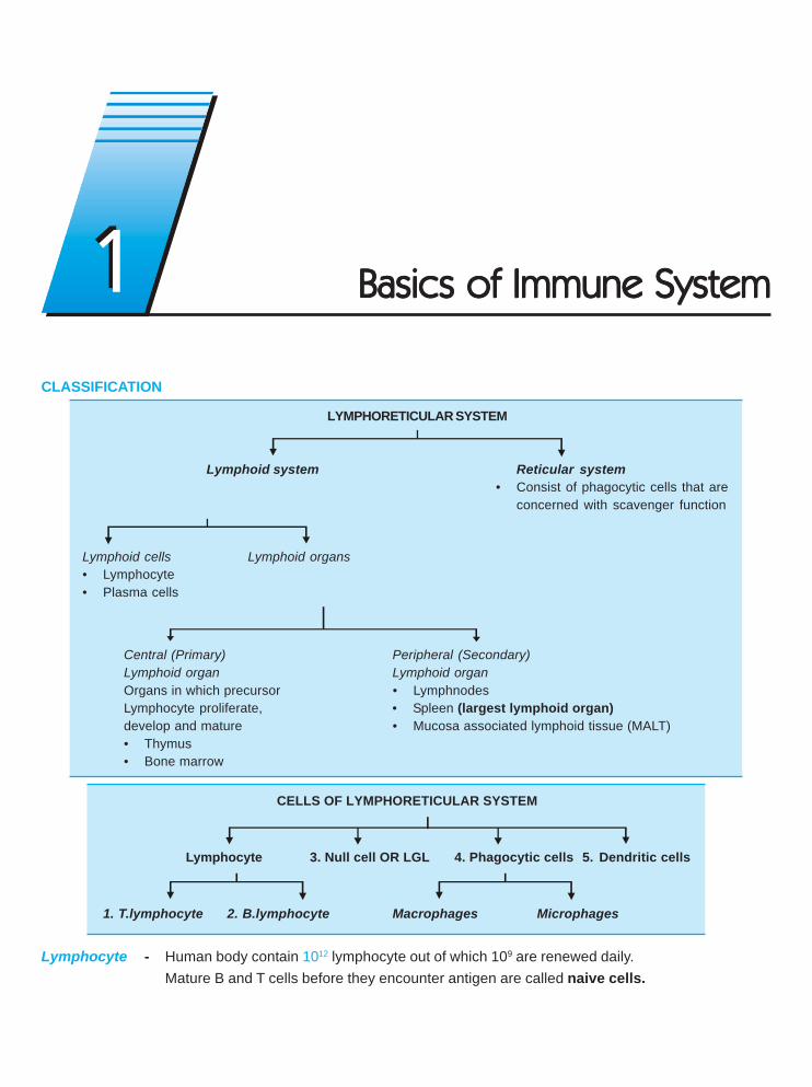
CLASSIFICATION
LYMPHORETICULAR SYSTEM
Lymphoid system Reticular system• Consist of phagocytic cells that are
concerned with scavenger function
Lymphoid cells Lymphoid organs• Lymphocyte• Plasma cells
Central (Primary) Peripheral (Secondary)Lymphoid organ Lymphoid organOrgans in which precursor • LymphnodesLymphocyte proliferate, • Spleen (largest lymphoid organ)develop and mature • Mucosa associated lymphoid tissue (MALT)• Thymus• Bone marrow
CELLS OF LYMPHORETICULAR SYSTEM
Lymphocyte 3. Null cell OR LGL 4. Phagocytic cells 5. Dendritic cells
1. T.lymphocyte 2. B.lymphocyte Macrophages Microphages
Lymphocyte - Human body contain 1012 lymphocyte out of which 109 are renewed daily.Mature B and T cells before they encounter antigen are called naive cells.
Basics of Immune SystemBasics of Immune SystemBasics of Immune SystemBasics of Immune SystemBasics of Immune System11
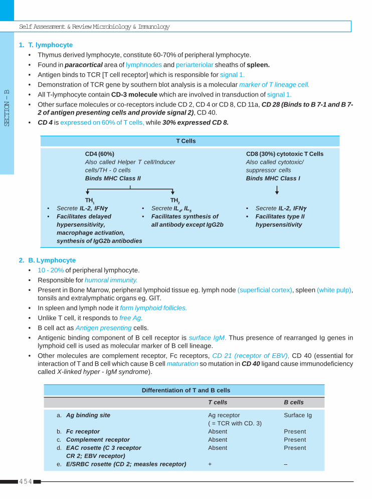
Self Assessment & Review Microbiology & Immunology
SECTION – B
454
1. T. lymphocyte• Thymus derived lymphocyte, constitute 60-70% of peripheral lymphocyte.• Found in paracortical area of lymphnodes and periarteriolar sheaths of spleen.• Antigen binds to TCR [T cell receptor] which is responsible for signal 1.• Demonstration of TCR gene by southern blot analysis is a molecular marker of T lineage cell.• All T-lymphocyte contain CD-3 molecule which are involved in transduction of signal 1.• Other surface molecules or co-receptors include CD 2, CD 4 or CD 8, CD 11a, CD 28 (Binds to B 7-1 and B 7-
2 of antigen presenting cells and provide signal 2), CD 40.• CD 4 is expressed on 60% of T cells, while 30% expressed CD 8.
T Cells
CD4 (60%) CD8 (30%) cytotoxic T CellsAlso called Helper T cell/Inducer Also called cytotoxic/cells/TH - 0 cells suppressor cellsBinds MHC Class II Binds MHC Class I
TH1 TH2
• Secrete IL-2, IFNγγγγγ • Secrete IL4, IL5 • Secrete IL-2, IFNγγγγγ• Facilitates delayed • Facilitates synthesis of • Facilitates type II
hypersensitivity, all antibody except IgG2b hypersensitivitymacrophage activation,synthesis of IgG2b antibodies
2. B. Lymphocyte• 10 - 20% of peripheral lymphocyte.• Responsible for humoral immunity.• Present in Bone Marrow, peripheral lymphoid tissue eg. lymph node (superficial cortex), spleen (white pulp),
tonsils and extralymphatic organs eg. GIT.• In spleen and lymph node it form lymphoid follicles.• Unlike T cell, it responds to free Ag.• B cell act as Antigen presenting cells.• Antigenic binding component of B cell receptor is surface IgM. Thus presence of rearranged Ig genes in
lymphoid cell is used as molecular marker of B cell lineage.• Other molecules are complement receptor, Fc receptors, CD 21 (receptor of EBV), CD 40 (essential for
interaction of T and B cell which cause B cell maturation so mutation in CD 40 ligand cause immunodeficiencycalled X-linked hyper - IgM syndrome).
Differentiation of T and B cells
T cells B cells
a. Ag binding site Ag receptor Surface Ig( = TCR with CD. 3)
b. Fc receptor Absent Presentc. Complement receptor Absent Presentd. EAC rosette (C 3 receptor Absent Present
CR 2; EBV receptor)e. E/SRBC rosette (CD 2; measles receptor) + –
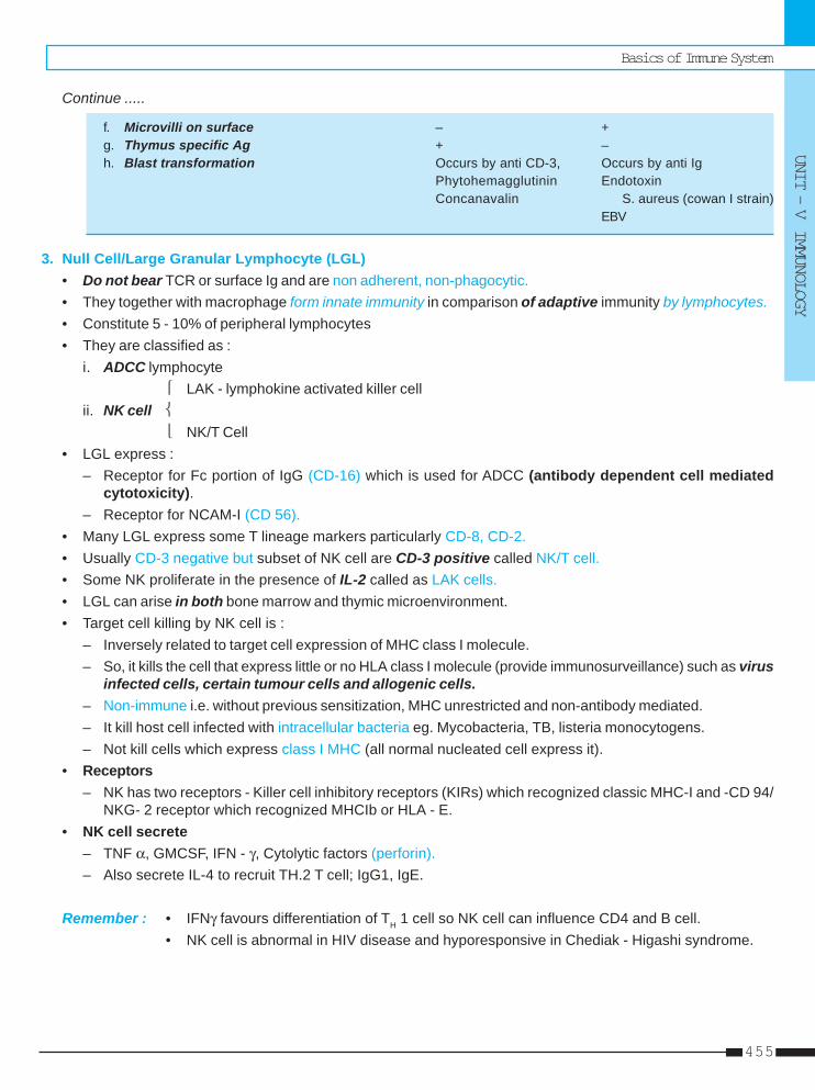
Basics of Immune System
UNIT – V IMMUNOLOGY
455
Continue .....
f. Microvilli on surface – +g. Thymus specific Ag + –h. Blast transformation Occurs by anti CD-3, Occurs by anti Ig
Phytohemagglutinin EndotoxinConcanavalin S. aureus (cowan I strain)
EBV
3. Null Cell/Large Granular Lymphocyte (LGL)• Do not bear TCR or surface Ig and are non adherent, non-phagocytic.• They together with macrophage form innate immunity in comparison of adaptive immunity by lymphocytes.• Constitute 5 - 10% of peripheral lymphocytes• They are classified as :
i. ADCC lymphocyte⎧ LAK - lymphokine activated killer cell
ii. NK cell ⎨⎩ NK/T Cell
• LGL express :– Receptor for Fc portion of IgG (CD-16) which is used for ADCC (antibody dependent cell mediated
cytotoxicity).– Receptor for NCAM-I (CD 56).
• Many LGL express some T lineage markers particularly CD-8, CD-2.• Usually CD-3 negative but subset of NK cell are CD-3 positive called NK/T cell.• Some NK proliferate in the presence of IL-2 called as LAK cells.• LGL can arise in both bone marrow and thymic microenvironment.• Target cell killing by NK cell is :
– Inversely related to target cell expression of MHC class I molecule.– So, it kills the cell that express little or no HLA class I molecule (provide immunosurveillance) such as virus
infected cells, certain tumour cells and allogenic cells.– Non-immune i.e. without previous sensitization, MHC unrestricted and non-antibody mediated.– It kill host cell infected with intracellular bacteria eg. Mycobacteria, TB, listeria monocytogens.– Not kill cells which express class I MHC (all normal nucleated cell express it).
• Receptors– NK has two receptors - Killer cell inhibitory receptors (KIRs) which recognized classic MHC-I and -CD 94/
NKG- 2 receptor which recognized MHCIb or HLA - E.• NK cell secrete
– TNF α, GMCSF, IFN - γ, Cytolytic factors (perforin).– Also secrete IL-4 to recruit TH.2 T cell; IgG1, IgE.
Remember : • IFNγ favours differentiation of TH 1 cell so NK cell can influence CD4 and B cell.• NK cell is abnormal in HIV disease and hyporesponsive in Chediak - Higashi syndrome.
Self Assessment & Review Microbiology & Immunology
SECTION – B
456
4. Phagocytic cellsIt is of two types :i. Mononuclear Macrophages of blood and tissues :
• Blood macrophages (monocytes) are largest of lymphoid cells.• Tissue macrophages (histiocytes) are :
– Microglia in CNS– Kupffer in liver– Alveolar macrophage in lung– Osteoclast in bone– Sinus histiocyte - spleen, lymph node.
• Half life of blood monocyte is about 1d while life span of tissue macrophage is several months.• Have role in chronic inflammation in the form of activated macrophages, fusion giant cell and epitheloid cell
of granulomatous lesion.• It is obligatory for induction of Cell mediated immunity since it is required to process and present antigen to
immuno competent T cell.• Like other Ag presenting / processing cell, it has both class of MHC I and II (more).• Also important for effector phase of humoral immunity.
ii. Microphages which are polymorphonuclear leucocytes of blood-neutrophil, eosinophil and basophil. They donot have any role in specific immune processes.
5. Dendritic Cells• These are Antigen presenting cells to T cell during primary immune response.• They are bone marrow derived cells of lineage different from macrophages and T or B cell.• They possess MHC I and II (more).• They have little or no phagocytic activity.• It is of following types -
a. Interdigitating dendritic ( = Dendritic) cells - Found in lymphoid tissue and interstitium of non-lymphoidorgans eg. heart and lung. Possess costimulatory molecules like B 7-1 and B 7-2.Most potent antigen presenting cell for naive T cell (ideally CD - 4).
b. Langerhans cell - They process and present antigens which reach the dermis.c. Follicular dendritic cells - Bear Fc receptor for IgG. Hence can trap antigen bound to antibodies. They are
present in germinal centers of lymphoid follicles in spleen and lymph nodes.
MAJOR HISTOCOMPATIBILITY COMPLEX (MHC)• Located on short arm of chromosome six which codes for Histocompatibility (transplantation) antigen.• Main function of Histocompatibility (MHC) molecule is to bind peptide fragments of foreign protein for presentation
to appropriate antigen specific T cells.• MHC gene products are classified as :
a. Class I antigens– Are glycoprotein expressed on all nucleated cells and platelets.– Cells with class I antigen presents antigen to CD - 8.– It is the principal antigen involved in graft rejection and cell mediated cytolysis.– Locus of class I gene - A, B, C.
Basics of Immune System
UNIT – V IMMUNOLOGY
457
b. Class II antigen– Glycoprotein, restricted to antigen presenting cells (macrophage, dendritic cell, B cell) to CD - 4.– Locus - D region.– Responsible for graft versus host response and mixed leucocyte reaction (MLR).
c. Class III– Soluble protein of complement system; (C2 and C4 of classical pathway; properdin factor B of alternative
pathway); heat shock protein; TNF alpha and beta.
COMPLEMENT SYSTEMConsist of heat labile factor called alexine or complement (Serum proteins).Fixation of Complement is not influenced by nature of antigens but only by class of Ig.• IgM > IgG3 > IgG1 > IgG2 – Classic pathway activators• IgA1, IgA2, IgD, IgG4 – Alternative pathway activators• Classic C3 convertase Classic C5 convertase
– C4b2b – C4b2b3b• Alternate C3 convertase Alternate C5 convertase
– C3bBb – C3bBb3b• Role of complement derived factors in inflammation :
i. C3b and C3bi - act as opsonin so cause phagocytosis.ii. C3a + C5a [ = Anaphylatoxin] - ↑ vascular permeability, vasodilation.iii. C5a - activate lipoxygenase pathway, chemotaxis, activation and adhesion.iv. C5b-9 ( = Membrane attack complex) - Lysis of cell.
ANTIGENSHaptens – Non immunogenic but has immunological reactivity i.e. incapable of inducing antibody formation but canreact with antibodies.They become immunogenic on combining with larger molecule carrier.Hapten is of two types :i. Simple haptens are non-precipitating.ii. Complex haptens are precipitating, since they have two or more antibody combining sites.
Epitope or antigenic determinant• It is smallest unit of antigenicity (small area on the antigen) which is capable of sensitising an immunocyte and
of reacting with its complementary site on specific antibody or T cell receptor.• T cell identify linear or sequential epitope, while B cell identify conformational epitope.
Paratope• It is combining area of the antibody molecule, corresponding to the epitope.• Epitope and paratope determine specificity which is hallmark of immunological reaction.• Bacteria/virus may contain antigen mosaic (different epitopes) while same epitope on different antigen may
present causing antigenic cross reaction.• Specific antigen determinants on paratope are called IDIOTOPES.
Determinants of Antigenicity– Molecular size (<5000 are non-antigenic)– Chemical nature (usually protein and polysaccharide)– Susceptibility to tissue enzymes– Foreignness– Antigen specificity– Species specificity– Isospecificity– Autospecificity (except lens protein and sperm)– Organ specificity– Heterogenetic / heterophile specificity (Forssman antigen; Weil felix reaction in typhus fever, Paul Bunnel test in
infectious mononucleosis; Cold agglutinin test in primary atypical pneumonia).
22 Antigen & AntibodyAntigen & AntibodyAntigen & AntibodyAntigen & AntibodyAntigen & Antibody
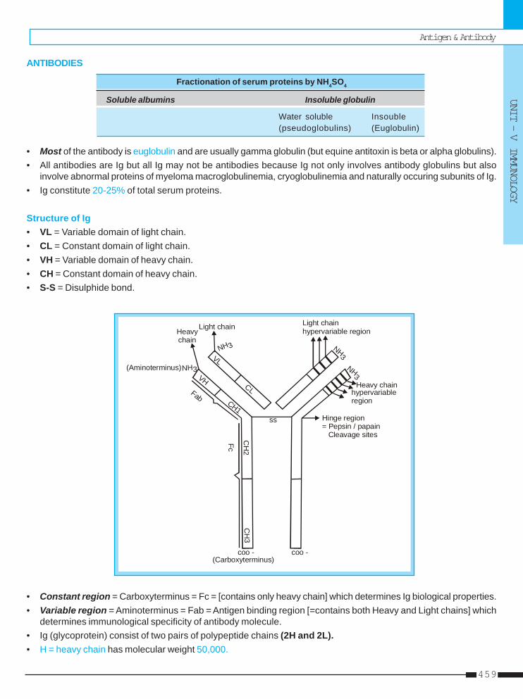
Antigen & Antibody
UNIT – V IMMUNOLOGY
459
ANTIBODIES
Fractionation of serum proteins by NH4SO4
Soluble albumins Insoluble globulin
Water soluble Insouble(pseudoglobulins) (Euglobulin)
• Most of the antibody is euglobulin and are usually gamma globulin (but equine antitoxin is beta or alpha globulins).• All antibodies are Ig but all Ig may not be antibodies because Ig not only involves antibody globulins but also
involve abnormal proteins of myeloma macroglobulinemia, cryoglobulinemia and naturally occuring subunits of Ig.• Ig constitute 20-25% of total serum proteins.
Structure of Ig• VL = Variable domain of light chain.• CL = Constant domain of light chain.• VH = Variable domain of heavy chain.• CH = Constant domain of heavy chain.• S-S = Disulphide bond.
coo -(Carboxyterminus)
coo -
CH
3C
H2
CH1
VH
VL
CL
NH3NH
3
NH3
NH3
ss Hinge region= Pepsin / papain Cleavage sites
Heavy chainhypervariableregion
Light chainhypervariable regionLight chain
Heavychain
(Aminoterminus)
Fab
Fc
• Constant region = Carboxyterminus = Fc = [contains only heavy chain] which determines Ig biological properties.• Variable region = Aminoterminus = Fab = Antigen binding region [=contains both Heavy and Light chains] which
determines immunological specificity of antibody molecule.• Ig (glycoprotein) consist of two pairs of polypeptide chains (2H and 2L).• H = heavy chain has molecular weight 50,000.
Self Assessment & Review Microbiology & Immunology
SECTION – B
460
– H chain are structurally and antigenically distinct for each class and are designated by Greek letter γ, α, μ, δ,ε corresponding to Ig class IgG, IgA, IgM, IgD, IgE. respectively.
– H chain consist of 1 variable (VH) and three domains in constant region (CH1, CH2, CH3).• L = Light chain with molecular weight of 25,000.
– L chain is similar in all classes of Ig.– 2 types of L chain are Kappa (K) and lambda (λ).– 1 molecule of Ig may have either kappa or lambda chains but never both.– Kappa and Lambda occur in ratio of about K : λ = 2 : 1.– L chain consist of 1 variable (VL) and 1 constant domain (CL).
• Highly variable zones (3 in L and 4 in H chain) are known as Hypervariable regions or Hot spots. They areinvolved in the formation of antigen-binding sites. Sites on the hypervariable regions which make actual contactwith the epitope are called complementarity determining regions or CDR’s.
• Fd piece - is portion of H chain present in Fab fragment.
Immunoglobulin classesIt has following properties :– Sedimentation coefficient - max. IgM– Molecular weight - max. IgM, min - IgG– Serum concentration, Half life in days, Daily production (mg/kg) – G > A > M > D > E– Intravascular distribution (%) - max IgM, min - IgA– Carbohydrate (%) - max IgE ..... Harrison 16/e, p 1922– Complement fixation : Classical - IgM > IgG
Alternative - IgA, IgD, IgG– Placental transport - only IgG– Present in milk - IgG and IgA– Selective secretion by seromucinous gland - IgA– Heat labile : only IgE– J chain - IgA and IgM
i. IgG – General purpose antibody, enhances phagocytosis by opsonization.– It has four subclasses G1 > G2 > G3 > G4
– IgG decrease from birth to reach minimum levels by 3rd month.
ii. IgA – Occur in two forms - Serum IgA (monomer) and secretory IgA i.e. SIgA (dimer joined by J chainpresent on respiratory/intestinal mucosa and in secretions).
– J chain is also produced by plasma cells but secretory piece/secretory component of SIgA is notproduced by lymphoid cells but by mucosal or glandular epithelial cells.
iii. IgM – Effective valency is five.– Earliest Ig to be synthesized by fetus (begin at 20 week of age) is IgM.– At 20th weeks Peyer’s patches and lymphoid cells in spleen, and lymph nodes are developed so
fetus has IgM, IgD, IgG (transplacentally) but not IgA and IgE.– IgM detection is useful in diagnosis of congenital infection.– Monomeric IgM is major antibody receptor on surface of B lymphocytes for antigen recognition.
iv. IgD – Resemble IgG structurally and also serve as recognition receptor for antigen.
v. IgE – Mostly extravascular and exhibits Homocytotrophism.
Antigen & Antibody
UNIT – V IMMUNOLOGY
461
– Chiefly produced in the lining of intestinal and respiratory tract.– It mediates Reaginic hypersensitivity and Prausnitz kustner (PK) reaction.
Remember : IgG protect body fluids, IgA body surfaces and IgM the blood stream.
Types of Antibody in various conditions :IgM – Biological false positive Ab in syphilis
– Rheumatoid factor– Ab against ABO– Ab to typhoid O Ag (endotoxin).
IgG – Ab against Rh factor (Anti Rh D)– (LATS) long acting thyroid stimulator Ab in Grave’s– AutoAb in SLE, GB Syndrome– Reagin Ab in syphilis (LA, aCL). ..... Harrison 16/e, p 1923
Abnormal Igi. Bence Jones Protein (BJP)
– Monoclonal Ig consist of light chain found typically in multiple myeloma.– Identified in urine by its characteristic property of coagulation when heated to 500C but redissolving at 700C.
ii. Myeloma (M) protein– Monoclonal Ab seen in multiple myeloma (IgG, IgA, IgD, IgE) and waldenstrom’s macroglobulinemia (IgM).
iii. FC parts of Ig heavy chain– ↑ in Heavy chain disease.
iv. Cryo globulinemia– Formation of gel or precipitate on cooling the serum, which redissolves on warming.– Most cryoglobulin consist of either IgG, IgM or mixed precipitates.
ANTIGEN – ANTIBODY REACTIONSAg-Ab reaction is reversible, occur at surface and there is no denaturation of Ag or Ab during reaction. Reactionsoccur in three stages :i. Primary stage :
Ag-Ab combined by weaker intermolecular forces such as Van-der waal’s, ionic bond and H2 binding rather thanby firmer covalent bonding; without any visible effects.
ii. Secondary stage :Usually present but not always. It leads to demonstrable events such as precipitation, agglutination, lysis of cellsetc.
iii. Tertiary stage (reaction) :Leads to tissue damage e.g. allergy and other immunological diseases.
Self Assessment & Review Microbiology & Immunology
SECTION – B
462
Serological reactions• Comparative efficiency of Ig in different serological reactions :
Reaction IgG IgM IgA
• Neutralization (N) More effective Less effective Variable
• Precipitation (P) Strong Weak Variable
• Classical complement fixation (C) Strong Strongest Negative
• Immunohemolysis (I) and Less effective More effective ModerateBactericidal (opsonization) (O)
• Agglutination (A) Weak Strong Moderate
• Lysis (L) Weak Strong Negative
..... Harrison 16/e, p 1992Mnemonic for strong reaction in IgG = G-N-P (Neutralization, Precipitation).
Mnemonic for strong reaction in IgM = CO-M-ALI (Classical Complement fixation, Opsonization, Agglutination,lysis, Immunohemolysis).
i. Precipitation ReactionWhen soluble antigen combines with its antibody in presence of electrolytes, it forms insoluble precipitates/floccules.• This reaction show “zone phenomenon” (also in agglutination) either in the form of prozone (antibody excess)
or postzone (antigen excess).• It occur in zone of equivalence due to lattice formation (also in agglutination).• It is very sensitive in antigen detection (but relatively less sensitive for antibody detection).• It is of following types :
a. Ring test– Simplest type eg. Ascoli’s thermoprecipitin test and streptococcal lancefield grouping.
b. Slide test – VDRL test of syphilis
c. Tube test – Kahn test of syphilis
d. Immunodiffusion (ppt in gel) – Eg. Elek test for toxigenicity in diphtheria (double diffusion).
e. Electro immunodiffusion – Eg. rocket electrophoresis for quantitative estimation of antigen.
ii. Agglutination ReactionIt is more sensitive than precipitation for antibody detection. Occur in presence of electrolytes.• Incomplete or monovalent antibodies (usually Ab are bivalent) do not cause agglutination, though they combine
with the antigen. They also act as blocking Ab since they inhibits agglutination by complete Ab. Agglutinationis of following types :a. Slide agglutination – used for blood grouping and cross matching.
b. Tube agglutination – Eg. Widal test, Brucellosis, Weil-Felix reaction, Paul Bunnel test, cold agglutinationand Streptococcus MG test.
c. Antiglobulin (Coombs) test – used for detecting incomplete Ab of brucellosis; anti-Rh Ab.
d. Passive agglutination test – used to detect Ab by adsorbing soluble Ag on carrier particles so precipitationreaction converts into agglutination test which are more convenient and more sensitive. Eg. Rose wallertest, test detecting RA factor by using amboceptor.
Antigen & Antibody
UNIT – V IMMUNOLOGY
463
e. Latex agglutination test (latex fixation test) – for detection of ASO, CRP, RA factor, HCG; Streptozymetest.
f. Reversed passive agglutination - Estimation of antigen by adsorbing antibody to carrier particles.
iii. Complement Fixation Test (CFT)• Antigen may be soluble or particulate.• Source of complement is guinea pig serum Eg. Wassermann reaction; coaglutinating complement adsorption
test using horse complement; immuno adherence of V.cholera and T. pallidum; Immobilization test of T.pallidum; Cytolytic or cytocidal test.
iv. Neutralisation TestsIncludes virus neutralisation test (plaque inhibition test), Toxin neutralisation (Schick, antistreptolysin O)test.
v. Radio - Immunoassay (RIA)• MC labels used are radio-isotopes and enzymes.• It measures analytes upto picogram (10-12g) quantities.• Used for quantitation of hormones, drugs, tumour markers, IgE and viral antigen.
vi. Enzyme Immunoassay (EIA)• Measures enzyme labelled antigen, hapten or antibody.• It may be homogenous or heterogenous.• Major type of heterogenous EIA is ELISA which involves the use of immunosorbent - an absorbing material
specific for one of components of reaction, the antigen or antibody. Eg ELISA for detection of Rotavirus infeces or HIV antibody in serum.
Type
1. Type I (Immediate or reaginic HSN)
Systemic acute Localanaphylaxis anaphylaxis
= atopy
2. Type II [cytotoxic or cytolytic] HSNa. Complement dependent
b) Type VI HSN =Antibody dependent cellmediated cytotoxicity (ADCC)
c) Type V HSN = Antibody mediatedcellular dysfunction.
Mechanism and Effects
• Ab - IgE (cytotropic)• Cells - IgE B cell, mast cells,
basophils, Eosinophil• Pivotol role - by TH2 cell• Most important vasoactive
amine : Histamine• Slow reacting substance of
anaphylaxis (SRS-A) =Leukotrienes (LT B4, C4, D4, E4)
• Ab : IgG or IgM• Lysis or phagocytosis by
opsonisation• Most commonly involves blood
cells (II HSN involve blood : b issecond alphabet → Mnemonic)
• Ab - usually IgG sometimes IgE• Cell lysis without phagocytosis by
monocytes, neutrophil, eosino-phils and NK cells
Antireceptor Antibody• Stimulation• Inhibition
Examples
• Urticaria; angioedema; hay fever andsome forms of asthma; eczema
• Anaphylactic shock• Casoni’s test,• Theobald smith phenomenon• Schultz dale phenomenon• Prausnitz kustner (PK) reaction
• Transfusion reactions• Erythoblastosis fetalis (Hemolytic
disease of neotates)• AI hemolytic anemia or
agraulocytosis or thrombocytopenia;• Pemphigus syndrome• Bullous pemphigoid• Pernicious anemia• Thrombotic phenomenon• Acute rheumatic fever• Some forms of vasculitides and
certain drug reactions.• Phagocytosis of tumour cells or
parasite• Some role in graft rejection
• Thyrotoxicosis / Grave’s disesas• Myasthenia gravis
Hypersensitivity is defined as immunologic tissue injury in response of subsequent exposure with the allergen(shocking or challenge dose). It is following types :
33 Hypersensitivity [HSN]Hypersensitivity [HSN]Hypersensitivity [HSN]Hypersensitivity [HSN]Hypersensitivity [HSN]
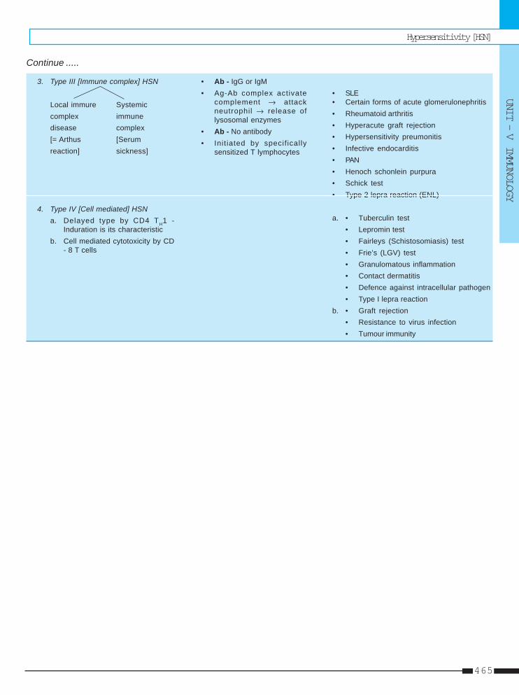
Hypersensitivity [HSN]
UNIT – V IMMUNOLOGY
465
• SLE• Certain forms of acute glomerulonephritis• Rheumatoid arthritis• Hyperacute graft rejection• Hypersensitivity preumonitis• Infective endocarditis• PAN• Henoch schonlein purpura• Schick test• Type 2 lepra reaction (ENL)
a. • Tuberculin test• Lepromin test• Fairleys (Schistosomiasis) test• Frie’s (LGV) test• Granulomatous inflammation• Contact dermatitis• Defence against intracellular pathogen• Type I lepra reaction
b. • Graft rejection• Resistance to virus infection• Tumour immunity
3. Type III [Immune complex] HSN
Local immure Systemiccomplex immunedisease complex[= Arthus [Serumreaction] sickness]
4. Type IV [Cell mediated] HSNa. Delayed type by CD4 TH1 -
Induration is its characteristicb. Cell mediated cytotoxicity by CD
- 8 T cells
• Ab - IgG or IgM• Ag-Ab complex activate
complement → attackneutrophil → release oflysosomal enzymes
• Ab - No antibody• Initiated by specifically
sensitized T lymphocytes
Continue .....
Self Assessment & Review Microbiology & Immunology
SECTION – B
466
1. The serum concentration of which of the follow-ing human IgG subclass is maximum : [AI 05]a) IgG1b) IgG2c) IgG3d) IgG4
2. Adenosine deaminase deficiency is seen in thefollowing : [AI 05, 01]a) Common variable immunodeficiencyb) Severe combined immunodeficiencyc) Chronic granulomatous diseased) Nezelof syndrome
3. A woman with infertility receives an ovary trans-plant from her sister who is an identical twin.What type of graft it is ? [AI 05]a) Xenograftb) Autograftc) Allograftd) Isograft
4. Which of the following statements is true abouthapten ? [AI 04]a) It induces brisk immune responseb) It needs carrier to induce immune responsec) It is a T-independent antigend) It has no association with MHC
5. The earliest immunoglobulin to be synthesized bythe fetus is : [AI 03]a) IgAb) IgGc) IgEd) IgM
6. Neonatal thymectomy leads to : [AI 02]a) Decreased size of germinal centerb) Decreased size of paracortical areasc) Increased antibody marrow production by B cellsd) Increased bone marrow production of lympho-
cytes7. IL-I produces : [AI 02]
a) T lymphocyte activationb) Delayed wound healingc) Increased pain perceptiond) Decreased PMN release from bone marrow
8. Regarding NK cells, false statement is : [AI 01]a) It is activated by IL-2
b) Expresses CD3 receptorc) It is a variant of large lymphocyted) There is antibody induced proliferation of NK
cells9. Type of graft, best suited for renal transplanta-
tion: [AI 99]a) Allograftb) Autograftc) Xenograftd) Isograft
10. Haptane is : [AI 98]a) Same as epitopeb) Small molecular weight proteinc) Requires carrier for specific antibody produc-
tiond) Simple haptens are precipitating
11. All of the following forces are involoved in Antigenantibody reaction except :a) Vander Waal's forces [AI 98, AIIMS 96]b) Electrostatic bondc) Hydrogen bondd) Covalent bond
12. Examples of type I hypersensitivity is : [AI 98]a) Lepromin testb) Tuberculinc) Casoni's testd) Arthus reaction
13. IL-2 is produced by : [AI 97; 00; AIIMS 97]a) T cellsb) B cellsc) Monocytesd) Neutrophils
14. All of the following are glycoproteins except :a) Blood antigen [AI 97; 94]b) Albuminc) Immunoglobind) HCG
15. Which of the following statements concerningimmunoglobins is wrong : [AI 97]a) IgM does not cross placentab) IgE is ↑ ed in parasitic infectionc) IgM increased in primary responsed) Fetal infection is characterized by increase in
IgG
QUESTIONS
Answer 1. a) IgG1 2. b) Severe ... 3. d) Isograft 4. b) It needs ... 5. d) IgM6. b) Decreased ... 7. a) T lymph ... 8. d) There is ... 9. d) Isograft 10. c) Requires ...
11. d) Covalent ... 12. c) Casoni’s ... 13. a) T cells 14. b) Albumin 15. d) Fetal
Hypersensitivity [HSN]
UNIT – V IMMUNOLOGY
467
Answer 16. d) Granulo ... 17. d) C S F 18. a) T cells 19. c) CD3 20. a) Phytohem ...21. a) Granul ... 22. b) Light ... 23. c) Kappa ... 24. b and d 25. c) 2 light ...26. b) Serum ... 27. a) Increased ... 28. b) Haem .... 29. d) T cells 30. b) H-chain
16. Which of the following is an example of Type IVhypersensitivity : [AI 97]a) Arthrus reactionb) Serum sicknessc) Schwartzmann reactiond) Granulomatous reaction
17. Diagnosis of ABO incompatability can be from allof the following except : [AI 97]a) Sweatb) Salivac) Semend) CSF
18. Helper cells belong to : [AI 96]a) T cellsb) Macrophagesc) B cellsd) Monocytes
19. The type of receptors present on T cells are :a) IgG [AI 96]b) IgDc) CD3d) Prostaglandins
20. What enhances multiplication of T cells in culturea) Phytohemagglutinin [AI 96]b) Chemotactic factorc) Leukotrienesd) Prostaglandins
21. Which of the following is an example of type IVhypersensitivity : [AI 96]a) Granulomatous reactionb) Schwartzman reactionc) Arthus reactiond) Serum sickness
22. Which precipitates at 500C-600C but disappearson heating : [AI 96]a) Heavy chainb) Light chainc) Bothd) None of the above
23. Bence Jones protein are best described as :a) μ chains [AI 96]b) γ chainsc) Kappa and Lambda chainsd) Fibrin split products
24. Prozone phenomenon is due to : [AI 96, PGI 03]a) Antigen excessb) Antibody excessc) False +ve reactiond) False -ve reaction
25. A single immunoglobulin molecule contains :a) 1 light chain, 1 heavy chain [AI 95]b) 2 heavy chains, 1 lightc) 2 light chains, 2 heavyd) 2 light chains, 1 heavy chains
26. Which of the following is false : [AI 95]a) Theobald-Smith phenomenon is a type I hyper-
sensitivity reactionb) Serum sickness is a type II hypersensitivity re-
actionc) Allograft rejection is a type IV hypersensitivity
reactiond) Transfusion reaction is a type II hypersensitivity
reaction27. NK cells kill the viral infected cells due to :
a) Increased expression of MHC class I moleculesb) Decreased expression of MHC class I mol-
ecules [AIIMS 06]c) Increased expression of MHC class II mol-
eculesd) Decreased expression of MHC class II mol-
ecules28. The following methods of diagnosis utilize labeled
antibodies except : [AIIMS 05]a) ELISAb) Haemagglutination inhibition testc) Radio immuno assayd) Immun-ofluorescence
29. All of the following are part of innate immunityexcept : [AIIMS 05]a) Complementb) NK cellsc) Macrophagesd) T cells
30. Which of the following is class specific antigenicdeterminants of an Ig : [AIIMS 04]a) L-chainb) H-chainc) J-chaind) Variable region
Self Assessment & Review Microbiology & Immunology
SECTION – B
468
Answer 31. a) C2, C4 .... 32. c) IgM 33. d) Lymphocytes 34. b) IgM fixes ... 35. d) Antigen, ...36. a) IgA 37. b) Precipitation 38. d) Increased ... 39. a and e 40. b) Type - II41. a, c and d 42. c) Antigenic ... 43. b and c 44. d) Schick ... 45. b and c46. d and e
31. Which of the following best denotes classicalcomplement pathway activation in immuno inflam-matory condition : [AIIMS 04]a) C2, C4 and C3 decreasedb) C2 and C4 normal, C3 is decreasedc) C3 normal and C2 C4 decreasedd) C2, C4, C3 all are elevated
32. The most avidly complement fixing antibody is :a) IgA [AIIMS 02]b) IgGc) IgMd) IgE
33. Which is not a Macrophage : [AIIMS 97]a) Monocyteb) Microgilac) Kupffer cellsd) Lymphocytes
34. True about immunoglobulins is : [AIIMS 97]a) IgE fixes complementsb) IgM fixes complementsc) IgG is found in minimum concentrationd) IgG is elevated in primary immune response
35. Which of the following is true : [AIIMS 96]a) Paul Bunnel test is used to diagnose measlesb) Rose Waller test is a complement fixation testc) Indirect hemagglutination test is less sensitive
than gel diffusion testd) Antigen, Antibody reaction cannot occur in ab-
sence of electrolytes36. In Respiratory and GIT infection, which is the most
affected immunoglobulin : [AIIMS 96]a) IgAb) IgGc) IgMd) IgD
37. The reaction between antibody and soluble anti-gen is demonstrated by : [AIIMS 96]a) Agglutinationb) Precipitationc) Complement fixationd) Hemagglutination test
38. C-reactive protein is : [AIIMS 96]a) An antibody as a result of pneumococcal infec-
tionb) Derived from pneumococcic) Detected by precipitation reactiond) Increased in pneumococcal infection
39. Common between B and T cells : [PGI June 07]a) Origin from same cell lineageb) Site differentiationc) Antigenic markerd) Both humoral and cellular immunitye) Further differentiation seen
40. Haemolytic disease of newborn is which type ofhypersensitivity reaction : [PGI June 07]a) Type - Ib) Type - IIc) Type - IIId) Type IVe) Type V
41. Skin test is used for which hypersensitivity reac-tion :a) I [PGI June 07]b) IIc) IIId) IV
42. Antibody diversity is due to : [PGI Dec. 07]a) Gene re-arrangementb) Gene translocationc) Antigenic variationd) CD40 moleculese) Mutation
43. MHC class III genes encode : [PGI 06]a) Complement component C3b) Tumor necrosis factor - alphac) Tumor necrosis factor - betad) Interleukin 2e) Beta 2 microglobulin
44. Skin test based on neutralization reaction is/are:a) Casoni test [PGI 04]b) Lepromin testc) Tuberculin testd) Schick test
45. Acute phase reactants (APR) in acute inflamma-tion are : [PGI 03]a) Albuminb) Fibrinogenc) Haptoglobulind) Gammaglobulin
46. Antigen binding site on antibody is : [PGI 02]a) Hinge regionb) Constant regionc) Variable regiond) Hypervariable regione) Idiotype region
Hypersensitivity [HSN]
UNIT – V IMMUNOLOGY
469
Answer 47. b) NK cell 48. d) Plasma .. 49. a and c 50. a and c 51. a) Cytotoxic ...52. c) Formed ... 53. b, c and d 54. c) IgG 55. b and d 56. a) Macrophage57. a) Autoimmune ... 58. a, b, c and d 59. b) Widal test 60. c and d 61. b) Inhibits ...62. a) Polysaccharide
47. Apart from B cells, and T cells, there is a 3rd dis-tinct type of lymphocyte. This is : [PGI 02]a) MHC cellb) NK cellc) Macrophaged) Neutrophile) Microglia
48. IgE is secreted by : [PGI 02]a) Mast cellb) Basophilsc) Eosinophilsd) Plasma cellse) Neutrophils
49. All of these are antigen presenting cells except :a) T cells [PGI 02]b) B cellsc) Fibroblastsd) Dendritic cellse) Langerhans cells
50. Classic complement is activated by : [PGI 02]a) IgGb) IgAc) IgMd) IgEe) IgD
51. Perforins are produced by : [PGI 02]a) Cytotoxic T cellsb) Suppressor T cellsc) Memory helper T cellsd) Plasma cellse) NK cells
52. The secretory component of molecule is :a) Formed by epithelial cell of lining mucosab) Formed by plasma cell [PGI 01]c) Formed by epithelial cell and plasma celld) Secreted by bone marrow
53. True of the following is/are : [PGI 01]a) IgA crosses placentab) Half life of IgG is 23 daysc) IgD is heat stabled) IgE has highest carbohydrate contente) IgG induces leukotrienes release during inflam-
mation54. Which of the following immunoglobulins can cross
placenta : [PGI 01]a) IgA
b) IgMc) IgGd) IgD
55. C.reactive proteins are : [PGI 00]a) Alpha-globulinb) Beta-1 globulinc) Alpha-2 globulind) Non-specific inflammatory protien
56. IL-I is produced by : [PGI 00]a) Macrophageb) Helper T lymphocytesc) B cellsd) Cytotoxic T-cells
57. Type-1 hypersensitivity includes all of the follow-ing except : [PGI 00]a) Autoimmune hemolytic anemiab) Anaphylaxisc) Extrinsic Asthmad) Hay Fever
58. True about anaphylaxis : [PGI 00]a) Type-1 reactionb) Large amount of histamine releasedc) Cytokines like IL4, IL5, and IL6 and GMCSF are
releasedd) Mediated through allergen specific IgE
59. Which is not heterophile agglutination test :a) Weil-Felix test [PGI 99]b) Widal testc) Paul-Bunnel testd) Streptoccus MG
60. True about secondary immune response is:a) Long latent period [PGI 98, 96]b) Usually of low titrec) Antibodies appear in short timed) Persist for long
61. True about interferon is : [PGI 98]a) It is a synthetic antiviral agentb) Inhibits viral replicatiion in cellsc) Is specific for particular virusd) None
62. Which of the following is very difficult to induceantibody : [PGI 97]a) Polysaccharideb) Proteinc) Antigensd) Repeated infection
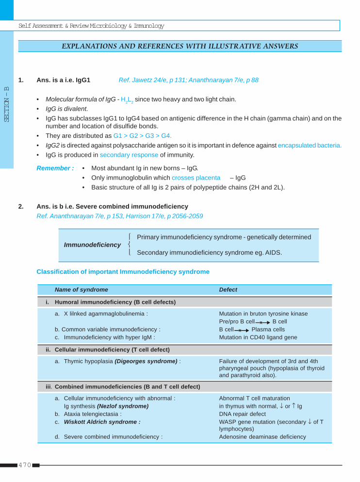
Self Assessment & Review Microbiology & Immunology
SECTION – B
470
1. Ans. is a i.e. IgG1 Ref. Jawetz 24/e, p 131; Ananthnarayan 7/e, p 88
• Molecular formula of IgG - H2L2 since two heavy and two light chain.• IgG is divalent.• IgG has subclasses IgG1 to IgG4 based on antigenic difference in the H chain (gamma chain) and on the
number and location of disulfide bonds.• They are distributed as G1 > G2 > G3 > G4.• IgG2 is directed against polysaccharide antigen so it is important in defence against encapsulated bacteria.• IgG is produced in secondary response of immunity.
Remember : • Most abundant Ig in new borns – IgG.• Only immunoglobulin which crosses placenta – IgG• Basic structure of all Ig is 2 pairs of polypeptide chains (2H and 2L).
2. Ans. is b i.e. Severe combined immunodeficiencyRef. Ananthnarayan 7/e, p 153, Harrison 17/e, p 2056-2059
⎧ Primary immunodeficiency syndrome - genetically determinedImmunodeficiency ⎨
⎩ Secondary immunodieficiency syndrome eg. AIDS.
Classification of important Immunodeficiency syndrome
Name of syndrome Defect
i. Humoral immunodeficiency (B cell defects)
a. X lilnked agammaglobulinemia : Mutation in bruton tyrosine kinasePre/pro B cell B cell
b. Common variable immunodeficiency : B cell Plasma cellsc. Immunodeficiency with hyper IgM : Mutation in CD40 ligand gene
ii. Cellular immunodeficiency (T cell defect)
a. Thymic hypoplasia (Digeorges syndrome) : Failure of development of 3rd and 4thpharyngeal pouch (hypoplasia of thyroidand parathyroid also).
iii. Combined immunodeficiencies (B and T cell defect)
a. Cellular immunodeficiency with abnormal : Abnormal T cell maturationIg synthesis (Nezlof syndrome) in thymus with normal, ↓ or ↑ Ig
b. Ataxia telengiectasia : DNA repair defectc. Wiskott Aldrich syndrome : WASP gene mutation (secondary ↓ of T
lymphocytes)d. Severe combined immunodeficiency : Adenosine deaminase deficiency
EXPLANATIONS AND REFERENCES WITH ILLUSTRATIVE ANSWERS
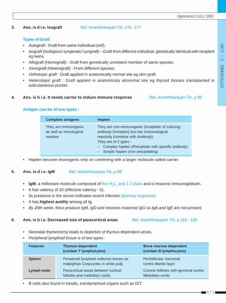
Hypersensitivity [HSN]
UNIT – V IMMUNOLOGY
471
3. Ans. is d i.e. Isograft Ref. Ananthnarayan 7/e, 176 - 177
Types of Graft• Autograft : Graft from same individual (self).• Isograft (isologous/ syngeneic/ syngraft) – Graft from different individual, genetically identical with recipient
eg twins.• Allograft (Homograft) : Graft from genetically unrelated member of same species.• Xenograft (Heterograft) : From different species.• Orthotopic graft : Graft applied in anatomically normal site eg skin graft.• Heterotopic graft : Graft applied in anatomicaly abnormal site eg thyroid tissues transplanted in
subcutaneous pocket.
4. Ans. is b i.e. It needs carrier to induce immune response Ref. Ananthnarayan 7/e, p 80
Antigen can be of two types :
Complete antigens Hapten
They are immunogenic They are non-immunogenic (incapable of inducingas well as immulogical antibody formation) but has immunologicalreactive reactivity (combine with Antibody).
They are of 2 types :– Complex hapten (Precipitate with specific antibody)– Simple hapten (non precipitating)
• Hapten become imunogenic only on combining with a larger molecule called carrier.
5. Ans. is d i.e. IgM Ref. Ananthnarayan 7/e, p 89
• IgM, a millionare molecule composed of five H2L2 and 1 J chain and is heaviest immunoglobulin.• It has valency of 10 (effective valency - 5).• Its presence in the serum indicates recent infection (primary response).• It has highest avidity among all Ig.• By 20th week, fetus produce IgM, IgD and receives maternal IgG so IgA and IgE are not present.
6. Ans. is b i.e. Decreased size of paracortical areas Ref. Ananthnarayan 7/e, p 119 - 120
• Neonatal thymectomy leads to depletion of thymus dependent areas.• Peripheral lymphoid tissue is of two types :
Features Thymus dependent Bone marrow dependent(contain T lymphocytes) (contain B lymphocytes)
Spleen Periaterial lymphoid collecion known as Perifollicular Germinalmalpighian Corpuscles in white pulp. centre Mantle layer
Lymph node Paracortical areas between cortical Corticle follicles with germinal centrefollicles and medullary cords. Medullary cords
• B cells also found in tonsils, extralymphoid organs such as GIT.
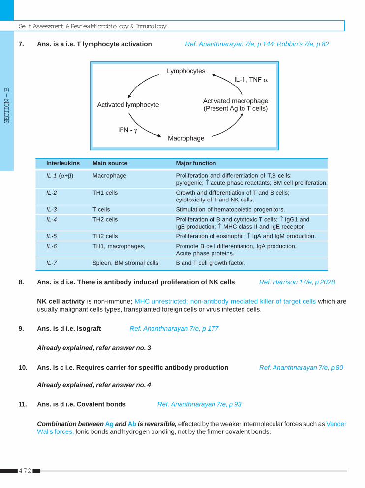
Self Assessment & Review Microbiology & Immunology
SECTION – B
472
7. Ans. is a i.e. T lymphocyte activation Ref. Ananthnarayan 7/e, p 144; Robbin’s 7/e, p 82
Lymphocytes
Activated lymphocyte
Macrophage
Activated macrophage(Present Ag to T cells)
Interleukins Main source Major function
IL-1 (α+β) Macrophage Proliferation and differentiation of T,B cells;pyrogenic; ↑ acute phase reactants; BM cell proliferation.
IL-2 TH1 cells Growth and differentiation of T and B cells;cytotoxicity of T and NK cells.
IL-3 T cells Stimulation of hematopoietic progenitors.
IL-4 TH2 cells Proliferation of B and cytotoxic T cells; ↑ IgG1 andIgE production; ↑ MHC class II and IgE receptor.
IL-5 TH2 cells Proliferation of eosinophil; ↑ IgA and IgM production.
IL-6 TH1, macrophages, Promote B cell differentiation, IgA production,Acute phase proteins.
IL-7 Spleen, BM stromal cells B and T cell growth factor.
8. Ans. is d i.e. There is antibody induced proliferation of NK cells Ref. Harrison 17/e, p 2028
NK cell activity is non-immune; MHC unrestricted; non-antibody mediated killer of target cells which areusually malignant cells types, transplanted foreign cells or virus infected cells.
9. Ans. is d i.e. Isograft Ref. Ananthnarayan 7/e, p 177
Already explained, refer answer no. 3
10. Ans. is c i.e. Requires carrier for specific antibody production Ref. Ananthnarayan 7/e, p 80
Already explained, refer answer no. 4
11. Ans. is d i.e. Covalent bonds Ref. Ananthnarayan 7/e, p 93
Combination between Ag and Ab is reversible, effected by the weaker intermolecular forces such as VanderWal's forces, lonic bonds and hydrogen bonding, not by the firmer covalent bonds.
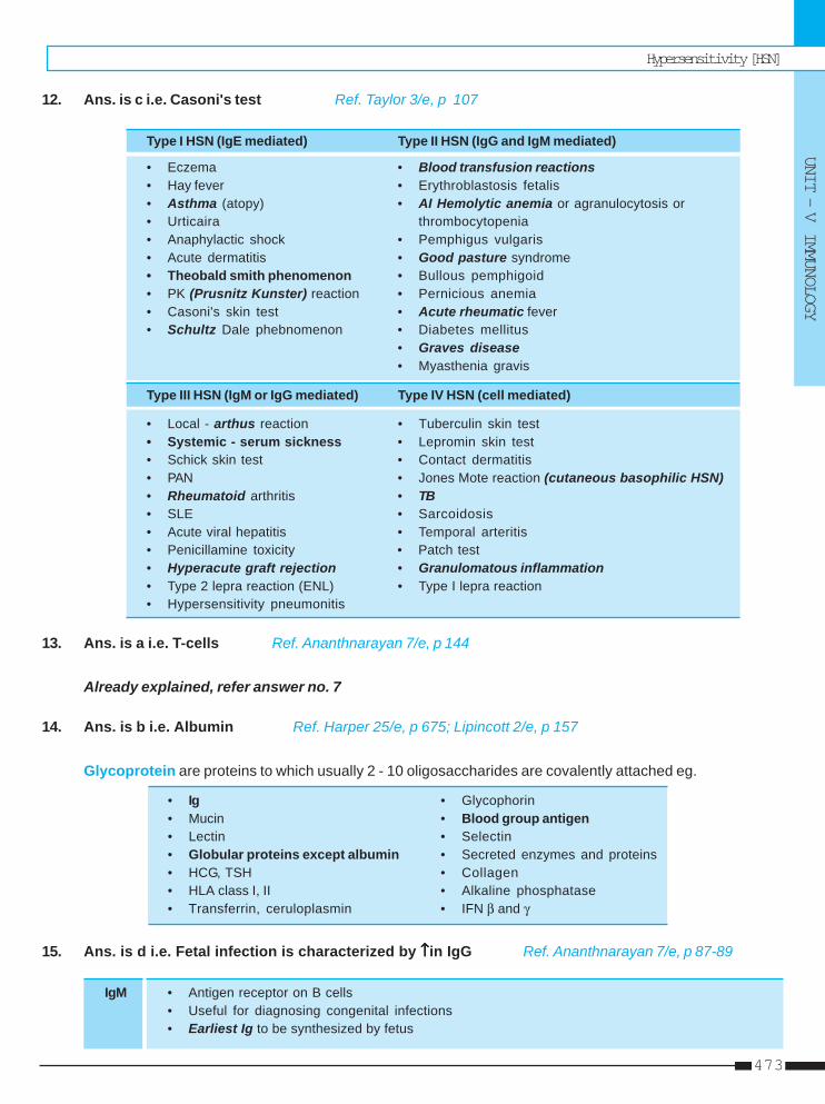
Hypersensitivity [HSN]
UNIT – V IMMUNOLOGY
473
12. Ans. is c i.e. Casoni's test Ref. Taylor 3/e, p 107
Type I HSN (IgE mediated) Type II HSN (IgG and IgM mediated)
• Eczema • Blood transfusion reactions• Hay fever • Erythroblastosis fetalis• Asthma (atopy) • AI Hemolytic anemia or agranulocytosis or• Urticaira thrombocytopenia• Anaphylactic shock • Pemphigus vulgaris• Acute dermatitis • Good pasture syndrome• Theobald smith phenomenon • Bullous pemphigoid• PK (Prusnitz Kunster) reaction • Pernicious anemia• Casoni's skin test • Acute rheumatic fever• Schultz Dale phebnomenon • Diabetes mellitus
• Graves disease• Myasthenia gravis
Type III HSN (IgM or IgG mediated) Type IV HSN (cell mediated)
• Local - arthus reaction • Tuberculin skin test• Systemic - serum sickness • Lepromin skin test• Schick skin test • Contact dermatitis• PAN • Jones Mote reaction (cutaneous basophilic HSN)• Rheumatoid arthritis • TB• SLE • Sarcoidosis• Acute viral hepatitis • Temporal arteritis• Penicillamine toxicity • Patch test• Hyperacute graft rejection • Granulomatous inflammation• Type 2 lepra reaction (ENL) • Type I lepra reaction• Hypersensitivity pneumonitis
13. Ans. is a i.e. T-cells Ref. Ananthnarayan 7/e, p 144
Already explained, refer answer no. 7
14. Ans. is b i.e. Albumin Ref. Harper 25/e, p 675; Lipincott 2/e, p 157
Glycoprotein are proteins to which usually 2 - 10 oligosaccharides are covalently attached eg.
• Ig • Glycophorin• Mucin • Blood group antigen• Lectin • Selectin• Globular proteins except albumin • Secreted enzymes and proteins• HCG, TSH • Collagen• HLA class I, II • Alkaline phosphatase• Transferrin, ceruloplasmin • IFN β and γ
15. Ans. is d i.e. Fetal infection is characterized by ↑↑↑↑↑in IgG Ref. Ananthnarayan 7/e, p 87-89
IgM • Antigen receptor on B cells• Useful for diagnosing congenital infections• Earliest Ig to be synthesized by fetus
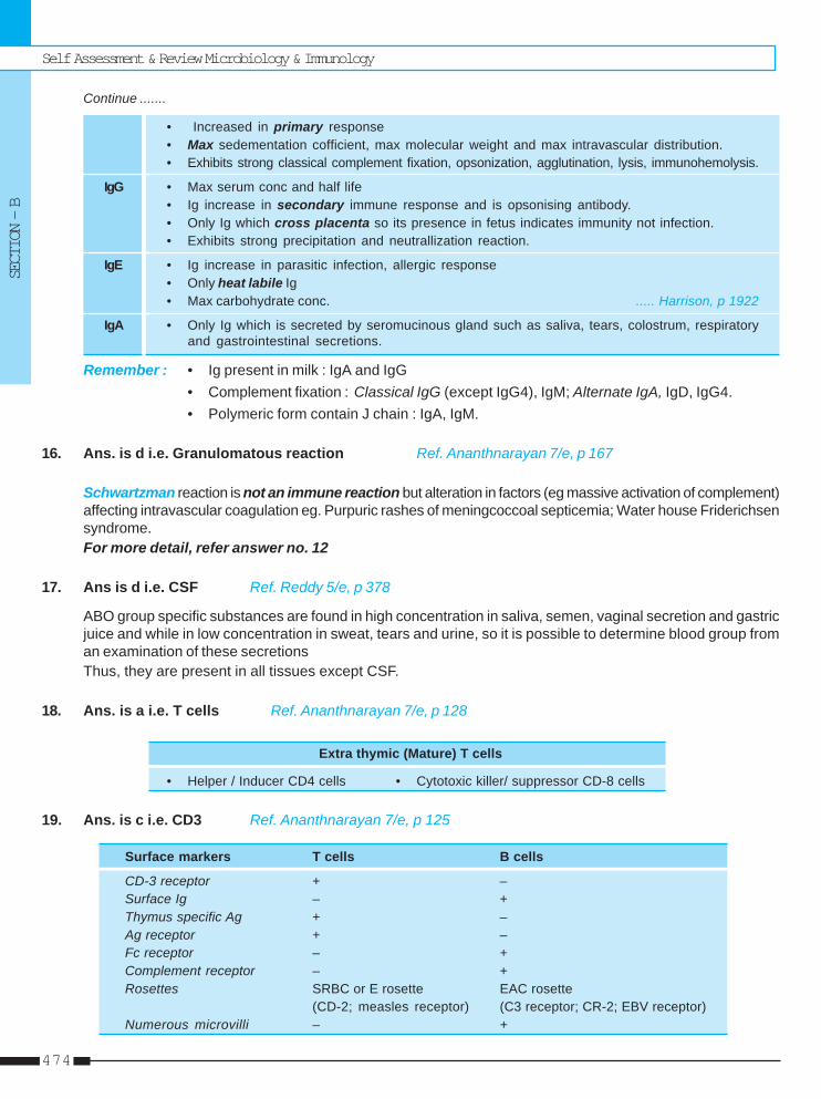
Self Assessment & Review Microbiology & Immunology
SECTION – B
474
Continue .......
• Increased in primary response• Max sedementation cofficient, max molecular weight and max intravascular distribution.• Exhibits strong classical complement fixation, opsonization, agglutination, lysis, immunohemolysis.
IgG • Max serum conc and half life• Ig increase in secondary immune response and is opsonising antibody.• Only Ig which cross placenta so its presence in fetus indicates immunity not infection.• Exhibits strong precipitation and neutrallization reaction.
IgE • Ig increase in parasitic infection, allergic response• Only heat labile Ig• Max carbohydrate conc. ..... Harrison, p 1922
IgA • Only Ig which is secreted by seromucinous gland such as saliva, tears, colostrum, respiratoryand gastrointestinal secretions.
Remember : • Ig present in milk : IgA and IgG• Complement fixation : Classical IgG (except IgG4), IgM; Alternate IgA, IgD, IgG4.• Polymeric form contain J chain : IgA, IgM.
16. Ans. is d i.e. Granulomatous reaction Ref. Ananthnarayan 7/e, p 167
Schwartzman reaction is not an immune reaction but alteration in factors (eg massive activation of complement)affecting intravascular coagulation eg. Purpuric rashes of meningcoccoal septicemia; Water house Friderichsensyndrome.For more detail, refer answer no. 12
17. Ans is d i.e. CSF Ref. Reddy 5/e, p 378
ABO group specific substances are found in high concentration in saliva, semen, vaginal secretion and gastricjuice and while in low concentration in sweat, tears and urine, so it is possible to determine blood group froman examination of these secretionsThus, they are present in all tissues except CSF.
18. Ans. is a i.e. T cells Ref. Ananthnarayan 7/e, p 128
Extra thymic (Mature) T cells
• Helper / Inducer CD4 cells • Cytotoxic killer/ suppressor CD-8 cells
19. Ans. is c i.e. CD3 Ref. Ananthnarayan 7/e, p 125
Surface markers T cells B cells
CD-3 receptor + –Surface Ig – +Thymus specific Ag + –Ag receptor + –Fc receptor – +Complement receptor – +Rosettes SRBC or E rosette EAC rosette
(CD-2; measles receptor) (C3 receptor; CR-2; EBV receptor)Numerous microvilli – +
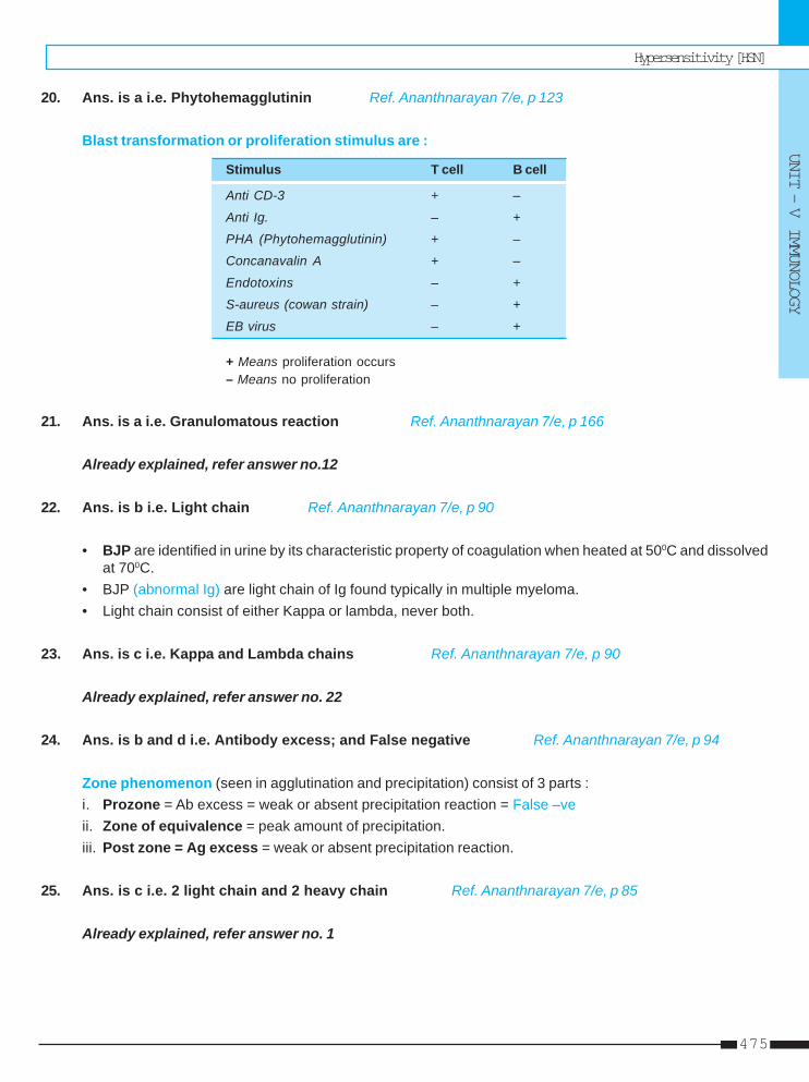
Hypersensitivity [HSN]
UNIT – V IMMUNOLOGY
475
20. Ans. is a i.e. Phytohemagglutinin Ref. Ananthnarayan 7/e, p 123
Blast transformation or proliferation stimulus are :
Stimulus T cell B cell
Anti CD-3 + –
Anti Ig. – +
PHA (Phytohemagglutinin) + –
Concanavalin A + –
Endotoxins – +
S-aureus (cowan strain) – +
EB virus – +
+ Means proliferation occurs– Means no proliferation
21. Ans. is a i.e. Granulomatous reaction Ref. Ananthnarayan 7/e, p 166
Already explained, refer answer no.12
22. Ans. is b i.e. Light chain Ref. Ananthnarayan 7/e, p 90
• BJP are identified in urine by its characteristic property of coagulation when heated at 500C and dissolvedat 700C.
• BJP (abnormal Ig) are light chain of Ig found typically in multiple myeloma.• Light chain consist of either Kappa or lambda, never both.
23. Ans. is c i.e. Kappa and Lambda chains Ref. Ananthnarayan 7/e, p 90
Already explained, refer answer no. 22
24. Ans. is b and d i.e. Antibody excess; and False negative Ref. Ananthnarayan 7/e, p 94
Zone phenomenon (seen in agglutination and precipitation) consist of 3 parts :i. Prozone = Ab excess = weak or absent precipitation reaction = False –veii. Zone of equivalence = peak amount of precipitation.iii. Post zone = Ag excess = weak or absent precipitation reaction.
25. Ans. is c i.e. 2 light chain and 2 heavy chain Ref. Ananthnarayan 7/e, p 85
Already explained, refer answer no. 1
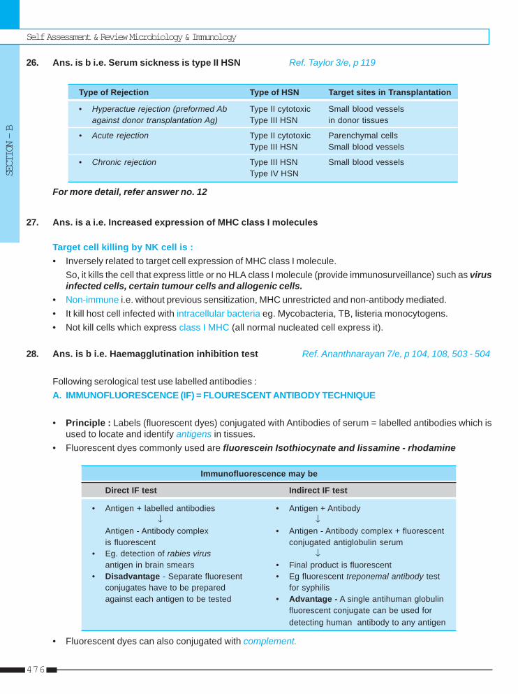
Self Assessment & Review Microbiology & Immunology
SECTION – B
476
26. Ans. is b i.e. Serum sickness is type II HSN Ref. Taylor 3/e, p 119
Type of Rejection Type of HSN Target sites in Transplantation
• Hyperactue rejection (preformed Ab Type II cytotoxic Small blood vesselsagainst donor transplantation Ag) Type III HSN in donor tissues
• Acute rejection Type II cytotoxic Parenchymal cellsType III HSN Small blood vessels
• Chronic rejection Type III HSN Small blood vesselsType IV HSN
For more detail, refer answer no. 12
27. Ans. is a i.e. Increased expression of MHC class I molecules
Target cell killing by NK cell is :• Inversely related to target cell expression of MHC class I molecule.
So, it kills the cell that express little or no HLA class I molecule (provide immunosurveillance) such as virusinfected cells, certain tumour cells and allogenic cells.
• Non-immune i.e. without previous sensitization, MHC unrestricted and non-antibody mediated.• It kill host cell infected with intracellular bacteria eg. Mycobacteria, TB, listeria monocytogens.• Not kill cells which express class I MHC (all normal nucleated cell express it).
28. Ans. is b i.e. Haemagglutination inhibition test Ref. Ananthnarayan 7/e, p 104, 108, 503 - 504
Following serological test use labelled antibodies :A. IMMUNOFLUORESCENCE (IF) = FLOURESCENT ANTIBODY TECHNIQUE
• Principle : Labels (fluorescent dyes) conjugated with Antibodies of serum = labelled antibodies which isused to locate and identify antigens in tissues.
• Fluorescent dyes commonly used are fluorescein Isothiocynate and lissamine - rhodamine
Immunofluorescence may be
Direct IF test Indirect IF test
• Antigen + labelled antibodies • Antigen + Antibody↓ ↓
Antigen - Antibody complex • Antigen - Antibody complex + fluorescentis fluorescent conjugated antiglobulin serum
• Eg. detection of rabies virus ↓antigen in brain smears • Final product is fluorescent
• Disadvantage - Separate fluoresent • Eg fluorescent treponemal antibody testconjugates have to be prepared for syphilisagainst each antigen to be tested • Advantage - A single antihuman globulin
fluorescent conjugate can be used fordetecting human antibody to any antigen
• Fluorescent dyes can also conjugated with complement.
Hypersensitivity [HSN]
UNIT – V IMMUNOLOGY
477
• Labelled complement can be used for detection of antigen or antibody.• Antibody is detected by Sandwich technique.• Major disadvantage of immuno fluorescence is frequent occurence of non specific flurescence in tissues
and other materials.
B. RADIOIMMUNOASSAY (RIA) = BINDER LIGAND ASSAYSPrinciple : Labels (MC radioisotopes and enzymes) conjugated to antigen / antibody = labelled reactantswhich measure antigen and antibodies.• The substance (antigen) whose concentration is to be determined is termed the analyte or ligand.• Binding protein (usually antibody) which bind to ligand is called the binder.• RIA measure analytes upto picogram 10-12g quantities.• RIA has application in quantitation of hormones, drugs, tumour markers, IgE and viral antigens.• Fixed amount of antibody reacts with radiolabelled antigen as well as varying known amount of unlabelled
(test) antigen competitively.• Concentration of test antigen is calculated from standard dose response or calibrating curve.
C. ENZYME IMMUNOASSAYS (EIA)• It is the most widely used procedure in clinical serology.• It uses enzyme labelled antigen and antibodies as serological reagents, for the assay of antibodies and
antigens.• It is of two types :
i. Homogenous EIA (one step test) -• Used only for assay of haptens such as drugs (opiates, cocaine etc)
ii. Heterogenous EIA (multistep test) -• Major type of heterogenous EIA is ELISA which involves the use of immunosorbent specific for
one of the component of reaction, the antigen or antibody.• Immunosorbent may be particulate or solid phase• ELISA is usually done on 96 well microtitre plates
Eg. – Non competitive sand witch ELISA- Detection of rotavirus antigen in feces.- Anti HIV antibody test.
– Competitive ELISA– IgM specific ELISA– Capture ELISA– Immunometric ELISA– Card and dipstick methods– Cylinder or Casette ELISA for the detection of HIV type 1 and 2 antibodies. It is rapid.
D. CHEMILUMINESCENCE IMMUNOASSAY (CLIA)Uses chemiluminescent compounds (such as luminol or acridinium esters) as the label to provide signalduring antigen- antibody reaction.
E. IMMUNOELECTROBLOT TECHNIQUESEg. western blot test.
Self Assessment & Review Microbiology & Immunology
SECTION – B
478
F. IMMUNOCHROMATOGRAPHIC TESTSTest system is a small cassette contaning membrane impregnated with anti HbsAg antibody - colloidalgold dye conjugate Eg. HBsAg detection.
G. IMMUNOFERRITIN TESTAntibody conjugate with ferritin.
H. IMMUNOENZYME TESTStable enzyme like peroxidase conjugated with antibodies.
Remember :Hemagglutination inhibition test - convenient method for detection and quantitation of antibody to the virus.
29. Ans. is d i.e. T cells Ref. Harrison 17/e, p 2021, 2031
Components of the Adaptive Immune System
Cellular Thymus-derived (T) lymphocytes - T cell precursors in the thymus; naive mature T lymphocytesbefore antigen exposure; memory T lymphocytes after antigen contact; helper T lymphocytesfor B and T cell responses; cytotoxic T lymphocytes that kill pathogen- infected target cells.
Humoral Bone-marrow-derived (B) lymphocytes - B cell precursors in bone marrow; naive B cellsprior to antigen recognition; memory B cells after antigen contact; plasma cells that secretespecific antibody.
Cytokines Soluble proteins that direct focus and regulate specific T versus B lymphocyte immuneresponses.
Major Components of the Innate Immune System
• Pattern recognition C type lectins, leucine-rich proteins, scavenger receptors, pentraxins,receptors (PRR) lipid transferases; integrins.
• Antimicrobial α-Defensins, β-defensins, cathelin, protegrin, granulsyin, histatin,peptides secretory leukoprotease inhibitor, and probiotics.
• Cells Macrophages, dendritic cells, NK cells, NK-T cells, neutrophils,eosinophils, mast cells, basophils, and epithelial cells.
• Complement Classic and alternative complement pathway, and proteins that bindcomponents complement components.
• Cytokines Autocrine, paracrine, endocrine cytokines that mediate host defense andinflammation, as well as recruit, direct, and regulate adaptive immuneresponses.
30. Ans. is b i.e. H chain Ref. Ananthnarayan 7/e, p 86
It is the class of heavy chain (H) which determines class of Ig. eg. gamma (γ), alpha (α), mu (μ), delta (δ),epsilon (ε) are H chain of IgG, IgA, IgM, IgD, IgE, respectively.
31. Ans. is a i.e. C2, C4, C3 decreasedRef. Jawetz 23/e, p 136, Harrison 17/e, p 2030; Robbins 7/e, p 66
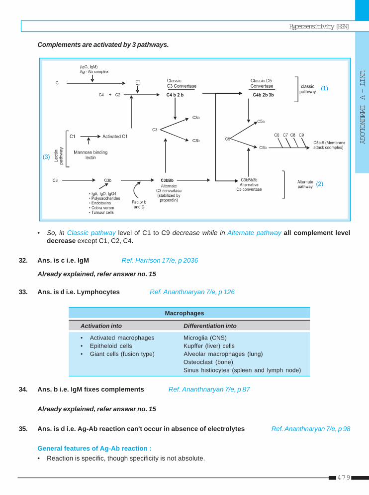
Hypersensitivity [HSN]
UNIT – V IMMUNOLOGY
479
Complements are activated by 3 pathways.
(1)
(3)
• So, in Classic pathway level of C1 to C9 decrease while in Alternate pathway all complement leveldecrease except C1, C2, C4.
32. Ans. is c i.e. IgM Ref. Harrison 17/e, p 2036
Already explained, refer answer no. 15
33. Ans. is d i.e. Lymphocytes Ref. Ananthnaryan 7/e, p 126
Macrophages
Activation into Differentiation into
• Activated macrophages Microglia (CNS)• Epitheloid cells Kupffer (liver) cells• Giant cells (fusion type) Alveolar macrophages (lung)
Osteoclast (bone)Sinus histiocytes (spleen and lymph node)
34. Ans. b i.e. IgM fixes complements Ref. Ananthnaryan 7/e, p 87
Already explained, refer answer no. 15
35. Ans. is d i.e. Ag-Ab reaction can't occur in absence of electrolytes Ref. Ananthnaryan 7/e, p 98
General features of Ag-Ab reaction :• Reaction is specific, though specificity is not absolute.
Self Assessment & Review Microbiology & Immunology
SECTION – B
480
• Entire molecule not fragments combine in the presence of electrolytes at specific pH and temperature.• Combination occur at the surface.• It is firm but reversible.• Ab are generally bivalent while Ag may have valency upto hundred.
Remember these test :
Serological test Type Used to diagnose
i. AscolisThermoprecipitin test Ring precipitation Anthrax
ii. Lancefield test Ring precipitation Grouping of streptococci
iii. Kahn flocculation test Tube precipitation Syphilis
iv. VDRL test Slide precipitation Syphilis
v. Paul Bunnel test Tube agglutination Infectious mononucleosis
vi. Rose waller test Passive hemagglutination Rheumatoid arthritis
vii. Widal test Tube agglutination Typhoid
viii.Weil Felix test Heterophile tube agglutintion Rickettsiae
ix. Wasserman reaction Complement fixation test Syphilis
• Agglutination (eg. indirect HA) is more sensitive than precipitation (eg. gel diffusion test) for antibodydetection.
36. Ans. is a i.e. IgA Ref. Ananthnarayan 7/e, p 87
Already explained, refer answer no. 15
37. Ans. is b i.e. Precipitation Ref. Ananthnarayan 7/e, p 94 - 101
Precipitation Agglutination Complement fixation
• Soluble antigen • Particulate antigen • Ag (particulate or soluble)• Antibody • Antibody • Antibody• Electrolyte • Electrolyte • Source of complement is guinea
• Hemagglutination is Pig serumtype of agglutination
38. Ans. is d i.e. Increased in pneumococcal infection Ref. Ananthnarayan 7/e, p 218
• CRP (beta1 globulin, not antibody) is acute phase reactant (produced in hepatocytes) which is produced inbacterial infections, malignancies, tissue inflammation, rheumatic fever etc (i.e., non specific inflammatoryprotein).
• CRP is detected by passive agglutinatoin using latex particles.
39. Ans. is a and e i.e. Origin from same cell lineage; further differentiation seenRef. Ganong 22/e, p 525

Hypersensitivity [HSN]
UNIT – V IMMUNOLOGY
481
40. Ans. is b i.e. Type - II Ref. Taylor 3/e, p 107
Already eplained, refer answer no. 12
41. Ans. is a, c and d I, III and IV Ref. Taylor 3/e, p 107; Jawetz 24/e, p 142-143
Already eplained, refer answer no. 12
42. Ans. is c i.e. Antigenic variation Ref. Harrison 17/e, p 2035
Learn it
43. Ans. is b and c i.e. Tumor necrosis factor - alpha; and Tumor necrosis factor - beta
“MHC Class genes is classified as Class I, Class II, Class III.”
Class III include : • Soluble protein of complement system • C2 and C4 of classical pathway• Properdin factor B of alternative pathway • Heat shock protein• TNF alpha and Beta.
44. Ans. is d i.e. Schick test Ref. Ananthnarayan 7/e, p 103
Neutralization test
Virus Bacteria
Eg. plaque inhibition test
In vivo In vitroeg. Schick test ASO test
• Bacterial exotoxin can be neutralized (eg. diptheria, tetanus) while endotoxins can't be neutralized byantitoxin.
45. Ans. is b and c i.e. Fibrinogen; and Haptoglobin Ref. Ananthnarayan 7/e, p 75; Meharban Singh, p 216
Acute phase reactants are produced during acute inflammation or certain types of tissues damage. It consistof :i. C-reactive protein (B1 globulin) ⎫⎫⎫⎫⎫ii. α 1 Antitrypsin ⎪⎪⎪⎪⎪iii. Haptoglobin (α2 glycoprotein) ⎬⎬⎬⎬⎬ ↑↑↑↑↑ with inflammationiv. α1 acid glycoprotein (orosomucoid) ⎪⎪⎪⎪⎪v. Fibrinogen ⎭⎭⎭⎭⎭
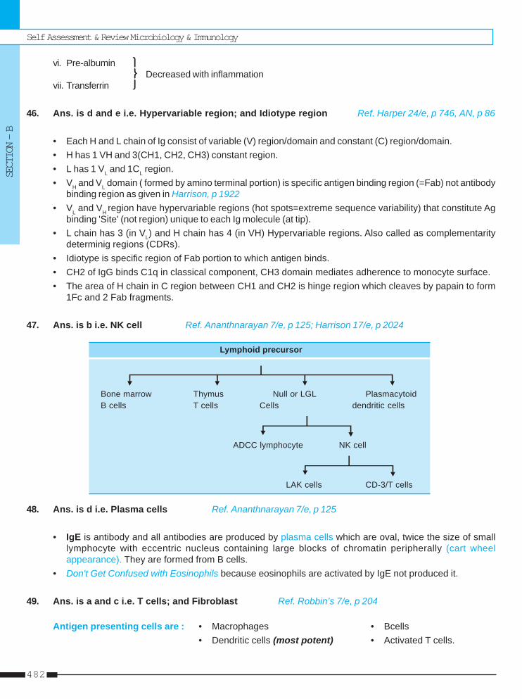
Self Assessment & Review Microbiology & Immunology
SECTION – B
482
vi. Pre-albumin ⎫⎫⎫⎫⎫⎬⎬⎬⎬⎬ Decreased with inflammation
vii. Transferrin ⎭⎭⎭⎭⎭
46. Ans. is d and e i.e. Hypervariable region; and Idiotype region Ref. Harper 24/e, p 746, AN, p 86
• Each H and L chain of Ig consist of variable (V) region/domain and constant (C) region/domain.• H has 1 VH and 3(CH1, CH2, CH3) constant region.• L has 1 VL and 1CL region.• VH and VL domain ( formed by amino terminal portion) is specific antigen binding region (=Fab) not antibody
binding region as given in Harrison, p 1922• VL and VH region have hypervariable regions (hot spots=extreme sequence variability) that constitute Ag
binding 'Site' (not region) unique to each Ig molecule (at tip).• L chain has 3 (in VL) and H chain has 4 (in VH) Hypervariable regions. Also called as complementarity
determinig regions (CDRs).• Idiotype is specific region of Fab portion to which antigen binds.• CH2 of IgG binds C1q in classical component, CH3 domain mediates adherence to monocyte surface.• The area of H chain in C region between CH1 and CH2 is hinge region which cleaves by papain to form
1Fc and 2 Fab fragments.
47. Ans. is b i.e. NK cell Ref. Ananthnarayan 7/e, p 125; Harrison 17/e, p 2024
Lymphoid precursor
Bone marrow Thymus Null or LGL PlasmacytoidB cells T cells Cells dendritic cells
ADCC lymphocyte NK cell
LAK cells CD-3/T cells
48. Ans. is d i.e. Plasma cells Ref. Ananthnarayan 7/e, p 125
• IgE is antibody and all antibodies are produced by plasma cells which are oval, twice the size of smalllymphocyte with eccentric nucleus containing large blocks of chromatin peripherally (cart wheelappearance). They are formed from B cells.
• Don't Get Confused with Eosinophils because eosinophils are activated by IgE not produced it.
49. Ans. is a and c i.e. T cells; and Fibroblast Ref. Robbin’s 7/e, p 204
Antigen presenting cells are : • Macrophages • Bcells• Dendritic cells (most potent) • Activated T cells.
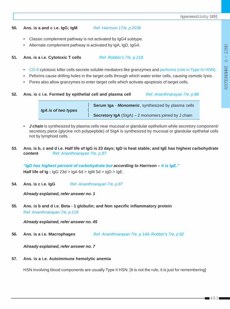
Hypersensitivity [HSN]
UNIT – V IMMUNOLOGY
483
50. Ans. is a and c i.e. IgG; IgM Ref. Harrison 17/e, p 2036
• Classic complement pathway is not activated by IgG4 subtype.• Alternate complement pathway is activated by IgA, IgD, IgG4.
51. Ans. is a i.e. Cytotoxic T cells Ref. Robbin’s 7/e, p 218
• CD-8 cytotoxic killer cells secrete soluble mediators like granzymes and perforins (role in Type IV HSN).• Peforins cause drilling holes in the target cells through which water enter cells, causing osmotic lysis.• Pores also allow granzymes to enter target cells which activate apoptosis of target cells.
52. Ans. is c i.e. Formed by epithelial cell and plasma cell Ref. Ananthnarayan 7/e, p 88
⎧ Serum Iga - Monomeric, synthesized by plasma cellsIgA is of two types ⎨
⎩ Secretory IgA (SIgA) – 2 monomers joined by J chain
• J chain is synthesized by plasma cells near mucosal or glandular epithelium while secretory component/secretory piece (glycine rich polypeptide) of SIgA is synthesized by mucosal or glandular epithelial cellsnot by lymphoid cells.
53. Ans. is b, c and d i.e. Half life of IgG is 23 days; IgD is heat stable; and IgE has highest carbohydratecontent Ref. Ananthnarayan 7/e, p 87
“IgD has highest percent of carbohydrate but according to Harrison – it is IgE.”Half life of Ig : IgG 23d > IgA 6d > IgM 5d > IgD > IgE.
54. Ans. is c i.e. IgG Ref. Ananthnarayan 7/e, p 87
Already explained, refer answer no. 1
55. Ans. is b and d i.e. Beta - 1 globulin; and Non specific inflammatory proteinRef. Ananthnarayan 7/e, p 218
Already explained, refer answer no. 45
56. Ans. is a i.e. Macrophages Ref. Ananthnarayan 7/e, p 144; Robbin’s 7/e, p 82
Already explained, refer answer no. 7
57. Ans. is a i.e. Autoimmune hemolytic anemia
HSN involving blood components are usually Type II HSN. [It is not the rule, it is just for remembering]
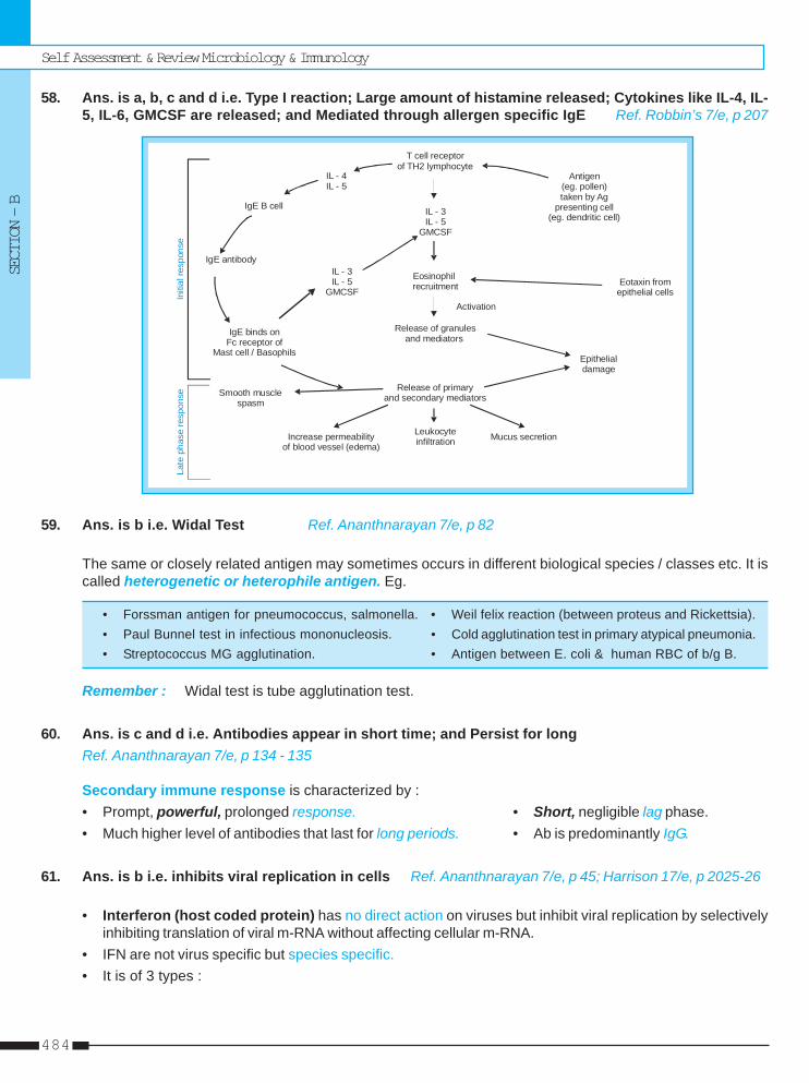
Self Assessment & Review Microbiology & Immunology
SECTION – B
484
58. Ans. is a, b, c and d i.e. Type I reaction; Large amount of histamine released; Cytokines like IL-4, IL-5, IL-6, GMCSF are released; and Mediated through allergen specific IgE Ref. Robbin’s 7/e, p 207
T cell receptorof TH2 lymphocyte
Antigen(eg. pollen)taken by Ag
presenting cell(eg. dendritic cell)
Eotaxin fromepithelial cells
IL - 3IL - 5
GMCSF
Eosinophilrecruitment
Release of granulesand mediators
Activation
Epithelialdamage
Release of primaryand secondary mediators
Leukocyteinfiltration Mucus secretionIncrease permeability
of blood vessel (edema)
Smooth muscle spasm
IgE binds onFc receptor of
Mast cell / Basophils
IgE antibody
IgE B cell
IL - 4IL - 5
IL - 3IL - 5
GMCSFIniti
alre
spon
seLa
teph
ase
resp
onse
59. Ans. is b i.e. Widal Test Ref. Ananthnarayan 7/e, p 82
The same or closely related antigen may sometimes occurs in different biological species / classes etc. It iscalled heterogenetic or heterophile antigen. Eg.
• Forssman antigen for pneumococcus, salmonella. • Weil felix reaction (between proteus and Rickettsia).• Paul Bunnel test in infectious mononucleosis. • Cold agglutination test in primary atypical pneumonia.• Streptococcus MG agglutination. • Antigen between E. coli & human RBC of b/g B.
Remember : Widal test is tube agglutination test.
60. Ans. is c and d i.e. Antibodies appear in short time; and Persist for longRef. Ananthnarayan 7/e, p 134 - 135
Secondary immune response is characterized by :• Prompt, powerful, prolonged response. • Short, negligible lag phase.• Much higher level of antibodies that last for long periods. • Ab is predominantly IgG.
61. Ans. is b i.e. inhibits viral replication in cells Ref. Ananthnarayan 7/e, p 45; Harrison 17/e, p 2025-26
• Interferon (host coded protein) has no direct action on viruses but inhibit viral replication by selectivelyinhibiting translation of viral m-RNA without affecting cellular m-RNA.
• IFN are not virus specific but species specific.• It is of 3 types :
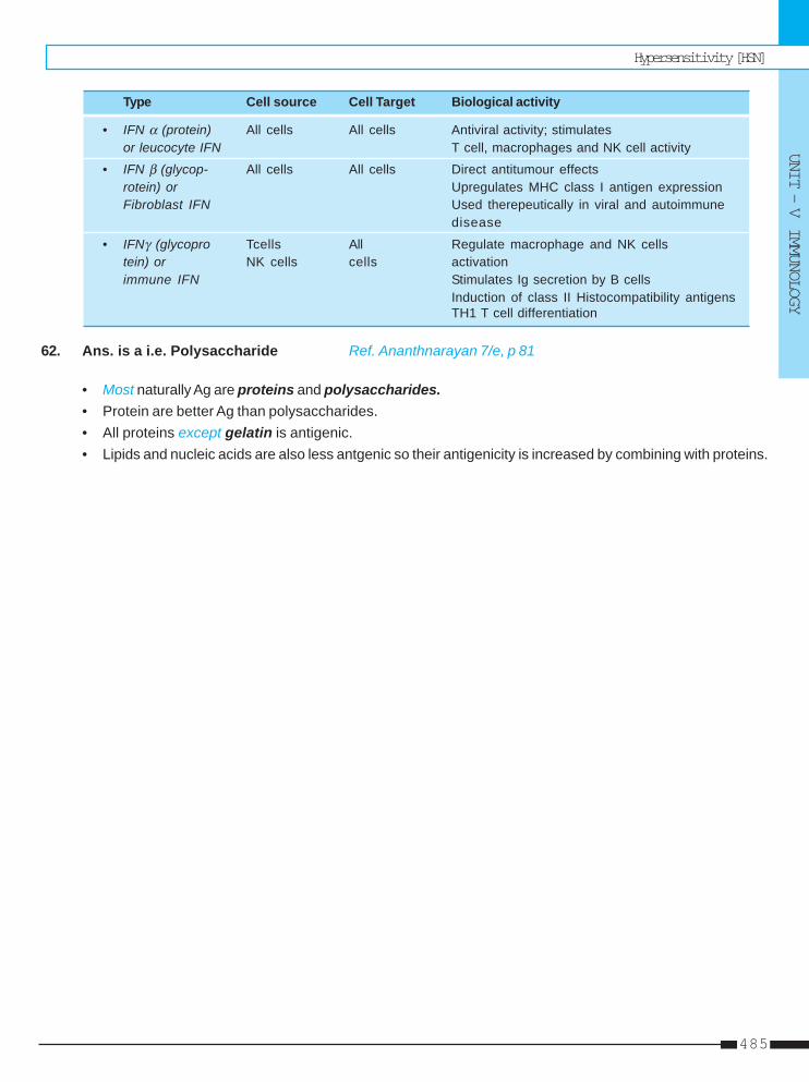
Hypersensitivity [HSN]
UNIT – V IMMUNOLOGY
485
Type Cell source Cell Target Biological activity
• IFN α (protein) All cells All cells Antiviral activity; stimulatesor leucocyte IFN T cell, macrophages and NK cell activity
• IFN β (glycop- All cells All cells Direct antitumour effectsrotein) or Upregulates MHC class I antigen expressionFibroblast IFN Used therepeutically in viral and autoimmune
disease
• IFNγ (glycopro Tcells All Regulate macrophage and NK cellstein) or NK cells cells activationimmune IFN Stimulates Ig secretion by B cells
Induction of class II Histocompatibility antigensTH1 T cell differentiation
62. Ans. is a i.e. Polysaccharide Ref. Ananthnarayan 7/e, p 81
• Most naturally Ag are proteins and polysaccharides.• Protein are better Ag than polysaccharides.• All proteins except gelatin is antigenic.• Lipids and nucleic acids are also less antgenic so their antigenicity is increased by combining with proteins.
Self Assessment & Review Microbiology & Immunology
SECTION – B
486
1. The use of monoclonal antibodies is : [PGI 90]a) Immunotherapyb) Immunological identification of cells and tissuesc) Radioimmunoimagingd) All of the above
[Ref. Ananthnarayan 7/e, p 105 - 106; Harrison17/e, p 2043]
2. Most sensitive test for antigen detection is :a) RIA [PGI 90]b) ELISAc) Immunoflurescenced) Passive hemaglutination
[Ref. Ananthnarayan 7/e, p 105]3. Function of T-lymphocyte is/are :
a) Production of interferon [TN 90]b) Lymphokine productionc) Rosette formationd) All of the above
[Ref. Ananthnarayan 7/e, p 124, 143]4. Binary (Secondary) exposure to an antigen results
in sudden increase in : [TN 90]a) IgAb) IgDc) IgGd) IgM
[Ref. Park 19/e, p 95]5. Reaction of Soluble antigen with antibody is known
is : [AI 90]a) Agglutinationb) Precipitationc) Flocculationd) Complement fixation
[Ref. Ananthnarayan 7/e, p 94]
6. Monoclonal antibodies are associated with nameof : [Orrisa 91]a) Medwarb) Ovenc) Burnerd) Pasteur
[Ref. Ananthnarayan 7/e, p 150]7. NK cells are : [DNB 91]
a) Activated macrophagesb) Antibody-activated T cellsc) Null cells activated by complementd) Derived from plasma cellse) Independent of antibody
[Ref. Ananthnarayan 7/e, p 126]8. T-cell mature in : [Kerala 91]
a) Peyers patchb) Lymph nodec) Thymusd) Bursa of fabricius
[Ref. Ananthnarayan 7/e, p 128]9. The prototype of type -II hypersensitivity reaction
is : [Kerala-91]a) Arthus reactionb) SLEc) Auto immune hemolytic anemiad) Contact dermatitis
[Ref. Robbin’s 7/e, p 210]10. T4/T8 ratio reversal is seen in : [JIPMER 91]
a) T-cell lymphomab) Hairy cell leukemiac) AIDSd) Infectious mononucleosis
[Ref. Ananthnarayan 7/e, p 588]
Chapter Review••••• This section includes questions of VThis section includes questions of VThis section includes questions of VThis section includes questions of VThis section includes questions of Various Other PGMEES from 1990 – 2007.arious Other PGMEES from 1990 – 2007.arious Other PGMEES from 1990 – 2007.arious Other PGMEES from 1990 – 2007.arious Other PGMEES from 1990 – 2007.••••• Questions are arQuestions are arQuestions are arQuestions are arQuestions are ar ranged in decreasing order of yearranged in decreasing order of yearranged in decreasing order of yearranged in decreasing order of yearranged in decreasing order of yearwiz. This is done to makwiz. This is done to makwiz. This is done to makwiz. This is done to makwiz. This is done to make refere refere refere refere refer ralralralralral
system easier and uncomplicated to save the precious time of PGMEE Aspirant.system easier and uncomplicated to save the precious time of PGMEE Aspirant.system easier and uncomplicated to save the precious time of PGMEE Aspirant.system easier and uncomplicated to save the precious time of PGMEE Aspirant.system easier and uncomplicated to save the precious time of PGMEE Aspirant.
1. d) All of the ... 2. a) RIA 3. d) All of the ... 4. d) IgM 5. b) Precipitation6. c) Burner 7. d) Derived ... 8. c) Thymus 9. c) Auto immune ... 10. c) AIDS
Answer
Questions & Answers
UNIT – V IMMUNOLOGY
487
11. d) Platelets 12. d) Casoni’s ... 13. d) Cases of ... 14. c) Migration ... 15. d) Presence ...16. c) Antigenicity 17. d) Lymphocytes 18. c) Serum ... 19. a and c 20. a) T-cells21. d) Mucosal ... 22. d) Plague 23. c) Excess ... 24. a) Virus 25. a) IgG1
Answer
11. Leukotrienes are secreted by all except :a) Macrophages [JIPMER-91]b) T4 cellsc) T8 cellsd) Platelets
[Ref. Ananthnarayan 7/e, p 163]12. An example of type-I hypersensitivity reaction is :
a) Schicks test [AI 92]b) Mantoux testc) Lepromin testd) Casoni’s test [Ref. Paniker 6/e, p 154]
13. HLA typing is useful in : [JIPMER 92]a) Blood gruopingb) Assesing prognosis of diseasec) Cancer therapyd) Cases of paternal dispute
[Ref. Ananthnarayan 7/e, p 132]14. T-cell functions are assesed by : [AIIMS 92]
a) Phagocytic indexb) T-cell countc) Migration inhibition testd) Immunoglobulin index
[Ref. Park 18/e, p 258]15. N.K. cell and cytotoxic cells are differentiated by :
a) Interferons reduce N.K. cell activityb) Antibody specificity [JIPMER 92]c) Receptor for IgGd) Presence in spleen
[Ref. Robbin’s 7/e, p 198-202]16. The main aim of an adjuvant is to increase :
a) Distribution [JIPMER 92]b) Absorptionc) Antigenicityd) Metabolism
[Ref. Ananthnarayan 7/e, p 140]17. Delayed hypersensitivity involves : [AIIMS 92]
a) Neutrophilsb) Monocytsc) Eosinophilsd) Lymphocytes [Ref. Robbin’s 7/e, p 206]
18. Which is an example of type-III hypersenstivity :a) Contact dermatitis [Karn. 92]b) Hemolytic anemiac) Serum sicknessd) Good pasture’s syndrome
[Ref. Robbin’s 7/e, p 211-212]
19. Interferon : [PGI 93]a) Is species specificb) Reacts directly with virus particles to inactivate
themc) Reacts with cells, and the affected cell then be-
come resistant to a number of different virusesd) To constitutively produced at high levels in cells
but requires an inducer for activity[Ref. Ananthnarayan 7/e, p 454, 455]
20. Rossette formation with sheep RBC’s indicate func-tioning of : [Delhi 93]a) T-cellsb) B-cellsc) Neutorphilsd) Monocytes
[Ref. Ananthnarayan 7/e, p 123]21. Secretory piece of IgA is synthesised in :
a) T-cells [PGI 93]b) B-cellsc) Lymph nodesd) Mucosal epithelium
[Ref. Ananthnarayan 7/e, p 88]22. Life long immunity is seen in all of the following
except : [PGI 93, Bihar 93]a) Pertusisb) Mumpsc) Rubellad) Plague
[Ref. Park 19/e, p 245]23. Prozone phenomenon is due to : [AI 93]
a) Disproprotionate antigen-antibody levelsb) Excess antigenc) Excess antibodyd) Hyper immune reaction
[Ref. Ananthnarayan 7/e, p 94]24. N.K. cells provides immunity agianst :
a) Virus [Jipmer 93]b) Bacteriac) Fungusd) Chlamydia
[Ref. Harrison 17/e, p 2028]25. Anti-Rh antibodies belong predominantly to :
a) IgG1 [JIPMER 93]b) IgG2c) IgG3d) IgG4
[Ref. Under consideration]
Self Assessment & Review Microbiology & Immunology
SECTION – B
488
26. d) Phytohemagglutin 27. d) CD4 28. b) T-cells 29. a) Rosette ... 30. d) Type-IV ...31. c) Thrombosis ... 32. a) Diagnosis ... 33. c) Immunoglo ... 34. c) IgG 35. b) CSF36. a) Capsulated ... 37. a) Slide ... 38. b) Opsonisation 39. c) Continous ... 40. d) Light chain ...
Answer
26. T-cell multiplication is stimulated by : [AI 93]a) Macrolinb) Heatc) Bovine serumd) Phytohemagglutin
[Ref. Ananthnarayan 7/e, p 123]27. Type of Receptor present on T-cells are : [AI 93]
a) IgAb) IgGc) Prostaglandinsd) CD4
[Ref. Robbin’s 7/e, p 197]28. Helper Cells belong to : [AI 93]
a) Macrophagesb) T-cellsc) B-cellsd) Monocytes
[Ref. Ananthnarayan 7/e, p 124]29. T-cell are identified by : [Kerala 94]
a) Rosette formation with sheep RBCb) Immunoglobulins on their surfacec) EAC Rossette with sheep erythrocytesd) Have filamentous projections on their surface
[Ref. Ananthnarayan 7/e, p 123]30. Contact dermatitis is : [Kerala 94]
a) Type-I hypersensitivityb) Type-II hypersensitivityc) Type-III hypersensitivityd) Type-IV hypersensitivity
[Ref. Robbin’s 7/e, p 217]31. Humoral graft rejection is characterised by :
a) Intense mononuclear infiltration [Kerala 94]b) Neovascularisation of grafted tissuec) Thrombosis of the blood vessels and ischemic
necrosisd) Monocytic infiltration
[Ref. Robbin’s 7/e, p 220]32. Insitu hypridisation is used for : [Kerla 94]
a) Diagnosis of specific genetic diseaseb) Diagnosis of enzyme dificiency diseasec) Treatment of genetic diseasesd) All of the above
[Ref. Harrison 17/e, p 408]33. Active immunity is not acquired by : [Kerla 94]
a) Infectionb) Vaccinationc) Immunoglobulin transfer
d) Sub-clinical infection[Ref. Ananthnarayan 7/e, p 77]
34. Opsonisation is by : [Kerala 94]a) IgAb) IgEc) IgGd) IgM
[Ref. Ananthnarayan 7/e, p 89]35. Lysozyme is present in the following secretions of
the body except : [Karnat 94]a) Lacrimal secretionsb) CSFc) Salivad) Respiratory tract secretions
[Ref. Ananthnarayan 7/e, p 73]36. Most common infections after splenectomy are :
a) Capsulated bacteria [JIPMER 95]b) Uncapsulated bacteriac) Gram positive sepsisd) Gram Negative bacteria
[Ref. Harrison 17/e, p 375]37. VDRL is a : [TN 95]
a) Slide fiocculation testb) Tube flocculation testc) Gel precipitationd) Indirect haemaglutination test
[Ref. Ananthnarayan 7/e, p 95]38. The process increasing the ability for phagocyto-
sis of foreign bodies by body is called :a) Cross reactivity [JIPMER 95]b) Opsonisationc) Immune Toleranced) Immune Surveiliance
[Ref. Robbin’s 7/e, p 59]39. Hep-2 cells are a type of : [Karn 95]
a) Primary cell cultureb) Diploid cell linesc) Continous cell linesd) Explant culture
[Ref. Ananthnarayan 7/e, p 441]40. M band is absent in : [PGI 95]
a) IgG myelomab) IgM myelomac) IgA myelomad) Light chain disease
[Ref. Ananthnarayan 7/e, p 90]
Questions & Answers
UNIT – V IMMUNOLOGY
489
41. a) IgG 42. a) Interferons 43. b) Chemotaxis 44. b and c 45. c) IgE46. b) IgG 2 47. b) Cytotoxic ... 48. b) Fc protion ... 49. c) Silicosis 50. d) Guinea pig51. b) C3b 52. a) IL1 53. c) Weil Felix 54. a) Tears and ...
Answer
41. Secondary response is mediated by : [UP 95]a) IgGb) IgMc) IgEd) IgA
[Ref. Park 19/e, p 95]42. Virus infected cell is killed by : [Kerala 96]
a) Interferonsb) Macrophagesc) Neutrophilsd) Autolysise) None of the above
[Ref. Ananthnarayan 7/e, p 454, 455]43. Phagocytosis enhanced by coating the surface of
antigen is called : [Kerala 96]a) Opsonisationb) Chemotaxisc) De codingd) CFT
[Ref. Ananthnarayan 7/e, p 59]44. The following are true for T lymphocytes except :
a) Consitute 70 to 80% of circulating pool of lym-phocytes [Karnat 96]
b) Release macrophage inhibition factorc) Secrete specific antibodiesd) Release lymphotoxin
[Ref. Ananthnarayan 7/e, p 119]45. The reagnic antibody is : [Karnat 96]
a) IgAb) IgDc) IgEd) IgG
[Ref. Ananthnarayan 7/e, p 90]46. The commonest IgG with maximum individual varia-
tion is : [PGI 96]a) IgG 1b) IgG 2c) IgG3d) IgG 4
[Ref. NMS Immunology]47. Blood tranfusion mismatch in erythroblastosis
foetalis is a type of : [NIMS 96]a) Atopic hypersensitivityb) Cytotoxic typec) Arthus phenomenond) Delaled hypersensistivity
[Ref. Robbins’s 7/e, p 210]
48. Antigen recoginition on the surface of antigen pro-cessing cells is by : [Delhi 96, PGI 90]a) T- cell recognition antigenb) Fc protion of immunoglobulinc) Fab protion of immunoglobulind) Cell recognition antigen
[Ref. Robbin’s 7/e, p 200]49. Immune granulomas are not seen in :
a) Tuberculosis [Kerala 97]b) Syphilisc) Silicosisd) Berryliosis
[Ref. Robbin’s 7/e, p 82 - 83]50. Animal used to demonstrate anaphylaxis in the lab
is : [JIPMER 98]a) Rabbitb) Adult micec) Monkeyd) Guinea pig
[Ref. Ananthnarayan 7/e, p 161]51. Opsonins are : [MP 98]
a) C3ab) C3bc) C5ad) C5-C9 complex
[Ref. Robbin’s 7/e, p 59]52. Which of the following cytokinin is secreted by T
helper cells except : [UP 98]a) IL1
b) IL2
c) IL4
d) IL5
[Ref. Ananthnarayan 7/e, p 144]53. Agglutination test is : [UP 98]
a) ABO incompatibilityb) VDRLc) Weil Felixd) FTA-AB
[Ref. Ananthnarayan 7/e, p 99]54. IgA secretion is seen in : [UP 98]
a) Tears and salivab) CSFc) Hairsd) Vaginal fluid
[Ref. Ananthnarayan 7/e, p 88]
Self Assessment & Review Microbiology & Immunology
SECTION – B
490
55. d) Graft rejection 56. b) IgM type 57. b) Paratope 58. a) TNF 59. a) IgM60. e) Stromal cells 61. a) CD-19 62. d) NK contain ... 63. b) Epitope 64. b) NK cells65. a) B4 66. b) Spectrophoto ... 67. d) IgD 68. b) T lymphocytes
Answer
55. All of the following are immune complex diseaseexcept : [GUPGEE 99]a) Serum sicknessb) Farmer’s lungsc) SLEd) Graft rejection
[Ref. Robbin’s 7/e, p 211, 212]56. When an antigen is administered for the first time
to animal or a human being who has never beenexposed to it, the first antibody to develop :a) IgG type [DNB 99]b) IgM typec) IgA typed) IgE type
[Ref. Park 19/e, p 95]57. Antigen combining site of the antibody is :
a) Idiotype [TN 99]b) Paratopec) Epitoped) Hapten
[Ref. Ananthnarayan 7/e, p 81]58. Most important inflammatory mediator : [UP 99]
a) TNFb) IL2c) Interferond) PAF
[Ref. Robbin’s 7/e, p 71]59. Pentameric structure : [JIPMER 99]
a) IgMb) IgGc) IgAd) IgD
[Ref. Ananthnarayan 7/e, p 89]60. Interleukin - 7 produced by which one of the fol-
lowing : [Kerla 00]a) Macrophagesb) B cellsc) T cellsd) Dendritic cellse) Stromal cells
[Ref. Ananthnarayan 7/e, 144]61. Group B cell lymphocyte belongs to : [UP 00]
a) CD-19b) CD-69c) CD-59d) CD-68
[Ref. Ananthnarayan 7/e, p 124]
62. Defference between natural killer and cytotoxiccells is : [UP 00]a) Interferons decreased natural killer actvityb) NK has CD4c) Cytotoxic cell lyse IgG coated target cellsd) NK contain azurophilic cytoplasmic granulocyte
[Ref. Ananthnarayan 7/e, p 126]63. The exact part of the antigen that reacts with the
immune system is called as : [Karnat 01]a) Cloneb) Epitopec) Idiotoped) Effector
[Ref. Ananthnarayan 7/e, p 82]64. Which of the following cells is known as large
granular lymphocyte (LGL) : [Karnat 01]a) Plasma cellsb) NK cellsc) T cellsd) K cells
[Ref. Ananthnarayan 7/e, p 126]65. Which leukotriene is the adhesion factor for the
neutorphill on the cell surface to attach to endot-helium : [Kerala 01]a) B4b) C4c) D4d) E4
[Ref. Robbin’s 7/e, p 69 Table (2.4)]66. Rapid serological diagnostic test include all ex-
cept : [UP 01]a) Latex agglutinationb) Spectrophotometryc) Gel electrophoresisd) Radiommunpassay
[Ref. Under consideration]67. Immunoglobulin found in B lymphocyte : [UP 01]
a) IgAb) IgEc) IgGd) IgD
[Ref. Ananthnarayan 7/e, p 124]68. Delayed tuberculin test response is due to :
a) B lymphocytes [TN 01]b) T lymphocytesc) Monocytesd) Histocytes
[Ref. Robbin’s 7/e, p 217]
Questions & Answers
UNIT – V IMMUNOLOGY
491
69. b) N-terminal 70. a and c 71. b) H chain 72. a) Acts as a ... 73. c) Deficient T ...74. a) IgG 75. d) NK cells 76. a) IgG 77. b) Complement 78. d) 2 long and ...79. a) T cells ... 80. c) Hypothyroi... 81. a) Helper T ... 82. b) Type II
Answer
69. Variable protion of antibody molecule is :a) C-terminal [UP 01]b) N-terminalc) Intermediate regiond) Carbohydrate moiety
[Ref. Ananthnarayan 7/e, p 86]70. Phagocytic cells elaborate : [JIPMER 01]
a) Prostaglandinsb) Thromboxanec) Leukotrienesd) All of the above
[Ref. Robbin’s 7/e, p 68]71. Portion of immunoglobulin molecule with molecu-
lar weight of 50,000 : [JIPMER 01]a) Secretory Pieceb) H chainc) L chaind) J peice
[Ref. Ananthnarayan 7/e, p 85]72. Function of IgA is : [Kerla 01]
a) Acts as a mucosa barrier for infectionb) Circulating antibodyc) Kills virus infected cellsd) Activates macrophagese) Cause delayed hyper sensitivity reaction
[Ref. Ananthnarayan 7/e, p 88, 89]73. Runt disease is : [TN 02]
a) Graft rejectionb) Graft versus host treactionc) Deficient T cell functiond) Complement dificiency
[Ref. Ananthnarayan 7/e, p 118]74. The following constitutes approximately 75% of
total immunoglobin in hymans : [Karnat 02]a) IgGb) IgMc) IgEd) IgA
[Ref. Ananthnarayan 7/e, p 87]75. Virus infected cells are killed by : [MP 02]
a) Macrophagesb) Complement systemc) MHC II related cellsd) NK cells
[Ref. Ananthnarayan 7/e, p 126]
76. The immunoglobin that can cross the placenta :a) IgG [SGPGI 03]b) IgAc) IgEd) IgM
[Ref. Ananthnarayan 7/e, p 88]77. HLA III gene codes in graft rejection : [MP 03]
a) Immunological raection in graft rejectionb) Complementc) Graft virsus host reactiond) Immunoglobulins
[Ref. Ananthnarayan 7/e, p 129]78. Structure of antibodies is composed of : [UP 03]
a) Single peptide chainb) Two peptide chainc) Non sulphur amino acidd) 2 long and 2 short peptide chain
[Ref. Ananthnarayan 7/e, p 85]79. IL-2 is produced by : [SGPGI 04]
a) T cells (CD4 cells)b) B cellsc) Monocytesd) Neutrophils
[Ref. Ananthnarayan 7/e, p 144]80. Dieorge syndrome is characterized by all except :
a) Congenital thymic hypoplasia [SGPGI 04]b) Abnormal developmental of third and fourth
pouchesc) Hypothyroidismd) Hypocalcemic tetany
[Ref. Ananthnarayan 7/e, p 155]81. CD4 are associated with : [UP 04]
a) Helper T cellb) Sypressor T cellsc) NK cellsd) T cells antigen receptor complex
[Ref. Ananthnarayan 7/e, p 129]82. All the following types of hypersensitivity reactions
can be demonstrated by skin test except :a) Type I [UP 04]b) Type IIc) Type IIId) Type IV
[Ref. Robbin’s 7/e, p 206 Table (6.2)]
Self Assessment & Review Microbiology & Immunology
SECTION – B
492
Answer
83. Isoantigens are : [UP 05]a) Found in some but all members of speciesb) Found in some but not all members of speciesc) Occurs in different biological species, class and
kingdomsd) All individuals in a particular species
[Ref. Ananthnarayan 7/e, p 82]84. Paul Bunnal antibodies are reactive in all except :
a) OX [SGPGI 05]b) Sheepc) Dogd) Horse
[Ref. Ananthnarayan 7/e, p 484]85. Antibodies are most responsive to : [SGPGI 05]
a) Recipients tissueb) Donor tissuec) Isograftsd) Allograft
[Ref. Ananthnarayan 7/e, p 84]86. First immunoglobin to appear following infections:
a) IgG [SGPGI 05]b) IgMc) IgAd) IgE[Ref. Park 19/e, p 95]
87. Phagocytic function is assessed by : [MP 06]a) Proliferative response to mitogenb) Reduction of NBT (Nitroblue tetrazolium test)c) Serum Immunoglobulin assayd) Skin test with purified protein derivative
[Ref. Harrison 17/e, p 381]88. The exact part of the antigen that reacts with im-
mune system is called as : [Kar 01]a) Cloneb) Epitopec) Idiotyped. Effector
[Ref. Ananthnaranay 7/e, p 80]89. Which of the following cells is knows as large
grnaular lymphocyte ? [Kar 01]a) Plasma cellsb) NK cellsc) K cellsd) T cells
[Ref. Ananthnarayan 7/e, p 126]
90. Which of the following is a flocculation test :a) Widal test [Kar 01]b) Weil-felix testc) VDRLd) Paul-Bunnell test
[Ref. Ananthnarayan 7/e, p 381]91. Transplantation of the hosts own tissue is known
as : [Kar 01]a) Isograftb) Allograftc) Xenograftd) Autograft [Ref. Learn it]
92. Pro-inflammatory cytokines include all except :a) Interlukin 1 [Kar 01]b) Interleukin 2c) Interleutkin 6d) TNF-alfa [Ref. Harrison 17/e, p 1156]
93. Delayed hypersensitivity reaction is mediated b thefollowing : [Kar 02]a) B lymphocytesb) NK cellsc) Mast cellsd) T lymphocytes
[Ref. Ananthnarayan 7/e, p 166]94. The following constitutes approximately 75% of to-
tal immunogobulin in humans : [Kar 02]a) IgGb) IgMc) IgEd) IgA
[Ref. Ananthnarayan 7/e, p 87]\95. Which immunoglobulin is least important in human
beings: [Kolkata 03]a) IgEb) IgDc) IgGd) IgA
[Ref. Ananthnarayan 7/e, p 90]96. Chemoattractant is : [Jharkhand 03]
a) C5ab) C1c) C3d) C2 [Ref. Robbin’s 7/e, p 56]
97. Complement C5-C9 predispose to which infectiona) Menigococi [Jharkhand 03]b) Pneumococcalc) Pseudomonasd) All
[Ref. Harrison 17/e, p 911]
83. b) Found in... 84. a) OX 85. b) Donor 86. b) IgM 87. b) Reduction ...88. b) Epitope 89. b) NK cells 90. c) VDRL 91. d) Autograft 92. b) Interleukin 293. d) T lymphocytes 94. a) IgG 95. b) IgD 96. a) C5a 97. a) Meningococci
Questions & Answers
UNIT – V IMMUNOLOGY
493
98. Opsonization occurs due to : [Bihar 04]a) Endotoxinb) Complementc) IgMd) IgG
[Ref. Aanthnarayan 7/e, p 104]99. Primary immune response is mediated by :
a) IgE [Jharkhand 04]b) IgMc) IgAd) IgD
[Ref. Park 19/e, p 95]100. Which is not pyrogenic IL : [Kolkata 05]
a) IL – 1b) TNF-c) IL – 4d) IL – 6
[Ref. Harrison 17/e, p 119]101. First antibody response is mediated by :
a) IgD [Jharkhand 05]b) TgMc) IgAd) IgE
[Ref. Park 19/e, p 95]102. Tuberculin test is reaction of : [UP 06]
a) Anaphyxis mediatedb) Cell mediated
Answer 98. b) Complement 99. b) IgM 100. a, b and c 101. b) TgM 102. c) Antibody ...103. None 104. a) IgE 105. a) Hemorrhagic ... 106. b) B cell
c) Antibody mediatedd) Immuno complex mediated
[Ref. Ananthnarayan 7/e, p 166]103. Antibody transfer mother to fetus is :
a) IgG [Bihar 06]b) IgMc) IgDd) IgA
[Ref. Ananthnarayan 7/e, p 87]104. Atopy is mediated by : [Jharkhand 06]
a) IgEb) IgDc) IgMd) IgA
[Ref. Anantharayan 7/e, p 164]105. All are type-II hypersensitivity reaction except :
a) Hemorrhagic disease of newbornb) Grave’s disease [Jharkhand 06]c) Autoimmune diseasesd) Hemolytic anemia
[Ref. Anantharayan 7/e, p 161]106. Humoral antibody arises from :
a) T cell [Jharkhand 06]b) B cellc) Null celld) K cell
[Ref. Anantharayan 7/e, p 133]

Self Assessment & Review Microbiology & Immunology
SECTION – B
494
NOTES

Self Assessment & Review Microbiology & Immunologyxii
MISCELLANEOUS

1. Which of the following is not transmitted by soil ?a) Coccidiodomycosis [AI 08]b) Tetanusc) Brucellad) Anthrax
2. Isolation is not useful for all except : [AI 08]a) Mumpsb) Measlesc) Hepatitis Ad) Pneumonic plague
3. Congenital infection affecting fetus with minimalteratogenic risk is : [AI 08]a) HIVb) Rubellac) Varicellad) CMV
4. Which of the following congenital infection leadsto maximum CNS damage in the fetus : [AI 08]a) Rubella and CMVb) Rubella and toxoplasmosisc) CMV and toxoplasmosisd) HIV and CMV
5. With reference to Bacteroides fragilis the follow-ing statements are true except :a) B. fragilis is the most frequent anaerobe iso-
lated from clinical samples [AI 07; AIIMS 06]b) B. fragilis is not uniformly sensitive to metron-
idazole
c) The lipopolysaccharide formed by B.fragilis isstructurally and functionally different from theconventional endotoxin
d) Shock and disseminated intravascular coagu-lation are common in Bacteroides bacteremia
6. Which of the following is least likely to cause in-fective endocarditis : [AI 06]a) Staphylococcus albusb) Streptococcus faecalisc) Salmonella typhid) Pseudomonas aeruginosa
7. A 40 years old woman presented to thegyencologist with complaint of profuse vaginaldischarge. There was no discharge from the cer-vical os on the the speculum examination. Thediagnosis of bacterial vaginosis was made basedupon all of the following findings on microscopyexcept : [AI 06]a) Abundance of gram variable coccobacillib) Absence of Lactobacillic) Abundance of polymorphsd) Presence of clue cells
8. Apart from Escherichia coli, the other most com-mon organism implicated in acute suppurativebacterial peritonitis is : [AI 06]a) Bacteroidesb) Klebsiellac) Peptostreptococcusd) Pseudomonas
MiscellaneousMiscellaneousMiscellaneousMiscellaneousMiscellaneous
QUESTIONS
Answer 1. c) Brucella 2. d) Pneumonic .. 3. a) HIV 4. c) CMV and.... 5. d) Shock and ...6. c) Salmonella ... 7. c) Abundance ... 8. b) Klebsiella
Self Assessment & Review Microbiology & ImmunologySECTION – B
498
9. Virus mediated transfer of host DNA from one cellto another is known is : [AI 05]a) Transductionb) Transformationc) Transcriptiond) Integration
10. All of the following organisms are known to sur-vive intracellularly except : [AI 05]a) Neisseria meningitidesb) Salmonella typhic) Streptococcus pyogenesd) Legionella pneumophilia
11. Virus can be isolated from clinical samples bycultivation in the following except : [AI 05]a) Tissue cultureb) Embryonated eggsc) Animalsd) Chemically defined media
12. It is true regarding the normal microbial florapresent on the skin and mucous membranes that:a) It cannot be eradicated by by antimicrobial
agentsb) It is absent in the stomach due to acidic pHc) It establishes in the body only after the neonatal
period [AI 05]d) The flora in the small bronchi is similar to that of
the trachea13. The most common pathogens responsible for
nosocomial pneumonia in the ICU : [AI 05]a) Gram positive organismb) Gram -ve organismc) Mycoplasmad) Virus infections
14. Which of the following toxins acts by inhibitingprotein synthesis : [AI 04]a) Cholera toxinb) Shiga toxinc) Pertusis toxind) LT of Enterotoxigenic E. coli
15. Toxins are implicated the major pathogenicmechanism in all of the following bacterial diar-rheas except : [AI 04]a) Vibrio choleraeb) Shigella sp.c) Vibrio parahaemolyticusd) Staphylococcus aureus
16. All of the following infections are often associ-
ated with acute intravascular hemolysis except :a) Clostridium tetani [AI 03; AIIMS 03]b) Bartonella bacilliformisc) Plasmodium falciprumd) Babesia microti
17. Bacteria may acquire characteristics by all of thefollowing except : [AI 02]a) Taking up soluble DNA fragments across their
cell wall from other speciesb) Incorporating part of host DNAc) Through bacteriophagesd) Through conjugation
18. Heat stable enterotoxin causing food poisoningis caused by all the following except : [AI 02]a) Bacillus cereusb) Yersinia enterocoliticac) Staphylococcusd) Clostridium perfringens
19. All of the following are sexually transmitted ex-cept : [AI 02]a) Candida albicansb) Echinococcusc) Molluscum contagiosumd) Group B streptococcus
20. All of the following infections may be transmittedvia blood transfusion except : [AI 02]a) Parvo B-19b) Dengue virusc) CMVd) Hepatitis G virus
21. The following diagnostic tests are useful for cor-responding purpose except : [AI 02]a) Zeihl Nelson staining - Detection of mycobacte-
riab) Immunoflorescence - Detection of Influenza vi-
rusc) Specific IgM antibodies - Immunity against Ru-
bellad) Specific IgM antibodies - Detection of acute in-
fection22. Cystine lactose enzyme deficient medium CLED
is preferred over McConkey agar in UTI because:a) Former prevents swarming of proteus [AI 01]b) Is a selective mediumc) Promotes growth of pseudomonasd) Promotes growth of Candida and Staphy
lococcus
Answer 9. a) Transduction 10. c) Streptococcus ... 11. d) Chemically ... 12. c) It establishes ... 13. b) Gram -...14. b) Shiga ... 15. c) Vibrio ... 16. a) Clostridium ... 17. b) Incorporating ... 18. d) Clostridium ...19. b) Echinococcus 20. b) Dengue virus 21. c) Specific ... 22. d) Promotes ...
Questions & AnswersMISCELLANEOUS
499
23. All of the following bacteria test “Urease posi-tive” except :[AI 98]a) E. colib) Proteusc) Kleibsellad) Staphylococcus
24. Plaque formation in virus is done for : [AI 98]a) Isolation and typing of viursesb) Cloning separation of specific virusesc) Determining infectivity of virusd) Assessing multiplication of virus
25. Bacterial genome has been completelyrecognised for which one of the following agents:a) H. pylori [AI 98]b) Yersinia enterocolitisc) Campylobacter jejunid) Streptococcus
26. Most common agents responsible, for human, biteinfections are : [AI 98]a) Gram -ve bacillib) Gram +ve bacillic) Spirochaeted) Anaerobic streptococci
27. Which bacteria acts by inhibiting protein synthe-sis : [AI 98]a) Pseudomonasb) Staphylococcusc) Streptococcusd) Kleibsiella
28. Prokaryotes are characterised by : [AI 97]a) Absence of nuclear membraneb) Presence of microvilli on its surfacec) Presence of smooth endoplasmic reticulumd) All of the above
29. A substance, when added to a culture causes in-hibition of multiplication but on removal causesenhanced growth. This substance is called :a) Bacteriostatic [AI 96]b) Bactericidalc) Sterilizationd) Bacteriophage
30. Reactive arthritis is caused by : [AIIMS 08]a) Staphylococcusb) H. influenzaec) N. gonorrhoed) Chlamydia trachomatus
31. HACEK group includes all of the following except:a) Hemophilus arophilus [AIIMS 08]b) Acinetobacter baumannic) Eikenella corrodensd) Cardiobacterium hominis
32. Cy Bromide green dye is used for : [AIIMS 06]a) HLPRb) PCRc) ELISAd) Immunofluroscence
33. The single most common cause of pyrexia of un-known origin is : [AIIMS 06, 03]a) Mycobacterium tuberculosisb) Salmonella typhic) Brucella sp.d) Salmonella paratyphi A
34. In the gut, anaerobic bacteria outnumber the aer-obes by a ratio of : [AIIMS 06]a) 10 : 1b) 100 : 1c) 1000 : 1d) 10,000 : 1
35. In all of the following diseases chronic carriersare found except : [AIIMS 06]a) Measlesb) Typhoidc) Hepatitis Bd) Gonorrhoea
36. A 30 year old male present with urethritis. All ofthe following can be causative agent except :a) N. gonorrhoeae [AIIMS 04]b) Chalmydia trachomatisc) Trichomonas vaginalisd) Hemophilus ducreyi
37. Humoral immunodeficiency is suspected in pa-tient and he is under investigation. Which of thefollowing infections would be consistent with thediagnosis : [AIIMS 04]a) Giardiasisb) Pneumocystics carinii pneumoniac) Recurrent sinusitisd) Recurrent subcutaneous abscess
Answer 23. a) E. coli 24. c) Determining ... 25. a) H. pylori 26. d) Anaerobic ... 27. a) Pseudomonas28. a) Absence ... 29. a) Bacteriostatic 30. d) Chlamydia ... 31. b) Aconetobacter .32. b) PCR33. a) Mycobacteri ... 34. c) 1000 : 1 35. a) Mealses 36. d) Hemophilus ... 37. b) Pneumocyst ...

Self Assessment & Review Microbiology & ImmunologySECTION – B
500
38. All the following are most common nosocomialinfection except : [AIIMS 03]a) Staph. aureusb) P. aeroginosac) Enterobacteriaceaed) Mycobacterium
39. The following phenomenon is responsible for an-tibiotic resistance in bacteria due to slime pro-duction : [AIIMS 03]a) Co-aggregationb) Biofilm formationc) Mutation involving an altered target site for anti-
bioticsd) Mutation involving a target bypass mechanism
40. With reference to Bacterioides fragilis all of thefollowing statement are true except : [AIIMS 03]a) It is the most frequent anaerobe isolated from
clinical sampleb) It is not uniformly sensitive to metronidazolec) The lipopolysaccharide formed by B. fragilis is
structurally and functionally different from con-ventional endotoxin
d) Shock and DIC are common in bacteremia dueto B. fragilis
41. A patient present with frontal abscess. Foul smell-ing. pus is aspirated. Pus shows red. Fluo-renscence on ultraviolet examination. The most,likely organism causing the frontal abscess is :a) Bacteriodes [AIIMS 02]b) Peptostreptococcusc) Pseudomonasd) Acanthamoeba
42. In a patient with UTI, CLED (Cysteine lactose elec-trolyte deficient) Media is preferred overMacConkey’s media because : [AIIMS 01]a) It is a differential mediumb) It inhibits swarming of proteusc) Promotes growth of pseudomonasd) Promotes growth of S. aureus and Candida
43. Preformed toxin is important in food poisoningdue to all except : [AIIMS 01]a) S. aureusb) Clostridium botulismc) ETECd) B. cereus
44. Endotoxin from gram negative organism is :a) Polysaccharide [AIIMS 00]b) Glycoproteinc) Lipoproteind) Lipo-polysaccharide
45. Most common tumour caused by virus is :a) Warts [AIIMS 97]b) Carcinoma cervixc) Nasopharyngeal carcinomad) Lymphoma
46. Which is not an oncogenic virus : [AIIMS 97]a) HTLV-1b) Herpessimplexc) Papilloma virusd) HBV
47. Viral plaque is made in lab for : [AIIMS 96]a) Quantitative assay of infectivity of virusb) Diagnosis of virusc) Qualitative assay of infectivity of virusd) Type of virus
48. Exotoxins are : [AIIMS 95]a) Lipopolysaccharide in natureb) Produced by gram -ve bacillic) Highly antigenicd) Very stable and resistant to chemical agents
49. The term “viable not cultivable” (VNC) is used for:a) M. leprae [PGI Dec. 07]b) M. Tuberculosisc) Trepenoma pallidumd) Salmonellae) Staph.
50. Micro-organisms invading the GIT causing gas-troenteritis : [PGI Dec. 07]a) EHECb) Shigellac) Vibrio parahaemolyticusd) Campylobactere) Salmonella
51. Which of the following can cause rhabdomyo-ly-sis? [PGI June 07]a) Clostridium Perfringesb) Streptococcusc) Clostridium difficled) Cl. tetani
Answer 38. d) Mycobacterium 39. b) Biofilm ... 40. d) Shock ... 41. a) Bacteriodes 42. d) Promotes ...43. c) ETEC 44. d) Lipo-... 45. a) Warts 46. b) Herpessimplex 47. a) Quantitative ...48. c) Highly ... 49. a and c 50. b, c, d and e 51. a, b and d
Questions & AnswersMISCELLANEOUS
501
Answer 52. a) Donovanosis 53. a) Fungus 54. a) Leptospira 55. a, b and c 56. a) 200 nm57. d and e 58. a, b and d 59. b) EBV 60. d) P. falciparum 61. b and d62. a, b and c 63. b, c, d and e 64. a, b and c 65. a, b and c 66. a, b, c, d and e
52. Genital elephantiasis is seen in : [AIA[PGI 06]a) Donovanosisb) Lymphogranuloma venerumc) Congenital syphilisd) Herpes simplex
53. Rhinosporidiosis is caused by : [PGI 06]a) Fungusb) Bucteriac) Virusd) Protozoane) Parasite
54. Which human infection spreads through urine :a) Leptospira [PGI 06]b) Legionellac) Plagued) Diphtheria
55. Urease test is positive in : [PGI 05]a) H.pylorib) S.aureusc) Klebsiellad) Bacillus cereuse) Pseudomonas
56. Resolution provided by light microscope is :a) 200 nm [PGI 05]b) 20 nmc) 0.2 nmd) 2.0 nme) 120 nm
57. Pus cell in diarrhoea seen in : [PGI 05]a) Vibrio cholerab) EPECc) Rotavirusd) Shigellae) Campylobacter
58. C.M.I is seen in : [PGI 05]a) Histoplasmosisb) Leprosyc) Tetanusd) Measles
59. Which of the following has malignant potential :a) HSV - 1 [PGI 05]b) EBVc) CMVd) Varicella
60. Man is intermediate host in : [PGI 05]a) Taenia saginatab) Trichenella spiralisc) Strongyloidisd) P. falciparum
61. Capsulated organism : [PGI 03]a) Candidab) Klebsiellac) Proteusd) Cryptococcuse) Histoplasma
62. Which of the following are transfusion transmit-ted viruses : [PGI 03]a) Hepatitis Bb) CMVc) HTLV - 1d) Rubellae) HHV - 8
63. Enteropathogenic organisms are : [PGI 02]a) Cryptococcusb) B. Colic) Microsporidium speciesd) E. dispare) Giardia intestinalis
64. Which of the following is a bacteria taxonomically:a) Chlamydia [PGI 01]b) Rickettsiac) Mycoplasmad) Prione) Bacteriophage
65. Which of the following is transmitted by blood:a) Toxoplasma [PGI 01]b) Syphilisc) CMVd) Hepatitis B and Ce) Hepatitis E
66. Stool examination is required for diagnosis of in-fection with : [PGI 01]a) Staph. food poisoningb) Clostridiac) Shigellad) Campylobactere) E. vermicularis
Self Assessment & Review Microbiology & ImmunologySECTION – B
502
67. True about bacteria : [PGI 00]a) Mitochondria always absentb) Sterols always present in cell wallc) Divide by binary fissiond) Can be seen only under electron microscope
68. Which of the following are intracellular :a) Viruses [PGI 00]b) Chlamydiac) Mycoplasmad) Rickettsia
69. Treatment of partner is required in all infectionexcept : [PGI 00]a) Candidab) Herpesc) Trichomonasd) Gardnerella
70. MC commensal gut flora in adult : [PGI 00]a) Lactobacillusb) Bacteroidesc) E, colid) Klebsiella
71. Obligatory anaerobes are all except : [PGI 99]a) Clostridia botulinumb) Eikenella corrodensc) Bacteriodesd) H. pylori
72. The difference between gram +ve and gram -veorganism is the gram-ve organism contains :a) Teichoic acid [PGI 98]b) Muramic acidc) N- acetyl neuraminic acidd) Aromatic amino acids
73. Maternal viremia most commonly spreading tofetus in utero : [PGI 98]a) CMVb) Rubellac) HIVd) Herpes
74. The following transmit drug resistance except :a) RTF [PGI 98]b) Plasmidsc) Hfrd) Chromosome
75. One virus particles prevents multiplication of 2ndvirus. This phenomena is : [PGI 97, 96]a) Viral interferenceb) Mutationc) Supervisiond) Permutation
76. DNA covering material in a virus is called as:a) Capsomere [PGI 96]b) Capsidc) Nucleocapsidd) Envelope
Answer 67. a and c 68. a, b and d 69. d) Gardnerella 70. a and b 71. b and d72. d) Aromatic ... 73. a) CMV 74. c) Hfr 75. a) Viral ... 76. b) Capsid
Questions & AnswersMISCELLANEOUS
503
1. Ans. is c i.e. Brucella Ref. Park 19/e, p 242
Modes of transmission of Brucella :• Contact infection (MC): Through direct contact with infected tissue, blood, urine vaginal discharge. Mostly
occupational.• Food borne infection: Through ingestion of raw milk, dairy products.• Air borne infection: In the environment of slaughter house.Infection transmitted through soil :• Man-soil-man- All the disease transmitted fecorally eg. typhoid, hepatitis A.• Soil as storehouse of spores - Tetanus, mycosis, botulism.
2. Ans. is d i.e. Pneumonic plage Ref. Park 19/e, p 103
Periods of isolation recommended
Disease Duration of isolation
Chickenpox Until all lesions crusted; usually about 6 days after onset of rash
Measles From the onset of catarrhal stage through 3rd day of rash
German measles None, except that women in the first trimester or sexually active, non-immunewomen in child–bearing years not using contraceptive measures should not beexposed
Cholera, Diphtheria 3 days after tetracyclines started, until 48 hours of antibiotics (or negative culturesafter treatment)
Shigellosis Until 3 consecutive negative stool cultures
Salmonellosis
Hepatitis A 3 weeks
Influenza 3 days after onset
Polio 2 weeks adult, 6 weeks paediatric
Tuberculosis Until 3 weeks of effective chemotherapy
(sputum +)
Herpes zoster 6 days after onset of rash
Mumps Until swelling subsides
Pertussis 4 weeks or until paroxysms cease
Meningococcal
meningitis Until the first 6 hours of effective
Streptococcal antibiotic therapy are completed
pharyngitis
EXPLANATIONS AND REFERENCES WITH ILLUSTRATIVE ANSWERS
}
}
Self Assessment & Review Microbiology & ImmunologySECTION – B
504
3. Ans. is a i.e. HIV Ref. Dutta 6/e, p 301
HIV has got no teratogenic effect on fetus.Viral infection in pergnancy :
Infection Fetal effect
Rubella Sensoneural deafnessSeptal defect, PDA, cataract, retinopathy
Varicella Hypoplasia of limbs, limb deformity, choroidoretinal scarring, cataract, microcephalyCMV IUGR, microcephaly, Intracranial calcification, Mental retardation, choriodoretinitis, deafnessPARVO virus Aplastric crisic, CHF, hydropsMumps No ill effect on fetus
4. Ans. is c i.e. Rubella and toxoplasmosis Ref. Dutta 6/e, p 296, 299
• Among Rubella and CMV, rubella is mainly associated with cardiac anomalies while CMV is associatedwith CNS anomalies.
• Toxoplasmosis leads to hydrocephalus, choriodoretinitis, cerebral calicfication, microcephaly and mentalretardation.
5. Ans. is d i.e. Shock and disseminated intravascular coagulation are common in BacteroidesbacteremiaRef. Ananthnarayan 7/e, p 267 - 268
Anaerobic Gram negative bacilli.Includes Bacteroides, Fusobacterium, Leptotrichia, Prophyromonas, Prevatella.
Bacteroides are MC anaerobes isolated from clinical specimen.• They are N. sporing, N. motile, strict anaerobes and capsulated (Virulence factor).• They are classified on the basis of colonial, biochemical features (Sacchrolytic effects) and on
characteristics of short chain fatty acid patterns in gas liquid chromatography.• MC isolate of Bacteroides is B. fragilis.• B. fragilis (also Prevotella melaninogenic) possess lipopolysaccharides (endotoxin) that are less
biologically potent than endotoxins associated with aerobic gram negative bacteria. Due to thisrelative biologic inactivity, infection caused by bacteriodes less frequently produce the clinical signs ofsepsis.
• First line therapy for anaerobes includes Metronidazole, Ticarcillin/ Clavulanic acid, Piperacillin/tazobactan,Imipenem.
• Resistance to metronidazole is seen in <2% cases i.e. not uniformly sensitive.... Harrison 17/e, p 1005, Tab. 157-2
6. Ans. is c i.e. Salmonella typhiRef. Jawetz 24/e, p 740; Mandell’s Principal & Practice of Infectious Disease 6/e, p 998; Braunwald’s HeartDisease 7/e, p 1637
Questions & AnswersMISCELLANEOUS
505
Though both salmonella and pseudomonas cause endocarditis, pseudomonas is a more commonetiologic agent than salmonella.
Organism causing endocarditis are :• Streptococci (MC) (60 - 80%) : – S. viridans (30 - 40%) – Enterococci (5 - 18%)
– Other streptococci (15 - 25%)• Staphylococci (20 - 35%) : – Coagulase positive (10 - 27%) – CoNS (1 - 3%)• Gram negative bacilli (1.5 - 13%) : – Enterobacteriaceae – Pseudomonas• HACEK Group of Organisms• Fungi : – Candida – Cryptococcus
Remember : • Among streptococci MC cause are S.sanguis, S.bovis, S.mutans, S.mitior.• Among enterobacteraciae MC cause – Salmonella.• P.aeroginosa is MC gram negative bacilli causing endocarditis.
7. Ans. is c i.e. Abundance of polymorphs Ref. Shaw’s 13/e, p 129; COGDT 10/e, p 670
Bacterial Vaginosis
Defined as Alteration in normal vaginal flora rather than true infection
Causative organism : – G. vaginalis
– H. vaginalis
– Mobiluncus
Microscopy of vaginal secretions in bacterial vaginosis shows : • Characteristic clue cells• Decreased or absent lactobacillus• Decreased leucocytes.
Clinical criteria for diagnosis : • Homogenous white non inflammatory discharge with fishy odour.• Microscopic presence of >20% clue cells.• Vaginal discharge with pH>4.5• Fishy odour with or without addition of 10% KOH.
Treatment : Metronidazole for both pregnant and non pregnant women.
Remember : • Clue cells represent epithelial cells adherant to G.vaginalis.• Bacterial vaginosis is most prevalent vaginal infection.
8. Ans. is b i.e. Klebsiella Ref. CSDT 12/e, p 494
Causative organism of acute bacterial peritonitis.
Aerobic (30%) Anaerobic (10%)
– E.coli (MC) – Bacteroides– Klebsiella – Peptostreptococci– Enterococci
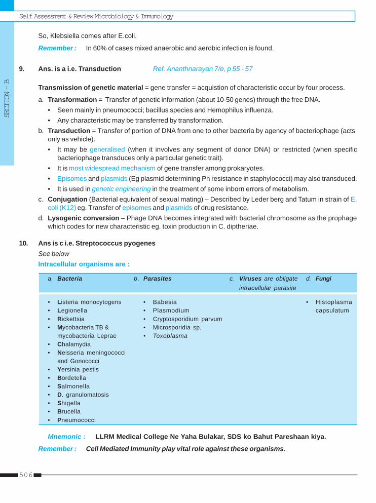
Self Assessment & Review Microbiology & ImmunologySECTION – B
506
So, Klebsiella comes after E.coli.
Remember : In 60% of cases mixed anaerobic and aerobic infection is found.
9. Ans. is a i.e. Transduction Ref. Ananthnarayan 7/e, p 55 - 57
Transmission of genetic material = gene transfer = acquistion of characteristic occur by four process.
a. Transformation = Transfer of genetic information (about 10-50 genes) through the free DNA.• Seen mainly in pneumococci; bacillus species and Hemophilus influenza.• Any characteristic may be transferred by transformation.
b. Transduction = Transfer of portion of DNA from one to other bacteria by agency of bacteriophage (actsonly as vehicle).• It may be generalised (when it involves any segment of donor DNA) or restricted (when specific
bacteriophage transduces only a particular genetic trait).• It is most widespread mechanism of gene transfer among prokaryotes.• Episomes and plasmids (Eg plasmid determining Pn resistance in staphylococci) may also transduced.• It is used in genetic engineering in the treatment of some inborn errors of metabolism.
c. Conjugation (Bacterial equivalent of sexual mating) – Described by Leder berg and Tatum in strain of E.coli (K12) eg. Transfer of episomes and plasmids of drug resistance.
d. Lysogenic conversion – Phage DNA becomes integrated with bacterial chromosome as the prophagewhich codes for new characteristic eg. toxin production in C. diptheriae.
10. Ans is c i.e. Streptococcus pyogenesSee belowIntracellular organisms are :
a. Bacteria b. Parasites c. Viruses are obligate d. Fungiintracellular parasite
• Listeria monocytogens • Babesia • Histoplasma• Legionella • Plasmodium capsulatum• Rickettsia • Cryptosporidium parvum• Mycobacteria TB & • Microsporidia sp.
mycobacteria Leprae • Toxoplasma• Chalamydia• Neisseria meningococci
and Gonococci• Yersinia pestis• Bordetella• Salmonella• D. granulomatosis• Shigella• Brucella• Pneumococci
Mnemonic : LLRM Medical College Ne Yaha Bulakar, SDS ko Bahut Pareshaan kiya.
Remember : Cell Mediated Immunity play vital role against these organisms.
Questions & AnswersMISCELLANEOUS
507
11. Ans. is d i.e. Chemically defined media Ref. Ananthnarayan 7/e, p 457
Isolation of virusMethod of isolation consist of :“Inoculation into animals, eggs or tissue culture after the specimen is processed to remove bacterialcontaminants.” ..... Ananthnarayan 7/e, p 457As many viruses (adenoviruses, enteroviruses) are frequently found in normal individuals so only recovery ofviral agent from patient doesn’t proved that it is the causative agent of the patient illness.
Organism not grown in artificial cultural media are :• M. Leprae • Pathogenic treponemes• Rickettsia • Chlamydia• Viruses.
12. Ans. is c i.e. It establishes in the body only after the neonatal period Ref. Jawetz 24/e, p 197-200
Term “normal microbial flora” denotes the population of microorganisms that inhabit the skin and mucousmembranes of healthy normal persons. They are not essential to life.MC resident organisms of upper respiratory tract is streptococci of viridans group.MC resident bacteria of large intestine is bacteroides species.Lines from Jawetz clears all choice to you -“Mucous membranes of mouth and pharynx are often sterile at birth Within 4-12 hrs after birth, viridansstreptococci become establish as most prominent member of resident flora and remain so for life.”
“In the pharynx and trachea, similar flora establish itself whereas few bacteria are found in normal bronchi.Small bronchi and alveoli are normally sterile.”
“Stomach acidity keep the number of micro-organisms at a minimum (103 - 105) unless obstruction atthe pylorus favours the proliferation of gram positive cocci and bacilli.”
“Antimicrobials drugs taken orally can, in humans, temporilly suppress the drug susceptible components ofthe fecal flora.”
13. Ans. is b i.e. Gram –ve organisms Ref. Jawetz 24/e, p 739; Harrison 16/e, p 1539
Guys, this is a twisted question, understand the choice clearly.– MC cause of nosocomial pneumonia in ICU now is S.aureus (Gram +ve). ..... Harrison 16/e, p 1539– After this comes enterobacteriacea followed by pseudomonas aeruginesa (Gram –ve).• But if we take enterobacteriacea and P. aeruginosa (Gram –ve organism) together they can over number S.
aureus (Gram +ve organism).So, the answer will be Gram –ve organism.Now word nosocomial has been replaced by hospital acquired pneumonia.
14. Ans. is b i.e. Shiga toxin Ref. Ananthnarayan 7/e, p 286; Harrison 17/e, p 963
Shiga toxin, a protein encoded by the iron regulated chromosomal gene. It composed of two peptide subunits-A subunit is N-glycosidase that hydrolyzes adenine from specific sites of ribosomal RNA of mammalian 60s-ribosomal subunit, irreversibly inhibiting protein synthesis.B subunit recognizes the receptor glycolipid Gp3 on host cells.
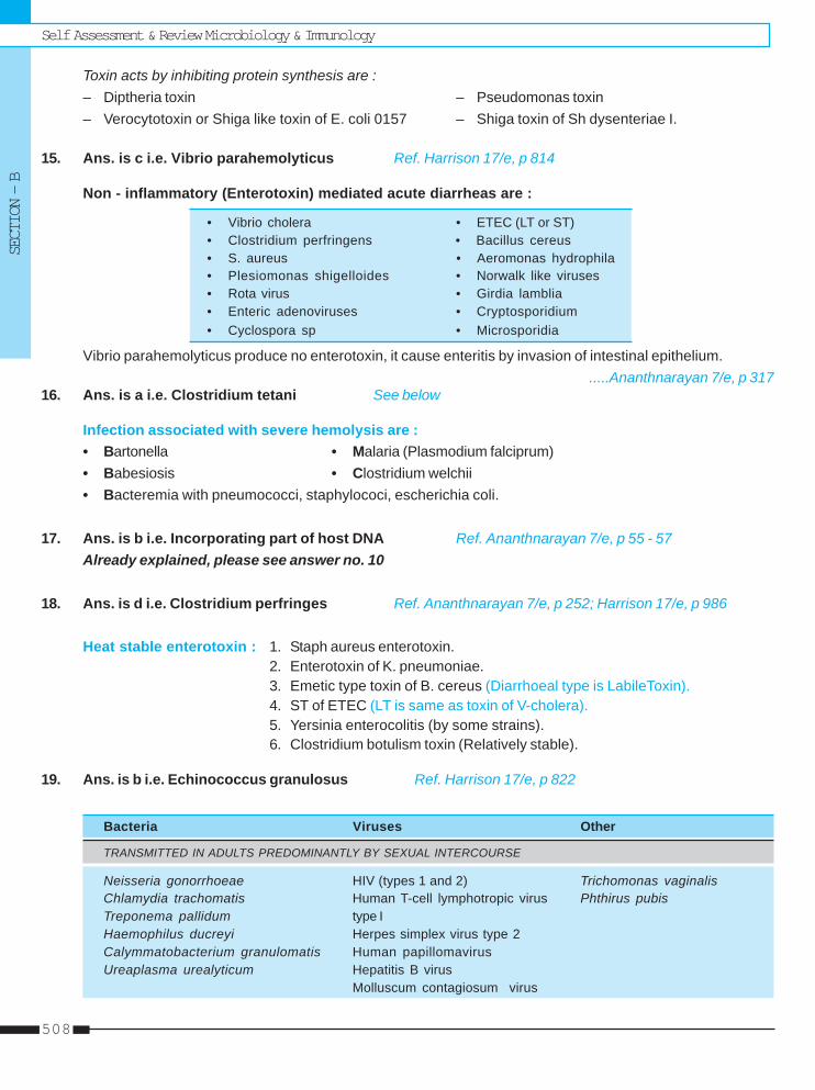
Self Assessment & Review Microbiology & ImmunologySECTION – B
508
Toxin acts by inhibiting protein synthesis are :– Diptheria toxin – Pseudomonas toxin– Verocytotoxin or Shiga like toxin of E. coli 0157 – Shiga toxin of Sh dysenteriae I.
15. Ans. is c i.e. Vibrio parahemolyticus Ref. Harrison 17/e, p 814
Non - inflammatory (Enterotoxin) mediated acute diarrheas are :
• Vibrio cholera • ETEC (LT or ST)• Clostridium perfringens • Bacillus cereus• S. aureus • Aeromonas hydrophila• Plesiomonas shigelloides • Norwalk like viruses• Rota virus • Girdia lamblia• Enteric adenoviruses • Cryptosporidium• Cyclospora sp • Microsporidia
Vibrio parahemolyticus produce no enterotoxin, it cause enteritis by invasion of intestinal epithelium......Ananthnarayan 7/e, p 317
16. Ans. is a i.e. Clostridium tetani See below
Infection associated with severe hemolysis are :• Bartonella • Malaria (Plasmodium falciprum)• Babesiosis • Clostridium welchii• Bacteremia with pneumococci, staphylococi, escherichia coli.
17. Ans. is b i.e. Incorporating part of host DNA Ref. Ananthnarayan 7/e, p 55 - 57Already explained, please see answer no. 10
18. Ans. is d i.e. Clostridium perfringes Ref. Ananthnarayan 7/e, p 252; Harrison 17/e, p 986
Heat stable enterotoxin : 1. Staph aureus enterotoxin.2. Enterotoxin of K. pneumoniae.3. Emetic type toxin of B. cereus (Diarrhoeal type is LabileToxin).4. ST of ETEC (LT is same as toxin of V-cholera).5. Yersinia enterocolitis (by some strains).6. Clostridium botulism toxin (Relatively stable).
19. Ans. is b i.e. Echinococcus granulosus Ref. Harrison 17/e, p 822
Bacteria Viruses Other
TRANSMITTED IN ADULTS PREDOMINANTLY BY SEXUAL INTERCOURSE
Neisseria gonorrhoeae HIV (types 1 and 2) Trichomonas vaginalisChlamydia trachomatis Human T-cell lymphotropic virus Phthirus pubisTreponema pallidum type IHaemophilus ducreyi Herpes simplex virus type 2Calymmatobacterium granulomatis Human papillomavirusUreaplasma urealyticum Hepatitis B virus
Molluscum contagiosum virus
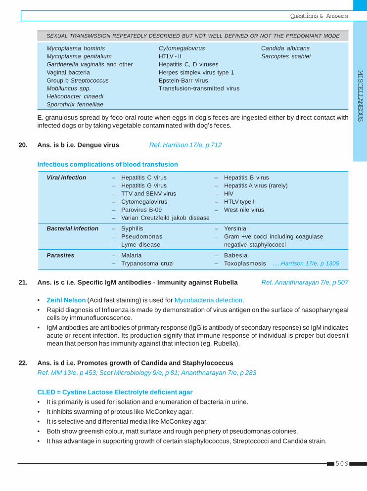
Questions & AnswersMISCELLANEOUS
509
SEXUAL TRANSMISSION REPEATEDLY DESCRIBED BUT NOT WELL DEFINED OR NOT THE PREDOMIANT MODE
Mycoplasma hominis Cytomegalovirus Candida albicansMycoplasma genitalium HTLV - II Sarcoptes scabieiGardnerella vaginalis and other Hepatitis C, D virusesVaginal bacteria Herpes simplex virus type 1Group b Streptococcus Epstein-Barr virusMobiluncus spp. Transfusion-transmitted virusHelicobacter cinaediSporothrix fennelliae
E. granulosus spread by feco-oral route when eggs in dog’s feces are ingested either by direct contact withinfected dogs or by taking vegetable contaminated with dog’s feces.
20. Ans. is b i.e. Dengue virus Ref. Harrison 17/e, p 712
Infectious complications of blood transfusion
Viral infection – Hepatitis C virus – Hepatitis B virus– Hepatitis G virus – Hepatitis A virus (rarely)– TTV and SENV virus – HIV– Cytomegalovirus – HTLV type I– Parovirus B-09 – West nile virus– Varian Creutzfeild jakob disease
Bacterial infection – Syphilis – Yersinia– Pseudomonas – Gram +ve cocci including coagulase– Lyme disease negative staphylococci .
Parasites – Malaria – Babesia– Trypanosoma cruzi – Toxoplasmosis .....Harrison 17/e, p 1305
21. Ans. is c i.e. Specific IgM antibodies - Immunity against Rubella Ref. Ananthnarayan 7/e, p 507
• Zeihl Nelson (Acid fast staining) is used for Mycobacteria detection.• Rapid diagnosis of Influenza is made by demonstration of virus antigen on the surface of nasopharyngeal
cells by immunofluorescence.• IgM antibodies are antibodies of primary response (IgG is antibody of secondary response) so IgM indicates
acute or recent infection. Its production signify that immune response of individual is proper but doesn’tmean that person has immunity against that infection (eg. Rubella).
22. Ans. is d i.e. Promotes growth of Candida and StaphylococcusRef. MM 13/e, p 453; Scot Microbiology 9/e, p 81; Ananthnarayan 7/e, p 283
CLED = Cystine Lactose Electrolyte deficient agar• It is primarily is used for isolation and enumeration of bacteria in urine.• It inhibits swarming of proteus like McConkey agar.• It is selective and differential media like McConkey agar.• Both show greenish colour, matt surface and rough periphery of pseudomonas colonies.• It has advantage in supporting growth of certain staphylococcus, Streptococci and Candida strain.
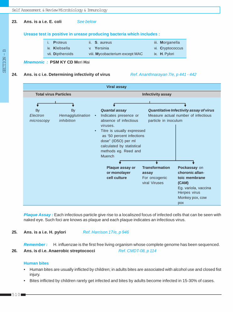
Self Assessment & Review Microbiology & ImmunologySECTION – B
510
23. Ans. is a i.e. E. coli See below
Urease test is positive in urease producing bacteria which includes :
i. Proteus ii. S. aureus iii. Morganellaiv. Klebsella v. Yersinia vi. Cryptococcusvii. Diptheroids viii. Mycobacterium except MAC ix. H. Pylori
Mnemonic : PSM KY CD Meri Hai
24. Ans. is c i.e. Determining infectivity of virus Ref. Ananthnarayan 7/e, p 441 - 442
Viral assay
Total virus Particles Infectivity assay
By By Quantal assay Quantitative Infectivity assay of virusElectron Hemagglutination • Indicates presence or Measure actual number of infectiousmicroscopy inhibition absence of infectious particle in inoculum
viruses.• Titre is usually expressed
as ‘50 percent infectionsdose” (ID5O) per mlcalculated by statisticalmethods eg. Reed andMuench
Plaque assay or Transformation Pockassay onor monolayer assay choronic allan-cell culture For oncogenic toic membrane
viral Viruses (CAM)Eg. variola, vaccinaHerpes virusMonkey pox, cowpox
Plaque Assay : Each infectious particle give rise to a localiszed focus of infected cells that can be seen withnaked eye. Such foci are knows as plaque and each plaque indicates an infectious virus.
25. Ans. is a i.e. H. pylori Ref. Harrison 17/e, p 946
Remember : H. influenzae is the first free living organism whose complete genome has been sequenced.26. Ans. is d i.e. Anaerobic streptococci Ref. CMDT-08, p 114
Human bites• Human bites are usually inflicted by children; in adults bites are associated with alcohol use and closed fist
injury.• Bites inflicted by children rarely get infected and bites by adults become infected in 15-30% of cases.
Questions & AnswersMISCELLANEOUS
511
• Bacteriology of bite infection:– Human bites are mixture of aerobes and anaerobes (54%) or due to aerobes only (44%).– Streptococcus, staphylococcus and Eikenella corrodens are most common aerobes.– Prevotella and fusobacterium are the most common anaerobe
Remember :• Cat bites are most likely to become infected (30-50%)• Dog bite become infected only in 5% of cases.• Pasturella species are the single most common isolate in dog and cat bites.
About the questionThis question is of 1998, at that time Harrison 15/e was running. In 15/e, Anaerobes (including pepto streptococcus)were given as more common, So, was the answer.
27. Ans. is a i.e. Pseudomonas Ref. Ananthnarayan 7/e, p 321
Already explained, refer answer no. 14
28. Ans. is a i.e. Absence of nuclear membrane Ref. Ananthnarayan 7/e, p 7
Differences between prokaryotic and eukaryotic cells
Character Prokaryotes Eukaryotes
Nucleus (Main basis of classification)
• Nuclear membrane Absent Present• Nucleolus Absent Present• Deoxyribonucleoprotein Absent Present• Chromosome One (Circular) More than one (linear)• Mitotic division Absent Present
Cytoplasm
• Cytoplasmic streaming Absent Present• Pinocytosis Absent Present• Mitochondria Absent Present• Lysosomes Absent Present• Golgi apparatus Absent Present• Ribosomes 70s 80s
Chemical composition
• Sterols Absent Present• Muramic acid Present Absent• Amoeboid movement + +• Flagella and Pilli + +• Phosphrylation site Plasma membrane Ribosomes
(Mesosomes) (Mitochondria)
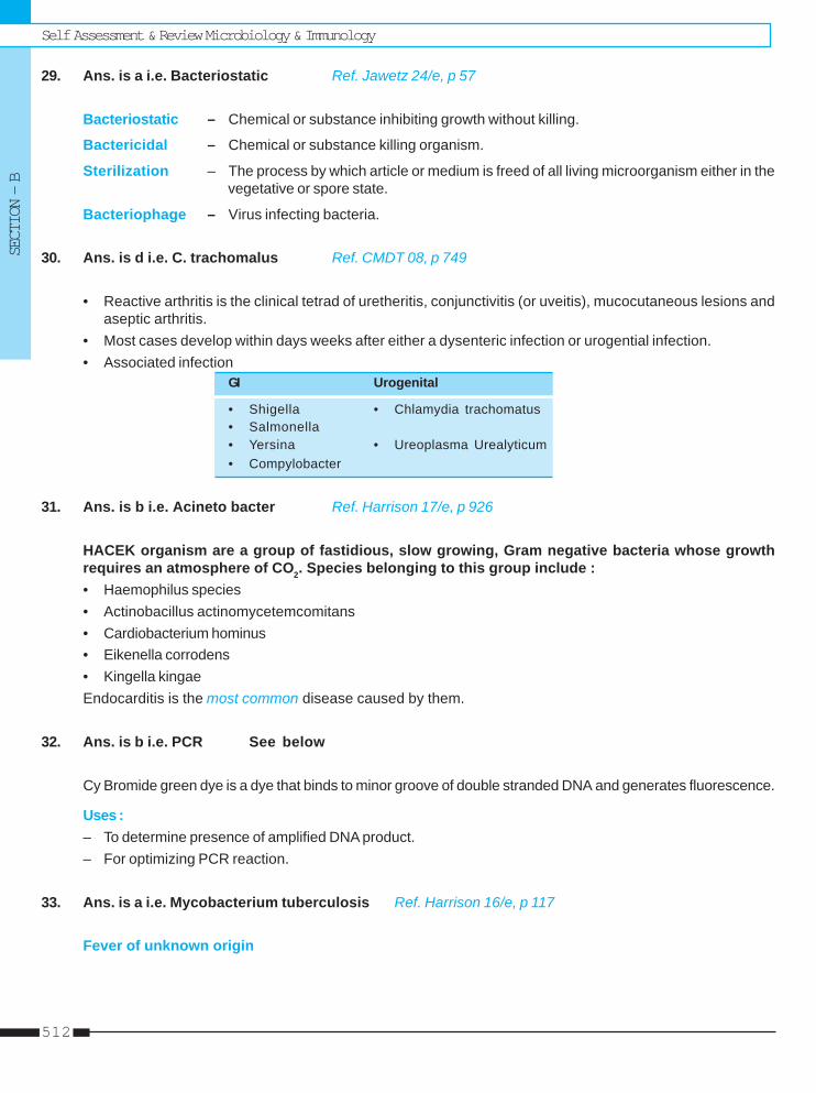
Self Assessment & Review Microbiology & ImmunologySECTION – B
512
29. Ans. is a i.e. Bacteriostatic Ref. Jawetz 24/e, p 57
Bacteriostatic – Chemical or substance inhibiting growth without killing.
Bactericidal – Chemical or substance killing organism.
Sterilization – The process by which article or medium is freed of all living microorganism either in thevegetative or spore state.
Bacteriophage – Virus infecting bacteria.
30. Ans. is d i.e. C. trachomalus Ref. CMDT 08, p 749
• Reactive arthritis is the clinical tetrad of uretheritis, conjunctivitis (or uveitis), mucocutaneous lesions andaseptic arthritis.
• Most cases develop within days weeks after either a dysenteric infection or urogential infection.• Associated infection
GI Urogenital
• Shigella • Chlamydia trachomatus• Salmonella• Yersina • Ureoplasma Urealyticum• Compylobacter
31. Ans. is b i.e. Acineto bacter Ref. Harrison 17/e, p 926
HACEK organism are a group of fastidious, slow growing, Gram negative bacteria whose growthrequires an atmosphere of CO2. Species belonging to this group include :• Haemophilus species• Actinobacillus actinomycetemcomitans• Cardiobacterium hominus• Eikenella corrodens• Kingella kingaeEndocarditis is the most common disease caused by them.
32. Ans. is b i.e. PCR See below
Cy Bromide green dye is a dye that binds to minor groove of double stranded DNA and generates fluorescence.
Uses :– To determine presence of amplified DNA product.– For optimizing PCR reaction.
33. Ans. is a i.e. Mycobacterium tuberculosis Ref. Harrison 16/e, p 117
Fever of unknown origin

Questions & AnswersMISCELLANEOUS
513
Causes
Infections (33%) Neoplasm (17%) Non infectious Miscellaneous Undiagnosedinflammatory (15%) (14%)disease (21%)
– Mycobacterium TB – Lymphoma– Abdominal abscess – Leukemia– Endocarditis – Solid tumours– UTI– Viral infections : - CMV
- EBV– Kala azar– Brucellosis
34. Ans. is c i.e. 1000 : 1 Ref. Jawetz 24/e, p 200
Anaerobes outnumber facultative organism by 1000 fold.
Normal flora of intestinal tract• At birth the intestine is sterile, but organism are soon introduced after birth.• In breast feed children lactic acid streptococci and lactobacilli seen.• Micro-organism are minimum (103 - 105g/ of contents) in stomach.• In upper intestine lactobacilli and enterococci predominate.• In colon bacteriodes fragilis is the most common organism found.• Intestinal bacteria are important in synthesis of vitamin K.
35. Ans. is a i.e. Measles Ref. Park 18/e, p 89; 19/e, p 90
In measles and whooping cough only cases are found with no carriers.
Chronic carriers seen in :• Typhoid • Dysentery• Hepatitis B • Cerebrospinal meningitis• Malaria • Gonorrhea
36. Ans. is d i.e. Hemophilus ducreyi Ref. Ananthnarayan 7/e, 228
Causes of urethritis are : – Neisseria gonorrhoea (MC) – Chlamydia trachomatis– Ureoplasma urealyticun – Mycoplasma hominis– Herpes virus – Cytomegalovirus– Gardenella vaginalis – Acinetobactor woffi, Ac calcoaceticus– Candida albicans – Trichomonas vaginalis
37. Ans. is b i.e. Pneumocystis carinii pneumonia See below
Infection in patients with defects in humoral immunity :• Recurrent or chronic sinopulmonary infection otitis media, meningitis and bacteremia; MC by pyogenic
bacteria such as H. influenzae; Strep pneumoniae; Staphylococci
Self Assessment & Review Microbiology & ImmunologySECTION – B
514
• Response to viral infection is good except increased risk of :– HBV – Polio– Echoviruses – Adenoviruses
• Parasitic – Giardial diarrhea• Less frequent – Bacterial infection of skin and urinary tract.
Infection in patient with deficient CMI :• Disseminates virus infection of Herpes simplex, Varicella zoster, and CMV• Mucocutaneous candidiasis – Almost invariably• Pneumonia caused by P. carinii• Severe Enteritis caused by cryptosporidium• T cell deficiency is always accompanied by some abnormality of antibody response so patient with T cell
defect are also subject to overwhelming bacterial infection.
38. Ans. is d i.e. Mycobacterium Ref. Ananthnarayan 7/e, p 635; Harrison 17/e, p 835-838
• Most important group of hospital pathogens are :i. Enteric gram negative bacilli :
– E. coli – Kliebsiella– Enterobacter – Proteus– Serratia
ii. S. aureusiii. Pseudomonas aeruginosa and other pseudomonasiv. Tetanus sporesv. Yeast (Candida albicans), moulds (Aspergillus mucor)vi. Protozoa (E. histolytica, Plasmodia, P. carinii, T. gondii)
Nosocomial infection Most common causative organism
• Urinary tract infection E. coli; Candida(MC Nosocomial infection)
• Early onset pneumonia (within 4 days) Strept pneumoniae• Late onset pneumonia S. aureus, P. aeruginosa• Surgical wound infections S.aureus, coagulase negative staphylococcus• Infections related to vascular acess Coagulase negative Staph, S. aureus
Remember :• Candida is now the MC pathogen in nosocomial UTI in ICU patients. ..... Harrison 17/e, p 837• Examples of some emerging and potential, epidemic problems in Hospitals are : Chickenpox TB, Group A
streptococci, Aspergillus, Legionella
39. Ans. is b i.e. Biofilm formulation Ref. Jawetz 24/e, p 158
• Slime is generally polysaccharide (Eg. pneumococcus) or polypeptide (eg. anthrax)
• A biofilm is an aggregate of interactive bacteria attached to solid or to each other and encased in anexopolysaccharide matrix of slime. It form slimy coat on solid surface and occur through out nature.
Questions & AnswersMISCELLANEOUS
515
• A single species or more than one species of bacteria coaggregate to form a biofilm. Fungi including yeastsare occasionally involved
• Biofilms make infections persistant and difficult to treat eg. S. epidermidis and S. aureus infection ofcentral venous catheters and with pseudomonas airway infection in cystic fibrosis patients.
40. Ans. is d i.e. Shock and DIC are common in bacteremia due to B. fragilisRef. Ananthnarayan 7/e, p 268; Jawetz 24/e, p 307
Already explained, refer answer no. 5
41. Ans. is a i.e. Bacteroides Ref. Ananthnarayan 7/e, p 268; Jawetz 24/e, p 307
Provotella is anaerobic gram negative bacilli.
MC isolate is P. melaninogenicus (Previously called Bacteroides melaningenicus)• P. melaninogenica forms black or brown colour colonies• Colony colour is not due to melanin but due to a hemin derivative• Cultures of P. melaninogenica and even dressings from wounds infected with the bacillus give characteristic
red fluorescence when exposed to ultraviolet light.
Remember : Anaerobes causing brain absecess – B. fragilis, Peptostreptococcus, Prevotella etc.
42. Ans. is d i.e. Promotes growth of S. arueus and CandidaRef. MM 13/e, p 453; Scot Microbiology 9/e, p 81; Ananthnarayan 7/e, p 283
Already explained, refer answer no. 22
43. Ans. is c i.e. ETEC Ref. Ananthnarayan 7/e, p 196, 247, 264
Preformed toxins have role in :• Staphylococcal food poisoning (enterotoxin)• Botulism food poisoning• Emetic type of food poisoning of B. cereus (resemble staph enterotoxin)
In case of preformed toxin, incubation period is ‘short’ in comparison to when organism has to producetoxin in the intestine.
44. Ans. is d i.e. Lipo-polysaccharide Ref. Ananthnarayan 7/e, p 67, Table (9.1)
Distinguishing features of exotoxins and endotoxins
Exotoxins Endotoxins
• Proteins Lipopolysaccharides• Heat labile Heat stable• Actively secreted by cells; diffuse into Form part of cell wall; do not diffuse into
surrounding medium surrounding medium• Readily separable from cultures by physical Obtained only by cell lysis
means such as filtration• Action often enzymic No enzymic action
Self Assessment & Review Microbiology & ImmunologySECTION – B
516
• Specific pharmacological effects for each Effect nonspecific; action common to allexotoxin endotoxins
• Specific tissue affinities No specific tissue affinity• Active in very minute doses Active only in very large doses• Highly antigenic Weakly antigenic• Action specifically neutralised by antibody Neutralisatioh by antibody ineffective• Can be toxoided Can’t toxoided• Generally formed by gram positive incuding Generally formed by gram negative bacteria
some gram negative shigella, vibrio cholera,ETEC, V parahemolyticus, AeromonasY. enterocolitica, Ps. aeroginosa
45. Ans. is a i.e. Warts Ref. Harrison 17/e, p 1117-1118
Remember :
Disease Associated Human papilloma viruses
Common warts (Verruca vulgaris) 1, 2, 4, 26, 27, 29, 41, 57, 65, 77
Condyloma accuminatum 6, 11, 30, 42, 43, 44, 45 51, 54(Anogenital warts)Cervical carcinoma 16, 18, 31, 33, 35, 39, 45
Laryngeal papillomas 6, 11Low grade intraepithelial neoplasias 6, 11, 16, 18
46. Ans. is b i.e. Herpes simplex Ref. Jawetz 24/e, p 586, Table (43.1)
Association of viruses with human cancers.
Virus family Virus Human cancer
Papillomaviridae Human papillomaviruses Genital tumors, squamous cell carcinomaOropharyngeal carcinoma
Herpesviridae EB virus Nasophryngeal carcinoma, African Burkitt’slymphoma, B cell lymphoma
Hepadna viridae Hepatitis B virus Hepatocellular carcinoma
Retroviridae HTL virus Adult T cell leukemiaHIV AIDS - related malignancies
Flaviviridae Hepatitis C virus Hepatocellular carcinoma
47. Ans. is a i.e. Quantitative assay of infectivitiy of virus Ref. Ananthnarayan 7/e, p 441 - 442
Already explained, refer answer no. 27
48. Ans. is c i.e. Highly antigenic Ref. Ananthnarayan 7/e, p 67, Table (9.1)
Already explained, refer answer no. 44
Questions & AnswersMISCELLANEOUS
517
49. Ans. is a and c. Leprae, Treponema pallidum Ref. Ananthnarayan 7/e, p 457
Already explained, refer answer no. 11
50. Ans. is b, c, d and e i.e. Shigella, Vibrio parahaemolyticus, Campylobacter and Salmonella
Invasive Diarrhoea
Minimal inflammation Variable inflammation Severe
• Rotavirus • Salmonella • Shigella• Norwalk agent • Campylobacter • EIEC
• Aeromonas • Entamoeba histolytica• Vibrio parahaemolyticus• Yersinia
51. Ans. is a, b, d i.e. Clostridium perfringes, Streptococcus and CI. tetani
Rhabdomyolysis
Viral causes Bacterial causes Fungal causes
• Influenzae types A and B • Francisella tularensis • Rickettsia species • Candida(most common) • Streptococcus pneumoniae • Salmonella species • Aspergillus
• HIV• Ebstein-Barr virus • Group B streptococci • Listeria species• Echovirus • Streptococcus pyogenes • Legionella species• Cytomegalovirus • Staphylococcus epidermidis • Mycoplasma species• Adenovirus • Escherichia coli • Vibrio species• Herpes simplex virus • Borrelia burgdorferi • Brucella species• Parainfluenza virus • Clostridium tetani • Bacillus species• Varicella-zoster virus • Viridans streptococci • Leptospira species• Coxsackievirus
52. Ans. is a and b i.e. Donovanosis; and Lymphogranuloma venerumRef. Ananthnaraya 7/e, p 404Genital elephantiasis is seen in Donovanosis which is caused by Calymmatobacterium granulomatosisor Granuloma iguinale / venerum.
Donovanosis• Chronic progressive bacterial infection of the genital region that is generally sexually transmitted.• Causative organism :
– Calymmatobacterium granulomatis (Gram negative encapsulated bacterium).– Morphologicaly and antigenically related to Klebsiella.– Grow on egg yolk or modified levinthal agar.
• I.P. – 1 - 4 weeks.• Clinical features :– Disease begin as one or more subcutaneous nodules that erode through the skin to
produce clean granulomatous, sharply defined usually painless lesion.– Genitalia are involved in 90% of cases.
Self Assessment & Review Microbiology & ImmunologySECTION – B
518
– Genital swelling particularly of labia is common and may progress to pseudo-elephantiasis.
– Complications include phimosis and paraphimosis.• Diagnosis : – Diagnosis is made by demonstration of typical intracellular Donovan bodies (safety
pin appearance) in Wright Giemsa stain.• Treatment : – Azithromycin - DOC.
– Doxycycline - Second DOC.• Remember : – Genital elephantiasis is also seen in lymphatic filariasis. ... Harrison 16/e, p 1260
– Vulval elephantiasis or esthiomene is seen in lymphogranuloma venerum...... Ananthnarayan 7/e, p 428
53. Ans. is a i.e. Fungus Ref. Ananthnarayan 7/e, p 620; Dhingra 3/e, p 197
• Rhinosporidiosis is a chronic granulomatous disease characterized by development of friable polyp usuallyconfined to the nose, mouth or eye.
• Polyp is highly vascular which bleeds easily on touch. Its surface is studded with white dots which representsthe sporangia of fungus.
• Causative agent – Rhinosporidium seeberi. The fungus has not been cultivated in media.– The infection is supposed to originate from stagnant water or aquatic life.
• Diagnosis : – Biopsy shows round or oval sporangia filled with spores which may burst throughchitinous wall.
• Treatment : – Complete excision with diathermy knife and cauterization of its based.– Dapsone and Amphotericin B are also effective.
54. Ans. is a i.e. Leptospira Ref. See below
Disease Mode of infection
Leptospira Water contaminated by the urine of carrier animals enter the body through cut or abrasions onthe skin or through intact mucosa of mouth, nose or conjunctiva.
Legionella Inhalation of aerosols produced by AC, cooling towers.Plague Bite of rat flea, droplet infectionDiptheria Droplet infection
55. Ans. is a, b, c i.e. H.pylori; S.aureus; and Klebsiella Ref. See below
Already explained, refer answer no. 23
56. Ans. is a i.e. 200 nm Ref.See below
Resolving power are :Light microscope – 0.25 μm – 0.3 μm = 200 - 300 nmElectron microscope – 2 - 10 A0 = 0.2 - 1 nm
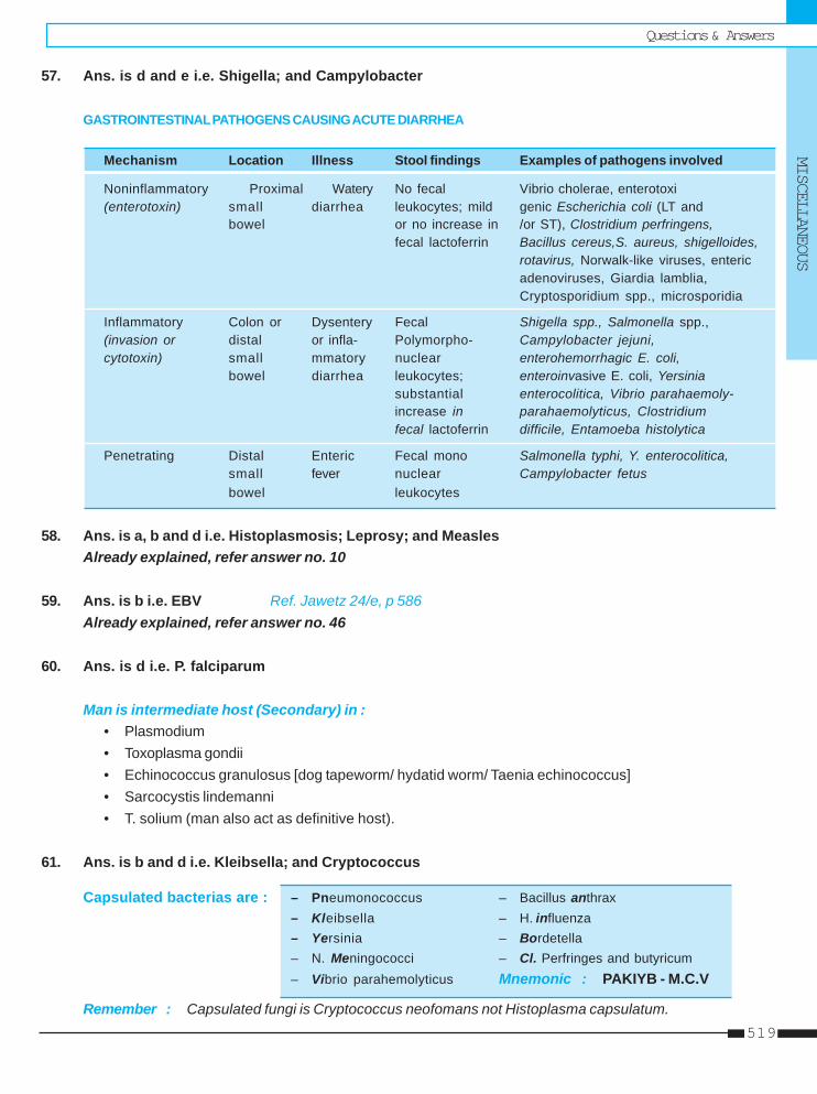
Questions & AnswersMISCELLANEOUS
519
57. Ans. is d and e i.e. Shigella; and Campylobacter
GASTROINTESTINAL PATHOGENS CAUSING ACUTE DIARRHEA
Mechanism Location Illness Stool findings Examples of pathogens involved
Noninflammatory Proximal Watery No fecal Vibrio cholerae, enterotoxi(enterotoxin) small diarrhea leukocytes; mild genic Escherichia coli (LT and
bowel or no increase in /or ST), Clostridium perfringens,fecal lactoferrin Bacillus cereus,S. aureus, shigelloides,
rotavirus, Norwalk-like viruses, entericadenoviruses, Giardia lamblia,Cryptosporidium spp., microsporidia
Inflammatory Colon or Dysentery Fecal Shigella spp., Salmonella spp.,(invasion or distal or infla- Polymorpho- Campylobacter jejuni,cytotoxin) small mmatory nuclear enterohemorrhagic E. coli,
bowel diarrhea leukocytes; enteroinvasive E. coli, Yersiniasubstantial enterocolitica, Vibrio parahaemoly-increase in parahaemolyticus, Clostridiumfecal lactoferrin difficile, Entamoeba histolytica
Penetrating Distal Enteric Fecal mono Salmonella typhi, Y. enterocolitica,small fever nuclear Campylobacter fetusbowel leukocytes
58. Ans. is a, b and d i.e. Histoplasmosis; Leprosy; and MeaslesAlready explained, refer answer no. 10
59. Ans. is b i.e. EBV Ref. Jawetz 24/e, p 586Already explained, refer answer no. 46
60. Ans. is d i.e. P. falciparum
Man is intermediate host (Secondary) in :• Plasmodium• Toxoplasma gondii• Echinococcus granulosus [dog tapeworm/ hydatid worm/ Taenia echinococcus]• Sarcocystis lindemanni• T. solium (man also act as definitive host).
61. Ans. is b and d i.e. Kleibsella; and Cryptococcus
Capsulated bacterias are : – Pneumonococcus – Bacillus anthrax– Kleibsella – H. influenza– Yersinia – Bordetella– N. Meningococci – Cl. Perfringes and butyricum– Vibrio parahemolyticus Mnemonic : PAKIYB - M.C.V
Remember : Capsulated fungi is Cryptococcus neofomans not Histoplasma capsulatum.
Self Assessment & Review Microbiology & ImmunologySECTION – B
520
62. Ans. is a, b and c i.e. Hepatitis B; CMV; and HTLV - 1 Ref. Harrison 17/e, p 712
Already explained, refer answer no. 20
63. Ans. is b, c, d and e i.e. B. Coli; Microsporidium species; E. dispar; and Giardia intestinalis
Let consider each options.a. Cyptococcus is fungus causing pulmonary, cutaneous and cryptococcal meningitis. ... AA 7/e, p 620b. Balantidum coli – only ciliate protozoan parasite of man. Its trophozoite lives in large intestine.
... Paniker 4/e, p 108c. Microsporidium sp. – It is intracellular parasites.
• Transmission is chiefly by ingestion of spores in food or water.• Transplacental transmission is common.• Also cause infection in AIDS patient ....Jawetz 24/e, p 684
d. E. dispar• It is protozoa (amoeba)• In case of intestinal amoebiasis, parasite remains luminal and trophozoite multiply as a bacteria feeding
colony, ultimately encyst and pass out in feces. These are presumed to be due to E. dispar.It is lumen dwelling non pathogenic commensal.It is distinguished from E. histolytica only by isoenzyme electrophoresis and DNA analysis.
... Jawetz 24/e, p 672e. Giardia lamblia is intestinal flagellates. ... Paniker 4/e, p 34
64. Ans. is a, b and c i.e. Chlamydia; Rickettsia; and MycoplasmaRef. See index of any Microbiology Book
Prion is proteinacoeus infectious particle, without nucleic acid, cause slow virus disease.Bacteriophage is the virus that infects bacteria.
65. Ans. is a, b and c i.e. Toxoplasma; Syphilis; and CMV Ref. Harrison 17/e, p 712
Already explained, refer answer no. 20
66. Ans. is a, b, c, d and e i.e. All are correct optionsRef. Jawetz 24/e, p 717-718; Harrison 17/e, p 814 Tab. (113-1)
Following infections required stool examination for diagnosis :i. Toxins (of staphylococcus, Clostridia, Vibrios, Toxigenic E.coli)ii. Shigellaiii. Salmonellaiv. Campylobactersv. Yersinia enterocoliticavi. Vibrios
Questions & AnswersMISCELLANEOUS
521
vii. Enteric bacteriaviii.Enterovirusesix. Intestinal parasites (Giardia, E. histolytica, B. coli, Cryptosporidium, Fasciola hepatica, E. vermicularis
etc).
67. Ans. is a and c i.e. Mitochondria always absent; and Divide by binary fissionRef. Ananthnarayan 7/e, p 8, table (2.1), p 395
• Only bacteria having sterol in its cell membrane : Mycoplasma
• Bacteria can be seen by : – Optical or light microscopy– Phage contrast microscopy– Dark field or dark ground microscopy– Electron microscopy.
68. Ans. is a, b and d i.e. Viruses; Chlamydia; and Rickettsia Ref. Ananthnarayan 7/e, p 55 - 57
Already explained, refer answer no. 10
69. Ans. is d i.e. Gardenella Ref. Harrison 17/e, p 827; Shaws 12/e, p 98 - 100
“Treatment of male partners with metronidazoe does not prevent recurrence of bacterial vaginosis /gardenella associated with vaginal discharge.”
Treatment of sexual partners is required in : – Candidiasis– Herpes genitalis– Trichomoniases.
70. Ans. is a and b i.e. Lactobacillus; and BacteroidesRef. Ananthnarayan 7/e, p 601; Jawetz 24/e, p 199
Normal flora of intestinal tract :• At birth intestine is sterile but organism are soon introduced with food.• Stomach’s acidity keep the number of microorganism at a minimum (103-105 g of iontents ) level,as the pH
of intestinal contents becomes alkaline, resident flora gradually increases.• In the upper intestine, lactobacilli and enterococci predominate but in lower ileum and caecum, flora is
fecal.• In normal adult colon, 96-99% of resident bacterial flora consist of anaerobes - bacteriodes sp. especially
B. fragilis, fusobacterium sp; anaerobic lactobacilli eg bifidobacterium; clostridium (C. perfringes) andanaerobic gram positive cocci (peptostreptococcus).– Only 1-4% are facultative aerobes (gram negative coliform bacteria, enterococci, pseudomonas etc.)
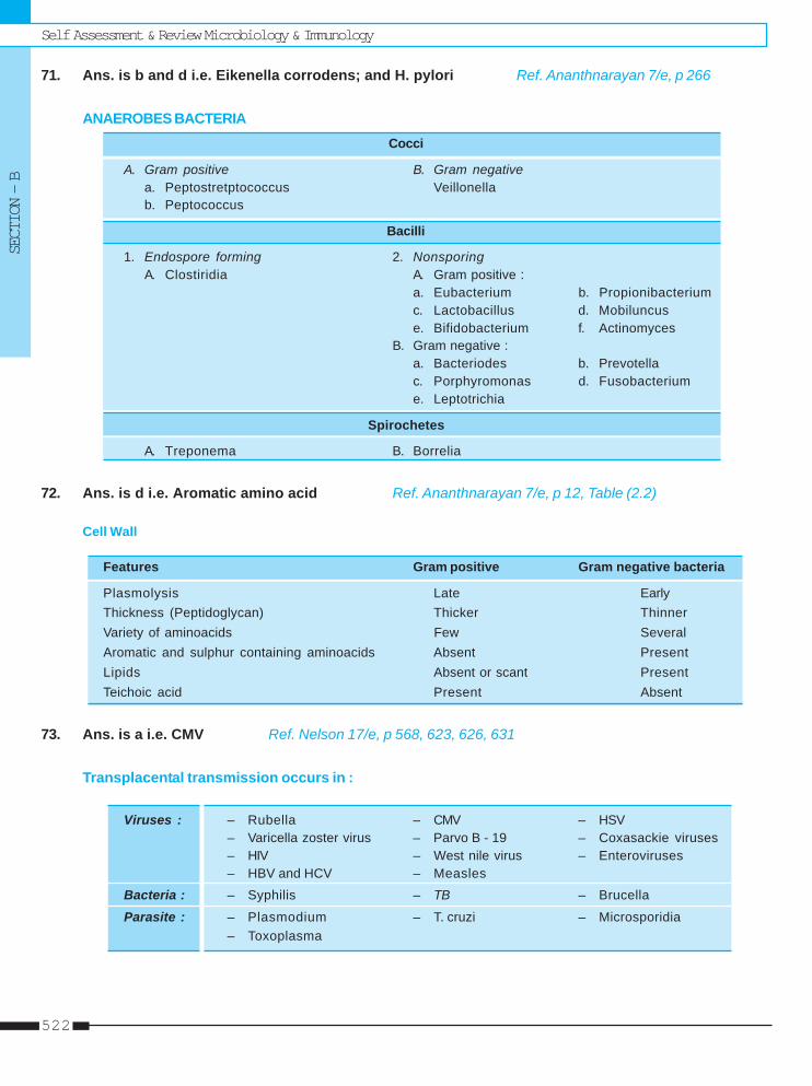
Self Assessment & Review Microbiology & ImmunologySECTION – B
522
71. Ans. is b and d i.e. Eikenella corrodens; and H. pylori Ref. Ananthnarayan 7/e, p 266
ANAEROBES BACTERIACocci
A. Gram positive B. Gram negativea. Peptostretptococcus Veillonellab. Peptococcus
Bacilli
1. Endospore forming 2. NonsporingA. Clostiridia A. Gram positive :
a. Eubacterium b. Propionibacteriumc. Lactobacillus d. Mobiluncuse. Bifidobacterium f. Actinomyces
B. Gram negative :a. Bacteriodes b. Prevotellac. Porphyromonas d. Fusobacteriume. Leptotrichia
Spirochetes
A. Treponema B. Borrelia
72. Ans. is d i.e. Aromatic amino acid Ref. Ananthnarayan 7/e, p 12, Table (2.2)
Cell Wall
Features Gram positive Gram negative bacteria
Plasmolysis Late EarlyThickness (Peptidoglycan) Thicker ThinnerVariety of aminoacids Few SeveralAromatic and sulphur containing aminoacids Absent PresentLipids Absent or scant PresentTeichoic acid Present Absent
73. Ans. is a i.e. CMV Ref. Nelson 17/e, p 568, 623, 626, 631
Transplacental transmission occurs in :
Viruses : – Rubella – CMV – HSV– Varicella zoster virus – Parvo B - 19 – Coxasackie viruses– HIV – West nile virus – Enteroviruses– HBV and HCV – Measles
Bacteria : – Syphilis – TB – Brucella
Parasite : – Plasmodium – T. cruzi – Microsporidia– Toxoplasma
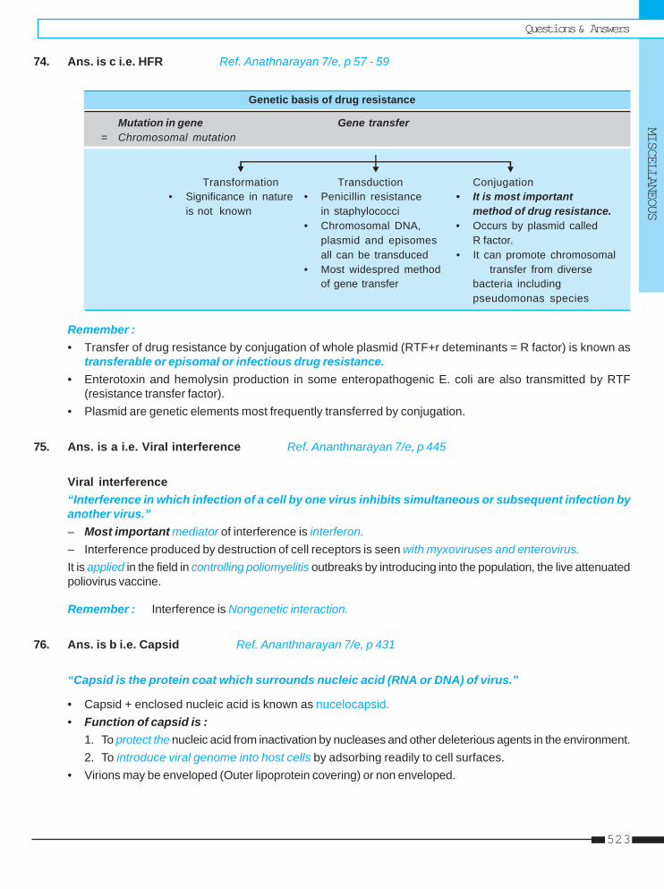
Questions & AnswersMISCELLANEOUS
523
74. Ans. is c i.e. HFR Ref. Anathnarayan 7/e, p 57 - 59
Genetic basis of drug resistance
Mutation in gene Gene transfer= Chromosomal mutation
Transformation Transduction Conjugation• Significance in nature • Penicillin resistance • It is most important
is not known in staphylococci method of drug resistance.• Chromosomal DNA, • Occurs by plasmid called
plasmid and episomes R factor.all can be transduced • It can promote chromosomal
• Most widespred method transfer from diverseof gene transfer bacteria including
pseudomonas species
Remember :• Transfer of drug resistance by conjugation of whole plasmid (RTF+r deteminants = R factor) is known as
transferable or episomal or infectious drug resistance.• Enterotoxin and hemolysin production in some enteropathogenic E. coli are also transmitted by RTF
(resistance transfer factor).• Plasmid are genetic elements most frequently transferred by conjugation.
75. Ans. is a i.e. Viral interference Ref. Ananthnarayan 7/e, p 445
Viral interference“Interference in which infection of a cell by one virus inhibits simultaneous or subsequent infection byanother virus.”– Most important mediator of interference is interferon.– Interference produced by destruction of cell receptors is seen with myxoviruses and enterovirus.It is applied in the field in controlling poliomyelitis outbreaks by introducing into the population, the live attenuatedpoliovirus vaccine.
Remember : Interference is Nongenetic interaction.
76. Ans. is b i.e. Capsid Ref. Ananthnarayan 7/e, p 431
“Capsid is the protein coat which surrounds nucleic acid (RNA or DNA) of virus.”
• Capsid + enclosed nucleic acid is known as nucelocapsid.• Function of capsid is :
1. To protect the nucleic acid from inactivation by nucleases and other deleterious agents in the environment.2. To introduce viral genome into host cells by adsorbing readily to cell surfaces.
• Virions may be enveloped (Outer lipoprotein covering) or non enveloped.
Self Assessment & Review Microbiology & ImmunologySECTION – B
524
Chapter Review••••• This section includes questions of VThis section includes questions of VThis section includes questions of VThis section includes questions of VThis section includes questions of Various Other PGMEES from 1990 – 2007.arious Other PGMEES from 1990 – 2007.arious Other PGMEES from 1990 – 2007.arious Other PGMEES from 1990 – 2007.arious Other PGMEES from 1990 – 2007.••••• Questions are arQuestions are arQuestions are arQuestions are arQuestions are ar ranged in decreasing order of yearranged in decreasing order of yearranged in decreasing order of yearranged in decreasing order of yearranged in decreasing order of yearwiz. This is done to makwiz. This is done to makwiz. This is done to makwiz. This is done to makwiz. This is done to make refere refere refere refere refer ralralralralral
system easier and uncomplicated to save the precious time of PGMEE Aspirant.system easier and uncomplicated to save the precious time of PGMEE Aspirant.system easier and uncomplicated to save the precious time of PGMEE Aspirant.system easier and uncomplicated to save the precious time of PGMEE Aspirant.system easier and uncomplicated to save the precious time of PGMEE Aspirant.
1. d) Adenovirus 2. d) Varicella ... 3. a) Hepatitis B 4. b) Thick & clean ... 5. d) Multiplication ...6. a) M. tuberculosis 7. b) Tetanus 8. b) Brucella ... 9. b and c 10. b) Retro
11. a) M. Avium ... 12. c) Influenza ... 13. b) Parvo virus 14. c) Discovery ...
1. DNA virus is : [Delhi 89; 90]a) Polio type Ib) Polio type IIc) Echovirusd) Adenovirus
[Ref. Ananthnarayan 7/e, p 446]2. All are oncogenic except : [AI 90]
a) EB virusb) Papilloma virusc) Herpes simplex virusd) Varicella zooster virus
[Ref. Ananthnarayan 7/e, Table (61.2]]3. Which of the following does not infect the CNS :
a) Hepatitis B [JIPMER 90]b) HTLV-IIIc) H. Simplex-Id) H. Simplex-II
[Ref. Harrison 17/e, p 2627]4. Which is true of pus : [AI 90]
a) Fecal smelling pus in E. colib) Thick and clean coloured in staph aureusc) Reddish in pseudomanasd) Sulphur granules in candida
[Ref. Under consideration]5. Septicemia is : [TN 91]
a) Bacteria in bloodb) Toxin in bloodc) Pus in bloodd) Multiplication of bacteria and toxins in blood
[Ref. Ananthnarayan 7/e, p 70]6. Niacin is required by growth of : [Kerala 91]
a) M. tuberculosisb) M. kanasasic) M. Scrofulodermad) M. Avium
[Ref. Ananthnarayan 7/e, p 353]7. Herd immunity is not seen in : [Kerala 91]
a) Poliob) Tetanusc) T.B.d) Leprosy
[Ref. Park 19/e, p 96]
8. Undulant fever is caused by : [JIPMER 91]a) Bartonellab) Brucella Melitensisc) Bordetellad) Borelia recurentis
[Ref. Ananthnarayan 7/e, p 345]9. Toxic Shock syndrome is caused by :
a) Staph albus [JIPMER 91]b) Staph aureusc) Strep viridansd) Strep Pyogenes
[Ref. Ananthnarayan 7/e, p 196, 208]10. Human immune deficiency virus is a virus :
a) Rheo [TN 91]b) Retroc) Rhadod) Flavi [Ref. Ananthnarayan 7/e, p 582]
11. Tuberculous infection most common in AIDS :a) M. Avium Intracellulare [JIPMER 91]b) M. Scrofulaceumc) M. Ulceransd) M. Tuberculosis
[Ref. Harrison 16/e, p 1108, 1107 Fig (173-28)]12. Segmented RNA is seen in : [AI 91]
a) Rabies virusb) Coxsackie B virusc) Influenza virusd) Rabies virus
[Ref. Ananthnarayan 7/e, Table (55.1)]13. Which of the following is a single stranded DNA :
a) Herpes simplex [AI 91]b) Parvo virusc) Papavo virusd) Rabies virus
[Ref. Ananthnarayan 7/e, p 447]14. Louis pasteur is not associated with :
a) Introduction of Complex media [JIPMER 91]b) Discovery of Rabies Vaccinec) Discovery of M.Tuberculosisd) Disproved spontaneous regression theory
[Ref. Ananthnarayan 7/e, p 2]
Answer
Questions & AnswersMISCELLANEOUS
525
15. Oncogenic RNA virus : [Kerala 91]a) Avian leuco virusb) Herpes virusc) Adenovirusd) Toga virus
[Ref. Ananthnarayan 7/e, Table (61.2]16. All are common organisms causing UTI except :
a) Streptococcus fecalis [AIIMS 92]b) Escherichia colic) Proteus Mirabilisd) Hemophilus influenzae
[Ref. Harrison 17/e, p 1820]17. Which of the following “oncogenic viruses” is so
far not shown to be oncogenic in man : [DNB 92]a) Hepatitis B virusb) Epstein - Barr virusc) Herpes simplex virusd) Adenoviruse) Human T cell lymphotrophic virus (HTLVI)
[Ref. Ananthnarayan 7/e, p 576]18. ELISA test when compared to western blot tech-
nique is : [AI 93]a) Less sensitive, less Specificb) More Sensitive, more specificc) Less sensitive, more specificd) More Sensitive, less specific
[Ref. Park 19/e, p 293]19. Sero conversiion in HIV infection takes places in:
a) 22 weeks [JIPMER 93]b) 14 weeksc) 9 weeksd) 12 weeks
[Ref. Harrison 17/e, p 1165]20. F factor integrates to form : [JIPMER 93]
a) HFrb) RTF+rc) F (-)d) RTF
[Ref. Ananthnarayan 7/e, p 54]21. Double straded RNA is seen in : [JIPMER 93]
a) Reo virusb) Rhabdo virusc) Parvo virusd) Retro virus
[Ref. Ananthnarayan 7/e, p 448]22. All are oncogenic viruses except : [UP 03]
a) EB virusb) Reo virus
c) Retrovirusd) Human papilloma virus
[Ref. Ananthnarayan 7/e, p Table (61.2)]23. The causative organism of traveller’s diarrohea
is: [AIIMS 94]a) Shigellab) E. colic) Salmonellad) Viral
[Ref. Ananthnarayan 7/e, p 278]24. What is true about Bordetella pertusis : [PGI 95]
a) Adhere to normal mucosab) Destroys ciliac) Local tissue destructiond) All of the above
[Ref. Ananthnarayan 7/e, p 278]25. Smallest DNA virus is : [Kerala 94]
a) Herpes virusb) Adeno virusc) Parvo virusd) Pox virus
[Ref. Ananthnarayan 7/e, p 447, 431]26. The discovery of “gene transformation” came
from the study of one of the following bacteria :a) Bacillus subtilis [Kar. 95]b) Streptococcus pyogenesc) Streptoccus pneumoniaed) Escherichia coli
[Ref. Ananthnarayan 7/e, p 55]27. “Stalactite” growth is a feature of : [TN 89, 95]
a) Hemphilusb) Pasteurellac) Cornybacteriumd) Mycoplasma
[Ref. Ananthnarayan 7/e, p 325]28. Prokaryotes are : [Kerala 96]
a) Have nucleusb) Contain DNA and RNAc) Does not contain cell walld) Unicellulare) None [Ref. Ananthnarayan 7/e, p 7]
29. Ebola virus is a : [Delhi 96]a) Reovirusb) Filovirusc) Arbovirusd) Arena virus
[Ref. Ananthnarayan 7/e, p 448]
15. a) Avian leuco ... 16. d) Hemophilus ... 17. d) Adenovirus 18. d) More Sensitive... 19. c) 9 weeks20. a) HFr 21. a) Reo virus 22. b) Reo virus 23. b) E. coli 24. d) All of ...25. c) Parvo virus 26. c) Streptoccus ... 27. b) Pasteurella 28. a and d 29. b) Filovirus
Answer
Self Assessment & Review Microbiology & ImmunologySECTION – B
526
30. Single stranded DNA virus is : [Kerala 96]a) Parvo virusb) Polio virusc) Adeno virusd) Rabies viruse) None of the above
[Ref. Ananthnarayan 7/e, p 447]31. All are capsulated bacteria except : [M.P. 96]
a) Step. Pneumoniab) Kleb. Pneumoniac) Vibrio cholerad) H. Influenza
[Ref. Learn list of our book]32. Which of the following does not posses both DNA
and RNA : [Kerala 96]a) Bacteriab) Fungusc) Virusd) Spirochete
[Ref. Ananthnarayan 7/e, p 430]33. The mechanism of genetic transfer where a ph-
age serves as a vehicle is : [Delhi 96]a) Transformationb) Translationc) Conjugationd) Lysogeny
[Ref. Ananthnarayan 7/e, p 56]34. Most of the drug resistance occurs due to :
a) Transduction [Kerala 96]b) Translationc) Mutationd) Conjugation
[Ref. Ananthnarayan 7/e, p 57]35. The mechanism by which specific information en-
coded in a nucleic acid chain in a virus transferredto mRNA known as : [Karanat 96]a) Transcriptionb) Translationc) Transformationd) Transduction
36. Property of acquiring antibiotic resistance fromviral colony is by : [SGPGI 96]a) Transferanceb) Conjugationc) Transductiond) Mutation
[Ref. Ananthnarayan 7/e, p 56]
37. Infection not transmitted transplacetally is :a) Herpes [DNB 05]b) EB virusc) CMVd) Polio
38. In a splenectomized patient there is increase ofinfection by all the organism except :a) Pneumococci [SGPGI 05]b) Klebsicllac) H. influenzaed) Staph. aurcus
[Ref. Ananthnarayan 7/e, p 19739. Nocardia resemble actinomyces morphological
but : [ICS 2K]a) Are anaerobicb) Are faculative anaerobicc) Are aerobicd) Require Co2 for growth
[Ref. Ananthnarayan 7/e, p 401]40. Polysaccharide capsule resistance to phagocyto-
sis is seen in : [Nimhans 01]a) N. meningitisb) K. pneumoniac) Streptococcus pneumoniad) Yeast
[Ref. Ananthnarayan 7/e, p 219]41. Which is not oncogenic out of the following viruses:
a) Varicella [UP 96]b) EBVc) HTLVd) All of the above
[Ref. Ananthnarayan 7/e, Table (61.2)]42. Which of the following is not cultured :
a) Sporothrix - schenckii [UP 07, 05]b) Rhinosporidium seeberic) Candida-albicansd) Aspergillus fumigatus
[Ref. Ananthnarayan 7/e, p 620]43. The important organism causing meningitis in
immunocompromised patient is :a) Histoplasma [AI 91; Jipmer 91]b) Cryptococcosisc) Coccidiomycosisd) Candida albicans
[Ref. Harrison 17/e, p 2642]
30. a) Parvo virus 31. c) Vibrio ... 32. c) Virus 33. d) Lysogeny 34. d) Conjugation35. a) Transcription 36. None 37. d) Polio 38. b) Klebsiclla 39. c) Are aerobic40. c) Streptococcus ... 41. a) Varicella 42. b) Rhinosporidium ... 43. b) Cryptoco...
Answer
Questions & AnswersMISCELLANEOUS
527
44. The following viruses has been associated withoncogenesis except: [UP 97]a) Human papiloma virusesb) Epstein papiloma virusc) Human T cell leukamia virusd) Varicella zoster virus
[Ref. Ananthnarayan 7/e, Table (61.2)]45. Most Probable cause of food poisoning in a child
who has eaten Ice Cream 16-18 hrs. earlier is :a) Staph aureus [AIIMS 92]b) Clostridum perfringensc) Clostridium botulinumd) Salmonella typhimurium
[Ref. Ananthnarayan 7/e, p 303]46. Type of human papiloma virus associated with
carcinoma cervix : [JIPMER 98]a) Types 6, 12, 18b) Types 16, 18, 31c) Types 6, 8, 11d) Types 3, 10, 19
[Ref. Ananthnarayan 7/e, p 575]47. DNA melting refers to : [Delhi 98]
a) Splitting of DNA into single strandsb) Breaking DNA into fragmentsc) Breaking DNA down to basesd) Fusion of DNA
[Ref. Learn it given in Harper]48. Plasmid : [JIPMER 98]
a) Involved in multidrug resistace transferb) Involved in conjugationc) Imparts capsule formationd) Imparts pili formation
[Ref. Ananthnarayan 7/e, p 53, 56]49. Drug resistance is not transmitted by : [Delhi 98]
a) HFrb) Transposonsc) Plasmidsd) Chromosomes
[Ref. Ananthnarayan 7/e, p 57]50. Which of the following organism required tryp-
tophan for growth : [DNB 98]a) H. influenzab) Vibrioc) Gonococcid) Salmonella typhi
[Ref. Ananthnarayan 7/e, p 290]51. Which is a live attenuated vaccine : [PGI 93]
a) Rabiesb) BCG
c) Hepatitis Bd) Cholera
[Ref. Park 19/e, p 97]52. Conjugation does not involve : [UPG 99]
a) Bacteriophagesb) HFr-c) Frd) Plasmids
[Ref. Ananthnarayan 7/e, p 56]53. Bacteria belongs to : [UP 99]
a) Plantaeb) Protistac) Monerad) Fungi
[Ref. N.C.E.R.T]54. Which virus can cause hemorrhage :
a) Parvo virus [JIPMER 93]b) Adeno virusc) HPVd) Corona virus
[Ref. Ananthnarayan 7/e, p 488]55. Fungal infection is diagnosed with : [UP 00]
a) Giemastainb) KOHc) Foot and pad cultured) Albert strain
[Ref. Ananthnarayan 7/e, p 611]56. The fungi which do not have sexual reproduction
belong to which of the following groups :a) Phycomycetes [Kerala 00]b) Fungi imperfectic) Basidomycetesd) Asomycetese) None of the above
[Ref. Ananthnarayan 7/e, p 611]57. Teichoic acids : [Kar 00]
a) Are found in the walls of many gram-positive bac-teria
b) Make up the outer wall of bacteriac) Provide receptors for phagesd) Influence the permeability of membrane
[Ref. Jawetz 24/e, p 22]58. Which of the following is a non culturable fungus:
a) Rhinosporidium [MAHE 01; UP 05]b) Candidac) Sporothrixd) Penicillium
[Ref. Ananthnarayan 7/e, p 620]
44. b) Epstein ... 45. d) Salmonella 46. b) Types ... 47. a) Splitting ... 48. a and b49. a) HFr 50. d) Salmonella ... 51. b) BCG 52. a) Bacteriop ... 53. c) Monera54. b) Adeno... 55. b) KOH 56. b) Fungi ... 57. a) Are found ... 58. a) Rhino...
Answer
Self Assessment & Review Microbiology & ImmunologySECTION – B
528
59. All the true about bacterial nucleus except :a) No nuclear membrane [Kolkata 02]b) Divides by binary fissionc) The bacterial chromosome is diplidd) No single molecule of double stranded DNA
[Ref. Ananthnarayan 7/e, p 14]60. Louis pasteur is assoicated with : [Kolkata 02]
a) Discovery of the bacillus of tuberculosisb) The cellular concept of immunityc) Introduction of anthrax vaccined) Discovery of penicillin
[Ref. Ananthnarayan 7/e, p 2]61. Nanometer equals : [Kolkata 02]
a) 10–5 mmb) 10–3 mmc) 10–4 mmd) 10–6 mm
62. The role of plasmids in conjugation was first de-scribed by Lederberg and Tatum in : [Kar. 02]a) Salmonellab) Staph aureusc) V. choleraed) Shigella dysenteriae
[Ref. Ananthnarayan 7/e, p 197, 466]63. The following microorganisms constitutes the nor-
mal flora of the oral cavity : [Kar 2002]a) E.colib) Staphylococcus epidermidisc) Branhamella caterrhalisd) Picorna virus
[Ref. Jawetz 24/e, p 199]64. Phage typing is useful as an epidemiological tool
in all except : [JIPMER 02]a) H. influenzaeb) Corynebacteriumc) Pseudomonasd) Esch. Coli
[Ref. Ananthnarayan 7/e, p 56]65. Most common mode of transmission of nosoco-
mial infections : [JIPMER 95]a) Nasal dropletsb) Cathetersc) Contact with hospital personeld) Contaminated needles
[Ref. Harrison 17/e, p 836]66. All of the following diseases are mosquito borne
except : [JIPMER 95]a) Japanese encephalitis
b) Yellow feverc) Dengue feverd) Sleeping sickness
[Ref. Park 19/e, p 622]67. Cell-Fraction derived vaccine is : [AP 97]
a) Hepatitis Bb) Measlesc) Mumpsd) Rubella
[Ref. Park 19/e, p 98]68. Not transmitted to man by an insect vector :
a) Sleeping sickness [Kerala 98]b) Q-feverc) Malariad) Kala-azar
[Ref. Park 19/e, p 622]69. Horizontal transmission of ‘R’ factor is by :
a) Trans dection [JIPMER 03]b) Transformationc) Conjugationd) Fusion
[Ref. Ananthnarayan 7/e, p 56]70. Which of the following isnot a killed vaccine ?
a) Yellow fever [Kar 03]b) Salk (Polio)c) Hepatitis Bd) Human diploid cell rabies vaccine
[Ref. Park 19/e, p 97]71. Which of the following is not an obligate parasite?
a) Virus [Bihar 03]b) Mycoplasmac) Chlamydiad) Rickettsia
[Ref. Ananthnarayan 7/e, p 395]72. Mesosmes are : [Bihar 03]
a) Respiratory enzymes in bacteriab) Cytoplasmic invaginationc) Destructive bodiesd) Protein forming bodies
[Ref. Anantharaya 7/e, p 13]73. The following are motile organisms except :
a) Proteus [DNB 04]b) Diphtheriasc) Clostridiad) Anthrax
[Ref. Ananthnarayan 7/e, p 231]
59. c) The bacterial ... 60. c) Introduction ... 61. d) 10–6 mm 62. d) Shigella ... 63. c. Branhamella ...64. b) Coryne ... 65. b) Catheters 66. d) Sleeping... 67. a) Hepatitis B 68. b) Q-fever69. c) Conjugation 70. a) Yellow fever 71. b. Mycoplasma 72. a) Respiratory ... 73. b) Diphtherias
Answer
Questions & AnswersMISCELLANEOUS
529
74. The biochemial composition of bacterial endotoxinis : [MP 05]a) Peptidoglycanb) Lipopolysaccharidec) Glycopeptidsd) Lipoprotein
[Ref. Ananthnarayan 7/e, p 68]75. Pigment producing colonies are seen in :
a) Pseudomonas [Kar 04]b) Atypical mycobacteriac) Serratia marcescensd) All of the above
[Ref. Ananthnarayan 7/e, p 366, 282, 319]76. Infective positive sense, nucleic acid genome are
found in : [UP 04; Kerala 01]a) Poliovirusb) Papova-virusc) Influenza virusd) Picorna virus
[Ref. Ananthnarayan 7/e, p 437]77. Brain abscess in immunodeficiant person is due
to : [Jharkhand 04]a) Cryptococusb) Staphylococcusc) Pnumococcusd) E coli
[Ref. Harrison 17/e,p 2636]78. Bacterial cell wall is composed of all except :
a) Muramic acid [Bihar 04]b) Teichoic acidc) Glucosamined) Mucopeptide
[Ref. Ananthnarayan 7/e, p 12]79. Bacterial capsule is made up of :
a) Monosaccharide [Bihar 04]b) Polysaccharidec) Long chain fatty acidd) Small chain fatty acid
[Ref. Jawetz 24/e, p 31]80. Condyloma accuminatum caused by human papil-
loma virus (HPV) types of : [UP 05]a) 18, 31b) 17, 12c) 6, 11d) 16, 18
[Ref. Ananthnarayana 7/e, p 575]
81. Virus growth in cell cultures detected by cytopathiceffect characteristic features of : [UP 05]a) Syncytium formationb) Budding formationc) Flatteningd) All of the above
[Ref. Ananthnarayana 7/e, p 440]82. Which among is not a fungus :
a) Rhinosporidiosis [Jharkhand 05]b) Sporrotrichosisc) Toruloisd) Candidosis
[Ref. Chakarborty 7/e, p 610]83. Mesosomes in bacteria are functional unit for :
a) Lipid storage [Bihar 05]b) Protein synthesisc) Respiratory enzymesd) None
[Ref. Ananthnarayan 7/e, p 13]84. Most common organism causing lobar pneumo-
nia: [MP 06]a) Klebsiella pneumoniaeb) Streptococci pneumoniaec) H. influenzaed) Proteus
[Ref. Robbins 7/e, p 748; 6/e, p 719; AA 7/e,p 219]
85. Most common organism in gut is : [MP 06]a) E. colib) Lactobacillusc) Bacteroidesd) Klebsiella
[Ref. Jawetz 24/e, p 200]86. Peptidoglycans are present in : [UP 2006]
a) Gram -ve bacteriab) Gram +ve bacteriac) Both a and bd) Protozoa
[Ref. Chakraborty 2/e, p 18]87. Cell wall structure is found in all except :
a) Staph aureus [UP 06]b) Pseudomonas aeurogeinosac) Mycoplasma pleumoniaed) Corynebacterium diphtheriae
[Ref. Ananthnarayan 7/e, p 395]
74. b) Lipopolysac ... 75. d. All of the ... 76. d) Picorna virus 77. a) Cryptococus 78. b) Teichoic acid79. b) Polysac ... 80. c) 6, 11 81. a) Syncytium ... 82. c) Torulois 83. None84. b) Streptococci ... 85. c) Bacteroides 86. b) Gram +ve ... 87. c) Mycoplasma ...
Answer

Self Assessment & Review Microbiology & ImmunologySECTION – B
530
88. Vertically transmitted disease caused by all ex-cept: [UP 06]a) Toxoplsmab) Cytomegalac) HIVd) Treponema-pertenue
[Ref. Park 19/e, p 91]89. All the disease caused by transfusion except :
a) HIV [UP 06]b) Hepatitis-Bc) Hepatitis-Cd) Plague
[Ref. Harrison 17/e, p 712]
88. d) Treponema ... 89. d) Plague 90. a) Selective ... 91. b) The structure ...Answer
90. The cytoplasmic membrane bacteria is respon-sible for : [Kar 06]a) Selective permeabilityb) Motilityc) Cell divisiond) Conjugation
91. In negative staining : [Kar 06]a) The structure to be demonstrated is stainedb) The structure to be demonstrated is not stainedc) The background is not stainedd) The background and structure are stained
[Ref. Ananthnarayan 7/e, p 9]
Index
A
Acanthamoeba 418Acellular vaccine 200Acid-fast organism 4Actinomycosis 106Acute HIV infection 346Acute phase reactant 6, 481Adaptive immune system 478ADCC lymphocyte 455Adenovirus 292Aerobic bacteria 4Agglutination reaction 462AIDS dementia complex 347Alcohols 21Aldehydes 21Amoebae 403Amoebiasis 421Anaerobes bacteria 522Antibodies 459Arboviruses 293Ascomycetes 14Aspergillus 388Atypical pneumonia 257
adenovirus 269Burkitt’s lymphoma 276chicken pox 265CMV 266epidermodyplasia verruciformis 274epstein-barr virus 267erythema infectosum 270Gullen Baire syndrome 268herpes gladiatorum 264herpes simplex virus 264herpes viruses 263herpes zoster 266, 275herpetic whitlow 264human papilloma virus 270molluscum contagiosum 269papova virus 270parvo virus 270Paul Bunnel test 268pox virus 268vaccinia 268varicella-zoster 265variola 268
Australia antigen 327Autoinfection 439
B
β hemolytic streptococci 48B.lymphocyte 454Babesia 424Bacillus anthrax 108Bactec 460 131Bacteria 3Bacteria in pair 5Bacterial vaginosis 505Bacteroides 504Balantidium coli 412Basidiomycetes 14Bcye agar 219Bence jones protein 461Bengal V. Cholera 173Bipolar staining 5Blastomycosis 394Blood flukes 443Bordetella 198Borrelia 243Botryomycosis 107Bradyzoites 413Brucella 197Brucellosis 202Burkholderia mallei 188Burkholderia pseudomallei 188Buruli ulcer 133
C
C. ulcerans 96C3 convertase 457C5 convertase 457Calciviruses 292Camp reaction 48Campylobacter fetus 207Campylobacter jejuni 207Candida 374Capsid 523Capsulated bacteria 3CD4 + Tcell count 349Cell wall 522Cells 511Cellulitis 62Cerebral malaria 423Cestodes 404
tapeworms 404Cetrimide agar 187Charcot leyden crystal 405
Chlamydiae 221elementary body 221PLT agent 221reticular body 221
Chlamydiae pneumoniae 223twar strain 223
Chlamydiae psittaci 223Chlamydiae trachomatis 222Cholera red reaction 172Cholera toxin 178Cidex 21CJD 320, 323Cl. Botulinum 81Cl. Perfringes 83Cl. Welchii 83Classification of fungi 14CLED 509Clostridial myonecrosis 83Clostridium tetani 79Clumping factor 32CMV retinitis 347Coagulase negative staphylococci 31, 34Coccidioidomycosis 394Cold sterilization 21Complement fixation test 463Complement system 457Complementarity determining regions
460Congenital toxoplasmosis 414Conjugation 506Constant region 459Coombs test 462Corynebacterium diphtheriae 94Cox-sackie virus 286Cryptococcus neofromans 373Cryptosporidium 425Culture media 19Cutaneous larva migrans 407Cutaneous mycoses 15Cysteine – tellurite blood agar 94Cysticercosis 437Cysticercosis bovis 438
D
Dendritic cells 456Determinants of antigenicity 458Dimorphic fungi 15Dimorphic fungus 393Diphtheroids 96Diphyllobothrum 444Disinfection 20
532
Self Assessment & Review Microbiology & Immunology
DNA virus 10Donovanosis 517Dry heat 20Dyes 21
E
E.Granulosus 445E.Multilocularis 446Echo virus 286Encephalitis 422Endemic (primary, systemic mycoses 15Endotoxins 515Entamoebae 416Enteric fever 152Enteritis necroticans 84Enteroadherant E.coli 148Enteroaggregative E.coli 149Enterobius vermicularis 440Enterococci 55Enterococcus 49Enterohemorrhagic E.coli 149, 158Enteroinvasive E.coli 149, 158Enterotoxigenic E. coli 149Env 344Enzyme immunoassay 463, 477Epidemic typhus 229Epidermophyton 369Epitope 458Erythema nodosum leproticum 128Escherichia coli 147Ethylene oxide 21Euglobulin 459Eumycetes 14Exotoxins 515Eye worm 441
F
Favus 371Fever of unknown origin 512Filtration 20Fish tapeworm 444Food poisoning 18Formaldehyde gas 21Francisella 190Fungi imperfecti 14, 385
G
Gag 344Gas gangrene 83Genital mycoplasmas 255Giardia lamblia 412Glycoprotein 473
Graft 471Gram positive cocci 31Griffith classification 59Group F-streptococci 49
H
H. influenzae 196HACEK 512Haemophilus aegyptius 197Haemophilus ducreyi 197Halogens 21Halophilic vibrios 179Hansen’s disease 127Haptens 458HBe Ag 329HBV DNA 329Helicobacter pylori 208Hematagglutination 11Hemoflagellates 403Hemolytic organism 3Hepatitis -A 327Hepatitis -B 327Hepatitis-C virus 330Hepatitis-D virus 330Hepatitis -E virus 331Hepatitis-G virus 331Hide porter’s disease 109, 113Histoplasma capsulatum 393HIV encephalopathy 347HIV RNA determination 349Hospital pathogens 514Hot spots 460HSN 464, 465Human bites 510Hypervariable regions 460
I
Idiotopes 458IgA 460IgD 460IgE 460IgG 460IgM 460Immunodeficiency syndrome 470Immunofluorescence 476Inclusion bodies 13, 310Indicator media 19Influenza 287Innate immune system 478Interferon 484Intestinal flagellates 403Intestinal flukes 443
Intracellular bacteria 5Intracellular parasites 407Invasive diarrhoea 517
J
Japanese encephalitis 315Jarisch herxheimer reaction 241
K
Kala-azar 417visceral leishmaniasis 417
Kaposi sarcoma 347, 356Kerion 371Klebsiella 150Klebsiella pneumoniae 150
L
L. forms 255Lancefield group 46Legionella 215Legionnaires disease 216Leishmania 419Lepromin test 137Leptospira 242Leptospirosis 246Liquid media 19Listeria monocytogenes 119Liver flukes 443LJ medium 138Loa loa 441Loeffler’s serum slope 94, 100Lucio’s phenomenon 128Lung fluke 443Lyme’s disease 243, 247Lymphadenitis 63Lymphangitis 63Lymphatic filariasis 436Lymphoreticular system 453Lysogenic conversion 506
M
M fadyeans reaction 108M. kansasii 133M. marinum 133M. ulcerans 133Mac conkey media 147Macrophages 456Madura foot 106Major histocompatibility complex 456Malignant pustule 113Malta fever 202
533
Index
Measles 288Meliodosis 193Melioidosis 194Meningitis 16Metachromatic granules 102Meyers kouwenaar syndrome 437Mf. bancrofti 438Mf. malayi 438Microphages 456Microsporum 369Moist heat 20Montoux test 141Motile bacteria 4Motility 3Muller-hinton agar 119Mumps virus 289Mycelial fungi 15Mycetoma 113Mycobacteria tuberculosis 129Mycoplasma pneumonia 254Myxovirus 287
N
Naegleri fowleri 416Nagler reaction 83Necrotizing enteritis 91Negri bodies 313Nematodes 404Neurocysticercosis 439Neuroparasites 407Neutralization tests 463NK cell 455NNN media 142Nocardia 107Nocardiosis 115Normal bacterial flora 8Normal flora 513Null cell 455
large granular lymphocyte 455
O
Onchocerca volvulus 441Oncogenic microbes 9Oncogenic viruses 6Opportunistic mycoses 15Oviparous 405Ovoviviparous 405
P
P. Aeruginosa 187Panton valentine toxin 33
Paracoccidiodes 395Parainfluenza virus 290Paratope 458Pasteurellae 189Pathogens causing acute diarrhea 8Pediatric AIDS 348Pfeiffer’s bacillus 196Phage assay 12Phage typing 12Photochromogenes 126Phycomycetes 14Picorna viruses 285Pigment producing bacteria 4Pike’s media 61Pinworm 440Plague 192Plasmodium 414PLET medium 108Pneumococcus 49Pneumocystis 375Pneumocystis jiroveci 379Pneumolysin 58Pock assay 12Pol 344Polio virus 285Pontiac fever 215PPLO 254Precipitation reaction 462Preformed toxins 515Premunition 406Prion 10, 320, 323Progressive multifocalleucoencephalopathy 321Prokaryotic 511Protein A 32Proteus bacilli 154Pseudocyst 413Pseudoglobulins 459Pseudomembranous colitis 82Pseudomycetes 14Purpura fulminans 69
agar 69amies medium 71cerebrospinal fever 73gonococci 70gonorrhea 74meningitis 73muller-Hinton 69thayer martin media 71
Q
Quellung reaction 50
R
Rabies vaccines 312Rabies virus 293Radiation 20Radioimmunoassay 463, 477Respiratory syncytial virus 290Respiratory viruses 13Rhabdomyolysis 517Rhabdoviridae 293Rhinosporidiosis 518Ring test 462Ritter’s disease 33RNA virus 10Roberson cooked meat medium 41Robertson’s cooked meat broth 79Rotavirus 291Rubella 301Rubeola 288
S
S. dysenteriae 161Salmonella 152Salmonella gastroenteritis 165Salt milk agar 41Scalded skin syndrome 33Scarlatinal toxin 47Scarlet fever 47Schick test 96Scotochromogens 126Secretory IgA 460Segmented nucleic acid 11Sereny test 159Serological reactions 462Serous cavity filariasis 436Serum IgA 460Sexually transmitted microorganisms 7Shigella 150Slide test 462Slim disease 344Solid media 19Spore producing bacteria 5Spores 15Spores of clostridia 91Sporothorix 395Sporozoa 403Staph.epidermidis/albus 34Staph. saprophyticus 34Staphylococci aureus 32Sterilization 20Streoptolysin S 64
fulminant meningococcemia 69meningococci 68
534
Self Assessment & Review Microbiology & Immunology
Strep pyogenes 46Strep. Agalactiae 48Streptolysin O 64Subacute sclerosing panecephalitis 321Subcutaneous filariasis 436Subcutaneous mycoses 15Superficial mycoses 15Surface active agents 21Synergohymenotropic toxin 33Syphilis 240
T
T.lymphocyte 454Taenia saginata 438Taenia solium 437Tinsdale medium 94Toxic shock syndrome 33Toxoplasma gondii 413Transduction 506Transformation 506Transplacental infection 9Transplacental transmission 522Transport media 3Traveller’s diarrhea 166Trematodes 403
flukes 403Treponema 239
Treponema pallidum 239Trichinella spiralis 439Trichophyton 369Trypanosomes 419Tube test 462Tuberculin test 131Tunica reaction 236Type I immediate 464Type I lepra reaction 128Type II lepra reaction 128Type II 464Type III 465Type IV 465Type V HSN 464Type VI HSN 464
U
Undulating membrane 406Urease positive 5Urethritis 513Urinary tract infection 17
V
Variable region 459Verotoxingenic E.coli 149Vibrio alginolyticus 175Vibrio mimicus 175
Vibrio parahaemolyticus 175Vibrio vulnificus 175Viral assay 510Viral interaction 12Viridans streptococci 49Virion 10Viroids 10Virus family 10Virus multiplication 12Visceral larva migrans 407Visceral leishmaniasis 417Viviparous 405
W
Warts 516Widal reaction 160Wool sorter’s disease 109, 113
Y
Yeast 15Yeast like fungi 15Yersinia pestis 189Yersinosis 190
Z
Zone phenomenon 462Zygomycetes 14
Related Documents


![1982 [Current Topics in Microbiology and Immunology] Current Topics in Microbiology and Immunology Volume 99 __ The Biol](https://static.cupdf.com/doc/110x72/613ca5b49cc893456e1e78c1/1982-current-topics-in-microbiology-and-immunology-current-topics-in-microbiology.jpg)