Supporting Information Self-Assembled Proteinaceous Wound Dressings Attenuate Secondary Trauma and Improve Wound Healing In Vivo Jian Zhao [1, †] , Yangcui Qu [2, †] , Hong Chen [2] , Rui Xu [3] *, Qian Yu [2] *, Peng Yang [1] * [†] These authors contributed equally to this work. [1] Key Laboratory of Applied Surface and Colloid Chemistry Ministry of Education School of Chemistry and Chemical Engineering Shaanxi Normal University Xi’an 710062, China. [2] State and Local Joint Engineering Laboratory for Novel Functional Polymeric Materials, College of Chemistry, Chemical Engineering and Materials Science, Soochow University, 199 Ren'ai Road, Suzhou, 215123, P. R. China. [3] Department of Neurology, Xinqiao Hospital and The Second Affiliated Hospital, Army Medical University (Third Military Medical University), 183 Xinqiao Zhengjie, Shapingba District, Chongqing 400037, China. Electronic Supplementary Material (ESI) for Journal of Materials Chemistry B. This journal is © The Royal Society of Chemistry 2018

Welcome message from author
This document is posted to help you gain knowledge. Please leave a comment to let me know what you think about it! Share it to your friends and learn new things together.
Transcript
Supporting Information
Self-Assembled Proteinaceous Wound Dressings Attenuate Secondary Trauma and Improve Wound Healing In Vivo Jian Zhao[1, †], Yangcui Qu[2, †], Hong Chen[2], Rui Xu[3] *, Qian Yu[2]*, Peng Yang[1]*
[†] These authors contributed equally to this work.
[1] Key Laboratory of Applied Surface and Colloid Chemistry Ministry of Education School
of Chemistry and Chemical Engineering Shaanxi Normal University Xi’an 710062, China.
[2] State and Local Joint Engineering Laboratory for Novel Functional Polymeric Materials,
College of Chemistry, Chemical Engineering and Materials Science, Soochow University,
199 Ren'ai Road, Suzhou, 215123, P. R. China.
[3] Department of Neurology, Xinqiao Hospital and The Second Affiliated Hospital, Army
Medical University (Third Military Medical University), 183 Xinqiao Zhengjie, Shapingba
District, Chongqing 400037, China.
Electronic Supplementary Material (ESI) for Journal of Materials Chemistry B.This journal is © The Royal Society of Chemistry 2018

Fig. S1 The photograph (left) and SEM image (right) of the product precipitation at 5 hours.
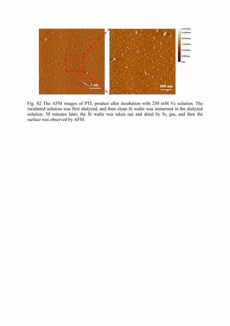
Fig. S2 The AFM images of PTL product after incubation with 250 mM Vc solution. The incubated solution was first dialyzed, and then clean Si wafer was immersed in the dialyzed solution. 30 minutes later, the Si wafer was taken out and dried by N2 gas, and then the surface was observed by AFM.
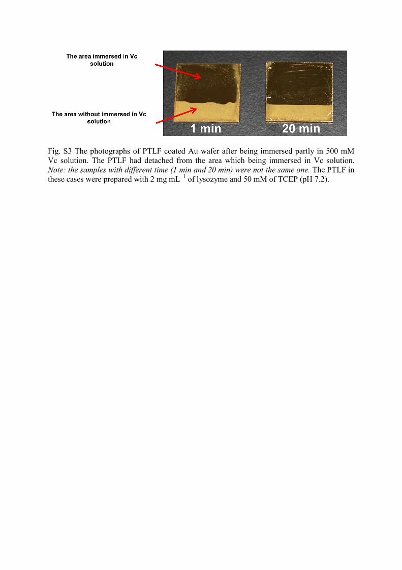
Fig. S3 The photographs of PTLF coated Au wafer after being immersed partly in 500 mM Vc solution. The PTLF had detached from the area which being immersed in Vc solution. Note: the samples with different time (1 min and 20 min) were not the same one. The PTLF in these cases were prepared with 2 mg mL−1 of lysozyme and 50 mM of TCEP (pH 7.2).

Fig. S4 XPS spectrum of the PTLF coated Au wafer surface after being immersed in 500 mM Vc solution for 20 minutes. The PTLF here was prepared with 2 mg mL−1 of lysozyme and 50 mM of TCEP (pH 7.2).

Fig. S5 The EDS results of the PTLF coated Au wafer without (left) and with (right) being immersed in 500 mM Vc solution for 20 minutes. The PTLF in these cases were prepared with 2 mg mL−1 of lysozyme and 50 mM of TCEP (pH 7.2).

Fig. S6 (A) The photographs of PTLF coated Si wafer after being immersed partly in 500 mM Vc solution. The PTLF had detached from the area which being immersed in Vc solution. Note: in (A), the samples with different time (1 min and 20 min) were not the same one; (B) The AFM images of the pristine Si wafer surface and the PTLF coated Si wafer surface before and after being immersed in 500 mM Vc solution for 20 minutes. (C) The EDS results of the PTLF coated Si wafer without (left) and with (right) being immersed in 500 mM Vc solution for 20 minutes. The PTLF in these cases were prepared with 2 mg mL−1 of lysozyme and 50 mM of TCEP (pH 7.2).

Fig. S7 A schematic illustration of the Vc-driven disassembly mechanism of the amyloid-like aggregates at the molecular level.[a]
(Ref. a: Y. Porat, A. Abramowitz, E. Gazit, Chem. Biol. Drug Des. 2006, 67, 27; W. Lee, I. Kim, S. W. Lee, H. Lee, G. Lee, S. Kim, S. W. Lee, D. S. Yoon, Macromol. Res. 2016, 24, 868)
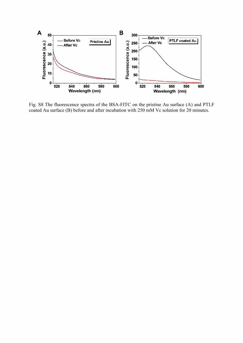
Fig. S8 The fluorescence spectra of the BSA-FITC on the pristine Au surface (A) and PTLF coated Au surface (B) before and after incubation with 250 mM Vc solution for 20 minutes.

Fig. S9 The release ratio of proteins, bacteria, and cells from PTLF coated Au surface under incubation with PBS or 250 mM Vc solution for 20 minutes. The values were calculated as the means ± standard deviation (n = 5). The PTLF in these cases were prepared with 2 mg mL−1 of lysozyme and 50 mM of TCEP (pH 6.2).

Fig. S10 The WCA change of the pristine Au wafer surface with time.
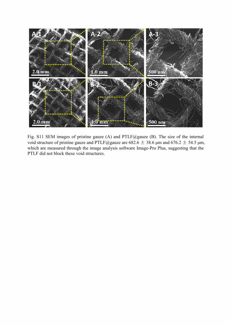
Fig. S11 SEM images of pristine gauze (A) and PTLF@gauze (B). The size of the internal void structure of pristine gauze and PTLF@gauze are 682.6 ± 38.6 μm and 676.2 ± 54.5 μm, which are measured through the image analysis software Image-Pro Plus, suggesting that the PTLF did not block these void structures.
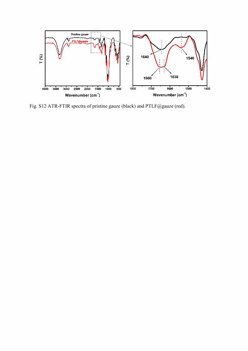
Fig. S12 ATR-FTIR spectra of pristine gauze (black) and PTLF@gauze (red).

Fig. S13 The cytotoxicity evaluation of the PTLF@gauze. (A) GFP transgenic fibroblast cells adhesion and (B) cell number on the control, pristine gauze, and PTLF@gauze after the cells were incubated for 3 days. The values are the mean ± SD (n = 3).
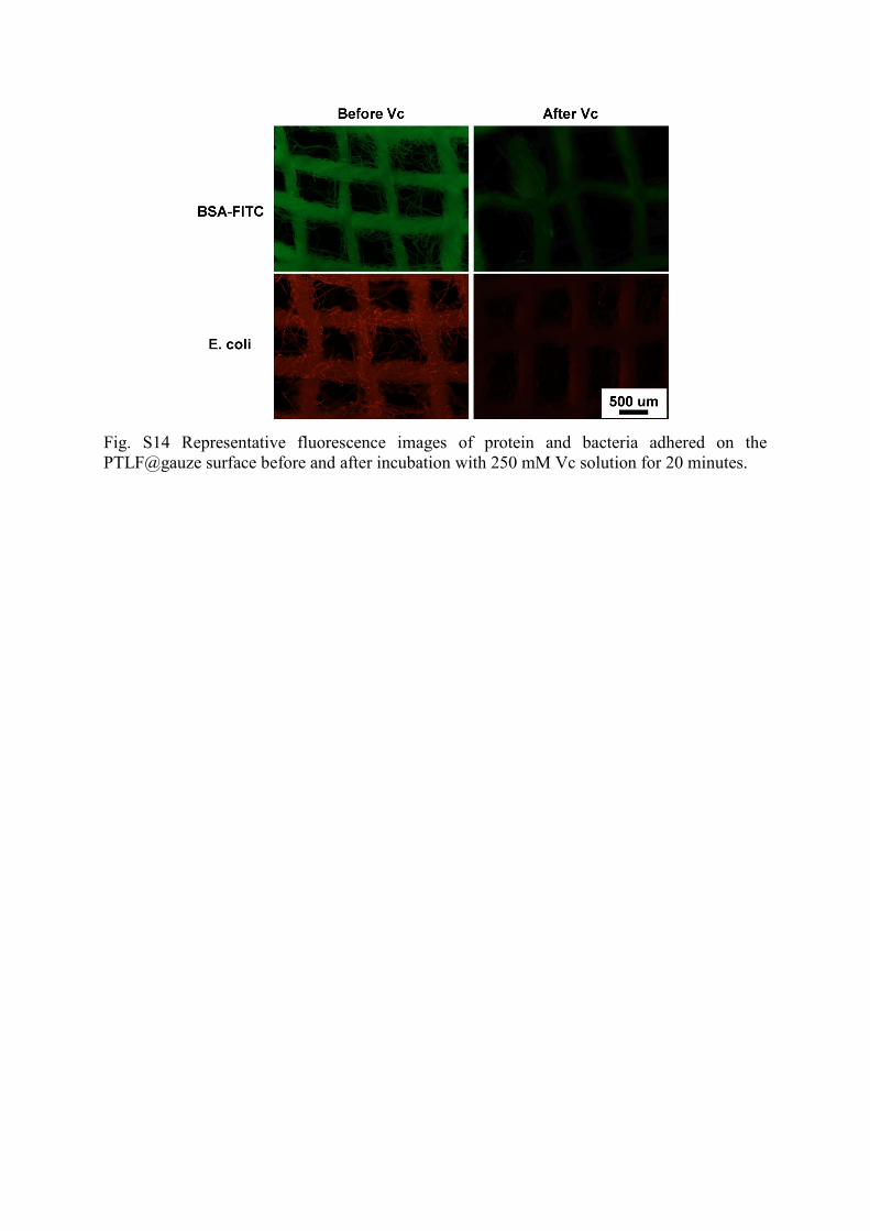
Fig. S14 Representative fluorescence images of protein and bacteria adhered on the PTLF@gauze surface before and after incubation with 250 mM Vc solution for 20 minutes.
Related Documents