Self-Assembled Peptide Amphiphile Nanofibers and PEG Composite Hydrogels as Tunable ECM Mimetic Microenvironment Melis Goktas, † Goksu Cinar, † Ilghar Orujalipoor, ‡ Semra Ide, ‡,§ Ayse B. Tekinay,* ,† and Mustafa O. Guler* ,† † Institute of Materials Science and Nanotechnology, National Nanotechnology Research Center (UNAM), Bilkent University, Ankara 06800, Turkey ‡ Department of Nanotechnology and Nanomedicine and § Faculty of Engineering, Department of Physics Engineering, Hacettepe University, Beytepe, Ankara 06800, Turkey * S Supporting Information ABSTRACT: Natural extracellular matrix (ECM) consists of complex signals interacting with each other to organize cellular behavior and responses. This sophisticated microenvironment can be mimicked by advanced materials presenting essential biochemical and physical properties in a synergistic manner. In this work, we developed a facile fabrication method for a novel nanofibrous self-assembled peptide amphiphile (PA) and poly(ethylene glycol) (PEG) composite hydrogel system with independently tunable biochemical, mechanical, and physical cues without any chemical modification of polymer backbone or additional polymer processing techniques to create synthetic ECM analogues. This approach allows noninteracting modification of multiple niche properties (e.g., bioactive ligands, stiffness, porosity), since no covalent conjugation method was used to modify PEG monomers for incorporation of bioactivity and porosity. Combining the self-assembled PA nanofibers with a chemically cross-linked polymer network simply by facile mixing followed by photopolymerization resulted in the formation of porous bioactive hydrogel systems. The resulting porous network can be functionalized with desired bioactive signaling epitopes by simply altering the amino acid sequence of the self-assembling PA molecule. In addition, the mechanical properties of the composite system can be precisely controlled by changing the PEG concentration. Therefore, nanofibrous self-assembled PA/ PEG composite hydrogels reported in this work can provide new opportunities as versatile synthetic mimics of ECM with independently tunable biological and mechanical properties for tissue engineering and regenerative medicine applications. In addition, such systems could provide useful tools for investigation of how complex niche cues influence cellular behavior and tissue formation both in two-dimensional and three-dimensional platforms. ■ INTRODUCTION Hydrogels have been intensively studied as molecularly engineered scaffolds 1 for controlled drug delivery, 2 cell encapsulation 3 and tissue regeneration 4 applications. They mimic native ECM in terms of its highly hydrated and porous network structure. 5-7 However, when the complexity of the natural ECM 8,9 is considered, hydrophilicity and porosity are not sufficient by themselves to meet the design requirements for guiding cellular behavior. The biological outcomes of introducing a biomaterial to the cellular microenvironment are dependent on cell-material interactions at the nanoscale level. 10 Cells can sense biochemical properties of a material such as the presence of bioactive ligands 11 as well as biophysical characteristics, including dimensionality 12 and matrix stiffness. 13 Therefore, functionalization of hydrogels is crucial for the modulation of cellular characteristics and plays an important role at biochemical and biophysical interfaces, depending on the desired cellular outcome for a specific application. 14 Synthetic polymers have been used as tools for the modification of biophysical characteristics of scaffolds since they provide convenient control over the mechanical proper- ties. 15 Cells can sense the mechanical properties of their environment and as a response to perceived mechanical stimuli, they generate biochemical activity along with the signal transduction mechanism called mechanotransduction. 16,17 The matrix stiffness can regulate cellular functions including Received: January 12, 2015 Revised: March 7, 2015 Article pubs.acs.org/Biomac © XXXX American Chemical Society A DOI: 10.1021/acs.biomac.5b00041 Biomacromolecules XXXX, XXX, XXX-XXX

Welcome message from author
This document is posted to help you gain knowledge. Please leave a comment to let me know what you think about it! Share it to your friends and learn new things together.
Transcript
Self-Assembled Peptide Amphiphile Nanofibers and PEG CompositeHydrogels as Tunable ECM Mimetic MicroenvironmentMelis Goktas,† Goksu Cinar,† Ilghar Orujalipoor,‡ Semra Ide,‡,§ Ayse B. Tekinay,*,†
and Mustafa O. Guler*,†
†Institute of Materials Science and Nanotechnology, National Nanotechnology Research Center (UNAM), Bilkent University, Ankara06800, Turkey‡Department of Nanotechnology and Nanomedicine and §Faculty of Engineering, Department of Physics Engineering, HacettepeUniversity, Beytepe, Ankara 06800, Turkey
*S Supporting Information
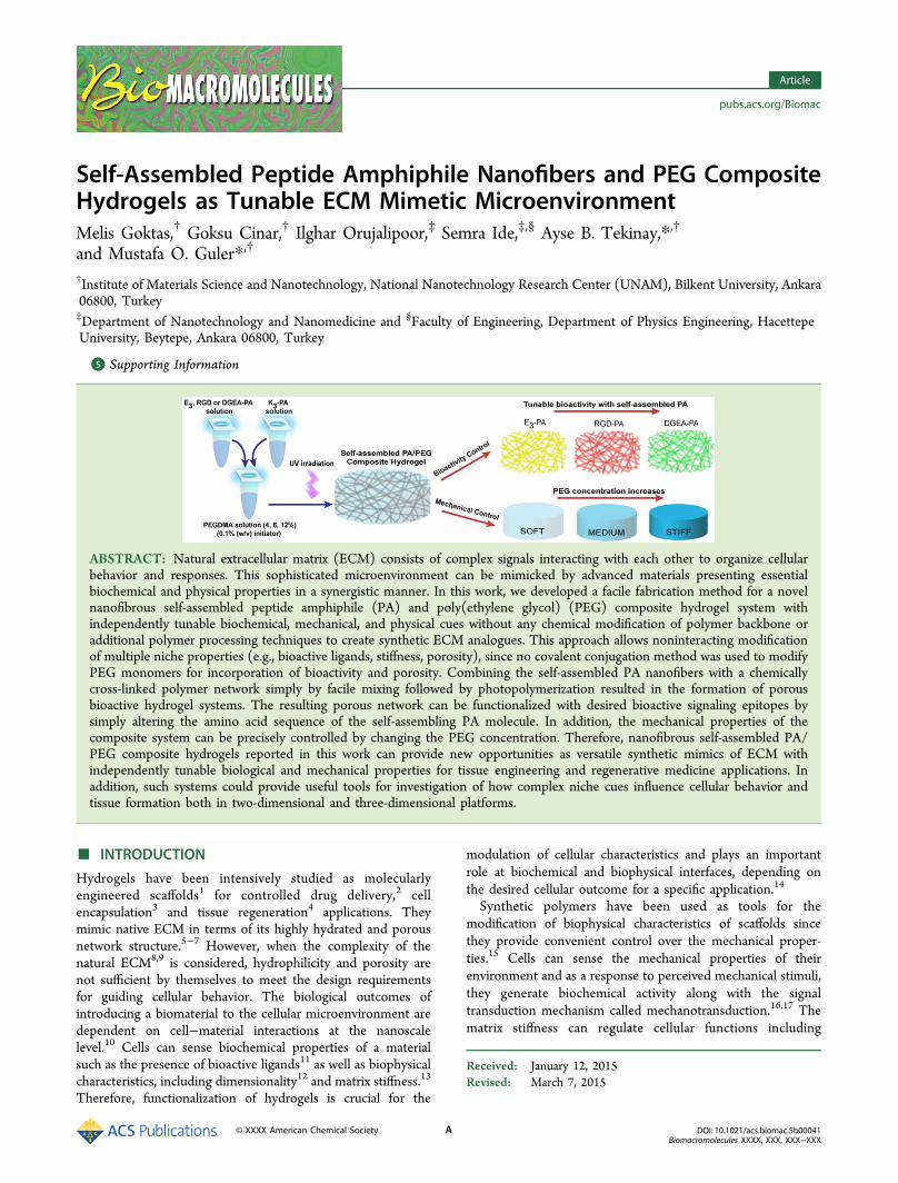
ABSTRACT: Natural extracellular matrix (ECM) consists of complex signals interacting with each other to organize cellularbehavior and responses. This sophisticated microenvironment can be mimicked by advanced materials presenting essentialbiochemical and physical properties in a synergistic manner. In this work, we developed a facile fabrication method for a novelnanofibrous self-assembled peptide amphiphile (PA) and poly(ethylene glycol) (PEG) composite hydrogel system withindependently tunable biochemical, mechanical, and physical cues without any chemical modification of polymer backbone oradditional polymer processing techniques to create synthetic ECM analogues. This approach allows noninteracting modificationof multiple niche properties (e.g., bioactive ligands, stiffness, porosity), since no covalent conjugation method was used to modifyPEG monomers for incorporation of bioactivity and porosity. Combining the self-assembled PA nanofibers with a chemicallycross-linked polymer network simply by facile mixing followed by photopolymerization resulted in the formation of porousbioactive hydrogel systems. The resulting porous network can be functionalized with desired bioactive signaling epitopes bysimply altering the amino acid sequence of the self-assembling PA molecule. In addition, the mechanical properties of thecomposite system can be precisely controlled by changing the PEG concentration. Therefore, nanofibrous self-assembled PA/PEG composite hydrogels reported in this work can provide new opportunities as versatile synthetic mimics of ECM withindependently tunable biological and mechanical properties for tissue engineering and regenerative medicine applications. Inaddition, such systems could provide useful tools for investigation of how complex niche cues influence cellular behavior andtissue formation both in two-dimensional and three-dimensional platforms.
■ INTRODUCTION
Hydrogels have been intensively studied as molecularlyengineered scaffolds1 for controlled drug delivery,2 cellencapsulation3 and tissue regeneration4 applications. Theymimic native ECM in terms of its highly hydrated and porousnetwork structure.5−7 However, when the complexity of thenatural ECM8,9 is considered, hydrophilicity and porosity arenot sufficient by themselves to meet the design requirementsfor guiding cellular behavior. The biological outcomes ofintroducing a biomaterial to the cellular microenvironment aredependent on cell−material interactions at the nanoscalelevel.10 Cells can sense biochemical properties of a materialsuch as the presence of bioactive ligands11 as well as biophysicalcharacteristics, including dimensionality12 and matrix stiffness.13
Therefore, functionalization of hydrogels is crucial for the
modulation of cellular characteristics and plays an importantrole at biochemical and biophysical interfaces, depending onthe desired cellular outcome for a specific application.14
Synthetic polymers have been used as tools for themodification of biophysical characteristics of scaffolds sincethey provide convenient control over the mechanical proper-ties.15 Cells can sense the mechanical properties of theirenvironment and as a response to perceived mechanical stimuli,they generate biochemical activity along with the signaltransduction mechanism called mechanotransduction.16,17 Thematrix stiffness can regulate cellular functions including
Received: January 12, 2015Revised: March 7, 2015
Article
pubs.acs.org/Biomac
© XXXX American Chemical Society A DOI: 10.1021/acs.biomac.5b00041Biomacromolecules XXXX, XXX, XXX−XXX

adhesion,18 spreading,19 migration,20 proliferation,21 and differ-entiation.13,22 One of the most commonly used syntheticpolymers to investigate the effects of mechanical stimuli oncellular behavior is poly(ethylene glycol) (PEG), whichprovides precise control of material stiffness.23 PEG is aninteresting hydrogel material with its good water solubility,biocompatibility, nonimmunogenicity, and resistance to proteinadsorption.24 However, PEG cannot provide cell attachmentand induce further cell−material interactions due to its protein-repellent property. Current strategies for creating functionalPEG hydrogels that provide the specific biochemical character-istics of native ECM, require incorporation of ECM-derivedbioactive molecules via cross-linking chemistries.25−28 Shortpeptide sequences are major epitopes for the addition ofbioactivity. Fibronectin-derived RGD peptide is one of themost commonly used adhesive peptide sequences to introducebioactivity to PEG hydrogels.25,29 Various strategies have beendescribed in the literature to create RGD-coupled hydrogelnetworks of PEG macromers. Michael addition reactions andacrylate polymerization are the most widely utilized cross-linking chemistries.30,31 Nevertheless, covalent conjugation offunctional epitopes to the polymer chain requires complexchemical reactions and can result in limited mobility andaccessibility of bioactive ligands.22 Also, peptide incorporationinto the hydrogels is limited since covalent conjugation affectshydrogel formation and mechanical properties. Since ligandpresentation and convenient control over the mechanicalproperties play important roles in controlling cell behavior,cross-linking-chemistries stay as insufficient approaches forincorporation of bioactivity to PEG hydrogels. In addition,limited porosity of the cross-linked network could prevent cellmotility, cell−cell interactions, and diffusion, especially in thecase of three-dimensional (3D) culture conditions. A numberof approaches have been shown to generate porous PEGnetworks such as salt leaching32 and gas foaming.33 However,these methods require multiple steps and they still have broadpore size distributions with poor pore interconnectivity.Therefore, these strategies are far from presenting bioactivenanoscale architecture for mimicking the ECM microenviron-ment.When compared to current PEG systems, supramolecular PA
networks have nanofibrous architecture and tailorable bioactiveproperties and they are versatile platforms that can eliminatethe limitations of covalent cross-linking.34 Under physiologicalconditions, PAs can self-assemble into one-dimensionalnanostructures, predominantly cylindrical nanofibers.35
Through incorporation of specific amino acids into thesequence, self-assembled PA networks allow construction ofbioactive hydrogels closely mimicking the nanoscale architec-ture and function of the native ECM.36 PA hydrogels canpresent a variety of bioactive signals on the nanofiber surfacesat high concentration without any limitation of ligandpresentation.37 Short peptide sequences derived from thenative ECM proteins could be incorporated into PA systems todirect the cellular processes. For instance, RGD epitope hasbeen attached to PAs to produce adhesive self-assembledpeptide networks.38,39 It has been shown in many studies thatαvβ3 integrin binding RGD sequence induce adhesion,spreading and migration of fibroblasts,40 osteoblasts,41 andmesenchymal stem cells.42 Another bioactive epitope of interestis α2β1 integrin binding DGEA (Asp-Gly-Glu-Ala) peptidederived from collagen type-1. The DGEA peptide was shown to
promote survival and osteogenic differentiation of hMSCs andmouse preosteoblast MC3T3 cells.43−45
Multicomponent hybrid hydrogel strategies such as incorpo-ration of two photo-cross-linkable architectures,46 integration ofcovalent and ionic cross-linking of polymers within hybridhydrogels,47,48 or combination of a biopolymer network withchemically cross-linked poly(ethylene glycol)diacrylate(PEGDA) hydrogel49 have been developed to overcomeindividual limitations of synthetic polymer hydrogels.28 Here,we employed a facile fabrication strategy to create a novel self-assembled PA/PEG composite hydrogel system without anymodification of polymer backbone or additional polymerprocessing techniques, to create synthetic ECM analogues.While mechanical reinforcement and stability is supported viacovalently cross-linked PEG monomers, the noncovalentinteractions between self-assembling PA molecules enable usto obtain bioactive nanofibrous architecture mimicking naturalECM. Synergistic combinations of different classes of materialsprovide us opportunities for designing new matrices withindependently tunable biochemical, mechanical, and physicalproperties. The design and synthesis of these ECM mimeticcomposite hydrogels were demonstrated; physical andmechanical properties (e.g., nanoarchitecture, porosity, stiff-ness, elasticity) of resulting multicomponent networks wereelucidated and the interactive effects of mechanical andbiochemical cues on cellular behavior were investigated.
■ EXPERIMENTAL SECTIONMaterials. All protected amino acids, lauric acid, Rink amide
MBHA resin, Fmoc-Glu(OtBu)-Wang resin (100−200 mesh), Fmoc-Aps(OtBu)-Wang resin (100−200 mesh), N,N,N′,N′-tetramethyl-O-(1H-benzotriazole-1-yl) uronium hexafluorophosphate (HBTU), anddiisopropylethylamine (DIEA) were purchased from Novabiochem,ABCR or Sigma-Aldrich. All other chemicals and materials used in thisstudy were analytical grade and purchased from Invitrogen, Fisher,Merch, Alfa Aesar, and Sigma-Aldrich.
Synthesis and Characterization of Peptide Amphiphiles.Fmoc solid phase peptide synthesis method was employed tosynthesize Lauryl-Val-Val-Ala-Gly-Lys-Lys-Lys-Am (K3-PA), Lauryl-Val-Val-Ala-Gly-Glu-Glu-Glu (E3-PA), Lauryl-Val-Val-Ala-Gly-Glu-Arg-Gly-Asp (RGD-PA), and Lauryl-Val-Val-Ala-Gly-Glu-Gly-Asp-Gly-Glu-Ala-Am (DGEA-PA). For K3-PA and DGEA-PA, Rinkamide MBHA resin (Novabiochem) served as the solid support,while Fmoc-Glu(OtBu)-Wang resin (100−200 mesh) and Fmoc-Asp(OtBu)-Wang resin (100−200 mesh) were used as solid supportsfor E3-PA and RGD-PA. Carboxylate group activation of 2 molequivalents of amino acid was succeeded by 1.95 mol equivalents ofN,N,N′,N′-tetramethyl-O-(1H-benzotriazole-1-yl) uronium hexafluor-ophosphate (HBTU), and 3 mol equivalents of diisopropylethylamine(DIEA) for 1 mol equivalent of functional sites on the solid resin.Fmoc groups were removed at each coupling step with 20%piperidine/dimethylformamide for 20 min. Amino acid couplingtime was set to be 2 h at each cycle. Lauric acid served as the source oflauryl group and its coupling mechanism was similar to amino acidcoupling. A 10% acetic anhydride−DMF solution was used to acetylatethe unreacted amine groups after each coupling step. Cleavage ofprotecting groups and peptide molecules from the solid support wascarried out by trifluoroacetic acid (TFA) cleavage cocktail (95% TFA,2.5% water, 2.5% triisopropylsilane) for 3 h. Excess TFA was removedby rotary evaporation. Synthesized PAs were then precipitated indiethyl ether overnight. The precipitate was collected by centrifugationand dissolved in ultrapure water. This solution was frozen at −80 °Cfollowed by lyophilization for 1 week. The purity of the peptides wasassessed using Agilent 6530 quadrupole time-of-flight (Q-TOF) massspectrometry with electrospray ionization (ESI) source equipped withreverse-phase analytical high performance liquid chromatography(HPLC). Synthesized peptides were purified with a preparative
Biomacromolecules Article
DOI: 10.1021/acs.biomac.5b00041Biomacromolecules XXXX, XXX, XXX−XXX
B
HPLC system (Agilent 1200 series). All PA molecules were freeze-dried and reconstituted in ultrapure water at pH 7.4 before use.Preparation of 2D Hydrogels. Stock solutions of poly(ethylene
glycol) dimethacrylate (PEGDMA) (Mn = 550, Aldrich) wereprepared in ultrapure water. Photoinitiator, 2,2′-Azobis (2-methyl-propionamidine) dihydro-chloride (Aldrich; 1% (w/v)) was dissolvedin ultrapure water and added to the PEGDMA-initiator solutions.Synthesized PAs were dissolved in ultrapure water at neutral pH,separately and 3% (w/v) stock PA solutions were prepared. Initially,negatively charged PA solution (E3-PA, RGD-PA, or DGEA-PA) wasmixed with PEGDMA stock solutions containing the initiator andvortexed. Then, positively charged PA (K3-PA) was added to themixture to trigger self-assembly of PAs via charge neutralization andobtain self-assembled nanofibrous network via noncovalent inter-actions. After the mixture of oppositely charged PAs with PEGDMAsolutions containing the initiator, the solutions were immediatelytransferred to cell culture plates (48 or 96 well-plate) and exposed toultraviolet (UV) light at 365 nm wavelength for 15 min for theformation of covalently cross-linked 2D hydrogel substrates. For UVcross-linking, 8 W UVP UVLMS-38 EL series UV lamp was used, andthe lamp was placed on top of cell culture plates directly (the distanceof the lamp from the samples was approximately 0.5 cm). Thevolumetric ratio of PEGDMA to mixture of PAs within the compositehydrogels was determined as 1:1. Final PEGDMA concentrations were4, 8, and 12% (w/v), and initiator concentration was 0.1% (w/v) in thecomposite system. Since PEGDMA solutions were mixed with PAsolutions at 1:1 volumetric ratio, the final PA concentrations within thesystem were 1.5% (w/v) for all combinations. The volumetric ratios ofoppositely charged PA combinations were determined as E3-PA + K3-PA (3:4), RGD-PA + K3-PA (3:2), and DGEA-PA + K3-PA (1:1) forcomplete charge neutralization within the composite hydrogels. Forthe preparation of control PEG hydrogels, ultrapure water (with samevolume of PA solutions) was added to the stock PEGDMA solutions.The final concentrations were also 4, 8, and 12% (w/v) PEGDMA and0.1% (w/v) initiator within the control groups.Preparation of 3D Hydrogels. Similar simple preparation
approach was applied to encapsulate Saos-2 cells into 3D matrices.Only difference was that all peptide and PEG-photoinitiator solutionswere prepared with Dulbecco’s modified essential medium (DMEM)instead of water and cell suspension (1 × 10⁶ cells/sample) was mixedwith PEG-photoinitiator solution before the addition of PA solutionsinto the mixture. Total volume of the pregel solutions was 200 μL.After the preparation of pregel solutions, mixtures were transferredinto the caps of eppendorf tubes and exposed to UV light at 365 nmfor 15 min. The resulting disc-shaped 3D gels containing encapsulatedSaos-2 cells were cultured in Synthecon RCCS-4H bioreactor system.Attenuated Total Reflectance-Fourier Transform Infrared
Spectroscopy (ATR-FT-IR). A Thermo Scientific NICOLET 6700-FTIR fitted with a universal ATR sampling accessory was utilized tocharacterize the secondary structure of cross-linked PEG (w/o PNFs)and E3/PEG samples. Prior to testing, samples were dried in ambientair at 25 °C for 48 h. All data was recorded at 25 °C, in the spectralrange of 4000−800 cm−1, utilizing a 16 scans per sample cycle.Scanning Electron Microscopy (SEM). To visualize the resulting
network formation within the polymerized samples, scanning electronmicroscopy (SEM) was employed. SEM samples were prepared oncleaned silicon wafer surfaces with a similar approach to preparation of2D hydrogels. Following UV cross-linking, samples were dehydrated ingradually increasing concentrations of ethanol solutions. Thedehydrated hydrogels were dried with a Tourismis Autosamdri-815Bcritical-point-drier to preserve the network structure. A FEI Quanta200 FEG scanning electron microscope with an ETD detector wasused for visualization of resulting networks. Samples were sputtercoated with 4 nm gold/palladium prior to imaging.Brunauer−Emmett−Teller (BET) Analysis. Pore size distribu-
tion, total pore volume and specific surface area of PEG and PA/PEGsamples were estimated by BET analysis. This technique is used for thedetermination of surface area and porosity of synthetic polymerichydrogels via nitrogen adsorption isotherms.50,51 Before the analysis,samples were dehydrated in gradually increasing concentrations of
ethanol solutions. Dehydrated samples were dried with a TourismisAutosamdri-815B critical-point-drier to preserve the networkstructures. Samples were degassed at 150 °C for 4 h and N2adsorption was conducted at 77 K. Total pore volume and specificsurface area of the samples were calculated by using quenched soliddensity functional theory (QSDFT).
Small Angle X-ray Scattering (SAXS) Analysis. The 4% (w/v)PEG (w/o PA nanofibers) and E3/PEG composite hydrogels for SAXSanalysis were prepared in quartz capillaries with a similar approach topreparation of 2D hydrogels. After loading of pregel solutions of 4%(w/v) PEG (w/o PA nanofibers) and E3/PEG composite hydrogelsinto the quartz capillaries, the samples were exposed to ultraviolet(UV) light at 365 nm wavelength for 15 min for the formation ofcross-linked hydrogels. The scattering experiments were performed ona SWAXS system with Kratky optic HECUS (Hecus X-ray systems, M.Braun, Graz, Austria) and equipped with a linear collimation system.Nickel-filtered Cu Kα radiation (λ = 1.54 Å), originating from a PhilipsX-ray generator with copper anode operating at a power of 2 kW (50kV and 40 mA) was used. A linear-position sensitive detector OED 50-M (M. Braun, Garching, Germany) with 1024 channel resolution wasused to record the scattering data in small angle region. Datacalibration was performed with silver behenate. Distances betweenchannels and the sample-detector were 54 μm and 28.1 cm,respectively. Scattering curves were monitored in q ranges of 0.004−0.55 Å−1 for SAXS. All samples were measured for exposure times of600 s at room temperature (23 °C). Data acquisition was completedby using the program ASAV2.3 (HECUS M. Braun, Graz, Austria).
Oscillatory Rheology. An Anton Paar Physica RM301 Rheometerwith a 25 mm parallel-plate configuration was used to characterize theviscoelastic properties of PEG, PA, and PA/PEG hydrogels. Cross-linked PEG and PA/PEG gels were formed inside 48-well cell cultureplates and then transferred on the lower plate of the rheometer, whilepeptide gels were formed in situ on the rheometer plate. Total volumeof the samples was 300 μL and shear gap distance was 500 nm. Allmeasurements were carried out at room temperature. Gelation kineticsof the gels was characterized with time-dependent rheology. Duringthe time-sweep test, angular frequency, and strain were held constantat 10 rad s−1 and 0.01%, respectively. To determine the linearviscoelastic range (LVR) of the gels, amplitude sweep test wasconducted at constant angular frequency of 10 rad s−1 withlogarithmically ramping the strain amplitude from 0.01 to 1000%.
Cell Culture and Maintenance. Saos-2 human osteosarcoma cells(ATCCHTB-85) were used in adhesion, spreading, viability,immunocytochemistry, and gene expression experiments. All cellswere cultured in 75 cm2 cell culture flasks using Dulbecco’s modifiedEagle medium (DMEM) supplemented with 10% fetal bovine serum(FBS), 1% penicillin/streptomycin, and 2 mM L-glutamine. The cellswere kept at 37 °C in a humidified chamber supplied with 5% CO2.Cell passage was carried out at cell confluency between 80 and 90%using trypsin/EDTA chemistry. The culture medium was changedevery 3−4 days. For osteogenic differentiation experiments (ICCstainings, qRT-PCR analysis), the seeded cell medium was replacedwith osteogenic medium, DMEM supplemented with 10% FBS, 10mM β-glycerophosphate, 50 μg mL−1 ascorbic acid, and 10 nMdexamethasone, after reaching confluency.
Viability of Saos-2 Cells on PEG and PA/PEG Substrates.Viability of Saos-2 cells was analyzed on PEG and PA/PEG substratesprepared in 48 well cell culture plates. Tissue culture plate surface wasalso used to evaluate the viability of the cells on a control sample. Priorto cell seeding, cross-linked substrates were washed with 1× phosphatebuffered saline (PBS) overnight. Cells were seeded onto hydrogel andtissue culture plate surfaces in DMEM media supplemented with 10%FBS, 1% penicillin/streptomycin and 2 mM L-glutamine at a density of1.5 × 104 cells/cm2. After 3 days of incubation the cell medium wasdiscarded, the cells were washed with PBS and then incubated with 2μM Calcein-AM/ethidium homodimer (Invitrogen) in PBS for 20−30min at room temperature. Finally, random images were taken at 10×magnification by a fluorescence microscope from each well for analysis.
Adhesion of Saos-2 Cells on PEG and PA/PEG Substrates. Todetermine the effect of protein-repellent property of PEG on cellular
Biomacromolecules Article
DOI: 10.1021/acs.biomac.5b00041Biomacromolecules XXXX, XXX, XXX−XXX
C
adhesion, adhesion of Saos-2 cells were analyzed on PEG and PA/PEGhydrogels prepared in 48-well cell culture plates. Cells were seeded onhydrogel surfaces at a density of 5 × 10³ cells/cm2 in serum-freeculture conditions with DMEM media supplemented with 1%penicillin/streptomycin and 2 mM L-glutamine. The cells wereincubated at standard cell culture conditions. After 24, 48, and 72 h,the unbound cells were washed away three times with PBS, and theremaining bound cells were stained with 2 μM Calcein-AM. At least 20random images were taken per substrate (n = 3). Cell adhesions werequantified by counting the number of cells with ImageJ program.Spreading and Cytoskeletal Organization Analysis of Saos-2
Cells on PEG and PA/PEG Substrates. Spreading and cytoskeletalorganization of Saos-2 cells were analyzed on PEG and PA/PEGsurfaces at 72 h. Preparation of the samples was the same as thesamples for the adhesion assay. Before staining, cells were fixed with3.7% formaldehyde for 15 min and permeabilized with 0.1% Triton X-100 for 10 min. Actin filaments of the cells were stained with TRITC-conjugated phalloidin (Invitrogen) in 1X PBS for 20 min. Spreadingand cytoskeletal organization of cells were analyzed with Zeiss LSM510 confocal microscope. Cell spreading was quantified by measuringthe spreading areas of cells with ImageJ program. At least 30 randomimages were taken per substrate (n = 3).Immunocytochemistry (ICC). Before ICC stainings, differ-
entiated cells were fixed with 4% formaldehyde for 15 min andpermeabilized with 0.5% Triton-X for 10 min at room temperature.The 3 wt % BSA/PBS was used for blocking for 1 h. Rabbit-raised,antihuman, RUNX2 and COL1 primary antibodies and a goat-raised,antirabbit, IgG H&L DyLight 488 conjugated secondary antibody(Abcam) were used for staining. The samples were visualized with aZeiss LSM 510 confocal microscope.
Quantitative Reverse Transcription Polymerase ChainReaction (qRT-PCR). RUNX2 and COL1 gene expression profileswere examined by qRT-PCR. Total RNA of differentiated Saos-2 cellswas isolated on days 3 and 7 using TRIzol reagent (Ambion)according to the manufacturer’s protocol. Nanodrop 2000 (ThermoScientific) was used to quantify the yield and purity of the isolatedRNA. Primer sequences were designed using Primer 3 software (TableS2). SuperScript III Platinum SYBR Green One-Step qRT-PCR kitwas used to carry out quantitative reverse transcription polymerasechain reaction. Temperature cycling for the reaction was determined as55 °C for 5 min, 95 °C for 5 min, 40 cycles of 95 °C for 15 s, Tm (58.0°C for RUNX2 and GAPDH, 60.0 °C for COL1) for 30 s, and 40 °Cfor 1 min. Gene expressions were normalized to GAPDH as theinternal control gene.
Statistical Analysis. All experiments were independently repeatedat least twice with at least three replicas for each experimental group.All quantitative results were expressed as ±standard error of means(SEM). Statistical analyses were carried out by one-way or two-wayanalysis of variance (ANOVA), whichever is applicable. For thestatistical significance, a P-value of less than 0.05 was considered assignificant.
■ RESULTS AND DISCUSSION
To fabricate self-assembled PA and PEG composite nano-structured systems, polyethylene glycol dimethacrylate (PEGD-MA, Mn = 550) was used because of its biological inertness, cellcompatibility and ability to photo-cross-link. Photo-cross-linking is suitable for biomedical applications due to the mildand rapid reaction conditions, which can be conducted at
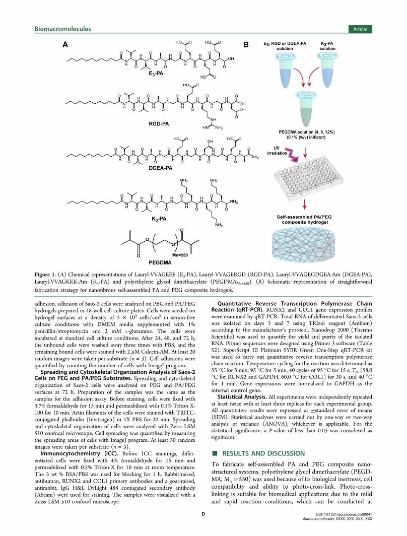
Figure 1. (A) Chemical representations of Lauryl-VVAGEEE (E3-PA), Lauryl-VVAGERGD (RGD-PA), Lauryl-VVAGEGDGEA-Am (DGEA-PA),Lauryl-VVAGKKK-Am (K3-PA) and polyethylene glycol dimethacrylate (PEGDMAMn=550). (B) Schematic representation of straightforward
fabrication strategy for nanofibrous self-assembled PA and PEG composite hydrogels.
Biomacromolecules Article
DOI: 10.1021/acs.biomac.5b00041Biomacromolecules XXXX, XXX, XXX−XXX
D
physiological temperature and pH. For the modulation ofmechanical stiffness, three different PEG concentrations (4, 8,and 12% (w/v)) were used.Self-assembling PA molecules consist of a hydrophilic
peptide segment conjugated to a hydrophobic fatty acidtriggering self-assembly of PA molecules into one-dimensionalnanostructures in aqueous solution.52 In addition, it was shownthat two oppositely charged PAs carrying different bioactiveepitopes can self-assemble into nanofibers at physiologicalconditions due to electrostatic interactions between ionicamino acids of PAs.53 Noncovalent forces such as hydrogenbonding, hydrophobic and electrostatic interactions betweenPAs trigger and stabilize the fiber formation.54 Fmoc solidphase peptide chemistry was employed to synthesize PAmolecules. Four different PA molecules [Lauryl-VVAGEEE(E3-PA), Lauryl-VVAGERGD (RGD-PA), Lauryl-VVA-GEGDGEA-Am (DGEA-PA), Lauryl-VVAGKKK-Am (K3-PA)] were synthesized (Figures 1A and S1). The E3-PA wasused as nonintegrin binding peptide sequence while RGD-PAand DGEA-PA were exploited as integrin binding epitopes toinvestigate the effect of different bioactive signals on cellularbehavior. Positively charged K3-PA was utilized to inducenanofibrous assembly when mixed with negatively charged PAmolecules at physiological conditions (Figure S2).To obtain porous hydrogel networks with independently
tunable mechanical and biochemical properties, a simplefabrication method was implemented (Figure 1B). A photo-
initiator, 2,2′-azobis(2-methyl-propionamidine)dihydro-chlor-ide) was dissolved in ultrapure water and added into thePEG solution with a final concentration of 0.1% (w/v). The PAmolecules were dissolved in ultrapure water, separately.Initially, negatively charged PA (E3-PA, RGD-PA, or DGEA-PA) solutions were mixed with PEG-photoinitiator solution,and then positively charged K3-PA was added to the solutionsto trigger nanofiber self-assembly through charge neutralizationat neutral pH. The final PA concentration within the PA/PEGmixture was determined as 1.5% (w/v) for all PA/PEGcomposite hydrogels. The PA/PEG solutions were transferredto the cell culture plates immediately without any incubationstep, and exposed to ultraviolet (UV) light at 365 nmwavelength for 15 min to induce photopolymerization. In ourdesign, both hydrophobic and electrostatic interactions(between lauryl group and oppositely charged amino acidresidues, respectively) provide driving forces for self-assemblyand nanofiber formation within the system before the cross-linking of PEG monomers in the solution. Cross-linkingoccurred through radical polymerization, where the methacry-late groups participated in an addition reaction to form abranched polymeric network55 (Figure S3A). In addition, ATR-FTIR analysis on PEG (w/o PA nanofibers) and PA/PEG (E3/PEG combination) samples after 15 min of UV exposure alsosupported presence of PA network53 and cross-linkedmethacrylate chains55 within the composite hydrogel (FigureS3B). A total of 12 groups were studied, including nonbioactive
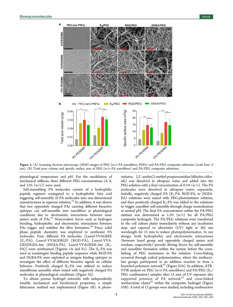
Figure 2. (A) Scanning electron microscopy (SEM) images of PEG (w/o PA nanofibers, PNFs) and PA/PEG composite substrates (scale bars: 4μm). (B) Total pore volume and specific surface area of PEG (w/o PA nanofibers) and PA/PEG composite substrates.
Biomacromolecules Article
DOI: 10.1021/acs.biomac.5b00041Biomacromolecules XXXX, XXX, XXX−XXX
E
PEG (w/o PA nanofibers) controls versus PA/PEG compositescaffolds. Three different PA groups (E3-PA as nonintegrinbinding sequence, RGD-PA and DGEA-PA as integrin bindingepitopes in Figure 1A) and three different mechanical stiffnessgroups (PEGDMA concentrations 4, 8, and 12% (w/v) definedas soft, medium, and stiff in Figure 4A) were exploited. Thedetails about net charges of PA molecules in water at neutralpH, nomenclature of PA/PEG composite systems, nanofibercompositions, and volumetric mixing ratios of PA moleculeswere given in Tables S1 and S2.When noncovalently self-assembled PA nanofibers were
incorporated within the cross-linked PEG network, nanofibrousporous scaffolds were formed, while the PEG (w/o PAnanofibers) gel was observed to be relatively nonporous(Figure 2A), as shown by scanning electron microscopy(SEM) imaging. The morphology of the porous networkswas similar for all of the groups with different PEGconcentrations and PA combinations (Figure 2A). We alsoquantitatively analyzed the porosity of the resulting networkswith BET (Brunauer−Emmett−Teller) analysis.50,51 Pore sizedistribution, cumulative pore volume, and specific surface areaof the samples were measured after the hydrogels were driedwith a critical-point drier to prevent the shrinkage of thenetworks. Due to the high water content of the 4% (w/v)PEGDMA group, it was not possible to obtain measurementsafter drying; therefore, the “soft” hydrogel group was eliminatedfrom BET analysis. Pore size distributions showed that theresulting networks consisted pores in a range of up to 35 nm incase of incorporation of the PA nanofibers in addition to severalsmaller pores (<5 nm; Figure S4). Such mesoporous structuresare useful for tissue engineering, since the pores in thenanometer range can support cell adhesion and proliferationand can potentially allow protein and growth factorabsorption.56,57 Also, incorporation of the self-assembled PAnanofibers resulted in significantly increased total pore volumeand specific surface area of the resulting networks up to 4-foldcompared to PEG (w/o PA nanofibers) scaffolds (Figure 2B).
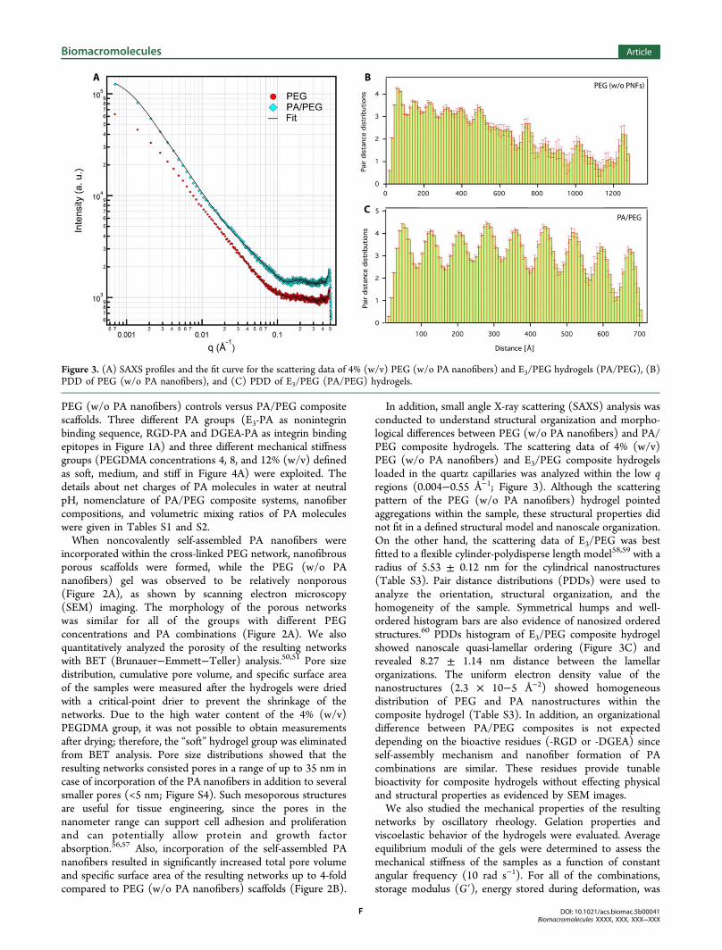
In addition, small angle X-ray scattering (SAXS) analysis wasconducted to understand structural organization and morpho-logical differences between PEG (w/o PA nanofibers) and PA/PEG composite hydrogels. The scattering data of 4% (w/v)PEG (w/o PA nanofibers) and E3/PEG composite hydrogelsloaded in the quartz capillaries was analyzed within the low qregions (0.004−0.55 Å−1; Figure 3). Although the scatteringpattern of the PEG (w/o PA nanofibers) hydrogel pointedaggregations within the sample, these structural properties didnot fit in a defined structural model and nanoscale organization.On the other hand, the scattering data of E3/PEG was bestfitted to a flexible cylinder-polydisperse length model58,59 with aradius of 5.53 ± 0.12 nm for the cylindrical nanostructures(Table S3). Pair distance distributions (PDDs) were used toanalyze the orientation, structural organization, and thehomogeneity of the sample. Symmetrical humps and well-ordered histogram bars are also evidence of nanosized orderedstructures.60 PDDs histogram of E3/PEG composite hydrogelshowed nanoscale quasi-lamellar ordering (Figure 3C) andrevealed 8.27 ± 1.14 nm distance between the lamellarorganizations. The uniform electron density value of thenanostructures (2.3 × 10⁻⁵ Å−2) showed homogeneousdistribution of PEG and PA nanostructures within thecomposite hydrogel (Table S3). In addition, an organizationaldifference between PA/PEG composites is not expecteddepending on the bioactive residues (-RGD or -DGEA) sinceself-assembly mechanism and nanofiber formation of PAcombinations are similar. These residues provide tunablebioactivity for composite hydrogels without effecting physicaland structural properties as evidenced by SEM images.We also studied the mechanical properties of the resulting
networks by oscillatory rheology. Gelation properties andviscoelastic behavior of the hydrogels were evaluated. Averageequilibrium moduli of the gels were determined to assess themechanical stiffness of the samples as a function of constantangular frequency (10 rad s−1). For all of the combinations,storage modulus (G′), energy stored during deformation, was
Figure 3. (A) SAXS profiles and the fit curve for the scattering data of 4% (w/v) PEG (w/o PA nanofibers) and E3/PEG hydrogels (PA/PEG), (B)PDD of PEG (w/o PA nanofibers), and (C) PDD of E3/PEG (PA/PEG) hydrogels.
Biomacromolecules Article
DOI: 10.1021/acs.biomac.5b00041Biomacromolecules XXXX, XXX, XXX−XXX
F
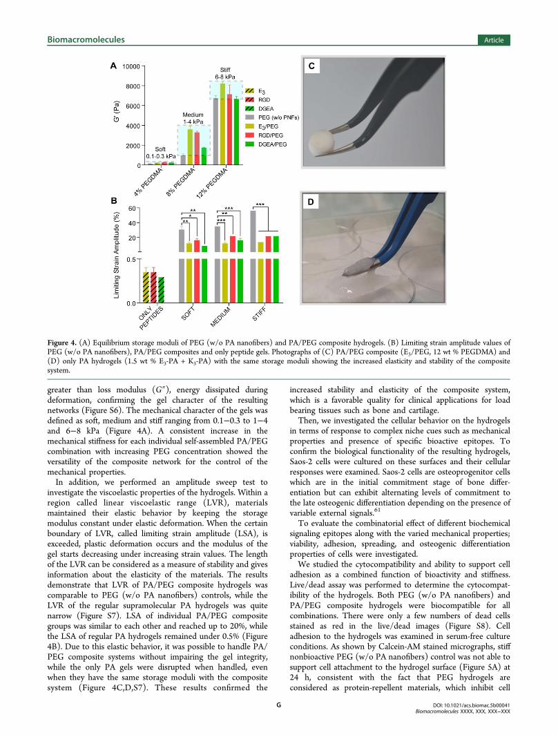
greater than loss modulus (G″), energy dissipated duringdeformation, confirming the gel character of the resultingnetworks (Figure S6). The mechanical character of the gels wasdefined as soft, medium and stiff ranging from 0.1−0.3 to 1−4and 6−8 kPa (Figure 4A). A consistent increase in themechanical stiffness for each individual self-assembled PA/PEGcombination with increasing PEG concentration showed theversatility of the composite network for the control of themechanical properties.In addition, we performed an amplitude sweep test to
investigate the viscoelastic properties of the hydrogels. Within aregion called linear viscoelastic range (LVR), materialsmaintained their elastic behavior by keeping the storagemodulus constant under elastic deformation. When the certainboundary of LVR, called limiting strain amplitude (LSA), isexceeded, plastic deformation occurs and the modulus of thegel starts decreasing under increasing strain values. The lengthof the LVR can be considered as a measure of stability and givesinformation about the elasticity of the materials. The resultsdemonstrate that LVR of PA/PEG composite hydrogels wascomparable to PEG (w/o PA nanofibers) controls, while theLVR of the regular supramolecular PA hydrogels was quitenarrow (Figure S7). LSA of individual PA/PEG compositegroups was similar to each other and reached up to 20%, whilethe LSA of regular PA hydrogels remained under 0.5% (Figure4B). Due to this elastic behavior, it was possible to handle PA/PEG composite systems without impairing the gel integrity,while the only PA gels were disrupted when handled, evenwhen they have the same storage moduli with the compositesystem (Figure 4C,D,S7). These results confirmed the
increased stability and elasticity of the composite system,which is a favorable quality for clinical applications for loadbearing tissues such as bone and cartilage.Then, we investigated the cellular behavior on the hydrogels
in terms of response to complex niche cues such as mechanicalproperties and presence of specific bioactive epitopes. Toconfirm the biological functionality of the resulting hydrogels,Saos-2 cells were cultured on these surfaces and their cellularresponses were examined. Saos-2 cells are osteoprogenitor cellswhich are in the initial commitment stage of bone differ-entiation but can exhibit alternating levels of commitment tothe late osteogenic differentiation depending on the presence ofvariable external signals.61
To evaluate the combinatorial effect of different biochemicalsignaling epitopes along with the varied mechanical properties;viability, adhesion, spreading, and osteogenic differentiationproperties of cells were investigated.We studied the cytocompatibility and ability to support cell
adhesion as a combined function of bioactivity and stiffness.Live/dead assay was performed to determine the cytocompat-ibility of the hydrogels. Both PEG (w/o PA nanofibers) andPA/PEG composite hydrogels were biocompatible for allcombinations. There were only a few numbers of dead cellsstained as red in the live/dead images (Figure S8). Celladhesion to the hydrogels was examined in serum-free cultureconditions. As shown by Calcein-AM stained micrographs, stiffnonbioactive PEG (w/o PA nanofibers) control was not able tosupport cell attachment to the hydrogel surface (Figure 5A) at24 h, consistent with the fact that PEG hydrogels areconsidered as protein-repellent materials, which inhibit cell
Figure 4. (A) Equilibrium storage moduli of PEG (w/o PA nanofibers) and PA/PEG composite hydrogels. (B) Limiting strain amplitude values ofPEG (w/o PA nanofibers), PA/PEG composites and only peptide gels. Photographs of (C) PA/PEG composite (E3/PEG, 12 wt % PEGDMA) and(D) only PA hydrogels (1.5 wt % E3-PA + K3-PA) with the same storage moduli showing the increased elasticity and stability of the compositesystem.
Biomacromolecules Article
DOI: 10.1021/acs.biomac.5b00041Biomacromolecules XXXX, XXX, XXX−XXX
G
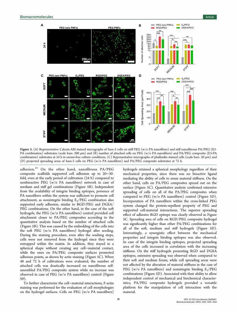
adhesion.25 On the other hand, nanofibrous PA/PEGcomposite scaffolds supported cell adhesion up to 20−30-fold, even at the early period of cultivation (24 h) compared tononbioactive PEG (w/o PA nanofibers) network in case ofmedium and stiff gel combinations (Figure 5B). Independentfrom the availability of integrin binding epitopes, presence ofPA nanofibers within the system was sufficient to promote cellattachment, as nonintegrin binding E3/PEG combination alsosupported early adhesion, similar to RGD/PEG and DGEA/PEG combinations. On the other hand, in the case of the softhydrogels, the PEG (w/o PA nanofibers) control provided cellattachment closer to PA/PEG composites according to thequantitative analysis based on the number of attached cells(Figure 5B). This was caused by the embedding of the cells intothe soft PEG (w/o PA nanofibers) hydrogel after seeding.During the staining procedure, even after the washing steps,cells were not removed from the hydrogel since they wereentrapped within the matrix. In addition, they stayed in aspherical shape without creating any cell−material contact,while the ones on PA/PEG composite surfaces promotedadhesion points, as shown by actin staining (Figure 5C). When48 and 72 h of cultivations were evaluated, the number ofattached cells was drastically increased on nanofibrous self-assembled PA/PEG composite system while no increase wasobserved in case of PEG (w/o PA nanofibers) control (FigureS9).To further characterize the cell−material interactions, F-actin
staining was performed for the evaluation of cell morphologieson the hydrogel surfaces. Cells on PEG (w/o PA nanofibers)
hydrogels retained a spherical morphology regardless of theirmechanical properties, since there was no bioactive ligandmediating the ability of cells to sense material stiffness. On theother hand, cells on PA/PEG composites spread out on thesurface (Figure 5C). Quantitative analysis confirmed extensivespreading of cells on all of the PA/PEG composites whencompared to PEG (w/o PA nanofibers) control (Figure 5D).Incorporation of PA nanofibers within the cross-linked PEGsystem changed the protein-repellent property of PEG andsupported cell-material interactions. The superior spreadingeffect of adhesive RGD epitope was clearly observed in Figure5C. Spreading area of cells on RGD/PEG composite hydrogelwas significantly higher than other PA/PEG combinations forall of the soft, medium and stiff hydrogels (Figure 5D).Interestingly, a synergistic effect between the mechanicalproperties and integrin binding epitopes was also observed.In case of the integrin binding epitopes, projected spreadingarea of the cells increased in correlation with the increasingstiffness. On the stiff hydrogels presenting RGD and DGEAepitopes, extensive spreading was observed when compared totheir soft and medium forms, while cell spreading areas werenot affected by the alteration of material stiffness in the case ofPEG (w/o PA nanofibers) and nonintegrin binding E3/PEGcombinations (Figure 5D). Associated with their ability to allowindependent control of mechanical and biochemical character-istics, PA/PEG composite hydrogels provided a versatileplatform for the manipulation of cell interaction with thematerial.
Figure 5. (A) Representative Calcein-AM stained micrographs of Saos-2 cells on stiff PEG (w/o PA nanofibers) and stiff nanofibrous PA/PEG (E3-PA combination) substrates (scale bars: 200 μm) and (B) number of attached cells on PEG (w/o PA nanofibers) and PA/PEG composite (E3-PAcombination) substrates at 24 h in serum-free culture conditions. (C) Representative micrographs of phalloidin stained cells (scale bars: 20 μm) and(D) projected spreading areas of Saos-2 cells on PEG (w/o PA nanofibers) and PA/PEG composite substrates at 72 h.
Biomacromolecules Article
DOI: 10.1021/acs.biomac.5b00041Biomacromolecules XXXX, XXX, XXX−XXX
H
In the natural ECM environment, cells receive complexsignals and interact with each other to create a combined effecton the orientation of cellular behavior. Since both biochemicaland biophysical properties of a material can affect cell fate, it isdifficult to create a scaffold, which optimally stimulatesdifferentiation and tissue regeneration with the utilization ofcurrent unifunctional strategies. The hydrogel system shownhere can serve as a convenient platform to direct cell behavioraccording to desired outcome with the independent control ofmaterial stiffness and bioactivity. For this purpose, weinvestigated the combined effect of complex niche cues onosteogenic commitment of Saos-2 cells. To investigate theosteoinductive effect of varied substrate stiffness and bio-chemical signals, Runt-related transcription factor 2 (RUNX2)and collagen type I (COL1) gene expressions were analyzed.Micrographs of the ICC analyses for RUNX2 and COL1,
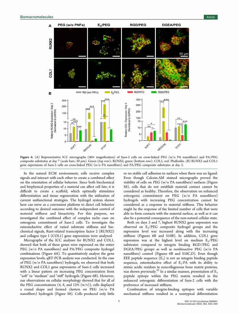
showed that both of these genes were expressed on the entirePEG (w/o PA nanofibers) and PA/PEG composite hydrogelcombinations (Figure 6A). To quantitatively analyze the geneexpression levels, qRT-PCR analysis was conducted. In the caseof PEG (w/o PA nanofibers) hydrogels, we observed that bothRUNX2 and COL1 gene expressions of Saos-2 cells increasedwith a linear pattern on increasing PEG concentration from“soft” to “medium” and “stiff” hydrogels (Figure 6B). However,our observations on cellular morphology showed that for all ofthe PEG concentrations (4, 8, and 12% (w/v)), cells displayeda round shape and formed clusters on PEG (w/o PAnanofibers) hydrogels (Figure S8). Cells produced only little
or no stable cell adhesion to surfaces when there was no ligand.Even though Calcein-AM stained micrographs proved theviability of cells on PEG (w/o PA nanofibers) surfaces (FigureS8), cells that do not establish material contact cannot beconsidered as healthy. Therefore, the observation on enhancedosteogenic commitment on PEG (w/o PA nanofibers)hydrogels with increasing PEG concentration cannot beconsidered as a response to material stiffness. This behaviormight be the response of the limited number of cells that wereable to form contacts with the material surface, as well as it canalso be a potential consequence of the non-natural cellular state.Both on days 3 and 7, highest RUNX2 gene expression was
observed on E3/PEG composite hydrogel groups and theexpression level was increased along with the increasingstiffness (Figures 6B and S10B). In addition, COL1 geneexpression was at the highest level on medium E3/PEGsubstrates compared to integrin binding RGD/PEG andDGEA/PEG groups as well as nonbioactive PEG (w/o PAnanofibers) control (Figures 6B and S10C,D). Even thoughEEE peptide sequence (E3) is not an integrin binding peptidesequence, osteoinductive effect of E3-PA with its ability tomimic acidic residues in noncollagenous bone matrix proteins,was shown previously.62 In a similar manner, presentation of E3
peptide epitope within the PEG matrix resulted in theenhanced osteogenic differentiation of Saos-2 cells with thepreference of increased stiffness.Combination of integrin-binding epitopes with variable
mechanical stiffness resulted in a nontypical differentiation
Figure 6. (A) Representative ICC micrographs (40× magnification) of Saos-2 cells on cross-linked PEG (w/o PA nanofibers) and PA/PEGcomposite substrates at day 7 (scale bars: 50 μm). Green (top row): RUNX2, green (bottom row): COL1, red: Phalloidin. (B) RUNX2 and COL1gene expressions of Saos-2 cells on cross-linked PEG (w/o PA nanofibers) and PA/PEG composite substrates at day 3.
Biomacromolecules Article
DOI: 10.1021/acs.biomac.5b00041Biomacromolecules XXXX, XXX, XXX−XXX
I
behavior compared to nonintegrin binding E3/PEG hydrogels.Instead of gradual increase of gene expression levels linear toincreasing substrate stiffness, integrin binding RGD/PEG andDGEA/PEG combinations exhibited different patterns forosteogenic differentiation. Gene expression patterns of RGD/PEG group were not affected by the mechanical properties andsimilar expression levels were obtained for all of the soft,medium, and stiff hydrogel groups (Figure 6B). Upregulation ofRUNX2 was not observed, while COL1 gene expression wasincreased up to 6-fold on RGD/PEG combinations independ-ent from substrate stiffness on day 3. Moreover, soft and stiffDGEA/PEG combinations presented higher expression ofRUNX2 and COL1 (Figure 6B) compared to medium DGEA/PEG. These results pointed to the presence of an interactiveeffect between integrin signaling and mechanical stimuli. It isknown that in the presence of complex niche cues, substratestiffness and biochemical signaling can substitute each otherunder certain conditions;13 however, further investigation isnecessary to clarify the underlying mechanism of this behavior.On the other hand, the preference of soft and stiff combinationsfor DGEA/PEG can be explained by previously elucidatedfactors related to osteoblast differentiation. DGEA is a collagentype I derived signaling sequence that binds to α2β1 integrinreceptor. α2-integrin is known as an early mechanotransducer ofmatrix elasticity in osteogenic cells and the increased expressionof α2-integrin on the cell membrane on stiffer matrices wasalready demonstrated.63 Along with increased stiffness,upregulated α2-integrin expression of the cells can lead to amore pronounced effect of DGEA signaling on osteoblastdifferentiation. On the other hand, during bone development,cellular differentiation into bone forming osteoblasts occurswithin a soft matrix in the range of 100−1000 Pa shearmodulus.64,65 Previous studies also showed that in vitroosteogenic differentiation can be supported on soft hydrogelmatrices, which have similar stiffness to developing bone.66,67
The gene expression results obtained from PA/PEG compositesystem also pointed to similar conclusions showing that theoptimal design of a material for the desired cellular outcomerequires the consideration of multiple factors since cells cansense complex niche cues, and these signals can direct cell fatein an interactive manner.14
Current strategies to introduce porosity into three-dimen-sional scaffolds are usually performed under nonphysiologicalconditions.32,33,68,69 Therefore, the biomedical applications ofthese systems only allow for cell seeding after the fabricationprocess, and as a result, nonuniform cell distribution can rise upas a problem. As a proof of concept, we also tested the capacityof nanofibrous self-assembled PA/PEG composite matrices asthree-dimensional (3D) scaffolds that allow for a cell-friendlyfabrication process and in situ application of engineeredscaffolds. To confirm the cell supportive effect of porositywithin our 3D scaffold systems, PEG (w/o PA nanofibers)versus RGD/PEG combinations were compared. For thispurpose, a similar facile sample preparation approach wasapplied to encapsulate Saos-2 cells into 3D matrices. Onlydifference was that all peptide and PEG-photoinitiator solutionswere prepared with culture medium (DMEM) instead of waterand cell suspension was mixed with PEG solution before theaddition of PA solutions into the mixture. Mixtures weretransferred into the caps of eppendorf tubes and exposed to UVlight at 365 nm for 15 min. The resulting disc-shaped 3D gelscontaining encapsulated Saos-2 cells were cultured in aSynthecon RCCS-4H bioreactor with rotating vessels. After 7
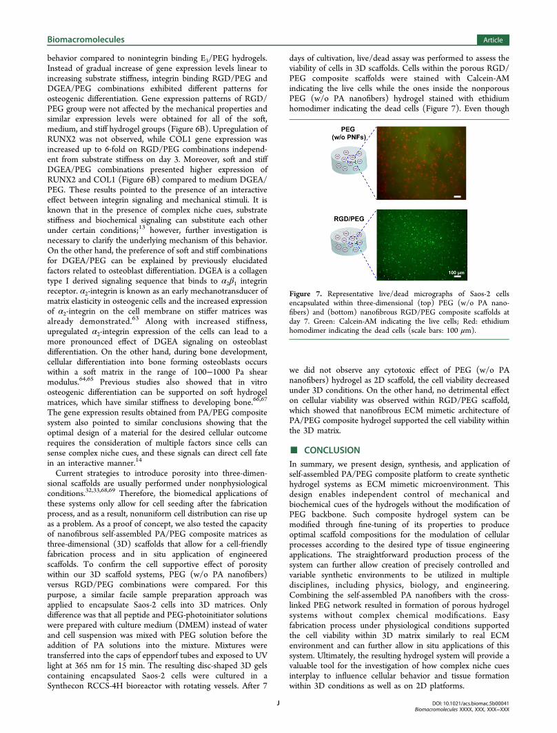
days of cultivation, live/dead assay was performed to assess theviability of cells in 3D scaffolds. Cells within the porous RGD/PEG composite scaffolds were stained with Calcein-AMindicating the live cells while the ones inside the nonporousPEG (w/o PA nanofibers) hydrogel stained with ethidiumhomodimer indicating the dead cells (Figure 7). Even though
we did not observe any cytotoxic effect of PEG (w/o PAnanofibers) hydrogel as 2D scaffold, the cell viability decreasedunder 3D conditions. On the other hand, no detrimental effecton cellular viability was observed within RGD/PEG scaffold,which showed that nanofibrous ECM mimetic architecture ofPA/PEG composite hydrogel supported the cell viability withinthe 3D matrix.
■ CONCLUSIONIn summary, we present design, synthesis, and application ofself-assembled PA/PEG composite platform to create synthetichydrogel systems as ECM mimetic microenvironment. Thisdesign enables independent control of mechanical andbiochemical cues of the hydrogels without the modification ofPEG backbone. Such composite hydrogel system can bemodified through fine-tuning of its properties to produceoptimal scaffold compositions for the modulation of cellularprocesses according to the desired type of tissue engineeringapplications. The straightforward production process of thesystem can further allow creation of precisely controlled andvariable synthetic environments to be utilized in multipledisciplines, including physics, biology, and engineering.Combining the self-assembled PA nanofibers with the cross-linked PEG network resulted in formation of porous hydrogelsystems without complex chemical modifications. Easyfabrication process under physiological conditions supportedthe cell viability within 3D matrix similarly to real ECMenvironment and can further allow in situ applications of thissystem. Ultimately, the resulting hydrogel system will provide avaluable tool for the investigation of how complex niche cuesinterplay to influence cellular behavior and tissue formationwithin 3D conditions as well as on 2D platforms.
Figure 7. Representative live/dead micrographs of Saos-2 cellsencapsulated within three-dimensional (top) PEG (w/o PA nano-fibers) and (bottom) nanofibrous RGD/PEG composite scaffolds atday 7. Green: Calcein-AM indicating the live cells; Red: ethidiumhomodimer indicating the dead cells (scale bars: 100 μm).
Biomacromolecules Article
DOI: 10.1021/acs.biomac.5b00041Biomacromolecules XXXX, XXX, XXX−XXX
J
■ ASSOCIATED CONTENT
*S Supporting InformationLC-MS results of PAs, TEM images of PA nanofibers, BETanalysis results, SAXS data analysis in details, additionalrheological characterizations of the composite hydrogels,Calcein-AM stained micrographs of cells on PEG (w/o PAnanofibers) and PA/PEG composite for 24, 48, and 72 h,additional analysis of RUNX2 and COL1 gene expressions atdays 3 and 7. This material is available free of charge via theInternet at http://pubs.acs.org.
■ AUTHOR INFORMATION
Corresponding Authors*E-mail: [email protected].*E-mail: [email protected].
NotesThe authors declare no competing financial interest.
■ ACKNOWLEDGMENTS
This work was supported by the Scientific and TechnologicalResearch Council of Turkey (TUBITAK), Grant Number213M406. M.G. and G.C. acknowledge support fromTUBITAK-BIDEB fellowship. M.O.G. and A.B.T. acknowledgesupport from the Turkish Academy of Sciences DistinguishedYoung Scientist Award (TUBA-GEBIP). We thank Dr. HakanCeylan for helpful scientific discussions and Elif Arslan for helpin the 3D bioreactor setup. Also, we appreciate Mustafa Guler’shelp with TEM imaging.
■ REFERENCES(1) Tibbitt, M. W.; Anseth, K. S. Hydrogels as extracellular matrixmimics for 3D cell culture. Biotechnol. Bioeng. 2009, 103, 655−663.(2) Langer, R.; Peppas, N. A. Advances in biomaterials, drug delivery,and bionanotechnology. AIChE J. 2003, 49, 2990−3006.(3) Thiele, J.; Ma, Y.; Bruekers, S.; Ma, S.; Huck, W. T. 25thanniversary article: Designer hydrogels for cell cultures: A materialsselection guide. Adv. Mater. 2014, 26, 125−148.(4) Drury, J. L.; Mooney, D. J. Hydrogels for tissue engineering:Scaffold design variables and applications. Biomaterials 2003, 24,4337−4351.(5) Lee, K. Y.; Mooney, D. J. Hydrogels for tissue engineering. Chem.Rev. 2001, 101, 1869−1880.(6) Hoffman, A. S. Hydrogels for biomedical applications. Adv. DrugDelivery Rev. 2002, 54, 3−12.(7) Lutolf, M.; Hubbell, J. Synthetic biomaterials as instructiveextracellular microenvironments for morphogenesis in tissue engineer-ing. Nat. Biotechnol. 2005, 23, 47−55.(8) Kleinman, H. K.; Philp, D.; Hoffman, M. P. Role of theextracellular matrix in morphogenesis. Curr. Opin. Biotechnol. 2003, 14,526−532.(9) Lutolf, M. P.; Blau, H. M. Artificial stem cell niches. Adv. Mater.2009, 21, 3255−3268.(10) Wheeldon, I.; Farhadi, A.; Bick, A. G.; Jabbari, E.;Khademhosseini, A. Nanoscale tissue engineering: Spatial controlover cell-materials interactions. Nanotechnology 2011, 22, 212001.(11) Hersel, U.; Dahmen, C.; Kessler, H. RGD modified polymers:Biomaterials for stimulated cell adhesion and beyond. Biomaterials2003, 24, 4385−4415.(12) Cukierman, E.; Pankov, R.; Stevens, D. R.; Yamada, K. M.Taking cell-matrix adhesions to the third dimension. Science 2001, 294,1708−1712.(13) Engler, A. J.; Sen, S.; Sweeney, H. L.; Discher, D. E. Matrixelasticity directs stem cell lineage specification. Cell 2006, 126, 677−689.
(14) Lutolf, M. P.; Gilbert, P. M.; Blau, H. M. Designing materials todirect stem-cell fate. Nature 2009, 462, 433−441.(15) Ifkovits, J. L.; Burdick, J. A. Review: photopolymerizable anddegradable biomaterials for tissue engineering applications. Tissue Eng.2007, 13, 2369−2385.(16) Wang, N.; Butler, J.; Ingber, D. Mechanotransduction across thecell surface and through the cytoskeleton. Science 1993, 260, 1124−1127.(17) Shao, Y.; Fu, J. Integrated micro/nanoengineered functionalbiomaterials for cell mechanics and mechanobiology: A materialsperspective. Adv. Mater. 2014, 26, 1494−1533.(18) Missirlis, D.; Spatz, J. P. Combined effects of PEG hydrogelelasticity and cell-adhesive coating on fibroblast adhesion andpersistent migration. Biomacromolecules 2013, 15, 195−205.(19) Solon, J.; Levental, I.; Sengupta, K.; Georges, P. C.; Janmey, P.A. Fibroblast adaptation and stiffness matching to soft elasticsubstrates. Biophys. J. 2007, 93, 4453−4461.(20) Saez, A.; Ghibaudo, M.; Buguin, A.; Silberzan, P.; Ladoux, B.Rigidity-driven growth and migration of epithelial cells on micro-structured anisotropic substrates. Proc. Natl. Acad. Sci. U.S.A. 2007,104, 8281−8286.(21) Hadjipanayi, E.; Mudera, V.; Brown, R. Close dependence offibroblast proliferation on collagen scaffold matrix stiffness. J. TissueEng. Regener. Med. 2009, 3, 77−84.(22) Singh, A.; Zhan, J.; Ye, Z.; Elisseeff, J. H. Modularmultifunctional poly(ethylene glycol) hydrogels for stem cell differ-entiation. Adv. Funct. Mater. 2013, 23, 575−582.(23) Mabry, K. M.; Lawrence, R. L.; Anseth, K. S. Dynamic stiffeningof poly(ethylene glycol)-based hydrogels to direct valvular interstitialcell phenotype in a three-dimensional environment. Biomaterials 2015,49, 47−56.(24) Alcantar, N. A.; Aydil, E. S.; Israelachvili, J. N. Polyethyleneglycol-coated biocompatible surfaces. J. Biomed. Mater. Res. 2000, 51,343−351.(25) Zhu, J. Bioactive modification of poly(ethylene glycol)hydrogels for tissue engineering. Biomaterials 2010, 31, 4639−4656.(26) Phelps, E. A.; Enemchukwu, N. O.; Fiore, V. F.; Sy, J. C.;Murthy, N.; Sulchek, T. A.; Barker, T. H.; García, A. J. Maleimidecross-linked bioactive peg hydrogel exhibits improved reaction kineticsand cross-linking for cell encapsulation and in situ delivery. Adv. Mater.2012, 24, 64−70.(27) Lee, H. J.; Lee, J.-S.; Chansakul, T.; Yu, C.; Elisseeff, J. H.;Seungju, M. Y. Collagen mimetic peptide-conjugated photopolymer-izable PEG hydrogel. Biomaterials 2006, 27, 5268−5276.(28) Lau, H. K.; Kiick, K. L. Opportunities for multicomponenthybrid hydrogels in biomedical applications. Biomacromolecules 2015,16, 28−42.(29) Yang, F.; Williams, C. G.; Wang, D.-a.; Lee, H.; Manson, P. N.;Elisseeff, J. The effect of incorporating RGD adhesive peptide inpolyethylene glycol diacrylate hydrogel on osteogenesis of bonemarrow stromal cells. Biomaterials 2005, 26, 5991−5998.(30) Hern, D. L.; Hubbell, J. A. Incorporation of adhesion peptidesinto nonadhesive hydrogels useful for tissue resurfacing. J. Biomed.Mater. Res. 1998, 39, 266−276.(31) Lutolf, M.; Hubbell, J. Synthesis and physicochemicalcharacterization of end-linked poly(ethylene glycol)-co-peptide hydro-gels formed by Michael-type addition. Biomacromolecules 2003, 4,713−722.(32) Chiu, Y.-C.; Larson, J. C.; Isom, A., Jr; Brey, E. M. Generation ofporous poly (ethylene glycol) hydrogels by salt leaching. Tissue Eng.,Part C 2010, 16, 905−912.(33) Keskar, V.; Marion, N. W.; Mao, J. J.; Gemeinhart, R. A. In vitroevaluation of macroporous hydrogels to facilitate stem cell infiltration,growth, and mineralization. Tissue Eng., Part A 2009, 15, 1695−1707.(34) Matson, J. B.; Stupp, S. I. Self-assembling peptide scaffolds forregenerative medicine. Chem. Commun. 2012, 48, 26−33.(35) Cui, H.; Webber, M. J.; Stupp, S. I. Self-assembly of peptideamphiphiles: From molecules to nanostructures to biomaterials.Biopolymers 2010, 94, 1−18.
Biomacromolecules Article
DOI: 10.1021/acs.biomac.5b00041Biomacromolecules XXXX, XXX, XXX−XXX
K
(36) Ravichandran, R.; Griffith, M.; Phopase, J. Applications of self-assembling peptide scaffolds in regenerative medicine: the way to theclinic. J. Mater. Chem. B 2014, 2, 8466−8478.(37) Sur, S.; Tantakitti, F.; Matson, J. B.; Stupp, S. I. Epitopetopography controls bioactivity in supramolecular nanofibers.Biomater. Sci. 2015, 3, 520−532.(38) Guler, M. O.; Hsu, L.; Soukasene, S.; Harrington, D. A.; Hulvat,J. F.; Stupp, S. I. Presentation of RGDS epitopes on self-assemblednanofibers of branched peptide amphiphiles. Biomacromolecules 2006,7, 1855−1863.(39) Galler, K. M.; Aulisa, L.; Regan, K. R.; D’Souza, R. N.;Hartgerink, J. D. Self-assembling multidomain peptide hydrogels:designed susceptibility to enzymatic cleavage allows enhanced cellmigration and spreading. J. Am. Chem. Soc. 2010, 132, 3217−3223.(40) Sur, S.; Matson, J. B.; Webber, M. J.; Newcomb, C. J.; Stupp, S.I. Photodynamic control of bioactivity in a nanofiber matrix. ACS Nano2012, 6, 10776−10785.(41) Burdick, J. A.; Anseth, K. S. Photoencapsulation of osteoblasts ininjectable RGD-modified PEG hydrogels for bone tissue engineering.Biomaterials 2002, 23, 4315−4323.(42) Kim, I. L.; Khetan, S.; Baker, B. M.; Chen, C. S.; Burdick, J. A.Fibrous hyaluronic acid hydrogels that direct MSC chondrogenesisthrough mechanical and adhesive cues. Biomaterials 2013, 34, 5571−5580.(43) Yoo, S. Y.; Kobayashi, M.; Lee, P. P.; Lee, S.-W. Early osteogenicdifferentiation of mouse preosteoblasts induced by collagen-derivedDGEA-peptide on nanofibrous phage tissue matrices. Biomacromole-cules 2011, 12, 987−996.(44) Anderson, J. M.; Kushwaha, M.; Tambralli, A.; Bellis, S. L.;Camata, R. P.; Jun, H.-W. Osteogenic differentiation of humanmesenchymal stem cells directed by extracellular matrix-mimickingligands in a biomimetic self-assembled peptide amphiphile nanomatrix.Biomacromolecules 2009, 10, 2935−2944.(45) Hennessy, K. M.; Pollot, B. E.; Clem, W. C.; Phipps, M. C.;Sawyer, A. A.; Culpepper, B. K.; Bellis, S. L. The effect of collagen Imimetic peptides on mesenchymal stem cell adhesion and differ-entiation, and on bone formation at hydroxyapatite surfaces.Biomaterials 2009, 30, 1898−1909.(46) Shin, H.; Olsen, B. D.; Khademhosseini, A. The mechanicalproperties and cytotoxicity of cell-laden double-network hydrogelsbased on photocrosslinkable gelatin and gellan gum biomacromole-cules. Biomaterials 2012, 33, 3143−3152.(47) Sun, J.-Y.; Zhao, X.; Illeperuma, W. R.; Chaudhuri, O.; Oh, K.H.; Mooney, D. J.; Vlassak, J. J.; Suo, Z. Highly stretchable and toughhydrogels. Nature 2012, 489, 133−136.(48) Choi, S.; Kim, J. Designed fabrication of super-stiff, anisotropichybrid hydrogels via linear remodeling of polymer networks andsubsequent crosslinking. J. Mater. Chem. B 2015, 3, 1479−1483.(49) Munoz-Pinto, D. J.; Jimenez-Vergara, A. C.; Gharat, T. P.;Hahn, M. S. Characterization of sequential collagen-poly(ethyleneglycol)diacrylate interpenetrating networks and initial assessment oftheir potential for vascular tissue engineering. Biomaterials 2015, 40,32−42.(50) Zhang, J.-T.; Petersen, S.; Thunga, M.; Leipold, E.; Weidisch, R.;Liu, X.; Fahr, A.; Jandt, K. D. Micro-structured smart hydrogels withenhanced protein loading and release efficiency. Acta Biomater. 2010,6, 1297−1306.(51) Yang, S.; Wang, J.; Tan, H.; Zeng, F.; Liu, C. Mechanicallyrobust PEGDA−MSNs−OH nanocomposite hydrogel with hierarch-ical meso-macroporous structure for tissue engineering. Soft Matter2012, 8, 8981−8989.(52) Hartgerink, J. D.; Beniash, E.; Stupp, S. I. Peptide-amphiphilenanofibers: a versatile scaffold for the preparation of self-assemblingmaterials. Proc. Natl. Acad. Sci. U.S.A. 2002, 99, 5133−5138.(53) Niece, K. L.; Hartgerink, J. D.; Donners, J. J.; Stupp, S. I. Self-assembly combining two bioactive peptide-amphiphile molecules intonanofibers by electrostatic attraction. J. Am. Chem. Soc. 2003, 125,7146−7147.
(54) Paramonov, S. E.; Jun, H.-W.; Hartgerink, J. D. Self-assembly ofpeptide-amphiphile nanofibers: the roles of hydrogen bonding andamphiphilic packing. J. Am. Chem. Soc. 2006, 128, 7291−7298.(55) Suh, K. Y.; Seong, J.; Khademhosseini, A.; Laibinis, P. E.;Langer, R. A simple soft lithographic route to fabrication ofpoly(ethylene glycol) microstructures for protein and cell patterning.Biomaterials 2004, 25, 557−563.(56) Karageorgiou, V.; Kaplan, D. Porosity of 3D biomaterialscaffolds and osteogenesis. Biomaterials 2005, 26, 5474−5491.(57) Mastrogiacomo, M.; Scaglione, S.; Martinetti, R.; Dolcini, L.;Beltrame, F.; Cancedda, R.; Quarto, R. Role of scaffold internalstructure on in vivo bone formation in macroporous calciumphosphate bioceramics. Biomaterials 2006, 27, 3230−3237.(58) Pedersen, J. S.; Schurtenberger, P. Scattering functions ofsemiflexible polymers with and without excluded volume effects.Macromolecules 1996, 29, 7602−7612.(59) Chen, W.-R.; Butler, P. D.; Magid, L. J. Incorporatingintermicellar interactions in the fitting of SANS data from cationicwormlike micelles. Langmuir 2006, 22, 6539−6548.(60) Bacıog lu, A.; Kazan, U.; Ide, S. Single and multilayered a-SiOx:H(x < 1) thin film samples analyzed by optical absorption and small-angle X-ray scattering. Mater. Chem. Phys. 2014, 146, 425−430.(61) Kocabey, S.; Ceylan, H.; Tekinay, A. B.; Guler, M. O.Glycosaminoglycan mimetic peptide nanofibers promote mineraliza-tion by osteogenic cells. Acta Biomater. 2013, 9, 9075−9085.(62) Ceylan, H.; Kocabey, S.; Unal Gulsuner, H.; Balcik, O. S.; Guler,M. O.; Tekinay, A. B. Bone-like mineral nucleating peptide nanofibersinduce differentiation of human mesenchymal stem cells into matureosteoblasts. Biomacromolecules 2014, 15, 2407−2418.(63) Shih, Y. R. V.; Tseng, K. F.; Lai, H. Y.; Lin, C. H.; Lee, O. K.Matrix stiffness regulation of integrin-mediated mechanotransductionduring osteogenic differentiation of human mesenchymal stem cells. J.Bone Miner. Res. 2011, 26, 730−738.(64) Shapiro, F. Bone development and its relation to fracture repair.The role of mesenchymal osteoblasts and surface osteoblasts. Eur. CellsMater. 2008, 15, 53−76.(65) Forgacs, G.; Foty, R. A.; Shafrir, Y.; Steinberg, M. S. Viscoelasticproperties of living embryonic tissues: A quantitative study. Biophys. J.1998, 74, 2227−2234.(66) Marí-Buye, N.; Luque, T.; Navajas, D.; Semino, C. E.Development of a three-dimensional bone-like construct in a softself-assembling peptide matrix. Tissue Eng., Part A 2013, 19, 870−881.(67) Wu, L. C.; Yang, J.; Kopecek, J. Hybrid hydrogels self-assembledfrom graft copolymers containing complementary β-sheets ashydroxyapatite nucleation scaffolds. Biomaterials 2011, 32, 5341−5353.(68) Rnjak-Kovacina, J.; Wise, S. G.; Li, Z.; Maitz, P. K.; Young, C. J.;Wang, Y.; Weiss, A. S. Tailoring the porosity and pore size ofelectrospun synthetic human elastin scaffolds for dermal tissueengineering. Biomaterials 2011, 32, 6729−6736.(69) Xiao, J.; Duan, H.; Liu, Z.; Wu, Z.; Lan, Y.; Zhang, W.; Li, C.;Chen, F.; Zhou, Q.; Wang, X. Construction of the recellularizedcorneal stroma using porous acellular corneal scaffold. Biomaterials2011, 32, 6962−6971.
Biomacromolecules Article
DOI: 10.1021/acs.biomac.5b00041Biomacromolecules XXXX, XXX, XXX−XXX
L
Related Documents


