REVIEW published: 19 May 2015 doi: 10.3389/fphys.2015.00153 Frontiers in Physiology | www.frontiersin.org 1 May 2015 | Volume 6 | Article 153 Edited by: Harley Takatsuna Kurata, University of British Columbia, Canada Reviewed by: Maria Isabel Bahamonde Santos, Pompeu Fabra University, Spain Harold H Zakon, The University of Texas, USA *Correspondence: J. David Spafford, Department of Biology, University of Waterloo, B1-173, 200 University Avenue West, Waterloo, ON N2L 3G1, Canada [email protected] Specialty section: This article was submitted to Membrane Physiology and Membrane Biophysics, a section of the journal Frontiers in Physiology Received: 12 March 2015 Accepted: 28 April 2015 Published: 19 May 2015 Citation: Stephens RF, Guan W, Zhorov BS and Spafford JD (2015) Selectivity filters and cysteine-rich extracellular loops in voltage-gated sodium, calcium, and NALCN channels. Front. Physiol. 6:153. doi: 10.3389/fphys.2015.00153 Selectivity filters and cysteine-rich extracellular loops in voltage-gated sodium, calcium, and NALCN channels Robert F. Stephens 1 , W. Guan 1 , Boris S. Zhorov 2, 3 and J. David Spafford 1 * 1 Department of Biology, University of Waterloo, Waterloo, ON, Canada, 2 Department of Biochemistry and Biomedical Sciences, McMaster University, Hamilton, ON, Canada, 3 Sechenov Institute of Evolutionary Physiology and Biochemistry, Russian Academy of Sciences, St. Petersburg, Russia How nature discriminates sodium from calcium ions in eukaryotic channels has been difficult to resolve because they contain four homologous, but markedly different repeat domains. We glean clues from analyzing the changing pore region in sodium, calcium and NALCN channels, from single-cell eukaryotes to mammals. Alternative splicing in invertebrate homologs provides insights into different structural features underlying calcium and sodium selectivity. NALCN generates alternative ion selectivity with splicing that changes the high field strength (HFS) site at the narrowest level of the hourglass shaped pore where the selectivity filter is located. Alternative splicing creates NALCN isoforms, in which the HFS site has a ring of glutamates contributed by all four repeat domains (EEEE), or three glutamates and a lysine residue in the third (EEKE) or second (EKEE) position. Alternative splicing provides sodium and/or calcium selectivity in T-type channels with extracellular loops between S5 and P-helices (S5P) of different lengths that contain three or five cysteines. All eukaryotic channels have a set of eight core cysteines in extracellular regions, but the T-type channels have an infusion of 4–12 extra cysteines in extracellular regions. The pattern of conservation suggests a possible pairing of long loops in Domains I and III, which are bridged with core cysteines in NALCN, Cav, and Nav channels, and pairing of shorter loops in Domains II and IV in T-type channel through disulfide bonds involving T-type specific cysteines. Extracellular turrets of increasing lengths in potassium channels (Kir2.2, hERG, and K2P1) contribute to a changing landscape above the pore selectivity filter that can limit drug access and serve as an ion pre-filter before ions reach the pore selectivity filter below. Pairing of extended loops likely contributes to the large extracellular appendage as seen in single particle electron cryo-microscopy images of the eel Na v 1 channel. Keywords: calcium channels, sodium channels, NALCN, ion selectivity, ion channel evolution, ion channels, Lymnaea stagnalis, ion pore Complexities in Resolving the 4x6TM Family of Cation Channels The mechanism of sodium and calcium selectivity within the superfamily of eukaryotic 4x6TM ion channels, which includes the voltage-gated sodium channels (Na v 1, Na v 2), voltage-gated calcium

Welcome message from author
This document is posted to help you gain knowledge. Please leave a comment to let me know what you think about it! Share it to your friends and learn new things together.
Transcript
REVIEWpublished: 19 May 2015
doi: 10.3389/fphys.2015.00153
Frontiers in Physiology | www.frontiersin.org 1 May 2015 | Volume 6 | Article 153
Edited by:
Harley Takatsuna Kurata,
University of British Columbia, Canada
Reviewed by:
Maria Isabel Bahamonde Santos,
Pompeu Fabra University, Spain
Harold H Zakon,
The University of Texas, USA
*Correspondence:
J. David Spafford,
Department of Biology, University of
Waterloo, B1-173, 200 University
Avenue West, Waterloo, ON N2L 3G1,
Canada
Specialty section:
This article was submitted to
Membrane Physiology and Membrane
Biophysics,
a section of the journal
Frontiers in Physiology
Received: 12 March 2015
Accepted: 28 April 2015
Published: 19 May 2015
Citation:
Stephens RF, Guan W, Zhorov BS and
Spafford JD (2015) Selectivity filters
and cysteine-rich extracellular loops in
voltage-gated sodium, calcium, and
NALCN channels.
Front. Physiol. 6:153.
doi: 10.3389/fphys.2015.00153
Selectivity filters and cysteine-richextracellular loops in voltage-gatedsodium, calcium, and NALCNchannelsRobert F. Stephens 1, W. Guan 1, Boris S. Zhorov 2, 3 and J. David Spafford 1*
1Department of Biology, University of Waterloo, Waterloo, ON, Canada, 2Department of Biochemistry and Biomedical
Sciences, McMaster University, Hamilton, ON, Canada, 3 Sechenov Institute of Evolutionary Physiology and Biochemistry,
Russian Academy of Sciences, St. Petersburg, Russia
How nature discriminates sodium from calcium ions in eukaryotic channels has beendifficult to resolve because they contain four homologous, but markedly different repeatdomains. We glean clues from analyzing the changing pore region in sodium, calciumand NALCN channels, from single-cell eukaryotes to mammals. Alternative splicingin invertebrate homologs provides insights into different structural features underlyingcalcium and sodium selectivity. NALCN generates alternative ion selectivity with splicingthat changes the high field strength (HFS) site at the narrowest level of the hourglassshaped pore where the selectivity filter is located. Alternative splicing creates NALCNisoforms, in which the HFS site has a ring of glutamates contributed by all four repeatdomains (EEEE), or three glutamates and a lysine residue in the third (EEKE) or second(EKEE) position. Alternative splicing provides sodium and/or calcium selectivity in T-typechannels with extracellular loops between S5 and P-helices (S5P) of different lengthsthat contain three or five cysteines. All eukaryotic channels have a set of eight corecysteines in extracellular regions, but the T-type channels have an infusion of 4–12 extracysteines in extracellular regions. The pattern of conservation suggests a possible pairingof long loops in Domains I and III, which are bridged with core cysteines in NALCN,Cav, and Nav channels, and pairing of shorter loops in Domains II and IV in T-typechannel through disulfide bonds involving T-type specific cysteines. Extracellular turretsof increasing lengths in potassium channels (Kir2.2, hERG, and K2P1) contribute to achanging landscape above the pore selectivity filter that can limit drug access and serveas an ion pre-filter before ions reach the pore selectivity filter below. Pairing of extendedloops likely contributes to the large extracellular appendage as seen in single particleelectron cryo-microscopy images of the eel Nav1 channel.
Keywords: calcium channels, sodium channels, NALCN, ion selectivity, ion channel evolution, ion channels,
Lymnaea stagnalis, ion pore
Complexities in Resolving the 4x6TM Family of Cation Channels
The mechanism of sodium and calcium selectivity within the superfamily of eukaryotic 4x6TM ionchannels, which includes the voltage-gated sodium channels (Nav1, Nav2), voltage-gated calcium
Stephens et al. Ion selectivity of voltage-gated channels
channels (Cav1, Cav2, Cav3) and NALCN, is not well understood.Invertebrate NALCN and T-type channels demonstrate mixedsodium and calcium selectivity due to alternative splicinginvolving (i) the HFS site at the pore selectivity filter and(ii) cysteine-rich extracellular turrets (Senatore et al., 2013,2014a,b). We use available phylogenetic data on 4x6TM channelsto examine different molecular determinants that underlinesodium and calcium selectivity in these channels. NALCNand Nav1 sodium channels have a HFS site where sodiumselectivity is generated by a lysine residue in the second or thirdrepeat domain. We find a core of eight conserved cysteinesin the long extracellular loops of all 4x6TM channels, andadditional cysteines in the loops of Cav3 T-type channels andmany of the vertebrate Nav1 channels. The DI-DIII and DII-DIV pairing of extracellular loops creates an appendage abovethe pore selectivity filter. Such appendages are seen in thesingle nanoparticle cryo-electron microscopy images of Nav1sodium channels and in the X-ray structures of K2P1 channels,where the disulfide bond-bridged extended extracellular turretsform an apex ∼35 angstroms above the membrane. In K2P1channels this appendage affects ion and drug access to the pore.Similar appendages contribute to sodium selectivity engenderedin invertebrate T-type channels with alternative cysteine-richextracellular turrets. Other resources on related topics coverstructure of the prokaryotic sodium channels (Charalambousand Wallace, 2011; Payandeh et al., 2011; Catterall, 2014;Scheuer, 2014; Payandeh and Minor, 2015), homology modelingof eukaryotic sodium channels (Tikhonov and Zhorov, 2012;Korkosh et al., 2014) and a proposed evolution of sodium andcalcium channels (Liebeskind et al., 2011; Cai, 2012; Zakon, 2012;Moran et al., 2015).
“4x6TM” refers to the shared architecture in the superfamilyof cation channels that contain four homologous repeat domainswith six transmembrane segments in each domain (Figure 1).4x6TM channels arose from two rounds of duplication in a1x6TM channel ancestor. The evidence for this evolutionaryhistory is in the kinship of the domains, where the pairs ofDomains I and III, and Domains II and IV more closely resembleeach other than other domains in these channels (Strong et al.,1993). Examples of the 1x6TM genes are bacterial sodiumchannels (e.g., NavAb) (Payandeh et al., 2011; Zhang et al., 2012)(Figure 1A) and voltage-gated potassium channels (Gutmanet al., 2005). Four repeat domains in the 4x6TM channels orfour subunits in the 1x6TM channels form an assembly thatsurrounds the aqueous pore through which ions pass (Figure 1).In the 1x6TM channels, four subunits can form homo-tetramersderived from identical gene products, or may co-assemble withsubunits from different gene products as a hetero-tetramer, asin Shaker type voltage-gated potassium channels (Li et al., 1992)(Figure 1A). A consequence of four repeat domains linked in afull-length 4x6TM channel gene is the absence of the diversitythat is observed in hetero-tetrameric voltage-gated potassiumchannels. What is gained in the 4x6TM genes is a sequentialasymmetry of their repeat domains, where each domain issubstantially diverged from other domains, and can make aunique contribution to the voltage-gating, ion selectivity andinteractions with the cytoplasmic or extracellular environments.The four repeat domains are connected by different cytoplasmic
linkers (I-II, II-III, and III-IV), as opposed to four pairs ofcytoplasmic N- and C-termini in K channels (Figure 1B). Theselinkers possess regulatory sites that interact with proteins, suchas auxiliary channel subunits. The linkers are diversified forunique cellular conditions. Examples of such conditions includethe axonal specific environment associated with Nav1 sodiumchannels in nodes of Ranvier of myelinated neurons (Leterrieret al., 2011), or the network of proteins appearing to associatewith Cav2 channels in presynaptic terminals (Spafford andZamponi, 2003). While K channels also have unique proteininteracting domains, these are mostly limited to the N- and C-termini, which are fourfold repeated in each K channel (Jan andJan, 2012).
It is challenging to study 4x6TM channels by site-directedmutagenesis or by generating chimeric channels where the inter-dependence of individual repeat domains is not understood.4x6TM channel structures are also difficult to resolve by X-ray crystallography because of their large size (∼2.1 to ∼2.9 ×
103 amino acids), large extra- and intra-cellular loops, as wellas N- and C-terminal ends. Single nanoparticle cryo-electronmicroscopy and advanced direct detection cameras may providegreater insights into the structure of 4x6TM channels (Liaoet al., 2013, 2014). Hypotheses on the structure of ion poresin 4x6TM channels in this review are proposed using thefollowing sources: (i) comparison of known X-ray structuresin 1x6TM channels, (ii) comparative analyses of sequences andphysiological characteristics of Nav1, Nav2, Cav1, Cav2, Cav3and NALCN channels, (iii) analysis of the full phylogeneticspectrum of channels starting from early eukaryotes where4x6TM channels first appear.
The Modularity of 4x6TM Channel Domains
Each of four homologous domains in a 4x6TM channel andthe single domain in a 1x6TM channel consists of a voltage-sensingmodule (involving transmembrane helices S1 to S4) and aquarter of the pore module (involving transmembrane segmentsS5 and S6 and membrane re-entrant P-loop between S5 and S6)(Figure 2). Some voltage sensing and pore-formingmodules existas natively expressed stand-alone proteins. Examples include aproton-gated channel Hv1 that lacks the pore module (Ramseyet al., 2006; Sasaki et al., 2006) and the pH-activated bacterialKcsA potassium channel that lacks voltage-sensing modules(Doyle et al., 1998). The voltage sensor and pore modulesexist as natively expressed proteins on their own, such as thevoltage-sensor containing phosphatase Ci-VSP from the seasquirt Ciona intestinalis (Murata et al., 2005), and the bacterialKcsA potassium channel, a pH-activated protein that has onlythe pore module (Doyle et al., 1998). Truncated subunits ofbacterial sodium channels can form in vitro functional tetramerswithout the voltage-sensor domains (McCusker et al., 2011; Tsaiet al., 2013; Shaya et al., 2014), illustrating the semi-autonomousnature of the voltage-sensing and pore modules in voltage-gatedchannels. However, it is typical in the 4X6TM channels for thevoltage-sensing module and pore module to appear together.
The voltage sensing helix S4 contains several positivelycharged residues located on one face of the helix. Uponmembrane depolarization S4 moves in the extracellular direction
Frontiers in Physiology | www.frontiersin.org 2 May 2015 | Volume 6 | Article 153
Stephens et al. Ion selectivity of voltage-gated channels
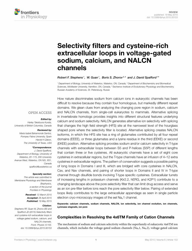
FIGURE 1 | High Field Strength (HFS) site EEEE formed by
selectivity filter glutamates (E177) in the homotetrameric bacterial
sodium channel NavAb is equivalent to the EEEE site in of Cav1
and Cav2 calcium channels and the DEKA site of Nav1 sodium
channels, which are contributed by the four homologous repeat
domains. (A) Residues to the EEEE HFS site in bacterial sodiumchannels are contributed by four identical gene products. (B) Residues to
asymmetric selectivity filters in eukaryotic calcium and sodium channelsare contributed by four homologous repeat domains. (C) Structuralcontours of the NavAb selectivity filter and positions of correspondingresidues of bacterial NavAb and human Cav1.2 and Nav1.2. Thenarrowest pore level is the selectivity filter HFS site, which canaccommodate a hydrated Na+ ion, but not a hydrated K+ ion. Panel(C)is adapted from Payandeh et al. (2011), with permission.
along helices S2 and S3 that contain negatively charged residues(Catterall, 2010) (Figure 2). Amphipathic S4-S5 linkers, whichconnect the voltage sensing and pore modules, allow movementsin the voltage sensors to induce movements of the S5 helices inthe pore module. As a result, the innermost S6 segments, whichline the aqueous pore and form a helical bundle, can widen ornarrow to allow or occlude ion permeation (Oelstrom et al.,2014).
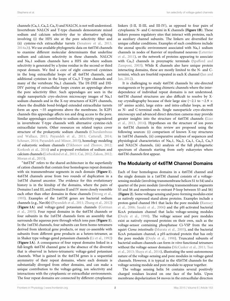
P-Loop Residues That Contribute to theSelectivity Filter
Each subunit or repeat domain of a P-loop channel containstransmembrane helices S5 and S6 that are connected bythe membrane-re-entrant P-loop. Each P-loop containsan extracellular turret-like linker (S5P) between S5 and amembrane-descending P-helix, an ascending limb, and anextracellular linker to S6. Four ascending limbs contain residuesthat contribute to the selectivity filter and line the outer pore(Figure 2). At the narrowest level of the pore within the P-loop domain, the four ascending limbs contain residues thatdetermine the K+, Ca2+, Na+, or mixed ion selectivity, anddivide the ion permeation pathway into an outer pore above andthe inner pore below the selective filter.
Potassium Ion SelectivityThe potassium ion selectivity in K channels is determinedby backbone carbonyl oxygens (Doyle et al., 1998) fromthe signature sequence residues (TVGYG), which mimic thehydration shell oxygen atoms that surround potassium ionsin solution (Figure 3A). There are four potassium bindingsites in the selectivity filter (s1-s4), which in the current-permeating channel can be occupied by two water moleculesand two potassium ions (s1/s3 or s2/s4 potassium occupancy)(Figure 3A).
Sodium Ion SelectivityThe general folding for the 4x6TM channels likely resemblesthat seen in the X-ray structures of the 1x6TM bacterial sodiumchannels NavAb (Figures 3A,C) (Payandeh et al., 2011), NavRh(Zhang et al., 2012), and NavMs (McCusker et al., 2012).The selectivity filter of bacterial Nav channels contains a ringof four glutamates (EEEE), one glutamate from each subunit(Figure 3C). The EEEE ring forms the narrowest level of theopen pore (Figure 1C) with the flexible carboxylate side chainsfacing toward the pore axis. The conformational flexibility of thecarboxylate side chains stabilizes multiple ionic occupancy states,helping sodium ions to pass through the selectivity filter via theknock-on mechanism (Chakrabarti et al., 2013).
Frontiers in Physiology | www.frontiersin.org 3 May 2015 | Volume 6 | Article 153
Stephens et al. Ion selectivity of voltage-gated channels
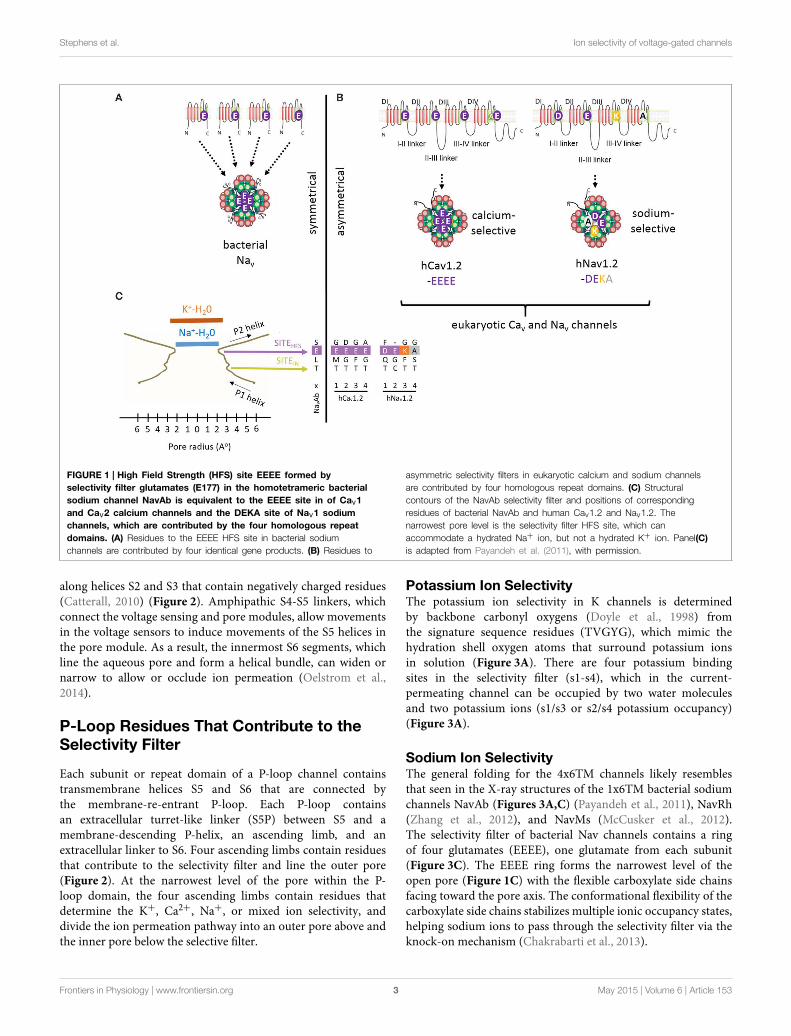
FIGURE 2 | Invertebrate NALCN and T-type channels splice in the
pore module (Domain II) to generate calcium- or
sodium-selective channels. Only repeat Domains II and IV areshown for clarity. Each repeat domain contains a voltage-sensingmodule (S1 to S4) and segments S5-P-S6 that contribute a quarter tothe pore module. (A) Exons 15a or 15b code for alternative HFS sitesgenerating calcium-selective (EEEE) or sodium-selective (EKEE) pores
(Senatore et al., 2013). (B) Expression of exon 12a or 12b upstreamof the HFS site in the Cav3 T-Type channels generates alternative ionselectivity in the of the same invertebrate species (giant pond snailLymnaea stagnalis). The exons code for extracellular loops with threeor five cysteines that engender the invertebrate T-type channels withgreater sodium selectivity or less sodium selectivity, respectively(Senatore et al., 2014a).
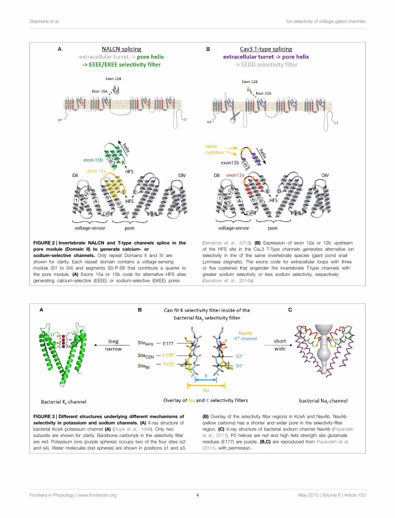
FIGURE 3 | Different structures underlying different mechanisms of
selectivity in potassium and sodium channels. (A) X-ray structure ofbacterial KcsA potassium channel (A) (Doyle et al., 1998). Only twosubunits are shown for clarity. Backbone carbonyls in the selectivity filterare red. Potassium ions (purple spheres) occupy two of the four sites (s2and s4). Water molecules (red spheres) are shown in positions s1 and s3.
(B) Overlay of the selectivity filter regions in KcsA and NavAb. NavAb(yellow carbons) has a shorter and wider pore in the selectivity-filterregion. (C) X-ray structure of bacterial sodium channel NavAb (Payandehet al., 2011). P2 helices are red and high field strength site glutamateresidues (E177) are purple. (B,C) are reproduced from Payandeh et al.(2011), with permission.
Frontiers in Physiology | www.frontiersin.org 4 May 2015 | Volume 6 | Article 153
Stephens et al. Ion selectivity of voltage-gated channels
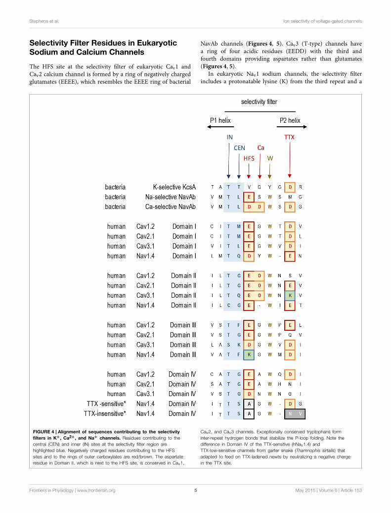
Selectivity Filter Residues in EukaryoticSodium and Calcium Channels
The HFS site at the selectivity filter of eukaryotic Cav1 andCav2 calcium channel is formed by a ring of negatively chargedglutamates (EEEE), which resembles the EEEE ring of bacterial
NavAb channels (Figures 4, 5). Cav3 (T-type) channels havea ring of four acidic residues (EEDD) with the third andfourth domains providing aspartates rather than glutamates(Figures 4, 5).
In eukaryotic Nav1 sodium channels, the selectivity filterincludes a protonatable lysine (K) from the third repeat and a
FIGURE 4 | Alignment of sequences contributing to the selectivity
filters in K+, Ca2+, and Na+ channels. Residues contributing to thecentral (CEN) and inner (IN) sites at the selectivity filter region arehighlighted blue. Negatively charged residues contributing to the HFSsites and to the rings of outer carboxylates are red/brown. The aspartateresidue in Domain II, which is next to the HFS site, is conserved in Cav1,
Cav2, and Cav3 channels. Exceptionally conserved tryptophans forminter-repeat hydrogen bonds that stabilize the P-loop folding. Note thedifference in Domain IV of the TTX-sensitive (hNav1.4) andTTX-low-sensitive channels from garter snake (Thamnophis sirtalis) thatadapted to feed on TTX-ladened newts by neutralizing a negative chargein the TTX site.
Frontiers in Physiology | www.frontiersin.org 5 May 2015 | Volume 6 | Article 153
Stephens et al. Ion selectivity of voltage-gated channels
neutral alanine (A) from the fourth repeat (the DEKA ring).Cnidarians, which possess the simplest body plan to includea nervous system and the most basal organisms with Nav1channels are the exception. Instead of the to the DEKA ringcnidarian selectivity filters have lysine and glutamate providedby the second and third repeats, respectively (the DKEAring) (Figure 5). The critical importance of the selectivity-filterresidues was established in experiments where the DKEA ringin the Nav1.2 channel was replaced by the EEEE ring to createcalcium-selective channels (Heinemann et al., 1992; Schlief et al.,1996).
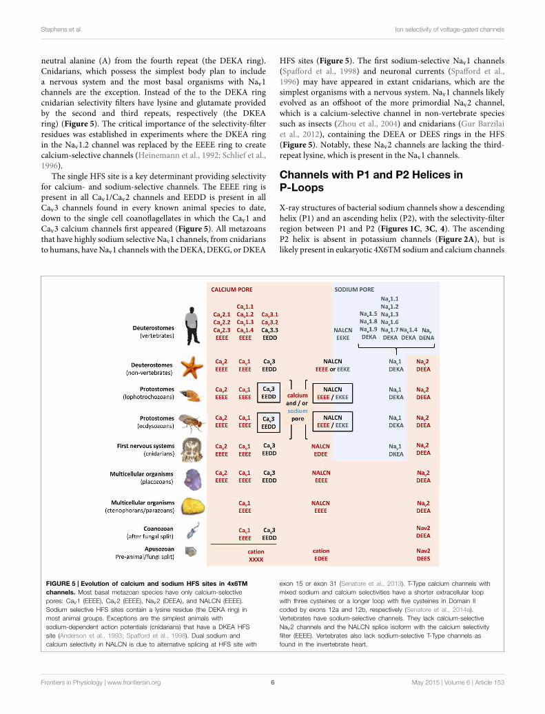
The single HFS site is a key determinant providing selectivityfor calcium- and sodium-selective channels. The EEEE ring ispresent in all Cav1/Cav2 channels and EEDD is present in allCav3 channels found in every known animal species to date,down to the single cell coanoflagellates in which the Cav1 andCav3 calcium channels first appeared (Figure 5). All metazoansthat have highly sodium selective Nav1 channels, from cnidariansto humans, have Nav1 channels with the DEKA, DEKG, or DKEA
HFS sites (Figure 5). The first sodium-selective Nav1 channels(Spafford et al., 1998) and neuronal currents (Spafford et al.,1996) may have appeared in extant cnidarians, which are thesimplest organisms with a nervous system. Nav1 channels likelyevolved as an offshoot of the more primordial Nav2 channel,which is a calcium-selective channel in non-vertebrate speciessuch as insects (Zhou et al., 2004) and cnidarians (Gur Barzilaiet al., 2012), containing the DEEA or DEES rings in the HFS(Figure 5). Notably, these Nav2 channels are lacking the third-repeat lysine, which is present in the Nav1 channels.
Channels with P1 and P2 Helices inP-Loops
X-ray structures of bacterial sodium channels show a descendinghelix (P1) and an ascending helix (P2), with the selectivity-filterregion between P1 and P2 (Figures 1C, 3C, 4). The ascendingP2 helix is absent in potassium channels (Figure 2A), but islikely present in eukaryotic 4X6TM sodium and calcium channels
FIGURE 5 | Evolution of calcium and sodium HFS sites in 4x6TM
channels. Most basal metazoan species have only calcium-selectivepores: Cav1 (EEEE), Cav2 (EEEE), Nav2 (DEEA), and NALCN (EEEE).Sodium selective HFS sites contain a lysine residue (the DEKA ring) inmost animal groups. Exceptions are the simplest animals withsodium-dependent action potentials (cnidarians) that have a DKEA HFSsite (Anderson et al., 1993; Spafford et al., 1998). Dual sodium andcalcium selectivity in NALCN is due to alternative splicing at HFS site with
exon 15 or exon 31 (Senatore et al., 2013). T-Type calcium channels withmixed sodium and calcium selectivities have a shorter extracellular loopwith three cysteines or a longer loop with five cysteines in Domain IIcoded by exons 12a and 12b, respectively (Senatore et al., 2014a).Vertebrates have sodium-selective channels. They lack calcium-selectiveNav2 channels and the NALCN splice isoform with the calcium selectivityfilter (EEEE). Vertebrates also lack sodium-selective T-Type channels asfound in the invertebrate heart.
Frontiers in Physiology | www.frontiersin.org 6 May 2015 | Volume 6 | Article 153
Stephens et al. Ion selectivity of voltage-gated channels
(Tikhonov and Zhorov, 2012). The outer vestibule is formedby P2 helices, which contain additional negatively chargedresidues positioned to attract cations to the pore. In sodiumchannels these residues are targeted by conotoxins, (see Korkoshet al., 2014) and references therein. The cluster of negativelycharged residues likely forms binding sites for incoming cationsabove the HFS site. Indeed, bacterial NavAb channel with threeengineered aspartates in the HFS site and P2 helix (replacementof TLESWSM by TLDDWSD) demonstrates calcium selectivity(Yue et al., 2002; Tang et al., 2014) (Figure 4).
In the same position where the second aspartate substitution(TLDDWSD) generates the calcium-selective bacterial sodiumchannel, the aspartate in the second repeat is conserveduniversally in all eukaryotic calcium channels, including Cav1,Cav2 and Cav3 channels from single cell coanoflagellates tomammalian channels (Tikhonov and Zhorov, 2011; Payandehand Minor, 2015). This aspartate, which is next to the HFS siteglutamate in Domain II, (e.g., TGEDWNS in Cav1.2, see “Ca” sitein Figure 4), is likely required for calcium ion selectivity.
Negatively charged glutamate or aspartate residues in the P2helix, three or four positions downstream from the HFS siteform an outer ring that is common in 4x6TM calcium andsodium channels (see “TTX” site, Figure 4). These positionscorrespond to the third aspartate residue in the TLDDWSDmotif that contributes to the engineered calcium selectivity inthe NavAb channel construct. Other negative charges, positionedmore distantly and above the HSF in the outer vestibule, havemore modest effects on ion selectivity (Chiamvimonvat et al.,1996; Favre et al., 1996; Schlief et al., 1996) and they are notconserved among all 4x6TM channels or within different sodiumand calcium channel subtypes. The rather poor conservationof negative charges in the outer vestibule contrasts with theinvariant EEEE HFS site and the adjoining aspartate just abovethe HFS site glutamate in Domain II, which are likely keydeterminants for calcium selectivity (Tikhonov and Zhorov,2011; Payandeh and Minor, 2015).
Tetrodotoxin Resistance and P2 HelixResidues
Selective pressures due to the presence of pore-blocking toxinshave led to adaptive resistance in certain organisms, involvingsubstitutions in the sodium channel residues three and fourpositions distant from the HFS site. Alterations in sodiumchannel pores can be used to escape the influences of tetrodotoxin(TTX). This highly potent neurotoxin is generated by symbioticbacteria that penetrate animal tissues of TTX-bearing animals.The latter include many invertebrate species (snails, crabs) aswell as vertebrates such as tetraodontiform fish (e.g., pufferfish)or newts. Highly TTX sensitive vertebrate nerve and skeletalmuscle-specific sodium channels possess an outer ring ofnegatively charged residues, EEDD, perched above the DEKAHFS site (e.g., the TSAGWD sequence in Domain IV of theNav1.4 channel, see “TTX” site, Figure 4). TTX resistance canbe generated by neutralizing this outer ring of negatively chargedresidues (Terlau et al., 1991). Invertebrates possess only one Nav1
channel gene, and almost complete TTX resistance is generatedin many invertebrate species (e.g., jellyfish, some flatworms,pulmonate snails, Varroa mite and sea squirts) due to the lackof negative charges in the outer ring (Du et al., 2009). Particularpopulations of common garter snake that feed on TTX-ladenednewts have similarly altered Nav1 channels with a neutralizedouter ring aspartate in Domain IV, most prevalently in skeletalmuscle Nav1.4 sodium channels (Figure 4) (Feldman et al., 2012).This is an example of rapid intra-species evolution of sodiumchannels, since common garter snake populations that do notfeed on TTX-ladened newts in their locale, possess unaltered,TTX-sensitive Nav1.4 sodium channels (Feldman et al., 2012)(Figure 4). There are also Domain IV mutations in Nav1.4sodium channels of pufferfish and other species (Jost et al., 2008),which prevent self-poisoning by TTX generated in their owntissues.
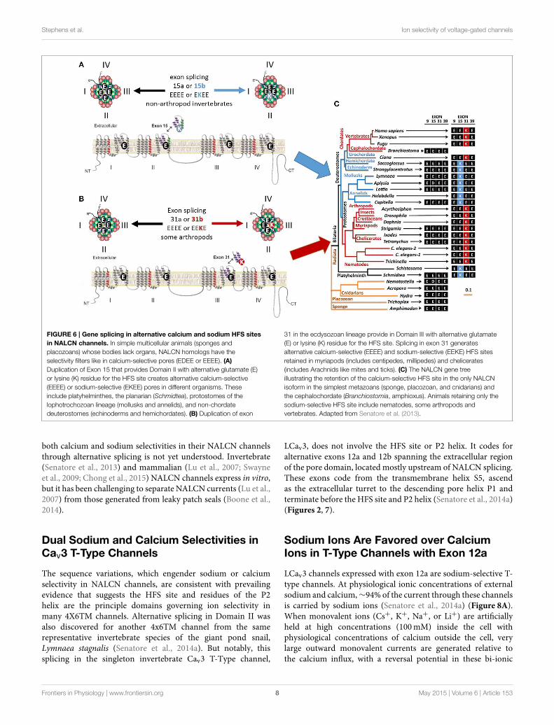
Mixed Calcium- and Sodium-Selective HFSSites in NALCN Ion Channels
NALCN ion channels provide a different example of adaptiveevolution, and it involves modifying the HFS site, as a meansto generate alternative ion selectivities (Senatore et al., 2013).NALCN is a separate lineage of 4x6TM channels with the ionselectivity in different species that can resemble either or bothcalcium-selective Cav1 and Cav2 channels, which have the EEEEHFS site, and the sodium selective Nav1 channels, which havethe DEKA, DEKG, or DKEA HFS sites (Senatore et al., 2013)(Figures 2, 6). NALCN channels possess a HFS site of a calciumchannel (EEEE) in basal multicellular organisms (sponge,placozoan and cnidarians) and non-vertebrate chordates (thecephalochordates) (Senatore et al., 2013) (Figure 6). However, inother animals, NALCN channels resemble sodium channels witha lysine in Domain II or III of the HFS site, respectively. Examplesinclude the EKEE ring in schistosome flatworms and Helobdellaleech and the EEKE ring in non-myriapod arthropods andvertebrates (Senatore et al., 2013) (Figure 6). A separate groupof non-vertebrates retain dual calcium and sodium selectivityat the HFS site, through mutually exclusive splicing, and theseevolved completely independently in at least two different animallineages. Almost all lophotrochozoan invertebrates (mollusks,annelids) and non-vertebrate deuterostomes (echinoderms,hemichordates) possess two forms of exon 15 in their expressibleNALCN transcripts, generating alternative calcium HFS site(EEEE) with exon 15a or a sodium HFS site (EKEE) with exon15b (Senatore et al., 2013) (Figure 6). Non-arthropod ecdysozoanprotostomes (myriapods, chelicerates) possess two forms of exon31 generating an alternative calcium HFS site (EEEE) with exon31a or a sodium HFS site (EEKE) with exon 31b (Senatore et al.,2013) (Figure 6).
There is compelling evidence from nematode (C. elegans),fruit fly (Drosophila) and mammals that NALCN channelscontribute to pacemaker currents that lead to rhythmic activitysuch as locomotion (Gao et al., 2015), circadian clock rhythms(Lear et al., 2013), and breathing (Lu et al., 2007). Why animalshave a calcium-selective or sodium-selective form of NALCN, or
Frontiers in Physiology | www.frontiersin.org 7 May 2015 | Volume 6 | Article 153
Stephens et al. Ion selectivity of voltage-gated channels
FIGURE 6 | Gene splicing in alternative calcium and sodium HFS sites
in NALCN channels. In simple multicellular animals (sponges andplacozoans) whose bodies lack organs, NALCN homologs have theselectivity filters like in calcium-selective pores (EDEE or EEEE). (A)Duplication of Exon 15 that provides Domain II with alternative glutamate (E)or lysine (K) residue for the HFS site creates alternative calcium-selective(EEEE) or sodium-selective (EKEE) pores in different organisms. Theseinclude platyhelminthes, the planarian (Schmidtea), protostomes of thelophotrochozoan lineage (mollusks and annelids), and non-chordatedeuterostomes (echinoderms and hemichordates). (B) Duplication of exon
31 in the ecdysozoan lineage provide in Domain III with alternative glutamate(E) or lysine (K) residue for the HFS site. Splicing in exon 31 generatesalternative calcium-selective (EEEE) and sodium-selective (EEKE) HFS sitesretained in myriapods (includes centipedes, millipedes) and chelicerates(includes Arachnids like mites and ticks). (C) The NALCN gene treeillustrating the retention of the calcium-selective HFS site in the only NALCNisoform in the simplest metazoans (sponge, placozoan, and cnidarians) andthe cephalochordate (Branchiostomia, amphioxus). Animals retaining only thesodium-selective HFS site include nematodes, some arthropods andvertebrates. Adapted from Senatore et al. (2013).
both calcium and sodium selectivities in their NALCN channelsthrough alternative splicing is not yet understood. Invertebrate(Senatore et al., 2013) and mammalian (Lu et al., 2007; Swayneet al., 2009; Chong et al., 2015) NALCN channels express in vitro,but it has been challenging to separate NALCN currents (Lu et al.,2007) from those generated from leaky patch seals (Boone et al.,2014).
Dual Sodium and Calcium Selectivities inCav3 T-Type Channels
The sequence variations, which engender sodium or calciumselectivity in NALCN channels, are consistent with prevailingevidence that suggests the HFS site and residues of the P2helix are the principle domains governing ion selectivity inmany 4X6TM channels. Alternative splicing in Domain II wasalso discovered for another 4x6TM channel from the samerepresentative invertebrate species of the giant pond snail,Lymnaea stagnalis (Senatore et al., 2014a). But notably, thissplicing in the singleton invertebrate Cav3 T-Type channel,
LCav3, does not involve the HFS site or P2 helix. It codes foralternative exons 12a and 12b spanning the extracellular regionof the pore domain, located mostly upstream of NALCN splicing.These exons code from the transmembrane helix S5, ascendas the extracellular turret to the descending pore helix P1 andterminate before the HFS site and P2 helix (Senatore et al., 2014a)(Figures 2, 7).
Sodium Ions Are Favored over CalciumIons in T-Type Channels with Exon 12a
LCav3 channels expressed with exon 12a are sodium-selective T-type channels. At physiological ionic concentrations of externalsodium and calcium,∼94% of the current through these channelsis carried by sodium ions (Senatore et al., 2014a) (Figure 8A).When monovalent ions (Cs+, K+, Na+, or Li+) are artificiallyheld at high concentrations (100mM) inside the cell withphysiological concentrations of calcium outside the cell, verylarge outward monovalent currents are generated relative tothe calcium influx, with a reversal potential in these bi-ionic
Frontiers in Physiology | www.frontiersin.org 8 May 2015 | Volume 6 | Article 153
Stephens et al. Ion selectivity of voltage-gated channels
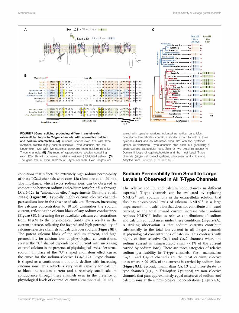
FIGURE 7 | Gene splicing producing different cysteine-rich
extracellular loops in T-type channels with alternative calcium
and sodium selectivities. (A) In snails, shorter exon 12a with threecysteines creates highly sodium selective T-type channels and thelonger exon 12b with five cysteines generates more calcium selectiveT-type channels. (B) Alignment of representative species containingexon 12a/12b with conserved cysteine residues (highlighted yellow). (C)
The gene tree of exon 12a/12b of T-type channels. Exon lengths are
scaled with cysteine residues indicated as vertical bars. Mostprotostome invertebrates contain a shorter exon 12a with a threecysteines (blue) and an alternative exon 12b with five cysteines(green). All vertebrate T-type channels have exon 12a generating asingle-cysteine extracellular loop. Zero or two cysteines appear inDomain II loops of cephalochordate and the most basal T-typechannels (single cell coanoflagellates, placozoan, and cnidarians).Adapted from Senatore et al. (2014a).
conditions that reflects the extremely high sodium permeabilityof these LCav3 channels with exon 12a (Senatore et al., 2014a).The imbalance, which favors sodium ions, can be observed ascompetition between sodium and calcium ions for influx throughLCav3-12a in “anomalous effect” experiments (Senatore et al.,2014a) (Figure 8B). Typically, highly calcium selective channelspass sodium ions in the absence of calcium. However, increasingthe calcium concentration to 10µM diminishes the sodiumcurrent, reflecting the calcium block of any sodium conductance(Figure 8B). Increasing the extracellular calcium concentrationsfrom 10µM to the physiological (mM) levels results in thecurrent increase, reflecting the favored and high permeability ofcalcium-selective channels for calcium over sodium (Figure 8B).The potent calcium block of the sodium current, and highpermeability for calcium ions at physiological concentrations,creates the “U” shaped dependence of current with increasingexternal calcium in the presence of physiological levels of externalsodium. In place of the “U” shaped anomalous effect curve,the curve for the sodium-selective LCav3-12a T-type channelis shaped as a continuous monotonic decline with increasingcalcium ions. This reflects a very weak capacity for calciumto block the sodium current and a relatively small calciumconductance through these channels even in the presence ofphysiological levels of external calcium (Senatore et al., 2014a).
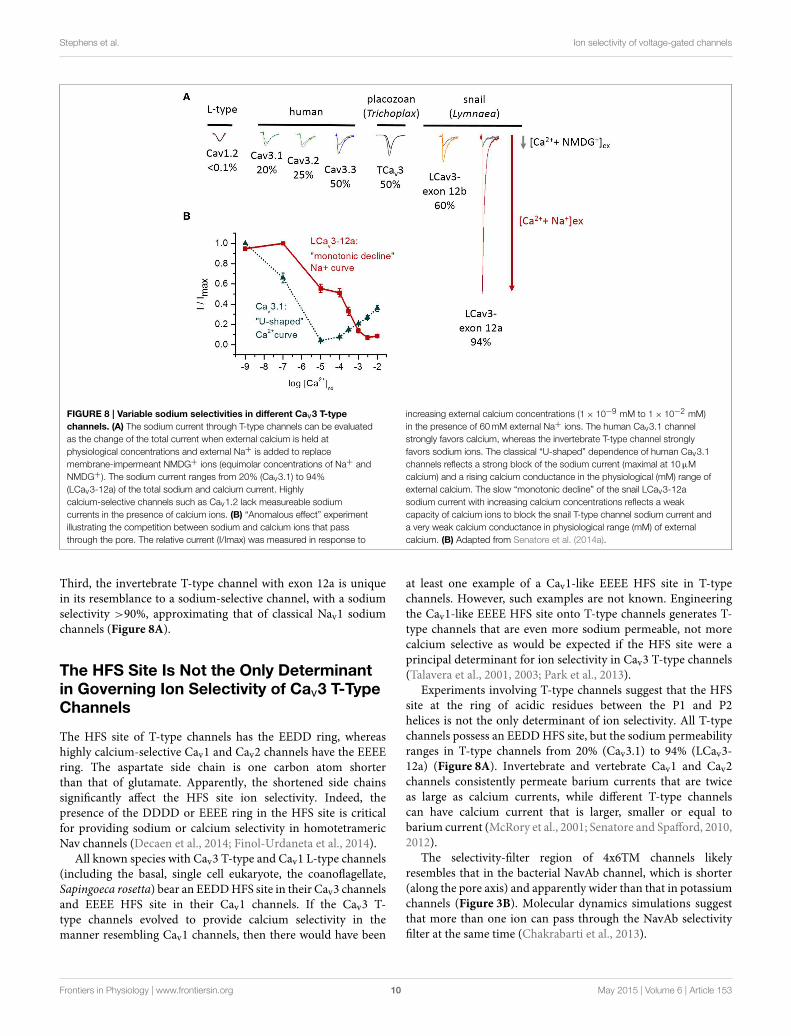
Sodium Permeability from Small to LargeLevels Is Observed in All T-Type Channels
The relative sodium and calcium conductances in differentexpressed T-type channels can be evaluated by replacingNMDG+ with sodium ions in the extracellular solution thatalso has physiological levels of calcium. NMDG+ is a largeimpermeant monovalent ion that does not contribute an inwardcurrent, so the total inward current increase when sodiumreplaces NMDG+ indicates relative contributions of sodiumand calcium conductances under these conditions (Figure 8A).A striking observation is that sodium currents contributesubstantially to the total ion current in all T-type channelsat physiological concentrations of calcium. This contrasts withhighly calcium-selective Cav1 and Cav2 channels where thesodium current is immeasurably small (<1% of the currentcarried by sodium ions). There are three categories of relativesodium permeability in T-type channels. First, mammalianCav3.1 and Cav3.2 channels are the most calcium selectiveones where ∼20–25% of the current is carried by sodium ions(Figure 8A). Second, mammalian Cav3.3 and invertebrate T-type channels (e.g., in Trichoplax, Lymnaea) are non-selectivechannels that pass approximately equal mixtures of sodium andcalcium ions at their physiological concentrations (Figure 8A).
Frontiers in Physiology | www.frontiersin.org 9 May 2015 | Volume 6 | Article 153
Stephens et al. Ion selectivity of voltage-gated channels
FIGURE 8 | Variable sodium selectivities in different Cav3 T-type
channels. (A) The sodium current through T-type channels can be evaluatedas the change of the total current when external calcium is held atphysiological concentrations and external Na+ is added to replacemembrane-impermeant NMDG+ ions (equimolar concentrations of Na+ andNMDG+). The sodium current ranges from 20% (Cav3.1) to 94%(LCav3-12a) of the total sodium and calcium current. Highlycalcium-selective channels such as Cav1.2 lack measureable sodiumcurrents in the presence of calcium ions. (B) “Anomalous effect” experimentillustrating the competition between sodium and calcium ions that passthrough the pore. The relative current (I/Imax) was measured in response to
increasing external calcium concentrations (1× 10⁻⁹ mM to 1× 10⁻² mM)in the presence of 60mM external Na+ ions. The human Cav3.1 channelstrongly favors calcium, whereas the invertebrate T-type channel stronglyfavors sodium ions. The classical “U-shaped” dependence of human Cav3.1channels reflects a strong block of the sodium current (maximal at 10µMcalcium) and a rising calcium conductance in the physiological (mM) range ofexternal calcium. The slow “monotonic decline” of the snail LCav3-12asodium current with increasing calcium concentrations reflects a weakcapacity of calcium ions to block the snail T-type channel sodium current anda very weak calcium conductance in physiological range (mM) of externalcalcium. (B) Adapted from Senatore et al. (2014a).
Third, the invertebrate T-type channel with exon 12a is uniquein its resemblance to a sodium-selective channel, with a sodiumselectivity >90%, approximating that of classical Nav1 sodiumchannels (Figure 8A).
The HFS Site Is Not the Only Determinantin Governing Ion Selectivity of Cav3 T-TypeChannels
The HFS site of T-type channels has the EEDD ring, whereashighly calcium-selective Cav1 and Cav2 channels have the EEEEring. The aspartate side chain is one carbon atom shorterthan that of glutamate. Apparently, the shortened side chainssignificantly affect the HFS site ion selectivity. Indeed, thepresence of the DDDD or EEEE ring in the HFS site is criticalfor providing sodium or calcium selectivity in homotetramericNav channels (Decaen et al., 2014; Finol-Urdaneta et al., 2014).
All known species with Cav3 T-type and Cav1 L-type channels(including the basal, single cell eukaryote, the coanoflagellate,Sapingoeca rosetta) bear an EEDDHFS site in their Cav3 channelsand EEEE HFS site in their Cav1 channels. If the Cav3 T-type channels evolved to provide calcium selectivity in themanner resembling Cav1 channels, then there would have been
at least one example of a Cav1-like EEEE HFS site in T-typechannels. However, such examples are not known. Engineeringthe Cav1-like EEEE HFS site onto T-type channels generates T-type channels that are even more sodium permeable, not morecalcium selective as would be expected if the HFS site were aprincipal determinant for ion selectivity in Cav3 T-type channels(Talavera et al., 2001, 2003; Park et al., 2013).
Experiments involving T-type channels suggest that the HFSsite at the ring of acidic residues between the P1 and P2helices is not the only determinant of ion selectivity. All T-typechannels possess an EEDDHFS site, but the sodium permeabilityranges in T-type channels from 20% (Cav3.1) to 94% (LCav3-12a) (Figure 8A). Invertebrate and vertebrate Cav1 and Cav2channels consistently permeate barium currents that are twiceas large as calcium currents, while different T-type channelscan have calcium current that is larger, smaller or equal tobarium current (McRory et al., 2001; Senatore and Spafford, 2010,2012).
The selectivity-filter region of 4x6TM channels likelyresembles that in the bacterial NavAb channel, which is shorter(along the pore axis) and apparently wider than that in potassiumchannels (Figure 3B). Molecular dynamics simulations suggestthat more than one ion can pass through the NavAb selectivityfilter at the same time (Chakrabarti et al., 2013).
Frontiers in Physiology | www.frontiersin.org 10 May 2015 | Volume 6 | Article 153
Stephens et al. Ion selectivity of voltage-gated channels
Cysteine-Rich Extracellular Turrets GovernSelectivity of Cav3 T-Type Channels
Exon 12a, which generates sodium-selective T-type channels,and exon 12b, which creates a more calcium-selective T-typechannel, are both located at the extracellular turret of DomainII (Figure 7). Exon 12a and exon 12b differ in their size andpattern of cysteines, while most of other residues are highlyvariable (Figure 7). The majority of protostome invertebratespecies (within nematodes, arthropods, annelids and mollusks)possess both exons 12a and 12b, although a few species possessonly exon 12a (e.g., Brugia nematode,Helobdella annelid) or onlyexon 12b (e.g., Capitella annelid) (Figure 7). Exon 12a is shorter(∼41 amino acids) with a highly conserved motif containingthree cysteines (CxC. . . .C), while exon 12b is longer (∼52 aminoacids) and has a highly conserved motifs with five cysteines(C. . .CxC. . .CxC or CxxC. . .C. . .CxC in nematodes) (Figure 7).Our consistent findings (unpublished) are that mutating thecysteine-containing motifs of exon 12a and 12b generates highlyunusual T-type channels that have a capacity to conduct multiplediffering ions simultaneously and independently, as if the poreselectivity is lost.
A Conserved Pattern of Cysteines withinExtracellular Turrets in 4x6TM Channels
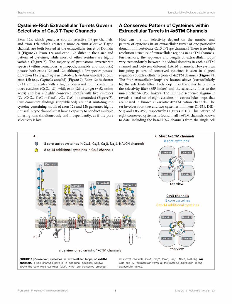
How can the ion selectivity depend on the number andpattern of cysteines in an extracellular turret of one particulardomain in invertebrate Cav3 T-Type channels? There is no highresolution structure of extracellular regions in 4x6TM channels.Furthermore, the sequence and length of extracellular loopsvary tremendously between individual domains in each 4x6TMchannel and between different 4x6TM channels. However, anintriguing pattern of conserved cysteines is seen in alignedsequences of extracellular regions of 4x6TM channels (Figure 9).The four extracellular loops are located above (extracellularlyto) the selectivity filter. Each loop links the outer helix S5 tothe selectivity filter (S5P linker) and the selectivity filter to theinner helix S6 (PS6 linker). The multiple sequence alignmentreveals a basal set of eight cysteines in extracellular loops thatare shared in known eukaryotic 4x6TM cation channels. Theset involves four, two and two cysteines in linkers DI-S5P, DIII-S5P, and DIV-PS6, respectively (Figures 9, 10). This pattern ofeight conserved cysteines is found in all 4x6TM channels knownto date, including the basal Nav2 channels from the single-cell
FIGURE 9 | Conserved cysteines in extracellular loops of 4x6TM
channels. T-type channels have 8–14 additional cysteines (yellow)above the core eight cysteines (blue), which are conserved amongst
all 4x6TM channels (Cav1, Cav2, Cav3, Nav1, Nav2, NALCN). (A)
Side and (B) extracellular views at the cysteine distribution in theextracellular turrets.
Frontiers in Physiology | www.frontiersin.org 11 May 2015 | Volume 6 | Article 153

Stephens et al. Ion selectivity of voltage-gated channels
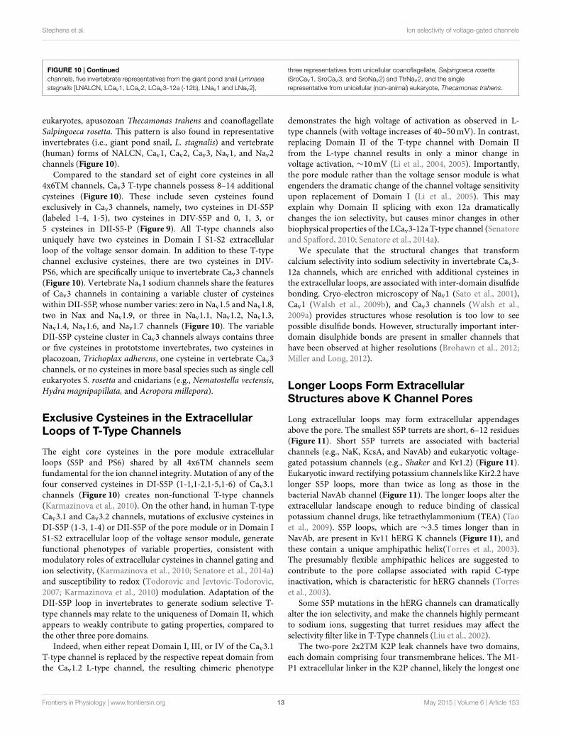
FIGURE 10 | Alignment of transmembrane helices and P-helices in
4x6TM channels illustrating location of extracellular-loop cysteines.
The pore module includes transmembrane helices S5, S6 (dark gray), andpore helices P1 and P2 (tan) on either side of the selectivity filter (SF).Conserved cysteines (yellow) are present in the variable-size extracellularloops (light gray). Eight core cysteines are located at positions 1-1, 1-2, 1-6,3-1, 3-2, 4-3, and 4-6. Additional cysteines are found in Domain II of most
vertebrate Nav1 channels and T-type channels. All T-type channels also haveconserved cysteines in S5P loops of Domain I (1-4,1-5) and Domain IV(4-1,4-2), as well as in the extracellular loop between S1-S2 segments ofDomain I. Two additional cysteines unique to invertebrate T-type channels arefound in Domain IV extracellular loop PS6, C-terminal to the P2 helix (4-4,4-5). The alignment is obtained using sequences of 21 human 4x6TM
(Continued)
Frontiers in Physiology | www.frontiersin.org 12 May 2015 | Volume 6 | Article 153
Stephens et al. Ion selectivity of voltage-gated channels
FIGURE 10 | Continued
channels, five invertebrate representatives from the giant pond snail Lymnaeastagnalis [LNALCN, LCav1, LCav2, LCav3-12a (-12b), LNav1 and LNav2],
three representatives from unicellular coanoflagellate, Salpingoeca rosetta(SroCav1, SroCav3, and SroNav2) and TtrNav2, and the singlerepresentative from unicellular (non-animal) eukaryote, Thecamonas trahens.
eukaryotes, apusozoan Thecamonas trahens and coanoflagellateSalpingoeca rosetta. This pattern is also found in representativeinvertebrates (i.e., giant pond snail, L. stagnalis) and vertebrate(human) forms of NALCN, Cav1, Cav2, Cav3, Nav1, and Nav2channels (Figure 10).
Compared to the standard set of eight core cysteines in all4x6TM channels, Cav3 T-type channels possess 8–14 additionalcysteines (Figure 10). These include seven cysteines foundexclusively in Cav3 channels, namely, two cysteines in DI-S5P(labeled 1-4, 1-5), two cysteines in DIV-S5P and 0, 1, 3, or5 cysteines in DII-S5-P (Figure 9). All T-type channels alsouniquely have two cysteines in Domain I S1-S2 extracellularloop of the voltage sensor domain. In addition to these T-typechannel exclusive cysteines, there are two cysteines in DIV-PS6, which are specifically unique to invertebrate Cav3 channels(Figure 10). Vertebrate Nav1 sodium channels share the featuresof Cav3 channels in containing a variable cluster of cysteineswithin DII-S5P, whose number varies: zero in Nav1.5 and Nav1.8,two in Nax and Nav1.9, or three in Nav1.1, Nav1.2, Nav1.3,Nav1.4, Nav1.6, and Nav1.7 channels (Figure 10). The variableDII-S5P cysteine cluster in Cav3 channels always contains threeor five cysteines in prototstome invertebrates, two cysteines inplacozoan, Trichoplax adherens, one cysteine in vertebrate Cav3channels, or no cysteines in more basal species such as single celleukaryotes S. rosetta and cnidarians (e.g., Nematostella vectensis,Hydra magnipapillata, and Acropora millepora).
Exclusive Cysteines in the ExtracellularLoops of T-Type Channels
The eight core cysteines in the pore module extracellularloops (S5P and PS6) shared by all 4x6TM channels seemfundamental for the ion channel integrity. Mutation of any of thefour conserved cysteines in DI-S5P (1-1,1-2,1-5,1-6) of Cav3.1channels (Figure 10) creates non-functional T-type channels(Karmazinova et al., 2010). On the other hand, in human T-typeCav3.1 and Cav3.2 channels, mutations of exclusive cysteines inDI-S5P (1-3, 1-4) or DII-S5P of the pore module or in Domain IS1-S2 extracellular loop of the voltage sensor module, generatefunctional phenotypes of variable properties, consistent withmodulatory roles of extracellular cysteines in channel gating andion selectivity, (Karmazinova et al., 2010; Senatore et al., 2014a)and susceptibility to redox (Todorovic and Jevtovic-Todorovic,2007; Karmazinova et al., 2010) modulation. Adaptation of theDII-S5P loop in invertebrates to generate sodium selective T-type channels may relate to the uniqueness of Domain II, whichappears to weakly contribute to gating properties, compared tothe other three pore domains.
Indeed, when either repeat Domain I, III, or IV of the Cav3.1T-type channel is replaced by the respective repeat domain fromthe Cav1.2 L-type channel, the resulting chimeric phenotype
demonstrates the high voltage of activation as observed in L-type channels (with voltage increases of 40–50mV). In contrast,replacing Domain II of the T-type channel with Domain IIfrom the L-type channel results in only a minor change involtage activation, ∼10mV (Li et al., 2004, 2005). Importantly,the pore module rather than the voltage sensor module is whatengenders the dramatic change of the channel voltage sensitivityupon replacement of Domain I (Li et al., 2005). This mayexplain why Domain II splicing with exon 12a dramaticallychanges the ion selectivity, but causes minor changes in otherbiophysical properties of the LCav3-12a T-type channel (Senatoreand Spafford, 2010; Senatore et al., 2014a).
We speculate that the structural changes that transformcalcium selectivity into sodium selectivity in invertebrate Cav3-12a channels, which are enriched with additional cysteines inthe extracellular loops, are associated with inter-domain disulfidebonding. Cryo-electron microscopy of Nav1 (Sato et al., 2001),Cav1 (Walsh et al., 2009b), and Cav3 channels (Walsh et al.,2009a) provides structures whose resolution is too low to seepossible disulfide bonds. However, structurally important inter-domain disulphide bonds are present in smaller channels thathave been observed at higher resolutions (Brohawn et al., 2012;Miller and Long, 2012).
Longer Loops Form ExtracellularStructures above K Channel Pores
Long extracellular loops may form extracellular appendagesabove the pore. The smallest S5P turrets are short, 6–12 residues(Figure 11). Short S5P turrets are associated with bacterialchannels (e.g., NaK, KcsA, and NavAb) and eukaryotic voltage-gated potassium channels (e.g., Shaker and Kv1.2) (Figure 11).Eukaryotic inward rectifying potassium channels like Kir2.2 havelonger S5P loops, more than twice as long as those in thebacterial NavAb channel (Figure 11). The longer loops alter theextracellular landscape enough to reduce binding of classicalpotassium channel drugs, like tetraethylammonium (TEA) (Taoet al., 2009). S5P loops, which are ∼3.5 times longer than inNavAb, are present in Kv11 hERG K channels (Figure 11), andthese contain a unique amphipathic helix(Torres et al., 2003).The presumably flexible amphipathic helices are suggested tocontribute to the pore collapse associated with rapid C-typeinactivation, which is characteristic for hERG channels (Torreset al., 2003).
Some S5P mutations in the hERG channels can dramaticallyalter the ion selectivity, and make the channels highly permeantto sodium ions, suggesting that turret residues may affect theselectivity filter like in T-Type channels (Liu et al., 2002).
The two-pore 2x2TM K2P leak channels have two domains,each domain comprising four transmembrane helices. The M1-P1 extracellular linker in the K2P channel, likely the longest one
Frontiers in Physiology | www.frontiersin.org 13 May 2015 | Volume 6 | Article 153
Stephens et al. Ion selectivity of voltage-gated channels
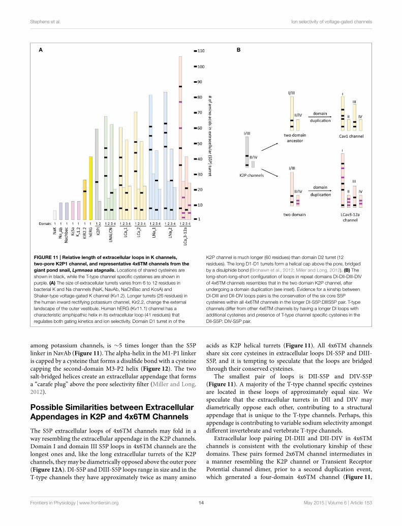
FIGURE 11 | Relative length of extracellular loops in K channels,
two-pore K2P1 channel, and representative 4x6TM channels from the
giant pond snail, Lymnaea stagnalis. Locations of shared cysteines areshown in black, while the T-type channel specific cysteines are shown inpurple. (A) The size of extracellular turrets varies from 6 to 12 residues inbacterial K and Na channels (NaK, NavAb, NaChBac and KcsA) andShaker-type voltage-gated K channel (Kv1.2). Longer turrets (26 residues) inthe human inward rectifying potassium channel, Kir2.2, change the externallandscape of the outer vestibule. Human hERG (Kv11.1) channel has acharacteristic amphipathic helix in its extracellular loop (41 residues) thatregulates both gating kinetics and ion selectivity. Domain D1 turret in of the
K2P channel is much longer (60 residues) than domain D2 turret (12residues). The long D1-D1 turrets form a helical cap above the pore, bridgedby a disulphide bond (Brohawn et al., 2012; Miller and Long, 2012). (B) Thelong-short-long-short configuration of loops in repeat domains DI-DII-DIII-DIVof 4x6TM channels resembles that in the two domain K2P channel, afterundergoing a domain duplication (see inset). Evidence for a kinship betweenDI-DIII and DII-DIV loops pairs is the conservation of the six core S5Pcysteines within all 4x6TM channels in the longer DI-S5P:DIIIS5P pair. T-typechannels differ from other 4x6TM channels by having a longer DI loops withadditional cysteines and presence of T-type channel specific cysteines in theDII-S5P: DIV-S5P pair.
among potassium channels, is ∼5 times longer than the S5Plinker in NavAb (Figure 11). The alpha-helix in the M1-P1 linkeris capped by a cysteine that forms a disulfide bond with a cysteinecapping the second-domain M3-P2 helix (Figure 12). The twosalt-bridged helices create an extracellular appendage that formsa “carafe plug” above the pore selectivity filter (Miller and Long,2012).
Possible Similarities between ExtracellularAppendages in K2P and 4x6TM Channels
The S5P extracellular loops of 4x6TM channels may fold in away resembling the extracellular appendage in the K2P channels.Domain I and domain III S5P loops in 4x6TM channels are thelongest ones and, like the long extracellular turrets of the K2Pchannels, theymay be diametrically opposed above the outer pore(Figure 12A). DI-S5P and DIII-S5P loops range in size and in theT-type channels they have approximately twice as many amino
acids as K2P helical turrets (Figure 11). All 4x6TM channelsshare six core cysteines in extracellular loops DI-S5P and DIII-S5P, and it is tempting to speculate that the loops are bridgedthrough their conserved cysteines.
The smallest pair of loops is DII-S5P and DIV-S5P(Figure 11). A majority of the T-type channel specific cysteinesare located in these loops of approximately equal size. Wespeculate that the extracellular turrets in DII and DIV maydiametrically oppose each other, contributing to a structuralappendage that is unique to the T-type channels. Perhaps, thisappendage is contributing to variable sodium selectivity amongstdifferent invertebrate and vertebrate T-type channels.
Extracellular loop pairing DI-DIII and DII-DIV in 4x6TMchannels is consistent with the evolutionary kinship of thesedomains. These pairs formed 2x6TM channel intermediates ina manner resembling the K2P channel or Transient ReceptorPotential channel dimer, prior to a second duplication event,which generated a four-domain 4x6TM channel (Figure 11,
Frontiers in Physiology | www.frontiersin.org 14 May 2015 | Volume 6 | Article 153
Stephens et al. Ion selectivity of voltage-gated channels
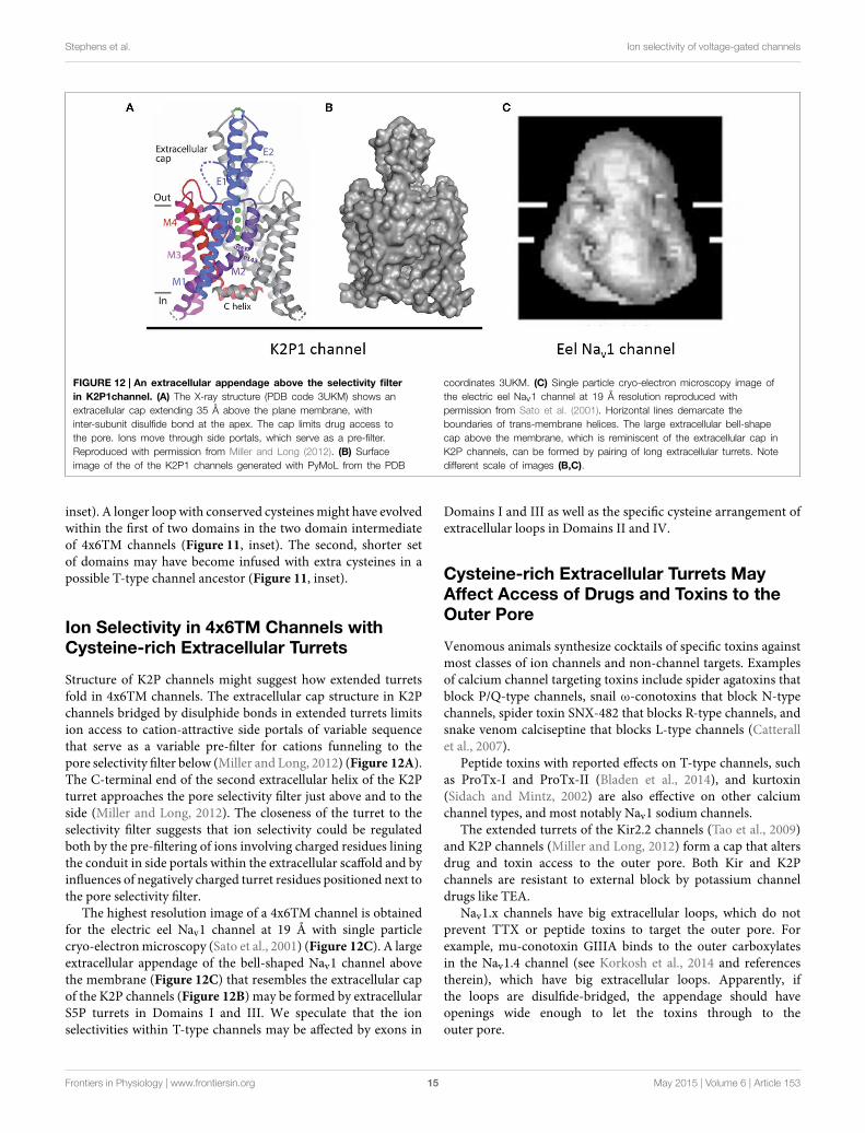
FIGURE 12 | An extracellular appendage above the selectivity filter
in K2P1channel. (A) The X-ray structure (PDB code 3UKM) shows anextracellular cap extending 35 Å above the plane membrane, withinter-subunit disulfide bond at the apex. The cap limits drug access tothe pore. Ions move through side portals, which serve as a pre-filter.Reproduced with permission from Miller and Long (2012). (B) Surfaceimage of the of the K2P1 channels generated with PyMoL from the PDB
coordinates 3UKM. (C) Single particle cryo-electron microscopy image ofthe electric eel Nav1 channel at 19 Å resolution reproduced withpermission from Sato et al. (2001). Horizontal lines demarcate theboundaries of trans-membrane helices. The large extracellular bell-shapecap above the membrane, which is reminiscent of the extracellular cap inK2P channels, can be formed by pairing of long extracellular turrets. Notedifferent scale of images (B,C).
inset). A longer loop with conserved cysteinesmight have evolvedwithin the first of two domains in the two domain intermediateof 4x6TM channels (Figure 11, inset). The second, shorter setof domains may have become infused with extra cysteines in apossible T-type channel ancestor (Figure 11, inset).
Ion Selectivity in 4x6TM Channels withCysteine-rich Extracellular Turrets
Structure of K2P channels might suggest how extended turretsfold in 4x6TM channels. The extracellular cap structure in K2Pchannels bridged by disulphide bonds in extended turrets limitsion access to cation-attractive side portals of variable sequencethat serve as a variable pre-filter for cations funneling to thepore selectivity filter below (Miller and Long, 2012) (Figure 12A).The C-terminal end of the second extracellular helix of the K2Pturret approaches the pore selectivity filter just above and to theside (Miller and Long, 2012). The closeness of the turret to theselectivity filter suggests that ion selectivity could be regulatedboth by the pre-filtering of ions involving charged residues liningthe conduit in side portals within the extracellular scaffold and byinfluences of negatively charged turret residues positioned next tothe pore selectivity filter.
The highest resolution image of a 4x6TM channel is obtainedfor the electric eel Nav1 channel at 19 Å with single particlecryo-electronmicroscopy (Sato et al., 2001) (Figure 12C). A largeextracellular appendage of the bell-shaped Nav1 channel abovethe membrane (Figure 12C) that resembles the extracellular capof the K2P channels (Figure 12B) may be formed by extracellularS5P turrets in Domains I and III. We speculate that the ionselectivities within T-type channels may be affected by exons in
Domains I and III as well as the specific cysteine arrangement ofextracellular loops in Domains II and IV.
Cysteine-rich Extracellular Turrets MayAffect Access of Drugs and Toxins to theOuter Pore
Venomous animals synthesize cocktails of specific toxins againstmost classes of ion channels and non-channel targets. Examplesof calcium channel targeting toxins include spider agatoxins thatblock P/Q-type channels, snail ω-conotoxins that block N-typechannels, spider toxin SNX-482 that blocks R-type channels, andsnake venom calciseptine that blocks L-type channels (Catterallet al., 2007).
Peptide toxins with reported effects on T-type channels, suchas ProTx-I and ProTx-II (Bladen et al., 2014), and kurtoxin(Sidach and Mintz, 2002) are also effective on other calciumchannel types, and most notably Nav1 sodium channels.
The extended turrets of the Kir2.2 channels (Tao et al., 2009)and K2P channels (Miller and Long, 2012) form a cap that altersdrug and toxin access to the outer pore. Both Kir and K2Pchannels are resistant to external block by potassium channeldrugs like TEA.
Nav1.x channels have big extracellular loops, which do notprevent TTX or peptide toxins to target the outer pore. Forexample, mu-conotoxin GIIIA binds to the outer carboxylatesin the Nav1.4 channel (see Korkosh et al., 2014 and referencestherein), which have big extracellular loops. Apparently, ifthe loops are disulfide-bridged, the appendage should haveopenings wide enough to let the toxins through to theouter pore.
Frontiers in Physiology | www.frontiersin.org 15 May 2015 | Volume 6 | Article 153
Stephens et al. Ion selectivity of voltage-gated channels
Scorpion toxins within the delta-KTx family specifically targetthe hERG extracellular loops. These toxins are distinct fromcharybdotoxin (alpha-KTx family of toxins), which plugs theERG channel pore (Vandenberg et al., 2012). T-type channelsmay resemble the Kir2.2 and K2P channels, because no naturally-occurring peptide toxins have been isolated that specifically targetthe pore module of T-type channels.
High throughput screening methods have revealedmany T-Type channel specific blockers (Giordanetto et al.,2011), including Z944 (Tringham et al., 2012) and TTA-P2(Dreyfus et al., 2010). High resolution structures obtainedwith direct detection camera and single nanoparticle cryo-electron microscopy (Liao et al., 2014) may help clarify thestructural peculiarities of extracellular loops that underlie thediversity within different T-type channels, as well as betweenT-type channels and other 4x6TM channels. Knowledge of theextracellular structures above T-type channels may also assist infuture drug design.
Conclusions
Invertebrates have evolved to generate sodium and calciumselectivity within the family of 4x6TM channels. Invertebratespecies normally have a singleton gene for NALCN, but threegenes for calcium channels (Cav1, Cav2, and Cav3) and twosodium channel genes (Nav2, Nav1). NALCN resembles a calciumchannel in basal metazoans, a sodium channel in animal groupssuch as vertebrates, and bears both dual sodium and calciumselective pores in most invertebrates (Senatore et al., 2013). Thereplacement of sodium and calcium selectivity in invertebrateNALCN channels by splicing at HFS site confirms this site witha lysine in the second or third domain as a key determinant ofthe sodium selectivity in 4x6TM channels. This splicing affectsthe same HFS site where artificial swapping of selectivity filterresidues between mammalian Cav1.2 and Nav1.2 channels has
been demonstrated to change the ion selectivity (Heinemannet al., 1992; Schlief et al., 1996).
Invertebrate Cav3 T-type channels also generate alternativesodium or calcium selectivity due to the extracellular S5P loopin Domain II, which is located above the HFS site and P2 helix(Senatore et al., 2014a). 4x6TM channels have cysteines in theextracellular loops of the diametrically opposing domains. In K2Pchannels, such cysteines form a disulfide bond to stabilize anextracellular appendage above the selectivity filter that serves asa pre-filter for permeating ions and as a shield that limits drugand toxin access to the selectivity filter. Consistent with this idea,cryo-electron microscopy of the eel Nav1 channel shows a dome-shaped extracellular appendage. It is tempting to speculate thatsuch extracellular appendages exist above the pores of eukaryotic4X6TM channels, and that T-type channels have a uniqueextracellular appendage that contributes to alternative sodium orcalcium selectivity. Various cysteine-rich loops in different T-typechannels appear to provide different ion selectivity propertiesand may regulate drug access. A possible avenue for future drugdesign is targeting the variable loop sequences. There is morevariability between different T-type channels in the extracellularloop regions than in the selectivity-filter and inner pore regions,so targeting the loop sequences could facilitate design of drugs.In the future, single nanoparticle cryo-electron microscopy andadvanced direct detection cameras may provide greater insightsinto the nature of the variable extracellular appendages above4x6TM channels.
Acknowledgments
We thank Robert French (University of Calgary) for his helpfulcomments. This manuscript was supported by the Heart andStroke Foundation of Canada (Grant-in-aid to JS) and from theNatural Sciences and Engineering Research Council of Canada(discovery grants to JS and BZ).
References
Anderson, P. A., Holman, M. A., and Greenberg, R. M. (1993). Deducedamino acid sequence of a putative sodium channel from the scyphozoanjellyfish Cyanea capillata. Proc. Natl. Acad. Sci. U.S.A. 90, 7419–7423. doi:10.1073/pnas.90.15.7419
Bladen, C., Hamid, J., Souza, I. A., and Zamponi, G. W. (2014). Block of T-typecalcium channels by protoxins I and II. Mol. Brain 7:36. doi: 10.1186/1756-6606-7-36
Boone, A. N., Senatore, A., Chemin, J., Monteil, A., and Spafford, J. D. (2014).Gd3+ and calcium sensitive, sodium leak currents are features of weakmembrane-glass seals in patch clamp recordings. PLoS ONE 9:e98808. doi:10.1371/journal.pone.0098808
Brohawn, S. G., Del Marmol, J., and Mackinnon, R. (2012). Crystal structure of thehuman K2P TRAAK, a lipid- and mechano-sensitive K+ ion channel. Science335, 436–441. doi: 10.1126/science.1213808
Cai, X. (2012). Ancient origin of four-domain voltage-gated Na+ channels predatesthe divergence of animals and fungi. J. Membr. Biol. 245, 117–123. doi:10.1007/s00232-012-9415-9
Catterall, W. A. (2010). Ion channel voltage sensors: structure, function,and pathophysiology. Neuron 67, 915–928. doi: 10.1016/j.neuron.2010.08.021
Catterall, W. A. (2014). Structure and function of voltage-gatedsodium channels at atomic resolution. Exp. Physiol. 99, 35–51. doi:10.1113/expphysiol.2013.071969
Catterall, W. A., Cestele, S., Yarov-Yarovoy, V., Yu, F. H., Konoki, K., and Scheuer,T. (2007). Voltage-gated ion channels and gating modifier toxins. Toxicon 49,124–141. doi: 10.1016/j.toxicon.2006.09.022
Chakrabarti, N., Ing, C., Payandeh, J., Zheng, N., Catterall, W. A., and Pomes, R.(2013). Catalysis of Na+ permeation in the bacterial sodium channel Na(V)Ab.Proc. Natl. Acad. Sci. U.S.A. 110, 11331–11336. doi: 10.1073/pnas.1309452110
Charalambous, K., and Wallace, B. A. (2011). NaChBac: the long lost sodiumchannel ancestor. Biochemistry 50, 6742–6752. doi: 10.1021/bi200942y
Chiamvimonvat, N., Perez-Garcia, M. T., Tomaselli, G. F., and Marban, E.(1996). Control of ion flux and selectivity by negatively charged residues inthe outer mouth of rat sodium channels. J. Physiol. 491(Pt 1), 51–59. doi:10.1113/jphysiol.1996.sp021195
Chong, J. X., McMillin, M. J., Shively, K. M., Beck, A. E., Marvin, C. T.,Armenteros, J. R., et al. (2015). De novo mutations in NALCN cause asyndrome characterized by congenital contractures of the limbs and face,hypotonia, and developmental delay. Am. J. Hum. Genet. 96, 462–473. doi:10.1016/j.ajhg.2015.01.003
Decaen, P. G., Takahashi, Y., Krulwich, T. A., Ito, M., and Clapham, D. E. (2014).Ionic selectivity and thermal adaptations within the voltage-gated sodium
Frontiers in Physiology | www.frontiersin.org 16 May 2015 | Volume 6 | Article 153
Stephens et al. Ion selectivity of voltage-gated channels
channel family of alkaliphilic Bacillus. Elife 3:e04387. doi: 10.7554/eLife.04387
Doyle, D. A., Morais, C. J., Pfuetzner, R. A., Kuo, A., Gulbis, J. M.,Cohen, S. L., et al. (1998). The structure of the potassium channel:molecular basis of K+ conduction and selectivity. Science 280, 69–77. doi:10.1126/science.280.5360.69
Du, Y., Nomura, Y., Liu, Z., Huang, Z. Y., and Dong, K. (2009). Functionalexpression of an arachnid sodium channel reveals residues responsible fortetrodotoxin resistance in invertebrate sodium channels. J. Biol. Chem. 284,33869–33875. doi: 10.1074/jbc.M109.045690
Dreyfus, F. M., Tscherter, A., Errington, A. C., Renger, J. J., Shin, H. S., Uebele,V. N., et al. (2010). Selective T-type calcium channel block in thalamicneurons reveals channel redundancy and physiological impact of I(T)window.J. Neurosci. 30, 99–109. doi: 10.1523/JNEUROSCI.4305-09.2010
Favre, I., Moczydlowski, E., and Schild, L. (1996). On the structural basis for ionicselectivity among Na+, K+, and Ca2+ in the voltage-gated sodium channel.Biophys. J. 71, 3110–3125. doi: 10.1016/S0006-3495(96)79505-X
Feldman, C. R., Brodie, E. D. Jr., Brodie, E. D. III, and Pfrender, M. E.(2012). Constraint shapes convergence in tetrodotoxin-resistant sodiumchannels of snakes. Proc. Natl. Acad. Sci. U.S.A. 109, 4556–4561. doi:10.1073/pnas.1113468109
Finol-Urdaneta, R. K., Wang, Y., Al-Sabi, A., Zhao, C., Noskov, S. Y., andFrench, R. J. (2014). Sodium channel selectivity and conduction: prokaryoteshave devised their own molecular strategy. J. Gen. Physiol. 143, 157–171. doi:10.1085/jgp.201311037
Gao, S., Xie, L., Kawano, T., Po, M. D., Guan, S., and Zhen, M. (2015). TheNCA sodium leak channel is required for persistent motor circuit activity thatsustains locomotion. Nat. Commun. 6, 6323. doi: 10.1038/ncomms7323
Giordanetto, F., Knerr, L., and Wallberg, A. (2011). T-type calcium channelsinhibitors: a patent review. Expert Opin. Ther. Pat. 21, 85–101. doi:10.1517/13543776.2011.536532
Gur Barzilai, M., Reitzel, A. M., Kraus, J. E., Gordon, D., Technau, U., Gurevitz,M., et al. (2012). Convergent evolution of sodium ion selectivity in metazoanneuronal signaling. Cell Rep. 2, 242–248. doi: 10.1016/j.celrep.2012.06.016
Gutman, G. A., Chandy, K. G., Grissmer, S., Lazdunski, M., McKinnon, D., Pardo,L. A., et al. (2005). International Union of Pharmacology. LIII. Nomenclatureand molecular relationships of voltage-gated potassium channels. Pharmacol.
Rev. 57, 473–508. doi: 10.1124/pr.57.4.10Heinemann, S. H., Terlau, H., Stuhmer, W., Imoto, K., and Numa, S. (1992).
Calcium channel characteristics conferred on the sodium channel by singlemutations. Nature 356, 441–443. doi: 10.1038/356441a0
Jan, L. Y., and Jan, Y. N. (2012). Voltage-gated potassium channels and thediversity of electrical signalling. J. Physiol. 590(Pt 11), 2591–2599. doi:10.1113/jphysiol.2011.224212
Jost, M. C., Hillis, D. M., Lu, Y., Kyle, J. W., Fozzard, H. A., and Zakon,H. H. (2008). Toxin-resistant sodium channels: parallel adaptive evolutionacross a complete gene family. Mol. Biol. Evol. 25, 1016–1024. doi:10.1093/molbev/msn025
Karmazinova, M., Beyl, S., Stary-Weinzinger, A., Suwattanasophon, C., Klugbauer,N., Hering, S., et al. (2010). Cysteines in the loop between IS5 and the pore helixof Ca(V)3.1 are essential for channel gating. Pflugers Arch. 460, 1015–1028. doi:10.1007/s00424-010-0874-5
Korkosh, V. S., Zhorov, B. S., and Tikhonov, D. B. (2014). Folding similarity ofthe outer pore region in prokaryotic and eukaryotic sodium channels revealedby docking of conotoxins GIIIA, PIIIA, and KIIIA in a NavAb-based model ofNav1.4. J. Gen. Physiol. 144, 231–244. doi: 10.1085/jgp.201411226
Lear, B. C., Darrah, E. J., Aldrich, B. T., Gebre, S., Scott, R. L., Nash,H. A., et al. (2013). UNC79 and UNC80, putative auxiliary subunitsof the NARROW ABDOMEN ion channel, are indispensable for robustcircadian locomotor rhythms in Drosophila. PLoS ONE 8:e78147. doi:10.1371/journal.pone.0078147
Leterrier, C., Brachet, A., Dargent, B., and Vacher, H. (2011). Determinants ofvoltage-gated sodium channel clustering in neurons. Semin. Cell Dev. Biol. 22,171–177. doi: 10.1016/j.semcdb.2010.09.014
Li, J., Stevens, L., Klugbauer, N., and Wray, D. (2004). Roles of molecularregions in determining differences between voltage dependence of activationof Cav3.1 and Cav1.2 calcium channels. J. Biol. Chem. 279, 26858–26867. doi:10.1074/jbc.M313981200
Li, J., Stevens, L., andWray, D. (2005). Molecular regions underlying the activationof low- and high-voltage activating calcium channels. Eur. Biophys. J. 34,1017–1029. doi: 10.1007/s00249-005-0487-7
Li, M., Jan, Y. N., and Jan, L. Y. (1992). Specification of subunit assembly by thehydrophilic amino-terminal domain of the Shaker potassium channel. Science257, 1225–1230. doi: 10.1126/science.1519059
Liao, M., Cao, E., Julius, D., and Cheng, Y. (2013). Structure of the TRPV1 ionchannel determined by electron cryo-microscopy. Nature 504, 107–112. doi:10.1038/nature12822
Liao, M., Cao, E., Julius, D., and Cheng, Y. (2014). Single particle electron cryo-microscopy of a mammalian ion channel. Curr. Opin. Struct. Biol. 27, 1–7. doi:10.1016/j.sbi.2014.02.005
Liebeskind, B. J., Hillis, D. M., and Zakon, H. H. (2011). Evolution of sodiumchannels predates the origin of nervous systems in animals. Proc. Natl. Acad.Sci. U.S.A. 108, 9154–9159. doi: 10.1073/pnas.1106363108
Liu, J., Zhang, M., Jiang, M., and Tseng, G. N. (2002). Structural and functional roleof the extracellular s5-p linker in the HERG potassium channel. J. Gen. Physiol.120, 723–737. doi: 10.1085/jgp.20028687
Lu, B., Su, Y., Das, S., Liu, J., Xia, J., and Ren, D. (2007). The neuronal channelNALCN contributes resting sodium permeability and is required for normalrespiratory rhythm. Cell 129, 371–383. doi: 10.1016/j.cell.2007.02.041
Miller, A. N., and Long, S. B. (2012). Crystal structure of the humantwo-pore domain potassium channel K2P1. Science 335, 432–436. doi:10.1126/science.1213274
McCusker, E. C., Bagneris, C., Naylor, C. E., Cole, A. R., D’Avanzo, N., Nichols,C. G., et al. (2012). Structure of a bacterial voltage-gated sodium channelpore reveals mechanisms of opening and closing. Nat. Commun. 3, 1102. doi:10.1038/ncomms2077
McCusker, E. C., D’Avanzo, N., Nichols, C. G., and Wallace, B. A. (2011).Simplified bacterial “pore” channel provides insight into the assembly, stability,and structure of sodium channels. J. Biol. Chem. 286, 16386–16391. doi:10.1074/jbc.C111.228122
McRory, J. E., Santi, C. M., Hamming, K. S., Mezeyova, J., Sutton, K. G., Baillie,D. L., et al. (2001). Molecular and functional characterization of a familyof rat brain T-type calcium channels. J. Biol. Chem. 276, 3999–4011. doi:10.1074/jbc.M008215200
Moran, Y., Barzilai, M. G., Liebeskind, B. J., and Zakon, H. H. (2015). Evolutionof voltage-gated ion channels at the emergence of Metazoa. J. Exp. Biol. 218,515–525. doi: 10.1242/jeb.110270
Murata, Y., Iwasaki, H., Sasaki, M., Inaba, K., and Okamura, Y. (2005).Phosphoinositide phosphatase activity coupled to an intrinsic voltage sensor.Nature 435, 1239–1243. doi: 10.1038/nature03650
Oelstrom, K., Goldschen-Ohm, M. P., Holmgren, M., and Chanda, B. (2014).Evolutionarily conserved intracellular gate of voltage-dependent sodiumchannels. Nat. Commun. 5, 3420. doi: 10.1038/ncomms4420
Park, H. J., Park, S. J., Ahn, E. J., Lee, S. Y., Seo, H., and Lee, J. H. (2013).Asp residues of the Glu-Glu-Asp-Asp pore filter contribute to ion permeationand selectivity of the Ca(v)3.2 T-type channel. Cell Calcium 54, 226–235. doi:10.1016/j.ceca.2013.06.006
Payandeh, J., and Minor, D. L. Jr. (2015). Bacterial voltage-gated sodium channels(BacNa(V)s) from the soil, sea, and salt lakes enlighten molecular mechanismsof electrical signaling and pharmacology in the brain and heart. J. Mol. Biol.
427, 3–30. doi: 10.1016/j.jmb.2014.08.010Payandeh, J., Scheuer, T., Zheng, N., and Catterall, W. A. (2011). The crystal
structure of a voltage-gated sodium channel. Nature 475, 353–358. doi:10.1038/nature10238
Ramsey, I. S., Moran, M. M., Chong, J. A., and Clapham, D. E. (2006). Avoltage-gated proton-selective channel lacking the pore domain. Nature 440,1213–1216. doi: 10.1038/nature04700
Sasaki, M., Takagi, M., and Okamura, Y. (2006). A voltage sensor-domainprotein is a voltage-gated proton channel. Science 312, 589–592. doi:10.1126/science.1122352
Sato, C., Ueno, Y., Asai, K., Takahashi, K., Sato, M., Engel, A., et al. (2001). Thevoltage-sensitive sodium channel is a bell-shapedmolecule with several cavities.Nature 409, 1047–1051. doi: 10.1038/35059098
Scheuer, T. (2014). Bacterial sodium channels: models for eukaryotic sodium andcalcium channels. Handb. Exp. Pharmacol. 221, 269–291. doi: 10.1007/978-3-642-41588-3_13
Frontiers in Physiology | www.frontiersin.org 17 May 2015 | Volume 6 | Article 153
Stephens et al. Ion selectivity of voltage-gated channels
Schlief, T., Schonherr, R., Imoto, K., and Heinemann, S. H. (1996). Pore propertiesof rat brain II sodium channels mutated in the selectivity filter domain. Eur.Biophys. J. 25, 75–91. doi: 10.1007/s002490050020
Senatore, A., Guan, W., Boone, A. N., and Spafford, J. D. (2014a). T-typechannels become highly permeable to sodium ions using an alternativeextracellular turret region (S5-P) outside the selectivity filter. J. Biol. Chem. 289,11952–11969. doi: 10.1074/jbc.M114.551473
Senatore, A., Guan, W., and Spafford, J. D. (2014b). Cav3 T-type channels:regulators for gating, membrane expression, and cation selectivity. PflugersArch. 466, 645–660. doi: 10.1007/s00424-014-1449-7
Senatore, A., Monteil, A., van Minnen, J., Smit, A. B., and Spafford,J. D. (2013). NALCN ion channels have alternative selectivity filtersresembling calcium channels or sodium channels. PLoS ONE 8:e55088. doi:10.1371/journal.pone.0055088
Senatore, A., and Spafford, J. D. (2010). Transient and big are key featuresof an invertebrate T-type channel (LCav3) from the central nervoussystem of Lymnaea stagnalis. J. Biol. Chem. 285, 7447–7458. doi:10.1074/jbc.M109.090753
Senatore, A., and Spafford, J. D. (2012). Gene transcription and splicing of T-Type channels are evolutionarily-conserved strategies for regulating channelexpression and gating. PLoS ONE 7:e37409. doi: 10.1371/journal.pone.0037409
Shaya, D., Findeisen, F., Abderemane-Ali, F., Arrigoni, C., Wong, S., Nurva, S. R.,et al. (2014). Structure of a prokaryotic sodium channel pore reveals essentialgating elements and an outer ion binding site common to eukaryotic channels.J. Mol. Biol. 426, 467–483. doi: 10.1016/j.jmb.2013.10.010
Sidach, S. S., andMintz, I. M. (2002). Kurtoxin, a gating modifier of neuronal high-and low-threshold ca channels. J. Neurosci. 22, 2023–2034.
Spafford, J., Grigoriev, N., and Spencer, A. (1996). Pharmacological propertiesof voltage-gated Na+ currents in motor neurones from a hydrozoan jellyfishPolyorchis penicillatus. J. Exp. Biol. 199, 941–948.
Spafford, J. D., Spencer, A. N., and Gallin, W. J. (1998). A putative voltage-gated sodium channel [alpha] Subunit (PpSCN1) from the hydrozoanjellyfish, Polyorchis penicillatus: structural comparisons and evolutionaryconsiderations. Biochem. Biophys. Res. Commun. 244, 772–780. doi:10.1006/bbrc.1998.8332
Spafford, J. D., and Zamponi, G. W. (2003). Functional interactions betweenpresynaptic calcium channels and the neurotransmitter release machinery.Curr. Opin. Neurobiol. 13, 308–314. doi: 10.1016/S0959-4388(03)00061-8
Strong, M., Chandy, K. G., and Gutman, G. A. (1993). Molecular evolution ofvoltage-sensitive ion channel genes: on the origins of electrical excitability.Mol.
Biol. Evol. 10, 221–242.Swayne, L. A., Mezghrani, A., Varrault, A., Chemin, J., Bertrand, G., Dalle, S., et al.
(2009). The NALCN ion channel is activated by M3 muscarinic receptors in apancreatic beta-cell line. EMBO Rep. 10, 873–880. doi: 10.1038/embor.2009.125
Talavera, K., Janssens, A., Klugbauer, N., Droogmans, G., and Nilius, B. (2003).Pore structure influences gating properties of the T-type Ca2+ channelalpha1G. J. Gen. Physiol. 121, 529–540. doi: 10.1085/jgp.200308794
Talavera, K., Staes, M., Janssens, A., Klugbauer, N., Droogmans, G., Hofmann, F.,et al. (2001). Aspartate residues of the Glu-Glu-Asp-Asp (EEDD) pore locuscontrol selectivity and permeation of the T-type Ca(2+) channel alpha(1G).J. Biol. Chem. 276, 45628–45635. doi: 10.1074/jbc.M103047200
Tang, L., Gamal El-Din, T.M., Payandeh, J., Martinez, G. Q., Heard, T.M., Scheuer,T., et al. (2014). Structural basis for Ca2+ selectivity of a voltage-gated calciumchannel. Nature 505, 56–61. doi: 10.1038/nature12775
Tao, X., Avalos, J. L., Chen, J., and Mackinnon, R. (2009). Crystal structure ofthe eukaryotic strong inward-rectifier K+ channel Kir2.2 at 3.1 A resolution.Science 326, 1668–1674. doi: 10.1126/science.1180310
Terlau, H., Heinemann, S. H., Stuhmer, W., Pusch, M., Conti, F., Imoto, K., et al.(1991). Mapping the site of block by tetrodotoxin and saxitoxin of sodiumchannel II. FEBS Lett. 293, 93–96. doi: 10.1016/0014-5793(91)81159-6
Tikhonov, D. B., and Zhorov, B. S. (2011). Possible roles of exceptionally conservedresidues around the selectivity filters of sodium and calcium channels. J. Biol.Chem. 286, 2998–3006. doi: 10.1074/jbc.M110.175406
Tikhonov, D. B., and Zhorov, B. S. (2012). Architecture and pore block ofeukaryotic voltage-gated sodium channels in view of NavAb bacterial sodiumchannel structure.Mol. Pharmacol. 82, 97–104. doi: 10.1124/mol.112.078212
Todorovic, S. M., and Jevtovic-Todorovic, V. (2007). Regulation of T-type calciumchannels in the peripheral pain pathway. Channels (Austin) 1, 238–245. doi:10.4161/chan.4953
Torres, A. M., Bansal, P. S., Sunde, M., Clarke, C. E., Bursill, J. A., Smith, D.J., et al. (2003). Structure of the HERG K+ channel S5P extracellular linker:role of an amphipathic alpha-helix in C-type inactivation. J. Biol. Chem. 278,42136–42148. doi: 10.1074/jbc.M212824200
Tringham, E., Powell, K. L., Cain, S. M., Kuplast, K., Mezeyova, J., Weerapura,M., et al. (2012). T-type calcium channel blockers that attenuate thalamicburst firing and suppress absence seizures. Sci. Transl. Med. 4:121ra119. doi:10.1126/scitranslmed.3003120
Tsai, C. J., Tani, K., Irie, K., Hiroaki, Y., Shimomura, T., McMillan, D. G., et al.(2013). Two alternative conformations of a voltage-gated sodium channel.J. Mol. Biol. 425, 4074–4088. doi: 10.1016/j.jmb.2013.06.036
Vandenberg, J. I., Perry, M. D., Perrin, M. J., Mann, S. A., Ke, Y., and Hill, A.P. (2012). hERG K(+) channels: structure, function, and clinical significance.Physiol. Rev. 92, 1393–1478. doi: 10.1152/physrev.00036.2011
Walsh, C. P., Davies, A., Butcher, A. J., Dolphin, A. C., and Kitmitto, A. (2009a).Three-dimensional structure of Cav3.1: comparison with the cardiac L-typevoltage-gated calcium channel monomer architecture. J. Biol. Chem. 284,22310–22321. doi: 10.1074/jbc.M109.017152
Walsh, C. P., Davies, A., Nieto-Rostro, M., Dolphin, A. C., and Kitmitto, A.(2009b). Labelling of the 3D structure of the cardiac L-type voltage-gatedcalcium channel. Channels (Austin) 3, 387–392. doi: 10.4161/chan.3.6.10225
Yue, L., Navarro, B., Ren, D., Ramos, A., and Clapham, D. E. (2002). The cationselectivity filter of the bacterial sodium channel, NaChBac. J. Gen. Physiol. 120,845–853. doi: 10.1085/jgp.20028699
Zakon, H. H. (2012). Adaptive evolution of voltage-gated sodium channels: thefirst 800 million years. Proc. Natl. Acad. Sci. U.S.A. 109(Suppl. 1), 10619–10625.doi: 10.1073/pnas.1201884109
Zhang, X., Ren, W., Decaen, P., Yan, C., Tao, X., Tang, L., et al. (2012). Crystalstructure of an orthologue of the NaChBac voltage-gated sodium channel.Nature 486, 130–134. doi: 10.1038/nature11054
Zhou, W., Chung, I., Liu, Z., Goldin, A. L., and Dong, K. (2004). A voltage-gatedcalcium-selective channel encoded by a sodium channel-like gene. Neuron 42,101–112. doi: 10.1016/S0896-6273(04)00148-5
Conflict of Interest Statement: The authors declare that the research wasconducted in the absence of any commercial or financial relationships that couldbe construed as a potential conflict of interest.
Copyright © 2015 Stephens, Guan, Zhorov and Spafford. This is an open-access
article distributed under the terms of the Creative Commons Attribution License
(CC BY). The use, distribution or reproduction in other forums is permitted,
provided the original author(s) or licensor are credited and that the original
publication in this journal is cited, in accordance with accepted academic practice.
No use, distribution or reproduction is permitted which does not comply with these
terms.
Frontiers in Physiology | www.frontiersin.org 18 May 2015 | Volume 6 | Article 153
Stephens et al. Ion selectivity of voltage-gated channels
Abbreviations
1x6TM, a subunit of six transmembrane helices found involtage-gated K channels, and prokaryotic voltage-gated sodiumchannels;4x6TM, four repeat domains, each domain having sixtransmembrane helices found in voltage-gated sodiumchannels, calcium channels, and NALCN;S1–S4, four transmembrane helices that form the voltage sensordomain;P-loop, the extracellular membrane-reentering loop between S5and S6. It includes S5P, selectivity filter, and PS6;S5P, the extracellular loop between transmembrane helix S5 andthe pore helix P1;PS6, the extracellular loop between the pore selectivity filter andtransmembrane helix S6;P1 and P2 helices, the pore helices flanking the selectivity filterresidues;DI, DII, DIII, DIV, Four repeat domains in 4x6TMchannels;HFS, High Field Strength site within the selectivity filter;Nav1, sodium-selective, voltage-gated sodium channels, that arelimited to extant animals with nervous systems;Nav2, the calcium-selective, voltage-gated sodium channel gene,first cloned and expressed in cockroach, Blatella BSC1, fruitfly, Drosophila as DSC1, and Nematostella Nav2.1-2.4 andnotably absent in vertebrates. It is the most basal sodiumchannel, appearing in single cell eukaryotes and should not be
confused with Nav2, neuron navigator 2, found in vertebrates,only;Cav1 (L-type) and Cav2 (Non-L-type), calcium-selectivevoltage-gated calcium channels;Cav3 (T-type), voltage gated channel structurally intermediatebetween calcium (Cav1/Cav2) and sodium (Nav2/Nav1)channels. Cav3 can be calcium-selective (vertebrates) or highlysodium-selective (exon 12a isoform in invertebrates);NALCN, Sodium (Na) Leak ChaNnel, an orphan channelusually a single gene in most species. It is more similar toyeast calcium transporter CCh1P than to 4x6TM channels.Mammalian NALCN is also known as “Voltage-IndependentSodiumChannel 2.1” (NaVI2.1) and “Voltage gated channel-likeprotein 1 (VGCNL1). In fruit flyDrosophila melanogaster, it hasbeen described as “Unknown- or U-type (Dmα1U)” channel, orby its mutant phenotype “narrow abdomen” or “na.” The twogenes in Caenorhabditis elegans, are known as nematode cationchannel 1 and 2 (nca-1/nca-2);KcsA, a prokaryotic pH-gated potassium channel from the soilbacteria Streptomyces lividans;NavAb, the 1x6TM prokaryotic voltage-gated sodium channelisolated from Arcobacter butzleri. NavAb ia a close homolog toNaChBac from Bacillus halodurans;K2P, two pore potassium voltage-independent channels,Include six families: TWIK, TREK, TALK, TASK, THIK,TRESK;hERG, human voltage-gated potassium channel Kv11. EAGstands for ether-a-go-go.
Frontiers in Physiology | www.frontiersin.org 19 May 2015 | Volume 6 | Article 153
Related Documents


![Mass Spectrometric Analysis of l-Cysteine Metabolism: … · tion of [U-13C3, 15N]L-cysteine to the culture, the levels of [13C3,15N]L-cysteine increased, and [13C3, 15N]L-cysteine](https://static.cupdf.com/doc/110x72/5fe663421198753c202620ce/mass-spectrometric-analysis-of-l-cysteine-metabolism-tion-of-u-13c3-15nl-cysteine.jpg)


