Selective nanoparticle-mediated targeting of renal tubular Toll-like receptor 9 attenuates ischemic acute kidney injury see commentary on page 48 Sang Jun Han 1 , Ryan M. Williams 2,3 , Vivette D’Agati 4 , Edgar A. Jaimes 5 , Daniel A. Heller 2 and H. Thomas Lee 1 1 Department of Anesthesiology, College of Physicians and Surgeons of Columbia University, New York, New York, USA; 2 Department of Molecular Pharmacology & Chemistry, Memorial Sloan Kettering Cancer Center, New York, New York, USA; 3 Department of Biomedical Engineering, City College of New York, New York, New York, USA; 4 Department of Pathology, College of Physicians and Surgeons of Columbia University, New York, New York, USA; and 5 Renal Service, Memorial Sloan Kettering Cancer Center, New York, New York, USA We developed an innovative therapy for ischemic acute kidney injury with discerning kidney-targeted delivery of a selective Toll-like receptor 9 (TLR9) antagonist in mice subjected to renal ischemia reperfusion injury. Our previous studies showed that mice deficient in renal proximal tubular TLR9 were protected against renal ischemia reperfusion injury demonstrating a critical role for renal proximal tubular TLR9 in generating ischemic acute kidney injury. Herein, we used 300-400 nm polymer-based mesoscale nanoparticles that localize to the renal tubules after intravenous injection. Mice were subjected to sham surgery or 30 minutes renal ischemia and reperfusion injury after receiving mesoscale nanoparticles encapsulated with a selective TLR9 antagonist (unmethylated CpG oligonucleotide ODN2088) or mesoscale nanoparticles encapsulating a negative control oligonucleotide. Mice treated with the encapsulated TLR9 antagonist either six hours before renal ischemia, at the time of reperfusion or 1.5 hours after reperfusion were protected against ischemic acute kidney injury. The ODN2088-encapsulated nanoparticles attenuated renal tubular necrosis, inflammation, decreased proinflammatory cytokine synthesis. neutrophil and macrophage infiltration and apoptosis, decreased DNA fragmentation and caspase 3/8 activation when compared to the negative control nanoparticle treated mice. Taken together, our studies further suggest that renal proximal tubular TLR9 activation exacerbates ischemic acute kidney injury by promoting renal tubular inflammation, apoptosis and necrosis after ischemia reperfusion. Thus, our studies suggest a potential promising therapy for ischemic acute kidney injury with selective kidney tubular targeting of TLR9 using mesoscale nanoparticle-based drug delivery. Kidney International (2020) 98, 76–87; https://doi.org/10.1016/ j.kint.2020.01.036 KEYWORDS: apoptosis; inflammation; ischemia and reperfusion injury; mesoscale nanoparticle; necrosis; neutrophil Copyright ª 2020, International Society of Nephrology. Published by Elsevier Inc. All rights reserved. A cute kidney injury (AKI) has been a major clinical problem with health care costs of more than $10 billion per year in the United States. 1 Unfortunately, there is no effective preventive measure or therapy for AKI. 2,3 Renal ischemia reperfusion (IR) injury is a major leading cause of AKI as patients undergoing cardiac, vascular, or liver transplant surgical procedures have an approximately 50%– 80% chance of developing AKI. 4,5 Renal IR results in rapid proximal tubular necrosis, and it is becoming increasingly clear that necrotic renal cells after IR release several damage- associated molecular pattern ligands that orchestrate addi- tional renal cell death. 6,7 Indeed, subsequent renal tubular apoptosis with rapid upregulation of proinflammatory cyto- kines and chemokines that causes influx of inflammatory leu- kocytes into the renal parenchyma potentiates ischemic AKI. 8–10 Toll-like receptors (TLRs) are pattern recognition re- ceptors that regulate innate as well as adaptive immunity. 11,12 Correspondence: H. Thomas Lee, Department of Anesthesiology, Anesthe- siology Research Laboratories, Columbia University, P&S Box 46 (PH-5), 630 West 168th Street, New York, New York 10032-3784, USA. E-mail: [email protected] Received 17 October 2019; revised 24 January 2020; accepted 31 January 2020; published online 22 February 2020 Translational Statement Acute kidney injury (AKI) due to ischemia and reperfu- sion is a frequent clinical problem with high morbidity and mortality. Here, we developed an innovative therapy for ischemic AKI with kidney-targeted delivery of a se- lective Toll-like receptor 9 (TLR9) antagonist in mice subjected to renal ischemia and reperfusion injury. We show that kidney-targeted delivery of a TLR9 antagonist encapsulated in a mesoscale nanoparticle (MNP) that allows approximately 30-fold kidney selective drug de- livery protects against ischemic AKI by reducing renal tubular necrosis, inflammation, and apoptosis. Our studies suggest a potential promising therapy for ischemic AKI with selective kidney tubular targeting of TLR9 using MNP-based drug delivery. basic research www.kidney-international.org 76 Kidney International (2020) 98, 76–87

Welcome message from author
This document is posted to help you gain knowledge. Please leave a comment to let me know what you think about it! Share it to your friends and learn new things together.
Transcript
bas i c re sea r ch www.kidney-international.org
mentary on page 48
Selective nanoparticle-mediated targeting of renaltubular Toll-like receptor 9 attenuatesischemic acute kidney injury
see com
Sang Jun Han1, Ryan M. Williams2,3, Vivette D’Agati4, Edgar A. Jaimes5, Daniel A. Heller2 andH. Thomas Lee1
1Department of Anesthesiology, College of Physicians and Surgeons of Columbia University, New York, New York, USA; 2Department ofMolecular Pharmacology & Chemistry, Memorial Sloan Kettering Cancer Center, New York, New York, USA; 3Department of BiomedicalEngineering, City College of New York, New York, New York, USA; 4Department of Pathology, College of Physicians and Surgeons ofColumbia University, New York, New York, USA; and 5Renal Service, Memorial Sloan Kettering Cancer Center, New York, New York, USA
Translational Statement
Acute kidney injury (AKI) due to ischemia and reperfu-sion is a frequent clinical problem with high morbidityand mortality. Here, we developed an innovative therapyfor ischemic AKI with kidney-targeted delivery of a se-lective Toll-like receptor 9 (TLR9) antagonist in micesubjected to renal ischemia and reperfusion injury. Weshow that kidney-targeted delivery of a TLR9 antagonistencapsulated in a mesoscale nanoparticle (MNP) thatallows approximately 30-fold kidney selective drug de-livery protects against ischemic AKI by reducing renaltubular necrosis, inflammation, and apoptosis. Ourstudies suggest a potential promising therapy forischemic AKI with selective kidney tubular targeting ofTLR9 using MNP-based drug delivery.
We developed an innovative therapy for ischemic acutekidney injury with discerning kidney-targeted delivery of aselective Toll-like receptor 9 (TLR9) antagonist in micesubjected to renal ischemia reperfusion injury. Ourprevious studies showed that mice deficient in renalproximal tubular TLR9 were protected against renalischemia reperfusion injury demonstrating a critical role forrenal proximal tubular TLR9 in generating ischemic acutekidney injury. Herein, we used 300-400 nm polymer-basedmesoscale nanoparticles that localize to the renal tubulesafter intravenous injection. Mice were subjected to shamsurgery or 30 minutes renal ischemia and reperfusion injuryafter receiving mesoscale nanoparticles encapsulated witha selective TLR9 antagonist (unmethylated CpGoligonucleotide ODN2088) or mesoscale nanoparticlesencapsulating a negative control oligonucleotide. Micetreated with the encapsulated TLR9 antagonist either sixhours before renal ischemia, at the time of reperfusion or1.5 hours after reperfusion were protected against ischemicacute kidney injury. The ODN2088-encapsulatednanoparticles attenuated renal tubular necrosis,inflammation, decreased proinflammatory cytokinesynthesis. neutrophil and macrophage infiltration andapoptosis, decreased DNA fragmentation and caspase 3/8activation when compared to the negative controlnanoparticle treated mice. Taken together, our studiesfurther suggest that renal proximal tubular TLR9 activationexacerbates ischemic acute kidney injury by promotingrenal tubular inflammation, apoptosis and necrosis afterischemia reperfusion. Thus, our studies suggest a potentialpromising therapy for ischemic acute kidney injury withselective kidney tubular targeting of TLR9 using mesoscalenanoparticle-based drug delivery.
Correspondence: H. Thomas Lee, Department of Anesthesiology, Anesthe-siology Research Laboratories, Columbia University, P&S Box 46 (PH-5), 630West 168th Street, New York, New York 10032-3784, USA. E-mail:[email protected]
Received 17 October 2019; revised 24 January 2020; accepted 31January 2020; published online 22 February 2020
76
Kidney International (2020) 98, 76–87; https://doi.org/10.1016/j.kint.2020.01.036
KEYWORDS: apoptosis; inflammation; ischemia and reperfusion injury;
mesoscale nanoparticle; necrosis; neutrophil
Copyright ª 2020, International Society of Nephrology. Published by
Elsevier Inc. All rights reserved.
A cute kidney injury (AKI) has been a major clinicalproblem with health care costs of more than $10billion per year in the United States.1 Unfortunately,
there is no effective preventive measure or therapy for AKI.2,3
Renal ischemia reperfusion (IR) injury is a major leadingcause of AKI as patients undergoing cardiac, vascular, or livertransplant surgical procedures have an approximately 50%–
80% chance of developing AKI.4,5 Renal IR results in rapidproximal tubular necrosis, and it is becoming increasingly clearthat necrotic renal cells after IR release several damage-associated molecular pattern ligands that orchestrate addi-tional renal cell death.6,7 Indeed, subsequent renal tubularapoptosis with rapid upregulation of proinflammatory cyto-kines and chemokines that causes influx of inflammatory leu-kocytes into the renal parenchyma potentiates ischemic AKI.8–10
Toll-like receptors (TLRs) are pattern recognition re-ceptors that regulate innate as well as adaptive immunity.11,12
Kidney International (2020) 98, 76–87
SJ Han et al.: Nanoparticle targeting of renal tubular TLR9 bas i c re sea r ch
Both cell surface and intracellular TLRs (13 identified formice and 11 for humans) play important roles in protectingagainst microbial invasion.11–13 Numerous endogenousdamage-associated molecular pattern ligands released aftercell death activate TLRs including histones, high mobilitygroup box 1, heat shock proteins, and mitochondrial DNA.
TLR9 is a cytosolic receptor for unmethylated cytosine-phosphate-guanosine deoxyribonucleic acid found in micro-bial DNA and DNA viruses.14,15 In addition, TLR9 also rec-ognizes endogenous mitochondrial DNA products releasedfrom injured cells to trigger MyD88-dependent gene tran-scription leading to inflammation and apoptosis.14,16–18 Werecently demonstrated that mice lacking renal proximaltubular TLR9 were protected against ischemic AKI withreduced renal tubular necrosis, inflammation, and apoptosiswhen compared with wild-type mice subjected to renal IR.Consistent with these findings, a selective TLR9 agonistODN1668 exacerbated renal IR injury in wild-type but not inrenal proximal tubular TLR9 null mice.19 TLR9-mediatedrenal tubular inflammation and injury after renal IR is medi-ated by NFkB activation as well as caspase 3 and 8 activation.
Based on our previous findings, a selective TLR9 antago-nist (ODN2088) would be a potential drug therapy to treatischemic AKI. The limitation of this approach is that a sys-temic TLR9 antagonist administered will target every cell typein the body. As TLR9 is expressed in almost every organsystem and because TLR9 activation produces diverse effectsdepending on cell types and organs studied,15 systemicadministration of the TLR9 antagonist may have limitedtherapeutic potential. For example, TLR9 activation inducesinflammation in hepatic IR and plays a role in septicAKI.16,17,20,21 In contrast, TLR9 induces cytoprotectivesignaling in immune, cardiac, and neuronal cells.22–24
Although renal proximal tubular TLR9 may exacerbate renalIR injury by inducing apoptosis and inflammation, other celltypes in the kidney may benefit from TLR9 signaling.
To circumnavigate these limitations, we used a novelapproach to deliver the selective TLR9 antagonist ODN2088to renal tubular cells to protect against ischemic AKI. Werecently demonstrated that mesoscale nanoparticles (MNPs),which are polyethylene glycol (PEG)-coated poly(lactic-co-glycolic acid) polymer particles with diameters of approxi-mately 300–400 nm, localize into renal tubule cells prefer-entially over other organs, with greater than 26-foldselectivity.25 We packaged the selective TLR9 antagonistODN2088 into this MNP and tested the hypothesis that se-lective renal tubular delivery of the TLR9 antagonist wouldattenuate ischemic AKI in mice by reducing renal tubularnecrosis, inflammation, and apoptosis.
RESULTSGeneration of MNPs containing ODN2088 and confirmationof renal tubular MNP deliveryLyophilized MNPs containing ODN2088 exhibited a meandiameter of 311.7 � 12.1 nm and a polydispersity indexof 0.316 � 0.048. These particles encapsulated 89 ng
Kidney International (2020) 98, 76–87
ODN2088/1 mg MNP. Lyophilized MNPs containing controlODN exhibited a mean diameter of 311.6 � 5.0 nm and apolydispersity index of 0.208 � 0.032. These particlesencapsulated 56.7 ng control ODN/1 mg MNP.
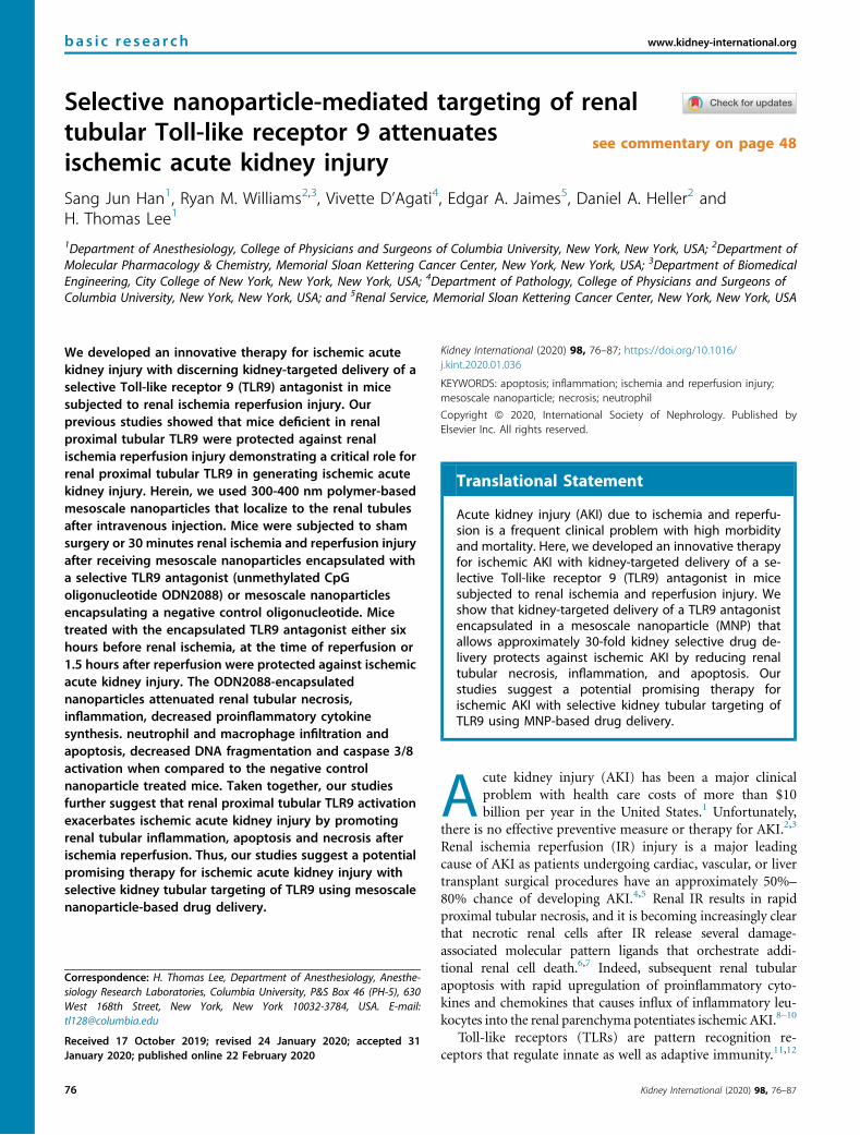
To confirm renal tubular delivery of MNPs, kidney sec-tions of mice injected with MNPs or with saline vehiclecontrol 6 hours before renal IR injury were stained with anti-PEG antibody. The renal proximal tubular distributionpattern for the MNPs was confirmed by colocalization of PEGstaining with phytohemagglutinin (PHA) lectin staining in 75mg/kg MNP-injected mice (Figure 1a, representative of 3experiments). As described previously, we again show thatthere was almost no MNP localization to other tubular seg-ments, endothelial cells, or mesangial cells in theglomeruli.25,26
We also confirmed that administration of MNPs does notresult in systemic distribution. Figure 1b shows representativein vivo near-infrared fluorescence images of the kidney, liver,spleen, lung, and heart (top), and average fluorescence in-tensity quantified (bottom) after injection of 75 mg/kg Cy5-loaded MNPs given i.v. The images show approximately 30-fold kidney selectivity over other organs including the lung,liver, spleen, and heart (N ¼ 3). Furthermore, administrationin mice that were subjected to 30-minute renal IR alsodemonstrated >30-fold kidney selectivity (N ¼ 3).
Selective tubular delivery of ODN2088-encapsulated MNPsprotects against ischemic AKI in micePlasma creatinine and blood urea nitrogen (BUN) values weresimilar between mice injected with MNPs encapsulated withcontrol ODN (control MNPs) and ODN2088-encapsulatedMNPs (MNP-ODN2088) subjected to sham operation(Figure 2). Mice treated with control MNPs and subjected torenal IR had significantly higher plasma creatinine and BUNas well as kidney neutrophil gelatinase-associated lipocalin(NGAL) mRNA (N ¼ 4–5) compared with sham-operatedmice (N ¼ 4). We show here that mice treated with 37.5 or75 mg/kg MNP-ODN2088 6 hours before renal ischemia wereprotected against ischemic AKI compared with control MNP-treated mice as demonstrated by reduced plasma BUN andcreatinine as well as kidney NGAL mRNA expression (N ¼ 6–7). Furthermore, mice treated with 75 mg/kg MNP-ODN2088at the time of reperfusion or 1.5 hours after reperfusion werealso significantly protected against ischemic AKI comparedwith control MNP-treated mice (N ¼ 6, Figure 2). Incontrast, naked ODN2088 given i.v. 6 hours before renalischemia failed to protect against ischemic AKI in mice(N ¼ 5–6).
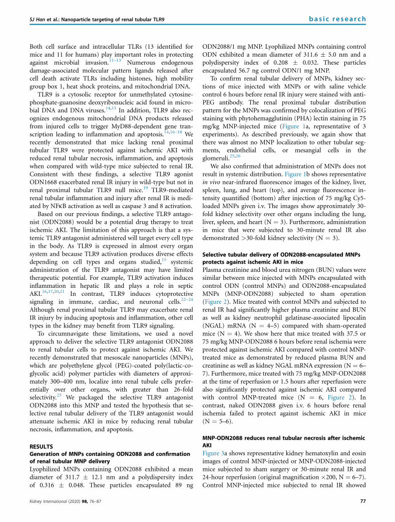
MNP-ODN2088 reduces renal tubular necrosis after ischemicAKIFigure 3a shows representative kidney hematoxylin and eosinimages of control MNP-injected or MNP-ODN2088-injectedmice subjected to sham surgery or 30-minute renal IR and24-hour reperfusion (original magnification �200, N ¼ 6–7).Control MNP-injected mice subjected to renal IR showed
77
Figure 1 | Confirmation of kidney selective and renal tubule cell mesoscale nanoparticle (MNP) delivery. (a) Mouse kidney sectionsstained with anti–polyethylene glycol (PEG) antibody to detect MNP localization and phytohemagglutinin (PHA) lectin to mark renal proximaltubule cells and counterstained with 40,6-diamidino-2-phenylindole (DAPI) to visualize cell nuclei. Mice were injected with ODN2088-encapsulated MNPs or with vehicle control 6 hours before renal ischemia reperfusion (RIR) injury. Renal proximal tubular distribution patternfor PEG was confirmed by colocalization of PEG antibody staining and PHA lectin staining in MNP-injected mice (representative of 3experiments). Mice injected with vehicle control showed no PEG staining (data not shown). There was almost no PEG localization in othertubular segments, endothelial cells, or mesangial cells in the glomeruli. (b) (Top) In vivo near-infrared fluorescence images of the kidney, liver,lung, heart, and spleen of mice injected with 75 mg/kg i.v. Cy5 mimic 3,30-diethylthiadicarbocyanine iodide–loaded MNPs 6 hours before shamsurgery or 30-minute renal ischemia and 24-hour reperfusion (RIR). (Bottom) Quantification of average fluorescence efficiency per squarecentimeter in each organ demonstrating approximately 30-fold selective delivery of Cy5 MNPs to the kidneys compared with other organs inboth sham-operated mice and mice subjected to RIR injury (N ¼ 3). Data are background subtracted and represent mean �SEM. To optimizeviewing of this image, please see the online version of this article at www.kidney-international.org.
bas i c re sea r ch SJ Han et al.: Nanoparticle targeting of renal tubular TLR9
severe tubular necrosis and proteinaceous casts as well asincreased tubular dilatation and congestion (Figure 3a).MNP-ODN2088 treatment 6 hours before renal ischemia, atthe time of reperfusion, or 1.5 hours after reperfusion led todecreased renal tubular necrosis, congestion, and cast for-mation compared with control MNP-injected mice subjectedto renal IR. Kidneys from MNP-ODN2088-injected mice hadsignificantly reduced renal tubular injury scores comparedwith control MNP-injected mice after IR (Figure 3b). Incontrast, naked ODN2088 given i.v. 6 hours before renalischemia failed to reduce kidney necrosis score in mice sub-jected to renal IR (N ¼ 5–6).
MNP-ODN2088 attenuates kidney apoptosis in mice afterischemic AKIFigure 4a shows representative terminal deoxynucleotidyltransferase biotin-dUTP nick end-labeling (TUNEL)–stainedimages indicative of renal apoptosis, and Figure 4b showscounts of TUNEL-positive kidney cells from control MNP-injected and MNP-ODN2088-injected mice subjected tosham surgery (N ¼ 4) or to renal IR (N ¼ 6, originalmagnification �200). Many TUNEL-positive (fragmented
78
DNA) cells were detected, suggestive of renal tubularapoptosis in the kidneys from control MNP-injected micesubjected to renal IR injury. TUNEL-positive kidney cellcounts were significantly reduced in MNP-ODN2088-injectedmice given 6 hours before renal ischemia, at the time ofreperfusion, or 1.5 hours after reperfusion. In contrast, nakedODN2088 given i.v. failed to reduce kidney apoptosis in micesubjected to renal IR (N ¼ 5–6). We also detected caspase 3and 8 activation by measuring cleaved caspases 3 and 8 in thekidney lysates from sham-operated mice and mice subjectedto renal IR injury (Figure 4c). Caspase 3 and 8 cleavages wereincreased in control MNP-injected mice subjected to renal IRand were significantly reduced in MNP-ODN2088-injectedmice 6 hours before renal ischemia. To show specific inhibi-tion of the TLR9 signaling pathway in tubular cells treatedwith ODN2088-encapsulated MNPs, freshly isolated mouseproximal tubule cells were treated with a selective murineTLR9 ligand 5 mM ODN1668 for 3 days with or withoutpretreatment of 1 mM ODN2088-encapsulated MNPs (N ¼3). We show that MNPs encapsulating ODN2088 completelyprevented ODN1688-mediated induction of caspase 3 and 8fragmentation (Figure 4d).
Kidney International (2020) 98, 76–87

Sham surgery or 30-min renal ischemia and 24-h reperfusion
0
1
2
3
4 *## #
PCr (
mg/
dl)
#**
0
50
100
150
200 **#
#
BUN
(mg/
dl)
## **
Pre-75 mg/kg ConODN MNP sham
Pre-75 mg/kg ODN2088 MNP sham
Pre-75 mg/kg ConODN MNP RIR
Pre-37.5mg/kg ODN2088 MNP RIR
Pre-75 mg/kg ODN2088 MNP RIR
0h Post-75 mg/kg ODN2088 MNP RIR
1.5h Post-75 mg/kg ODN2088 MNP RIR
Naked ConODN RIR
Naked ODN2088 RIR012
50
150
250
350
450
550
* #
#*$
NGAL
mRN
A/GA
PDH
(fold
incr
ease
ove
r75
mg/
kg C
onOD
N MN
P sh
am)
#*
**
#*
Figure 2 | Mesoscale nanoparticle (MNP)–ODN2088-mediatedprotection against ischemic acute kidney injury (AKI) in mice.Mice were injected with control MNP-ODN or with ODN2088encapsulated in MNPs (MNP-ODN2088) and subjected to shamsurgery (N ¼ 4) or to 30-minute renal ischemia and 24-hourreperfusion (ischemia reperfusion, N ¼ 6–7). Some mice wereinjected with MNP-ODN2088 6 hours before renal ischemia. Aseparate cohort of mice were injected with MNP-ODN2088 at thetime of reperfusion or 1.5 hours after reperfusion. Another cohort ofmice were injected with 5 mg/kg naked ODN2088 i.v. 6 hoursbefore renal ischemia (N ¼ 5–6). Plasma blood urea nitrogen (BUN)and creatinine as well as kidney neutrophil gelatinase-associatedlipocalin (NGAL) mRNA were measured. For statistical analysis, the1-way analysis of variance plus Tukey’s post hoc multiplecomparison test was used to detect significant changes. *P < 0.05versus control MNP-injected mice subjected to sham surgery. #P <0.05 versus control ODN mice subjected to renal ischemiareperfusion (RIR). Error bars represent 1 SEM. GAPDH,glyceraldehyde-3-phosphate dehydrogenase; PCR, polymerasechain reaction.
SJ Han et al.: Nanoparticle targeting of renal tubular TLR9 bas i c re sea r ch
MNP-ODN2088 reduces kidney neutrophil infiltration afterischemic AKIFigure 5a shows representative immunohistochemistry im-ages, and Figure 5b shows counts of infiltrating kidney neu-trophils in the kidneys of control MNP-injected mice andMNP-ODN2088-injected mice subjected to sham surgery(N ¼ 4) or renal IR (N ¼ 6, original magnification �200).
Kidney International (2020) 98, 76–87
Kidney neutrophil infiltration increased significantly in con-trol MNP-injected mice subjected to renal IR. MNP-ODN2088 treatment either 6 hours before renal ischemia,at the time of reperfusion, or 1.5 hours after reperfusionsignificantly attenuated kidney neutrophil infiltration afterrenal IR compared with MNP-ODN2088-injected mice. Incontrast, naked ODN2088 given i.v. 6 hours before renalischemia failed to neutrophil infiltration after renal IR in mice(N ¼ 5–6).
MNP-ODN2088 reduces kidney macrophage infiltration afterischemic AKIFigure 5c shows representative immunohistochemistry im-ages, and Figure 5d shows counts of infiltrating kidneymacrophages in the kidneys of control MNP-injected miceand MNP-ODN2088-injected mice subjected to sham surgery(N ¼ 3) or renal IR (N ¼ 4, original magnification �200).Kidney macrophage infiltration increased significantly incontrol MNP-injected mice subjected to renal IR. MNP-ODN2088 treatment 6 hours before renal ischemia signifi-cantly attenuated kidney macrophage infiltration after renalIR compared with MNP-ODN2088-injected mice.
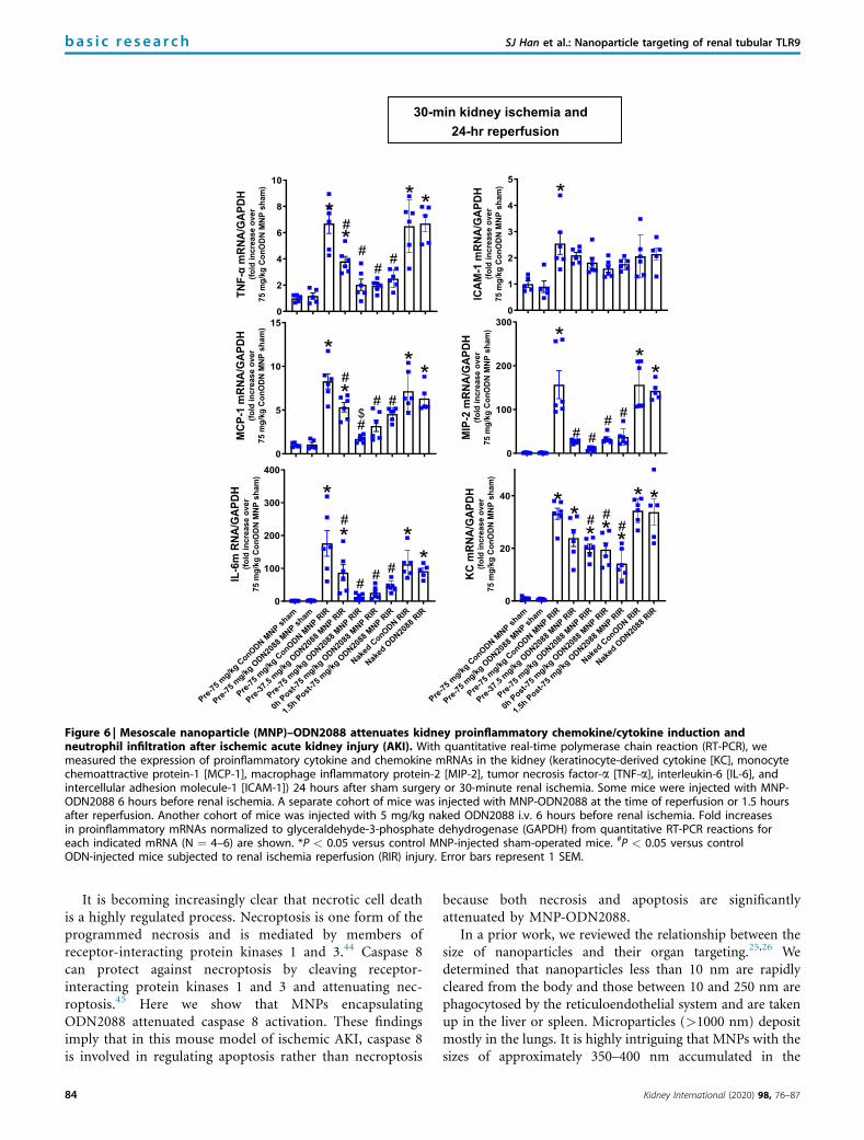
MNP-ODN2088 downregulates proinflammatory chemokineand cytokine induction after ischemic AKIFigure 6 shows fold increases in proinflammatory mRNAsnormalized to glyceraldehyde-3-phosphate dehydrogenase foreach indicated mRNA in the kidneys of control MNP-injectedmice and MNP-ODN2088-injected mice subjected to shamsurgery (N ¼ 4–5) or renal IR (N ¼ 6). Ischemic AKIincreased all proinflammatory genes measured in controlMNP-injected mice. Consistent with the renal protective roleof TLR9 antagonism via reduction of neutrophil andmacrophage attracting chemokines, we show that macro-phage inflammatory protein-2 and monocyte chemoattractiveprotein-1 expression was significantly attenuated in miceinjected with MNP-ODN2088 6 hours before renal ischemia,at the time of reperfusion, or 1.5 hours after reperfusion.Moreover, interleukin-6, keratinocyte-derived cytokine, andtumor necrosis factor-a induction was attenuated in MNP-ODN2088-injected mice. In contrast, naked ODN2088 giveni.v. 6 hours before renal ischemia failed to attenuate proin-flammatory cytokines in the kidney of mice subjected to renalIR injury (N ¼ 5–6).
DISCUSSIONIn this study, we found that MNP technology allows selectiverenal tubular delivery of a potent TLR9 antagonist ligandoligonucleotide ODN2088 to protect against ischemic AKI.The MNP-loaded ODN2088 reduced renal tubular necrosis,noted by decreased plasma BUN and creatinine as well askidney NGAL mRNA induction compared with controlMNP-injected mice. Increasing clinical significance further,MNP-ODN2088 given at the time of reperfusion or 1.5 hoursafter reperfusion also protected against renal IR injury.
79
Figure 3 | Mesoscale nanoparticle (MNP)–ODN2088-mediated reduction of renal tubular necrosis after ischemic acute kidney injury(AKI). Representative hematoxylin and eosin images (from 4 to 7 experiments) of kidneys from mice injected with control MNPs or with MNP-ODN2088 and subjected to sham surgery (N ¼ 4) or to 30-minute renal ischemia and 24-hour reperfusion (N ¼ 6–7, originalmagnification �200). Some mice were injected with MNP-ODN2088 6 hours before renal ischemia. A separate cohort of mice were injectedwith MNP-ODN2088 at the time of reperfusion or 1.5 hours after reperfusion. Another cohort of mice were injected with 5 mg/kg nakedODN2088 i.v. 6 hours before renal ischemia (N ¼ 5–6). Renal injury scores assessing the degree of renal tubular necrosis are also shownhere (scale: 0–4) 24 hours after renal ischemia reperfusion (RIR). *P < 0.05 versus control MNP-injected mice subjected to RIR injury.For statistical analysis, the Mann-Whitney nonparametric test was used to detect significant changes. To optimize viewing of thisimage, please see the online version of this article at www.kidney-international.org.
bas i c re sea r ch SJ Han et al.: Nanoparticle targeting of renal tubular TLR9
After IR, necrotic renal cells release mitochondrial DNAthat can target intracellular renal tubular TLR9.20 We previ-ously demonstrated that renal proximal tubular TLR9 acti-vation exacerbates ischemic AKI by promoting renal tubularepithelial apoptosis and inflammation. Mice deficient in renalproximal tubular TLR9 were protected against ischemic AKIwith reduced plasma creatinine and kidney NGAL expressionas well as renal tubular apoptosis and necrosis.19 Further-more, a selective TLR9 agonist exacerbated renal tubularinjury in wild-type mice but not in mice deficient in renaltubular TLR9 subjected to renal IR injury. These findingssuggest a potential therapeutic role for a specific TLR9antagonist in protecting against ischemic AKI. However,systemic administration of a selective TLR9 antagonist maynot be effective as a treatment for ischemic AKI. This isbecause TLR9 activation produces complex and perhapsdivergent effects depending on cell types and organ systemicaffected22,23,27 and renal IR injury results in systemic in-flammatory response and extra-renal organ dysfunction.28,29
Renal tubular epithelial TLR9 promotes cell injury anddeath, whereas TLR9 signaling in other cell types may pro-mote cytoprotective effects. Indeed, TLR9 activation protects
80
against cerebral as well as cardiac IR injury via activation ofphosphatidylinositol-30-kinase/protein kinase B (also knownas AKT) signaling.22,23 In addition, TLR9 activation maymediate cardiac and neuronal protection by modulating en-ergy metabolism.27 Furthermore, in cisplatin-induced AKI,TLR9 may promote tissue protection by promoting accu-mulation of beneficial regulatory T cells.30 Consistent withthese findings, global TLR9-deficient mice were not protectedagainst ischemic AKI again demonstrating the complex anddifferential effects of TLR9 activation in different organs andcell types.19,31 In agreement with the global TLR9 knockoutmice studies, we demonstrated here that systemic TLR9blockade with 5 mg/kg naked ODN2088 (15 nucleotides inlength) failed to protect against ischemic AKI in mice. It ispossible that global TLR9 knockout mice or mice injectedwith naked ODN2088 systemically i.v. were not protectedagainst ischemic AKI, as they may lack the tissue protectiveregulatory T cells modulated by TLR9 signaling.
Previous studies demonstrated that i.v. injection of oligo-nucleotides (approximately 16–21 nucleotides) displays broaddistribution into most organ systems (liver, kidney, bonemarrow, adipocytes, and lymph nodes) except the central
Kidney International (2020) 98, 76–87
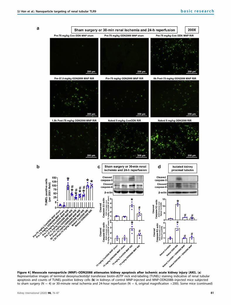
Figure 4 | Mesoscale nanoparticle (MNP)–ODN2088 attenuates kidney apoptosis after ischemic acute kidney injury (AKI). (a)Representative images of terminal deoxynucleotidyl transferase biotin-dUTP nick end-labeling (TUNEL) staining indicative of renal tubularapoptosis and counts of TUNEL-positive kidney cells (b) in kidneys of control MNP-injected and MNP-ODN2088–injected mice subjectedto sham surgery (N ¼ 4) or 30-minute renal ischemia and 24-hour reperfusion (N ¼ 6, original magnification �200). Some mice (continued)
SJ Han et al.: Nanoparticle targeting of renal tubular TLR9 bas i c re sea r ch
Kidney International (2020) 98, 76–87 81
bas i c re sea r ch SJ Han et al.: Nanoparticle targeting of renal tubular TLR9
nervous system, due to the blood-brain barrier.32 Therefore,broad biodistribution of the naked TLR9 antagonist oligo-nucleotide again demonstrates the need for kidney selectivedelivery to effect renal protection. It is exciting and clinicallyrelevant that MNPs encapsulating 3.26 or 6.5 mg/kgODN2088 provided protection against ischemic AKI andattenuated renal tubular necrosis, inflammation, andapoptosis.
The highest dose of MNPs encapsulating ODN2088 (75mg/kg) used here is higher than doses administered (25–50mg/kg) to characterize the kidney selective delivery of MNPsin previous studies.25,26 However, the imaging studies stillresulted in similar approximately 30-fold kidney-specific de-livery of MNPs compared with other organs studied(Figure 1b). Furthermore, it was unknown until this studywhether kidneys subjected to IR would still allow similarkidney selective delivery of MNPs given i.v., because renaltubular necrosis and inflammation have severe effects onkidney function. Our studies found that MNPs delivered i.v.continued to demonstrate kidney selective delivery, even inmice that were subjected to ischemic AKI, further addingclinical relevance to these studies.
There is no effective therapy or preventive measure forclinical AKI. Many promising potential therapies based onpreclinical research failed to be effective clinically.33,34 Lack ofeffective pharmacological therapy is clearly due to the com-plex nature of clinical AKI in patients with multiple comor-bidities in comparison with simplified laboratory models ofAKI.35,36 An additional factor that may preclude effectiveclinical therapy for AKI is that systemic administration ofdrugs to treat clinical AKI is problematic for several phar-macologic reasons. Drugs given systemically (oral or intra-venous) normally target every organ system and multiple celltypes. Concomitantly, experimental therapeutics that produceprotective effects in renal epithelial cells may produce un-wanted detrimental effects in other cell types (e.g., liver,heart) as discussed above. Another reason for the failure ofsystemic therapies is the pharmacokinetics/biodistributionwith respect to the nephron. Often, an insufficient amount ofdrug reaches the renal tubular cells when drugs are given innontoxic doses. Therefore, one way to circumvent the con-founding effects of multiple-organ targeting of drugs and less-than-effective dosing problems is to devise a strategy toselectively deliver or concentrate drugs in the kidney by renal-specific delivery methods.
=
Figure 4 | (continued) were injected with MNP-ODN2088 6 hours beforMNP-ODN2088 at the time of reperfusion or 1.5 hours after reperfusion.i.v. 6 hours before renal ischemia (N ¼ 5–6). (c) We detected caspase 3lysates from sham-operated mice and mice subjected to renal ischemia re*P < 0.05 versus control MNP-injected mice subjected to sham surgery.represent 1 SEM. (d) We detected caspase 3 and 8 activation in primarycontrol ODN or with 5 mM ODN1668 (a selective mouse TLR9 activatingencapsulating control ODN or MNPs encapsulating 1 mM ODN2088. *P <0.05 versus ODN1668-treated mouse proximal tubule cells. Error bars reof this image, please see the online version of this article at www.kidne
82
The recently developed MNP drug delivery system mayhold a promise in targeting renal cells specifically and providepotential therapy for clinical AKI. Recent studies demon-strated that the relatively large (approximately 300–400 nm)polymer-based MNPs preferentially localized to the kidneywhen compared with other organs by approximately 26- to94-fold.25 MNPs injected via the i.v. route preferentiallylocalized to renal tubular cells and remained for approxi-mately 7 days. These findings are exciting, as renal tubularcells are the major site of injury during and after ischemicAKI.19,37 Moreover, MNP treatment does not have anydetrimental effect on renal or hepatic function, inflammation,or hematological problems as MNP toxicity was tracked forup to 30 days in preclinical studies.25,26,38
Specific renal tubular localization of MNPs appears to bedependent on the large size (approximately 300–400 nm) andhydrophilic PEG surface chemistry of the MNP.25 PEGylationalso appears to prevent macrophages scavenging theMNPs.39,40 The clinically relevant major components of theMNP (poly(lactic-co-glycolic acid) and PEG) are US Foodand Drug Administration approved.41 Our previous studies,using both immunofluorescence and immunohistochemistry,showing renal tubular specific delivery of MNPs,25 wereconfirmed in this study. It appears that MNP localization isgreater in renal proximal tubules when compared with distaltubules and this is significant as renal proximal tubules are themost susceptible cell types for injury and death due to hyp-oxia after renal IR.19,25,26,33,38,42 Furthermore, i.v. injectedMNPs appear not to localize in endothelial cells and renalglomeruli based on immunofluorescence and immunohisto-chemistry studies.
Another exciting aspect of our study is that MNP-ODN2088 delivered at the time of reperfusion as well as 1.5hours after reperfusion were as protective as MNP-ODN2088given 6 hours before renal ischemia. Because MNPs takeapproximately 3–6 hours to maximally localize to the kid-ney,26 these findings show that MNP-ODN reaching renaltubular cells 3–7.5 hours after ischemic injury can be effectiveand equally protective as pretreatment raising clinical signif-icance greatly as not all cases of ischemic AKI can be antici-pated in advance.
Based on our findings, MNP-ODN2088 protects againstischemic AKI by reducing renal tubular necrosis andapoptosis after IR. In particular, MNP-ODN2088 significantlyattenuated renal tubular apoptosis in mice subjected to severe
e renal ischemia. A separate cohort of mice were injected withAnother cohort of mice were injected with 5 mg/kg naked ODN2088and 8 activation by measuring cleaved caspases 3 and 8 in kidneyperfusion (RIR) injury after control MNP or MNP-ODN2088 treatment.#P < 0.05 versus control ODN mice subjected to RIR. Error barscultures of mouse renal proximal tubule cells treated withligand). Some proximal tubule cells were pretreated with MNPs0.05 versus control ODN-treated mouse proximal tubule cells. #P <
present 1 SEM. TLR9, Toll-like receptor 9. To optimize viewingy-international.org.
Kidney International (2020) 98, 76–87
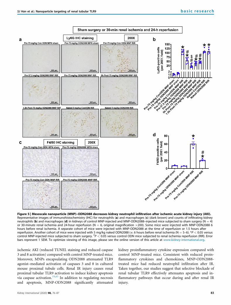
Figure 5 | Mesoscale nanoparticle (MNP)–ODN2088 decreases kidney neutrophil infiltration after ischemic acute kidney injury (AKI).Representative images of immunohistochemistry (IHC) for neutrophils (a) and macrophages (c) (dark brown) and counts of infiltrating kidneyneutrophils (b) and macrophages (d) in kidneys of control MNP-injected and MNP-ODN2088–injected mice subjected to sham surgery (N ¼ 4)or 30-minute renal ischemia and 24-hour reperfusion (N ¼ 6, original magnification �200). Some mice were injected with MNP-ODN2088 6hours before renal ischemia. A separate cohort of mice were injected with MNP-ODN2088 at the time of reperfusion or 1.5 hours afterreperfusion. Another cohort of mice were injected with 5 mg/kg naked ODN2088 i.v. 6 hours before renal ischemia (N ¼ 5–6). *P < 0.05 versuscontrol MNP-injected mice subjected to sham surgery. #P < 0.05 versus control ODN mice subjected to renal ischemia reperfusion (RIR). Errorbars represent 1 SEM. To optimize viewing of this image, please see the online version of this article at www.kidney-international.org.
SJ Han et al.: Nanoparticle targeting of renal tubular TLR9 bas i c re sea r ch
ischemic AKI (reduced TUNEL staining and reduced caspase3 and 8 activation) compared with control MNP-treated mice.Moreover, MNPs encapsulating ODN2088 attenuated TLR9agonist–mediated activation of caspases 3 and 8 in culturedmouse proximal tubule cells. Renal IR injury causes renalproximal tubular TLR9 activation to induce kidney apoptosisvia caspase activation.19,43 In addition to regulating necrosisand apoptosis, MNP-ODN2088 significantly attenuated
Kidney International (2020) 98, 76–87
kidney proinflammatory cytokine expression compared withcontrol MNP-treated mice. Consistent with reduced proin-flammatory cytokines and chemokines, MNP-ODN2088-treated mice had reduced neutrophil infiltration after IR.Taken together, our studies suggest that selective blockade ofrenal tubular TLR9 effectively attenuates apoptosis and in-flammatory pathways that occur during and after renal IRinjury.
83
30-min kidney ischemia and 24-hr reperfusion
0
5
10
15
*
#
#$
#*
* *#
0
100
200
300
*
#
##
**
#
0
2
4
6
8
10
*
##TN
F-α
mRN
A/G
APDH
(fold
incr
ease
ove
r75
mg/
kg C
onO
DN M
NP s
ham
)
ICAM
-1 m
RNA/
GAP
DH(fo
ld in
crea
se o
ver
75 m
g/kg
Con
ODN
MNP
sha
m)
MCP
-1 m
RNA/
GAP
DH(fo
ld in
crea
se o
ver
75 m
g/kg
Con
ODN
MNP
sha
m)
MIP
-2 m
RNA/
GAP
DH(fo
ld in
crea
se o
ver
75 m
g/kg
Con
ODN
MNP
sha
m)
IL-6
m R
NA/G
APDH
(fold
incr
ease
ove
r75
mg/
kg C
onO
DN M
NP s
ham
)
KC m
RNA/
GAP
DH(fo
ld in
crea
se o
ver
75 m
g/kg
Con
ODN
MNP
sha
m)
#*
* *
#
0
1
2
3
4
5
*
Pre-75
mg/kgConODN MNP sh
am
Pre-75
mg/kgODN20
88MNP sh
am
Pre-75
mg/kgConODN MNP RIR
Pre-37
.5mg/kg
ODN2088
MNP RIR
Pre-75
mg/kgODN20
88MNP RIR
0hPost-
75mg/kg
ODN2088
MNP RIR
1.5h Post-
75mg/kg
ODN2088
MNP RIR
Naked
ConODN RIR
Naked
ODN2088
RIR0
100
200
300
400
*
##
#* *
*#
Pre-75
mg/kgConODN MNP sh
am
Pre-75
mg/kgODN20
88MNP sh
am
Pre-75
mg/kgConODN MNP RIR
Pre-37
.5mg/kg
ODN2088
MNP RIR
Pre-75
mg/kgODN20
88MNP RIR
0hPost-
75mg/kg
ODN2088
MNP RIR
1.5h Post-
75mg/kg
ODN2088
MNP RIR
Naked
ConODN RIR
Naked
ODN2088
RIR0
20
40 *##
**
* *#**
Figure 6 | Mesoscale nanoparticle (MNP)–ODN2088 attenuates kidney proinflammatory chemokine/cytokine induction andneutrophil infiltration after ischemic acute kidney injury (AKI). With quantitative real-time polymerase chain reaction (RT-PCR), wemeasured the expression of proinflammatory cytokine and chemokine mRNAs in the kidney (keratinocyte-derived cytokine [KC], monocytechemoattractive protein-1 [MCP-1], macrophage inflammatory protein-2 [MIP-2], tumor necrosis factor-a [TNF-a], interleukin-6 [IL-6], andintercellular adhesion molecule-1 [ICAM-1]) 24 hours after sham surgery or 30-minute renal ischemia. Some mice were injected with MNP-ODN2088 6 hours before renal ischemia. A separate cohort of mice was injected with MNP-ODN2088 at the time of reperfusion or 1.5 hoursafter reperfusion. Another cohort of mice was injected with 5 mg/kg naked ODN2088 i.v. 6 hours before renal ischemia. Fold increasesin proinflammatory mRNAs normalized to glyceraldehyde-3-phosphate dehydrogenase (GAPDH) from quantitative RT-PCR reactions foreach indicated mRNA (N ¼ 4–6) are shown. *P < 0.05 versus control MNP-injected sham-operated mice. #P < 0.05 versus controlODN-injected mice subjected to renal ischemia reperfusion (RIR) injury. Error bars represent 1 SEM.
bas i c re sea r ch SJ Han et al.: Nanoparticle targeting of renal tubular TLR9
It is becoming increasingly clear that necrotic cell deathis a highly regulated process. Necroptosis is one form of theprogrammed necrosis and is mediated by members ofreceptor-interacting protein kinases 1 and 3.44 Caspase 8can protect against necroptosis by cleaving receptor-interacting protein kinases 1 and 3 and attenuating nec-roptosis.45 Here we show that MNPs encapsulatingODN2088 attenuated caspase 8 activation. These findingsimply that in this mouse model of ischemic AKI, caspase 8is involved in regulating apoptosis rather than necroptosis
84
because both necrosis and apoptosis are significantlyattenuated by MNP-ODN2088.
In a prior work, we reviewed the relationship between thesize of nanoparticles and their organ targeting.25,26 Wedetermined that nanoparticles less than 10 nm are rapidlycleared from the body and those between 10 and 250 nm arephagocytosed by the reticuloendothelial system and are takenup in the liver or spleen. Microparticles (>1000 nm) depositmostly in the lungs. It is highly intriguing that MNPs with thesizes of approximately 350–400 nm accumulated in the
Kidney International (2020) 98, 76–87
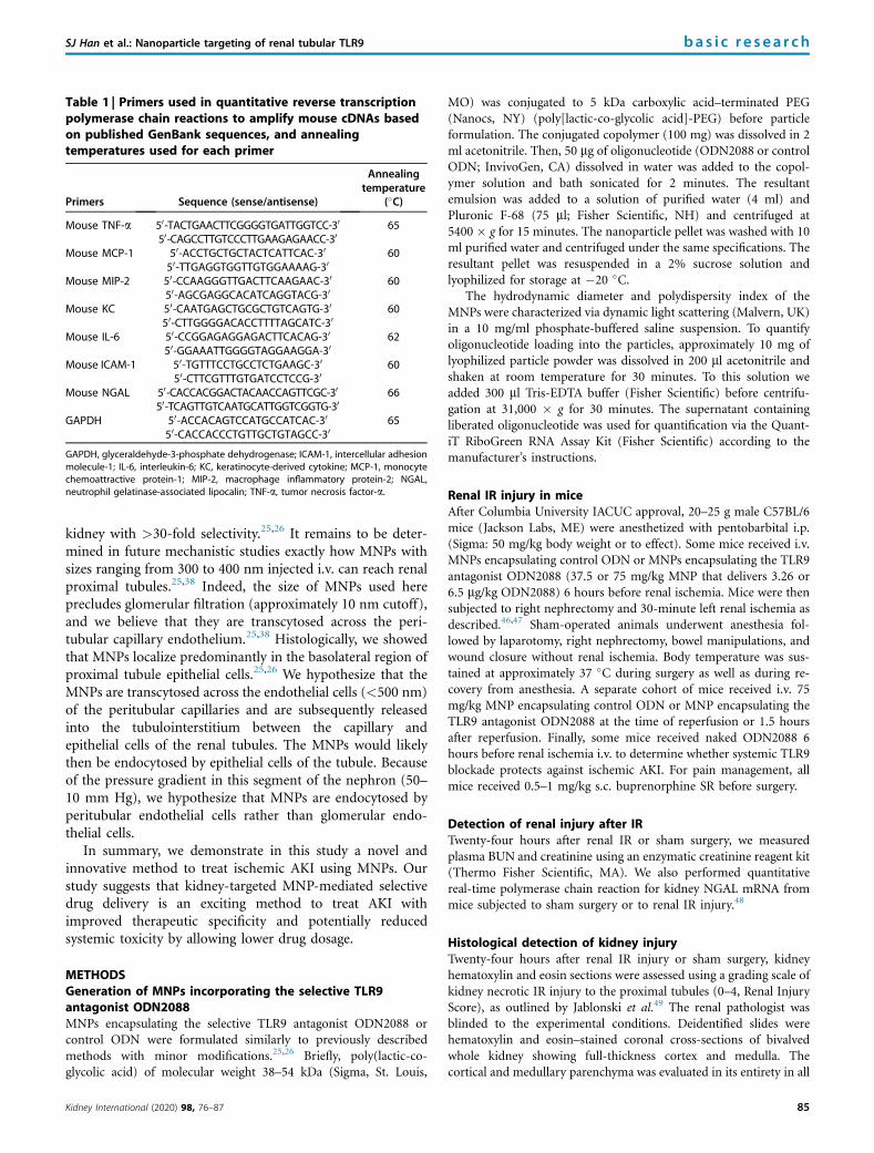
Table 1 | Primers used in quantitative reverse transcriptionpolymerase chain reactions to amplify mouse cDNAs basedon published GenBank sequences, and annealingtemperatures used for each primer
Primers Sequence (sense/antisense)
Annealingtemperature
(�C)
Mouse TNF-a 50-TACTGAACTTCGGGGTGATTGGTCC-30 6550-CAGCCTTGTCCCTTGAAGAGAACC-30
Mouse MCP-1 50-ACCTGCTGCTACTCATTCAC-30 6050-TTGAGGTGGTTGTGGAAAAG-30
Mouse MIP-2 50-CCAAGGGTTGACTTCAAGAAC-30 6050-AGCGAGGCACATCAGGTACG-30
Mouse KC 50-CAATGAGCTGCGCTGTCAGTG-30 6050-CTTGGGGACACCTTTTAGCATC-30
Mouse IL-6 50-CCGGAGAGGAGACTTCACAG-30 6250-GGAAATTGGGGTAGGAAGGA-30
Mouse ICAM-1 50-TGTTTCCTGCCTCTGAAGC-30 6050-CTTCGTTTGTGATCCTCCG-30
Mouse NGAL 50-CACCACGGACTACAACCAGTTCGC-30 6650-TCAGTTGTCAATGCATTGGTCGGTG-30
GAPDH 50-ACCACAGTCCATGCCATCAC-30 6550-CACCACCCTGTTGCTGTAGCC-30
GAPDH, glyceraldehyde-3-phosphate dehydrogenase; ICAM-1, intercellular adhesionmolecule-1; IL-6, interleukin-6; KC, keratinocyte-derived cytokine; MCP-1, monocytechemoattractive protein-1; MIP-2, macrophage inflammatory protein-2; NGAL,neutrophil gelatinase-associated lipocalin; TNF-a, tumor necrosis factor-a.
SJ Han et al.: Nanoparticle targeting of renal tubular TLR9 bas i c re sea r ch
kidney with >30-fold selectivity.25,26 It remains to be deter-mined in future mechanistic studies exactly how MNPs withsizes ranging from 300 to 400 nm injected i.v. can reach renalproximal tubules.25,38 Indeed, the size of MNPs used hereprecludes glomerular filtration (approximately 10 nm cutoff),and we believe that they are transcytosed across the peri-tubular capillary endothelium.25,38 Histologically, we showedthat MNPs localize predominantly in the basolateral region ofproximal tubule epithelial cells.25,26 We hypothesize that theMNPs are transcytosed across the endothelial cells (<500 nm)of the peritubular capillaries and are subsequently releasedinto the tubulointerstitium between the capillary andepithelial cells of the renal tubules. The MNPs would likelythen be endocytosed by epithelial cells of the tubule. Becauseof the pressure gradient in this segment of the nephron (50–10 mm Hg), we hypothesize that MNPs are endocytosed byperitubular endothelial cells rather than glomerular endo-thelial cells.
In summary, we demonstrate in this study a novel andinnovative method to treat ischemic AKI using MNPs. Ourstudy suggests that kidney-targeted MNP-mediated selectivedrug delivery is an exciting method to treat AKI withimproved therapeutic specificity and potentially reducedsystemic toxicity by allowing lower drug dosage.
METHODSGeneration of MNPs incorporating the selective TLR9antagonist ODN2088MNPs encapsulating the selective TLR9 antagonist ODN2088 orcontrol ODN were formulated similarly to previously describedmethods with minor modifications.25,26 Briefly, poly(lactic-co-glycolic acid) of molecular weight 38–54 kDa (Sigma, St. Louis,
Kidney International (2020) 98, 76–87
MO) was conjugated to 5 kDa carboxylic acid–terminated PEG(Nanocs, NY) (poly[lactic-co-glycolic acid]-PEG) before particleformulation. The conjugated copolymer (100 mg) was dissolved in 2ml acetonitrile. Then, 50 mg of oligonucleotide (ODN2088 or controlODN; InvivoGen, CA) dissolved in water was added to the copol-ymer solution and bath sonicated for 2 minutes. The resultantemulsion was added to a solution of purified water (4 ml) andPluronic F-68 (75 ml; Fisher Scientific, NH) and centrifuged at5400 � g for 15 minutes. The nanoparticle pellet was washed with 10ml purified water and centrifuged under the same specifications. Theresultant pellet was resuspended in a 2% sucrose solution andlyophilized for storage at �20 �C.
The hydrodynamic diameter and polydispersity index of theMNPs were characterized via dynamic light scattering (Malvern, UK)in a 10 mg/ml phosphate-buffered saline suspension. To quantifyoligonucleotide loading into the particles, approximately 10 mg oflyophilized particle powder was dissolved in 200 ml acetonitrile andshaken at room temperature for 30 minutes. To this solution weadded 300 ml Tris-EDTA buffer (Fisher Scientific) before centrifu-gation at 31,000 � g for 30 minutes. The supernatant containingliberated oligonucleotide was used for quantification via the Quant-iT RiboGreen RNA Assay Kit (Fisher Scientific) according to themanufacturer’s instructions.
Renal IR injury in miceAfter Columbia University IACUC approval, 20–25 g male C57BL/6mice (Jackson Labs, ME) were anesthetized with pentobarbital i.p.(Sigma: 50 mg/kg body weight or to effect). Some mice received i.v.MNPs encapsulating control ODN or MNPs encapsulating the TLR9antagonist ODN2088 (37.5 or 75 mg/kg MNP that delivers 3.26 or6.5 mg/kg ODN2088) 6 hours before renal ischemia. Mice were thensubjected to right nephrectomy and 30-minute left renal ischemia asdescribed.46,47 Sham-operated animals underwent anesthesia fol-lowed by laparotomy, right nephrectomy, bowel manipulations, andwound closure without renal ischemia. Body temperature was sus-tained at approximately 37 �C during surgery as well as during re-covery from anesthesia. A separate cohort of mice received i.v. 75mg/kg MNP encapsulating control ODN or MNP encapsulating theTLR9 antagonist ODN2088 at the time of reperfusion or 1.5 hoursafter reperfusion. Finally, some mice received naked ODN2088 6hours before renal ischemia i.v. to determine whether systemic TLR9blockade protects against ischemic AKI. For pain management, allmice received 0.5–1 mg/kg s.c. buprenorphine SR before surgery.
Detection of renal injury after IRTwenty-four hours after renal IR or sham surgery, we measuredplasma BUN and creatinine using an enzymatic creatinine reagent kit(Thermo Fisher Scientific, MA). We also performed quantitativereal-time polymerase chain reaction for kidney NGAL mRNA frommice subjected to sham surgery or to renal IR injury.48
Histological detection of kidney injuryTwenty-four hours after renal IR injury or sham surgery, kidneyhematoxylin and eosin sections were assessed using a grading scale ofkidney necrotic IR injury to the proximal tubules (0–4, Renal InjuryScore), as outlined by Jablonski et al.49 The renal pathologist wasblinded to the experimental conditions. Deidentified slides werehematoxylin and eosin–stained coronal cross-sections of bivalvedwhole kidney showing full-thickness cortex and medulla. Thecortical and medullary parenchyma was evaluated in its entirety in all
85
bas i c re sea r ch SJ Han et al.: Nanoparticle targeting of renal tubular TLR9
the microscopic fields covering the entire slide to generate theJablonski score.
Detection of kidney apoptosisTwenty-four hours after renal IR or sham surgery, TUNEL stainingdetected fragmented DNA as described.50 Apoptotic TUNEL-positivecells were quantified in 5–7 randomly chosen �200 microscopeimages fields in the corticomedullary junction and results wereexpressed as apoptotic cells counted per �200 field. In addition,kidney caspase 3 and 8 immunoblotting were performed as describedpreviously.51 Primary antibodies for mouse caspase 3 and 8 werefrom Cell Signaling Technology (Danvers, MA).
Detection of kidney neutrophil and macrophage infiltrationWe performed kidney immunohistochemistry using rat anti-mouseLy6G monoclonal antibody or with anti-mouse F4/80 (ThermoFisher Scientific, Inc., Pittsburgh, PA) as described.52,53 PrimaryIgG2a antibody (MCA1212; AbD Serotec, Raleigh, NC) was used as anegative isotype control. Quantification of kidney infiltrating neu-trophils or macrophages was performed using 5–7 randomlychosen �200 microscope image fields and results were expressed asneutrophils counted per �200 field.
Quantitative real-time polymerase chain reaction forproinflammatory cytokine and chemokine mRNA expressionRenal inflammation after IR was also assessed by measuring proin-flammatory mRNAs including interleukin-6, intercellular adhesionmolecule-1, monocyte chemoattractive protein-1, keratinocyte-derived cytokine, macrophage inflammatory protein-2, and tumornecrosis factor-a with quantitative real-time polymerase chain re-action as described previously with primers listed in Table 1.52,54 Toconfirm equal RNA loading, glyceraldehyde-3-phosphate dehydro-genase mRNA expression was also measured.
PEG immunohistochemistryWe performed florescent immunohistochemistry for PEG to detectrenal proximal tubular localization of MNPs administered as thesurface of MNPs are composed of PEGylated poly(lactic-co-glycolicacid). Kidneys from mice treated with 75 mg/kg MNP encapsu-lating ODN2088 6 hours before renal IR injury were fixed with 4%paraformaldehyde, dehydrated with 30% sucrose, frozen in optimalcutting temperature compound (Tissue-Tek, Torrance, CA), andcryosectioned (5 mm). Kidney sections were incubated with anti-PEG antibody (ab94764; Abcam, MA) specific to the PEG back-bone plus PHA lectin antibody (proximal tubule specific marker;Molecular Probes, Eugene, OR). After washes, kidney slides werecounterstained with 40,6-diamidino-2-phenylindole to visualize cellnuclei, mounted with Vectashield (Vector, Burlingame, CA)mounting media, and imaged with a fluorescent microscope(Olympus IX81, Melville, NY).
In vivo imaging to test kidney selective delivery of MNPsMice were injected with fluorescent 75 mg/kg i.v. Cy5 mimic 3,30-diethylthiadicarbocyanine iodide MNP 6 hours before either shamsurgery or renal IR as described above. Twenty-four hours later, theheart, lungs, liver, spleen, and kidney were harvested and fluo-rescently imaged using an IVIS Spectrum Preclinical In Vivo ImagingSystem (Perkin Elmer, Waltham, MA) using 640/680 nm excitation/emission filters. Living Image Software v4.3 (Perkin Elmer) quanti-fied average fluorescence efficiency per square centimeter in eachorgan of interest.
86
Mouse proximal tubule cell culture and TLR9 ligandtreatmentsMouse kidney proximal tubules were isolated with Percoll densitygradient separation as described previously.55 Confluent cells weretreated with control oligonucleotides (ODN) or with selective 5 mMTLR9 agonist ligand ODN1668 (InvivoGen) for 3 days asdescribed.56 Some cells were pretreated with MNPs encapsulatingODN2088 (87 ng ODN2088/mg MNPs) 30 minutes before controlODN or ODN1668 treatment. We then performed caspase 3 and 8immunoblotting as described.51
Statistical analysisData were analyzed with Student’s t-test, 1-way analysis of varianceplus Tukey’s post hoc multiple comparison test or Mann-Whitneynonparametric U test to analyze renal injury scores. All data areexpressed throughout the text as means � SEM.
DISCLOSUREDAH is a cofounder and officer with equity interest in LipidSense, Inc.and Goldilocks Therapeutics, Inc. and is a member of the scientificadvisory board of Concarlo Holdings, LLC. RMW is a scientific advisorwith equity interest in Goldilocks Therapeutics, Inc. EAJ is a cofounderand chief medical officer with equity interest in GoldilocksTherapeutics Inc.
ACKNOWLEDGMENTSThis work was supported in part by Department of Anesthesiology,Columbia University and by National Institute of Diabetes andDigestive and Kidney Diseases (NIDDK) (DK-109544 and DK-115694)(to HTL), Basic Science Research Program through the NationalResearch Foundation of Korea (NRF) funded by the Ministry ofEducation NRF-2018R1A6A3A03011633 (to SJH), the National ScienceFoundation CAREER Award (1752506), NIDDK (DK114321), NationalCancer Institute (CA215719-), the Cancer Center Support Grant (P30CA008748), the Mr. William H. Goodwin and Mrs. Alice Goodwin andthe Commonwealth Foundation for Cancer Research, and theExperimental Therapeutics Center at Memorial Sloan Kettering CancerCenter (to DAH). RMW was supported by the American HeartAssociation Postdoctoral Fellowship (17POST33650043) and the CityCollege of New York Grove School of Engineering.
REFERENCES1. Chertow GM, Burdick E, Honour M, et al. Acute kidney injury, mortality,
length of stay, and costs in hospitalized patients. J Am Soc Nephrol.2005;16:3365–3370.
2. Kork F, Balzer F, Spies CD, et al. Minor postoperative increases ofcreatinine are associated with higher mortality and longer hospitallength of stay in surgical patients. Anesthesiology. 2015;123:1301–1311.
3. Hilmi IA, Damian D, Al-Khafaji A, et al. Acute kidney injury followingorthotopic liver transplantation: incidence, risk factors, and effects onpatient and graft outcomes. Br J Anaesth. 2015;114:919–926.
4. Jones DR, Lee HT. Perioperative renal protection. Best Pract Res ClinAnaesthesiol. 2008;22:193–208.
5. Aronson S, Blumenthal R. Perioperative renal dysfunction andcardiovascular anesthesia: concerns and controversies. J CardiothoracVasc Anesth. 1998;17:117–130.
6. Rabadi MM, Kim M, Li H, et al. ATP induces PAD4 in renal proximal tubulecells via P2X7 receptor activation to exacerbate ischemic AKI. Am JPhysiol Renal Physiol. 2018;314:F293–F305.
7. Rosin DL, Okusa MD. Dangers within: DAMP responses to damage andcell death in kidney disease. J Am Soc Nephrol. 2011;22:416–425.
8. Jang HR, Rabb H. Immune cells in experimental acute kidney injury. NatRev Nephrol. 2015;11:88–101.
9. Kinsey GR, Okusa MD. Expanding role of T cells in acute kidney injury.Curr Opin Nephrol Hypertens. 2014;23:9–16.
Kidney International (2020) 98, 76–87

SJ Han et al.: Nanoparticle targeting of renal tubular TLR9 bas i c re sea r ch
10. Kusch A, Hoff U, Bubalo G, et al. Novel signallingmechanisms and targets inrenal ischaemia and reperfusion injury. Acta Physiol (Oxf). 2013;208:25–40.
11. Leventhal JS, Schroppel B. Toll-like receptors in transplantation: sensingand reacting to injury. Kidney Int. 2012;81:826–832.
12. Gluba A, Banach M, Hannam S, et al. The role of Toll-like receptors inrenal diseases. Nat Rev Nephrol. 2010;6:224–235.
13. Arumugam TV, Okun E, Tang SC, et al. Toll-like receptors in ischemia-reperfusion injury. Shock. 2009;32:4–16.
14. Bamboat ZM, Balachandran VP, Ocuin LM, et al. Toll-like receptor 9inhibition confers protection from liver ischemia-reperfusion injury.Hepatology. 2010;51:621–632.
15. Robson MG. Toll-like receptors and renal disease. Nephron Exp Nephrol.2009;113:e1–e7.
16. Yasuda H, Leelahavanichkul A, Tsunoda S, et al. Chloroquine andinhibition of Toll-like receptor 9 protect from sepsis-inducedacute kidney injury. Am J Physiol Renal Physiol. 2008;294:F1050–F1058.
17. Huang H, Evankovich J, Yan W, et al. Endogenous histones function asalarmins in sterile inflammatory liver injury through Toll-like receptor 9 inmice. Hepatology. 2011;54:999–1008.
18. Chen CJ, Kono H, Golenbock D, et al. Identification of a key pathwayrequired for the sterile inflammatory response triggered by dying cells.Nat Med. 2007;13:851–856.
19. Han SJ, Li H, Kim M, et al. Kidney proximal tubular TLR9 exacerbatesischemic acute kidney injury. J Immunol. 2018;201:1073–1085.
20. Tsuji N, Tsuji T, Ohashi N, et al. Role of mitochondrial DNA inseptic AKI via Toll-like receptor 9. J Am Soc Nephrol. 2016;27:2009–2020.
21. Liu L, Li Y, Hu Z, et al. Small interfering RNA targeting Toll-like receptor 9protects mice against polymicrobial septic acute kidney injury. NephronExp Nephrol. 2012;122:51–61.
22. Lu C, Ha T, Wang X, et al. The TLR9 ligand, CpG-ODN, induces protectionagainst cerebral ischemia/reperfusion injury via activation of PI3K/Aktsignaling. J Am Heart Assoc. 2014;3:e000629.
23. Cao Z, Ren D, Ha T, et al. CpG-ODN, the TLR9 agonist, attenuatesmyocardial ischemia/reperfusion injury: involving activation of PI3K/Aktsignaling. Biochim Biophys Acta. 2013;1832:96–104.
24. Stevens SL, Ciesielski TM, Marsh BJ, et al. Toll-like receptor 9: a new targetof ischemic preconditioning in the brain. J Cereb Blood Flow Metab.2008;28:1040–1047.
25. Williams RM, Shah J, Tian HS, et al. Selective nanoparticle targeting of therenal tubules. Hypertension. 2018;71:87–94.
26. Williams RM, Shah J, Ng BD, et al. Mesoscale nanoparticles selectivelytarget the renal proximal tubule epithelium. Nano Lett. 2015;15:2358–2364.
27. Shintani Y, Kapoor A, Kaneko M, et al. TLR9 mediates cellular protectionby modulating energy metabolism in cardiomyocytes and neurons. ProcNatl Acad Sci U S A. 2013;110:5109–5114.
28. Park SW, Kim M, Kim JY, et al. Paneth cell-mediated multiorgandysfunction after acute kidney injury. J Immunol. 2012;189:5421–5433.
29. Park SW, Chen SW, Kim M, et al. Cytokines induce small intestine andliver injury after renal ischemia or nephrectomy. Lab Invest. 2011;91:63–84.
30. Alikhan MA, Summers SA, Gan PY, et al. Endogenous Toll-like receptor 9regulates AKI by promoting regulatory T cell recruitment. J Am SocNephrol. 2016;27:706–714.
31. Li X, Yun Z, Tan Z, et al. The role of Toll-like receptor (TLR) 2 and 9 in renalischemia and reperfusion injury. Urology. 2013;81:1379.e1315–1320.
32. Geary RS, Norris D, Yu R, Bennett CF. Pharmacokinetics, biodistributionand cell uptake of antisense oligonucleotides. Adv Drug Deliv Rev.2015;87:46–51.
33. Molitoris BA. Therapeutic translation in acute kidney injury: theepithelial/endothelial axis. J Clin Invest. 2014;124:2355–2363.
Kidney International (2020) 98, 76–87
34. de Caestecker M, Humphreys BD, Liu KD, et al. Bridging translation byimproving preclinical study design in AKI. J Am Soc Nephrol. 2015;26:2905–2916.
35. Jo SK, Rosner MH, Okusa MD. Pharmacologic treatment of acute kidneyinjury: why drugs haven’t worked and what is on the horizon. Clin J AmSoc Nephrol. 2007;2:356–365.
36. Elapavaluru S, Kellum JA. Why do patients die of acute kidney injury?Acta Clin Belg. 2007;62(Suppl 2):326–331.
37. Yuan X, Lee JW, Bowser JL, et al. Targeting hypoxia signaling forperioperative organ injury. Anesth Analg. 2018;126:308–321.
38. Williams RM, Jaimes EA, Heller DA. Nanomedicines for kidney diseases.Kidney Int. 2016;90:740–745.
39. Yap ML, Wang X, Pietersz GA, Peter K. Mesoscale nanoparticles: anunexpected means for selective therapeutic targeting of kidney diseases!Hypertension. 2018;71:61–63.
40. Pietersz GA, Wang X, Yap ML, et al. Therapeutic targeting innanomedicine: the future lies in recombinant antibodies. Nanomedicine(Lond). 2017;12:1873–1889.
41. Tong R, Gabrielson NP, Fan TM, Cheng J. Polymeric nanomedicinesbased on poly(lactide) and poly(lactide-co-glycolide). Curr Opin SolidState Mater Sci. 2012;16:323–332.
42. Endre ZH, Ratcliffe PJ, Tange JD, et al. Erythrocytes alter the pattern ofrenal hypoxic injury: predominance of proximal tubular injury withmoderate hypoxia. Clin Sci (Lond). 1989;76:19–29.
43. Bao W, Xia H, Liang Y, et al. Toll-like receptor 9 can be activated byendogenous mitochondrial DNA to induce podocyte apoptosis. Sci Rep.2016;6:22579.
44. Pefanis A, Ierino FL, Murphy JM, Cowan PJ. Regulated necrosis in kidneyischemia-reperfusion injury. Kidney Int. 2019;96:291–301.
45. Tummers B, Green DR. Caspase-8: regulating life and death. Immunol Rev.2017;277:76–89.
46. Kim M, Park SW, Kim M, et al. Selective renal over-expression of humanheat shock protein 27 reduces renal ischemia-reperfusion injury in mice.Am J Physiol Renal Physiol. 2010;299:F347–F358.
47. Lee HT, Park SW, Kim M, et al. Interleukin-11 protects against renalischemia and reperfusion injury. Am J Physiol Renal Physiol. 2012;303:F1216–F1224.
48. Mishra J, Ma Q, Prada A, et al. Identification of neutrophil gelatinase-associated lipocalin as a novel early urinary biomarker for ischemic renalinjury. J Am Soc Nephrol. 2003;14:2534–2543.
49. Jablonski P, Howden BO, Rae DA, et al. An experimental model forassessment of renal recovery from warm ischemia. Transplantation.1983;35:198–204.
50. Park SW, Chen SW, Kim M, et al. Human heat shock protein 27overexpressing mice are protected against acute kidney injury afterhepatic ischemia and reperfusion. Am J Physiol Renal Physiol. 2009;297:F885–F894.
51. Lee HT, Xu H, Siegel CD, Krichevsky IE. Local anesthetics induce humanrenal cell apoptosis. Am J Nephrol. 2003;23:129–139.
52. Park SW, Kim M, Brown KM, et al. Inhibition of sphingosine 1-phosphatereceptor 2 protects against renal ischemia-reperfusion injury. J Am SocNephrol. 2012;23:266–280.
53. Park SW, Kim M, Kim M, et al. Sphingosine kinase 1 protects against renalischemia-reperfusion injury in mice by sphingosine-1-phosphate1receptor activation. Kidney Int. 2011;80:1315–1327.
54. Park SW, Kim JY, Ham A, et al. A1 adenosine receptor allosteric enhancerPD-81723 protects against renal ischemia-reperfusion injury. Am J PhysiolRenal Physiol. 2012;303:F721–F732.
55. Vinay P, Gougoux A, Lemieux G. Isolation of a pure suspension of ratproximal tubules. Am J Physiol. 1981;241:F403–F411.
56. Tsai F, Homan PJ, Agrawal H, et al. Bim suppresses the development ofSLE by limiting myeloid inflammatory responses. J Exp Med. 2017;214:3753–3773.
87
Related Documents


