Selective killing of CD8 + cells with a ‘memory’ phenotype (CD62L lo ) by the N-acetyl-D-galactosamine-specific lectin from Viscum album L Arndt Bu ¨ ssing 1,3 , Gerburg M. Stein 1 and Uwe Pfu ¨ ller 2 1 Krebsforschung Herdecke, Department of Applied Immunology, Communal Hospital Herdecke, D-58313 Herdecke. Tel: ++49-2330-623246; fax: ++49- 2330-624003; e-mail: [email protected] 2 Institute of Phytochemistry, University Witten/Herdecke, D-58448 Witten, Germany 3 corresponding author: Dr A Bu ¨ ssing, Krebsforschung Herdecke, Department of Applied Immunology, Communal Hospital Herdecke, D-58313 Herdecke, Germany Received 1.6.97; revised 1.9.97; accepted 14.10.97 Edited by M.L. Gougeon Abstract As reported previously by our group, among the toxic proteins from Viscum album L. only the mistletoe lectins (MLs) induce the apoptotic killing pathway in human lymphocytes. Although one may expect a homogenous distribution of carbohydrate domains on cell surface receptors for the carbohydrate binding B chains of the toxic protein, the sensitivity of cells to these B chains obviously differ. Here we report a selective killing of CD8 + CD62L lo cells from healthy individuals by the galNAc- specific ML III (and RCA 60 , which binds to gal and galNAc), while the gal-specific ML I was less effective. This selective killing is not sufficiently explained by protein synthesis inhibition alone, since this subset was not affected by other ribosome inhibiting proteins such as the lectin from Ricinus communis (RCA 120 ), lectin from Abrus precatorus (APA), abrin A, and inhibitors of RNA, DNA and/or protein synthesis such as actinomycin D, mitomycin C, and cycloheximide. We conclude that CD8 + cells with ‘memory’ phenotype (CD62L lo ) are more sensitive to the ML III- mediated killing than their CD8 + CD62L hi counterparts, CD4 + T cells, and CD19 + B cells. These cells probably express a distinct receptor with galNAc domains that is missing or not active on CD8 + cells with a ‘naive’ phenotype. Keywords: CD8 + cells; cytotoxicity; apoptosis; cell surface molecules; mistletoe lectins; ricin; abrin; protein synthesis inhibition Abbreviations: RIPs: ribosome-inactivating proteins; ML: mistletoe lectins; RCA: Ricinus communis agglutinin; APA: Abrus precatorus agglutinin; Mit C: mitomycin C; Act D: actinomycin D; CHX: cycloheximide; galNAc: N-acetyl-D- galactosamine; b-gal: b-galactose; WBCC: whole blood cell cultures; IL: interleukin; TNF-a: tumour necrosis factor alpha Introduction There is emerging evidence that lectins are dynamic contributors to tumour cell recognition, cell adhesion, signal transduction across membranes, mitogenic stimulation, and augmentation of host immune defence (reviewed by Mody et al, 1995). However, lectins were also recognised to induce apoptosis in several tumour cell lines, epithelial cells, macrophages, and human lymphocytes (Griffiths et al, 1987; Kim et al, 1993; Janssen et al, 1993; Khan and Waring, 1993; Kulkarni and McCulloch, 1995; Perillo et al, 1995; Bu ¨ ssing et al, 1996a). A number of plant proteins such as the toxic lectins from Ricinus communis, Abrus precatorus, and Viscum album have been identified that catalytically damage eukaryotic ribosomes making them unable to bind the elongation factor 2, and consequently unable to perform the elongation step of protein synthesis (Stirpe et al, 1992). These ‘ribosome-inactivating proteins’ (RIPs) possess carbohy- drate-binding B chains linked by hydrophobic bonds and disulphide bridges to the catalytic A chain (type 2 and type 4 RIPs). The lectin domains of these RIPs can bind to any appropriate carbohydrate domain on cell surface receptors, enabling the protein to enter the cell by receptor-mediated endocytosis (Stirpe et al, 1982, 1992; Endo et al, 1988). Subsequently, the catalytic A chain of the misletoe lectins (MLs) inhibit protein synthesis (Olsnes et al, 1989; Endo et al, 1988; Stirpe et al, 1992), and the cells undergo apoptosis (reviewed in Bu ¨ssing, 1996b). It appears that both, glycoproteins and -lipids on the cell surface may act as binding sites for toxins. The lectin chains of the various RIPs differ in their cellular interactions, as do the enzymic chains. This is suggested by the different lesions each toxin causes in animals (Stirpe et al, 1992), with ricin at high concentra- tions damaging primarily Kupffer and other macrophagic cells, whereas modeccin and volkensin affect both parenchymal and non-parenchymal liver cells. Since the type 1 RIPs are lacking a lectin subunit, these single chain RIPs are less toxic than type 2 RIPs; however, they are highly toxic to some cells, for instance macrophages and trophoblasts, possibly due to their high pinocytic activity (Stirpe et al, 1992). Thus, although one may expect a homogenous distribution of ‘receptors’ for the lectin B chains on the cell surface, the sensitivity of cells to the lectin-mediated cytotoxicity obviously differ. An unexpected finding reported recently by our group was related to the cytotoxicity mediated by the N-acetyl-D-galactosamine (galNAc)-specific ML III, which was more effective against CD8 + T cells than CD19 + B cells and CD4 + T cells (Bu ¨ ssing et al, 1997). Here we report a predominant killing of CD8 + CD62L lo cells by the galNAc-specific ML III, while the b- Cell Death and Differentiation (1998) 5, 231 – 240 1998 Stockton Press All rights reserved 13509047/98 $12.00

Welcome message from author
This document is posted to help you gain knowledge. Please leave a comment to let me know what you think about it! Share it to your friends and learn new things together.
Transcript
Selective killing of CD8+ cells with a `memory' phenotype(CD62Llo) by the N-acetyl-D-galactosamine-speci®c lectinfrom Viscum album L
Arndt BuÈ ssing1,3, Gerburg M. Stein1 and Uwe PfuÈ ller2
1 Krebsforschung Herdecke, Department of Applied Immunology, CommunalHospital Herdecke, D-58313 Herdecke. Tel: ++49-2330-623246; fax: ++49-2330-624003; e-mail: [email protected]
2 Institute of Phytochemistry, University Witten/Herdecke, D-58448 Witten,Germany
3 corresponding author: Dr A BuÈssing, Krebsforschung Herdecke, Departmentof Applied Immunology, Communal Hospital Herdecke, D-58313 Herdecke,Germany
Received 1.6.97; revised 1.9.97; accepted 14.10.97Edited by M.L. Gougeon
AbstractAs reported previously by our group, among the toxicproteins from Viscum album L. only the mistletoe lectins(MLs) induce the apoptotic killing pathway in humanlymphocytes. Although one may expect a homogenousdistribution of carbohydrate domains on cell surfacereceptors for the carbohydrate binding B chains of thetoxic protein, the sensitivity of cells to these B chainsobviously differ. Here we report a selective killing of CD8+
CD62Llo cells from healthy individuals by the galNAc-specific ML III (and RCA60, which binds to gal and galNAc),while the gal-specific ML I was less effective. This selectivekilling is not sufficiently explained by protein synthesisinhibition alone, since this subset was not affected by otherribosome inhibiting proteins such as the lectin fromRicinus communis (RCA120), lectin from Abrus precatorus(APA), abrin A, and inhibitors of RNA, DNA and/or proteinsynthesis such as actinomycin D, mitomycin C, andcycloheximide. We conclude that CD8+ cells with `memory'phenotype (CD62Llo) are more sensitive to the ML III-mediated killing than their CD8+ CD62Lhi counterparts,CD4+ T cells, and CD19+ B cells. These cells probablyexpress a distinct receptor with galNAc domains that ismissing or not active on CD8+ cells with a `naive'phenotype.
Keywords: CD8+ cells; cytotoxicity; apoptosis; cell surfacemolecules; mistletoe lectins; ricin; abrin; protein synthesis inhibition
Abbreviations: RIPs: ribosome-inactivating proteins; ML:mistletoe lectins; RCA: Ricinus communis agglutinin; APA:Abrus precatorus agglutinin; Mit C: mitomycin C; Act D:actinomycin D; CHX: cycloheximide; galNAc: N-acetyl-D-galactosamine; b-gal: b-galactose; WBCC: whole blood cellcultures; IL: interleukin; TNF-a: tumour necrosis factor alpha
IntroductionThere is emerging evidence that lectins are dynamiccontributors to tumour cell recognition, cell adhesion, signaltransduction across membranes, mitogenic stimulation, andaugmentation of host immune defence (reviewed by Mody etal, 1995). However, lectins were also recognised to induceapoptosis in several tumour cell lines, epithelial cells,macrophages, and human lymphocytes (Griffiths et al, 1987;Kim et al, 1993; Janssen et al, 1993; Khan and Waring, 1993;Kulkarni and McCulloch, 1995; Perillo et al, 1995; BuÈssing etal, 1996a).
A number of plant proteins such as the toxic lectins fromRicinus communis, Abrus precatorus, and Viscum albumhave been identified that catalytically damage eukaryoticribosomes making them unable to bind the elongationfactor 2, and consequently unable to perform the elongationstep of protein synthesis (Stirpe et al, 1992). These`ribosome-inactivating proteins' (RIPs) possess carbohy-drate-binding B chains linked by hydrophobic bonds anddisulphide bridges to the catalytic A chain (type 2 and type4 RIPs). The lectin domains of these RIPs can bind to anyappropriate carbohydrate domain on cell surface receptors,enabling the protein to enter the cell by receptor-mediatedendocytosis (Stirpe et al, 1982, 1992; Endo et al, 1988).Subsequently, the catalytic A chain of the misletoe lectins(MLs) inhibit protein synthesis (Olsnes et al, 1989; Endo etal, 1988; Stirpe et al, 1992), and the cells undergoapoptosis (reviewed in BuÈssing, 1996b). It appears thatboth, glycoproteins and -lipids on the cell surface may actas binding sites for toxins.
The lectin chains of the various RIPs differ in theircellular interactions, as do the enzymic chains. This issuggested by the different lesions each toxin causes inanimals (Stirpe et al, 1992), with ricin at high concentra-tions damaging primarily Kupffer and other macrophagiccells, whereas modeccin and volkensin affect bothparenchymal and non-parenchymal liver cells. Since thetype 1 RIPs are lacking a lectin subunit, these single chainRIPs are less toxic than type 2 RIPs; however, they arehighly toxic to some cells, for instance macrophages andtrophoblasts, possibly due to their high pinocytic activity(Stirpe et al, 1992). Thus, although one may expect ahomogenous distribution of `receptors' for the lectin Bchains on the cell surface, the sensitivity of cells to thelectin-mediated cytotoxicity obviously differ. An unexpectedfinding reported recently by our group was related to thecytotoxicity mediated by the N-acetyl-D-galactosamine(galNAc)-specific ML III, which was more effective againstCD8+ T cells than CD19+ B cells and CD4+ T cells (BuÈssinget al, 1997). Here we report a predominant killing of CD8+
CD62Llo cells by the galNAc-specific ML III, while the b-
Cell Death and Differentiation (1998) 5, 231 ± 240 1998 Stockton Press All rights reserved 13509047/98 $12.00

galactose (b-gal)-specific ML I was less effective. Weconclude that CD8+ cells with `memory' phenotype(CD62Ll0) are more sensitive to the ML III-mediated killingthan CD4+ cells and CD19+ cells and suggest that theymay harbour `receptors' with galNAc domains not ex-pressed or less active on the surface of their CD8+
CD62Lhi counterparts.
Results
Killing of cultured human lymphocytes by ML III
The MLs induce an apoptotic cell death in culturedlymphocytes (BuÈssing et al, 1996a). Within 72 h, thenumber of Annexin-V+ lymphocytes with low PI fluores-cence (PIlo) increased by the addition of ML III, however,even at a final concentration of 3 ng/ml (Table 1). Thenumber of Annexin-V+ PI7 apoptotic cells and Annexin-V+
PIhi necrotic cells did not significantly change by the additionof ML III, or protein and RNA synthesis inhibitors such ascycloheximide (CHX) and actinomycin D (Act D) that servedas a positive control (Table 1). As shown in Figure 1, within6 h of incubation with ML III at 10 ng/ml, no apoptotic cellswere detected by flow cytometry as compared to controls,i.e. the cells did not bind Annexin-V and did not stain formitochondrial membrane protein Apo2.7 which is expressedin cells undergoing apoptosis (Zhang et al, 1996), while thenumber of both, apoptotic and necrotic cells increasedwithin 24 h. The cells undergoing apoptosis becamepermeable to PI within 48 h (Annexin-V+ PIlo) (Figure 1).However, within 48 h the number of lymphocytes that stainfor mitochondrial membrane protein Apo2.7 went beyondthe number of cells that bind Annexin-V, indicating thatmuch more cells received a `death signal' than actuallychange membrane configuration.
Killing of defined lymphocyte subsets by ML III
We wondered whether lymphocyte subsets may differ inregard of sensitivity to the ML III-mediated killing andanalyzed the surviving lymphocytes by flow cytometry. SinceML III binds to granulocytes, monocytes and erythrocytes (butshows no blood group specificity and no hemolysis butagglutination), and thus the overall killing capacity towards thelymphocytes from whole blood cell cultures (WBCC) maydecrease, we investigated both, Ficoll-isolated lymphocytes
and lymphocytes from WBCC incubated with increasingconcentrations of ML III.
In lymphocytes from WBCC, ML III at final concentra-tions of 50 and 100 ng/ml decreased the amount of CD3+
T cells (Table 2), while CD19+ B cells were less sensitiveto the ML-mediated killing, as reported previously(BuÈssing et al, 1997). At a final concentration of 10 ng/mL ML III, CD8+ cells declined, while the number of CD4+
T cells remained almost unchanged. Predominantly theCD287 CD8+ subset declined (Table 2). Within the CD8+
cells, the CD8lo CD38+ subset was eliminated, i.e. CD3+
CD8lo T cells and CD37 CD8lo natural killer cells (datanot shown). However, by increasing the concentration ofML III, also the CD4+ T cells and the CD28+ CD8+ subsetdied.
Similar to lymphocytes from WBCC, in Ficoll-isolatedlymphocytes the number of CD8+ cells decreased by theaddition of ML III, while the relative number of CD4+ T cellsand CD19+ B cells increased at final concentrations of 3, 6and 10 ng/ml ML III (Table 3). At 30 and 50 ng/ml ML III,also the number of CD4+ cells declined, while the relativenumber of CD19+ B increased. Addition of CHX at a finalconcentration of 10 mg/ml decreased the relative number ofCD19+ B cells, but did not affect the CD8+ subset, while ActD killed CD19+ B cells and CD8+ cells at 1 mg/ml (Table 3).These results indicate that the CD8+ cells are moresensitive to the ML III-mediated killing than the CD4+ Tcells and CD19+ B cells.
Decrease of CD8+ cells with `memory' phenotype(CD62Llo) by ML III
Using three-colour immunofluorescence staining, we ob-served that the CD8+ cells from WBCC dying after applicationof 10 ng/ml ML III were predominantly of CD287 CD62Llo
phenotype (Figure 2), while in Ficoll-isolated lymphocytes,also the CD28+ CD62Llo subset decreased. However, afterCD8 enrichment using magnetic beads, no significantnumbers of CD8+ CD28+ CD62Llo cells were detected; thus,only the CD287 CD62Llo subset was affected by the toxicprotein (Figure 2). Also in cultured WBCC from six HIVinfected individuals, addition of ML III at 10 ng/ml resulted in asignificant decline of CD8+ CD287 CD62Llo cells, while theCD8+ cells with CD62Lhi phenotype were not affected by thetoxic proteins (A BuÈssing, C SchnuÈ rer, U PfuÈ ller, unpublishedresults). Thus, it is obvious that in both, CD8+ cells from
Table 1 Flow cytometric analysis of lymphocytes undergoing cell death in response to ML III
ML III (ng/ml) CHX (mg/ml) Act D (mg/ml)
0 3 6 10 30 50 10 1
Annexin-V7/PI- 68.3+7.0 47.9+6.5 41.9+6.9 40.2+8.9 29.0+1.3 30.2+3.2 46.6+11.8 34.6+6.0Annexin-V+/PI- 7.1+3.4 5.8+1.4 6.9+0.6 5.8+1.9 6.9+2.0 6.0+0.1 7.8+3.1 4.6+1.1Annexin-V+/PIlo 7.0+3.2 27.2+2.7 31.2+4.2 33.9+5.0 38.2+5.4 41.5+0.6 24.5+5.8 39.0+1.5Annexin-V+/PIhi 14.0+1.5 15.8+2.5 16.0+1.8 17.1+2.8 18.9+0.1 17.8+0.1 16.0+3.0 19.1+6.0
Ficoll-isolated lymphocytes from three healthy individuals were incubated for 72 h with the galNAc-speci®c toxic lectin from Viscum album L. (ML III) at variousconcentrations, and cycloheximide (CHX) or actinomycin D (Act D) as controls. Cells undergoing apoptosis bind Annexin-V but exclude the DNA-intercalating dyepropidium iodide (PI), while necrotic cells (primary necrosis and late apoptosis) became permeable to PI. Viable cells are Annexin-V7 and PI7. ML III at ®nalconcentrations of 0.5 and 1 ng/ml did not signi®cantly increase the number of Annexin-V+ PIlo cells (data not shown)
Killing of CD8+ `memory' cells by ML IIIA BuÈssing et al
232

WBCC and isolated lymphocytes, the CD62Llo subsetdecreased in response to ML III, while the CD62Lhi subsetremains almost unaffected.
In Ficoll-isolated lymphocytes, the CD8+ CD62Llo cellsdeclined at 3 and 6 ng/ml ML III, both, with and withoutCD28 molecules (Table 4), while the amount of CD8+
CD62Lhi cells increased. However, at higher concentrationssuch as 10 and 50 ng/ml ML III or treatment with Act D at1 mg/ml, the relative amount of CD8+ CD287 CD62Llo cellsraised, while the CD8+ CD62Lhi cells declined (Table 4).CHX did not significantly affect the CD8+ CD287 subsetsbut decreased the CD62Lhi cells within the CD8+ CD28+
Table 2 Flow cytometric analysis of lymphocyte subsets from WBCC treated with ML III
ML III [ng/ml]
0 10 50 100
Lymphocytes 4455+785 3354+1146 883+383 395+130CD3+cells 73.2+3.2 71.4+4.7 52.7+7.16 44.7+5.7CD19+cellsa 14.2+1.9 22.8+5.7 28.5+4.5 37.7+4.6CD4+cells 43.8+6.7 47.9+10.0 36.0+16.9 20.2+7.9CD8+cells 29.3+6.5 18.7+6.1 14.4+3.9 14.8+4.9CD8+CD28+cells 16.4+4.3 12.3+4.4 7.8+1.9 9.3+2.9CD8+CD287cells 12.8+2.2 6.4+2.4 6.6+3.2 5.5+2.0CD4/CD8 ratio 1.60+0.60 2.82+1.27 2.48+1.44 1.55+1.04
WBCC from healthy individuals were incubated for 72 h at 378C in the presence of ML III (n = 5). The number of lymphocytes was given in arbitrary counts (cells in gatewith lymphocytes of adequate cell size and granularity), while the relative amount of lymphocyte subsets were given in % of lymphocytes. an = 3
Figure 1 Flow cytometric analysis of lymphocytes undergoing cell death in response to ML III. Ficoll-isolated lymphocytes from healthy individuals were incubated for6 h, 24 h and 48 h with ML III at a final concentration of 10 ng/ml. Annexin-V binds to membrane phospholipid phosphatidylserine translocated from the inner to the outerleaflet of the plasma membrane in cells undergoing apoptosis. Thus, cells undergoing apoptosis (lower right) bind Annexin-V but exclude the DNA-intercalating dyepropidium iodide (PI), while necrotic cells (primary necrosis and late apoptosis) became permeable to PI (upper right). Viable cells are Annexin-V7 and PI7 (lower left).Fluorescence of mitochondrial membrane molecule Apo2.7 is given in the histograms, while the fluorescence of Annexin-V and PI is given in the dot plots
Killing of CD8+ `memory' cells by ML IIIA BuÈssing et al
233

Figure 2 Representative three-colour immunofluorescence staining of CD8+ cells in 72 h cell cultures from WBCC, Ficoll-isolated lymphocytes, and lymphocytesafter CD8 enrichment (99% CD3+ CD8+ cells) using magnetic beads (MACS, Miltenyi Biotec). ML III was added at a final concentration of 10 ng/ml. The gate wasset on lymphocytes stained with ECD-conjugated anti-CD8 (Coulter). CD8+ cells were differentiated in subsets with high (CD62Lhi) and low (CD62Llo) fluorescenceintensity of L-selectin. L-selectin expression (g-axis) was correlated with expression of CD28 molecules (w-axis)
Table 3 Flow cytometric analysis of cultured lymphocytes treated with ML III
ML III (ng/ml) CHX (mg/ml) Act D (mg/ml)
0 3 6 10 30 50 10 1
Cells in gate (%) 77.7+8.3 68.4+8.0 59.1+11.7 49.1+13.8 21.0+21.4 16.6+18.4 68.2+11.3 11.3+7.8CD19+cells 8.0+2.8 11.2+3.3 13.7+3.0 14.4+0.4 30.6+12.7 19.9+3.7 2.7+1.1 0.8+0.8CD3+cells 80.0+7.9 84.0+3.1 79.5+4.9 77.2+1.2 50.0+6.6 61.1+34.9 91.6+5.5 61.9+21.9CD4+cells 44.5+17.7 54.4+15.3 52.8+15.5 50.7+16.9 28.4+24.2 20.6+18.2 54.6+15.0 32.3+6.3CD8+cells 29.8+5.4 22.3+3.8 20.6+5.2 19.3+4.8 10.1+0.3 6.9+2.4 29.9+8.2 13.5+5.2CD4/CD8 ratio 1.60+0.84 2.55+1.01 2.78+1.24 2.87+1.35 2.78+2.32 2.62+1.79 2.01+0.88 2.71+1.26
Ficoll-isolated lymphocytes from three healthy individuals were incubated for 72 h at 378C in the presence of ML III at various concentrations, and cycloheximide (CHX)or actinomycin D (Act D) that served as a control. Results of lymphocyte subsets are given in % of lymphocytes with adequate cell size and granularity. To calculatetheir relative number, the number of gated events is given in % of all cultured cells
Killing of CD8+ `memory' cells by ML IIIA BuÈssing et al
234
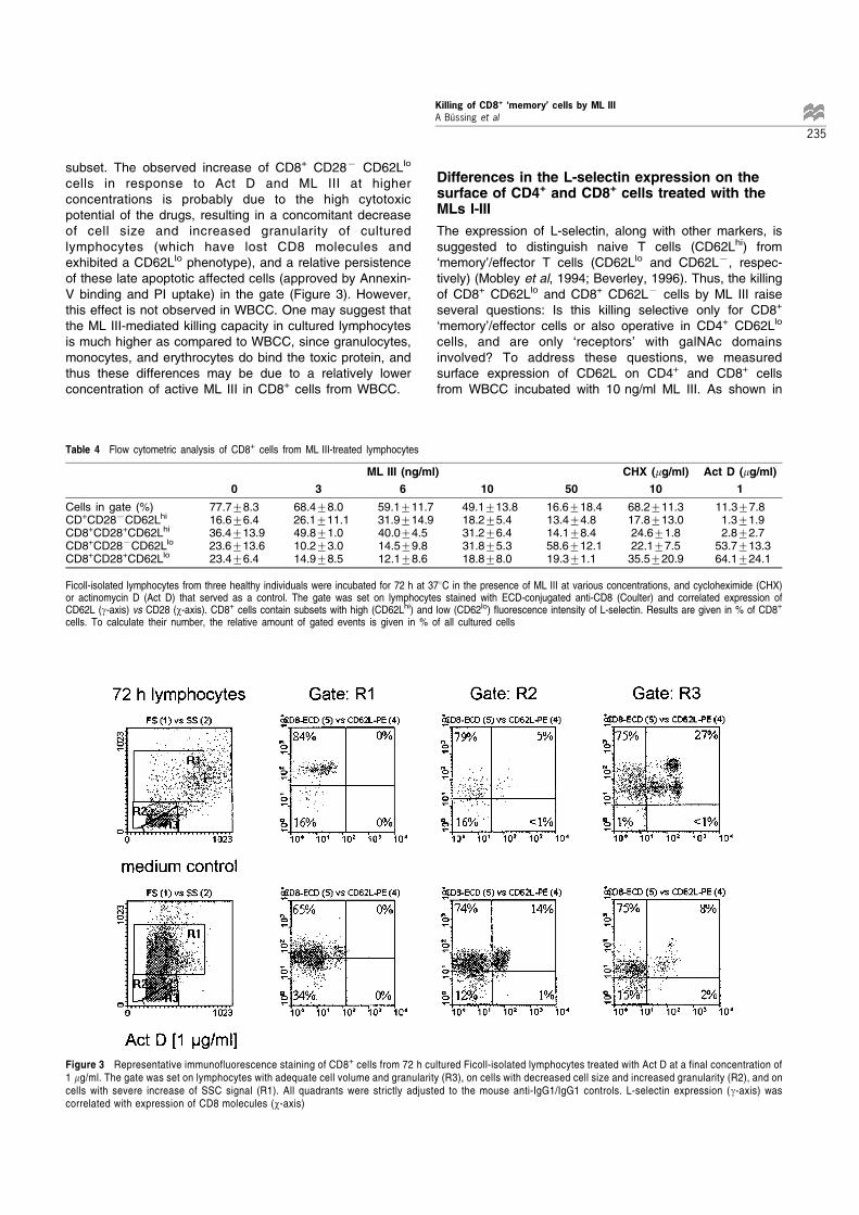
subset. The observed increase of CD8+ CD287 CD62Llo
cells in response to Act D and ML III at higherconcentrations is probably due to the high cytotoxicpotential of the drugs, resulting in a concomitant decreaseof cell size and increased granularity of culturedlymphocytes (which have lost CD8 molecules andexhibited a CD62Llo phenotype), and a relative persistenceof these late apoptotic affected cells (approved by Annexin-V binding and PI uptake) in the gate (Figure 3). However,this effect is not observed in WBCC. One may suggest thatthe ML III-mediated killing capacity in cultured lymphocytesis much higher as compared to WBCC, since granulocytes,monocytes, and erythrocytes do bind the toxic protein, andthus these differences may be due to a relatively lowerconcentration of active ML III in CD8+ cells from WBCC.
Differences in the L-selectin expression on thesurface of CD4+ and CD8+ cells treated with theMLs I-III
The expression of L-selectin, along with other markers, issuggested to distinguish naive T cells (CD62Lhi) from`memory'/effector T cells (CD62Llo and CD62L7, respec-tively) (Mobley et al, 1994; Beverley, 1996). Thus, the killingof CD8+ CD62Llo and CD8+ CD62L7 cells by ML III raiseseveral questions: Is this killing selective only for CD8+
`memory'/effector cells or also operative in CD4+ CD62Llo
cells, and are only `receptors' with galNAc domainsinvolved? To address these questions, we measuredsurface expression of CD62L on CD4+ and CD8+ cellsfrom WBCC incubated with 10 ng/ml ML III. As shown in
Table 4 Flow cytometric analysis of CD8+ cells from ML III-treated lymphocytes
ML III (ng/ml) CHX (mg/ml) Act D (mg/ml)
0 3 6 10 50 10 1
Cells in gate (%) 77.7+8.3 68.4+8.0 59.1+11.7 49.1+13.8 16.6+18.4 68.2+11.3 11.3+7.8CD+CD287CD62Lhi 16.6+6.4 26.1+11.1 31.9+14.9 18.2+5.4 13.4+4.8 17.8+13.0 1.3+1.9CD8+CD28+CD62Lhi 36.4+13.9 49.8+1.0 40.0+4.5 31.2+6.4 14.1+8.4 24.6+1.8 2.8+2.7CD8+CD287CD62Llo 23.6+13.6 10.2+3.0 14.5+9.8 31.8+5.3 58.6+12.1 22.1+7.5 53.7+13.3CD8+CD28+CD62Llo 23.4+6.4 14.9+8.5 12.1+8.6 18.8+8.0 19.3+1.1 35.5+20.9 64.1+24.1
Ficoll-isolated lymphocytes from three healthy individuals were incubated for 72 h at 378C in the presence of ML III at various concentrations, and cycloheximide (CHX)or actinomycin D (Act D) that served as a control. The gate was set on lymphocytes stained with ECD-conjugated anti-CD8 (Coulter) and correlated expression ofCD62L (g-axis) vs CD28 (w-axis). CD8+ cells contain subsets with high (CD62Lhi) and low (CD62lo) ¯uorescence intensity of L-selectin. Results are given in % of CD8+
cells. To calculate their number, the relative amount of gated events is given in % of all cultured cells
Figure 3 Representative immunofluorescence staining of CD8+ cells from 72 h cultured Ficoll-isolated lymphocytes treated with Act D at a final concentration of1 mg/ml. The gate was set on lymphocytes with adequate cell volume and granularity (R3), on cells with decreased cell size and increased granularity (R2), and oncells with severe increase of SSC signal (R1). All quadrants were strictly adjusted to the mouse anti-IgG1/IgG1 controls. L-selectin expression (g-axis) wascorrelated with expression of CD8 molecules (w-axis)
Killing of CD8+ `memory' cells by ML IIIA BuÈssing et al
235

Table 5, ML III significantly decreased the amount of CD8+
cells, thus, the proportion of CD4+ cells increased. Withinthe CD8+ cells, the amount of CD62Llo cells significantlydeclined, while, however, the decrease of CD4+ CD62Llo
cells was less severe. As shown in Table 6, the decline ofCD8+ cells was more pronounced by the addition of thegalNAc-specific MLs II and III, while the b-gal-specific ML Iwas less effective (ML I 5 ML II 5 ML III). Predominantlythe CD287 CD8+ subset decreased (Table 6). The ML IIand ML III-mediated decrease of the CD8+ CD62Llo subsetwas substantially prevented by galNAc, while, however, theproportion of CD4+ CD62Llo cells increased by thesimultaneous addition of MLs and galNAc (Table 7).Several other sugars such as D-galactose, a-lactose, a-methyl-galactosid, lactitol, and melibiose were ineffective toinhibit the ML III-mediated killing (data not shown), whilemethyl-a-D-galactopyranosid resulted in a slight but insignif-icant inhibition.
We next examined whether the observed killing effectswere specific for ML III or also observed in response toother drugs with properties of protein synthesis inhibition.Thus, we measured the L-selectin expression on thesurface of CD8+ cells from WBCC incubated for 72 h withthe Ricinus communis agglutinin (RCA120), ricin (RCA60),Abrus precatorus agglutinin (APA), Abrin A, and ML III(each at a final concentration of 10 ng/ml), and with CHX(10 mg/ml). As shown in Figure 4, RCA120, APA and CHXdid not significantly alter the expression of CD62L andCD28 on the surface of CD8+ cells as compared to thecontrols, while ML III and RCA60 eliminated the CD8+
CD62Llo cells, however, with and without CD28 molecules.Further application of streptomyces toxins Mit C (0.5 mg/ml) and Act D (100 and 1000 ng/ml) did not result insignificant changes of surface marker expression on CD8+
cells as compared to CHX and medium control (data notshown).
Cytokines in the supernatants of lymphocytefractions
In order to analyse whether the ML III-mediated killing of theCD8+ CD62Llo cells may change the cytokine pattern in theculture supernatants, we determined the concentrations oftype 1 and type 2-related cytokines, such as interferon-g,interleukin (IL)-12, -4, -5, -6, -10, and also tumour necrosisfactor (TNF)-a in the 72 h supernatants of CD8-enrichedand CD8-depleted lymphocyte (n=6). As shown in Table 8,apart from large interindividual differences in the release ofcytokines, application of ML III at 10 ng/ml did notsignificantly alter the supernatant concentrations of theinvestigated cytokines (a decrease of IL-6 was observed inthree out of six experiments), while in the CD8-depletedcultures ML III raised the concentration of TNF-a. In Mit C-treated samples, a higher concentration of interferon-g wasobserved in three out of six experiments. Thus, in CD8+
cells no relevant changes of the cytokine pattern in theculture supernatants was observed in response to ML III.The TNF-a release in the supernatants of CD4+ cells isprobably due to a `cytotoxic release' induced by ML III andMit C.
Discussion
We wondered whether lymphocyte subsets may differ in theirsensitivity to cell death induced by the toxic lectins fromViscum album, and observed a selective killing of CD8+ cellswith a `memory' phenotype (CD62Llo) by low concentrationsof the galNAc-specific MLs. One may speculate that the CD8+
Table 5 Flow cytometric analysis of CD4+ and CD8+ cells from WBCC afteraddition of ML III
ML III (ng/ml) 0 10
CD4+ cells 40.3+8.6 48.5+10.1CD8+ cells 32.6+5.0 23.5+5.5CD4/CD8 ratio 1.31+0.51 2.26+0.98
%CD62Lhi in CD4+ 73.4+7.0 80.4+7.5%CD62Llo in CD4+ 21.0+6.7 16.0+8.4%CD62Lhi in CD8+ 48.8+13.4 84.1+4.9%CD62Llo in CD8+ 37.9+9.6 10.3+4.8
Results are means+S.D. of experiments with lymphocytes from 72 h culturedWBCC (n = 16). ML III was added at a ®nal concentration of 10 ng/ml. Thesurface expression of CD4 and CD8 molecules was given in % of lymphocytes,while the L-selectin expression is given in % CD62Lhi and % CD62Llo withinCD4+ or CD8+ cells, respectively. Results are signi®cantly different compared tocontrols (p40.001; t-test)
Table 7 Flow cytometric analysis of CD62L expression on the surface of CD4+
and CD8+ cells from WBCC incubated in the presence of MLs I ± III and ofgalNAc
galNAc CD4+cells CD8+cells
%CD62Lhi %CD62Llo %CD62Lhi %CD62Llo
Controls 7+
72.0+5.870.7+7.4
19.7+4.222.7+6.0
40.6+1.740.3+1.8
34.2+1.236.3+0.3
ML I 7+
76.3+6.970.0+9.4
16.2+4.322.5+8.2
72.0+15.160.5+4.4
21.6+1.923.0+3.9
ML II 7+
76.7+6.766.0+12.3
16.9+4.627.0+9.8
68.4+4.146.2+4.8
16.7+2.231.8+3.5
ML III 7+
80.4+7.768.7+10.1
14.3+5.624.2+8.3
83.6+6.656.6+1.7
9.3+3.924.0+1.3
WBCC were incubated for 72 h at 378C in the presence of mistletoe lectins(MLs) I ± III. Results are means+S.D. of three different experiments. The MLswere added at ®nal concentrations of 10 ng/ml, while the controls representsamples without added toxins. 15 mM galNAc were added to block the galNAc-speci®c MLs II and III. Results are given in %CD62Lhi and %CD62Llo withinCD4+ or CD8+ cells, respectively
Table 6 Flow cytometric anaysis of CD4+ and CD8+ cells from WBCC afteraddition of MLs I-III
n controls ML I ML II ML III
CD4+ 5 40.3+9.8 47.9+7.7 46.7+2.7 50.8+9.5CD8+ 5 33.7+3.5 28.9+4.2 27.3+3.8 25.2+5.0CD28+CD8+ 3 14.8+2.5 14.7+2.1 17.0+2.1 15.8+3.4CD287CD8+ 3 19.4+4.2 15.6+4.1 14.3+3.5 11.4+4.3CD4/CD8
ratio5 1.23+0.46 1.71+0.47 1.75+0.32 2.11+0.68
WBCC were incubated for 72 h at 378C in the presence of MLs I ± III at a ®nalconcentration of 10 ng/ml. Results are means+S.D. of three to ®ve differentexperiments
Killing of CD8+ `memory' cells by ML IIIA BuÈssing et al
236

CD62Llo subset may differ from its CD8+ CD62Lhi counterpartby the expression of a yet undefined galNAc-containingreceptor which mediates or facilitates endocytosis of theapoptosis-inducing proteins. This suggestion is corroboratedby recent findings that interaction of ML B chains (but not theA chains) with appropriate receptors on lymphocytesincreased intracellular Ca2+ (BuÈssing et al, 1996a) andinhibited the uptake of [3H]thymidine (Metzner et al, 1987).However, this `activation'-pathway probably does not involveT cell receptor triggering, since T cells did not upregulateCD25 or CD71 expression in response to the MLs (BuÈssing etal, 1996a). The reason why ML III selectively killed CD8+
CD62Llo cells as compared to their CD8+ CD62Lhi counter-
parts, CD4+ T cells and CD19+ B cells is unclear. One maysuggest different possibilities, such as (1) differences in ML-binding to the lymphocyte subsets, (2) hyperresponsivenessof CD8+ CD62Llo cells to several cytokines, (3) highersusceptibility of these `memory' cells to cell death triggeredby the inhibition of protein synthesis, and (4) the presence of aspecific `receptor' preferentially expressed on CD8+ CD62Llo
cells which mediates subsequently cell death.One explanation of the observed differences could be
due to differences in ML-binding to the lymphocyte subsets.However, ML III exhibited similar cytotoxic activity to humanlymphocytes as ML I (GoÈckeritz et al, 1994; BuÈssing et al,1996a), although the binding of ML III was much lower as
Figure 4 Flow cytometric analysis of CD8+ cells from cultured WBCC incubated for 72 h in the presence of the plant lectins RCA120 (10 ng/ml), RCA60, (10 ng/ml), Abrin A (10 ng/ml), APA (1 mg/ml), ML III (10 ng/ml), and the streptomyces toxin CHX (10 mg/ml). The gate was set on lymphocytes stained with ECD-conjugated anti-CD8 (Coulter). CD8+ cells were differentiated in subsets with high (CD62Lhi) and low (CD62Llo) fluorescence intensity of L-selectin. L-selectinexpression (g-axis) was correlated with expression of CD28 molecules (w-axis). Results are representative for four independent experiments
Table 8 Cytokines in the supernatants of 72 h cultured lymphocyte fractions
CD8+ cells CD87 cells (89% CD4+ cells)
Cut off Medium ML III MitC Medium ML III MitC
(pg/ml) (pg/ml) control (10 ng/ml) (10 mg/ml) control (10 ng/ml) (10 mg/ml)
IFN-g 50 26+35 41+46 320+645* 117+148 155+163 155+163IL-12 500 5720+6484 6205+6481 707+969 6372+7429 6229+7074 913+1326IL-4 50 73+76 71+66 21+39 162+171 165+185 40+32IL-5 50 79+96 76+84 113+234 105+112 95+97 18+11*IL-10 20 35+15 57+77 61+84 728+689 243+261 427+4588IL-6 50 468+891 300+558 339+380 15193+11775 14481+9462 22790+8727*TNF-a 3 0.8+1.9 0.8+1.3 2.2+4.9 8+9 55+848 183+1788
The cytokines in the supernatants of CD8-enriched lymphocytes (99% CD3+ CD8+ cells) and CD8-depleted lymphocytes (89% CD4+ T cells) from six healthy individualswere determined by ELISA after 72 h incubation in the presence of ML III at a ®nal concentration of 10 ng/ml. The alkylating drug mitomycin C (Mit C), that served as apositive control, was added at 10 mg/ml. Results are signi®cantly different compared to control samples (*P40.05; 8P = 0.03; Wilkoxon's sign rank test)
Killing of CD8+ `memory' cells by ML IIIA BuÈssing et al
237
compared to ML I (GoÈckeritz et al, 1994). Since ML I-mediated killing of CD8+ CD62Llo cells was less effectiveas compared to ML III, one may suggest that binding of MLIII to its `receptor' is a more effective `death signal'. But thisdoes not explain the observed differences in the sensitivitybetween B cells and CD4+ and CD8+ T cell subsets to thetoxic protein, since these subsets did not significantly differin cell size. Alternatively, the binding sites may differ inCD8+ CD62Lhi `naive' cells and CD8+ CD62Llo `memory'cells. As reported recently, the endogenous lectin galectin-1, which induced apoptosis in thymocytes and activated Tcells (Baum et al, 1995; Perillo et al, 1995, 1997),preferentially binds to immature thymocytes as comparedto mature cells (Baum et al, 1995). This was suggested toresult from regulated expression of preferred oligosacchar-ide ligands on these cells. Thus, one may suggest similardifferences in the expression of ML III ligands in `naive'cells and `memory' cells. However, this may be true forCD8+ cells, but the ML III-mediated killing was lesseffective in the other subsets. An alternative view wouldbe that cells exhibiting a `memory' phenotype may behyperresponsive to several cytokines released in responseto ML III. Cytokines such as IL-1, -6 and TNF-a arerecognised to be induced by the addition of ML I (Hajto etal, 1990). However, in CD8-enriched cultures treated withML III, no significant changes in the release of type 1 andtype 2-related cytokines, such as interferon-g, IL-12, -4, -5, -10, and TNF-a, as compared to the control samples wereobserved.
Detailed studies suggest that for `naive' phenotype cells(CD44lo CD45RBhi CD62Lhi), the rate of cell division isrelatively slow, while CD44hi CD45RBlo CD62Llo cellsdivide at much higher rate (reviewed in Beverley, 1996).Such a rate of cell division obviously also implies a highrate of cell loss. Monteiro et al (1996) stated that thetelomeric length was significantly shorter in CD8+ CD287
cells, indicating an exhaustion of their replicative capacity.Indeed, the CD8+ CD287 subset is reported not only torespond inadequately to a variety of stimuli (Lewis et al,1994), but also to have reduced proliferative potential(Azuma et al, 1993). CD8+ CD287 cells with an activatedphenotype (CD38, CD45R0, HLA-DR) emerge during HIVinfection (Levacher et al, 1992; Landay et al, 1993;Gougeon et al, 1996), and especially the CD45R0+
(`memory') T cells from HIV infected persons were moresusceptible to apoptosis as compared with the samesubset in control subjects (Gougeon et al, 1996). There-fore, we suggest that the CD8+ CD287 cells with `memory'phenotype (CD62Llo) are in a state of `replicativesenescence', and thus may be responsive to any deathsignal, or alternatively, are proned to undergo cell death,anyway. The CD8+ CD287 subset was preferentiallyaffected in WBCC, while, however, in isolated lympho-cytes cultured in the presence of ML III, both the CD8+
CD62Llo cells with and without CD28 molecules wereaffected. One may suggest that the less effective killing ofCD8+ CD28+ CD62Llo subset in the WBCC might be dueto distinct survival signals and/or growth factors present inthe serum or released by monocytes or granulocytes.Monocytes may rescue this subset by an interaction of
costimulatory signal with CD28 molecules on T cells,which provides important costimulatory signals for thedevelopment of T cell responses (reviewed by Boise et al,1995).
It was reported previously that binding of the endogen-ous lectin galectin-1 to T cells involves recognition ofcarbohydrate epitopes on the T cell surface glycoproteinsCD43 (leukosialin, sialophorin) and CD45 (tyrosine phos-phatase involved in signal transduction) (Baum et al, 1995;Perillo et al, 1995). However, the presence of lectin-bindingepitopes, while essential, may not be sufficient for cell lysisand probably some intracellular mechanisms are involvedin the regulation of lectin-mediated cytotoxicity (Kim et al,1993). Although the inhibitors of protein and RNA synthesiswere potent toxicants for lymphocytes and inducedapoptosis, the observed selective killing of CD8+ CD62Llo
cells by ML III is not sufficiently explained by proteinsynthesis inhibition alone. This subset was not affected byCHX, which acts at the level of the ribosome by preventingtranslation, or by the type 4 and 2 RIPs RCA120, APA, andAbrin A. We suggest that ML III (and RCA60) requires ahigh affinity surface receptor with galNAc domains tomediate the signal that ultimately leads to cell death withinthe CD8+ CD62Llo subset, since (1) the ML III-mediatedkilling was substantially prevented by the addition ofgalNAc, and (2) the b-gal/galNAc-specific RCA60 but notthe RIPs that binds preferentially to b-gal killed this definedsubset. This suggestion is corroborated by preliminaryresults indicating that even a 2 h incubation of humanlymphocytes with ML III, which was followed by anextensive washing of cells and continued incubation for48 h in fresh medium, did kill the lymphocytes (BuÈssing,Wagner and PfuÈ ller, unpublished observations).
In conclusion, this report describes for the first time thata plant toxin kills a defined subset of lymphocytes. Althoughthe exact underlying mechanisms remain to be clarified, weobserved a selective killing of CD8+ cells with a `memory'phenotype (CD62Llo) by the type 2 RIPs ML III and RCA60
which both bind to terminal galNAc residues. It is unclearwhy CD4+ T cells and CD19+ B cells exhibiting thisphenotype are less sensitive to the toxin as compared tothe CD8+ cells. This selectivity of cell targeted ML III makesit a candidate for the use in immunotoxin or ligand toxinsynthesis, and immunomodulation, and also interposesseveral new aspects in the ongoing discussion (Matzin-ger, 1994; Ahmed and Gray, 1996) on the fate of `memory'T cells.
Materials and Methods
Blood samples and culture conditions
Since in a previous investigation apparent differences in the cellfunction of T cells from WBCC and lymphocyte cultures treated withthe streptococcal pyrogenic exotoxin A were observed (BuÈ ssing et al,1995), for this investigation, the more `in vivo-like' WBCC was used.Heparinised (50 IU/ml) peripheral blood from healthy individuals wasincubated 1 : 10 for 72 h at 378C in chromosome medium A (BiochromKG, Berlin, Germany).
Killing of CD8+ `memory' cells by ML IIIA BuÈssing et al
238

In further experiments, Ficoll-isolated lymphocytes (16106 cells/ml) were cultured in chromosome medium A before flow cytometricanalysis. For enrichment of CD8+ cells, isolated lymphocytes (36106
cells/ml) were suspended in RPMI 1640 (Biochrom KG), supplementedwith antibiotics and autologous plasma (10%), and separated withmagnetic beads (MACS, Miltenyi Biotec, Bergisch Gladbach,Germany) according to the manufacturers instructions. The CD8+
cells were further incubated for 72 h at 378C. Culture supernatantswere carefully collected and kept frozen at 7208C.
Toxic proteins
The MLs differ due to their molecular weights (between 50 kDa and63 kDa) and specificity. The ML I binds to b-gal, ML III to galNAc,whereas ML II binds to b-gal and galNAc (Franz et al, 1981). The MLswere extracted and purified as described previously (Eifler et al, 1993).For blocking experiments, 15 mM galNAc (Sigma Chemie) were addedto block the galNAc-specific MLs II and III.
Lectin from Ricinus communis (RCA120), lectin from Abrusprecatorus (APA), and Abrin A from Abrus precatorus, which bind togalactose, and the DNA synthesis inhibitor mitomycin C (Mit C) fromStreptomyces caespitosus, RNA synthesis inhibitor actinomycin D(Act D) from Streptomyces species, and the protein synthesis inhibitorcycloheximide (CHX) from Streptomyces griseus were obtained fromSigma Chemicals, Deisenhofen, Germany. Ricin (RCA60), which bindsto b-gal and galNAc, was a kind gift of Dr. Rudolf Eifler, Institute ofPhytochemistry, University Witten/Herdecke.
Flow cytometric analysis of cultured cells
Flow cytometric analysis of surface molecules was performed onEPICS1 XL-MCL flow cytometer (Coulter, Krefeld, Germany) usingmAb against the following Ag: CD3, CD4, CD8, CD16, CD19, CD25,CD28, CD38, CD56, CD62L, CD95 (Coulter). For each sample,10 ml of the mAb was added to 100 ml of cell suspension. Forwardangle light scatter (FSC) and high perpendicular light scatter (SSC)were used to gate lymphocytes and to exclude cell debris. Forsurface molecule analysis, only lymphocytes with adequate cellvolume (FSC signal) and granularity (SSC) were gated. These cellsinclude both, viable cells and cells undergoing apoptosis (Annexin-V+ PI7), while cells with decreased FSC signal and increased SSCsignal, which were excluded from analysis, contain late apoptoticcells (Annexin-V+ PIlo). All quadrants were adjusted to the anti-mouse isotype controls.
Viability analysis
To differentiate apoptotic and necrotic cells, Ficoll-isolated lympho-cytes were analyzed for Annexin-V binding and propidium iodide (PI)uptake as described (Koopman et al, 1994; Vermes et al, 1995). Briefly,after washing of cultured cells (16106/ml) with PBS and resuspensionin binding buffer (10 nM Hepes/NaOH, pH 7.4, 140 nM NaCl, 2.5 nMCaCl2), the cells were stained with 5 ml FITC-labeled Annexin-V(PharMingen, San Diego, CA, USA) and PI (5 mg/ml; Sigma). After15 min of incubation at room temperature in the dark, again the bindingbuffer was added and cells were analyzed by flow cytometry.Additionally, the mitochondrial membrane protein Apo2.7, a 38 kDprotein localised on cells undergoing apoptosis (Zhang et al, 1996), wasanalyzed. After digitonin-permeabilisation of cultured lymphocytesaccording to the method described by Zhang et al (1996), the cells werewashed and resuspended in PBS, and subsequently stained with thePE-labeled mAb Apo2.7 (Immunotech, Hamburg, Germany).
Cytokine assays
The cytokines interferon-g, IL-12, -4, -5, -10, -6, and TNF-a weredetermined in the culture supernatants as described previously(Stein et al, 1996). Briefly, microtiter plates were incubatedovernight (48C) with the monoclonal capture anti-cytokine Ab(PharMingen; IL-6 from R & D Systems, Wiesbaden, Germany),diluted in hydrogencarbonate buffer (0.1 M, pH 9.6), Recombinantor natural cytokines (standards) were added, and the supernatantsof CD8-enriched and CD8-depleted lymphocytes. After 2 h at roomtemperature, plates were incubated with the biotinylated detectureAbs for another 2 h (room temperature), followed by peroxidase-conjugated avidine (Sigma) for 30 min at room temperature.Detection was performed by adding substrate (o-phenylenedia-mine) solved in citrate buffer (0.1 M, pH 5.0). The individualconcentrations were measured with a SLT Spectra ELISA reader,using the SLT EasyFit software.
AcknowledgementsThe technical assistance of Kristin Backhaus and Eva Koloch isgratefully acknowledged. This work was supported by grants ofKrebsforschung Herdecke e.V. (Germany) and Helixor Heilmittel,Rosenfeld (Germany). Thanks to Rudolf Ei¯er for helpful discussionsand comments.
References
Ahmed R and Gray D (1996) Immunological memory and protective immunity:
understanding their relation. Science 271: 54 ± 60
Azuma M, Phillips JH and Lanier (1993) CD28- T lymphocytes: antigenic and
functional properties. J. Immunol. 150: 1147 ± 1159
Baum LG, Pang M, Perillo NL, Wu T, Delegeane A, Uittenbogaart CH, Fukuda M and
Seilhamer JJ (1995) Human thymic epithelial cells express an endogenous
lectin, galectin-1, which binds to core 2 O-glycans on thymocytes and T
lymphoblastoid cells. J. Exp. Med. 181: 877 ± 887
Beverley PCL (1996) Generation of T-cell memory. Curr. Opin. Immunol. 8: 327 ± 330
Boise LH, Minn AJ, Noel PJ and Thompson CB (1995) CD28 and apoptosis. Curr.
Opin. Immunol. 7: 620 ± 625
BuÈ ssing A, Klotz M, Suzart K, Efferth T, Gerlach D, Schnitzler N, Osieka R, Schweizer
K and Kaufhold A (1995) Sister chromatid exchange-inducing DNA lesions and
depression of activation markers on the surface of cultured peripheral bloodmononuclear cells after the addition of streptococcal pyrogenic exotoxins A and
C. Med. Microbiol. Immunol. 184: 87 ± 96
BuÈ ssing A, Suzart K, Bergmann J, PfuÈ ller U, Schietzel M and Schweizer K (1996a)
Induction of apoptosis in human lymphocytes treated with Viscum album L. is
mediated by the mistletoe lectins. Cancer Lett. 99: 59 ± 72
BuÈ ssing A (1996b) Induction of apoptosis by the mistletoe lectins. A review on the
mechanisms of cytotoxicity mediated by Viscum album L. Apoptosis 1: 25 ± 32
BuÈ ssing A, Suzart K and Schweizer K (1997) To live and let die - Differences in the
apoptosis-inducing properties of Viscum album L. extracts. Anti-Cancer Drugs 8
(Suppl. 1): S9 ± S14
Eifler R, PfuÈ ller K, GoÈ ckeritz W and PfuÈ ller U (1993) In Lectins: Biology, Biochemistry,
Clinical Biochemistry. Vol. 9, In Basu J, Kundu M and Chakrabarti P, eds (New
Delhi: Wiley Eastern Limited) pp. 144 ± 151
Endo Y, Tsurugi K and Franz H (1988) The site of action of the A-chain of mistletoe
lectin I on eukaryotic ribosomes - the RNA N-glycosidase activity of the protein.
FEBS Lett. 231: 378 ± 380
Franz H, Ziska P and Kindt A (1981) Isolation and properties of three lectins from
mistletoe (Viscum album L.) Biochem. J. 195: 481 ± 484GoÈ ckeritz W, KoÈ rner IJ, Kopp J, Bergmann J, PfuÈ ller K, Eifler R and PfuÈ ller U (1994)
Misletoe lectins: Comparative studies on cytotoxicity, receptor binding and their
effect on the cytosolic calcium content in human lymphocytes. In Lectins:
Biology, Biochemistry, Clinical Biochemistry. Vol. 10, In Van Driessche E, Fisher
J, Beeckmans S, Bog-Hansen TC, eds. (Hellerup, Denmark: Textop) pp. 345 ±
354
Killing of CD8+ `memory' cells by ML IIIA BuÈssing et al
239

Gougeon ML, Lecoeur H, Dulioust A, Enouf MG, Crouvoisier M, Goujard C, Debord T
and Montagnier L (1996) Programmed cell death in peripheral lymphocytes from
HIV-infected persons. Increased susceptibility to apoptosis of CD4 and CD8 T
cells correlates with lymphocyte activation and with disease progression. J.
Immunol. 156: 3509 ± 3520Griffiths GD, Leek MD and Gee DJ (1987) The toxic plant proteins ricin and abrin
induce apoptotic changes in mammalian lymphoid tissues and intestine. J.
Pathol. 151: 221 ± 229
Hajto T, Hostanska, Frey K, Rordorf C and Gabius HJ (1990) Increased secretion of
tumor necrosis factor a, interleukin 1, and interleukin 6 by human mononuclear
cells exposed to b-galactoside-specific lectin from clinically applied mistletoe
extract. Cancer Res. 50: 3322 ± 3326
Janssen O, Scheffler A and Kabelitz D (1993) In vitro effects of mistletoe extracts and
mistletoe lectins. Cytotoxicity towards tumor cells due to the induction of
programmed cell death (apoptosis). Arzneim-Forsch/Drug Res. 43: 1221 ± 1227
Khan T and Waring P (1993) Macrophage adherence prevents apoptosis induced by
ricin. Eur. J. Cell. Biol. 62: 406 ± 414
Kim M, Rao MV, Tweardy DJ, Prakash M, Galili U, Gorelik E (1993) Lectin induced
apoptosis in tumour cells. Glycobiol. 3: 447 ± 453
Koopman G, Reutelingsperger CPM, Kuijten GAM, Keehnen RMJ, Pals ST and van
Oers MHJ (1994) Annexin V for flow cytometric detection of phosphatidylserine
expression on B cells undergoing apoptosis. Blood 85: 532 ± 540
Kulkarni GV and McCulloch CAG (1995) Concanavalin A induced apoptosis infibroblasts: the role of cell surface carbohydrates in lectin mediated cytotoxicity.
J. Cell. Physiol. 165: 119 ± 133
Landay AL, Meckewicz CE and Levy JA (1993) An activated CD8+ T cell phenotype
correlates with anti-HIV activity and asymptomatic clinical status. Clin. Immunol.
Immunopath. 69: 106 ± 116
Levacher M, Hulstraert F, Tallet S, Ullery S, Pocidalo JJ and Bach BA (1992) The
significance of activation markers on CD8 lymphocytes in human immunode-
ficiency syndrome: staging and prognostic values. Clin. Exp. Immunol. 90: 376 ±
382
Lewis DE, Ng Tang DS, Adu-Oppong A, Schober W and Rogers JR (1994) Anergy
and apoptosis of CD8+ cells in HIV infection. J. Immunol. 153: 412 ± 420
Matzinger P (1994) Memories are made of this? Nature 369: 605 ± 606
Metzner G, Franz H, Kindt A, Schuman I and Fahlbusch B (1987) Effects of lectin I
from mistletoe (ML I) and ist isolated A and B chains on human mononuclear cells:
mitogenic activity and lymphokine release. Pharmazie 42: 337 ± 340
Mobley JL, Rigby SM and Dailey MO (1994) Regulation of adhesion molecule
expression by CD8T cells in vivo. II. Expression of L-selectin (CD62L) by memorycytolytic T cells responding to minor histocompatibility antigens. J. Immunol. 153:
5443 ± 5452
Mody R, Joshi S and Chaney W (1995) Use of lectins as diagnostic and therapeutic
tools for cancer. J. Pharmacol. Toxicol. Methods 33: 1 ± 10
Monteiro J, Batliwalla F, Ostrer H and Gregersen PK (1996) Shortened telomeres in
clonally expanded CD28-CD8+ T cells imply a replicative history that is distinct
from their CD28+CD8+ counterparts. J. Immunol. 156: 3587 ± 3590
Olsnes S, Sandvig K, Peteren OL and van Deur B (1989) Immunotoxins - entry into
cells and mechanisms of action. Immunol. Today 10: 291 ± 295
Perillo NL, Pace KE, Seilhamer JJ, Baum LG (1995)Apoptosis in T cells mediated by
galectin-1. Nature 378: 736 ± 739
Perillo NL, Uittenbogaart CH, Nguyen JT, Baum LG (1997) Galectin-1, an
endogenous lectin produced by thymic epithelial cells, induces apoptosis in
human thymocytes. J. Exp. Med. 185: 1851 ± 1858
Stein GM, Meink H, Durst J and Berg (1996) The release of cytokines by a fermented
lectin-1 (ML-1) free mistletoe extract reflects differences in the reactivity of PBMC
in healthy and allergic individuals and tumor patients. Eur. J. Clin. Pharmacol. 51:
247 ± 252Stirpe F, Sandvig K, Olsnes S and Pihl A (1982) Action of Viscumin, a toxic lectin from
mistletoe, on cells in culture. J. Biol. Chem. 257: 13271 ± 13277
Stirpe F, Barbieri L, Giulia M, Soria M and Lappi DA (1992) Ribosome-inactivating
proteins from plants: present status and future prospects. Bio/Technology 10:
405 ± 412
Vermes I, Haanen C, Steffens-Nakken H and Reutelingsperger C (1995) A novel
assay for apoptosis. Flow cytometric detection of phosphatidylserine expression
on early apoptotic cells using fluorescein labelled Annexin V.J. Immunol. Meth.
184: 39 ± 51
Zhang C, Ao Z, Seth A and Schlossman SF (1996) A mitochondrial membrane protein
defined by a novel monoclonal antibody is preferentially detected in apoptotic
cells. J. Immunol. 157: 3980 ± 3987
Killing of CD8+ `memory' cells by ML IIIA BuÈssing et al
240
Related Documents