SPECIAL ISSUE 4: INVITED ARTICLE Selecting Different Approaches for Palate and Pharynx Surgery: Palatopharyngeal Arch Staging System Rodolfo Lugo-Saldaña 1 , Karina Saldívar-Ponce 2 , Irina González-Sáez 3 , Daniela Hernández-Sirit 4 , Patricia Mireles-García 5 A BSTRACT The examination of the anatomical structures involved in the upper airway collapse in patients with the obstructive sleep apnea-hypopnea syndrome (OSAHS) is a key for integrated evaluation of patients. Our proposal is for a noninvasive classification system that guides us about the presence of anatomical differences between the palatopharyngeal muscle (PFM). The functions of the PFM are narrowing the isthmus, descending the palate, and raising the larynx during swallowing; these characteristics give the PFM a special role in the collapse of the lateral pharyngeal wall. Complete knowledge of the anatomy and classification of different variants can guide us to choose the appropriate surgical procedures for the lateral wall collapse. Until now there is not a consensus about description of the trajectory or anatomical variants of the PFM into oropharynx, the distance between both muscles, and the muscle tone. Here we also present the relationship between the lateral wall surgeries currently available (lateral pharyngoplasty by Cahali, expansion sphincteroplasty by Pang, relocation pharyngoplasty by Li, Roman blinds pharyngoplasty by Mantovani, and barbed sutures pharyngoplasty by Vicini) with the proposed classification of the palatopharyngeal arch staging system (PASS). Keywords: Obstructive sleep apnea-hypopnea syndrome, Palatopharyngeal muscle, Palatal surgery, Sleep disordered breathing, Sleep surgery. International Journal of Head and Neck Surgery (2019): 10.5005/jp-journals-10001-1383 I NTRODUCTION The multilevel surgery planning depends on numerous factors; of capital importance is a proper selection of the patient as well as the right evaluation of the anatomical structures responsible for the collapse and obstruction. Staging systems have been modified over time, to achieve a better surgery planning, as described by Iketmasu, who described many anatomical abnormalities related with OSAHS, 1 as well as a six sections to describe the oropharynx structures, which was simplified by Fujita as type I: oropharynx obstruction, type II: oropharynx and hypopharynx and type III: hypopharynx obstruction. 2 At 2004, Friedman made a staging system with the Friedman Tongue Position, FTP, body mass index (BMI) and the Brudsky tonsils classification as a modification of the Mallampati. In 2014 Friedman reported the lingual tonsil hypertrophy scale (LTH). 3 A complete physical examination of the patient considering the upper airway structures as well as the dynamics of the pharynx during the drug-induced sleep endoscopy (DISE), given our experience, is mandatory for the surgical planning, at the different levels, and allows for better postsurgical results. 4–6 Surgical techniques in OSAHS patients have evolved from the initial description of uvulapalatopharyngoplasty (UPPP) by Fujita in 1981 7 to less aggressive variations today, 8 viz., lateral pharyngoplasty described by Cahali, 9 the expansion sphincteroplasty by Pang and Woodson, 10 the relocation pharyngoplasty of Li, 11 the velo-uvulo- pharyngeal lift or “roman blinds” of Mantovani, 12 and barbed reposition pharyngoplasty (BRP) of Vicini. 13 In all of these different techniques, the principal and focal point is the dissection of the PFM. The palatopharyngeal muscle (PFM) is flattened in three beams whose main functions are narrowing the isthmus, lowering the palate, and raising the larynx. The PFM varies as to its position in the oropharynx, distance between the two sides or the narrowing of the pharynx, and the muscular tone. The main objective of this study is to propose a staging system to describe the different anatomical variants of PFM; the structure configuration of the palatopharyngeal arch (PFA), interpalatopharyngeal gap, and the PFM tone, as a guideline to select a lateral pharyngeal surgical technique; and to obtain an easy way to describe the anatomical variants in the palate and the pharyngeal lateral wall with the palatopharyngeal arch staging system (PASS). A NATOMY The muscle structures of the veil of the palate and pharynx are complex because of the relationships that these muscles keep. The soft palate is composed of five muscles, PFM, palatoglossus muscle (PGM), palatal veil lift muscle (PVLM), tensor of the palate veil (TPV), and uvula muscle (UM), which have a complex relationship with each other. The soft palate is considered as a barrier between the posterior part of the nose and the oral cavity; the origin of the PFM is the 1,3–5 Department of Otolaryngology and Head and Neck Surgery, University of Nuevo León UANL/ISSSTE, Monterrey, Nuevo León, Mexico 2 Department of Otolaryngology and Sleep Disordered Breathing Surgery, Instituto Mexicano Del Seguro Social, Torreon, Coahuila, Mexico Corresponding Author: Rodolfo Lugo-Saldaña, Department of Otolaryngology and Head and Neck Surgery, University of Nuevo León UANL/ISSSTE, Monterrey, Nuevo León, Mexico, Phone: +52 81 8356 0100, e-mail: [email protected] How to cite this article: Lugo-Saldaña R, Saldívar-Ponce K, González- Sáez I, et al. Selecting Different Approaches for Palate and Pharynx Surgery: Palatopharyngeal Arch Staging System. Int J Head Neck Surg 2019;10(4):92–97. Source of support: Nil Conflict of interest: None © The Author(s). 2019 Open Access This article is distributed under the terms of the Creative Commons Attribution 4.0 International License (https://creativecommons. org/licenses/by-nc/4.0/), which permits unrestricted use, distribution, and non-commercial reproduction in any medium, provided you give appropriate credit to the original author(s) and the source, provide a link to the Creative Commons license, and indicate if changes were made. The Creative Commons Public Domain Dedication waiver (http://creativecommons.org/publicdomain/zero/1.0/) applies to the data made available in this article, unless otherwise stated.

Welcome message from author
This document is posted to help you gain knowledge. Please leave a comment to let me know what you think about it! Share it to your friends and learn new things together.
Transcript
SPECIAL ISSUE 4: INVITED ARTICLE
Selecting Different Approaches for Palate and Pharynx Surgery: Palatopharyngeal Arch Staging SystemRodolfo Lugo-Saldaña1 , Karina Saldívar-Ponce2 , Irina González-Sáez3 , Daniela Hernández-Sirit4 , Patricia Mireles-García5
Ab s t r Ac tThe examination of the anatomical structures involved in the upper airway collapse in patients with the obstructive sleep apnea-hypopnea syndrome (OSAHS) is a key for integrated evaluation of patients. Our proposal is for a noninvasive classification system that guides us about the presence of anatomical differences between the palatopharyngeal muscle (PFM). The functions of the PFM are narrowing the isthmus, descending the palate, and raising the larynx during swallowing; these characteristics give the PFM a special role in the collapse of the lateral pharyngeal wall. Complete knowledge of the anatomy and classification of different variants can guide us to choose the appropriate surgical procedures for the lateral wall collapse. Until now there is not a consensus about description of the trajectory or anatomical variants of the PFM into oropharynx, the distance between both muscles, and the muscle tone. Here we also present the relationship between the lateral wall surgeries currently available (lateral pharyngoplasty by Cahali, expansion sphincteroplasty by Pang, relocation pharyngoplasty by Li, Roman blinds pharyngoplasty by Mantovani, and barbed sutures pharyngoplasty by Vicini) with the proposed classification of the palatopharyngeal arch staging system (PASS).Keywords: Obstructive sleep apnea-hypopnea syndrome, Palatopharyngeal muscle, Palatal surgery, Sleep disordered breathing, Sleep surgery.International Journal of Head and Neck Surgery (2019): 10.5005/jp-journals-10001-1383
In t r o d u c t I o nThe multilevel surgery planning depends on numerous factors; of capital importance is a proper selection of the patient as well as the right evaluation of the anatomical structures responsible for the collapse and obstruction.
Staging systems have been modified over time, to achieve a better surgery planning, as described by Iketmasu, who described many anatomical abnormalities related with OSAHS,1 as well as a six sections to describe the oropharynx structures, which was simplified by Fujita as type I: oropharynx obstruction, type II: oropharynx and hypopharynx and type III: hypopharynx obstruction.2 At 2004, Friedman made a staging system with the Friedman Tongue Position, FTP, body mass index (BMI) and the Brudsky tonsils classification as a modification of the Mallampati. In 2014 Friedman reported the lingual tonsil hypertrophy scale (LTH).3
A complete physical examination of the patient considering the upper airway structures as well as the dynamics of the pharynx during the drug-induced sleep endoscopy (DISE), given our experience, is mandatory for the surgical planning, at the different levels, and allows for better postsurgical results.4 – 6
Surgical techniques in OSAHS patients have evolved from the initial description of uvulapalatopharyngoplasty (UPPP) by Fujita in 19817 to less aggressive variations today,8 viz., lateral pharyngoplasty described by Cahali,9 the expansion sphincteroplasty by Pang and Woodson,10 the relocation pharyngoplasty of Li,11 the velo-uvulo-pharyngeal lift or “roman blinds” of Mantovani,12 and barbed reposition pharyngoplasty (BRP) of Vicini.13 In all of these different techniques, the principal and focal point is the dissection of the PFM.
The palatopharyngeal muscle (PFM) is flattened in three beams whose main functions are narrowing the isthmus, lowering the palate, and raising the larynx. The PFM varies as to its position in the oropharynx, distance between the two sides or the narrowing of the pharynx, and the muscular tone.
The main objective of this study is to propose a staging system to describe the different anatomical variants of PFM; the structure configuration of the palatopharyngeal arch (PFA), interpalatopharyngeal gap, and the PFM tone, as a guideline to select a lateral pharyngeal surgical technique; and to obtain an easy way to describe the anatomical variants in the palate and the pharyngeal lateral wall with the palatopharyngeal arch staging system (PASS).
An Ato m yThe muscle structures of the veil of the palate and pharynx are complex because of the relationships that these muscles keep.
The soft palate is composed of five muscles, PFM, palatoglossus muscle (PGM), palatal veil lift muscle (PVLM), tensor of the palate veil (TPV), and uvula muscle (UM), which have a complex relationship with each other.
The soft palate is considered as a barrier between the posterior part of the nose and the oral cavity; the origin of the PFM is the
1,3–5 Department of Otolaryngology and Head and Neck Surgery, University of Nuevo León UANL/ISSSTE, Monterrey, Nuevo León, Mexico2 Department of Otolaryngology and Sleep Disordered Breathing Surgery, Instituto Mexicano Del Seguro Social, Torreon, Coahuila, MexicoCorresponding Author: Rodolfo Lugo-Saldaña, Department of Otolaryngology and Head and Neck Surgery, University of Nuevo León UANL/ISSSTE, Monterrey, Nuevo León, Mexico, Phone: +52 81 8356 0100, e-mail: [email protected] to cite this article: Lugo-Saldaña R, Saldívar-Ponce K, González-Sáez I, et al. Selecting Different Approaches for Palate and Pharynx Surgery: Palatopharyngeal Arch Staging System. Int J Head Neck Surg 2019;10(4):92–97.Source of support: NilConflict of interest: None
© The Author(s). 2019 Open Access This article is distributed under the terms of the Creative Commons Attribution 4.0 International License (https://creativecommons.org/licenses/by-nc/4.0/), which permits unrestricted use, distribution, and non-commercial reproduction in any medium, provided you give appropriate credit to the original author(s) and the source, provide a link to the Creative Commons license, and indicate if changes were made. The Creative Commons Public Domain Dedication waiver (http://creativecommons.org/publicdomain/zero/1.0/) applies to the data made available in this article, unless otherwise stated.
Selecting Different Approaches for Palate and Pharynx Surgery
International Journal of Head and Neck Surgery, Volume 10 Issue 4 (October–December 2019) 93
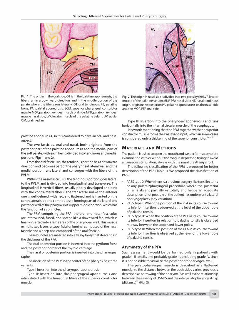
palatine aponeurosis, so it is considered to have an oral and nasal aspect.
The two fascicles, oral and nasal, both originate from the posterior part of the palatine aponeurosis and the medial part of the soft palate, with each being divided into tendinous and medial portions (Figs 1 and 2).
From the oral fasciculus, the tendinous portion has a downward direction and becomes part of the pharyngeal lateral wall and the medial portion runs lateral and converges with the fibers of the PVLM.
Within the nasal fasciculus, the tendinous portion goes lateral to the PVLM and is divided into longitudinal and transverse. The longitudinal is vertical fibers, usually poorly developed and bind with the contralateral fibers. The transverse unlike the anterior one is well defined, widened, and flattened and is inserted on the contralateral side and contributes to forming part of the lateral and posterior wall of the pharynx in its upper middle portion, which has the function of a sphincter.
The PFM comprising the PFA, the oral and nasal fasciculus are intertwined, fused, and spread like a downward fan, which is finally inserted into a large area of the pharyngeal wall. This muscle exhibits two layers: a superficial or luminal composed of the nasal fascicle and a deep one composed of the oral fascicle.
These bundles are inserted into a fleshy body that descends in the thickness of the PFA.
The oral or anterior portion is inserted into the pyriform fossa and the posterior border of the thyroid cartilage.
The nasal or posterior portion is inserted into the pharyngeal raphe.
The insertion of the PFM in the center of the pharynx has three variants:
Type I: Insertion into the pharyngeal aponeurosisType II: Insertion into the pharyngeal aponeurosis and
intercalated with the horizontal fibers of the superior constrictor muscle
Type III: Insertion into the pharyngeal aponeurosis and runs horizontally into the internal circular muscle of the esophagus.
It is worth mentioning that the PFM together with the superior constrictor muscle forms the Passavant input, which in some cases is considered only a thickening of the superior constrictor.14 – 16
mAt e r I A l s A n d me t h o d sThe patient is asked to open the mouth and we perform a complete examination with or without the tongue depressor, trying to avoid a nauseous stimulation, always with the nasal breathing effort.
The following classification of the PFM is proposed for better description of the PFA (Table 1). We proposed the classification of PASS.
• PASS type 0: When there is a previous surgery like tonsillectomy or any palatal/pharyngeal procedure where the posterior pillar is absent partially or totally and hence an adequate description is not possible or the patient has underwent a lateral pharyngoplasty (any variation).
• PASS type I: When the position of the PFA in its course toward its inferior insertion is observed at the level of the upper pole of palatine tonsils.
• PASS type II: When the position of the PFA in its course toward its inferior insertion in relation to palatine tonsils is observed midway between the upper and lower poles.
• PASS type III: When the position of the PFA in its course toward its inferior insertion is observed at the level of the lower pole of palatine tonsils.
Asymmetry of the PFASuch assessment would be performed only in patients with grade I–II tonsils, and probably grade III, excluding grade IV, since it is not possible to visualize the posterior oropharyngeal wall.
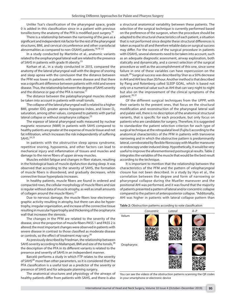
The palatopharyngeal muscle is described as a flattened muscle, so the distance between the both sides varies, previously described as narrowing of the pharynx,16 as well as the relationship between the severity of OSAHS and the interpalatopharyngeal gap (distance)17 (Fig. 3).
Fig. 1: The origin in the oral side; OT is in the palatine aponeurosis; the fibers run in a downward direction, and in the middle portion of the palate where the fibers run laterally. OT oral tendinous; PB, palatine bone; PA, palatal aponeurosis; SCM, superior pharyngeal constrictor muscle; MOP, palatopharyngeal muscle oral side; MNP, palatopharyngeal muscle nasal side; LVP, levator muscle of the palatine velum; UV, uvula; OM, oral median
Fig. 2: The origin in nasal side is divided into two parts by the LVP, levator muscle of the palatine velum; MNP, PFA nasal side; NT, nasal tendinous origin, origin in the posterior; PA, palatine aponeurosis on the nasal side and the MOP, PFA oral side
Selecting Different Approaches for Palate and Pharynx Surgery
International Journal of Head and Neck Surgery, Volume 10 Issue 4 (October–December 2019)94
The following system is proposed to assess the distance or narrowness between the two muscles during the physical examination (interpalatopharyngeal gap):
• Grade 0: Surgical absence of PFM• Grade I: PFA > 4 cm• Grade II: PFA > 3 cm• Grade III: PFA 2–3 cm• Grade IV: PFA < 2 cm
While describing the PFA staging system, it is also important to take into account the degree of muscle hypertrophy, as a hypertrophied muscle yields a much better result when performing
a lateral pharyngoplasty, as described by Cahali. Whereas, if the muscle is hypotrophic, then relocation pharyngoplasty as described by Li or the barbed sutured technique as described by Mantovani or Vicini11 , 12 performs better. The muscle can thus be described as (a) normotrophic, (b) hypotrophic, or (c) hypertrophic.
dI s c u s s I o nLateral pharyngeal wall surgery involves numerous variants that have developed over time but so far it has not been described whether any of the anatomic variants of the PFA may have any relation to surgical success. Cahali considers hypertrophy of the PFM as a surgical indication of the lateral pharyngoplasty technique.9
A detailed description and classification of PFM is considered important since the morphology, position of its arches, and muscular tone may influence the pattern of palatal obstruction. These haven’t been investigated in the past (only the scale of narrowing of folds is described as anatomic variants of the PFA)16 (Table 1).
In other words, the description of the position of the PFA during the examination, which sometimes shows the fold in the upper part of the posterior wall of the pharynx, without distinguishing the rest of its path from the posterior pharyngeal wall (type I), when it is observed at the mid-level of the tonsils (type II) or is observed in its entirety because it can be differentiated from the posterior wall of the pharynx (type III), this supposes according to the anatomical corpse descriptions in the nasal fascicle superficial is underdeveloped and therefore the accomplishment of some surgical procedure at lateral wall level is difficult its dissection and therefore to be able to realize a suitable surgical planning to have a better result.
Fig. 3: Interpalatopharyngeal gap distance
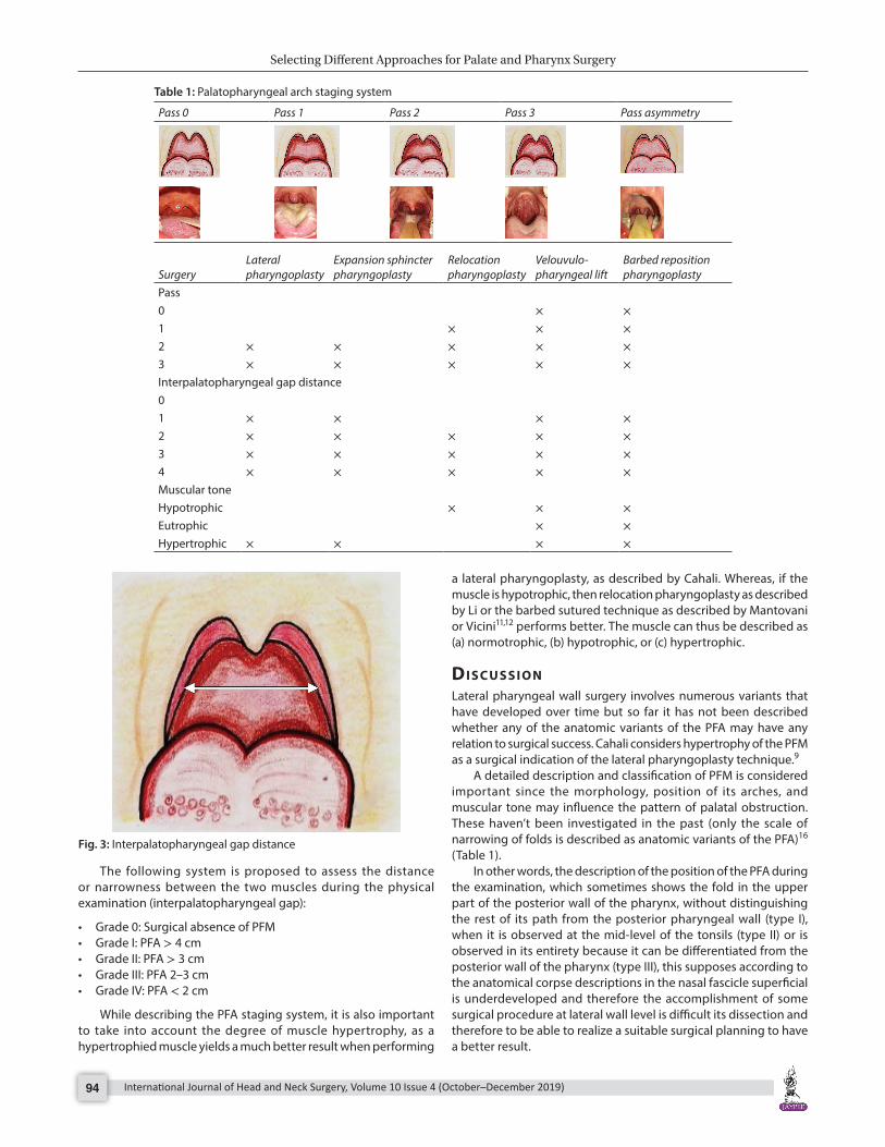
Table 1: Palatopharyngeal arch staging system
Pass 0 Pass 1 Pass 2 Pass 3 Pass asymmetry
SurgeryLateral pharyngoplasty
Expansion sphincter pharyngoplasty
Relocation pharyngoplasty
Velouvulo-pharyngeal lift
Barbed reposition pharyngoplasty
Pass0 × ×1 × × ×2 × × × × ×3 × × × × ×Interpalatopharyngeal gap distance01 × × × ×2 × × × × ×3 × × × × ×4 × × × × ×Muscular toneHypotrophic × × ×Eutrophic × ×Hypertrophic × × × ×
Selecting Different Approaches for Palate and Pharynx Surgery
International Journal of Head and Neck Surgery, Volume 10 Issue 4 (October–December 2019) 95
Unlike Tsai’s classification of the pharyngeal space, grade 0 is added in this classification since in a patient with previous tonsillectomy the anatomy of the PFA is modified post surgery.16
There is a relationship between the narrowing of the jaws as a significant and independent risk factor for the rest of the pharyngeal structures, BMI, and cervical circumference and other craniofacial abnormalities as compared to non-OSAHS patients.16 , 18 – 22
In a study conducted by Martinho et al., anatomic variants related to the oropharyngeal lateral wall are related to the presence of SAHS in patients with grade III obesity.23
Korhan et al., in a study conducted in 2015, compared the anatomy of the lateral pharyngeal wall with the degree of snoring and sleep apnea with the conclusion that the distance between the PFM was lower in patients with severe disease and that there was a significant difference between patients with mild and severe disease. Thus, the relationship between the degree of SAHS severity and the distance or gap of the PFA is narrow.17
The distance between the palatopharyngeal muscles should be taken into account in patients with small tonsils.
The collapse of the lateral pharyngeal wall is related to a higher BMI, greater IDO, greater apnea-hypopnea index, and lower O2 saturation, among other values, compared to patients with partial lateral collapse or without oropharynx collapse.23
The espesor of lateral pharyngeal walls measured by nuclear magnetic resonance (NMR) in patients with SAHS compared to healthy patients are greater at the expense of muscle tissue and not fat infiltration, which increases the risk independently of suffering SAHS.24 , 25
In patients with the obstructive sleep apnea syndrome, repetitive snoring, hypoxemia, and other factors can lead to mechanical injury and inflammation of tissues and muscles and damage to the nerve fibers of upper airway muscles.
Muscles exhibit fatigue and changes in fiber stature, resulting in the histological basis of muscle dysfunction during sleep. It was observed that according to the severity of SAHS, the formation of muscle fibers is disordered, and gradually decreases, while connective tissue hyperplasia increases.
In healthy patients, muscle fibers are found in ordered and compacted rows, the cellular morphology of muscle fibers and size is regular without data of muscle atrophy, as well as small amounts of collagen around the muscle fibers.27
Due to nervous damage, the muscle fibers lose electromyo-graphic activity resulting in atrophy, but there can also be hyper-trophy, irregular organization, and increase of the connective tissue resulting in muscular hypertrophy and thickening of the oropharynx wall that increases the stenosis.
The changes in the PFM are related to the severity of the disease, since the proportion of muscle fibers PASS 1 and PASS 2 is altered; the most important changes were observed in patients with severe disease in contrast to those classified as moderate disease or controls, so the effect of treatment may vary.26
As previously described by Friedman, the relationship between SAHS severity according to Mallampati, BMI and size of the tonsils,28 the description of the PFA in its different variants is related to the presence and severity of SAHS in an independent manner.
Barceló performs a study in which FTP relates to the severity of SAHS28 more than other parameters, so it is considered that the PFA classification is a useful tool as a predictor of the severity or presence of SAHS and for adequate planning surgery.
The anatomical structures and physiology of the airways of healthy patients differ from patients with SAHS, and there is also
a structural anatomical variability between these patients. The selection of the surgical technique is currently performed based on the preference of the surgeon, when the procedure should be adapted to the structural characteristics of each patient, a situation that is not performed since despite these structural differences is taken as equal to all and therefore reliable data on surgical success may differ. For the success of the surgical procedure in patients with OSAHS, several elements need to be taken into account, such as an adequate diagnostic assessment, airway exploration, both statically and dynamically, and a correct selection of the surgical procedure as well as the accomplishment of this one, since some failure in one of these variables can have repercussion on the result.29 Surgical success was described by Sher as a 50% decrease in AHI and AHI less than 20/hour. Another method is that described by Pang and Rotenberg called SLEEP GOAL, which is based not only on a numerical value such as AHI that can vary night to night but also on the improvement of the clinical symptoms of the patient.30 , 31
Of the different surgical techniques from the UPPP, with their variants to the present ones, that focus on the structural modification and reconstruction of the pharyngeal lateral wall and palate veil, there is no description of the anatomical structural variants, that is specific for each procedure, but only focus on patients who are candidates for surgery. Therefore, it is suggested to standardize the patient selection criterion for each type of surgical technique at the retropalatal level (Fujita I) according to the anatomical characteristics of the PFM in patients with transverse narrowing and in which the obstructive pattern is predominantly lateral, corroborated by flexible fibroscopy with Mueller maneuver or endoscopy under induced sleep. Hypothetically, it would be very useful to improve the aforementioned postsurgical results. Table 2 integrates the variables of the muscle that would be the best result according to the technique.
It is important to mention that the relationship between the characteristics of the PFM and the pattern of velopharyngeal closure has not been described. In a study by Hyo et al., the correlation between the degree and form of narrowing or pharyngeal collapse during the Mueller maneuver and the positional AHI was performed, and it was found that the majority of patients presented a pattern of lateral and/or concentric collapse compared to the pattern of anteroposterior collapse. “Additonally, AHI was higher in patients with lateral collapse pattern than
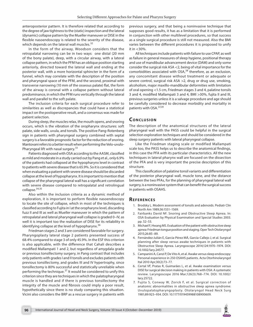
Table 2: Obstruction patterns according to vote classification
Concentric Lateral A–PVelum
Oropharynx
You can see the videos of the obstruction patterns scanning the QR codes in your smartphone or electronic device
Selecting Different Approaches for Palate and Pharynx Surgery
International Journal of Head and Neck Surgery, Volume 10 Issue 4 (October–December 2019)96
anteroposterior pattern. It is therefore related that according to the degree of jaw tightness to the (static) inspection and the lateral (dynamic) collapse pattern by the Mueller maneuver or DISE in the flexible nasoendoscopy is related to the severity of the disease, which depends on the lateral wall muscles.32
In the form of the airway, Woodson considers that the retropalatal narrowing can be in two ways: one distal (20 mm of the bony palate), deep, with a circular airway, with a lateral collapse pattern, in which the PFM has an oblique position starting anteriorly, directed toward the lateral wall and ending at the posterior wall, with a more horizontal sphincter in the form of a funnel, which may correlate with the description of the position and pharyngeal space of the PFM, and the second, proximal with transverse narrowing (10 mm of the osseous palate) flat, the form of the airway is coronal with a collapse pattern without lateral predominance, in which the PFM runs vertically through the lateral wall and parallel to the soft palate.
The inclusion criteria for each surgical procedure refer to similarities as well as discrepancies that could have a statistical impact on the postoperative result, and a consensus was made for patient selection.
During sleep, the muscles relax, the mouth opens, and snoring occurs, which is the vibration of the oropharynx structures: soft palate, side walls, uvula, and tonsils. The positive Pang-Rotenberg sign in patients with pharyngeal surgery combined with septal surgery is a favorable prognostic factor for the reduction of snoring. Mantovani refers to a better result when performing the Velo-uvulo-Pharyngeal lift with nasal surgery.33
Patients diagnosed with OSA according to the AASM, classified as mild and moderate in a study carried out by Pang et al., only 6.9% of the patients had collapsed at the hypopharynx level in contrast to patients with severe disease that is 65.9%. So it is considered that when evaluating a patient with severe disease should be discarded collapse at the level of hypopharynx. It is important to mention that collapse of the pharyngeal lateral wall has a significant correlation with severe disease compared to retropalatal and retrolingual collapse.34 , 35
Also within the inclusion criteria as a dynamic method of exploration, it is important to perform flexible nasoendoscopy to locate the site of collapse, which in most of the techniques is classified according to Fujita in I at the oropharynx level, discarding fuzz II and III as well as Mueller maneuver in which the pattern of retropalatal and lateral pharyngeal wall collapse is graded 0–IV, as well it is important too the realization of DISE for its reliability in identifying collapse at the level of hypopharynx.36
Friedman stages 2 and 3 are considered favorable for surgery. Pharyngoplasty lateral stage 2 patients presented success of 68.4% compared to stage 3 of only 45.9%. In the ESF this criterion is also applicable, with the difference that Cahali describes a modified Mallampati 1 and 2, but regardless of amygdala grade or previous tonsillectomy surgery, in Pang contrast that includes only patients with grades I and II tonsils and excludes patients with previous tonsillectomy, and grades III and IV hypertrophy, since tonsillectomy is 80% successful and statistically unreliable when performing the technique.37 It would be considered to unify this criterion since they are techniques in which the palatopharyngeal muscle is handled and if there is previous tonsillectomy the integrity of the muscle and fibrosis could imply a poor result, hypothetically since there is no study comparing this situation. Vicini also considers the BRP as a rescue surgery in patients with
previous surgery, and that being a noninvasive technique that supposes good results, it has as a limitation that it is performed in conjunction with other multilevel procedures, so that success as a single surgical procedure is still not measured. Also the BMI varies between the different procedures it is proposed to unify it in <30%.
All techniques include patients with failure to use CPAP, as well as failure in general measures of sleep hygiene, positional therapy and use of mandibular advancement device (DAM) and only some consider the surgical risk ASA <2, being of vital importance for the comorbidities associated with OSA,38 therefore, as an exclusion, any concomitant disease without treatment or adequate or severe control, surgical risk ASA >2, drug or drug use, smoking, alcoholism, major maxillo-mandibular deformities with limitation of oral opening <1.5 cm, Friedman stages 3 and 4, palatine tonsils 3 and 4, modified Mallampati 3 and 4, BMI >30%, fujita II and III, previous surgeries unless it is a salvage procedure and age should be carefully considered to decrease morbidity and mortality in patients with OSA.37 , 39
co n c lu s I o nThe description of the anatomical structures of the lateral pharyngeal wall with the PASS could be helpful in the surgical selection exploration techniques and should be considered in the sleep surgery patients with lateral pharyngeal collapse.
Like the Friedman staging scale or modified Mallampati scale too, the PASS helps us to describe the anatomical findings, in this case the PFA with its particular characteristics. All the new techniques in lateral pharynx wall are focused on the dissection of the PFA and is very important the precise description of this structure.
This classification of palatine tonsil variants and differentiation of the posterior pharyngeal wall, muscle tone, and the distance between the two PFAs, for the planning of pharyngeal lateral wall surgery, is a noninvasive system that can benefit the surgical success in patients with OSAHS.
re f e r e n c e s 1. Brodsky L. Modern assessment of tonsils and adenoids. Pediatr Clin
North Am 1989;36:1551–1569. 2. Fairbanks David NF. Snoring and Obstructive Sleep Apnea. In:
OSA Evaluation by Physical Examination and Special Studies 2003. pp. 52–53.
3. Michael F, Hwang MS. Evaluation of the patient with obstructive sleep apnea: Friedman tongue position and staging. Oper Tech Otolaryngol 2015;26:85–89.
4. Fernández-Julián E, García-Pérez MÁ, García-Callejo J, et al. Surgical planning after sleep versus awake techniques in patients with Obstructive Sleep Apnea. Laryngoscope 2014;124:1970–1974. DOI: 10.1002/lary.24577.
5. Campanini A, Canzi P, De Vito A, et al. Awake versus sleep endoscopy: Personal experience in 250 OSAHS patients. Acta Otorhinolaryngol Ital 2010 Apr;30(2):73–77.
6. Certal VF, Pratas R, Guimarães L, et al. Awake examination versus DISE for surgical decision making in patients with OSA: A systematic review. Laryngoscope 2016 Mar;126(3):768–774. DOI: 10.1002/lary.25722.
7. Fujita S, Conway W, Zorick F, et al. Surgical correction of anatomic abnormalities in obstructive sleep apnea syndrome: Uvulopalatopharyngoplasty. Otolaryngol Head Neck Surg 1981;89:923–934. DOI: 10.1177/019459988108900609.
Selecting Different Approaches for Palate and Pharynx Surgery
International Journal of Head and Neck Surgery, Volume 10 Issue 4 (October–December 2019) 97
8. Powell N, Riley R, Guilleminault C, et al. A reversible uvulopalatal flap for snoring and sleep apnea syndrome. Sleep 1996;19:593–599. DOI: 10.1093/sleep/19.7.593.
9. Cahali MB. Lateral pharyngoplasty: a new treatment for obstructive sleep apnea hypopnea syndrome. Laryngoscope 2003;113:1961–1968. DOI: 10.1097/00005537-200311000-00020.
10. Pang KP, Woodson BT. Expansion sphincter pharyngoplasty: A new technique for the treatment of obstructive sleep apnea. Otolaryngol Head Neck 2007;137:110–114. DOI: 10.1016/j.otohns.2007.03.014.
11. Li HY. Relocation pharyngoplasty. Oper Tech Otolayngol 2012;23: 25–29. DOI: 10.1016/j.otot.2011.06.002.
12. Mantovani M, Minetti A, Torretta S, et al. The velo-uvulo-pharyngeal lift or “roman blinds” technique for treatment of snoring: a preliminary report. Acta Otorhinolaryngol Ital 2012 Feb;32(1):48–53.
13. Vicini C, Hendawy E, Campanini A, et al. Barbed reposition pharyngoplasty (BRP) for OSAHS: a feasibility, safety, efficacy and teachability pilot study. “We are on the giant’s shoulders”. Eur Arch Otorhinolaryngol 2015 Oct;272(10):3065–3070. DOI: 10.1007/s00405-015-3628-3.
14. Cho JH, Kim JK, Lee HY, et al. Surgical anatomy of human soft palate. The Laryngoscope 2013;123(11):2900–2904. DOI: 10.1002/ lary.24067.
15. Okuda S, Abe S, Kim HJ, et al. Morphologic Characteristics of Palatopharyngeal Muscle. Dysphagia 2008;23:258–266. DOI: 10.1007/s00455-007-9133-0.
16. Choi DY, Bae JH, Youn KH, et al. Anatomical Considerations of the Longitudinal Pharyngeal Muscles in Relation to their Function on the Internal Surface of Pharynx. Dysphagia 2014;29(6):722–730. DOI: 10.1007/s00455-014-9568-z.
17. Tsai WH, Remmers JE, Brant R, et al. A decision rule for diagnostic testing in obstructive sleep apnea. Am J Respir Crit Care Med 2003;167:1427–1432. DOI: 10.1164/rccm.200112-110OC.
18. Korhan I, Gode S, Midilli R, et al. The influence of the lateral pharyngeal wall anatomy on snoring and sleep apnea. J Pak Med Assoc 2015 Feb;65(2):125–130.
19. Yagi H, Nakata S, Tsuge H, et al. Morphological examination of upper airway in obstructive sleep apnea. Auris Nasus Larynx 2009;36: 444–449. DOI: 10.1016/j.anl.2008.11.003.
20. Schellenberg Joseph B, Maislin G, Schwab RJ. Physical Findings and the Risk for Obstructive Sleep Apnea, the Importance of Oropharyngeal Structures. Am J Respir Crit Care Med 2000;;162: 740–748. DOI: 10.1164/ajrccm.162.2.9908123.
21. Zonato AI, Martinho FL, Bittencourt LR, et al. Head and neck physical examination: Comparison between nonapneic and obstructive sleep apnea patients. Laryngoscope 2005;115:1030–1034. DOI: 10.1097/01.MLG.0000163494.19965.DC.
22. Thulesius HL, Thulesius HO, Jessen M. Pharyngometric correlations with obstructive sleep apnea syndrome. Acta Otolaryngol 2004;124:1182–1186. DOI: 10.1080/00016480410018232.
23. Martinho FL, Tangerina RP, Moura SM, et al. Systematic head and neck physical examination as a predictor of obstructive sleep apnea in class III obese patients. Braz J Med Biol Res 2008;41(12):1093–1097. DOI: 10.1590/S0100-879X2008001200008.
24. Lan MC, Liu SY, Lan MY, et al. Lateral Pharyngeal Wall Collapse Associated With Hypoxemia in Obstructive Sleep Apnea. Laryngoscope 2015 Oct;125(10):2408–2412. DOI: 10.1002/lary.25126.
25. Schwab RJ, Gupta KB, Gefter WB, et al. Upper airway and soft tissue anatomy in normal subjects and patients with sleep-disordered breathing. Significance of the lateral pharyngeal walls. Am J Respir Crit Care Med 1995;152:1673–1689. DOI: 10.1164/ajrccm.152.5. 7582313.
26. Schwab RJ, Pasirstein M, Pierson R, et al. Identification of upper airway anatomic risk factors for obstructive sleep apnea with volumetric magnetic resonance imaging. Am J Respir Crit Care Med 2003;168:522–530. DOI: 10.1164/rccm.200208-866OC.
27. Shi S, Xia Y, Chen S, et al. The relationship between structural/MHC changes in upper air way palatophar yngeal muscle morphology and obstructive sleep apnea/hypopnea syndrome. Eur Arch Otorhinolaryngol 2014;271:109. DOI: 10.1007/s00405-013- 2361-z.
28. Friedman M, Tanyeri H, La Rosa M, et al. Clinical Predictors of Obstructive Sleep Apnea. Laryngoscope 1999;109(12):1901–1907. DOI: 10.1097/00005537-199912000-00002.
29. Barceló X, Mirapeix RM, Bugés J, et al. Oropharyngeal examination to predict sleep apnea severity. Arch Otolaryngol Head Neck Surg 2011;137:990–996. DOI: 10.1001/archoto.2011.176.
30. Tucker Woodson B. Structural effectiveness of pharyngeal sleep apnea surgery. Sleep Medicine Reviews 2008;12:463–479. DOI: 10.1016/j.smrv.2008.07.010.
31. Sher AE, Schechtman KB, Piccirillo JF. The efficacy of surgical modifications of the upper airway in adults with obstructive sleep apnea syndrome. Sleep 1996;19(2):156–177. DOI: 10.1093/sleep/19.2.156.
32. Pang KP, Rotenberg BW. The SLEEP GOAL as a success criteria in obstructive sleep apnea therapy. Eur Arch Otorhinolaryngol 2016;273:1063. DOI: 10.1007/s00405-016-3944-2.
33. Kim HY, Bok KH, Dhong HJ, et al. The correlation between pharyngeal narrowing and the severity of sleep-disordered breathing. Otolaryngol Head Neck 2008;138:289–293. DOI: 10.1016/ j.otohns.2007.09.019.
34. Pang KP, Kishore S, Kit JC, et al. Pang-Rotenberg Sign—Snoring Surgery Prognosticator: A Prospective Clinical Trial of 153 Patients. The Laryngoscope 2016 Jan;126(1):260–265. DOI: 10.1002/ lary.25392.
35. Pang KP, Terris DJ, Podolsky R. Severity of obstructive sleep apnea: correlation with clinical examination and patient perception. Otolaryngol Head Neck Surg 2006 Oct;135(4):555–560. DOI: 10.1016/ j.otohns.2006.03.044.
36. Liu SY, Huon LK, Lo MT, et al. Static craniofacial measurements and dynamic airway collapse patterns associated with severe obstructive sleep apnoea: a sleep MRI study. Clin Otolaryngol 2016 Dec;41(6): 700–706. DOI: 10.1111/coa.12598.
37. Pang KP. Advanced Surgical Techniques in Snoring and Obstructive Sleep Apnea. Current Concepts In Evaluation And Surgical Planning. The Pang-Woodson Protocol, Prural Plublishing Inc; 2013. pp. 37–42.
38. Sawyer AM, Gooneratne NS, Marcus CL, et al. A systematic review of CPAP adherence across age groups: clinical and empiric insights for developing CPAP adherence interventions. Sleep Med 2011;15(6): 343–356. DOI: 10.1016/j.smrv.2011.01.003.
39. The AASM Manual for the Scoring of Sleep and Associated Events RULES, TERMINOLOGY AND TECHNICAL SPECIFICATIONS IV. Sleep Staging Rules Part 1: Rules for Adults 16.
Related Documents