SEIZURES AND NEURODEGENERATION INDUCED BY 4-AMINOPYRIDINE IN RAT HIPPOCAMPUS IN VIVO: ROLE OF GLUTAMATE- AND GABA-MEDIATED NEUROTRANSMISSION AND OF ION CHANNELS F. PEN ˜ A and R. TAPIA* Departamento de Neurociencias, Instituto de Fisiologı ´a Celular, Universidad Nacional Auto ´noma de Me ´xico, AP 70-253, 04510 Mexico City, D.F., Mexico Abstract —Infusion of the K 1 channel blocker 4-aminopyridine in the hippocampus induces the release of glutamate, as well as seizures and neurodegeneration. Since an imbalance between excitation and inhibition, as well as alterations of ion channels, may be involved in these effects of 4-aminopyridine, we have studied whether they are modified by drugs that block glutamatergic transmission or ion channels, or drugs that potentiate GABA-mediated transmission. The drugs were administered to anesthetized rats subjected to intrahippocampal infusion of 4-aminopyridine through microdialysis probes, with simultaneous collection of dialysis perfusates and recording of the electroencephalogram, and subsequent histological analysis. Ionotropic glutamate receptor antagonists clearly diminished the intensity of seizures and prevented the neuronal damage, but did not alter substantially the enhancement of extracellular glutamate induced by 4-aminopyridine. None of the drugs facilitating GABA-mediated transmission, including uptake blockers, GABA-transaminase inhibitors and agonists of the A-type receptor, was able to reduce the glutamate release, seizures or neuronal damage produced by 4-aminopyridine. In contrast, nipecotate, which notably increased extracellular levels of the amino acid, potentiated the intensity of seizures and the neurodegeneration. GABA A receptor antagonists partially reduced the extracellular accumulation of glutamate induced by 4-aminopyridine, but did not exert any protective action. Tetro- dotoxin largely prevented the increase of extracellular glutamate, the electroencephalographic epileptic discharges and the neuronal death in the CA1 and CA3 hippocampal regions. Valproate and carbamazepine, also Na 1 channel blockers that possess general anticonvulsant action, failed to modify the three effects of 4-aminopyridine studied. The N-type Ca 21 channel blocker v-conotoxin, the K 1 channel opener diazoxide, and the non-specific ion channel blocker riluzole diminished the enhancement of extracellular glutamate and slightly protected against the neurodegeneration. However, the two former compounds did not antagonize the 4- aminopyridine-induced epileptiform discharges, and riluzole instead markedly increased the intensity and duration of the disharges. Moreover, at the highest dose tested (8 mg/kg, i.p.), riluzole caused a 75% mortality of the rats. We conclude that 4-aminopyridine stimulates the release of glutamate from nerve endings and that the resultant augmented extracellular glutamate is directly related to the neurodegeneration and is involved in the generation of epileptiform discharges through the concomitant overactivation of glutamate receptors. Under these conditions, a facilitated GABA-mediated transmission may paradoxically boost neuronal hyperexcitation. Riluzole, a drug used to treat amyotrophic lateral sclerosis, seems to be toxic when combined with neuronal hyperexcitation. q 2000 IBRO. Published by Elsevier Science Ltd. All rights reserved. Key words: hippocampus, tetrodotoxin, riluzole, glutamate release, GABA transport, epilepsy. Excitatory and inhibitory neurotransmission in the CNS is mediated mainly by glutamate and GABA, respectively. A dysfunction of any of these neurotransmitter systems may be implicated in the generation of epilepsy, since an imbal- ance between excitation and inhibition produced by a decrease in GABAergic and/or an increase in glutamatergic transmission has been associated with the generation of this pathological condition, both in animal models and in humans. 9,56 In addition, a considerable body of evidence has shown that an enhancement of glutamatergic transmission is involved in the excitotoxic mechanisms of neurodegenera- tion, mainly by overactivation of N-methyl-d-aspartate (NMDA) receptors. 14,55 4-Aminopyridine (4-AP) is a K 1 channel blocker that stimulates the release of both excitatory and inhibitory neurotransmitters in different CNS preparations in vitro, 84,86,89 and produces intense epileptiform activity in brain slices 4,8,13,20,32,38,61,69 and in vivo. 17,25,59,92 An enhancement of glutamatergic transmission has been related to the convulsant action of 4-AP, 83 since excitatory amino acid receptor antagonists, of both the NMDA and the non-NMDA types, are effective anticonvulsants against 4-AP-induced seizures, both in brain slices 4,69 and in vivo. 17,25,59 Furthermore, we have demonstrated that the intrahippocampal perfusion of 4-AP through microdialysis probes produces intense electroence- phalographic (EEG) seizures associated with neuronal damage in the CA1 and CA3 regions, effects that correlate well with an increase in the concentration of extracellular glutamate. 68 Besides the use of glutamate receptor antagonists, one strategy to protect against seizures and neuronal damage is an enhancement of GABAergic neurotransmission, which should reduce the hyperexcitability through an increased inhi- bition. Following this approach, it has been found that pro- GABAergic drugs protect against ischemia- or epilepsy- induced neuronal death. 39,50,70,73,95 The first aim of the present work was therefore to evaluate whether the blockade of gluta- matergic transmission or the enhancement of GABAergic 547 Neuroscience Vol. 101, No. 3, pp. 547–561, 2000 q 2000 IBRO. Published by Elsevier Science Ltd Printed in Great Britain. All rights reserved 0306-4522/00 $20.00+0.00 PII: S0306-4522(00)00400-0 Pergamon www.elsevier.com/locate/neuroscience *To whom correspondence should be addressed. Tel.: 152-5-6225642; fax: 152-5-6225607. E-mail address: rtapia@ifisiol.unam.mx (R. Tapia). Abbreviations: AOAA, amino-oxyacetic acid; 4-AP, 4-aminopyridine; CPP, (3-phosphonopropyl)-piperazine-2-carboxylic acid; EEG, electro- encephalogram; MK-801, (1)-5-methyl-10,11-dihydro-5H-diben- zo(a,d)cyclohept-5,10-imine maleate; NBQX, 2,3-dihydroxy-6-nitro-7- sulfamoyl-benzo(F)quinoxaline; NMDA, N-methyl-d-aspartate; NNC- 711, 1-(2-{[(diphenylmethylene)amino]oxy}ethyl)-1,2,5,6-tetrahydro- 3-pyridine-carboxylic acid hydrochloride; NPCA, nipecotic acid; NS1619, 1,3-dihydro-1-(2-hydroxy-5-(trifluoromethyl)phenyl)-5-(trifluoro- methyl)-2H-benzimidazol-2-one; TTX, tetrodotoxin.

Welcome message from author
This document is posted to help you gain knowledge. Please leave a comment to let me know what you think about it! Share it to your friends and learn new things together.
Transcript
SEIZURES AND NEURODEGENERATION INDUCED BY 4-AMINOPYRIDINE IN
RAT HIPPOCAMPUS IN VIVO: ROLE OF GLUTAMATE- AND GABA-MEDIATED
NEUROTRANSMISSION AND OF ION CHANNELS
F. PENÄ A and R. TAPIA*
Departamento de Neurociencias, Instituto de FisiologõÂa Celular, Universidad Nacional AutoÂnoma de MeÂxico, AP 70-253,04510 Mexico City, D.F., Mexico
AbstractÐInfusion of the K1 channel blocker 4-aminopyridine in the hippocampus induces the release of glutamate, as well asseizures and neurodegeneration. Since an imbalance between excitation and inhibition, as well as alterations of ion channels, maybe involved in these effects of 4-aminopyridine, we have studied whether they are modi®ed by drugs that block glutamatergictransmission or ion channels, or drugs that potentiate GABA-mediated transmission. The drugs were administered to anesthetizedrats subjected to intrahippocampal infusion of 4-aminopyridine through microdialysis probes, with simultaneous collection ofdialysis perfusates and recording of the electroencephalogram, and subsequent histological analysis. Ionotropic glutamate receptorantagonists clearly diminished the intensity of seizures and prevented the neuronal damage, but did not alter substantially theenhancement of extracellular glutamate induced by 4-aminopyridine. None of the drugs facilitating GABA-mediated transmission,including uptake blockers, GABA-transaminase inhibitors and agonists of the A-type receptor, was able to reduce the glutamaterelease, seizures or neuronal damage produced by 4-aminopyridine. In contrast, nipecotate, which notably increased extracellularlevels of the amino acid, potentiated the intensity of seizures and the neurodegeneration. GABAA receptor antagonists partiallyreduced the extracellular accumulation of glutamate induced by 4-aminopyridine, but did not exert any protective action. Tetro-dotoxin largely prevented the increase of extracellular glutamate, the electroencephalographic epileptic discharges and the neuronaldeath in the CA1 and CA3 hippocampal regions. Valproate and carbamazepine, also Na1 channel blockers that possess generalanticonvulsant action, failed to modify the three effects of 4-aminopyridine studied. The N-type Ca21 channel blocker v-conotoxin,the K1 channel opener diazoxide, and the non-speci®c ion channel blocker riluzole diminished the enhancement of extracellularglutamate and slightly protected against the neurodegeneration. However, the two former compounds did not antagonize the 4-aminopyridine-induced epileptiform discharges, and riluzole instead markedly increased the intensity and duration of the disharges.Moreover, at the highest dose tested (8 mg/kg, i.p.), riluzole caused a 75% mortality of the rats.
We conclude that 4-aminopyridine stimulates the release of glutamate from nerve endings and that the resultant augmentedextracellular glutamate is directly related to the neurodegeneration and is involved in the generation of epileptiform dischargesthrough the concomitant overactivation of glutamate receptors. Under these conditions, a facilitated GABA-mediated transmissionmay paradoxically boost neuronal hyperexcitation. Riluzole, a drug used to treat amyotrophic lateral sclerosis, seems to be toxicwhen combined with neuronal hyperexcitation. q 2000 IBRO. Published by Elsevier Science Ltd. All rights reserved.
Key words: hippocampus, tetrodotoxin, riluzole, glutamate release, GABA transport, epilepsy.
Excitatory and inhibitory neurotransmission in the CNS ismediated mainly by glutamate and GABA, respectively. Adysfunction of any of these neurotransmitter systems maybe implicated in the generation of epilepsy, since an imbal-ance between excitation and inhibition produced by adecrease in GABAergic and/or an increase in glutamatergictransmission has been associated with the generation of thispathological condition, both in animal models and inhumans.9,56 In addition, a considerable body of evidence hasshown that an enhancement of glutamatergic transmission isinvolved in the excitotoxic mechanisms of neurodegenera-tion, mainly by overactivation of N-methyl-d-aspartate(NMDA) receptors.14,55
4-Aminopyridine (4-AP) is a K1 channel blocker thatstimulates the release of both excitatory and inhibitoryneurotransmitters in different CNS preparations in vitro,84,86,89
and produces intense epileptiform activity in brainslices4,8,13,20,32,38,61,69 and in vivo.17,25,59,92 An enhancement ofglutamatergic transmission has been related to the convulsantaction of 4-AP,83 since excitatory amino acid receptorantagonists, of both the NMDA and the non-NMDA types,are effective anticonvulsants against 4-AP-induced seizures,both in brain slices4,69 and in vivo.17,25,59 Furthermore, we havedemonstrated that the intrahippocampal perfusion of 4-APthrough microdialysis probes produces intense electroence-phalographic (EEG) seizures associated with neuronaldamage in the CA1 and CA3 regions, effects that correlatewell with an increase in the concentration of extracellularglutamate.68
Besides the use of glutamate receptor antagonists, onestrategy to protect against seizures and neuronal damage isan enhancement of GABAergic neurotransmission, whichshould reduce the hyperexcitability through an increased inhi-bition. Following this approach, it has been found that pro-GABAergic drugs protect against ischemia- or epilepsy-induced neuronal death.39,50,70,73,95 The ®rst aim of the presentwork was therefore to evaluate whether the blockade of gluta-matergic transmission or the enhancement of GABAergic
Mechanisms of 4-AP excitotoxicity in vivo 547
547
Neuroscience Vol. 101, No. 3, pp. 547±561, 2000q 2000 IBRO. Published by Elsevier Science Ltd
Printed in Great Britain. All rights reserved0306-4522/00 $20.00+0.00PII: S0306-4522(00)00400-0
Pergamon
www.elsevier.com/locate/neuroscience
*To whom correspondence should be addressed. Tel.: 152-5-6225642; fax:152-5-6225607.
E-mail address: rtapia@i®siol.unam.mx (R. Tapia).Abbreviations: AOAA, amino-oxyacetic acid; 4-AP, 4-aminopyridine;
CPP, (3-phosphonopropyl)-piperazine-2-carboxylic acid; EEG, electro-encephalogram; MK-801, (1)-5-methyl-10,11-dihydro-5H-diben-zo(a,d)cyclohept-5,10-imine maleate; NBQX, 2,3-dihydroxy-6-nitro-7-sulfamoyl-benzo(F)quinoxaline; NMDA, N-methyl-d-aspartate; NNC-711, 1-(2-{[(diphenylmethylene)amino]oxy}ethyl)-1,2,5,6-tetrahydro-3-pyridine-carboxylic acid hydrochloride; NPCA, nipecotic acid;NS1619, 1,3-dihydro-1-(2-hydroxy-5-(tri¯uoromethyl)phenyl)-5-(tri¯uoro-methyl)-2H-benzimidazol-2-one; TTX, tetrodotoxin.
transmission can protect against the seizures and the neuro-degeneration induced by 4-AP in the rat hippocampus in vivo.For this purpose, we have tested the effect of several NMDAand non-NMDA receptor antagonists, as well as pro-GABAergic drugs, such as GABA receptor agonists, GABAuptake blockers and inhibitors of GABA-transaminase, on theEEG changes, extracellular glutamate levels and neuronaldamage induced by the intrahippocampal administration of4-AP.
A role of ion channels in such 4-AP-induced, glutamate-mediated neurodegeneration and neuronal hyperexcitation ishighly probable, since 4-AP itself is a K1 channel blockercapable of depolarizing nerve endings and, in addition, itsneurotransmitter-releasing action is dependent on externalCa21.85,87 In agreement with this possibility, it has beenshown that the epileptogenic action of 4-AP in hippocampalslices is prevented by some Na1 channel blockers, such astetrodotoxin (TTX), carbamazepine and valproate.4,10,28,29,69
Moreover, TTX and other Na1 channel blockers may protectagainst neurodegeneration after brain tissue insults thought toinvolve glutamate-mediated toxicity, such as ischemia,hypoxia or hypoglycemia.63,66,71,93
Only L-type Ca21 channel antagonists have been testedagainst the convulsant effect of 4-AP, with contradictoryresults. Nifedipine and other dihydropyridines potentiatedthe behavioral and EEG seizures induced by the systemicand the intrahippocampal administration of 4-AP in therat,24 and nimodipine did not prevent the behavioral seizuresand death induced by 4-AP in mice.92 In contrast, ¯uspirileneand PN 200-110 protected against the effects of i.c.v. applied4-AP.30
Of particular interest among the ion channel blockers isriluzole, which has been considered as a ªglutamate releaseinhibitorº and is one of the few drugs approved for clinicaluse in amyotrophic lateral sclerosis, on the basis of theexcitotoxic hypothesis of this disease.47 Riluzole affects awide variety of ion channels, including sodium,66,79,100
calcium66,79 and potassium100 channels, and exerts severalpre- and postsynaptic effects.12 This drug has shown neuro-protective and anticonvulsant action in some experimentalmodels,36,63,66,80 but was ineffective against the hyperexcit-ability induced by i.c.v. 4-AP.80
Several K1 channel openers are known, including diaz-oxide and NS1619, which activate ATP-sensitive and Ca21-sensitive channels, respectively.11,35,63,94 These and other K1
channel openers possess anticonvulsant and neuroprotectiveproperties in some experimental models,1,31,34,45,54 but theyhave been ineffective against 4-AP-induced hyperexcitabilityin hippocampal slices54 or after i.c.v. administration.31
In view of the above, and since no data are availableregarding the effect of ion channel blockers on the stimulatoryaction of 4-AP on the release of glutamate and other aminoacids in vivo, or regarding the neurodegeneration induced bythis drug, the second aim of the present work was to gaininformation on these questions. For this purpose, we haveused several Na1 and Ca21 channel blockers, including rilu-zole, as well as K1 channel openers.
EXPERIMENTAL PROCEDURES
Microdialysis procedure and measurement of extracellular aminoacids
Adult male Wistar rats (200±250 g) were used throughout and
handled with all precautions necessary to minimize the number ofanimals used and their suffering, according to the Rules for Researchin Health Matters (Mexico), with approval of the local Animal CareCommittee. The microdialysis procedure was carried out as describedpreviously.68 In brief, under halothane anesthesia, rats were implantedwith microdialysis cannulae (membrane of 2 mm length and 0.5 mmdiameter, CMA/12, Acton, MA, USA) in the left dorsal hippocampus(coordinates: A 23.6 mm, L 2.4 mm and V 4.2 mm from bregma67),and the probes were perfused with a Ringer±Krebs medium containing(in mM): 118 NaCl, 4.5 KCl, 2.5 MgSO4, 4.0 NaH2PO4, 2.5 CaCl2, 25NaHCO3 and 10 glucose (pH 7.4), at a rate of 2 ml/min. After a 1 hequilibrium period, 25 ml (12.5 min) consecutive fractions of perfusatewere continuously collected. The ®rst three fractions collected servedto obtain the basal release of amino acids; 4-AP (35 mM) was thenperfused during one 12.5 min fraction and three subsequent fractionswith normal medium were collected.
The amino acid content of the 25 ml perfusate fractions wasmeasured by high-performance liquid chromatography after o-phthal-dialdehyde derivatization, as described previously.52,75 The valuesreported were not corrected for the ef®ciency of the dialysismembrane, which as reported previously was 7±11%.52
Electroencephalographic recording
The EEG was recorded simultaneously and continuously during themicrodialysis procedure. For this purpose, the microdialysis cannulaewere used as electrodes, after electrically insulating them by varnish-ing the whole surface of the needle excluding 1 mm just above thebeginning of the dialysis membrane. A Grass polygraph with a low-frequency ®lter at 3 Hz and a high-frequency ®lter at 100 Hz was usedfor EEG recording. For quantitative analysis, the latency to the ®rstEEG discharge and the duration of the discharges during the last10 min of perfusion, when the EEG seizures were constant, were calcu-lated. The duration of each discharge was measured from the beginningof the hypersynchronic activity to the last high-amplitude spike in acontinuous train.
Histological evaluation
Five days after the experiment, animals were anesthetized withsodium pentobarbital, transcardially perfused with 250 ml of 0.9%NaCl followed by 250 ml of 4% paraformaldehyde in 0.1 M phosphatebuffer (pH 7.4), and brains were removed and prepared for the histo-logical procedure, as described previously.68 Coronal sections (30 mmthick) were stained with Cresyl Violet, the correct location of themicrodialysis probes was con®rmed and the morphologically unda-maged neurons (i.e. large, .15 mm neurons with clear cytoplasm,similar in appearance to those of the contralateral hippocampus) inthe dorsal hippocampal regions were counted in a £ 20 microscopic®eld (21,600 mm2), with the help of an image analyser system (NIHImage 1.6). Two sections, 80 mm apart, were counted in each hippo-campus, covering the whole CA1 and CA3 areas, which were thoseshowing signi®cant damage.68
Drugs
The following drugs were administered through the dialysis probeduring the fourth 12.5 min collection fraction, mixed with 4-AP (seeabove), at the concentrations indicated, in order to test their possibleantagonist action against 4-AP's effects: the NMDA receptor antagon-ist (3-phosphonopropyl)-piperazine-2-caboxylic acid (CPP; 100 mM);the non-NMDA receptor antagonist 2,3-dihydroxy-6-nitro-7-sulfamoyl-benzo(F)quinoxaline (NBQX; 500 mM); the GABAA receptor agonistsisoguvacine (1 mM) and muscimol (100 mM); the GABAB receptoragonist baclofen (50 mM); the GABAA receptor antagonists bicucul-line methiodide (25 mM) and picrotoxin (150 mM); the GABAB recep-tor antagonist saclofen (750 mM); the Na1 channel blockers TTX(100 mM) and carbamazepine (HBC complex, water-soluble prepara-tion; 150 mM); the N-type Ca21 channel blocker v-conotoxin GVIA(100 mM); and the Ca21-dependent K1 channel opener 1,3-dihydro-1-(2-hydroxy-5-(tri¯uoromethyl)phenyl)-5-(tri¯uoromethyl)-2H-benzi-midazol-2-one (NS1619) (300 mM). In addition, the GABA uptakeblockers 1-(2-{[(diphenylmethylene)amino]oxy}ethyl)-1,2,5,6-tetra-hydro-3-pyridine-carboxylic acid hydrochloride (NNC-711; 40 mM)and nipecotic acid (NPCA; 5 mM) were perfused two fractions before,during and two fractions after 4-AP administration, at the concentra-tions indicated. Osmolarity of the medium was maintained in all cases
F. PenÄa and R. Tapia548
by reducing NaCl proportionally. Furthermore, the NMDA receptorantagonist (1)-5-methyl-10,11-dihydro-5H-dibenzo(a,d)cyclohept-5,10-imine maleate (MK-801; 2 mg/kg) and the GABA-transaminaseinhibitor amino-oxyacetic acid (AOAA; 50 mg/kg), magnesiumvalproate (300 mg/kg), riluzole (4 and 8 mg/kg) and the ATP-sensitiveK1 channel opener diazoxide (30 mg/kg) were administered i.p.30 min before 4-AP perfusion. With the exception of AOAA, picro-toxin and TTX (Sigma, St Louis, MO, USA) and magnesium valproate(Mexican Ministry of Health Laboratories), all drugs were obtainedfrom RBI-Sigma (Natick, MA, USA).
The doses of the drugs used were chosen on the basis of previouswork showing that they were effective in other experimental models ofepilepsy or neurodegeneration (see references in the introductorysection and Discussion). In the case of those co-administered with 4-AP through the microdialysis probe, it should be considered that themembrane dialysis ef®ciency for most compounds is approximately10%.52,58 We veri®ed that, when administered alone, none of thedrugs used produced any signi®cant effect on extracellular glutamatelevels, EEG activity or the morphology of hippocampal neurons(groups of three or four rats were assessed for each drug).
Statistical comparisons were carried out using unpaired Student's t-tests. P , 0.05 was considered statistically signi®cant.
RESULTS
Effect of glutamate receptor antagonists
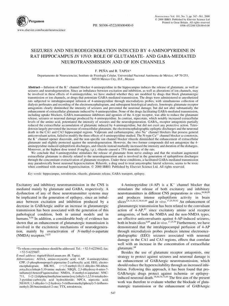
As reported previously,68 the intrahippocampal infusion of35 mM 4-AP induced EEG seizures characterized by aninitial hypersynchronic activity followed by trains of high-amplitude spikes (Fig. 1). The latency to the ®rst dischargewas 12.8^ 1.07 min and the mean duration of the discharges,once established, was 59.8^ 4.5 s (Fig. 3c).
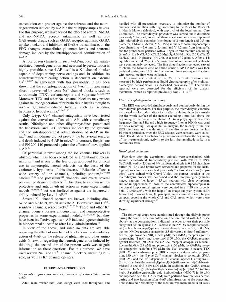
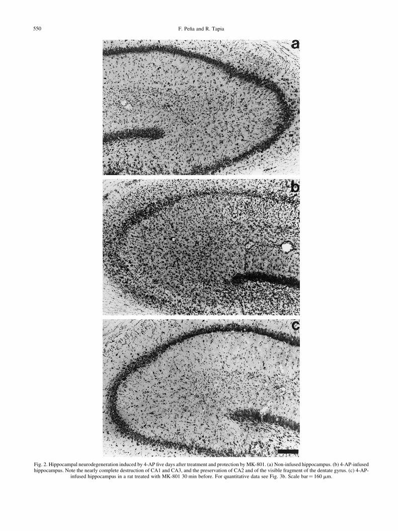
Also as described previously,68 the appearance of EEGdischarges was accompanied by an elevation of extracellularglutamate, which in the present experiments reached a valuethreefold greater than the basal concentration of the aminoacid (Fig. 3a), and by a notable increase in extracellularGABA levels, from practically nil values to 4.2 ^0.34 pmol/10 ml (Fig. 4b). The changes in the other aminoacids measured (aspartate, glutamine, alanine, glycine andtaurine) were similar to those observed in our previouscommunication68 and are not shown here. Five days afterthe experiment, rats treated with 4-AP showed a completeneurodegeneration of CA1 and CA3 pyramidal neurons,with little or no damage in CA2 and the dentate gyrus (Figs2b, 3b).
The i.p. administration of MK-801 30 min before 4-APnotably reduced the intensity (Fig. 1) and the duration (79%reduction; Fig. 3c) of the EEG seizures induced by 4-AP,without affecting the latency to the ®rst discharge, whichwas 14.6^ 0.68 min. This protective effect was more evidenton the high-amplitude spike phase of the discharges, while thehypersynchronic activity was less affected. MK-801 did notsigni®cantly modify the 4-AP-induced increase in extra-cellular glutamate concentration (Fig. 3a), but completelyprevented the neurodegeneration induced by the drug, inboth the CA1 and CA3 regions (Figs 2c, 3b).
Similar protective effects against seizures and neuro-degeneration to those exerted by i.p. MK-801 were observedwhen the other competitive NMDA receptor antagonist used,CPP, or the non-NMDA receptor antagonist, NBQX, wereinfused together with 4-AP (Fig. 3). These drugs did notalter the latency of 4-AP-induced seizures (15.2^ 1.52 minwith CPP and 13.4^ 0.94 min with NBQX), but reduced theirintensity (not shown) and their duration (59% and 62% reduc-tions, respectively; Fig. 3c). The protection against neuro-degeneration produced by CPP and NBQX was highlysigni®cant, although less notable than that observed withMK-801 (Fig. 3b). In a different group of rats, the co-applicationof CPP and NBQX together did not potentiate the protectiveaction of these drugs by themselves (n� 4; results notshown).
None of the glutamate receptor antagonists studied had anyeffect on the enhancement of extracellular glutamate (Fig. 3a)or on the changes in other amino acids induced by 4-AP.
Effect of GABAergic drugs and GABA receptor blockers
Because the pro-GABAergic drugs used included inhibitorsof GABA transport, it was important to assess whether theymodify the extracellular concentration of amino acids. In thegroups of rats treated with these drugs alone, only NPCA andto a lesser extent NNC-711 produced an increase in GABA,which reached peak values of 18.2 and 3.16 pmol/10 ml,respectively (Fig. 4); the other amino acids measured, includ-ing glutamate, were not affected at all by these drugs (notshown). None of the pro-GABAergic drugs tested producedany alteration in the EEG or in neuronal morphology.
As shown in Fig. 4a and b, when NPCA or NNC-711 were
Mechanisms of 4-AP excitotoxicity in vivo 549
Fig. 1. EEG seizure activity induced by 4-AP and protection by MK-801. Traces are representative of the activity before 4-AP (basal) and of the changesobserved once the seizure activity was stabilized. MK-801 was administered i.p. 30 min before 4-AP infusion. See Fig. 3c for quantitative evaluation.
F. PenÄa and R. Tapia550
Fig. 2. Hippocampal neurodegeneration induced by 4-AP ®ve days after treatment and protection by MK-801. (a) Non-infused hippocampus. (b) 4-AP-infusedhippocampus. Note the nearly complete destruction of CA1 and CA3, and the preservation of CA2 and of the visible fragment of the dentate gyrus. (c) 4-AP-
infused hippocampus in a rat treated with MK-801 30 min before. For quantitative data see Fig. 3b. Scale bar� 160 mm.
administered with 4-AP the increase of extracellular GABAlevels was potentiated, so that the peak levels attained wereapproximately the sum of the action of the drugs alone. Incontrast, with the other pro-GABAergic drugs tested, muscimol,isoguvacine and AOAA, no signi®cant modi®cation ofGABA besides that induced by 4-AP was observed (Fig. 4b).
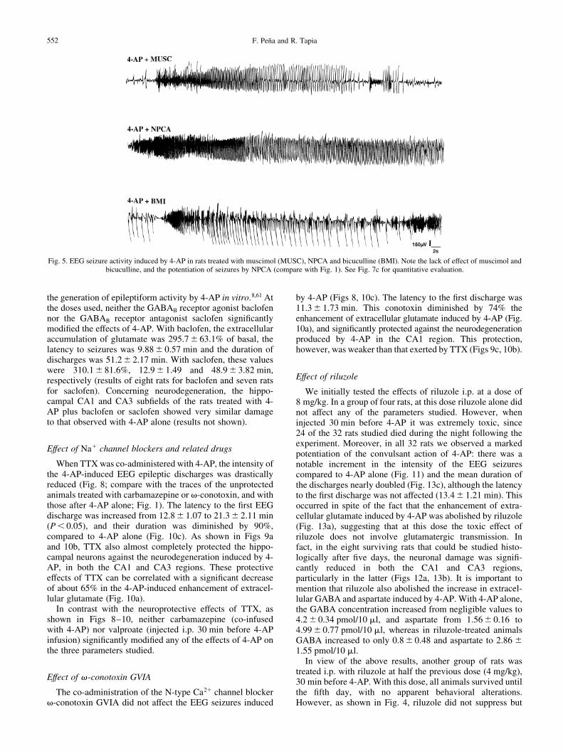
Neither NNC-711 nor AOAA, muscimol or isoguvacinesigni®cantly modi®ed the latency, intensity and duration ofthe 4-AP-induced EEG seizures. In contrast, surprisingly,NPCA increased by 40% the mean duration of the EEGdischarges (Figs 5, 7c) without affecting the latency(12.4^ 1.57 min). This potentiation of the convulsant effectof 4-AP by NPCA could be due to a depolarizing action of theexcess extracellular GABA, since it has been shown inhippocampal slices4,44,57,69 that, during epileptiform activityinduced by 4-AP, GABA depolarizes neurons through theactivation of GABAA receptors. To test this possibility, weapplied the GABAA receptor antagonist bicuculline, at asubconvulsant dose that did not alter the EEG, on theNPCA-induced potentiation of seizures. As shown in Fig.7c, this treatment did not reduce the potentiation. Similarly,neither bicuculline (Fig. 5) nor picrotoxin, another GABAA
receptor blocker, affected per se the 4-AP-induced EEG
discharges (Fig. 7c), indicating that the enhancement of extra-cellular GABA produced by 4-AP is not a determining factorin the induction of seizures. In agreement with this interpreta-tion, it was observed that both bicuculline and picrotoxinsigni®cantly reduced the latency to seizures (from 12.8 to8.77^ 0.42 and 7.25^ 0.41 min, respectively, P , 0.05).
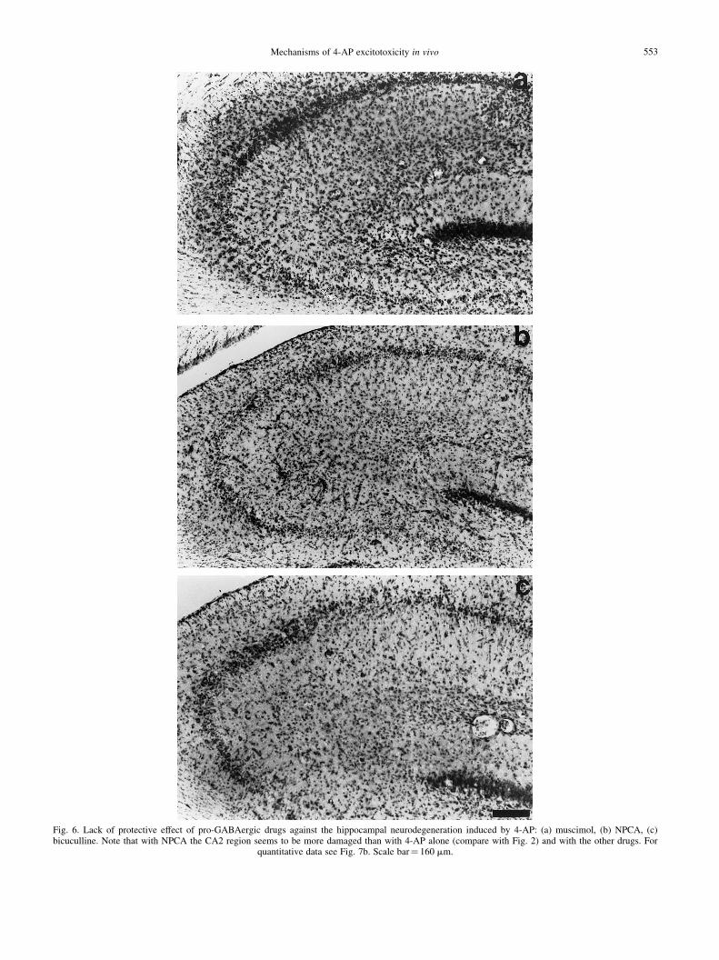
None of the pro-GABAergic drugs tested had any protec-tive effect on the neurodegeneration induced by 4-AP (Figs 6,7b). On the contrary, and similarly to its effect on the EEGdischarges, NPCA seemed to potentiate such damagingaction, as indicated not only by the decrease in the numberof healthy neurons in CA1 and CA3 (Fig. 7b), but also byproducing a signi®cant decrement of approximately 44% inthe number of healthy cells in the CA2 region, with respect to4-AP alone (P , 0.05; see Fig. 6b).
In agreement with the lack of protective effect of the pro-GABAergic drugs used on the seizures and neurodegenera-tion induced by 4-AP, these compounds did not prevent theextracellular accumulation of glutamate (Fig. 7a) or thechanges induced by 4-AP in other amino acids. In contrast,the two GABAA receptor antagonists, bicuculline and picro-toxin, reduced the accumulation of extracellular glutamate. Inaddition, bicuculline also diminished the accumulation ofextracellular aspartate and GABA and the reduction of gluta-mine induced by 4-AP58 (not shown). We cannot offer anexplanation for this ®nding, but one possibility is that thealready mentioned depolarizing action resulting fromGABAA receptor activation may be blocked by the antagon-ists, thus decreasing the neuronal excitation and consequentlythe release of glutamate.
We also assessed the possible role of GABAB receptors,since some reports suggest that these receptors are involved in
Mechanisms of 4-AP excitotoxicity in vivo 551
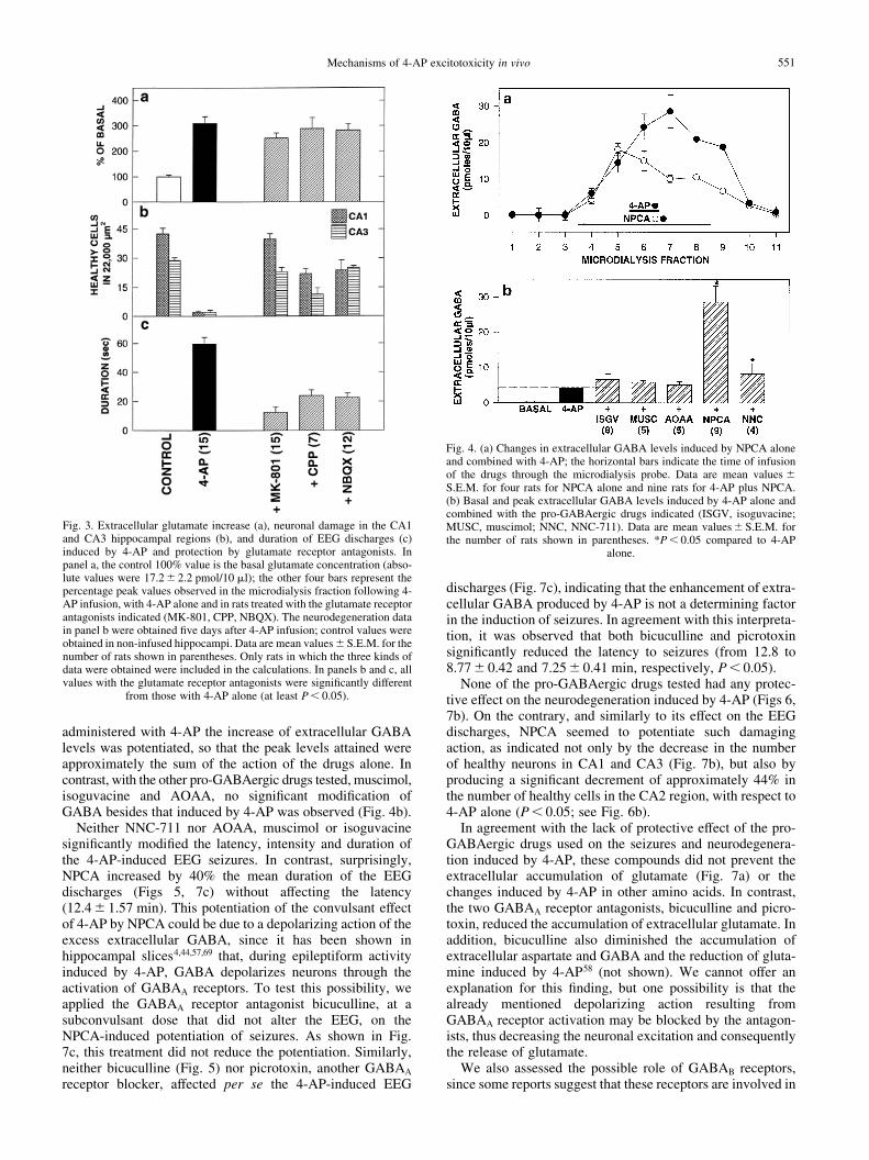
Fig. 3. Extracellular glutamate increase (a), neuronal damage in the CA1and CA3 hippocampal regions (b), and duration of EEG discharges (c)induced by 4-AP and protection by glutamate receptor antagonists. Inpanel a, the control 100% value is the basal glutamate concentration (abso-lute values were 17.2^ 2.2 pmol/10 ml); the other four bars represent thepercentage peak values observed in the microdialysis fraction following 4-AP infusion, with 4-AP alone and in rats treated with the glutamate receptorantagonists indicated (MK-801, CPP, NBQX). The neurodegeneration datain panel b were obtained ®ve days after 4-AP infusion; control values wereobtained in non-infused hippocampi. Data are mean values^S.E.M. for thenumber of rats shown in parentheses. Only rats in which the three kinds ofdata were obtained were included in the calculations. In panels b and c, allvalues with the glutamate receptor antagonists were signi®cantly different
from those with 4-AP alone (at least P , 0.05).
Fig. 4. (a) Changes in extracellular GABA levels induced by NPCA aloneand combined with 4-AP; the horizontal bars indicate the time of infusionof the drugs through the microdialysis probe. Data are mean values ^S.E.M. for four rats for NPCA alone and nine rats for 4-AP plus NPCA.(b) Basal and peak extracellular GABA levels induced by 4-AP alone andcombined with the pro-GABAergic drugs indicated (ISGV, isoguvacine;MUSC, muscimol; NNC, NNC-711). Data are mean values^ S.E.M. forthe number of rats shown in parentheses. *P , 0.05 compared to 4-AP
alone.
the generation of epileptiform activity by 4-AP in vitro.8,61 Atthe doses used, neither the GABAB receptor agonist baclofennor the GABAB receptor antagonist saclofen signi®cantlymodi®ed the effects of 4-AP. With baclofen, the extracellularaccumulation of glutamate was 295.7^ 63.1% of basal, thelatency to seizures was 9.88^ 0.57 min and the duration ofdischarges was 51.2^ 2.17 min. With saclofen, these valueswere 310.1^ 81.6%, 12.9^ 1.49 and 48.9^ 3.82 min,respectively (results of eight rats for baclofen and seven ratsfor saclofen). Concerning neurodegeneration, the hippo-campal CA1 and CA3 sub®elds of the rats treated with 4-AP plus baclofen or saclofen showed very similar damageto that observed with 4-AP alone (results not shown).
Effect of Na1 channel blockers and related drugs
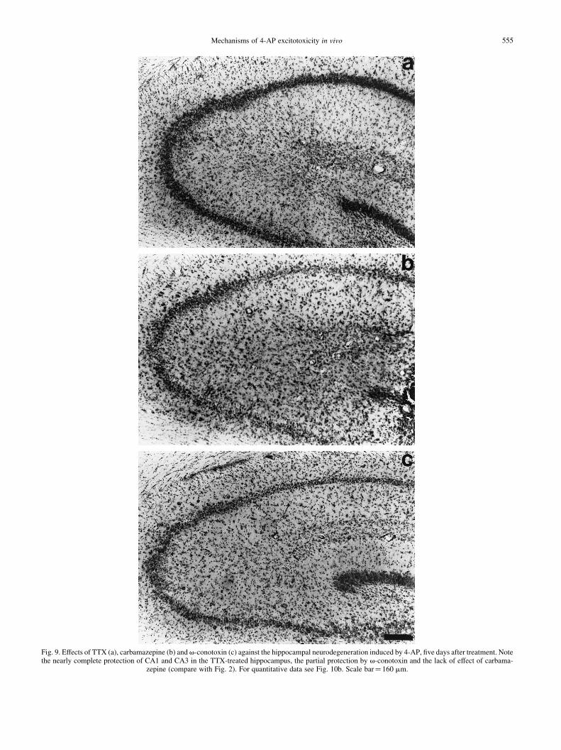
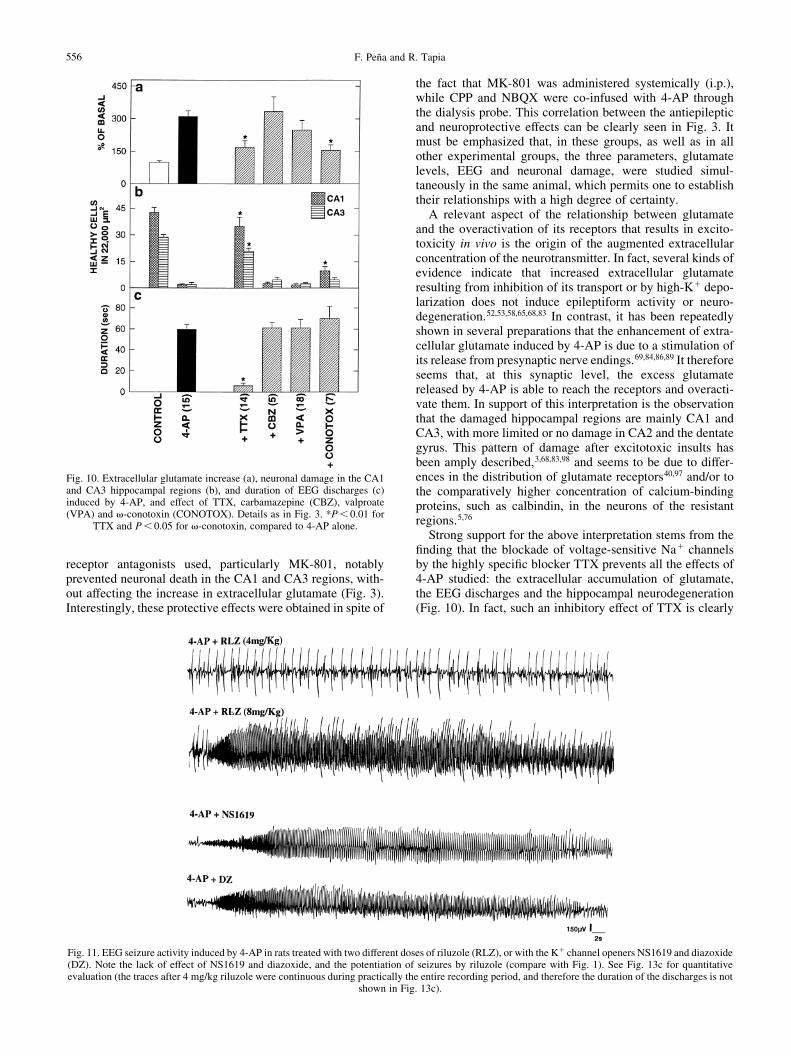
When TTX was co-administered with 4-AP, the intensity ofthe 4-AP-induced EEG epileptic discharges was drasticallyreduced (Fig. 8; compare with the traces of the unprotectedanimals treated with carbamazepine or v-conotoxin, and withthose after 4-AP alone; Fig. 1). The latency to the ®rst EEGdischarge was increased from 12.8^ 1.07 to 21.3^ 2.11 min(P , 0.05), and their duration was diminished by 90%,compared to 4-AP alone (Fig. 10c). As shown in Figs 9aand 10b, TTX also almost completely protected the hippo-campal neurons against the neurodegeneration induced by 4-AP, in both the CA1 and CA3 regions. These protectiveeffects of TTX can be correlated with a signi®cant decreaseof about 65% in the 4-AP-induced enhancement of extracel-lular glutamate (Fig. 10a).
In contrast with the neuroprotective effects of TTX, asshown in Figs 8±10, neither carbamazepine (co-infusedwith 4-AP) nor valproate (injected i.p. 30 min before 4-APinfusion) signi®cantly modi®ed any of the effects of 4-AP onthe three parameters studied.
Effect of v-conotoxin GVIA
The co-administration of the N-type Ca21 channel blockerv-conotoxin GVIA did not affect the EEG seizures induced
by 4-AP (Figs 8, 10c). The latency to the ®rst discharge was11.3^ 1.73 min. This conotoxin diminished by 74% theenhancement of extracellular glutamate induced by 4-AP (Fig.10a), and signi®cantly protected against the neurodegenerationproduced by 4-AP in the CA1 region. This protection,however, was weaker than that exerted by TTX (Figs 9c, 10b).
Effect of riluzole
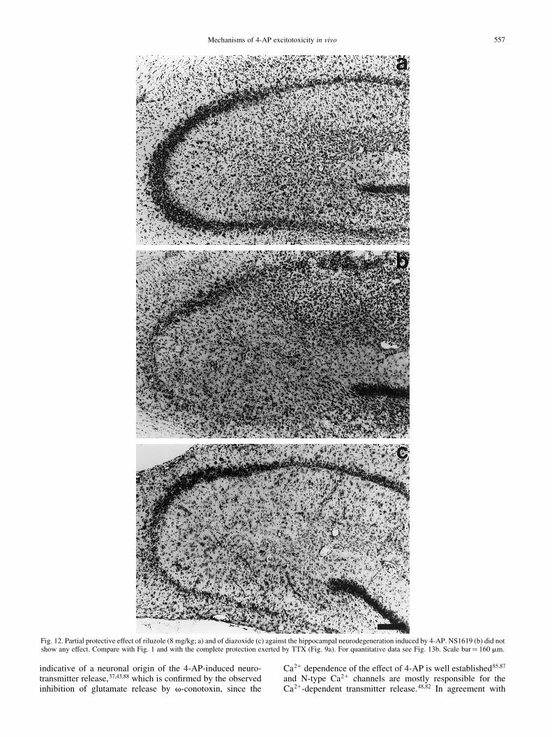
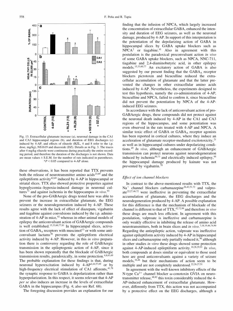
We initially tested the effects of riluzole i.p. at a dose of8 mg/kg. In a group of four rats, at this dose riluzole alone didnot affect any of the parameters studied. However, wheninjected 30 min before 4-AP it was extremely toxic, since24 of the 32 rats studied died during the night following theexperiment. Moreover, in all 32 rats we observed a markedpotentiation of the convulsant action of 4-AP: there was anotable increment in the intensity of the EEG seizurescompared to 4-AP alone (Fig. 11) and the mean duration ofthe discharges nearly doubled (Fig. 13c), although the latencyto the ®rst discharge was not affected (13.4^ 1.21 min). Thisoccurred in spite of the fact that the enhancement of extra-cellular glutamate induced by 4-AP was abolished by riluzole(Fig. 13a), suggesting that at this dose the toxic effect ofriluzole does not involve glutamatergic transmission. Infact, in the eight surviving rats that could be studied histo-logically after ®ve days, the neuronal damage was signi®-cantly reduced in both the CA1 and CA3 regions,particularly in the latter (Figs 12a, 13b). It is important tomention that riluzole also abolished the increase in extracel-lular GABA and aspartate induced by 4-AP. With 4-AP alone,the GABA concentration increased from negligible values to4.2^ 0.34 pmol/10 ml, and aspartate from 1.56^ 0.16 to4.99^ 0.77 pmol/10 ml, whereas in riluzole-treated animalsGABA increased to only 0.8^ 0.48 and aspartate to 2.86 ^1.55 pmol/10 ml.
In view of the above results, another group of rats wastreated i.p. with riluzole at half the previous dose (4 mg/kg),30 min before 4-AP. With this dose, all animals survived untilthe ®fth day, with no apparent behavioral alterations.However, as shown in Fig. 4, riluzole did not suppress but
F. PenÄa and R. Tapia552
Fig. 5. EEG seizure activity induced by 4-AP in rats treated with muscimol (MUSC), NPCA and bicuculline (BMI). Note the lack of effect of muscimol andbicuculline, and the potentiation of seizures by NPCA (compare with Fig. 1). See Fig. 7c for quantitative evaluation.
Mechanisms of 4-AP excitotoxicity in vivo 553
Fig. 6. Lack of protective effect of pro-GABAergic drugs against the hippocampal neurodegeneration induced by 4-AP: (a) muscimol, (b) NPCA, (c)bicuculline. Note that with NPCA the CA2 region seems to be more damaged than with 4-AP alone (compare with Fig. 2) and with the other drugs. For
quantitative data see Fig. 7b. Scale bar� 160 mm.

altered the pattern of the EEG discharges induced by 4-AP.No initial hypersynchronic activity occurred and thefrequency of the high-amplitude spikes decreased. Never-theless, once established, the trains of these spikes werecontinuous during the rest of the experiment, so that the dura-tion of the discharges could not be calculated. The latency tothe ®rst discharge increased to 20.8^ 1.73 min. At this lowdose, riluzole also signi®cantly blocked the 4-AP-induced
increase in extracellular glutamate, albeit to a lesser extentthan after 8 mg/kg (Fig. 13a), and protected against the hippo-campal damage similarly to the latter (Fig. 13b). The increasein extracellular GABA and aspartate induced by 4-AP wasalso reduced in the animals treated with 4 mg/kg riluzole(64% decrease for GABA and 33% for aspartate, comparedto 4-AP alone).
Effect of K1 channel openers
The Ca21-dependent K1 channel opener NS1619 did notsigni®cantly modify any of the effects of 4-AP studied (Figs12, 13), whereas the ATP-sensitive K1 channel opener diaz-oxide did not suppress the seizures but inhibited by 55% theextracellular glutamate increase and slightly protected againstneuronal death in both the CA1 and CA3 regions (Figs 12,13).
DISCUSSION
Effect of glutamate receptor antagonists and GABAergicdrugs
The results of the present work strongly support thepreviously proposed close relationship between an increasedlevel of synaptic extracellular glutamate, seizures and neuro-degeneration, in the hippocampus in vivo,68,83 after 4-APtreatment. Since such a postulation implies that an overacti-vation of glutamate receptors is responsible for the epilepto-genic and neurotoxic effects of 4-AP, it would be expectedthat the blockade of glutamate receptors should protectagainst such effects of 4-AP. This expectation was ful®lled,since we observed that both MK-801 and CPP, two antagon-ists of NMDA receptors, as well as NBQX, an antagonist ofnon-NMDA receptors, clearly inhibited the 4-AP-inducedconvulsions. Such anticonvulsant action is in agreementwith previous results obtained with a bolus injection of 4-AP in the hippocampus25 or in the lateral cerebral ventricle,59
as well as with the protection by the non-NMDA receptorantagonist GIKY-52466 against seizures induced by thelocal application of 4-AP on the somatosensory cortex.6
Besides their anticonvulsant effect, the three glutamate
F. PenÄa and R. Tapia554
Fig. 7. Extracellular glutamate increase (a), neuronal damage in the CA1and CA3 hippocampal regions (b), and duration of EEG discharges (c),induced by 4-AP, and effects of pro-GABAergic and anti-GABAergicdrugs. Details as in Fig. 3. PTX, picrotoxin. Data are mean values^S.E.M.for the number of rats indicated in parentheses. *P , 0.05 compared to
4-AP alone.
Fig. 8. Protection by TTX against the EEG seizure activity induced by 4-AP, and lack of protection by carbamazepine (CBZ) and v-conotoxin. Traces arerepresentative of the changes observed once the seizure activity was stabilized. Drugs were co-infused with 4-AP through the microdialysis probe. See Fig. 10c
for quantitative evaluation.
Mechanisms of 4-AP excitotoxicity in vivo 555
Fig. 9. Effects of TTX (a), carbamazepine (b) and v-conotoxin (c) against the hippocampal neurodegeneration induced by 4-AP, ®ve days after treatment. Notethe nearly complete protection of CA1 and CA3 in the TTX-treated hippocampus, the partial protection by v-conotoxin and the lack of effect of carbama-
zepine (compare with Fig. 2). For quantitative data see Fig. 10b. Scale bar� 160 mm.
receptor antagonists used, particularly MK-801, notablyprevented neuronal death in the CA1 and CA3 regions, with-out affecting the increase in extracellular glutamate (Fig. 3).Interestingly, these protective effects were obtained in spite of
the fact that MK-801 was administered systemically (i.p.),while CPP and NBQX were co-infused with 4-AP throughthe dialysis probe. This correlation between the antiepilepticand neuroprotective effects can be clearly seen in Fig. 3. Itmust be emphasized that, in these groups, as well as in allother experimental groups, the three parameters, glutamatelevels, EEG and neuronal damage, were studied simul-taneously in the same animal, which permits one to establishtheir relationships with a high degree of certainty.
A relevant aspect of the relationship between glutamateand the overactivation of its receptors that results in excito-toxicity in vivo is the origin of the augmented extracellularconcentration of the neurotransmitter. In fact, several kinds ofevidence indicate that increased extracellular glutamateresulting from inhibition of its transport or by high-K1 depo-larization does not induce epileptiform activity or neuro-degeneration.52,53,58,65,68,83 In contrast, it has been repeatedlyshown in several preparations that the enhancement of extra-cellular glutamate induced by 4-AP is due to a stimulation ofits release from presynaptic nerve endings.69,84,86,89 It thereforeseems that, at this synaptic level, the excess glutamatereleased by 4-AP is able to reach the receptors and overacti-vate them. In support of this interpretation is the observationthat the damaged hippocampal regions are mainly CA1 andCA3, with more limited or no damage in CA2 and the dentategyrus. This pattern of damage after excitotoxic insults hasbeen amply described,3,68,83,98 and seems to be due to differ-ences in the distribution of glutamate receptors40,97 and/or tothe comparatively higher concentration of calcium-bindingproteins, such as calbindin, in the neurons of the resistantregions.5,76
Strong support for the above interpretation stems from the®nding that the blockade of voltage-sensitive Na1 channelsby the highly speci®c blocker TTX prevents all the effects of4-AP studied: the extracellular accumulation of glutamate,the EEG discharges and the hippocampal neurodegeneration(Fig. 10). In fact, such an inhibitory effect of TTX is clearly
F. PenÄa and R. Tapia556
Fig. 10. Extracellular glutamate increase (a), neuronal damage in the CA1and CA3 hippocampal regions (b), and duration of EEG discharges (c)induced by 4-AP, and effect of TTX, carbamazepine (CBZ), valproate(VPA) and v-conotoxin (CONOTOX). Details as in Fig. 3. *P , 0.01 for
TTX and P , 0.05 for v-conotoxin, compared to 4-AP alone.
Fig. 11. EEG seizure activity induced by 4-AP in rats treated with two different doses of riluzole (RLZ), or with the K1 channel openers NS1619 and diazoxide(DZ). Note the lack of effect of NS1619 and diazoxide, and the potentiation of seizures by riluzole (compare with Fig. 1). See Fig. 13c for quantitativeevaluation (the traces after 4 mg/kg riluzole were continuous during practically the entire recording period, and therefore the duration of the discharges is not
shown in Fig. 13c).
indicative of a neuronal origin of the 4-AP-induced neuro-transmitter release,37,43,88 which is con®rmed by the observedinhibition of glutamate release by v-conotoxin, since the
Ca21 dependence of the effect of 4-AP is well established85,87
and N-type Ca21 channels are mostly responsible for theCa21-dependent transmitter release.48,82 In agreement with
Mechanisms of 4-AP excitotoxicity in vivo 557
Fig. 12. Partial protective effect of riluzole (8 mg/kg; a) and of diazoxide (c) against the hippocampal neurodegeneration induced by 4-AP. NS1619 (b) did notshow any effect. Compare with Fig. 1 and with the complete protection exerted by TTX (Fig. 9a). For quantitative data see Fig. 13b. Scale bar� 160 mm.
these observations, it has been reported that TTX preventsboth the release of neurotransmitter amino acids23,69 and theepileptiform activity62,69 induced by 4-AP in hippocampal orstriatal slices. TTX also showed protective properties againsthypoglycemia±hypoxia-induced damage in neuronal cul-tures71 and against ischemia in the hippocampus in vivo.93
None of the pro-GABAergic drugs tested here was able toprevent the increase in extracellular glutamate, the EEGseizures or the neurodegeneration induced by 4-AP. Theseresults agree with the lack of effect of diazepam, vigabatrinand tiagabine against convulsions induced by the i.p. admini-stration of 4-AP in mice,92 whereas in other animal models ofepilepsy the anticonvulsant action of GABAergic compoundsis well established.15,33,60,77,81 In hippocampal slices, activa-tion of GABAA receptors with muscimol13 or with some anti-convulsant lactams38 prevents the epileptiform electricalactivity induced by 4-AP. However, in this in vitro prepara-tion there is controversy regarding the role of GABAergictransmission in the epileptogenic action of 4-AP, since ithas been shown repeatedly that the blockade of GABAergictransmission results, paradoxically, in some protection.4,8,61,69
The probable explanation for these ®ndings is that, duringneuronal hyperexcitation induced by 4-AP4,44,57,69 or byhigh-frequency electrical stimulation of CA1 afferents,41,78
the synaptic response to GABA is depolarization rather thanhyperpolarization. In this respect, it seems relevant that 4-APper se also induces an increase in the levels of extracellularGABA in the hippocampus (Fig. 4; also see Ref. 68).
The foregoing discussion provides an explanation for our
®nding that the infusion of NPCA, which largely increasedthe concentration of extracellular GABA, enhanced the inten-sity and duration of EEG seizures, as well as the neuronaldamage, produced by 4-AP. In support of this interpretation isthe potentiation of the depolarizing action of GABA inhippocampal slices by GABA uptake blockers such asNPCA2 or tiagabine.41 Also in agreement with thispostulation is the paradoxical proconvulsant action in vivoof some GABA uptake blockers, such as NPCA, NNC-711,tiagabine and 2,4-diaminobutyric acid, in other epilepsymodels.15,33,60,77 An excitatory action of GABA is alsosuggested by our present ®nding that the GABAA receptorblockers picrotoxin and bicuculline reduced the extra-cellular accumulation of glutamate and that the latter pre-vented the changes in other extracellular amino acidsinduced by 4-AP. Nevertheless, the experiments designed totest this hypothesis, namely the co-administration of 4-AP,bicuculline and NPCA, failed to con®rm it, since bicucullinedid not prevent the potentiation by NPCA of the 4-AP-induced EEG seizures.
In accordance with the lack of anticonvulsant action of pro-GABAergic drugs, these compounds did not protect againstthe neuronal death induced by 4-AP in the CA1 and CA3regions of the hippocampus, and some potentiation waseven observed in the rats treated with 4-AP and NPCA. Asimilar toxic effect of GABA or GABAA receptor agonistshas been reported in cortical cultures, where they induce anacceleration of glutamate receptor-mediated excitotoxicity,21
as well as in hippocampal cultures under depolarizing condi-tions.49 In vivo, although an enhancement of GABAergictransmission can protect neurons against neurodegenerationinduced by ischemia50,73 and electrically induced epilepsy,95
the hippocampal damage produced by kainate was notprevented by vigabatrin.70
Effect of ion channel blockers
In contrast to the above-mentioned results with TTX, theNa1 channel blockers carbamazepine26,42,51,72 and valpro-ate22,27,46,72 were ineffective in preventing the extracellularaccumulation of glutamate, the EEG discharges and theneurodegeneration produced by 4-AP. A possible explanationfor this difference is that the mechanism of blockade of thechannel is different to that of TTX,42,72,99 and therefore in vivothese drugs are much less ef®cient. In agreement with thispostulation, valproate is ineffective and carbamazepine isonly weakly effective in inhibiting the release of amino acidneurotransmitters, both in brain slices and in vivo.7,18,19,46,74,90
Regarding the antiepileptic action, valproate was ineffectiveagainst epileptiform activity induced by 4-AP in hippocampalslices and carbamazepine only partially reduced it,96 althoughin other studies in vitro these drugs showed some protectionagainst 4-AP-induced epileptiform activity.10,28,29,91 In vivo,both compounds at doses similar or equivalent to those usedhere are good anticonvulsants against a variety of seizuremodels,16,46 but their mechanisms of action seem to bemultiple and are not completely understood.22,26,27,51
In agreement with the well-known inhibitory effects of theN-type Ca21 channel blocker v-conotoxin GVIA on neuro-transmitter release,48,82 this toxin considerably reduced the 4-AP-induced enhancement of extracellular glutamate. How-ever, differently from TTX, this action was not accompaniedby a protective effect on the EEG discharges, although a
F. PenÄa and R. Tapia558
Fig. 13. Extracellular glutamate increase (a), neuronal damage in the CA1and CA3 hippocampal regions (b), and duration of EEG discharges (c)induced by 4-AP, and effects of riluzole (RZL, 4 and 8 refer to the i.p.dose, mg/kg), NS1619 and diazoxide (DZ). Details as in Fig. 3. The tracesafter 4 mg/kg riluzole were continuous during practically the entire record-ing period, and therefore the duration of the discharges is not shown. Dataare mean values^S.E.M. for the number of rats indicated in parentheses.
*P , 0.05 compared to 4-AP alone.
signi®cant protection against neurodegeneration was observed,particularly in the CA1 region. In order to reconcile theseresults with those with TTX, we propose that, as we havesuggested previously,68 the convulsant effect of 4-AP involvesan increased excitatory glutamate-mediated transmissioncombined with an increment in neuronal ®ring frequency.TTX should be expected to inhibit both types of action,whereas the action of v-conotoxin is restricted to the pre-synaptic region. This interpretation may also account for theresults obtained with riluzole and with diazoxide, which,similarly to v-conotoxin, inhibited glutamate release butwere unable to suppress the EEG seizures and neuronaldeath. Interestingly, however, all three drugs partiallyprotected against neurodegeneration, suggesting that thiseffect of 4-AP is closely related to the increased extracellularconcentration of glutamate, as suggested previously.68
Some reports have shown protective effects of riluzoleagainst ischemia- or nitropropionic acid-induced neurodegen-eration.36,63,66 However, riluzole failed to protect against theconvulsant action of i.c.v. 4-AP.80 In this respect, our ®ndingthat riluzole at the highest dose tested (8 mg/kg) potentiatedthe convulsant action of 4-AP and produced a high mortalityof the rats is worth noting, insofar as riluzole is practically theonly known drug with some bene®cial action in the treatmentof amyotrophic lateral sclerosis.47 Moreover, half the dose ofriluzole, although not inducing mortality, also seemed topotentiate the 4-AP-induced EEG discharges, which becamecontinuous. Although these doses are higher than those usedclinically (about 1±3 mg/kg orally on a daily basis), theireffect was observed after a single administration. Hence, thepresent results indicate that the clinical use of riluzole shouldbe considered with caution. In addition, as shown in thepresent work and has been stressed previously,64 riluzolereduces the extracellular accumulation not only of glutamate,but also of GABA and aspartate, and the possible adverse
consequences of this effect cannot be ignored. Finally, rilu-zole acts on a great variety of ion channels, including Na1,Ca21 and K1 channels,66,79,100 which may add other undesir-able side-effects.
CONCLUSIONS
The ®ndings of the present work indicate that an overacti-vation of glutamate receptors, mainly of the NMDA type, isinvolved in the generation of epileptiform activity and in theneurodegeneration produced by the infusion of 4-AP in rathippocampus in vivo, and that ionic channels play a relevantrole. In addition, our data clearly show that the glutamate-mediated effects of 4-AP are a consequence of an increasedrelease of the transmitter from excitatory nerve endings. Suchincreased release is directly related to the neurodegenerationof hippocampal regions CA1 and CA3. Since increased extra-cellular glutamate levels by inhibition of its transport or byK1 depolarization do not result in excitotoxicity,52,53,65,68 weconclude that the site of origin of augmented extracellularglutamate is determinant for the overactivation of glutamatereceptors. We also conclude that, under conditions of hippo-campal hyperexcitation, an enhancement of GABAergictransmission may result in a potentiation of excitotoxicity,possibly via a depolarizing action mediated by GABAA
receptor activation. Finally, although riluzole did reduce therelease of glutamate and this partially protected againstneurodegeneration, this compound may be toxic when com-bined with neuronal hyperexcitation, and thus its clinical useshould be considered with caution.
AcknowledgementsÐThis work was supported in part by DGAPA,UNAM (IN207598) and CONACYT (31750-N). We wish to thankCONACYT for a fellowship to F.P., Federico Jandete forhistological procedures and Arturo Franco for photographic artwork.
REFERENCES
1. Abele A. E. and Miller R. J. (1990) Potassium channel activators abolish excitotoxicity in cultured hippocampal pyramidal neurons. Neurosci. Lett. 115,195±200.
2. Alger B. E. and Nicoll R. A. (1982) Pharmacological evidence for two kinds of GABA receptor on rat hippocampal pyramidal cells studied in vitro.J. Physiol. 328, 125±141.
3. Arias C., Arrieta I., Massieu L. and Tapia R. (1997) Neuronal damage and MAP2 changes induced by the glutamate transport inhibitor dihydrokainateand by kainate in rat hippocampus in vivo. Expl Brain Res. 116, 467±476.
4. Avoli M., Barbarosie M., LuÈcke A., Nagao T., Lopantsev V. and KoÈhling R. (1996) Synchronous GABA-mediated potentials and epileptiformdischarges in the rat limbic system in vitro. J. Neurosci. 16, 3912±3924.
5. Baimbridge K. G. and Miller J. J. (1982) Immunohistochemical localization of calcium-binding protein in the cerebellum, hippocampal formation andolfactory bulb of the rat. Brain Res. 245, 223±229.
6. Barna B., SzaÂsz A., VilaÂgi I. and Szente M. (2000) Anticonvulsive effect of AMPA receptor antagonist GYKI 52466 on 4-aminopyridine-inducedcortical activity in rat. Brain Res. Bull. 51, 241±248.
7. Biggs C. S., Pearce B. R., Fowler L. J. and Whitton P. S. (1992) The effect of sodium valproate on extracellular GABA and other amino acids in the ratventral hippocampus: an in vivo microdialysis study. Brain Res. 594, 138±142.
8. Bijak M. and Misgeld U. (1996) Suppression by GABAB receptors of 4-aminopyridine-induced hyperactivity in guinea-pig neurons. Neurosci. Lett. 205,49±52.
9. Bradford H. F. (1995) Glutamate, GABA and epilepsy. Prog. Neurobiol. 47, 477±511.10. BruÈckner C. and Heinemann U. (2000) Effects of standard anticonvulsant drugs on different patterns of epileptiform discharges induced by 4-
aminopyridine in combined entorhinal cortex±hippocampal slices. Brain Res. 859, 15±20.11. Cai S., Garneau L. and Sauve R. (1998) Single-channel characterization of the pharmacological properties of the K(Ca21) channel of intermediate
conductance in bovine aortic endothelial cells. J. Membrane Biol. 163, 147±158.12. Centonze D., Calabresi P., Pisani A., Marinelli S., Mar®a G. A. and Bernardi G. (1998) Electrophysiology of the neuroprotective agent riluzole on
striatal spiny neurons. Neuropharmacology 37, 1063±1070.13. Chesnut T. J. and Swann J. W. (1990) Suppression of 4-aminopyridine-induced epileptogenesis by the GABAA agonist muscimol. Epilepsy Res. 5, 8±17.14. Choi D. W. (1988) Glutamate neurotoxicity and diseases of the nervous system. Neuron 1, 623±634.15. Coenen A. M. L., Blezer E. H. M. and van Luijtelaar E. L. J. M. (1995) Effects of GABA-uptake inhibitor tiagabine on electroencephalogram, spike±
wave discharges and behaviour of rats. Epilepsy Res. 21, 89±94.16. Coleman H., Yamaguchi S. and Rogawski M. A. (1992) Protection against dendrotoxin-induced clonic seizures in mice by anticonvulsant drugs. Brain
Res. 575, 138±142.17. Cramer C. L., Stagnitto M. L., Knowles M. A. and Palmer G. C. (1994) Kainic acid and 4-aminopyridine seizure models in mice: evaluation of ef®cacy
of antiepileptic agents and calcium antagonists. Life Sci. 54, 271±275.
Mechanisms of 4-AP excitotoxicity in vivo 559
18. de Boer T., Stoof J. C. and van Duijn H. (1982) The effects of convulsant and anticonvulsant drugs on the release of radiolabeled GABA, glutamate,noradrenaline, serotonin and acetylcholine from rat cortical slices. Brain Res. 253, 153±160.
19. Dixon J. F. and Hokin L. E. (1997) The antibipolar drug valproate mimics lithium in stimulating glutamate release and inositol 1,4,5-trisphosphateaccumulation in brain cortex slices but not accumulation of inositol monophosphates and bisphosphates. Proc. natn. Acad. Sci. USA 94, 4757±4760.
20. Doczi J., Banczerowski-Pelyhe I., Barna B. and VilaÂgi I. (1999) Effect of glutamate receptor antagonist (GYKI 52466) on 4-aminopyridine-inducedseizure activity developed in rat cortical slices. Brain Res. Bull. 49, 435±440.
21. ErdoÈ S. L., Michler A. and Wolff J. R. (1991) GABA accelerates excitotoxic cell death in cortical cultures: protection by blockers of GABA-gatedchloride channels. Brain Res. 542, 254±258.
22. Fariello R. G., Varasi M. and Smith M. C. (1995) Valproic acid mechanism of action. In Antiepileptic Drugs (eds Levy R. H., Mattson R. H. andMeldrum B. S.). Raven, New York.
23. Flores-HernaÂndez J., Galarraga E., Pineda J. C. and Bargas J. (1994) Patterns of excitatory and inhibitory synaptic transmission in the rat neostriatum asrevealed by 4-AP. J. Neurophysiol. 72, 2246±2256.
24. Fragoso-Veloz J., Massieu L., Alvarado R. and Tapia R. (1990) Seizures and wet-dog shakes induced by 4-aminopyridine, and their potentiation bynifedipine. Eur. J. Pharmac. 178, 275±284.
25. Fragoso-Veloz J. and Tapia R. (1992) NMDA receptor antagonists protect against seizures and wet-dog shakes induced by 4-aminopyridine. Eur. J.Pharmac. 221, 275±280.
26. Fromm G. H. (1992) Antiepileptic actions of carbamazepine. In Drugs for Control of Epilepsy: Actions on Neuronal Networks Involved in SeizureDisorders (eds Faingold C. L. and Fromm G. H.). CRC, Boca Raton, FL.
27. Fromm G. H. (1992) Antiepileptic actions of valproate. In Drugs for Control of Epilepsy: Actions on Neuronal Networks Involved in Seizure Disorders(eds Faingold C. L. and Fromm G. H.). CRC, Boca Raton, FL.
28. Fueta Y. and Avoli M. (1992) Effects of antiepileptic drugs on 4-aminopyridine-induced epileptiform activity in young and adult rat hippocampus.Epilepsy Res. 12, 207±215.
29. Fueta Y., Siniscalchi A., Tancredi V. and Avoli M. (1995) Extracellular magnesium and anticonvulsant effects of valproate in young rat hippocampus.Epilepsia 36, 404±409.
30. Gandolfo G., Gottesmann C., Bidard J. N. and Lazdunski M. (1989) Ca21 channel blockers prevent seizures induced by a class of K1 channel inhibitors.Eur. J. Pharmac. 160, 173±177.
31. Gandolfo G., Gottesmann C., Bidard J. N. and Lazdunski M. (1989) Subtypes of K1 channels differentiated by the effect of K1 channel openers upon K1
channel blocker-induced seizures. Brain Res. 495, 189±192.32. Gean P. W., Chou S. M. and Chang F. C. (1990) Epileptiform activity induced by 4-aminopyridine in rat amygdala neurons: the involvement of N-
methyl-d-aspartate receptors. Eur. J. Pharmac. 184, 213±221.33. Gonsalves S. F., Twitchell B., Harbaugh R. E., Krogsgaard-Larsen P. and Schousboe A. (1989) Anticonvulsant activity of intracerebroventricularly
administered glial GABA uptake inhibitors and other GABA mimetics in chemical seizure models. Epilepsy Res. 4, 34±41.34. Goodman Y. and Mattson M. P. (1996) K1 channel openers protect hippocampal neurons against oxidative injury and amyloid b-peptide toxicity. Brain
Res. 706, 328±332.35. Gribkoff V. K., Lum-Ragan J. T., Boissard C. G., Post-Munson D. J., Meanwell N. A., Starret J. E., Jr, Kozlowski E. S., Romine J. L., Trojnacki J. T.,
McKay M. C., Zhong J. and Dworetzky S. I. (1996) Effects of channel modulators on cloned large-conductance calcium-activated potassium channels.Molec. Pharmac. 50, 206±217.
36. Guyot M. C., Pal® S., Stutzmann J. M., MazieÁre M., Hantraye P. and Brouillet E. (1997) Riluzole protects from motor de®cits and striatal degenerationproduced by systemic 3-nitropropionic acid intoxication in rats. Neuroscience 81, 141±149.
37. Herrera-Marschitz M., You Z. B., Goiny M., Meana J. J., Silveira R., Godukhin O. V., Chen Y., Espinoza S., Pettersson E., Loidl C. F., Lubec G.,Andersson K., Nylander I., Terenius L. and Ungerstedt U. (1996) On the origin of extracellular glutamate levels monitored in the basal ganglia of the ratby in vivo microdialysis. J. Neurochem. 66, 1726±1735.
38. Hill M. W., de la Cruz M. A. M., Covey D. F. and Rothman S. M. (1999) Effects of anticonvulsant lactams on in vitro seizures in the hippocampal slicepreparation. Epilepsy Res. 37, 121±131.
39. Ingle®eld J. R., Perry J. M. and Schwartz R. D. (1995) Postischemic inhibition of GABA reuptake by tiagabine slows neuronal death in the gerbilhippocampus. Hippocampus 5, 460±468.
40. Insel T. R., Miller L. P. and Gelhard R. E. (1990) The ontogeny of excitatory amino acid receptors in rat forebrainÐI. N-Methyl-d-aspartate andquisqualate receptors. Neuroscience 35, 31±43.
41. Jackson M. F., Esplin B. and Capek R. (1999) Activity-dependent enhancement of hyperpolarizing and depolarizing g-aminobutyric acid (GABA)synaptic responses following inhibition of GABA uptake tiagabine. Epilepsy Res. 37, 25±36.
42. Kuo C. C., Chen R. S., Lu L. and Chen R. C. (1997) Carbamazepine inhibition of neuronal Na1 currents: quantitative distinction from phenytoin andpossible therapeutic implications. Molec. Pharmac. 51, 1077±1083.
43. Lada M. W., Vickroy T. W. and Kennedy R. T. (1998) Evidence for neuronal origin and metabotropic receptor-mediated regulation of extracellularglutamate and aspartate in rat striatum in vivo following electrical stimulation of the prefrontal cortex. J. Neurochem. 70, 617±625.
44. Lamsa K. and Kaila K. (1997) Ionic mechanisms of spontaneous GABAergic events in rat hippocampal slices exposed to 4-aminopyridine.J. Neurophysiol. 78, 2582±2591.
45. Lauritzen I., De Weille J. R. and Lazdunski M. (1997) The potassium channel opener (2)-cromakalim prevents glutamate-induced cell death inhippocampal neurons. J. Neurochem. 69, 1570±1579.
46. LoÈscher W. (1999) Valproate: a reappraisal of its pharmacodynamic properties and mechanisms of action. Prog. Neurobiol. 58, 31±59.47. Louvel E., Hugon J. and Doble A. (1997) Therapeutic advances in amyotrophic lateral sclerosis. Trends pharmac. Sci. 18, 196±203.48. Luebke J. I., Dunlap K. and Turner T. J. (1993) Multiple calcium channel types control glutamatergic synaptic transmission in the hippocampus. Neuron
11, 895±902.49. Lukasiuk K. and PitkaÈnen A. (2000) GABAA-mediated toxicity of hippocampal neurons in vitro. J. Neurochem. 74, 2445±2454.50. Lyden P. D. (1997) GABA and neuroprotection. Int. Rev. Neurobiol. 40, 233±258.51. Macdonald R. L. (1995) Carbamazepine mechanism of action. In Antiepileptic Drugs (eds Levy R. H., Mattson R. H. and Meldrum B. S.). Raven, New York.52. Massieu L., Morales-VillagraÂn A. and Tapia R. (1995) Accumulation of extracellular glutamate by inhibition of its uptake is not suf®cient for inducing
neuronal damage: an in vivo microdialysis study. J. Neurochem. 64, 2262±2272.53. Massieu L. and Tapia R. (1997) Glutamate uptake impairment and neuronal damage in young and aged rats in vivo. J. Neurochem. 69, 1151±1160.54. Mattia D., Nagao T., Rogawski M. A. and Avoli M. (1994) Potassium channel activators counteract anoxic hyperexcitability but not 4-aminopyridine-
induced epileptiform activity in the rat hippocampal slice. Neuropharmacology 33, 1515±1522.55. Meldrum B. (1991) Excitotoxicity and epileptic brain damage. Epilepsy Res. 10, 55±61.56. Meldrum B. (1995) Neurotransmission in epilepsy. Epilepsia 36, Suppl. 1, S30±S35.57. Michelson H. B. and Wong R. K. S. (1991) Excitatory synaptic responses mediated by GABAA receptors in the hippocampus. Science 253, 1420±1423.58. Morales-VillagraÂn A. and Tapia R. (1996) Preferential stimulation of glutamate release by 4-aminopyridine in rat striatum in vivo. Neurochem. Int. 28,
35±40.59. Morales-VillagraÂn A., UrenÄa-Guerrero M. E. and Tapia R. (1996) Protection by NMDA receptor antagonists against seizures induced by intracerebral
administration of 4-aminopyridine. Eur. J. Pharmac. 305, 87±93.
F. PenÄa and R. Tapia560
60. Morimoto K., Sato H., Yamamoto Y., Watanabe T. and Suwaki H. (1997) Antiepileptic effects of tiagabine, a selective GABA uptake inhibitor, in therat kindling model of temporal lobe epilepsy. Epilepsia 38, 966±974.
61. Motalli R., Louvel J., Tancredi V., Kurcewicz I., Wan-Chow-Wah D., Pumain R. and Avoli M. (1999) GABAB receptor activation promotes seizureactivity in the juvenile rat hippocampus. J. Neurophysiol. 82, 638±647.
62. MuÈller W. and Misgeld U. (1991) Picrotoxin- and 4-aminopyridine-induced activity in hilar neurons in the guinea pig hippocampal slice. J. Neuro-physiol. 65, 141±147.
63. Obrenovitch T. P. (1997) Sodium and potassium channel modulators: their role in neuroprotection. Int. Rev. Neurobiol. 40, 109±135.64. Obrenovitch T. P. (1998) Amyotrophic lateral sclerosis, excitotoxicity and riluzole. Trends pharmac. Sci. 19, 9±11.65. Obrenovitch T. P. and Urenjak J. (1997) Altered glutamatergic transmission in neurological disorders: from high extracellular glutamate to excessive
synaptic ef®cacy. Prog. Neurobiol. 51, 39±87.66. O'Neill J. M., Bath C. P., Dell C. P., Hicks C. A., Gilmore J., Ambler S. J., Ward M. A. and Bleakman D. (1997) Effects of Ca21 and Na1 channel
inhibitors in vitro and in global cerebral ischaemia in vivo. Eur. J. Pharmac. 332, 121±131.67. Paxinos G. and Watson C. (1982) The Rat Brain in Stereotaxic Coordinates. Academic, Sydney.68. PenÄa F. and Tapia R. (1999) Relationships among seizures, extracellular amino acid changes, and neurodegeneration induced by 4-aminopyridine in rat
hippocampus: a microdialysis and electroencephalographic study. J. Neurochem. 72, 2006±2014.69. Perrault P. and Avoli M. (1991) Physiology and pharmacology of epileptiform activity induced by 4-aminopyridine in rat hippocampal slices.
J. Neurophysiol. 65, 771±785.70. PitkaÈnen A., Nissinen J., Jolkkonen E., Tuunanen J. and Halonen T. (1991) Effects of vigabatrin treatment on status epilepticus-induced neuronal
damage and mossy ®ber sprouting in the rat hippocampus. Epilepsy Res. 33, 67±85.71. Probert A. W., Borosky S., Marcoux F. W. and Taylor C. P. (1997) Sodium channel modulators prevent oxygen and glucose deprivation injury and
glutamate release in rat neocortical cultures. Neuropharmacology 36, 1031±1038.72. Ragsdale D. S. and Avoli M. (1998) Sodium channels as molecular targets for antiepileptic drugs. Brain Res. Rev. 26, 16±28.73. Ravindran J., Shuaib A., Ijaz S., Galazka P., Waqar T., Ishaqzay R., Miyashita H. and Liu L. (1994) High extracellular GABA levels in hippocampusÐ
as a mechanism of neuronal protection in cerebral ischemia in adrenalectomized gerbils. Neurosci. Lett. 176, 209±211.74. Rowley H. L., Marsden C. A. and Martin K. F. (1995) Differential effects of phenytoin and sodium valproate on seizure-induced changes in g-
aminobutyric acid and glutamate release in vivo. Eur. J. Pharmac. 294, 541±546.75. Salazar P., Montiel T., Brailowsky S. and Tapia R. (1994) Decrease of glutamate decarboxylase after in vivo cortical infusion of g-aminobutyric acid.
Neurochem. Int. 24, 363±368.76. Sloviter R. S., Sollas A. L., Barbaro N. M. and Laxer K. D. (1991) Calcium-binding protein (calbindin-D28K) and parvalbumin immunocytochemistry
in the normal and epileptic human hippocampus. J. comp. Neurol. 308, 381±396.77. Smith S. E., Parvez N. S., Chapman A. G. and Meldrum B. S. (1995) The g-aminobutyric acid uptake inhibitor, tiagabine, is anticonvulsant in two
animal models of re¯ex epilepsy. Eur. J. Pharmac. 273, 259±265.78. Staley K. J., Soldo B. L. and Proctor W. R. (1995) Ionic mechanisms of neuronal excitation by inhibitory GABAA receptors. Science 269, 977±981.79. Stefani A., Spanodi F. and Bernardi G. (1997) Differential inhibition by riluzole, lamotrigine, and phenytoin of sodium and calcium currents in cortical
neurons: implications for neuroprotective strategies. Expl Neurol. 147, 115±122.80. Stutzmann J. M., BoÈhme G. A., Gandolfo G., Gottesmann C., Lafforgue J. L., Blanchard J. C., Laduron P. M. and Lazdunski M. (1991) Riluzole prevents
hyperexcitability produced by the mast cell degranulating peptide and dendrotoxin I in the rat. Eur. J. Pharmac. 193, 223±229.81. Swinyard E. A., White H. S., Wolf H. H. and Bondinell W. E. (1991) Anticonvulsant pro®les of the potent and orally active GABA uptake inhibitors
SK&F 89976-A and SK&F 100300-A and four prototype antiepileptic drugs in mice and rats. Epilepsia 32, 569±577.82. Takahashi T. and Momiyama A. (1993) Different types of calcium channels mediate central synaptic transmission. Nature 366, 156±158.83. Tapia R., Medina-Ceja L. and PenÄa F. (1999) On the relationship between extracellular glutamate, hyperexcitation and neurodegeneration, in vivo.
Neurochem. Int. 34, 23±31.84. Tapia R. and Sitges M. (1982) Effect of 4-aminopyridine on transmitter release in synaptosomes. Brain Res. 250, 291±299.85. Tapia R., Sitges M. and Morales E. (1985) Mechanism of the calcium-dependent stimulation of transmitter release by 4-aminopyridine in synaptosomes.
Brain Res. 361, 373±382.86. Thesleff S. (1980) Aminopyridines and synaptic transmission. Neuroscience 5, 1413±1419.87. Tibbs G. R., Barrie A. P., Van Mieghem F. J. E., McMahon H. T. and Nicholls D. G. (1989) Repetitive action potentials in isolated nerve terminals in the
presence of 4-aminopyridine: effects on cytosolic free Ca21 and glutamate release. J. Neurochem. 53, 1693±1699.88. Timmerman W. and Westernik B. H. C. (1997) Brain microdialysis of GABA and glutamate: what does it signify? Synapse 27, 242±261.89. Versteeg D. H. G., Heemskerk F. M. J., Spierenburg H. A., de Graan P. N. E. and Schrama L. H. (1995) 4-Aminopyridine differentially affects the
spontaneous release of radiolabelled transmitters from rat brain slices in vitro. Brain Res. 686, 233±238.90. Waldmeier P. C., Martin P., StoÈcklin K., Portet C. and Schmutz M. (1996) Effect of carbamazepine, oxcarbazepine and lamotrigine on the increase in
extracellular glutamate elicited by veratridine in rat cortex and striatum. Naunyn-Schmiedeberg's Arch. Pharmac. 354, 164±172.91. Watts A. E. and Jefferys J. G. R. (1993) Effects of carbamazepine and baclofen on 4-aminopyridine-induced epileptic activity in rat hippocampal slices.
Br. J. Pharmac. 108, 819±823.92. Yamaguchi S. and Rogawski M. A. (1992) Effects of anticonvulsant drugs on 4-aminopyridine-induced seizures in mice. Epilepsy Res. 11, 9±16.93. Yamasaki Y., Kogure K., Hara H., Ban H. and Akaike N. (1991) The possible involvement of tetrodotoxin-sensitive ion channels in ischemic neuronal
damage in the rat hippocampus. Neurosci. Lett. 121, 251±254.94. Ye G. L., Leung C. K. S. and Yung W. H. (1997) Pre-synaptic effect of ATP-sensitive potassium channel opener diazoxide on rat substantia nigra pars
reticulata neurons. Brain Res. 753, 1±7.95. Ylinen A. M. A., Miettinen R., PitkaÈnen A., Gulyas A. I., Freund T. F. and Riekkinen P. J. (1991) Enhanced GABAergic inhibition preserves
hippocampal structure and function in a model of epilepsy. Proc. natn. Acad. Sci. USA 88, 7650±7653.96. Yonekawa W. D., Kapetanovic I. M. and Kupferberg H. J. (1995) The effects of anticonvulsant agents on 4-aminopyridine induced epileptiform activity
in rat hippocampus in vitro. Epilepsy Res. 20, 137±150.97. Young A. B., Sakurai S. Y., Albin R. L., Makowiec R. and Penney J. B. (1991) Excitatory amino acid receptor distribution: quantitative autoradio-
graphic studies. In Excitatory Amino Acids and Synaptic Transmission (eds Wheal H. V. and Thomson A. M.). Academic, San Diego.98. Zhang X., Boulton A. A. and Yu P. H. (1996) Expression of heat shock protein-70 and limbic seizure-induced neuronal death in the rat brain. Eur. J.
Neurosci. 8, 1432±1440.99. Zona C. and Avoli M. (1990) Effects induced by the antiepileptic drug valproic acid upon the ionic currents recorded in rat neocortical neurons in cell
culture. Expl Brain Res. 81, 313±317.100. Zona C., Siniscalchi A., Mercuri N. B. and Bernardi G. (1998) Riluzole interacts with voltage-activated sodium and potassium currents in cultured rat
cortical neurons. Neuroscience 85, 931±938.
(Accepted 24 August 2000)
Mechanisms of 4-AP excitotoxicity in vivo 561
Related Documents