Seizure-Induced Neuronal Injury: Vulnerability to Febrile Seizures in an Immature Rat Model Zsolt Toth, 1 Xiao-Xin Yan, 1,2 Suzie Haftoglou, 1 Charles E. Ribak, 1 and Tallie Z. Baram 1,2 Departments of 1 Anatomy and Neurobiology and 2 Pediatrics, University of California, Irvine, Irvine, California 92697-4475 Febrile seizures are the most common seizure type in young children. Whether they induce death of hippocampal and amyg- dala neurons and consequent limbic (temporal lobe) epilepsy has remained controversial, with conflicting data from prospec- tive and retrospective studies. Using an appropriate-age rat model of febrile seizures, we investigated the acute and chronic effects of hyperthermic seizures on neuronal integrity and sur- vival in the hippocampus and amygdala via molecular and neuroanatomical methods. Hyperthermic seizures–but not hy- perthermia alone–resulted in numerous argyrophilic neurons in discrete regions of the limbic system; within 24 hr of seizures, a significant proportion of neurons in the central nucleus of the amygdala and in the hippocampal CA3 and CA1 pyramidal cell layer were affected. These physicochemical alterations of hip- pocampal and amygdala neurons persisted for at least 2 weeks but were not accompanied by significant DNA fragmentation, as determined by in situ end labeling. By 4 weeks after the seizures, no significant neuronal dropout in these regions was evident. In conclusion, in the immature rat model, hyperthermic seizures lead to profound, yet primarily transient alterations in neuronal structure. Key words: seizures; animal model; febrile seizures; epilepsy; neuronal death; excitotoxicity; apoptosis; in situ end labeling Febrile seizures are the most common seizure type in the human infant and young child (Verity et al., 1985; Shinnar, 1990; Hauser, 1994). Prospective epidemiological studies have indicated that febrile seizures do not progress to temporal lobe epilepsy (TLE) (Nelson and Ellenberg, 1976; Shinnar, 1990; Knudsen, 1996). However, retrospective analyses of adults with TLE document a high prevalence (30–50%) of a history of febrile seizures during early childhood, suggesting an etiological role for these seizures in the development of TLE (Gloor, 1991; Cendes et al., 1993). A role for neuronal damage induced by febrile seizures in the pathogenesis of mesial temporal sclerosis, the pathological hall- mark of TLE, has been postulated (Falconer et al., 1964; Sagar and Oxbury, 1987; Gloor, 1992). An alternative mechanism for the correlation of febrile seizures and TLE involves pre-existing neuronal injury that triggers both the febrile seizures and the subsequent TLE. In general, both electrophysiological and behavioral manifesta- tions of seizures in the immature human and in developing experimental animals are more severe than are those resulting from equivalent proconvulsant drugs or insults in the mature brain (Sperber et al., 1992). For example, during the second postnatal week in the rat, kainic acid leads to status epilepticus with dramatic mortality at doses that cause only mild seizures in the adult (Albala et al., 1984; Holmes and Thompson, 1988; Chang and Baram, 1994). Seizure induction by a hypoxic insult is also age-dependent, peaking on the 10th postnatal day (Jensen et al., 1991; Owens et al., 1997). In addition, seizures induced by hyperthermia and fever in the rat and human, respectively, are almost exclusive to the developmental period (Berg et al., 1992; Baram et al., 1997). Despite the severity of seizures in a number of experimental paradigms, neuronal death, i.e., cell loss as a result of these seizures, does not appear to occur during the first 2 postnatal weeks in the rat (Nitecka et al., 1984; Sperber et al., 1992; Holmes, 1997). Studies using DNA fragmentation or silver- staining methods have failed to reveal acute cell death after severe seizures induced by convulsants such as kainic acid (Sper- ber et al., 1991), pilocarpine, or hypoxia (Owens et al., 1997), even though all three treatments cause widespread acute cell death in older animals (Ben-Ari et al., 1981; Clifford et al., 1987). Fur- thermore, long-term studies of kainic acid-induced seizures have revealed neither a decrease in neuronal number in limbic areas that are vulnerable in the adult nor a sprouting response that is observed in adult animals after loss of “target” postsynaptic neurons (Sperber et al., 1992; Baram and Ribak, 1995). A model of febrile seizures in the immature rat has recently been characterized (Baram et al., 1997). This paradigm relies on rat pups during a brain-development age equivalent to that of the human infant and young child (Dobbing and Sands, 1973). Fur- thermore, the model is associated with little immediate morbidity, permitting prospective long-term analyses of the effects of hyper- thermic seizures on neuronal integrity and survival. The goals of the current study were to (1) investigate potential acute injury induced by hyperthermic seizures, (2) define the topographical distribution of vulnerable neurons, and (3) determine whether acute neuronal injury after hyperthermic seizures produces sig- nificant neuronal loss. MATERIALS AND METHODS Hyperthermic seizures experimental design Animals. Rat pups were offspring of time-pregnant Sprague Dawley- derived rats obtained from Zivic-Miller (Zelienople, PA). Mothers were kept on a 12 hr light/dark schedule and given lab chow and water ad Received Nov. 7, 1997; revised March 5, 1998; accepted March 10, 1998. This work was supported by National Institutes of Health Grants NS28912 and NS35439 to T.Z.B. and NS15669 to C.E.R. The technical assistance of L. Schultz and M. Shiba-Noz is appreciated. We thank Drs. G. Popken and I. Soltesz for helpful discussions, and A. Owens for her help with this manuscript. Correspondence should be addressed to Dr. Tallie Z. Baram, Departments of Anatomy and Neurobiology and Pediatrics, ZOT 4475, University of California, Irvine, Irvine, CA 92697-4475. Copyright © 1998 Society for Neuroscience 0270-6474/98/184285-10$05.00/0 The Journal of Neuroscience, June 1, 1998, 18(11):4285–4294

Welcome message from author
This document is posted to help you gain knowledge. Please leave a comment to let me know what you think about it! Share it to your friends and learn new things together.
Transcript

Seizure-Induced Neuronal Injury: Vulnerability to Febrile Seizures inan Immature Rat Model
Zsolt Toth,1 Xiao-Xin Yan,1,2 Suzie Haftoglou,1 Charles E. Ribak,1 and Tallie Z. Baram1,2
Departments of 1Anatomy and Neurobiology and 2Pediatrics, University of California, Irvine, Irvine, California 92697-4475
Febrile seizures are the most common seizure type in youngchildren. Whether they induce death of hippocampal and amyg-dala neurons and consequent limbic (temporal lobe) epilepsyhas remained controversial, with conflicting data from prospec-tive and retrospective studies. Using an appropriate-age ratmodel of febrile seizures, we investigated the acute and chroniceffects of hyperthermic seizures on neuronal integrity and sur-vival in the hippocampus and amygdala via molecular andneuroanatomical methods. Hyperthermic seizures–but not hy-perthermia alone–resulted in numerous argyrophilic neurons indiscrete regions of the limbic system; within 24 hr of seizures, asignificant proportion of neurons in the central nucleus of the
amygdala and in the hippocampal CA3 and CA1 pyramidal celllayer were affected. These physicochemical alterations of hip-pocampal and amygdala neurons persisted for at least 2 weeksbut were not accompanied by significant DNA fragmentation,as determined by in situ end labeling. By 4 weeks after theseizures, no significant neuronal dropout in these regions wasevident. In conclusion, in the immature rat model, hyperthermicseizures lead to profound, yet primarily transient alterations inneuronal structure.
Key words: seizures; animal model; febrile seizures; epilepsy;neuronal death; excitotoxicity; apoptosis; in situ end labeling
Febrile seizures are the most common seizure type in the humaninfant and young child (Verity et al., 1985; Shinnar, 1990; Hauser,1994). Prospective epidemiological studies have indicated thatfebrile seizures do not progress to temporal lobe epilepsy (TLE)(Nelson and Ellenberg, 1976; Shinnar, 1990; Knudsen, 1996).However, retrospective analyses of adults with TLE document ahigh prevalence (30–50%) of a history of febrile seizures duringearly childhood, suggesting an etiological role for these seizuresin the development of TLE (Gloor, 1991; Cendes et al., 1993). Arole for neuronal damage induced by febrile seizures in thepathogenesis of mesial temporal sclerosis, the pathological hall-mark of TLE, has been postulated (Falconer et al., 1964; Sagarand Oxbury, 1987; Gloor, 1992). An alternative mechanism forthe correlation of febrile seizures and TLE involves pre-existingneuronal injury that triggers both the febrile seizures and thesubsequent TLE.
In general, both electrophysiological and behavioral manifesta-tions of seizures in the immature human and in developingexperimental animals are more severe than are those resultingfrom equivalent proconvulsant drugs or insults in the maturebrain (Sperber et al., 1992). For example, during the secondpostnatal week in the rat, kainic acid leads to status epilepticuswith dramatic mortality at doses that cause only mild seizures inthe adult (Albala et al., 1984; Holmes and Thompson, 1988;Chang and Baram, 1994). Seizure induction by a hypoxic insult isalso age-dependent, peaking on the 10th postnatal day (Jensen etal., 1991; Owens et al., 1997). In addition, seizures induced by
hyperthermia and fever in the rat and human, respectively, arealmost exclusive to the developmental period (Berg et al., 1992;Baram et al., 1997).
Despite the severity of seizures in a number of experimentalparadigms, neuronal death, i.e., cell loss as a result of theseseizures, does not appear to occur during the first 2 postnatalweeks in the rat (Nitecka et al., 1984; Sperber et al., 1992;Holmes, 1997). Studies using DNA fragmentation or silver-staining methods have failed to reveal acute cell death aftersevere seizures induced by convulsants such as kainic acid (Sper-ber et al., 1991), pilocarpine, or hypoxia (Owens et al., 1997), eventhough all three treatments cause widespread acute cell death inolder animals (Ben-Ari et al., 1981; Clifford et al., 1987). Fur-thermore, long-term studies of kainic acid-induced seizures haverevealed neither a decrease in neuronal number in limbic areasthat are vulnerable in the adult nor a sprouting response that isobserved in adult animals after loss of “target” postsynapticneurons (Sperber et al., 1992; Baram and Ribak, 1995).
A model of febrile seizures in the immature rat has recentlybeen characterized (Baram et al., 1997). This paradigm relies onrat pups during a brain-development age equivalent to that of thehuman infant and young child (Dobbing and Sands, 1973). Fur-thermore, the model is associated with little immediate morbidity,permitting prospective long-term analyses of the effects of hyper-thermic seizures on neuronal integrity and survival. The goals ofthe current study were to (1) investigate potential acute injuryinduced by hyperthermic seizures, (2) define the topographicaldistribution of vulnerable neurons, and (3) determine whetheracute neuronal injury after hyperthermic seizures produces sig-nificant neuronal loss.
MATERIALS AND METHODSHyperthermic seizures experimental designAnimals. Rat pups were offspring of time-pregnant Sprague Dawley-derived rats obtained from Zivic-Miller (Zelienople, PA). Mothers werekept on a 12 hr light /dark schedule and given lab chow and water ad
Received Nov. 7, 1997; revised March 5, 1998; accepted March 10, 1998.This work was supported by National Institutes of Health Grants NS28912 and
NS35439 to T.Z.B. and NS15669 to C.E.R. The technical assistance of L. Schultz andM. Shiba-Noz is appreciated. We thank Drs. G. Popken and I. Soltesz for helpfuldiscussions, and A. Owens for her help with this manuscript.
Correspondence should be addressed to Dr. Tallie Z. Baram, Departments ofAnatomy and Neurobiology and Pediatrics, ZOT 4475, University of California,Irvine, Irvine, CA 92697-4475.Copyright © 1998 Society for Neuroscience 0270-6474/98/184285-10$05.00/0
The Journal of Neuroscience, June 1, 1998, 18(11):4285–4294
libitum (Yi and Baram, 1994; Baram et al., 1997). The time of birth ofpups was determined every 12 hr, and the day of birth was considered day0. Litters were culled to 12 pups on the first postnatal day and kept inquiet, uncrowded American Association for Accreditation of LaboratoryAnimal Care-approved facilities at a room temperature of 21–22°C.Overall, 89 rat pups participated in the study and were divided intoexperimental groups as described below.
Hyperthermia induction paradigm. The hyperthermic seizure paradigmhas been described previously in detail (Baram et al., 1997). Briefly, onpostnatal day 10, the core temperature of pups was raised using aregulated stream of moderately heated air. Pups were placed on the floorof a 3 l glass container, and the air stream was directed ;50 cm abovethem. Rectal temperatures were measured at baseline, at 2 min intervals,and at the onset of hyperthermic seizures, which occur in .93% of rats(Baram et al., 1997). Hyperthermia was maintained for 30 min, aimingfor a core temperature of 41–42°C, and the presence and duration ofseizures for each pup were noted at 2 min intervals. After the hyper-thermia period, rats were placed on a cool surface, monitored for 15 min,and then returned to home cages for rehydration by the mothers. Pupswho were sedated because of diazepam or pentobarbital pretreatmentwere hydrated orally and returned to their cages when their behaviornormalized (typically ,1 hr). The behavioral seizures in this paradigmare stereotyped and easily monitored and have been shown to correlatewith EEG rhythmic epileptiform discharges from the hippocampus andamygdala (Baram et al., 1997). These seizures consist of complete arrestof the heat-induced hyperkinesis, unilateral body flexion, and biting of anextremity, occasionally followed by clonic or “swimming” motions.
Experimental groups. The overall strategy was to compare the presenceof neuronal injury in three experimental groups. (1) Controls (n 5 24)were maintained normothermic for age (rectal temperature, 33–34°C)throughout the experiments. (2) Hyperthermia controls (n 5 14) wererendered hyperthermic, but seizures were prevented by pretreatmentwith intraperitoneal diazepam (5 mg/kg) or the short-acting barbituratepentobarbital (30 mg/kg) before hyperthermia induction. (3) The thirdgroup experienced hyperthermic seizures (n 5 51).
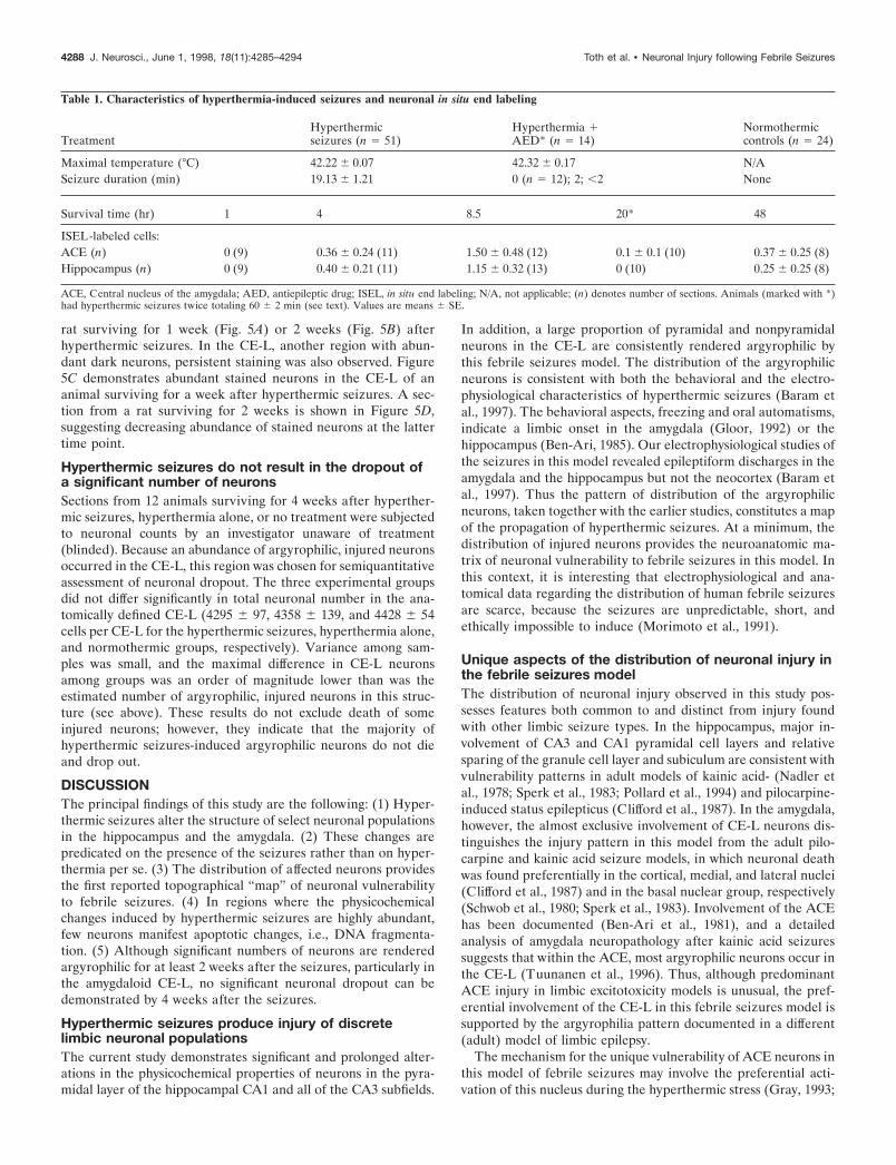
For determination of DNA fragmentation, groups (n 5 3–4 per group)of animals subjected to hyperthermic seizures were killed at the follow-ing time points after the seizures: 1, 4, 8.5, 20, and 48 hr. An additionalgroup (n 5 4) was subjected to hyperthermia twice, leading to a totalseizure duration of 60 6 2 min, and was killed 20 hr after the secondseizure episode. Normothermic animals (n 5 7) and pups in whomhyperthermia-induced seizures were prevented with pentobarbital (seeabove) were used as controls. For analysis of neuronal injury using theGallyas “dark”-neuron stain, animals were killed 24 hr, 1 week, or 2weeks after seizure induction. For cell counting, animals (n 5 12; 4 perexperimental group) were killed 4 weeks after the hyperthermic seizures.
In situ end labelingIn situ end labeling (ISEL) provides a positive label of dying cells,leading to increased sensitivity over methods relying on the “dropout” ofneurons. ISEL was modified from Sakhi et al. (1994). Briefly, frozen 20mm sections were thawed, dried, and fixed in 4% buffered paraformal-dehyde, followed by ethanol dehydration. After rehydration and prein-cubation in a Tris buffer, 50 ml of reaction mix [966 ml of buffer, 4 ml ofnucleotide mix (Sakhi et al., 1994), 10 ml of DNA polymerase I (10 U/ml;Promega, Madison, WI), and 20 ml of dUTP–biotin (Sigma, St. Louis,MO) per 1000 ml] were applied for 1 hr. UTP–biotin-tagged cleavedDNA ends were visualized using a commercial avidin–biotin kit (VEC-TASTAIN; Vector Laboratories, Burlingame, CA). Sections were coun-terstained with acid fuchsin (Chang and Baram, 1994), and the numberof labeled neurons with clumped chromatin in the central nucleus of theamygdala and the hippocampus was determined. Sections from adult ratssubjected to kainic acid-induced status epilepticus and allowed a 20 hrsurvival time were run in parallel as “positive controls” to confirm thevalidity of the ISEL method at this time point.
Histological methodsFor all histological methods, control and experimental groups wereprocessed together, without knowledge of treatment group. Animalswere killed and perfused as described elsewhere (Baram and Ribak,1995; Ribak and Baram, 1996). Briefly, under deep anesthesia, rats wereperfused transcardially with saline followed by a 4% paraformaldehyde–2.5% glutaraldehyde–0.1 M phosphate buffer solution. Brains were left insitu overnight and then post-fixed for a week in the same fixative. Brainswere sectioned coronally with a vibratome at 100 mm for silver staining
and at 20 mm for Nissl staining for neuronal counts. Sections (immersedin fixative) were stored at 4°C.
Silver-staining (dark-neuron) method. This method has been describedearlier (Gallyas et al., 1990). Briefly, after dehydration in 50, 75, and100% propanol, sections were esterified with propanol containing 0.8%sulfuric acid. Sections were rehydrated through 50 and 25% propanoland distilled water, treated with 3% acetic acid for 5 min, and developedfor 10 min. The fresh developing solution consisted of equal volumes of10% Na2CO3 and a solution composed of 0.2% AgNO3 , 0.25%NH4NO3 , 2% tungstosilicic acid, and 0.4% formaldehyde. When devel-oped, sections were dehydrated and coverslipped.
Semithin sections and electron microscopy methods. Unstained sectionsadjacent to those containing argyrophilic neurons were post-fixed with1% osmium tetroxide, dehydrated, and embedded in Epon. Semithin, 2mm sections were cut and stained with 1% toluidine blue to identifydeeply basophilic neurons. For electron microscopy (EM) analysis, ultra-thin sections were obtained from the same tissue blocks, subjected touranyl acetate and lead citrate, and examined with a transmission EM.Electron-dense neuronal profiles and their synapses were further exam-ined in serial sections to confirm their identification.
Semiquantitative neuronal analysis. For counting both silver- and Nissl-stained neurons, sections were analyzed without knowledge of treatment(“blindly”). Based on the limbic phenomenology of hyperthermia-induced seizures (Ben-Ari et al., 1981; Baram et al., 1997), the exami-nation of argyrophilic neurons focused on the amygdala, hippocampus,and cortical regions interconnected with these structures. For determin-ing the fate of most argyrophilic neurons, an estimate of their numberwas undertaken in the most affected structure, the lateral division (CE-L)of the central nucleus of the amygdala (ACE). For purposes of neuronalcounting, the anatomical boundaries of the CE-L were defined usingSherwood and Timiras (1970). Rostral and caudal boundaries wereselected to permit an easily identifiable coronal profile of the nucleus.The rostral boundary was at the level of the anterior paraventricularhypothalamic nucleus and the rostral border of the hippocampal CA3(5.0 mm anterior to bregma in the 39-d-old rat); the caudal boundary wasdefined at the level of the infundibulum (4.1 mm anterior to bregma).The lateral boundary of the ACE was defined by the clearly visibleteardrop-shaped lateral nucleus of the amygdala. The medial border wasdefined by the cell-poor white matter of the internal capsule. Within theACE, the CE-L was defined according to Tuunanen et al. (1996). Briefly,the tightly packed capsular region was defined laterally, and the crescent-shaped medial division was distinguished medially (see Figs. 3, 5).Because a determination of the absolute, unbiased number of argyro-philic neurons was not needed for the purposes of this study, a modifiedprofile sampling method was used [for a recent discussion, see Popkenand Farel (1997); Saper (1997)]. A grid reticule of 100 frames covering anarea of 250 3 250 mm was centered over every sixth section of the CE-L,and all frames were subjected to counting for cell bodies throughout thedepth of the section (Popken and Farel, 1997). The resulting number wasmultiplied to yield an estimate of the overall number of argyrophilicneurons in the CE-L as defined above. For Nissl-stained sections, everyninth 20 mm section was subjected to cell counts. To minimize the bias ofthe “splitting” of large cell bodies resulting in an overestimate of counts,we counted nucleoli (Popken and Farel, 1997). Counts of each sectionwere averaged to yield an estimate of variance, and the overall neuronalnumbers in the CE-L, as defined above, were obtained for each experi-mental animal. The statistical significance of differences among the threetreatment groups used ANOVA (PRISM statistical software; GraphPad,San Diego, CA).
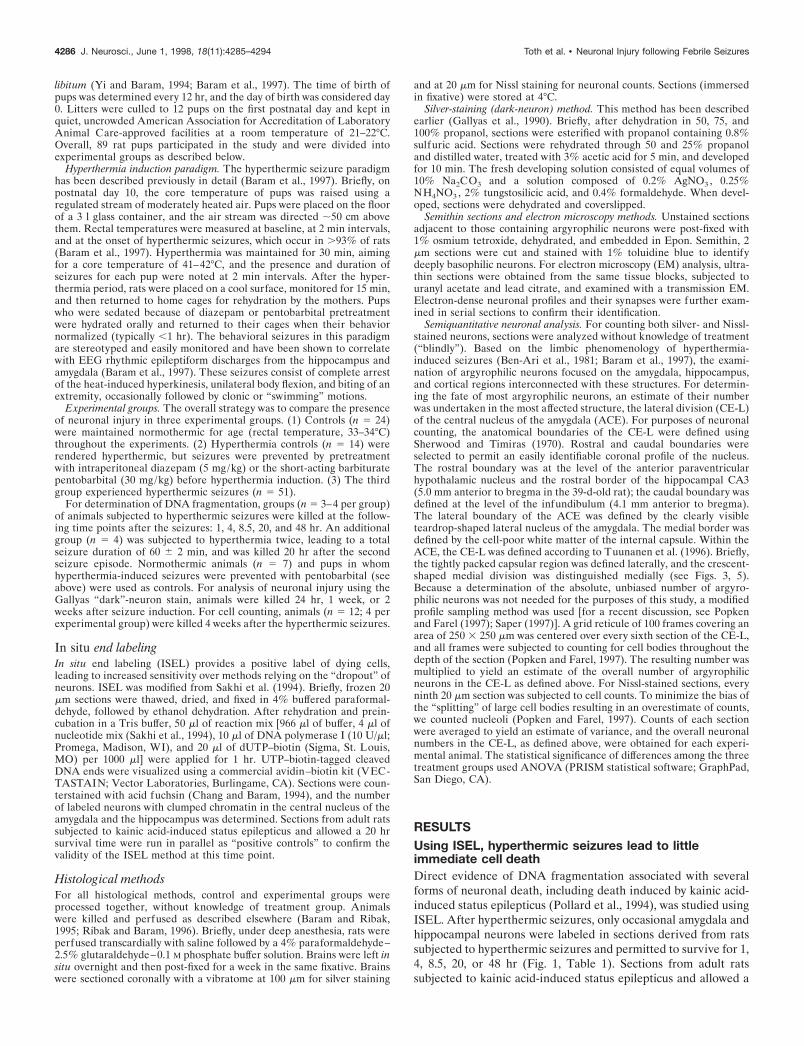
RESULTSUsing ISEL, hyperthermic seizures lead to littleimmediate cell deathDirect evidence of DNA fragmentation associated with severalforms of neuronal death, including death induced by kainic acid-induced status epilepticus (Pollard et al., 1994), was studied usingISEL. After hyperthermic seizures, only occasional amygdala andhippocampal neurons were labeled in sections derived from ratssubjected to hyperthermic seizures and permitted to survive for 1,4, 8.5, 20, or 48 hr (Fig. 1, Table 1). Sections from adult ratssubjected to kainic acid-induced status epilepticus and allowed a
4286 J. Neurosci., June 1, 1998, 18(11):4285–4294 Toth et al. • Neuronal Injury following Febrile Seizures
20 hr survival time, which were run in parallel, contained abun-dant labeled neurons in hippocampal CA3 and most amygdalanuclei, confirming the validity of the ISEL method (data notshown).
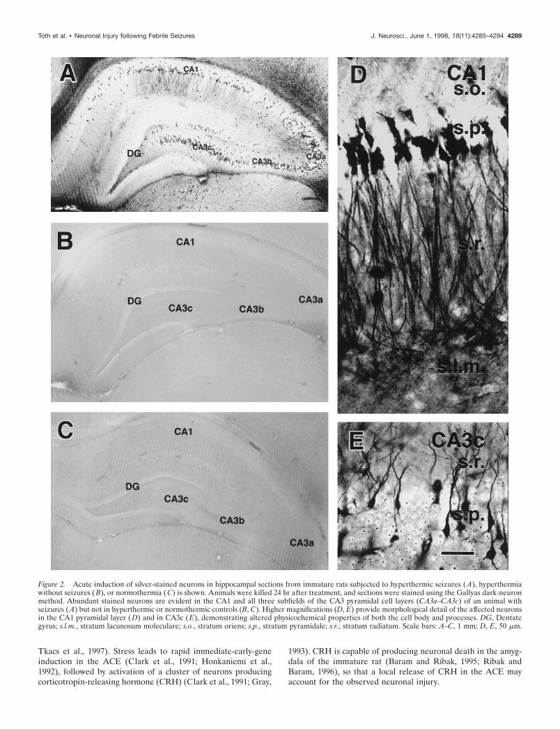
Hyperthermic seizures lead to neuronal injury indiscrete hippocampal and amygdala regionsSections from animals killed 24 hr after hyperthermic seizurescontained significant populations of silver-stained, dark neurons.Figure 2 demonstrates clusters of stained neurons in the hip-pocampus. All subfields of the CA3 pyramidal cell layer wereinvolved, as well as the entire extent of CA1 and occasionalgranule cells (Fig. 2A). Affected neurons were interspersedamong normal ones, and their stained apical and basal dendritesindicated that they were pyramidal cells. No argyrophilic neuronswere apparent in control sections (Fig. 2C) or in sections fromanimals pretreated with anticonvulsants before hyperthermia in-duction (Fig. 2B). Higher magnifications of the silver-stainedneurons, revealing pronounced physicochemical alterations(shrunken appearance; Gallyas et al., 1992a), are shown in Figure2, D and E.
The remarkable involvement of the lateral division of the ACEis evident in Figure 3. Sections from rats killed 24 hr afterhyperthermia and seizures (Fig. 3A) revealed an abundance ofargyrophilic neurons, whereas sections from both of the controlgroups [hyperthermia without seizures (Fig. 3B) and normother-mia (Fig. 3C)] did not contain affected cells. Semiquantitativeanalysis of affected neurons in the CE-L suggested that thenumber of injured neurons was in the order of 185.8 6 10.1 per100-mm-thick section or 1645.5 6 178 within the defined anatom-ical boundaries of this nucleus (see Materials and Methods). Thedistribution of hyperthermic seizures-induced argyrophilic neu-rons included also the medial and lateral basal amygdala nuclei(Fig. 3D), as well as a delimited portion of the perirhinal cortex(Fig. 3E).
EM was used to show the ultrastructural features of the silver-stained neurons in the hippocampal CA1 and the amygdaloidCE-L. Figure 4 demonstrates a hyperelectron-dense shrunkenpyramidal cell (Fig. 4B). Intact synapses are apparent along thesurfaces of the soma and the shrunken, spiny dendrites of thisneuron (Fig. 4C), which are postsynaptic to immature axon ter-minals. In the CE-L, the electron-dense cells maintained intactcell membranes and contained distinct organelles including Golgicomplexes and endoplasmic reticulum (Fig. 4D,E).
Hyperthermic seizures, but not the hyperthermia perse, lead to neuronal injurySeizure duration in the hyperthermic seizure group averaged19.13 6 1.21 min. Animals pretreated with pentobarbital beforehyperthermia induction did not have behavioral seizures, al-though the degree of hyperthermia, as defined by maximal coretemperature, and the duration of hyperthermia were comparablewith those of non-pretreated pups (Table 1). Sections fromanticonvulsant-pretreated animals who did not develop seizuresand were killed either 24 hr or a week subsequently did notcontain silver-stained neurons (see below). A broad correlationbetween seizure duration and the extent of neuronal injury wasevident. Thus, sections from one of two animals, in whom shortseizures (,2 min) developed despite diazepam pretreatment,contained occasional silver-stained neurons (data not shown).
Neuronal changes induced by hyperthermic seizurespersist for at least 2 weeksAltered structural neuronal properties, manifested as silver stain-ing of affected neurons, persisted in the regions in which theywere noted within 24 hr after the seizures. Figure 5 demonstratesclusters of injured neurons in the hippocampus of an immature
Figure 1. Rare acute neuronal death after hyperthermic seizures in theimmature rat. A, In situ end labeling reveals two hippocampal CA3neurons (arrows) undergoing DNA fragmentation from an animal killed20 hr after hyperthermic seizures. B, Higher magnification shows thetypical DNA fragmentation in a pyramidal layer neuron (arrow corre-sponding to the lower neuron in A). Run in parallel, sections fromnormothermic controls did not contain labeled neurons, whereas, asexpected, sections from kainic acid-treated mature rats revealed numer-ous dying cells (data not shown). s.o., Stratum oriens; s.p., stratumpyramidale; s.r., stratum radiatum. Scale bars: A, 50 mm; B, 10 mm.
Toth et al. • Neuronal Injury following Febrile Seizures J. Neurosci., June 1, 1998, 18(11):4285–4294 4287
rat surviving for 1 week (Fig. 5A) or 2 weeks (Fig. 5B) afterhyperthermic seizures. In the CE-L, another region with abun-dant dark neurons, persistent staining was also observed. Figure5C demonstrates abundant stained neurons in the CE-L of ananimal surviving for a week after hyperthermic seizures. A sec-tion from a rat surviving for 2 weeks is shown in Figure 5D,suggesting decreasing abundance of stained neurons at the lattertime point.
Hyperthermic seizures do not result in the dropout ofa significant number of neuronsSections from 12 animals surviving for 4 weeks after hyperther-mic seizures, hyperthermia alone, or no treatment were subjectedto neuronal counts by an investigator unaware of treatment(blinded). Because an abundance of argyrophilic, injured neuronsoccurred in the CE-L, this region was chosen for semiquantitativeassessment of neuronal dropout. The three experimental groupsdid not differ significantly in total neuronal number in the ana-tomically defined CE-L (4295 6 97, 4358 6 139, and 4428 6 54cells per CE-L for the hyperthermic seizures, hyperthermia alone,and normothermic groups, respectively). Variance among sam-ples was small, and the maximal difference in CE-L neuronsamong groups was an order of magnitude lower than was theestimated number of argyrophilic, injured neurons in this struc-ture (see above). These results do not exclude death of someinjured neurons; however, they indicate that the majority ofhyperthermic seizures-induced argyrophilic neurons do not dieand drop out.
DISCUSSIONThe principal findings of this study are the following: (1) Hyper-thermic seizures alter the structure of select neuronal populationsin the hippocampus and the amygdala. (2) These changes arepredicated on the presence of the seizures rather than on hyper-thermia per se. (3) The distribution of affected neurons providesthe first reported topographical “map” of neuronal vulnerabilityto febrile seizures. (4) In regions where the physicochemicalchanges induced by hyperthermic seizures are highly abundant,few neurons manifest apoptotic changes, i.e., DNA fragmenta-tion. (5) Although significant numbers of neurons are renderedargyrophilic for at least 2 weeks after the seizures, particularly inthe amygdaloid CE-L, no significant neuronal dropout can bedemonstrated by 4 weeks after the seizures.
Hyperthermic seizures produce injury of discretelimbic neuronal populationsThe current study demonstrates significant and prolonged alter-ations in the physicochemical properties of neurons in the pyra-midal layer of the hippocampal CA1 and all of the CA3 subfields.
In addition, a large proportion of pyramidal and nonpyramidalneurons in the CE-L are consistently rendered argyrophilic bythis febrile seizures model. The distribution of the argyrophilicneurons is consistent with both the behavioral and the electro-physiological characteristics of hyperthermic seizures (Baram etal., 1997). The behavioral aspects, freezing and oral automatisms,indicate a limbic onset in the amygdala (Gloor, 1992) or thehippocampus (Ben-Ari, 1985). Our electrophysiological studies ofthe seizures in this model revealed epileptiform discharges in theamygdala and the hippocampus but not the neocortex (Baram etal., 1997). Thus the pattern of distribution of the argyrophilicneurons, taken together with the earlier studies, constitutes a mapof the propagation of hyperthermic seizures. At a minimum, thedistribution of injured neurons provides the neuroanatomic ma-trix of neuronal vulnerability to febrile seizures in this model. Inthis context, it is interesting that electrophysiological and ana-tomical data regarding the distribution of human febrile seizuresare scarce, because the seizures are unpredictable, short, andethically impossible to induce (Morimoto et al., 1991).
Unique aspects of the distribution of neuronal injury inthe febrile seizures modelThe distribution of neuronal injury observed in this study pos-sesses features both common to and distinct from injury foundwith other limbic seizure types. In the hippocampus, major in-volvement of CA3 and CA1 pyramidal cell layers and relativesparing of the granule cell layer and subiculum are consistent withvulnerability patterns in adult models of kainic acid- (Nadler etal., 1978; Sperk et al., 1983; Pollard et al., 1994) and pilocarpine-induced status epilepticus (Clifford et al., 1987). In the amygdala,however, the almost exclusive involvement of CE-L neurons dis-tinguishes the injury pattern in this model from the adult pilo-carpine and kainic acid seizure models, in which neuronal deathwas found preferentially in the cortical, medial, and lateral nuclei(Clifford et al., 1987) and in the basal nuclear group, respectively(Schwob et al., 1980; Sperk et al., 1983). Involvement of the ACEhas been documented (Ben-Ari et al., 1981), and a detailedanalysis of amygdala neuropathology after kainic acid seizuressuggests that within the ACE, most argyrophilic neurons occur inthe CE-L (Tuunanen et al., 1996). Thus, although predominantACE injury in limbic excitotoxicity models is unusual, the pref-erential involvement of the CE-L in this febrile seizures model issupported by the argyrophilia pattern documented in a different(adult) model of limbic epilepsy.
The mechanism for the unique vulnerability of ACE neurons inthis model of febrile seizures may involve the preferential acti-vation of this nucleus during the hyperthermic stress (Gray, 1993;
Table 1. Characteristics of hyperthermia-induced seizures and neuronal in situ end labeling
TreatmentHyperthermicseizures (n 5 51)
Hyperthermia 1AED* (n 5 14)
Normothermiccontrols (n 5 24)
Maximal temperature (°C) 42.22 6 0.07 42.32 6 0.17 N/ASeizure duration (min) 19.13 6 1.21 0 (n 5 12); 2; ,2 None
Survival time (hr) 1 4 8.5 20* 48
ISEL-labeled cells:ACE (n) 0 (9) 0.36 6 0.24 (11) 1.50 6 0.48 (12) 0.1 6 0.1 (10) 0.37 6 0.25 (8)Hippocampus (n) 0 (9) 0.40 6 0.21 (11) 1.15 6 0.32 (13) 0 (10) 0.25 6 0.25 (8)
ACE, Central nucleus of the amygdala; AED, antiepileptic drug; ISEL, in situ end labeling; N/A, not applicable; (n) denotes number of sections. Animals (marked with *)had hyperthermic seizures twice totaling 60 6 2 min (see text). Values are means 6 SE.
4288 J. Neurosci., June 1, 1998, 18(11):4285–4294 Toth et al. • Neuronal Injury following Febrile Seizures
Tkacs et al., 1997). Stress leads to rapid immediate-early-geneinduction in the ACE (Clark et al., 1991; Honkaniemi et al.,1992), followed by activation of a cluster of neurons producingcorticotropin-releasing hormone (CRH) (Clark et al., 1991; Gray,
1993). CRH is capable of producing neuronal death in the amyg-dala of the immature rat (Baram and Ribak, 1995; Ribak andBaram, 1996), so that a local release of CRH in the ACE mayaccount for the observed neuronal injury.
Figure 2. Acute induction of silver-stained neurons in hippocampal sections from immature rats subjected to hyperthermic seizures (A), hyperthermiawithout seizures (B), or normothermia ( C) is shown. Animals were killed 24 hr after treatment, and sections were stained using the Gallyas dark-neuronmethod. Abundant stained neurons are evident in the CA1 and all three subfields of the CA3 pyramidal cell layers (CA3a–CA3c) of an animal withseizures (A) but not in hyperthermic or normothermic controls (B, C). Higher magnifications (D, E) provide morphological detail of the affected neuronsin the CA1 pyramidal layer (D) and in CA3c (E), demonstrating altered physicochemical properties of both the cell body and processes. DG, Dentategyrus; s.l.m., stratum lacunosum moleculare; s.o., stratum oriens; s.p., stratum pyramidale; s.r., stratum radiatum. Scale bars: A–C, 1 mm; D, E, 50 mm.
Toth et al. • Neuronal Injury following Febrile Seizures J. Neurosci., June 1, 1998, 18(11):4285–4294 4289

Figure 3. Neuronal injury induced by hyperthermic seizures in the amygdala, demonstrated using the Gallyas dark-neuron method. A, Significantinvolvement of the lateral division of the central amygdaloid nucleus (CE-L) is manifested by large numbers of densely silver-stained neurons. B, C,Sections from hyperthermic (B) or normothermic (C) controls contain few stained neurons. D, The distribution of silver-stained neurons in otheramygdaloid nuclei, the basomedial (ABM ) and basolateral (ABL) nuclei, is shown. E, The presence of argyrophilic neurons in the perirhinal cortex isevident. ALA, Lateral amygdala nucleus; IC, internal capsule; rf, rhinal fissure. Scale bars: A–C, 500 mm; D, E, 200 mm.
4290 J. Neurosci., June 1, 1998, 18(11):4285–4294 Toth et al. • Neuronal Injury following Febrile Seizures

Figure 4. Light and electron micrographs of hyperelectron-dense neurons in the hippocampal CA1 (A–C) and the lateral division of the centralamygdaloid nucleus (CE-L) (D, E). A, B, A shrunken, deeply basophilic neuron in A appears electron-dense in B compared with a normal neuron (N).The hyperelectron-dense nucleus (n) and apical dendrite (d) with a spine (arrow) of this neuron, as well as abnormal processes (curved arrows) and aglial cell (G), are shown in B. C, Higher magnification of the dendritic spine (s; arrow in B) reveals two axodendritic synapses (arrows) formed byvesicle-containing axon terminals (t1 and t2 ). D, Three hyperelectron-dense neurons with intact nuclei ( n), Golgi complex, and endoplasmic reticulumare shown. E, Enlargement of an axosomatic synapse (arrow in D) shows synaptic vesicles and a multivesicular body ( m) within the electron-dense soma.Scale bars: A, 20 mm; B, D, 2 mm; C, E, 0.1 mm.
Toth et al. • Neuronal Injury following Febrile Seizures J. Neurosci., June 1, 1998, 18(11):4285–4294 4291

Neuronal injury in the immature, as compared with theadult, brainThe spatiotemporal evolution of neuronal changes observed inthe current studies provides a useful perspective into issues ofseizure-induced cell death in the immature brain. Although pro-longed and severe seizures typically occur when convulsants such
as kainic acid or pilocarpine are administered to immature rats(younger than 20 d), it is generally considered that they are notfollowed by neuropathological changes (Nitecka et al., 1984; Sper-ber et al., 1992; Chang and Baram, 1994; but see Wasterlain,1997). The present study, revealing profound but transient alter-ations of neuronal integrity in regions known to be affected by
Figure 5. Persistent injury to hippocampal and amygdaloid neurons after hyperthermic seizures. Sections obtained from immature rats killed 1 week(A, C) or 2 weeks (B, D) after hyperthermic seizures are shown. Silver-stained neurons are evident in the CA1 pyramidal cell layer (A, B) and in thelateral division of the central nucleus of the amygdala (CE-L) (C, D) at both time points. Decreased numbers of affected neurons are apparent at the2-week time point (B, D). DG, Dentate gyrus; IC, internal capsule; s.p., stratum pyramidale. Scale bar, 50 mm.
4292 J. Neurosci., June 1, 1998, 18(11):4285–4294 Toth et al. • Neuronal Injury following Febrile Seizures

other limbic seizure paradigms, may provide a mechanism toreconcile conflicting reports regarding the effects of developmen-tal limbic seizures on neuronal survival. Specifically, our findingssuggest that similar neuronal populations share vulnerability tolimbic seizures in both the immature and mature rat, but imma-ture neurons may undergo injury followed by recovery, whereasmature neurons progress from injury to death (Chang and Baram,1994; Owens et al., 1997). In addition, as has been shown foreosinophilic acid fuchsin staining (Chang and Baram, 1994),neuronal argyrophilia may carry a different significance regardingneuronal fate in the immature, as compared with the adult, CNS.Along the same lines, ISEL may underestimate neuronal deathparticularly in the immature brain, leading to the limited celldeath found in the current studies. The ISEL time course studyand the lack of significant neuronal dropout using an independentmethod at the 4 week time point, however, render this possibilityless likely (and see below).
An intriguing feature of the distribution of affected neuronsafter hyperthermic seizures in this study is their concordancewith the neuropathological features of human temporal lobe(limbic) epilepsy known as mesial temporal sclerosis. Within thehippocampus, the most common sites of neuronal loss are theCA3 and CA4 (of which the hilus is the rodent counterpart)(Sagar and Oxbury, 1987; Bruton, 1988). Less is known regardingthe precise topography of neuronal loss in the TLE amygdala(Gloor, 1992), and most studies refer to gliosis in the “basal”nuclear group (Cavanagh and Meyer, 1956; Bruton, 1988; Gloor,1992). The amygdala is preferentially affected, however, whenTLE is caused by lesions outside the hippocampus (Cavanaghand Meyer, 1956; Gloor, 1992).
Hyperthermic seizures lead to neuronal injury withoutevidence of significant neuronal dropoutThe hyperthermic seizures-induced neuronal injury demon-strated in this study was not associated with significant DNAfragmentation, as determined by ISEL. DNA cleavage is gener-ally considered a marker of cell death (Wyllie, 1993), although thetype of cell death (apoptosis vs necrosis) may be unsettled (Kureet al., 1991; Pollard et al., 1994). The advantage of ISEL andsimilar techniques derives from the resulting positive stain, whichrenders even single cells highly visible, in contrast to cell counts,which determine a decrease in overall cell numbers. A significantdisadvantage of methods using DNA fragmentation markersstems from the short temporal “window” (within the process ofneuronal death) during which such DNA fragmentation may bedetectable. Thus, these methods tend to underestimate neuronaldeath, particularly when cells do not die synchronously(Voyvodic, 1996). However, the current study included severaltime points spanning 1–48 hr after seizure induction, so thattemporal underestimation of the number of dying neurons is lesslikely. Therefore, the discrepancy between the large numbers ofargyrophilic neurons and the rare ISEL-labeled cells indicatesthat argyrophilia in the current study does not constitute evidenceof “irreversible injury” or death (Gallyas et al., 1992b; Toth et al.,1997) in the majority of neurons.
A major advantage of the silver-staining method used in thecurrent study lies in its ability to demonstrate substantial popu-lations of injured hippocampal and ACE neurons. The precisealterations in the physicochemical properties of injured neuronsthat lead to their enhanced affinity to the silver stain has not beenresolved (Gallyas et al., 1992b). However, aside from the ISELdata, two additional lines of evidence point against equating
argyrophilia with neuronal death. First, Toth et al. (1997) dem-onstrated that fluid percussion trauma can lead to the formationof argyrophilic neurons even when applied to a dead animal.Second, the number of argyrophilic neurons observed in thecurrent study, at least in the CE-L (;1700), far exceeded anypotential cell loss determined by cell counts 4 weeks later (;150neurons, maximal intergroup difference). Counts were performedwithout knowledge of treatment, minimizing a systematic bias,but inherent estimation errors cannot be excluded (Popken andFarel, 1997). However, in the context of the current study, even a100% error in counting (Popken and Farel, 1997; Saper, 1997)would not alter the conclusion that the majority of argyrophilic,injured neurons induced by hyperthermic seizures do notdrop out.
Implications for human febrile seizuresThe studies described provide a prospective analysis of the spa-tiotemporal profile of neuronal changes induced by hyperthermicseizures, in an appropriate-age model of human febrile seizures.Febrile seizures, the most common human seizures, affect at least500,000 individuals per year (Hauser, 1994). These seizures areessentially restricted to the developmental period of infancy andearly childhood (Knudsen, 1996). The contribution of febrileseizures to the development of TLE has remained controversial(Cendes et al., 1993; Knudsen, 1996) and has centered on theissue of induction of limbic neuronal death by these seizures(Sagar and Oxbury, 1987; Gloor, 1991). These fundamental issuesof developmental epilepsy are difficult to study in the human. Thecurrent study relied on an appropriate-age model in which sei-zures can be induced in .93% of animals, thus eliminating anyconcerns of genetic predisposition and pre-existing lesions. Usingthis model, we have demonstrated the pattern of vulnerability ofhippocampal and amygdala neuronal populations to hyperthermicseizures. The absence of neuronal injury (argyrophilia) in hyper-thermic rats in whom the seizures were prevented confirms thespecificity of these neuronal changes to the seizures themselves.In addition, the results of this study suggest that the majority ofinjured neurons do not progress to death. Thus, these data do notsupport the supposition that early-life febrile seizures result di-rectly in hippocampal cell death, the neuropathological lesionfound in human TLE. However, the widespread and prolongedneuronal injury demonstrated in this study may lead to significantalterations of the function and electrophysiology of the affectedneuronal circuits (Kapur and Coulter, 1995), which could leadeventually to an epileptic state.
REFERENCESAlbala BJ, Moshe SL, Okada R (1984) Kainic acid-induced seizures: a
developmental study. Brain Res 315:139–148.Baram TZ, Ribak CE (1995) Peptide-induced status epilepticus causes
neuronal death and synaptic reorganization. NeuroReport 6:277–280.Baram TZ, Gerth A, Schultz L (1997) Febrile seizures: an age appro-
priate model. Brain Res Dev Brain Res 246:134–143.Ben-Ari Y (1985) Limbic seizure and brain damage produced by kainic
acid: mechanisms and relevance to human temporal lobe epilepsy.Neuroscience 14:375–403.
Ben-Ari Y, Tremblay E, Riche D, Ghilini G, Naquet R (1981) Electro-graphic, clinical and pathological alterations following systemic admin-istration of kainic acid, bicuculline or pentetrazole: metabolic mappingusing the deoxyglucose method with special reference to the pathologyof epilepsy. Neuroscience 6:1361–1391.
Berg AT, Shinnar S, Hauser WA, Alemany M, Shapiro ED, Salomon ME,Crain EF (1992) A prospective study of recurrent febrile seizures.N Engl J Med 327:1122–1127.
Bruton CJ (1988) The neuropathology of temporal lobe epilepsy. NewYork: Oxford UP.
Toth et al. • Neuronal Injury following Febrile Seizures J. Neurosci., June 1, 1998, 18(11):4285–4294 4293

Cavanagh JB, Meyer A (1956) Aetiological aspects of Ammon’s hornsclerosis associated with temporal lobe epilepsy. Br Med J 2:1403–1407.
Cendes F, Andermann F, Dubeau F, Gloor P, Evans A, Jones-Gotman M,Olivier A, Andermann E, Robitaille Y, Lopes-Cendes I (1993) Earlychildhood prolonged febrile convulsions, atrophy and sclerosis of me-sial structures, and temporal lobe epilepsy: an MRI volumetric study.Neurology 43:1083–1087.
Chang D, Baram TZ (1994) Status epilepticus results in reversible neu-ronal injury in infant rat hippocampus: novel use of a marker. Brain ResDev Brain Res 77:133–136.
Clark M, Weiss SR, Post RM (1991) Expression of c-fos mRNA in ratbrain after intracerebroventricular administration of corticotropin-releasing hormone. Neurosci Lett 132:235–238.
Clifford DB, Olney JW, Maniotis A, Collins RC, Zorumski CF (1987)The functional anatomy and pathology of lithium-pilocarpine and high-dose pilocarpine seizures. Neuroscience 23:953–968.
Dobbing J, Sands J (1973) Quantitative growth and development ofhuman brain. Arch Dis Child 48:757–767.
Falconer MA, Serafetinides EA, Corsellis JAN (1964) Etiology andpathogenesis of temporal lobe epilepsy. Arch Neurol 10:233–248.
Gallyas F, Guldner FH, Zoltay G, Wolff JR (1990) Golgi-like demon-stration of “dark” neurons with an argyrophil III method for experi-mental neuropathology. Acta Neuropathol (Berl) 79:620–628.
Gallyas F, Zoltay G, Balas I (1992a) An immediate light microscopicresponse of neuronal somata, dendrites and axons to contusing concus-sive head injury in the rat. Acta Neuropathol (Berl) 83:394–401.
Gallyas F, Zoltay G, Dames W (1992b) Formation of “dark” (argyro-philic) neurons of various origins proceeds with a common mechanismof biophysical nature (a novel hypothesis). Acta Neuropathol (Berl)83:504–509.
Gloor P (1991) Mesial temporal sclerosis: historical background and anoverview from a modern perspective. In: Epilepsy surgery (Luders HO,ed), pp 689–703. New York: Raven.
Gloor P (1992) Role of the amygdala in temporal lobe epilepsy. In: Theamygdala: neurobiological aspects of emotion, memory, and mentaldysfunction (Aggleton JP, ed), pp 505–538. New York: Wiley-Liss.
Gray TS (1993) Amygdaloid CRF pathways. Role in autonomic, neu-roendocrine, and behavioral responses to stress. Ann NY Acad Sci697:53–60.
Hauser WA (1994) The prevalence and incidence of convulsive disor-ders in children. Epilepsia 35 [Suppl] 2:S1–S6.
Holmes GL (1997) Epilepsy in the developing brain: lessons from thelaboratory and clinic. Epilepsia 38:12–30.
Holmes GL, Thompson JL (1988) Effects of kainic acid on seizuresusceptibility in the developing brain. Brain Res 467:51–59.
Honkaniemi J, Pelto-Huikko M, Rechardt L, Isola J, Lammi A, Fuxe K,Gustafsson JA, Wikstrom AC, Hokfelt T (1992) Colocalization ofpeptide and glucocorticoid receptor immunoreactivities in rat centralamygdaloid nucleus. Neuroendocrinology 55:451–459.
Jensen FE, Applegate CD, Holtzman D, Belin TR, Burchfiel JL (1991)Epileptogenic effect of hypoxia in the immature rodent brain. AnnNeurol 29:629–637.
Kapur J, Coulter DA (1995) Experimental status epilepticus altersgamma-aminobutyric acid type A receptor function in CA1 pyramidalneurons. Ann Neurol 38:893–900.
Knudsen FU (1996) Febrile seizures–treatment and outcome. Brain Dev18:438–449.
Kure S, Tominaga T, Yoshimoto T, Tada K, Narisawa K (1991) Gluta-mate triggers internucleosomal DNA cleavage in neuronal cells. Bio-chem Biophys Res Commun 179:39–45.
Morimoto T, Nagao H, Sano N, Takahashi M, Matsuda H (1991) Elec-troencephalographic study of rat hyperthermic seizures. Epilepsia32:289–293.
Nadler JV, Perry BW, Cotman CW (1978) Intraventricular kainic acidpreferentially destroys hippocampal pyramidal cells. Nature271:676–677.
Nelson KB, Ellenberg JH (1976) Predictors of epilepsy in children whohave experienced febrile seizures. N Engl J Med 295:1029–1033.
Nitecka L, Tremblay E, Charton G, Bouillot JP, Berger ML, Ben-Ari Y(1984) Maturation of kainic acid seizure-brain damage syndrome inthe rat. Neuroscience 13:1073–1094.
Owens JJ, Robbins CA, Wenzel HJ, Schwartzkroin PA (1997) Acute andchronic effects of hypoxia on the developing hippocampus. Ann Neurol41:187–199.
Pollard H, Charriaut-Marlangue C, Cantagrel S, Represa A, Robain O,Moreau J, Ben-Ari Y (1994) Kainate-induced apoptotic cell death inhippocampal neurons. Neuroscience 63:7–18.
Popken GJ, Farel PB (1997) Sensory neuron number in neonatal andadult rats estimated by means of stereologic and profile-based methods.J Comp Neurol 386:8–15.
Ribak CE, Baram TZ (1996) Selective death of hippocampal CA3 pyra-midal cells with mossy fiber afferents after CRH-induced status epilep-ticus in infant rats. Brain Res Dev Brain Res 91:245–251.
Sagar HJ, Oxbury JM (1987) Hippocampal neuron loss in temporal lobeepilepsy: correlation with early childhood convulsions. Ann Neurol22:334–340.
Sakhi S, Bruce A, Sun N, Tocco G, Baudry M, Schreiber SS (1994) p53induction is associated with neuronal damage in the central nervoussystem. Proc Natl Acad Sci USA 91:7525–7529.
Saper CB (1997) Counting on our reviewers to set the standards. J CompNeurol 386:1.
Schwob JE, Fuller T, Price JL, Olney JW (1980) Widespread patterns ofneuronal damage following systemic or intracerebral injections ofkainic acid: a histological study. Neuroscience 5:991–1014.
Sherwood NM, Timiras PS (1970) A stereotaxic atlas of the developingrat brain. Berkeley, CA: University of California.
Shinnar S (1990) Febrile seizures. In: Current therapy in neurologicaldisease (Johnson RT, ed), pp 29–32. Philadelphia: Decker.
Sperber EF, Haas KZ, Stanton PK, Moshe SL (1991) Resistance of theimmature hippocampus to seizure-induced synaptic reorganization.Brain Res Dev Brain Res 60:88–93.
Sperber EF, Stanton PK, Haas K, Ackerman RF, Moshe SL (1992)Developmental differences in the neurobiology of epileptic brain dam-age. In: Molecular neurobiology of epilepsy, pp 67–81. Amsterdam:Elsevier.
Sperk G, Lassmann H, Baran H, Kish SJ, Seitelberger F, Hornykiewicz O(1983) Kainic acid-induced seizures: neurochemical and histopatho-logical changes. Neuroscience 10:1301–1315.
Tkacs NC, Li J, Strack AM (1997) Central amygdala Fos expressionduring hypotensive or febrile, nonhypotensive endotoxemia in con-scious rats. J Comp Neurol 379:592–602.
Toth Z, Hollrigel GS, Gorcs T, Soltesz I (1997) Instantaneous perturba-tion of dentate interneuronal networks by a pressure wave-transientdelivered to the neocortex. J Neurosci 17:8106–8117.
Tuunanen J, Halonen T, Pitkanen A (1996) Status epilepticus causesselective regional damage and loss of GABAergic neurons in the ratamygdaloid complex. Eur J Neurosci 8:2711–2725.
Verity CM, Butler NR, Golding J (1985) Febrile convulsions in a na-tional cohort followed up from birth. Prevalence and recurrence in thefirst five years of life. Br Med J 290:1307–1310.
Voyvodic JT (1996) Cell death in cortical development: how much?Why? So what? Neuron 16:693–696.
Wasterlain CG (1997) Recurrent seizures in the developing brain areharmful. Epilepsia 38:728–734.
Wyllie AH (1993) Apoptosis. Br J Cancer 67:205–208.Yi SJ, Baram TZ (1994) Corticotropin-releasing hormone mediates the
response to cold stress in the neonatal rat without compensatory en-hancement of the peptide’s gene expression. Endocrinology 135:2364–2368.
4294 J. Neurosci., June 1, 1998, 18(11):4285–4294 Toth et al. • Neuronal Injury following Febrile Seizures
Related Documents