INT. J. BIOL., 12 (3): 457-479, 2015. SEEDLING CHARACTERISTICS OF PONGAMIA PINNATA (L.) PIERRE (PAPILIONACEAE) D. KHAN, M. JAVED ZAKI, S.S. SHAUKAT AND ZULFIQAR A. SAHITIO Department of Botany, University of Karachi, Karachi, Pakistan. ABSTRACT Seedling characteristics of Pongamia pinnata (L.) Pierre are described. Its seeds were collected from a tree growing in the Campus of University of Karachi and germinated without any dormancy breaking treatment in pots filled with garden sandy loam soil maintained at 75% MWHC. The seeds were sown in surface layer of soil not below than 1 cm. Seedlings of 10- and 60 days were studied. The seedling was of Phanerocotylar – Epigeal Reserve type. The major allocation of biomass in 10-day old seedlings was in cotyledons followed by leaves, roots and stem. Tap root had profuse laterals. Cotyledons large, more or less sessile, green fleshy –food laden, concave inside and convex outside, with no visible venation and retained with the seedling for some 50 - 60 days after emergence. The hypocotyl short, green, shining. Epicotyl longer, pubescent. The basal stem may sometimes be pruinose. The primary leaf simple, alternate. There is a great irregularity with respect to the first appearance of imparipinnate trifoliate leaf with ovate leaflets. It may arise after 2, 4 or 6-7 simple leaves. Leaf stipulate, apex acute (at times acuminate, basally obtuse. Aspect ratio 0.6 - 0.67. Venation brachidodromous. Angle of divergence (AOD) moderate between 1 o and 2 o veins and wide between 2 o and 3 o veins. Areole small and veinlets endings linear. There were four types of trichomes on very young stipules and leaves – 1) Short flat, bent near base, curved, brown, pointed-at-the-apex and unicellular, non-glandular trichomes, 2) Septate multicellular non-glandular trichomes, 3) Very long and thin non-glandular trichomes and 4) Stalked capitate uniseriate filiform non-glandular trichomes. Leaves multistomatic ventrally; dorsally generally devoid of stomata except few rarely. Stomata were identified on the basis of Prabhakar (2004). Paracytic, anomocytic, staurocytic, tetracytic and anisocytic stomata were present on the inner surface of cotyledon. Hypocotyl had paracytic stomata. Epicotylar stomata included paracytic, anisocytic, anomocytic and anisotricytic types where as paracytic, anisocytic, anomocytic, staurocytic and tetracytic characterized the ventral surface of leaf. Paracytic type of stoma characterized P. pinnata and was common amongst all the organs. Abnormal stomata included paracytic stomata with common subsidiary and contiguous paracytic stomata with no subsidiaries. Paracytic hemitricyclic stoma was also present but very rarely. Stomata density on ventral surface of leaves averaged to 211.59 ± 2.60.mm -2 and anomocytic stomata were larger than the paracytic stomata. Key words: Pongamia pinnata, Seedling characteristics, Leaf architecture and ornamentation, Stomatal types INTRODUCTION seedling morphology is a less explored but emerging domain in plant science (Paria, 2014) which documents the morphological characters and the changes that occur during development from early stages to adult (Fogliani et al, 2009). In this domain, Burger (1972), Vogel (1980), Smith (1981,1983), Nenggan (1983-84), Smith and Scott (1985), Compton (1912), Deb and Paria (1986), Wright et al. (2000), Lack et al. (2008), Garwood (1996, 2009), Miller and Miller (2011), Sinjushin and Akopian (2011), Barbosa et al. (2014), etc. are some of the very important publications; several of which dealt with Family Leguminosae. Seedling morphology of some taxa of Bauhinia (Caesalpiniaceae) has been studied by Lubbock (1892), Compton (1912), Burger (1972), Smith (1981, 1983), Smith and Scott (1985), Das and Paria (1999), etc. Das and Paria (1999) have described seedling morphology of nine Indian species of Bauhinia. In this paper we undertake to describe the seedling characteristics of Pongamia pinnata (L.) Pierre (syn. Millettia pinnata (L.) Panigrahi; vern. Indian beach tree, Karanj, Karum tree or Pongum) with respect to its germination type, seedling growth and morphology and leaf architecture and ornamentation. It appears to be pertinent in view of the fact that seedlings related studies are not only important taxonomically but also from conservation and restoration viewpoint particularly in tropical dry forests (Khurana and Singh, 2001). This species `is ornamentally attractive and cultivated in Sindh and Punjab. It is distributed throughout India in tidal and beach forests, often as mangrove plant (Kumar et al., 2013). Pongamia is a monotypic genus distributed in tropical Asia, Australia and Mascarenes (Ali, 1977). It can withstand salinity and waterlogging and slight frost (Raut et al., 2011) but it is sensitive to ozone (Kamalakar, 2004). It is used in many ailments (Menpara and Chanda, 2014). Its seeds are oil-yielding and useful as potential resource for biodiesel (Scott et al., 2008; Ahmad, M. et al., 2009). MATERIALS AND METHODS The seeds of P. pinnata were collected from its tree growing in the Campus of University of Karachi and germinated without any dormancy-breaking treatment in pots filled with garden loam soil maintained at 75% water holding capacity. The seeds are large with thin reddish brown testa easily separable from cotyledons on soaking. The seeds were sown in the superficial layer of soil, not more than a cm deep. Maximum germination was 50% achieved within a week. The seedlings were studied, when they were 10 and 60-day old for their morphological characters including stomatal types and biomass allocation into various seedling components. Seedlings type was described according to Vogel (1980) and Garwood (1996).

Welcome message from author
This document is posted to help you gain knowledge. Please leave a comment to let me know what you think about it! Share it to your friends and learn new things together.
Transcript

INT. J. BIOL., 12 (3): 457-479, 2015.
SEEDLING CHARACTERISTICS OF PONGAMIA PINNATA (L.) PIERRE
(PAPILIONACEAE)
D. KHAN, M. JAVED ZAKI, S.S. SHAUKAT AND ZULFIQAR A. SAHITIO
Department of Botany, University of Karachi, Karachi, Pakistan.
ABSTRACT Seedling characteristics of Pongamia pinnata (L.) Pierre are described. Its seeds were collected from a tree growing in
the Campus of University of Karachi and germinated without any dormancy breaking treatment in pots filled with garden
sandy loam soil maintained at 75% MWHC. The seeds were sown in surface layer of soil not below than 1 cm. Seedlings of
10- and 60 days were studied. The seedling was of Phanerocotylar – Epigeal Reserve type. The major allocation of biomass
in 10-day old seedlings was in cotyledons followed by leaves, roots and stem. Tap root had profuse laterals. Cotyledons
large, more or less sessile, green fleshy –food laden, concave inside and convex outside, with no visible venation and
retained with the seedling for some 50 - 60 days after emergence. The hypocotyl short, green, shining. Epicotyl longer,
pubescent. The basal stem may sometimes be pruinose. The primary leaf simple, alternate. There is a great irregularity with
respect to the first appearance of imparipinnate trifoliate leaf with ovate leaflets. It may arise after 2, 4 or 6-7 simple leaves.
Leaf stipulate, apex acute (at times acuminate, basally obtuse. Aspect ratio 0.6 - 0.67. Venation brachidodromous. Angle of
divergence (AOD) moderate between 1o and 2
o veins and wide between 2
o and 3
o veins. Areole small and veinlets endings
linear. There were four types of trichomes on very young stipules and leaves – 1) Short flat, bent near base, curved, brown,
pointed-at-the-apex and unicellular, non-glandular trichomes, 2) Septate multicellular non-glandular trichomes, 3) Very long
and thin non-glandular trichomes and 4) Stalked capitate uniseriate filiform non-glandular trichomes. Leaves multistomatic
ventrally; dorsally generally devoid of stomata except few rarely. Stomata were identified on the basis of Prabhakar (2004).
Paracytic, anomocytic, staurocytic, tetracytic and anisocytic stomata were present on the inner surface of cotyledon.
Hypocotyl had paracytic stomata. Epicotylar stomata included paracytic, anisocytic, anomocytic and anisotricytic types
where as paracytic, anisocytic, anomocytic, staurocytic and tetracytic characterized the ventral surface of leaf. Paracytic type
of stoma characterized P. pinnata and was common amongst all the organs. Abnormal stomata included paracytic stomata
with common subsidiary and contiguous paracytic stomata with no subsidiaries. Paracytic hemitricyclic stoma was also
present but very rarely. Stomata density on ventral surface of leaves averaged to 211.59 ± 2.60.mm-2 and anomocytic
stomata were larger than the paracytic stomata.
Key words: Pongamia pinnata, Seedling characteristics, Leaf architecture and ornamentation, Stomatal types
INTRODUCTION
seedling morphology is a less explored but emerging domain in plant science (Paria, 2014) which documents
the morphological characters and the changes that occur during development from early stages to adult (Fogliani et
al, 2009). In this domain, Burger (1972), Vogel (1980), Smith (1981,1983), Nenggan (1983-84), Smith and Scott
(1985), Compton (1912), Deb and Paria (1986), Wright et al. (2000), Lack et al. (2008), Garwood (1996, 2009),
Miller and Miller (2011), Sinjushin and Akopian (2011), Barbosa et al. (2014), etc. are some of the very important
publications; several of which dealt with Family Leguminosae. Seedling morphology of some taxa of Bauhinia
(Caesalpiniaceae) has been studied by Lubbock (1892), Compton (1912), Burger (1972), Smith (1981, 1983), Smith
and Scott (1985), Das and Paria (1999), etc. Das and Paria (1999) have described seedling morphology of nine
Indian species of Bauhinia. In this paper we undertake to describe the seedling characteristics of Pongamia pinnata
(L.) Pierre (syn. Millettia pinnata (L.) Panigrahi; vern. Indian beach tree, Karanj, Karum tree or Pongum) with
respect to its germination type, seedling growth and morphology and leaf architecture and ornamentation. It appears
to be pertinent in view of the fact that seedlings related studies are not only important taxonomically but also from
conservation and restoration viewpoint particularly in tropical dry forests (Khurana and Singh, 2001). This species
`is ornamentally attractive and cultivated in Sindh and Punjab. It is distributed throughout India in tidal and beach
forests, often as mangrove plant (Kumar et al., 2013). Pongamia is a monotypic genus distributed in tropical Asia,
Australia and Mascarenes (Ali, 1977). It can withstand salinity and waterlogging and slight frost (Raut et al., 2011)
but it is sensitive to ozone (Kamalakar, 2004). It is used in many ailments (Menpara and Chanda, 2014). Its seeds
are oil-yielding and useful as potential resource for biodiesel (Scott et al., 2008; Ahmad, M. et al., 2009).
MATERIALS AND METHODS
The seeds of P. pinnata were collected from its tree growing in the Campus of University of Karachi and
germinated without any dormancy-breaking treatment in pots filled with garden loam soil maintained at 75% water
holding capacity. The seeds are large with thin reddish brown testa easily separable from cotyledons on soaking.
The seeds were sown in the superficial layer of soil, not more than a cm deep. Maximum germination was 50%
achieved within a week. The seedlings were studied, when they were 10 and 60-day old for their morphological
characters including stomatal types and biomass allocation into various seedling components. Seedlings type was
described according to Vogel (1980) and Garwood (1996).

458 D. KHAN ET AL.,
INTERNATIONAL JOURNAL OF BIOLOGY AND BIOTECHNOLOGY 12 (3): 457-479, 2015.
Hickey (1973) and LAWG (1999) were followed for description of leaf architecture. Leaf epidermal
impressions were made with clear nail polish (Wang et al., 2006). Stomatal nomenclature suggested by Prabhakar
(2004) being simple and based upon structure of stomata and not their ontogenetic pathways was adopted to
ascertain stomatal types. He recognized eleven types of stomata. This nomenclature does not recognize actinocytic
and stephanocytic stomata and categorize them as anomocytic type. As a basic criterion, all the cells abutting the
guard cells are considered distinct by Prabhakar (2004) from the other epidermal cells by virtue of their position (i.e.
abutting nature to the guard cells) hence he prefers to call them subsidiaries. Length and width of stomatal pores was
measured in μm with calibrated micrometer. For biomass determination, various components of seedlings were
dried in oven at 60oC for 24 h. The data was analyzed statistically (Zar, 2010).
D E F
G
Hypocotyl
Root
Discoidal
Apical
Bud
A
B
C
Short
Hypocotyl
Epicotyl
Short
Hypocotyl
Figure 1. Seedling of Pongamia
pinnata. A, Close up view of
Cotyledons (Concave inside, convex
outside) enclosing epicotyl; B, Short
hypocotyl – part of testa lying besides
the seedlings; C, 10-day old seedling -
the trifoliate leaves produced after two
simple leaves; D, 50-day old seedling;
E, Lower part of a pruinose stem (seen
in few individuals only); F, Apex; G,
Hypocotyl and Root of 50-day old
seedling.

SEEDLING CHARACTERISTICS OF PONGAMIA PINNATA (L.) PIERRE (PAPILIONACEAE) 459
INTERNATIONAL JOURNAL OF BIOLOGY AND BIOTECHNOLOGY 12 (3): 457-479, 2015.
RESULTS AND DISCUSSION
Seedling type, Seedling growth and Biomass Apportionment
“Seedling” is considered to be the final stage of the regenerative process of a plant from a seed. The use of this
term is quite liberal. We have used this term as ecologists employ i.e. stage up to which the cotyledons are attached
with the juvenile. We studied P. pinnata seedlings at two stages i.e. when they were 10-day old (younger
seedlings) and 60-day old (elder seedlings - when cotyledons have generally exhausted and abscized). The
germination of seeds in this species was around 50% when untreated seeds were sown in garden soil.
Vogel (1980) have classified dicot seedlings of Malaysia into 16 types. Pongamia seedling (Fig. 1) resembles to
Heliciopsis type (Vogel, 1980; subtype 6-a, p. 105) – i.e. cotyledons are attached and borne at or below soil level the
hypocotyl being short and subterranean. The enclosed cotyledons are situated in or on soil depending on where the
fruit was deposited. During germination the tap root is the first organ to emerge out of the fruit. Pongamia seedling
is also resembles to Horsfieldia type (Vogel, 1980; subtype 7-a, p. 65), which is similar to Heliciopsis type i.e. testa
remain persistent around cotyledons and are shed together. On germination tap root, hypocotyl and plumule are
pushed free from testa by elongation of cotyledonary petioles. Small hypocotyl is the most common, long epigeal
hypocotyl is rare. The food storing cotyledons as well as haustorial ones were considered to be homologous
structures (Vogel, 1980; pp55/56). It may be mentioned that in our experiment in stead of fruits seeds were sown in
superficial soil layer. Under similar conditions food laden cotyledons of Erythrina suberosa also came out of the soil
and remained near soil after emergence (Khan et al., 2014). The thin testa separated from the cotyledons. Garwood
scheme of seedling types is based on the characters of cotyledonary position (epigeal or hypogeal), exposition
(cryptocotylar or phanerocotylar) and texture (fleshy or foliaceous) during germination. As per scheme of seedling
classification of Garwood (1996). P. pinnata seedling appeared to be “Phanerocotylar Epigeal Reserve Type” (Fig.
1A). In this respect it is similar to Erythrina suberosa (Khan et al., 2014). The cotyledons remained associated with
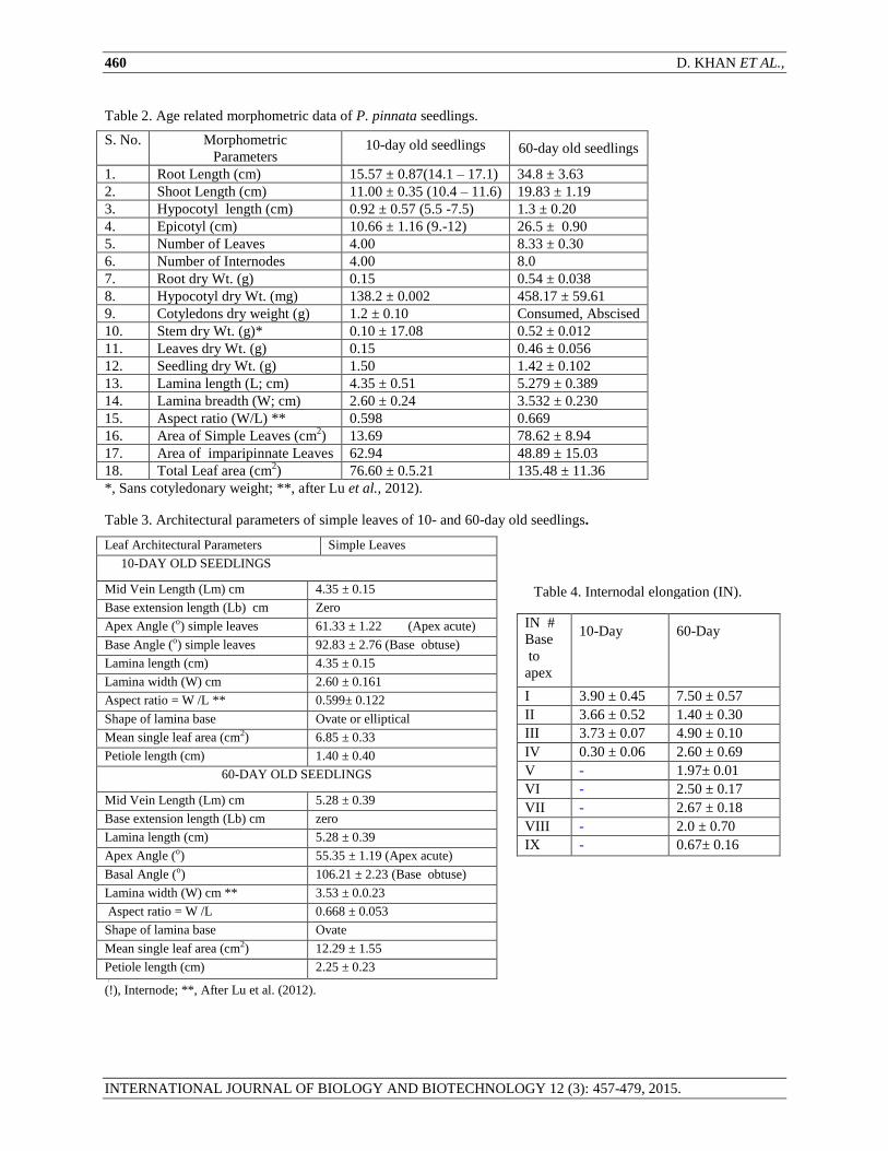
the seedlings for c 50 days. In 10-day old seedlings, various components of the seedlings were in the following order
of heaviness (Table2).
Cotyledons > > Leaves > Root > stem
Fig. 2. Stipules (A) and (B) Leaf venation in a leaflet of a trifoliate leaf (Brachidodromous type).
Table 1. Leaf types of P. pinnata seedlings.
Leaf Seedling I seedling II Seedling III Seedling IV Seedling V Seedling VI
1 * Simple Simple Simple Simple Simple Simple
2 Simple Simple Simple Simple Simple Simple
3 Simple Simple Simple Simple Simple Imparipinnate
4 Simple Simple Simple Simple Simple Imparipinnate
5 Simple Simple Simple Imparipinnate Simple Imparipinnate
6 Simple Simple Simple Imparipinnate Simple Imparipinnate
7 Imparipinnate Imparipinnate Simple Imparipinnate Imparipinnate Imparipinnate
8 Simple Imparipinnate Imparipinnate Imparipinnate Imparipinnate Imparipinnate
9 Imparipinnate Imparipinnate Imparipinnate Imparipinnate Imparipinnate Imparipinnate
*, From base to apex. Generally first 6-7 leaves are simple alternate and subsequent leaves are
mostly imparipinnate with exception in case of seedling # IV and VI.
A
BB Stipules
460 D. KHAN ET AL.,
INTERNATIONAL JOURNAL OF BIOLOGY AND BIOTECHNOLOGY 12 (3): 457-479, 2015.
Table 2. Age related morphometric data of P. pinnata seedlings.
S. No. Morphometric
Parameters
10-day old seedlings
60-day old seedlings
1. Root Length (cm) 15.57 ± 0.87(14.1 – 17.1) 34.8 ± 3.63
2. Shoot Length (cm) 11.00 ± 0.35 (10.4 – 11.6) 19.83 ± 1.19
3. Hypocotyl length (cm) 0.92 ± 0.57 (5.5 -7.5) 1.3 ± 0.20
4. Epicotyl (cm) 10.66 ± 1.16 (9.-12) 26.5 ± 0.90
5. Number of Leaves 4.00 8.33 ± 0.30
6. Number of Internodes 4.00 8.0
7. Root dry Wt. (g) 0.15 0.54 ± 0.038
8. Hypocotyl dry Wt. (mg) 138.2 ± 0.002 458.17 ± 59.61
9. Cotyledons dry weight (g) 1.2 ± 0.10 Consumed, Abscised
10. Stem dry Wt. (g)* 0.10 ± 17.08 0.52 ± 0.012
11. Leaves dry Wt. (g) 0.15 0.46 ± 0.056
12. Seedling dry Wt. (g) 1.50 1.42 ± 0.102
13. Lamina length (L; cm) 4.35 ± 0.51 5.279 ± 0.389
14. Lamina breadth (W; cm) 2.60 ± 0.24 3.532 ± 0.230
15. Aspect ratio (W/L) ** 0.598 0.669
16. Area of Simple Leaves (cm2) 13.69 78.62 ± 8.94
17. Area of imparipinnate Leaves 62.94 48.89 ± 15.03
18. Total Leaf area (cm2) 76.60 ± 0.5.21 135.48 ± 11.36
*, Sans cotyledonary weight; **, after Lu et al., 2012).
Table 3. Architectural parameters of simple leaves of 10- and 60-day old seedlings.
Leaf Architectural Parameters Simple Leaves
10-DAY OLD SEEDLINGS
Mid Vein Length (Lm) cm 4.35 ± 0.15
Base extension length (Lb) cm Zero
Apex Angle (o) simple leaves 61.33 ± 1.22 (Apex acute)
Base Angle (o) simple leaves 92.83 ± 2.76 (Base obtuse)
Lamina length (cm) 4.35 ± 0.15
Lamina width (W) cm 2.60 ± 0.161
Aspect ratio = W /L ** 0.599± 0.122
Shape of lamina base Ovate or elliptical
Mean single leaf area (cm2) 6.85 ± 0.33
Petiole length (cm) 1.40 ± 0.40
60-DAY OLD SEEDLINGS
Mid Vein Length (Lm) cm 5.28 ± 0.39
Base extension length (Lb) cm zero
Lamina length (cm) 5.28 ± 0.39
Apex Angle (o) 55.35 ± 1.19 (Apex acute)
Basal Angle (o) 106.21 ± 2.23 (Base obtuse)
Lamina width (W) cm ** 3.53 ± 0.0.23
Aspect ratio = W /L 0.668 ± 0.053
Shape of lamina base Ovate
Mean single leaf area (cm2) 12.29 ± 1.55
Petiole length (cm) 2.25 ± 0.23 [ (!), Internode; **, After Lu et al. (2012).
Table 4. Internodal elongation (IN).
IN #
Base
to
apex
10-Day
60-Day
I 3.90 ± 0.45 7.50 ± 0.57
II 3.66 ± 0.52 1.40 ± 0.30
III 3.73 ± 0.07 4.90 ± 0.10
IV 0.30 ± 0.06 2.60 ± 0.69
V - 1.97± 0.01
VI - 2.50 ± 0.17
VII - 2.67 ± 0.18
VIII - 2.0 ± 0.70
IX - 0.67± 0.16
SEEDLING CHARACTERISTICS OF PONGAMIA PINNATA (L.) PIERRE (PAPILIONACEAE) 461
INTERNATIONAL JOURNAL OF BIOLOGY AND BIOTECHNOLOGY 12 (3): 457-479, 2015.
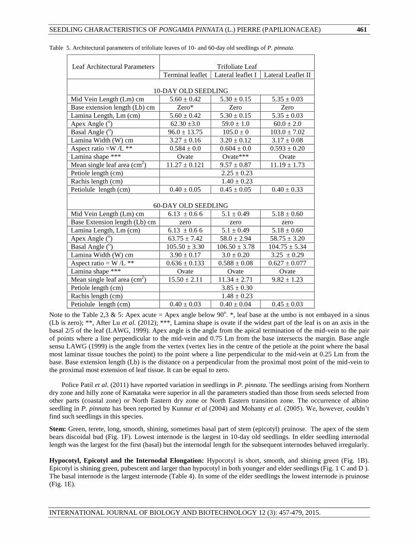
Table 5. Architectural parameters of trifoliate leaves of 10- and 60-day old seedlings of P. pinnata.
Leaf Architectural Parameters
Trifoliate Leaf
Terminal leaflet Lateral leaflet I Lateral Leaflet II
10-DAY OLD SEEDLING
Mid Vein Length (Lm) cm 5.60 ± 0.42 5.30 ± 0.15 5.35 ± 0.03
Base extension length (Lb) cm Zero* Zero Zero
Lamina Length, Lm (cm) 5.60 ± 0.42 5.30 ± 0.15 5.35 ± 0.03
Apex Angle (o) 62.30 ±3.0 59.0 ± 1.0 60.0 ± 2.0
Basal Angle (o) 96.0 ± 13.75 105.0 ± 0 103.0 ± 7.02
Lamina Width (W) cm 3.27 ± 0.16 3.20 ± 0.12 3.17 ± 0.08
Aspect ratio =W /L ** 0.584 ± 0.0 0.604 ± 0.0 0.593 ± 0.20
Lamina shape *** Ovate Ovate*** Ovate
Mean single leaf area (cm2) 11.27 ± 0.121 9.57 ± 0.87 11.19 ± 1.73
Petiole length (cm) 2.25 ± 0.23
Rachis length (cm) 1.40 ± 0.23
Petiolule length (cm) 0.40 ± 0.05 0.45 ± 0.05 0.40 ± 0.33
60-DAY OLD SEEDLING
Mid Vein Length (Lm) cm 6.13 ± 0.6 6 5.1 ± 0.49 5.18 ± 0.60
Base Extension length (Lb) cm zero zero zero
Lamina Length, Lm (cm) 6.13 ± 0.6 6 5.1 ± 0.49 5.18 ± 0.60
Apex Angle (o) 63.75 ± 7.42 58.0 ± 2.94 58.75 ± 3.20
Basal Angle (o) 105.50 ± 3.30 106.50 ± 3.78 104.75 ± 5.34
Lamina Width (W) cm 3.90 ± 0.17 3.0 ± 0.20 3.25 ± 0.29
Aspect ratio = W /L ** 0.636 ± 0.133 0.588 ± 0.08 0.627 ± 0.077
Lamina shape *** Ovate Ovate Ovate
Mean single leaf area (cm2) 15.50 ± 2.11 11.34 ± 2.71 9.82 ± 1.23
Petiole length (cm) 3.85 ± 0.30
Rachis length (cm) 1.48 ± 0.23
Petiolule length (cm) 0.40 ± 0.03 0.40 ± 0.04 0.45 ± 0.03
Note to the Table 2,3 & 5: Apex acute = Apex angle below 90o. *, leaf base at the umbo is not embayed in a sinus
(Lb is zero); **, After Lu et al. (2012); ***, Lamina shape is ovate if the widest part of the leaf is on an axis in the
basal 2/5 of the leaf (LAWG, 1999). Apex angle is the angle from the apical termination of the mid-vein to the pair
of points where a line perpendicular to the mid-vein and 0.75 Lm from the base intersects the margin. Base angle
sensu LAWG (1999) is the angle from the vertex (vertex lies in the centre of the petiole at the point where the basal
most laminar tissue touches the point) to the point where a line perpendicular to the mid-vein at 0.25 Lm from the
base. Base extension length (Lb) is the distance on a perpendicular from the proximal most point of the mid-vein to
the proximal most extension of leaf tissue. It can be equal to zero.
Police Patil et al. (2011) have reported variation in seedlings in P. pinnata. The seedlings arising from Northern
dry zone and hilly zone of Karnataka were superior in all the parameters studied than those from seeds selected from
other parts (coastal zone) or North Eastern dry zone or North Eastern transition zone. The occurrence of albino
seedling in P. pinnata has been reported by Kunnur et al (2004) and Mohanty et al. (2005). We, however, couldn’t
find such seedlings in this species.
Stem: Green, terete, long, smooth, shining, sometimes basal part of stem (epicotyl) pruinose. The apex of the stem
bears discoidal bud (Fig. 1F). Lowest internode is the largest in 10-day old seedlings. In elder seedling internodal
length was the largest for the first (basal) but the internodal length for the subsequent internodes behaved irregularly.
Hypocotyl, Epicotyl and the Internodal Elongation: Hypocotyl is short, smooth, and shining green (Fig. 1B).
Epicotyl is shining green, pubescent and larger than hypocotyl in both younger and elder seedlings (Fig. 1 C and D ).
The basal internode is the largest internode (Table 4). In some of the elder seedlings the lowest internode is pruinose
(Fig. 1E).
462 D. KHAN ET AL.,
INTERNATIONAL JOURNAL OF BIOLOGY AND BIOTECHNOLOGY 12 (3): 457-479, 2015.
Root: The tap root was light brown in colour with numerous light brown lateral roots (Fig.1 G). No nodules could
be found on roots. Primary root at base was 2.5 mm in diamter. Howeever, nodules do occur in P. pinnata and
Kesari et al. (2013) have isolated Rhizobium pongamiae sp. nov. from root nodules of P. pinnata. Roots 2-3 mm in
thickness in P. pinnata seedling are reported by Balasubramanyan and Swarupanandan (1986).
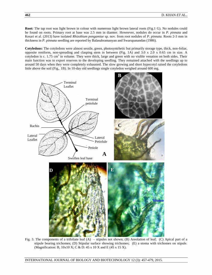
Cotyledons: The cotyledons were almost sessile, green, photosyntrhetic but primarily storage type, thick, non-foliar,
opposite reniform, non-spreading and clasping stem in between (Fig. 1A) and 3.0 x 2.0 x 0.65 cm in size. A
cotyledon is c. 1.75 cm3 in volume. They were thick, large and green with no visible venation on both sides. Their
main function was to export reserves to the developing seedling. They remained attached with the seedlings up to
around 50 days when they were completely exhausted. The slow growing and short hypocotyl raised the cotyledons
little above the soil (Fig,. 1B). In 10-day old seedlings single cotyledon weighed around 600 mg.
Fig. 3. The components of a trifoliate leaf (A) – stipules not shown; (B) Areolation of leaf; (C) Apical part of a
stipule bearing trichomes; (D) Stipular surface showing trichomes; (E) a stoma with trichomes on stipule.
(Magnification: B, 10x10 X; C & D: 45 x 10 X and E (45 x 15 X).
Swollen leaf base
Lateral
Petiolule
Petiole
Rachis
Terminal
petiolule
B E
Stoma
C
D
A
C
Terminal
Leaflet
Lateral
Leaflet
B
SEEDLING CHARACTERISTICS OF PONGAMIA PINNATA (L.) PIERRE (PAPILIONACEAE) 463
INTERNATIONAL JOURNAL OF BIOLOGY AND BIOTECHNOLOGY 12 (3): 457-479, 2015.
Leaf Architecture: Leaf architecture denotes the placement and form of those elements constituting the venation
pattern, marginal configuration, leaf shape and gland postion (Hickey, 1973). Hickey (1973, 1979) and LAWG
(1999) were important references to follow in this respect. Data on leaf architecture of P. pinnata leaves is given in
Table 3 and 5. Leaves stipulate (ovate, 2-2.5 mm in length, pinkish in winter but turning green after sometime) (Fig.
2A). petiolate, ovate, shining green and pubescent in early age. Leaf base and petiolule are darker green than petiole
and rachis. Leaf base obtuse. Apex often acuminate, acumen short.
The petiole length of simple as well as trifoliate leaves was larger in elder seedlings (Table 3 and 5). The
petiolule of terminal leaflet was comparable in length to the petiolules of the laterals leaflet (Table 5). The two
opposite lateral leaflets of a trifoliate leaf were somewhat unequal with each other and were individually smaller
than the terminal leaflet. In younger seedlings, the total area of trifoliate leaves (62.94 ± cm2) was larger than that of
the simple leaves (13.69 ± cm2). In elder seedlings the total leaf area of seedlings amounted to 135.48 ± 11.36 cm
2
per seedling. The total leaf area of younger seedlings was 76.6 ± 5.86 cm2
(Table 2). Petiolular lengths were almost
comparable in terminal and lateral leaflets. They were darker green in colour than that of the rachis and the petiole.
Petioles are little swollen at the base. Lateral leaflets of trifoliate leaves were opposite. The number of leaflets is
three in seedlings but five or more (odd number) in saplings and trees.
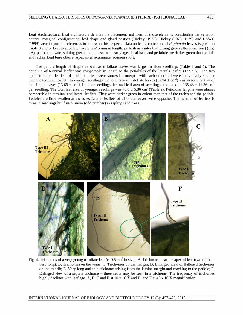
Fig. 4. Trichomes of a very young trifoliate leaf (c. 0.5 cm2 in size). A, Trichomes near the apex of leaf (two of them
very long); B, Trichomes on the veins; C, Trichomes on the margin; D, Enlarged view of flattened trichomes
on the midrib; E, Very long and thin trichome arising from the lamina margin and reaching to the petiole; F,
Enlarged view of a septate trichome – three septa may be seen in a trichome. The frequency of trichomes
highly declines with leaf age. A, B, C and E at 10 x 10 X and D, and F at 45 x 10 X magnification.
A B
D
C
C
E
FC
Type II
Trichome
Type I
Trichomes
Type I
Trichomes Type I
Trichomes
Type III
Trichome
Type III
Trichome
464 D. KHAN ET AL.,
INTERNATIONAL JOURNAL OF BIOLOGY AND BIOTECHNOLOGY 12 (3): 457-479, 2015.
[
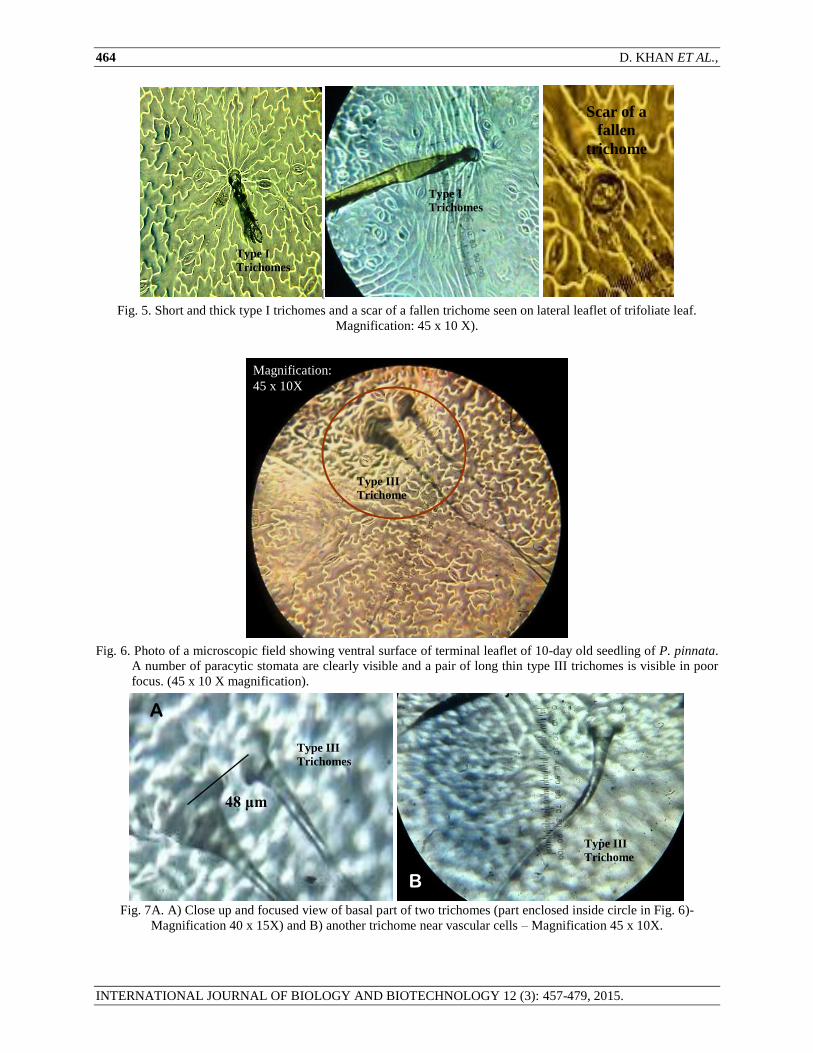
Fig. 5. Short and thick type I trichomes and a scar of a fallen trichome seen on lateral leaflet of trifoliate leaf.
Magnification: 45 x 10 X).
Fig. 6. Photo of a microscopic field showing ventral surface of terminal leaflet of 10-day old seedling of P. pinnata.
A number of paracytic stomata are clearly visible and a pair of long thin type III trichomes is visible in poor
focus. (45 x 10 X magnification).
Fig. 7A. A) Close up and focused view of basal part of two trichomes (part enclosed inside circle in Fig. 6)-
Magnification 40 x 15X) and B) another trichome near vascular cells – Magnification 45 x 10X.
A
B
Magnification:
45 x 10X
Scar of a
fallen
trichome
48 μm
Type III
Trichome
s
Type III
Trichome
Type III
Trichomes
Type I
Trichomes
Type I
Trichomes

SEEDLING CHARACTERISTICS OF PONGAMIA PINNATA (L.) PIERRE (PAPILIONACEAE) 465
INTERNATIONAL JOURNAL OF BIOLOGY AND BIOTECHNOLOGY 12 (3): 457-479, 2015.
Fig 7B. Diagrammatic representation of two adjacent Type III trichomes on the ventral surface of terminal leaflet of
a trifoliate leaf of 10 day old seedling to show their extra-ordinary lengths. (Not drawn to scale). The two
trichomes at the base were 48 μm apart.
Fig. 8. Stalked capitate uniseriate filiform trichomes on the ventral surface of very young trifoliate leaf. A,
somewhat dehydrating young leaf (10 x 10 X); B, C and D, fresh young leaf (45 x 10 X).
A
B B: 928 μm
A: 608 μm
Stoma
Basal cell
Unicellular body
(1.6 to 2.4 μm in
thickness)
A
Main
Body
Stalk
Tracheids
B
C D
Vein
Type I
trichome
Type IV
Trichome
Type IV
Trichome
Type IV
Trichome
Type I
trichome
Type IV
Trichome

466 D. KHAN ET AL.,
INTERNATIONAL JOURNAL OF BIOLOGY AND BIOTECHNOLOGY 12 (3): 457-479, 2015.
Fig. 9. Surface views of young cotyledon showing apparently no stomata on the outer surface (A) and the presence
of stomata on the inner surface (B). 10 X 10 X magnification.
Fig. 10. Cotyledonary stomatal types of Pongamia pinnata (45 x 10X). A, Young anomocytic stoma with five
subsidiaries; B, young staurocytic stomata; C and D, tetracytic stomata and E, One anisotricytic, one
tetracytic and a anomocytic stomata in close proximity; F, Anomocytic with seven subsidiaries. A, B and C,
Stomatal pore developing. Subsidiaries shown by asterisks.
E *
F
C A B
D
A B
*
*
*
*
*
*
*
*
*
*
*
*
*
*
* *
*
*
*
*
* *
*
*
* *
*
*
*
*
*
*
*
*
*
*

SEEDLING CHARACTERISTICS OF PONGAMIA PINNATA (L.) PIERRE (PAPILIONACEAE) 467
INTERNATIONAL JOURNAL OF BIOLOGY AND BIOTECHNOLOGY 12 (3): 457-479, 2015.
Fig. 11. An image of cotyledonary stomata showing a large anomocytic type stoma on the left with three abutting
stoma in the mid with large guard cells but no subsidiaries in between and another anomocytic stoma in the
right Magnification 45 x 15 X.
Fig. 12. A, a hypocotylar stoma (paracytic); B, a meristemoid on the hypocotylar surface; C and D, the Ventral
surface of young simple leaf of Pongamia; C, the general view showing developing stomata (45x10 X); D,
Two anomocytic stomata in laminar islands (45x10 X).
Fig. 13. Surface views of dorsal (A) and ventral (B) sides of lateral leaflets of trifoliate leaf of P. pinnata showing
stomata. The anticlinal cell is wavy (sinuate). Magnification: 10 x 10 X
A B
A B
C D

468 D. KHAN ET AL.,
INTERNATIONAL JOURNAL OF BIOLOGY AND BIOTECHNOLOGY 12 (3): 457-479, 2015.
Fig. 14. The paracytic stomata with cuticular striations seen on the dorsal surface of simple leaf (near veinlet) of 10-
day old seedling – a rare feature (A and B) and Common paracytic stomata on ventral surface (C). Note one
stoma with unequal subsidiaries (shown in circle). Magnification: 45 x 10 X.
Fig. 15. Few stomatal types seen on the ventral surface of lateral leaflet of trifoliate leaf. A and B, Differentially
developing stomata of anomocytic type on young leaflet with five and seven subsidiaries. C, a stoma with
cuticular striations. D, an anomocytic stoma besides paracytic stomata. E, normal paracytic stomata and large
anomocytic stoma with cuticular striations and seven subsidiaries of various sizes. Lobation of epidermal
cells develops with maturity. Subsidiaries shown by the asterisks. (Magnification 45 x 10 X) except E, 45 x
15X).
There are two types of leaves in the seedlings of P. pinnata. Simple leaves which arise first on germination but
imparipinnate compound (trifoliate) leaves subsequently (Table 1). There is, however, a great deal of irregularity
with respect to the first appearance of the trifoliate leaf. The trifoliate leaf may arise after two simple leaves in some
individuals or after four simple leaves in some cases or even after appearance of 6-7 simple leaves (Table 1). In
most of the seedlings the trifoliate leaf appeared subsequent to the successive appearance of six simple leaves.
Interestingly, a simple leaf may also be produced subsequent to a trifoliate leaf in rare cases (Table 1). Burger
A B C
D E
Vein let
Tissue
A B C
*
* *
*
*
*
*
* * *
* *
*
*
*
*
*

SEEDLING CHARACTERISTICS OF PONGAMIA PINNATA (L.) PIERRE (PAPILIONACEAE) 469
INTERNATIONAL JOURNAL OF BIOLOGY AND BIOTECHNOLOGY 12 (3): 457-479, 2015.
(1972) have reported that in Desoxylium dentifolium (Meliaceae) trifoliate first leaves are succeeded by simple ones
and still higher are trifoliate again. A similar sequence was recorded for Ulex (Leguminosae / Papilionaceae) by
Duke (1969).
The primary leaf of a seedling is simple, glaucous with small stipules, and no stipels. Petiole of simple as well
as trifoliate leaves was larger in elder seedlings (Table 3 and 5). Petiole inserted to lamina at c 90o. The leaves were
arranged on the stem spirally. Lamina symmetrical, margin entire, dorsiventral, thin and ovate in shape (axis of the
greatest width intersecting the leaf basal to the mid point of the leaf axis). Aspect ratio was 0.6 and 0.67 in case of
10 and 60-day seedlings, respectively. The mean aspect ratio of simple leaves was 0.599 ± 0.122 and 0.66 8 ± 0.053
in 10- and 60-day old seedlings, respectively. Leaf apex acute (apex angle < 90 o). There were similar ranges of
aspect ratio in leaflets of trifoliate leaves. Leaf apex was at times shortly acuminate. Leaf base is obtuse (base angle
> 90o and leaf base extension is zero and lamina not embayed in sinus in umbo region). Petiole is more or less equal
in leaves of 10- and 60-day old seedlings. Tertiaries were arising at right angle to the secondaries. The angle of
divergence (AOD) measured between the branch vein and the continuation of the source vein above the point of
branching was moderate (50-60o) between 1
o and 2
o veins and wide (c 90
o) between 2
o and 3
o veins. Intersecondaries
are narrow. Secondaries were alternate and distance between secondaries on left or right side of the midrib is more
or less equal. Tertiaries ramify in quaternaries and then they join to form reticulum. The areoles were wee-developed
pentagonal to polygonal in shape (Fig. 3 B). A few triangular areoles were also present. Areole size was small
(0.125.3 mm). Vein lets were generally simple linear.
Fig. 16. A few rare stomatal types on the ventral surface of terminal leaflet of trifoliate leaf of P. pinnata. A,
hemitricyclic paracytic stomata with distinct type of subsidiaries; B and C, large anomocytic stomata having
large more or less indistinct subsidiaries with less wavy contours; D, An arrested stomatal aperture (a variant of
paracytic stoma); E, A paracytic hemitricyclic stomata with distinct as well as indistinct subsidiaries.
Subsidiaries shown by the asterisks.
P. pinnata leaves exhibited brachidodromous type of venation (Fig. 2B) – secondaries joint to form in a series
of prominent arches. Brachidodromous venation (BD) is loop-veined venation in which main Secondaries emerging
from the midrib at more or less at regular interval turns upwardly to the apex at or near margin and loops to join the
next vein upwards to form prominent arches. Besides arches formed due to subsidiaries, additional sets of loops
outside the main brachidodromous loop were also observed. Tertiaries, percurrent, faint and weak. BD pattern of
venation has also been reported in woody perennials of Family Cunoniaceae (Dickson, 1975), Family Rubiaceae
(Coffea arabica) (Misra et al., 2010), Family Bignoniaceae (Jain, 1978) and Cinnamomum spp. (Ravindran et al.,
2003). BD venation has been reported in Erythrina suberosa (Papilionaceae) and Bauhinia racemosa (Khan et al.,
A B C
D E
*
* *
* *
470 D. KHAN ET AL.,
INTERNATIONAL JOURNAL OF BIOLOGY AND BIOTECHNOLOGY 12 (3): 457-479, 2015.
2014, 2015). In arborescent flora, the brachidodromous pattern prevails in tropical floras whereas non-
brachidodromous patterns prevail in Northern temperate floras (Bailey and Sinnot, 1916).
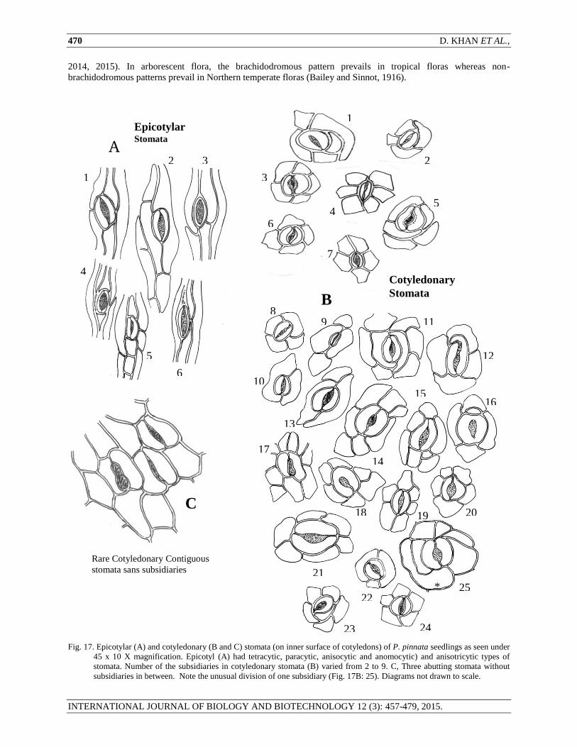
Fig. 17. Epicotylar (A) and cotyledonary (B and C) stomata (on inner surface of cotyledons) of P. pinnata seedlings as seen under
45 x 10 X magnification. Epicotyl (A) had tetracytic, paracytic, anisocytic and anomocytic) and anisotricytic types of
stomata. Number of the subsidiaries in cotyledonary stomata (B) varied from 2 to 9. C, Three abutting stomata without
subsidiaries in between. Note the unusual division of one subsidiary (Fig. 17B: 25). Diagrams not drawn to scale.
Epicotylar
Stomata
Cotyledonary
Stomata
Rare Cotyledonary Contiguous
stomata sans subsidiaries
C
A
B
1
2 3
4
5
6
1
2
3
4 5
6
7
8 9
10
11
12
13
14
3
15
3 16
3
17
3
18
3 19
3
20
3
21
3 22
3
23
3
24
3
25
3
*

SEEDLING CHARACTERISTICS OF PONGAMIA PINNATA (L.) PIERRE (PAPILIONACEAE) 471
INTERNATIONAL JOURNAL OF BIOLOGY AND BIOTECHNOLOGY 12 (3): 457-479, 2015.
Fig. 18. Common and less frequent foliar stomatal types on ventral surface of leaves of Pongamia pinnata
seedlings - as seen under 45 x 15 X magnification. Note wavy cell contours of subsidiaries as well as the
pavement epidermal cells of leaf and varying orientation of the stomatal long axis. Diagrams not drawn to
scale. Note the cell wall developing in one of the subsidiary (shown by the asterisk). Paracytic (1A, 1B, 4),
anisocytic (5, 6), anisotricytic (2), tetracytic (3, 7), anomocytic (8, 9) and anomocytic with cuticular striations
(10).
Surface ornamentation of seedlings
Trichomes: The young leaves were pubescent but turned glabrescent with age. There were four types of non-
glandular trichomes in P. pinnata. Trichomes do occur on dorsal surface of young leaf but rarely. They are
numerous on ventral surface;
Type I: Short flat, bent near base, curved, brown, pointed at the apex and unicellular trichomes were present on the
ventral surface (on and along the veins) of the young stipules and trifoliate leaves, on petiole, apex and margins (Fig.
3 (C,D and E and Fig. 4 B,C, and D). Being frequently curved, it was difficult to measure them. However, those
trichomes which could be measured (N = 50) were found to be 248.54 ± 15.24 μm in length (80 - 464 μm; CV =
43.37%). These trichomes were less variant in width (CV= 4.77%; 4.8 to 24 μm) and concentrated around the mean
value of 13.80 ± 0.66 μm). In many cases the basal part of the trichome was black in colour. When they fall, they
may leave a scar on the leaf surface (Fig. 5). Some of such trichomes were sword like (Fig. 8A).
Type II: Septate multicellular non-glandular trichome - Interspersed among the type I trichome on petiole of the
young leaflet, a trichome was found with discrete septa. It was more or less comparable to average type I trichomes
in length (Fig. 4 E and F). The visible part of the trichome measured c. 268.8 μm in length.
Common Paracytic
Foliar Stomata
Less frequent Foliar Stomatal
Complexes
Cuticular
Striations
1B
2
3
4
5
6
7 8
9
10
*
1A 1
472 D. KHAN ET AL.,
INTERNATIONAL JOURNAL OF BIOLOGY AND BIOTECHNOLOGY 12 (3): 457-479, 2015.
Type III: Long and thin non-glandular trichomes – These unicellular non-glandular trichomes were long and thin
provided with large basal cell (Fig. 4 A and E and Fig. 6 and 7 A and B). They easily weathered from the leaves and
stipules with age so that adult and mature leaves were glabrous and glaucous. We could, however, find five such
trichomes on adult lateral leaflet of trifoliate leaf measuring 608, 928, 2720, 560, and 640 μm in length – the last two
were broken ones. Two such trichomes originated in close proximity of 48 μm (Fig. 7A).
Type IV: Interspersed among the type I trichomes, generally on the vascular nerves (rarely on the island as well)
several stalked capitate uniseriate filiform non-glandular trichomes were observed in case of young lateral leaflet of
trifoliate leaves (Fig. 8). These trichomes averaged to 87.52 ± 2.56 μm in length (CV = 16.02%). The trichomes
consisted of a stalk of 2-4 cells (20.05 ± 1.0 μm in length, N = 30, CV= 26.9%) and the main body made up of 6-7
cells (67.47 ± 2.23 μm in length, N = 30, CV= 15.6%). The main body was 22.9 ± 0.63 μm wide (CV = 15.2%).
Leelavathi and Ramayya (1983) described 28 types of trichomes from taxon Papilionoideae. On the basis of
trichome distribution Papilionoideae was more related to Mimosoideae than Caesalpinioideae. Unicellular, conical,
warty and bent-near-base trichomes have been reported by Kumar et al., 2013) on the pods of P. pinnata. Simple
unbranched long trichomes are known from the genus Abrus. Abrus prectaurius bears trichome of 244.50 ± 50.57
and 195.0 ±49.038 μm in length on adaxial and abaxial surface of leaf, respectively. The trichomes of A. canescens
(392.10 ± 114.14 and 495.12 ± 190.11 μm on adaxial and abaxial surfaces, respectively) were larger than A.
prectaurius. The size of trichome we recorded from terminal leaflet of first trifoliate leaf was exceptionally larger.
Eglandular solitary one-celled filiform hairs of variable lengths are reported from Okra by Osawaru and Dania-
Ogbe., 2011) – short conical hairs – 20 to 70 μm ; long conical unicellular hairs hair of 1200 to 1700 μm and long
stellate hair 800 to 1100 μm in lengths. Long uniseriate trichomes are known from Trifolium pretense (635-1357
μm). Medicago arabica has scarce but large uniseriate trichomes (460 - 930 μm in length) constituted by variable
number of cells (Yagneddu et al., 2009). Non-glandular trichomes are also present on both surfaces of Adenanthera
pavonina (around 128 μm in length) (Partha and Rahaman, 2015). The unicellular unbranched trichomes of
Indigofera numularifolia L. Alston on abaxial surface of leaf measured 495 μm (Umar et al., 2014). The conical
eglandular trichomes of Hibiscus trianum measured 350 to 700 μm in length (Shaheen et al., 2009).
The glabrescent – pubescent foliar spectrum on the basis of the absence and presence of trichomes found
taxonomic value due to the work of Rao and Rao (1992). Adeniji and Ariwaodo (2012) have differentiated
Pericopsis elata and P. laxiflora on the basis of hypostomatosis and the presence of trichomes. The leaves of
Pericopsis elata are hypostomatic and trichomes are absent where as that of Pericopsis laxiflora are amphistomatic
and bear trichomes which are unicellular and non-glandular type (Adeniji and Ariwaodo, 2012).
The presence of trichomes on ventral surface of terminal leaflet of first trifoliate leaf of P. pinnata appears to be
an early age phenomenon. The trichomes are extremely delicate and easily weather with leaf age due to random
events resulting in shining glabrescent leaves.
Stomata: Several types of stomata were found on the surface of hypocotyl, epicotyl, inner surface of cotyledons and
dorsal and ventral surfaces of leaf.
Cotyledonary Stomata: No stomata were seen on outer surface of cotyledons (Fig. 9). Cotyledonary stomata were
of paracytic (Fig. 9, 10 and Fig.17 B- 8, -9, -10, -11, -12, -13), anomocytic ( Fig.17B – 1, -3, -4 ,-7, -14, -15, -16, -
17,-18, -19, -20, -21, -23, -24, -25) staurocytic (Fig. 17B -2), tetracytic (Fig 17B – 6), anisocytic, (Fig. 17B – 5, 22).
The number of subsidiaries abutting stoma varied from 2 to 9. There were abnormal stomata such as stomata lacking
subsidiaries and contiguously adjacent to each other (Fig. 11, 17C), stomata with incomplete pore. Stomatal length
of inner surface cotyledonary stomata ranged from 6.96 to 31.32 μm (mean = 26.55 ± 1.18). Indeed, there were two
distinct sizes; smaller (6.96-12 μm) and larger (20.0-32 μm) stomata,.
Hypocotylar Stomata: Stomata are less frequent on hypocotyl. Some meristemoids and paracytic stomata were
seen (Fig. 12A and B).
Epicotylar stomata: The stomata present on epicotylar stem were paracytic (a stomatal type in which one or more
of the subsidiaries that flank the stoma are parallel to the long axis of the guard cells (Fig. 17A: 2)), anisocytic (a
stoma completely surrounded by only three subsidiaries variable in position and shape but one of the subsidiaries is
distinctly small (Fig. 17A: 3)), anomocytic (a stoma completely surrounded by four or more subsidiaries variable in
position, shape and size (Fig. 17A: 1, 4, 5)), and anisotricytic type (a stoma completely surrounded by only three
subsidiaries, variable in position and shape, but one of the subsidiary is distinctly large (Fig. 17A: 6)). Epicotylar
stomatal length ranged from 18.08 to 25.52 μm (mean = 19.08 ± 1.32 μm).
Foliar Stomata: The leaves of P. pinnata seedlings are considered to be hypostomatic i.e. no stomata found on
dorsal surface of leaf (Park and Farukawa, 1999; Rai and Misra, 2013) and it also appears to be the case as in Fig.

SEEDLING CHARACTERISTICS OF PONGAMIA PINNATA (L.) PIERRE (PAPILIONACEAE) 473
INTERNATIONAL JOURNAL OF BIOLOGY AND BIOTECHNOLOGY 12 (3): 457-479, 2015.
12A. We, however, could discover two paracytic stomata on the dorsal surface of simple leaf of 10-day old seedling
(Fig. 14A and B) which is an extremely rare feature. The ventral surface is multistomatic (Fig. 12C and D and 13B).
The stomatal frequency is known to vary on upper and lower surfaces of leaf (Ekenayake et al., 1998). The greater
number of stomata on ventral surface of leaf is common in species that occur in xeromorphic environments, a fact
explained as a feature that minimizes water loss by ostiolar evapo-transpiration (Esau, 1974; Cutter, 1986). The
orientation of stomata is variable on the leaf surface. The ground epidermal cells are irregular in shape due to wavy
anticlinal cell wall pattern as is also reported in genus Abrus (Agbagwa and Okoli (2006) and Erythrina suberosa
(Khan et al., 2014) and several other papilionaceous plants. There were several types of stomata on ventral surface
of the leaf (Fig.12B, 13B, 14, 15, 16, and 18). These included paracytic, anisocytic, anomocytic, staurocytic and
tetracytic type of stomata (sensu Prabhakar, 2004). The most common stomatal type in P. pinnata is of paracytic
type followed by anomocytic type. Staurocytic stomata are known to develop from anisocytic stomatal complexes of
the seedling leaves in Monocalyptus (Eucalyptus, Myrtaceae) (Carr and Carr, 1990b).
There were some abnormal stomata such as two paracytic stomata with common subsidiary cell (Fig. 18 - 4),
contiguous paracytic stomatal complexes (Fig. 18 -1) and large anomocytic stomata with cuticular striations (Fig. 18
- 10). Paracytic hemitricytic stomata with distinct subsidiaries or distinct as well as indistinct subsidiaries were also
rarely present on the ventral surface of leaflet of trifoliate leaf (Fig. 16 A and E). The number of subsidiary cells
associated with different types of stomata varied from two to nine. Regarding the number of subsidiaries associated
with stomata, the studies by Car and Car (1990a), Obiremi and Oladale (2001) and Oyeleke et al. (2004) had
confirmed that larger the number of subsidiaries cells surrounding the guard cells, the faster the opening of the
stomata i.e. more transpiration and CO2 absorption.
Several types of stomata on leaf surface signified the diversity of stomatal types even on the same surface of a
leaf as also been reported by Saheed and Illoh (2010) and Aniesua and Silas (2012). Metcalfe and Chalk (1979) have
reported several types of stomata in Papilionaceae – Anomocytic, paracytic, and parallelocytic. They have reported
anomocytic stomata from 171 families of angiosperms, paracytic stomata from 118 families and anisocytic stomata
from 45 families but not from Papilionaceae. However, the thirteen species of the family Fabaceae (Genus Ademsia,
Galega, Lotus, Lupinus, Melilotus, Parkinsonia, Senna, Trifolium and Vicia) were reported to be characterized with
anisocytic, anomocytic stomata. Stomata are predominantly paracytic in leaves of Citrus spp. (Obiremi and Oladele,
2001) and many Macaranga spp. (Norfaizal et al., 2012). Stomata are extremely variable even in the members of a
tribe and even within a genus (Metcalfe and Chalk, 1950) and in a species as well. That is more than one type of
stomata frequently occur on the same leaf surface. Diversity of stomatal types even on the same surface (leaf) as
noted in the present studies has also previously been reported (Shah and Gopal, 1969; Ahmed et al., 2009). In spite
of diversity, most frequent type of stomata can be, however, used as a taxonomic character. Epidermal (stomatal)
studies may act as markers in taxonomic delimitation (Saheed and Illoh (2010). But Paliwal (1969) considered that
stomatal studies may have little taxonomic value unless the development of different stomata types is studied. In
Papilionaceae, Shah and Gopal (1969) reported that different types of stomata follow a similar pattern of
development. The diversity of stomatal types, even on the same surface of an organ, indicates the weakness in using
stomata as a taxonomic character (Pant and Kidwai, 1964). Shah and Gopal (1970), however, asserted that in spite
of diversity, the most frequent type of stomata can be used as taxonomic character. Epidermal surface structure is
reported to bear definite diagnostic features justifying the separation of the genera Senna and Chamaecrista from
their initial genus Cassia (Saheed and Illoh, 2010).
Based on the idea that epidermal cells surrounding stomata are not differentiated into subsidiary cells, P.
pinnata is described to bear anomocytic type of stomata by Vidya Lakshmi and Avinash (2013). Menpara and
Chanda (2014) have described paracytic type of stomata from P. pinnata. We found anomocytic stomata more
frequent in simple leaves. Paracytic stomata were more numerous in trifoliate leaves. Anomocytic stomata were
more frequent on or near the vascular zone whereas paracytic stomata scattered all over laminar islands. It may be
asserted that Paracytic stomata are the most frequent in several papilionaceous plants (Alysicarpus bupleurifolius, A.
monilifer, A. rugosus, Arachis hypogea, Cajanus cajan, Canavalvia gladiata, Clitoria terneata, Erythrina
cristagalli, E. indica, Lathyrus sativus, Lens esculentus, Medicago sativa, and Tephrosia purpurea. The genus
Senna has been reported to have paracytic stomata (Freire et al., 2005). There are, however, anisocytic stomata in
Glycine soja, Pisum sativum and Sesbania sesban and anomocytic stomata in Sesbania grandiflora and Trigonella
foenum-graceum. The stomata on leaf of Alhagi maurorum (Fabaceae) are paracytic and anisocytic types and on
stem anomocytic type (Bokhari and Dasti, 1991). Thirty-six dicotyledonous species of 34 genera and 20 families of
district Tank (Khyber Pakhtoonkhwah, Pakistan) were examined by Ahmad et al. (2009). Most of them were
amphistomatic. Anisocytic type of stomata were the dominant type in 12 spp. Staurocytic and diacytic stomata were
only present in seven and six species, respectively. In six species two or three types of stomata were present
simultaneously. Staurocytic stomata are reported in Erythrina subrosa L. (? E. suberosa Roxb.) by Khan et al.
(2011). Several species of genus Erythrina have been reported to have paracytic stomata for instance, E. speciosa

474 D. KHAN ET AL.,
INTERNATIONAL JOURNAL OF BIOLOGY AND BIOTECHNOLOGY 12 (3): 457-479, 2015.
and E. falcata (Almeida 2010, 2011); E. velutina (da Silva et al., 2013), E. variegata (Matheus and Lopes, 2007), E.
suberosa (Khan et al, 2011; Biradar, et al., 2013), and E. indica (Tripathi and Mondal, 2012). Stomata on dorsal
surface of P. pinnata leaf are very rare but not absolutely absent. Of 45 species of order Leguminales, 31 species are
reported to be amphistomatic and only 14 spp. hypostomatic by Tripathi and Mondal (2012). Family Fabaceae is
more diverse in stomata than Families Caesalpiniaceae and Mimosaceae (Tripathi and Mondal, 2012). According to
them, three stomata types of Leguminales were paracytic, anisocytic and anomocytic - found in various
combinations. The most common stomata in legumes are of the paracytic type and paracytic and anomocytic types
may although occur together in Caesalpiniaceae but never occur together in Fabaceae. This contention of Tripathi
and Mondal (2012) is, however, open to discussion. The non-occurrence of paracytic and anomocytic stomata
together in 24 papilionaceous species, they have investigated, may hold well in those species but not in case of
whole Family Fabaceae (Papilionaceae) as obvious from the above discussion and evident from the Fig. 19.
Fig. 19. An image of ventral surface of an adult lateral leaflet of the trifoliate leaf of 100 day old sapling of P. pinnata. A large
anomocytic and several paracytic stomata scattered on the surface are visible. Substantial cuticular deposit is apparent in the
vascular zone. It follows that paracytic and anomocytic stomata may occur together in Papilionaceae. (Magnification: 45 x
10 X)
The stomatal density in P. pinnata on simple and trifoliate leaves was recorded to vary from 184.59 to
235.11.mm-2
averaging to 211.59 ± 2.60.mm-2 (Table 6). Stomatal density on leaves varied by a quantum ranging
from 10.51 to 14.58. In both types of leaves and therir pooled data, tendency of normal distribution was apparent as
indicated by the insignificant values of KS-z (Table 6; Fig.20). It indicated that spatial distribution of stomata on the
leaf surface, irrespective of their kind, was heterogeneous. ). Our data is in agreement with Park and Farukawa
(1999). They reported stomatal density in P. pinnata collected from rain forest of Malaysia to be 219 ± 44 per mm2.
According to Rai and Misra (2013) in this species average number of stomata per mm2 was higher in air-polluted
environment (134.16 ± 1.42 in mature leaves) due to vehicular exhaust than non-polluted conditions (117.73 ± 1.30,
in mature leaves. Stomata were, however, larger in unpolluted conditions (23.87 ± 0.07 μm in length and 7.80 ± 0.03
μm in width
The pore size of the stomata in of paracytic and anomocytic types of stomata in P. pinnata leaves is given in
Table 7. The stomata of P. pinnata are of moderate size. The length and width of paracytic stomata averaged to
22.78 ± 0.344 and 11.46 μm, respectively. Average length and width of anomocytic stomata were 28.70 ± 1.48 and
15.87 ± 1.21 μm, respectively. It is obvious that anomocytic stomata were larger in length and width in comparison
to the paracytic stomata. Stomata of various sizes have been reported from Family Leguminosae. The stomata of
Erythrina variegata are reported to range from 112.2 to 19.6 μm and 4.9 to 11.9 μm in length in flat and mountain
areas of Philippines, respectively (Combalicer et al., 2010). Our stomatal measurements in P. pinnata are lesser
than the stomatal length on the adaxial surface of leaf of Erythrina indica Lamk. (38.49 μm) but comparable to that
of Dalbergia sissoo (24.3 μm) as reported by Tripathi and Mondal (2012). The guard cell size of Prosopis cineraria,
Alhagi maurorum and Crotalaria burhia are reported to be 27.39 ± 2.01, 15.16 ± 1.55 and 28.05 ± 6.93 μm in
length, respectively on the lower foliar epidermis (Bokhari and Dasti, 1991). Stomata in legume herbs such as Vicia
faba, Melilotus indica, Lathyrus aphaca are smaller (13 -14 μm) (Ahmad et al., 2009). The stomata of tree legume
species are generally larger than that of herbs (Tripathi and Mondal, 2012). Carbon dioxide concentration and
temperature are, however, reported to influence the stomatal density on the leaf (Beerling and Chaloner, 1993).

SEEDLING CHARACTERISTICS OF PONGAMIA PINNATA (L.) PIERRE (PAPILIONACEAE) 475
INTERNATIONAL JOURNAL OF BIOLOGY AND BIOTECHNOLOGY 12 (3): 457-479, 2015.
Table 6. Foliar stomatal density per mm2 of ventral surface of leaf of P. pinnata seedling.
100.00 150.00 200.00 250.00 300.00 350.00
STOMATAL DENSITY SIZE CLASSES
0
10
20
30
40
FR
EQ
UE
NC
Y
Fig. 20. Frequency distribution of foliar stomatal density per mm2 (pooled data).
Table 7. Stomatal sizes (μm) of paracytic and anomocytic stomata on ventral surface of leaf.
It may be mentioned that the locality of collection of specimens may influence the epidermal structure and show
wide variation in stomatal types e.g., specimens of Heliotropium europium collected from Quetta had anomocytic,
anisocytic, brachyparacytic, staurocytic, cyclocytic and actinocytic stomata as common types while specimens
Parameters
Leaf # 1
(Simple)
Leaf # 2
(Simple)
Leaf # 3
(Trifoliate)
Pooled
data
Mean 215.05 184.59 235.11 211.59
SE 3.198 2.8541 4.1162 2.6012
Median 216.24 186.75 226.066 216.238
CV (%) 10.51 10.93 12.38 14.58
Skewness -0.109 .088 0.970 0.493
SE g1 0.337 .337 0.337 0.198
Kurtosis -0.619 -1.136 2.031 0.890
SE g2 0.662 .662 .662 0.394
Minimum 167.09 147.43 167.09 147.43
Maximum 265.38 216.24 334.19 334.19
KS-z 0.897 1.039 1.287 1.039
p 0.397 0.230 0.073 0.232
N 50 50 50 150
Paracytic stomata
Parameter Mean ± SE Range CV (%)
Stomatal length 22.78 ± 0.344 16.0-25.60 9.3
Stomatal width 11.46 ± 0.435 8.0-17.60 23.40
Anomocytic stomata
Stomatal length 28.70 ± 1.477 16.0-32.0 17.83
Stomatal width 15.87 ± 1.205 9.60-24.0 26.32

476 D. KHAN ET AL.,
INTERNATIONAL JOURNAL OF BIOLOGY AND BIOTECHNOLOGY 12 (3): 457-479, 2015.
collected from Pishin have no anisocytic but has an additional brachyparatetracytic stomata which didn’t occur in
the specimens of Quetta (Dasti et al., 2003). There were some abnormal stomata in P. pinnata. Stomatal
abnormalities are common in Papilionaceae. Contiguous stomata are found in Erythrina indica but rarely. They are,
however, frequent in Lathyrus sativus (Shah and Gopal, 1969) and may be formed by budding. Melilotus albus,
Alysicarpus vaginallis, Aeschymonene indica, and Desmodium spp. also reported to possess contiguous stomata
(Kothari and Shah, 1975; Bora and Baruah, 1979). Contiguous stomata are very common in Solanaceae (Inamdar
and Patil, 1976); according to them, they develop from the adjacently placed meristemoids or as a result of
readjustment during maturation of the epidermis. Aniesua and Silas (2012) have reported un-open stomatal pores,
two-stomata sharing one subsidiary cell, one guard cell, parallel contiguous and aborted guard cell in Acalypha
(Euphorbiaceae). Stomatal clustering on epidermis is reported in more than 60 species (Gan et al., 2010). Drought
and salinity increase the occurrence of contiguous stomata which indicates environmental- signaling-correlation
with contiguous stomata (Gan et al., 2010). Abnormal stomata are suggested to be the result of environmental
perturbations (Carr and Carr, 1990a) and environmental stress like drought and salinity (Gan et al., 2010). Warming
may significantly decrease the average nearest neighbour distance between stomata (Zheng et al. (2013). Croxdale
(2000) has suggested that structure, development and patterning of stomata on the leaf surface is the function of
complex processes, they should be viewed from evolutionary, physiological, ecological and organ view-point.
Research should be undertaken with local flora from this view-point.
REFERENCES
Abubakar, B.Y. and I.A. Yunusa (1998). Epidermal structure and stomatal ontogeny as an aid to the taxonomic
identification of some species of Acacia (Leguminosae: Mimisoideae) from Nigeria. Nigerian J. Bot. 11: 117-
123.
Adeniji, K.A. and Ariwaodo, J.O. (2012). Comparative foliar epidermal studies of genus Pericopsis (Papilionaceae)
in Nigeria. Phytologia Balcanica 18(12): 37-41, Sofia, 2012.
Agbagwa, T.O. and Okoli (B.E. (2006). Leaf epidermal micromorphology in the systematics of Abrus
(Papilionaceae) in parts of tropical West Africa. Asian J. Plant Sci. 5(1): 41-49.
Ahmad, K., M.A. Khan, M. T. Ahmad, M. Zafar, M. Arshad, and F. Ahmad (2009). Taxonomic diversity of stomata
in dicot flora of a district Tank (NWFP) in Pakistan. African J. Biotech. 8(6): 1052-1055.
Ahmad, M., Zafar, M., Khan, M.A. and S. Sultana (2009). Biodiesel from Pongamia pinnata L. Oil: A promising
alternative Bioenergy source. Energy Sources. Part A: Recovery utilization and Environmental Effects. 31(16):
1436-1442. (doi- 10.1080/15567030802092882).
Ali, S.I. (1977). Papilionaceae: Flora of West Pakistan. # 100. (Eds. E. Nasir and S.I. Ali). 389 pp.
Almeida, E.E. (2010). Caracteriza ção farmacognóstica da espécie Erythrina falcata Benth. Fabaceae. Rev. Bras.
Farmacogn 20: 105.
Almeida, E.E. (2011). Caracterizaéão farmacotógica das folias de espécie Erythrina speciosa Andrews, Bio Far 5:
34-47.
Amritphale, Dilip and S.K. Sharma (2008). Seedlings of dicots: Form and Function. Resonance J. Sci. Edu. 13 (5):
468 -474.
Aniesua, E.U. and E.I. Silas (2012). Leaf epidermal studies of three species of Acalypha Linn. (Euphorbiaceae).
Adv. Appl. Sci. Res. 3 (5): 3185-3199.
Bailey, I.W. and E.W. Sinnot (1916). The climatic distribution of certain types of angiosperm leaves. Am. J. Bot. 3:
24-39.
Balasubramanyan, K. and Swarupanandan, K. (1986). A study of the seedlings of some commercially important trees
of Kerala. KFRI Res. Rep. # 41. Kerala Forest Res. Inst., Thrissur. 64 Pp.
Beering, D.J. and W.G. Chaloner (1993). The impact of atmospheric carbon dioxide and temperature change on
stomatal density: Observations from Quercus rober Lammas leaves. Ann. Bot. 71 (3):231-235.
Biradar, R.M., V.S. Gambhire, and A.S. Dhabe (2013). Pharmacognostic studies in Erythrina suberosa Roxb. and
E. variegata L. Bioinfolet 10 (2b): 610-611.
Bokhari, M.H. and A.A. Dasti (1991). Ecological guidelines for exploitation of natural resources in Thal and
Cholistan sand dunes. Final Tech. Rep. (1990-91). Pak. Sci. Found. Res. Proj. PBZ-4/Bio-154. Inst. Pure &
Appl. Biol. Bahauddin Zakariya Univ. Multan, Pakistan.
Bora, N. and P. Baruah (1979). Contiguous stomata in Desmodium Desv. (Papilionaceae). Curr. Sci. 487(1): 27-28.
Burger, Hzn. D. (1972). Seedlings of some tropical trees and shrubs, mainly of South West Asia. 399 Pp. Centre of
Agricultural Publishing and Documentation (PUDOC), Wageningen.
Carr, D.J. and S.G.M. Carr (`1990b). Staurocytic stomatal complexes in species of Monocalyptus sensu Car and Car
(Eucalyptus, Myrtaceae). Aust. J. Bot. 38 (1): 45-52.

SEEDLING CHARACTERISTICS OF PONGAMIA PINNATA (L.) PIERRE (PAPILIONACEAE) 477
INTERNATIONAL JOURNAL OF BIOLOGY AND BIOTECHNOLOGY 12 (3): 457-479, 2015.
Carr, S.G. and D.J. Carr (1990a). Cuticular features of the central Australian bloodwoods Eucalyptus section
Corymbose (Myrtaceae). Bot. J. Linnean Soc. 102: 126-156.
Croxdale, J.L. (2000). Stomatal patterning in angiosperms. Am. J. Bot. 87 (8):1069-1080.
Cutter, E.G. (1986). Anatomia Vegetal: Celulas e tecidas. Sâo Paulo. Roca. (Seen in da Silve et al., 2013).
da Silva, M.M.B., Santana, A.S.C.O., Pimentel, R.M.M., Silva, F.C.L., Randan, K.P and Soares, L.A.L. (2013).
Anatomy of leaf and stem of Erythrina velutina. Rev. Bras. Farmacognosia (Braz. J. Pharmacognosy = AOP
00713.
Das, D. Ch. and N.D. Paria, (1999). Seedling morphology in identification of some Indian species of Bauhinia L.
(Caeselpiniaceae). Fedes Repertorium 110(5-6): 375-379.
Dasti, A.A., T.Z. Bokhari, S.A. Malik and R. Akhtar (2003). Epidermal morphology in some members of family
Boraginaceae in Balochistan. Asian J. Pl. Sci. 2 (1): 42-47.
Deb, D.K. and N. Paria (1986). Seedling morphology of some economic species. Ind. Agriculturist 30(2): 133-142.
Dickson, W.C. (1975). Leaf Anatomy of Cunoniaceae. Bot. J. Linn. Soc.71: 275-294.
Dilcher, D.LK. (1974). Approaches to the identification of angiosperms leaves. Bot. Rev. 40(1): New York.
Duke, J.A. (1969). On tropical tree seedlings I. seeds, seedlings, systems and Systematics. Ann. Miss. Bot. Gard. 56:
125-161.
Ekenayake, I.J., Osini, D.V.S. and Porto, M.C.M. (1998). Physiology of Cassava. (http://www. Iita.org)
Elkhalifa, K.F and Aref, I.M.(2004). Morphological studies of fourteen Acacia species seedlings in Saudi Arabia.
Res. Bullet. No. 122. Agr. Res. Center, King Saud University, pp. 5-11.
Esau, K. (1974). Anatomia des Plantas com Sementes. Sâo Paulo; Edgard Blücher.
Freire, S.E., A.M. Arambari, N.D. Bayon, G. Sancho, U Urtubey, C. Monti, M.C. Novoa and M.N. Colares (2005).
Epidermal characteristics of toxic plants for cattle from Salado River Basin (Buenos Aires, Argentina). Bol.
Soc. Argent. Bot. 40(3-4): 241-281.
Gan, Yi, L. Zhou, Zhong-Ji Shen, Yi-Qiong Zhang, Gen-Xian Wang (2010), Stomatal clustering, a new marker for
environmental perception and adaptation in terrestrial plants. Bot. Studies 51: 325-336.
Garwood, N.C. (1996). Functional morphology of tropical tree seedlings (pp. 59-129). In: The Ecology of Tropical
Forest Tree Seedlings (Ed. M.D. Swaine), MAB Series, Vol.17, UNESCO, Paris.
Hickey, L.J. (1973). Classification of the architecture of dicotyledonous leaves. Am. J. Bot. 60(1): 17-33.
Hickey, L.J. (1979). A revised classification of the architecture of dicotyledonous leaves. Pp. 25-39. In (C.R.
Metcalfe and L. Chalk, Eds.). Anatomy of the Dicotyledons. II Ed. Vol I. Systematic Anatomy of the Leaf and
Stem with Brief History of the Subject. Oxford: Clarendon Press. 176 Pp.
Inamdar, J.A. and R.C. Patel (1976). Ontogeny of normal and abnormal stomata in seedlings of some Solanaceae.
Phyton (Austria) 17 (3-4): 265-276.
Jain, D.K. (1978). Studies in Bignoniaceae. III. Leaf architecture. J. Ind. Bot. Soc. 57: 369-387.
Kamalakar, J.A. (2004). Metabolic response of tropical trees to ozone pollution. J. Environ. Biol. 25(3): 287-290.
Kannur, S., Vasudev, K.L., Ganiger, B.S., Rathod, R. and Devar, K.V. (2004). Report of albino seedlings in
Pongamia pinnata. Karnataka J. Agric. Sci. 17(4): 884-885.
Kesari, V., Ramesh, A.M. and Rangan, L. (2013). Rhizobium pongamiae sp. nov. from root nodules of Pongamia
pinnata. Biomed. Res. Int. 2013. 165198. doi.10.1155/2013/165198.
Khan, D., Sahito, Z.A. and Zaki, M.J. (2014). Seedling characteristics of Erythrina suberosa Roxb. Int. J. Biol.
Biotech. 11(4): 563-579.
Khan, D., Zaki, M.J. and Anis, M, (2015). Seedling characteristics of Jhinjera (Bauhinia racemosa Lamk,). Int. J.
Biol. Biotech. 12(1): 143-154.
Khan, F., Z. Yousuf, S. Rani, and Khan Farah (2011). Taxonomic treatment of medicinally important arboreal flora
of tropical and subtropical region based on leaf epidermal anatomical markers. J. Med. Pl. Res. 5 (28): 6439-
6454.
Kothari, M.J. and G.L. Shah (1975). Epidermal structure and ontogeny of stomata in the Papilionaceae (Tribe
Hedysareae). Bot. Gaz. 136: 372-379.
Kumar, P., Kumar, M. and da Silva, J.A.T. (2013). Pharmacognostic and phytochemical investigation of Pongamia
pinnata. Open Access Sci. Reports 2(2): (http://dx.org/10.4172/scientificreports.634).
LAWG (Leaf Working Group). (1999). Manual of Leaf Architecture: Morphological description and Categorization
of Dicotyledonous and Net-Veined Monocotyledonous Angiosperms. Smithsonian Institution, USA. Pp. 65.
Leelavathi, P. and. Ramayya, N. (1983). Structure, distribution and classification of plant trichomes in relation to
taxonomy. III. Papilionoideae. Proc. Ind. Acad. Sci. (plant Sci.) Vol 92(5): 421-441.
Lu, H., W. Jiang, M. Ghiassi, S. Lee and M. Nitin (2012). Classification of Camellia (Theaceae) species using leaf
architecture variations and patte4rn recognition technique. PLOS One 7(1): e29704.
doi:10.1371/journalpone.0029704.
478 D. KHAN ET AL.,
INTERNATIONAL JOURNAL OF BIOLOGY AND BIOTECHNOLOGY 12 (3): 457-479, 2015.
Matheus, M.T. and J.C. Lopes (2007). Fruit, seed and seedling morphology and seed germination in Erythrina
variegata L. Rev. Bras. Sementes (online vol. 27(3): 8-12.
Menpara, D. and Changa, S. (2014). Phytochemical and pharmacognostic evaluation of leaves of Pongamia pinnata
L. (Fabaceae). Pharma. Comm. 4(2): 3-7.
Metcalfe, C.R. and L. Chalk (1950). Anatomy of the dicotyledons: Leaves, Stem and Wood in Relation to Taxonomy
with Notes on Economic Uses. Oxford, Clarendon Press.
Metcalfe, C.R. and L. Chalk (1979). Anatomy of the Dicotyledons (Second Ed.). Vol. I. Systematics, Anatomy of Leaf
and Stem with Brief history of the Subject. Oxford, 176 pp.
Miller, J.T and C. Miller (2011). Acacia seedling morphology: phyllotaxy and its relationship for seed mass. Aust. J.
Bot. 59 (2): 185-196.
Misra, M.K., D. Padamajyoti, N.S. Prakash, A.S. Rana C.S. Srinivasan and M.S. Sreenivassan (2010). Leaf
architecture in Indian coffee (Coffea arabica L.) cultivars and their adaptive significance. World J. Fungal and
Pl. Biol. 1(2): 37-41.
Mohanty, M., N. Bhola and S. Mohanty (2005). Albino seedling in Pongamia pinnata (L.) Pierre. Indian Forester
131(1): 127-128.
Nenggan, Ye (1983-84). Description of various seedlings of leguminous plants. Phytologia V.54 (3): 190-218.
Norfaizal, G.M., H. Khalijah and A.R. Muhammad Ruzi (2012). Leaf anatomical study of five Macaranga species
(Euphorbiaceae). J. Trop. Agric. & Food. Sci. 40(2): 289-296.
Obiremi, E.O, and F.A. Oladale (2001). Water conserving stomatal systems in selected Citrus species. South Afr. J.
Bot. 67: 258-260.
Osawaru, M.E., Dania-Ogbe, F.M.A.O (2011). Epidermal morphology of West African Okra, Abelmoschus cauillei
(A. Chev.) Stevels from South Western Nigeria. Sci. World J. 6(3): 15-23.
Oyeleke, M.O., A.A. Abdul Rahman, and F.A. Oladele (2004). Stomatal anatomy and transpiration rate in some
afforestation species. Nigerian Soc. Exp. Biology Journal 4 (2): 83-90.
Paliwal, G.S. (1966). Structure and ontogeny of stomata in some Acanthaceae. Phytomorphology 16: 527-532.
Pant, D.D. and P.K. Kidwai (1954). On the diversity in the development and organization of stomata in Phyla
nodiflora Michx. Curr. Sci. 33: 653-654.
Park, S.-Y. and. Farukawa, A. (1999). Photosynthetic and stomatal responses of tropical and two-temperate trees of
atmospheric humidity. Photosynthetica 36(1-2): 181-186.
Partha, G. and Rahaman, C.H. (2015). Pharmacognostic, phytochemical and anti-oxidant studies of Adenanthera
pavonina L. Int. J. Pharmacog. & Phytochem. Res. 7(1): 30-37.
Patil, A.M. and D.A. Patil (2011). Investigations on foliar epidermal characteristics in some Acanthaceae. Curr. Bot.
2 (9):01-08.
Police Patil, V.M., Shivanna, H., Surendra, P. Manjunath, G.O., Krishna, A. and Desar, G.V. (2011). Variability
studies for seed and seedling traits in Pongamia pinnata (L.) Pierre. Karnataka J. Agric. Sci. 24(2): 201-203.
Prabhakar, M. (2004). Structure, delimitation, nomenclature and classification of stomata. Acta Botanica Sinica 46
(2): 242-252.
Rai, P. and Misra, R.M. (2013). Effect of urban air pollution on epidermal traits of roadside tree species Pongamia
pinnata (L.) Merr. IOSR J. Environ. Sci, Toxicology & Food Tech. (IOSR-JESTFT) 2(6): 4-7.
Rao, S.R.S. and Rao, J.V.S. (1992). Structure, distribution and taxonomic importance of trichomes in Tephrosia
(Fabaceae). SIDA 15(2): 271-283.
Raut, S.S., Narkhade, S.S. Rane, A.D. and Gunaga, R.P. (2011). Seed and fruit variability in Pongamia pinnata (L.)
Pierre from Kangan region of Maharashtra. J. Biodiversity 2(1): 27-30.
Ravindran, P.N., K. Nirmal-Babu, and M. Shylaja (2003). Cinnamon and Cassia: The genus Cinnamomum. CRC
Press, 384 Pp.
Reddy, P.K.R. and G.L. Shah (1979). Observations on the cotyledonary and hypocotyledonary stomata and
trichomes in some Caesalpiniaceae with a note on their taxonomy. Feddes Repertorium 90: 239 -250.
Saheed, S.A and H.C. Illoh (2010). A taxonomic study of some species in Cassiinae (Leguminosae) using leaf
epidermis characters. Nortulae Bot. Hort. Agrobot. Cluj. 38 (1): 21-27.
Scott, P.T., Pregelj, L. and Chen, N. (2008). Pongamia pinnata: An untapped resource for biofuels industry of the
future. Bioenergy. Res. 1: 2-11.
Shah, G.L. and B.V. Gopal (1969). Development of stomata in some Papilionaceae. Can. J. Bot. 47: 387-393.
Shaheen, M. Ajab, M., Hayat, M.K. and Yamin, G. (2009). Diversity of foliar trichomes and their systematic
relevance to the genus Hibiscus (Malvaceae). Int. J. Agric. Biology 11: 279-284.
Singh, V., P.C. Pande and D.K. Jain (1987). Anatomy of Seed Plants. Rastogi Publication. Meerut. ix + 391 Pp.
Sinjushin, A.A. and J.A. Akopian (2011). On seedling structure in Pisum L., Lathyrus L. and Vavilova Fed. (Fabae:
Fabaceae). Wulfenia 18: 81-93.

SEEDLING CHARACTERISTICS OF PONGAMIA PINNATA (L.) PIERRE (PAPILIONACEAE) 479
INTERNATIONAL JOURNAL OF BIOLOGY AND BIOTECHNOLOGY 12 (3): 457-479, 2015.
Tripathi, S. and Mondal, A.K. (2012). Taxonomic diversity in epidermal cells (stomata) of some selected
Anthophyta under the order Leguminales (Caeselpiniaceae, Mimosaceae and Fabaceae) based on numerical
analysis: A systematic approach. IJSN 3(4): 788-798.
Umar, S. Fagwalawa, L.D. Kkutamu, A.S. and Isah, Bl. (2014). Epidermal structure and stomatal ontogeny in some
species of Indigofera (Leguminosae – Papilionaceae) from Nigeria. Standard Res. J. Agric. Sciences 2 (1): 007-
011.
Vidya Lakshmi, A. and Avinash, V.S. (2013). Stomatal variations among certain wild angiospermic plants of
Mysore, India. Biotech. 2(10): 1-4.
Vogel, E.F de (1980). Seedlings of dicotyledons: structure, development, types: Distribution of 150 woody Malesian
taxa. Wageningen.
Wang, Xiu-Wei, Mao Zi-Jun, Choi, Kyung and Park, Kwang-Woo (2006). Significance of the leaf epidermis
fingerprint for taxonomy of Genus Rhododendron. J. Forest. Res. 17(3): 171-176.
Wright, I.J., H.T. Clifford, R. Kidson, M.L. Reed, B.L. Rice and M. Westoby (2000). A survey of seed and seedling
characters in 1744 Australian dicotyledons species: cross-species trait correlations and correlated trait-shifts
within evolutionary lineages. Biol. L. Linnean. Soc. 69: 521-547.
Yagueddu, C., Comparatore, V. and Paoletti (Y.G. (2009). Identification of six Papilionaceae species by epidermal
characteristics: Microanalysis of hand composed mixtures. Bull. De la Sociedad Argentina de Botanica v. 44 (3-
4): Córdoba ago./dic.2009.
Zar, J.H. (2010). Biostatistical Analysis. 5th
Ed. Prentice-Hall, Englewood Cliffs. New Jersey, USA.
Zheng, Y., M. Xu, R. Hou, R. Shen, S. Qiu, and Z. Ouyang (2013). Effect of experimental warming on stomatal
traits in leaves of maize (Zea mays L.). Ecology and Evolution 3(9): 3095-3111.
(Accepted for publication June 2015)
Related Documents