Epilepsy Res., 11(1992) 173-182 Elsevier 173 EPIRES 00469 Section of the corpus callosum in kainic acid induced seizures in rats: behavioral, electroencephalographic and neuropathological study E. Hirschat”, O.C. Sneada, I. Gomezb, T.Z. Barama and M. VergnesC ‘Department of Neurology and Pathology, University South California School of Medicine and Children’s Hospital of Los Angeles, bDepartment of Pathology, Children’s Hospital of Los Angeles, Los Angeles, CA (USA) and WPR 419 Centre de Neurochimie, Strasbourg (France) (Received 5 August 1991; accepted 25 January 1992) Key worak Corpus caUosum; Seizure; Status epilepticus; Kainic acid; Animal model Clinical and experimental data suggest that the role of corpus callosum in epilepsy includes synchronization, spread, excitation and inhibition. Section of the corpus callosum (SCC) is known to be a useful therapy in selected types of generalized epilepsy, i.e., tonic, atonic and generalized convulsive seizures, but not partial seizures which may be exacerbated by this procedure. The goal of this study was to determine the effect of SCC in the kainic acid (KA) model of limbic se.ixuresin rata. Using several doses of KA (2.5,s and 10 mg/kg) injected systemically, we found a potentiation of the behavioral, electrographic and histological effects of KA in the SCC group of animals compared to the sham-operated control rats. A low dose of kainic acid (2.5 and 5 mg/kg) induced status epilepticus in the SCC animals, but not in the sham-operated control rats. These data demonstrate that in the KA model of temporal lobe seizures, SCC not only fails to protect, but actually intensifies seixures. This finding is compatible with the hypothesis that there is an inhibitory influence, via the corpus callosum, of the non epileptic neocortex on its contralateral homologue in the kainic acid model. INTRODUCTION Kainic acid (KA) is a rigid glutamate analogue, which induces electroencephalographic (EEG) and behavioral seizures in rats2*14. The behavioral and EEG aspects of the seizures are dose-depen- dent and progress over time to status epilepti- CUS*~~~*~~. Behavioral, EEG, metabolic, and neuro- pathological studies show that systemic KA pref- erentially activates seizures in the limbic system, Correspondence to: O.C. Snead III, M.D., Children’s Hospi- tal of Los Angeles, Department of Neurology, Box 82, 4650 Sunset Boulevard, Los Angeles, CA 90027, USA. particularly in the hippocampus’9’4. The corpus callosum serves primarily to connect the neocortex” of the two hemispheres, while the ventral hippocampal commissure connects the hip pocampus of each sidez. Experimentally, the role of the corpus callosum in epilepsy has been docu- mented in a number of animal models of general- ized or focal seizures in the cat, monkey and rat3. Erickson* showed that the corpus callosum is a substrate for propagation, bilateralization and generalization of partial seizures. The corpus cal- losum has also been found to be necessary for bi- lateral synchronization of epileptic discharges in the feline generalized penicillin epilepsy modelm, 0920-1211/92/$05.00 0 1992 Elsevier Science Publishers B.V. All rights reserved

Welcome message from author
This document is posted to help you gain knowledge. Please leave a comment to let me know what you think about it! Share it to your friends and learn new things together.
Transcript
Epilepsy Res., 11(1992) 173-182 Elsevier
173
EPIRES 00469
Section of the corpus callosum in kainic acid induced seizures in rats: behavioral, electroencephalographic
and neuropathological study
E. Hirschat”, O.C. Sneada, I. Gomezb, T.Z. Barama and M. VergnesC
‘Department of Neurology and Pathology, University South California School of Medicine and Children’s Hospital of Los Angeles,
bDepartment of Pathology, Children’s Hospital of Los Angeles, Los Angeles, CA (USA) and WPR 419 Centre de Neurochimie, Strasbourg (France)
(Received 5 August 1991; accepted 25 January 1992)
Key worak Corpus caUosum; Seizure; Status epilepticus; Kainic acid; Animal model
Clinical and experimental data suggest that the role of corpus callosum in epilepsy includes synchronization, spread, excitation and inhibition. Section of the corpus callosum (SCC) is known to be a useful therapy in selected types of generalized epilepsy, i.e., tonic, atonic and generalized convulsive seizures, but not partial seizures which may be exacerbated by this procedure. The goal of this study was to determine the effect of SCC in the kainic acid (KA) model of limbic se.ixures in rata. Using several doses of KA (2.5,s and 10 mg/kg) injected systemically, we found a potentiation of the behavioral, electrographic and histological effects of KA in the SCC group of animals compared to the sham-operated control rats. A low dose of kainic acid (2.5 and 5 mg/kg) induced status epilepticus in the SCC animals, but not in the sham-operated control rats. These data demonstrate that in the KA model of temporal lobe seizures, SCC not only fails to protect, but actually intensifies seixures. This finding is compatible with the hypothesis that there is an inhibitory influence, via the corpus callosum, of the non epileptic neocortex on its contralateral homologue in the kainic acid model.
INTRODUCTION
Kainic acid (KA) is a rigid glutamate analogue, which induces electroencephalographic (EEG) and behavioral seizures in rats2*14. The behavioral and EEG aspects of the seizures are dose-depen- dent and progress over time to status epilepti- CUS*~~~*~~. Behavioral, EEG, metabolic, and neuro- pathological studies show that systemic KA pref- erentially activates seizures in the limbic system,
Correspondence to: O.C. Snead III, M.D., Children’s Hospi- tal of Los Angeles, Department of Neurology, Box 82, 4650 Sunset Boulevard, Los Angeles, CA 90027, USA.
particularly in the hippocampus’9’4. The corpus callosum serves primarily to connect
the neocortex” of the two hemispheres, while the ventral hippocampal commissure connects the hip pocampus of each sidez. Experimentally, the role of the corpus callosum in epilepsy has been docu- mented in a number of animal models of general- ized or focal seizures in the cat, monkey and rat3. Erickson* showed that the corpus callosum is a substrate for propagation, bilateralization and generalization of partial seizures. The corpus cal- losum has also been found to be necessary for bi- lateral synchronization of epileptic discharges in the feline generalized penicillin epilepsy modelm,
0920-1211/92/$05.00 0 1992 Elsevier Science Publishers B.V. All rights reserved
174
pentylenetetrazol induced seizures in rats’s and in the genetic model of absence in rats3*.
In addition to mediating spread and synchroni- zation of epileptiform discharges, interhemispher- ic connections may have either inhibitory or exci- tatory function. Wada et a1.19l33 showed an inhibi- tory role of interhemispheric connections in the amygdala kindling model in the rat and cat. How- ever, complete section of the corpus callosum pre- vents the development of status epilepticus in the lithium-pilocarpine model of status epilepticus in rats*‘, suggesting an excitatory role of interhemi- spheric connections via the corpus callosum in this model.
In the human, the available literature suggests that section of the corpus callosum (SCC) has a therapeutic benefit in selected types of general- ized seizures that include tonic, atonic and tonic- clonic seizures3v3’. Spencer et al.‘” reported that focal seizures were more intense after corpus cal- losum section. They argued for an inhibitory influ- ence of the contralateral hemisphere.
However, there are few experimental data that address the efficacy of SCC in experimental mod- els of seizures.
The goal of this study was to determine the effect of corpus callosum sectioning in the kainic acid model of focal limbic seizures and status epilepti- cus in rats.
MATERIAL AND METHODS
Sprague-Dawley rats (n = 45) (Harlan Co. In- dianapolis) weighing 200-280 g at the time of sur- gery were used for all experiments. Animals were maintained on a 12-h day/night cycle and allowed free access to food and water.
SCC (n = 15) was performed stereotactically un-
der halothane anesthesia as previously de- scribed3*. During the same surgical procedure, four cortical stainless steel electrodes were im- planted. Depth recordings were obtained from bi- polar twisted concentric enameled stainless steel electrodes (tip diameter 100 pm, vertical inter- electrode distance 1 mm) placed in the dorsal hip- pocampus (anteroposterior (AP) -4 mm, medio- lateral (ML) 2 mm, dorsoventral (DV) 4 mm, ref- erence from bregma)24.
There were two surgical control groups: (1) a sham-operated group of animals (n = 15) had sec- tion of the skull and the longitudinal sinus, and
electrode implantation; (2) the second control group (n = 15) had an electrolytic lesion (2 mA, 20 s) of the ventral hippocampal commissure (AP -1.3 mm, DV 5 mm, ML 0 mm) and electrode im- plantation, but no callosal section and limited damage to the midline sinus. This control group was necessary because complete SCC almost al- ways damaged the ventral hippocampal commis- sure as well.
Surgery was well tolerated with no deaths. After 5-6 days of recovery, EEG was recorded
from freely moving animals with a 16-channel Grass polygraph 30 min prior to subcutaneous in- jection of kainic acid (Sigma, St. Louis) dissolved in saline. Three doses of kainic acid were utilized: 2.5 mg/kg, 5 mg/kg, 10 mg/kg. EEG and behavior
was observed continuously for 5-6 h after kainic acid injection.
Kainic acid caused several clearcut behavioral alterations which could be divided into three stages’“. Stage 1 was defined as staring. stage 2 consisted of automatisms and mild limbic convulsions. This included head bobbing, sniffing, blinking, chewing, yawning and twitching of the
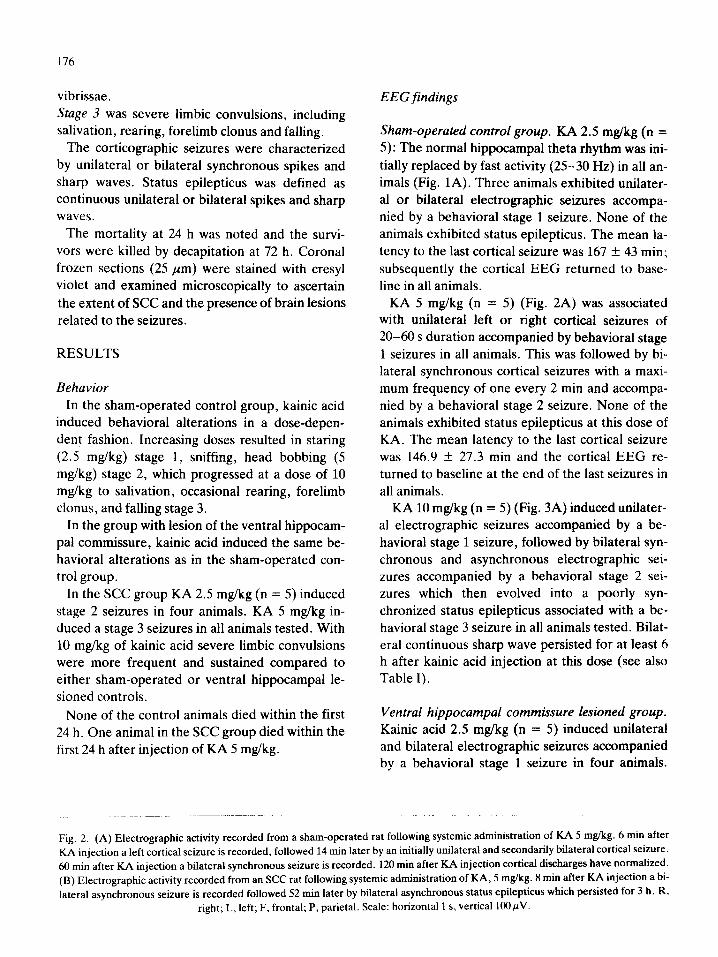
Fig. 1. (A) Electrographic activity recorded from a sham-operated rat following systemic administration of KA 2.5 mg/kg. 60 min af- ter KA injection, hippocampal theta rhythm is replaced by fast activity (25-30 I-Ix) while a single electrographic seizure is recorded in the left cortical leads. 180 min after KA injection cortical discharges have returned to baseline. (B) Electrographic activity recorded from an SCC rat following systemic administration of KA 2.5 mg/kg. 60 min after KA injection, hippocampal theta rhythm is replaced by fast activity and repetitive sharp waves while frequent unilateral right seizures are recorded in the cortex. 180 min after KA injec- tion, continuous spikes and sharp waves are present in cortical and hippocampus leads. 360 min after KA injection, bilateral asynchro- nous spikes are seen in cortical leads. R, right; L, left; F, frontal cortex; P, parietal cortex; HIP, hippocampus. Scale: horizontal 1 s,
vertical 1OOpV.
BASELINE
L ( , ,,I ( / ,, /,, ,,, /, (
6Omin
360min
BASELINE
6Omin
18Omin
13 360min
176
vibrissae. Stage 3 was severe limbic convulsions, including salivation, rearing, forelimb clonus and falling.
The corticographic seizures were characterized by unilateral or bilateral synchronous spikes and sharp waves. Status epilepticus was defined as continuous unilateral or bilateral spikes and sharp waves.
The mortality at 24 h was noted and the survi- vors were killed by decapitation at 72 h. Coronal frozen sections (25 pm) were stained with cresyl violet and examined microscopically to ascertain the extent of SCC and the presence of brain lesions
related to the seizures.
RESULTS
Behavior In the sham-operated control group, kainic acid
induced behavioral alterations in a dose-depen- dent fashion. Increasing doses resulted in staring (2.5 mg/kg) stage 1, sniffing, head bobbing (5 mg/kg) stage 2, which progressed at a dose of 10 mg/kg to salivation, occasional rearing, forelimb clonus, and falling stage 3.
In the group with lesion of the ventral hippocam- pal commissure, kainic acid induced the same be- havioral alterations as in the sham-operated con- trol group.
In the SCC group KA 2.5 mg/kg (n = 5) induced stage 2 seizures in four animals. KA 5 mg/kg in- duced a stage 3 seizures in all animals tested. With 10 mg/kg of kainic acid severe limbic convulsions were more frequent and sustained compared to either sham-operated or ventral hippocampal le- sioned controls.
None of the control animals died within the first 24 h. One animal in the SCC group died within the first 24 h after injection of KA 5 mg/kg.
EEG findings
Sham-operated control group. KA 2.5 mglkg (n = 5): The normal hippocampal theta rhythm was ini- tially replaced by fast activity (25-30 Hz) in all an- imals (Fig. 1A). Three animals exhibited unilater- al or bilateral electrographic seizures accompa- nied by a behavioral stage 1 seizure. None of the animals exhibited status epilepticus. The mean la- tency to the last cortical seizure was 167 + 43 min; subsequently the cortical EEG returned to base- line in all animals.
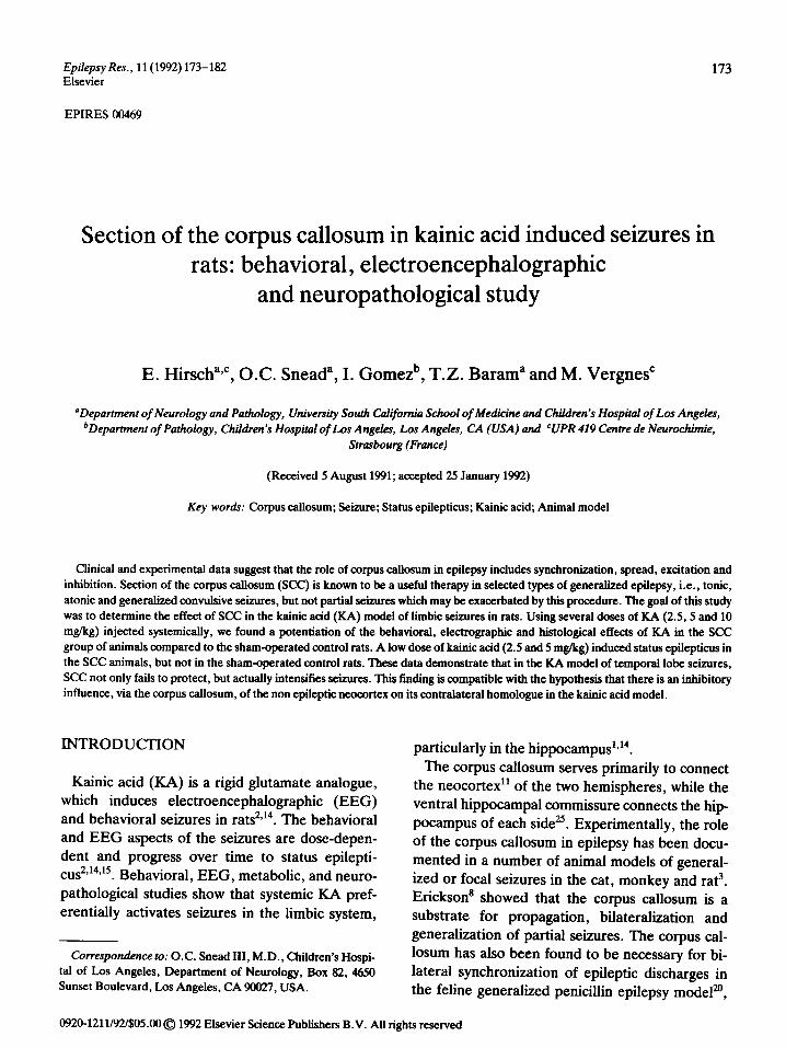
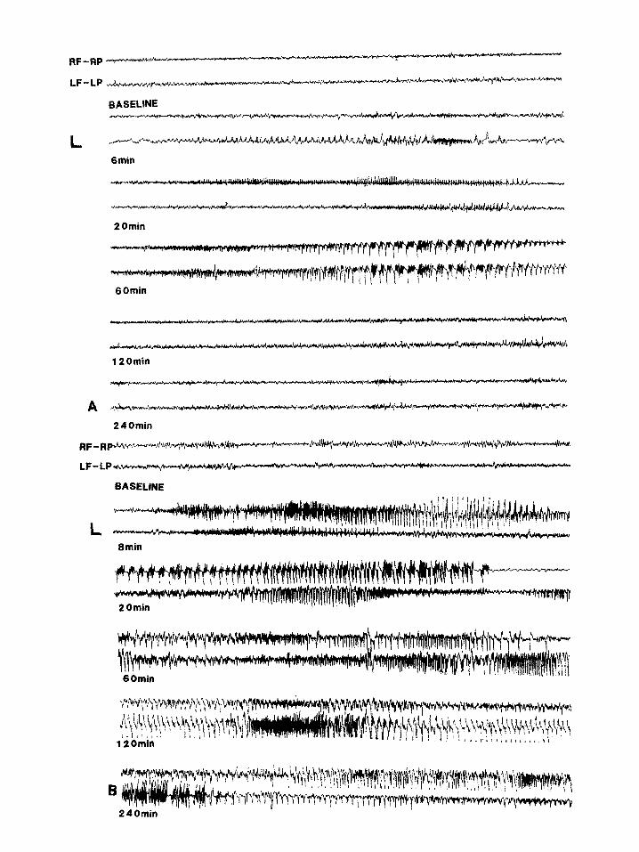
KA 5 mg/kg (n = 5) (Fig. 2A) was associated with unilateral left or right cortical seizures of 20-60 s duration accompanied by behavioral stage 1 seizures in all animals. This was followed by bi- lateral synchronous cortical seizures with a maxi- mum frequency of one every 2 min and accompa- nied by a behavioral stage 2 seizure. None of the animals exhibited status epilepticus at this dose of KA. The mean latency to the last cortical seizure was 146.9 + 27.3 min and the cortical EEG re- turned to baseline at the end of the last seizures in all animals.
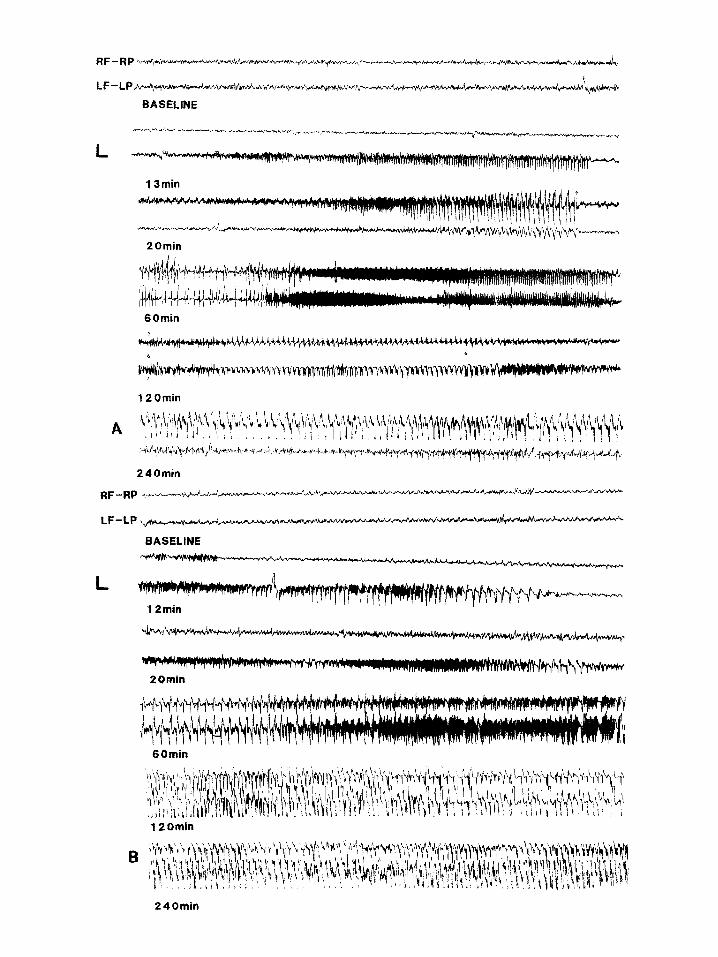
KA 10 mg/kg (n = 5) (Fig. 3A) induced unilater- al electrographic seizures accompanied by a be- havioral stage 1 seizure, followed by bilateral syn- chronous and asynchronous electrographic sei- zures accompanied by a behavioral stage 2 sei- zures which then evolved into a poorly syn- chronized status epilepticus associated with a be- havioral stage 3 seizure in all animals tested. Bilat- eral continuous sharp wave persisted for at least 6 h after kainic acid injection at this dose (see also Table I).
Ventral hippocampal commksure lesioned group. Kainic acid 2.5 mg/kg (n = 5) induced unilateral and bilateral electrographic seizures accompanied by a behavioral stage 1 seizure in four animals.
Fig. 2. (A) Electrographic activity recorded from a sham-operated rat following systemic administration of KA 5 mg/kg. 6 mitt after KA injection a left cortical seizure is recorded, followed 14 min later by an initially unilateral and secondarily bilateral cortical seizure. 60 min after KA injection a bilateral synchronous seizure is recorded. 120 min after KA injection cortical discharges have normalized. (B) Electrographic activity recorded from an SCC rat following systemic administration of KA, 5 mg/kg. 8 min after KA injection a bi- lateral asynchronous seizure is recorded followed 52 min later by bilateral asynchronous status epilepticus which persisted for 3 h. R,
right; L, left; F, frontal; P, parietal. Scale: horizontal 1 s. vertical 1OOpV.
20min
6Omin
20min .,
60min
12 Omin
240min

179
TABLE I
Latency to first seizure and stam epilepticus (S. E.)
Kainic acid Sham-operated VHC lesioned XC
tm&) latency (min) latency (min) latency (min) latency (min) latency (min) latency (min)
to first to S. E. to first to S. E. to first to S. E.
seizure seizure seizure
2.5 17.5 + 1.6 not seen 40.7 + 14.8 not seen 35.7 + 5.3 57.2 f 7.3
(n = 3) (n = 4) (n = 5) (n = 5) 5 17.3 + 5.1 not seen 25.2 f 5.7 not seen 18.0 + 3.6 86.2 + 24.4
(n = 5) (n = 5) (n = 5) (n = 5) 10 11.6 + 4.2 72.0 + 9.3 6.4 + 1.2 51.6 + 3.4 12.0 f 2.8 42.8 + 6.5
(n = 5) (n = 5) (n = 5) (n = 5) (n = 5) (n = 5)
VHC, ventral hippocampal commissure. Results are expressed as means f SE.
None of the animals exhibited status epilepticus.
The mean latency to the last cortical seizure was 96.6 f 37.0 min; subsequently the cortical EEG returned to baseline.
KA 5 mg/kg (n = 5) induced unilateral and bilat- eral cortical seizures accompanied by a behavioral stage 2 seizure in all animals. One animal ex- hibited status epilepticus with a latency of 59 min but did not progress beyond a behavioral stage 2 seizure. In the other four animals, the mean laten- cy to the last cortical seizure was 133.0 + 15.5 min. The EEG returned to baseline at the end of the last seizure in all but one animal.
KA 10 mg/kg (n = 5) induced seizures accompa- nied by a behavioral stage 2 seizure which pro- gressed to status epilepticus in all animals. Bilater- al continuous sharp wave persisted for at least 6 h after kainic acid injection.
SCC group. KA 2.5 mg/kg (n = 5) induced fast ac- tivity (25-30 Hz) in the hippocampus and unilater- al and bilateral synchronous seizures with high fre- quency (at least one per minute) in all animals (Fig. 1B). Four animals exhibited status epilepti- cus accompanied by behavioral stage 2 seizures.
After 6 h the EEG was still abnormal, showing bi-
lateral asynchronous spikes. KA 5 mg/kg (n = 5) induced unilateral and bilat-
eral asynchronous seizures after a mean latency of 18.0 f 3.6 min (Fig. 2B). All animals developed poorly synchronized electrocorticographic EEG status epilepticus accompanied by behavioral
stage 3 seizure, with a mean latency of 86.2 + 24.4 min. Status epilepticus persisted for at least 6 h af- ter IL4 injection.
KA 10 mg/kg (n = 5) induced unilateral and bi- lateral seizures in all animals (Fig. 3B). Poorly synchronized electrographic status epilepticus, ac- companied by behavioral stage 3 seizure and sus- tained forelimb clonus, developed in all animals. Status epilepticus persisted for at least 6 h after KA injection.
Neuropathological effects of KA Forty-five animals were examined. In the sham-
operated control group (n = 15), neuropathologi- cal changes correlated with the dose of KA. An asymmetric, focal loss of neurons in CA3 of the hippocampus was seen in all five animals at the dose of 2.5 mg/kg. 5 mg/kg (n = 5) induced similar
Fig. 3. (A) Electrographic activity recorded from a sham-operated rat following systemic administration of RA 10 mgkg. 13 min af- ter KA injection a left cortical seizure is recorded followed 7 min later by a bilateral seixure and 47 min later by a bilateral poorly syn- chronized status epilepticus which persisted for at least 3 h. (B) Electrographic activity recorded from an SCC rat following systemic administration of KA 10 mgkg. 12 min after KA injection a left cortical seixure is recorded followed 48 min later by bilateral status ep
ilepticus persisting for at least 3 h. R, right; L, left; F, frontal; P, parietal Scale: horizontal 1 s, vertical lOO@.

180
but bilateral lesions, which were more severe with the 10 mg/kg dose (n = 5). In addition four animals showed large asymmetric regions of necrosis in several brain regions, including piriform cortex, lateral olfactory tubercle, endopiriform nucleus and anterior amygdaloid area.
In the ventral hippocampal commissure lesioned group, 3115 animals had an associated mild lesion of the triangular septal nucleus and the dorsal for- nix. Kainic acid 2.5 mg/kg, 5 mg/kg and 10 mg/kg (n = 15) induced th e same neuropathological changes observed in the sham-operated control group, except that the additional lesions in the ventral forebrain area were seen in oniy two ani- mals.
In the SCC group (n = 15) the SCC was found to be complete in 14 animals and limited to the pos- terior two-thirds in one. The ventral hippocampal commissure was sectioned in 13 animals. Neuro- pathological changes were related to the dose of KA. Lesions were similar but more severe than those of the control groups at all KA doses. How- ever, no ventral forebrain lesions were found.
DISCUSSION
These data demonstrate no protective effect of complete SCC against seizures and status epilepti- cus in the kainic acid model. Rather, SCC poten- tiates KA induced seizures and the resuiting brain lesions. This effect of SCC is independent of skull, longitudinal sinus, and ventral hippocampal com- missure lesions.
The behavioral, EEG, and neuropathological changes in our sham-operated control group were similar to those previously described in kainic acid induced seizures and status epilepticus27’4*15726.
The mechanisms underlying the potentiation of KA induced seizures by SCC are unclear. The cor- pus callosum, ventral hippocampal commissure and anterior commissure may participate at differ- ent levels in the bilateral synchronization of epi- leptic discharges and spread, depending on the type and site of origin of the seizures’6-21. Previous studies along with the current data show that the EEG seizures induced by KA are poorly syn- chronized (Figs. lA, 2A, 3A). SCC (Figs. lB, 2B, 3B) or lesion of the ventral hippocampal commis-
sure did not significantly alter electrocorticogra- phic seizure synchronization. This is in agreement with Collins et al ?, who showed a weak role of the commissural system in the functional anatomy of limbic seizures.
The available literature on the functional role of the hippocampal commissure in temporal lobe sei- zure in human is controversial. Lieb and Babb’” reported that commissural connections between the hippocampal formation are unimportant, while Spencer et al .29 suggested the existence of an operational hippocampal commissure. In the amygdala kindling model in cats, the hippocampal ~ommissure is involved in mediation of the trans- hemispheric positive transfer effectY. The current study however suggests that in the kainic acid model of temporal lobe epilepsy, the ventral hip- pocampal commissure plays a weak role, since ani- mals with lesion of the ventral ~ommissure alone developed a similar behavioral, EEG and neuro- pathology to the sham-operated control group.
We have previously demonstrated the protective effect of complete SCC against status epilepticus and death in the lithium-piloca~ine model”. However, the kainic acid and lithium-piloca~ine models differ on several points. Lithium-pilocar- pine seizure activity is first detected in the ventral forebrain and secondarily spreads to the neocor- tex4, whereas seizures induced by kainic acid ap- pear to originate in the hippo~ampus~,‘4,‘~. Kainic acid seizures entail a gradual buildup with unilat- eral asynchronous EEG seizures and progressive spread of electrographic activity through the lim- bit system while lithium-pilocarpine induced sei- zures typically become generalized much more rapidly. L~thium”pilocarpine induces symmetric necrosis in the ventrolateral forebrain4127,“’ while kainic acid induces loss of neurons in the hippo-
campus’4,26. Using 2-deoxyglucose quantitative autora~ography Clifford et al4 reported a higher rate of glucose consumption in the corpus callosum and an increased glucose utilization in the ventral pallidum, the substantia nigra, the neocortex and the entire limbic system in the lithium-pilocarpine model. In kainic acid induced seizures, Collins et aL6 reported an increase in glucose utilization in the entire limbic system during stage 3 seizure, while the glucose utilization in the neocortex was
181
depressed. Those authors postulated an inhibition of the neocortex during the stage 3 seizure6.
A synthetical basis for the ~tentiation by SCC of KA induced seizures is that the neocortex and the hippocampus of each hemisphere exert an in- hibitory influence via the corpus callosum on the development of epileptogenic processes on its con- tralateral homologue. Douglas et al.’ showed that stimulation of the contralateral hippocampus had a marked inhibitor effect on granule cell excit- ability in the dentate fascia in rats. Mutani et al.*l>=, studying the mechanisms of interaction of asymmetrical bilateral epileptogenic foci in cat neocortex, suggested that callosal connections mediated an interhemispheric inhibitory influ- ence. Ottino et al.23, studying the mechanisms of interaction of symmetrical bilateral epileptogenic
REFERENCES
1 Ben-Ari, Y., Tremblay, E., Otterson, O.P. and Meldrum, B.S., The role of epileptic activity in hippocampal and re- mote cerebral lesions induced by kainic acid, Bruin Res., 191(1980) 79-97.
2 Ben-A+ Y., Limbic seizure damage produced by kainic acid: mechanism and relevance to human temporal lobe epilepsy, Neuroscience, 14 (1985) 375-403.
3 Blume, W.T., Corpus caflosum section for seizure control: rationale and review of experimental and clinical data, Cleveland Clin. Q., 51 (1984) 319-332.
4 Clifford, D.B., Olney, J.W., Maniotis, A., Collins, C. and Zorumsky, F., The functional anatomy and pathology of lithium-pilocarpine and high-dose pilocarpine seizures, Neuroscience, 23 (1987) 953-9.58.
5 Douglass, R.M., McNaughton, B.L. and Goddard, G.V., Commissural inhibition and facilitation of granule cell dis- charge in fascia dentata, J. Camp. Neural., 219 (1983) 285-294.
6 Collins, R.C., McLean, M. and Olney, J., Cerebral meta- bolic response to systemic kainic acid: 14-C-deoxyglucose studies, Lge Sci., 27 (1980) 8X5-862.
7 Collins, R.C., Tearse, R.G. and Lothman, E.W., Func- tional anatomy of limbic seizures: focal discharges from me- dial entorhinal cortex in rat, Bruin Res., 280 (1983) 25-40.
8 Erickson, T.C., Spread of the epileptic discharge: an ex- perimental study of the after-discharge induced by electri- cal stimulation of the cerebral cortex, Arch. Neural. Psy- chiatry, 43 (1940) 429-452.
9 Fukuda, H., Wada, J.A., Riche, D. and Naquet, R., Role of the corpus callosum and hippocampal commissure on transfer phenomenon in amygdala-kindled cats, Exp. Neu- rol., 98 (1987) 189-197.
foci in cat neocortex, demonstrated that splitting of the corpus callosum and hippocampal commis- sure disrupted bilateral synchrony of epileptic dis- charges without affecting the frequency of the dis- charges. These data are compatible with the hy- pothesis that in the current experiments SCC sup- pressed the inhibitory influence of the non epilep- tic neocortex and hippocampus on its contralateral homologue in the kainic acid model.
Our results are consistent with the results of a clinical study in which more intense focal seizures occurred after SCCB. The current study also vali- dates the use of the kainic acid model as an animal model of temporal lobe seizures since this form of seizure is not helped, and may be made worse by corpus callosotomy3~30.
10 Honchar, M.P., Oiney, J. W. and Sherman, W.R., System- ic cholinergic agents induce seizures and brain damage in lithium-treated rats, Science, 220 (1983) 323-325.
11 Innocenti, G.M., General organization of callosal connec- tions in the cerebral cortex. In: E.G. Jones and A. Peters (Eds.), Cerebral Cortex, Vol. 5, Plenum Press, New York, NY, 1986, pp. 291-353.
12 Jope, R.S., Morrisett, R. and Snead, O.C., Characteriza- tion of lithium potentiation of pilocarpine-induced status epilepticus in rats, Exp. Neural., 91(1986) 471-480.
13 Lieb, J.P. and Babb, T.L., Interhemispheric propagation time of human hippocampal seizures: II. Relationship to pathology and cell density, Epilepsiu, 27 (1986) 294-300.
14 Lothman, W. and Collins, R., Kainic acid induced limbic seizures: metabolic, behavioral, electroencephalographic and neuropathological correlates, Brain Res., 218 (1981) 299-318.
15 Iothman, W., Collins, R. and Ferendeili, J.A., Kainic acid-induced limbic seizures: electrophysiologic studies, Neurology, 31(1981) 806-812.
16 Marcus, M.E. and Watson, C.W., Symmetrical epilepto- genie foci in monkey cerebral cortex, Arch. Neural., 19 (1968) 99-116.
17 Marcus, M.E. and Watson, C.W., Bilateral synchronous spike wave electrographic patterns in the cat, Arch. Neu- rol., 14 (1966) 601-610.
18 Marcus, E.M., Watson, C.V. and Jacobson, S., Role of the corpus callosum in bilateral synchronous discharges in- duced by intravenous pentylenetetrazol, Neurology, 19 (1969) 309.
19 McCaughram, J.A., Corcoran, M.E. and Wada, J.A., Role of the forebrain commissure in amygdaloid kindling in rats, Epilepsiu, 19 (1978) 19-33.
20 Musgrave, J. and Gloor, P., The role of the corpus callosum
182
in bilateral interhemispheric synchrony of spike and wave discharge in feline generalized penicillin epilepsy, Epilep- sin, Zl(l980) 369-378.
21 Mutani, R., Bergamini, L., Fariello, R. and Quattrocolo, G., Bilateral synchrony of epileptic discharge associated with chronic asymmetrical cortical foci, Electroenceph. Clin. Neurophysiol., 34 (1973) 53-59.
22 Mutani, R. and Durelli, L., Mechanisms of interaction of asymmetrical bitateral epileptogenic foci in neocortex, Epi- lepsia, 21 f 1980) 549-556.
23 Ottino, C.A., Meglio, M., Rossi, G.F. and Tercero, E., An experimental study of structures mediating bilateral syn- chrony of epileptogenic discharges of cortical origin, Epi- lepsia, 12(1971)299-311.
24 Paxinos, G. and Watson, C., The Rat Brain in Stereotaxic Coordinutes, Academic Press, Orlando, FL, 1986.
25 Raisman, G., Cowan, W.N. and Powell, T.P.S., The intrin- sic afferent commissural and associations fibres of the hip- pocampus, Brain, 88 (1965) 963-1000.
26 Schwab, J.E., Fuller, T., Price, J.L. and Olney, J. W., Widespread patterns of neuronal damage following system- ic or intracerebral injections of kainic acid: a histological study, Neuroscience, 5 (1980) 991-1014.
27 Snead, O.C., Hirsch, E. and Vergnes, M., Corpus calfoso- tomy in an experimental model of status epilepticus, Neu-
rology, 41 (1991) 404. 28 Spencer, S.S., Spencer, D.D., Glaser, G.H., Williamson,
P.D. and Mattson, R.H., More intense focal seizures types after callosal section: the rofe of inhibition, Ann. Neural., 16 (1984) 686-693.
29 Spencer, S.S., Williamson, P.D., Spencer, D.D. and Matt- son, R.H., Human hippocampal seizure spread studied by depth and subdural recording: the hippocampal commis- sure, Epilepsia, 28 (1987) 479-489.
30 Spencer, S.S., Corpus callosum section and other discon- nection procedures for medically intractable epilepsy, Epi-
lepsia, 29 (Suppl.) (1988) S85-S99. 31 Turski, L., Ikonomidou, C., Turski, W., Bortolotto, Z.A.
and Cavalheiro, E.A., Review: Cholinergic mechanisms and epileptogenesis. The seizures induced by pilocarpine: a novel experimentaf model of intractable epilepsy, Synupse, 3 (1989) 154-171.
32 Vergnes, M., Marescaux, C., Lannes, B., Depaulis, A., Micheletti, G. and Warter, J.M., Interhemispheric desyn- chronization of spontaneous spike-wave discharges by cor- pus callosum transection in rats with petit ma1 like epilepsy, Epilepsy Res., 4 (1989) U-13.
33 Wada, J.A. and Sato, M., The generalized convulsive sei- zure state induced by daily stimulation of the amygdala in split brain cats, Epilepsia, 16 (1975) 417-430.
Related Documents