Secreted subtilisin gene family in Trichophyton rubrum Olivier Jousson a , Barbara Le ´chenne a , Olympia Bontems a , Bernard Mignon b , Utz Reichard c , Jachen Barblan d , Manfredo Quadroni d , Michel Monod a, * a Dermatology Service (DHURDV), Centre Hospitalier Universitaire Vaudois, Lausanne, Switzerland b Department of Infectious and Parasitic Diseases, Faculty of Veterinary Medicine, University of Lie `ge, Belgium c Department of Bacteriology, University of Go ¨ttingen, Germany d Protein Analysis Facility, Institute of Biochemistry, University of Lausanne, Epalinges, Switzerland Received 10 December 2003; received in revised form 15 March 2004; accepted 10 June 2004 Available online 18 August 2004 Received by B. Dujon Abstract Secreted proteases constitute potential virulence factors of dermatophytes. A total of seven genes encoding putative serine proteases of the subtilisin family (SUB) were isolated in Trichophyton rubrum. Based on sequence data and intron – exon structure, a phylogenetic analysis of subtilisins from T. rubrum and other fungi revealed a presumed ancestral lineage comprising T. rubrum SUB2 and Aspergillus SUBs. All other SUBs (SUB1, SUB3 – 7) are dermatophyte-specific and have apparently emerged more recently, through successive gene duplication events. We showed that two subtilisins, Sub3 and Sub4, were detected in culture supernatants of T. rubrum grown in a medium containing soy protein as a sole nitrogen source. Both recombinant enzymes produced in Pichia pastoris are highly active on keratin azure suggesting that these proteases play an important role in invasion of keratinised tissues by the fungus. The set of deduced amino acid sequences of T. rubrum SUB ORFs allowed the identification of orthologous Subs secreted by other dermatophyte species using proteolysis and mass spectrometry. D 2004 Elsevier B.V. All rights reserved. Keywords: Dermatophytes; Phylogeny; Secreted proteases; Recombinant proteins 1. Introduction Trichophyton rubrum is the most commonly observed dermatophyte isolated from humans in European countries. This species is especially dominant in onychomycosis with a prevalence of approximately 80% (Monod et al., 2002a). In nail infections, T. rubrum destroys keratin and forms channels and sizeable lacunae in the nail plates. These are considerably larger than the hyphae within them, implying the occurrence of extracellular enzymatic activity. Substan- tial serine and metalloprotease activities were secreted by dermatophytes in a soy protein growth medium and a five- member metalloprotease (MEP) gene family encoding fun- galysins was isolated in T. rubrum as well as in T. menta- grophytes and Microsporum canis (Jousson et al., 2004). Although secreted metalloprotease activity was rather dom- inant, the serine protease activity was about 25–50% of the total activity of the culture supernatant as attested by inhibition of this fraction by PMSF. One 34.7 kDa serine alkaline protease (Sanyal et al., 1985) and two serine keratinolytic proteases with molecular masses of 93 and 71 kDa (Asahi et al., 1989) were isolated and characterized in T. rubrum. These proteases were shown to be active as dimers with subunits of 44 and 36 kDa, respectively. 0378-1119/$ - see front matter D 2004 Elsevier B.V. All rights reserved. doi:10.1016/j.gene.2004.06.024 Abbreviations: PCR, polymerase chain reaction; SUB/Sub, subtilisin gene/protein; MEP/Mep, metalloprotease gene/protein; ALP/Alp, alkaline protease gene/protein; AOX1, alcohol oxydase gene; ORF, open reading frame; aa, amino acid(s); nt, nucleotide(s); bp, base pairs; kDa, kilodalton; Ag, microgram; ng, nanogram; MP, maximum parsimony; NJ, neighbor joining; ML, maximum likelihood; PAM, point accepted mutation; TBR, tree bisection recognition; GTR, general time reversible; G, gamma distribution shape parameter; I, proportion of invariable sites; LC, liquid chromatography; MS, mass spectrometry; PMSF, phenyl methyl sulphonyl fluoride; YNB, yeast nitrogen base; SDS-PAGE, sodium dodecyl sulfate polyacrylamide gel electrophoresis; w/v, weight/volume ratio; pI, isoelectric point. * Corresponding author. Tel.: +41-21-3140376; fax: +41-21-3140378. E-mail address: [email protected] (M. Monod). www.elsevier.com/locate/gene Gene 339 (2004) 79 – 88

Welcome message from author
This document is posted to help you gain knowledge. Please leave a comment to let me know what you think about it! Share it to your friends and learn new things together.
Transcript
www.elsevier.com/locate/gene
Gene 339 (2004) 79–88
Secreted subtilisin gene family in Trichophyton rubrum
Olivier Joussona, Barbara Lechennea, Olympia Bontemsa, Bernard Mignonb,Utz Reichardc, Jachen Barbland, Manfredo Quadronid, Michel Monoda,*
aDermatology Service (DHURDV), Centre Hospitalier Universitaire Vaudois, Lausanne, SwitzerlandbDepartment of Infectious and Parasitic Diseases, Faculty of Veterinary Medicine, University of Liege, Belgium
cDepartment of Bacteriology, University of Gottingen, GermanydProtein Analysis Facility, Institute of Biochemistry, University of Lausanne, Epalinges, Switzerland
Received 10 December 2003; received in revised form 15 March 2004; accepted 10 June 2004
Available online 18 August 2004
Received
by B. DujonAbstract
Secreted proteases constitute potential virulence factors of dermatophytes. A total of seven genes encoding putative serine proteases of the
subtilisin family (SUB) were isolated in Trichophyton rubrum. Based on sequence data and intron–exon structure, a phylogenetic analysis of
subtilisins from T. rubrum and other fungi revealed a presumed ancestral lineage comprising T. rubrum SUB2 and Aspergillus SUBs. All
other SUBs (SUB1, SUB3–7) are dermatophyte-specific and have apparently emerged more recently, through successive gene duplication
events. We showed that two subtilisins, Sub3 and Sub4, were detected in culture supernatants of T. rubrum grown in a medium containing
soy protein as a sole nitrogen source. Both recombinant enzymes produced in Pichia pastoris are highly active on keratin azure suggesting
that these proteases play an important role in invasion of keratinised tissues by the fungus. The set of deduced amino acid sequences of T.
rubrum SUB ORFs allowed the identification of orthologous Subs secreted by other dermatophyte species using proteolysis and mass
spectrometry.
D 2004 Elsevier B.V. All rights reserved.
Keywords: Dermatophytes; Phylogeny; Secreted proteases; Recombinant proteins
1. Introduction
Trichophyton rubrum is the most commonly observed
dermatophyte isolated from humans in European countries.
This species is especially dominant in onychomycosis with
0378-1119/$ - see front matter D 2004 Elsevier B.V. All rights reserved.
doi:10.1016/j.gene.2004.06.024
Abbreviations: PCR, polymerase chain reaction; SUB/Sub, subtilisin
gene/protein; MEP/Mep, metalloprotease gene/protein; ALP/Alp, alkaline
protease gene/protein; AOX1, alcohol oxydase gene; ORF, open reading
frame; aa, amino acid(s); nt, nucleotide(s); bp, base pairs; kDa, kilodalton;
Ag, microgram; ng, nanogram; MP, maximum parsimony; NJ, neighbor
joining; ML, maximum likelihood; PAM, point accepted mutation; TBR,
tree bisection recognition; GTR, general time reversible; G, gamma
distribution shape parameter; I, proportion of invariable sites; LC, liquid
chromatography; MS, mass spectrometry; PMSF, phenyl methyl sulphonyl
fluoride; YNB, yeast nitrogen base; SDS-PAGE, sodium dodecyl sulfate
polyacrylamide gel electrophoresis; w/v, weight/volume ratio; pI, isoelectric
point.
* Corresponding author. Tel.: +41-21-3140376; fax: +41-21-3140378.
E-mail address: [email protected] (M. Monod).
a prevalence of approximately 80% (Monod et al., 2002a).
In nail infections, T. rubrum destroys keratin and forms
channels and sizeable lacunae in the nail plates. These are
considerably larger than the hyphae within them, implying
the occurrence of extracellular enzymatic activity. Substan-
tial serine and metalloprotease activities were secreted by
dermatophytes in a soy protein growth medium and a five-
member metalloprotease (MEP) gene family encoding fun-
galysins was isolated in T. rubrum as well as in T. menta-
grophytes and Microsporum canis (Jousson et al., 2004).
Although secreted metalloprotease activity was rather dom-
inant, the serine protease activity was about 25–50% of the
total activity of the culture supernatant as attested by
inhibition of this fraction by PMSF. One 34.7 kDa serine
alkaline protease (Sanyal et al., 1985) and two serine
keratinolytic proteases with molecular masses of 93 and
71 kDa (Asahi et al., 1989) were isolated and characterized
in T. rubrum. These proteases were shown to be active as
dimers with subunits of 44 and 36 kDa, respectively.
O. Jousson et al. / Gene 339 (2004) 79–8880
However, there is no reported amino acid sequencing of the
N-terminus of these proteins which would have allowed
their identification and characterization.
A gene family encoding serine proteases of the subtilisin
family was recently isolated in M. canis (Descamps et al.,
2002). One gene (SUB3) encoded a 31.5-kDa subtilisin
previously isolated and characterized from the culture su-
pernatant ofM. canis grown in a keratin medium (Mignon et
al., 1998). We describe here a seven-member gene family
encoding subtilisins in T. rubrum. We show that two
subtilisins are secreted in a soy protein medium. Both
enzymes, expressed as recombinant proteins in P. pastoris,
are highly active on keratine azure. The set of deduced
amino acid sequences of T. rubrum SUB ORFs allowed
orthologous Subs secreted by other dermatophyte species to
be identified using proteolysis and mass spectrometry.
2Nmax i¼11þ e
2. Materials and methods
2.1. Strains and plasmids
Clinical isolates of T. rubrum, T. mentagrophytes, M.
canis and Arthroderma benhamiae from patients at the
University Hospital, Lausanne (Switzerland) were used.
All strains were identified on the basis of macroscopic
appearance, microscopic examination of the cultures and
partial 28S ribosomal DNA sequencing (Ninet et al., 2003).
From each of the following species, one isolate of T. rubrum
(LAU862-01), T. mentagrophytes (LAU2434-02), M. canis
(LAU709-03) and A. benhamiae (LAU2352-02) was chosen
for protein extract analysis.
Escherichia coli LE392 was used for the propagation of
the bacteriophage EEMBL3 (Promega). All plasmid sub-
cloning experiments were performed in E. coli XL-1 blue
with plasmids pMTL21 (Chambers et al., 1988) and pUC19
(Sambrook et al., 1989). SUB cDNAs were cloned in
plasmid pKJ113 (Borg-von Zepelin et al., 1998) and Pichia
pastoris GS115 (Invitrogen) was used for transformation
and production of recombinant proteins.
2.2. Growth media
T. rubrum, T. mentagrophytes, A. benhamiae and M.
canis were grown on Sabouraud agar and liquid medium
(Bio-Rad) or, to promote production of proteolytic activity,
in liquid medium containing 0.2% soy protein (Supro 1711,
Protein Technologies International), as previously described
(Jousson et al., 2004).
2.3. SUB gene cloning
Previously constructed T. rubrum EEMBL3 genomic and
pSport6 cDNA libraries (Jousson et al., 2004) were used.
Recombinant plaques (2� 104) of the genomic libraries of
T. rubrum were immobilized on GeneScreen nylon mem-
branes (NEN Life Science products). The filters were
hybridized with 32P-labelled DNA fragments under low-
stringency conditions (Monod et al., 1994). All positive
plaques were purified and the associated bacteriophage
DNAs were isolated as described previously (Grossberger,
1987). Agarose gel electrophoresis of restricted recombinant
bacteriophage EEMBL3 DNA, Southern blotting and sub-
cloning of hybridizing fragments from bacteriophages into
pMTL21 or pUC19 were performed using standard proto-
cols (Sambrook et al., 1989). DNA sequencing was per-
formed by Microsynth (Balgach, Switzerland).
T. rubrum cDNAs were obtained by PCR using 200 ng of
DNA prepared from 106 clones of the cDNA library. PCR
were performed with homologous primers designed on
DNA sequences of the different SUB genes.
2.4. Phylogenetic analyses
Nucleotide and amino acid dermatophyte SUB sequences
were aligned using Clustal W (Thompson et al., 1994) as
implemented in BioEdit Sequence Alignement Editor soft-
ware (Hall, 1999). Sequences of genes encoding other
fungal serine proteases were incorporated in the alignment,
including alkaline proteinases from Aspergillus fumigatus
(Accession numbers: ALP1: Z11580; ALP2: Y13338), A.
niger (pepC: M96758; pepD: L19059), A. oryzae (alpA:
S75278), A. nidulans (prtA: L31778), allergens from differ-
ent Trichophyton species (T. rubrum Tri r 2: AF082515; T.
schoenleinii Tri m 2: AJ430843; T. mentagrophytes Tri m 2:
AJ430840), as well as sequences of vacuolar serine pro-
teases from Penicillium oxalixum (AF243425), P. chryso-
genum (AF263454), and Saccharomyces cerevisiae
(P09232). Phylogenetic analyses of SUB genes and proteins
were performed in PAUP* v4.0b10 (Swofford, 1998). DNA
and protein sequences were analysed using MP, NJ, and ML
methods. Modeltest 3.06 (Posada and Crandall, 1998) was
used to select the appropriate model of substitution for ML
and NJ analyses of nucleotide sequences. The Dayhoff PAM
model of protein evolution was used to compute the
distances between the amino acid sequences using the
PROTDIST program implemented in BioEdit. Analyses
were performed using heuristic search with TBR branch
swapping algorithm. The reliability of internal branches was
assessed using the bootstrap method (Felsenstein, 1988),
with 1000 replicates. Phylogenetic trees were edited using
TreeView (Page, 1996).
Phylogenetic analyses based on exon–intron structure of
SUB genes were also performed. The first procedure used an
intron presence/absence matrix as an input, which was
analysed using MP. The second procedure consisted of a
measure of gene structure similarity (Betts et al., 2001) that
uses the presence, location, and phase of introns to estimate
the exon–intron similarity.
SGða; bÞ ¼1 XNequiv
1cðdi�dÞ þ uðai; biÞ
� �
O. Jousson et al. / Gene 339 (2004) 79–88 81
SG is the similarity measure for the two proteins (a and b);
Nmax is the largest number of introns found in either protein;
Nequiv is the number of equivalent (homologous) introns; aiand bi are the ith equivalent intron positions in the two
proteins; di is the difference in position of the introns within
the two proteins (in amino acids); u(ai,bi) is 1 if the intron
phases are the same and 0 if they are different; c and d are
constants (0.2 and 30) optimised such that the sigmoid
function is insensitive to small changes in intron positions
(F 10 residues) (Betts et al., 2001). A matrix of distances
calculated by this method was used to reconstruct a tree by
NJ.
2.5. Production of recombinant Subs
Expression plasmids were constructed by cloning
cDNA PCR products in the multiple cloning site of the
E. coli–P. pastoris shuttle vector pKJ113. The PCR
products were purified using a PCR purification kit
(Roche Diagnostics) and digested by restriction enzymes
for which a recognition site was previously designed at the
5V extremity of the primers (Table 1). P. pastoris GS115
(Invitrogen) was transformed by electroporation with 10
Ag of plasmid DNA linearized by EcoRI or SmaI. Trans-
formants selected on histidine-deficient medium [1 M
sorbitol, 1% (w/v) dextrose, 1.34% (w/v) yeast nitrogen
base (YNB) without amino acids, 4� 10� 5% (w/v) biotin,
5� 10� 3% amino acids (i.e. 5� 10� 3% (w/v) of each L-
glutamic acid, L-methionine, L-lysine, L-leucine, L-isoleu-
cine), 2% (w/v) agarose] were screened for insertion of the
construct at the AOX1 site on minimal methanol plates
[1.34% (w/v) YNB without amino acids, 4� 10� 5% (w/v)
biotin, 0.5% (v/v) methanol, 2% (w/v) agarose]. The
transformants unable to grow on media containing only
Table 1
Primers used for amplification of T. rubrum SUBs cDNAs
Gene Amplification primersa
SUB1 GTTC#TCGAGTTAATGCCGCCCAAATCCTGTCTGTAG#GATCCTTAGTTCCAGAAGCTGCTAAAC
SUB2 GGTTC#TCGAGACCTCGCTCCACAGCCTGAGCCGCTTG#GATCCTCAGTAATACTTGGGCAGTTTGC
SUB3 CTTG#TCGACTTGATGCCCGCGCTTTCTTCCACAACCTTG#GATCCTTATCGTCCACTTCCGTTGTAGAGG
SUB4 CTTG#TCGACTTGATGCCCGCGCAGTCTTCAAGCTTG#GATCCCTACTGGCCACTTCCGTTGTAGAG
SUB5 GTTC#TCGAGTTGCACAGATCTTAAGTGTCCCCCTTG#GATCCTTAACGGCCACTGCCATTATAAAGC
SUB6 GTGC#TCGAGATGGTGCTAGAATCCTTGAGGCCGGTGTTGC#GGCCGCTTATTTGCCGCTGCCGTTGTA
SUB7 GTTGC#TCGAGCTGAGATCTTGGAGACTCGCGCTGTTG#GATCCTTACATGCCAGATCGGTTGTTGATGAGCTTGC
Amplification products were cloned in E. coli–P. pastoris shuttle vector pKJ113a Vertical arrows indicate the cleavage site of restriction endonucleases; underb In parentheses are shown amino acids encoded by the recognition site seque
indicate stop codons.c The numbers in parentheses represent the nucleotide position of amplificatio
methanol as carbon source were assumed to contain the
construct at the correct yeast genomic location by integra-
tion events in the AOX1 locus displacing the AOX1 coding
region. They were grown to near saturation (OD= 20 at
600 nm) at 30 jC in 10 ml of glycerol-based yeast media
(0.1 M potassium phosphate buffer at pH 6.0, containing
1% (w/v) yeast extract, 2% (w/v) peptone, 1.34% (w/v)
YNB without amino acids, 1% (v/v) glycerol and
4� 10� 5 % (w/v) biotin). Cells were harvested and
resuspended in 2 ml of the same medium with 0.5% (v/
v) methanol instead of glycerol and incubated for 2 days.
The supernatant was then harvested and tested for protein
production on SDS-PAGE gels. Salts and small molecular
weight solutes were removed from 2.5 ml of P. pastoris
culture supernatant by passing through a PD10 column
(Pharmacia) using 20 mM Tris–HCl buffer, pH 7.5 before
testing for proteolytic activity. Protein concentrations were
measured by the method of Bradford using a commercial
reagent (Bio-Rad). Supernatant of P. pastoris GS115
grown in the same conditions was used as a negative
control for comparison.
2.6. Proteolytic assays
Subtilisin Carlsberg (Bacillus licheniformis subtilisin A)
and proteinase K were from Sigma. A. fumigatus and A.
oryzae alkaline proteases were prepared as previously
described (Monod et al., 1991).
The proteolytic activity was measured in 50 mM Tris–
HCl buffer at pH between 7.0 and 9.5 using resorufin-
labeled casein (Roche Diagnostics), azocollagen (Sigma)
and elastin-Congo red (Sigma) as described previously
(Monod et al., 1991; Jousson et al., 2004). Using Azoal-
bumin, the reaction mixture contained 0.1% substrate, 50
Orientation Encoded amino acid
sequencebPCR productc
(with cloning sites)
5V–3V (R)(V)NAAQILS (47) XhoI
3V–5V FSSFWN* (1515) BamHI
5V–3V (R)DLAPQPEP (45) XhoI
3V–5V KLPKYY* (1275) BamHI
5V–3V (R)(L)DARAFFHN (45) SalI
3V–5V LYNGSGR* (1194) BamHI
5V–3V (R)(L)DARAVFK (45) SalI
3V–5V LYNGSGQ* (1200) BamHI
5V–3V (R)(V)AQILSVP (54) XhoI
3V–5V LYNGSGR* (1191) BamHI
5V–3V (R)(D)GARILEAG (51) XhoI
3V–5V YNGSGK* (1239) NotI
5V–3V (R)(A)EILETRA (57) XhoI
3V–5V KLINNGSGM* (1203) BamHI
.
lined nucleotides represent the recognition sequence.
nce and added to the N-terminal extremity of Sub prosequences; asterisks
n primers on SUB cDNAs.
O. Jousson et al. / Gene 339 (2004) 79–8882
mM Tris–HCl buffer and 1.0 Ag of enzyme. After incuba-
tion at 37 jC during 30 min, the undigested substrate was
precipitated by trichloroacetic acid (5% final concentration)
and separated from the supernatant by centrifugation. The
absorbance of the mixture was measured at 400 nm.
Keratin degradation was measured using keratin azure as
a substrate (Sigma). The reaction mixture contained 10 Agof substrate, Tris–HCl buffer (50 mM at different pH
values), and 2.5 Ag of enzyme in a total volume of 500
Al. The degradation of keratin azure was estimated by
measuring the absorbance at 595 nm of the supernatant
after centrifugation. The assays were run in duplicate. For
practical purposes, one arbitrary unit (U) of proteolytic
activity was defined as that producing an absorbance of
0.01 per min.
2.7. Protein extracts analysis
Secreted proteins were precipitated from dermatophyte
culture supernatants using trichloroacetic acid and purified
using PlusOne SDS-PAGE Clean-Up Kit (Amersham).
Extracts were analysed by SDS-PAGE (Laemmli, 1970)
with a separation gel of 8.5% polyacrylamide. Gels were
stained with Coomassie brilliant blue R-250 (Bio-Rad).
Measures of band molecular mass and optical density
were performed using the TotalLab software (Nonlinear
Dynamics).
2.8. Protein identification by LC-MS/MS
Coomassie blue stained bands were excised from the
SDS-PAGE gels and transferred to special 96-well plates
(Perkin Elmer Life Sciences, Cambridge, United King-
dom). In–gel proteolytic cleavage with sequencing grade
trypsin (Promega) was performed automatically in the
robotic workstation Investigator ProGestk (Perkin Elmer
Life Sciences) according to the protocol of Wilm et al.
(1996). Supernatants containing proteolytic peptides were
analysed by LC-MS/MS on a SCIEX QSTAR Pulsar
(Concord, Ontario, Canada) hybrid quadrupole-time of
flight instrument equipped with a nanoelectrospray source
and interfaced to an LC-Packings Ultimate (Amsterdam,
Holland) HPLC system. Peptides were separated on a
PepMap reversed-phase capillary C18 (75 Am ID� 15
cm) column at a flow rate of 200 nl min� 1 along a 52-
min gradient of acetonitrile (0–40%). The instrument
controlling Analyst software was used to perform peak
detection and automatically select peptides for collision-
induced fragmentation. Collections of non-interpreted col-
lision-induced fragmentation spectra were bundled and
used for searching an in-house built database of T.
rubrum subtilisin sequences with the Mascot software
www.matrixscience.com) (Perkins et al., 1999). Only
peptide sequences ranked as statistically significant were
taken into account, and all peptide hits were manually
examined for validation.
3. Results
3.1. SUB gene cloning
PCR products of M. canis SUB1, SUB2 and SUB3
genes were used as a probe to screen 2� 104 plaques of
the EEMBL3 T. rubrum genomic library corresponding to
about 10 genome equivalents of the fungi. Restriction
fragments hybridizing with the M. canis SUB1–3 probe
were identified by Southern blotting, subcloned in pUC19
or pMTL21 and sequenced. Translated blast searches
(http://www.ncbi.nlm.nih.gov/BLAST/) were performed
for all sequences obtained and revealed five different
sequences encoding parts of putative Sub proteins in T.
rubrum. A preliminary phylogenetic analysis suggested
that three sequences were the orthologs of M. canis
SUB1, SUB2, and SUB3. The two remaining sequences
apparently corresponded to new SUB genes and were
subsequently named SUB4 and SUB5. However, none of
these sequences corresponded to that of the previously
cloned cDNA of Tri r 2 antigen encoding a putative
subtilisin of T. rubrum (Woodfolk et al., 1998). Therefore,
in a second step, 2� 104 plaques of the genomic library
were screened with a Tri r 2 PCR product. In addition to
Tri r 2, one new T. rubrum gene encoding a putative Sub
protein was found. These new SUB genes were named
SUB6 and SUB7, respectively. No further new SUB paral-
ogs were found in a third screening performed with the
seven T. rubrum SUBs as probes. As a final result, a total
of seven genes (SUB1–7) putatively encoding Sub proteins
were found in T. rubrum.
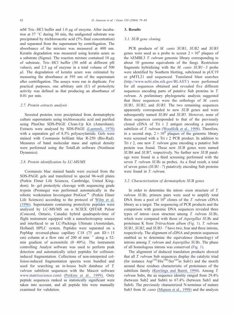
3.2. Characterization of dermatophyte SUB genes
In order to determine the intron–exon structure of T.
rubrum SUBs, primers pairs were used to amplify total
DNA from a pool of 106 clones of the T. rubrum cDNA
library as a target. The sequencing of PCR products and the
comparison with genomic DNA sequences revealed three
types of intron–exon structure among T. rubrum SUBs,
which were compared with those of Aspergillus SUBs and
proteinase K from Tritirachium album (Fig. 1). T. rubrum
SUB1, SUB2, and SUB3–7 have two, four and three introns,
respectively. The alignment of cDNA and protein sequences
enabled us to determine the equivalence (homology) of
introns among T. rubrum and Aspergillus SUBs. The phase
of all homologous introns was conserved (Fig. 1).
The alignment of deduced translation products showed
that all T. rubrum Sub sequences display the catalytic triad
(for instance Asp158/His190/Ser345in Sub1) and the motifs
around these residues characteristic of proteinases of the
subtilisin family (Rawlings and Barett, 1994). Among T.
rubrum Subs, the aa sequence identity ranged from 29.4%
(between Sub2 and Sub6) to 67.4% (between Sub3 and
Sub4). The previously characterized N-terminus of mature
Sub3 from M. canis (Mignon et al., 1998) and the analysis
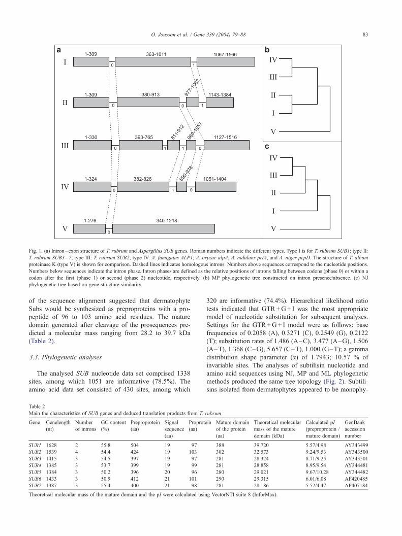
Fig. 1. (a) Intron–exon structure of T. rubrum and Aspergillus SUB genes. Roman numbers indicate the different types. Type I is for T. rubrum SUB1; type II:
T. rubrum SUB3–7; type III: T. rubrum SUB2; type IV: A. fumigatus ALP1, A. oryzae alpA, A. nidulans prtA, and A. niger pepD. The structure of T. album
proteinase K (type V) is shown for comparison. Dashed lines indicates homologous introns. Numbers above sequences correspond to the nucleotide positions.
Numbers below sequences indicate the intron phase. Intron phases are defined as the relative positions of introns falling between codons (phase 0) or within a
codon after the first (phase 1) or second (phase 2) nucleotide, respectively. (b) MP phylogenetic tree constructed on intron presence/absence. (c) NJ
phylogenetic tree based on gene structure similarity.
O. Jousson et al. / Gene 339 (2004) 79–88 83
of the sequence alignment suggested that dermatophyte
Subs would be synthesized as preproproteins with a pro-
peptide of 96 to 103 amino acid residues. The mature
domain generated after cleavage of the prosequences pre-
dicted a molecular mass ranging from 28.2 to 39.7 kDa
(Table 2).
3.3. Phylogenetic analyses
The analysed SUB nucleotide data set comprised 1338
sites, among which 1051 are informative (78.5%). The
amino acid data set consisted of 430 sites, among which
Table 2
Main the characteristics of SUB genes and deduced translation products from T.
Gene Genelength
(nt)
Number
of introns
GC content
(%)
Preproprotein
(aa)
Signal
sequence
(aa)
Proprot
(aa)
SUB1 1628 2 55.8 504 19 97
SUB2 1539 4 54.4 424 19 103
SUB3 1415 3 54.5 397 19 97
SUB4 1385 3 53.7 399 19 99
SUB5 1384 3 50.2 396 20 96
SUB6 1433 3 50.9 412 21 101
SUB7 1387 3 55.4 400 21 98
Theoretical molecular mass of the mature domain and the pI were calculated usin
320 are informative (74.4%). Hierarchical likelihood ratio
tests indicated that GTR+G+ I was the most appropriate
model of nucleotide substitution for subsequent analyses.
Settings for the GTR+G+ I model were as follows: base
frequencies of 0.2058 (A), 0.3271 (C), 0.2549 (G), 0.2122
(T); substitution rates of 1.486 (A–C), 3.477 (A–G), 1.506
(A–T), 1.368 (C–G), 5.657 (C–T), 1.000 (G–T); a gamma
distribution shape parameter (a) of 1.7943; 10.57 % of
invariable sites. The analyses of subtilisin nucleotide and
amino acid sequences using NJ, MP and ML phylogenetic
methods produced the same tree topology (Fig. 2). Subtili-
sins isolated from dermatophytes appeared to be monophy-
rubrum
ein Mature domain
of the protein
(aa)
Theoretical molecular
mass of the mature
domain (kDa)
Calculated pI
(preproprotein /
mature domain)
GenBank
accession
number
388 39.720 5.57/4.98 AY343499
302 32.573 9.24/9.53 AY343500
281 28.324 8.71/9.25 AY343501
281 28.858 8.95/9.54 AY344481
280 29.021 9.67/10.28 AY344482
290 29.315 6.01/6.08 AF420485
281 28.186 5.52/4.47 AF407184
g VectorNTI suite 8 (InforMax).
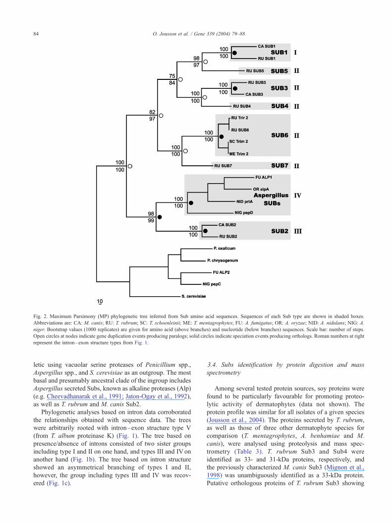
Fig. 2. Maximum Parsimony (MP) phylogenetic tree inferred from Sub amino acid sequences. Sequences of each Sub type are shown in shaded boxes.
Abbreviations are: CA: M. canis; RU: T. rubrum; SC: T. schoenleinii; ME: T. mentagrophytes; FU: A. fumigatus; OR: A. oryzae; NID: A. nidulans; NIG: A.
niger. Bootstrap values (1000 replicates) are given for amino acid (above branches) and nucleotide (below branches) sequences. Scale bar: number of steps.
Open circles at nodes indicate gene duplication events producing paralogs; solid circles indicate speciation events producing orthologs. Roman numbers at right
represent the intron–exon structure types from Fig. 1.
O. Jousson et al. / Gene 339 (2004) 79–8884
letic using vacuolar serine proteases of Penicillium spp.,
Aspergillus spp., and S. cerevisiae as an outgroup. The most
basal and presumably ancestral clade of the ingroup includes
Aspergillus secreted Subs, known as alkaline proteases (Alp)
(e.g. Cheevadhanarak et al., 1991; Jaton-Ogay et al., 1992),
as well as T. rubrum and M. canis Sub2.
Phylogenetic analyses based on intron data corroborated
the relationships obtained with sequence data. The trees
were arbitrarily rooted with intron–exon structure type V
(from T. album proteinase K) (Fig. 1). The tree based on
presence/absence of introns consisted of two sister groups
including type I and II on one hand, and types III and IV on
another hand (Fig. 1b). The tree based on intron structure
showed an asymmetrical branching of types I and II,
however, the group including types III and IV was recov-
ered (Fig. 1c).
3.4. Subs identification by protein digestion and mass
spectrometry
Among several tested protein sources, soy proteins were
found to be particularly favourable for promoting proteo-
lytic activity of dermatophytes (data not shown). The
protein profile was similar for all isolates of a given species
(Jousson et al., 2004). The proteins secreted by T. rubrum,
as well as those of three other dermatophyte species for
comparison (T. mentagrophytes, A. benhamiae and M.
canis), were analysed using proteolysis and mass spec-
trometry (Table 3). T. rubrum Sub3 and Sub4 were
identified as 33- and 31-kDa proteins, respectively, and
the previously characterized M. canis Sub3 (Mignon et al.,
1998) was unambiguously identified as a 33-kDa protein.
Putative orthologous proteins of T. rubrum Sub3 showing
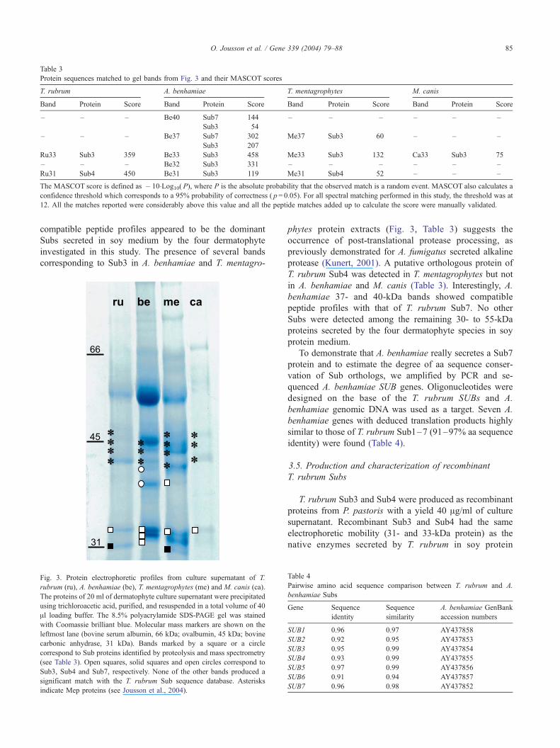
Table 3
Protein sequences matched to gel bands from Fig. 3 and their MASCOT scores
T. rubrum A. benhamiae T. mentagrophytes M. canis
Band Protein Score Band Protein Score Band Protein Score Band Protein Score
– – – Be40 Sub7 144 – – – – – –
Sub3 54
– – – Be37 Sub7 302 Me37 Sub3 60 – – –
Sub3 207
Ru33 Sub3 359 Be33 Sub3 458 Me33 Sub3 132 Ca33 Sub3 75
– – – Be32 Sub3 331 – – – – – –
Ru31 Sub4 450 Be31 Sub3 119 Me31 Sub4 52 – – –
The MASCOT score is defined as � 10�Log10( P), where P is the absolute probability that the observed match is a random event. MASCOT also calculates a
confidence threshold which corresponds to a 95% probability of correctness ( p= 0.05). For all spectral matching performed in this study, the threshold was at
12. All the matches reported were considerably above this value and all the peptide matches added up to calculate the score were manually validated.
O. Jousson et al. / Gene 339 (2004) 79–88 85
compatible peptide profiles appeared to be the dominant
Subs secreted in soy medium by the four dermatophyte
investigated in this study. The presence of several bands
corresponding to Sub3 in A. benhamiae and T. mentagro-
Fig. 3. Protein electrophoretic profiles from culture supernatant of T.
rubrum (ru), A. benhamiae (be), T. mentagrophytes (me) and M. canis (ca).
The proteins of 20 ml of dermatophyte culture supernatant were precipitated
using trichloroacetic acid, purified, and resuspended in a total volume of 40
Al loading buffer. The 8.5% polyacrylamide SDS-PAGE gel was stained
with Coomassie brilliant blue. Molecular mass markers are shown on the
leftmost lane (bovine serum albumin, 66 kDa; ovalbumin, 45 kDa; bovine
carbonic anhydrase, 31 kDa). Bands marked by a square or a circle
correspond to Sub proteins identified by proteolysis and mass spectrometry
(see Table 3). Open squares, solid squares and open circles correspond to
Sub3, Sub4 and Sub7, respectively. None of the other bands produced a
significant match with the T. rubrum Sub sequence database. Asterisks
indicate Mep proteins (see Jousson et al., 2004).
phytes protein extracts (Fig. 3, Table 3) suggests the
occurrence of post-translational protease processing, as
previously demonstrated for A. fumigatus secreted alkaline
protease (Kunert, 2001). A putative orthologous protein of
T. rubrum Sub4 was detected in T. mentagrophytes but not
in A. benhamiae and M. canis (Table 3). Interestingly, A.
benhamiae 37- and 40-kDa bands showed compatible
peptide profiles with that of T. rubrum Sub7. No other
Subs were detected among the remaining 30- to 55-kDa
proteins secreted by the four dermatophyte species in soy
protein medium.
To demonstrate that A. benhamiae really secretes a Sub7
protein and to estimate the degree of aa sequence conser-
vation of Sub orthologs, we amplified by PCR and se-
quenced A. benhamiae SUB genes. Oligonucleotides were
designed on the base of the T. rubrum SUBs and A.
benhamiae genomic DNA was used as a target. Seven A.
benhamiae genes with deduced translation products highly
similar to those of T. rubrum Sub1–7 (91–97% aa sequence
identity) were found (Table 4).
3.5. Production and characterization of recombinant
T. rubrum Subs
T. rubrum Sub3 and Sub4 were produced as recombinant
proteins from P. pastoris with a yield 40 Ag/ml of culture
supernatant. Recombinant Sub3 and Sub4 had the same
electrophoretic mobility (31- and 33-kDa protein) as the
native enzymes secreted by T. rubrum in soy protein
Table 4
Pairwise amino acid sequence comparison between T. rubrum and A.
benhamiae Subs
Gene Sequence
identity
Sequence
similarity
A. benhamiae GenBank
accession numbers
SUB1 0.96 0.97 AY437858
SUB2 0.92 0.95 AY437853
SUB3 0.95 0.99 AY437854
SUB4 0.93 0.99 AY437855
SUB5 0.97 0.99 AY437856
SUB6 0.91 0.94 AY437857
SUB7 0.96 0.98 AY437852
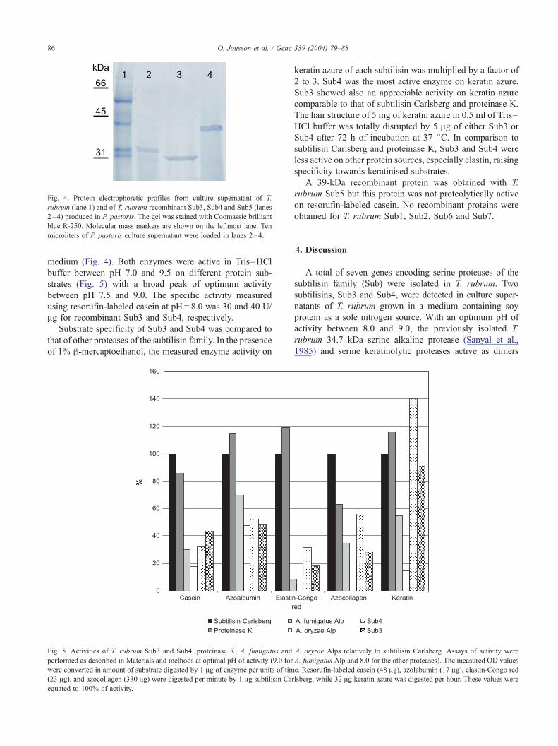
Fig. 4. Protein electrophoretic profiles from culture supernatant of T.
rubrum (lane 1) and of T. rubrum recombinant Sub3, Sub4 and Sub5 (lanes
2–4) produced in P. pastoris. The gel was stained with Coomassie brilliant
blue R-250. Molecular mass markers are shown on the leftmost lane. Ten
microliters of P. pastoris culture supernatant were loaded in lanes 2–4.
O. Jousson et al. / Gene 339 (2004) 79–8886
medium (Fig. 4). Both enzymes were active in Tris–HCl
buffer between pH 7.0 and 9.5 on different protein sub-
strates (Fig. 5) with a broad peak of optimum activity
between pH 7.5 and 9.0. The specific activity measured
using resorufin-labeled casein at pH = 8.0 was 30 and 40 U/
Ag for recombinant Sub3 and Sub4, respectively.
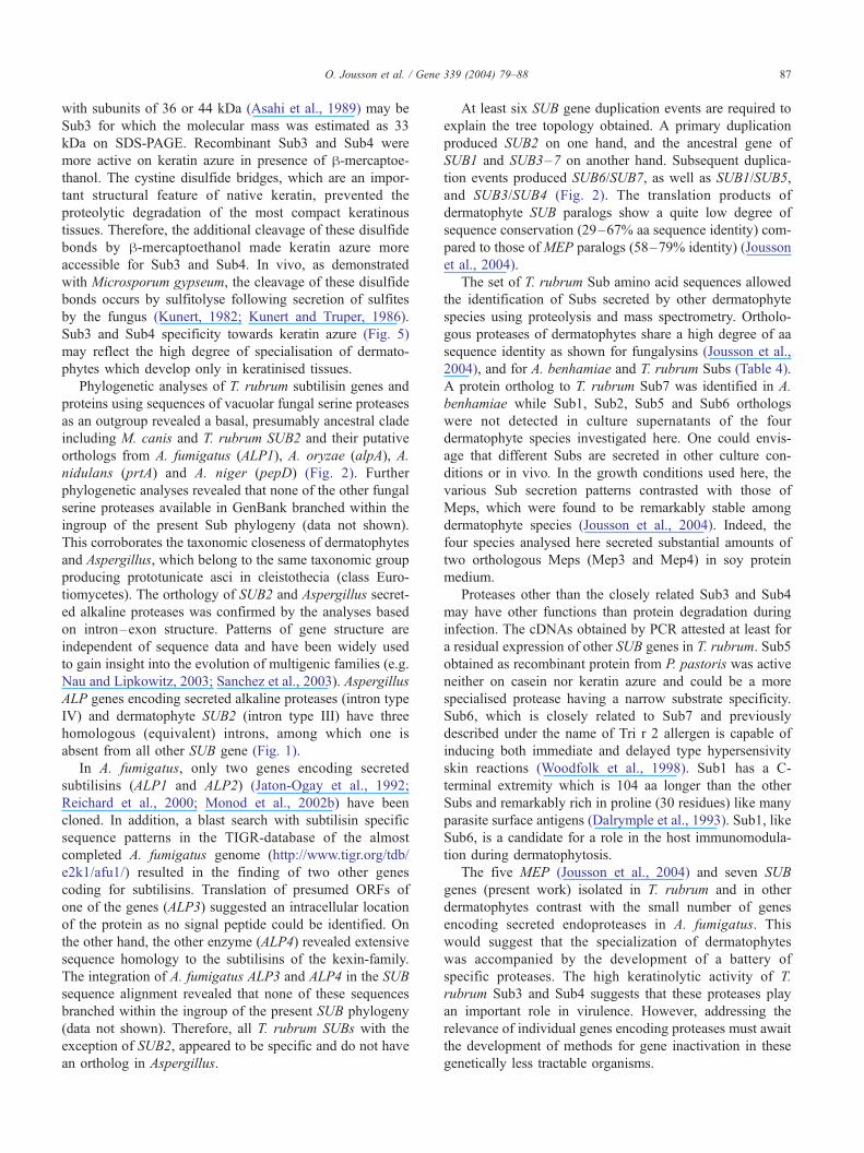
Substrate specificity of Sub3 and Sub4 was compared to
that of other proteases of the subtilisin family. In the presence
of 1% h-mercaptoethanol, the measured enzyme activity on
Fig. 5. Activities of T. rubrum Sub3 and Sub4, proteinase K, A. fumigatus and
performed as described in Materials and methods at optimal pH of activity (9.0 for
were converted in amount of substrate digested by 1 Ag of enzyme per units of tim
(23 Ag), and azocollagen (330 Ag) were digested per minute by 1 Ag subtilisin Car
equated to 100% of activity.
keratin azure of each subtilisin was multiplied by a factor of
2 to 3. Sub4 was the most active enzyme on keratin azure.
Sub3 showed also an appreciable activity on keratin azure
comparable to that of subtilisin Carlsberg and proteinase K.
The hair structure of 5 mg of keratin azure in 0.5 ml of Tris–
HCl buffer was totally disrupted by 5 Ag of either Sub3 or
Sub4 after 72 h of incubation at 37 jC. In comparison to
subtilisin Carlsberg and proteinase K, Sub3 and Sub4 were
less active on other protein sources, especially elastin, raising
specificity towards keratinised substrates.
A 39-kDa recombinant protein was obtained with T.
rubrum Sub5 but this protein was not proteolytically active
on resorufin-labeled casein. No recombinant proteins were
obtained for T. rubrum Sub1, Sub2, Sub6 and Sub7.
4. Discussion
A total of seven genes encoding serine proteases of the
subtilisin family (Sub) were isolated in T. rubrum. Two
subtilisins, Sub3 and Sub4, were detected in culture super-
natants of T. rubrum grown in a medium containing soy
protein as a sole nitrogen source. With an optimum pH of
activity between 8.0 and 9.0, the previously isolated T.
rubrum 34.7 kDa serine alkaline protease (Sanyal et al.,
1985) and serine keratinolytic proteases active as dimers
A. oryzae Alps relatively to subtilisin Carlsberg. Assays of activity were
A. fumigatus Alp and 8.0 for the other proteases). The measured OD values
e. Resorufin-labeled casein (48 Ag), azolabumin (17 Ag), elastin-Congo red
lsberg, while 32 Ag keratin azure was digested per hour. These values were
O. Jousson et al. / Gene 339 (2004) 79–88 87
with subunits of 36 or 44 kDa (Asahi et al., 1989) may be
Sub3 for which the molecular mass was estimated as 33
kDa on SDS-PAGE. Recombinant Sub3 and Sub4 were
more active on keratin azure in presence of h-mercaptoe-
thanol. The cystine disulfide bridges, which are an impor-
tant structural feature of native keratin, prevented the
proteolytic degradation of the most compact keratinous
tissues. Therefore, the additional cleavage of these disulfide
bonds by h-mercaptoethanol made keratin azure more
accessible for Sub3 and Sub4. In vivo, as demonstrated
with Microsporum gypseum, the cleavage of these disulfide
bonds occurs by sulfitolyse following secretion of sulfites
by the fungus (Kunert, 1982; Kunert and Truper, 1986).
Sub3 and Sub4 specificity towards keratin azure (Fig. 5)
may reflect the high degree of specialisation of dermato-
phytes which develop only in keratinised tissues.
Phylogenetic analyses of T. rubrum subtilisin genes and
proteins using sequences of vacuolar fungal serine proteases
as an outgroup revealed a basal, presumably ancestral clade
including M. canis and T. rubrum SUB2 and their putative
orthologs from A. fumigatus (ALP1), A. oryzae (alpA), A.
nidulans (prtA) and A. niger (pepD) (Fig. 2). Further
phylogenetic analyses revealed that none of the other fungal
serine proteases available in GenBank branched within the
ingroup of the present Sub phylogeny (data not shown).
This corroborates the taxonomic closeness of dermatophytes
and Aspergillus, which belong to the same taxonomic group
producing prototunicate asci in cleistothecia (class Euro-
tiomycetes). The orthology of SUB2 and Aspergillus secret-
ed alkaline proteases was confirmed by the analyses based
on intron–exon structure. Patterns of gene structure are
independent of sequence data and have been widely used
to gain insight into the evolution of multigenic families (e.g.
Nau and Lipkowitz, 2003; Sanchez et al., 2003). Aspergillus
ALP genes encoding secreted alkaline proteases (intron type
IV) and dermatophyte SUB2 (intron type III) have three
homologous (equivalent) introns, among which one is
absent from all other SUB gene (Fig. 1).
In A. fumigatus, only two genes encoding secreted
subtilisins (ALP1 and ALP2) (Jaton-Ogay et al., 1992;
Reichard et al., 2000; Monod et al., 2002b) have been
cloned. In addition, a blast search with subtilisin specific
sequence patterns in the TIGR-database of the almost
completed A. fumigatus genome (http://www.tigr.org/tdb/
e2k1/afu1/) resulted in the finding of two other genes
coding for subtilisins. Translation of presumed ORFs of
one of the genes (ALP3) suggested an intracellular location
of the protein as no signal peptide could be identified. On
the other hand, the other enzyme (ALP4) revealed extensive
sequence homology to the subtilisins of the kexin-family.
The integration of A. fumigatus ALP3 and ALP4 in the SUB
sequence alignment revealed that none of these sequences
branched within the ingroup of the present SUB phylogeny
(data not shown). Therefore, all T. rubrum SUBs with the
exception of SUB2, appeared to be specific and do not have
an ortholog in Aspergillus.
At least six SUB gene duplication events are required to
explain the tree topology obtained. A primary duplication
produced SUB2 on one hand, and the ancestral gene of
SUB1 and SUB3–7 on another hand. Subsequent duplica-
tion events produced SUB6/SUB7, as well as SUB1/SUB5,
and SUB3/SUB4 (Fig. 2). The translation products of
dermatophyte SUB paralogs show a quite low degree of
sequence conservation (29–67% aa sequence identity) com-
pared to those of MEP paralogs (58–79% identity) (Jousson
et al., 2004).
The set of T. rubrum Sub amino acid sequences allowed
the identification of Subs secreted by other dermatophyte
species using proteolysis and mass spectrometry. Ortholo-
gous proteases of dermatophytes share a high degree of aa
sequence identity as shown for fungalysins (Jousson et al.,
2004), and for A. benhamiae and T. rubrum Subs (Table 4).
A protein ortholog to T. rubrum Sub7 was identified in A.
benhamiae while Sub1, Sub2, Sub5 and Sub6 orthologs
were not detected in culture supernatants of the four
dermatophyte species investigated here. One could envis-
age that different Subs are secreted in other culture con-
ditions or in vivo. In the growth conditions used here, the
various Sub secretion patterns contrasted with those of
Meps, which were found to be remarkably stable among
dermatophyte species (Jousson et al., 2004). Indeed, the
four species analysed here secreted substantial amounts of
two orthologous Meps (Mep3 and Mep4) in soy protein
medium.
Proteases other than the closely related Sub3 and Sub4
may have other functions than protein degradation during
infection. The cDNAs obtained by PCR attested at least for
a residual expression of other SUB genes in T. rubrum. Sub5
obtained as recombinant protein from P. pastoris was active
neither on casein nor keratin azure and could be a more
specialised protease having a narrow substrate specificity.
Sub6, which is closely related to Sub7 and previously
described under the name of Tri r 2 allergen is capable of
inducing both immediate and delayed type hypersensivity
skin reactions (Woodfolk et al., 1998). Sub1 has a C-
terminal extremity which is 104 aa longer than the other
Subs and remarkably rich in proline (30 residues) like many
parasite surface antigens (Dalrymple et al., 1993). Sub1, like
Sub6, is a candidate for a role in the host immunomodula-
tion during dermatophytosis.
The five MEP (Jousson et al., 2004) and seven SUB
genes (present work) isolated in T. rubrum and in other
dermatophytes contrast with the small number of genes
encoding secreted endoproteases in A. fumigatus. This
would suggest that the specialization of dermatophytes
was accompanied by the development of a battery of
specific proteases. The high keratinolytic activity of T.
rubrum Sub3 and Sub4 suggests that these proteases play
an important role in virulence. However, addressing the
relevance of individual genes encoding proteases must await
the development of methods for gene inactivation in these
genetically less tractable organisms.
O. Jousson et al. / Gene 339 (2004) 79–8888
Acknowledgements
We thank Dr. Harold Pooley for the critical review of the
manuscript and assistance with the English. This work was
supported by the Swiss National Foundation for Scientific
Research, grant 3100-043193. B. Mignon was supported by
grant 3.4534.01 from FRSM in Belgium.
References
Asahi, M., Linquist, R., Fukuyama, K., Apodaca, G., Epstein, W.L.,
McKerrow, J.H., 1989. Purification and characterization of major ex-
tracellular proteinases from Trichophyton rubrum. Biochem. J. 232,
139–144.
Betts, M.J., Guigo, R., Agarwal, P., Russell, R.B., 2001. Exon structure
conservation despite low sequence similarity: a relic of dramatic events
in evolution? EMBO J. 20, 5354–5360.
Borg-von Zepelin, M., Beggah, S., Boggian, K., Sanglard, D., Monod, M.,
1998. The expression of the secreted aspartyl proteinases Sap4 to Sap6
from Candida albicans in murine macrophages. Mol. Microbiol. 28,
543–554.
Chambers, S.P., Prior, S.E., Barstow, D.A., Minton, N.P., 1988. The pMTL
nic- cloning vectors: I. Improved pUC polylinker regions to facilitate the
use of sonicated DNA for nucleotide sequencing. Gene 68, 139–149.
Cheevadhanarak, S., Renno, D.V., Saunders, G., Holt, G., 1991. Cloning
and selective overexpression of an alkaline protease-encoding gene
from Aspergillus oryzae. Gene 108, 151–155.
Dalrymple, B.P., Peters, J.M., Goodger, B.V., Bushell, G.R., Waltisbuhl,
D.J., Wright, I.G., 1993. Cloning and characterisation of cDNA clones
encoding two Babesia bovis proteins with homologous amino- and
carboxy-terminal domains. Mol. Biochem. Parasitol. 59, 181–189.
Descamps, F., Brouta, F., Monod, M., Zaugg, C., Baar, D., Losson, B.,
Mignon, B., 2002. Isolation of aMicrosporum canis gene family encod-
ing three subtilisin-like proteases expressed in vivo. J. Invest. Dermatol.
119, 830–835.
Felsenstein, J., 1988. Phylogenies from molecular sequences: inference and
reliability. Annu. Rev. Genet. 22, 521–565.
Grossberger, D., 1987. Minipreps of DNA from bacteriophage lambda.
Nucleic Acids Res. 15, 6737.
Hall, T.A., 1999. BioEdit: a user-friendly biological sequence alignment
editor and analysis program for Windows 95/98/NT. Nucl. Acids Symp.
Ser. 41, 95–98.
Jaton-Ogay, K., Suter, M., Crameri, R., Falchetto, R., Fatih, A., Monod,
M., 1992. Nucleotide sequence of a genomic and cDNA clone encoding
an extracellular alkaline protease of Aspergillus fumigatus. FEMS
Microbiol. Lett. 92, 163–168.
Jousson, O., Lechenne, B., Bontems, O., Capoccia, S., Mignon, B., Bar-
blan, J., Quadroni, M., Monod, M., 2004. Multiplication of an ancestral
gene encoding secreted fungalysin preceded species differentiation in
the dermatophytes Trichophyton and Microsporum. Microbiology 150,
301–310.
Kunert, J., Truper, H.G., 1986. Cystine catabolism in mycelia of Micro-
sporum gypseum, a dermatophytic fungus. Arch. Microbiol. 145,
181–186.
Kunert, J., 1982. Utilization of L- and DL-cystine by the fungus Micro-
sporum gypseum. Folia Mcrobiol. 27, 390–394.
Kunert, J., 2001. Further studies on the multiple forms of protease ALP of
Aspergillus fumigatus. Mycoses 44, 307–310.
Laemmli, U.K., 1970. Cleavage of structural proteins during the assembly
of the head of bacteriophage T4. Nature 227, 680–685.
Mignon, B., Swinnen, M., Bouchara, J.P., Hofinger, M., Nikkels, A., Pier-
ard, G., Gerday, C., Losson, B., 1998. Purification and characterization
of a 31.5 kDa keratinolytic subtilisin-like serine protease from Micro-
sporum canis and evidence of its secretion in naturally infected cats.
Med. Mycol. 36, 395–404.
Monod, M., Togni, G., Rahalison, L., Frenk, E., 1991. Isolation and char-
acterisation of an extracellular alkaline protease of Aspergillus fumiga-
tus. J. Med. Microbiol. 35, 23–28.
Monod, M., Togni, G., Hube, B., Sanglard, D., 1994. Multiplicity of genes
encoding secreted aspartic proteinases in Candida species. Mol. Micro-
biol. 13, 357–368.
Monod, M., Jaccoud, S., Zaugg, C., Lechenne, B., Baudraz, F., Panizzon,
R., 2002a. Survey of dermatophyte infections in the Lausanne area
(Switzerland). Dermatology 205, 201–203.
Monod, M., Capoccia, S., Lechenne, B., Zaugg, C., Holdom, M., Jousson,
O., 2002b. Secreted proteases from pathogenic fungi. Int. J. Med.
Microbiol. 292, 405–419.
Nau, M.M., Lipkowitz, S., 2003. Comparative genomic organization of the
cbl genes. Gene 308, 103–113.
Ninet, B., Jan, I., Bontems, O., Lechenne, B., Jousson, O., Panizzon, R.,
Lew, D., Monod, M., 2003. Identification of dermatophyte species by
28S ribosomal DNA sequencing with a commercial kit. J. Clin. Micro-
biol. 41, 826–830.
Page, R.D.M., 1996. TREEVIEW: an application to display phylogenetic
trees on personal computers. Comput. Appl. Biosci. 12, 357–358.
Perkins, D.N., Pappin, D.J., Creasy, D.M., Cottrell, J.S., 1999. Probability-
based protein identification by searching sequence databases using mass
spectrometry data. Electrophoresis 20, 3551–3567.
Posada, D., Crandall, K.A., 1998. Modeltest: testing the model of DNA
substitution. Bioinformatics 14, 817–818.
Rawlings, D., Barett, A.J., 1994. Families of serine peptidases. Methods
Enzymol. 244, 19–61.
Reichard, U., Cole, G.T., Hill, T.W., Ruechel, R., Monod, M., 2000. Mo-
lecular characterization and influence on fungal development of ALP2,
a novel serine proteinase from Aspergillus fumigatus. Zentralbl. Bakter-
iol. 290, 549–558.
Sambrook, J., Fritsch, E.F., Maniatis, T., 1989. Molecular cloning: a labo-
ratory manual, 2nd ed. Cold Spring Harbor Laboratory, Cold Spring
Harbor, NY.
Sanchez, D., Ganfornina, M.D., Gutierrez, G., MarOn, A., 2003. Exon–
intron structure and evolution of the lipocalin gene family. Mol. Biol.
Evol. 20, 775–783.
Sanyal, A.K., Das, S.K., Banerjee, A.B., 1985. Purification and partial
characterization of an extracellular proteinase from Trichophyton
rubrum. Sabouraudia 23, 65–178.
Swofford, D.L., 1998. PAUP*: Phylogenetic Analysis Using Parsimony (*
and other methods) Sinauer Associates, Sunderland, MA.
Thompson, J.D., Higgins, D.G., Gibson, T.J., 1994. CLUSTALW: improv-
ing the sensitivity of progressive multiple sequence alignment through
sequence weighting, position-specific gap penalties and weight matrix
choice. Nucleic Acids Res. 22, 4673–4680.
Wilm, M., Shevchenko, A., Houthaeve, T., Breit, S., Schweigerer, L.,
Fotsis, T., Mann, M., 1996. Femtomole sequencing of proteins from
polyacrylamide gels by nano-electrospray mass spectrometry. Nature
379, 466–469.
Woodfolk, J.A., Wheatley, L.M., Piyasena, R.V., Benjamin, D.C., Platts-
Mills, T.A., 1998. Trichophyton antigens associated with IgE antibodies
and delayed type hypersensitivity. Sequence homology to two families
of serine proteinases. J. Biol. Chem. 273, 29489–29496.
Related Documents