Washington University School of Medicine Digital Commons@Becker Open Access Publications 2007 Secreted NS1 of dengue virus aaches to the surface of cells via interactions with heparan sulfate and chondroitin sulfate E Panisadee Avirutnan Washington University School of Medicine in St. Louis Lijuan Zhang Washington University School of Medicine in St. Louis Nuntaya Punyadee Mahidol University, Bangkok, ailand Ananya Manuyakorn Mahidol University, Bangkok, ailand Chunya Puikhunt National Science and Technology Development Agency NSTDA, Pathumthani, ailand See next page for additional authors Follow this and additional works at: hps://digitalcommons.wustl.edu/open_access_pubs Part of the Medicine and Health Sciences Commons is Open Access Publication is brought to you for free and open access by Digital Commons@Becker. It has been accepted for inclusion in Open Access Publications by an authorized administrator of Digital Commons@Becker. For more information, please contact [email protected]. Recommended Citation Avirutnan, Panisadee; Zhang, Lijuan; Punyadee, Nuntaya; Manuyakorn, Ananya; Puikhunt, Chunya; Kasinrerk, Watchara; Malasit, Prida; Atkinson, John P.; and Diamond, Michael S., ,"Secreted NS1 of dengue virus aaches to the surface of cells via interactions with heparan sulfate and chondroitin sulfate E." PLoS Pathogens.,. e183. (2007). hps://digitalcommons.wustl.edu/open_access_pubs/828

Welcome message from author
This document is posted to help you gain knowledge. Please leave a comment to let me know what you think about it! Share it to your friends and learn new things together.
Transcript
Washington University School of MedicineDigital Commons@Becker
Open Access Publications
2007
Secreted NS1 of dengue virus attaches to thesurface of cells via interactions with heparan sulfateand chondroitin sulfate EPanisadee AvirutnanWashington University School of Medicine in St. Louis
Lijuan ZhangWashington University School of Medicine in St. Louis
Nuntaya PunyadeeMahidol University, Bangkok, Thailand
Ananya ManuyakornMahidol University, Bangkok, Thailand
Chunya PuttikhuntNational Science and Technology Development Agency NSTDA, Pathumthani, Thailand
See next page for additional authors
Follow this and additional works at: https://digitalcommons.wustl.edu/open_access_pubs
Part of the Medicine and Health Sciences Commons
This Open Access Publication is brought to you for free and open access by Digital Commons@Becker. It has been accepted for inclusion in OpenAccess Publications by an authorized administrator of Digital Commons@Becker. For more information, please contact [email protected].
Recommended CitationAvirutnan, Panisadee; Zhang, Lijuan; Punyadee, Nuntaya; Manuyakorn, Ananya; Puttikhunt, Chunya; Kasinrerk, Watchara; Malasit,Prida; Atkinson, John P.; and Diamond, Michael S., ,"Secreted NS1 of dengue virus attaches to the surface of cells via interactions withheparan sulfate and chondroitin sulfate E." PLoS Pathogens.,. e183. (2007).https://digitalcommons.wustl.edu/open_access_pubs/828

AuthorsPanisadee Avirutnan, Lijuan Zhang, Nuntaya Punyadee, Ananya Manuyakorn, Chunya Puttikhunt, WatcharaKasinrerk, Prida Malasit, John P. Atkinson, and Michael S. Diamond
This open access publication is available at Digital Commons@Becker: https://digitalcommons.wustl.edu/open_access_pubs/828
Secreted NS1 of Dengue Virus Attachesto the Surface of Cells via Interactions withHeparan Sulfate and Chondroitin Sulfate EPanisadee Avirutnan
1,2, Lijuan Zhang
3, Nuntaya Punyadee
2, Ananya Manuyakorn
4, Chunya Puttikhunt
5,
Watchara Kasinrerk5,6
, Prida Malasit5
, John P. Atkinson1,3,7
, Michael S. Diamond1,3,7*
1 Department of Medicine, Washington University School of Medicine, St. Louis, Missouri, United States of America, 2 Medical Molecular Biology Unit, Office for Research and
Development, Mahidol University, Bangkok, Thailand, 3 Department of Pathology and Immunology, Washington University School of Medicine, St. Louis, Missouri, United
States of America, 4 Department of Pathology, Faculty of Medicine Siriraj Hospital, Mahidol University, Bangkok, Thailand, 5 Medical Biotechnology Unit, National Center for
Genetic Engineering and Biotechnology BIOTEC, National Science and Technology Development Agency NSTDA, Pathumthani, Thailand, 6 Department of Medical
Technology, Faculty of Associated Medical Sciences, Chiang Mai University, Chiang Mai, Thailand, 7 Department of Molecular Microbiology, Washington University School of
Medicine, St. Louis, Missouri, United States of America
Dengue virus (DENV) nonstructural protein-1 (NS1) is a secreted glycoprotein that is absent from viral particles butaccumulates in the supernatant and on the plasma membrane of cells during infection. Immune recognition of cellsurface NS1 on endothelial cells has been hypothesized as a mechanism for the vascular leakage that occurs duringsevere DENV infection. However, it has remained unclear how NS1 becomes associated with the plasma membrane, asit contains no membrane-spanning sequence motif. Using flow cytometric and ELISA-based binding assays and mutantcell lines lacking selective glycosaminoglycans, we show that soluble NS1 binds back to the surface of uninfected cellsprimarily via interactions with heparan sulfate and chondroitin sulfate E. DENV NS1 binds directly to the surface ofmany types of epithelial and mesenchymal cells yet attaches poorly to most peripheral blood cells. Moreover, DENVNS1 preferentially binds to cultured human microvascular compared to aortic or umbilical cord vein endothelial cells.This binding specificity was confirmed in situ as DENV NS1 bound to lung and liver but not intestine or brainendothelium of mouse tissues. Differential binding of soluble NS1 by tissue endothelium and subsequent recognitionby anti-NS1 antibodies could contribute to the selective vascular leakage syndrome that occurs during severesecondary DENV infection.
Citation: Avirutnan P, Zhang L, Punyadee N, Manuyakorn A, Puttikhunt C, et al. (2007) Secreted NS1 of dengue virus attaches to the surface of cells via interactions withheparan sulfate and chondroitin sulfate E. PLoS Pathog 3(11): e183. doi:10.1371/journal.ppat.0030183
Introduction
Dengue hemorrhagic fever and dengue shock syndrome(DHF/DSS) are severe and potentially fatal complications ofinfection by dengue virus (DENV), a mosquito-borne RNAvirus of the Flaviviridae family. Globally, DENV infects 25 to100 million people per year, but the life-threateningcomplications primarily occur in school-age children [1].Four serotypes of DENV exist, and DHF/DSS is commonlyassociated with secondary infection with a different virusserotype [2,3]. In the most severe cases, clinical deteriorationis characterized by a rapid decline after several days ofcontinuous high fever, thrombocytopenia, and selectivevascular leakage at serosal sites [4]. The vascular leakagesyndrome results in hemoconcentration, pleural effusionsand ascites, and hypotension [4]. An effective strategy fordisease prevention or treatment is currently lacking.
The pathogenesis of DHF/DSS reflects a complex interplayof the host immune response and the viral determinants ofvirulence [5–7]. A model of immunopathogenesis has beensuggested based on an increased risk of DHF with secondaryinfection and in children within the first year of life born toDENV-immune mothers [8,9]. From this, the hypothesis ofantibody-dependent immune enhancement of infectionemerged. In support of it, enhancement of DENV infectionin monocytes in vitro with pre-illness serum correlates withincreased risk of DHF in vivo [9,10], and peak viremia is
higher in patients with severe secondary DENV infection [11–13]. Differences in specific genetic determinants among viralisolates [14–16] also modulate virulence, as some DENVstrains fail to cause severe disease [17,18]. A pathologiccytokine response that occurs after extensive T cell activationalso likely contributes to the syndrome [5]. Elevated levels ofcytokines including IFN-a, TNF- a, and IL-10 correlate with
Editor: Michael J. Buchmeier, Scripps Research Institute, United States of America
Received July 30, 2007; Accepted October 19, 2007; Published November 30,2007
Copyright: � 2007 Avirutnan et al. This is an open-access article distributed underthe terms of the Creative Commons Attribution License, which permits unrestricteduse, distribution, and reproduction in any medium, provided the original authorand source are credited.
Abbreviations: 2-OST, iduronic/glucuronic acid 2-O-sulfotransferase; 3-OST,glucosaminyl 3-O-sulfotransferase; BHK, baby hamster kidney fibroblast; BHKDENV-2 Rep, BHK DENV-2 subgenomic replicon; BSA, bovine serum albumin; CHO,Chinese hamster ovarian epithelial cell; CS, chondroitin sulfate; DENV, dengue virus;DENV-2, dengue virus serotype 2; DHF/DSS, dengue hemorrhagic fever/dengueshock syndrome; GAG, glycosaminoglycan; GalNAc, N-acetylgalactosamine; Gal-NAc4S, 4-O-sulfated GalNAc; GalNAc4S6S, 4-O-and 6-O-sulfated GalNAc; GlcA,glucuronic acid; GlcA2S, 2-O-sulfated GlcA; GlcNAc, N-acetylglucosamine; GlcNS, N-sulfated glucosamine; GlcNS 6 6S, N-sulfated glucosamine with or without 6-O-sulfate; GPI, glycosylphosphatidylinositol; HMEC, human microvascular endothelialcell; HP, heparin; HS, heparan sulfate; HSV, herpes simplex virus; HUVEC, humanumbilical cord vein endothelial cell; IdoA, 2-O-sulfated IdoA; IdoA, Iduronic acid;MFI, mean fluorescent intensity; NS1, nonstructural protein-1
* To whom correspondence should be addressed. E-mail: [email protected]
PLoS Pathogens | www.plospathogens.org November 2007 | Volume 3 | Issue 11 | e1831798
severe disease [19–24], and the expansion of cross-reactivelow-affinity DENV-specific T cells produces vasoactive cyto-kines [25–27]. Finally, accumulation of soluble NS1 in serumalso correlates with disease severity and is believed tocontribute to changes in vascular permeability throughantibody-dependent activation of the complement cascade[28].
NS1 is a ;48-kDa glycoprotein that is absent from theinfectious viral particle. NS1 is, however, an essential genewithin infected cells, as it functions as a cofactor for viralRNA replication, colocalizing with the double-stranded RNAreplicative form [29,30]. NS1 is synthesized in an infected cellas a soluble monomer and rapidly dimerizes after post-translational modification in the lumen of the endoplasmicreticulum, with subsequent transport to the cell surface andrelease into extracellular milieu [31]. In solution, secretedNS1 behaves as a hexamer [32] and accumulates in serum inhigh amounts (up to 50 lg/ml) [11,28,33]. The mechanism(s)by which soluble and cell surface–associated NS1 contributesto flavivirus pathogenesis remains uncertain. It has beenproposed to facilitate immune complex formation [28,34],elicit auto-antibodies that react with platelet and extracel-lular matrix proteins [35,36], cause endothelial cell damagevia antibody-dependent complement-mediated cytolysis[35,37–39], directly enhance infection [40], and attenuatethe alternative pathway of complement activation by bindingfactor H [41].
The mechanism by which NS1 becomes associated with theplasma membrane is poorly understood. The protein lacksamino- or carboxyl-terminal membrane-spanning or anchor-ing sequences [42]. Although covalent linkage by a glyco-sylphosphatidylinositol (GPI) anchor to the plasmamembrane of DENV-infected cells has been suggested [43],this mechanism has not been confirmed. Also, it does notexplain how uninfected, as well as infected cells, accumulateNS1 on their surfaces. In this study, we demonstrate thatsoluble DENV NS1 attaches to uninfected cells primarily viaan interaction with the glycosaminoglycans (GAG) heparan
sulfate (HS) and chondroitin sulfate E (CS-E), and that GAGsulfation is a critical cellular modification necessary forbinding. Moreover, soluble DENV NS1 preferentially bindssubsets of cells including human microvascular endothelialcells. Our findings suggest that the selective vascular leakagethat occurs in severe DENV infection may be related to therelative ability of endothelial cells in different tissues to bindsoluble NS1 and be targeted by cross-reactive anti-NS1antibodies during secondary infection.
Results
Flavivirus NS1 is a secreted nonstructural glycoprotein thatlacks a membrane-spanning region yet becomes cell surface–associated [31,42]. As NS1 attachment to the cell surface maybe important for immune recognition [28,44] and immuneevasion [41], we set out to identify the mechanism by whichsoluble NS1 binds to the surface of cells. Although it has beensuggested that DENV NS1 attaches to the plasma membranevia a GPI anchor [43], this is unlikely to explain how DENVNS1 binds to uninfected cells. To assess this issue in greaterdetail, we developed a flow cytometric–based NS1 cell surfacebinding assay. We purified soluble DENV NS1 from thesupernatants of baby hamster kidney (BHK) cells that stablyexpress a DENV-2 subgenomic replicon (BHK DENV-2 Repcells [45]). These cells produce high levels of intracellular(Figure 1A) and soluble NS1 as determined by a captureELISA (3.6 6 0.2 lg/ml of supernatant) and western blot(unpublished data). Elution profiles of purified NS1 afterimmunoaffinity or ion exchange chromatography are shown(Figure 1B and 1C, respectively). The yield of purified NS1from BHK DENV-2 Rep cells (Figure 1D) was similar to thatpreviously obtained from DENV-2-infected cell supernatants[28] and retained immunoreactivity as judged by western blot(Figures 1E and S1) or ELISA with a panel of conformation-ally sensitive anti-NS1 mAbs (unpublished data).
DENV NS1 Selectively Binds to Uninfected Cells of SpecificLineageUninfected cells were tested for the ability to bind soluble
DENV NS1. Incubation with either purified DENV NS1 orsupernatants from BHK DENV-2 Rep cells resulted in rapidbinding of NS1 to many cell types (Figure 2 and Table 1).Binding of NS1 to Chinese hamster ovarian epithelial (CHO)-K1, Vero, and 4/4 RM4 cells was dose-dependent andsaturable, and maximum binding was achieved at a concen-tration of 20 lg/ml, which is in the range reported in vivoduring secondary infection [11,28]. DENV NS1 bound to thesurface of several types of epithelial and fibroblast trans-formed cell lines (BHK, CHO-K1, Vero, 293T, HepG2, Hep3B,and L929) including those of human and nonhuman origin.NS1 also bound to primary, untransformed cells includingkeratinocytes (HaCat, CCD-1102), skin and lung fibroblasts(Detroit-551 and IMR-90), and freshly isolated tonsillarepithelial cells.As high levels of soluble NS1 are detected in the blood of
DHF patients [11,28,33], we extended our analysis to humanperipheral blood cells. Notably, DENV NS1 failed to bind tothe surface of freshly isolated peripheral blood mononuclearcells or erythrocytes. Similar negative results were obtainedwith purified CD14þ monocytes, CD19þ B lymphocytes, andCD4þ T lymphocytes. Moreover, DENV NS1 only weakly
PLoS Pathogens | www.plospathogens.org November 2007 | Volume 3 | Issue 11 | e1831799
Mechanism NS1 Attachment to Cells
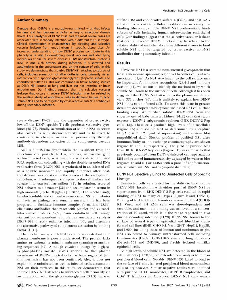
Author Summary
Dengue virus (DENV) is a mosquito-transmitted virus that infectshumans and has become a global emerging infectious diseasethreat. Four serotypes of DENV exist, and the most severe cases areassociated with secondary infection with a different virus serotype.Clinical deterioration is characterized by bleeding and selectivevascular leakage from endothelium in specific tissue sites. Anincreased understanding of how DENV proteins contribute to thisphenotype is vital to developing novel vaccines and identifyingindividuals at risk for severe disease. DENV nonstructural protein-1(NS1) is one such protein: during infection, it is secreted andaccumulates in the supernatant and on the surface of cells. In thisstudy, we demonstrate that soluble DENV NS1 attaches to subsets ofcells, including some but not all endothelial cells, primarily via aninteraction with specific glycosaminoglycans (heparan sulfate andchondroitin sulfate E). This was confirmed in tissue binding studiesas DENV NS1 bound to lung and liver but not intestine or brainendothelium. Our findings suggest that the selective vascularleakage that occurs in severe DENV infection may be related tothe relative ability of endothelial cells in different tissues to bindsoluble NS1 and to be targeted by cross-reactive anti-NS1 antibodiesduring secondary infection.

bound to the surface of monocyte-derived macrophages. Incontrast to that observed with primary lymphocytes, DENVNS1 bound strongly to the surface of several malignant T celllines, including Jurkat, H9, and EL-4 (Figure 2 and unpub-lished data).
As vascular leakage is a hallmark of DHF/DSS, and
endothelial cells are believed to be targets of immune-mediated damage, we analyzed the binding of NS1 to humanendothelial cells. Interestingly, DENV NS1 bound strongly tohuman dermal and lung microvascular endothelial cells(HMEC) and HMEC-lung blood (HEMC-LB), modestly toaortic endothelial cells, but minimally to primary or
Figure 1. Purification of DENV NS1 from BHK DENV-2 Rep Cells
(A) A flow cytometry histogram of expression of DENV NS1 in BHK DENV-2 Rep cells. Cells were fixed and stained with anti-DENV-2 NS1 mAbs or isotypecontrol Abs. One representative experiment of three is shown.(B) Elution profile of purified NS1 after immunoaffinity chromatography of serum-free supernatants of BHK DENV-2 Rep cells. Peak fractions (19–21)were combined and passed over an ion-exchange column, and purified NS1 was eluted (C) with a linear NaCl gradient (diagonal line).(D) SDS-PAGE of purified soluble DENV NS1 from BHK DENV-2 Rep cells. Purified NS1 was boiled prior to nonreducing 12% SDS-PAGE and silver staining.Molecular weight markers are shown at the left.(E) Immunoreactivity of purified DENV-2 NS1. Western blot analysis of purified DENV-2 NS1 after nonreducing 12% SDS-PAGE was performed using the2G6 anti-DENV NS1 mAb.doi:10.1371/journal.ppat.0030183.g001
PLoS Pathogens | www.plospathogens.org November 2007 | Volume 3 | Issue 11 | e1831800
Mechanism NS1 Attachment to Cells

immortalized human umbilical vein endothelial cells (HU-VEC or Eahy926).
Soluble DENV NS1 Binds to GAGAs our data indicated that soluble DENV NS1 bound a
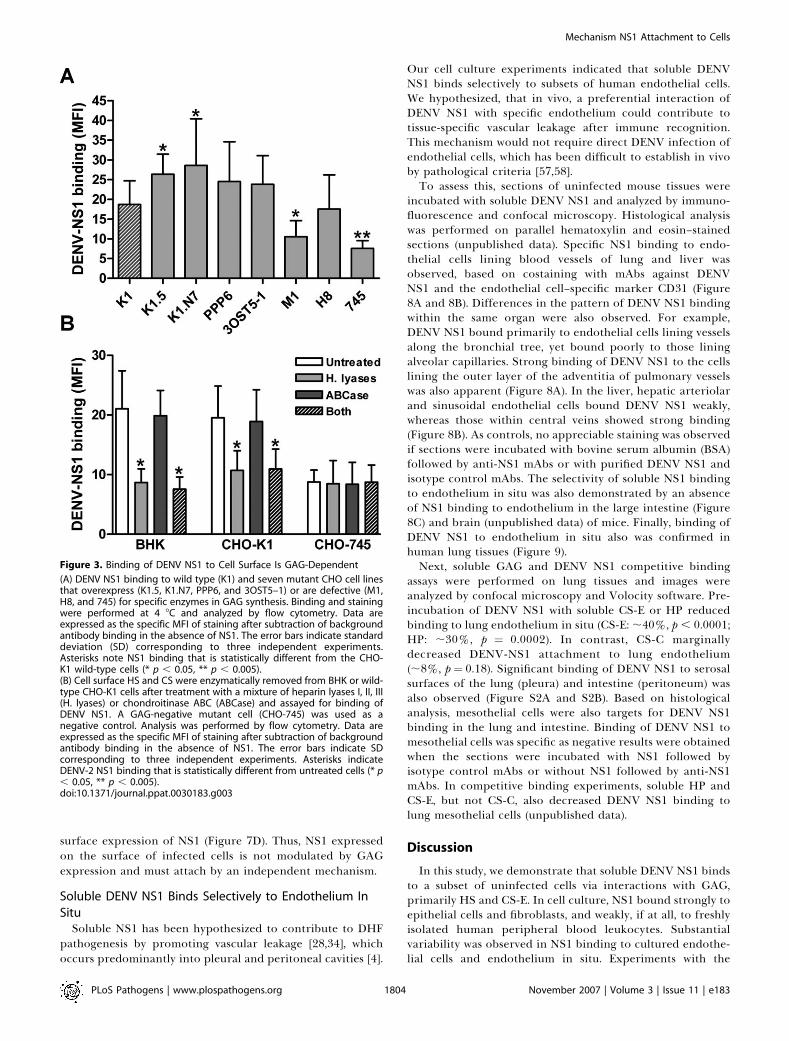
subset of uninfected mammalian cells of different lineages,we sought to identify the mechanism of attachment. Wehypothesized that NS1 might interact with a highly conservedmoiety, such as a GAG. To test this, we compared DENV NS1binding to wild-type CHO-K1 cells and seven different CHOcell lines (Table 2) that are either defective in GAG biosyn-thesis or express GAG with distinct structural specificities.CHO-745 cells, which genetically lack xylosyltransferase, theenzyme required for biosynthesis of both HS and CS [46],showed a 60%–70% reduction (p , 0.0001) in binding ofDENV NS1 (Figure 3A and Table 2). Similarly, CHO-M1 cells,which are defective in HS biosynthesis [47], exhibited a ;50%reduction (p ¼ 0.02) in DENV NS1 binding. However, CHO-H8 cells [48], which are a variant of CHO-M1 HS-deficientcells overexpressing CS 2-O-sulfotransferase (2-OST) (pro-ducing a 30-fold increase in double sulfated disaccharideresidues on CS), showed levels of DENV NS1 binding thatwere similar to CHO-K1 wild-type cells. Thus, both HS and
CS sustain cell surface binding to DENV NS1, at least on CHOcells.To determine if DENV NS1 required modification of HS
structures for binding, we tested CHO cell lines thatoverexpress different 3-O-sulfotransferases (3-OST) (Figure3A and Table 2). All 3-OSTs transfer sulfate moieties to the 3-O-position of N-sulfated glucosamine (GlcNS) and generate ahighly sulfated HS motif with distinct biological properties(Table 2). CHO-K1.5 cells overexpress 3-OST-1, which adds 3-O-sulfate to GlcNS in a glucuronic acid (GlcA)-GlcNS 6 6Ssequence context, and facilitate high affinity antithrombin IIIbinding [49]. CHO-K1.N7 cells overexpress 3-OST-3, whichadds 3-O-sulfate to GlcNS in a GlcA2S/iduronic acid (IdoA)2S-GlcNS 6 6S sequence context, and promote herpessimplex virus (HSV) glycoprotein D binding and entry [50].By contrast, overexpression of 3-OST-5 (CHO-3-OST-5 cells)adds 3-O-sulfate to GlcNS in both GlcA-GlcNS 6 6S andGlcA2S/IdoA2S-GlcNS 6 6S sequence contexts. Notably, allCHO cell lines expressing distinct 3-OST showed enhancedDENV NS1 binding by 30%–50% (p , 0.05) without apreference for any of the distinct 3-OST-modified HSstructures. Thus, unlike other glycoproteins, DENV NS1
Figure 2. DENV NS1 Binds to Cells of Specific Lineages
Dose-dependent binding of DENV NS1 to cell surfaces. Purified DENV NS1 (5, 10, 20, and 40 lg/ml) was added to different cell types at 4 8C for 1 h. Afterwashing, bound NS1 was detected by anti-NS1 antibody staining and flow cytometry. An example of DENV NS1 binding to CHO-K1 cells usingsupernatant from BHK-DENV-2 Rep cells is depicted in the inset at the upper right corner of the top left histogram. Examples of histogram profiles ofDENV NS1 binding to epithelial, fibroblast, hematopoietic, and endothelial cells are depicted. One representative of three experiments is shown. Nospecific staining was observed after BSA binding and incubation with an NS1 antibody or NS1 binding and incubation with an isotype-matched controlantibody (unpublished data).doi:10.1371/journal.ppat.0030183.g002
PLoS Pathogens | www.plospathogens.org November 2007 | Volume 3 | Issue 11 | e1831801
Mechanism NS1 Attachment to Cells

prefers highly sulfated HS motifs for binding and does notrequire specific HS sequences.
On CHO cell lines, the specificity of the GAG had a moredominant effect on regulating NS1 binding than the absolutelevel. Using a previously developed high-pressure liquidchromatography method [51], we quantified the amount ofcell surface HS and CS GAG in different CHO cell lines andcompared it to NS1 binding (Table 2). Although CHO-M1cells expressed twice the amount of total GAG compared towild-type CHO-K1 cells, CHO-M1 cells bound NS1 poorly.Thus, the total level of GAG did not directly correlate withNS1 binding. Consistent with this, similar amount of GAGwere observed on CHO-K1 and other 3-OST-expressing CHOcell lines, yet 3-OST-expressing CHO cells bound NS1 morestrongly. Moreover, Vero cells express ;3-fold higher levelsof HS compared to CHO-K1 cells (unpublished data) butbound NS1 less well compared to CHO-K1 cells (Figure 2 andTable 2).
A requirement of GAG for DENV NS1 binding to the cellsurface was confirmed independently by enzymatic treatmentof BHK and CHO-K1 cells with specific heparin lyases andchondroitinases (Figure 3B). Treatment of BHK or CHO-K1cells with heparin lyases I, II, and III, which specificallyremove HS, reduced DENV NS1 binding (;50%–60%, p ,
0.05) to the levels observed in GAG-negative CHO-745 cells.
In contrast, DENV NS1 attachment was not significantlyaffected by treatment with chondroitinase ABC, whichdegrades all types of CS [52]. As expected, treatment withboth enzymes had no effect on DENV NS1 binding to CHO-745 cells, which lack GAG. Nonetheless, a small amount ofNS1 binding to CHO-745 cells was observed, presumablythrough a subordinate GAG-independent pathway. Overall,the analysis of CHO cell mutants and enzymatic treatmentssuggested that HS and highly sulfated forms of CS were theprimary cell surface GAG that sustained DENV NS1 bindingon CHO and BHK cells.As an additional confirmation, we performed competitive
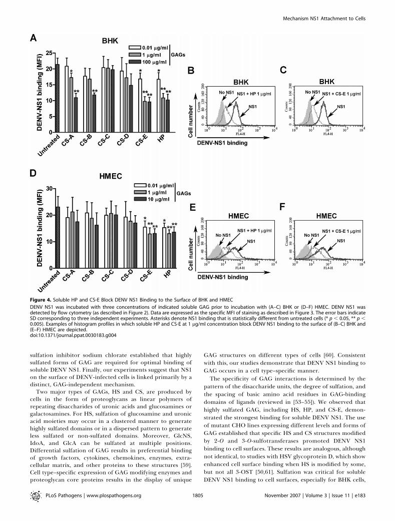
binding assays with soluble GAG. Pre-incubation with solubleCS-E or heparin (HP) substantially decreased (;50%, p ,
0.005), in a dose-dependent manner, DENV NS1 binding toBHK cells (Figure 4A–4C). Small, albeit significant inhibition(;15%, p , 0.05), was observed even at relatively lowconcentrations CS-E and HP (0.01 lg/ml or 0.033 nM). Incontrast, CS-A and -B inhibited NS1 binding only at higherconcentrations (100 lg/ml or 3.3 lM, p , 0.05) whereas CS-Cand -D showed no significant effect (p . 0.2). Similarly, pre-incubation with CS-E and HP reduced DENV NS1 attachmentto HMEC cells (;40%, 0.01 lg/ml, p , 0.01) (Figure 4D–4F). Ina more direct in vitro binding ELISA, an interaction betweenDENV NS1 and HS or CS-E was established, whereas only
Table 1. DENV NS1 Binds to a Variety of Cell Types
Cell Type Cell Description Mean Fluorescent Intensity p-Value
No DENV NS1 þAnti-NS1 mAb
DENV NS1 þIsotype Control mAb
DENV NS1 þAnti-NS1 mAb
BHK Hamster kidney fibroblast 5.4 6 0.3 4.1 6 0.5 55.4 6 4.9 ,0.0001
CHO-K1 Chinese hamster ovarian epithelium 4.5 6 0.3 3.8 6 0.6 45.0 6 4.6 ,0.0001
Vero Monkey kidney epithelium 3.3 6 0.1 3.6 6 0.3 26.2 6 8.2 ,0.05
293T Human kidney epithelium 3.8 6 0.2 3.1 6 0.1 17.9 6 1.1 ,0.0001
IMR-90 Human lung fibroblast 4.0 6 0.4 3.6 6 0.7 44.6 6 2.5 ,0.0001
Detroit-550 Human skin fibroblast 3.4 6 0.7 3.6 6 0.5 21.6 6 4.7 ,0.005
CCD-1102 Human keratinocytes 3.5 6 0.4 3.0 6 0.2 33.9 6 3.2 ,0.0001
HepG2 Human hepatocellular carcinoma 4.2 6 0.6 3.5 6 0.5 96.2 6 11.6 ,0.0001
4/4 RM4 Rat visceral mesothelium 4.2 6 0.1 4.0 6 0.9 11.3 6 0.8 ,0.005
HUVEC 1 8 human umbilical cord vein EC 3.4 6 0.1 3.5 6 0.1 4.6 6 0.2 ,0.05
HMEC Human dermal microvascular EC 3.8 6 0.2 3.6 6 0.1 34.9 6 1.9 ,0.0001
HMEC-LB 1 8 Human lung blood
microvascular EC
5.0 6 0.4 4.6 6 1.0 21.2 6 0.7 ,0.0001
HAEC 1 8 Human aotic EC 3.9 6 0.2 ND 12.5 6 0.7 ,0.001
Eahy926 Human umbilical cord vein EC 3.7 6 0.1 3.1 6 0.2 5.2 6 0.5 ,0.05
U937 Human monocytic cell 3.8 6 0.2 2.8 6 0.1 4.8 6 0.9 0.12
MEG-01 Human megakaryoblast 4.9 6 0.4 3.1 6 0.5 11.7 6 2.0 ,0.05
Human monocytes Peripheral blood 3.5 6 0.1 2.0 6 0.4 4.4 6 0.5 0.34
Human macrophages Peripheral blooda 5.9 6 0.0 5.6 6 1.3 9.8 6 0.9 ,0.05
Human platelets Peripheral blood 2.2 6 0.2 ND 1.9 6 0.2 0.29
Human CD19þ B lymphocytes Peripheral blood 5.5 6 0.6 ND 6.7 6 0.5 0.21
Human CD4þ T lymphocytes Peripheral blood 5.1 6 0.1 ND 4.9 6 0.3 0.59
Human peripheral blood
mononuclear cells
Peripheral blood 4.3 6 0.6 3.6 6 0.7 5.8 6 0.6 0.06
Human erythrocytes Peripheral blood 4.2 6 0.4 3.2 6 0.6 5.4 6 0.3 0.08
Cells were incubated with serum-free supernatants from BHK DENV-2-Rep (DENV NS1) or BHK (negative control, No DENV NS1) cells for 1 h at 4 8C. Bound DENV NS1 was detected by anti-DENV NS1 mAb (2G6) followed by secondary antibody. Analysis was performed on gated viable cells (10, 000 events) by flow cytometry. Data are mean 6 SD of three independentexperiments. p-Values were determined by a one-way ANOVA test after comparing the MFI of the cells after incubation with control protein or DENV NS1 with anti-NS1 antibody or DENVNS1 with isotype control antibody. Among these cell types, BHK, Vero, 293T, HepG2, HUVEC, Eahy926, U937, monocytes, and macrophages can be infected with highly passaged (e.g.,strains 16681 or New Guinea C) DENV-2 isolates [73]. However, only a subset of cells (BHK, Vero, HepG2, and monocytes) are infected with primary DENV-2 isolates [73].aMacrophages were generated from peripheral blood monocytes cultured for 7 d in medium containing 50 ng/ml of GM-CSF.HAEC, human aortic endothelial cell; ND, not determined.doi:10.1371/journal.ppat.0030183.t001
PLoS Pathogens | www.plospathogens.org November 2007 | Volume 3 | Issue 11 | e1831802
Mechanism NS1 Attachment to Cells
weak or no appreciable binding was detected with CS-A, -B,-C, and -D (Figure 5). Taken together, these results suggestthat NS1 interacts more strongly with HP, HS, and CS-E andmore weakly with CS-A and CS-B. CS-A consists of mainlyGlcA-GalNAc4S repeating disaccharides, CS-B contains bothIdoA-GalNAc4S and GlcA-GalNAc4S as dominant repeatingdisaccharides, and CS-C consists of mainly GlcA-GalNAc6Srepeating disaccharides. CS-D and CS-E are enriched indisulfated disaccharides, GlcA2S-GalNAc6S and GlcA-Gal-NAc4S6S, respectively. CS-E is unique, as two sulfates arepresent in the same GalNAc residue compared to other CS,and suggests that clustered sulfates rather than net negativecharge on the disaccharide backbone facilitate DENV NS1binding.
GAG Sulfation Is Critical for DENV NS1 Cellular AttachmentThe specificity of GAG-protein interactions often depends
on the position and degree of sulfation (reviewed in [53–55]).To clarify if GAG sulfation is required for cell binding ofDENV NS1, we treated BHK and CHO-K1 cells with thereversible sulfation inhibitor, sodium chlorate [56] (Figure 6Aand 6B). This treatment did not alter cell viability, as analyzedby propidium iodide exclusion (unpublished data). However,binding of DENV NS1 to BHK cells was abolished by theaddition of sodium chlorate (.5 mM). In contrast, atcomparable doses, DENV NS1 binding to wild-type CHO-K1was reduced to the level observed with CHO-745 cells lackingGAG. Importantly, the phenotype was reversed by the
addition of excess exogenous (10 mM) sodium sulfate (Figure6C and 6D). Thus, GAG sulfation is required for optimalbinding of soluble DENV NS1 to cells.
Expression of DENV NS1 on the Surface of DENV-InfectedCells Is Largely GAG-IndependentAlthough our data suggests that soluble DENV NS1 binds to
the cell surface of uninfected cells via HS and CS-E, weevaluated if this was the dominant mechanism of attachmentof NS1 to DENV-infected cells. Previous studies havesuggested that flavivirus NS1 may be displayed on the cellsurface of infected cells by a GPI anchor or transmembranelinkage [43], even though the protein lacks anchor attachmentsequences or consensus membrane spanning domains. Toevaluate this, BHK cells were infected with DENV-2 andcultured in the presence of high concentrations of solubleHP. No reduction of DENV NS1 surface expression oninfected cells was observed (Figure 7A). These results wereconfirmed by studies with the sulfate inhibitor, sodiumchlorate. In contrast to the inhibitory effect of sodiumchlorate on soluble NS1 binding (Figure 6), no reduction ofDENV NS1 expression on the surface of infected BHK cellswas observed (Figure 7B). Similar studies with BHK DENV-2Rep cells also showed no change in surface NS1 expressionafter sodium chlorate treatment (Figure 7C). Finally, in directcontrast to that observed with soluble NS1 and uninfectedcells (Figure 3B), treatment of DENV-infected cells withheparin lyases and/or chondroitinase ABC did not decrease
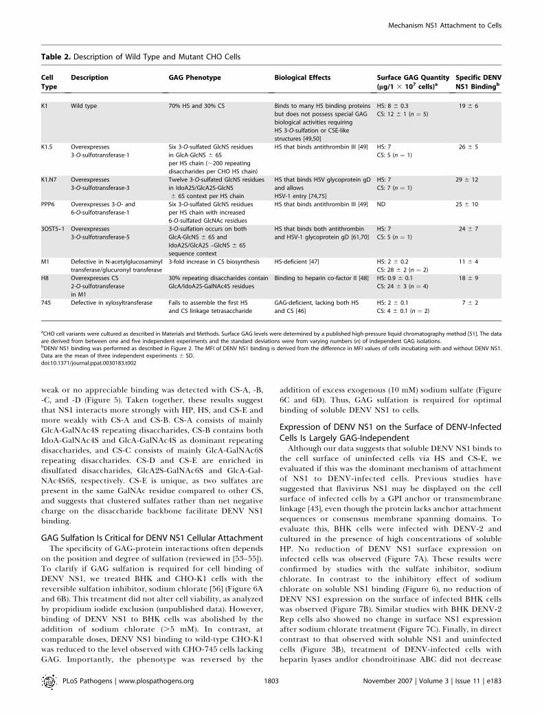
Table 2. Description of Wild Type and Mutant CHO Cells
Cell
Type
Description GAG Phenotype Biological Effects Surface GAG Quantity
(lg/1 3 107 cells)aSpecific DENV
NS1 Bindingb
K1 Wild type 70% HS and 30% CS Binds to many HS binding proteins
but does not possess special GAG
biological activities requiring
HS 3-O-sulfation or CSE-like
structures [49,50]
HS: 8 6 0.3
CS: 12 6 1 (n ¼ 5)
19 6 6
K1.5 Overexpresses
3-O-sulfotransferase-1
Six 3-O-sulfated GlcNS residues
in GlcA-GlcNS 6 6S
per HS chain (;200 repeating
disaccharides per CHO HS chain)
HS that binds antithrombin III [49] HS: 7
CS: 5 (n ¼ 1)
26 6 5
K1.N7 Overexpresses
3-O-sulfotransferase-3
Twelve 3-O-sulfated GlcNS residues
in IdoA2S/GlcA2S-GlcNS
6 6S context per HS chain
HS that binds HSV glycoprotein gD
and allows
HSV-1 entry [74,75]
HS: 7
CS: 7 (n ¼ 1)
29 6 12
PPP6 Overexpresses 3-O- and
6-O-sulfotransferase-1
Six 3-O-sulfated GlcNS residues
per HS chain with increased
6-O-sulfated GlcNAc residues
HS that binds antithrombin III [49] ND 25 6 10
3OST5–1 Overexpresses
3-O-sulfotransferase-5
3-O-sulfation occurs on both
GlcA-GlcNS 6 6S and
IdoA2S/GlcA2S –GlcNS 6 6S
sequence context
HS that binds both antithrombin
and HSV-1 glycoprotein gD [61,70]
HS: 7
CS: 5 (n ¼ 1)
24 6 7
M1 Defective in N-acetylglucosaminyl
transferase/glucuronyl transferase
3-fold increase in CS biosynthesis HS-deficient [47] HS: 2 6 0.2
CS: 28 6 2 (n ¼ 2)
11 6 4
H8 Overexpresses CS
2-O-sulfotransferase
in M1
30% repeating disaccharides contain
GlcA/IdoA2S-GalNAc4S residues
Binding to heparin co-factor II [48] HS: 0.9 6 0.1
CS: 24 6 3 (n ¼ 4)
18 6 9
745 Defective in xylosyltransferase Fails to assemble the first HS
and CS linkage tetrasaccharide
GAG-deficient, lacking both HS
and CS [46]
HS: 2 6 0.1
CS: 4 6 0.1 (n ¼ 2)
7 6 2
aCHO cell variants were cultured as described in Materials and Methods. Surface GAG levels were determined by a published high-pressure liquid chromatography method [51]. The dataare derived from between one and five independent experiments and the standard deviations were from varying numbers (n) of independent GAG isolations.bDENV NS1 binding was performed as described in Figure 2. The MFI of DENV NS1 binding is derived from the difference in MFI values of cells incubating with and without DENV NS1.Data are the mean of three independent experiments 6 SD.doi:10.1371/journal.ppat.0030183.t002
PLoS Pathogens | www.plospathogens.org November 2007 | Volume 3 | Issue 11 | e1831803
Mechanism NS1 Attachment to Cells
surface expression of NS1 (Figure 7D). Thus, NS1 expressedon the surface of infected cells is not modulated by GAGexpression and must attach by an independent mechanism.
Soluble DENV NS1 Binds Selectively to Endothelium In
SituSoluble NS1 has been hypothesized to contribute to DHF
pathogenesis by promoting vascular leakage [28,34], whichoccurs predominantly into pleural and peritoneal cavities [4].
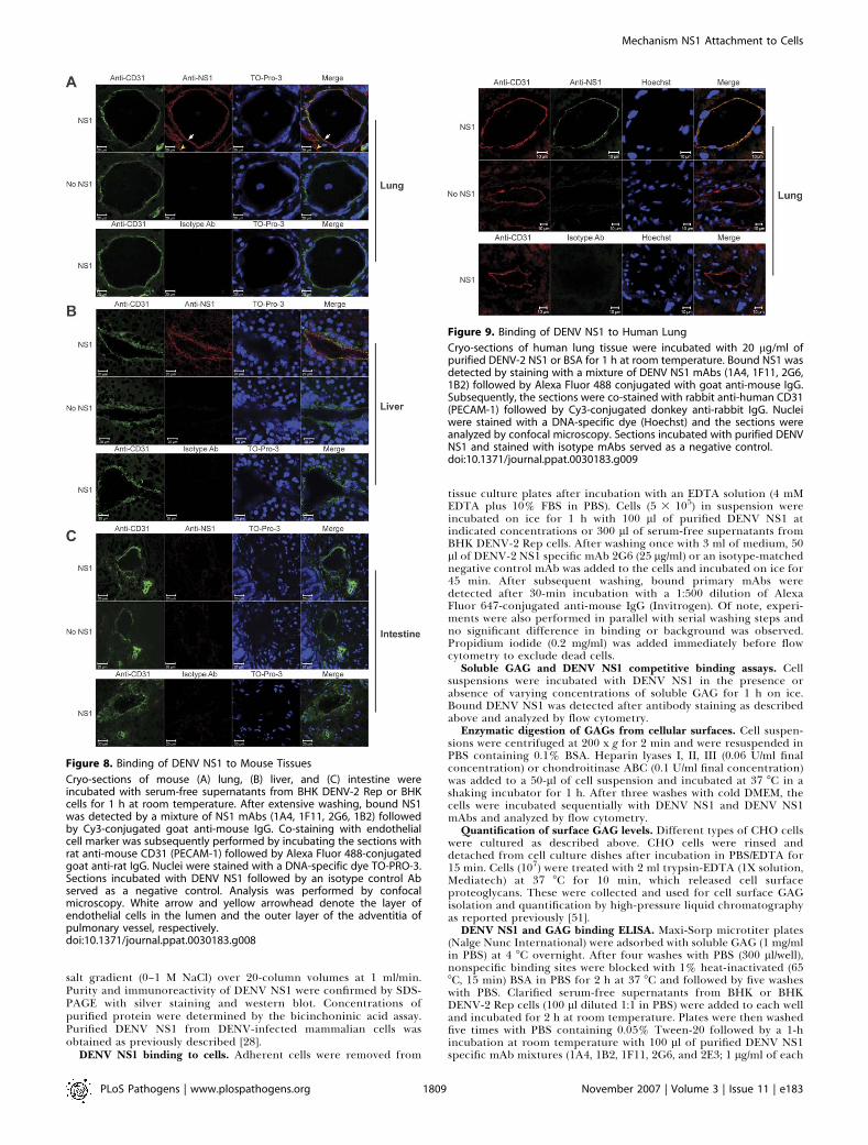
Our cell culture experiments indicated that soluble DENVNS1 binds selectively to subsets of human endothelial cells.We hypothesized, that in vivo, a preferential interaction ofDENV NS1 with specific endothelium could contribute totissue-specific vascular leakage after immune recognition.This mechanism would not require direct DENV infection ofendothelial cells, which has been difficult to establish in vivoby pathological criteria [57,58].To assess this, sections of uninfected mouse tissues were
incubated with soluble DENV NS1 and analyzed by immuno-fluorescence and confocal microscopy. Histological analysiswas performed on parallel hematoxylin and eosin–stainedsections (unpublished data). Specific NS1 binding to endo-thelial cells lining blood vessels of lung and liver wasobserved, based on costaining with mAbs against DENVNS1 and the endothelial cell–specific marker CD31 (Figure8A and 8B). Differences in the pattern of DENV NS1 bindingwithin the same organ were also observed. For example,DENV NS1 bound primarily to endothelial cells lining vesselsalong the bronchial tree, yet bound poorly to those liningalveolar capillaries. Strong binding of DENV NS1 to the cellslining the outer layer of the adventitia of pulmonary vesselswas also apparent (Figure 8A). In the liver, hepatic arteriolarand sinusoidal endothelial cells bound DENV NS1 weakly,whereas those within central veins showed strong binding(Figure 8B). As controls, no appreciable staining was observedif sections were incubated with bovine serum albumin (BSA)followed by anti-NS1 mAbs or with purified DENV NS1 andisotype control mAbs. The selectivity of soluble NS1 bindingto endothelium in situ was also demonstrated by an absenceof NS1 binding to endothelium in the large intestine (Figure8C) and brain (unpublished data) of mice. Finally, binding ofDENV NS1 to endothelium in situ also was confirmed inhuman lung tissues (Figure 9).Next, soluble GAG and DENV NS1 competitive binding
assays were performed on lung tissues and images wereanalyzed by confocal microscopy and Volocity software. Pre-incubation of DENV NS1 with soluble CS-E or HP reducedbinding to lung endothelium in situ (CS-E: ;40%, p , 0.0001;HP: ;30%, p ¼ 0.0002). In contrast, CS-C marginallydecreased DENV-NS1 attachment to lung endothelium(;8%, p¼ 0.18). Significant binding of DENV NS1 to serosalsurfaces of the lung (pleura) and intestine (peritoneum) wasalso observed (Figure S2A and S2B). Based on histologicalanalysis, mesothelial cells were also targets for DENV NS1binding in the lung and intestine. Binding of DENV NS1 tomesothelial cells was specific as negative results were obtainedwhen the sections were incubated with NS1 followed byisotype control mAbs or without NS1 followed by anti-NS1mAbs. In competitive binding experiments, soluble HP andCS-E, but not CS-C, also decreased DENV NS1 binding tolung mesothelial cells (unpublished data).
Discussion
In this study, we demonstrate that soluble DENV NS1 bindsto a subset of uninfected cells via interactions with GAG,primarily HS and CS-E. In cell culture, NS1 bound strongly toepithelial cells and fibroblasts, and weakly, if at all, to freshlyisolated human peripheral blood leukocytes. Substantialvariability was observed in NS1 binding to cultured endothe-lial cells and endothelium in situ. Experiments with the
Figure 3. Binding of DENV NS1 to Cell Surface Is GAG-Dependent
(A) DENV NS1 binding to wild type (K1) and seven mutant CHO cell linesthat overexpress (K1.5, K1.N7, PPP6, and 3OST5–1) or are defective (M1,H8, and 745) for specific enzymes in GAG synthesis. Binding and stainingwere performed at 4 8C and analyzed by flow cytometry. Data areexpressed as the specific MFI of staining after subtraction of backgroundantibody binding in the absence of NS1. The error bars indicate standarddeviation (SD) corresponding to three independent experiments.Asterisks note NS1 binding that is statistically different from the CHO-K1 wild-type cells (* p , 0.05, ** p , 0.005).(B) Cell surface HS and CS were enzymatically removed from BHK or wild-type CHO-K1 cells after treatment with a mixture of heparin lyases I, II, III(H. lyases) or chondroitinase ABC (ABCase) and assayed for binding ofDENV NS1. A GAG-negative mutant cell (CHO-745) was used as anegative control. Analysis was performed by flow cytometry. Data areexpressed as the specific MFI of staining after subtraction of backgroundantibody binding in the absence of NS1. The error bars indicate SDcorresponding to three independent experiments. Asterisks indicateDENV-2 NS1 binding that is statistically different from untreated cells (* p, 0.05, ** p , 0.005).doi:10.1371/journal.ppat.0030183.g003
PLoS Pathogens | www.plospathogens.org November 2007 | Volume 3 | Issue 11 | e1831804
Mechanism NS1 Attachment to Cells
sulfation inhibitor sodium chlorate established that highlysulfated forms of GAG are required for optimal binding ofsoluble DENV NS1. Finally, our experiments suggest that NS1on the surface of DENV-infected cells is linked primarily by adistinct, GAG-independent mechanism.
Two major types of GAGs, HS and CS, are produced bycells in the form of proteoglycans as linear polymers ofrepeating disaccharides of uronic acids and glucosamines orgalactosamines. For HS, sulfation of glucosamine and uronicacid moieties may occur in a clustered manner to generatehighly sulfated domains or in a dispersed pattern to generateless sulfated or non-sulfated domains. Moreover, GlcNS,IdoA, and GlcA can be sulfated at multiple positions.Differential sulfation of GAG results in preferential bindingof growth factors, cytokines, chemokines, enzymes, extra-cellular matrix, and other proteins to these structures [59].Cell type–specific expression of GAG modifying enzymes andproteoglycan core proteins results in the display of unique
GAG structures on different types of cells [60]. Consistentwith this, our studies demonstrate that DENV NS1 binding toGAG occurs in a cell type–specific manner.The specificity of GAG interactions is determined by the
pattern of the disaccharide units, the degree of sulfation, andthe spacing of basic amino acid residues in GAG-bindingdomains of ligands (reviewed in [53–55]). We observed thathighly sulfated GAG, including HS, HP, and CS-E, demon-strated the strongest binding for soluble DENV NS1. The useof mutant CHO lines expressing different levels and forms ofGAG established that specific HS and CS structures modifiedby 2-O and 3-O-sulfotransferases promoted DENV NS1binding to cell surfaces. These results are analogous, althoughnot identical, to studies with HSV glycoprotein D, which showenhanced cell surface binding when HS is modified by some,but not all 3-OST [50,61]. Sulfation was critical for solubleDENV NS1 binding to cell surfaces, especially for BHK cells,
Figure 4. Soluble HP and CS-E Block DENV NS1 Binding to the Surface of BHK and HMEC
DENV NS1 was incubated with three concentrations of indicated soluble GAG prior to incubation with (A–C) BHK or (D–F) HMEC. DENV NS1 wasdetected by flow cytometry (as described in Figure 2). Data are expressed as the specific MFI of staining as described in Figure 3. The error bars indicateSD corresponding to three independent experiments. Asterisks denote NS1 binding that is statistically different from untreated cells (* p , 0.05, ** p ,0.005). Examples of histogram profiles in which soluble HP and CS-E at 1 lg/ml concentration block DENV NS1 binding to the surface of (B–C) BHK and(E–F) HMEC are depicted.doi:10.1371/journal.ppat.0030183.g004
PLoS Pathogens | www.plospathogens.org November 2007 | Volume 3 | Issue 11 | e1831805
Mechanism NS1 Attachment to Cells

as treatment with the sulfate inhibitor sodium chlorateabolished DENV NS1 attachment.
Also consistent with our results, CS-E, but not CS-A, -B, or-C, interfered with HSV binding to the target cells [62]. Usingradioactive binding or gel mobility shift assays, we observedsimilarly strong binding between West Nile virus NS1 and HSor CS-E (L. Zhang, K. Chung, and M. Diamond, unpublisheddata). As binding of soluble DENV NS1 to GAG requiressulfation, it is plausible that the interaction is primarilyelectrostatic in nature and depends on the relative degree ofnegative charge on a GAG motif. The major repeatingdisaccharides in HP are IdoA2S-GlcNS6S (;2.7 sulfates/disaccharides), whereas the major repeating disaccharides inCS-D and CS-E are GlcA2S-GalNAc6S and GlcA-GalNAc4S6S,respectively (;1.7 sulfates/disaccharide) [62]. Because NS1preferentially interacts with CS-E and HP compared to CS-D,we hypothesize that NS1 binding may prefer closely spacedsulfates within the same sugar residue. Since HP and CS-Ehave distinct sugar sequences, the combination of disacchar-ide unit and position of sulfation on an individual GAG maydetermine the strength of DENV NS1 binding.
The linear sequences XBBXBX and XBBBXXBX (where B isa basic Arg or Lys amino acid) are common HP bindingmotifs in proteins [55]. However, GAG binding sites may notbe exclusively defined by linear sequences but also caninclude conformational epitopes that juxtapose basic aminoacids from different segments of a protein [55]. Amino acidsequence analysis of DENV NS1 reveals no apparent canon-ical GAG binding motifs. Although reverse genetic strategiesare planned, the identification of the amino acids involved inGAG recognition may await solution of the NS1 structure.
Our data showing that soluble NS1 can bind to the surfaceof the GAG-deficient line, CHO-745, albeit at significantlylower levels, suggest that additional molecules also serve as
ligands for DENV NS1 attachment. This secondary attach-ment ligand may be expressed only on subsets of cells asaddition of sodium chlorate completely abrogated solubleNS1 binding to BHK cells. Consistent with this, soluble HSand CS-E only partially reduced DENV NS1 binding toHMEC. Thus, different cell types may express multipleligands for attachment of soluble DENV NS1.The expression of NS1 on the surface of DENV-infected
cells was insensitive to treatments that reduce GAG levels.This also suggests the existence of an alternate mechanism ofcell surface NS1 attachment. Some have speculated that atransmembrane form of NS1 exists, although the proteinsequence lacks a canonical hydrophobic membrane-spanningdomain [42]. Others have postulated that at least somefraction of cell surface NS1 is linked via a GPI anchor [43,63].Based on our data, we hypothesize that DENV NS1 isexpressed on cell surfaces by at least two mechanisms: onuninfected cells soluble NS1 binds to the surface via a GAG-dependent and GAG-independent mechanism, whereas oninfected cells NS1 attaches via a GAG-independent mecha-nism, possibly via a membrane or GPI anchor. What remainsuncertain, and is a direction for future research, is whetherthe mechanism of cell surface attachment of NS1 has uniquefunctional consequences, especially in terms of immunerecognition or evasion.Our studies demonstrate that soluble DENV NS1 differ-
entially binds to cultured endothelial cells in vitro andendothelium in situ in human and mouse tissues. In contrast,others have shown that intravenous injection of C57BL/6 xSJL mice with high concentrations (250 lg/ml) of solubleDENV-1 NS1 results in binding to and accumulation inhepatocytes but not to other cell types in the liver or othertissues [40]. A possible reason for the disparity in results isthat the route of antigen administration may modulate NS1binding: intravenous injection could prompt rapid first-passclearance of antigen by the liver. Although further studies arenecessary, our data are consistent with specific GAGmodification by subsets of endothelial cells in differenttissues modulating the level of bound NS1. Indeed, tissue-specific expression of different isoforms of enzymes in GAGbiosynthesis has been reported [64,65]. Selective IL-8 bindingto endothelial cells has been observed [66] and could be dueto subtle differences in the display of GAG on the surface ofcells. In favor of this hypothesis, IL-8 selectively binds tosubsets of HP and HS [67].The pathologic mechanism underlying selective vascular
leakage at serosal sites during DHF/DSS [4] remains unknown.High levels of intravascular soluble NS1, as observed inDENV-infected patients, could promote binding and surfaceexpression of NS1 on selective endothelium without arequirement for direct viral infection, which has beendifficult to establish histopathologically in fatal DHF cases[57,58]. In addition, specific binding to mesothelial cells thatline pleura and peritoneum, as observed in our DENV NS1binding experiments in situ, could contribute to the pleuraleffusions or ascites that are observed in DHF/DSS patients.Although more experiments are necessary, preferentialbinding of soluble NS1 to subsets of endothelial andmesothelial cells in vivo could lead to tissue-specific vascularleakage that occurs during severe secondary DENV infectionafter recognition by anti-NS1 antibodies, immune complexformation, and inflammatory damage [28,68].
Figure 5. Direct Binding of DENV NS1 with HS and CS-E
Microtiter plates were coated with indicated soluble GAG overnight at 48C. After incubation with supernatants from BHK-DENV-2-Rep cells orcontrol BHK cells, bound DENV NS1 was detected using DENV NS1–specific mAbs, followed by biotin-conjugated anti-mouse IgG and HRP-conjugated streptavidin. Plates were read at an optical density of 450 nmon a 96-well plate reader. Data are the mean 6 SD from fourindependent experiments. Asterisks denote NS1 binding that is statisti-cally different from binding to background (* p , 0.05, ** p , 0.005). O.D., optical density.doi:10.1371/journal.ppat.0030183.g005
PLoS Pathogens | www.plospathogens.org November 2007 | Volume 3 | Issue 11 | e1831806
Mechanism NS1 Attachment to Cells

Materials and Methods
Reagents. CS-A, -B, -C, -D, -E, HP, HS, heparin lyases I, II, and III,chondroitinase ABC, anti-heparin lyase-digested HS (3G10), and anti-chondroitinase ABC-digested CS (2B6) antibodies were all purchased(Seikagaku). DENV-2 NS1–specific monoclonal antibodies (mAbhybridomas 2G6, 1A4, 1B2, 1F11, 2E3, and 2E11 [69], and unpublisheddata) were purified by protein G affinity chromatography. Mousepolyclonal anti-DENV-2 NS1 was produced after BALB/c mice wereintraperitoneally immunized three times with purified DENV-2 NS1(10 lg/dose) at a 2-wk interval. Mice were subsequently treated withpristane followed by injection with myeloma cells to induce ascitesformation. Ascites fluid containing anti-NS1 polyclonal antibody wascollected, and antibodies were purified by protein-G affinitychromatography. Anti-mouse IgG conjugated with Alexa Fluor 647,Alexa Fluor 488, or Cy3 were purchased from Invitrogen. Sodiumchlorate and sodium sulfate were obtained commercially (Sigma).
Cell culture. The following transformed cell lines were obtainedfrom the ATCC: BHK fibroblasts cells, African green monkey Verocells, HEK-293T human embryonic kidney carcinoma cells, L929
mouse fibroblasts, IMR-90 human lung fibroblasts, Detroit-550human skin fibroblasts, CCD-1102 human keratinocytes, HepG2and Hep3B human hepatocellular carcinoma cells, 4/4RM4 rat lungmesothelial cells, Eahy926 HUVEC, MEG-01 human megakaryoblastcells, Jurkat human leukemic T lymphoblasts, H9 human T lymphomacells, EL-4 mouse T lymphoma cells, and U937 human myelomono-cyte cells. HMEC and keratinocytes (HaCat) were gifts (M. Caparon).Wild-type CHO-K1 cells and CHO mutant lines with altered GAGexpression (CHO-745, CHO-M1, CHO-H8, CHO-K1.5, CHO-K1.N7,CHO-3OST5–1, and CHO-PPP6) have been described previously [46–49,61,70]. BHK, Vero, 293T, L929, and HaCat cells were cultured inDulbecco’s modified Eagle’s medium (DMEM) supplemented with10% fetal bovine serum (FBS), 50 mM HEPES, 4 mM L-glutamine, 100units/ml penicillin G, and 100 lg/ml streptomycin sulfate. IMR-90,Detroit-551, HepG2, and Hep3B were grown in minimum essentialEagle’s medium with Earle’s BSS, 10% FBS, 1% non-essential aminoacids (NEAA), 1 mM sodium pyruvate, 2 mM L-glutamine, 100 units/ml penicillin G, and 100 lg/ml streptomycin sulfate. CCD-1102 cellswere grown in keratinocyte serum-free medium supplemented withkeratinocyte growth factors (Invitrogen). MEG-01, U937, Jurkat, and
Figure 6. Sulfation Is Required for Soluble DENV NS1 Binding to Cells
(A–B) BHK, wild-type CHO-K1, and GAG-deficient mutant CHO-745 were cultured overnight in sulfate-free medium in the presence of indicated amountsof sodium chlorate. Cells were harvested and tested for NS1 binding by flow cytometry as described in Figure 2. No appreciable cell death was observedby this treatment as judged by exclusion of propidium iodide. Data (A) are expressed as the specific MFI of staining as described in Figure 3. The errorbars indicate SD corresponding to three independent experiments. Asterisks denote NS1 binding that is statistically different from untreated cells (** p, 0.005)(C–D) Addition of sodium sulfate (10 mM) restores binding of NS1 to cells. Cells were cultured overnight in sulfate-free medium and 25 mM sodiumchlorate with or without 10 mM sodium sulfate (as indicated). Cells were processed for DENV NS1 binding and analyzed by flow cytometry (see Figure2). Data (C) are expressed as the specific MFI of staining as described in Figure 3. The error bars indicate SD corresponding to three independentexperiments. Asterisks denote NS1 binding that is statistically different from untreated cells (** p , 0.005).doi:10.1371/journal.ppat.0030183.g006
PLoS Pathogens | www.plospathogens.org November 2007 | Volume 3 | Issue 11 | e1831807
Mechanism NS1 Attachment to Cells

H9 were cultured in RPMI 1640 medium with 10% FBS, 2 mM L-glutamine, 10 mM HEPES, 1 mM sodium pyruvate, 100 units/mlpenicillin G, and 100 lg/ml streptomycin sulfate. EL-4 cells werepropagated in Iscove’s medium supplemented with 10% FBS, 1%non-essential amino acids, 2 mM L-glutamine, 1 mM sodiumpyruvate, 100 units/ml penicillin G, and 100 lg/ml streptomycinsulfate. HMEC were grown in MCDB 131 (Invitrogen) supplementedwith 10% FBS, 2 mM L-glutamine, 0.3% NaHCO3, 1 lg/ml hydro-cortisone (Sigma), 10 ng/ml epidermal growth factor (Sigma), 100units/ml penicillin G, and 100 lg/ml streptomycin sulfate. All CHOcell lines were grown in Ham’s F12 medium containing 10% FBS, 100units/ml penicillin G, and 100 lg/ml streptomycin sulfate, except forCHO-30ST5–1 cells, which were cultured in CHO medium supple-mented with 400 lg/ml of G418 sulfate (Cellgro).
Primary human tonsil epithelial cells were isolated according to apublished protocol [71] and cultured in keratinocyte serum-freemedium supplemented with keratinocyte growth factor (Invitrogen).Primary HMEC-LB and aortic endothelial cells were purchased fromClonetics (Cambrex Bio Science) and maintained according to themanufacturer’s protocol. HUVEC were grown in RPMI 1640containing 10% FBS on 1% gelatin-coated surfaces. Primary humanperipheral blood mononuclear cells were isolated from buffy coatsobtained from the blood bank or healthy volunteers by Ficoll-Hypaque (Pharmacia) density gradient centrifugation. Human CD14þ
monocytes, CD19þ B cells, CD4þ T lymphocytes were purified from
primary human peripheral blood mononuclear cells via positiveselection using antibody-coated magnetic beads (Miltenyi Biotec). Togenerate macrophages, monocytes were cultured for 7 d in RPMI1640 supplemented with 50 ng/ml of GM-CSF, 10% FBS, 2 mMglutamine, 1% nonessential amino acids, 1% sodium pyruvate, 100units/ml penicillin G, and 100 lg/ml streptomycin sulfate. Humanplatelets were isolated from whole blood of healthy volunteers. Aftercentrifugation at 750 x g for 20 min at 22 8C, platelet-rich plasma wascollected and centrifuged at 1,200 x g for 15 min at 22 8C to obtainplatelets. Human erythrocytes were obtained from healthy donorsfollowing a published protocol [72]. All primary cells were culturedfor five or fewer passages for the NS1 binding experiments.
Purification of DENV NS1. BHK cells that stably propagate aDENV-2 subgenomic replicon (BHK DENV-2 Rep cells [45]) weregrown to 80%–90% confluence in DMEM containing 10% FBS and 3lg/ml puromycin (Sigma). Cell monolayers were washed several timeswith DMEM and the cells were cultured for another 3 d in serum-freeDMEM supplemented with 3 lg/ml puromycin. DENV NS1 secreted inthe supernatants of BHK DENV-2 Rep was quantified by NS1 captureELISA as previously described [28]. Supernatants were collected,centrifuged, pooled, and passed through a 0.2-lm filter prior toimmunoaffinity chromatography with anti-NS1 mAb 2G6 [28]. Peakelution fractions were combined, diluted 7-fold with 20 mM Tris (pH8.0), and loaded onto a 1-ml Mono Q ion exchange column (GEHealthcare) at a rate of 1 ml/min. The protein was eluted with a linear
Figure 7. Expression of DENV NS1 on the Surface of Infected Cells Is Not Dependent on GAG
(A) BHK cells were infected with DENV-2 at the MOI of 3 and cultured for 24 h in the presence of indicated concentrations of soluble HP or CS-C. Cellswere then harvested for analysis of surface NS1 expression.(B) BHK cells were infected with DENV-2 at the MOI of 3 and cultured for 24 h in the presence of 25 mM sodium chlorate. DENV NS1 expression wasdetermined and analyzed by flow cytometry using anti-DENV NS1 mAb (2G6) or polyclonal anti-DENV NS1.(C) BHK DENV-2 Rep cells were cultured in medium containing 25 mM sodium chlorate for 48 h. Expression of NS1 on the cell surface was determinedby using anti-DENV NS1 mAb (2G6) and analyzed by flow cytometry.(D) BHK cells were infected with DENV-2 at the MOI of 3. At 24 h after infection, the cells were treated with a mixture of heparin lyases I, II, III (H. lyases)or chondroitinase ABC (ABCase). NS1 expression was analyzed by flow cytometry using the 2G6 anti-DENV NS1 mAb. Data are the mean 6 SD of threeindependent experiments as described in Figure 6.doi:10.1371/journal.ppat.0030183.g007
PLoS Pathogens | www.plospathogens.org November 2007 | Volume 3 | Issue 11 | e1831808
Mechanism NS1 Attachment to Cells
salt gradient (0–1 M NaCl) over 20-column volumes at 1 ml/min.Purity and immunoreactivity of DENV NS1 were confirmed by SDS-PAGE with silver staining and western blot. Concentrations ofpurified protein were determined by the bicinchoninic acid assay.Purified DENV NS1 from DENV-infected mammalian cells wasobtained as previously described [28].
DENV NS1 binding to cells. Adherent cells were removed from
tissue culture plates after incubation with an EDTA solution (4 mMEDTA plus 10% FBS in PBS). Cells (5 3 105) in suspension wereincubated on ice for 1 h with 100 ll of purified DENV NS1 atindicated concentrations or 300 ll of serum-free supernatants fromBHK DENV-2 Rep cells. After washing once with 3 ml of medium, 50ll of DENV-2 NS1 specific mAb 2G6 (25 lg/ml) or an isotype-matchednegative control mAb was added to the cells and incubated on ice for45 min. After subsequent washing, bound primary mAbs weredetected after 30-min incubation with a 1:500 dilution of AlexaFluor 647-conjugated anti-mouse IgG (Invitrogen). Of note, experi-ments were also performed in parallel with serial washing steps andno significant difference in binding or background was observed.Propidium iodide (0.2 mg/ml) was added immediately before flowcytometry to exclude dead cells.
Soluble GAG and DENV NS1 competitive binding assays. Cellsuspensions were incubated with DENV NS1 in the presence orabsence of varying concentrations of soluble GAG for 1 h on ice.Bound DENV NS1 was detected after antibody staining as describedabove and analyzed by flow cytometry.
Enzymatic digestion of GAGs from cellular surfaces. Cell suspen-sions were centrifuged at 200 x g for 2 min and were resuspended inPBS containing 0.1% BSA. Heparin lyases I, II, III (0.06 U/ml finalconcentration) or chondroitinase ABC (0.1 U/ml final concentration)was added to a 50-ll of cell suspension and incubated at 37 8C in ashaking incubator for 1 h. After three washes with cold DMEM, thecells were incubated sequentially with DENV NS1 and DENV NS1mAbs and analyzed by flow cytometry.
Quantification of surface GAG levels. Different types of CHO cellswere cultured as described above. CHO cells were rinsed anddetached from cell culture dishes after incubation in PBS/EDTA for15 min. Cells (107) were treated with 2 ml trypsin-EDTA (1X solution,Mediatech) at 37 8C for 10 min, which released cell surfaceproteoglycans. These were collected and used for cell surface GAGisolation and quantification by high-pressure liquid chromatographyas reported previously [51].
DENV NS1 and GAG binding ELISA. Maxi-Sorp microtiter plates(Nalge Nunc International) were adsorbed with soluble GAG (1 mg/mlin PBS) at 4 8C overnight. After four washes with PBS (300 ll/well),nonspecific binding sites were blocked with 1% heat-inactivated (658C, 15 min) BSA in PBS for 2 h at 37 8C and followed by five washeswith PBS. Clarified serum-free supernatants from BHK or BHKDENV-2 Rep cells (100 ll diluted 1:1 in PBS) were added to each welland incubated for 2 h at room temperature. Plates were then washedfive times with PBS containing 0.05% Tween-20 followed by a 1-hincubation at room temperature with 100 ll of purified DENV NS1specific mAb mixtures (1A4, 1B2, 1F11, 2G6, and 2E3; 1 lg/ml of each
Figure 8. Binding of DENV NS1 to Mouse Tissues
Cryo-sections of mouse (A) lung, (B) liver, and (C) intestine wereincubated with serum-free supernatants from BHK DENV-2 Rep or BHKcells for 1 h at room temperature. After extensive washing, bound NS1was detected by a mixture of NS1 mAbs (1A4, 1F11, 2G6, 1B2) followedby Cy3-conjugated goat anti-mouse IgG. Co-staining with endothelialcell marker was subsequently performed by incubating the sections withrat anti-mouse CD31 (PECAM-1) followed by Alexa Fluor 488-conjugatedgoat anti-rat IgG. Nuclei were stained with a DNA-specific dye TO-PRO-3.Sections incubated with DENV NS1 followed by an isotype control Abserved as a negative control. Analysis was performed by confocalmicroscopy. White arrow and yellow arrowhead denote the layer ofendothelial cells in the lumen and the outer layer of the adventitia ofpulmonary vessel, respectively.doi:10.1371/journal.ppat.0030183.g008
Figure 9. Binding of DENV NS1 to Human Lung
Cryo-sections of human lung tissue were incubated with 20 lg/ml ofpurified DENV-2 NS1 or BSA for 1 h at room temperature. Bound NS1 wasdetected by staining with a mixture of DENV NS1 mAbs (1A4, 1F11, 2G6,1B2) followed by Alexa Fluor 488 conjugated with goat anti-mouse IgG.Subsequently, the sections were co-stained with rabbit anti-human CD31(PECAM-1) followed by Cy3-conjugated donkey anti-rabbit IgG. Nucleiwere stained with a DNA-specific dye (Hoechst) and the sections wereanalyzed by confocal microscopy. Sections incubated with purified DENVNS1 and stained with isotype mAbs served as a negative control.doi:10.1371/journal.ppat.0030183.g009
PLoS Pathogens | www.plospathogens.org November 2007 | Volume 3 | Issue 11 | e1831809
Mechanism NS1 Attachment to Cells

in PBS containing 0.1% BSA). After washing, biotinylated goat anti-mouse IgG (1 lg/ml) and horseradish peroxidase-conjugated strepta-vidin were added sequentially for 1-h incubations at room temper-ature. After six final washes with PBS, signal was detected by adding150 ll of TMB substrate (DakoCytomation) and 50 ll of 0.1 N H2SO4stop solution to each well. Plates were evaluated at 450 nm on a 96-well plate reader (Genios Pro; Tecan Instruments).
Sodium chlorate treatment of cell cultures. Sulfation was inhibitedby sodium chlorate treatment [56]. BHK and CHO cells were culturedin sulfate-free Joklik Modification Minimum Essential Medium Eagle(Sigma) or Ham’s F12 medium (Tissue culture support center,Washington University) supplemented with 10% dialyzed FBS(Sigma) containing different concentrations (1–75 mM) of sodiumchlorate. In some experiments, 10 mM sodium sulfate was added toreplenish sulfate to the cells. After overnight culture, cells wereprocessed for DENV NS1 binding as described above.
Analysis of NS1 expression on the surface of DENV-infected cells.BHK cells (1.6 3 105/well) were seeded onto 12-well tissue cultureplate (Costar). 24 h later, cells were infected with DENV-2 (strain16681) at a multiplicity of infection (MOI) of 3. After a 2-h incubationat 37 8C, cell monolayers were extensively washed and cultured inDMEM containing 4% FBS. In some experiments, DENV-infectedcells were cultured in medium containing 1, 10, or 100 lg/ml of HP orCS-C or 25 mM sodium chlorate. At specific intervals post-infection,cells were harvested from culture plates with an EDTA solution (4mM EDTA plus 10% FBS in PBS). DENV NS1 on the surface wasdetermined by incubation of 53105 cells with anti-DENV-2 NS1 mAbclone 2G6 or mouse polyclonal anti-DENV-2 NS1 (20 lg/ml finalconcentration) for 1 h at 4 8C. After three washes, cells wereincubated with Alexa Fluor 647-conjugated anti-mouse IgG (1:500)and analyzed by flow cytometry. In some experiments, DENV-infected cells were treated with a mix of heparin lyases I, II, III orchondroitinase ABC prior to detection of surface NS1.
DENV NS1 binding to tissues. Human lung tissue: The protocol toobtain human lung tissues from patients was approved by theInstitutional Review Board at the Faculty of Medicine SirirajHospital, Mahidol University. A human lung, surgically removed withinformed consent from a patient with lung cancer, was evaluatedmacroscopically for cancer-free regions. Selected tissues, cut from thecentral part of the cancer-free region of the lung, were cryoprotectedin 30% sucrose for generation of 6- to 7-lm frozen sections.Cryosections were thawed, fixed in 4% paraformaldehyde (Sigma)in PBS at room temperature for 10 min, and washed extensively withPBS. Tissues were incubated with purified DENV NS1 or BSA at 20lg/ml for 1 h at room temperature and followed by three washes with0.1% BSA in PBS 5 min each. Sections were incubated with a mixtureof mAbs (1A4, 1F11, 2G6, 1B2; 20 lg/ml final concentration) againstDENV NS1. A mixture of IgG1 (MOPC-21, Sigma) and IgG2a (UPC-10,Sigma) at 20 lg/ml was used as isotype control Abs. After threewashes, sections were incubated with Alexa Fluor 488 conjugated goatanti-mouse IgG secondary antibody (Invitrogen) and followed by 1-hincubation at room temperature with rabbit anti-human CD31(PECAM) (Santa Cruz Biotechnology) at the dilution of 1:10. Sectionswere washed three times and incubated with Cy3 conjugated donkeyanti-rabbit IgG (Jackson Immuno Research Laboratories). Nuclearstaining was achieved by incubation for 15 min with 1:100 dilution ofHoechst dye (Invitrogen) and visualized using a Zeiss 510 Meta LSMconfocal microscope.
Mouse tissues: To obtain tissues from mice, 4- to 6-wk-olduninfected C57BL/6 mice were anesthesized with ketamine andxylazine and perfused with 20 ml of PBS. Tissues were dissected andcryoprotected in 30% sucrose for generation of frozen sections.Serial 6-lm cryosections were air-dried for 1 h at room temperaturefollowed by washing with PBS. Sections were fixed with 50% acetonein PBS for 10 min on ice, incubated with PBS containing 2 M NaCl(pH 7.4) for 10 min at room temperature, and blocked in PBScontaining 0.5% BSA for 1 h at room temperature or overnight at 48C. Subsequently, sections were incubated with serum-free super-natants from BHK DENV-2 Rep or BHK cells for 1 h at roomtemperature followed by incubation with a mixture of anti-NS1-mAbs (1A4, 1F11, 2G6, 1B2; 10 lg/ml final concentration) or isotypecontrol mAbs (10 lg/ml) diluted in PBS containing 0.5% BSA. Afterthree washes, sections were incubated with Cy3 conjugated with goatanti-mouse IgG (Zymed) at the dilution of 1:200. Co-staining withendothelial cell marker, CD31 (PECAM), was accomplished by a 1-hincubation with rat anti-mouse PECAM-1 (CD31) (BD Pharmingen) ata dilution of 1:250 followed by incubation with a 1:500 dilution ofsecondary Alexa Fluor 488 conjugated anti-rat IgG (Invitrogen).
Nuclear staining was achieved by incubation for 15 min with 1:2,500dilution of TO-Pro-3 (Invitrogen). Sections were mounted usingfluorescent mounting medium (Vector Laboratories) and analyzedusing a Zeiss 510 Meta LSM confocal microscope. For soluble GAGsand DENV NS1 competitive binding experiments, serum-free super-natants from BHK DENV-2 Rep were mixed with soluble CS-C, CS-E,or HP (3.3 lM final concentration) prior to incubation with sectionsof mouse lung. The sections were further processed for DENV NS1and CD31 staining as described above. 50–60 images of eachcondition were captured by confocal microscopy, and fluorescentintensity of each image was analyzed using the Volocity software(Improvision). Regions that stained positively for CD31 (intensitygreater than 427 arbitrary units on the intensity scale of 0–4,095)were selected for analysis. Mean intensity of DENV NS1 binding wascalculated for all measured regions. For analysis of DENV NS1binding to pleura, regions were manually selected based onanatomical sites at the surface of the lung.
Statistical analysis. Datasets were compared by a two-tailed,unpaired t test. Multiple comparisons were performed using anANOVA test. Statistical significance was achieved when p-values were, 0.05. Data analysis was performed using Prism software (Graph-Pad).
Supporting Information
Figure S1. Immunoreactivity of Purified DENV-2 NS1 with mAbs
Purified NS1 from BHK DENV-2 Rep cells (rNS1) or DENV-2 infectedcells (iNS1) (250 ng/well) was maintained at room temperature orheated (at 95 8C for 5 min) in SDS nonreducing sample bufferimmediately prior to 12% SDS-PAGE. Western blot was performedwith the individual NS1-specific mAbs 2E11, 2E3, 1F11, 2G6, 1A4, and1B2. Monomer and dimer forms of NS1 are indicated.
Found at doi:10.1371/journal.ppat.0030183.sg001 (1.5 MB TIF).
Figure S2. Binding of DENV NS1 to Mesothelial Cells Lining Pleuraand Peritoneum
Cryo-sections of mouse (A) lung, (B) intestine were incubated withserum-free supernatants from BHK DENV-2 Rep or BHK cells for 1 hat room temperature. After extensive washing, bound NS1 wasdetected by a mixture of NS1 mAbs (1A4, 1F11, 2G6, 1B2) followed byCy3-conjugated goat anti-mouse IgG. Costaining with endothelial cellmarker was subsequently performed by incubating the sections withrat anti-mouse CD31 (PECAM) followed by Alexa Fluor 488-conjugated goat anti-rat IgG. Nuclei were stained with a DNA-specific dye TO-Pro-3. Sections incubated with DENV NS1 followedby an isotype control Ab served as a negative control. Analysis wasperformed by confocal microscopy. Arrowheads denote a single layerof mesothelial cells lining pleura (A) or peritoneum (B).Found at doi:10.1371/journal.ppat.0030183.sg002 (38.7 MB TIF).
Acknowledgments
We thank H. Liapis for reviewing the results of NS1 binding on tissuesand R. Hauhart, S. Thiemmeca, S. Noisakran, M. Samuel, K. Liszewski,J. Price, A. Fuchs, C. Kemper, D. Spitzer, X. Wu, P. Bertram, D. Gill,and W. Beatty for experimental help and advice.
Author contributions. PA and LZ conceived, designed, andperformed experiments. NP, AM, CP, WK, PM, JPA, and MSDconceived and designed experiments. PA, LZ, PM, JPA, and MSDwrote the paper.
Funding. This work was supported by a Burroughs Wellcome FundClinical Translational Award to MSD; the National Center forGenetic Engineering and Biotechnology (BIOTEC), Thailand to PAand PM; the Senior Research Scholar Program of the ThailandResearch Fund TRF to PM; the National Institutes of Health(GM069968) to LZ; and the Midwest Regional Centers for Excellencefor Biodefense and Emerging Infectious Disease Research (U54-AI057160) to JPA and MSD. PA has been supported by an NIHpostdoctoral training grant from the Division of Dermatology of theDepartment of Medicine, Washington University School of Medicineand a pre-clinical grant from the Faculty of Medicine Siriraj Hospital,Mahidol University.
Competing interests. The authors have declared that no competinginterests exist.
PLoS Pathogens | www.plospathogens.org November 2007 | Volume 3 | Issue 11 | e1831810
Mechanism NS1 Attachment to Cells

References1. Gubler DJ (2002) Epidemic dengue/dengue hemorrhagic fever as a public
health, social, and economic problem in the 21st century. Trends Microbiol10: 100–103.
2. Sangkawibha N, Rojanasuphot S, Ahandrik S, Viriyapongse S, Jatanasen S,et al. (1984) Risk factors in dengue shock syndrome: a prospectiveepidemiologic study in Rayong, Thailand. I. The 1980 outbreak. Am JEpidemiol 120: 653–669.
3. Guzman MG, Kouri G, Valdes L, Bravo J, Alvarez M, et al. (2000)Epidemiologic studies on dengue in Santiago de Cuba, 1997. Am JEpidemiol 152:: 793–799; discussion 804.
4. Nimmannitya S (1987) Clinical spectrum and management of denguehaemorrhagic fever. Southeast Asian J Trop Med Public Health 18: 392–397.
5. Rothman AL, Ennis FA (1999) Immunopathogenesis of dengue hemor-rhagic fever. Virology 257: 1–6.
6. Rothman AL (2004) Dengue: defining protective versus pathologicimmunity. J Clin Invest 113: 946–951.
7. Halstead SB (1988) Pathogenesis of dengue: challenges to molecularbiology. Science 239: 476–481.
8. Halstead SB, O’Rourke EJ (1977) Dengue viruses and mononuclearphagocytes. I. Infection enhancement by non-neutralizing antibody. J ExpMed 146: 201–217.
9. Kliks SC, Nimmanitya S, Nisalak A, Burke DS (1988) Evidence that maternaldengue antibodies are important in the development of dengue hemor-rhagic fever in infants. Am J Trop Med Hyg 38: 411–419.
10. Kliks SC, Nisalak A, Brandt WE, Wahl L, Burke DS (1989) Antibody-dependent enhancement of dengue virus growth in human monocytes as arisk factor for dengue hemorrhagic fever. Am J Trop Med Hyg 40: 444–451.
11. Libraty DH, Young PR, Pickering D, Endy TP, Kalayanarooj S, et al. (2002)High circulating levels of the dengue virus nonstructural protein NS1 earlyin dengue illness correlate with the development of dengue hemorrhagicfever. J Infect Dis 186: 1165–1168.
12. Murgue B, Roche C, Chungue E, Deparis X (2000) Prospective study of theduration and magnitude of viraemia in children hospitalized during the1996–1997 dengue-2 outbreak in French Polynesia. J Med Virol 60: 432–438.
13. Vaughn DW, Green S, Kalayanarooj S, Innis BL, Nimmannitya S, et al.(2000) Dengue viremia titer, antibody response pattern, and virus serotypecorrelate with disease severity. J Infect Dis 181: 2–9.
14. Leitmeyer KC, Vaughn DW, Watts DM, Salas R, Villalobos I, et al. (1999)Dengue virus structural differences that correlate with pathogenesis. J Virol73: 4738–4747.
15. Cologna R, Rico-Hesse R (2003) American genotype structures decreasedengue virus output from human monocytes and dendritic cells. J Virol 77:3929–3938.
16. Pryor MJ, Carr JM, Hocking H, Davidson AD, Li P, et al. (2001) Replicationof dengue virus type 2 in human monocyte-derived macrophages:comparisons of isolates and recombinant viruses with substitutions atamino acid 390 in the envelope glycoprotein. Am J Trop Med Hyg 65: 427–434.
17. Watts DM, Porter KR, Putvatana P, Vasquez B, Calampa C, et al. (1999)Failure of secondary infection with American genotype dengue 2 to causedengue haemorrhagic fever. Lancet 354: 1431–1434.
18. Messer WB, Vitarana UT, Sivananthan K, Elvtigala J, Preethimala LD, et al.(2002) Epidemiology of dengue in Sri Lanka before and after theemergence of epidemic dengue hemorrhagic fever. Am J Trop Med Hyg66: 765–773.
19. Green S, Vaughn DW, Kalayanarooj S, Nimmannitya S, Suntayakorn S, etal. (1999) Elevated plasma interleukin-10 levels in acute dengue correlatewith disease severity. J Med Virol 59: 329–334.
20. Green S, Vaughn DW, Kalayanarooj S, Nimmannitya S, Suntayakorn S, etal. (1999) Early immune activation in acute dengue illness is related todevelopment of plasma leakage and disease severity. J Infect Dis 179: 755–762.
21. Hober D, Poli L, Roblin B, Gestas P, Chungue E, et al. (1993) Serum levels oftumor necrosis factor-alpha (TNF-alpha), interleukin-6 (IL-6), and inter-leukin-1 beta (IL-1 beta) in dengue-infected patients. Am J Trop Med Hyg48: 324–331.
22. Hober D, Delannoy AS, Benyoucef S, De Groote D, Wattre P (1996) Highlevels of sTNFR p75 and TNF alpha in dengue-infected patients. MicrobiolImmunol 40: 569–573.
23. Hober D, Nguyen TL, Shen L, Ha DQ, Huong VT, et al. (1998) Tumornecrosis factor alpha levels in plasma and whole-blood culture in dengue-infected patients: relationship between virus detection and pre-existingspecific antibodies. J Med Virol 54: 210–218.
24. Bethell DB, Flobbe K, Cao XT, Day NP, Pham TP, et al. (1998)Pathophysiologic and prognostic role of cytokines in dengue hemorrhagicfever. J Infect Dis 177: 778–782.
25. Mongkolsapaya J, Dejnirattisai W, Xu XN, Vasanawathana S, Tangth-awornchaikul N, et al. (2003) Original antigenic sin and apoptosis in thepathogenesis of dengue hemorrhagic fever. Nat Med 9: 921–927.
26. Mongkolsapaya J, Duangchinda T, Dejnirattisai W, Vasanawathana S,
Avirutnan P, et al. (2006) T cell responses in dengue hemorrhagic fever:are cross-reactive T cells suboptimal? J Immunol 176: 3821–3829.
27. Bashyam HS, Green S, Rothman AL (2006) Dengue virus-reactive CD8þ Tcells display quantitative and qualitative differences in their response tovariant epitopes of heterologous viral serotypes. J Immunol 176: 2817–2824.
28. Avirutnan P, Punyadee N, Noisakran S, Komoltri C, Thiemmeca S, et al.(2006) Vascular leakage in severe dengue virus infections: a potential rolefor the nonstructural viral protein NS1 and complement. J Infect Dis 193:1078–1088.
29. Lindenbach BD, Rice CM (1999) Genetic interaction of flavivirus non-structural proteins NS1 and NS4A as a determinant of replicase function. JVirol 73: 4611–4621.
30. Mackenzie JM, Jones MK, Young PR (1996) Immunolocalization of thedengue virus nonstructural glycoprotein NS1 suggests a role in viral RNAreplication. Virology 220: 232–240.
31. Winkler G, Maxwell SE, Ruemmler C, Stollar V (1989) Newly synthesizeddengue-2 virus nonstructural protein NS1 is a soluble protein but becomespartially hydrophobic and membrane-associated after dimerization.Virology 171: 302–305.
32. Flamand M, Megret F, Mathieu M, Lepault J, Rey FA, et al. (1999) Denguevirus type 1 nonstructural glycoprotein NS1 is secreted from mammaliancells as a soluble hexamer in a glycosylation-dependent fashion. J Virol 73:6104–6110.
33. Alcon S, Talarmin A, Debruyne M, Falconar A, Deubel V, et al. (2002)Enzyme-linked immunosorbent assay specific to dengue virus type 1nonstructural protein NS1 reveals circulation of the antigen in the bloodduring the acute phase of disease in patients experiencing primary orsecondary infections. J Clin Microbiol 40: 376–381.
34. Young PR, Hilditch PA, Bletchly C, Halloran W (2000) An antigen captureenzyme-linked immunosorbent assay reveals high levels of the dengue virusprotein NS1 in the sera of infected patients. J Clin Microbiol 38: 1053–1057.
35. Falconar AK (1997) The dengue virus nonstructural-1 protein (NS1)generates antibodies to common epitopes on human blood clotting,integrin/adhesin proteins and binds to human endothelial cells: potentialimplications in haemorrhagic fever pathogenesis. Arch Virol 142: 897–916.
36. Chang HH, Shyu HF, Wang YM, Sun DS, Shyu RH, et al. (2002) Facilitationof cell adhesion by immobilized dengue viral nonstructural protein 1 (NS1):arginine-glycine-aspartic acid structural mimicry within the dengue viralNS1 antigen. J Infect Dis 186: 743–751.
37. Lin CF, Lei HY, Shiau AL, Liu HS, Yeh TM, et al. (2002) Endothelial cellapoptosis induced by antibodies against dengue virus nonstructuralprotein 1 via production of nitric oxide. J Immunol 169: 657–664.
38. Lin CF, Lei HY, Shiau AL, Liu CC, Liu HS, et al. (2003) Antibodies fromdengue patient sera cross-react with endothelial cells and induce damage. JMed Virol 69: 82–90.
39. Lin CF, Chiu SC, Hsiao YL, Wan SW, Lei HY, et al. (2005) Expression ofcytokine, chemokine, and adhesion molecules during endothelial cellactivation induced by antibodies against dengue virus nonstructuralprotein 1. J Immunol 174: 395–403.
40. Alcon-LePoder S, Drouet MT, Roux P, Frenkiel MP, Arborio M, et al. (2005)The secreted form of dengue virus nonstructural protein NS1 isendocytosed by hepatocytes and accumulates in late endosomes: implica-tions for viral infectivity. J Virol 79: 11403–11411.
41. Chung KM, Liszewski MK, Nybakken G, Davis AE, Townsend RR, et al.(2006) West Nile virus nonstructural protein NS1 inhibits complementactivation by binding the regulatory protein factor H. Proc Natl Acad Sci US A 103: 19111–19116.
42. Wright PJ, Cauchi MR, Ng ML (1989) Definition of the carboxy termini ofthe three glycoproteins specified by dengue virus type 2. Virology 171: 61–67.
43. Jacobs MG, Robinson PJ, Bletchly C, Mackenzie JM, Young PR (2000)Dengue virus nonstructural protein 1 is expressed in a glycosyl-phospha-tidylinositol-linked form that is capable of signal transduction. FASEB J 14:1603–1610.
44. Schlesinger JJ, Brandriss MW, Putnak JR, Walsh EE (1990) Cell surfaceexpression of yellow fever virus nonstructural glycoprotein NS1: con-sequences of interaction with antibody. J Gen Virol 71 (Pt 3): 593–599.
45. Whitby K, Pierson TC, Geiss B, Lane K, Engle M, et al. (2005)Castanospermine, a potent inhibitor of dengue virus infection in vitroand in vivo. J Virol 79: 8698–8706.
46. Esko JD, Stewart TE, Taylor WH (1985) Animal cell mutants defective inglycosaminoglycan biosynthesis. Proc Natl Acad Sci U S A 82: 3197–3201.
47. Broekelmann TJ, Kozel BA, Ishibashi H, Werneck CC, Keeley FW, et al.(2005) Tropoelastin interacts with cell-surface glycosaminoglycans via itsCOOH-terminal domain. J Biol Chem 280: 40939–40947.
48. Zhang L, Lawrence R, Frazier BA, Esko JD (2006) CHO glycosylationmutants: proteoglycans. Methods Enzymol 416: 205–221.
49. Zhang L, Beeler DL, Lawrence R, Lech M, Liu J, et al. (2001) 6-O-sulfotransferase-1 represents a critical enzyme in the anticoagulantheparan sulfate biosynthetic pathway. J Biol Chem 276: 42311–42321.
50. Shukla D, Liu J, Blaiklock P, Shworak NW, Bai X, et al. (1999) A novel rolefor 3-O-sulfated heparan sulfate in herpes simplex virus 1 entry. Cell 99:13–22.
51. Studelska DR, Giljum K, McDowell LM, Zhang L (2006) Quantification of
PLoS Pathogens | www.plospathogens.org November 2007 | Volume 3 | Issue 11 | e1831811
Mechanism NS1 Attachment to Cells
glycosaminoglycans by reversed-phase HPLC separation of fluorescentisoindole derivatives. Glycobiology 16: 65–72.
52. Kinoshita A, Yamada S, Haslam SM, Morris HR, Dell A, et al. (1997) Noveltetrasaccharides isolated from squid cartilage chondroitin sulfate E containunusual sulfated disaccharide units GlcA(3-O-sulfate)beta1-3GalNAc(6-O-sulfate) or GlcA(3-O-sulfate)beta1-3GalNAc. J Biol Chem 272: 19656–19665.
53. Turnbull J, Powell A, Guimond S (2001) Heparan sulfate: decoding adynamic multifunctional cell regulator. Trends Cell Biol 11: 75–82.
54. Rosenberg RD, Shworak NW, Liu J, Schwartz JJ, Zhang L (1997) Heparansulfate proteoglycans of the cardiovascular system. Specific structuresemerge but how is synthesis regulated? J Clin Invest 99: 2062–2070.
55. Hileman RE, Fromm JR, Weiler JM, Linhardt RJ (1998) Glycosaminoglycan-protein interactions: definition of consensus sites in glycosaminoglycanbinding proteins. Bioessays 20: 156–167.
56. Baeuerle PA, Huttner WB (1986) Chlorate: a potent inhibitor of proteinsulfation in intact cells. Biochem Biophys Res Commun 141: 870–877.
57. Bhamarapravati N, Tuchinda P, Boonyapaknavik V (1967) Pathology ofThailand haemorrhagic fever: a study of 100 autopsy cases. Ann Trop MedParasitol 61: 500–510.
58. Jessie K, Fong MY, Devi S, Lam SK, Wong KT (2004) Localization of denguevirus in naturally infected human tissues by immunohistochemistry and insitu hybridization. J Infect Dis 189: 1411–1418.
59. Esko JD, Selleck SB (2002) Order out of chaos: assembly of ligand bindingsites in heparan sulfate. Annu Rev Biochem 71: 435–471.
60. Bishop JR, Schuksz M, Esko JD (2007) Heparan sulphate proteoglycans fine-tune mammalian physiology. Nature 446: 1030–1037.
61. Xia G, Chen J, Tiwari V, Ju W, Li JP, et al. (2002) Heparan sulfate 3-O-sulfotransferase isoform 5 generates both an antithrombin-binding site andan entry receptor for herpes simplex virus, type 1. J Biol Chem 277: 37912–37919.
62. Bergefall K, Trybala E, Johansson M, Uyama T, Naito S, et al. (2005)Chondroitin sulfate characterized by the E-disaccharide unit is a potentinhibitor of herpes simplex virus infectivity and provides the virus bindingsites on gro2C cells. J Biol Chem 280: 32193–32199.
63. Noisakran S, Dechtawewat T, Rinkaewkan P, Puttikhunt C, Kanjanahalue-thai A, et al. (2007) Characterization of dengue virus NS1 stably expressedin 293T cell lines. J Virol Methods 142: 67–80.
64. Turnbull J, Drummond K, Huang Z, Kinnunen T, Ford-Perriss M, et al.(2003) Heparan sulphate sulphotransferase expression in mice andCaenorhabditis elegans. Biochem Soc Trans 31: 343–348.
65. Zhang L, Schwartz JJ, Miller J, Liu J, Fritze LM, et al. (1998) The retinoicacid and cAMP-dependent up-regulation of 3-O-sulfotransferase-1 leads toa dramatic augmentation of anticoagulantly active heparan sulfate biosyn-thesis in F9 embryonal carcinoma cells. J Biol Chem 273: 27998–28003.
66. Rot A, Hub E, Middleton J, Pons F, Rabeck C, et al. (1996) Some aspects ofIL-8 pathophysiology. III: Chemokine interaction with endothelial cells. JLeukoc Biol 59: 39–44.
67. Witt DP, Lander AD (1994) Differential binding of chemokines toglycosaminoglycan subpopulations. Curr Biol 4: 394–400.
68. Avirutnan P, Malasit P, Seliger B, Bhakdi S, Husmann M (1998) Denguevirus infection of human endothelial cells leads to chemokine production,complement activation, and apoptosis. J Immunol 161: 6338–6346.
69. Puttikhunt C, Kasinrerk W, Srisa-ad S, Duangchinda T, Silakate W, et al.(2003) Production of anti-dengue NS1 monoclonal antibodies by DNAimmunization. J Virol Methods 109: 55–61.
70. Duncan MB, Chen J, Krise JP, Liu J (2004) The biosynthesis of anticoagulantheparan sulfate by the heparan sulfate 3-O-sulfotransferase isoform 5.Biochim Biophys Acta 1671: 34–43.
71. Pegtel DM, Middeldorp J, Thorley-Lawson DA (2004) Epstein-Barr virusinfection in ex vivo tonsil epithelial cell cultures of asymptomatic carriers. JVirol 78: 12613–12624.
72. Pattanapanyasat K, Noulsri E, Fucharoen S, Lerdwana S, Lamchiagdhase P,et al. (2004) Flow cytometric quantitation of red blood cell vesicles inthalassemia. Cytometry B Clin Cytom 57: 23–31.
73. Diamond MS, Edgil D, Roberts TG, Lu B, Harris E (2000) Infection ofhuman cells by dengue virus is modulated by different cell types and viralstrains. J Virol 74: 7814–7823.
74. Lawrence R, Kuberan B, Lech M, Beeler DL, Rosenberg RD (2004) Mappingcritical biological motifs and biosynthetic pathways of heparan sulfate.Glycobiology 14: 467–479.
75. Liu J, Shriver Z, Pope RM, Thorp SC, Duncan MB, et al. (2002)Characterization of a heparan sulfate octasaccharide that binds to herpessimplex virus type 1 glycoprotein D. J Biol Chem 277: 33456–33467.
PLoS Pathogens | www.plospathogens.org November 2007 | Volume 3 | Issue 11 | e1831812
Mechanism NS1 Attachment to Cells
Related Documents