RESEARCH ARTICLE Open Access Screening of traditional Chinese medicines with therapeutic potential on chronic obstructive pulmonary disease through inhibiting oxidative stress and inflammatory response Ming-Xing Zhou 1 , Xuan Wei 2 , Ai-Ling Li 1 , Ai-Min Wang 1 , Ling-Zi Lu 1 , Yue Yang 1 , Dong-Mei Ren 1 , Xiao-Ning Wang 1 , Xue-Sen Wen 1 , Hong-Xiang Lou 1 and Tao Shen 1* Abstract Background: Chronic obstructive pulmonary disease (COPD) is a major public health problem and gives arise to severe chronic morbidity and mortality in the world. Inflammatory response and oxidative stress play dominant roles in the pathological mechanism of COPD, and have been regarded to be two important targets for the COPD therapy. Traditional Chinese medicines (TCMs) possess satisfying curative effects on COPD under guidance of the TCM theory in China, and merit in-depth investigations as a resource of lead compounds. Methods: One hundred ninety-six of TCMs were collected, and extracted to establish a TCM extract library, and then further evaluated for their potency on inhibitions of oxidative stress and inflammatory response using NADP(H):quinone oxidoreductase (QR) assay and nitric oxide (NO) production assay, respectively. Results: Our investigation observed that 38 of the tested TCM extracts induced QR activity in hepa 1c1c7 murine hepatoma cells, and 55 of them inhibited NO production in RAW 264.7 murine macrophages at the tested concentrations. Noteworthily, 20 of TCM extracts simultaneously inhibited oxidative stress and inflammatory responses. Conclusion: The observed bioactive TCMs, particularly these 20 TCMs with dual inhibitory effects, might be useful for the treatment of COPD. More importantly, the results of the present research afford us an opportunity to discover new lead molecules as COPD therapeutic agents from these active TCMs. Keywords: Traditional Chinese medicines, Chronic obstructive pulmonary disease, Oxidative stress, Inflammatory response Background Chronic obstructive pulmonary disease (COPD) is a dis- ease characterized by progressive and not fully reversible airflow limitation, which is associated with abnormal inflammatory response of the lung to noxious particles and gases [1]. Tobacco smoke, indoor and outdoor air pollutions, as well as exposure to occupational dust and chemicals are the three dominant risk factors for COPD. It is the fourth leading cause of chronic morbidity and mortality in the United States. On the basis of investiga- tion by the World Bank/World Health Organization, COPD is predicted to rank fifth in 2020 as a worldwide burden of disease. A horrifying fact is that half of global deaths from COPD occur in the Western Pacific Region, with the majority of these existing in China, which might be contributing to high incidence of smoking and severe air pollution in the industrialization advancement [2]. * Correspondence: [email protected] 1 Key Lab of Chemical Biology (MOE), School of Pharmaceutical Sciences, Shandong University, 44 West Wenhua Road, Jinan 250012, People’s Republic of China Full list of author information is available at the end of the article © 2016 The Author(s). Open Access This article is distributed under the terms of the Creative Commons Attribution 4.0 International License (http://creativecommons.org/licenses/by/4.0/), which permits unrestricted use, distribution, and reproduction in any medium, provided you give appropriate credit to the original author(s) and the source, provide a link to the Creative Commons license, and indicate if changes were made. The Creative Commons Public Domain Dedication waiver (http://creativecommons.org/publicdomain/zero/1.0/) applies to the data made available in this article, unless otherwise stated. Zhou et al. BMC Complementary and Alternative Medicine (2016) 16:360 DOI 10.1186/s12906-016-1347-y

Welcome message from author
This document is posted to help you gain knowledge. Please leave a comment to let me know what you think about it! Share it to your friends and learn new things together.
Transcript

RESEARCH ARTICLE Open Access
Screening of traditional Chinese medicineswith therapeutic potential on chronicobstructive pulmonary disease throughinhibiting oxidative stress andinflammatory responseMing-Xing Zhou1, Xuan Wei2, Ai-Ling Li1, Ai-Min Wang1, Ling-Zi Lu1, Yue Yang1, Dong-Mei Ren1, Xiao-Ning Wang1,Xue-Sen Wen1, Hong-Xiang Lou1 and Tao Shen1*
Abstract
Background: Chronic obstructive pulmonary disease (COPD) is a major public health problem and gives arise tosevere chronic morbidity and mortality in the world. Inflammatory response and oxidative stress play dominantroles in the pathological mechanism of COPD, and have been regarded to be two important targets for the COPDtherapy. Traditional Chinese medicines (TCMs) possess satisfying curative effects on COPD under guidance of theTCM theory in China, and merit in-depth investigations as a resource of lead compounds.
Methods: One hundred ninety-six of TCMs were collected, and extracted to establish a TCM extract library, andthen further evaluated for their potency on inhibitions of oxidative stress and inflammatory response usingNADP(H):quinone oxidoreductase (QR) assay and nitric oxide (NO) production assay, respectively.
Results: Our investigation observed that 38 of the tested TCM extracts induced QR activity in hepa 1c1c7 murinehepatoma cells, and 55 of them inhibited NO production in RAW 264.7 murine macrophages at the testedconcentrations. Noteworthily, 20 of TCM extracts simultaneously inhibited oxidative stress and inflammatoryresponses.
Conclusion: The observed bioactive TCMs, particularly these 20 TCMs with dual inhibitory effects, might be usefulfor the treatment of COPD. More importantly, the results of the present research afford us an opportunity todiscover new lead molecules as COPD therapeutic agents from these active TCMs.
Keywords: Traditional Chinese medicines, Chronic obstructive pulmonary disease, Oxidative stress, Inflammatoryresponse
BackgroundChronic obstructive pulmonary disease (COPD) is a dis-ease characterized by progressive and not fully reversibleairflow limitation, which is associated with abnormalinflammatory response of the lung to noxious particlesand gases [1]. Tobacco smoke, indoor and outdoor air
pollutions, as well as exposure to occupational dust andchemicals are the three dominant risk factors for COPD.It is the fourth leading cause of chronic morbidity andmortality in the United States. On the basis of investiga-tion by the World Bank/World Health Organization,COPD is predicted to rank fifth in 2020 as a worldwideburden of disease. A horrifying fact is that half of globaldeaths from COPD occur in the Western Pacific Region,with the majority of these existing in China, which mightbe contributing to high incidence of smoking and severeair pollution in the industrialization advancement [2].
* Correspondence: [email protected] Lab of Chemical Biology (MOE), School of Pharmaceutical Sciences,Shandong University, 44 West Wenhua Road, Jinan 250012, People’s Republicof ChinaFull list of author information is available at the end of the article
© 2016 The Author(s). Open Access This article is distributed under the terms of the Creative Commons Attribution 4.0International License (http://creativecommons.org/licenses/by/4.0/), which permits unrestricted use, distribution, andreproduction in any medium, provided you give appropriate credit to the original author(s) and the source, provide a link tothe Creative Commons license, and indicate if changes were made. The Creative Commons Public Domain Dedication waiver(http://creativecommons.org/publicdomain/zero/1.0/) applies to the data made available in this article, unless otherwise stated.
Zhou et al. BMC Complementary and Alternative Medicine (2016) 16:360 DOI 10.1186/s12906-016-1347-y
Cumulative evidences indicate that inflammatory re-sponse, oxidative stress, and protease imbalance playdominant roles in the pathological mechanism of COPD[3, 4]. Briefly, exogenous irritants and reactive oxygenspecies (ROS) activate inflammatory cells (e.g. macro-phages, neutrophils) and epithelial cells in the respira-tory tract that release ROS, inflammatory mediators [e.g.leukotriene B4 (LTB4), interleukin-8 (IL-8), tumor ne-crosis factor α (TNFα), transforming growth factor-β(TGF-β)], proteases (e.g. cathepsins, matrix metallopro-teinases)[3, 5, 6]. ROS stimulates nuclear factor kB (NF-kB) and increase the release of inflammatory cytokines,inflammatory mediators promote the production ofendogenous ROS, while proteases cause alveolar de-struction and mucus secretion. Hence, the synergistic re-actions of inflammation, oxidative stress, and proteaseimbalance amplify pathophysiology of COPD, and inhi-bitions of these three processes are regarded to be effect-ive strategies for the treatment, as well as drug researchand development of COPD [7].Plenty of traditional Chinese medicines (TCMs)
have been used clinically to treat COPD in the formof single or compound prescription under guidance ofthe TCM theory in China, and demonstrated satisfy-ing curative effects [8, 9]. Their clinical effectivenessimplies that TCM is an important resource of newdrugs and/or lead compounds with COPD therapeuticpotential. Based on this rationale, we have launched asystemic research on discovering new drugs and leadmolecules for COPD treatment from TCM targetinginhibitions of oxidative stress and inflammatory response.We firstly collected and extracted TCM materials toestablish a TCM extract library, and then carried out abiological screening of these TCMs using NADP(H):qui-none oxidoreductase (QR) assay and nitric oxide (NO)production assay to find the TCMs with potential thera-peutic effect on COPD.
MethodsChemicalsSulforaphane (SF, purity >98 %) was purchased fromSigma-Aldrich (St. Louis, MO, USA). Didox (purity >98 %)was purchased from MedChem Express (MonmouthJunction, ON, USA). Solvents used for extraction were ofanalytical grade and obtained from Tianjin Fuyu ChemicalCompany (Tianjin, China).
Collection and Identification of tested TCMsTraditional Chinese medicine (TCM) materials werepurchased from the Jinan Jianlian TCM Co. Ltd inShandong province, Anguo TCM market in Hebeiprovince, and Bozhou TCM market in Anhui Province.These TCMs were identified by Prof. Lan Xiang, School ofPharmaceutical Sciences, Shandong University, through
comparing their characteristics in plant morphology andtaxonomy with that described in Chinese Pharmacopoeia.Voucher specimens (Voucher ID see Table 1) of TCMshave been deposited at the Laboratory of Pharmacognosy,School of Pharmaceutical Sciences, Shandong University.
Preparations of TCM extractionsCrushed aerial parts or leaves of plant materials (50 g)were extracted under reflux for 2 h with 75 % ethanol(EtOH, 2 × 500 mL), and then EtOH was removed underreduced pressure. The yield of each extract was pre-sented as a percentage of weight of dried plant material,and has been summarized in Table 1.
Cell culturesHepa 1c1c7 murine hepatoma cells (American Type Cul-ture Collection, ATCC) were maintained in Eagle’sminimal essential medium (MEM, Gibco) supplementedwith 10 % fetal bovine serum (FBS, Gemini Bio-product).RAW 264.7 murine macrophages (ATCC) were culturedin Dulbecco’s Modified Eagle’s Medium (DMEM, Gibco)supplemented with 10 % FBS. All cells were incubated at37 °C in a humidified incubator containing 5 % CO2.
NADP(H): quinone oxidoreductase (QR) assayNADP(H):quinone oxidoreductase (QR) assay was modi-fied from previously described method [10]. Hepa 1c1c7cells (1.0 × 10⁴ cells/well) were seeded in 96-well platesand treated with the indicated doses of tested extractsfor 24 h. The medium was decanted, and the cells wereincubated with 40 μL of lysing solution [0.8 % digitoninand 2 mM EDTA solution (pH 7.8)] for 15 min at 37 °C.Then, 170 μL of a complete reaction mixture containingbovine serum albumin, 3-(4,5-dimethylthiazol-2-yl)-2,5-diphenyltetrazoliumbromide (MTT), 1.5 % Tween 20,0.5 M Tris–HCl, 7.5 mM flavin adenine dinucleotide(FAD), 150 mM glucose-6-phosphate, 10 units/μLglucose-6-phosphate dehydrogenase, 50 mM NADP, and50 mM menadione was added into each well. After incu-bation for 4 min, a blue color was developed and thereaction was arrested by adding 50 μL per well of a0.3 mM dicoumarol solution (pH 7.4). Absorbance wasmeasured at 630 nm on the Model 680 plate reader(Bio-rad). SF (2.0 μM) was adopted as a positive control.
Nitric oxide (NO) production assayInhibition of NO production by LPS-stimulated RAW264.7 murine macrophages was applied to evaluate anti-inflammatory functions of TCM extracts. RAW 264.7cells (8.0 × 10⁴ cells/well) were seeded in 96-well platesand treated with 1 μg/mL LPS, in the absence orpresence of tested TCM extractions for 24 h. Then,100 μL of supernatant was removed to a new 96-wellplate and added with 100 μL of Griess reagent (0.1 %
Zhou et al. BMC Complementary and Alternative Medicine (2016) 16:360 Page 2 of 11

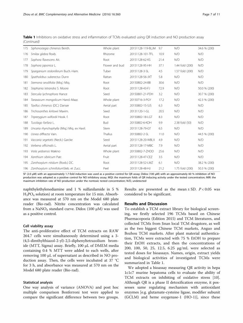
Table 1 Inhibitions on oxidative stress and inflammation of TCMs evaluated using QR induction and NO production assay
No Plant name Part usedin TCM
Voucher ID Yields (%) Induction of QRactivity (MQI)
Inhibition of NOproduction (MIR)
1 Acacia catechu (L.f.) Wild. Branch 20151128-100-EC 69.8 N/D N/D
2 Acanthopanax gracilistylus W. W. Smith Root-bark 20150802-20-WJP 9.8 N/D 68.0 % (100)
3 Acanthopanax senticosus (Rupr. et Maxim.) Harms Rhizome 20150801-8-CWJ 7.7 N/D 52.0 % (200)
4 Achyranthes bidentata Bl. Rhizome 20150801-4-NX 8.7 N/D N/D
5 Aconitum carmichaeli Debx. Root 20151128-58-FZ 14.7 N/D N/D
6 Acorns tatarinowii Schott Rhizome 20151128-30-SCP 10.5 N/D N/D
7 Adenophora tetraphylla (Thunb.) Fisch. Root 20151128-76-SS 19.1 N/D N/D
8 Agrimonia pilosa Ledeb. Aerial part 20151128-31-XHC 9.8 N/D 41.2 % (200)
9 Akebia trifoliata (Thunb.) Koidz. subsp.australis (Diels) T. Shimizu
Rattan 20150802-14-BMT 11.6 N/D N/D
10 Albizia julibrissin Durazz. Bark 20151128-134-HHP 8.9 1.64 fold (200) N/D
11 Alisma orientalis (Sam.) Juzep. Root 20151128-71-ZX 13.6 N/D 34.2 % (200)
12 Allium tuberosum Rottl. Seed 20150717-4-JCZ 3.8 N/D N/D
13 Amomum kravanh Pierre ex Gagnep. Fruit 20151128-137-DK 2.1 N/D N/D
14 Amomum villosum Lour. Fruit 20150801-9-SR 8.3 N/D N/D
15 Ampelopsis japonica (Thunb.) Makino Root 20151128-127-BL 10.7 N/D N/D
16 Andrographis paniculata (Burm.f.) Nees Aerial part 20151128-83-CXL 9.9 2.04 fold (200) N/D
17 Anemarrhena asphodeloides Bge. Rhizome 20150802-17-ZM 10.2 N/D 41.2 % (200)
18 Angelica dahurica (Fisch. ex Hoffm.) Benth. et Hook.f. Root 20151128-125-BZ 8.9 N/D N/D
19 Angelica pubescens Maxim. f. biserrata Shan et Yuan Root 20150802-16-DH 25.1 N/D N/D
20 Angelica sinensis (Oliv.) Diels Root 20151128-34-DG 19.4 1.41 fold (200) N/D
21 Arctium lappa L. Fruit 20151128-33-NBZ 18.4 N/D N/D
22 Areca catechu L. Peel 20151128-104-DFP 2.3 N/D N/D
23 Areca catechu L. Fruit 20151128-145-BL 8.2 1.33 fold (25) N/D
24 Arisaema erubescens (Wall.) Schott Tuber 20151128-23-TNX 1.2 1.36 fold (200) N/D
25 Aristolochia debilis Sieb. et Zucc. Aerial part 20151128-21-TXT 3.2 N/D N/D
26 Artemisia argyi Levi. et Vant. Leaf 20150716-6-AY 12.9 N/D 86.2 % (200)
27 Artemisia scoparia Waldst. et Kit. Aerial part 20151128-74-YC 12.8 1.48 fold (200) 38.8 % (100)
28 Asarum heterotropoides Fr. Schmldt var.mandshuricum (Maxim.) Kitag.
Root and rhizome 20151128-72-XX 11.4 N/D N/D
29 Atractylode lancea (Thunb.) DC. Rhizome 20151128-136-CZ 24.2 N/D 52.3 %(200)
30 Atractylodes macrocephala Koldz. Rhizome 20151128-123-BZ 12.9 N/D N/D
31 Aucklandia lappa Decne. Root 20151128-15-MX 16.3 2.31 fold (25) 94.7 % (25)
32 Belamcanda chinensis (L.) DC. Rhizome 20151128-106-SG 29.6 N/D N/D
33 Bletilla striata (Thunb.) Reichb.f. Rhizome 20150801-12-BJ 2.1 N/D N/D
34 Bolbostemma paniculatum (Maxim.) Franquet Tuber 20151128-5-TBM 15.1 N/D N/D
35 Callicarpa macrophylla Vahl Leaf 20151128-7-DYZZ 4.3 N/D 85.8 % (200)
36 Cassia angustifolia Vahl. Leaf 20150802-25-FXY 9.4 1.54 fold (50) 79.7 % (200)
37 Cassia obtusifolia L. Seed 20151128-38-JMZ 7.9 N/D N/D
38 Chaenomeles speciosa (Sweet) Nakai Fruit 20151128-24-MG 30.5 N/D N/D
39 Chrysanthemum morifolium Ramat. Flower 20150802-28-JH 23.3 N/D 90.2 % (200)
40 Cimicifuga heracleifolia Kom. Rhizome 20151128-28-SM 14.1 1.95 fold (100) 86.4 % (200)
41 Cinnamomum cassia Presl Branch 20150717-2-GZ 5.0 N/D N/D
42 Cirsium japonicum Fisch. ex DC. Aerial part 20151128-9-DJ 6.7 N/D N/D
Zhou et al. BMC Complementary and Alternative Medicine (2016) 16:360 Page 3 of 11

Table 1 Inhibitions on oxidative stress and inflammation of TCMs evaluated using QR induction and NO production assay(Continued)
43 Cirsium setosum (Willd.) M.Bieb. Aerial part 20151128-10-XJ 12.7 1.51 fold (200) 38.8 % (200)
44 Cistanchede deserticola Y. C. Ma Stem 20150801-7-RCR 23.6 N/D N/D
45 Citrus aurantium L. Fruit 20150801-11-ZQ 17.0 N/D N/D
46 Citrus limon (L.) Burm. f. Fruit 20150716-16-NM 19.5 1.79 fold (200) N/D
47 Citrus medica L. var. sarcodac-tylis Swingle Fruit 20151128-55-FS 39.6 N/D N/D
48 Citrus reticulata Blanco. Pericarp 20150716-10-CP 22.0 N/D N/D
49 Clematis armandii Franch. Rattan 20150802-12-CMT 4.5 N/D N/D
50 Codonopsis pilosula(Franch.) Nannf. Root 20151128-59-DS 48.4 N/D N/D
51 Coix lacryma-jobi L. var. ma-yuen (Roman.) Stapf Seed 20150716-15-YYR 6.5 N/D N/D
52 Commelina communis L. Aerial part 20151128-88-YZC 4.4 N/D N/D
53 Coptis chinensis Franch. Rhizome 20150802-31-HL 6.6 N/D 57.1 %(200)
54 Cornus officinalis Sieb. et Zucc. Fruit 20150802-27-SZY 43.6 N/D N/D
55 Crataegus pinnatifida Bge. var. major N. E. Br. Fruit 20150716-2-SZ 34.8 N/D N/D
56 Cremastra appendiculata (D. Don) Makino Pseudobulb 20151128-12-SCG 2.6 N/D 61.0 % (200)
57 Croton tiglium L. Fruit 20150802-8-BD 1.1 N/D N/D
58 Curculigo orchioides Gaertn. Rhizome 20151128-121-XM 4.5 1.57 fold (200) 47.8 % at (200)
59 Curcuma phaeocaulis Val. Rhizome 20150801-20-EZ 2.6 N/D 66.9 % (25)
60 Curcuma wenyujin Y. H. Chen et C. Ling Root 20150802-26-YJ 9.0 N/D N/D
61 Cynanchum atratum Bge. Root and rhizome 20151128-40-BW 23.6 N/D N/D
62 Cynanchum stauntonii (Decne.) Schltr ex Lévl. Rhizome 20150801-13-BQ 10.6. N/D N/D
63 Cynomorium songaricum Rupr. Stem 20150716-14-SY 17.9 N/D N/D
64 Cyperus rotundus L. Rhizome 20151128-80-XF 11.6 1.74 fold (200) N/D
65 Dendrobium nobile Lindl. Stem 20150802-13-MH 9.0 N/D 38.9 % (200)
66 Dictamnus dasycarpus Turcz. Velamen 20150802-4-BXP 9.0 N/D N/D
67 Dioscorea opposita Thunb. Rhizome 20150716-1-SY 1.7 N/D N/D
68 Dipsacus asperoides C. Y. Cheng et T. M. Ai Rhizome 20150716-13-XD 17.5 N/D 38.6 % (200)
69 Drynaria fortunei (Kunze) J. Sm. Rhizome 20151128-78-GSB 4.8 N/D N/D
70 Eclipta prostrata L. Aerial part 20151128-99-MHL 9.0 N/D N/D
71 Epimedium brevicornum Maxim. Aerial part 20150716-9-YYH 20.5 N/D N/D
72 Equisetum hiemale L. Aerial part 20151128-108-MZ 4.9 N/D 41.7 %(200)
73 Eriocaulon buergerianum Koern. Flower 20151128-139-GJC 7.9 N/D N/D
74 Eucommia ulmoides Oliv. Root-bark 20151128-51-DZ 8.3 1.56 fold (200) 62.5 % (200)
75 Eugenia caryophyllata Thunb. Bud 20151128-1-DX 27.5 N/D N/D
76 Eupatorium fortunei Turcz. Aerial part 20151128-66-PL 10.1 N/D N/D
77 Euphorbia humifusa Willd. Whole plant 20151128-37-DJC 9.5 N/D N/D
78 Ferula Sinkiangensis K. M. Shen Resin 20151128-140-EW 6.1 N/D N/D
79 Forsythia suspense (Thnub.) Vahl Fruit 20151128-53-LQ 28.3 N/D 48.3 % (200)
80 Fraxinus rhynchophylla Hance Bark 20151128-86-QP 8.0 N/D 90.7 % (200)
81 Fritillaria ussuriensis Maxim. Bulb 20151128-118-PBM 4.1 N/D N/D
82 Ganoderma sinense Zhao, Xu et Zhang Sporophore 20150801-2-ZZ 2.9 N/D N/D
83 Gardenia jasminoides Ellis. Fruit 20150717-7-ZZ 16.1 N/D N/D
84 Gastrodia elata Bl. Tuber 20150801-16-TM 7.5 N/D N/D
85 Glycyrrhiza uralensis Fisch. Rhizome 20150716-5-GC 15.7 2.19 fold (100) 82.9 % (200)
86 Hippophae rhamnoides L. Fruit 20151128-57-SJ 36.5 N/D N/D
Zhou et al. BMC Complementary and Alternative Medicine (2016) 16:360 Page 4 of 11

Table 1 Inhibitions on oxidative stress and inflammation of TCMs evaluated using QR induction and NO production assay(Continued)
87 Homalomena occulta (Lour.) Schott Rhizome 20151128-13-QNJ 9.3 N/D N/D
88 Hordeum vulgare L. Fruit 20151128-135-MY 11.5 N/D N/D
89 Houttuynia cordata Thunb. Aerial part 20150801-18-YXC 16.7 N/D N/D
90 Illicium difengpi K. I .B. et K. I. M. Bark 20151128-132-DFP 1.9 1.52 fold (100) N/D
91 Illicium verum Hook. f. Fruit 20151128-2-BJHX 13.3 N/D 55.7 % (200)
92 Inula helenium L. Root 20150802-5-TMX 13.2 1.77 fold (12.5) 100 % (100)
93 Isatis indigotica Fort. Root 20150802-9-BLG 17.9 N/D N/D
94 Isatis indigotica Fort. Leaf 20151128-102-DQY 13.6 1.66 fold (50) N/D
95 Kaempferia galanga L. Rhizome 20151128-105-SN 4.7 N/D N/D
96 Kochia scoparia (L.) Schrad. Fruit 20150802-23-DFZ 8.5 N/D N/D
97 Laminaria Japonica Aresch. Thallus 20150802-10-KB 18.5 N/D N/D
98 Lepidium apetalum Willd. Seed 20150802-22-TLZ 2.9 N/D N/D
99 Ligusticum chuanxiong Hort. Rhizome 20151128-19-CX 16.1 1.73 fold (200) 69.0 % (100)
100 Ligustrum lucidum Ait. Fruit 20151128-18-NZZ 24.2 N/D N/D
101 Lilium lancifolium Thunb. Leaf 20151128-32-BH 4.3 N/D N/D
102 Lindera aggregata (Sims) Kosterm. Root 20150801-15-WY 10.7 1.59 fold (200) N/D
103 Lithospermum erythrorhizon Sieb. et Zucc. Root 20151128-93-ZC 6.4 1.52 fold (50) 57.1 % (200)
104 Lobelia chinensi Lour. Whole plant 20151128-129-BBL 23.7 N/D N/D
105 Lonicera hypoglauca Miq. Flower 20150801-1-SYH 31.4 N/D N/D
106 Lonicera japonica Thunb. Flower 20150801-3-JYH 26.7 N/D N/D
107 Lophatherum gracile Brongn. Stem and leaf 20151128-91-DZY 8.6 N/D N/D
108 Lycium barbarum L. Fruit 20150801-6-GQ 11.5 N/D N/D
109 Lycium chinense Mill. Root-bark 20150802-21-DGP 7.5 N/D N/D
110 Lycopodium japonicum Thunb. Whole plant 20151128-138-SJC 22.3 N/D 53.0 % (200)
111 Lycopus lucidusTurcz. var. hirtus Regel Aerial part 20151128-70-ZL 13.5 N/D 61.6 % (200)
112 Lysimachia christinae Hance Whole plant 20151128-67-JQC 11.2 N/D N/D
113 Mahonia bealei (Fort.) Carr. Stem 20151128-114-GLM 6.1 N/D N/D
114 Melia toosendan Sleb. et Zucc. Fruit 20151128-107-CLZ 16.2 N/D N/D
115 Menispermum dauricum DC. Rhizome 20151128-120-BDG 11.0 N/D N/D
116 Mentha haplocalyx Briq. Aerial part 20150716-8-BH 19.1 N/D N/D
117 Mignolia officinalis Rehd. et Wils. Bark 20151128-84-HP 22.9 N/D N/D
118 Misla chinensis Maxim. Aerial part 20151128-81-XR 6.9 1.60 fold (100) N/D
119 Morinda officinalis How. Root 20151128-112-BJT 28.8 N/D N/D
120 Morus alba L. Branch 20151128-142-SZ 7.4 1.37 fold (100) 61.2 % (200)
121 Morus alba L. Fruit 20151128-143-SS 29.9 N/D N/D
122 Nardostachys chinensis Batal. Root and rhizome 20151128-115-GS 12.3 N/D N/D
123 Oroxylum inddicum (L.) Vent. Seed 20151128-26-MHD 14.0 N/D 85.4 % (200)
124 Orostachys fimbriatus (Turcz.) Berg. Aerial part 20151128-109-WS 6.3 N/D N/D
125 Paeonia lactiflora Pall. Rhizome 20150716-7-BS 6.4 N/D N/D
126 Panax ginseng C. A. Mey Rhizome 20150801-10-SSS 39.4 N/D N/D
127 Panax quinque folium L. Root 20150802-29-XYS 14.8 N/D N/D
128 Perilla frutescens (L.) Britt. Leaf 20150717-6-ZS 2.8 1.73 fold (200) 57.8 % (200)
129 Peucedanum praeruptorum Dunn Root 20151128-85-QH 20.6 N/D 77.6 % (200)
130 Phellodendron chinense Schneid. Tree-bark 20151128-41-HB 12.7 N/D N/D
Zhou et al. BMC Complementary and Alternative Medicine (2016) 16:360 Page 5 of 11

Table 1 Inhibitions on oxidative stress and inflammation of TCMs evaluated using QR induction and NO production assay(Continued)
131 Phragmites communis Trin. Root 20151128-49-LG 3.8 N/D N/D
132 Physalis alkekengi L. var. franchetii (Mast.) Makino Calyx 20150730-2-GJD 14.2 1.79 fold (200) 91.4 % (200)
133 Pinellia ternate (Thunb.) Breit. Tuber 20151128-130-BX 0.8 1.74 fold (200) N/D
134 Piper nigrum L. Fruit 20151128-92-HHJ 5.4 N/D N/D
135 Plantago asiatica L. Whole plant 20150716-4-CQC 13.7 N/D N/D
136 Platycodon grandiflorum (Jacq.) A. DC. Root 20151128-87-JG 33.4 N/D N/D
137 Pogostemon cablin (Blanco) Benth. Aerial part 20151128-16-GHX 5.5 1.73 fold (100) 56.6 % (200)
138 Polygonatum kingianum Coll. et Hemsl. Rhizome 20151128-90-HJ 6.8 N/D N/D
139 Polygonatum odoratum (Mill.) Druce Rhizome 20151128-113-YZ 21.6 N/D N/D
140 Polygonum aviculare L. Aerial part 20151128-89-BX 10.8 N/D N/D
141 Polygonum cuspidatum Sieb. et Zucc. Root and rhizome 20151128-63-HZ 21.7 N/D 60.5 % (200)
142 Polygonum multiflorum Thunb. Root 20151128-54-HSW 6.8 N/D 35.5 % (200)
143 Polyyala tenuifolia Willd. Root 20151128-46-YZ 34.4 N/D N/D
144 Potentilla chinensis Ser. Whole plant 20151128-65-WLC 8.9 N/D N/D
145 Prunella vulgaris L. Peel 20150716-11-XKC 4.1 N/D N/D
146 Prunus armeniaca L. var. ansu Maxim. Seed 20151128-60-KXR 5.7 N/D N/D
147 Prunus persica (L.) Batsch Seed 20151128-61-TR 3.7 N/D N/D
148 Pseudolarx kaempleri Gord. Velamen 20151128-6-TJP 14.8 N/D N/D
149 Pseudostellaria beterphylla (Miq.) Pax ex Pax et Hoffm. Root 20151128-27-TZS 13.5 N/D N/D
150 Psoralea corylifolia L. Fruit 20150801-19-BGZ 10.2 N/D N/D
151 Pulsatilla chinensis (Bunge) Regel Root 20151128-124-BTW 22.7 N/D N/D
152 Punica granatum L. Peel 20151128-117-SLP 31.3 N/D N/D
153 Pyrrosia sheareri (Bak.) Ching Leaf 20151128-116-SW 12.2 1.85 fold (50) N/D
154 Rabdosia rubescens (Hemsl.) Hara Aerial part 20151128-39-DLC 8.4 1.38 fold (100) 43.2 % (200)
155 Raphanus sativus L. Seed 20150802-30-LFZ 13.8 N/D N/D
156 Rhaponlicum uniflorum (L.) DC. Root 20151128-97-LL 4.4 1.54 fold (200) N/D
157 Rheum palmatum L. Root and rhizome 20151128-103-DH 25.6 N/D 60.2 % (200)
158 Rhodiola crenulata (Hook. f. et Thoms.) H. Ohba Rhizome 20150801-5-HJT 13.3 N/D 46.3 % (200)
159 Rosa chinensis Jacq. Flower 20151128-110-YJH 18.9 N/D N/D
160 Rosa laevigata Michx. Fruit 20151128-68-JYZ 25.2 1.67 fold (50) 57 % (200)
161 Rubia cordifolia L. Root and rhizome 20151128-73-QC 10.0 N/D N/D
162 Salvia miltiorrhiza Bge. Root and rhizome 20151128-29-DS 39.7 1.44 fold (100) 64.5 % (200)
163 Sanguisorba officinalis L. Root 20151128-36-DY 3.5 N/D N/D
164 Saposhnikovia divaricata (Turcz.) Schischk. Root 20150802-19-FF 15.9 1.95 fold (100) N/D
165 Sareassum pallidum (Turn.) C. Ag. Frond 20150802-1-HZ 11.5 N/D N/D
166 Sargentodoxa cuneate (Oliv.) Rehd. et Wils. Rattan 20151128-8-DXT 16.9 N/D N/D
167 Scrophularia ningpoensis Hemsl. Root 20151128-128-XS 50.5 N/D N/D
168 Scutellaria baicalensis Georgi. Rhizome 20150716-12-HQ 30.2 N/D 87.4 %(200)
169 Scutellaria barbata D. Don Whole plant 20151128-35-BZL 10.2 N/D 59.0 % (200)
170 Sedum sarmentosum Bunge. Whole plant 20151128-64-CPC 17.5 N/D N/D
171 Selaginella tamariscina (Beauv.) Spring Whole plant 20151128-69-JB 8.9 N/D N/D
172 Senecio scandens Buch.-Ham. Aerial part 20151128-14-QLG 11.4 N/D 38.1 % (200)
173 Sesamum indicum L. Seed 20150717-3-HZM 3.3 N/D N/D
174 Siegesbeckia orientalis L. Aerial part 20151128-98-XXC 4.8 1.91 fold (200) 54.9 % (200)
Zhou et al. BMC Complementary and Alternative Medicine (2016) 16:360 Page 6 of 11
naphthylethylenediamine and 1 % sulfanilamide in 5 %H3PO4 solution) at room temperature for 15 min. Absorb-ance was measured at 570 nm on the Model 680 platereader (Bio-rad). Nitrite concentration was calculatedfrom a NaNO2 standard curve. Didox (100 μM) was usedas a positive control.
Cell viability assayThe anti-proliferative effect of TCM extracts on RAW264.7 cells were simultaneously determined using a 3-(4,5-dimthylthiazol-2-yl)-2,5-diphenyltetrazolium brom-ide (MTT, Sigma) assay. Briefly, 100 μL of DMEM mediacontaining 0.4 % MTT were added to each wells, afterremoving 100 μL of supernatant as described in NO pro-duction assay. Then, the cells were incubated at 37 °Cfor 3 h, and absorbance was measured at 570 nm on theModel 680 plate reader (Bio-rad).
Statistical analysisOne way analysis of variance (ANOVA) and post hocmultiple comparison Bonferroni test were applied tocompare the significant difference between two groups.
Results are presented as the mean ± SD. P < 0.05 wasconsidered to be significant.
Results and DiscussionTo establish a TCM extract library for biological screen-ing, we firstly selected 196 TCMs based on ChinesePharmacopoeia (Edition 2015) and TCM literatures, andcollected TCMs from Jinan local TCM drugstore, as wellas the two biggest Chinese TCM markets, Anguo andBozhou TCM markets. After plant material authentica-tion, TCMs were extracted with 75 % EtOH to preparetheir EtOH extracts, and then the concentrations of200, 100, 50, 25, 12.5, 6.25 μg/mL were selected astested doses for bioassays. Names, origin, extract yieldsand biological activities of investigated TCMs weresummarized in Table 1.We adopted a bioassay measuring QR activity in hepa
1c1c7 murine hepatoma cells to evaluate the ability ofTCM extracts on inhibiting of oxidative stress [10].Although QR is a phase II detoxification enzyme, it pos-sesses same regulating mechanism with antioxidantenzymes [e.g. glutamate-cysteine ligase, modifier subunit(GCLM) and heme oxygenase-1 (HO-1)], since these
Table 1 Inhibitions on oxidative stress and inflammation of TCMs evaluated using QR induction and NO production assay(Continued)
175 Siphonostegia chinensis Benth. Whole plant 20151128-119-BLJM 9.7 N/D 34.6 % (200)
176 Smilax glabra Roxb. Rhizome 20151128-101-TFL 10.9 N/D N/D
177 Sophora flavescens Ait. Root 20151128-62-KS 21.4 N/D N/D
178 Sophora japonica L. Flower and bud 20151128-95-HH 37.1 1.44 fold (200) N/D
179 Sparganium stoloniferum Buch.-Ham. Tuber 20151128-3-SL 4.5 1.57 fold (200) N/D
180 Spatholobus suberectus Dunn Rattan 20151128-56-JXT 5.8 N/D N/D
181 Stemona sessilifolia (Miq.) Miq. Root 20150802-24-BB 30.6 N/D N/D
182 Stephania tetrandra S. Moore Root 20151128-43-FJ 72.9 N/D 50.0 % (200)
183 Sterculia lychnophora Hance Seed 20150801-21-PDH 3.2 N/D 30.7 % (200)
184 Taraxacum mongolicum Hand.-Mazz. Whole plant 20150716-3-PGY 17.2 N/D 42.3 % (200)
185 Taxillus chinensis (DC.) Danser Aerial part 20150802-15-SJS 6.3 N/D N/D
186 Trichosanthes kirilowii Maxim. Seed 20131120-1-GL 20.5 N/D N/D
187 Tripterygium wilfordii Hook. f. Root 20150802-18-LGT 8.3 N/D N/D
188 Tussilago farfara L. Bud 20150802-6-KDH 9.9 2.38 fold (50) N/D
189 Uncaria rhynchophylla (Miq.) Miq. ex Havil. Stem 20151128-79-GT 6.5 N/D N/D
190 Usnea diffracta Vain. Thallus 20150802-2-SL 11.0 N/D 44.5 % (200)
191 Vaccaria segetatis (Neck.) Garcke Seed 20151128-20-WBLX 4.9 N/D N/D
192 Verbena officinalis L. Aerial part 20151128-17-MBC 7.9 N/D N/D
193 Viola yedoensis Makino Whole plant 20150802-7-ZHDD 25.6 N/D N/D
194 Xanthium sibiricum Patr. Fruit 20151128-47-CEZ 3.5 N/D N/D
195 Zanthoxylum nitidum (Roxb.) DC. Root 20151128-52-LMZ 6.1 N/D 38.2 % (200)
196 Zanthoxylum schinifoliumSleb. et Zucc. Peel 20151128-48-HJ 21.2 1.75 fold (200) 50.3 % (200)
SF (2.0 μM) with an approximately 1.7-fold induction was used as a positive control for QR assay; Didox (100 μM) with an approximately 60 % inhibition of NOproduction was adopted as a positive control for NO inhibitory assay; MQI: the maximum folds of QR inducing activity under the tested concentration; MIR: themaximum inhibition rate of NO production under the nontoxic tested concentration; N/D, undetected
Zhou et al. BMC Complementary and Alternative Medicine (2016) 16:360 Page 7 of 11
enzymes are antioxidant response element (ARE)-con-taining target genes and are mediated by ARE located intheir promoter region [11]. Specially, upon the exposureof cells to oxidative stress and/or toxicants, nuclear fac-tor E2-related factor 2 (Nrf2) translocates into the nu-cleus, binds to the ARE sequence, and activates thetranscription of these ARE-target genes [12]. Therefore,QR and antioxidant enzymes (e.g. GCLM and HO-1)possess same responses against endogenous and exogen-ous insults, which have also been verified by our recentresearches [13, 14]. Considering above mentioned indu-cing mechanism of QR and antioxidant enzymes, deter-mination of QR activity is a rational and effectivemethod for analyzing the potency of oxidative stress in-hibition. In the current study, we normalized the data bysetting the untreated control group as 1, and then theQR inducing activity of tested extracts was representedby the maximum folds of QR inducing activity (MQI)compared with the untreated control group. SF as a posi-tive control displayed an approximately 1.7-fold inductionat 2.0 μM. 1.3-fold of QR inducing activity (MQI = 1.3)under the tested concentrations was adopted as a criterionfor bioactive TCM extracts. To be more precise, the levelof QR inducing activity was ranked as the followingcriteria: strong (MQI ≥ 1.8); moderate (1.8 >MQI ≥1.5);weak (1.5 >MQI ≥1.3); undetected (MQI < 1.3).Ultimately, 38 TCM extracts demonstrated the QR
inducing activities with MQI ranging from 1.33- to2.38- folds under the tested concentrations (Table 1).Of which, eight TCM extracts strongly induced QRactivity in hepa 1c1c7 cells (MQI ≥ 1.8), includingAndrographis paniculata (aerial part, 16), Aucklandialappa (root, 31), Cimicifuga heracleifolia (rhizome, 40),Glycyrrhiz uralensis (rhizome, 85), Pyrrosia sheareri(leaf, 153), Saposhnikovia divaricate (root, 164), Sieges-beckia orientalis (aerial part, 174), and Tussilago farfara(bud, 188). Twenty-two extracts are moderate QRinducers (1.8 > MQI ≥1.5), containing Albizia julibrissin(bark, 10), Cassia angustifolia (leaf, 36), Cirsium setosum(aerial part, 43), Citrus limon (fruit, 46), Curculigoorchioides (rhizome, 58), Cyperus rotundus (rhizome, 64),Eucommia ulmoides (root-bark, 74), Illicium difengpi(bark, 90), Inula helenium (root, 92), Isatis indigotica (leaf,93), Ligusticum chuanxiong (rhizome, 99), Lindera aggre-gate (root, 102), Lithospermum erythrorhizon (root, 103),Misla chinensis (aerial part, 118), Perilla frutescens (leaf,128), Physalis alkekengi L. var. franchetii (calyx, 132),Pinellia ternata (tuber, 133), Pogostemon cablin (aerialpart, 137), Rhaponlicum uniflorum (root, 156), Rosa laevi-gata (fruit, 160), Sparganium stoloniferum (tuber, 179),and Zanthoxylum schinifolium (peel, 196). Moreover, eightextracts possessed weak QR inducing effect (1.5 >MQI≥1.3), including Angelica sinensis (root, 20), Areca catechu(fruit, 23), Arisaema erubescens (tuber, 24), Artemisia
scoparia (aerial part, 27), Morus alba (branch, 120),Rabdosia rubescens (aerial part, 154), Salvia miltiorrhiza(root and rhizome, 162), and Sophora japonica (flowerand bud, 178). QR inducing effects of 38 bioactive TCMextracts in hepa 1c1c7 cells have been detailedly summa-rized in Additional file 1: Table S1 and Figure S1.During the chronic inflammation process, excessive
NO have been produced and involved in the tissueinjury through damages to proteins, lipids, DNA, andthe modulation of leukocyte activity [15]. Accordingly,inhibiting NO production is regarded to be an effectivestrategy for the therapy of inflammation-related dis-eases. Herein, we detected NO level in LPS-stimulatedRAW264.7 macrophages to evaluate anti-inflammatoryfunction of TCM extracts. Cytotoxicities of tested TCMextracts were simultaneously evaluated by a MTT assayto confirm that the decrease of NO production was notattributed to inhibition of cell proliferation. The max-imum inhibition rate (MIR) of NO production underthe nontoxic tested concentration, which was calculatedby comparing the decreased NO concentration inTCM-treated group with that in LPS-stimulated group,was adopted to evaluate the anti-inflammatory property.Didox with an approximately 60 % inhibition of NO pro-duction at 100 μM was used as a positive control. Theinhibitory potency of TCM extracts on NO productionwas ranked according to the criteria as follows: strong(MIR ≥ 80 %); moderate (80 % >MIR ≥ 50 %); weak(50 % >MIR ≥ 30 %); undetected (MIR <30 %).Our investigation indicated that 55 TCM extracts inhib-
ited the LPS-induced NO production with MIRs between30.7 % and 100 % under the tested nontoxic concentra-tions (Table 1). Thereinto, 11 TCM extracts stronglyinhibited NO production in RAW 264.7 cells (MIR ≥80 %), including Artemisia argyi (leaf, 26), Aucklandialappa (root, 31), Callicarpa macrophylla (leaf, 35),Chrysanthemum morifolium (flower, 39), Cimicifuga hera-cleifolia (rhizome, 40), Fraxinus rhynchophylla (bark, 80),Glycyrrhiza uralensis (rhizome, 85), Inula helenium (root,92), Oraxylum inddicum (seed, 123), Physalis alkekengi L.var. franchetii (calyx, 132), and Scutellaria baicalensis (rhi-zome, 168). Moreever, 25 extracts displayed moderate in-hibitory effect of NO production (80 % >MIR ≥ 50 %), and19 extracts weakly inhibited NO production (50 % >MIR≥30 %). Inhibitory effects on NO production of 55 bio-active TCM extracts in RAW 264.7 cells have beendetailedly summarized in Additional file 1: Table S1 andFigure S1.Since oxidative stress and inflammatory response have
the synergistic reactions in the pathophysiology ofCOPD, TCMs having dual inhibitions on the two targetsare apt to be the resource for discovering lead molecules[5, 7]. Our results indicated that the extracts ofArtemisia scoparia (aerial part, 27), Aucklandia lappa
Zhou et al. BMC Complementary and Alternative Medicine (2016) 16:360 Page 8 of 11
(root, 31), Cassia angustifolia (leaf, 36), Cimicifugaheracleifolia (rhizome, 40), Cirsium setosum (aerial part,43), Curculigo orchioides (rhizome, 58), Eucommiaulmoides (root-bark, 74), Glycyrrhiza uralensis (rhizome,85), Inula helenium (root, 92), Ligusticum chuanxiong(rhizome, 99), Lithospermum erythrorhizon (root, 103),Morus alba (branch, 120), Perilla frutescens (leaf, 128),Physalis alkekengi var. franchetii (calyx, 132), Pogoste-mon cablin (aerial part, 137), Rabdosia rubescens (aerialpart, 154), Rosa laevigata (fruit, 160), Salvia miltiorrhiza(root and rhizome, 162), Siegesbeckia orientalis (aerialpart, 174), and Zanthoxylum schinifolium (peel, 196)simultaneously inhibited oxidative stress and inflamma-tion (Table 1 and Additional file 1: Table S1). Most ofall, both QR inducing effects and NO inhibitory activ-ities of the extracts of Aucklandia lappa (31), Cimicifugaheracleifolia (40), and Glycyrrhiza uralensis (85) are la-belled as the level of strong. In addition, the extracts ofInula helenium (92) and Physalis alkekengi L. var. fran-chetii (132) also demonstrated the potencies that areclosed to the strong level.To our knowledge, this is the first systemic screening
of QR inducing extracts from TCMs to discover TCMswith the capacity of inhibiting oxidative stress. Plenty ofwork on investigation of natural-derived molecules fortheir regulation on oxidative stress have been carriedout, and acquired some active ingredients existed inabove evaluated TCMs, such as andrographolide fromAndrographis paniculata (16) [16], (Z)-ligustilide fromAngelica sinensis (20) [17], dehydroglyasperin C fromGlycyrrhiza uralensis (85) [18], isoalantolactone fromInula helenium (92) [19], 2’,3’-dihydroxy-4’,6’-dimethoxy-chalcone from Perilla frutescens (128) [20], oridoninfrom Rabdosia rubescens (154) [21], danshensu and tan-shinone I from Salvia miltiorrhiza (162) [22]. These datasupport our observed QR inducing effects of the activeTCMs. More importantly, the majority of QR inducingTCMs tested in present research have still not been phy-tochemically investigated through targeting oxidativestress inhibition, which affords us an opportunity to dis-cover new lead molecules from them [23].TCMs have been adopted for the therapy of
inflammation-related diseases with a long history in China.Compared with QR inducing assay, NO inhibitory effectassay and other in vitro and in vivo anti-inflammatorymodels are classical and commonly adopted biologicalresearch methods, and accordingly more literatures con-cerning inflammation of TCMs have been published. Basedon our findings, we carried out a systemic search ofreported inflammation-related literatures of our observed55 active TCM extracts, and concluded that: some TCMshave been comprehensively investigated for their anti-inflammatory property and resulted in the discovery of di-verse types of natural products, covering agrimonolide from
Agrimonia pilosa (8) [24], (-)-nyasol from Anemarrhenaasphodeloides (17) [25], alantolactone from Aucklandialappa (31) [26], berberine from Coptis chinensis (53) [27],forsythiaside from Forsythia suspensa (79) [28], resveratrolfrom Polygonum cuspidatum (141) [29], etc. Besidethese comprehensively investigated molecules, a greatdeal of constituents have been isolated from theseactive TCMs, and required further confirmation oftheir anti-inflammatory function. Meanwhile, a num-ber of TCMs [e.g. Alisma orientalis (11), Equisetumhiemale (72), Cirsium setosum (43)] have not been in-vestigated in the field of inflammation. Significantly,little research on the therapeutic effect of COPD hasbeen performed, and thus these active TCMs are stillbeing researched.In the present screening assay, we only adopted two
typical markers, QR and NO, to evaluate the potential ofTCMs as oxidative stress and inflammation inhibitoryagents. Based on our preliminary results, active TCMextracts could be subjected to further research in thefield of phytochemistry and pharmacology, however,solid evidences on their biological functions are requiredbefore a systemic investigation [14]. With regard to theinhibition on oxidative stress, the levels of endogenousglutathione (GSH) and reactive ROS, as well as the pro-tein level of key intracellular redox-balancing proteinGCLM, are suggested to be detected to estimate theintracellular redox state and antioxidant capacity whenexposed to TCM extracts [30–32]. Concerning the inhib-ition of inflammation by the active TCMs, the levels ofcrucial inflammatory mediators in the COPD pathology,including TNFα, LTB4, and IL-8, should be determined toconfirm their anti-inflammatory potential [33].Additionally, the pivotal regulators for oxidative
stress and inflammation should be sufficiently investi-gated to verify action of mechanism of the activeTCMs. The transcription factor Nrf2 plays a dominantrole for regulating oxidative stress. It is ubiquitouslyexpressed in human organs, particularly rich in lung,and counteracts oxidative injury through activatingintracellular redox-balancing proteins (e.g. GCLM,GST, HO-1) and up-regulating endogenous antioxi-dants (e.g. GSH) [11, 34]. NF-kB regulates the expres-sion of proinflammatory genes including cytokines,chemokines, and adhesion molecules, and its inhibitiontherefore definitely relieves the inflammatory responseof COPD [7, 35]. It has also been verified that phos-phatidylinositol 3-kinase (PI3K) and mitogen-activatedprotein kinase (MAPK) are involved in the regulationof inflammatory response [36, 37]. Hence, the furtherresearch on active TCM extracts and purified ingredi-ents could focus on their action of mechanism on Nrf2,NF-kB, PI3K, and MAPK signaling pathways, as well asthe cross talk between these pathways.
Zhou et al. BMC Complementary and Alternative Medicine (2016) 16:360 Page 9 of 11

ConclusionAlthough the present research indicates that someTCMs possessed inhibitory effects on inflammation andoxidative stress, further pharmacological investigationsin vitro and in vivo models are warranted. Furthermore,bioassay-guided fractionations and identifications ofactive ingredients should be launched to help us illus-trate the mechanism of these active species, and discovernew lead molecules with unknown mechanisms andpotent functions on oxidative stress- and inflammation-related diseases, especially COPD. Accordingly, theseresults may give new insight in research and develop-ment of COPD therapeutic agents.
Additional file
Additional file 1: Figure S1. NADP(H): quinone oxidoreductase (QR)inducing effects of 38 bioactive TCM extracts in hepa 1c1c7 cells. The QRinducing effect was determined after 24h treatment of the hepa 1c1c7cells in the presence or absence of tested TCMs. The data of theuntreated control group was normalized as 1, and then the QR inducingactivity of tested extracts was represented by the maximum folds of QRinducing activity (MQI) compared with the untreated control group.Sulforaphane (SF, 2.0 μM) was used as a positive control. The data arereported the means ± SD from three independent experiments. FigureS2. Inhibitory effects on NO production of 55 bioactive TCM extracts inRAW 264.7 cells. The NO concentration in the RAW 264.7 cell culturemedia was determined through the Griess reaction 24 h after treated inthe presence or absence of tested TCMs and lipopolysaccharides (LPS, 1.0μg/mL). Didox (100 μM) was adopted as a positive control. The data arereported the means ± SD from three independent experiments. Themaximum inhibition rates (MIRs) of NO production under the untoxictested concentration were calculated by comparing the decreased NOconcentration in TCM-treated group with that in LPS-stimulated group.Table S1. TCM extracts with QR inducing activity and/or NO inhibitoryeffect. (DOCX 4312 kb)
AbbreviationsARE: Antioxidant response element; COPD: Chronic obstructive pulmonarydisease; GCLM: Glutamate-cysteine ligase, modifier subunit; GSH: Glutathione;GST: Glutathione S transferase; HO-1: Heme oxygenase-1; IL-8: Interleukin-8;LPS: Lipopolysaccharides; LTB4: Leukotriene B4; MAPK: Mitogen-activatedprotein kinase; MIR: Maximum inhibition rate; MQI: Maximum folds of QRinducing activity; MTT: 3-(4,5-dimthylthiazol-2-yl)-2,5-diphenyltetrazoliumbromide; NF-kB: Nuclear factor kB; Nrf2: Nuclear factor E2-related factor 2;PI3K: Phosphatidylinositol 3-kinase; QR: NADP(H):quinone oxidoreductase;ROS: Reactive oxygen species; SF: Sulforaphane; TCM: Traditional Chinesemedicine; TGF-β: Transforming growth factor-β; TNFα: Tumor necrosis factor α
AcknowledgmentsThe authors would like to appreciate Profs. Lan Xiang and Hu-Ning Chen, aswell as Mr. Yu Zhao in Shandong University for TCM collection andidentification.
FundingThis work was supported by NNSF of China (31470419), NSF of Shandong(ZR2014HM019 and 2015ZRE27209), Science & Technology DevelopmentPlan Project of Shandong (2014GSF118023) and Young Scholars Program ofShandong University (2015WLJH50).
Availability of data and materialsThe datasets during and/or analysed during the current study available fromthe corresponding author on reasonable request. Moreover, Additional file 1is available along with the manuscript.
Authors’ contributionsD-MR, H-XL and TS conceived and designed the experiments; M-XZ, XW, A-MW, L-ZL, YY and X-SW performed the experiments; X-NW and TS analyzedthe data; A-LL contributed reagents, materials, and analysis tools; M-XZ andTS wrote the paper. All authors read and approved the final manuscript.
Competing interestsThe authors state no conflict or competing interests are associated with thepresent study.
Consent for publicationNot applicable.
Ethics approval and consent to participateNot applicable.
Author details1Key Lab of Chemical Biology (MOE), School of Pharmaceutical Sciences,Shandong University, 44 West Wenhua Road, Jinan 250012, People’s Republicof China. 2School of Pharmaceutical Sciences, Shandong University ofTraditional Chinese Medicine, Jinan, People’s Republic of China.
Received: 19 July 2016 Accepted: 10 September 2016
References1. Pauwels RA, Buist AS, Calverley PM, Jenkins CR, Hurd S. Global strategy for the
diagnosis, management, and prevention of chronic obstructive pulmonarydisease. NHLBI/WHO Global Initiative for Chronic Obstructive Lung Disease(GOLD) workshop summary. Am J Respir Crit Care Med. 2001;163:1256–76.
2. Lopez A, Shibuya K, Rao C, Mathers C, Hansell A, Held L, Schmid V, Buist S.Chronic obstructive pulmonary disease: current burden and futureprojections. Eur Respir J. 2006;27:397–412.
3. Barnes PJ, Shapiro S, Pauwels R. Chronic obstructive pulmonary disease:molecular and cellular mechanisms. Eur Respir J. 2003;22:672–88.
4. Barnes PJ, Hansel TT. Prospects for new drugs for chronic obstructivepulmonary disease. Lancet. 2004;364:985–96.
5. Barnes PJ. Mediators of chronic obstructive pulmonary disease. PharmacolRev. 2004;56:515–48.
6. Yao H, Rahman I. Current concepts on oxidative/carbonyl stress,inflammation and epigenetics in pathogenesis of chronic obstructivepulmonary disease. Toxicol Appl Pharmacol. 2011;254:72–85.
7. Barnes PJ. Novel approaches and targets for treatment of chronicobstructive pulmonary disease. Am J Respir Crit Care Med. 1999;160:S72–9.
8. Jiang YX, Wu JF, Du J. Survey of study on therapeutic mechanism ofchronic obstructive pulmonary disease treated by traditional Chinesemedicine. Zhongguo Zhong Xi Yi Jie He Za Zhi. 2005;9:860–4.
9. Gao Z, Li FS, Yang J, Xu D, Yang CH. Investigation on the literatureconcerning the clinically used traditional Chinese medicines for thetreatment of chronic obstructive pulmonary disease in the past ten years.Zhongguo Shi Yan Fang Ji Xue Za Zhi. 2010;16:286–8.
10. Gerhäuser C, You M, Liu J, Moriarty RM, Hawthorne M, Mehta RG, Moon RC,Pezzuto JM. Cancer chemopreventive potential of sulforamate, a novelanalogue of sulforaphane that induces phase 2 drug-metabolizing enzymes.Cancer Res. 1997;57:272–8.
11. Lau A, Villeneuve NF, Sun Z, Wong PK, Zhang DD. Dual roles of Nrf2 incancer. Pharmacol Res. 2008;58:262–70.
12. Bryan HK, Olayanju A, Goldring CE, Park BK. The Nrf2 cell defence pathway:Keap1-dependent and-independent mechanisms of regulation. BiochemPharmacol. 2013;85:705–17.
13. Shen T, Jiang T, Long M, Chen J, Ren D-M, Wong PK, Chapman E, Zhou B,Zhang DD. A curcumin derivative that inhibits vinyl carbamate-inducedlung carcinogenesis via activation of the Nrf2 protective response. AntioxidRedox Signal. 2015;23:651–64.
14. Shen T, Chen X-M, Harder B, Long M, Wang X-N, Lou H-X, Wondrak GT, RenD-M, Zhang DD. Plant extracts of the family Lauraceae: a potential resourcefor chemopreventive agents that activate the nuclear factor-erythroid 2-related factor 2/antioxidant response element pathway. Planta Med.2014;80:426–34.
15. Faro MLL, Fox B, Whatmore JL, Winyard PG, Whiteman M. Hydrogen sulfideand nitric oxide interactions in inflammation. Nitric Oxide. 2014;41:38–47.
Zhou et al. BMC Complementary and Alternative Medicine (2016) 16:360 Page 10 of 11

16. Guan S, Tee W, Ng D, Chan T, Peh H, Ho W, Cheng C, Mak J, Wong W.Andrographolide protects against cigarette smoke‐induced oxidative lunginjury via augmentation of Nrf2 activity. Br J Pharmacol. 2013;168:1707–18.
17. Dietz BM, Liu D, Hagos GK, Yao P, Schinkovitz A, Pro SM, Deng S,Farnsworth NR, Pauli GF, van Breemen RB. Angelica sinensis and itsalkylphthalides induce the detoxification enzyme NAD(P)H: quinoneoxidoreductase 1 by alkylating Keap1. Chem Res Toxicol. 2008;21:1939–48.
18. Kim HJ, Lim SS, Park IS, Lim JS, Seo JY, Kim JS. Neuroprotective effects ofdehydroglyasperin C through activation of heme oxygenase-1 in mousehippocampal cells. J Agric Food Chem. 2012;60:5583–9.
19. Seo JY, Park J, Kim HJ, Lee IA, Lim JS, Lim SS, Choi SJ, Park JHY, Kang HJ,Kim JS. Isoalantolactone from Inula helenium caused Nrf2-mediatedinduction of detoxifying enzymes. J Med Food. 2009;12:1038–45.
20. Izumi Y, Matsumura A, Wakita S, Akagi KI, Fukuda H, Kume T, Irie K, Takada-Takatori Y, Sugimoto H, Hashimoto T. Isolation, identification, and biologicalevaluation of Nrf2-ARE activator from the leaves of green perilla (Perillafrutescens var. crispa f. viridis). Free Radic Biol Med. 2012;53:669–79.
21. Du Y, Villaneuva NF, Wang X-J, Sun Z, Chen W, Li J, Lou H, Wong PK, ZhangDD. Oridonin confers protection against arsenic-induced toxicity throughactivation of the Nrf2-mediated defensive response. Environ Health Persp.2008;116:1154–61.
22. Tao S, Zheng Y, Lau A, Jaramillo MC, Chau BT, Lantz RC, Wong PK, WondrakGT, Zhang DD. Tanshinone I activates the Nrf2-dependent antioxidantresponse and protects against As(III)-induced lung inflammation in vitro andin vivo. Antioxid Redox Signal. 2013;19:1647–61.
23. Kumar H, Kim IS, More SV, Kim BW, Choi DK. Natural product-derivedpharmacological modulators of Nrf2/ARE pathway for chronic diseases. NatProd Rep. 2014;31:109–39.
24. Chen L, Teng H, Fang T, Xiao J. Agrimonolide from Agrimonia pilosasuppresses inflammatory responses through down-regulation of COX-2/iNOS and inactivation of NF-kB in lipopolysaccharide-stimulatedmacrophages. Phytomedicine. 2016;23:846–55.
25. Lee HJ, Li H, Chang HR, Jung H, Lee DY, Ryu JH. (-)-Nyasol, isolated fromAnemarrhena asphodeloides suppresses neuroinflammatory responsethrough the inhibition of I-kB alpha degradation in LPS-stimulated BV-2microglial cells. J Enzyme Inhib Med Chem. 2013;28:954–9.
26. Chun J, Choi RJ, Khan S, Lee DS, Kim YC, Nam YJ, Lee DU, Kim YS.Alantolactone suppresses inducible nitric oxide synthase andcyclooxygenase-2 expression by down-regulating NF-kB, MAPK and AP-1 viathe MyD88 signaling pathway in LPS-activated RAW 264.7 cells. IntImmunopharmacol. 2012;14:375–83.
27. Li Z, Geng Y-N, Jiang J-D, Kong W-J. Antioxidant and anti-Inflammatoryactivities of berberine in the treatment of diabetes mellitus. Evid-BasedCompl Alt. 2014;2014:289264.
28. Cheng L, Li F, Ma R, Hu XP. Forsythiaside inhibits cigarette smoke-inducedlung inflammation by activation of Nrf2 and inhibition of NF-kappa B. IntImmunopharmacol. 2015;28:494–9.
29. Das S, Das DK. Anti-inflammatory responses of resveratrol. Inflamm AllergyDrug Targets. 2007;6:168–73.
30. Sies H. Glutathione and its role in cellular functions. Free Radic Biol Med.1999;27:916–21.
31. Lim J, Nakamura BN, Mohar I, Kavanagh TJ, Luderer U. Glutamate cysteineligase modifier subunit (Gclm) null mice have increased ovarian oxidativestress and accelerated age-telated ovarian failure. Endocrinology. 2015;156:3329–43.
32. Tong L, Chuang CC, Wu S, Zuo L. Reactive oxygen species in redox cancertherapy. Cancer Lett. 2015;367:18–25.
33. Barnes PJ. Cellular and molecular mechanisms of chronic obstructivepulmonary disease. Clin Chest Med. 2014;35:71–86.
34 Cho HY, Kleeberger SR. Nrf2 protects against airway disorders. Toxicol ApplPharmacol. 2010;244:43–56.
35. Lawrence T. The Nuclear Factor NF-kappa B Pathway in Inflammation. ColdSpring Harb Perspect Biol. 2009;1:a001651.
36. Johnson GL, Lapadat R. Mitogen-activated protein kinase pathwaysmediated by ERK, JNK, and p38 protein kinases. Science. 2002;298:1911–2.
37. Sasaki T, Irie-Sasaki J, Jones RG, Oliveira-dos-Santos AJ, Stanford WL, Bolon B,Wakeham A, Itie A, Bouchard D, Kozieradzki I, Joza N, Mak TW, Ohashi PS,Suzuki A, Penninger JM. Function of PI3Kγ in thymocyte development, T cellactivation, and neutrophil migration. Science. 2000;287:1040–6.
• We accept pre-submission inquiries
• Our selector tool helps you to find the most relevant journal
• We provide round the clock customer support
• Convenient online submission
• Thorough peer review
• Inclusion in PubMed and all major indexing services
• Maximum visibility for your research
Submit your manuscript atwww.biomedcentral.com/submit
Submit your next manuscript to BioMed Central and we will help you at every step:
Zhou et al. BMC Complementary and Alternative Medicine (2016) 16:360 Page 11 of 11
Related Documents


