Case report Screening of subtle copy number changes in Aicardi syndrome patients with a high resolution X chromosome array-CGH Saliha Yilmaz a , Herve ´ Fontaine a , Kare `ne Brochet a , Marie-Jose ´ Gre ´goire a , Marie-Dominique Devignes b , Jean-Luc Schaff c , Christophe Philippe a , Christophe Nemos a , John Louis McGregor d , Philippe Jonveaux a, * a Laboratoire de ge ´ne ´tique, EA 4002-IFR111, Nancy-Universite ´ University Hospital (CHU) of Nancy-Brabois, Rue du Morvan, 54511 Vandoeuvre-les-Nancy, France b CNRS-UMR 7503, LORIA, 54506 Vandoeuvre-les-Nancy, France c Service de Neurologie, CHU Nancy, France d INSERM Unite ´ 689, Ho ˆpital Lariboisie `re, Paris, France Received 9 February 2007; accepted 21 May 2007 Abstract Aicardi syndrome (AIC) is an uncommon neurodevelopmental disorder affecting almost exclusively fe- males. Chief features include infantile spasms, corpus callosal agenesis, and chorioretinal abnormalities. AIC is a sporadic disorder and hypothesized to be caused by heterozygous mutations in an X-linked gene but up to now without any defined candidate region on the X chromosome. Array based comparative genomic hybridisation (array-CGH) has become the method of choice for the detection of microdeletions and microduplications at high resolution. In this study, for the first time, 18 AIC patients were analyzed with a full coverage X chromosomal BAC arrays at a theoretical resolution of 82 kb. Copy number changes were validated by real-time quantitation (qPCR). No disease associated aberrations were identified. For such conditions as AIC, in which there are no familial cases, additional patients should be studied in order to * Corresponding author. Tel.: þ33 3 83 15 37 71; fax: þ33 3 83 15 37 72. E-mail address: [email protected] (P. Jonveaux). 1769-7212/$ - see front matter Ó 2007 Elsevier Masson SAS. All rights reserved. doi:10.1016/j.ejmg.2007.05.006 European Journal of Medical Genetics xx (2007) 1e6 http://www.elsevier.com/locate/ejmg ARTICLE IN PRESS Please cite this article in press as: S. Yilmaz et al., Screening of subtle copy number changes in Aicardi syndrome patients with a high resolution X chromosome array-CGH, Eur. J. Med. Genet. (2007), doi:10.1016/ j.ejmg.2007.05.006 + MODEL

Welcome message from author
This document is posted to help you gain knowledge. Please leave a comment to let me know what you think about it! Share it to your friends and learn new things together.
Transcript
European Journal of Medical Genetics xx (2007) 1e6http://www.elsevier.com/locate/ejmg
ARTICLE IN PRESS+ MODEL
Case report
Screening of subtle copy number changesin Aicardi syndrome patients with a high
resolution X chromosome array-CGH
Saliha Yilmaz a, Herve Fontaine a, Karene Brochet a,Marie-Jose Gregoire a, Marie-Dominique Devignes b,
Jean-Luc Schaff c, Christophe Philippe a, Christophe Nemos a,John Louis McGregor d, Philippe Jonveaux a,*
a Laboratoire de genetique, EA 4002-IFR111, Nancy-Universite University Hospital (CHU) of Nancy-Brabois,
Rue du Morvan, 54511 Vandoeuvre-les-Nancy, Franceb CNRS-UMR 7503, LORIA, 54506 Vandoeuvre-les-Nancy, France
c Service de Neurologie, CHU Nancy, Franced INSERM Unite 689, Hopital Lariboisiere, Paris, France
Received 9 February 2007; accepted 21 May 2007
Abstract
Aicardi syndrome (AIC) is an uncommon neurodevelopmental disorder affecting almost exclusively fe-males. Chief features include infantile spasms, corpus callosal agenesis, and chorioretinal abnormalities.AIC is a sporadic disorder and hypothesized to be caused by heterozygous mutations in an X-linkedgene but up to now without any defined candidate region on the X chromosome. Array based comparativegenomic hybridisation (array-CGH) has become the method of choice for the detection of microdeletionsand microduplications at high resolution. In this study, for the first time, 18 AIC patients were analyzed witha full coverage X chromosomal BAC arrays at a theoretical resolution of 82 kb. Copy number changes werevalidated by real-time quantitation (qPCR). No disease associated aberrations were identified. For suchconditions as AIC, in which there are no familial cases, additional patients should be studied in order to
* Corresponding author. Tel.: þ33 3 83 15 37 71; fax: þ33 3 83 15 37 72.
E-mail address: [email protected] (P. Jonveaux).
1769-7212/$ - see front matter � 2007 Elsevier Masson SAS. All rights reserved.
doi:10.1016/j.ejmg.2007.05.006
Please cite this article in press as: S. Yilmaz et al., Screening of subtle copy number changes in Aicardi syndrome
patients with a high resolution X chromosome array-CGH, Eur. J. Med. Genet. (2007), doi:10.1016/
j.ejmg.2007.05.006
2 S. Yilmaz et al. / European Journal of Medical Genetics xx (2007) 1e6
ARTICLE IN PRESS
identify rare cases with submicroscopic abnormalities, and to pursue a positional candidate gene approach.� 2007 Elsevier Masson SAS. All rights reserved.
Keywords: Aicardi syndrome; Array-CGH; X chromosome
1. Introduction
Aicardi syndrome (MIN 304050) was initially described as a congenital abnormality witha triad of total or partial agenesis of the corpus callosum, typical chorioretinal lacunae, and in-fantile spasms [1]. However, the spectrum of AIC seems broader than previously defined witha small proportion of the affected girls with moderate retardation. Additional features of thecondition related to developmental defects of the central nervous system, eye, and skeletal sys-tem have been reported [2]. The brain malformation is complex with cortical polymicrogyriaand migration abnormalities, often cystic formations and choroid plexus papillomas; the eyeanomalies, often feature coloboma in addition to the lacunae, and focal seizures rather spasms,are common. In addition, it has recently been reported that AIC has a distinctive facial pheno-type including a prominent premaxilla, upturned nasal tip, decreased angle of the nasal bridge,and sparse lateral eyebrows [3]. All Aicardi syndrome cases known to date are sporadic, andexcept for one isolated pair of sisters [4], no familial occurrence has been described. AIC pres-ents only in females except three reported males with a confirmed diagnosis and a 47,XXY kar-yotype [5,6]. It is believed that AIC is an X-linked dominant disorder in females with earlyembryonic lethality in the hemizygous males [7]. As the Microphthalmia with Linear Skin de-fects syndrome (MLS) is associated with an Xp22.31 deletion and shares some symptoms withAIC, exhaustive sequencing and deletion studies were performed in AIC patients. No genomicabnormality was identified in AIC patients [8]. Many X-linked mental retardation genes havebeen identified by mapping chromosomal aberrations such as inversions, deletions, and trans-locations. Array based comparative genomic hybridisation (CGH) is a new powerful approachto detect submicroscopic chromosomal abnormalities too subtle for traditional cytogenetictechniques. The current study describes the analysis of a set of 18 AIC patients screenedwith a full coverage array for deletions and duplications on the X chromosome.
2. Patients and methods
Eighteen AIC patients were included in this study through the Association AAL-SyndromeAicardi (http://www.aicardi.info). The diagnosis on all individuals was confirmed by reviewof medical records, neuroimaging studies and ophthalmological examination. Thirteen patientsexhibited the classic features of AIC, including agenesis of the corpus callosum, chorioretinallacunae, infantile spasms, severe physical and mental delay, and the remainder met criteriafor diagnosis based on the expanded diagnostic criteria [6]. The ages ranged from 6 years to25 years (mean 13.5 years; median 10.5 years). For each family the index patient showed a nor-mal high resolution, G-banded peripheral lymphocyte chromosome analysis. Genomic DNAfrom AIC patients and their parents was isolated from blood samples according to standard proce-dures, and was purified using a QIAamp kit (QIAgen) following the supplier’s instructions. Thecontrol sample (Co1) consisted of the DNA from a heterozygous woman for a partial dystrophin
Please cite this article in press as: S. Yilmaz et al., Screening of subtle copy number changes in Aicardi syndrome
patients with a high resolution X chromosome array-CGH, Eur. J. Med. Genet. (2007), doi:10.1016/
j.ejmg.2007.05.006
3S. Yilmaz et al. / European Journal of Medical Genetics xx (2007) 1e6
ARTICLE IN PRESS
gene deletion spanning exon 18 to exon 44. Blood samples (patients and control) were obtainedaccording to our institutional ethical committee and after appropriate informed consent.
We used a full coverage X chromosome array provided by the Flanders Interuniversity In-stitute of Human Genetics [9]. This array contains 1875 validated X clones with a theoreticalresolution of 82 kb. Labelling and hybridisation were performed essentially as described [9]. Inbrief genomic DNA was labelled with the Bioprime DNA Labelling System (Invitrogen, Carls-bad, CA) using Cy3- and Cy5-labelled dCTP’s (Amersham Biosciences) as described by themanufacturer. The concentrations and labelling efficiencies were measured with the NanodropND-1000 spectrophotometer (Nanodrop Technologies, Rockland, DE). For each hybridisation,200 pmol of Cy5 and Cy3 probe each was mixed together with 100 mg Cot-1 DNA and probepreparation, pre-blocking and washing of the slide were performed as described previously [9].Arrays were scanned with an Axon 4000B scanner (Axon Instruments, Burlingame, CA) andthe acquired images were analyzed using the GenePix Pro 5.0 software (Axon Instruments).Only spots with signal intensities at least twofold above background signal intensities wereincluded in the analysis. For each clone, a ratio of Cy5 to Cy3 fluorescent intensity was calcu-lated. Data normalisation was performed over the mean of the spot ratios of all clones. Finally,the normalised ratio values of the duplicates were averaged and a log2 value was calculated.Color-flip hybridisations were always conducted.
Subsequent confirmation experiments for the presence of copy number changes were doneby qPCR using the SYBR-green method as described elsewhere [10]. The DDCT method wasused for data analysis and the WWOX gene (located on chromosome 16) as a control gene. Astandard curve was performed for each primer pair. Serial fivefold dilutions of each target (induplicate) from 100 ng to 0.01 ng per experiment served as standard quantitation curves. Effi-ciency of each reaction was checked for all primer pairs and triplicate PCR amplifications wereperformed for each sample. Primers were selected within regions of unique sequence anddesigned with the Primer-Express V2.0 software (Applied Biosystems): a unique sequence atthe beginning at the clone, CTD-2511C7-1f: 50-CTGGGCACCAGTTCCGACTA-30; CTD-2511C7r: 50-GGACCGAAACGTGAAGTCGT-30; in exon 3 of the FLNA gene, FLNAf: 50-GTGAAC TCTGCCCGCTTCTT-30; FLNAr: 50-CCCTGCCAGGCATCG-30; in exon 5 of theFAM50A gene, FAM50Af: 50-GGCGTCGGCCATATCAAA-30; FAM50Ar: 50-TTGTGTCAACGTCTGGGTTCT-30. The WWOX gene (WWOXf: 50-TTAACATTTCTCGGGTGAACACA-30;WWOXr: 50-GCC ATGAGGTGATGCCCTAA-30) was used for normalisation.
3. Results
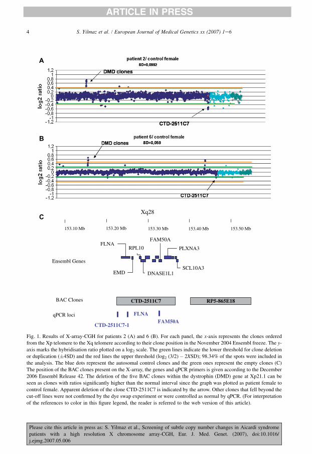
Using the quality criteria and threshold values as previously described [9,11], we regularlyidentified the deletion of the five BAC clones (RP11-142J18, RP5-1147O16, RP4-556A22,RP4-639D23 and RP11-64I1) encompassing exon 18 to exon 44 of the dystrophin gene in theDNA of the control female (Fig. 1). Four clones (RP11-388L20, RP5-1178I21 and RP11-66N11, RP11-54I20) fell beyond the 4SD threshold for patient 2 and patient 6. Six clones(RP11-441L6, RP11-23N11, CTD-2511C7, RP5-1000K24, RP11-142K4, RP11-97N5) fell be-tween the 4SD and log2 (3/2)� 2XSD thresholds for four patients. RP11-23N11 was known asa polymorphic clone filtered by the data analysis software. Three clones (RP11-97N5, RP11-388L20, RP11-441L6) were previously listed as copy number polymorphism by database of ge-nomic variants (http://projects.tcag.ca/variation/). Eight clones (RP11-388L20, RP5-1178I21,RP11-66N11, RP11-54I20, RP11-441L6, RP11-142K4, RP5-1000K24, RP11-97N5) were con-trolled as normal by qPCR using probes from regions of unique sequence within the clones.
Please cite this article in press as: S. Yilmaz et al., Screening of subtle copy number changes in Aicardi syndrome
patients with a high resolution X chromosome array-CGH, Eur. J. Med. Genet. (2007), doi:10.1016/
j.ejmg.2007.05.006
4 S. Yilmaz et al. / European Journal of Medical Genetics xx (2007) 1e6
ARTICLE IN PRESS
153.50 Mb153.40 Mb153.30 Mb153.20 Mb153.10 Mb
CTD-2511C7 RP5-865E18
qPCR loci FLNAFAM50A
BAC Clones
CTD-2511C7-1
EMD
FLNARPL10
DNASE1L1SCL10A3
PLXNA3
Ensembl Genes
FAM50A
Xq28C
B
A
Fig. 1. Results of X-array-CGH for patients 2 (A) and 6 (B). For each panel, the x-axis represents the clones ordered
from the Xp telomere to the Xq telomere according to their clone position in the November 2004 Ensembl freeze. The y-
axis marks the hybridisation ratio plotted on a log2 scale. The green lines indicate the lower threshold for clone deletion
or duplication (�4SD) and the red lines the upper threshold (log2 (3/2)� 2XSD); 98.34% of the spots were included in
the analysis. The blue dots represent the autosomal control clones and the green ones represent the empty clones (C)
The position of the BAC clones present on the X-array, the genes and qPCR primers is given according to the December
2006 Ensembl Release 42. The deletion of the five BAC clones within the dystrophin (DMD) gene at Xp21.1 can be
seen as clones with ratios significantly higher than the normal interval since the graph was plotted as patient female to
control female. Apparent deletion of the clone CTD-2511C7 is indicated by the arrow. Other clones that fell beyond the
cut-off lines were not confirmed by the dye swap experiment or were controlled as normal by qPCR. (For interpretation
of the references to color in this figure legend, the reader is referred to the web version of this article).
Please cite this article in press as: S. Yilmaz et al., Screening of subtle copy number changes in Aicardi syndrome
patients with a high resolution X chromosome array-CGH, Eur. J. Med. Genet. (2007), doi:10.1016/
j.ejmg.2007.05.006
5S. Yilmaz et al. / European Journal of Medical Genetics xx (2007) 1e6
ARTICLE IN PRESS
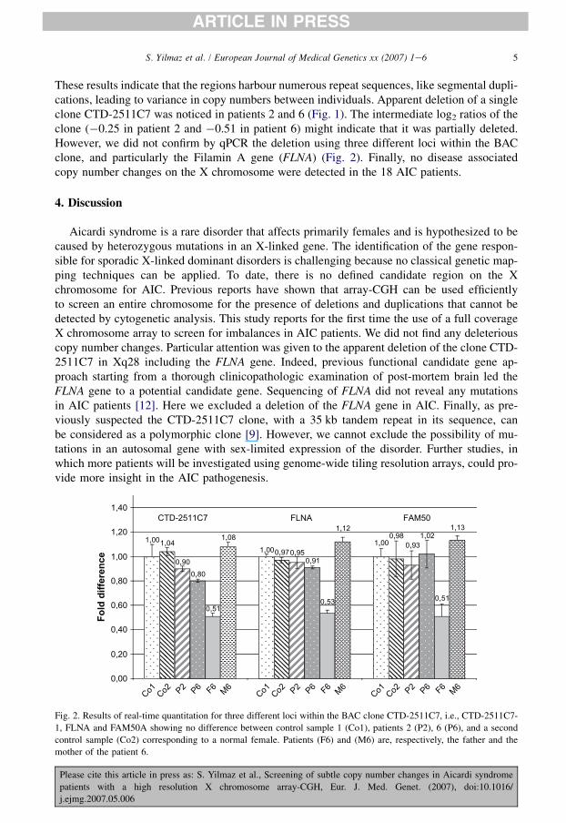
These results indicate that the regions harbour numerous repeat sequences, like segmental dupli-cations, leading to variance in copy numbers between individuals. Apparent deletion of a singleclone CTD-2511C7 was noticed in patients 2 and 6 (Fig. 1). The intermediate log2 ratios of theclone (�0.25 in patient 2 and �0.51 in patient 6) might indicate that it was partially deleted.However, we did not confirm by qPCR the deletion using three different loci within the BACclone, and particularly the Filamin A gene (FLNA) (Fig. 2). Finally, no disease associatedcopy number changes on the X chromosome were detected in the 18 AIC patients.
4. Discussion
Aicardi syndrome is a rare disorder that affects primarily females and is hypothesized to becaused by heterozygous mutations in an X-linked gene. The identification of the gene respon-sible for sporadic X-linked dominant disorders is challenging because no classical genetic map-ping techniques can be applied. To date, there is no defined candidate region on the Xchromosome for AIC. Previous reports have shown that array-CGH can be used efficientlyto screen an entire chromosome for the presence of deletions and duplications that cannot bedetected by cytogenetic analysis. This study reports for the first time the use of a full coverageX chromosome array to screen for imbalances in AIC patients. We did not find any deleteriouscopy number changes. Particular attention was given to the apparent deletion of the clone CTD-2511C7 in Xq28 including the FLNA gene. Indeed, previous functional candidate gene ap-proach starting from a thorough clinicopathologic examination of post-mortem brain led theFLNA gene to a potential candidate gene. Sequencing of FLNA did not reveal any mutationsin AIC patients [12]. Here we excluded a deletion of the FLNA gene in AIC. Finally, as pre-viously suspected the CTD-2511C7 clone, with a 35 kb tandem repeat in its sequence, canbe considered as a polymorphic clone [9]. However, we cannot exclude the possibility of mu-tations in an autosomal gene with sex-limited expression of the disorder. Further studies, inwhich more patients will be investigated using genome-wide tiling resolution arrays, could pro-vide more insight in the AIC pathogenesis.
1,001,00
1,001,040,97
0,98
0,900,80
0,91
1,02
0,510,51
1,080,93
0,95
0,53
1,12 1,13
0,00
0,20
0,40
0,60
0,80
1,00
1,20
1,40
Fo
ld
d
ifferen
ce
FLNA FAM50CTD-2511C7
Co1 Co2 P2 P6 F6 M
6Co1 Co2 P2 P6 F6
M6
Co1 Co2 P2 P6 F6 M
6
Fig. 2. Results of real-time quantitation for three different loci within the BAC clone CTD-2511C7, i.e., CTD-2511C7-
1, FLNA and FAM50A showing no difference between control sample 1 (Co1), patients 2 (P2), 6 (P6), and a second
control sample (Co2) corresponding to a normal female. Patients (F6) and (M6) are, respectively, the father and the
mother of the patient 6.
Please cite this article in press as: S. Yilmaz et al., Screening of subtle copy number changes in Aicardi syndrome
patients with a high resolution X chromosome array-CGH, Eur. J. Med. Genet. (2007), doi:10.1016/
j.ejmg.2007.05.006
6 S. Yilmaz et al. / European Journal of Medical Genetics xx (2007) 1e6
ARTICLE IN PRESS
Acknowledgments
We warmly thank all the patients and their families who participated in this study and themedical staff involved in the diagnosis of AIC patients. We thank Dr Marijke Bauters (FlandersInteruniversity Institute of Human Genetics) for helpful discussions and Dr Francis Martin(INRA, Champenoux). We are also grateful to Conseil Regional de Lorraine, CommunauteUrbaine du Grand Nancy, Dr Bihain (JE 2482, Laboratoire medecine et therapeutique mole-culaire) and the Association AAL-Syndrome d’Aicardi for their valuable support andcontribution.
References
[1] J. Aicardi, J. Levebre, A. Lerique-Koechlin, A new syndrome: spasms in flexion, callosal agenesis, ocular abnor-
malities, Electroencephalogr. Clin. Neurophysiol. 19 (1965) 609e610.
[2] J. Aicardi, Aicardi syndrome, Brain Dev. 27 (2005) 164e171.
[3] V.R. Sutton, B.J. Hopkins, T.N. Eble, N. Gambhir, R.A. Lewis, I.B. Van den Veyver, Facial and physical features
of Aicardi syndrome: infants to teenagers, Am. J. Med. Genet. 138A (2005) 254e258.
[4] J.A. Molina, F. Mateos, M. Merino, J.L. Epifanio, M. Gorrono, Aicardi syndrome in two sisters, J. Pediatr. 115
(1989) 282e283.
[5] I.J. Hopkins, I. Humphrey, C.G. Keith, M. Susman, G.C. Webb, E.K. Turner, The Aicardi syndrome in a 47,XXY
male, Aust. Paediatr. J. 15 (1979) 278e280.
[6] J. Aicardi, Aicardi syndrome: old and new findings, Int. Pediatr. 14 (1999) 5e8.
[7] I.B. Van den Veyver, Microphthalmia with linear skin defects (MLS), Aicardi, and Goltz syndrome: are they re-
lated X-linked dominant male lethal disorders? Cytogenet. Genome Res. 99 (2002) 289e296.
[8] S.K. Prakash, R. Paylor, S. Jenna, N. Lamarche-Vane, D.L. Armstrong, B. Xu, M.A. Mancini, H.Y. Zoghbi, Func-
tional analysis of ARHGAP6, a novel GTPase-activating protein for RhoA, Hum. Mol. Genet. 9 (2000) 477e488.
[9] M. Bauters, H. van Esch, P. Marynen, G. Froyen, X chromosome array-CGH for the identification of novel X-
linked mental retardation genes, Eur. J. Med. Genet. 48 (2005) 263e275.
[10] H. Van Esch, M. Bauters, J. Ignatius, M. Jansen, M. Raynaud, K. Hollanders, D. Lugtenberg, T. Bienvenu,
L.R. Jensen, J. Gecz, C. Moraine, P. Marynen, J.P. Fryns, G. Froyen, Duplication of the MECP2 region is a frequent
cause of severe mental retardation and progressive neurological symptoms in males, Am. J. Hum. Genet. 77 (2005)
442e453.
[11] J.R. Vermeesch, C. Melotte, G. Froyen, S. van Vooren, B. Dutta, N. Maas, S. Vermeulen, B. Menten, F. Speleman,
B. de Moor, P. van Hummelen, P. Marynen, J.P. Fryns, K. Devriendt, Molecular karyotyping: array CGH quality
criteria for constitutional genetic diagnosis, J. Histochem. Cytochem. 53 (2005) 413e422.
[12] I.B. Van den Veyver, P.P. Panichkul, B.A. Antalffy, Y. Sun, J.V. Hunter, D.D. Armstrong, Presence of filamin in the
astrocytic inclusions of Aicardi syndrome, Pediatr. Neurol. 30 (2004) 7e15.
Please cite this article in press as: S. Yilmaz et al., Screening of subtle copy number changes in Aicardi syndrome
patients with a high resolution X chromosome array-CGH, Eur. J. Med. Genet. (2007), doi:10.1016/
j.ejmg.2007.05.006
Related Documents