Florida International University FIU Digital Commons FIU Electronic eses and Dissertations University Graduate School 11-4-2016 Screening of Plants for Antibacterial Properties: Growth Inhibition of Staphylococcus aureus by Artemisia Tridentata Steven Ross Eichelbaum Florida International University, eichelbs@fiu.edu DOI: 10.25148/etd.FIDC004040 Follow this and additional works at: hps://digitalcommons.fiu.edu/etd Part of the Biology Commons , Ecology and Evolutionary Biology Commons , Microbiology Commons , Molecular Biology Commons , and the Plant Sciences Commons is work is brought to you for free and open access by the University Graduate School at FIU Digital Commons. It has been accepted for inclusion in FIU Electronic eses and Dissertations by an authorized administrator of FIU Digital Commons. For more information, please contact dcc@fiu.edu. Recommended Citation Eichelbaum, Steven Ross, "Screening of Plants for Antibacterial Properties: Growth Inhibition of Staphylococcus aureus by Artemisia Tridentata" (2016). FIU Electronic eses and Dissertations. 3526. hps://digitalcommons.fiu.edu/etd/3526

Welcome message from author
This document is posted to help you gain knowledge. Please leave a comment to let me know what you think about it! Share it to your friends and learn new things together.
Transcript

Florida International UniversityFIU Digital Commons
FIU Electronic Theses and Dissertations University Graduate School
11-4-2016
Screening of Plants for Antibacterial Properties:Growth Inhibition of Staphylococcus aureus byArtemisia TridentataSteven Ross EichelbaumFlorida International University, [email protected]
DOI: 10.25148/etd.FIDC004040Follow this and additional works at: https://digitalcommons.fiu.edu/etd
Part of the Biology Commons, Ecology and Evolutionary Biology Commons, MicrobiologyCommons, Molecular Biology Commons, and the Plant Sciences Commons
This work is brought to you for free and open access by the University Graduate School at FIU Digital Commons. It has been accepted for inclusion inFIU Electronic Theses and Dissertations by an authorized administrator of FIU Digital Commons. For more information, please contact [email protected].
Recommended CitationEichelbaum, Steven Ross, "Screening of Plants for Antibacterial Properties: Growth Inhibition of Staphylococcus aureus by ArtemisiaTridentata" (2016). FIU Electronic Theses and Dissertations. 3526.https://digitalcommons.fiu.edu/etd/3526
FLORIDA INTERNATIONAL UNIVERSITY
Miami, Florida
SCREENING OF PLANTS FOR ANTIBACTERIAL PROPERTIES: GROWTH
INHIBITION OF STAPHYLOCOCCUS AUREUS BY ARTEMISIA TRIDENTATA
A dissertation submitted in partial fulfillment of
the requirements for the degree of
DOCTOR OF PHILOSOPHY
in
BIOCHEMISTRY
by
Steven Eichelbaum
2017

ii
To: Dean Michael R. Heithaus
College of Arts, Sciences and Education
This dissertation, written by Steven Eichelbaum, and entitled Screening of Plants for
Antibacterial Properties: Growth Inhibition of Staphylococcus aureus by Artemisia
tridentata, having been approved in respect to style and intellectual content, is referred to
you for judgment.
We have read this dissertation and recommend that it be approved.
_______________________________________
Lou W. Kim
_______________________________________
Martin Tracey
_______________________________________
Xiaotang Wang
_______________________________________
Javier-Francisco Ortega
_______________________________________
Alejandro Barbieri, Major Professor
Date of Defense: November 4, 2016
The dissertation of Steven Eichelbaum is approved.
_______________________________________
Dean Michael R. Heithaus
College of Arts, Sciences and Education
_______________________________________
Andrés G. Gil
Vice President for Research and Economic Development
and Dean of the University Graduate School
Florida International University, 2017

iii
ACKNOWLEDGMENTS
I would first like to acknowledge Dr. Alejandro Barbieri for allowing me into his
lab, and for providing me with the guidance and materials necessary to complete my
project. I would also like to acknowledge Dr. Kathleen Rein for providing guidance in
aspects of the project related to organic chemistry. Further, I would acknowledge
Chemistry Department Graduate Secretary Maggie Autie for her overall helpfulness and
willingness to listen to my constant grievances. Lastly, I would acknowledge the
International Center for Tropical Botany for providing the Fellowship funding which
allowed me to complete my studies.

iv
ABSTRACT OF THE DISSERTATION
SCREENING OF PLANTS FOR ANTIBACTERIAL PROPERTIES: GROWTH
INHIBITION OF STAPHYLOCOCCUS AUREUS BY ARTEMISIA TRIDENTATA
by
Steven Eichelbaum
Florida International University, 2017
Miami, Florida
Professor Alejandro Barbieri, Major Professor
Drug-resistant pathogenic and opportunistic bacteria are increasing in occurrence
and prevalence, and pose a dangerous threat to human health. In the search for novel
antibiotics with which to combat this threat, plants, specifically those used in traditional
medicine with ascribed antibacterial properties, offer a promising and potentially vast
source of such therapeutic compounds. The purpose of this study was therefore to screen
chemical extracts created from various plant species for antibacterial properties versus
pathogenic bacterial species. In the course of these antibacterial assays, we successfully
identified a methanol extract derived from Artemisia tridentata tridentata plant material
as capable of inhibiting the growth of the opportunistic pathogen Staphylococcus aureus.
Three sub-fractions were created using hexane, ethyl acetate and water solvents. Each of
these extracts displayed significant antibacterial activity versus a wild-type strain over a
period of six hours, at concentrations as low as 62.5 µg/ml. The extracts also
demonstrated an enhancement of antibiotic effects when combined with ampicillin, G418
sulfate or amikacin, for a period of up to twelve hours. Though the efficacy of the
extracts was lessened when tested against an ampicillin-resistant strain, significant

v
enhancement of the efficacy of this antibiotic was still observed. Gas chromatography-
mass spectrometry analysis of these three extracts revealed the sesquiterpene lactone
achillin as present in each. Column chromatography of the hexane extract resulted in a
fraction retaining its antibacterial activity, and still containing this compound, further
implicating it as responsible for the antibacterial activity of this plant. The results of
serial dilution and plating of extract-treated samples, along with those of ethidium
bromide assays and transmission electron microscopy analysis, indicated a bacteriostatic
mechanism of action involving disruption of the bacterial membrane, which is in
agreement with the literature on the antibacterial properties of this plant, and those of
sesquiterpene lactones, respectively. We therefore conclude that achillin, likely produced
as a secondary metabolite by Artemisia tridentata tridentata, possesses growth inhibitory
properties versus Staphylococcus aureus, and should be isolated and studied further for
the purposes of evaluating its potential use, either as a stand-alone antibiotic, or as an
adjunctive therapeutic, in the treatment of drug-resistant bacterial pathogens.

vi
TABLE OF CONTENTS
CHAPTER PAGE
I. INTRODUCTION ...........................................................................................................1
II. LITERATURE REVIEW ...............................................................................................5
Why Plants? .............................................................................................................5
Plant Selection .........................................................................................................9
Methods of Extraction............................................................................................12
Antibacterial Testing ..............................................................................................24
Methods of Isolation and Identification .................................................................37
Mechanisms of Action ...........................................................................................47
Conclusion .............................................................................................................54
III. MATERIALS, METHODS AND RESULTS ............................................................58
Artemisia ................................................................................................................58
Bacterial Species Assayed .....................................................................................60
Preliminary Plant Screenings .................................................................................66
Initial Artemisia tridentata Assays ........................................................................68
Growth Inhibition and Combination With Antibiotics ..........................................74
Time-Extended Assays ........................................................................................110
Extracts in Combination ......................................................................................116
Ampicillin-Resistant Staphylococcus aureus ......................................................120
Biofilm Formation Assays ...................................................................................125
Static Biofilm Assays ...........................................................................................128
Serial Dilution and Plating ...................................................................................131
Toxicity Assay .....................................................................................................134
pH Measurement ..................................................................................................136
Ethidium Bromide Assays ...................................................................................137
DNA-nicking Assays ...........................................................................................139
Transmission Electron Microscopy .....................................................................143
GC-MS Analysis ..................................................................................................147
Column Chromatography.....................................................................................155
IV. DISCUSSION ...........................................................................................................160
Evaluation of Results ...........................................................................................160
Experimental Shortcomings .................................................................................166
Future Experiments ..............................................................................................168
REFERENCES ................................................................................................................171
VITA ................................................................................................................................182

vii
LIST OF TABLES
TABLE PAGE
1. Summary of preliminary plant screening assays ..........................................................66
2. Biofilm formation assay results ..................................................................................127
3. Static biofilm assay results..........................................................................................130
4. Serial and dilution of A. tridentata extract-treated S. aureus .....................................133
5. Ethidium bromide binding assay results .....................................................................138
6. Summary of GC-MS results from hexane extract analysis .........................................151
7. Summary of GC-MS results from ethyl acetate extract analysis ................................152
8. Summary of GC-MS results from water extract analysis ...........................................154
9. Summary of GC-MS results from 10 % acetonitrile column fraction analysis ..........159

viii
LIST OF FIGURES
FIGURE PAGE
1. S. aureus treated with A. tridentata methanol extract ...................................................69
2. P. aeruginosa treated with A. tridentata methanol extract ...........................................69
3. S. enterica treated with A. tridentata methanol extract ................................................70
4. L. monocytogenes treated with A. tridentata methanol extract .....................................70
5. 2nd
trial of S. aureus treated with A. tridentata methanol extract .................................71
6. 3rd
trial of S. aureus treated with A. tridentata methanol extract ..................................73
7. S. aureus treated with A. tridentata hexane extract ......................................................78
8. S. aureus treated with G418 sulfate ..............................................................................78
9. S. aureus treated with G418 sulfate and hexane extract (500 µg/ml) ...........................79
10. S. aureus treated with G418 sulfate and hexane extract (250 µg/ml) .........................79
11. S. aureus treated with G418 sulfate and hexane extract (125 µg/ml) .........................80
12. S. aureus treated with G418 sulfate and hexane extract (62.5 µg/ml) ........................80
13. S. aureus treated with G418 sulfate and hexane extract (31.25 µg/ml) ......................81
14. S. aureus treated with A. tridentata hexane extract ....................................................81
15. S. aureus treated with amikacin ..................................................................................82
16. S. aureus treated with amikacin and hexane extract (500 µg/ml) ...............................82
17. S. aureus treated with amikacin and hexane extract (250 µg/ml) ...............................83
18. S. aureus treated with amikacin and hexane extract (125 µg/ml) ...............................83
19. S. aureus treated with amikacin and hexane extract (62.5 µg/ml) ..............................84
20. S. aureus treated with amikacin and hexane extract (31.25 µg/ml) ............................84

ix
21. S. aureus treated with A. tridentata hexane extract ....................................................85
22. S. aureus treated with ampicillin ................................................................................85
23. S. aureus treated with ampicillin and hexane extract (500 µg/ml) .............................86
24. S. aureus treated with ampicillin and hexane extract (250 µg/ml) .............................86
25. S. aureus treated with ampicillin and hexane extract (125 µg/ml) .............................87
26. S. aureus treated with ampicillin and hexane extract (62.5 µg/ml) ............................87
27. S. aureus treated with ampicillin and hexane extract (31.25 µg/ml) ..........................88
28. S. aureus treated with A. tridentata ethyl acetate extract ...........................................88
29. S. aureus treated with G418 sulfate ............................................................................89
30. S. aureus treated with G418 sulfate and ethyl acetate extract (500 µg/ml) ................89
31. S. aureus treated with G418 sulfate and ethyl acetate extract (250 µg/ml) ................90
32. S. aureus treated with G418 sulfate and ethyl acetate extract (125 µg/ml) ................90
33. S. aureus treated with G418 sulfate and ethyl acetate extract (62.5 µg/ml) ...............91
34. S. aureus treated with G418 sulfate and ethyl acetate extract (31.25 µg/ml) .............91
35. S. aureus treated with A. tridentata ethyl acetate extract ...........................................92
36. S. aureus treated with amikacin ..................................................................................92
37. S. aureus treated with amikacin and ethyl acetate extract (500 µg/ml) ......................93
38. S. aureus treated with amikacin and ethyl acetate extract (250 µg/ml) ......................93
39. S. aureus treated with amikacin and ethyl acetate extract (125 µg/ml) ......................94
40. S. aureus treated with amikacin and ethyl acetate extract (62.5 µg/ml) .....................94
41. S. aureus treated with amikacin and ethyl acetate extract (31.25 µg/ml) ...................95
42. S. aureus treated with A. tridentata ethyl acetate extract ...........................................95
43. S. aureus treated with ampicillin ................................................................................96

x
44. S. aureus treated with ampicillin and ethyl acetate extract (500 µg/ml) ....................96
45. S. aureus treated with ampicillin and ethyl acetate extract (250 µg/ml) ....................97
46. S. aureus treated with ampicillin and ethyl acetate extract (125 µg/ml) ....................97
47. S. aureus treated with ampicillin and ethyl acetate extract (62.5 µg/ml) ...................98
48. S. aureus treated with ampicillin and ethyl acetate extract (31.25 µg/ml) .................98
49. S. aureus treated with A. tridentata water extract .......................................................99
50. S. aureus treated with G418 sulfate ............................................................................99
51. S. aureus treated with G418 sulfate and water extract (500 µg/ml) .........................100
52. S. aureus treated with G418 sulfate and water extract (250 µg/ml) .........................100
53. S. aureus treated with G418 sulfate and water extract (125 µg/ml) .........................101
54. S. aureus treated with G418 sulfate and water extract (62.5 µg/ml) ........................101
55. S. aureus treated with G418 sulfate and water extract (31.25 µg/ml) ......................102
56. S. aureus treated with A. tridentata water extract .....................................................102
57. S. aureus treated with amikacin ................................................................................103
58. S. aureus treated with amikacin and water extract (500 µg/ml) ...............................103
59. S. aureus treated with amikacin and water extract (250 µg/ml) ...............................104
60. S. aureus treated with amikacin and water extract (125 µg/ml) ...............................104
61. S. aureus treated with amikacin and water extract (62.5 µg/ml) ..............................105
62. S. aureus treated with amikacin and water extract (31.25 µg/ml) ............................105
63. S. aureus treated with A. tridentata water extract .....................................................106
64. S. aureus treated with ampicillin ..............................................................................106
65. S. aureus treated with ampicillin and water extract (500 µg/ml) ..............................107
66. S. aureus treated with ampicillin and water extract (250 µg/ml) ..............................107

xi
67. S. aureus treated with ampicillin and water extract (125 µg/ml) ..............................108
68. S. aureus treated with ampicillin and water extract (62.5 µg/ml) .............................108
69. S. aureus treated with ampicillin and water extract (31.25 µg/ml) ...........................109
70. S. aureus treated with A. tridentata extracts .............................................................111
71. S. aureus treated with G418 sulfate, amikacin, ampicillin .......................................112
72. S. aureus treated with G418 sulfate and A. tridentata extracts .................................114
73. S. aureus treated with amikacin and A. tridentata extracts.......................................114
74. S. aureus treated with ampicillin and A. tridentata extracts .....................................115
75. S. aureus treated with A. tridentata hexane extract ..................................................116
76. S. aureus treated with A. tridentata ethyl acetate extract .........................................117
77. S. aureus treated with A. tridentata water extract .....................................................117
78. S. aureus treated with A. tridentata hexane and ethyl acetate extracts .....................118
79. S. aureus treated with A. tridentata hexane and water extracts ................................119
80. S. aureus treated with A. tridentata ethyl acetate and water extracts .......................119
81. S. aureus BAA-44 treated with hexane extract .........................................................121
82. S. aureus BAA-44 treated with ethyl acetate extract ................................................121
83. S. aureus BAA-44 treated with water extract ...........................................................122
84. S. aureus BAA-44 treated with ampicillin ................................................................123
85. S. aureus BAA-44 treated with ampicillin and hexane extract .................................123
86. S. aureus BAA-44 treated with ampicillin and ethyl acetate extract ........................124
87. S. aureus BAA-44 treated with ampicillin and water extract ...................................124
88. Artemisia tridentata ethyl acetate extract-treated MDA-MB-231 cells ...................135
89. Gel electrophoresis of extract-treated pGEX (1st assay) ...........................................140

xii
90. Gel electrophoresis of extract-treated pGEX (2nd assay) .........................................141
91. Gel electrophoresis of extract-treated pGEX (3rd assay) .........................................142
92. TEM image of untreated Staphylococcus aureus .....................................................145
93. TEM image of water extract-treated Staphylococcus aureus ...................................145
94. TEM image of hexane extract-treated Staphylococcus aureus .................................146
95. TEM image of ethyl acetate extract-treated Staphylococcus aureus ........................146
96. GC-MS spectral results for Artemisia tridentata hexane extract ..............................148
97. GC-MS spectral results for Artemisia tridentata ethyl acetate extract .....................148
98. GC-MS spectral results for Artemisia tridentata water extract ................................149
99. GC-MS results for 10 % acetonitrile column fraction of hexane extract .................157
100. GC-MS results for 100 % acetonitrile column fraction of hexane extract .............157
101. Antibacterial assay of hexane extract column fractions .........................................158
1
I. INTRODUCTION
Antibiotic-resistant bacteria pose a global health threat. Infectious diseases,
increasingly resulting from antibiotic-resistant pathogenic or opportunistic bacteria,
represent the leading annual cause of human fatalities1. These deaths include a
staggering ½ of all fatalities in tropical countries, with gastrointestinal infections alone,
for example, claiming the lives up to 3 million pre-school aged children per year2. In
addition, infectious diseases have also arisen as a significant source of morbidity and/or
mortality in immunocompromised patients, in both developing and developed
countries3,4
. In HIV cases for example, opportunistic infections represent the leading
cause of death in AIDS patients5, with bacterial complications both facilitating the
infection rate of the HIV virus and reducing the onset time of this disease2. Further, there
has been an alarming increase in the occurrence of new diseases, and a re-emergence of
old ones, accompanied by an increasing prevalence of resistance to antibiotics in clinical
use6. In fact, despite the availability of more than 200 varieties of antibiotics and
chemotherapeutics, the occurrence of multi-drug resistant bacteria is at its peak7. The
seriousness of the threat posed by antibiotic-resistant bacteria is thus underscored both by
its current impact, and the potential ramifications of its increasing prevalence.
The recent reporting of increases in both the number and prevalence of
staphylococcal infections, such as those of methicillin-resistant Staphylococcus aureus,
combined with the emergence of vancomycin-resistant isolates8, serves as evidence of the
current impact of antibiotic resistance on human disease. In fact, the level of resistance
in clinical isolates of Staphylococcus pneumoniae to antibiotics routinely used for such
infections has been reported to be as high as 40% in some parts of Europe8. Therapeutic

2
options for methicillin-resistant Staphylococcus aureus (MRSA) have become limited as
well, as strains resistant to synthetic antibiotics including macrolides, aminoglycosides,
fluroquinolones, chloramphenicol, clindamycin, tetracycline, vancomycin, oxazolidine-
type and streptogramin-type antibiotics, in addition to β-lactams, have emerged9,10
. It is
also worth noting that the increase in the occurrence of drug-resistance has been mirrored
by a significant reduction in the number of pharmaceutical companies developing new
antimicrobial agents11
.
However, antibiotic resistant bacteria also have a significant negative impact on
agriculture. In fish aquaculture for example, the fast development of the industry,
combined with increasing product demands, can lead to overcrowding, poor water
quality, or poor nutritional status, which can contribute to stress and immunosuppression
in the fish, increasing the risk of disease dissemination12
. This infection risk necessitates
the administration of antimicrobials and other veterinary drugs, which are also used for
growth promotion, for prophylactic and therapeutic purposes12
, all of which are practices
which can unfortunately select for antibiotic-resistance. As an example of drug-
resistance in agricultural animals, chicken and pork have become vehicles for livestock-
associated MRSA strains, adding an epidemiological dimension to the pathogen in the
food supply13
.
Alternatively, in crop farming, biocide use at sub-lethal concentrations, in some
cases due to limited availability14
, while in others possibly the result of legal impositions
on the application of synthetic antibiotics to crops, hampers disease control, and is
another practice which may select for antibiotic resistance15
. In Europe for example, the
protocol of EUREPGAP (European Good Agricultural Practice) places restrictions on the

3
allowable residue limits of pesticides on fruits and vegetables16
, and the use of antibiotic
and copper compounds is restricted in many countries over concerns pertaining to human
and animal health or the environment14
. Arguably as a direct result of such policies,
several resistant populations of plant pathogens have been reported14
, and plant diseases
caused by plant pathogens, including bacteria, represent a major cause of crop loss17
. It is
therefore worth considering that the threat to human health posed by drug-resistant
bacteria in agriculture may be just as great, or even greater than that posed by infectious
human diseases, as in agriculture drug resistance threatens both the quantity and safety of
the food supply, with food-borne infections currently among the most serious and costly
global health concerns18
.
Two obvious strategies present themselves when considering means to combat the
increasing emergence of drug-resistant bacteria. The first would be to remove or reduce
the causative factors of bacterial drug-resistance by implementing policies promoting
responsible and efficient use of antibiotics, thus reducing “selective pressure” as much as
is feasible. The second of course, is the development or discovery of novel antibiotics
with unique mechanisms of action which may yet be effective against bacteria otherwise
resistant to existing therapeutics. To this end, there has been a renewed interest in
exploring “medicinal plants,” or plants used in traditional medicine, as possible sources
of such pre-existing novel antibiotic compounds. As research of plants for antibiotic
compounds is, in recent years if not historically, a relatively underexplored field of study,
there is consequentially a lack of standardization in the experimental methods and
techniques used in such research. Unfortunately, this discord may limit the ability of an
investigator to accurately assess the antibacterial potential of a plant, while also making
4
the recognition of significant results, and the comparison of results from separate
investigations, difficult. The following therefore begins with an explanation of the
reasoning behind the investigation of plants, specifically medicinal plants, as a source of
novel antibiotic compounds, then provides a summary and some evaluation of the
experimental methods and techniques currently used in this field as they relate to plant
selection, chemical extraction, antibiotic screening, the identification of compounds of
interest, and the determination of the mechanisms of action of antibacterial compounds.

5
II. LITERATURE REVIEW
Why Plants?
There are several arguments to be made in support of the research of plants, and
medicinal plants in particular, as potential sources of novel antibiotic compounds with
which to combat drug resistance. The first of these arguments is to point out the
historical role that plants have played in the promotion of human health. A medicinal
plant, as defined by the World Health Organization, is a plant which contains substances,
in one or more of its parts, which can either be used directly for therapeutic purposes, or
are precursors for chemo-pharmaceutical semi-synthesis14
, and it is important to consider
that plants have served as the starting point for many of the modern pharmaceuticals in
use today. Indeed, it has been estimated that more than a full quarter of prescribed
medications in industrialized nations derive their origins either directly or indirectly from
plants19
. As this historical role relates specifically to plants providing a source of novel
antibiotics for the treatment of infectious diseases, it is believed that plants possess
secondary metabolites, in various plant tissues, which are produced and used by plants
for defensive purposes20,21
. It has been postulated that these compounds are an
evolutionary response to protect the plants from insects, predators, and most importantly
in this context, microbes11,22,23
, and this postulation is directly supported by the discovery
that syntheses of some of these compounds occurs post-infection24
. It is therefore
reasonable to hypothesize that some of these compounds produced to fight bacterial plant
pathogens may also be beneficial in combating bacterial human pathogens.
Second, plants in general are overwhelmingly abundant, yet vastly underexplored.
There exist an estimated 2.5 million species of higher plants, and a large number have yet

6
to be studied in this context25,26
. India alone for example, where much of this type of
research in the reviewed literature originates, possesses within its borders approximately
130,000 plant species encompassing some 120 families10
. In North America, while
Native Americans are believed to have used an estimated 2,500 plant species in their
traditional medicine practices, the region is believed to contain as many as 20,000 native
species27
. Plants thus represent a potentially vast source of bioactive molecules, which is
likely why they have been referred to as the “sleeping giants of the pharmaceutical
industry”19
. Further, while recognition of the antiseptic qualities of medicinal plants date
to antiquity, efforts to characterize these properties in the laboratory setting date only to
the early 1900s28
. In fact, the approximately 12,000 secondary metabolites so far isolated
from plants are believed to comprise less than 10% of the total in existence20
. Therefore,
it is entirely plausible that investigation of previously untested plant species will reveal
medicinal plants possessing unrecognized antibiotic activities.
The third argument to be made in favor of the research of plants as sources of
novel antibiotic compounds is to note that the use of medicinal plants in the practice of
traditional medicine persists very prominently even today. The World Health
Organization estimates that 80% of people in developing countries engage in such
practices23
, a number which corresponds to approximately 65% of the global population.
The antimicrobial activity of plant extracts and oils has led to their administration as food
preservatives, natural therapies, alternative medicines and pharmaceuticals29
. While it
can be pointed out that there is often little solid scientific evidence to support such claims
touting the antibiotic attributes of medicinal plants, it can also be reasonably argued that
without thorough scientific evaluation, it would be inappropriate to summarily dismiss

7
their virtue. Additionally, given the prevalence of their use, it is necessary to ensure the
safety of these plants, as insufficient patient awareness or improper use result in many
cases of adverse reactions in traditional medicine, including, among others, allergic
reactions, fever and vomiting1. Also, in cases where products derived from these plants
are indeed found to be beneficial to human health, there is a need for standardization in
terms of raw materials, production methods and quality control of finished products1.
Such safety and standardization issues could be addressed through the increased
investigation of these medicinal plants.
Additional arguments to be made in support of the investigation of plants as a
potential source of novel antibiotics relate to the potential advantages such products could
provide over synthetic antibiotics. There currently exists a public mistrust of synthetic
antimicrobials stemming from the potential toxicity or even carcinogenicity of these
products30
, as the use of synthetic antibiotics for example, may in some cases be harmful
to distinct organs and threaten consumer health31,32
. Further, the application of synthetic
antimicrobials in agriculture creates additional opportunities for human exposure to such
chemicals, either through the consumption of chemical residues on crops, or bio-
accumulated chemicals in agricultural animals. Antimicrobials may also be applied to
finished food products for preservation and safety purposes, to protect against natural
spoilage processes and pathogenic microorganisms, respectively33
. Consequentially,
consumers are increasingly demanding minimally processed foods, at the same time
desiring products free from pathogens, yet simultaneously containing fewer synthetic
preservatives18,33
, with mounting pressure from both consumers and legal authorities for
alternative, natural product shelf-life extending additives30
. These demands are becoming

8
reflected in public policy. In Europe for example, in-feed antibiotics for livestock were
banned by the European Union as far back as 200634
.
It is noteworthy that plant-derived substances have already served as food
preservatives for centuries, with many herbs and spices used in food seasoning in fact
also yielding useful medicinal compounds35
. Additionally, there are some positive results
reported for medicinal plants used as animal feed in the reviewed literature. They have
been reported to promote growth and appetite, as well as have immunostimulatory and
anti-pathogen effects in fish and shrimp aquaculture12
for example, and it has been
suggested that prophylactic administration of immunostimulants and pro-, pre- and
synbiotics is the most promising method of disease control in aquaculture animals36
.
However, it is important to note, as described previously, that there are also existing
reports of adverse reactions to traditional medicines. Though the fact that an antibiotic is
derived from a natural source may sway public opinion to view it as less hazardous, there
are no guarantees to be made as to the advantages, in terms of safety, of substituting
plant-derived antibiotics for synthetic antibiotics. Still, it is possible that further plant
investigations may identify novel, and comparatively safer antibiotics for clinical and
agricultural applications.
Also, from an environmental perspective, the use of plant-derived antibiotics
would seem greatly advantageous over the use of synthetic antibiotics. Plant-derived
substances, in their natural states, would likely be entirely biodegradable, and thus avoid
pollution and environmental degradation issues related to orthodox medicines37
. Lastly,
in comparison to synthetic antibiotics, using plant-derived antibiotics would lessen cost
and increase the accessibility to these medications. The possibility, at least in some

9
locales, of being able to culture medicinal plants and perhaps even purify or otherwise
concentrate substances of medicinal value would be of great benefit to the destitute or
those living in geographic areas without reasonable accessibility to modern medications.
Thus, given their history as a medicinal source and the number of plant species which
have yet to be investigated, their widespread use in traditional medicine and the need to
ensure the safety of these practices, as well as the advantages their use may provide over
that of synthetic medications, in terms of safety, environmental impact and cost, there is a
solid case to be made in favor of the increased and expanded exploration of plants as
potential sources of novel antibiotics.
Plant Selection
Studies of plants that investigate their therapeutic potential are typically screening
exercises for the evaluation of antioxidant, anti-inflammatory, antifungal or antibacterial
properties of plant extracts, and are usually based on ethnobotanical leads38
. The general
strategy of these screening exercises as it pertains specifically to the evaluation of
antibacterial properties of the plant can be broken down into several steps; plant selection
is naturally the first of these steps. There are both practical and logistical limitations to
consider however, before finalizing the selection of a plant or plants for study. In the
search for novel antimicrobial compounds, as there are so many species from which to
choose, plants with a long standing history of medicinal use constitute the most practical
starting point21
. For example, Tchouya et al. conducted ethnopharmacological surveys in
Gabon to identify fifty-two species of medicinal plants used there to treat HIV/AIDS-
related opportunistic diseases, prior to selecting five for phytochemical screening and
antibacterial testing5.

10
However, the availability or accessibility of a medicinal plant must also be taken
into consideration before a selection is finalized. If the plant(s) of particular interest
require crossing national boundaries to obtain, the likelihood of encountering proprietary
ownership issues, as well as the monetary cost, may both increase. Additionally, if it is
believed that proprietary issues may arise, estimations should be made prior to the
beginning of work, as comprehensively as possible, regarding how much plant sample
will be necessary to complete the intended studies. It then needs to be determined if it is
possible to procure this amount of plant material, as well as to do so over a reasonable
period of time. For example, if a plant(s) is only accessible by means of agreement with
a private botanical collection, either foreign or domestic, there may be strict limits
imposed on the amount of plant material provided.
Further, if limits are imposed, and on-site cultivation of plants is unfeasible, it
should be considered that these limits may unduly prolong studies, potentially creating
further issues related to the continuity of the researchers involved, cost, etc., as well as
possibly causing variability in experimental results due to chemotype differences in the
starting material. These chemotype differences, even among members of the same plant
species, may result from geographic39
or seasonal growth variations. Smida et al., for
example, in investigating the antibacterial activities of Ludwigia peploides and Ludwigia
grandiflora over a period of several months, documented time-dependent differences in
plant extract efficacy versus multiple strains of bacteria40
, suggesting temporal
differences in plant chemotype. Further, the specific microenvironment in which a plant
is grown may also affect its chemotype. According to “plant defense theory,” defensive
versus growth allocation of plant resources may depend upon the specific light
11
environment in which the plant grows, with plants growing in open habitats likely to
contain higher levels of defensive compounds and reduced herbivory in comparison to
those growing in shaded environments41
. Thus, as the geographical location, time of
sample collection and the microenvironment in which a plant was grown may affect its
chemical composition, limited accessibility to a plant may cause difficulty in obtaining
plant samples capable of providing consistent experimental results, and this should also
be considered prior to the selection of a plant for study.
The selection of which plant part(s) to study is an additional consideration in
study design. While it has been stated that antibacterial compounds are more likely to be
located within growth buds, young leaves, reproductive organs and parts of annual
growth20
, it is quite evident from the literature that different plant parts possess varying
chemotypes, and that antibacterial compounds are not universally relegated to specific
tissue types. For example, Yasanuka et al., in testing the antibacterial potential of various
Mexican medicinal plants, created extracts from fruits, heartwoods, leaves, fruit peels,
roots, stems and twigs, and reported differences in results between plant parts from the
same species26
. The inclusion and separate study of all parts of a selected plant in search
of these differences, while appealing, adds substantially to the potential workload and
financial cost. Hypothetically, the separate study of individual plant parts is not
mandatory, and whole-plant extracts may certainly be used in these screening exercises.
However, as will be discussed below, there exists the possibility that if an antibacterial
compound(s) present in a plant under study are relegated specifically to one plant part,
inclusion of additional plant parts in the creation of an extract may dilute the
concentration of this compound(s) to levels low enough to impede detection in

12
antibacterial assays. Here again, it would be wise to consult traditional practitioners or
users of the medicinal plant(s) of interest, so that plant parts selected for study match
those used in practice.
However, the selection of a specific plant part(s) may also increase the logistical
difficulty of obtaining samples for study. The harvest of a crop can significantly alter the
future growth patterns of a plant population. Mooney et al., for example, found both the
number of flowering stalks and the proportion of these flowering stalks to leaves reduced
in Ligusticum porteri populations up to two years post-harvest of rhizome and
adventitious root material41
. Generally speaking, the harvesting of plant leaves is more
sustainable than that of stems or roots42
. Therefore, individual suppliers may be
understandably reluctant to provide stem or root samples of their plant(s). Thus, the
determination of what plant part(s) is of greatest interest, and how this may affect
procurement of plant samples, should also be considered when selecting a specific
plant(s) for study. In conclusion, while it is in the best interest of the researcher when
choosing a plant(s) for study to make practical selections based on ethnobotanical leads,
it is also important to take into account how logistical issues pertaining to plant
procurement may affect both the availability of plant material and its potential
consistency in terms of chemical composition, as well as how the inclusion of various
plant parts for study might further complicate this procurement, as well as potentially
inflate workload and cost.
Methods of Extraction
Beginning with the treatment of plant material prior to the creation of chemical
extracts, there appears in the literature a notable lack of standardization in the

13
experimental methods and materials used in this and in subsequent steps of these plant
investigations. For example, after sometimes being washed with water, plant material is
almost always dried prior to chemical extraction. However, plant material may be dried
in the light or in the shade, for varying periods of time, and at different temperatures, and
there is no rationale provided by the authors for their method selection. Kuppusamy et al.
for example, washed Commelina nudiflora plant material twice in running tap water prior
to cutting the material into small pieces, then drying it in the shade at 35°C in 12-hour
cycles of light and dark31
. Alternatively, Voravuthikunchai and Limsuwan, in sampling
parts of eight species used in traditional medicine in Thailand, simply cut plant material
into small pieces and dried these samples overnight at 60°C43
, whereas Kenny et al. by
comparison, sliced and freeze-dried dandelion roots prior to extraction44
.
Presumably, the purpose of drying plant material is to eliminate excess water and
increase the concentrations of any antibacterial compounds present in order to enhance
the probability of detection. However, even such a commonplace practice as drying plant
material prior to extract creation must be questioned. Alabri et al. for example, reported
the comparatively enhanced antimicrobial efficacy of several chemical extracts derived
from fresh Datura metel leaves in comparison to similar extracts created from dry leaf
material when tested against multiple bacterial species27
. It is therefore possible that
drying risks the loss of volatile compounds in the plant material, potentially lessening
antibacterial potential. Regardless of the cause for differential antibacterial results
between fresh and dried plant material, a sound investigation of a single plant may
require testing of both.

14
Following drying, plant parts are then often, but again not always, ground or
pulverized. In the abovementioned studies, both Kuppusamy et al. and Kenny et al.
powdered dried plant material using mechanical blenders, the former following this step
with sieving of blended material through a 40 µm mesh31,44
, whereas Voravuthikunchai
and Limsuwan, using a similar approach, crushed dried plant material in a mechanical
mortar43
. The use of grinding or pulverization is presumably to increase surface area
exposure and enhance extraction efficiency, which is defined below. This enhancement
likely improves the chances of recognizing any antibacterial compounds potentially
present within the plant material, by increasing the odds that these compounds will be
present in the resulting extracts in high enough concentrations as to be detectable in
antibacterial assays.
In the pharmaceutical sense, the creation of extracts from plant material refers to
the separation of therapeutically active constituents, with simultaneous elimination of
unwanted insoluble material through treatment with selective solvents45
. However,
extraction efficiency or extract yield, meaning the amount of extract produced per
amount of starting material, may vary by plant part or by solvent used, or even when
different methods are applied using the same solvent in treatment of the same plant
material45
. This potential variability was illustrated by Kaneria et al. for example, during
an investigation of the antibacterial and antioxidant properties of five plants traditionally
used as health supplements in Saurashtra folk medicine. A cold percolation method was
used to create successive petroleum ether, ethyl acetate, methanol and aqueous extracts
from a part(s) of each plant, and it was found that extract yield variability resulted not
only among the five plants, but also between parts of individual plants4. Therefore, some

15
trial and error may be unavoidable in efforts to optimize extract yields from a plant or
plant part(s) selected for study.
Solvent choice and the methods employed in the treatment of plant material with
selected solvents vary greatly. Additionally, solvent to plant material ratios are not
standardized, or even discussed in the reviewed literature, thus once again, a trial and
error method may be necessary to determine at what ratio most efficient extraction yields
are achieved. Commonly selected solvents appearing in the literature include acetone,
ethanol, ethyl acetate, n-hexane, dichloromethane, methanol, water, n-butanol, petroleum
ether and hydro-alcoholic mixtures, though additional solvents appear as well. Akeel et
al., for example, used sodium acetate buffer and sodium phosphate citrate buffer, the
latter at six separate pH values, in the extraction of peptides and proteins from seeds of
six plant species used in traditional medicine35
. Alternatively, Roy et al. used chloroform
and chloroform with added hydrochloric acid in chemical extractions of the medicinal
herb Andrographis paniculata, claiming that this plants metabolites are known to be
extracted at higher yields in more acidic solvents46
.
Given the number of solvent possibilities, it would once again be reasonable, at
least initially, to attempt to replicate the solvent choice, if not the entire extraction
methodology, used in traditional folk medicine or phytotherapy for the specific plant(s)
under study25
. For example, if the plant of interest is typically prepared and administered
as a tea (water solvent) or tincture (alcohol solvent), it would be reasonable to perform
extractions using these same treatment methods and solvents to best refute or support
claims of therapeutic value. In the screening of twelve northwestern Argentinian plants
used in folk medicine, Soberon et al. for example, prepared infusions, decoctions and
16
tinctures in accordance with traditional practices, and found that both the aqueous and
alcoholic extracts of Tripodanthus acutifolius demonstrated antibacterial efficacy against
several strains of bacteria comparable to those of commercially available antibiotics25
.
There are also a number of different methods in the reviewed literature by which
solvents have been applied to plant material in the creation of extracts, with most if not
all investigations relying on a single method. Again however, there is little rationale
provided by the authors to justify the application method selected. Some methods are
very obvious or straightforward. Using Voravuthikunchai and Limsuwan again as an
example; crushed Thai medicinal plant material was simply soaked in 95% ethanol for 7
days at room temperature43
. In a similar approach, Aqil et al. soaked powdered material
from four Indian medicinal plants in a 70% ethanol solution for 8-10 days, stirring the
mixture every ten hours10
. In a slightly more complicated approach, Regazzoni et al.
stirred dried and minced Rhus coriaria leaves in cold water bubbled with nitrogen gas,
claiming this helps avoid polyphenol oxidation47
.
Simple boiling or other heat-based treatments appear in the literature as well.
Stanojevic et al. for example, created an aqueous extract from dried and ground Salvia
officinalis leaves by cooking in a water bath at 80°C48
, while Ganie et al. used a Soxhlet
extractor at 60-80°C to produce a methanol extract from powdered Arnebia benthamii
plant material49
. Though these heat-based methods may potentially increase extract
yields in comparison to non-heat requiring methods, they may also risk denaturing or
destroying heat-labile antibacterial proteins or compounds. The length of heat exposure
may be a deciding factor in some cases. Kousha and Ringo for example, reported the
stronger efficacy, against some bacterial strains, of aqueous extracts created from

17
Heracleum persicum and Heracleum mantegazzianum plant material which were
prepared by 2 or 24 hours of boiling, in comparison to an identical extract which was not
subjected to any heat-based treatment. Yet in several comparisons, extracts derived from
2 hour-boiling treatments demonstrated more antibacterial potency than those subjected
to 24 hours of treatment36
. It is therefore possible in this study that while 2 hours of
boiling may have enhanced the extraction of an antibacterial compound(s) in comparison
to the non-heat-based treatment, 24 hours of boiling resulted in the subsequent
degradation of these compounds. Thus, the application of heat-based methods of
extraction may in some cases result in a tradeoff between extract yield and the
antibacterial potency or efficacy of the extract.
In one interesting comparative study of techniques, Kothari performed extractions
from seeds of five plants (Annona squamosa, Manilkara zapota, Phoenix sylvestris,
Syzygium cumini and Tamarindus indica) using water, methanol or ethanol solvents, and
employing five different treatment methods: Soxhlet extraction, ultrasonication,
continuous shaking at room temperature, and microwave extraction with or without
intermittent cooling. Though various attributes of the created extracts were measured, it
was the Soxhlet method that was concluded to be the best method in terms of extract
efficiency. However, other methods were reported as resulting in extracts with superior
antibacterial activity. Thus, the authors concluded that no single method is likely
superior for the extraction of all types of bioactive metabolites45
. It is therefore
unfortunately very likely that a good deal of trial and error, combining the same starting
plant material with various preparation methods, solvents, and treatment methods, may be
necessary to detect the antibacterial properties of the plant under study, assuming they

18
exist. Again, as such trial and error may add substantially to workload and cost,
reference to the preparation and treatment methods applied in traditional medicine for the
plant under study may help guide successful strategies.
The creation of an extract solution through the treatment of plant material with a
solvent(s) may be achieved using independent treatment, sequential treatment, or by
fractionation of pre-existing solutions created using other solvents. Independent
treatment involves the application of a single solvent to a single sample of plant material.
This will likely provide the greatest extract yields, though these results are likely to vary
by solvent, as each solvent will likely fail to extract compounds of polarity greatly
dissimilar to that of its own. Water, being the universal solvent, constitutes the most
logical starting choice for the chemical extraction of plant material, as compounds found
therein are very likely to be soluble in an aqueous environment. However, solvents of
intermediate polarity, such as alcohols, may enhance the extraction of compounds
possessing both polar and non-polar moieties. Methanol, for example, has been claimed
to be superior to water, ethanol and hexane in the extraction of antibacterial
compounds2,50
. A potential advantage of an independent treatment strategy is that if
solvents of different polarities are applied independently to separate samples of the same
plant material, a collection of solutions is created that likely encompasses a large number
of the compounds present in the starting material. Jesionek et al. for example, used
methanol, ethanol and ethyl acetate to create multiple extracts from dried plant material
of each of three species: Sambucus nigra flos, Melisa officinalis and Viola tricolor51
.
However, such a strategy increases the amount of starting plant material required, and has
the additional potential disadvantage of resulting in final extracts containing large

19
numbers of compounds for subsequent identification should an extract(s) demonstrate
antibacterial activity.
Sequential treatment is the application of multiple solvents to a single sample of
plant material. Rocha-Gracia et al., for example, treated samples of eighteen Mexican
plant species, with one or more solvents each, using the following polarity-based order of
solvent application: n-hexane, acetone, ethyl acetate, methanol, ethanol, and a methanol-
water mixture20
. Similar to the independent treatment of multiple plant material samples
using different solvents, a sequential treatment strategy such as this may be used to
extract compounds of widely differing polarities. Sequential treatment also provides the
advantage over independent treatment of requiring only a single sample of plant material.
However, if more than one of the solvents being used in sequential treatment withdraws a
compound(s) possessing antibacterial activity from the plant material, and these solvents
do so with different efficiencies, it is conceivable that the compound(s) of interest could
be dispersed among the final extracts in low enough concentrations to the point where
detection of antibacterial activity could be hindered.
Fractionation is accomplished by the addition of a different solvent to a pre-
existing extract solution. Lee et al. for example, in testing seven edible plants from
Thailand in efforts to identify alternative antibiotics for feed additives, first fractionated
methanol extract solutions using n-hexane/water mixtures, then further partitioned the
resulting aqueous layers by means of sequential treatments with chloroform-water, ethyl
acetate-water and butanol-water mixtures34
. Fractionation allows for the enhanced
partitioning of compounds of slightly different polarities into separate solutions in
comparison to independent or sequential treatment, and reduces the number of

20
compounds for identification. However, though this enhanced allocation may improve
the chances of detecting a compound(s) with antibacterial activity by increasing its
concentration in a single extract solution, the fractionation of every solution created by
independent or sequential treatment of a plant material sample is likely to increase cost
and workload. Therefore, it may be desirable to reserve fractionation for extracts which
have already displayed antibacterial activity.
Additional factors complicating the extraction process, which are nearly
impossible to predict, are those of antagonistic or additive effects between
phytocompounds in the same crude extract. For example, in investigating the
antimycobacterial effects of compounds found in Fructus Euodiae, Hochfellner et al.
reported antagonistic effects between indoloquinazoline alkaloids and the quinolone
alkaloid evocarpine, both of which were present in the original plant material52
. It is
therefore possible that if an antibacterial compound(s) exists within a plant, but is
normally physically sequestered from an antagonistic compound(s) in another plant part,
that the use of whole plants, or particular solvents in the creation of an extract may result
in the antibacterial activity of the first compound being masked if the two are
simultaneously withdrawn from the plant material. Conversely, it is also possible that a
chosen strategy for extract creation may fail to simultaneously withdraw compounds
which would otherwise act in concert to exert antibacterial effects. Once again, as
exhaustive testing of these possibilities would mandate the physical separation of every
compound present in the plant material, it would once again be advisable to consult the
preparation methods used for the plant under study in traditional medicine for reference
in planning initial experiments.

21
Following solvent treatment of plant material, insoluble matter can be removed
from the resulting solution, if so desired, using means as simple as the passage of the
liquid through cotton, cloth or filter paper. Adwan and Mhanna for example, filtered
water extracts made from plants obtained in Palestine using Whatman No. 2 filter paper
under vacuum53
. However, a single passage through materials such as these is unlikely to
remove all insoluble matter. Perhaps for this reason, Motz et al. for example, first
vacuum filtered methanol extracts of Impatiens capensis using a Whatman Grade 1 filter,
then repeated the process using a Whatman Grade GF/F filter54
. However, multiple
filtrations increases the amount of extract likely lost to absorption by the filter material.
Centrifugation, alone or followed by filtration of the supernatant, may provide a viable
alternative. Tolmacheva for example, in the creation of extracts from Eastern European
medicinal plants, centrifuged aqueous or ethanol solutions at 1000 times g for 10 min,
then passed the supernatants through 0.2 µm polyethersulfone syringe filters to ensure the
sterility of the final products55
. Dried extracts can also be sterilized. Chatterjee for
example, subjected dried Vangueria spinosa extracts to UV exposure for 24 hours, then
streaked samples on nutrient agar plates to monitor for contamination56
. However, it is
possible that such irradiation may inadvertently cause photochemical reactions to occur
within the extract.
Following filtration, excess chemical solvent may need to be removed from the
extract solution. Removal of excess solvent not only increases the concentration of the
plant compounds in the final solution, but is also necessary if the solvent is insoluble in
the media intended to be used for antibacterial testing, or if the solvent itself is toxic to
bacteria. Exposure to an open air environment, the use of a laminar flow hood, and
22
biosafety cabinets represent simple options for solvent removal. Alabri et al. for
example, evaporated methanol, hexane, chloroform, ethyl acetate and butanol solvents
from Datura metel extracts by simply allowing them to dry in a fume hood27
. However,
at room temperature, these simple methods are likely to be time consuming. Without the
application of heat, water or water-based solvents in particular, can be very difficult to
remove. Lyophilizing of these extracts provides a good alternative method, if available,
and use of this technique appears numerous times in the reviewed literature. Yildirim et
al. for example, in the creation of extracts from Turkish medicinal plants, lyophilized a
filtered aqueous solution with a freeze-dryer at -65 °C, while for alcoholic filtered
solutions, the solvents were first removed via rotary evaporation under vacuum at 60 °C,
following which these were dissolved in distilled water and also lyophilized28
.
Heat-based methods of solvent removal however, such as the use of rotary
evaporation, a speed vacuum concentrator, or even a water bath, similar to heat-based
extraction methods, may damage heat-sensitive compounds. Perhaps to minimize this
heat degradation risk, Ocheng et al., in creating extracts from Ugandan plants used in the
treatment of oral/dental diseases, first removed the solvents from filtrated hexane and
methanol solutions using rotary evaporation until volumes of approximately 50 ml were
reached, then dried the remaining solutions using an oven at 40-50 °C42
. It is reasonable
to expect that during either solvent application or removal, exposure to temperatures
greater than those to which the plant under study would normally be exposed in a natural
environment might result in at least some level of chemical degradation within the plant
material, and control experiments may be useful in exploring this possibility.

23
Two unique methods of extract preparation appearing in the reviewed literature
meriting discussion are those of essential oil preparation and supercritical fluid
extraction. Obtained by steam distillation of plant material, often using a Clevenger-type
system, essential oils are mixtures of volatile compounds which often carry the aroma
and scent of the plant7. Liquid, limpid, and mostly colorless
18, these oils can
subsequently be dehydrated through the use of various drying agents. Salehi et al., for
example, used anhydrous sodium sulfate in the removal of water from an essential oil
prepared from the hydrodistillation of Ziziphora clinopodioides subsp. rigida plant
material57
. By relying on an aqueous solvent, this method has the advantage of producing
an extract free from organic solvents. However, as high temperatures are applied, as
discussed above, there exists the potential for thermal degradation of plant peptides and
compounds7. Additionally, essential oil production has the shortcoming of being unable
to extract metabolites of large molecular mass7.
By comparison, supercritical fluid extraction subjects solvents to higher than
critical temperatures and pressures higher than critical pressures, giving the solvent a
high density, yet allowing it to retain its diffusion ability. As a result, the solvent can
more easily penetrate the plant material. Pressure reduction then converts the solvent to a
gas, separating it completely from the liquid or semiliquid extract, providing a solvent-
free sample7. Further, thermal degradation of peptides or other compounds can be
avoided by using carbon dioxide for the extraction of plant material, which requires
temperatures of only about 40°C7. Additional advantages of SFE, similar to microwave
assisted or ultrasonic assisted extraction, are the reduction in organic solvent
consumption and relative minimal sample degradation45
. Thus, as it potentially offers

24
good yields and can circumvent the problem of thermal degradation of plant compounds,
this second method appears to have advantages over more commonly used chemical
extraction methods, and may very well see greater favor of usage in the future.
Though not completely analogous to extracting antimicrobial compounds from
plants, another recently developed technique which appeared several times in the
reviewed literature is the application of plant extracts in the biosynthesis of metallic
nanoparticles, which has become an important branch of this field58
. These synthesized
nanoparticles are believed to have antibacterial properties, and may prove useful in
topical applications, such as the coating of medical devices for sterility purposes59
. Ionic
silver, for example, is believed to be capable of causing cell death by inactivating
bacterial enzymes, inhibiting DNA replication and damaging the bacterial cell
membrane60
. These antibacterial properties can be enhanced by using plant extracts as
capping ligands, which are believed to inhibit aggregation by binding to the nanoparticle
surface, thereby enhancing their water solubility and stability60
. The advantages of
nanoparticle formation using plant extracts include being simple, cost-effective and easily
scaled up to large production, as well as reducing waste products, improving efficiency,
and being eco-friendly58,59
. Therefore, while not directly applicable to the treatment of
infectious diseases, nanoparticle production from plant extracts may help reduce the
spread of pathogens via nosocomial means, while also averting some of the negative
environmental and financial consequences of synthetic bactericidal use.
Antibacterial Testing
The ultimate goal of antibacterial testing in plant studies is the determination of
the minimum inhibitory concentration (MIC), the minimum extract concentration at
25
which growth of a specific strain of bacteria is halted, and/or the minimum bactericidal
concentration (MBC), the minimum extract concentration at which a specific strain of
bacteria is killed. Though there is little standardization in terms of the antibacterial
assays employed in the testing of plant extracts, the techniques appearing in the reviewed
literature are very similar to those used in synthetic antibiotic evaluations. The Kirby-
Bauer test, or disc diffusion assay for example, is the standard antibacterial assay in
widest use23
, and is also often used in plant extract testing in the literature. Extract-
treated discs are placed on solid media plates which have been inoculated on the entirety
of their surfaces with bacterial cultures. During incubation, as the bacteria attempt to
grow to complete confluence, the extracts diffuse from the discs, and those extracts
containing antibacterial compounds create clearings, or zones of inhibition, surrounding
the discs. The antibacterial potency of an extract can be estimated by comparison of the
size of its zone of inhibition to that created by a synthetic antibiotic control. The disc
diffusion method therefore provides a relatively easy and inexpensive means of testing
multiple extracts against a single pathogen at one time.
However, the disc diffusion method has several potential drawbacks. Obtaining
zones of inhibition uniform in diameter necessitates even impregnation of the disc with
the extract21
, which is not necessarily easily accomplished by hand. Soaking of the disc
in an extract prior to use may represent a preferable alternative. However, to allow for
disc soaking, the extract will need to be in liquid form, or re-suspended in solvent if it is
dry. The choice of solvent for extract suspension is important in disc diffusion assays, as
the solubility of the extract, or the solvent in which it is suspended, can affect the rate of
diffusion in the growth media being used, thereby influencing the distance travelled from
26
the disc4,10,22
. This can potentially diminish the size of a zone of inhibition if the extract
or solvent are not completely soluble in the growth media used for the disc diffusion.
The disc diffusion assay is therefore not suited to antimicrobial compounds or plant
extracts which are insoluble or scarcely soluble in water61
.
Further, the initial bacterial inoculum level21
, the growth rate and metabolic
activity of the microorganism being assayed in the media used4, and the temperature at
which the assay is conducted22
, may similarly prejudice the size of zones of inhibition, as
an extract’s antibacterial efficacy may, for example, be masked by overwhelming
inoculum levels or particularly virulent growth, both of which may be temperature-
dependent. It may therefore be best to reference existing literature pertaining to the
specific strain(s) of bacteria under study to mimic bacterial inoculum levels and
temperatures employed in studies of other extracts or synthetic antibiotics. As a result of
these drawbacks, the disc diffusion method is considered an essentially qualitative, non-
standardized method, primarily useful only for the preliminary screening of multiple
samples in a single assay2,22
.
An essentially identical method which appears in the reviewed literature is the
agar well diffusion method, wherein uniform holes, or “wells”, are punched into solid
growth media and filled with liquid extract samples. In the antibacterial testing of
extracts and fractions from ten species of Indian medicinal plants, Aqil et al. for example,
first spread test organism inoculums on the surface of Muller-Hinton agar plates, then
punched 8 mm wells in the media, filled these wells with 100 µl of plant extracts each,
and incubated the plates overnight before measuring zones of inhibition62
. However,
bacterial inoculums can also be mixed directly into the media. This direct-mixing
27
method was used by Khan et al. for example, in testing the antibacterial activities of
extracts created from Gloriosa superba rhizomes. Bacterial cultures were diluted in
cooled molten agar prior to plate pouring, and wells were then eventually dug into the
solidified, bacteria-containing media19
. While the well-punching method eliminates
concerns regarding the uneven distribution of the extract in treated discs, extract
solubility and diffusion rates in the media, as well as initial bacterial inoculum levels,
virulence and temperature, still represent variables capable of influencing results.
An alternative approach is to pre-treat the media surface or the media itself with
the plant extract under study before applying the bacterial inoculum. For example, using
the slanting tube method, wherein media is solidified in a vessel tilted at an angle to
increase surface area, Jiang et al. treated solidified potato dextrose agar growth media
with Blumea balsamifera “volatile oil” samples prior to inoculation with plant pathogenic
fungi, citing the minimal oil concentration capable of preventing visible growth as the
MIC63
. By contrast, Weckesser et al. mixed plant extracts and isolated compounds
directly into Mueller-Hinton and Wilkins-Chalgren agar prior to inoculation with
bacterial strains and yeasts of dermatological relevance, though an almost identical
criterion for MIC evaluation was used64
. However, mixing plant extracts in the media,
while potentially eliminating the diffusion problems inherent to disc usage or wells, only
allows for the detection of the presence or absence of bacterial growth, with no
quantitative zone of inhibition measurement, with results still susceptible to bacterial
inoculum level, virulence and temperature influences.
The microbroth dilution method, in which the growth of bacteria in extract-treated
liquid media is measured, is arguably the strongest of the described techniques for the
28
antibacterial testing of plant extracts. Provided the extract or the solvent in which it has
been suspended is soluble in the liquid media, extract homogeneity should be easily
achieved. Further, more accurate quantitation of results is possible using this method,
either through visual, colony counting, or spectroscopic means. For example, growth
inhibition in a microbroth sample can be easily assessed by visual inspection of the
sample for a lack of cloudiness or turbidity indicative of bacterial growth, though this is a
somewhat subjective measurement. Antibacterial efficacy can also be quantified by
diluting and plating extract-treated samples on nutrient agar plates and counting colony
forming units after incubation, a method used for example by Wojnicz et al. in examining
the effects of extracts derived from medicinal plants used in urinary tract infections on a
clinical Escherichia coli strain65
. However, this method is likely to be time consuming.
Spectroscopic means however, such as microplate readers, can be used to provide quick
and specific quantification of bacterial growth. The measurement of the optical density
of a culture sample at a wavelength of 600 nm for example, indicates the amount of light
scattering by bacterial cells, which correlates to cell density or growth59
.
However, when using the microbroth dilution method, the effect of initial
bacterial inoculum levels and temperature on experimental results remain potential
concerns. Additionally, a common problem encountered when using the microbroth
dilution method is that plant extracts may possess color22
. Coloring in an extract can
complicate the evaluation of test samples when relying on growth detection methods such
as visual inspection for turbidity or spectroscopic means. For example, when visually
inspecting a sample, coloring or particulate matter in an extract may make it difficult to
discern turbidity attributable to bacterial growth from that resulting from these extract

29
properties. Similarly, when using spectroscopic means to evaluate microbroth test
samples, initial optical density levels may be significantly inflated by the addition of
extract, in comparison to the optical density attributable to the media and the bacterial
inoculum. It will therefore be difficult to detect bacterial growth in these extract-treated
samples until they reach optical density levels surpassing those of the initial test mixtures.
Therefore, when using spectroscopic means, to account for the optical density of the
extracts, as well as the media and the bacterial inoculum, the optical density of extract-
treated samples should be measured after extract addition, but prior to incubation, so that
these initial values can be subtracted from post-incubation measurements. Still, in
comparison to solid-media based methods, the microbroth dilution method allows for
more accurate quantification of MIC levels, the simultaneous screening of combinations
of different plant extracts and bacterial strains in the same sample, and is more
economical in terms of time and resources required61
.
Colorimetric indicators, such as tetrazolium salts, which living bacteria convert to
colored formazan derivatives, can be added directly to extract-treated samples, and also
allow for a quantifiable method of detecting bacterial growth61
. Eldeen et al. for
example, applied the salt p-iodonitrotetrazolium violet to microbroth dilution bacterial
samples treated with extracts derived from trees used in South African traditional
medicine, interpreting the appearance of a red color as indicative of bacterial growth1.
Alternatively, Tekwu et al. added Cameroonian plant extracts and bacterial cultures to
Mueller Hinton Agar containing glucose and the pH indicator phenol red, which turns
yellow upon acidification of the media in the event of metabolism of the added sugar by
living bacteria2. The detection of ATP in living cells may provide another means of

30
detecting bacterial metabolic activity, and can be assayed using the reagent Bac-Titer
Glo™ with a plate reader61
, a method used by Mekinic for example, in the investigation
of the antibacterial properties of selected Lamiaceae species66
. Other colorimetric
indicators appearing in the reviewed literature include the tetrazoium salts TTC61
, MTT51
,
and Nitro blue tetrazolium67
and the dye resazurin11,68,69
(also known as Alamar Blue).
However, the use of colorimetric indicators, in the reviewed literature at least, is
done for MIC determination. A lack of metabolic activity demonstrated by colorimetric
indicators does not necessarily imply that the treated cells have been killed. Additional
testing is necessary to discern whether plant extract antibacterial effects, if present, are
bacteriostatic or bactericidal in nature, or at what concentration effects may become
bactericidal. Bactericidal effects can be confirmed by a lack of observable growth after
transfer of extract-treated samples to fresh media. For example, in examining the
antibacterial activities of Humulus lupulus components against bacterial strains believed
to play a role in the etiology of acne vulgaris, Yamaguchi et al. plated microbroth dilution
samples displaying no visible growth onto agar plates, then incubated these plates and
observed them for lack of bacterial growth in order to determine bactericidal
concentrations70
. Similarly, Weckesser et al. again for example, first mixed plant extracts
or single plant compounds directly into agar plates, inoculated and incubated these plates,
then excised and streaked inoculation spots displaying no visible growth onto fresh plates
for MBC determination, choosing the lowest MIC concentrations failing to demonstrate
visible growth after transfer64
. If a plant extract is found to be bactericidal, time-kill
assays, using liquid media, are a useful means of quantifying the bactericidal potency of
the extract. Transfer of extract-treated samples to fresh media at various time points

31
allows for a measurement of the length of exposure necessary for lethal effects to be
exerted. Leandro et al. for example, in investigating the antibacterial efficacy of the
compound dehydroabietic acid, derived from Pinus elliottii, removed aliquots from
treated samples of Staphylococcus epidermidis at time points of 30 minutes, 6, 12 and 24
hours, then serially diluted, plated, and observed these samples for visible growth after
incubation68
.
As another consequence of the lack of standardization in this field of research,
there is a wide discrepancy in the reviewed literature with regards to the working
concentrations of extracts used in antibacterial assays. In the aforementioned evaluation
of Salvia officinalis extracts, Stanojevic et al. for example, reported testing concentrations
as high as 40 mg/ml48
, while Hussain et al. assayed concentrations as high as 100 mg/ml
in the evaluation of extracts from plants used in traditional medicine in Pakistan71
.
Remarkably, Ugoh et al. reported testing concentrations as high as 500 mg/ml when
evaluating the effects of Khaya senegalensis stem bark extracts on a subspecies of
Salmonella enterica3. It is important to consider that besides the potential experimental
influences described earlier, such as inoculum level or temperature, antibacterial assay
results for plant extracts may also be affected by factors such as salt formation,
precipitation, autofluorescence, or antioxidant properties of the extracts22,61
.
Consequently, the use of such extreme concentrations as those described above increases
the probability that one or more of these potential influences will adversely affect assay
results. However, it may be possible to account for at least some extract property
influences experimentally. When investigating the potential of several organic acids and
plant extracts in food preservation applications, Over et al. for example, in an effort to

32
preclude pH effects as a contributing factor to antibacterial efficacy, included control
samples with pH values titrated to match those of organic acid-treated samples72
.
There is also an apparent lack of agreement as to what constitutes a desirable or
relevant MIC value for a plant extract, which likely contributes to the use of the extreme
working concentrations described above. Prior to sub-fractionation or the isolation of an
individual compound(s), the crude state of the initial extract and the potentially low
concentration of the compound(s) responsible for its antibacterial activity46
, if present,
will likely result in high experimental MIC values in comparison to those of synthetic
antibiotics. Consequently, acceptable or meaningful MIC levels for extracts are
subjective, as the concentration of any antibacterial compound(s) is unknown. It has
been suggested that crude extracts and essential oils demonstrating antibacterial effects at
or below concentrations of 100 µg/ml are promising candidates for potential
pharmaceutical use, while for an isolated compound, this number drops to 10 µg/ml, and
preferably less than 2 µg/ml40,68
.
Synergistic relationships among extracts, or between extracts and synthetic
antibiotics, are explored numerous times in the reviewed literature. The exploration of
extract/extract combinations is justified when it is remembered that traditional healers
often use combinations of plants in disease treatment56
. For example, Aqil et al. reported
synergistic antibacterial effects among four Indian medicinal plant extracts, as well as
between these extracts and synthetic antibiotics, in testing against a number of clinical
MRSA strains10
. Similarly, Adwan and Mhanna reported reductions in synthetic
antibiotic MIC values against Staphylococcus aureus clinical strains when applied in
combination with aqueous extracts of plants obtained in Palestine53
. Chatterjee et al. as

33
well, reported an ethanol leaf extract of Vangueria spinosa as lowering the MIC values
and enhancing the time-kill assay results of the synthetic antibiotics doxycycline and
ofloxacin when combined in the treatment of numerous bacterial strains56
.
Mutagenicity, toxicity, and stability testing of plant extracts are not always
included in the literature articles reviewed, but are certainly necessary components of a
thorough investigation. As noted by Eldeen et al., there exist claims of plants used in
traditional medicine having displayed mutagenic effects in in vitro assays. Therefore, in
the aforementioned screening of extracts derived from trees used in South African
traditional medicine, this group employed the Ames test with a strain of Salmonella
typhimurium, considering the extracts “active” if the number of revertant colonies after
incubation doubled that of the untreated control1. Direct toxicity of plant extracts
towards eukaryotes can be tested using numerous cell types and animals. Owais et al. for
example, evaluated the safety of Withania somnifera extracts by monitoring for lysis of
treated and incubated human erythrocytes using hemoglobin absorbance readings at 600
nm73
. By comparison, Miceli et al. tested for Borago officinalis and Brassica juncea
extract lysis of sheep erythrocytes by observing for the failure of treated and incubated
cells to form a pellet after centrifugation13
. Using in vivo methods, in the aforementioned
Jiang et al. study of Blumea balsamifera, “volatile” oil-treated prawn larvae were
evaluated by microscopy after cultivation to determine lethal effects63
. Alternatively,
Toyang et al. tested the toxicity of a Vernonia guineensis extract using a slight
modification of the 2000 World Health Organization method for assessing the acute
toxicity of medicinal plants, in which the extract was administered to Sprague-Dawley
rats by oral gavage, and the animals monitored for behavioral changes for a period of

34
seven days prior to surviving specimens being euthanized and necropsied74
. Finally, use
of plant extracts in different applications also requires that the extract be quite stable30
.
To evaluate stability, Miceli et al. again for example, tested extracts before and after one
year of storage at room temperature, 4°C and -20°C, and reported the loss of antibacterial
efficacy versus the seven bacterial strains showing greatest sensitivity prior to extract
storage for all temperature treatments assessed13
.
If plant extracts are being considered for use in agricultural or food preservation
applications, such intentions necessitate additional evaluations. Previous research has
demonstrated that the intrinsic properties of food, including fat, protein, water or salt
content, water activity, pH, the presence of other additives, antioxidants or preservatives,
partition coefficients, and extrinsic determinants such as processing and storage
temperatures, storage atmosphere, vacuum packaging, air and gas levels, as well as target
microorganism characteristics and the interactions between these factors, can influence
bacterial sensitivity to antibacterial compounds and may require higher extract
concentrations in comparison to those demonstrating efficacy in in vitro assays18,33
.
These influences may be the result of the binding or inactivation of antibacterial
compounds by food components or additives, or changes in extract solubility or phase
distribution13,33
. Another possibility is that the greater nutrient availability in food
compared to that in growth media may result in faster bacterial cell damage repair33
, thus
diminishing potential antibacterial effects. For example, Miceli et al. tested their Borago
officinalis and Brassica juncea aqueous extracts using three food model systems (meat,
fish and vegetable broths), and reported that in vitro concentrations demonstrating
antibacterial efficacy failed to do so in these models. However, these extracts did show

35
antibacterial effects when applied at ten times the levels used in the in vitro assays13
.
Similarly, Klancnik et al. evaluated the antibacterial potency of commercial rosemary
extracts versus L. monocytogenes and E. coli in food models of meat, vegetable, and
dairy products, and reported that the type of food model tested influenced MIC results,
with higher values registered for high-protein and high-fat meat and dairy products, as
opposed to vegetable models33
. In an encouraging report of synergistic effects,
Abdollahzadeh et al., in testing various plant essential oils and extracts for antibacterial
activities versus Listeria monocytogenes, found the essential oil of thyme, alone and in
combination with the antimicrobial peptide nisin, to be effective in inhibiting the growth
of this pathogen in minced fish samples. However, the authors also noted that since
nisin, when tested alone, was more effective in a model of cooked fish in comparison to
the minced fish model, that the manner in which the food model is prepared may
influence results18
, thus making direct comparisons of MIC values among similar studies
difficult.
A complicating factor to the use of plant extracts in agricultural or food
preservation applications is the potential effect(s) they might have on the organoleptic
profile of treated products13
. Plant extracts may change food flavor or smell13,18
, possibly
making treated crops less appealing to agricultural animals, or making treated finished
food products less palatable to human consumers. Also, selective activity against
particular strains of bacteria, without broad spectrum antibacterial effects, may be
desirable if the extract is intended for use as a preservative in foods already containing
bacterial cultures, such as yogurt or cottage cheese. The selective inhibitory action of
sinapic acid, which has been identified as a component of Bassica juncea by Engels et al.,

36
in starter cultures, protective cultures, or probiotics for example, allows for the
elimination of foodborne pathogens without growth or metabolic inhibition of the
beneficial lactic acid bacteria present in these products75
.
Finally, without in vivo testing using animal or human subjects, the former
appearing rarely in the reviewed literature, the latter appearing not at all, in vitro results
alone are limited in their predictive ability of the usefulness of plant extracts as clinical
antibiotics. In the evaluation of plant extracts as potential oral antibiotics for example, it
should be remembered that the gastrointestinal system significantly affects the final
bioavailability of conventional medicines38
, and that this is of particular relevance given
that oral consumption is perhaps the most prevalent route of administration in traditional
medicine. The human stomach has a pH level of 1-2, while the small intestine contains
numerous additional digestive enzymes and has a pH of roughly 5.1-7.538
. It is difficult
to predict how these environments will affect the final bioavailable concentration(s) of an
antibacterial compound(s) present in an extract53
. However, some information regarding
the potential effects of the digestive system can be obtained using simulated
gastrointestinal fluids, which can be prepared and used in in vitro antibacterial assays.
For example, using the 1990 U.S. Pharmacopoeia, Vermaak et al. created simulated
gastric and intestinal fluids, and found that Tarchonanthus camphoratus extracts lost
some of their antibacterial activity after exposure to simulated gastric fluid, and
completely after exposure to simulated intestinal fluid, suggesting possible degradation of
an antibacterial compound(s) in these environments. Interestingly however, they also
reported that one Agathosma betulina extract became active only after exposure to
simulated intestinal fluid, suggesting the possible activation of an antibacterial
37
compound(s) in this simulated environment. The authors therefore concluded that in
vitro screening results may in fact both overestimate or underestimate in vivo
antibacterial potential38
.
Methods of Isolation and Identification
The isolation and identification of the compound(s) present in a plant extract
which are responsible for its antibacterial activity are steps not routinely included in plant
investigations in the reviewed literature. This may be the result, in some cases, of a lack
of access to the necessary reagents or equipment for such analyses, while in other cases it
is possible that the authors do indeed regard these plant investigations primarily as mere
screening exercises, and leave such analyses for future studies. In any event, the isolation
of the active compound(s) in an extract demonstrating antibacterial activity is necessary
for a more exact determination of MIC and MBC levels. Identification of the
compound(s) will reveal whether a novel antibiotic has been discovered, which may be of
therapeutic value in combatting drug-resistant bacterial pathogens, or whether a
previously identified naturally occurring compound has been found. However, it is
possible that an already discovered, and even commercially available, naturally occurring
compound may have never been previously recognized as having antibacterial properties.
Once again, as there again is no standardized protocol or preferred technique(s)
for the isolation and identification of plant extract constituents, a wide variety of
chromatographic, chemical, instrumental, and combinations of these methods appear in
the reviewed literature. Though some of the more advanced chromatographic and
instrumental methods are clearly superior in terms of providing a detailed account of
extract composition, the cost of such equipment is potentially prohibitive. Yet there exist

38
a variety of less expensive, readily available methods, capable of detecting compound
classes or individual compounds within a plant extract. While an exhaustive discussion
of the advantages and disadvantages of all these methods is beyond the scope of this
written work, it is instead the author’s intent to focus on making the reader aware of the
numerous methods of isolation and identification currently used in the analysis of plant
extracts.
Readily available methods for the detection of compound classes in a plant extract
often involve simple chemical assays using reagents which can be applied directly to the
extracts, or to extract fractions separated by chromatographic methods. Compound
classes routinely identified in the reviewed literature using such methods, and which are
believed in some cases to possess antibacterial activities, include alkaloids, amino acids
and proteins, flavone derivatives such as flavonoids and flavonols, glycosides, iridoids,
phenols and phenol derivatives such as phenolcarboxylic acids or proanthocyanidins,
saponins, steroids or sterols, tannins, and terpene derivatives such as terpenoids or
triterpenoids. Chemical methods employed in the reviewed literature for the detection of
alkaloids in plant extracts for example, include colorimetric indicators such as
Dragendorff’s reagent31
, Hager’s reagent24
, Mayer’s reagent73
, Wagner’s reagent73
, and
the Marquis reagent, which is composed of a mixture of sulfuric acid and formaldehyde
76,77. Similarly, the colorimetric indicator Ninhydrin can be used for the detection of
amino acids or proteins76,78
. The presence of flavonoids in plant extracts can be
confirmed using a colorimetric method involving magnesium powder and hydrochloric
acid78
, with variations of this method appearing in the reviewed literature as well, and
being referred to as the “Shinoda test”24
, “Shibata reaction” or “cyanide test”5. A

39
separate, very simple colorimetric method that has also been used for flavonoid detection
involves the observance of a yellow color change after the addition of sodium hydroxide
to a hydrochloric acid-containing extract, or the disappearance of this color following the
addition of the acid to a sodium hydroxide-containing extract27,31
.
The presence of glycosides can be confirmed using a colorimetric test known as
“Legal’s test”, involving a pink to blood red color formation after extract treatment with
dilute hydrochloric acid, followed by the addition of sodium nitroprusside in pyridine and
methanolic alkali24
. Alternatively, Owais et al., in the aforementioned study of Withania
somnifera extracts, used a modification of the colorimetric “Keller-Killani” test method
as a means of glycoside detection, a longer method involving lead acetate, chloroform,
glacial acetic acid, ferric chloride and sulfuric acid reagents73
. The presence of phenols
can be confirmed by the appearance of a blue or green color31
, or a bluish black color24
following the addition of a solution of ferric chloride to a plant extract, or by a red color
following the application of Fast blue B reagent76
.
Alternatively, the detection of saponins or saponosides actually requires no
chemical treatment, and can be demonstrated by the observance of foaming after shaking
of the plant extract5,27,31
, though a “blood reagent” can be used for chemical detection25
.
Steroids can be detected in plant extracts using variations of a colorimetric method
involving chloroform and sulfuric acid known as “Salkowski’s test”27
, or the “Libermann
Burchard test”, a slight variation which includes the use of acetic anhydride24,31,79
. The
Libermann Burchard test can also been used to detect sterols and triterpenes5. Tannins
can also be detected using this method27
, or by the observation of a bluish or greenish
black color following the addition of ferric chloride to an extract31
. Srinivas and Reddy
40
used a method wherein the formation of a white precipitate upon Pedalium murex extract
mixture with a sodium chloride-containing gelatin solution indicated the presence of
tannins24
. Similarly, Liao et al. also used a gelatin-based precipitation method for the
detection of tannins in Polygonum capitatum extracts, which was followed by a vanillin-
hydrochloric acid-based method to identify tannin structural types78
. Finally, terpenoids
can be detected by observing for a green color formation following the addition of a
copper acetate solution to an extract suspended in water24
.
Quantification of the concentrations of some of these classes of compounds can
also be accomplished using chemical methods coupled to some chromogenic or light-
based method, along with the use of a reference standard. Bobis et al. for example, used
spectrophotometric measurement after the addition of an aluminum chloride solution to
extracts created from nettle, basil, thyme, costmary and yarrow, along with a quercetin
reference standard, for quantification of total flavone/flavonol levels80
. The
concentration of flavonol monomers in plant extracts can also be determined using a
spectrophotometry-based method involving p-dimethylaminocinnamaldehyde, with
epicatechin as a reference standard66
. To quantify flavonoid content, a
spectrophotometric method based on the successive addition of sodium nitrate, aluminum
chloride and sodium hydroxide solutions to an extract can be used, with butylated
hydroxytoluene or quercetin serving as reference standards31,80
, as can a separate method
involving spectrophotometric quantification and the appearance of a yellow color
following the addition of ethanol, aluminum chloride and potassium acetate solutions to
an extract, also using quercetin as a reference standard34
. Total phenol or polyphenol
content of plant extracts can be determined using the “Folin-Denis” method, also known
41
as the “Folin-Ciocalteu” method, involving treatment of plant extracts with a reagent of
the same name and a sodium carbonate solution, followed by spectrophotometric analysis
with reference standards such as tannic acid, gallic acid or quercetin19,34,45,57,69,80-83
. For
the quantification of proanthocyanidins, Hori et al. for example, used the vanillin assay
with d-catechin as a reference standard in the study of aqueous extracts of Azuki beans84
.
Alternatively, without spectrophotometry, the total tannin content of plant extracts can be
measured using the Lowenthal method, which uses titration with potassium
permanganate85,86
.
Chromatographic methods are useful in the fractionation of plant extracts prior to
efforts to identify their contents. By isolating, to different extents, the chemical
constituents of an extract, these means of physical separation enhance identification, and
such methods are often found to precede constituent analyses in the reviewed literature.
Fractioning of an extract may be accomplished using as simple a method as paper
chromatography. Nemereshina et al. for example, combined one and two-dimensional
paper chromatography with separate solvent systems for the fractioning of aqueous
extracts of Plantago and Veronica species, prior to tentatively identifying individual
compounds using the characteristic fluorescence of reagent-treated spots85
. Alternatively,
Leandro et al. first used vacuum liquid chromatography to fractionate a “resin-oil” oil of
Pinus elliottii with an n-hexane/ethyl acetate based solvent system of increasing polarity,
followed by classic chromatography of the fraction demonstrating the greatest
antibacterial efficacy, to eventually isolate the candidate compound dehydroabietic
acid68
. Similarly, Joray et al. used vacuum liquid chromatography on a silica gel with a
hexane-ethyl ether-methanol solvent gradient to fractionate an Achyrocline satureioides
42
ethanol extract. The fractions demonstrating antibacterial activity were then subjected to
successive column chromatography to produce sub-fractions, from which the candidate
compound 23-methyl-6-O-desmethylauricepyrone was eventually isolated67
. In the
aforementioned study of Fructus Euodiae, Hochfellner et al. used solid phase extraction
and elution with acetonitrile/water solvents, followed by semi-preparative high pressure
liquid chromatography (HPLC) of selected resultant fractions, to isolate the compounds
evodiamine and rutaecarpine52
. By comparison, to collect hydrophobic fractions from
extracts of Eastern-European medicinal plants, Tolmacheva et al. used reversed-phase
column chromatography with an acetonitrile/water/trifluoroacetic acid elution mixture55
.
Using an extensive combinatorial approach, Liao et al., in the previously described study
of Polygonum capitatum, employed macroporous resin column chromatography, MCl
column chromatography and liquid column chromatography to fractionate an aqueous-
ethanol extract78
. Though it is technically not a chromatographic method, a dialysis
treatment of dandelion root extracts was used in the aforementioned Kenny et al. study
for fractionation according to molecular weight44
.
However, in addition to providing for physical separation, chromatographic
methods can also provide for a tentative means of identifying individual compounds.
When plant extract samples are run in tandem with reference compound standards,
retention times and absorption spectra can be compared for identification, as well as
quantification, purposes. For example, in the previously described study by Mekinic et
al., phenolic compounds in the sage, thyme, lemon balm, peppermint and oregano
extracts were identified using HPLC, by comparing retention times and absorbance
spectra with those of reference standards, and quantified by comparing peak areas with

43
those of external standard calibration curves66
. Similarly, Bobis et al., in addition to
performing spectrophotometric measurements as described earlier, used HPLC with
phenolic acid and flavonoid standards to identify and quantify these types of compounds
in extracts of nettle, basil, thyme, costmary and yarrow80
.
Thin layer chromatography (TLC) represents a simple chromatographic method
that may be very well suited to the investigation of plant extracts for antibacterial
properties, and its use appears several times in the reviewed literature. Similar to other
chromatographic methods, reference standards can be used to tentatively identify
compounds by comparing their distances migrated during physical separation, known as
Rf values, to those of the reference plate spots. Again using Srinivas and Reddy as an
example, Pedalium murex extracts were separated using both thin layer chromatography
and high pressure thin layer chromatography, with compounds tentatively identified by
comparison of their Rf values to those of standard values24
. Also, chemical reagents for
compound class identification can be applied directly to fractionated samples on the
chromatographic plate. Bashir et al. for example, used several of the aforementioned
chemical indicators in the form of sprays in assessing compound classes present in thin
layer chromatography-fractionated green tea extracts76
. Additionally, spots may be
physically scraped or otherwise removed from the chromatographic plate, allowing for
recovery of the fraction or compound(s) of interest25
. Further, thin layer chromatography
provides for the possibility of analyzing many samples in one reaction, requires limited
sample pre-treatment in comparison to high pressure liquid chromatography, for example,
and allows for the evaporation of solvents used as mobile phase components, making it
ideal for bioassays51
.

44
The particularly suitability of thin layer chromatography to plant investigations
comes from its use in a bioassay wherein completed gels are overlaid with growth media
containing bacterial cultures. Zones of growth inhibition may then be observed directly
atop plate spots, thus readily identifying the fraction(s) or compound(s) possessing
antibacterial activity. Additionally, some of the chemical methods previously described
for detection of growth inhibition can be applied directly to the plate spots for further
confirmation of antibacterial activity. Aqil et al. again for example, first fractionated
Indian medicinal plant extract samples on silica gel thin layer chromatography plates,
then overlaid the developed plates with nutrient agar containing methicillin-resistant
Staphylococcus aureus. After incubation, p-iodonitro-tetrazolium violet was applied to
the plates to confirm the antibacterial activity of chromatogram spots surrounded by zone
of inhibitions10
. Similarly, in the aforementioned study of Argentinian plants, Soberon et
al. also overlaid dried TLC plates in bacteria-containing media. Following incubation,
the plates were covered with an MTT-buffer solution and incubated in the dark, after
which the plates were observed for the appearance of yellow spots indicative of growth
inhibition25
.
In comparison to chemical or chromatographic methods, advanced instrumental
methods provide for a more definitive identification of the individual compounds present
in a plant extract. However, these instrumental methods are usually preceded by or
coupled to a chromatographic method to fractionate the extracts prior to analysis. In
combination, comparison of chromatographic retention times and mass spectral results to
those of reference standards recorded in pre-existing databases allows for a more
definitive identification of individual compounds. LC-MS, or liquid chromatography-

45
mass spectrometry, for example, combines the physical separation capabilities of liquid
chromatography with mass analysis capabilities21
, and this coupling has seen increasing
use in the structural characterization of complex matrices such as plant extracts47
.
Combining gas chromatography (GC) with mass spectrometry, Rodrigues et al.
characterized essential oils derived from Mentha cervina by comparison of GC retention
indices to those of a standard hydrocarbon mixture, and GC-MS spectra to a home-made
library constructed from laboratory-synthesized components, reference oils and
commercially available standards39
. Alternatively, Kuppusamy et al. compared gas
chromatography-mass spectrometry results of various Commelina nudiflora extracts with
those of standards using the National Institute of Standards and Technology database31
.
Finally, combining the use of internal and external databases, Salehi et al. identified
individual compounds in the essential oil of the subspecies rigida of Ziziphora
clinopodioides by first comparing GC-MS results to those of an internal reference mass
spectra library and standard compounds, then confirming these results through
comparison of retention indices to those of standard compounds and literature reports57
.
Multiple other uses of chromatographic techniques in combination with or
coupled to instrumental analytical methods appear in the reviewed literature as well.
Chaweepack et al. for example, followed HPLC with nuclear magnetic resonance
spectroscopy and mass analysis to first isolate and then identify trans-p-coumaryl
diacetate extracted from a galangal extract87
. Alternatively, Lu et al. combined ultra-
performance liquid chromatography with mass spectrometry in the analysis of tea, Galla
chinensis and rhubarb extracts88
, while Wojnicz et al. combined this chromatographic
method with a quadrupole-time of flight mass spectrometry instrument in the analysis of

46
various extracts derived from plants used to treat urinary tract infections65
. In the
aforementioned Kenny et al. study, flash chromatography, followed by liquid
chromatography and solid phase extraction nuclear magnetic resonance, were employed
in the analysis of dandelion root extract fractions, with further compound verification
done using liquid chromatography-mass spectrometry44
. Dadasoglu et al. used gas
chromatography with a flame ionization detector for quantitative characterization of the
essential oils of various Origanum species, followed by the use of gas chromatography-
mass spectrometry for compound identification89
. Finally, in a somewhat unique and
potentially very useful approach, Liu et al., in an investigation of Chinese plants used to
treat snake bites, first partitioned the extracts demonstrating the greatest antibacterial
efficacy directly into 96-well microplates. The individual well contents were then tested
for antibacterial activity, and the results for each were correlated against their HPLC
retention times. This allowed for identification of the compounds of interest, using
HPLC coupled to high resolution mass spectrometry-solid phase extraction-nuclear
magnetic resonance90
. One advanced instrumental method that may prove to be of
particular use in the evaluation of plant extracts was described by Regazzoni et al. in the
previously mentioned study of aqueous extracts of Rhus coriaria, wherein flow injection
analysis was coupled to high resolution mass spectrometry. According to the authors,
this method provides the advantages, in comparison to a similar method using HPLC in
place of flow injection, of requiring no chromatographic separation of extracts prior to
analysis, having generally short analysis times, and needing no solvent consumption for
liquid chromatography47
. Therefore, by eliminating the need for coupling to a
chromatographic method, this method may simplify plant extract analysis and compound

47
identification. However, it is possible that there exist other advanced instrumental
methods capable of more efficient or more precise analysis offering similar advantages,
which did not appear in the reviewed literature.
Mechanisms of Action
Elucidating the specific mechanism of action by which a plant extract or
compound exerts its antibacterial activity can help to predict its potential clinical or
agricultural significance, either as a stand-alone antibiotic with a novel mechanism of
action, or as an adjunctive therapeutic to be applied in combination with a pre-existing
antibiotic(s). Indeed, attempts to define mechanisms of action often appear as the final
step of plant investigations in the reviewed literature. Some of these antibacterial
mechanisms may be broad spectrum in effect, meaning that the extract or compound(s)
target cellular components or traits common to many bacteria. In other cases, effects may
be narrower in range, demonstrable against only a small number of species, implying a
mechanism of action targeting a relatively unique cellular component or trait. It is of
course most desirable that a plant extract or compound will exert an antibacterial effect
against a drug-resistant bacterial strain(s) through a novel mechanism of action,
subverting drug-resistance capabilities. However, it is also possible that an extract or
compound will act only through inhibition of the means by which a bacterial species is
conferred drug-resistance, making it useful only as an adjunctive therapeutic.
Numerous bacterial resistance mechanisms to existing antibiotics and biocides
have been identified and provide potential chemotherapeutic targets. These include drug-
efflux pumps, porin deletion, drug metabolism in the periplasmic space, alterations to
membrane fluidity, the over-production or alteration of the drug target15
, biofilms, and

48
quorum sensing. Drug efflux pumps are energy-dependent, integral membrane proteins
capable of expelling antibiotics across the cell membrane against their concentration
gradient91
. The majority of such pumps in bacteria are non-specific proteins able to
recognize and expel a broad range of structurally and chemically unrelated compounds6,
and are believed to play an important role in bacterial pathogenesis, virulence and biofilm
formation11
. Porins, which are outer membrane proteins, are used by some small
hydrophilic antibiotics as a means of entry to Gram negative bacteria91
. Thus, deletion of
these proteins presumably bars these antibiotics from entering the cell and exerting their
effect(s). Alternatively, drug metabolism in the periplasmic space of Gram-negative
bacteria is due to the presence of enzymes capable of breaking down foreign molecules
introduced from outside of the cell61
.
In contrast to porin deletion, alterations to cell membrane fluidity presumably
bestow drug resistance by restricting the passage of hydrophobic antibiotics into the cell.
It has been reported that in Gram-negative bacteria, the evolution of the outer membrane
in combination with drug efflux pumps has come to provide a significant permeability
barrier to amphipathic compounds as well11
, and it is possible that alterations to the
fluidity of the outer membrane fluidity play a role in this means of antibiotic resistance.
Alternatively, over-production or alteration of the drug target presumably lessens or
negates antibiotic effects by either increasing the drug concentration necessary for the
exertion of these effects, or rendering the antibiotics ineffective through elimination of
the binding site, respectively. Biofilm formation confers antibiotic resistance through the
physical shielding of bacterial cells to drug exposure in an extracellular matrix. After
initial attachment to host tissues for example, Escherichia coli begin to grow and spread

49
on the surface as a monolayer, forming microcolonies which can eventually create
biofilms65
. Cells contained within these biofilms are protected from antibiotic exposure
by the extracellular material, and have been reported to be as much as 1000-fold more
resistant to antibiotics in comparison to planktonic cells91,92
. Lastly, quorum sensing, or
chemical communication among bacterial cells within a colony, potentially represents
another drug-resistance mechanism, as it has been described as possibly regulating
multidrug efflux pumps91
. It has also been reported that the use of a quorum sensing
inhibitor in combination with an antibiotic may increase bacterial biofilm susceptibility91
.
While technically not drug-resistance mechanisms, other traits related to
pathogenicity may provide additional opportunities for therapeutic interventions using
plant extracts or compounds. For example, adhesion to eukaryotic cells often represents
the first stage in many microbial infections, and bacterial traits such as cell surface
hydrophobicity, or the specific binding of bacterial adhesins to the host cell-surface, are
believed to play a role in these bacterium-host interactions43
. Alternatively, some
species, such as Pseudomonas aeruginosa for example, produce an extracellular shielding
mechanism which confers resistance to the phagocytic activity of polymorphonuclear
leukocytes82
. Therefore, as disruption of microbial adhesion or extracellular matrix
formation by plant extracts or compounds may inhibit pathogenesis, these bacterial traits
offer additional potential applications for plant extracts or compounds as therapeutics.
A limited number of assays measuring plant extract or compound effects on some
of the antibiotic resistance mechanisms described above are found in the reviewed
literature. Ohene-Agyei et al. used an in silico method to predict possible plant
compound/drug efflux pump interactions, further confirming some of these predictions
50
using an assay designed to assess reductions in the efflux of the dye Nile Red as a
measure of pump inhibition by these plant compounds. Notably, they reported an
increased sensitivity of drug-resistant bacterial strains to some synthetic antibiotics in the
presence of these compounds11
, suggesting that pump inhibition by the plant compounds
prolonged the presence and activity of the synthetic antibiotics within the cells.
Alternatively, to test the plant compound dehydroleucodine’s effects on biofilm
formation, Mustafi et al. grew treated bacterial cells on polyvinyl chloride microtiter
plates. The plates were stained with a crystal violet solution, washed, and the number of
remaining attached cells quantified by solubilizing the dye in ethanol and measuring it
using spectrophotometric means93
. Tolmacheva et al., in the aforementioned study of
Eastern-European medicinal plants, tested extract interference on quorum sensing
abilities using the opportunistic plant pathogen Chromobacterium violaceum. Production
of the purple pigment violacein is controlled by the same system as is responsible for
quorum sensing activity in this organism, thus making possible a colorimetric assay,
wherein reduction in pigment levels may be interpreted as indicative of antagonism of
quorum sensing by the tested extract55
.
Assays testing extract or compound effects on bacterial traits which do not
provide a means of drug-resistance, but which still relate to pathogenicity, were more
commonly found in the literature. Wojnicz et al. for example, in the previously described
investigation of plants used to treat urinary tract infections, tested extract effects on the
microbial adhesion and biofilm formation capabilities of a clinical strain of
uropathogenic Escherichia coli. For example, the ability of extract-treated cells to
aggregate in increasing salt concentrations was used to assess extract reductions in cell

51
hydrophobicity, while hemagglutination of human erythrocytes was used as a means to
assess extract impact on P. fimbriae expression or function, which is believed to play a
role in microbial adhesion to host tissue. Additionally, the cell binding of congo red dye
was used to assess extract effects on the expression of curli fibers, which may be
involved in biofilm formation, while measurement of swimming zones was made to
determine extract effects on motility, another virulence determinant in uropathogenic
Escherichia coli strains65
. Similarly, Voravuthikunchai and Limsuwan, in their
aforementioned investigation of Thai medicinal plants, tested extract effects on the
microbial adhesion of enterohemorrhagic strains of Escherichia coli, using an almost
identical salt aggregation test, wherein the ability of bacteria to aggregate in increasing
ammonium sulfate concentrations in the presence and absence of plant extract was
assessed using light microscopy43
.
Broad spectrum antibacterial mechanisms of action can be assessed as well. For
example, effects on bacterial membrane permeability represent a potentially important
mechanism of action, as even sub-lethal injuries to the membrane can affect the cell’s
ability to adequately osmoregulate or exclude toxic materials15
, and these effects can be
assessed using various assays. Lu et al. for example, tested the effects of formulated
plant extracts on the cell membrane of the aquatic pathogen Aeromonas hydrophila
through means of visual inspection via transmission electron microscopy, by measuring
treated cells for potassium leakage with atomic absorption spectrometry, and using flow
cytometry to detect propidium iodide nucleic acid intercalation88
. Alternatively,
Tomlinson and Palombo investigated Eremophila duttonii extract effects on
Staphylococcus aureus membrane permeability using both detection of propidium iodide
52
binding and a salt tolerance assay, which measured the ability of treated cells to grow on
sodium-supplemented agar after extract exposure15
.
Bacterial DNA damage by an extract or compound represents another broad
spectrum mechanism of action, and can also be assayed. Stagos et al. for example, in
investigating the potential protective effects of Greek Lamiaceae species extracts against
hydroxyl radical-induced DNA strand damage, also tested the extracts themselves for
potential DNA damaging effects. Gel electrophoresis of extract-treated pBluescript-SK
plasmid DNA was used to detect conversion of supercoiled topological states to open and
linear forms, which is indicative of DNA damage30
. Also, Ganie et al. investigated
potential DNA damage by a methanol extract of Arnebia benthamii, using a similar
methodology with the plasmid pBR32249
.
Comparison of the antibacterial testing results of an extract or compound versus
bacterial strains with different phenotypes can also yield clues to its general mechanism
of action. For example, the detection of antibacterial activity against both Gram-positive
and Gram-negative bacteria may indicate broad spectrum antibiotic compounds or
general metabolic toxins23
. Alternatively, reduced efficacy versus Gram-negative in
comparison to Gram-positive bacteria may indicate a mechanism of action impeded by
differences in cell wall composition8. In an investigation of the antibacterial efficacy of
several Turkish plant extracts, Oskay et al. noted that the greater efficacy demonstrated
by several of these extracts versus drug-resistant bacterial strains in comparison to the
reference strains might be indicative of a unique mechanism of action8. However,
comparisons may also yield clues as to the specific nature of a mechanism of action. For
example, Ahmad et al. concluded that the increased inhibitory activities of Heydotis
53
capitellata and Heydotis dichota extracts against a DNA-repair deficient Bacillus subtilis
strain in comparison to the wild-type was suggestive of a possible DNA inhibitory
mechanism of action94
. Conversely, in other cases, such comparisons can also rule out a
specific mechanism of action. For example, Yasanuka et al. concluded that the similar
efficacies of Mexican medicinal plant extracts against both methicillin-sensitive and
methicillin-resistant strains of Staphylococcus aureus suggested that lactam rings could
be ruled out as a possible mechanism of action26
.
Multiple possible mechanisms of action have been ascribed to multiple classes of
plant compounds in the literature. Flavonoids for example, are believed to complex with
both extracellular and soluble proteins, as well as detrimentally interact with enzymes
essential for maintaining the stability of the cell wall22
. There have also been reports of
flavonoid inhibition of enzymes involved in mycolic and fatty acid biosynthesis5.
Further, it has also been suggested that flavonoids may exert antibacterial effects or
inhibit pathogenesis by inhibiting nucleic acid synthesis, cytoplasmic membrane
function, microbial attachment and biofilm formation91
. Biofilm inhibition, by
flavonoids such as quercetin, kaempherol, apigenin and naringenin for example, has been
attributed to suppression of autoinducer-2 activity, which plays a role in cell-to-cell
communication65
. The toxicity of phenolics to bacterial cells has been ascribed to
disruption of the cell membrane95
, or possible non-specific protein interactions69
.
Catechins however, phenolic compounds found in green tea, have been reported to kill
bacterial cells through specific inhibition of the enzyme DNA gyrase76
. Polyphenolic
compounds have been postulated to exert antibacterial effects not only through disruption
of the cell membrane, but also by causing liposome leakage, by generating hydrogen

54
peroxide, by exerting mutagenic effects, and/or by inhibiting or killing the cell through
adsorbing onto the surface of the bacterial cell wall84,96
. Saponins may affect cell
membrane permeability through the formation of plasma membrane pores, and may also
interfere with enzyme activity54
. Tannins have been suggested to bind and form
complexes with cellular enzymes, cell wall proteins and metal ions, and disrupt the cell
membrane90
. Terpenes have also been claimed to act through cell membrane
disruption15
.
General metabolic effects may be responsible for the antibacterial activity of
some plant extracts and compounds. Oxidative stress, for example, has been reported to
be involved in the lethality of antibiotics exerting their effects through different
mechanisms of action97
, and this may be the case with some plant extracts and
compounds as well. It has been demonstrated that under antibiotic treatment, superoxide
production results in the disruption of iron metabolism regulation, and the generation of
highly toxic hydroxyl radicals97
. Autocidal activities resulting from free radical
accumulation due to metabolic imbalance and impaired ionic homeostasis may thus add
to the biocidal effects of antibiotics or plant extracts and compounds15
. For example,
organic acids in plant extracts may act as antibacterial compounds when undissociated
forms of the acids enter the cell and partially dissociate, thereby decreasing cytoplasmic
pH and interfering with the proton motive force of the cell membrane72
.
Conclusion
There is a strong case to be made, based on several compelling arguments, for the
increased exploration of plants, particularly “medicinal plants”, as a potential source of
novel antibiotic compounds with which to treat drug-resistant bacteria. Plants have a

55
longstanding history as a source of therapeutics used in both traditional and modern
medicine, and the sheer abundance of plant species which have yet to be investigated in
this regard suggests that this natural resource may be thus far underutilized.
Additionally, plants and plant products are still in wide use as traditional medicine by
much of the world’s population, thus there are benefits to be gained by assessing and
improving the safety and standards of these products. Further, plant-derived antibiotics
may offer the additional benefits, in comparison to synthetic antibiotics, of lessened
toxicity and side effects, biodegradability, reduced cost and increased accessibility.
However, before they begin, the investigation of the antibacterial properties of
plants can be complicated by logistical issues pertaining to the procurement of plant
material for study. These issues can directly impact which plant(s), which plant part(s),
and what amounts may be obtained. These impacts can affect experimental results, for
example, due to potential fluctuations in the chemical composition of plants which may
occur seasonally, or be dependent upon the geographic locations or microenvironments in
which the plants were grown. In consideration of potential logistical issues, and given
the abundance of available species from which to choose, reference to traditional
medicinal practices may provide useful in the selection of specific plants or parts for
study.
Additionally, the study of plants for the possession of novel antibiotic compounds
is a field greatly lacking in the standardization of experimental methods. For example,
even the treatment of plant material prior to chemical extraction varies greatly, and the
effects such treatments may have on experimental results in unknown. Further, a wide
variety of solvents and methods are used in the chemical extraction of plant material, and

56
all of these factors combine to affect extract yields, and possibly experimental results.
However, as the presence or chemical composition of an antibiotic compound in a plant
is unknown prior to experimentation, trial and error methods may be unavoidable in
thorough investigations. However, reproduction of traditional preparation and extraction
methods can help refute or support historical claims of a plant’s medicinal benefit.
This field of research is further hindered by the variety of assays used in the
antibacterial testing of plant extracts. The use of solid media assays versus microbroth
dilution methods, as well as discrepancies in bacterial inoculum levels and working
concentrations of extracts tested, makes study comparisons and recognition of potentially
significant results difficult. Further, studies often neglect to include assays for extract
toxicity to eukaryotic cells or organisms. Definitive identification of the compound(s)
within a plant extract which are responsible for its antibacterial activity appears only
sporadically in the literature, though this may be due in some cases to lack of access to
more advanced instrumental methods. Finally, testing of the mechanism of action by
which the antibacterial effects of a plant extract or compound are exerted, are few in
number as well. Perhaps as a result, several plant compound classes are ascribed in the
literature to exert antibacterial effects by a multitude of different mechanisms.
However, despite the difficulties inherent to these investigations, there is a
preponderance of encouraging results to be found in these written works. Crude extracts
and compounds derived from plant material have demonstrated antibacterial activity
against a wide array of disease-relevant bacterial species. Importantly, they have also
demonstrated an ability to enhance the efficacy of standard antibiotics when combined
together in the treatment of these bacteria. Given the number and chemical diversity of

57
plant compounds so far identified as possessing antibacterial properties, it is unreasonable
to expect that a single chemical extraction procedure can be universally applied to all
plant species, though adherence to historical practices may at least be useful in validating
the efficacy of traditional medicines. Still, in the future, standardization of the
antibacterial testing methods used in this field, along with an increased emphasis on
assaying drug-resistant bacterial pathogens, may help better identify which extracts and
compounds are effective in concentrations low enough to be considered of therapeutic
benefit, while standardization of toxicity assays may better ensure their safety.
Nevertheless, the results appearing in the reviewed literature have further verified the
existence of antibacterial compounds in a variety of plant species, and when it is
considered that the overwhelming majority of species have yet to be investigated in such
a manner, the continuation and expansion of this work appears to hold great promise.

58
III. MATERIALS, METHODS AND RESULTS
Artemisia
The genus Artemisia consists of some 522 species98
of annual or perennial
shrubby or herbaceous plants99
, belonging to the Anthemideae tribe of the Asteraceae or
“Compositae” family100
. The genus is globally distributed amongst a variety of habitats,
being found in northern temperate regions of Europe and North America, as well as in
South America, southern Africa and the Pacific Islands98,100,101
. These plants possess an
aromatic odor99
, believed to be attributable to the presence of volatile terpenes100
.
However, the scent is similar to that of various salvia species, a genus of the Lamiaceae
or “mint” family, and it has been suggested that it is this relationship which explains the
“sage” portion of the common nomenclature for several Artemisia species, such as
“sagebrush”, “sagewort” or “sageweed”102
.
Artemisia tridentata, in addition to being the most common shrub species found
in the American West, is one of the most ecologically important and widely distributed
shrub species in western North America, growing in dry mountain basins and high deserts
ranging from British Columbia to Baja, and extending east through the Rocky Mountains
and the Dakotas100,102
. It is the largest of the shrubs in this genus, growing as large as
four feet, and serves both as an important habitat and food source for animals and
invertebrates100,102
. The plant has a silvery gray appearance, with alternate leaves divided
into three parts at their tips102
. The blooming period runs from July through September,
with the flowers small, tubular, yellowish, and growing in loosely arranged terminate
inflorescences102
.

59
Artemisia species in general, and the tridentata species specifically, have
extensive histories of use in traditional medicine. Plants of this species are believed to
possess bacteriostatic qualities capable of combatting various forms of infection, and
have been used for such purposes in topical applications102
. Though they have also been
used for internal applications, such as in the treatment of internal bleeding, such practices
are discouraged by modern-day herbalists, due to potential liver and digestive tract
toxicity102
. Historically, herbs from this genus have also been used, for example in
Anatolian medicine, as tonics, for antimalarial and antihelmintic purposes, for diabetes,
bronchitis, ulcers, tuberculosis, and in wound treatments98
. Artemisia species have also
been reported to have been used to treat fever, malaria, tuberculosis and intestinal
worms101
. Even today, these species are one of the most popular in Chinese traditional
preparations, used for the treatment of malaria, hepatitis, inflammation and infections
resulting from fungi, bacteria and viruses, as well as in cancer treatment100
. Further, the
essential oils of these species are used in a variety of antimicrobial applications, including
embalmment, food preservation, as microbicidals, as well as in applications as sedative,
analgesic, spasmolytic, anti-inflammatory and local anesthetic remedies, with 300
commercially important varieties important to the pharmaceutical, cosmetic, perfume,
food, agricultural, and sanitary industries100
.
Artemisia tridentata is also seen in a variety of medicinal applications in modern
times. Mexican Indians in Colorado for example, use it in the form of tea or hot vapor
baths to treat colds103
. Alternatively, New Mexico Spanish-Americans apply it in an
external poultice to the umbilical stump in order to prevent infection, and to the small of
the back as both an anti-infective remedy following miscarriage, and as a treatment for
60
menstrual cramps103
. The Navajo use this plant for a variety of medical purposes:
employing its odor to treat headaches, using boiled plant material as a childbirth aid or
for treatment of indigestion and constipation, for making a poultice used in the treatment
of colds, swellings, tuberculosis and corns, or to make tea for the treatment of colds and
fevers103
. The Hopi, Tewa and Zuni are also reported to use Artemisia species, including
tridentata, for medicinal purposes103
.
Bacterial Species Assayed
Staphylococcus aureus is a member of the genus Staphylococcus, comprised of at
least 45 species of gram-positive, non-motile, spherical bacteria, approximately 1 µm in
diameter, which often grow in irregular, grapelike clusters104
. They comprise some
members of the natural microbiota of human skin and mucous membranes104
. In fact, it
has been estimated that approximately 30% of all people are rhinal carriers of
Staphylococcus aureus105
. Staphylococcus infection typically occurs through the
formation of a furuncle, by means of contamination of a wound, through contamination
of implanted medical devices, or via urinary tract infections104
. Infections resulting from
some staphylococci may cause suppuration, abscess formation, pyogenic infections, or
potentially fatal septicemia, while pathogenic species produce extracellular enzymes and
toxins, and infections with these species may lead to blood hemolysis or plasma
coagulation104
. It is noteworthy that the most common type of food poisoning results
from ingestion of a staphylococcal heat-stable enterotoxin104
.
Staphylococcus aureus is the major human pathogen of this genus, and it is
believed that most people will incur a Staphylococcus aureus infection at some point in
their lifetimes, ranging from food poisoning and mild skin infections to more serious,

61
potentially fatal conditions, such as pneumonia, osteomyelitis, endocarditis, bacteremia
and sepsis104,105
. Staphylococcus aureus and Staphylococci in general have become
resistant to multiple antibiotics, due in part to the acquisition or development of the
“Staphylococcal cassette chromosome,” a genetic element encoding a penicillin-binding
protein enabling resistance to methicillin, nafcillin and oxacillin, though this element may
contain genes conferring resistance to additional antibiotics as well104
. β-lactamase
production is also observed in staphylococci, exclusive of the cassette chromosome, and
imparting penicillin resistance104
. Vancomycin resistance is seen in this genus as well,
resulting from alterations and proportional changes in cell wall components, while
resistance to erythromycins, tetracyclines, aminoglycosides and other drugs is the result
of plasmid-acquired resistance104
. Further, as mentioned above, staphylococci produce
various enzymes and toxins important in their pathology. In addition to enzymes such as
clumping factor and coagulase, Staphylococcus aureus produces hemolysins, the toxin
Panton-Valentine Leukocidin, exfoliative toxins, toxic shock syndrome toxin-1, and other
enterotoxins104
.
In a 2014 World Health Organization (WHO) global surveillance study of
antimicrobial resistance, the prevalence of infections due to methicillin-resistant
Staphylococcus aureus (MRSA) strains among all Staphylococci infections was assessed.
More than 25 percent of Staphylococci infections were attributed to MRSA in Southeast
Asia, greater than 50 percent in some Eastern Mediterranean regions, greater than or
equal to 60 percent in some parts of Europe, approximately 80 percent in some African
regions, greater than or equal to 80 percent in the Western Pacific, and as high as 90
percent in some regions of the Americas. However, the data was admittedly incomplete
62
due to the difficulties of tracking these infections in countries without the policies and
procedures in place to collect such information106
. Therefore, it is possible that the WHO
estimates may in fact underestimate the prevalence of MRSA. Finally, it was determined
that those infected with a MRSA strain were 64% more likely to succumb to infections
than were those infected with non-resistant strains, providing some metric of the potential
mortality attributable to this drug-resistant species106
.
Pseudomonas aeruginosa is a member of the family Pseudomonadaceae, which
consists of gram-negative, motile, aerobic rods, occurring widely in soil, water, plants
and animals104
. Pseudomonas aeruginosa is the major human pathogen of the genus
Pseudomonas, normally found in moist environments, in or on the human intestine or
skin, and in hospitals104
. Infection is typically the result of bacterial circumvention of
host defenses by means of a physically compromised site providing a route of entry, such
as a burn, puncture wound or catheter for example104
, with most infections nosocomial in
nature and primarily affecting the immunocompromised, potentially resulting in
infections of the blood, pneumonia, or post-surgery complications107
. More serious
infections may manifest as fever, shock or oliguria, among other conditions, while milder
manifestations include ear infections in children or skin rashes, both believed to be
attributable to insufficiently chlorinated water in pools or hot tubs104,107
.
Pseudomonas aeruginosa possesses fimbriae for host-cell attachment, and
produces exopolysaccharide and sometimes lipopolysaccharide, the latter of which
imparts some endotoxic effects and plays a direct role in the more serious infections
described above104
. Also, most clinical isolates produce extracellular elastases, proteases
and hemolysins104
. Further, Pseudomonas aeruginosa strains may produce the

63
necrotizing Exotoxin A, as well as exoenzymes S, T, U and Y, which are also toxins and
are believed to cause cell death or adversely affect the host immune response104
. Centers
for Disease Control data for Pseudomonas aeruginosa infections in the United States
include 51,000 healthcare-associated annual incidences, 6,000 of which are attributable to
multi-drug resistant strains, with 400 annual deaths107
.
Listeria monocytogenes is a member of the Listeria genus, and is a gram-positive,
non-spore forming, facultative anaerobic motile rod104
. There exist 13 known serovars,
with clinical isolates typically 0.4-0.5 µm in diameter and 0.5-2 µm in length104
.
Commonly found in soil, water and animals (CDC), this species is extremely resilient,
capable of tolerating acidic and high salt environments, as well as refrigeration
temperatures of 4°C104
. Listeriosis is the result of the ingestion of contaminated food or
food products, such as uncooked or undercooked meats, vegetables or dairy products,
though contamination may also occur during food processing at a manufacturing
facility108
. Those most susceptible to infection or disease are newborns, the pregnant,
the elderly, or the immunocompromised, with in utero transmission possible108
. Clinical
manifestations in the immunocompromised include gastroenteritis, bacteremia,
meningoencephalitis and septicemia, as well as neonatal sepsis and meningitis, and
postpartum infections104
. Listeria monocytogenes is also known to cause disease in
animals, both domestic and wild104
.
Listeria monocytogenes produces several adhesion proteins for the purposes of
host-cell attachment, as well as internalins on its cell wall which promote phagocytosis
by epithelial cells104
. The in vivo life cycle of this species transpires intracellularly
among epithelial cells, macrophages and hepatocytes, allowing it to avoid exposure to

64
antibodies, complement or polymorphonuclear leukocytes104
. This complicates antibiotic
therapy, as the drugs must enter the eukaryotic host cells to exert their effects104
.
According to the Centers for Disease Control, from 2000-2008, Listeria monocytogenes
infections resulted in 1,600 illnesses each year in the United States, with 1,500 resultant
hospitalizations and 250 annual deaths108
.
Salmonellae are members of the Enterobacteriaceae family, a grouping composed
of gram-negative, mostly motile, flagellous, rod-shaped facultative anaerobes or
aerobes104
. These are among the most common disease-causing bacteria, with 20-25
clinically relevant species104
. Most are animal pathogens, and the animals they typically
infect, such as pigs, poultry, rodents, pets and others, act as bacterial reservoirs.
Salmonella enterica is one of two species of the Salmonella genus, which consists of
greater than 2500 serotypes104
. It can be further divided into at least five subspecies, with
enterica representing the subspecies of greatest clinical relevance to humans104
.
Salmonellae are also fairly resilient, for example, being capable of surviving in
freezing water for extended periods. Human infection or disease is the result of the
ingestion of contaminated foods, such as meat products, shellfish, dried or frozen eggs,
beverages such as water or milk and dairy products, recreational drugs such as marijuana,
animal dyes and household pets104
. The most at-risk populations again are infants, the
elderly, and the immunocompromised109
. Clinical manifestations include bacteremia,
enteric fever, enterocolitis and possible systemic infection109
. According to the Centers
for Disease Control, from 2000-2008, on an annual basis the United States witnessed 1
million foodborne illnesses, 19,000 hospitalizations and 380 deaths resulting from non-
65
typhoidal Salmonella species, with an additional 1,800 annual illnesses and 200 annual
hospitalizations due to Salmonella enterica serotype Typhi109
.

66
Preliminary Plant Screenings
Fifteen plants, from five families, were screened for potential antibacterial
properties prior to the investigation of Artemisia tridentata. The extracts tested were pre-
existing in the lab, having been created from the boiling of dried plant material in
methanol, cotton filtration of the supernatant, solvent removal via rotary evaporation,
desiccation, and suspension of the dried extract in dimethyl sulfoxide (DMSO) after
solvent removal. The species and family names of the plants from which each extract
was derived, the concentration assayed, the number of samples run in the assay, and the
incubation conditions are summarized in Table 1 below and appear in chronological
order.
Table 1: Summary of preliminary plant screening assays
All extracts were assayed against the following four species of bacteria:
Staphylococcus aureus, Pseudomonas aeruginosa, Listeria monocytogenes, and
Salmonella enterica. Bacterial cultures were inoculated from frozen stock into Luria-
Broth (LB) and incubated with rotation overnight. The following day, the cultures were
diluted in 5 ml fresh LB to an approximate optical density reading of 0.100, measured at
Species Name FamilyTested Extract
Concentration Sample Repeats Assay Conditions
Garciadelia castilloae Euphorbiaceae 100 µg/ml Single 39°C and 235 rpm
Grimmeodendron eglandulosum Euphorbiaceae 100 µg/ml Single 37°C and 235 rpm
Lasiocroton bahamensis Euphorbiaceae 100 µg/ml Triplicate 37°C and 235 rpm
Omphelia ekmanii Euphorbiaceae 100 µg/ml Triplicate 37°C and 235 rpm
Phyllanthus epiphyllanthus Phyllanthaceae 100 µg/ml Triplicate 37°C and 235 rpm
Theophrasta jussieui Primulaceae 100 µg/ml Triplicate 37°C and 250 rpm
Catesbaea parviflora Rubiaceae 100 µg/ml Triplicate 37°C and 250 rpm
Cubanola daphnoides Rubiaceae 200 µg/ml Triplicate 37°C and 250 rpm
Cubanola domingensis Rubiaceae 200 µg/ml Triplicate 37°C and 250 rpm
Isadorea pungens Rubiaceae 200 µg/ml Triplicate 37°C and 250 rpm
Osa pulchra Rubiaceae 200 µg/ml Triplicate 37°C and 250 rpm
Pilea grandifolia Urticaceae 200 µg/ml Triplicate 37°C and 250 rpm
Phyllanthus myriophyllus Phyllanthaceae 600 µg/ml Duplicate 37°C and 250 rpm
Clavija domingensis Primulaceae 600 µg/ml Duplicate 37°C and 250 rpm
Pilea microphylla Urticaceae 600 µg/ml Duplicate 37°C and 250 rpm
67
a wavelength of 600 nm (OD600). Measurements were read using an Ultrospec 2100 Pro
UV/Visible Spectrophotometer (Amersham Biosciences), and were taken again after the
addition of sample treatments, and at 1, 2, 3, 4 and 24-hour time points post-incubation.
Initial values, or the averages of values for samples run in duplicate or triplicate, taken
after sample treatment but prior to incubation, were subtracted from all subsequent time
point readings to account for the light dispersal contributions of the media, the
antibiotics, and the extracts, the last of which were quite significant in many cases.
Negative values were recorded as a reading of zero.
Untreated bacterial cultures served as negative controls. Initially, the positive
controls for each bacterial species were as follows and were based on previously
experimentally determined values (data not shown): tetracycline at a concentration of 25
µg/ml for Staphylococcus aureus, and amikacin at a concentration of 100 µg/ml for
Pseudomonas aeruginosa, Listeria monocytogenes and Salmonella enterica. However,
after the first assay, the concentration of amikacin used as a positive control for Listeria
monocytogenes was increased to 200 µg/ml, as the lesser concentration failed to
satisfactorily inhibit bacterial growth for a full 24 hours.
Under the specific conditions used in these assays, none of the methanol extracts
demonstrated any discernable antibacterial effects against the bacterial species tested
(data not shown). This was despite increases in the extract concentrations assayed, as can
be seen in Table 1. Additionally, the growth curves observed for both the treated and
untreated samples often dipped between the 3 and 4-hour time points, implying
overgrowth of the culture in the volume of media used, likely attributable to an excessive
initial inoculum level.

68
Initial Artemisia tridentata Assays
Artemisia tridentata leaves and stems were collected in November of 2012 in
Ephrata, Washington, and their identity verified by botanist Dr. David W. Lee. The plant
material was placed inside a heated cabinet without light at approximately 80°F until dry.
The first extract was created from this material by boiling 4.9758 g of mostly leaves in 50
ml methanol (approximate 1 g/10 ml plant material to solvent ratio) with the use of a
reflux condenser. The material was boiled for approximately 45 minutes, and the
resulting supernatant decanted and filtered through cotton. 50 ml of fresh methanol was
added to the plant material, and the process was repeated. The combined extracts were
dried using a rotary evaporator, then placed in a desiccator under vacuum to remove any
remaining methanol. The final extract was then suspended in DMSO to a concentration
of 100 µg/ml.
This extract was tested for antibacterial properties in triplicate at a concentration
of 100 µg/ml, employing the same experimental conditions as were used in the screening
of the preceding fifteen plant extracts. Bacterial cultures were grown overnight at
approximately 37°C and 250 rpm for a period of about 16 hours and 30 minutes. After
dilution and treatment, samples were again incubated at approximately 37°C and 250
rpm. The Artemisia tridentata extract had no apparent growth inhibitory effects (data not
shown). Also, there was again evidence of bacterial overgrowth.
These results came as a surprise, as an essential oil derived from this plant had
been previously reported to inhibit the growth of Staphylococcus aureus99
. Therefore, the
assay was repeated as before, running single extract-treated samples at concentrations of
100, 500, and 1000 µg/ml. Bacterial cultures were grown overnight at approximately

69
37°C and 250 rpm for a period of about 17 hours and 15 minutes. After culture dilution
and treatment, samples were again incubated at approximately 37°C and 250 rpm. As
can be seen in figures 1-4, this extract showed some growth inhibition of Staphylococcus
aureus at the highest concentration of 1000 µg/ml, but only to a modest extent, and this
effect dissipated after 4 hours, disappearing by the 23-hour mark (time constraints forced
the last measurement to be taken one hour early).
Figure 1: S. aureus treated with A. tridentata methanol extract
Figure 2: P. aeruginosa treated with A. tridentata methanol extract

70
Figure 3: S. enterica treated with A. tridentata methanol extract
Figure 4: L. monocytogenes treated with A. tridentata methanol extract
A second methanol extract was made from the same Artemisia tridentata plant
material for further testing. 2.0018 g of previously dried plant material, including leaves,
some buds and stems, was boiled in 20 ml methanol (approximate 1 g/10 ml plant
material to solvent ratio) with the use of a reflux condenser. This material was boiled for
approximately 45 minutes, and the resulting supernatant decanted and filtered, this time
using Whatman #1 filter paper in an effort to reduce the amount of insoluble material in

71
the final extract. 20 ml of fresh methanol was added to the plant material, and the
process was repeated. The combined extracts were dried using a rotary evaporator, then
placed in a desiccator under vacuum to remove any remaining methanol. The final
extract was then suspended in DMSO to a concentration of 100 µg/ml.
The antibacterial assay was repeated using this new extract, running extract-
treated samples in triplicate, but only at a concentration of 1000 µg/ml. Bacterial cultures
were grown overnight at approximately 37°C and 225 rpm, for a period of about 19 hours
and 20 minutes. After culture dilution and treatment, samples were again incubated at
approximately 37°C and 225 rpm. A DMSO-treated sample, using a volume equivalent
to the volume of extract added, was included as a vehicle control for each bacterial
species. As can be seen in figure 5 (results for other bacteria not shown), this extract
exhibited only slight inhibition of Staphylococcus aureus, and tetracycline as a positive
control failed to inhibit growth for a full 24 hours.
Figure 5: 2
nd trial of S. aureus treated with A. tridentata methanol extract
72
Upon further review of the aforementioned paper, “Antibacterial Action of
Essential Oils of Artemisia as an Ecological Factor,” from 1967, it was observed that the
bacterial inoculum level used in that study was significantly lower than that used in the
assays described above99
. The authors reported diluting bacterial cultures to levels of 1
x 104 to 10
5 bacteria per ml prior to sample treatment. However, they also reported
essential oil treatments in terms of volumes added, with no concentrations given, thus
direct comparisons to our treatments was difficult. The number of bacteria giving an
OD600
reading of 1.000 is believed to be approximately 8 x 10⁸ cells/ml
110. However, this
number is representative of an Escherichia coli culture, and as Staphylococcus aureus
possesses a different size and cell morphology, it likely disperses the light passed through
a culture sample to a different extent, making the use of this number only an estimate.
Therefore, the assay was repeated yet again to assess whether the mild inhibitory
effect observed for Artemisia tridentata versus Staphylococcus aureus in the previous
assays was due to the use of too high a bacterial inoculum level. Extract-treated samples
were tested in duplicate at a concentration of 1000 µg/ml. Bacterial cultures were grown
overnight at approximately 37°C and 225 rpm for a period of about 20 hours and 15
minutes. However, this time, overnight cultures were diluted to an OD600 value of
approximately 1.000, following which 2 µl of these diluted cultures were used to
inoculate 5 ml samples of fresh LB. Based on the Escherichia coli optical density
number, this should have resulted in a concentration of approximately 320,000
bacteria/ml ([8 x 10⁸ cells/ml x 0.002 ml]/5ml). This is significantly less than the 8 x 10
7
bacteria/ml used in the previous assays (based on the dilution of culture samples in those
assays to an OD600 value of 0.100, the equivalent of a 10-fold dilution of a culture giving

73
an OD600 reading of 1.000). After treatment and inoculation, samples were again
incubated at approximately 37°C and 225 rpm. DMSO-treated samples were included as
vehicle controls. OD600 readings were taken prior to incubation, and at 1, 2, 3, 4, 6 and
24-hour time points.
As can be seen in figure 6, overall growth of the Staphylococcus aureus cultures
was slow, which would be expected given the low inoculum level. However, the extract
prevented detectable growth of Staphylococcus aureus through the 6-hour time point.
Though this effect disappeared by the 24-hour mark, it was the first demonstration of
efficient growth inhibition using this extract/bacterial combination. There was no visible
inhibition of the other bacterial species, with the exception of Pseudomonas aeruginosa,
which appeared to demonstrate some minor extract effects. However, this effect failed to
appear when investigated in additional trials (data not shown).
Figure 6: 3rd
trial of S. aureus treated with A. tridentata methanol extract

74
Growth Inhibition and Combination with Antibiotics
Based upon these positive results, demonstrating Artemisia tridentata methanol
extract growth inhibition of Staphylococcus aureus, additional methanol extracts were
created from the same plant material. Eventually, these were fractionated into hexane,
ethyl acetate and water extracts, as will be described below, and the antibacterial
efficacies of each assayed. None showed growth inhibitory effects versus the other
bacterial species investigated, acting only on Staphylococcus aureus (data not shown),
with each extract demonstrating some efficacy against this species. However, it was
noted that there was some variation in the antibacterial assay results among the various
methanol extracts, despite their having been originally derived from the same original
plant material. This discrepancy was likely attributable to variations in extraction yield.
Therefore, for the sake of producing a set of consistent results for evaluation, the
antibacterial trials described in the following pages were performed with an extract
derived from a single sample of plant material.
43.6105 g of Artemisia tridentata dried plant leaves (buds and stems were
excluded as much as possible) was ground with a mortar and pestle, and subjected to
boiling in methanol with the use of a reflux condenser. Approximately 258 ml of
methanol was added to the plant material (≈ 6 ml solvent per g of plant material), and the
mixture boiled for about 45 minutes. The supernatant was decanted, an approximate
equivalent volume of fresh methanol was added to the plant material, and the boiling
process was repeated. Each supernatant was independently filtered through cotton after
boiling, and the pooled filtrates then passed through Whatman #1 filter paper.

75
The resultant solution had a remaining volume of nearly 350 ml, with some
methanol likely absorbed by the plant material or lost during the boiling process despite
the use of a reflux condenser. Using separatory funnels, this solution was fractionated
into hexane using an approximately equivalent volume, with an additional 50 ml hexane
added during the process to enhance visible separation of the solvents. The hexane
portion was filtered through Whatman #1 filter paper to remove visible particulate matter,
dried using rotary evaporation, and desiccated under vacuum. The methanol portion was
also dried using these methods.
The dried methanol extract was eventually re-suspended in ethyl acetate, and this
solution fractionated with deionized water. Approximate volumes of 300 ml of ethyl
acetate and 240 ml of water were necessary to achieve visible separation of the solvents
and avoid saturation of either. Interestingly, overnight storage of the fractions at 4°C
resulted in further separation of ethyl acetate in the water portion, suggesting that the
fractionation of these two solvents might be enhanced in the future through the use of
longer wait times. In addition to the top layer of immiscible ethyl acetate, there was also
a middle layer visible between this and the aqueous layer, appearing to consist of fat
micelles. Both of these top two layers were removed, and the remaining water portion
filtered through Whatman #1 filter paper. The original ethyl acetate portion also had
some visible fat micelles, though an attempt to remove these by means of paper filtration
was unsuccessful. This ethyl acetate mixture was dried in a manner similar to the
methanol and hexane solutions, while the water portion required freeze drying to remove
the solvent. All dried extracts were suspended in DMSO to a concentration of 100 µg/ml.

76
The antibacterial assays testing these extracts were performed in 96-well plates.
Prior to inoculation, all wells contained a final volume of 100 µl. Overnight cultures of
Staphylococcus aureus were routinely grown at approximately 37°C and 225 rpm for a
length of 16-17 hours, as it was observed from the results of earlier experiments that
harvesting the cultures during this time frame resulted in better growth of untreated
bacterial controls in the assays, thereby enhancing detection of antibacterial effects. The
following day, the cultures were diluted to an OD600 reading between 0.950-1.050, and 2
µl culture added to the appropriate wells. Again using the Escherichia coli optical
density standard for numeration, this should have resulted in approximate cell counts of
1.52-1.68 x 10⁷ cells/ml ([8 x 10
8 cell/ml x 0.002]/ 0.1 ml), less than the inoculum level
used in the screenings of the other plant species described above, but still considerably
greater than those used in the aforementioned 1967 study.
Untreated LB samples served as sterility controls, untreated Staphylococcus
aureus samples were used as negative controls, and samples treated with 25 µg/ml
tetracycline served as positive controls. DMSO vehicle controls were no longer included
due to a lack of available space on the plates, and the fact that the highest concentration
of extract assayed required the addition of a lower volume than that of DMSO deemed to
have no inhibitory effects when tested in prior assays. In each assay, the extract to be
tested was diluted from stock to concentrations of 500, 250, 125, 62.5 and 31.25 µg/ml in
100 µl LB. Also in each assay, one of three antibiotics: ampicillin, amikacin or G418
sulfate, was diluted in LB to a series of concentrations previously determined to possess
intermediate antibacterial efficacy versus Staphylococcus aureus under similar assay
conditions (data not shown).

77
Each extract concentration and each antibiotic concentration was tested
independently, as was each possible extract/antibiotic combination. All samples,
including controls, were prepared in triplicate, and each assay was run in triplicate.
Averaged initial OD600 values, following sample treatment and bacterial inoculation, but
prior to incubation, were subtracted from all subsequent readings to account for
background absorbance of the media, antibiotics and extracts. Negative values were
considered as readings of zero. Plates were incubated at approximately 37°C and 125
rpm, with additional OD600 measurements taken at 1-hour intervals for 6 hours using a
PowerWave XS Microplate Spectrophotometer (Bio-Tek). The averaged results from
each of the three assays were then combined and averaged together. Statistical analysis
was performed on the 6-hour time point values using an unpaired t-test assuming equal
variances. P values of less than 0.05 were considered statistically significant.
The first set of assays tested the hexane extract, alone and in combination with the
antibiotic G418 sulfate. As can be seen in figure 7, the extract at a concentration of 500
µg/ml demonstrated significant antibacterial efficacy through 6 hours, with a growth
curve similar to that of the positive control, while a concentration of 250 µg/ml also
resulted in moderate, but significant growth inhibition. The growth curves of the samples
treated at lower extract concentrations more closely resembled those of the untreated
control, with no statistically significant bacterial inhibition.

78
Figure 7: S. aureus treated with A. tridentata hexane extract
As can be seen in figure 8, G418 sulfate demonstrated strong antibacterial
efficacy at a concentration of 10 µg/ml, with moderate effects seen at the 5 µg/ml level,
and mild effects at a concentration of 2.5 µg/ml, with all results statistically significant.
G418 sulfate at a concentration of 1.25 µg/ml failed to significantly inhibit bacterial
growth.
Figure 8: S. aureus treated with G418 sulfate

79
As can be seen in figures 9-13, the combination of hexane extract with G418
sulfate enhanced growth inhibition. At a G418 sulfate concentration of 1.25 µg/ml for
example, growth inhibition became significant in comparison to the untreated control
when combined with extract concentrations of 62.5 µg/ml and greater, and in comparison
to the antibiotic alone at this concentration when combined with extract levels of
125µg/ml and greater.
Figure 9: S. aureus treated with G418 sulfate and hexane extract (500 µg/ml)
Figure 10: S. aureus treated with G418 sulfate and hexane extract (250 µg/ml)

80
Figure 11: S. aureus treated with G418 sulfate and hexane extract (125 µg/ml)
Figure 12: S. aureus treated with G418 sulfate and hexane extract (62.5 µg/ml)
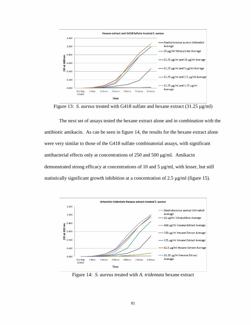
81
Figure 13: S. aureus treated with G418 sulfate and hexane extract (31.25 µg/ml)
The next set of assays tested the hexane extract alone and in combination with the
antibiotic amikacin. As can be seen in figure 14, the results for the hexane extract alone
were very similar to those of the G418 sulfate combinatorial assays, with significant
antibacterial effects only at concentrations of 250 and 500 µg/ml. Amikacin
demonstrated strong efficacy at concentrations of 10 and 5 µg/ml, with lesser, but still
statistically significant growth inhibition at a concentration of 2.5 µg/ml (figure 15).
Figure 14: S. aureus treated with A. tridentata hexane extract

82
Figure 15: S. aureus treated with amikacin
In combination (figures 16-20), the efficacy of growth inhibition was again
visibly improved. Growth inhibition by amikacin at a concentration of 1.25 µg/ml
became statistically significant compared to the untreated control when combined with
extract at concentrations of 62.5 µg/ml and greater, and in comparison to the antibiotic
alone at this concentration at extract levels of 125µg/ml and greater.
Figure 16: S. aureus treated with amikacin and hexane extract (500 µg/ml)

83
Figure 17: S. aureus treated with amikacin and hexane extract (250 µg/ml)
Figure 18: S. aureus treated with amikacin and hexane extract (125 µg/ml)

84
Figure 19: S. aureus treated with amikacin and hexane extract (62.5 µg/ml)
Figure 20: S. aureus treated with amikacin and hexane extract (31.25 µg/ml)
The next set of assays tested the hexane extract alone and in combination with the
antibiotic ampicillin. As can be seen in figure 21, the results for the hexane extract alone
were very similar to those of the previous assays, though inhibition at the concentration
of 125 µg/ml was also statistically significant in these assays. Growth inhibition by

85
ampicillin (figure 22), though not comparable to the positive control, was statistically
significant at all concentrations tested.
Figure 21: S. aureus treated with A. tridentata hexane extract
Figure 22: S. aureus treated with ampicillin
In combination (figures 23-27), antibacterial efficacy was again enhanced. The
lower concentrations of 0.625 and 1.25 µg/ml for example, showed significantly
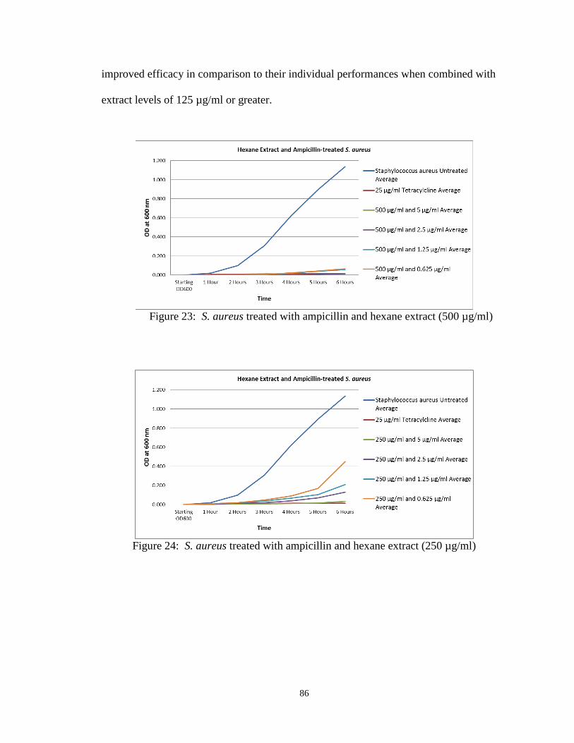
86
improved efficacy in comparison to their individual performances when combined with
extract levels of 125 µg/ml or greater.
Figure 23: S. aureus treated with ampicillin and hexane extract (500 µg/ml)
Figure 24: S. aureus treated with ampicillin and hexane extract (250 µg/ml)
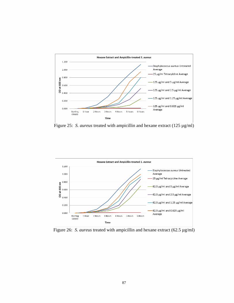
87
Figure 25: S. aureus treated with ampicillin and hexane extract (125 µg/ml)
Figure 26: S. aureus treated with ampicillin and hexane extract (62.5 µg/ml)

88
Figure 27: S. aureus treated with ampicillin and hexane extract (31.25 µg/ml)
The ethyl acetate extract was the next to be assayed in combination with these
antibiotics, beginning with G418 sulfate. As can be seen in figure 28, the ethyl acetate
extract alone displayed stronger antibacterial efficacy than did the hexane extract, with
statistically significant growth inhibition at concentrations as low as 62.5 µg/ml. G418
sulfate performed as it did before (figure 29), with significant inhibition at levels of 2.5
µg/ml and greater.
Figure 28: S. aureus treated with A. tridentata ethyl acetate extract

89
Figure 29: S. aureus treated with G418 sulfate
In combination (figures 30-34), G418 sulfate at a concentration of 1.25 µg/ml,
which alone was ineffective, exhibited significant growth inhibition in comparison to
both the untreated control and the antibiotic alone at this level with extract concentrations
as low as 62.5 µg/ml. This combinatorial effect in comparison to the antibiotic alone is
greater than that observed with the hexane extract and G418 sulfate.
Figure 30: S. aureus treated with G418 sulfate and ethyl acetate extract (500 µg/ml)

90
Figure 31: S. aureus treated with G418 sulfate and ethyl acetate extract (250 µg/ml)
Figure 32: S. aureus treated with G418 sulfate and ethyl acetate extract (125 µg/ml)

91
Figure 33: S. aureus treated with G418 sulfate and ethyl acetate extract (62.5 µg/ml)
Figure 34: S. aureus treated with G418 sulfate and ethyl acetate extract (31.25 µg/ml)
When tested alone (figures 35 and 36), both the ethyl acetate extract and amikacin
performed as they had done previously, with significant inhibition seen at concentrations
as low as 62.5 µg/ml by the extract, and at levels as low as 2.5 µg/ml for the antibiotic.

92
Figure 35: S. aureus treated with A. tridentata ethyl acetate extract
Figure 36: S. aureus treated with amikacin
At 1.25 µg/ml amikacin, growth inhibition became statistically significant in
comparison to both the untreated control and the antibiotic alone at this level with extract
concentrations of 62.5 µg/ml or greater (figures 37-41). Again, this combinatorial effect
in comparison to the antibiotic alone was greater than that observed for amikacin with the
hexane extract.

93
Figure 37: S. aureus treated with amikacin and ethyl acetate extract (500 µg/ml)
Figure 38: S. aureus treated with amikacin and ethyl acetate extract (250 µg/ml)
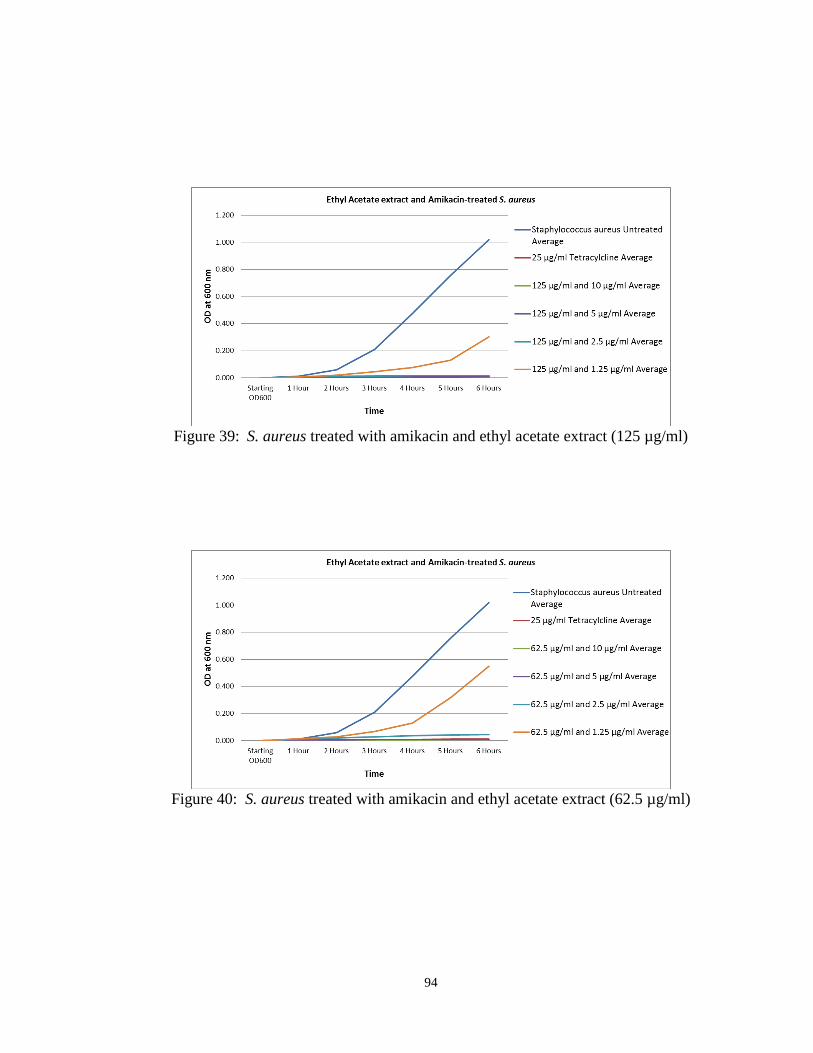
94
Figure 39: S. aureus treated with amikacin and ethyl acetate extract (125 µg/ml)
Figure 40: S. aureus treated with amikacin and ethyl acetate extract (62.5 µg/ml)

95
Figure 41: S. aureus treated with amikacin and ethyl acetate extract (31.25 µg/ml)
When tested with ampicillin, the ethyl acetate extract (figure 42) again performed
as before, with significant inhibition at concentrations as low as 62.5 µg/ml, while the
antibiotic (figure 43) again displayed significant antibacterial effects at all concentrations
tested.
Figure 42: S. aureus treated with A. tridentata ethyl acetate extract

96
Figure 43: S. aureus treated with ampicillin
Interestingly, though also demonstrating enhanced growth inhibition, the
combinatorial effects observed for the ethyl acetate extract with ampicillin were less
pronounced than they were for the hexane extract with this antibiotic, despite the
seemingly superior growth inhibition of the ethyl acetate extract alone (figures 44-48).
At the lower ampicillin concentrations of 0.625 and 1.25 µg/ml, improved efficacy was
only significant in comparison to the antibiotic alone at these levels with extract
concentrations of 250 and 500 µg/ml.
Figure 44: S. aureus treated with ampicillin and ethyl acetate extract (500 µg/ml)
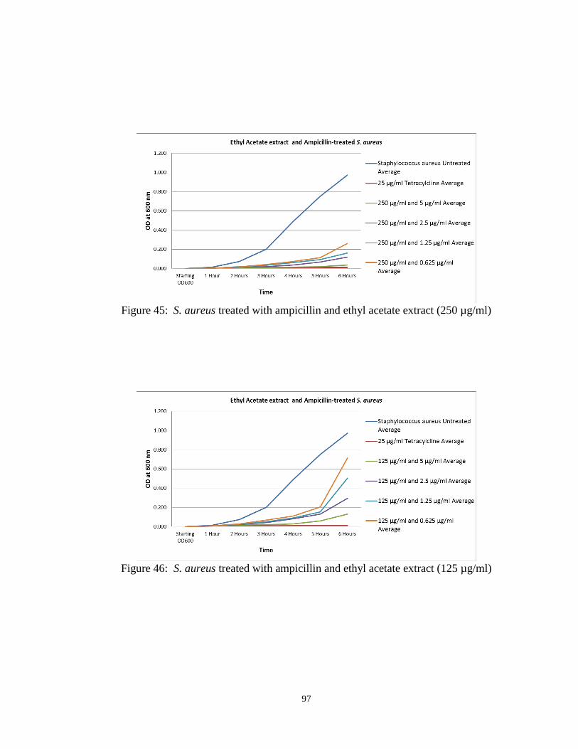
97
Figure 45: S. aureus treated with ampicillin and ethyl acetate extract (250 µg/ml)
Figure 46: S. aureus treated with ampicillin and ethyl acetate extract (125 µg/ml)

98
Figure 47: S. aureus treated with ampicillin and ethyl acetate extract (62.5 µg/ml)
Figure 48: S. aureus treated with ampicillin and ethyl acetate extract (31.25 µg/ml)
Finally, the antibiotics were tested alone and in combination with the water
extract. As can be seen in figure 49, the water extract alone demonstrated lesser
antibacterial efficacy than did the other two extracts, with significant inhibition observed
only at a concentration of 500 µg/ml. The antibiotic G418 sulfate (figure 50) alone
performed in a manner similar to that observed in the preceding assays, yet with
statistically significant inhibition seen only at concentrations of 5 and 10 µg/ml.
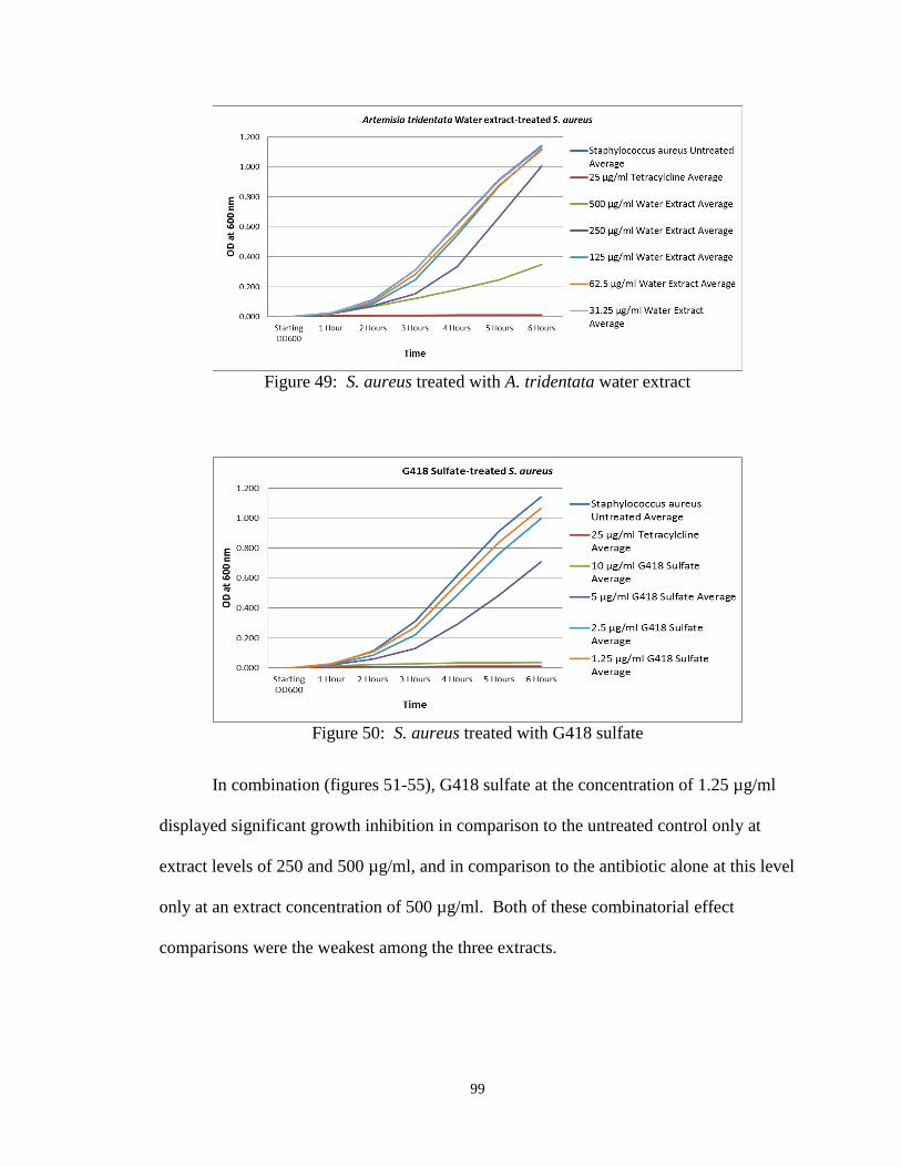
99
Figure 49: S. aureus treated with A. tridentata water extract
Figure 50: S. aureus treated with G418 sulfate
In combination (figures 51-55), G418 sulfate at the concentration of 1.25 µg/ml
displayed significant growth inhibition in comparison to the untreated control only at
extract levels of 250 and 500 µg/ml, and in comparison to the antibiotic alone at this level
only at an extract concentration of 500 µg/ml. Both of these combinatorial effect
comparisons were the weakest among the three extracts.
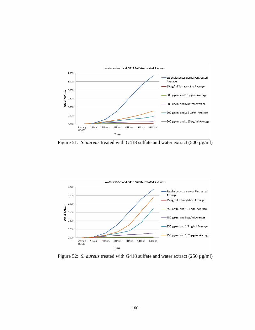
100
Figure 51: S. aureus treated with G418 sulfate and water extract (500 µg/ml)
Figure 52: S. aureus treated with G418 sulfate and water extract (250 µg/ml)

101
Figure 53: S. aureus treated with G418 sulfate and water extract (125 µg/ml)
Figure 54: S. aureus treated with G418 sulfate and water extract (62.5 µg/ml)

102
Figure 55: S. aureus treated with G418 sulfate and water extract (31.25 µg/ml)
When tested with amikacin, the water extract alone (figure 56) performed slightly
better, with significant growth inhibition seen at both 250 and 500 µg/ml. Amikacin
performance was similar to previous assays (figure 57), though in this case inhibition at
the 1.25 µg/ml level was statistically significant.
Figure 56: S. aureus treated with A. tridentata water extract

103
Figure 57: S. aureus treated with amikacin
In combination (figures 58-62), inhibition by amikacin at a concentration of 1.25
µg/ml became significant in comparison to the antibiotic alone at this level with extract
levels of 125 µg/ml and greater. This level of enhancement is similar to that observed for
amikacin with the hexane extract, though still less than that seen when this antibiotic was
combined with the ethyl acetate extract.
Figure 58: S. aureus treated with amikacin and water extract (500 µg/ml)
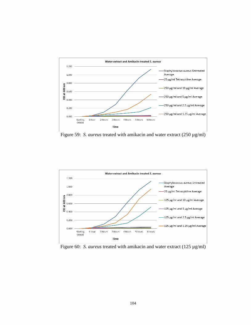
104
Figure 59: S. aureus treated with amikacin and water extract (250 µg/ml)
Figure 60: S. aureus treated with amikacin and water extract (125 µg/ml)

105
Figure 61: S. aureus treated with amikacin and water extract (62.5 µg/ml)
Figure 62: S. aureus treated with amikacin and water extract (31.25 µg/ml)
Finally, when tested with ampicillin, the water extract alone (figure 63) again
showed significant inhibition only at concentrations of 250 and 500 µg/ml, while the
antibiotic (figure 64) once again showed some inhibition at all concentrations tested,
though in this case the effect was not significant at a concentration of 0.625 µg/ml.

106
Figure 63: S. aureus treated with A. tridentata water extract
Figure 64: S. aureus treated with ampicillin
In combination (figures 65-69), inhibition by ampicillin at 0.625 µg/ml became
significant in comparison to the untreated control with extract levels of 62.5 µg/ml and
greater. However, improved growth inhibition by ampicillin both at this concentration
and at 1.25 µg/ml became significant in comparison to these antibiotic concentrations
alone only when combined with extract levels of 250 or 500 µg/ml. This latter result is
similar to that observed for the combination of ampicillin with the ethyl acetate extract,
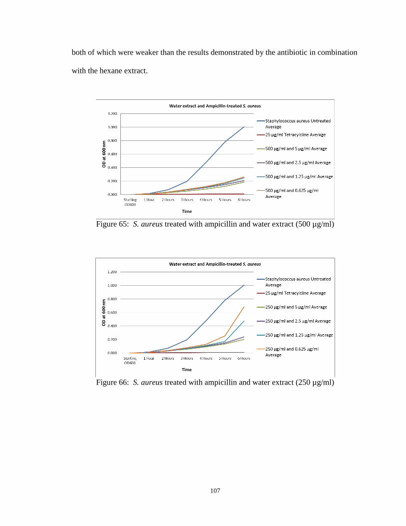
107
both of which were weaker than the results demonstrated by the antibiotic in combination
with the hexane extract.
Figure 65: S. aureus treated with ampicillin and water extract (500 µg/ml)
Figure 66: S. aureus treated with ampicillin and water extract (250 µg/ml)

108
Figure 67: S. aureus treated with ampicillin and water extract (125 µg/ml)
Figure 68: S. aureus treated with ampicillin and water extract (62.5 µg/ml)

109
Figure 69: S. aureus treated with ampicillin and water extract (31.25 µg/ml)

110
Time-Extended Assays
Using selected concentrations of the extracts and antibiotics, further assays were
performed to approximate the length of time over which the Artemisia tridentata extracts
were capable of enhancing the antibacterial efficacy of the antibiotics ampicillin,
amikacin and G418 sulfate versus Staphylococcus aureus. The experimental conditions
were identical to those used in the immediately above described antibacterial assays.
Each of the extracts was tested alone at a concentration of 500 µg/ml. Each of the
antibiotics was tested alone at concentrations of 5 and 2.5 µg/ml, as these levels, when
used in the previous assays, demonstrated both intermediate bacterial growth inhibitory
efficacy by themselves, as well as improved performance when combined with the
extracts. Finally, each antibiotic at each of these two concentrations was tested in
combination with each extract (500 µg/ml). OD600 measurements were taken
immediately following sample treatment and bacterial inoculation, and at hourly time
points for twelve hours. Again, the averaged results from each of the three assays were
then combined and averaged together. Statistical analysis was performed on the 12-hour
time point values using an unpaired t-test assuming equal variances. P values of less than
0.05 were considered statistically significant.
As can be seen in figures 70 and 71 below, among the three extracts when tested
alone, the hexane extract appears to have demonstrated the strongest antibacterial
efficacy through six hours, a result consistent with previous assays at the extract
concentration of 500 µg/ml. The hexane and ethyl acetate extracts continued to display
statistically significant growth inhibition through twelve hours in comparison to the
untreated control, though these effects were minimal. Among the antibiotics when tested
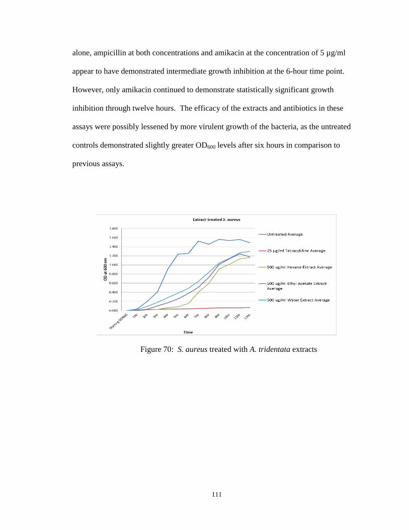
111
alone, ampicillin at both concentrations and amikacin at the concentration of 5 µg/ml
appear to have demonstrated intermediate growth inhibition at the 6-hour time point.
However, only amikacin continued to demonstrate statistically significant growth
inhibition through twelve hours. The efficacy of the extracts and antibiotics in these
assays were possibly lessened by more virulent growth of the bacteria, as the untreated
controls demonstrated slightly greater OD600 levels after six hours in comparison to
previous assays.
Figure 70: S. aureus treated with A. tridentata extracts

112
Figure 71: S. aureus treated with G418 sulfate, amikacin, ampicillin
Still, in combination, as can be seen in figures 72-74, antibacterial efficacy was
again improved. G418 sulfate growth inhibition at the 12-hour time point became
statistically significant in comparison to the untreated control when combined with each
extract, with the growth curves of samples treated with G418 sulfate and either the
hexane or ethyl acetate extracts resembling that of the positive control. Enhancement of
growth inhibition by all extracts was also statistically significant in comparison to the
antibiotic alone at both concentrations.
Similarly, the growth curves of samples treated with amikacin at 5 µg/ml, in
combination with either the hexane or ethyl acetate extracts, resembled that of the
positive control, with the enhancement of growth inhibition statistically significant in
comparison to the antibiotic alone at this concentration. However, the water extract
significantly reduced the efficacy of amikacin at 5 µg/ml. At 2.5 µg/ml, growth
inhibition by amikacin became significant in comparison to the untreated control when
combined with the hexane or ethyl acetate extracts, but not the water extract, again with

113
the growth curves of samples treated with combinations of amikacin and the hexane or
ethyl acetate extracts resembling that of the positive control. While the enhancement of
growth inhibition by the combination of amikacin at 2.5 µg/ml with all of the extracts
was statistically significant in comparison to the antibiotic alone at this concentration, it
can be seen that the effect of the water extract was minimal.
Extract enhancement of growth inhibition by ampicillin was less pronounced.
At 5 µg/ml, growth inhibition in comparison to the untreated control only became
statistically significant when ampicillin was combined with the hexane extract. Though
enhancement of growth inhibition by all extracts was statistically significant in
comparison to ampicillin alone at this concentration, this is attributable to the high final
OD600 level of the ampicillin-treated sample, and it can be seen from the figure that only
the hexane extract noticeably improved antibiotic efficacy. At 2.5 µg/ml, similar results
were observed, with only the hexane extract significantly improving the efficacy of
amikacin in comparison to the untreated control, or the antibiotic alone at this
concentration.

114
Figure 72: S. aureus treated with G418 sulfate and A. tridentata extracts
Figure 73: S. aureus treated with amikacin and A. tridentata extracts
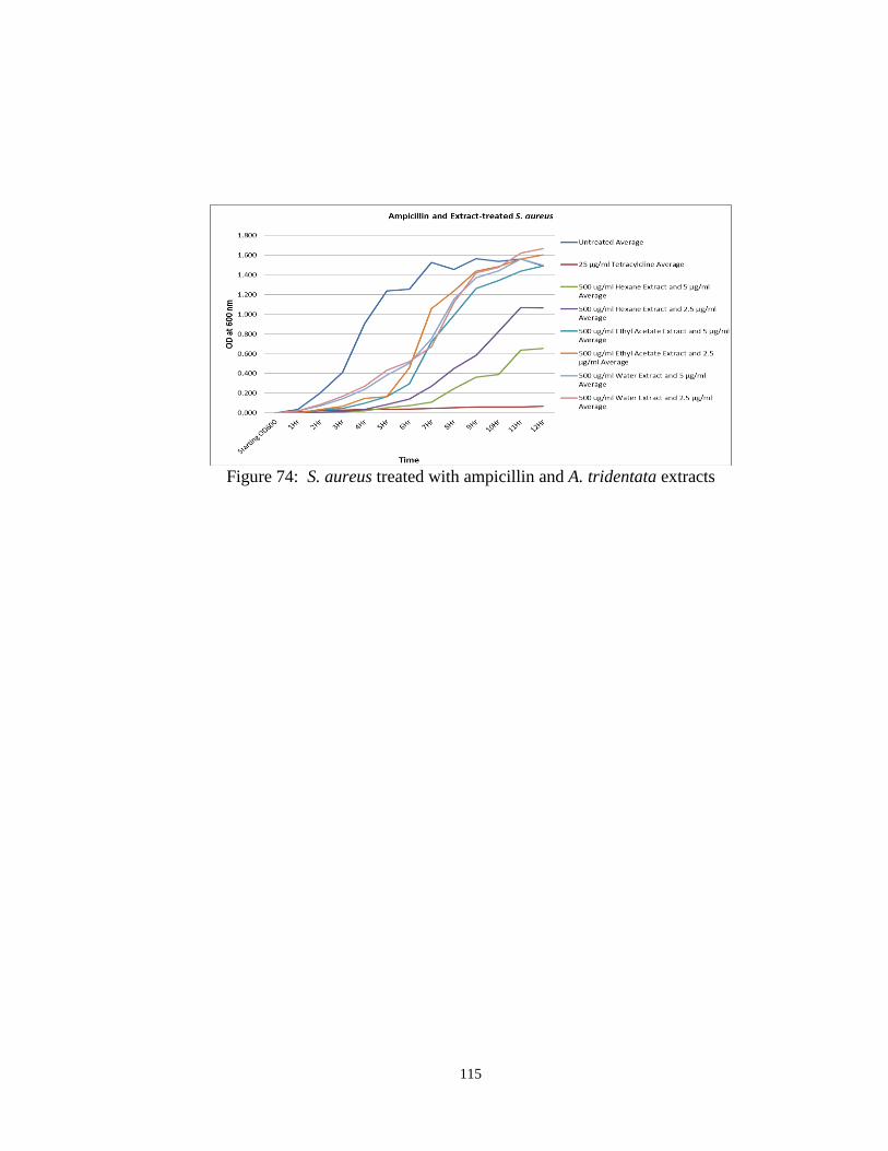
115
Figure 74: S. aureus treated with ampicillin and A. tridentata extracts

116
Extracts in Combination
The extracts were also assayed in combination with each other to observe for any
negating or enhancement of antibacterial efficacy which might indicate the presence of
antagonistic, additive or synergistic compounds present in the original plant material.
Experimental conditions were the same as those described above. Each extract was
tested alone at concentrations of 500, 250, 125 and 62.5 µg/ml, and in combination at
concentrations of 250, 125, 62.5 and 31.25 µg/ml.
As can be seen in figures 75-77, the performances of the extracts alone were
similar to those observed in previous assays, with statistically significant growth
inhibition observed at all hexane and ethyl acetate extract concentrations, and at water
extract concentrations of 500 and 250 µg/ml.
Figure 75: S. aureus treated with A. tridentata hexane extract

117
Figure 76: S. aureus treated with A. tridentata ethyl acetate extract
Figure 77: S. aureus treated with A. tridentata water extract
Using the same method of statistical analysis, the OD600 values measured at the 6-
hour time point for each pairing of extracts was compared to the average of the values of
each extract alone at the total extract concentration. For example, the final reading for
the combination of the ethyl acetate and hexane extracts (figure 78) at 250 µg/ml each

118
was compared to the average between the final readings for the ethyl acetate and hexane
extracts alone when tested at 500 µg/ml. Though the dose-response relationships
between the extracts and growth inhibition in these or previous assays do not appear to
have demonstrated perfectly linear correlations, it would be reasonable to expect that
dramatic antagonistic, additive or synergistic effects between extracts might result in
observable effects on the growth curves of treated samples. In the case of the hexane and
ethyl acetate extracts, this combination showed significant inhibition versus the untreated
control only at concentrations of 125 and 250 µg/ml each, with statistically significant
diminishment of antibacterial efficacy in comparison to the averages of the extracts alone
at all concentrations tested.
Figure 78: S. aureus treated with A. tridentata hexane and ethyl acetate extracts
The hexane and water extracts in combination also demonstrated significant
growth inhibition at concentrations of 125 and 250 µg/ml each (figure 79). While the
readings for this extract pairing at lesser concentrations were significantly larger than the
average values from each extract alone, at 250 µg/ml each, diminishment was minimal.

119
Figure 79: S. aureus treated with A. tridentata hexane and water extracts
Finally, in the case of the combination of the ethyl acetate and water extracts
(figure 80), significant inhibition in comparison to the untreated control was observed at
concentrations of 62.5 µg/ml and greater. Though there was a slight but statistically
significant diminishment of antibacterial efficacy at extract concentrations of 250 µg/ml
each in comparison to the averaged values of each extract at 500 µg/ml, lower
combinatorial values were similar to what was anticipated.
Figure 80: S. aureus treated with A. tridentata ethyl acetate and water extracts

120
Ampicillin-Resistant Staphylococcus aureus
To determine if these extracts also possess growth inhibitory efficacy alone and/or
in combination with a synthetic antibiotic versus a drug-resistant strain of Staphylococcus
aureus, antibacterial assays were conducted using SA-BAA-44, an ampicillin-resistant
strain of this bacterial species. Experimental conditions were similar to those described
above for the previous extract/antibiotic combinatorial assays. However, extracts were
only tested alone at concentrations of 500, 250, 125 and 62.5 µg/ml, with the previously
used lowest extract concentration of 31.25 µg/ml excluded. Also, ampicillin was tested
alone at considerably higher concentrations of 800, 400, 200 and 100 µg/ml, as these
levels were determined in a separate experiment as necessary to demonstrate intermediate
growth inhibitory effects (data not shown). Each extract, at a concentration of 500 µg/ml,
was also tested in combination with each concentration of ampicillin. The method of
statistical analysis employed, using the 6-hour time point readings, remained the same.
As can be seen in the following figures, growth of the untreated BAA-44 strain
was substantially less than that of the antibiotic-susceptible strain, despite identical
culture conditions. The hexane extract appears to have retained some inhibitory efficacy
(figure 81), though this was only statistically significant at a concentration of 500 µg/ml.
The ethyl acetate extract (figure 82) was also only significantly effective at the maximum
concentration assayed. The water extract (figure 83) demonstrated no significant
antibacterial efficacy at any concentration.

121
Figure 81: S. aureus BAA-44 treated with hexane extract
Figure 82: S. aureus BAA-44 treated with ethyl acetate extract

122
Figure 83: S. aureus BAA-44 treated with water extract
Ampicillin alone (figure 84) demonstrated statistically significant efficacy versus
strain BAA-44 only at a concentration of 800 µg/ml. However, in combination with the
hexane extract at a concentration of 500 µg/ml (figure 85), growth inhibition became
significant in comparison to both the untreated control and each ampicillin concentration
alone, with growth curves resembling those of the positive control. In combination with
the ethyl acetate extract (figure 86), growth inhibition again became statistically
significant at all antibiotic concentrations using these comparisons, though the flattening
of the growth curves of treated samples was less impressive than that of the hexane
extract/antibiotic combinatorial samples. The water extract, by contrast, failed to
demonstrate any statistically significant enhancement of antibiotic efficacy (figure 87).

123
Figure 84: S. aureus BAA-44 treated with ampicillin
Figure 85: S. aureus BAA-44 treated with ampicillin and hexane extract

124
Figure 86: S. aureus BAA-44 treated with ampicillin and ethyl acetate extract
Figure 87: S. aureus BAA-44 treated with ampicillin and water extract

125
Biofilm Formation Assays
In order to explore their potential in preventing Staphylococcus aureus biofilm
formation on solid surfaces, the Artemisia tridentata extracts (hexane, ethyl acetate,
water) were assayed for this ability using COSTAR brand 96-well polystyrene plates. All
wells contained a final volume of 100 µl prior to inoculation, and all sample types were
prepared in triplicate. Two sets of wells were excluded from inoculation, and contained
only LB. The first served to ensure sterility of the media and the plate, while the second
served as a background control, measuring stain binding to the plate in the absence of a
biofilm. Each extract was tested at concentrations of 500, 250 and 125 µg/ml in LB.
DMSO in LB at 0.5% matched the dilution level of the highest extract concentration, and
served as a vehicle control. Untreated samples served as a negative control, while 3%
Nonidet P40 in LB served as a positive control. Overnight cultures of Staphylococcus
aureus were diluted to OD600 levels of between 0.950 and 1.050, as in previous assays.
However, 5 µl of this diluted culture was added to the appropriate wells on the plate, as 2
µl inoculums failed to demonstrate consistent biofilm formation in the untreated control
samples in a previous assay (data not shown).
As the experimental design did not cause more than half the wells in the plate to
be occupied, the setup was repeated a second time on the lower half of the plate, with the
exception of the sterility control wells and the background stain binding control wells,
thus providing two sets of samples in the event that one failed. The plate was incubated
without rotation for approximately 10 hours at about 37°C. Spectrophotometric readings
of the plate were taken at 600 nm before and after incubation to detect media or plate

126
contamination in the sterility control wells. Following incubation, the plate contents were
decanted, and the plate rinsed three times in water. 100 µl of a 0.1% crystal violet
solution was added to the wells, and the plate incubated at room temperature for about 15
min. The plate contents were again decanted, the plate rinsed seven times in water, and
the wells allowed to air-dry overnight in an open environment.
The following day, 100 µl of a 95% ethanol solution was added to all of the wells
except the sterility control wells, and a spectrophotometric measurement was taken at 595
nm to measure crystal violet retention. The averaged readings from the background stain
binding control wells were subtracted from the values of the other samples, with negative
numbers considered as readings of zero. The results from the six sets of samples are
averaged in table 2 below, where it can be seen that none of the extracts demonstrated
inhibition of Staphylococcus aureus biofilm formation under these experimental
conditions. In fact, the extract-treated wells routinely retained more stain than did the
untreated controls and DMSO-treated samples, with several results excluded due to
readings too high to be accurately measured by the spectrophotometer.

127
Table 2: Biofilm formation assay results
Starting
OD60010Hr OD595
Untreated Average 0.100 1.326 1.232
Nonidet P40 3% solution Average 0.289 0.000 0.051
Staphylococcus aureus and 0.5% DMSO Average 0.106 1.358 1.363
Staphylococcus aureus and 500 ug/ml Hexane Extract Average 0.300 1.082 1.853
Staphylococcus aureus and 250 ug/ml Hexane Extract Average 0.197 1.247 1.953
Staphylococcus aureus and 125 ug/ml Hexane Extract Average 0.151 1.392 1.681
Staphylococcus aureus and 500 ug/ml Ethyl Acetate Extract Average 0.275 0.797 2.746
Staphylococcus aureus and 250 ug/ml Ethyl Acetate Extract Average 0.188 1.173 2.759
Staphylococcus aureus and 125 ug/ml Ethyl Acetate Extract Average 0.141 1.363 2.417
Staphylococcus aureus and 500 ug/ml Water Extract Average 0.131 0.870 2.601
Staphylococcus aureus and 250 ug/ml Water Extract Average 0.126 1.331 1.806
Staphylococcus aureus and 125 ug/ml Water Extract Average 0.119 1.476 1.714

128
Static Biofilm Assays
Additional assays were performed to determine the antibacterial efficacy of the
Artemisia tridentata extracts and the antibiotics ampicillin, amikacin or G418 sulfate,
alone or in combination, versus static Staphylococcus aureus biofilms. As in the biofilm
formation assays, COSTAR brand polystyrene 96-well polystyrene plates were used, and
sample types were prepared in triplicate. 100 µl LB was added to all wells of the plate to
be used in the assay. An overnight culture of Staphylococcus aureus was diluted in LB to
an OD600 level between 0.950-1.050, and 5 µl of the diluted culture was added to all LB-
containing wells, excluding the first set of three. The plate was then incubated without
rotation at approximately 37°C for about 10 hours. OD600 measurements of the first three
wells, taken before and after incubation, served to ensure sterility of the media and the
plate.
Following incubation, treatments were added to the static biofilms. The sterility
control wells, to which no bacteria was added, also served to measure background stain
binding to the plate in the absence of a biofilm. Therefore no treatment of these wells
was necessary. The second and third sets of wells served as negative controls. To the
second set of wells, nothing was added, while 100 µl fresh LB was added to the wells of
the third set.
As the extracts and antibiotics were to be added already pre-diluted to twice their
desired final concentrations in 100µl volumes of LB, the purpose of this differential
treatment of negative controls was to determine if leaving the untreated controls without
an added 100µl fresh LB during the period of sample treatment would result in cell death.
This could result in reduced biofilm stain binding, possibly masking extract or antibiotic

129
biofilm-removal effects. Alternatively, adding 100 µl LB could result in additional
growth of the biofilm during the sample treatment period, possibly overstating extract or
antibiotic effects. Therefore both possibilities needed to be explored. Each extract and
antibiotic, pre-diluted in LB, was added and tested alone at final concentrations of 500
µg/ml and 1 mg/ml, respectively. Each extract and antibiotic were also tested in
combination. Qiagen Lysis buffer, containing SDS and sodium hydroxide, served as a
positive control. Following sample treatment, the plate was returned to incubation under
the same conditions for approximately twelve hours.
The following day, the plate was washed and stained in a manner similar to the
biofilm formation assays described above. As can be seen in table 3 below, results for
the untreated controls were relatively similar regardless of the addition of fresh LB
during sample treatment. However, none of the extracts or antibiotics by themselves,
with the exception of ampicillin, demonstrated any reduction in stain retention in
comparison to the untreated controls. Also, in combination, only the water extract
applied together with ampicillin demonstrated any biofilm reduction, but this result was
almost identical to that of ampicillin alone. Thus, the assay was abandoned after two
repetitions.

130
Table 3: Static biofilm assay results

131
Serial Dilution and Plating
In an effort to determine whether the Artemisia tridentata extract antibacterial
effects observed in earlier assays were bactericidal or bacteriostatic in nature, samples of
Staphylococcus aureus cultures were serially diluted and plated after extract treatment to
perform viable cell counts. Overnight cultures were first diluted to an OD600 level of
approximately 1.000. A 50 µl sample was removed and mixed with a 450 µl volume of
LB for a 1:10 dilution. 50 µl of this mixture was removed and mixed in another 450 µl
volume of LB, and the process repeated until a final 1 x 10⁻⁶
dilution was achieved. The
entirety of the last dilution was plated on an LB agar plate, and served as a measure of the
number of Staphylococcus aureus cells represented by an OD600 measurement
approximating 1.000.
To test extract antibacterial effects, 10 µl of the dilution of the overnight culture
(OD600 level of approximately 1.000) was added to each of three tubes containing an
Artemisia tridentata extract (hexane, ethyl acetate, water) at a concentration of 500 µg/ml
in 500 µl volumes of LB, thus keeping consistent with the inoculum to media ratio used
in the 96-well plate antibacterial assays (2 µl culture to 100 µl LB). A fourth tube
containing tetracycline at a concentration of 25 µg/ml, also in a 500 µl volume of LB,
served as a positive control, while two untreated tubes containing only 500 µl LB each
served as negative controls. 1 ml LB in a sixth tube was included as a sterility control.
All tubes were incubated at approximately 37°C and 125 rpm.
After about 25 min, the extract-treated samples, the tetracycline-treated sample
and one of the untreated samples were removed from incubation. This time point was
chosen based on a previous assay which indicated the doubling time of Staphylococcus
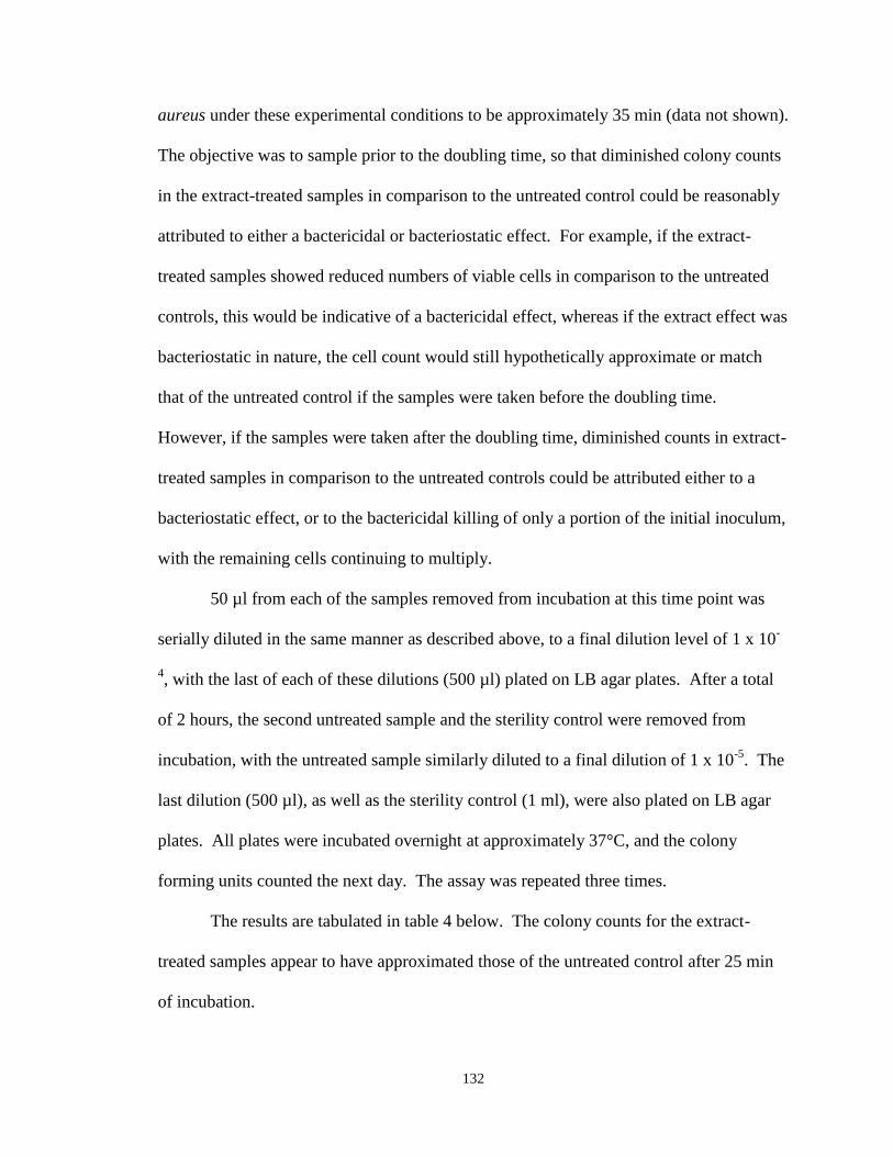
132
aureus under these experimental conditions to be approximately 35 min (data not shown).
The objective was to sample prior to the doubling time, so that diminished colony counts
in the extract-treated samples in comparison to the untreated control could be reasonably
attributed to either a bactericidal or bacteriostatic effect. For example, if the extract-
treated samples showed reduced numbers of viable cells in comparison to the untreated
controls, this would be indicative of a bactericidal effect, whereas if the extract effect was
bacteriostatic in nature, the cell count would still hypothetically approximate or match
that of the untreated control if the samples were taken before the doubling time.
However, if the samples were taken after the doubling time, diminished counts in extract-
treated samples in comparison to the untreated controls could be attributed either to a
bacteriostatic effect, or to the bactericidal killing of only a portion of the initial inoculum,
with the remaining cells continuing to multiply.
50 µl from each of the samples removed from incubation at this time point was
serially diluted in the same manner as described above, to a final dilution level of 1 x 10-
4, with the last of each of these dilutions (500 µl) plated on LB agar plates. After a total
of 2 hours, the second untreated sample and the sterility control were removed from
incubation, with the untreated sample similarly diluted to a final dilution of 1 x 10⁻⁵
. The
last dilution (500 µl), as well as the sterility control (1 ml), were also plated on LB agar
plates. All plates were incubated overnight at approximately 37°C, and the colony
forming units counted the next day. The assay was repeated three times.
The results are tabulated in table 4 below. The colony counts for the extract-
treated samples appear to have approximated those of the untreated control after 25 min
of incubation.

133
Table 4: Serial and dilution of A. tridentata extract-treated S. aureus
1st Assay 2nd Assay 3rd Assay Average
Overnight Culture 2.72 x 10⁸
9.92 x 10⁷
6.29 x 10⁷
1.45 x 10⁸
Untreated (25 min) 6.64 x 10⁶
3.00 x 10⁶
1.72 x 10⁶
3.79 x 10⁶
Tetracycline [25 µg/ml] 6.00 x 10⁴
2.00 x 10⁴
4.40 x 10⁵
1.73 x 10⁵
Hexane extract [500 µg/ml] 6.18 x 10⁶
2.16 x 10⁶
1.60 x 10⁶
3.31 x 10⁶
Ethyl acetate extract [500 µg/ml] 6.62 x 10⁶
3.44 x 10⁶
2.16 x 10⁶
4.07 x 10⁶
Water extract [500 µg/ml] 5.30 x 10⁶
2.58 x 10⁶
1.62 x 10⁶
3.17 x 10⁶
Untreated (2 hr) 4.68 x 10⁷
2.44 x 10⁷
1.28 x 10⁷
2.80 x 10⁷

134
Toxicity Assay
To determine their potential toxicity to humans, a line of breast cancer cells
(MDA-MB-231 ATCC-HTB-26) was treated with various concentrations of the
Artemisia tridentata ethyl acetate extract. Cell samples (5 x 10⁵) were cultured in
Dulbecco’s modified Eagle’s medium (Fisher Scientific), containing 10% fetal bovine
serum, 0.1 mg/mL streptomycin, 100 U/mL penicillin, and 0.25 μg/mL amphotericin B at
37 °C in a humidified CO#-controlled (5%) incubator. The samples were then treated
with various concentrations of the extract (50, 100, 200, 400 or 600 ug/ml) for a period of
24 hours. Untreated samples served as negative controls, while DMSO-treated samples
(0.1%) served as vehicle controls. Following extract treatment, cells were washed with
PBS, and 2 ml serum-free culture media containing 1 mg/ml of the tetrazolium salt [3-
(4,5-dimethylthiazol-2-yl)-2,5-diphenyltetrazolium bromide] (Sigma-Aldrich) was added
to each well. After 4 hours, the media was discarded, and DMSO was added to dissolve
MTT-derived formazan. The level of formazan was quantified by measurement of
absorbance at a wavelength of 550 nm. The assay was performed in triplicate.
The amount of formazan in extract-treated samples was calculated as a percentage
of the formazan in the untreated controls. As can be seen in figure 88, the percentage of
viable cells, as indicated by conversion of the tetrazolium salt to formazan, decreased
with increasing concentrations of the ethyl acetate extract, with concentrations of 400 and
600 µg/ml reducing the level of formazan converted to less than 10% of that of the
untreated control. DMSO had little to no effect (data not shown).
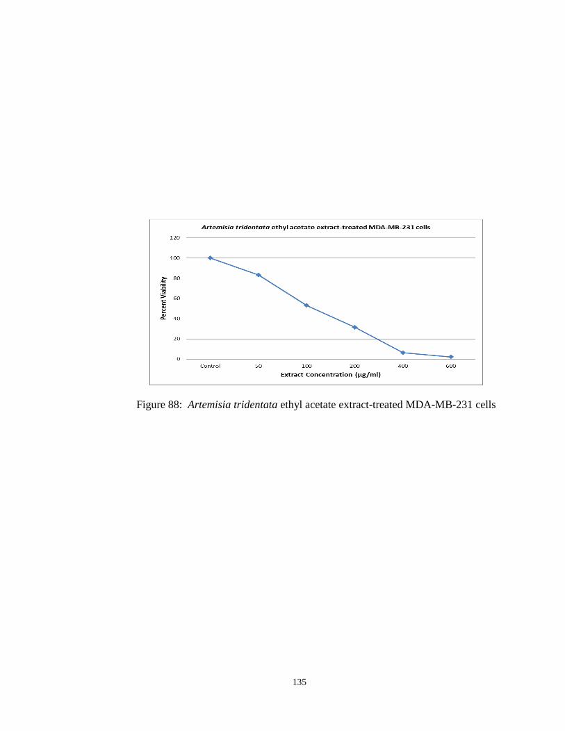
135
Figure 88: Artemisia tridentata ethyl acetate extract-treated MDA-MB-231 cells

136
pH Measurement
The pH of extract-containing LB samples was measured to investigate the
possibility that the antibacterial effects previously demonstrated were due to changes in
media acidity or basicity. Each of the three Artemisia tridentata extracts (hexane, ethyl
acetate, water) was diluted to a concentration of 500 µg/ml in a 50 ml volume of LB, and
the pH of each sample measured with a Mettler Toledo “SevenEasy” meter. An
equivalent volume of untreated LB from the same preparation was also tested to ensure
the media used was in fact at or close to the expected value of pH 7. The untreated LB
provided a pH reading of 6.96. The pH readings for the extract-containing samples were
only slightly reduced, with measurements of 6.95, 6.91 and 6.90 for the hexane, ethyl
acetate and water extracts, respectively.

137
Ethidium Bromide Assays
Ethidium bromide binding assays, based on the work of MA Jabra-Rizk et al111
,
were performed to determine if the mechanism of action by which the extracts exert their
antibacterial effect is through cell membrane disruption. Bacterial cell membrane
disruption should result in the increased intercalation of ethidium bromide to bacterial
DNA in cells exposed to this compound. As in the antibacterial assays, overnight
cultures of Staphylococcus aureus were first diluted in LB to OD600 levels between
0.950-1.050. Several ml were centrifuged for 15 min @ 5k rpm, the supernatant
removed, and the cells suspended in an equal volume of PBS.
500 µl of this cell suspension was added to 500 µl PBS in each of six tubes.
Three of these six tubes each contained one of the Artemisia tridentata extracts (hexane,
ethyl acetate, water) at a concentration of 1 mg/ml, yielding a final concentration of 500
µg/ml following the addition of the cell suspension. The fourth tube contained DMSO at
a concentration of 2%, yielding a 1% final concentration following the addition of the
cell suspension, and served as a vehicle control. A fifth tube containing only PBS served
as a negative control, while the sixth contained the detergent Nonidet P40 at a
concentration of 2%, yielding a 1% final concentration following the addition of the cell
suspension, and served as a positive control.
All samples were incubated at approximately 37°C and 125 rpm for about 30 min.
5 µg ethidium bromide in an aqueous solution was then added to each, and the samples
were allowed to incubate at room temperature for approximately 15 min. Samples were
then centrifuged for 15 min @ 5k rpm, after which the supernatants were removed, and
the cell pellets washed in 1 ml PBS each. The centrifugation step was repeated, the

138
supernatants were again removed, and each pellet was suspended in 3 ml PBS. Samples
were then measured in a spectrofluorometer at 510 nm excitation and 605 nm emission to
detect ethidium bromide binding to DNA. In an effort to account for possible differences
in cell numbers, samples were then measured in a spectrophotometer at 600 nm to
measure bacterial cell density. The assay was repeated in triplicate. As can be seen
below in table 5, the spectrophotometer readings were used to adjust the emission
measurements of each treated sample to that of the untreated control based on cell
density, and a ratio of the emission of the sample to that of the untreated control was then
calculated for each. Ethidium bromide binding to DNA was noticeably enhanced in the
extract-treated samples, both in comparison to the negative control and the Nonidet P40-
treated positive control, with DMSO appearing to have little effect.
Table 5: Ethidium bromide binding assay results
Emission OD600
Adjusted
ValueRatio
Untreated 23.490 0.062 NA NA
1 % Nonidet P-40 106.783 0.121 55.520 2.286
DMSO 14.697 0.049 20.205 0.888
Hexane extract 211.633 0.137 93.610 3.950
Ethyl acetate extract 228.412 0.126 112.913 4.670
Water extract 49.013 0.116 25.412 1.148

139
DNA-nicking Assays
In order to determine if the antibacterial effect of these extracts is the result of a
mechanism of action involving DNA damage, a DNA-nicking assay was performed to
observe for strand cleavage using a construct variant of plasmid pGEX. Plasmid DNA
purified from bacteria is typically in its native supercoiled state. DNA strand damage
results in a loss of supercoiling, which causes a slower migration of the plasmid when
compared to the supercoiled form in an electrophoretic gel.
Five samples were prepared for each assay. To each sample, 2 µl Fisher
OPTIZYME™ 10X Buffer #5 (10 mM Tris-HCl, pH 8.5, 10 mM MgCl2, 100 mM KCl,
0.1 mg/ml BSA) and 1 µl plasmid were added. To three of the samples, an Artemisia
tridentata extract (hexane, ethyl acetate, water) was added to a final concentration of 500
µg/ml. The fourth sample contained untreated plasmid and served as a negative control.
To the fifth was added Fenton’s reagent (10 mM ferrous ammonium sulfate, 10 mM
disodium EDTA, 9 mM hydrogen peroxide final concentrations)112
, which served as a
positive control. All samples contained a total of 20 µl, with DI water constituting the
remaining volume in each.
Samples were incubated in a water bath at about 37°C. After approximately 10
minutes, the positive control was removed and packed in ice. After a total incubation
time of approximately 60 minutes, the remaining samples were removed, and 6 µl of 4X
loading dye was added to all samples. The entirety of each sample was loaded into the
wells of a 1 % agarose gel in 1X TAE buffer, and the gel run at 100 V for 90 minutes.
The gel was stained with 30 µg ethidium bromide in 50 ml water for approximately 30
minutes, and photographed. The assay was repeated in triplicate. As can be seen in

140
figures 89-91 below, treatment with the extracts did not appear to damage the DNA
strands of the plasmid under the conditions assayed. From right to left, the lanes
represent the DNA ladder (λ-DNA-HindIII/φX-HaeIII), the untreated control, the
Fenton’s reagent-treated positive control, the hexane extract, ethyl acetate extract and
water extract-treated samples. Band analysis performed using ImageJ software
confirmed these negative results.
Figure 89: Gel electrophoresis of extract-treated pGEX (1st assay)

141
Figure 90: Gel electrophoresis of extract-treated pGEX (2nd assay)

142
Figure 91: Gel electrophoresis of extract-treated pGEX (3rd assay)

143
Transmission Electron Microscopy
Six 1 ml samples of the ethyl acetate and water Artemisia tridentata extracts in
LB were prepared, each at a final concentration of 500 µg/ml. An overnight culture of
Staphylococcus aureus was diluted to an OD600 reading approximating 1.000, and 20 µl
of this was added to each extract-containing tube. The samples were then incubated for
approximately 1 hour at about 37° C and 125 rpm. Following incubation, the tubes were
centrifuged for 10 min at 8k rpm, the supernatants were removed, and the resultant pellets
for each extract treatment were pooled and washed in 1 ml PBS. The centrifugation was
repeated a second time, and following removal of the supernatants, each pellet was fixed
with 500 µl 2.5 % glutaraldehyde in PBS, and stored at 4°C. Cells added to six tubes
containing LB without extract underwent the same treatment, and served as a negative
control.
A separate procedure was used for the hexane extract, as treated cells repeatedly
failed to form an observable pellet using this protocol. In this case, 200 µl cells were
added to an extract-containing tube. The cells were washed five times with fresh LB,
then fixed with 1ml 2.5% glutaraldehyde in phosphate buffer at 4°C for 1 h. The cells
were then washed three more times using phosphate buffer.
Prior to transmission electron microscopy (TEM), all samples were fixed with 1%
osmium tetroxide for a period of 2 hours. This was followed by three 10 minute
washings with phosphate buffer, and subsequent dehydration in a series of ethanol
concentrations (30%, 50%, 70%, 90% and 95%), for 15 min each. Osmified tissues were
dehydrated in a 95% ethanol/5% acetone solution, followed by absolute acetone, and
embedded in epoxy resin (Epon 812, Pelco—EEUU). Samples were then infiltrated and

144
embedded in Spurr’s resin (Epon 812, Pelco—EEUU). Ultrathin sections were cut with a
diamond knife using an ultramicrotome (Ultracut equipment -Leitz), and then mounted
on bare copper grids. Finally, specimens were counter stained with a 2% (w/v) uranyl
acetate and lead citrate solution for 3 minutes, then with a 0.25% (w/v) solution for 2
minutes, and examined with a Zeiss EM 900 microscopy (Zeiss, Oberkochen, Germany).
As can be seen in figure 92 below, transmission electron microscopy of the
untreated control demonstrated intact cell walls and membranes. A similar result was
observed for the water extract-treated cells (figure 93). However, after treatment with
either the hexane (figure 94) or ethyl acetate (figure 95) extracts, Staphylococcus aureus
cells showed an apparent absence of cytoplasmic contents and unique chromatin
organization (i.e., non-condensed chromatin). Specifically, hexane extract-treated cells
demonstrated damage to membrane integrity, resulting in a partial release of cellular
content (indicated by arrowheads), while ethyl acetate extract-treated cells appear to have
suffered cell wall disruption (indicated by asterisks). The residual cell walls appeared as
bacterial “ghosts”, with a complete absence of cytoplasmic contents, indicating cellular
rupture. Interestingly, these ethyl acetate extract-treated cells also demonstrated cellular
aggregation, or possible fusing of the cells.

145
Figure 92: TEM image of untreated Staphylococcus aureus
Figure 93: TEM image of water extract-treated Staphylococcus aureus
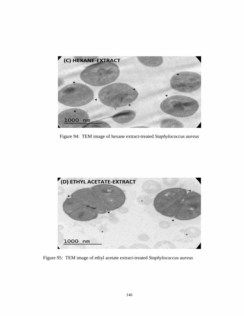
146
Figure 94: TEM image of hexane extract-treated Staphylococcus aureus
Figure 95: TEM image of ethyl acetate extract-treated Staphylococcus aureus

147
GC-MS Analysis
Each of the three extracts was submitted for gas chromatography-mass
spectrometry analysis to determine chemical composition. DMSO served as a control.
20 µl of each sample was mixed with 50 µl methanol and 50 µl dichloromethane.
Sample injection and gas chromatography were performed using an Agilent 6890 gas
chromatograph equipped with an autosampler and an HP-5MS UI capillary column (30m,
0.250 mm, 0.25 µm). 5 µl of each sample was injected at an injection temperature of
320°C, using an injection mode with a split 1:10 ratio. Helium served as the carrier gas
at a constant rate flow of 0.9 ml/min. The oven temperature was held at 60°C for 1 min,
then increased to 320°C at a rate of 5°C, and held at this final temperature for 4 min. An
Agilent 5973 single-quadrupole mass spectrometer was used for detection using electron
impact ionization operating in positive mode, operating under full scan mode (50-450 Da,
1 Hz) with a solvent delay of 7 min.
A total of 60 peaks were identified in the hexane extract sample (figure 96), 93 in
the ethyl acetate extract sample (figure 97), and 87 in the water extract sample (figure
98). Spectral peaks were identified based using the NIST 1998 data library.

148
Figure 96: GC-MS spectral results for Artemisia tridentata hexane extract
Figure 97: GC-MS spectral results for Artemisia tridentata ethyl acetate extract

149
Figure 98: GC-MS spectral results for Artemisia tridentata water extract
Among the three extracts, compounds identified with a probability value of at
least 50 % and a peak area of at least 1 % were tabulated and compared. 43 compounds
in the hexane extract analysis met these criteria (table 6). Of these, seven had peak areas
of at least 5 %. Three of these seven are tentative identifications of the peak detected at
the retention time of 11.04 minutes, with a peak area of 24.93 % and probable
identification values of at least 90%. These three are stereoisomer variations of the first
listed compound, a terpene commonly known as “camphor.” The next two compounds,
with a retention time of 34.12 minutes, a peak area of 13.35 %, and also with probable
identification values of at least 90 %, are again stereoisomers of the same compound, a
sesquiterpene lactone with the molecular formula C15H18O3, commonly referred to as
“achillin” or “azuleno.” The final two compounds are again possible identifications of
the same peak, with a retention time 10.81 minutes and a peak area of 8.49 %, tentatively

150
labeled as either “exo-2-bromonoborane,” or “Bicyclo[2.2.1]heptane, 2-(2-propenyl)-,“
the latter of which is also known as “2-(2-Methyl-1-propenyl)-bicyclo[2.2.1]heptane.”
However, these two compounds have probable identification values of only 50 and 59 %,
respectively.

151
Table 6: Summary of GC-MS results from hexane extract analysis
Peak
Number
Retention
TimeArea Library/ID CAS#
Probable
Identification
4 11.04 24.93 Camphor 000076-22-2 90
4 11.04 24.93 Bicyclo[2.2.1]heptan-2-one, 1,7,7- 000464-48-2 97
4 11.04 24.93 Bicyclo[2.2.1]heptan-2-one, 1,7,7- 000464-49-3 96
44 34.12 13.35 Azuleno[4,5-b]furan-2,7-dione, 3,3 005956-04-7 91
44 34.12 13.35 Achillin 071616-00-7 90
3 10.81 8.49 exo-2-Bromonorbornane 002534-77-2 50
3 10.81 8.49 Bicyclo[2.2.1]heptane, 2-(2-propen 002633-80-9 59
37 31.40 4.68 Unidentified NA NA
56 51.40 3.30 .alpha.-Amyrin 000638-95-9 91
56 51.40 3.30 .beta.-Amyrin 000559-70-6 90
56 51.40 3.30 .beta.-Amyrin trimethylsilyl ether 001721-67-1 56
41 33.26 2.79 9,12-Octadecadienoic acid (Z,Z)- 000060-33-3 99
41 33.26 2.79 9,12-Octadecadienoic acid, methyl 002462-85-3 76
41 33.26 2.79 2-Chloroethyl linoleate 025525-76-2 87
34 30.03 2.74 Pentadecanoic acid 001002-84-2 76
34 30.03 2.74 n-Hexadecanoic acid 000057-10-3 89
34 30.03 2.74 Tridecanoic acid 000638-53-9 78
19 19.67 2.70 Benzene, 1-(1,5-dimethyl-4-hexenyl 000644-30-4 99
19 19.67 2.70 6-(p-Tolyl)-2-methyl-2-heptenol 039599-18-3 59
31 28.52 2.53 3,5-Di-tert-butylbenzoic acid 016225-26-6 59
1 7.88 2.33 Eucalyptol 000470-82-6 95
1 7.88 2.33 Trifluoroacetyl-.alpha.-terpineol 1000058-17-6 62
53 50.40 2.06 .beta.-Sitosterol 000083-46-5 50
53 50.40 2.06 .gamma.-Sitosterol 000083-47-6 99
53 50.40 2.06 Stigmasterol, 22,23-dihydro- 1000214-20-7 93
22 22.14 1.85 Caryophyllene oxide 001139-30-6 81
22 22.14 1.85 Caryophyllene oxide 1000156-32-9 94
42 33.36 1.81 9,12,15-Octadecatrien-1-ol, (Z,Z,Z 000506-44-5 83
42 33.36 1.81 9,12,15-Octadecatrienoic acid, met 000301-00-8 74
42 33.36 1.81 Bicyclo[10.1.0]tridec-1-ene 054766-91-5 53
5 11.19 1.52 Unidentified NA NA
59 52.52 1.40 .beta.-Amyrin 000559-70-6 53
59 52.52 1.40 .alpha.-Amyrin 000638-95-9 83
36 30.76 1.39 Naphtho[1,2-b]furan-2,8(3H,4H)-dio 013902-54-0 59
7 13.43 1.20 exo-2-Bromonorbornane 002534-77-2 59
7 13.43 1.20 Cyclohexene, 3-methyl-6-(1-methyle 005256-65-5 64
7 13.43 1.20 2-Butanone, 4-cyclopentylidene- 051004-21-8 64
11 14.32 1.19 Cyclobutane, 1,2-diethenyl-3,4-dim 1000034-00-1 59
16 18.15 1.04 Caryophyllene 000087-44-5 91
16 18.15 1.04 Bicyclo[7.2.0]undec-4-ene, 4,11,11 000118-65-0 76
55 50.83 1.04 Olean-12-ene 000471-68-1 72
55 50.83 1.04 Pyrrolo[2,3-b]indole, 1,2,3,3a,8,8 046479-70-3 72
16 18.15 1.04 Bicyclo[5.2.0]nonane, 2-methylene- 1000159-38-9 93
Hexane Extract GC-MS Results

152
14 compounds met these criteria (probability value of at least 50 % and a peak
area of at least 1 %) in the results for the ethyl acetate extract analysis (table 7). In this
case however, only four represented compounds with peak areas of at least 5 %. The first
two, with a retention time of 34.27 minutes, peak area of 43.13 %, and probable
identification values of at least 90 %, again represent the sesquiterpene lactone known as
“achillin” or “azuleno.” The third compound, with a retention time of 49.73 minutes,
peak area of 7.93 %, and a probable identification value of 89%, was identified as a
tricyclic compound commonly referred to as “jaceidin.” The fourth compound, with a
retention time of 31.41 minutes and a peak area of 7.14 %, was unidentified. Further
analysis with the NIST 2008 data library failed to identify this peak with a probable
identification value over 50 %.
Table 7: Summary of GC-MS results from ethyl acetate extract analysis
Peak
Number
Retention
TimeArea Library/ID CAS#
Probable
Identification
70 34.27 43.13 Azuleno[4,5-b]furan-2,7-dione, 3,3 005956-04-7 91
70 34.27 43.13 Achillin 071616-00-7 90
89 49.73 7.93 4H-1-Benzopyran-4-one, 5,7-dihydro 010173-01-0 89
62 31.41 7.14 Unidentified NA NA
6 10.95 4.36 Bicyclo[2.2.1]heptan-2-one, 1,7,7- 000464-49-3 95
6 10.95 4.36 Camphor 000076-22-2 94
6 10.95 4.36 Bicyclo[2.2.1]heptan-2-one, 1,7,7- 000464-48-2 97
14 13.49 3.29 1H-Indene, octahydro-5-methyl- 019744-64-0 50
73 35.58 2.24 Unidentified NA NA
72 34.82 1.57 Azuleno[4,5-b]furan-2,7-dione, 3,3 005956-04-7 96
72 34.82 1.57 Achillin 071616-00-7 98
74 35.89 1.40 Unidentified NA NA
60 30.76 1.27 Unidentified NA NA
35 19.04 1.07 Unidentified NA NA
Ethyl Acetate Extract GC-MS Results

153
33 compounds in the results for the water extract analysis had probable
identification values of at least 50 % and peak areas of at least 1% (table 8). However,
only two had peak areas of greater than 5 %. The first, with a retention time of 34.02
minutes, a peak area of 15.51 %, and a probable identification value of 94 %, was once
again the sesquiterpene lactone “azuleno.” The second, with a retention time of 23.54
minutes and a peak area of 6.08 %, was unidentified. Further analysis with the NIST
2008 data library failed to identify this peak with probable identification value over 50 %.

154
Table 8: Summary of GC-MS results from water extract analysis
Peak #Retention
TimeArea Library/ID CAS#
Probable
Identification
67 34.02 15.51 Azuleno[4,5-b]furan-2,7-dione, 3,3 005956-04-7 94
45 23.54 6.08 Unidentified NA NA
65 32.85 4.24 Neo-Inositol 000488-54-0 50
64 32.58 4.00 Allo-Inositol 000643-10-7 64
64 32.58 4.00 Neo-Inositol 000488-54-0 72
61 31.34 3.56 Unidentified NA NA
78 38.57 3.11 Unidentified NA NA
86 46.59 2.95 Unidentified NA NA
33 18.77 2.94 Unidentified NA NA
38 20.43 2.60 Unidentified NA NA
28 17.22 2.34 Unidentified NA NA
15 13.49 2.01 2,2-Dimethyl-1-(2-methylene-cycloh 1000187-17-4 59
15 13.49 2.01 2,5,5-Trimethyl-cyclohex-3-enone 1000193-78-3 53
15 13.49 2.01 1,3-Pentadiene, 2,4-dimethyl- 001000-86-8 50
46 23.82 1.89 Unidentified NA NA
82 40.32 1.76 Thieno[2,3-b]thiophene, 2-ethyl- 005912-01-6 52
29 17.35 1.68 Unidentified NA NA
52 26.21 1.67 Unidentified NA NA
35 18.99 1.64 3,6-Octadien-1-ol, 3,7-dimethyl-, 005944-20-7 83
72 35.46 1.62 Unidentified NA NA
54 28.71 1.37 Unidentified NA NA
63 31.94 1.37 Allo-Inositol 000643-10-7 50
63 31.94 1.37 Scyllo-Inositol 000488-59-5 50
63 31.94 1.37 Inositol 006917-35-7 50
79 39.20 1.21 Unidentified NA NA
73 35.80 1.20 Unidentified NA NA
75 37.65 1.12 Unidentified NA NA
87 49.63 1.08 4H-1-Benzopyran-4-one, 5,7-dihydro 010173-01-0 83
49 24.68 1.03 Phenol, 2,6-dimethoxy-4-(2-propeny 006627-88-9 91
49 24.68 1.03 2-Propenoic acid, 3-(4-hydroxy-3-m 001135-24-6 89
51 25.52 1.03 Phenol, 4-(3-hydroxy-1-propenyl)-2 000458-35-5 89
51 25.52 1.03 (+)-s-2-Phenethanamine, 1-methyl-N 1000127-89-6 50
51 25.52 1.03 2-Butanone, 3-(phenylthio)- 013023-53-5 64
Water Extract GC-MS Results

155
Column Chromatography
Following analysis of the GC-MS results, newly created hexane extracts were
subjected to solid-phase extraction to potentially identify the compound(s) responsible for
growth inhibition of Staphylococcus aureus. Two hexane extracts were created by direct
treatment of the plant material with this solvent: The first extract was created by the
boiling of 5.0067 g of Artemisia tridentata plant material, dried and ground using a
mortar and pestle, in 50 ml hexane for approximately 45 minutes using a reflux
condenser. After treatment, the supernatant was decanted and filtered through cotton. 50
ml fresh hexane was added to the plant material, and the process repeated. The pooled
filtrates were subjected to an additional filtration using Whatman #1 paper to remove still
visible particulate matter. The second extract was created by adding 5.0076 g more of
dried and ground plant material to the original material, and repeating a similar process.
Solvent was removed from both samples using a rotary evaporator and desiccation under
vacuum. Each resulting extract was suspended in 5 ml DMSO, filtered first through a 0.2
µm syringe filter, and then through Whatman #1 filter paper to remove still visible
particulate matter.
Column chromatography of the extracts was based on the method of Mojarrab et
al.113
. 3 ml of a solution composed of 95% methanol and 5% DMSO was passed through
a Sep-Pak C18 cartridge (Waters) to pre-treat the column. This process was repeated
four times. The first hexane extract was then passed through the column, after which the
column was washed using the methanol/DMSO solution in four applications of 3 ml
each. The column was then treated successively with 3 ml volumes of acetonitrile/water
solutions to create fractioned samples of the hexane extract. The first solution contained

156
10% acetonitrile and 90% water, with each following solution containing a 10%
incremental increase in acetonitrile, thus the last solution consisted only of this solvent.
The flow-through from each of these ten solutions was collected in separate vials. The
process was then repeated with a new column and the second hexane extract, and the
flow-through from each acetonitrile/water solution treatment combined with the
corresponding collected flow-through from the first extract.
Solvent was removed from the column fractions using rotary evaporation. Based
on the results of a previous experiment (data not shown), the 10% acetonitrile and 100%
acetonitrile fractions were subjected to GC-MS analysis. 10 µl of the 10% acetonitrile
sample, which had an oily appearance, was re-suspended in 100 µl of a 50%
dichloromethane/methanol solution. Alternatively, as the 100 % acetonitrile fraction
appeared as a non-motile residue in only a trace amount, 300 µl of
dichloromethane/methanol solution was used to wash the vial containing this sample. 5
µl of each resulting solution was injected (split mode, 1:10 ratio) into an Agilent 6890
Gas Chromatograph equipped with an Agilent 5973 single quadrupole mass
spectrometer. The 10 % acetonitrile fraction sample yielded 14 peaks (figure 99), while
the 100 % acetonitrile sample yielded 34 (figure 100). Compounds were identified using
the NIST 1998 data library.
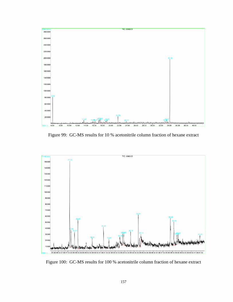
157
Figure 99: GC-MS results for 10 % acetonitrile column fraction of hexane extract
Figure 100: GC-MS results for 100 % acetonitrile column fraction of hexane extract

158
After rotary evaporator removal of the dichlormethane/methanol solvent from the
100% acetonitrile fraction, all ten pooled column fractionation samples were assayed for
antibacterial effects versus Staphylococcus aureus. As most of the samples yielded only
small amounts of visible residue after acetonitrile/water removal, each vial was washed
with 500 µl LB, and this sample-containing media added in 100 µl aliquots in triplicate to
the wells of a 96-well plate. An overnight culture of Staphylococcus aureus was
incubated at approximately 37°C and 225 rpm for about 16 hours, and diluted to an
OD600 reading of 1.020 before the addition of 2 µl culture to the appropriate wells.
Controls (100 µl each) included untreated LB (sterility), tetracycline in LB at 25 µg/ml
(positive), and untreated Staphylococcus aureus (negative), with each performed in
triplicate. The plate was incubated at 37°C and 125 rpm, with measurements taken
immediately prior to incubation, and at 1-hour intervals for 6 hours. As can be seen in
figure 101, only the 10% acetonitrile fraction appeared to inhibit bacterial growth over
this period.
Figure 101: Antibacterial assay of hexane extract column fractions

159
Compounds identified in the 10% acetonitrile fraction with a probability value of
at least 50 % and a peak area of at least 1 %, were tabulated. Seven compounds met
these criteria (table 9). Of these, only the sesquiterpene lactone “achillin” or “azuleno,”
had a probable identification value of greater than 90 %, and a peak of greater than 5 %.
Table 9: Summary of GC-MS results from 10 % acetonitrile column fraction analysis
Peak
Number
Retention
TimeArea Library/ID CAS#
Probable
Identification
2 13.52 2.56 1,3-Pentadiene, 2,4-dimethyl- 001000-86-8 59
2 13.52 2.56 2,5,5-Trimethyl-cyclohex-3-enone 1000193-78-3 53
11 23.71 1.31 Methyl jasmonate 001211-29-6 93
11 23.71 1.31 Cyclopentaneacetic acid, 3-oxo-2-( 042536-97-0 93
11 23.71 1.31 (3-Oxo-2-pent-2-enylcyclopentyl)ac 1000211-13-7 95
14 34.15 56.36 Achillin 071616-00-7 99
14 34.15 56.36 Azuleno[4,5-b]furan-2,7-dione, 3,3 005956-04-7 96
Hexane Extract GC-MS Results

160
IV. DISCUSSION
Evaluation of Results
The results of the antibacterial assays using the ethyl acetate, hexane and water
extracts derived from Artemisia tridentata plant material strongly indicate the presence of
an antibacterial compound(s) capable of selective growth inhibition of Staphylococcus
aureus. Given the initial bacterial inoculum levels and experimental conditions used in
these assays, all three extracts significantly negatively impacted the growth of this
bacterial species over a period of 6 hours in comparison to untreated controls. The ethyl
acetate extract displayed statistically significant antibacterial effects at the lowest
concentration (62.5 µg/ml), followed by the hexane extract (125 µg/ml), and finally the
water extract (250 µg/ml), which demonstrated the least amount of growth inhibition.
Interestingly however, at a concentration of 500 µg/ml, it was the hexane extract-treated
growth curves which most closely resembled those of the positive control.
Further, applications of these extracts in combination with the antibiotics G418
sulfate, amikacin and ampicillin enhanced the antibacterial efficacy of all three drugs.
Statistically significant improvement of growth inhibition was routinely observed when
these combinatorial effects were compared to those of the antibiotics alone. At the
antibiotic concentration of 1.25 µg/ml for example, G418 sulfate and amikacin
performances were enhanced by ethyl acetate extract concentrations as low as 62.5
µg/ml, while ampicillin was enhanced by a hexane extract concentration as low as 125 µl.
The failure of the ethyl acetate extract to also demonstrate enhancement of ampicillin
effects at the lowest concentration among the extracts is explicable upon examination of
the growth curve graphs. It can be observed that in the hexane extract/ampicillin

161
combination assays that the untreated controls grew to higher levels in comparison to the
ethyl acetate/ampicillin combination assays, and that the final readings for combinations
of extracts with 1.25 µg/ml ampicillin were actually higher in the latter. Thus, some of
the inhibitory effects of the hexane extract/ampicillin combinations were statistically
significant due to the greater growth of the untreated control in those assays, despite
growth inhibition at these combined concentrations being greater in the ethyl
acetate/ampicillin assays.
Over 12 hours, the extracts continued to enhance the antibacterial efficacy of
these antibiotics. Combinations of the ethyl acetate and hexane extracts, though at
concentrations of 500 µg/ml, with either G418 sulfate or amikacin, strongly inhibited
Staphylococcus aureus, with growth curves mimicking those of the positive control.
Combined effects with ampicillin were less dramatic, which is consistent with the results
of the 6-hour assays. However, the hexane extract clearly demonstrated the greatest
combined effects with ampicillin in the 12-hour assays.
The assays assessing the performance of the extracts in combination did not
reveal any significant enhancement of antibacterial efficacy which might suggest the
presence of additive or synergistic compounds. However, the values for the hexane/ethyl
acetate extract combinations showed enough reduction of growth inhibition to suggest
possible interference or antagonism between the two. Yet it is also possible that these
elevated growth levels were due to nutrient enrichment of the media by the extracts,
which resulted in more rapid bacterial growth. Though not discussed in the reviewed
literature, it is important to consider that along with potential antibacterial compounds,
chemical extraction of plant material also potentially withdraws many chemicals which
162
can be used by bacteria as energy sources, thereby enriching the level and variety of
nutrients in media when added in antibacterial assays. In many instances in this study,
lower concentrations of extracts alone or in combination with antibiotics were observed
to exhibit growth curves surpassing those of the untreated controls. This can complicate
the assessment of an extract’s antibacterial efficacy, as its true potency may be
diminished by the promotion of bacterial growth due to these additional compounds. In
any case, as there was still significant growth inhibition by the hexane/ethyl acetate
extract combination in comparison to the untreated control at the highest concentrations
tested, antagonist effects could be considered moderate.
The antibacterial efficacy of each extract was lessened when tested against the
ampicillin resistant strain BAA-44, with significant growth inhibition seen only at the
highest tested concentration (500 µg/ml) of the ethyl acetate and hexane extracts. As
ampicillin resistance in this strain is the result of the action of a β-lactamase, this lessened
efficacy is difficult to explain. If this enzyme were to react in some manner with the
antibacterial compound(s) in the extracts, a similar lessening of the combined efficacy of
the extracts with ampicillin would be expected. However, both the ethyl acetate and
hexane extracts significantly improved the growth inhibition of all ampicillin
concentrations tested in comparison to their performances alone, with the growth curves
of hexane extract/ampicillin combinations mimicking those of the positive control. It is
possible that the antibacterial efficacy of the extracts, when applied alone, was masked by
the slow growth of the untreated controls in these assays.
Under the experimental conditions tested, none of the extracts displayed any
inhibitory effects on Staphylococcus aureus biofilm formation. The 10-hour incubation

163
period allowed for biofilm formation in these assays exceeded the antibacterial assays by
4 hours, so this may have diminished extract effects to some extent. Still, the
quantification of crystal violet binding indicated higher biofilm formation in the extract-
treated samples. It is again possible that the extracts enriched the test media, and that this
resulted in more rapid bacterial growth after the antibacterial capacity of the extracts was
overwhelmed. Alternatively, as the plates were incubated without rotation, biofilm
formation early in the assay may have protected the cells within from the extracts,
rendering them ineffective.
The extracts, alone or in combination with antibiotics, also had no effects in the
treatment of static biofilms. The length of sample treatment in these assays was 12 hours.
Given the results of extract/antibiotic combinations from the earlier 12-hour antibacterial
assays, and the use of 1 mg concentrations of antibiotics in the treatment of the static
biofilms, some reduction of the biofilms might have been expected. The lack of positive
results is most likely attributable to the resistive capabilities of the biofilm to the extracts
and antibiotics. However, if the antibacterial effects of the extracts are bacteriostatic as
opposed to bactericidal in nature, this could also explain their ineffectiveness versus a
static biofilm.
In the serial dilution and plating of extract-treated Staphylococcus aureus
samples, the overnight cultures, after adjustment to an OD600 reading approximating
1.000, yielded a colony count average of 1.45 x 10⁸ cells/ml. Given the approximate 2 µl
culture:100 µl media ratio that was used in both these assays and the earlier antibacterial
assays, this number would translate to bacterial inoculum concentrations of about 2.9 x
106 cells/ml ([1.45 x 10
8 cell/ml x 0.002]/ 0.1 ml), considerably less than the 1.52-1.68 x

164
107 cells/ml estimated for the earlier antibacterial assays using the standard Escherichia
coli value for an OD600 reading of 1.000. However, these experimental numbers are
estimates, based on only three trials, and neglecting to take into consideration the optical
alignment of the spectrophotometer114
.
The average colony count of 3.79 x 10⁶ cells/ml for the untreated control sampled
after 25 minutes of incubation is reasonably representative of the calculated inoculum
level, given that the experiment was performed only three times. The comparatively low
average colony count of 1.73 x 10⁵ cells/ml for the tetracycline-treated samples could
either be due to a bacteriostatic effect on cells in the inoculums which were already in the
process of dividing, or to a bactericidal effect of the aminoglycoside at this concentration
to cell ratio115
. The average colony count for the 2-hour untreated sample (2.8 x 10⁷
cells/ml) indicates 3 to 4 rounds of division by the cells in the initial inoculum, which
reinforces an estimated doubling time of around 35 minutes under these experimental
conditions. Therefore, as the colony counts for the extract-treated samples closely
approximated that of the untreated control after 25 minutes, these results are indicative of
a bacteriostatic, as opposed to bactericidal, mechanism of action, which is in agreement
with the literature.
The toxicity assay results imply that at the concentrations which demonstrated
significant growth inhibition in the earlier antibacterial assays, these extracts are
moderately to very lethal against MDA-MB-231 cells. They could therefore be
hazardous to human health if taken internally, which would also be in agreement with the
literature. However, it is possible that the antibiotic compound(s) in the extracts are not
responsible for this toxicity, and that the use of an alternative solvent in the creation of an

165
extract, or the physical isolation of the antibiotic compound(s), might result in safer
products. Also, these results do not necessarily discount the usefulness of extracts or
compounds derived from Artemisia tridentata in external applications, such as those also
described in the literature.
The GC-MS results for the three extracts indicate achillin, a sesquiterpene
lactone, as the most likely candidate responsible for these effects, as this was the only
compound identified as present in all three tested samples under the chromatographic and
mass spectral analytical conditions employed. The identification of this compound in the
hexane extract column fraction exhibiting antibacterial activity further supports this
conclusion. However, it should be noted that the antibacterial assay was performed by
adding media directly to the samples without regard to concentration, due to the
fractionation resulting in very limited individual volumes. The 10% acetonitrile sample
demonstrating Staphylococcus aureus growth inhibition consisted of perhaps as much as
20-30 µl, a volume larger than that typically added to achieve the highest concentration
tested in the extract assays (500 µg/ml). Also, as the concentration of individual
compounds was likely changed as a result of the column fractionation process, there
exists the possibility, however unlikely, that another compound(s) present in this fraction
was responsible for this antibacterial effect, and that this effect was only exerted due to
the use of an excessive concentration of this compound(s). It should also be noted that
GC-MS analysis of the three extract samples revealed additional compounds unique to
each extract which may possess antibacterial properties. Therefore, it is possible that
achillin is not the only antibacterial compound present in Artemisia tridentata, and that its
natural bacterial defense mechanisms consist of the use of more than one compound.

166
Nevertheless, the crediting of the antibacterial properties of this plant to the
presence of a sesquiterpene lactone is in line with existing literature on the subject.
Sesquiterpene lactones are lipophilic fifteen-carbon compounds believed to be formed
through the condensation of three isoprene units, followed by cyclization and oxidative
transformation to cis or trans-fused lactones101
. These compounds are typically found in
glandular trichomes and secretory canals of plants, and are believed to be one of the main
mechanisms of plant microbial defense, disrupting the bacterial cell membrane through
interaction of the constituent polar groups with the phospholipid membrane101
. Indeed, it
has been suggested that this class of compound is perhaps the largest of those to be found
to exist as secondary metabolites in plants, with over 5,000 structures identified to
date101
.
While assays investigating the potential role of pH or DNA-damaging effects
failed to demonstrate positive results, the ethidium bromide assay and electron
microscopy results both support the idea of a mechanism of action involving bacterial
membrane disruption. The ethidium bromide assay results for example, demonstrated
enhanced binding to cellular DNA in the presence of the extracts, which is indicative of
increased membrane permeability, while the transmission electron microscopy results
indicated cellular rupture in the ethyl acetate and hexane extract-treated samples.
However, these results alone cannot completely discount the possibility of autocidal
activity resulting from extract toxicity towards Staphylococcus aureus.
Experimental Shortcomings
As noted in the introductory literature review, a completely exhaustive
examination of the antibacterial properties of a plant would likely require an enormous
167
amount of trial and error using a variety of sampling, extraction, testing and identification
methods. Therefore, it is important to acknowledge the limitations of this particular
study. First, as reported, the Artemisia tridentata plant material obtained was collected
from one location during the month of November. It is possible that sample collections
from other locations or from the same location at a different time(s) would have resulted
in variability in the level of achillin or other compounds contributing to the antibacterial
activity of the plant extracts. For example, it has been reported that autumn is the time of
year when the essential oil content of sagebrush species is at its peak99
. However, it has
also been reported that this species remains photosynthetically active even during periods
of cold, thus these fluctuations may be minimal116
if metabolic energy availability is
consistent throughout the year. Second, the extracts tested were made predominantly
from leaf material, as limited amounts of stems or flowers, and no roots were obtained.
The chemical composition and antibacterial activity of extracts created from different
parts of this plant may vary, perhaps significantly if the roots possess excretory secretory
canals containing achillin or other antibacterial compounds.
Additionally, in the creation of the extracts used for the bulk of the antibacterial
testing, the original solvent applied to the plant material was methanol, with subsequent
extracts created by fractionation of the resulting solution. It is very likely that methanol
failed to extract all constituent compounds in the treated plant material. Had time and the
availability of plant material allowed, the treatment of individual plant samples with
various solvents on an individual basis, or the sequential treatment of a single sample
with these solvents, may have potentially resulted in extracts with greater and/or wider-
ranging antibacterial properties. Further, the antibacterial testing included only four

168
bacterial species, and only three antibiotics were screened for additive or synergistic
effects in combination with the plant extracts. A more expansive screening of both may
have revealed greater potential applicability of the extracts. Finally, attempts to
determine the chemical composition of the extracts were potentially limited by the
approaches taken in the chromatographic and spectral analyses. For example, it is
possible that the chromatographic settings used, the molecular mass cutoff values of the
mass spectral analyses, or the use of outdated reference databases resulted in the
misidentification or failed identification of a relevant compound(s) in the samples.
Future Experiments
We were unable to obtain purified achillin through commercial sources. The
physical isolation and purification of achillin from Artemisia tridentata plant material
would allow for a more accurate assessment of its antibacterial potency and mechanism
of action against Staphylococcus aureus, as well as perhaps some other bacterial species
with similar cell membrane compositions. Such testing would also reveal whether these
effects, exhibited by the chemical extracts in the experiments described herein, are the
result of this compound alone, or if it is possible that one or more of the other chemicals
identified by GC-MS analysis as present in these extracts contributed to bacterial growth
inhibition in some manner. Further, the chemical structure of achillin suggests 16
possible stereo-configurations. While the bioactivity of sesquiterpene lactones has been
reported as attributable to the presence of the α-methylene-γ-lactone group101
, isolation
and purification would allow for structural determination of the specific stereoisomer
produced by this plant species, and perhaps allow for the design of experiments to test
whether this configuration is relevant to this compound’s antibacterial activity.

169
It would also be interesting to modify and repeat the antibacterial assays testing
the methanol extracts of the fifteen plant species screened prior to Artemisia tridentata.
The demonstration of any “meaningful” antibacterial activity possessed by the extracts
created from this plant was only observed after reducing the bacterial inoculum level in
the assays performed. Further, these effects were initially detected using a working
concentration as high as 1000 µg/ml. This illustrates the difficulties in detecting
antibacterial properties in plant investigations, as these properties would likely have been
missed without these experimental modifications. In an antibacterial assay, the working
concentration of the tested chemical and the inoculum level are interdependent, with
either value relatively meaningless without consideration of the other. Therefore, a better
experimental approach in the future, for example, may be to initially test extracts using a
pre-determined, set value for working concentration, doing so against a gradient of
bacterial inoculum levels. Such an assay design might allow for a faster and more
practical assessment of an extract’s potency.
Finally, with regards to further experiments involving Artemisia tridentata, as
well as other plants, it would be beneficial to expand the number of pathogenic bacterial
species assayed, as the antibacterial effects of some plants may be exerted against a very
limited number of bacteria, and including a greater number of test species may help
detect them. Also, as described in the literature review, comparisons of results from
testing bacterial species with different phenotypes can be useful in discerning
antibacterial mechanisms of action. Similarly, when investigating combined applications
with antibiotics, a greater number of drugs representing different classes should be

170
included, which will improve the odds of discovering additive or synergistic effects
between these and the plant extracts.

171
REFERENCES
1. Eldeen IM, Elgorashi EE, van Staden J. Antibacterial, anti-inflammatory, anti-
cholinesterase and mutagenic effects of extracts obtained from some trees used in South
African traditional medicine. J Ethnopharmacol. 2005;102(3):457-464.
2. Tekwu EM, Pieme AC, Beng VP. Investigations of antimicrobial activity of some
Cameroonian medicinal plant extracts against bacteria and yeast with gastrointestinal
relevance. J Ethnopharmacol. 2012;142(1):265-273.
3. Ugoh SC, Agarry OO, Garba SA. Studies on the antibacterial activity of Khaya
senegalensis [(Desr.) A. Juss)] stem bark extract on Salmonella enterica subsp. enterica
serovar Typhi [(ex Kauffmann and Edwards) Le Minor and Popoff. Asian Pac J Trop
Biomed. 2014;4(Suppl 1):S279-83.
4. Kaneria M, Baravalia Y, Vaghasiya Y, Chanda S. Determination of antibacterial and
antioxidant potential of some medicinal plants from saurashtra region, India. Indian J
Pharm Sci. 2009;71(4):406-412.
5. Tchouya GR, Souza A, Tchouankeu JC, et al. Ethnopharmacological surveys and
pharmacological studies of plants used in traditional medicine in the treatment of
HIV/AIDS opportunistic diseases in Gabon. J Ethnopharmacol. 2015;162:306-316.
6. Adwan G, Abu-Shanab B, Adwan K. Antibacterial activities of some plant extracts
alone and in combination with different antimicrobials against multidrug-resistant
Pseudomonas aeruginosa strains. Asian Pacific journal of tropical medicine.
2010;3(4):266-269.
7. Dusan M, Ruzica A, Jasna I, Irena Z. Investigation of antibacterial activity of
supercritical extracts of plants, as well as of extracts obtained by other technological
processes on some bacteria isolated from animals. Acta Vet (Beograd). 2009;59(5-6):557-
568.
8. Oskay M, Oskay D, Kalyoncu F. Activity of some plant extracts against multi-drug
resistant human pathogens. Iranian journal of pharmaceutical research : IJPR.
2009;8(4):293-300.
9. Nuno G, Zampini IC, Ordonez RM, Alberto MR, Arias ME, Isla MI.
Antioxidant/antibacterial activities of a topical phytopharmaceutical formulation
containing a standardized extract of Baccharis incarum, an extremophile plant species
from Argentine Puna. Phytother Res. 2012;26(11):1759-1767.
10. Aqil F, Ahmad I, Owais M. Evaluation of anti-methicillin-resistant Staphylococcus
aureus (MRSA) activity and synergy of some bioactive plant extracts. Biotechnol J.
2006;1(10):1093-1102.

172
11. Ohene-Agyei T, Mowla R, Rahman T, Venter H. Phytochemicals increase the
antibacterial activity of antibiotics by acting on a drug efflux pump. Microbiologyopen.
2014;3(6):885-896.
12. Reverter M, Bontemps N, Lecchini D, Banaigs B, Sasal P. Use of plant extracts in
fish aquaculture as an alternative to chemotherapy: Current status and future perspectives.
Aquaculture. 2014;433:50-61.
13. Miceli A, Aleo A, Corona O, Sardina MT, Mammina C, Settanni L. Antibacterial
activity of Borago officinalis and Brassica juncea aqueous extracts evaluated in vitro and
in situ using different food model systems. Food control. 2014;40:157-164.
14. Korpe DA, Iseri OD, Sahin FI, Cabi E, Haberal M. High-antibacterial activity of
Urtica spp. seed extracts on food and plant pathogenic bacteria. Int J Food Sci Nutr.
2013;64(3):355-362.
15. Tomlinson S, Palombo EA. Characterisation of antibacterial Australian medicinal
plant extracts by investigation of the mechanism of action and the effect of interfering
substances. J Basic Microbiol. 2005;45(5):363-370.
16. Basim E, Basim H, Özcan M. Antibacterial activities of Turkish pollen and propolis
extracts against plant bacterial pathogens. J Food Eng. 2006;77(4):992-996.
17. Kotan R, Dadasoglu F, Karagoz K, et al. Antibacterial activity of the essential oil and
extracts of Satureja hortensis against plant pathogenic bacteria and their potential use as
seed disinfectants. Scientia horticulturae. 2013;153:34-41.
18. Abdollahzadeh E, Rezaei M, Hosseini H. Antibacterial activity of plant essential oils
and extracts: The role of thyme essential oil, nisin, and their combination to control
Listeria monocytogenes inoculated in minced fish meat. Food control. 2014;35(1):177-
183.
19. Khan H, Ali Khan M, Mahmood T, Choudhary MI. Antimicrobial activities of
Gloriosa superba Linn (Colchicaceae) extracts. Journal of enzyme inhibition and
medicinal chemistry. 2008;23(6):855-859.
20. Rocha-Gracia RdC, Arroyo MMH, Zarain PL, et al. Antibacterial activity of crude
extracts from Mexican plants against methicillin-resistant Staphylococcus. African
journal of biotechnology. 2011;10(61):13202-13218.
21. Razmavar S, Abdulla MA, Ismail SB, Hassandarvish P. Antibacterial activity of leaf
extracts of Baeckea frutescens against methicillin-resistant Staphylococcus aureus.
BioMed Research International. 2014;1-5.

173
22. Pinho E, Ferreira IC, Barros L, Carvalho AM, Soares G, Henriques M. Antibacterial
potential of northeastern Portugal wild plant extracts and respective phenolic compounds.
BioMed Research International. 2014:1-8.
23. Karakas FP, Yildirim A, Turker A. Biological screening of various medicinal plant
extracts for antibacterial and antitumor activities. Turkish journal of biology.
2012;36(6):641-652.
24. Srinivas P, Reddy SR. Screening for antibacterial principle and activity of Pedalium
murex Linn. Proceedings of the National Academy of Sciences, India.Section B:
Biological sciences. 2014;84(4):1083-1090.
25. Soberon JR, Sgariglia MA, Sampietro DA, Quiroga EN, Vattuone MA. Antibacterial
activity of plant extracts from northwestern Argentina. J Appl Microbiol.
2007;102(6):1450-1461.
26. Yasunaka K, Abe F, Nagayama A, et al. Antibacterial activity of crude extracts from
Mexican medicinal plants and purified coumarins and xanthones. J Ethnopharmacol.
2005;97(2):293-299.
27. Alabri THA, Al Musalami AHS, Hossain MA, Weli AM, Al-Riyami Q. Comparative
study of phytochemical screening, antioxidant and antimicrobial capacities of fresh and
dry leaves crude plant extracts of Datura metel L. Journal of King Saud
University.Science. 2014;26(3):237-243.
28. Yildirim AB, Karakas FP, Turker AU. In vitro antibacterial and antitumor activities
of some medicinal plant extracts, growing in Turkey. Asian Pac J Trop Med.
2013;6(8):616-624.
29. Szabóová R, Laukova A, Chrastinova L, et al. Benefficial effect of plant extracts in
rabbit husbandry. Acta veterinaria Brno. 2012;81(3):245-250.
30. Stagos D, Portesis N, Spanou C, et al. Correlation of total polyphenolic content with
antioxidant and antibacterial activity of 24 extracts from Greek domestic Lamiaceae
species. Food Chem Toxicol. 2012;50(11):4115-4124.
31. Kuppusamy P, Yusoff MM, Parine NR, Govindan N. Evaluation of in-vitro
antioxidant and antibacterial properties of Commelina nudiflora L. extracts prepared by
different polar solvents. Saudi J Biol Sci. 2015;22(3):293-301.
32. Turker H, Yildirim AB, Karakas FP, Koyluoglu H. Antibacterial activities of extracts
from some Turkish endemic plants on common fish pathogens. Turkish journal of
biology. 2009;33(1):73-78.

174
33. Klančnik A, Piskernik S, Smole Mozina S, Gasperlin L, Jersek B. Investigation of
some factors affecting the antibacterial activity of rosemary extracts in food models by a
food microdilution method factors affecting MIC of plant extract. Int J Food Sci Tech.
2011;46(2):413-420.
34. Lee JH, Cho S, Paik HD, et al. Investigation on antibacterial and antioxidant
activities, phenolic and flavonoid contents of some thai edible plants as an alternative for
antibiotics. Asian-Australas J Anim Sci. 2014;27(10):1461-1468.
35. Al Akeel R, Al-Sheikh Y, Mateen A, Syed R, Janardhan K, Gupta VC. Evaluation of
antibacterial activity of crude protein extracts from seeds of six different medical plants
against standard bacterial strains. Saudi J Biol Sci. 2014;21(2):147-151.
36. Kousha A, Ringo E. Antibacterial effect of aquatic extract of Heracleum spp.
hogweed plants from Europe on thirteen different bacteria. Pharmaceutical chemistry
journal. 2015;48(10):677-680.
37. Chah KF, Eze CA, Emuelosi CE, Esimone CO. Antibacterial and wound healing
properties of methanolic extracts of some Nigerian medicinal plants. J Ethnopharmacol.
2006;104(1-2):164-167.
38. Vermaak I, Viljoen AM, Hamman JH, Van Vuuren SF. The effect of simulated
gastrointestinal conditions on the antimicrobial activity and chemical composition of
indigenous South African plant extracts. S Afr J Bot. 2009;75(3):594-599.
39. Rodrigues L, Duarte A, Figueiredo AC, et al. Chemical composition and antibacterial
activity of the essential oils from the medicinal plant Mentha cervina L. grown in
Portugal. Medicinal chemistry research. 2012;21(11):3485-3490.
40. Smida I, Charpy-Roubaud C, Cherif SY, et al. Antibacterial properties of extracts of
Ludwigia peploides subsp. montevidensis and Ludwigia grandiflora subsp. hexapetala
during their cycle of development. Aquat Bot. 2015;121:39-45.
41. Mooney EH, Martin AA, Blessin RP. Effects of light environment on recovery from
harvest and antibacterial properties of Osha Ligusticum porteri (Apiaceae). Econ Bot.
2015;69(1):72-82.
42. Ocheng F, Bwanga F, Joloba M, Borg-Karlson AK, Gustafsson A, Obua C.
Antibacterial activities of extracts from Ugandan medicinal plants used for oral care. J
Ethnopharmacol. 2014;155(1):852-855.
43. Voravuthikunchai SP, Limsuwan S. Medicinal plant extracts as anti-Escherichia coli
O157:H7 agents and their effects on bacterial cell aggregation. J Food Prot.
2006;69(10):2336-2341.

175
44. Kenny O, Brunton NP, Walsh D, Hewage CM, McLoughlin P, Smyth TJ.
Characterisation of antimicrobial extracts from dandelion root (Taraxacum officinale)
using LC-SPE-NMR. Phytother Res. 2015;29(4):526-532.
45. Kothari V, Gupta A, Naraniwal M. Comparative study of various methods for
extraction of antioxidant and antibacterial compounds from plant seeds. Journal of
Natural Remedies. 2012;12(2):162-173.
46. Roy S, Rao K, Bhuvaneswari CH, Giri A, Mangamoori LN. Phytochemical analysis
of Andrographis paniculata extract and its antimicrobial activity. World J Microbiol
Biotechnol. 2010;26(1):85-91.
47. Regazzoni L, Arlandini E, Garzon D, Santagati NA, Beretta G, Maffei Facino R. A
rapid profiling of gallotannins and flavonoids of the aqueous extract of Rhus coriaria L.
by flow injection analysis with high-resolution mass spectrometry assisted with database
searching. J Pharm Biomed Anal. 2013;72:202-207.
48. Stanojevic D, Comic L, Stefanovic O, Solujic-Sukdolak S. In vitro synergistic
antibacterial activity of Salvia officinalis l. and some preservatives. Archives of
biological sciences. 2010;62(1):167-174.
49. Ganie SA, Jan A, Muzaffar S, Zargar BA, Hamid R, Zargar MA. Radical scavenging
and antibacterial activity of Arnebia benthamii methanol extract. Asian Pac J Trop Med.
2012;5(10):766-772.
50. Mahida Y, Mohan JSS. Screening of Indian plant extracts for antibacterial activity.
Pharm Biol. 2006;44(8):627-631.
51. Jesionek W, Choma IM, Majer-Dziedzic B, Malinowska I. Screening bacterial and
radical scavenging properties of chosen plant extracts using thin-layer chromatography–
direct bioautography. J Liq Chromatogr Rel Technol. 2014;37(20):2882-2891.
52. Hochfellner C, Evangelopoulos D, Zloh M, et al. Antagonistic effects of
indoloquinazoline alkaloids on antimycobacterial activity of evocarpine. J Appl
Microbiol. 2015;118(4):864-872.
53. Adwan G, Mhanna M. Synergistic effects of plant extracts and antibiotics on
Staphylococcus aureus strains isolated from clinical specimens. Middle-East Journal of
Scientific Research. 2008;3(3):134-139.
54. Motz VA, Bowers CP, Kneubehl AR, et al. Efficacy of the saponin component of
impatiens capensis Meerb.in preventing urushiol-induced contact dermatitis. J
Ethnopharmacol. 2015;162:163-167.

176
55. Tolmacheva AA, Rogozhin EA, Deryabin DG. Antibacterial and quorum sensing
regulatory activities of some traditional Eastern-European medicinal plants. Acta Pharm.
2014;64(2):173-186.
56. Chatterjee SK, Bhattacharjee I, Chandra G. In vitro synergistic effect of doxycycline
& ofloxacin in combination with ethanolic leaf extract of Vangueria spinosa against four
pathogenic bacteria. Indian J Med Res. 2009;130(4):475-478.
57. Salehi P, Sonboli A, Eftekhar F, Nejad-Ebrahimi S, Yousefzadi M. Essential oil
composition, antibacterial and antioxidant activity of the oil and various extracts of
Ziziphora clinopodioides subsp. rigida (BOISS.) RECH. f. from Iran. Biol Pharm Bull.
2005;28(10):1892-1896.
58. Ashokkumar S, Ravi S, Kathiravan V, Velmurugan S. Synthesis of silver
nanoparticles using A. indicum leaf extract and their antibacterial activity. Spectrochim
Acta A Mol Biomol Spectrosc. 2015;134:34-39.
59. Ambika S, Sundrarajan M. Antibacterial behaviour of Vitex negundo extract assisted
ZnO nanoparticles against pathogenic bacteria. J Photochem Photobiol B. 2015;146:52-
57.
60. Khan M, Khan ST, Khan M, et al. Antibacterial properties of silver nanoparticles
synthesized using Pulicaria glutinosa plant extract as a green bioreductant. Int J
Nanomedicine. 2014;9:3551-3565.
61. Klancnik A, Piskernik S, Jersek B, Mozina SS. Evaluation of diffusion and dilution
methods to determine the antibacterial activity of plant extracts. J Microbiol Methods.
2010;81(2):121-126.
62. Aqil F, Khan MS, Owais M, Ahmad I. Effect of certain bioactive plant extracts on
clinical isolates of beta-lactamase producing methicillin resistant Staphylococcus aureus.
J Basic Microbiol. 2005;45(2):106-114.
63. Jiang ZL, Zhou Y, Ge WC, Yuan K. Phytochemical compositions of volatile oil from
Blumea balsamifera and their biological activities. Pharmacogn Mag. 2014;10(39):346-
352.
64. Weckesser S, Engel K, Simon-Haarhaus B, Wittmer A, Pelz K, Schempp CM.
Screening of plant extracts for antimicrobial activity against bacteria and yeasts with
dermatological relevance. Phytomedicine. 2007;14(7-8):508-516.
65. Wojnicz D, Kucharska AZ, Sokol-Letowska A, Kicia M, Tichaczek-Goska D.
Medicinal plants extracts affect virulence factors expression and biofilm formation by the
uropathogenic Escherichia coli. Urol Res. 2012;40(6):683-697.
177
66. Mekinic IG, Skroza D, Ljubenkov I, Simat V, Mozina SS, Katalinic V. In vitro
antioxidant and antibacterial activity of Lamiaceae phenolic extracts: A correlation study.
Food technology and biotechnology. 2014;52(1, sp. iss. si):119-127.
67. Joray MB, del Rollan MR, Ruiz GM, Palacios SM, Carpinella MC. Antibacterial
activity of extracts from plants of central Argentina--isolation of an active principle from
Achyrocline satureioides. Planta Med. 2011;77(1):95-100.
68. Leandro LF, Cardoso MJ, Silva SD, et al. Antibacterial activity of Pinus elliottii and
its major compound, dehydroabietic acid, against multidrug-resistant strains. J Med
Microbiol. 2014;63(Pt 12):1649-1653.
69. Bag A, Bhattacharyya SK, Pal NK, Chattopadhyay RR. Antibacterial potential of
hydroalcoholic extracts of triphala components against multidrug-resistant uropathogenic
bacteria--a preliminary report. Indian J Exp Biol. 2013;51(9):709-714.
70. Yamaguchi N, Satoh-Yamaguchi K, Ono M. In vitro evaluation of antibacterial,
anticollagenase, and antioxidant activities of hop components (Humulus lupulus)
addressing acne vulgaris. Phytomedicine. 2009;16(4):369-376.
71. Hussain T, Arshad M, Khan S, Sattar H, Qureshi MS. In vitro screening of methanol
plant extracts for their antibacterial activity. Pak J Bot. 2011;43(1):531-538.
72. Over KF, Hettiarachchy N, Johnson MG, Davis B. Effect of organic acids and plant
extracts on Escherichia coli O157:H7, Listeria monocytogenes, and Salmonella
Typhimurium in broth culture model and chicken meat systems. J Food Sci.
2009;74(9):M515-21.
73. Owais M, Sharad KS, Shehbaz A, Saleemuddin M. Antibacterial efficacy of Withania
somnifera (ashwagandha) an indigenous medicinal plant against experimental murine
salmonellosis. Phytomedicine. 2005;12(3):229-235.
74. Toyang NJ, Ateh EN, Keiser J, et al. Toxicity, antimicrobial and anthelmintic
activities of Vernonia guineensis Benth. (Asteraceae) crude extracts. J Ethnopharmacol.
2012;144(3):700-704.
75. Engels C, Schieber A, Ganzle MG. Sinapic acid derivatives in defatted Oriental
mustard (Brassica juncea L.) seed meal extracts using UHPLC-DAD-ESI-MS n and
identification of compounds with antibacterial activity. European food research &
technology. 2012;234(3):535-542.
76. Bashir S, Khan BM, Babar M, et al. Assessment of bioautography and spot screening
of TLC of green tea (Camellia) plant extracts as antibacterial and antioxidant agents.
Indian J Pharm Sci. 2014;76(4):364-370.

178
77. Marquis reagent. https://en.wikipedia.org/wiki/Marquis_reagent. Updated 2017.
Accessed 11/15, 2017.
78. Liao SG, Zhang LJ, Sun F, et al. Antibacterial and anti-inflammatory effects of
extracts and fractions from Polygonum capitatum. J Ethnopharmacol. 2011;134(3):1006-
1009.
79. Liebermann-burchard test.
https://en.wikipedia.org/wiki/Liebermann%E2%80%93Burchard_test. Updated 2014.
Accessed 11/15, 2017.
80. Bobis O, Dezmirean DS, Tomos L, Chirila F, Al. Marghitas L. Influence of
phytochemical profile on antibacterial activity of different medicinal plants against gram-
positive and gram-negative bacteria. Appl Biochem Microbiol. 2015;51(1):113-118.
81. Pirvu L, Hlevca C, Nicu I, Bubueanu C. Comparative studies on analytical,
antioxidant, and antimicrobial activities of a series of vegetal extracts prepared from eight
plant species growing in Romania. JPC.Journal of planar chromatography, modern TLC.
2014;27(5):346-356.
82. Anani K, Adjrah Y, Ameyapoh Y, et al. Effects of hydroethanolic extracts of
Balanites aegyptiaca (L.) Delile (Balanitaceae) on some resistant pathogens bacteria
isolated from wounds. J Ethnopharmacol. 2015;164:16-21.
83. Folin-ciocalteu reagent.
https://en.wikipedia.org/wiki/Folin%E2%80%93Ciocalteu_reagent. Updated 2017.
Accessed 11/15, 2017.
84. Hori Y, Sato S, Hatai A. Antibacterial activity of plant extracts from azuki beans
(Vigna angularis) in vitro. Phytother Res. 2006;20(2):162-164.
85. Nemereshina ON, Tinkov AA, Gritsenko VA, Nikonorov AA. Influence of
Plantaginaceae species on E. coli K12 growth in vitro: Possible relation to phytochemical
properties. Pharm Biol. 2015;53(5):715-724.
86. Packer L. Methods in enzymology. Academic Press, 1999.:506.
87. Chaweepack T, Chaweepack S, Muenthaisong B, Ruangpan L, Nagata K, Kamei K.
Effect of galangal (Alpinia galanga Linn.) extract on the expression of immune-related
genes and Vibrio harveyi resistance in Pacific white shrimp (Litopenaeus vannamei).
Aquacult Int. 2015;23(1):385-399.
88. Lu C, Luo X, Luo R, et al. Assessment of antibacterial properties and the active
ingredient of plant extracts and its effect on the performance of crucian carp (Carassius
auratus gibelio var. E'erqisi, Bloch). J Sci Food Agric. 2013;93(4):902-909.

179
89. Dadasoglu F, Aydin T, Kotan R, et al. Antibacterial activities of extracts and essential
oils of three Origanum species against plant pathogenic bacteria and their potential use as
seed disinfectants. J Plant Pathol. 2011;93(2):271-282.
90. Liu Y, Nielsen M, Staerk D, Jager AK. High-resolution bacterial growth inhibition
profiling combined with HPLC-HRMS-SPE-NMR for identification of antibacterial
constituents in Chinese plants used to treat snakebites. J Ethnopharmacol.
91. Kouidhi B, Al Qurashi YM, Chaieb K. Drug resistance of bacterial dental biofilm and
the potential use of natural compounds as alternative for prevention and treatment.
Microb Pathog. 2015;80:39-49.
92. Kouidhi B, Al Qurashi, YM, Chaieb K. Drug resistance of bacterial dental biofilm
and the potential use of natural compounds as alternative for prevention and
treatment. abstract. Microb Pathog. 2015;80(1):39-49.
93. Mustafi S, Veisaga ML, López LA, Barbieri MA. A novel insight into
dehydroleucodine mediated attenuation of Pseudomonas aeruginosa virulence
mechanism. BioMed research international. 2015;1-11.
94. Ahmad R, Ali AM, Israf DA, Ismail NH, Shaari K, Lajis NH. Antioxidant, radical-
scavenging, anti-inflammatory, cytotoxic and antibacterial activities of methanolic
extracts of some Hedyotis species. Life Sci. 2005;76(17):1953-1964.
95. Sindhu T, Rajamanikandan S, Srinivasan P. In vitro antioxidant and antibacterial
activities of methanol extract of Kyllinga nemoralis. Indian J Pharm Sci. 2014;76(2):170-
174.
96. Taguri T, Tanaka T, Kouno I. Antibacterial spectrum of plant polyphenols and
extracts depending upon hydroxyphenyl structure. Biol Pharm Bull. 2006;29(11):2226-
2235.
97. Smirnova G, Samoilova Z, Muzyka N, Oktyabrsky O. Influence of plant polyphenols
and medicinal plant extracts on antibiotic susceptibility of Escherichia coli. J Appl
Microbiol. 2012;113(1):192-199.
98. EREL ŞB, Reznicek G, ŞENOL SG, YAVAŞOĞLU NÜK, Konyalioğlu S, Zeybek
AU. Antimicrobial and antioxidant properties of Artemisia L. species from western
Anatolia. Turkish Journal of Biology. 2012;36(1):75-84.
99. Nagy JG, Tengerdy RP. Antibacterial action of essential oils of Artemisia as an
ecological factor. I. Antibacterial action of the volatile oils of Artemisia tridentata and
Artemisia nova on aerobic bacteria. Appl Microbiol. 1967;15(4):819-821.
180
100. Abad MJ, Bedoya LM, Apaza L, Bermejo P. The Artemisia L. genus: a review of
bioactive essential oils. Molecules. 2012;17(3):2542-2566.
101. Ivanescu B, Miron A, Corciova A. Sesquiterpene lactones from Artemisia genus:
Biological activities and methods of analysis. J.Anal.Methods Chem.
2015;21(10.1155):2015.
102. Tilford GL. Edible and medicinal plants of the west. Mountain Press Publishing;
1997.
103. Austin DF. Healing with plants in the American and Mexican west. Econ Bot.
1997;51(3):327-327.
104. Carroll KC, Detrick B, Hobden JA, et al. Jawetz, melnick & adelberg's medical
microbiology. 27th ed. ed. New York: Mcgraw-Hill Education; 2016.
http://ezproxy.fiu.edu/login?url=http://accessmedicine.mhmedical.com/book.aspx?bookid
=1551.
105. CDC - staphylococcus aureus pneumoniae in healthcare settings - HAI.
http://www.cdc.gov/HAI/organisms/staph.html. Accessed 10/10/2016, 2016.
106. WHO | antimicrobial resistance: Global report on surveillance 2014.
http://www.who.int/drugresistance/documents/surveillancereport/en/. Accessed
10/10/2016, 2016.
107. CDC - pseudomonas aeruginosa in healthcare settings - HAI.
http://www.cdc.gov/hai/organisms/pseudomonas.html. Accessed 10/10/2016, 2016.
108. Listeria (listeriosis) | listeria | CDC. http://www.cdc.gov/listeria/index.html.
Accessed 10/10/2016, 2016.
109. General information on salmonella | salmonella | CDC.
http://www.cdc.gov/salmonella/general/index.html. Accessed 10/10/2016, 2016.
110. Agilent genomics : Tools - bio calculators.
http://www.genomics.agilent.com/biocalculators/calcODBacterial.jsp. Accessed
10/10/2016, 2016.
111. Jabra-Rizk MA, Meiller TF, James CE, Shirtliff ME. Effect of farnesol on
staphylococcus aureus biofilm formation and antimicrobial susceptibility. Antimicrob
Agents Chemother. 2006;50(4):1463-1469.
112. Shcherbakova I, Mitra S, Beer RH, Brenowitz M. Fast fenton footprinting: A
laboratory-based method for the time-resolved analysis of DNA, RNA and proteins.
Nucleic Acids Res. 2006;34(6):e48.

181
113. Mojarrab M, Delazar A, Moghadam SB, et al. Armenin and isoarmenin–two
prenylated coumarins from the aerial parts of artemisia armeniaca. Chemistry &
biodiversity. 2011;8(11):2097-2103.
114. nanodroptryPOSTER.pdf.
https://tools.thermofisher.com/content/sfs/posters/nanodroptryPOSTER.pdf. Accessed
11/14/2017, 2017.
115. Aminoglycosides - infectious diseases - merck manuals professional edition.
http://www.merckmanuals.com/professional/infectious-diseases/bacteria-and-
antibacterial-drugs/aminoglycosides. Accessed 11/14/2017, 2017.
116. Wright CW. Artemisia. London ; New York: Taylor & Francis; 2002.

182
VITA
STEVEN EICHELBAUM
Born, Allentown, Pennsylvania
B.S. Biology
University of Pittsburgh
Pittsburgh, PA
M.S. Microbiology
Thomas Jefferson University
Philadelphia, PA
2007-2008 Research Technician
Florida International University
Miami, FL
2008-2010 Senior Production Technician
Beckman Coulter
Hialeah, FL
2010 Assistant Scientist
Colgate-Palmolive
Piscataway, NJ
2011-2016 Doctoral Student
Florida International University
Miami, FL
Related Documents