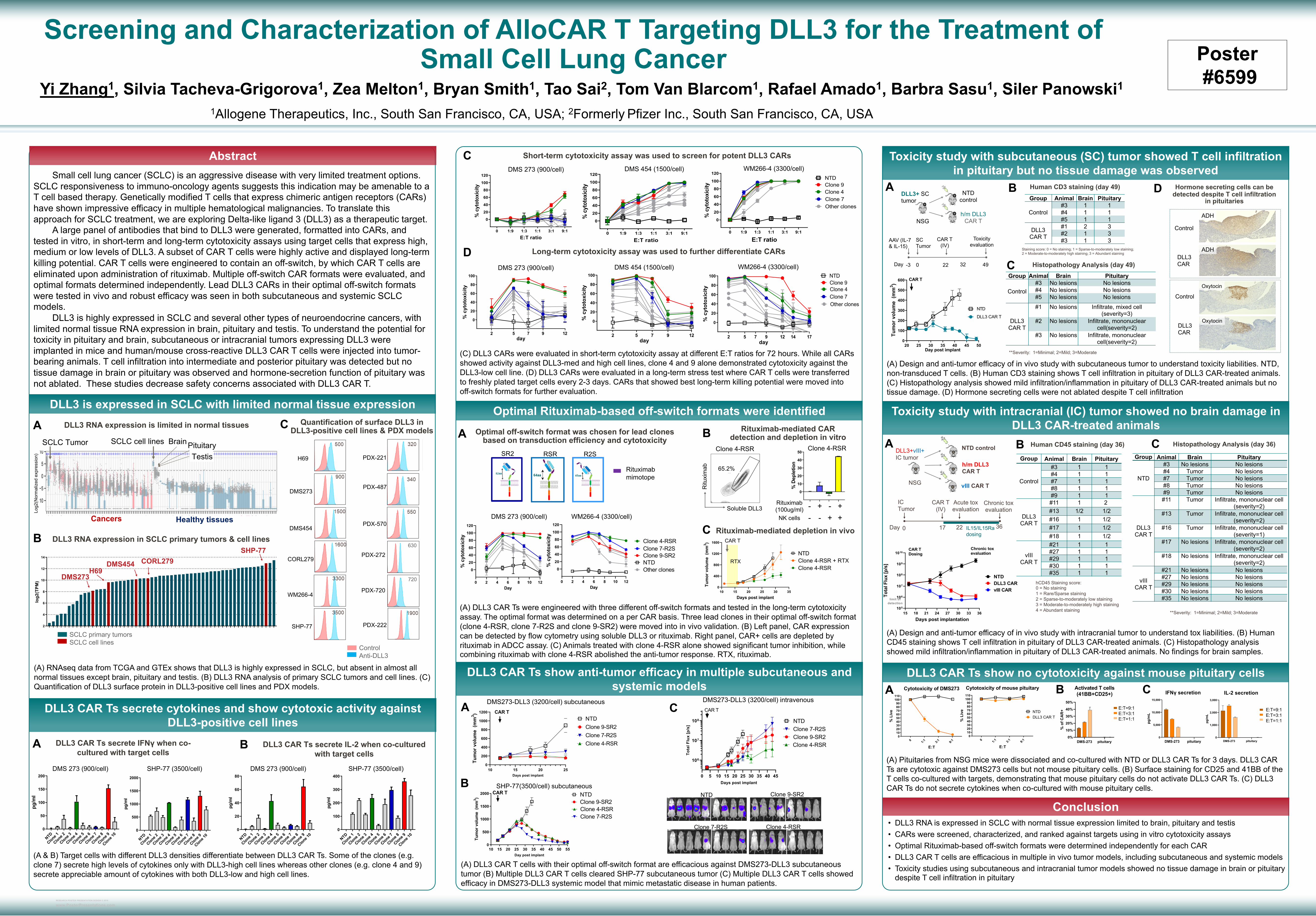
RESEARCH POSTER PRESENTATION DESIGN © 2019 www.PosterPresentations.com DMS-273 pituitary 0% 10% 20% 30% 40% 50% Activated T cells (41BB+CD25+) % of CAR+ Small cell lung cancer (SCLC) is an aggressive disease with very limited treatment options. SCLC responsiveness to immuno-oncology agents suggests this indication may be amenable to a T cell based therapy. Genetically modified T cells that express chimeric antigen receptors (CARs) have shown impressive efficacy in multiple hematological malignancies. To translate this approach for SCLC treatment, we are exploring Delta-like ligand 3 (DLL3) as a therapeutic target. A large panel of antibodies that bind to DLL3 were generated, formatted into CARs, and tested in vitro, in short-term and long-term cytotoxicity assays using target cells that express high, medium or low levels of DLL3. A subset of CAR T cells were highly active and displayed long-term killing potential. CAR T cells were engineered to contain an off-switch, by which CAR T cells are eliminated upon administration of rituximab. Multiple off-switch CAR formats were evaluated, and optimal formats determined independently. Lead DLL3 CARs in their optimal off-switch formats were tested in vivo and robust efficacy was seen in both subcutaneous and systemic SCLC models. DLL3 is highly expressed in SCLC and several other types of neuroendocrine cancers, with limited normal tissue RNA expression in brain, pituitary and testis. To understand the potential for toxicity in pituitary and brain, subcutaneous or intracranial tumors expressing DLL3 were implanted in mice and human/mouse cross-reactive DLL3 CAR T cells were injected into tumor- bearing animals. T cell infiltration into intermediate and posterior pituitary was detected but no tissue damage in brain or pituitary was observed and hormone-secretion function of pituitary was not ablated. These studies decrease safety concerns associated with DLL3 CAR T. (A) RNAseq data from TCGA and GTEx shows that DLL3 is highly expressed in SCLC, but absent in almost all normal tissues except brain, pituitary and testis. (B) DLL3 RNA analysis of primary SCLC tumors and cell lines. (C) Quantification of DLL3 surface protein in DLL3-positive cell lines and PDX models. (A) DLL3 CAR Ts were engineered with three different off-switch formats and tested in the long-term cytotoxicity assay. The optimal format was determined on a per CAR basis. Three lead clones in their optimal off-switch format (clone 4-RSR, clone 7-R2S and clone 9-SR2) were moved into in vivo validation. (B) Left panel, CAR expression can be detected by flow cytometry using soluble DLL3 or rituximab. Right panel, CAR+ cells are depleted by rituximab in ADCC assay. (C) Animals treated with clone 4-RSR alone showed significant tumor inhibition, while combining rituximab with clone 4-RSR abolished the anti-tumor response. RTX, rituximab. (C) DLL3 CARs were evaluated in short-term cytotoxicity assay at different E:T ratios for 72 hours. While all CARs showed activity against DLL3-med and high cell lines, clone 4 and 9 alone demonstrated cytotoxicity against the DLL3-low cell line. (D) DLL3 CARs were evaluated in a long-term stress test where CAR T cells were transferred to freshly plated target cells every 2-3 days. CARs that showed best long-term killing potential were moved into off-switch formats for further evaluation. (A) DLL3 CAR T cells with their optimal off-switch format are efficacious against DMS273-DLL3 subcutaneous tumor (B) Multiple DLL3 CAR T cells cleared SHP-77 subcutaneous tumor (C) Multiple DLL3 CAR T cells showed efficacy in DMS273-DLL3 systemic model that mimic metastatic disease in human patients. (A) Design and anti-tumor efficacy of in vivo study with subcutaneous tumor to understand toxicity liabilities. NTD, non-transduced T cells. (B) Human CD3 staining shows T cell infiltration in pituitary of DLL3 CAR-treated animals. (C) Histopathology analysis showed mild infiltration/inflammation in pituitary of DLL3 CAR-treated animals but no tissue damage. (D) Hormone secreting cells were not ablated despite T cell infiltration (A) Design and anti-tumor efficacy of in vivo study with intracranial tumor to understand tox liabilities. (B) Human CD45 staining shows T cell infiltration in pituitary of DLL3 CAR-treated animals. (C) Histopathology analysis showed mild infiltration/inflammation in pituitary of DLL3 CAR-treated animals. No findings for brain samples. Yi Zhang 1 , Silvia Tacheva-Grigorova 1 , Zea Melton 1 , Bryan Smith 1 , Tao Sai 2 , Tom Van Blarcom 1 , Rafael Amado 1 , Barbra Sasu 1 , Siler Panowski 1 1 Allogene Therapeutics, Inc., South San Francisco, CA, USA; 2 Formerly Pfizer Inc., South San Francisco, CA, USA Screening and Characterization of AlloCAR T Targeting DLL3 for the Treatment of Small Cell Lung Cancer Abstract DLL3 is expressed in SCLC with limited normal tissue expression Conclusion A B C DLL3 RNA expression is limited in normal tissues SCLC Tumor SCLC cell lines Pituitary Testis Cancers Healthy tissues Brain Log2(Normalized expression) DLL3 RNA expression in SCLC primary tumors & cell lines SCLC primary tumors SCLC cell lines 2 4 6 8 10 12 14 log2(TPM) SHP-77 DMS454 CORL279 H69 DMS273 Quantification of surface DLL3 in DLL3-positive cell lines & PDX models DLL3 CAR Ts secrete cytokines and show cytotoxic activity against DLL3-positive cell lines DMS 273 (900/cell) SHP-77 (3500/cell) NTD Clone 1 Clone 2 Clone 3 Clone 4 Clone 5 Clone 6 Clone 7 Clone 8 Clone 9 Clone 10 0 50 100 150 200 pg/ml NTD Clone 1 Clone 2 Clone 3 Clone 4 Clone 5 Clone 6 Clone 7 Clone 8 Clone 9 Clone 10 0 500 1000 1500 2000 pg/ml A DLL3 CAR Ts secrete IFNγ when co- cultured with target cells C D 0 1:9 1:3 1:1 3:1 9:1 0 20 40 60 80 100 120 E:T ratio % cytotoxicity 0 1:9 1:3 1:1 3:1 9:1 0 20 40 60 80 100 120 E:T ratio % cytotoxicity 0 1:9 1:3 1:1 3:1 9:1 0 20 40 60 80 100 120 E:T ratio % cytotoxicity NTD Clone 4 Clone 7 Clone 9 Other clones Short-term cytotoxicity assay was used to screen for potent DLL3 CARs 0 20 40 60 80 100 day % cytotoxicity NTD Clone 4 Clone 7 Clone 9 2 5 7 9 12 14 17 Other clones 0 20 40 60 80 100 day % cytotoxicity 2 5 7 9 12 0 20 40 60 80 100 day % cytotoxicity 2 5 7 9 12 Long-term cytotoxicity assay was used to further differentiate CARs Optimal off-switch format was chosen for lead clones based on transduction efficiency and cytotoxicity SR2 RSR R2S Optimal Rituximab-based off-switch formats were identified DLL3 CAR Ts show anti-tumor efficacy in multiple subcutaneous and systemic models 10 15 20 25 30 35 40 45 50 55 0 500 1000 1500 2000 Day post implant Tumor volume (mm 3 ) Clone 9-SR2 NTD Clone 7-R2S Clone 4-RSR CAR T 0 5 10 15 20 25 30 35 40 45 10 6 10 7 10 8 Days post implant Total Flux [p/s] Clone 9-SR2 NTD Clone 7-R2S Clone 4-RSR CAR T Toxicity study with subcutaneous (SC) tumor showed T cell infiltration in pituitary but no tissue damage was observed AAV (IL-7 & IL-15) Day -3 0 SC Tumor 22 CAR T (IV) 49 Toxicity evaluation 32 h/m DLL3 CAR T NTD control DLL3+ SC tumor NSG **Severity: 1=Minimal; 2=Mild; 3=Moderate Group Animal Brain Pituitary Control #3 No lesions No lesions #4 No lesions No lesions #5 No lesions No lesions DLL3 CAR T #1 No lesions Infiltrate, mixed cell (severity=3) #2 No lesions Infiltrate, mononuclear cell(severity=2) #3 No lesions Infiltrate, mononuclear cell(severity=2) Group Animal Brain Pituitary Control #3 1 1 #4 1 1 #5 1 1 DLL3 CAR T #1 2 3 #2 1 3 #3 1 3 Staining score: 0 = No staining; 1 = Sparse-to-moderately low staining; 2 = Moderate-to-moderately high staining; 3 = Abundant staining B C A DMS 273 (900/cell) DMS 454 (1500/cell) WM266-4 (3300/cell) DMS 273 (900/cell) DMS 454 (1500/cell) WM266-4 (3300/cell) B DLL3 CAR Ts secrete IL-2 when co-cultured with target cells NTD Clone 1 Clone 2 Clone 3 Clone 4 Clone 5 Clone 6 Clone 7 Clone 8 Clone 9 Clone 10 0 20 40 60 80 pg/ml DMS 273 (900/cell) NTD Clone 1 Clone 2 Clone 3 Clone 4 Clone 5 Clone 6 Clone 7 Clone 8 Clone 9 Clone 10 0 100 200 300 400 pg/ml SHP-77 (3500/cell) Toxicity study with intracranial (IC) tumor showed no brain damage in DLL3 CAR-treated animals 15 18 21 24 27 30 33 36 10 5 10 6 10 7 10 8 10 9 10 10 Days post implantation Total Flux [p/s] NTD DLL3 CAR vIII CAR limit of detection CAR T Dosing Chronic tox evaluation Day 0 IC Tumor 17 CAR T (IV) 36 Chronic tox evaluation 22 Acute tox evaluation IL15/IL15Ra dosing h/m DLL3 CAR T NTD control DLL3+vIII+ IC tumor vIII CAR T NSG **Severity: 1=Minimal; 2=Mild; 3=Moderate Group Animal Brain Pituitary Control #3 1 1 #4 1 1 #7 1 1 #8 1 1 #9 1 1 DLL3 CAR T #11 1 2 #13 1/2 1/2 #16 1 1/2 #17 1 1/2 #18 1 1/2 vIII CAR T #21 1 1 #27 1 1 #29 1 1 #30 1 1 #35 1 1 hCD45 Staining score: 0 = No staining 1 = Rare/Sparse staining 2 = Sparse-to-moderately low staining 3 = Moderate-to-moderately high staining 4 = Abundant staining Group Animal Brain Pituitary NTD #3 No lesions No lesions #4 Tumor No lesions #7 Tumor No lesions #8 Tumor No lesions #9 Tumor No lesions DLL3 CAR T #11 Tumor Infiltrate, mononuclear cell (severity=2) #13 Tumor Infiltrate, mononuclear cell (severity=2) #16 Tumor Infiltrate, mononuclear cell (severity=1) #17 No lesions Infiltrate, mononuclear cell (severity=2) #18 No lesions Infiltrate, mononuclear cell (severity=2) vIII CAR T #21 No lesions No lesions #27 No lesions No lesions #29 No lesions No lesions #30 No lesions No lesions #35 No lesions No lesions A B C Human CD3 staining (day 49) Histopathology Analysis (day 49) D Hormone secreting cells can be detected despite T cell infiltration in pituitaries Control DLL3 CAR Control DLL3 CAR ADH ADH Oxytocin Oxytocin A Human CD45 staining (day 36) B Histopathology Analysis (day 36) C DLL3 CAR Ts show no cytotoxicity against mouse pituitary cells 0 1:1 3:1 9:1 0 10 20 30 40 50 60 70 80 90 100 110 Cytotoxicity of DMS273 E:T % Live 0 1:1 3:1 9:1 0 10 20 30 40 50 60 70 80 90 100 110 Cytotoxicity of mouse pituitary E:T % Live NTD DLL3 CAR T E:T=9:1 E:T=3:1 E:T=1:1 DMS-273 pituitary 0 5,000 10,000 15,000 IFNγ secretion pg/mL DMS-273 pituitary 0 1,000 2,000 3,000 IL-2 secretion pg/mL Poster #6599 A B E:T=9:1 E:T=3:1 E:T=1:1 C (A) Pituitaries from NSG mice were dissociated and co-cultured with NTD or DLL3 CAR Ts for 3 days. DLL3 CAR Ts are cytotoxic against DMS273 cells but not mouse pituitary cells. (B) Surface staining for CD25 and 41BB of the T cells co-cultured with targets, demonstrating that mouse pituitary cells do not activate DLL3 CAR Ts. (C) DLL3 CAR Ts do not secrete cytokines when co-cultured with mouse pituitary cells. 20 25 30 35 40 45 50 0 100 200 300 400 500 600 Day post implant Tumor volume (mm 3 ) NTD DLL3 CAR T CAR T 630 720 H69 DMS273 DMS454 CORL279 WM266-4 SHP-77 PDX-221 PDX-487 PDX-570 PDX-272 PDX-720 PDX-222 Control Anti-DLL3 • DLL3 RNA is expressed in SCLC with normal tissue expression limited to brain, pituitary and testis • CARs were screened, characterized, and ranked against targets using in vitro cytotoxicity assays • Optimal Rituximab-based off-switch formats were determined independently for each CAR • DLL3 CAR T cells are efficacious in multiple in vivo tumor models, including subcutaneous and systemic models • Toxicity studies using subcutaneous and intracranial tumor models showed no tissue damage in brain or pituitary despite T cell infiltration in pituitary A B Rituximab-mediated CAR detection and depletion in vitro Rituximab (100ug/ml) - + - + NK cells - - + + 10 15 20 25 0 200 400 600 800 1000 1200 Days post implant Tumor volume (mm 3 ) NTD Clone 9-SR2 Clone 7-R2S Clone 4-RSR CAR T DMS273-DLL3 (3200/cell) subcutaneous SHP-77(3500/cell) subcutaneous DMS273-DLL3 (3200/cell) intravenous NTD Clone 7-R2S Clone 9-SR2 Clone 4-RSR (A & B) Target cells with different DLL3 densities differentiate between DLL3 CAR Ts. Some of the clones (e.g. clone 7) secrete high levels of cytokines only with DLL3-high cell lines whereas other clones (e.g. clone 4 and 9) secrete appreciable amount of cytokines with both DLL3-low and high cell lines. 0 2 4 6 8 10 12 0 20 40 60 80 100 120 Day % cytotoxicity 0 2 4 6 8 10 12 0 20 40 60 80 100 120 Day % cytotoxicity NTD Clone 4-RSR Clone 7-R2S Clone 9-SR2 Other clones Rituximab mimotope DMS 273 (900/cell) WM266-4 (3300/cell) Soluble DLL3 Rituximab Clone 4-RSR 65.2% 0 10 20 30 40 50 % Depletion Clone 4-RSR C Rituximab-mediated depletion in vivo 10 15 20 25 30 35 0 400 800 1200 1600 Days post implant Tumor volume (mm 3 ) Clone 4-RSR + RTX Clone 4-RSR NTD CAR T RTX

Welcome message from author
This document is posted to help you gain knowledge. Please leave a comment to let me know what you think about it! Share it to your friends and learn new things together.
Transcript
RESEARCH POSTER PRESENTATION DESIGN © 2019
www.PosterPresentations.com
DMS-273 pituitary0%
10%
20%
30%
40%
50%
Activated T cells(41BB+CD25+)
% o
f CA
R+
Small cell lung cancer (SCLC) is an aggressive disease with very limited treatment options. SCLC responsiveness to immuno-oncology agents suggests this indication may be amenable to a T cell based therapy. Genetically modified T cells that express chimeric antigen receptors (CARs) have shown impressive efficacy in multiple hematological malignancies. To translate this approach for SCLC treatment, we are exploring Delta-like ligand 3 (DLL3) as a therapeutic target.
A large panel of antibodies that bind to DLL3 were generated, formatted into CARs, and tested in vitro, in short-term and long-term cytotoxicity assays using target cells that express high, medium or low levels of DLL3. A subset of CAR T cells were highly active and displayed long-term killing potential. CAR T cells were engineered to contain an off-switch, by which CAR T cells are eliminated upon administration of rituximab. Multiple off-switch CAR formats were evaluated, and optimal formats determined independently. Lead DLL3 CARs in their optimal off-switch formats were tested in vivo and robust efficacy was seen in both subcutaneous and systemic SCLC models.
DLL3 is highly expressed in SCLC and several other types of neuroendocrine cancers, with limited normal tissue RNA expression in brain, pituitary and testis. To understand the potential for toxicity in pituitary and brain, subcutaneous or intracranial tumors expressing DLL3 were implanted in mice and human/mouse cross-reactive DLL3 CAR T cells were injected into tumor-bearing animals. T cell infiltration into intermediate and posterior pituitary was detected but no tissue damage in brain or pituitary was observed and hormone-secretion function of pituitary was not ablated. These studies decrease safety concerns associated with DLL3 CAR T.
(A) RNAseq data from TCGA and GTEx shows that DLL3 is highly expressed in SCLC, but absent in almost all normal tissues except brain, pituitary and testis. (B) DLL3 RNA analysis of primary SCLC tumors and cell lines. (C) Quantification of DLL3 surface protein in DLL3-positive cell lines and PDX models.
(A) DLL3 CAR Ts were engineered with three different off-switch formats and tested in the long-term cytotoxicity assay. The optimal format was determined on a per CAR basis. Three lead clones in their optimal off-switch format (clone 4-RSR, clone 7-R2S and clone 9-SR2) were moved into in vivo validation. (B) Left panel, CAR expression can be detected by flow cytometry using soluble DLL3 or rituximab. Right panel, CAR+ cells are depleted by rituximab in ADCC assay. (C) Animals treated with clone 4-RSR alone showed significant tumor inhibition, while combining rituximab with clone 4-RSR abolished the anti-tumor response. RTX, rituximab.
(C) DLL3 CARs were evaluated in short-term cytotoxicity assay at different E:T ratios for 72 hours. While all CARs showed activity against DLL3-med and high cell lines, clone 4 and 9 alone demonstrated cytotoxicity against the DLL3-low cell line. (D) DLL3 CARs were evaluated in a long-term stress test where CAR T cells were transferred to freshly plated target cells every 2-3 days. CARs that showed best long-term killing potential were moved into off-switch formats for further evaluation.
(A) DLL3 CAR T cells with their optimal off-switch format are efficacious against DMS273-DLL3 subcutaneous tumor (B) Multiple DLL3 CAR T cells cleared SHP-77 subcutaneous tumor (C) Multiple DLL3 CAR T cells showed efficacy in DMS273-DLL3 systemic model that mimic metastatic disease in human patients.
(A) Design and anti-tumor efficacy of in vivo study with subcutaneous tumor to understand toxicity liabilities. NTD, non-transduced T cells. (B) Human CD3 staining shows T cell infiltration in pituitary of DLL3 CAR-treated animals. (C) Histopathology analysis showed mild infiltration/inflammation in pituitary of DLL3 CAR-treated animals but no tissue damage. (D) Hormone secreting cells were not ablated despite T cell infiltration
(A) Design and anti-tumor efficacy of in vivo study with intracranial tumor to understand tox liabilities. (B) Human CD45 staining shows T cell infiltration in pituitary of DLL3 CAR-treated animals. (C) Histopathology analysis showed mild infiltration/inflammation in pituitary of DLL3 CAR-treated animals. No findings for brain samples.
Yi Zhang1, Silvia Tacheva-Grigorova1, Zea Melton1, Bryan Smith1, Tao Sai2, Tom Van Blarcom1, Rafael Amado1, Barbra Sasu1, Siler Panowski11Allogene Therapeutics, Inc., South San Francisco, CA, USA; 2Formerly Pfizer Inc., South San Francisco, CA, USA
Screening and Characterization of AlloCAR T Targeting DLL3 for the Treatment of Small Cell Lung Cancer
Abstract
DLL3 is expressed in SCLC with limited normal tissue expression
Conclusion
A
B
CDLL3 RNA expression is limited in normal tissues
SCLC Tumor SCLC cell lines PituitaryTestis
Cancers Healthy tissues
Brain
Log2
(Nor
mal
ized
exp
ress
ion)
DLL3 RNA expression in SCLC primary tumors & cell lines
SCLC primary tumorsSCLC cell lines
S02139
S02296
S02286
S02209
S02288
S01542
S00829
S02298
S02375T
S02256
S02328
DMS273
S02293
S02294
S01873T
NCIH69
S00825
S02093
S02163
S02299
S02290
S02194
DMS454
S02242
S02397
CORL279
S02378T
S01242
S02382T
S02284
S02376T
S00838
S02291
S02243
S02360
S00831
S02248
S02120
S01524
S01861T
S02285
S00832
S02249
S00035T
S01864
S01297T
S02244
S02287
S02241
S01578
S01366
S02297
S02295
S00213
S01512
SHP77
S02289
S02322
2
4
6
8
10
12
14
log2(TPM
)
SHP-77
DMS454 CORL279H69
DMS273
Quantification of surface DLL3 in DLL3-positive cell lines & PDX models
DLL3 CAR Ts secrete cytokines and show cytotoxic activity against DLL3-positive cell lines
DMS 273 (900/cell) SHP-77 (3500/cell)
NTD
Clone 1
Clone 2
Clone 3
Clone 4
Clone 5
Clone 6
Clone 7
Clone 8
Clone 9
Clone 10
0
50
100
150
200
pg/m
l
NTD
Clone 1
Clone 2
Clone 3
Clone 4
Clone 5
Clone 6
Clone 7
Clone 8
Clone 9
Clone 10
0
500
1000
1500
2000
pg/m
l
A DLL3 CAR Ts secrete IFNγ when co-cultured with target cells
C
D
0 1:9 1:3 1:1 3:1 9:1
0
20
40
60
80
100
120
E:T ratio
% c
ytot
oxic
ity
0 1:9 1:3 1:1 3:1 9:1
0
20
40
60
80
100
120
E:T ratio
% c
ytot
oxic
ity
0 1:9 1:3 1:1 3:1 9:1
0
20
40
60
80
100
120
E:T ratio
% c
ytot
oxic
ity
NTD
Clone 4Clone 7
Clone 9
Other clones
Short-term cytotoxicity assay was used to screen for potent DLL3 CARs
0
20
40
60
80
100
day
% c
ytot
oxic
ity
NTD
Clone 4Clone 7
Clone 9
2 5 7 9 12 14 17
Other clones
0
20
40
60
80
100
day
% c
ytot
oxic
ity
2 5 7 9 12
0
20
40
60
80
100
day
% c
ytot
oxic
ity
2 5 7 9 12
Long-term cytotoxicity assay was used to further differentiate CARs
Optimal off-switch format was chosen for lead clones based on transduction efficiency and cytotoxicity
SR2 RSR R2S
Optimal Rituximab-based off-switch formats were identified
DLL3 CAR Ts show anti-tumor efficacy in multiple subcutaneous and systemic models
10 15 20 25 30 35 40 45 50 550
500
1000
1500
2000
Day post implant
Tum
or v
olum
e (m
m3 )
Clone 9-SR2NTD
Clone 7-R2SClone 4-RSR
CAR T
0 5 10 15 20 25 30 35 40 45
106
107
108
Days post implant
Tota
l Flu
x [p
/s]
Clone 9-SR2
NTDClone 7-R2S
Clone 4-RSR
CAR T
Toxicity study with subcutaneous (SC) tumor showed T cell infiltration in pituitary but no tissue damage was observed
AAV (IL-7 & IL-15)
Day -3 0
SC Tumor
22
CAR T(IV)
49
Toxicity evaluation
32
h/m DLL3 CAR T
NTD control
DLL3+ SC tumor
NSG
**Severity: 1=Minimal; 2=Mild; 3=Moderate
Group Animal Brain Pituitary
Control#3 No lesions No lesions#4 No lesions No lesions#5 No lesions No lesions
DLL3 CAR T
#1 No lesions Infiltrate, mixed cell (severity=3)
#2 No lesions Infiltrate, mononuclear cell(severity=2)
#3 No lesions Infiltrate, mononuclear cell(severity=2)
Group Animal Brain Pituitary
Control#3 1 1#4 1 1#5 1 1
DLL3 CAR T
#1 2 3#2 1 3#3 1 3
Staining score: 0 = No staining; 1 = Sparse-to-moderately low staining; 2 = Moderate-to-moderately high staining; 3 = Abundant staining
B
CA
DMS 273 (900/cell) DMS 454 (1500/cell) WM266-4 (3300/cell)
DMS 273 (900/cell) DMS 454 (1500/cell) WM266-4 (3300/cell)
B DLL3 CAR Ts secrete IL-2 when co-cultured with target cells
NTD
Clone 1
Clone 2
Clone 3
Clone 4
Clone 5
Clone 6
Clone 7
Clone 8
Clone 9
Clone 10
0
20
40
60
80
pg/m
l
DMS 273 (900/cell)
NTD
Clone 1
Clone 2
Clone 3
Clone 4
Clone 5
Clone 6
Clone 7
Clone 8
Clone 9
Clone 10
0
100
200
300
400
pg/m
l
SHP-77 (3500/cell)
Toxicity study with intracranial (IC) tumor showed no brain damage in DLL3 CAR-treated animals
15 18 21 24 27 30 33 36105
106
107
108
109
1010
Days post implantation
Tota
l Flu
x [p
/s]
NTDDLL3 CARvIII CAR
limit ofdetection
CAR TDosing
Chronic toxevaluation
Day 0
ICTumor
17
CAR T(IV)
36
Chronic tox evaluation
22
Acute tox evaluation
IL15/IL15Ra dosing
h/m DLL3 CAR T
NTD controlDLL3+vIII+IC tumor
vIII CAR TNSG
**Severity: 1=Minimal; 2=Mild; 3=Moderate
Group Animal Brain Pituitary
Control
#3 1 1#4 1 1#7 1 1#8 1 1#9 1 1
DLL3 CAR T
#11 1 2#13 1/2 1/2#16 1 1/2#17 1 1/2#18 1 1/2
vIIICAR T
#21 1 1#27 1 1#29 1 1#30 1 1#35 1 1
hCD45 Staining score:0 = No staining1 = Rare/Sparse staining2 = Sparse-to-moderately low staining3 = Moderate-to-moderately high staining4 = Abundant staining
Group Animal Brain Pituitary
NTD
#3 No lesions No lesions#4 Tumor No lesions#7 Tumor No lesions#8 Tumor No lesions#9 Tumor No lesions
DLL3 CAR T
#11 Tumor Infiltrate, mononuclear cell (severity=2)
#13 Tumor Infiltrate, mononuclear cell (severity=2)
#16 Tumor Infiltrate, mononuclear cell (severity=1)
#17 No lesions Infiltrate, mononuclear cell (severity=2)
#18 No lesions Infiltrate, mononuclear cell (severity=2)
vIIICAR T
#21 No lesions No lesions#27 No lesions No lesions#29 No lesions No lesions#30 No lesions No lesions#35 No lesions No lesions
A B
C
Human CD3 staining (day 49)
Histopathology Analysis (day 49)
D Hormone secreting cells can be detected despite T cell infiltration
in pituitaries
Control
DLL3 CAR
Control
DLL3 CAR
ADH
ADH
Oxytocin
Oxytocin
A Human CD45 staining (day 36)B Histopathology Analysis (day 36)C
DLL3 CAR Ts show no cytotoxicity against mouse pituitary cells
0 1:1 3:1 9:10
102030405060708090
100110
Cytotoxicity of DMS273
E:T
% L
ive
0 1:1 3:1 9:10
102030405060708090
100110
Cytotoxicity of mouse pituitary
E:T
% L
ive NTD
DLL3 CAR T
1:1 starting CART #
DMS-273 293T pituitary0
5×10⁴
1×10⁵
1.5×10⁵ T cell count - 10G1k
Co
un
t o
f T
cel
lsfr
om
live
sin
gle
cel
ls p
op
ula
tio
n
10G1k-SR2 9:110G1k-SR2 3:110G1k-SR2 1:110G1k-SR2 0
9:1 starting CART #
3:1 starting CART #
E:T=9:1E:T=3:1E:T=1:1
DMS-273 pituitary0
5,000
10,000
15,000
IFNγ secretion
pg/m
L
DMS-273 pituitary0
1,000
2,000
3,000
IL-2 secretion
pg/m
L
Poster#6599
A B 1:1 starting CART #
DMS-273 293T pituitary0
5×10⁴
1×10⁵
1.5×10⁵ T cell count - 10G1k
Co
un
t o
f T
cel
lsfr
om
live
sin
gle
cel
ls p
op
ula
tion
10G1k-SR2 9:110G1k-SR2 3:110G1k-SR2 1:110G1k-SR2 0
9:1 starting CART #
3:1 starting CART #
E:T=9:1E:T=3:1E:T=1:1
C
(A) Pituitaries from NSG mice were dissociated and co-cultured with NTD or DLL3 CAR Ts for 3 days. DLL3 CAR Ts are cytotoxic against DMS273 cells but not mouse pituitary cells. (B) Surface staining for CD25 and 41BB of the T cells co-cultured with targets, demonstrating that mouse pituitary cells do not activate DLL3 CAR Ts. (C) DLL3 CAR Ts do not secrete cytokines when co-cultured with mouse pituitary cells.
20 25 30 35 40 45 500
100
200
300
400
500
600
Day post implant
Tum
or v
olum
e (m
m3 )
NTD
DLL3 CAR T
CAR T
630
720
H69
DMS273
DMS454
CORL279
WM266-4
SHP-77
PDX-221
PDX-487
PDX-570
PDX-272
PDX-720
PDX-222
ControlAnti-DLL3
• DLL3 RNA is expressed in SCLC with normal tissue expression limited to brain, pituitary and testis• CARs were screened, characterized, and ranked against targets using in vitro cytotoxicity assays• Optimal Rituximab-based off-switch formats were determined independently for each CAR• DLL3 CAR T cells are efficacious in multiple in vivo tumor models, including subcutaneous and systemic models• Toxicity studies using subcutaneous and intracranial tumor models showed no tissue damage in brain or pituitary
despite T cell infiltration in pituitary
A B Rituximab-mediated CAR detection and depletion in vitro
Rituximab(100ug/ml) - + - +NK cells - - + +
10 15 20 250
200
400
600
800
1000
1200
Days post implant
Tum
or v
olum
e (m
m3 )
NTDClone 9-SR2Clone 7-R2SClone 4-RSR
CAR T
DMS273-DLL3 (3200/cell) subcutaneous
SHP-77(3500/cell) subcutaneous
DMS273-DLL3 (3200/cell) intravenous
NTD
Clone 7-R2S
Clone 9-SR2
Clone 4-RSR
(A & B) Target cells with different DLL3 densities differentiate between DLL3 CAR Ts. Some of the clones (e.g. clone 7) secrete high levels of cytokines only with DLL3-high cell lines whereas other clones (e.g. clone 4 and 9) secrete appreciable amount of cytokines with both DLL3-low and high cell lines.
0 2 4 6 8 10 12
0
20
40
60
80
100
120
Day
% c
ytot
oxic
ity
0 2 4 6 8 10 12
0
20
40
60
80
100
120
Day
% c
ytot
oxic
ity
NTD
Clone 4-RSRClone 7-R2SClone 9-SR2
Other clones
Rituximab mimotope
DMS 273 (900/cell) WM266-4 (3300/cell)Soluble DLL3
Ritu
xim
ab
Clone 4-RSR
65.2%
0
10
20
30
40
50
% D
eple
tion
Clone 4-RSR
C Rituximab-mediated depletion in vivo
10 15 20 25 30 350
400
800
1200
1600
Days post implant
Tum
or v
olum
e (m
m3 )
Clone 4-RSR + RTXClone 4-RSR
NTD
CAR T
RTX
Related Documents