REVIEW Open Access Scoliosis and dental occlusion: a review of the literature Matteo Saccucci 1 , Lucia Tettamanti 2 , Stefano Mummolo 3 , Antonella Polimeni 1 , Felice Festa 4 and Simona Tecco 3* Abstract Background: Idiopathic scoliosis is a deformity without clear etiology. It is unclear wether there is an association between malocclusion and scoliosis. Several types of occlusion were described in subjects with scoliosis, mostly case-reports. Objectives: The aim of this review was to evaluate the type of occluslins more prevalent in subjects with scoliosis Search strategy: All randomised and controlled clinical trials identified from the Cochrane Oral Health Group Trials Register, a MEDLINE search using the Mesh term scoliosis, malocclusion, and relevant free text words, and the bibliographies of papers and review articles which reported the outcome of orthodontic treatment in subjects with scoliosis that were published as abstracts or papers between 1970 and 2010. Selection criteria: All randomised and controlled clinical trials published as full papers or abstracts which reported quantitative data on the outcomes malocclusion in subjects with scoliosis. Data collection and analysis: Data were extracted without blinding to the authors, age of patients or type of occlusion. Main results: Using the search strategy eleven observational longitudinal studies were identified. No randomized clinical trials were recorded. Twenty-three cross-sectional studies were recorderd, and the others studies were reviews, editorials, case-reports, or opinions. The clinical trials were often not controlled and were about the cephalometric evaluation after treatment with the modified Milwuakee brace, followed by the orthodontic treatment of the class II relationship with a functional appliance. Clinical trials also included the study of the associations between scoliosis and unilateral crossbite, in children with asymmetry of the upper cervical spine. This association was also investigated in rats, pigs and rabbits in clinical trials. The other associations between scoliosis and occlusion seems to be based only on cross-sectional studies, case-reports, opinions. Authors’ conclusions: Based on selected studies, this review concludes that there is plausible evidence for an increased prevalence of unilateral Angle Class II malocclusions associated with scoliosis, and an increased risk of lateral crossbite, midline deviation in children affected by scoliosis. Also, documentation of associations between reduced range of lateral movements and scoliosis seem convincing. Data are also mentioned about the association between plagiocephaly and scoliosis. Introduction Idiopathic scoliosis is a deformity without clear etiology. Depending on the age of presentation it has been classi- fied into 3 types: infantile (presenting from birth to 3 years), juvenile (presenting from 3 to 10 years) and ado- lescent (presenting from 10 years to skeletal maturity) [1]. Eighty percent or more of idiopathic scoliosis is of the adolescent variety [2]. The most infantile curves pre- sent in the first six months of life are left thoracic apex, and males are more frequently affected, whereas the most common juvenile curves are right thoracic apex and females are more frequently affected, as in the ado- lescent group [3] In the case of the most common form of scoliosis, adolescent idiopathic scoliosis, there is no clear causal agent and it is generally believed to be multifactorial. * Correspondence: [email protected] 3 Department of Health Science, University of L’Aquila, Edificio Delta 6 L’Aquila Fraz. Coppito, 67010, L’Aquila, Italy Full list of author information is available at the end of the article Saccucci et al. Scoliosis 2011, 6:15 http://www.scoliosisjournal.com/content/6/1/15 © 2011 Saccucci et al; licensee BioMed Central Ltd. This is an Open Access article distributed under the terms of the Creative Commons Attribution License (http://creativecommons.org/licenses/by/2.0), which permits unrestricted use, distribution, and reproduction in any medium, provided the original work is properly cited.

Welcome message from author
This document is posted to help you gain knowledge. Please leave a comment to let me know what you think about it! Share it to your friends and learn new things together.
Transcript
REVIEW Open Access
Scoliosis and dental occlusion: a review of theliteratureMatteo Saccucci1, Lucia Tettamanti2, Stefano Mummolo3, Antonella Polimeni1, Felice Festa4 and Simona Tecco3*
Abstract
Background: Idiopathic scoliosis is a deformity without clear etiology. It is unclear wether there is an associationbetween malocclusion and scoliosis. Several types of occlusion were described in subjects with scoliosis, mostlycase-reports.
Objectives: The aim of this review was to evaluate the type of occluslins more prevalent in subjects with scoliosis
Search strategy: All randomised and controlled clinical trials identified from the Cochrane Oral Health Group TrialsRegister, a MEDLINE search using the Mesh term scoliosis, malocclusion, and relevant free text words, and thebibliographies of papers and review articles which reported the outcome of orthodontic treatment in subjects withscoliosis that were published as abstracts or papers between 1970 and 2010.
Selection criteria: All randomised and controlled clinical trials published as full papers or abstracts which reportedquantitative data on the outcomes malocclusion in subjects with scoliosis.
Data collection and analysis: Data were extracted without blinding to the authors, age of patients or type ofocclusion.
Main results: Using the search strategy eleven observational longitudinal studies were identified. No randomizedclinical trials were recorded. Twenty-three cross-sectional studies were recorderd, and the others studies werereviews, editorials, case-reports, or opinions. The clinical trials were often not controlled and were about thecephalometric evaluation after treatment with the modified Milwuakee brace, followed by the orthodontictreatment of the class II relationship with a functional appliance. Clinical trials also included the study of theassociations between scoliosis and unilateral crossbite, in children with asymmetry of the upper cervical spine. Thisassociation was also investigated in rats, pigs and rabbits in clinical trials. The other associations between scoliosisand occlusion seems to be based only on cross-sectional studies, case-reports, opinions.
Authors’ conclusions: Based on selected studies, this review concludes that there is plausible evidence for anincreased prevalence of unilateral Angle Class II malocclusions associated with scoliosis, and an increased risk oflateral crossbite, midline deviation in children affected by scoliosis. Also, documentation of associations betweenreduced range of lateral movements and scoliosis seem convincing. Data are also mentioned about the associationbetween plagiocephaly and scoliosis.
IntroductionIdiopathic scoliosis is a deformity without clear etiology.Depending on the age of presentation it has been classi-fied into 3 types: infantile (presenting from birth to 3years), juvenile (presenting from 3 to 10 years) and ado-lescent (presenting from 10 years to skeletal maturity)
[1]. Eighty percent or more of idiopathic scoliosis is ofthe adolescent variety [2]. The most infantile curves pre-sent in the first six months of life are left thoracic apex,and males are more frequently affected, whereas themost common juvenile curves are right thoracic apexand females are more frequently affected, as in the ado-lescent group [3]In the case of the most common form of scoliosis,
adolescent idiopathic scoliosis, there is no clear causalagent and it is generally believed to be multifactorial.
* Correspondence: [email protected] of Health Science, University of L’Aquila, Edificio Delta 6L’Aquila Fraz. Coppito, 67010, L’Aquila, ItalyFull list of author information is available at the end of the article
Saccucci et al. Scoliosis 2011, 6:15http://www.scoliosisjournal.com/content/6/1/15
© 2011 Saccucci et al; licensee BioMed Central Ltd. This is an Open Access article distributed under the terms of the CreativeCommons Attribution License (http://creativecommons.org/licenses/by/2.0), which permits unrestricted use, distribution, andreproduction in any medium, provided the original work is properly cited.
Genetics are believed to play a role [4]. There is often apositive family history but the pattern of inherited sus-ceptibility is not clear [5]. Adolescent idiopathic scoliosisis defined as a spinal curve or curves of ten degrees ormore in about 2.5% of most populations [5]. However,in only about 0.25% the curve does progress to thepoint that treatment is warranted [5].Some hypothesis exists in the possible underlying
pathophysiological mechanism leading to this deformity.The major types of non-idiopathic scoliosis are congeni-tal scoliosis due to malformation or faulty segmentationof the vertebrae and neuromuscular scoliosis due tomuscular imbalance.The scoliosis can be due to malformation or faulty
segmentation of the vertebrae or can be due to muscu-lar imbalance [1].Different factors have been suggested as causal.
Among these, the following should be highlighted:deviation from the standard growth pattern, neuromus-cular or conjunctive tissue alterations, asymmetricgrowth of the limbs and trunk, alterations in the sagittalconfiguration of the spine; and environmental factors[6,7].Non-congenital scoliosis has many etiologies. The her-
editary musculoskeletal disorders, such as osteogenesisimperfecta, Marfan syndrome, Stickler syndrome,Ehlers-Danlos syndrome, and the muscular dystrophies,can each include scoliosis as a manifestation. Neuro-muscular diseases, such as cerebral palsy and myelome-ningocele, are associated with the development ofscoliosis secondary to muscle imbalance. Paralytic disor-ders resulting from polio or spinal trauma may lead to aprogressive scoliosis [1].In dentistry, the study of the relationship between
occlusal problems and the spine are of increasinginterest. This is the result of a greater incidence ofpain in the muscles of the neck, trunk, the upper andlower limbs, and in the temporomandibular joints(TMJ) of patients with occlusal dysfunction [5]. Thereare several conditions that impede normal trunk align-ment in the frontal plane, and it appear interesting toinvestigate whether such conditions also affect dentalocclusion.Since ‘70 Fonder, [8] a dentist, presented case history
evidence to evidence a causal relationship betweenocclusion and scoliosis, and vice-versa, as he underlinedthe relationship of dental malocclusions to various ske-letal problems such as scoliosis, kyphosis, and other pos-tural defects. He showed full spine radiographs, bothlateral and frontal, before and after dental treatment formalocclusions in three patients. Case I exhibited notice-able scoliosis and other “defects of posture” notablyexcess thoracic kyphosis, in the pre-treatment films. Fol-lowing a course of dental treatment for a bite defect, the
post-treatment radiograph revealed a non scoliotic spinewith normal lateral and antero-posterior curvatures.Case 2 was similar except that the scoliosis and kyphosisbefore the treatment were less marked, described asbeing only a case of bad posture. After orthodontictreatment for deep overbite related to posterior maloc-clusion, the post-treatment x-ray revealed a normalappearing spine. Fonder described the patient as havinggreatly improved posture. In the third case, a womanwith similar abnormal scoliotic and kyphotic curves inthe spine also complained of general ill health withheadaches, backaches and limited range of motion ofthe back. Following prosthetic and other standard dentalwork, all of these symptoms were said to disappear andthe spine on post-treatment x-ray examination appearedmore normal.The purpose of this review is to summarize what is
known about the data in literature regarding the asso-ciation of scoliosis with altered teeth occlusion, heredi-tated or acquired, and possibly to evidence the naturalhistory of idiopathic scoliosis after the malocclusiontreatment, as well as the long term effects of treatment,if investigated (Table 1).
ObjectivesPrimary objectiveOur primary objective was to systematically review theliterature to determine the incidence of malocclusion inadult and adolescents with scoliosis.We did not consider other postural orthopaedic pro-
blems since scoliosis is a well defined pathology inliterature.
Secondary objectivesOur secondary objectives were to1) Assess the clinical consequences for the malocclu-
sion, after the treatment of scoliosis (clinical symptoms).2) Assess the clinical consequences for the scoliosis,
after the orthodontic treatment (clinical complicationsand symptoms associated with scoliosis, and severity ofcomplications and symptoms among patients).
MethodsCriteria for considering studies for this review
Types of studiesWe looked for randomized clinical trials (RCTs), cohortand case-control studies, and case reports.
Types of patientsWe included adolescent subjects with malocclusion andscoliosis. For our secondary objective we includedpatients if they were children/adolescents treated fortheir scoliosis or malocclusion.
Saccucci et al. Scoliosis 2011, 6:15http://www.scoliosisjournal.com/content/6/1/15
Page 2 of 15
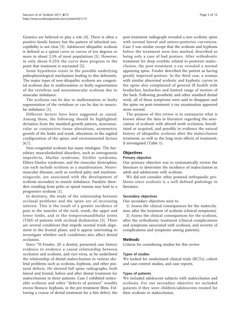
Table 1 Principal papers showed in this review.
Paper Type Main topic Sample Age Main result
McMaster J(1965)
Reference [7]
3 clinical cases Casual relationship betweenmalocclusion and scoliosis,
and viceversa
3 adolescents 10-15years
After orthodontic treatment, the authorobserved the improvment of posture
Paper Type Main topic Sample Age Main result
Rock andBaker (1972).Reference [9]
Case-report Class II due to the wearedcast
A girl 14 yearsold
to recommend the use of a removableappliance to prevent the malocclusionbefore the surgeon operation and
during the period of the wearing thecast.
Paper Type Main topic Sample Age Main result
Dayan et al.(1977).
Reference[10]
TransversalCase-control
study
To compare facialmorphology of children
affected with scoliosis andtreated with brace, with
health children
15 5-19years(mean
10 years)
Children treated with braces (for theirscoliosis) showed all vertical
measurements of face significantly lowerthan the control group, and moreprotruted maxillary and mandibular
bases
Paper Type Main topic Sample Age Main result
HotchcockHP (1969).
Reference [8]
Observationalstudy onprevalence
Plagiocephaly in subjects withscoliosis
144 The study suggested the existence of anassociation between infantile scoliosis
and plagiocephaly
Paper Type Main topic Sample Age Main result
Ben-Bassat Yet al. (2006)Reference
[16]
Observationalstudy onprevalence
Prevalence of scoliosis inpatients with ereditated
malocclusion
202 adolescents 10-15 The detection of hereditary orthodonticanomalies in young children allows theidentification of a group of children whohave a high risk of developing scoliosis
in later years.
Paper Type Main topic Sample Age Main result
Segatto et al.(2008)
Reference[17]
Cohort study Malocclusion in subjects withidiopathic scoliosis
98 subjects with scoliosis and705 controls
6.2 -25.3;mean
age 13.9+/- 3.5
a significant higher prevalence ofunilateral Angle class II (asymmetric classII malocclusion) was evident among thepatients with scoliosis (21.9%) compared
with the control group (8.5%). Thedifferences between the two groups inthe prevalence of the midline deviationwere statistically significant both in theupper and the lower dental arches.
Paper Type Main topic Sample Age Main result
Lippold C etal. (2003).Reference
[18]
Case-controlstudy
To evaluate the differencesin occlusion
28 with scoliosis and 68health children
Meanage
14.7 +/-2.3
In the group of adolescents withscoliosis, infacts, the unilateral Angle
class II relationship showed a significanthigher prevalence respect to the control
group
Paper Type Main topic Sample Age Main result
Lippold etal. (2007)Reference
[19]
Observational To compare 53 adult patients with ClassII and Class III, but withut
scoliosis
24.6 +/-9
an orthopedic examination can beconsidered for patients undergoing anorthodontic-operative therapy, alsowhen they don’t show scoliosis.
Paper Type Main topic Sample Age Main result
KorbmacherH. et al.(2007).
Reference[22]
Case-controlstudy
Prevalence of scoliosis insubjects with jaw deformity
85 patients with jawdeformity and 20 control
subjects
adults Of the 85 patients with jaw deformity,23 (27.1%) had a Cobb angle exceeding10°. None of the control group had
scoliosis exceeding 10°.
Paper Type Main topic Sample Age Main result
Pedrotti etal. (2007).Reference
[23]
Case-control To assess the congruenceof the laterality of cross-bite and the orthopaedic
asymmetry
55 children with unilateralcross-bite, and 55 childrenwith asymmetric cervicalspine (and no cross-bite)
3-10 among the children who revealed anasymmetric upper cervical spine, theunilateral crossbite was not necessarily
combined with a pathologicalorthopaedic variable,
Saccucci et al. Scoliosis 2011, 6:15http://www.scoliosisjournal.com/content/6/1/15
Page 3 of 15
Type of studiesWe included studies that reported incidence and descip-tion of malocclusion associated with scoliosis.
Types of outcomesOur primary and first secondary outcome of interestwas incidence and description of malocclusion in sub-jects with scoliosis.Our secondary outcomes of interest were the clinical
consequences associated with treatments of malocclu-sions or scoliosis.
Electronic searches and data retrievalWe searched MEDLINE and EMBASE without languagerestrictions in September 2010. We also manuallysearched reference lists from recent review articles. Allrandomised and controlled clinical trials identified fromthe Cochrane Oral Health Group Trials Register, aMEDLINE search using the Mesh term scoliosis, maloc-clusion, and relevant free text words, handsearching theBritish, European and American journals of orthodonticsand Angle Orthodontist, and the bibliographies ofpapers and review articles which reported the outcomeof orthodontic treatment in subjects with scoliosis thatwere published as abstracts or papers between 1970 and2010.
Study selection and Data Extractiones of interventionsTwo reviewers (ST and MS) independently reviewed theabstracts for potential eligibility and subsequently fulltext publications for eligibility. Disagreements wereresolved by discussion.We extracted a number of variables on study design
and methodological characteristics, patient and interven-tion characteristics, and outcomes from all eligible
studies (see Table 1). Data extraction was done indepen-dently by two reviewers (ST and MS) and disagreementswere resolved by discussion.
Methodological Quality AssessmentNo Randomized clinical trials were recorded for thisargument.For the observational longitudinal studies we noted
the presence of control groups only in a few studies.The great part of transversal studies showed a controlgroup.
ResultsUsing the search strategy eleven observational longitudi-nal studies were identified. No randomized clinical trialswere recorded. Twenty-three cross-sectional studieswere recorderd, and the others studies was reviews, edi-torials, case-reports, or opinions. The clinical trials wereoften not controlled and were about the cephalometricevaluation after treatment with the modified Milwuakeebrace, followed by the orthodontic treatment of theclass II relationship with a functional appliance. Clinicaltrials also included the study of the associations betweenscoliosis and unilateral crossbite, in children with asym-metry of the upper cervical spine. This association wasalso investigated in rats, pigs and rabbits in clinicaltrials. The other associations between scoliosis andocclusion seems to be based only on cross-sectional stu-dies, case-reports, opinions.
Scoliosis and PlagiocephalyIn literature, the association between plagyocephaly andscoliosis was observed during ‘80 decade in prematureinfants. The existence of an association is based on clin-ical case-reports, opinins and cross-sectional studies.
Table 1 Principal papers showed in this review. (Continued)
Paper Type Main topic Sample Age Main result
Lippold etal. (2000)Reference
[24]
A prevalencestuydy
Prevalence of bilateralcrossbite in subjects with
scoliosis
428 9-14 an incidence of scoliotic attitudes of9.5%, with a statistically significantrelationship among that disorders ofposture, and the presence of ogival
palate with bilateral crossbite
Paper Type Main topic Sample Age Main result
Azuma Y etal. (1999);D’Attilio Met al. (2005);Poikela A etal. (1997);Nerder PHet al. (1999).References[34-37]
Animalstudies
The appearence of scoliosisafter an imbalance of
occlusion
animals / these experimental studies revealed ahigh level of asymmetry in craniofacialstructures, temporomandibular structures
and muscle functions after anexperimentally induced crossbite
Saccucci et al. Scoliosis 2011, 6:15http://www.scoliosisjournal.com/content/6/1/15
Page 4 of 15
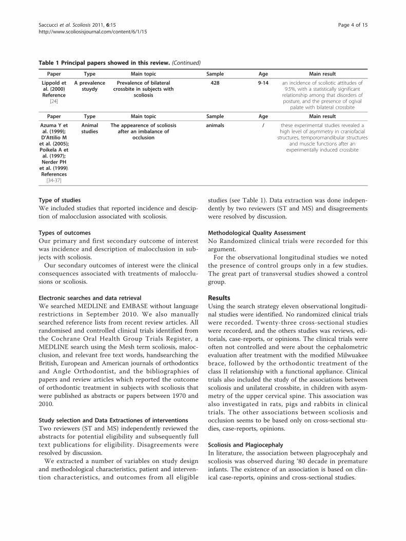
In a study performed on 144 infantile patients whoattended the Edinburgh Scoliosis Clinic between 1968and 1982, [9] plagiocephaly was present in 124 infants(86%) and absent in nine (6%), all with resolving curves;no clinical records had been made in the remaining 11infants (8%). In the patients with progressive curves,either single or double curves, the “recessed” side of theplagiocephaly always corresponded with the convex sideof the thoracic or thoracolumbar curve, suggesting theexistence of an association between infantile scoliosisand plagiocephaly [8]. (Figure 1)Also in the patients affected by scoliosis and plagioce-
phaly, who showed a resolving scoliotic curve, the“recessed” side of the head corresponded with the con-vex side of the curve [8,9].The association between these two conditions has
been explained by the nature of plagiocephaly, that is aplastic deformation of the skull. It was hypothesizedthat when an immobile infant habitually lies towardsone side (the case of premature babies) the action ofgravity on the plastic skull could cause the uppermostside of the face and head to flow backwards andbecome recessed, while the lower ear is pushed for-wards producing the commonly associated contralat-eral “bat ear”. Associated with this immobility,plagiocephaly, however, rarely persists and once thechild becomes mobile, it usually resolves by the age ofsix years. The scoliosis in these infants was rarelynoted at birth but, like the plagiocephaly, developedwithin the first six months of life in 70% of subjects.In the cited sample, the convex side of the curve cor-responded with the recessed side of the head in allexcept four infants with resolving curves. This closeassociation between the presence, time of presentationand side of the two deformities (both plagiocephalyand infantile idiopathic scoliosis) suggested a possiblecommon pathogenesis.
The Milwuakee brace and malocclusionA lot of studies made on 60’ and 70’ years on the use ofthe original Milwuakee brace in scoliosis therapydemonstrated the damageable effects on teeth occlusion.About this argument, longitudinal clinical trials were
recorded, in addition to clinical cases and observationalstudies.In 1969, a clinical case was published about the ortho-
dontic treatment of a class II malocclusion in a patient,probably caused by the cast worn after the surgeonoperation to reduce the scoliosis, in a fourteen year oldgirl who received the Harrington operation in 1963 [10].This article suggest the great attention given to theassociation between class II malocclusion (Figures 2, 3,4 and 5) and the use of this type of orthopedic brace,due to the presence of a force on the chin (Figure 6);the correction of the class II malocclusion requested thewearing of only a removable appliance (a positioner).The patient was treated soon after the operation and,for a year later, with a positioner, after which the maloc-clusion resulted corrected. For this, the conclusion ofthat article was to recommend the use of this removableappliance to prevent the malocclusion before the sur-geon operation and during the period of wearing thecast.In the April of 1972, the effect of the Milwaukee brace
upon dentofacial growth was investigated in a longitudi-nal clinical trial in a large sample and compared with acontrol group [11]. Measurements of facial morphologyat different age of subjects affected by scoliosis and
Figure 1 Left sided plagiocephaly with controlateral bat ear.Tracted by the paper referenced in [3].
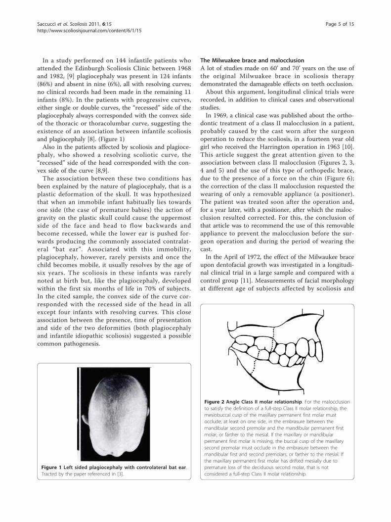
Figure 2 Angle Class II molar relationship. For the malocclusionto satisfy the definition of a full-step Class II molar relationship, themesiobuccal cusp of the maxillary permanent first molar mustocclude, at least on one side, in the embrasure between themandibular second premolar and the mandibular permanent firstmolar, or farther to the mesial. If the maxillary or mandibularpermanent first molar is missing, the buccal cusp of the maxillarysecond premolar must occlude in the embrasure between themandibular first and second premolars, or farther to the mesial. Ifthe maxillary permanent first molar has drifted mesially due topremature loss of the deciduous second molar, that is notconsidered a full-step Class II molar relationship.
Saccucci et al. Scoliosis 2011, 6:15http://www.scoliosisjournal.com/content/6/1/15
Page 5 of 15
treated with this device, were compared of a healthmatched sample. The age range was about 5 to 19 years,with a mean of 10 years. The differences were associatedto the wearing of the brace (Figure 7). All vertical mea-surements of face were significantly lower than the con-trol group. The mandible and the maxilla weresignificantly more protruted in the study group than inthe control one. The suggestion was to wear a teethpositioner during the therapy. However, as no pre-treat-ment data were available, it is not sure that these char-acteristics were caused by the brace, although it isevident that the brace can reduce the vertical dimensionof the face. The Milwaukee brace has undergone manymodifications since its creation. The chin pad in the ori-ginal brace was replaced by a plastic throat piece in alower position and closer to the neck. In this newdesign, its posture was underneath the body of themandible just above the thyroid cartilage, so that thepatient would not be able to rest the mandible on thethroat piece, as was previously done with the chin pad.The rigid occipital pad was changed into flexible plasticuprights to allow the patient to tip his head backwards.This modified brace was more comfortable to wear with
less pressure under the mandible. Also the use of aremovable splint was suggested to avoid dental conse-quences when the patient did not show permanentteeth, and wore the brace for more than 24 months[12]. The effect of an orthodontic device was also evi-denced in clinical case-reports [13,14]. Bracing is nor-mally done when the patient has bone growthremaining and is generally implemented to hold thecurve and prevent it from progressing to the point
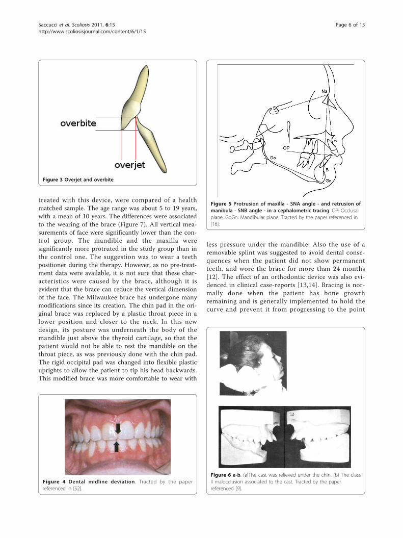
Figure 5 Protrusion of maxilla - SNA angle - and retrusion ofmanibula - SNB angle - in a cephalometric tracing. OP: Occlusalplane; GoGn: Mandibular plane. Tracted by the paper referenced in[16].
Figure 6 a-b. (a)The cast was relieved under the chin. (b) The classII malocclusion associated to the cast. Tracted by the paperreferenced [9].
Figure 3 Overjet and overbite.
Figure 4 Dental midline deviation. Tracted by the paperreferenced in [52].
Saccucci et al. Scoliosis 2011, 6:15http://www.scoliosisjournal.com/content/6/1/15
Page 6 of 15
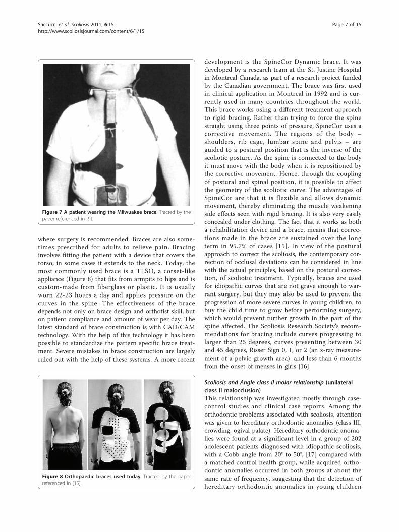
where surgery is recommended. Braces are also some-times prescribed for adults to relieve pain. Bracinginvolves fitting the patient with a device that covers thetorso; in some cases it extends to the neck. Today, themost commonly used brace is a TLSO, a corset-likeappliance (Figure 8) that fits from armpits to hips and iscustom-made from fiberglass or plastic. It is usuallyworn 22-23 hours a day and applies pressure on thecurves in the spine. The effectiveness of the bracedepends not only on brace design and orthotist skill, buton patient compliance and amount of wear per day. Thelatest standard of brace construction is with CAD/CAMtechnology. With the help of this technology it has beenpossible to standardize the pattern specific brace treat-ment. Severe mistakes in brace construction are largelyruled out with the help of these systems. A more recent
development is the SpineCor Dynamic brace. It wasdeveloped by a research team at the St. Justine Hospitalin Montreal Canada, as part of a research project fundedby the Canadian government. The brace was first usedin clinical application in Montreal in 1992 and is cur-rently used in many countries throughout the world.This brace works using a different treatment approachto rigid bracing. Rather than trying to force the spinestraight using three points of pressure, SpineCor uses acorrective movement. The regions of the body –shoulders, rib cage, lumbar spine and pelvis – areguided to a postural position that is the inverse of thescoliotic posture. As the spine is connected to the bodyit must move with the body when it is repositioned bythe corrective movement. Hence, through the couplingof postural and spinal position, it is possible to affectthe geometry of the scoliotic curve. The advantages ofSpineCor are that it is flexible and allows dynamicmovement, thereby eliminating the muscle weakeningside effects seen with rigid bracing. It is also very easilyconcealed under clothing. The fact that it works as botha rehabilitation device and a brace, means that correc-tions made in the brace are sustained over the longterm in 95.7% of cases [15]. In view of the posturalapproach to correct the scoliosis, the contemporary cor-rection of occlusal deviations can be considered in linewith the actual principles, based on the postural correc-tion, of scoliotic treatment. Typically, braces are usedfor idiopathic curves that are not grave enough to war-rant surgery, but they may also be used to prevent theprogression of more severe curves in young children, tobuy the child time to grow before performing surgery,which would prevent further growth in the part of thespine affected. The Scoliosis Research Society’s recom-mendations for bracing include curves progressing tolarger than 25 degrees, curves presenting between 30and 45 degrees, Risser Sign 0, 1, or 2 (an x-ray measure-ment of a pelvic growth area), and less than 6 monthsfrom the onset of menses in girls [16].
Scoliosis and Angle class II molar relationship (unilateralclass II malocclusion)This relationship was investigated mostly through case-control studies and clinical case reports. Among theorthodontic problems associated with scoliosis, attentionwas given to hereditary orthodontic anomalies (class III,crowding, ogival palate). Hereditary orthodontic anoma-lies were found at a significant level in a group of 202adolescent patients diagnosed with idiopathic scoliosis,with a Cobb angle from 20° to 50°, [17] compared witha matched control health group, while acquired ortho-dontic anomalies occurred in both groups at about thesame rate of frequency, suggesting that the detection ofhereditary orthodontic anomalies in young children
Figure 8 Orthopaedic braces used today. Tracted by the paperreferenced in [15].
Figure 7 A patient wearing the Milwuakee brace. Tracted by thepaper referenced in [9].
Saccucci et al. Scoliosis 2011, 6:15http://www.scoliosisjournal.com/content/6/1/15
Page 7 of 15
allows the identification of a group of children who havea high risk of developing scoliosis in later years.In 2006, the occlusions (Figure 12, 3, 4 and 5) of
patients with idiopathic scoliosis were clinically exam-ined in a group of 96 consecutive orthopedic patientswith idiopathic scoliosis (79 females and 17 males: meanage, 13.9 y; SD: 3.5 y; range, 6.2-25.3 y) [18]. Occlusalfeatures of a random group of 705 children served asthe control. In the considered sample, the interarch rela-tionships in the antero-posterior dimension (Angle clas-sification) were similar in the 2 groups for the frequencydistributions for normocclusion and Class I malocclu-sion, but they were significantly different when con-cerned the Angle class II malocclusion (Figure 2, 3 and5).The distribution of the Angle class II malocclusion
was significantly different in the scoliotic patientsrespect to the orthopedic health group.Specifically, taking in consideration the group of sub-
jects with Class II malocclusion, (Figure 2) with a highoverjet (Figure 3), a significant higher prevalence of uni-lateral Angle class II (asymmetric class II malocclusion)was evident among the patients with scoliosis (21.9%)compared with the control group (8.5%), indicating thatthe asymmetry in the antero-posterior relationshipsseems a clinical sign associated with scoliosis.In particular, while the frequency of asymmetrical
molar relationships was identical in the scoliosis and thecontrol groups, great differences in the frequency ofasymmetrical canine relationships were encountered; thescoliosis patients were more asymmetric in this regard.In addition, in the same sample, the prevalence of uppermidline deviation (Figure 4) (this is a clear clinical signof occlusal asymmetry) was 21% in the group of scoliosisand 9.5% in the control group; at the same time, theprevalence of lower midline deviation was 53.7% in thestudy group and 32.9% in the control group. The differ-ences between the two groups in the prevalence of themidline deviation were statistically significant both inthe upper and the lower dental arches. No associationwas found between site, side, or severity of scoliosis andthe appearance or site of the malocclusion featuresexamined.Later, the severity of scoliosis was related to the occlu-
sal relationship again, but no significant relationship wasobserved between the severity of scoliosis and the occlu-sal characteristics.In 2008, in facts, the scoliosis was related again to the
Angle class malocclusion, [19] with the analysis of 28children with scoliosis at various degree of severity(mean age: 14.7 y; SD: 2.3 y) matched with a controlgroup of 68 orthopedically healthy children (mean age:14.8 y; SD: 0.11 y). In the group of scoliotic subjects, theindication of the corset was represented by the values of
Cobb angle > 20° measured at the level of main curva-ture, so these children belonged to the severe group. Inthe analyzed sample, nine children were wearing corsetbecause of the severity of their orthopedic malformation.The moderate subgroup consisted of children with mal-formations requiring no constant posture correction,namely wearing corset. Besides the clarified differentorthopedic situation, the selection criteria of the twogroups were: similar age, no previous orthodontic treat-ment, as well as no missing teeth, carious lesions, orpathologic periodontal status. In this sample, when ana-lyzing the sagittal deviations in the molar region, theincidence rate of the bilateral deviation, being present asa sign of symmetry, as well as of the unilateral occlusaldeviation, related to the asymmetry, revealed a signifi-cant importance. In the group of adolescents with sco-liosis, in fact, the unilateral Angle class II relationshipshowed a significant higher prevalence respect to thecontrol group (Figure 2). Specifically, in the group ofsubjects with scoliosis, the 57.12% showed a normalbilateral occlusion, but the 28.56% showed a unilateralAngle class II malocclusion, with a significant higherfrequency respect to the health group. This unilateraldeviation (unilateral Angle class II malocclusion) wascharacteristic for almost one-third of scoliotic subjects,while in the control health group its incidence rate washardly 8.82%. In the group of scoliotic subjects, the uni-lateral class II relationship was significantly higher fre-quent then the bilateral class II relationship, pointing onthe importance of the asymmetry of malocclusion, inrelation to the scoliosis.These studies are in accordance with what affirmed by
Lippold et al. (2003), [20] that the scoliotic curves occurin the frontal plane and - through the head posture thatis tilted sideways -play an important role in the develop-ment of the different dentofacial asymmetries. Results ofseveral studies, as seen, confirm a potential correlationbetween scoliosis and unilateral Class II malocclusion.Unilateral Class II malocclusion is not the only type of
malocclusion significantly associated to the scoliosis.Segatto et al. (2008) [18] analyzed also other occlusal
characteristics of the frontal region of dental arch andfound some other significant differences between thescoliotic and the health groups.In particular, the subjects with scoliosis showed a sig-
nificant higher overjet (see Figure 3 for details on thisvariable) and a higher midline deviation (Figure 4)(Table 2) respect to the control group. Then, the scolio-tic group was characterized by lower overbite (Figure 3for details on this variable) compared to the determinedmean values (3.10 mm) of the control health group(Table 3) [18].Finally, on the basis of the evaluation of cephalograms
in the same sample, a slightly protrusive maxilla and a
Saccucci et al. Scoliosis 2011, 6:15http://www.scoliosisjournal.com/content/6/1/15
Page 8 of 15
slightly retrusive mandibula (Figure 5), characteristic ofa class II skeletal discrepancy, resulted more pro-nounced in the scoliotic group than in the controlgroup [19].In addition to the studies that compared scoliotic to
healthy subjects, other investigations underlined a rela-tion between the occlusion and the vertebral columnalignment, also in not scoliotic subjects [20,21].These studies thus suggest a multidisciplinary ortho-
dontic and orthopaedic approach to patients who do notshow any clinical evidence of scoliosis or malocclusion.For example, in 2007, Lippold et al. noted [21] a rela-
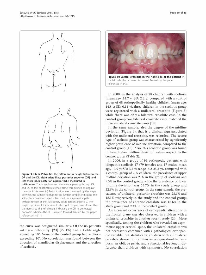
tionship between the pelvic tilt and pelvic torsion (Fig-ure 9a-b) and the facial shape (facial axis and facialdepth), variables which affect the occlusion of teeth andinfluence the orthodontic treatment.The study was performed on a group of fifty-three
adult patients (32 women and 21 men; mean age 24.6years, SD 9.0 years) with skeletal malformations (ClassII and III malocclusion) who came to medical center fora consultation regarding an orthodontic treatment, with-out anamnestically established motor or neurologicalfindings and/or previous internal or orthopaedicillnesses.In the sample, some correlations were observed with
the facial depth (mesial/distal) and the facial axis (verti-cal/horizontal).Patients with a vertical value on the facial axis and a
skeletal distal value in the facial depth (long face) had aslight pelvic torsion (Figure 9b) where the DL (left crista
iliaca posterior superior) was rotated backward withrespect to the DR (right crista iliaca posterior superior),while patients with a horizontal facial axis and mesialrelation of facial depth (short face) revealed a slightrotation of the DR rotated backward regarding the con-tralateral side. Although the investigation was performedon subjects without a diagnosed scoliosis, and on thebase of a rasterstereographic surface reconstruction ofthe back profile of a patient (and not a radiographic eva-luation of scoliosis), it suggested an extension of theinterdisciplinary concepts within the sense that anorthopedic examination can be considered for patientsundergoing an orthodontic-operative therapy.
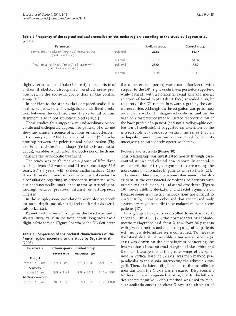
Scoliosis and crossbite (Figure 10)This relationship was investigated mostly through case-control studies and clinical case-reports. In general, itwas stated that left-right asymmetries are among themost common anomalies in patients with scoliosis [22].As seen in literature, these anomalies seem to be also
evident in the craniofacial complexes of patients withcertain malocclusions, as unilateral crossbites (Figure10), lower midline deviations, and facial asymmetries.Because some asymmetric malocclusions are difficult tocorrect fully, it was hypothesized that generalized bodyasymmetry might underlie these malocclusions in somepatients [17].In a group of subjects controlled from April 2002
through July 2003, [23] the posteroanterior cephalo-metric radiographs and chest X-rays from 85 patientswith jaw deformities and a control group of 20 patientswith no jaw deformities were controlled. To measurethe lateral shift of the mandible, a horizontal baseline (Xaxis) was drawn on the cephalogram connecting theintersection of the external margins of the orbits andthe most lateral points of the greater wings of the sphe-noid. A vertical baseline (Y axis) was then marked per-pendicular to the × axis, intersecting the ethmoid cristagalli. Then, the lateral displacement of the mandibularmentum from the Y axis was measured. Displacementto the right was designated positive; that to the left wasdesignated negative. Cobb’s method was used to mea-sure scoliosis curves on chest X-rays; the direction of
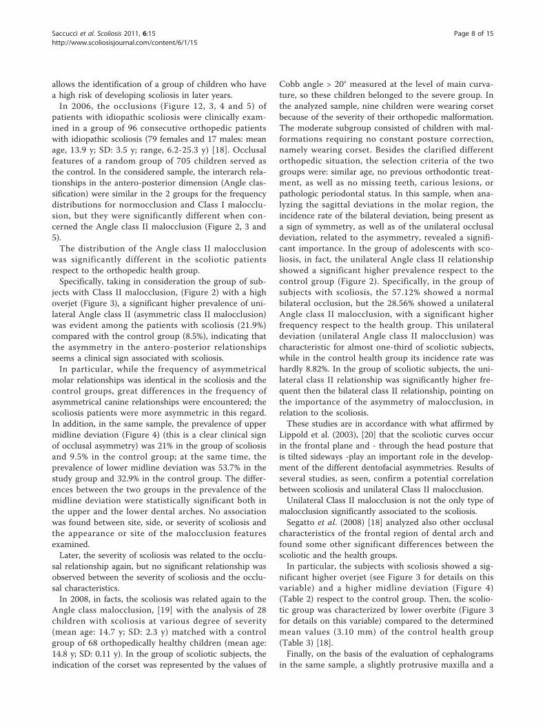
Table 2 Frequency of the sagittal occlusal anomalies on the molar region, according to the study by Segatto et al.(2008).
Parameters Scoliosis group Control group
Normal molar occlusion (Angle Cl.I) frequency (%)(health occlusion)
unilateral 28.56 16.17
bilateral 57.12 64.68
Distal molar occlusion (Angle Cl.II) frequency(%)(pathological occlusion)
unilateral 28.56 8.82
bilateral 10.07 16.17
Table 3 Comparison of the occlusal characteristics of thefrontal region, according to the study by Segatto et al.(2008).
Parameters Scoliosis group Control group
severe type moderate type
Overjet
mean ± SD (mm) 2.74 ± 1.851 2.55 ± 1.509 2.21 ± 1.201
Overbite
mean ± SD (mm) 2.58 ± 2.168 2.78 ± 1.715 3.10 ± 1.585
Midline deviation
mean ± SD (mm) 2.08 ± 1.121 1.76 ± 0.972 1.47 ± 0.898
Saccucci et al. Scoliosis 2011, 6:15http://www.scoliosisjournal.com/content/6/1/15
Page 9 of 15
the curve was designated similarly. Of the 85 patientswith jaw deformity, [23] (27.1%) had a Cobb angleexceeding 10°. None of the control group had scoliosisexceeding 10°. No correlation was found between thedirection of mandibular displacement and the directionof scoliosis.
In 2008, in the analysis of 28 children with scoliosis(mean age: 14.7 y; SD: 2.3 y) compared with a controlgroup of 68 orthopedically healthy children (mean age:14.8 y; SD: 0.11 y), three children in the scoliotic groupwere registered with a unilateral crossbite (Figure 8)while there was only a bilateral crossbite case. In thecontrol group two bilateral crossbite cases matched thethree unilateral crossbite cases [18].In the same sample, also the degree of the midline
deviation (Figure 4), that is a clinical sign associatedwith the unilateral crossbite, was recorded. The severetype of scoliotic group was characterized by significantlyhigher prevalence of midline deviation, compared to thecontrol group [18]. Also, this scoliotic group was foundto have higher midline deviation values respect to thecontrol group (Table 2).In 2006, in a group of 96 orthopedic patients with
idiopathic scoliosis 17 (79 females and 17 males: meanage, 13.9 y; SD: 3.5 y; range, 6.2-25.3 y), compared witha control group of 705 children, the prevalence of uppermidline deviation was 21% in the group of scoliosis and9.5% in the control group; while the prevalence of lowermidline deviation was 53.7% in the study group and32.9% in the control group. In the same sample, the pre-valence of unilateral posterior crossbite was 28.1% and18.1% respectively in the study and the control group;the prevalence of anterior crossbite was 16.6% in thestudy group and 9.3% in the control group.An increased occurrence of orthopaedic alterations in
the frontal plane was also observed in children with aunilateral crossbite in another recent study [24]. Morespecifically, among the children who revealed an asym-metric upper cervical spine, the unilateral crossbite wasnot necessarily combined with a pathological orthopae-dic variable, but statistically, children with a unilateralcrossbite showed more often an oblique shoulder, sco-liosis, an oblique pelvis, and a functional leg length dif-ference than children with symmetry. No correlation
Figure 9 a-b. (a)Pelvic tilt: the difference in height between theDR and the DL (right crista iliaca posterior superior [DR], andleft crista iliaca posterior superior [DL]) measured inmillimeters. The angle between the vertical passing through DRand DL to the horizontal reference plane was defined as angularmeasure in degrees. (b) Pelvic torsion was measured by the anglebetween the surface normals to the lumbar dimples indicating thespina iliaca posterior superior landmark. In a symmetric pelviswithout torsion of the iliac bones, pelvic torsion angle is 0. Theangle is positive if the normal to the right dimple points lower thanthe normal to the left dimple, indicating the DR to be rotatedbackward whereas the DL is rotated forward. Tracted by the paperreferenced in [11].
Figure 10 Lateral crossbite in the right side of the patient. Inthe left side, the occlusion is normal. Tracted by the paperreferenced in [43].
Saccucci et al. Scoliosis 2011, 6:15http://www.scoliosisjournal.com/content/6/1/15
Page 10 of 15
was found between the laterality of the crossbite sideand any orthopaedic asymmetry. The study was con-ducted comparing fifty-five children aged 3-10 years (22girls and 33 boys) with a unilateral crossbite and 55 gen-der- and age-matched children with asymmetric uppercervical spine, but no crossbite, who served as the con-trol group, selected from an orthopaedic cohort of 240patients. The certain asymmetry of the upper cervicalregion was confirmed in all the subjects by radiographsand palpation.Also, in 2008, in the analysis of 28 children with sco-
liosis (mean age: 14.7 y; SD: 2.3 y) compared with a con-trol group of 68 orthopedically healthy children (meanage: 14.8 y; SD: 0.11 y), 18 the clinical examination ofthe Temporo-mandibular joint (TMJ) at almost a quar-ter of the scoliotic group revealed a pathological symp-tom: the mandibular lateral movements showed areduced range in only one side. More specifically, onlyhalf of the patients in the scoliotic group were able tomake the same range of bilateral movements. On thecontrary, this rate was 82.32% in the control group.The results of all these cited studies suggest that den-
tal asymmetries correlate with orthopaedic asymmetriesin the frontal plane, when the analysis is conducted in asample of young boys and girls.However, it must be noted that also the bilateral
crossbite was related to the scoliosis. In 2007, also a cor-relation between scoliosis and bilateral crossbite wasreported: in a study on 428 subjects (211 females and217 males), aged 9 to 14 years, 25 a 2.8% scoliosis inci-dence has emerged, and an incidence of scoliotic atti-tudes of 9.5%, with a statistically significant relationshipamong that disorders of posture, and the presence ofogival palate with bilateral crossbite.Among the studies on the association between asym-
metric occlusion and trunk asymmetry, a few studiesmust be cited, that have investigated these co-relation-ship in health subjects without any pathological ortho-paedic condition [25-28]. They reported deviatingfindings: Lippold et al. (2000) [25] found a statisticallysignificant correlation between midline deviation andoblique pelvis as well as leg length differences, consid-ered in the limits of physiology. The other two studies[26,27] showed that moderate trunk asymmetry (notpathological condition) did not affect facial asymmetryor vice versa. With regard to the study design and theinvestigated patients, the three studies can hardly becompared: one [26] compared 29 children with a right-sided midline shift with 28 children with a symmetricocclusion; Lippold et al. (2000) [25] investigated midlinediscrepancies in 50 patients, aged 4-55 y, who wererecruited from physiotherapy appointments, while Zepaet al. (2003) [27] analyzed frontal cephalograms and
compared them with rib hump or lumbar prominenceand spinal posture.In order to investigate the possible effects of orthopae-
dic asymmetric disorders on dentofacial developmentand head posture, other clinical studies have been pre-viously conducted on patients with scoliosis, and theresults given by this previous literature are very similarto the more recent cited studies.In general, in the previous literature on this field, the
statistically recorded prevalence of unilateral crossbitesin subjects with scoliosis amounted to 26-55 per cent[28-31].Prager (1980) [30] interpreted the crossbite as a trans-
mission of the asymmetry of the body, whereas Hirsch-felder and Hirschfelder (1983) [32] considered, althoughthey had not yet clarified transmission, the crossbite tobe a new compensatory curvature of a scoliosis. Inde-pendent of the different offered explanations of the highprevalence of crossbite in those patients, an interdisci-plinary treatment approach to alleviate facial asymmetryand to stabilize head posture, initiated as early as possi-ble, has been unanimously recommended since ‘60 dec-ade [28,33,34].But it must be underlined that several studies about
the association between unilateral crossbite and scoliosiswere also conducted on animals, and the obtainedresults tended to confirm the observations recorded onhumans.In general, the results from experimental animal stu-
dies suggested that alterations in the occlusion evokechanges in many other regions of the body [35-37].For example, teeth occlusion seems to have an impact
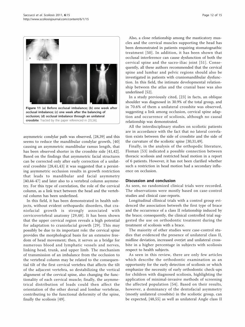
on head position, spinal column alignment, and mastica-tory muscles which control posture and modulate car-diac function via the trigeminal system. After unilateralocclusal destruction, a postural abnormality in terms ofinability of head maintenance, T-wave inversion on elec-trocardiograph, hair loss, changes in tongue mobility,and eating disorders as well as pathologies of the eyehave been observed [35]. Then, a scoliotic curve hasbeen developed after induction of a unilateral crossbitein rats [34,36]. In these studies, the evoked changeswere observed within 1 week of unilateral manipulationand normalized after harmonization of the occlusalplane (Figure 11)Experimental studies also tried to explain the origin
and the mechanism for the occurrence of an asymmetricgrowth of the head due to a unilateral crossbite: [38]more specifically, this experimental study in rabbitsrevealed a high level of asymmetry in craniofacial struc-tures, temporomandibular structures and muscle func-tions in rabbits after an experimentally inducedcrossbite [37]. Moreover, in patients with a unilateralmalocclusion, asymmetric condylar position with an
Saccucci et al. Scoliosis 2011, 6:15http://www.scoliosisjournal.com/content/6/1/15
Page 11 of 15
asymmetric condylar path was observed, [28,39] and thisseems to reduce the mandibular condylar growth, [40]causing an asymmetric mandibular ramus length, thathas been observed shorter in the crossbite side [41,42].Based on the findings that asymmetric facial structurescan be corrected only after early correction of a unilat-eral crossbite [28,41,43] it was suggested that a persist-ing asymmetric occlusion results in growth restrictionthat leads to mandibular and facial asymmetry[40,44-47] and later also to a vertebral column asymme-try. For this type of correlation, the role of the cervicalcolumn, as a link tract between the head and the verteb-ral column has been underlined.In this field, it has been demonstrated in health sub-
jects, without evident orthopaedic disorders, that cra-niofacial growth is strongly associated withcervicovertebral anatomy [29,48]. It has been shownthat the upper cervical region reveals a high potentialfor adaptation to craniofacial growth [29]. This maypossibly be due to its important role: the cervical spineprovides the morphological basis for an extensive free-dom of head movement; then, it serves as a bridge fornumerous blood and lymphatic vessels and nerves,linking head, trunk, and upper limb. The mechanismof transmission of an imbalance from the occlusion tothe vertebral column may be related to the consequen-tial tilt of the first cervical vertebra that affects the tiltof the adjacent vertebra, so destabilizing the verticalalignment of the cervical spine, also changing the func-tionality of each cervical muscle; finally, the asymme-trical distribution of loads could then affect theorientation of the other dorsal and lombar vertebrae,contributing to the functional deformity of the spine,finally the scoliosis [49].
Also, a close relationship among the masticatory mus-cles and the cervical muscles supporting the head hasbeen demonstrated in patients requiring stomatognathictreatment [50]. In addition, it has been shown thatocclusal interference can cause dysfunction of both thecervical spine and the sacro-iliac joint [51]. Conse-quently, all these authors recommended that the cervicalspine and lumbar and pelvic regions should also beinvestigated in patients with craniomandibular dysfunc-tion. In this field, the intimate developmental relation-ship between the atlas and the cranial base was alsounderlined [52].In a study previously cited, [23] in facts, an oblique
shoulder was diagnosed in 30.9% of the total group, andin 70.6% of them a unilateral crossbite was observed,suggesting a link among occlusion, cervical spine adap-tion and occurrence of scoliosis, although no causalrelationship was demonstrated.All the interdisciplinary studies on scoliotic patients
are in accordance with the fact that no lateral correla-tion exists between the side of crossbite and the side ofthe curvature of the scoliotic spine [30,31,49].Finally, in the analysis of the orthopedic literature,
Floman [53] indicated a possible connection betweenthoracic scoliosis and restricted head motion in a reportof 6 patients. However, it has not been clarified whethersuch a restriction in head motion had a secondary influ-ence on occlusion.
Discussion and conclusionAs seen, no randomized clinical trials were recorded.The observations were mostly based on case-controlstudies and clinical case-reports.Longitudinal cllinical trials with a control group evi-
denced the association betweeh the first type of braceand the occurrence of a class II relationship induced bythe brace; consequently, the clinical controlled trial sug-gested the use on orthodontic treatment during thetreatment of scoliosis with a brace.The maiority of other studies were case-control stu-
dies that evidenced the presence of unilateral class II,midline deviation, increased overjet and unilateral cross-bite in a higher percentage in subjects with scoliosisrespect to health subjects.As seen in this review, there are only few articles
which describe the orthodontic examination as anopportunity for the early detection of scoliosis or whichemphasize the necessity of early orthodontic check-upsfor children with diagnosed scoliosis, highlighting theapplication of minimal-invasive methods of screeningthe affected population [54]. Based on their results,however, a dominancy of the dentofacial asymmetry(mostly unilateral crossbite) in the scoliotic group, canbe expected, [48,55] as well as unilateral Angle class II
D
Figure 11 (a) Before occlusal imbalance; (b) one week afterocclusal imbalance; (c) one week after the balancing ofocclusion; (d) occlusal imbalance through an unilateralcrossbite. Tracted by the paper referenced in [35,56].
Saccucci et al. Scoliosis 2011, 6:15http://www.scoliosisjournal.com/content/6/1/15
Page 12 of 15
malocclusion and midline deviation. As seen in thisreview, the data in literature prove also the existence ofother dentofacial anomalies in children with scoliosis(ogival palate, increased overjet, reduced overbite,reduced range in lateral movements in one side),although these studies did not analyzed the orthopaedicsample on the base of the scoliotic angle, or the pre-sence of one or more curves, and on the location of thecurvature, which may affect the gravity and the type ofscoliosis.Some hypothesis to explain the correlation between
the occlusion and the scoliosis can be made, althoughwithout certain conclusions.The assumption on which the hypothesis on this cor-
relation is based is that there is an anatomical and func-tional relationship between the stomatognathicapparatus and the spinal column. This relationship washypothesized by several authors, based upon variousobservations [56].Neurophysiological principles of convergence and sen-
sitization: a constant input, such as a nociceptive input,on second-order neurons may increase the sensitivity ofthese neurons. Then, non-nociceptive neural impulsesfrom other areas within the same segment, which con-verge onto these neurons, may give rise to altered sensa-tions from these areas. For the craniocervical region, forexample, a constant nociceptive input from, the upperpart of the trapezius muscle can lead to an increasedsensitivity of the spinal trigeminal nucleus and, conse-quently, non-nociceptive stimuli from the masticatorysystem would then lead to painful sensations from thetrigeminal region [55]. This occurs as the different inputconverges onto the nucleus caudal portion of the tri-geminal spinal tract nucleus [55]. As a consequence, forexample, a significantly higher prevalence of cervicalspinal pain was observed in a group of patients with cra-niomandibular pain than in a matched control groupwithout craniomandibular pain, thus causing posturaldisease, and affecting in final the whole vertebralcolumn.2. Anatomical details: There is an anatomical relation-
ship between the mandible and the cervical column,since the cranium and the mandible have muscular andligament attachments to the cervical area. The functionof the head, neck, and jaws is closely interrelated, form-ing a combined functional system [55,56]. observed asignificant correlation between mandibular length andcervical lordosis angle on lateral skull radiographs (innatural head position) in Caucasian adult women with askeletal class II malocclusion. The longer the mandibu-lar body was, the straighter the cervical columnappeared to be [56]. In a group of 50 Caucasian adultwomen with internal derangement, compared with acontrol group of 50 Caucasian women without internal
derangement, cephalometric tracings on lateral skullradiographs in natural head position showed a signifi-cantly lower cervical lordosis angle. Beyond possiblycausing TMJ diseases, dental malocclusions could, bythe same mechanism, be linked to a functional asymme-try of trunk muscles. We suggest that one pathway isthrough the atlas. The atlas is linked to occipital con-dyles and thus affect the rest of the spine alignment,leading to further profound compensatory chenges, thatmay become pathological.In conclusion, from a clinical point of view, if the
asymmetry underlying idiopathic scoliosis and asym-metric malocclusion originates from the same etiology,it might be difficult to fully correct all features of themalocclusion or maintain the correction. This difficultywas observed, for example, in patients with posteriorcrossbites in whom relapse of lower midline deviationsor tendency toward crossbites was evident also afterorthodontic treatment. To clarify this point, the possibi-lity of a connection between the reverse cycle in masti-catory movements and asymmetrical posture should beevaluated.In conclusion, all the observed frequent and severe
dentofacial deviations in the scoliotic group draw theattention to the necessity of the early examination ofthis patient group from an orthodontics and orthopaedicpoint of view.However, whether scoliosis affects mandibular den-
toalveolar symmetry (whether there is a causal relation-ship) needs further elucidation.
Acknowledgements and FundingWe acknowledge Editage for revision of english language.
Author details1Department of Oral Science, University La Sapienza, Via Caserta 6, 00161,Rome, Italy. 2Department of Surgical Reconstructive Sciences and AdvancedTechnologie, University of Insubria, Via G. Piatti, 10 - 21100 Velate, Varese,Italy. 3Department of Health Science, University of L’Aquila, Edificio Delta 6L’Aquila Fraz. Coppito, 67010, L’Aquila, Italy. 4Department of Oral Science,Nano and Biotechnology, University G.D’Annunzio, Via dei Vestini 31, 66013,Chieti, Italy.
Authors’ contributionsST and MSare the Principal Investigator of this review. They 1) have madesubstantial contributions to conception and design, acquisition of data,analysis and interpretation of data; 2) have been involved in drafting themanuscript or revising it critically for important intellectual content; and 3)have given final approval of the version to be published. ST and MScontributed in the same way, as principal investigators, to this research. LTparticipated in drafting the manuscript. SM helped in the revision of themanuscript. AP helped in the first revision of the manuscript. FF participatedin drafting the manuscript. All authors read and approved the finalmanuscript.
Competing interestsThe authors declare that they have no competing interests.
Received: 21 January 2011 Accepted: 29 July 2011Published: 29 July 2011
Saccucci et al. Scoliosis 2011, 6:15http://www.scoliosisjournal.com/content/6/1/15
Page 13 of 15
References1. Trobisch P, Suess O, Schwab F: Idiopathic Scoliosis. Dtsch Arztebl Int 2010,
107:875-884.2. Riseborough EJ, Wynne-Davies R: A genetic survey of idiopathic scoliosis
in Boston, Massachusetts. J Bone Joint Surg Am 1973, 55:974-982.3. Brown JK, Bell E, Fulford GE: Mechanism of deformity in children with
cerebral palsy-with special reference to postural deformity. P/idiotForthidk Praxis 1982, 53:78-94.
4. Kouwenhoven JW, Castelein RM: The pathogenesis of adolescentidiopathic scoliosis: review of the literature. Spine 2008, 33:2898-2908.
5. Visscher CM, Lobbezoo F, de Boer W, van der Zaag J, Naeije M: Prevalenceof cervical spinal pain in craniomandibular pain patients. Eur J OralScience 2001, 109:76-80.
6. Alden KJ, Marosy B, Nzegwu N, Justice CM, Wilson AF, Miller NH: Idiopathicscoliosis: Identification of candidate regions on chromosome 19p13.Spine 2006, 31:1815-1819.
7. White AA, Panjabi MM: Clinical Biomechanics of the Spine. Lippincott , 21990, ISBN 0-397-50720-8.
8. Hitchcock HP: Treatment of malocclusion associated with scoliosis. AngleOrthod 1969, 39:64-68.
9. Rock VP, Baker R: The effect of Milwaukee brace upon dentofacialgrowth. Angle Orthod 1972, 42:96-102.
10. Dayan N, Abraham Z, Lieberman M: Cephalometric evaluation of an oralstabilizing appliance with the modified Milwuakee brace. Angle Orthod1977, 47:300-303.
11. Janicki JA, Poe-Kochert C, Armstrong DG, Thompson GH: A comparison ofthe thoracolumbosacral orthoses and providence orthosis in thetreatment of adolescent idiopathic scoliosis: results using the new SRSinclusion and assessment criteria for bracing studies. J Pediatr Orthop2007, 27:369-374.
12. Herring JA: Tachdjian’s Pediatric Orthopaedics Philadelphia PA: W.B. Saunders;2002.
13. Pavsic J: The effect of orthodontic measures on the growth of themandible in scoliosis patients during treatment with the Milwaukeebrace. Stomatol DDR 1983, 33:676-82.
14. Luedtke GL: Management of the dentition of patients under treatmentfor scoliosis using the Milwaukee brace. Am J Orthod 1970, 57:607-14.
15. Pećina M, Lulluć-Dukić O, Pećina-Hrncević A: Hereditary orthodonticanomalies and idiopathic scoliosis. Int Orthop 1991, 15:57-59.
16. Ben-Bassat Y, Yitschaky M, Kaplan L, Brin I: Occlusal patterns in patientswith idiopathic scoliosis. Am J Orthod Dentofac Orthop 2006, 130:629-633.
17. Segatto E, Lippold C, Végh A: Craniofacial features of children with spinaldeformities. BMC Musculoskelet Disord 2008, 22;9:169.
18. Lippold C, van den Bos L, Hohoff A, Danesh G, Ehmer U: Interdisciplinarystudy of orthopedic and orthodontic findings in pre-school infants. JOrofac Orthop 2003, 64:330-340.
19. Lippold C, Danesh G, Hoppe G, Drerup B, Hackenberg L: Trunk Inclination,Pelvic Tilt and Pelvic Rotation in Relation to the Craniofacial Morphologyin Adults. Angle Orthod 2007, 77:29-35.
20. Burwell RG: Aetiology of idiopathic scoliosis: current concepts. PediatrRehabil 2004, 6:137-170.
21. Ikemitsu H, Zeze R, Yuasa K, Izumi K: The relationship between jawdeformity and scoliosis. Oral Radiol 2006, 22:14-17.
22. Korbmacher H, Koch L, Eggers-Stroeder G, Kahl-Nieke B: Associationsbetween orthopaedic disturbances and unilateral crossbite in childrenwith asymmetry of the upper cervical spine. Eur J Orthod 2007,29:100-104.
23. Pedrotti L, Mora R, Bertani B, Tuvo G, Crivellari I: Association amongpostural and skull-cervico-mandibular disorders in childhood andadolescence. Analysis of 428 subjects. Pediatr Med Chir 2007, 29:94-98.
24. Lippold C, Ehmer U, van den Bos L: Beziehungen zwischenkieferorthopädischen und orthopädischen Befunden. Manuelle Medizin2000, 38:346-350.
25. Dußler E, Raab P, Kunz B, Kirschner S, Witt E: MandibuläreMittellinienverschiebungen und Asymmetrien des Halte- undBewegungsapparates bei Kindern und Jugendlichen. Manuelle Medizin.2002, 40:116-119.
26. Zepa I, Hurmerinta K, Kovero O, Nissinen M, Könönen M, Huggare J: Trunkasymmetry and facial symmetry in young adults. Acta Odontol Scand2003, 61:149-153.
27. Pirttiniemi P, Kantomaa T, Lahtela P: Relationship between craniofacialand condyle path asymmetry in unilateral cross-bite patients. Eur JOrthod 1990, 12:408-413.
28. Huggare J: Postural disorders and dentofacial morphology. Acta OdontolScand 1998, 56:383-386.
29. Prager A: Vergleichende Untersuchungen über die Häufigkeit vonZahnstellungs- und Kieferanomalien bei Patienten mit Deformitäten derWirbelsäule. Fortschr Kieferorthop 1980, 41:163-168.
30. Hirschfelder U, Hirschfelder H: Auswirkungen der Skoliose auf denGesichtsschädel. Fortschr Kieferorthop 1983, 44:457-467.
31. Müller-Wachendorff R: Untersuchungen über die Häufigkeit desAuftretens von Gebißanomalien in Verbindung mitSkelettdeformierungen mit besonderer Berücksichtigung der Skoliosen.Fortschr Kieferorthop 1961, 22:399-408.
32. Peéina M, Lulié-Dukié O, Peéina-Hrn , INCLUDEPICTURE: Hereditaryorthodontic anomalies and idiopathic scoliosis. Int Orthop 1991, 15:57-59[http://ejo.oxfordjournals.org/math/ccaron.gif\*MERGEFORMATINETeviéA].
33. Festa F, D’Attilio M, Vecchiet F: Effects of a horizontal oscillation of themandible on the spinal column of the rat in vivo using radiographicmonitoring. Orthognatodonzia Italiana 1997, 6:539-550.
34. Azuma Y, Maehara K, Tokunaga T, Hashimoto M, Ieoka K, Sakagami H:Systemic effects of the occlusal destruction in guinea pigs. In Vivo 1999,13:519-524.
35. D’Attilio M, Filippi MR, Femminella B, Festa F, Tecco S: The influence of anexperimentally-induced malocclusion on vertebral alignment in rats: acontrolled pilot study. J Craniomandib Pract 2005, 23:119-129.
36. Poikela A, Kantomaa T, Pirttiniemi P: Craniofacial growth after a period ofunilateral masticatory function in young rabbits. Eur J Oral Sci 1997,105:331-337.
37. Nerder PH, Bakke M, Solow B: The functional shift of the mandible inunilateral posterior crossbite and the adaptation of thetemporomandibular joints: a pilot study. Eur J Oral Sci 1999, 21:155-166.
38. Tadej G, Engström C, Borrman H, Christiansen EL: Mandibular condylemorphology in relation to malocclusions in children. Angle Orthod 1989,59:187-194.
39. Schmid W, Mongini F, Felisio A: A computer-based assessment ofstructural and displacement asymmetries of the mandible. Am J OrthodDentofac Orthop 1991, 100:19-34.
40. Santos Pinto A, Buschang PH, Throckmorton GS, Chen P: Morphologicaland positional asymmetries of young children with functional unilateralposterior crossbite. Am J Orthod Dentofac Orthop 2001, 120:13-20.
41. Mimura H, Deguchi T: Relationship between craniofacial and condylepath asymmetry in unilateral crossbite patients. J Craniomandib Pract1994, 12:161-166.
42. Pirttiniemi P, Raustia A, Kantomaa T, Pyhtinen J: Relationships ofbicondylar position to occlusal asymmetry. Eur J Orthod 1991, 12:441-445.
43. O’Bryrn BL, Sadowsky C, Schneider B, BeGole EA: An evaluation ofmandibular asymmetry in adults with unilateral posterior crossbite. Am JOrthod Dentofac Orthop 1995, 107:394-400.
44. Hesse KL, Årtun J, Joondeph DR, Kennedy DB: Changes in the condylarposition and occlusion associated with maxillary expansion forcorrection of functional unilateral posterior crossbite. Am J OrthodDentofac Orthop 1997, 111:410-418.
45. Langberg BL, Arai K, Miner RM: Transverse skeletal and dental asymmetryin adults with unilateral posterior crossbite. Am J Orthod Dentofac Orthop2005, 127:6-16.
46. Solow B, Siersbæk-Nielsen S: Cervical and craniocervical posture aspredictors of craniofacial growth. Am J Orthod Dentofac Orthop 1992,101:449-458.
47. Wong ML, Sandham A, Ang PK, Wong DC, Tan WC, Huggare J: Craniofacialmorphology, head posture, and nasal respiratory resistance inobstructive sleep apnoea: an inter-ethnic comparison. Eur J Orthod 2005,27:91-97.
48. Huggare JA, Raustia AM: Head posture and cervicovertebral andcraniofacial morphology in patients with craniomandibular dysfunction.J Craniomandib Pract 1992, 10:173-177.
49. Fink M, Stiesch-Scholz M, Tschernitschek H: The functional relationshipbetween the craniomandibular system, cervical spine, and the sacroiliacjoint: a preliminary investigation. J Craniomandib Pract 2003, 1:202-208.
Saccucci et al. Scoliosis 2011, 6:15http://www.scoliosisjournal.com/content/6/1/15
Page 14 of 15
50. Ludvig KS: Die Frühentwicklung des Atlas und der Occipitalwirbel beimMenschen. Acta Anatomica 1957, 30:444-461.
51. Floman Y: Thoracic scoliosis and restricted head motion: a newsyndrome? A report of six cases. Eur Spine J 1998, 7:155-157.
52. Korbmacher H, Eggers-Stroeder G, Koch L, Kahl-Nieke B: Correlationsbetween dentition anomalies and diseases of the of the postural andmovement apparatus–a literature review. J Orofac Orthop 2004,65:190-203.
53. Zepa I, Hurmerinta K, Kovero O, Nissinen M, Könönen M, Huggare J:Associations between thoracic kyphosis, head posture, and craniofacialmorphology in young adults. Acta Odontol Scand 2000, 58:237-242.
54. Okeson JP, ed: Orofacial pain. Guidelines for assessment, diagnosis, andmanagement. The American Academy of Orofacial Pain Chicago:Quintessence; 1996, 197.
55. Festa F, Tecco S, Dolci M, Ciuffolo F, Di Meo S, Filippi MR, Ferritto AL,D’Attilio M: Relation between cervical lordosis and facial morphology inCaucasian adult women with a skeletal class II malocclusion: atransversal study. J Craniomandib Pract 2003, 21:121-129.
56. D’Attilio M, Tecco S, Dolci M, Ciuffolo F, Epifania E, Filippi MR, Festa F:Cervical lordosis angle measured on lateral cephalograms: findings inskeletal class II female subjects with and without TMD. A cross-sectionalstudy. J Craniomandib Pract 2004, 22:17-44.
doi:10.1186/1748-7161-6-15Cite this article as: Saccucci et al.: Scoliosis and dental occlusion: areview of the literature. Scoliosis 2011 6:15.
Submit your next manuscript to BioMed Centraland take full advantage of:
• Convenient online submission
• Thorough peer review
• No space constraints or color figure charges
• Immediate publication on acceptance
• Inclusion in PubMed, CAS, Scopus and Google Scholar
• Research which is freely available for redistribution
Submit your manuscript at www.biomedcentral.com/submit
Saccucci et al. Scoliosis 2011, 6:15http://www.scoliosisjournal.com/content/6/1/15
Page 15 of 15
Related Documents


![]Dental Occlusion part 1](https://static.cupdf.com/doc/110x72/5885f84e1a28ab864f8b72bf/dental-occlusion-part-1.jpg)