Molecular Computation of Solutions to Combinatorial Problems Leonard M. Adleman Science, New Series, Vol. 266, No. 5187. (Nov. 11, 1994), pp. 1021-1024. Stable URL: http://links.jstor.org/sici?sici=0036-8075%2819941111%293%3A266%3A5187%3C1021%3AMCOSTC%3E2.0.CO%3B2-%23 Science is currently published by American Association for the Advancement of Science. Your use of the JSTOR archive indicates your acceptance of JSTOR's Terms and Conditions of Use, available at http://www.jstor.org/about/terms.html. JSTOR's Terms and Conditions of Use provides, in part, that unless you have obtained prior permission, you may not download an entire issue of a journal or multiple copies of articles, and you may use content in the JSTOR archive only for your personal, non-commercial use. Please contact the publisher regarding any further use of this work. Publisher contact information may be obtained at http://www.jstor.org/journals/aaas.html. Each copy of any part of a JSTOR transmission must contain the same copyright notice that appears on the screen or printed page of such transmission. The JSTOR Archive is a trusted digital repository providing for long-term preservation and access to leading academic journals and scholarly literature from around the world. The Archive is supported by libraries, scholarly societies, publishers, and foundations. It is an initiative of JSTOR, a not-for-profit organization with a mission to help the scholarly community take advantage of advances in technology. For more information regarding JSTOR, please contact [email protected]. http://www.jstor.org Tue Nov 13 20:10:32 2007

Welcome message from author
This document is posted to help you gain knowledge. Please leave a comment to let me know what you think about it! Share it to your friends and learn new things together.
Transcript
Molecular Computation of Solutions to Combinatorial Problems
Leonard M. Adleman
Science, New Series, Vol. 266, No. 5187. (Nov. 11, 1994), pp. 1021-1024.
Stable URL:
http://links.jstor.org/sici?sici=0036-8075%2819941111%293%3A266%3A5187%3C1021%3AMCOSTC%3E2.0.CO%3B2-%23
Science is currently published by American Association for the Advancement of Science.
Your use of the JSTOR archive indicates your acceptance of JSTOR's Terms and Conditions of Use, available athttp://www.jstor.org/about/terms.html. JSTOR's Terms and Conditions of Use provides, in part, that unless you have obtainedprior permission, you may not download an entire issue of a journal or multiple copies of articles, and you may use content inthe JSTOR archive only for your personal, non-commercial use.
Please contact the publisher regarding any further use of this work. Publisher contact information may be obtained athttp://www.jstor.org/journals/aaas.html.
Each copy of any part of a JSTOR transmission must contain the same copyright notice that appears on the screen or printedpage of such transmission.
The JSTOR Archive is a trusted digital repository providing for long-term preservation and access to leading academicjournals and scholarly literature from around the world. The Archive is supported by libraries, scholarly societies, publishers,and foundations. It is an initiative of JSTOR, a not-for-profit organization with a mission to help the scholarly community takeadvantage of advances in technology. For more information regarding JSTOR, please contact [email protected].
http://www.jstor.orgTue Nov 13 20:10:32 2007

era1 minutes to obtain near-field fluores-
cence spectra with good signal-to-noise ra-
tios. Furthermore, the recent work of Xie
and Dunn (33) and by Ambrose et al. (34)
showed that the metal-coated probe tip can
significantly perturb the electronic proper-
ties of the molecule being detected. In con-
trast, the far-field confocal fluorescence aD-
proach provides unlimited laser throughput
and a three-dimensional sectioning capabil-
ity and is truly noninvasive, although its
resolution is diffraction limited. These fea-
tures are expected to allow important appli-
cations such as enhanced Raman spectrosco-
py at the single-molecule level and on-line
fluorescence identification and sorting of in-
dividual molecules and auantum-confined
nanostructures. The extraordinary sensitivity
achieved in this work allows the direct, real-
time study of the dynamics of a single mol-
ecule and the chemical and biochemical re-
actions that such a molecule may undergo in
solution.
REFERENCES AND NOTES
1. W. E. Moerner, Science 265, 46 (1994), and refer-ences therein.
2. M. Orrit. J. Bernard. R. I. Personov.J. Phvs. Chem. 97, 1 0 k 6 (1993).
3. F. Guttler, T. Irnqartinqer,T. Plakhotnik, A. Renn, U. P. Wild, Chem. Phys.~ e t t .217, 393 (1994).
4. M, shikawa, K. Hirano, T. Hayakawa, S. Hosoi, S. Brenner,Jpn. J Appl. Phys. 33, 1571 (1994).
5. E. Betzig and R. J. Chichester, Science 262, 1422 ( I993).
6. J. K. Trautman, J. J. Macklin, L. E. Brus, E. Betzig, Nature 369, 40 (1994).
7. D. C. Nguyen, R. A. Keler, J. H. Jett, J. C. Martin, Anal. Chem. 59, 2158 (1987).
8. K. Peck, L. Stryer,A. N. Glazer, R. A. Mathies,Proc. Natl. Acad. Sci. U.S.A. 86, 4087 (1989).
9. W. B. Whitten, L. M. Ramsey, S. A. Arnold, B. V. Bronk,Anal. Chem. 63, 1027 (1991); K. C. Ng, W. B. Whitten, S. A. Arnold, L. M. Ramsey,ibid. 64, 2914 (1992).
10. M. Eigen and R. Rigler, Proc. Natl. Acad. Sci. U.S.A. 91,5740 (1994).
11. In fluorescence correlation spectroscopy, the inten-sity recorded at time t is multiplied by that recorded at t + At , and the product is integrated over a finite period of time; see D. E. Koppel, Phys. Rev. A 10, 1938 (1974).
12. T. T. Perkins,D. E. Smith, S. Chu, Science 264, 819 (1994); T. T. Perkns, S. R. Quake, D. E. Smith, S. Chu, ibid., p. 822.
13. S. B. Smith, L. Finzi,C. Bustamante, ibid. 258, 1122 (1992); C. Bustamante, Annu. Rev. Biophys. Bio-phys. Chem. 20, 415 (1991).
14. M.Washizu and 0.Kurosawa,IEEETrans, ind. Appi. 26, 1165 (1990).
15. S. B. Smith, P. K. Aldridge, J. B. Calis, Science 243, 203 (1989).
16. N. J. Rampino and A. Chrambach, Anal. Biochem. 194, 278 (1991).
17. K. Morikawa and M. Yanagida,J. Biochem. 89, 693 (1981).
18. I . Auzanneau, C. Barreau, L. Salome, C. R. Acad. Sci. Paris 316, 459 (1993)
19. H. Kabata et a/.,Science 262, 1561 (1993). 20. D. A. Schafer, J. Gelles, M. P. Sheetz, R. Landick,
Nature 352, 444 (1991).
21. Laser excitationat 488.0 and 514.5 nm was provided by an argon ion laser (LexelLasers,Fremont,CA).The laser beam entered the microscope through a back port and was directed to an oil-immersionobjective ( ~ 1 0 0 ,NA = 1.3, Nikon Instrument Group, Melville, NY) by a dichroic beamsplitter (505DRLP02 or
540DRLP02, Omega Optical Inc., Brattleboro, VT). The laser beam was focused to a diffraction-imited spot by the high NA objectivein our study,which was verified qualitatively by comparing the laser focal size and I-pm polystyrenemicrospheres(DukeScientific, Palo Alto, CA). Fluorescence was collected by the same objective,passedthe same dichroic beamsplit-ter, and was then directedto a side port by a reflective mirror. Efficient rejection of out-of-focus signals was achieved by placing a pinhole (50 to 100 Km diame-ter, Newport Corp., I ~ i n e ,CA) in the primary image plane. A single interierence bandpass filter (Omega Optical Inc., Brattleboro, VT) was used to reject the laser light and the Rayleigh and Raman scattered photons. The fluorescence signal was then focused on a photon-counting Si avalanche photodiode (quantumefficiency, 55% at 630 nm, and dark noise, 7 counts per second)(ModelSPCM-200, EG&GCan-ada, Vaudreuil, Quebec).Time-dependentdata were acquired by using a multichannel scalar (EG&G OR-TEC, Oak Ridge, TN) run on a personal computer (IBM PC-AT). Fluorescent dyes and other materials were purchasedfrom Molecular Probes,Inc,(Eugene, OR), Eastman Chemicals (Kingsport,TN), and Sigma Chemical Corp. (St. Louis, MO). M. B. Schneider and W. W. Webb, Appi. Opt. 20, 1382 (1981).
23. R. Ricer, U. Mets, J. Widenqren, P. Kask, Eur. Bio-phys.-~.22, 169 (1993).
-
24. W. Feller,An Introduction to Probability Theory and its Applications (Wiey, New York, ed. 3, 1968).
25. S. A. Soper, H. L. Nutter, R.A. Keler, L. M. Davis, E. B. Shera, Photochem. Photobioi. 57, 972 (1993).
26. A complicating factor is photobeaching, which con-verts the molecule being detected into a nonfuores-cent state and prevents its further detection.The mul-tiple detection and similar fluorescence intensity ob-sewed for molecules of greatly different photode-struction efficiencies (that is, R6G and fluorescein) indicate however that photobleaching is not signifi-cant in this study.
27. This calculation is based on the diffusion equation T,
= w2/2D, where T, is the diffusion time, o is the diffusion distance in one dimension, and D is the diffusion coefficient (2.8 x cm2 s-' for rhoda-mine 6G in water/ethanol).
28. D. Magde, E. L. Elson, W. W. Webb, Biopolymers 13, 1 (1974); ibid., p. 29.
29. D. N. Dempster, T. Morrow, M. F. Quinn, J. Photo-chem. 2, 343 (1973).
30. M. M. Asimov, V. N. Gavrilenko, A. N. Rubinov, J. Lumin. 46, 243 (1990).
31. H. Qian and E. L. Elson,Appl. Opt. 30, 1185 (1991).
32. E. Betzig, J. K. Trautman, T. D. Harris,J. S. Weiner, R. L. Kostelak, Science 251, 1468 (1991); E. Betzig and J. K. Trautman, ibid. 257, 189 (1992).
33. X. S. Xie and R. C. Dunn, ibid. 265, 361 (1994).
34. W. P. Ambrose, P. M. Goodwin, J. C. Martin, R. A. Keler, ibid., p. 364.
35. S.N. acknowledges the Whitaker Foundation for a young investigator award. D.T.C,is a Beckman Cell Science Scholar of Stanford University. This work was supported by Beckman Instruments, Inc.
18 July 1994; accepted 19 September 1994
Molecular Computation of Solutions to Combinatorial Problems
Leonard M. Adleman
The tools of molecular biology were used to solve an instance of the directed Hamiltonian path problem. A small graph was encoded in molecules of DNA, and the "operations" of the computation were performed with standard protocols and enzymes. This experiment demonstrates the feasibility of carrying out computations at the molecular level.
I n 1959, Richard Feynman gave a visionary
talk describing the possibility of building
computers that were "sub-microscopic" ( I ). Despite remarkable progress in computer
miniaturization, this goal has yet to be
achieved. Here, the possibility of comput-
ing directly with molecules is explored.
A directed graph G with designated ver-
tices v , , and vc,ut is said to have a Hamilto-
nian nath ( 2 ) if and onlv if there exists a. ,
sequence of compatible "one-way" edges e,,
e,, . . ., e , (that is, a path) that begins at v,,,, ends at vout, and enters every other vertex
exactly once. F~gure1 shows a graph that
for v,, = 0 and vClut= 6 has a Hamiltonian
path, given by the edges 0-1, 1-2, 2-3, 3-4, 4-5, 5+6. If the edge 2-3 were
removed from the graph, then the result-
ing graph with the same designated verti-
ces would not have a Hamiltonian path.
Similarlv. if the designated vertices were, , "
changed to w i n = 3 and w ~ , ~ ,= 5 there
Departmentof Computer Science and Institutefor Moec-ular Medicine and Technology,Universityof Southern Ca-ifornia, 941 West 37th Place, Los Angeles, CA 90089, USA.
would be no Hamiltonian path (because,
for example, there are no edges entering
vertex 0). There are well-known algorithms for de--
tiding whether an arbitrary directed graph
with designated vertices has a Hamiltonian
path or not. However, all known algorithms
for this problem have exponential worst-case
complexity, and hence there are instances of
modest size for which these algorithms re-
quire an impractical amount of computer
time to render a decision. Because the direct-
ed Hamiltonian path problem has been
proven to be NP-complete, it seems likely
that no efficient (that is, polynomial time)
algorithm exists for solving it (2, 3). The following (nondeterministic) algo-
rithm solves the directed Hamiltonian path
problem:
Step 1: Generate random paths through the
graph.
Step 2: Keep only those paths that begin wi th e8,,,
and end wi th e8clL,,.
Step 3: If the graph has n vertices, then keep
only those paths that enter exactly n vertices.
Step 4: Keep only those paths that enter al l of
SCIENCE VOL. 266 11 NOVEMBER 1994

the vertices of the graph at least once. Step 5: If any paths remain, say "Yes"; otherwise,
say "No."
The graph shown in Fig. 1 with designated vertices v, = 0 and v,,, = 6 was solved with the algorithm above implemented at the mo- lecular level. Note that the labeling of the - vertices in such a way that the (unique) Hamiltonian ~ a t h enters the vertices in se- quential order is only for convenience in this exposition and provides no advantage in the computation. The graph is small enough that the Hamiltonian path can be found by visual inspection; however, it is large enough to demonstrate the feasibility of this approach. It seems clear that the methods described here could be scaled-up to accommodate much larger graphs.
To implement Step 1 of the algorithm, each vertex i in the graph was associated with a random 20-mer sequence of DNA denoted Oi. For each edge i+j in the graph, an oligonucleotide Oi,. was created that was the 3' 10-mer of Oi (unless i = 0, in which case it was all of Oi) followed by the 5' 10-mer of Oj (unless j = 6, in which case it was all of 0; 1. Notice that this construction
I '
preserves edge orientation. For example, 02,3 will not be the same as 03,2. The 20-mer oligonucleotide with the sequence that is Watsoz-Crick complementary to Oi was denoted Oi (Fig. 2).
For each vertex i in the graph (except i =
0 and i = 6) and for each edge i+j in the graph, 50 pmol of ni and 50 pmol of Oi+, respectively, were mixed togcher in a single ligation reaction (4). The Oi oligonucleo- tides served as splints to bring oligonucleo- tides associated with compatible edges to-
gether for ligation (Fig. 2). Hence the liga- tion reaction resulted in the formation of DNA molecules encoding random paths through the graph.
The scale of this ligation reaction far exceeded what was necessary for the graph under consideration. For each edge in the graph, approximately 3 X 10¹³ copies of the associated oligonucleotide were added to the ligation reaction. Hence it is likely that many DNA molecules encoding the Hamil- tonian path were created. In theory, the creation of a single such molecule would be "
sufficient. As a result, for this graph quanti- ties of oligonucleotides less than an attomole would probably have been sufficient. Alter- natively, a much larger graph could have been processed with the picomole quantities used here.
To implement Step 2 of the algorithm, the product of Step 1 was amplified by polymerase chain reaction (PCR) using primers 0, and o6 (5). Thus, only those molecules encoding paths that begin with
vertex 0 and end with vertex 6 were ampli- fied. To implement Step 3 of the algorithm, the product of Step 2 was run on an agarose
Fig. 1. Directed graph. When vi, = 0 and v, = 6, a unique Hamiltonian path exists: 0+1, 1+2, 2+3,3+4,4+5,5+6.
gel, and the 140-base pair (bp) band (cor- responding to double-stranded DNA en- coding paths entering exactly seven verti- ces) was excised and soaked in doubly dis- tilled H20 (ddH20) to extract DNA (6). This product was PCR-amplified and gel- purified several times to enhance its purity.
To implement Step 4 of the algorithm, the product of Step 3 was affinity-purified with a biotin-avidin magnetic beads system. This was accomplished by first generating single-stranded DNA from the double-
stranded DNA product of Step 3 and then incubating the single-stranded DNA with - 0, conjugated to magnetic beads (7). Only those single-stranded DNA molecules that contained the sequence O1 (and hence en-
coded paths that entered vertex 1 at least once) annealed to the bound Dl and were retained. This - p r o c ~ - was repeated succes- sively with 02 , 03, 04, and 0,. To imple- ment Step 5, the product of Step 4 was amplified by PCR and run on a gel.
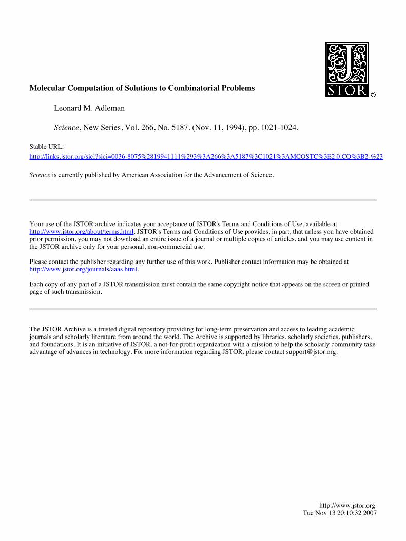
Figure 3 shows the results of these pro- cedures. In Fig. 3A, lane 1 is the result of the ligation reaction in Step 1. The smear with striations is consistent with the con- struction of molecules encoding random paths through the graph (8). Lanes 2 through 5 show the results of the PCR reac- tion in Step 2. The dominant bands corre- spond to the amplification of molecules en- coding paths that begin at vertex 0 and end at vertex 6.
Figure 3B shows the results of a "gradu- ated PCR performed on the single-stranded DNA molecules generated from the band excised in Step 3. Graduated PCR is a meth- od for "printing" results and is performed by running six different PCR reactions with the use of 0, as the right primer and gi as the left primer in the ith tube. For example, on the molecules encoding the Hamiltonian path 0 4 , 1+2, 2+3, 3+4, 4 4 , 5+6, graduated PCR will produce bands of 40,60, 80,100,120, and 140 bp in successive lanes. On the molecules encoding the path 0+1, 1+3,3+4,4+5,5+6, graduated FCR will . produce bands of 40, x, 60,80,100, and 120 bp in successive lanes, where x denotes the
aTATATCCCACCTATTCGAGCTTAIUDCTADDCTA00TAC
COATAAGCTCGAATTTCGAT
Fig. 2 Encoding a graph in DNA. For each vertex i in the graph, a random 20-mer oligonucleotide 0, is generated (shown are O,, 03, and O,, for vertices 2,3, and 4, respectively). For edgei+j in the graph, an oligonucleotide Oi+j is derived from the 3' 10- mer of Oi and from the 5' 1 0-mer of Oj (shown are 0,- for edge 2 4 and b for edge 3+4). For each vertex i in the graph, is the Watson-Click complement of Oi (shown is 03, the complement of 04. o3 serves as a splint to bind O,, and 03, in preparation for ligation,All oligonucleotides are written 5' to 3', except O3
absence of a band in lane 2 (corresponding to the omission of vertex 2 along this path). On molecules encoding the path 0+3,3+2, 2+3,3+4,4+5,5+6, graduated PCR will produce bands of x, 60,80-40, 100, 120, and 140 bp in successive lanes, where 80-40 de- notes that both a 40-bp and an 80-bp band will be produced in lane 3 (corresponding to the double passage of vertex 3 along this path). The most prominent bands in Fig. 3B appear to be those that would arise from the superimposition of the bands predicted for the three paths described above. The bands corresponding to path 0-4, 1+3, 3+4, 4 4 , 5+6 were not expected and suggest that the band excised in S t e ~ 3 contained contamination from 120-bp molecules. However, such low weight contamination is not a problem because it does not persist through Step 4. Figure 3C shows the results of graduated PCR applied to the molecules in the final product of Step 4. These bands de,monstrate that these molecules encode the Hamiltonian path 0+1, 1+2, 2+3, 3+4, 4 4 , 5+6 (9).
This computation required approximately 7 days of lab work. Step 4 (magnetic bead separation) was the most labor-intensive, re- quiring a full day at the bench. In general, with use of the algorithm above the number of procedures required should grow linearly with the number of vertices in the ma~h. The labor required for large graphs miihibe reduced with use of alternative ~~0cedure.s. automation, or less labor-intensive molecu- lar algorithms.
The number of different oligonucleotides required should grow linearly with the num- ber of edges. The quantity of each oligonu-
SCIENCE VOL. 266 11 NOVEMBER 1994
Fig. 3. Agarose gel electrophoresis of various products of the experiment. (A) Product of the ligation reaction (lane I), PCR amplification of the product of the ligation reaction (lanes 2 through 5), and molecular weight marker in base pairs (lane 6). (B) Graduated PCR of the product from Step 3 (lanes 1 through 6); the molecular weight marker is in lane 7. (C) Graduated PCR of the final product of the experiment, revealing the Hamiltonian path (lanes 1 through 6); the molecular weight marker is in lane 7.
cleotide needed is a rather subtle graph the- oretic question (8). Roughly, the quantity used should be just sufficient to insure that during the ligation step (Step 1) a molecule encoding a Hamiltonian path will be formed with high probability if such a path exists in the graph. This quantity should grow expo- nentially with the number of vertices in the graph. The molecular algorithm used here was rather nalve and inefficient, and as with classical computation, finding improved al- gorithms will extend the applicability of the method.
As die computation is scaled up, the pos- sibility of errors will need to be looked at carefully. During Step 1, the occasional liga- tion of incompatible edge oligonucleotides may result in the formation of molecules encoding "pseudopaths" that do not actually occur in the graph. Although such molecules may be amplified during Step 2 and persist through Step 3, they seem unlikely to sur- vive the separation in Step 4. Nonetheless, at the completion of a computation, it would be ~rudent to confirm that a ~utative Ham- iltonian path actually occurs in the graph. During the separation step, molecules encod- ing Hamiltonian paths may fail to bind ad- equately and be lost, whereas molecules en- coding non-Hamiltonian paths may bind nonspecifically and be retained. The latter problem might be mitigated by more strin- gent or repeated separation procedures. One might deal with the former problem by pe- riodically applying PCR with primers de- signed to amplify Hamiltonian paths (in the example above, primers O0 and 06)' The balanced use of these techniques may be adequate to control such errors.
The choice of random 20-mer oligonucle- otides for encoding the graph was based on the following rationale. First, because 420 20-mer olieonucleotides exist. choosing ran- domly made it unlikely that oligonucl&tides associated with different vertices would share
ing between "splint" and "edge" oligonucle- otides would involve 10 nucleotide pairs and would consequently be stable at room tem- perature. This approach was successful for the small graph considered above; however, how to best proceed for larger graphs may require additional research.
What is the power of this method of computation? It is premature to give defini- tive answers; however, some remarks seem in order. A typical desktop computer can exe- cute approximately lo6 operations per sec- ond. The fastest supercomputers currently available can execute approximately 1012 operations per second. If the ligation (con- catenation) of two DNA molecules is con- sidered as a single operation and if it is assumed that about half of the approximately 4 X 10¹⁴ edge oligonucleotides in Step 1 were ligated, then during Step 1 approxi- mately 1014 operations were executed. Clearly, this step could be scaled-up consid- erablv. and loZ0 or more e r a t i o n s seems , , entirely plausible (for example, by using mi- cromole rather than ~icomole auantities). At this scale, the number of operations per second during the ligation step would exceed that of current supercomputers by more than a thousandfold. Furthermore, hydrolysis of a single molecule of adenosine tiphosphate to adenosine monophosphate plus pyrophos- phate provides the Gibbs free energy (AG = -8 kcal mol-') for one ligation operation (10, 1 1 ); hence in principle 1 J is sufficient for approximately 2 X 10¹⁹ such operations.
This is remarkable energy efficiency, con- sidering that the second law of thermody- namics dictates a theoretical maximum of 34 X 1019 (irreversible) operations per joule (at 300 K) (1 2,13). Existing supercomputers are far less energy-efficient, executing at most lo9 operations per joule. The energy con- sumed during other parts of the molecular computation, such as oligonucleotide syn- thesis and PCR. should also be small in
long common subsequences that might result comparison to that consumed by current su- in "unintended" binding during the ligation percomputers. Finally, storing information in step (Step 1). Second, it was guessed. that molecules of DNA allows for an information with high probability potentially deleterious density of approximately 1 bit per cubic (and presumably rare) features such as severe nanometer, a dramatic improvement over hairpin loops would not be likely to arise. existing storage media such as videotapes, Finally, choosing 20-mers assured that bind- which store information at a density of ap-
proximately 1 bit per 10'' nm3. Thus, the potential of molecular compu-
tation is impressive. What is not clear is whether such massive numbers of inexpen- sive operations can be productively used to solve real computational problems. One ma- jor advantage of electronic computers is the variety of operations they provide and the flexibility with which these operations can be applied. Whereas two 100digit integers can be multiplied quite efficiently on an electronic computer, it would be a daunting task to do such a calculation on a molecular computer using currently available protocols and enzymes (1 4).
Nonetheless, for certain intrinsically complex problems, such as the directed Hamiltonian path problem where existing electronic comDuters are verv inefficient and where massively parallel searches can be or- ganized to take advantage of the operations that molecular biology currently provides, it is conceivable that molecular computation might compete with electronic computation in the near term. It is a research problem of considerable interest to elucidate the kinds of algorithms that are possible with the use of molecular methods and the kinds of rob- lems that these algorithms can efficiently solve (1 2. 15. 16). . , , ,
For the long term, one can only speculate about the prospects for molecular computa- tion. It seems likely that a single molecule of DNA can be used to encode the "instanta- neous description" of a Turing machine ( 1 7) and that currently available protocols and enzvmes could (at least under idealized conditions) be used to induce successive sequence modifications, which would cor- respond to the execution of the machine. In the future. research in molecular biol- ogy may provide improved techniques for manipulating macromolecules. Research in chemistry may allow for the develop- ment of synthetic designer enzymes. One can imagine the eventual emergence of a general purpose computer consisting of nothing more than a single macromole- cule conjugated to a ribosomelike collec- tion of enzymes that act on it.
REFERENCES AND NOTES
1. R. P. Feynman, in Minaturization, D. H. Gilbert, Ed. (Reinhold, New York, 1961), pp. 282-296.
2. M. R. Garey and D. S. Johnson, Computers and htractabiI& (Freeman, San Francisco, CA, 1979).
3. R. M. Karp, in CompJexiiy of Computer Computa- tions, R. E. Miller and J. W. Thatcher, Eds. (Plenum, New York, 1972), pp. 85-103.
4. Each oligonucleotide (50 pmol) with 5'-terminal phos- phate residue, 5 units of T4 DNA ligase (Boehringer- Mannheim, Germany), ligase buffer, and ddH,O to a total voluh of 100 pl was incubated for 4 hours at room temperature.
5. All PCR a m p l i o n s were performed on a Perkin- Elmer (Norwalk, CT) 9600 th& cycler. For amplifi- cation in Step 2,50 pmol of each primer and 5 units of Taq DNA polymerase (Gibco-BRL, Grand Island, NY) in PCR buffer to a total volume of 50 pl were pro- cessed for 35 cydes at 94°C for 15 s and at 30°C for
SCIENCE VOL. 266 11 NOVEMBER 1994

60 s. For graduatedPCR,50 pmol of each primer and 2.5 units of Taq DNA polymerase in PCR buffer to a total volume of 50 ~1 were processedfor 25 cycles at 94°C for 15 s and at 40°C for 60 s.
6. All gels were 3 or 5% agarose (Nusieve, FMC Bio-Products, Rockland, ME) in tris-borate-EDTA buffer with ethidium bromide staining (14).
7. Oligonucleotides were 5' biotinylatedwith LC Biotin-ON Phosphoramidite (Clontech). To obtain single-strandedDNA,the product from Step 3 was amplified by PCR with the use of primers 0, and biotinylated 0,. The amplified product was annealed to stfeptavi-din paramagnetic particles (Promega, Madison, WI) by incubating in 100 pl of 0.5x saline sodium citrate (SSC)for 45 min at room temperature with constant shaking. Particles were washed three times in 200 pl of 0.5X SSC and then heated to 80°C in 100 ~1 of ddH,O for 5 min to denature the bound double-stranded DNA. The aqueous phase with single-stranded DNA was retained.For affinity purification,1 nmol of biotinylated0, was annealed to particles as above andwashedthree times in400 pl of 0 . 5 ~SSC. Single-strandedDNA was then incubated with these particles in 150 ~1 of 0 . 5 ~SSC for 45 min at room temperature with constant shaking. Particles were washed four times in 400 ~1 of 0.5X SSC to remove unbound single-stranded DNA and then heated to 80°C in 100 FI of ddH,O-for 5 min to release single-stranded DNA bound to 0,.The aaueous ohase with single-strandedDNA was ;etained.'~hispiocess was then repeatedfor o,, a,, a,,and 05.
8. From a graph theoretic point of view, the use of equal quantities of each oligonucleotidein the ligation reac-tion is not optimal and leads to the formation of large amounts of molecules encoding paths that do not start at vertex 0 nor end at vertex 6. A better way to proceed is to first calculate a flow on the graph and to use the results to determine the quantity of each oli-gonucleotidethat is necessary.
9. On an n vertex graph G with designated vertices v,, and v,,,, there may be multiple Hamiltonian paths, If it is desirable to have an explicit description of some Hamiltonian path, that can be accomplished by ex-tendingthe algorithm as follows. At the end of Step 4, one has a solution (inthe chemistrysense)containing molecules encoding all Hamiltonian paths for (G, v,,, v,,,). The graduated PCR performed at the end of Step 4 will produce the superimpositionof the bands correspondingto all of these Hamiltonian paths in the n - 1 successive lanes. For some lane i,a band of least weight (40 bp) will appear. This indicates that some Hamiltonian path beginswith v,, and proceeds directly to vertex i.By amplifying by PCR the solution with primers 0,andon,runninga gel, and excisingthe 20 x (n - 1)bp band, one can ensurethat only those molecules encoding such Hamiltonian paths will be retained. One now has a solution containing mole-cules encoding all Hamiltonian paths for (GI, i, v,,,), where G' is the graph where vertex v,, has been re-moved. This procedure is now iterated.
10. J. D. Watson, N. H. Hopkins, J. W. Roberts, J. A. Steitz, A. M. Weiner, Molecular Biology of the Gene (Benjamin/Cummings, Menlo Park, CA, ed. 3, 1987).
11. M.J. Engler and C. C. Richardson,in The Enzyme, P. D. Boyer, Ed. (Academic Press, New York, ed. 3, 19821,vol. XVB, pp. 3-29.
12. T. D. Schneider,J. Theor. Biol. 148, 125 (1991). 13. R. C. Merkle, Nanotechnology 4, 21 (1993). 14. J. Sambrook, E. F. Fritsch, T. Maniatis, Molecular
Cloning (Cold Spring Harbor Laboratory, Cold Spring Harbor, NY, ed. 2, 1989).
15. B. C. Crandall and J. Lewis, Eds., Nanotechnology (MIT Press, Cambridge, MA, 1992).
16. D. Bradley,Science 259, 890 (1993). 17. H. Rogers Jr., Theory of Recursive Functions and
Effective Computability (McGraw-Hill, New York, 1967).
18. The author expresses gratitude to N. Chelypov for teaching him molecular biology and thanks S. Salahuddin for making the resources of the Insti-tute for Molecular Medicine and Technology avail-able. The author also thanks R. Deonier for helpful discussions. Supported in part by NSF grant CCR-9214671 and a Zumberg Research Initiation Fund grant from the University of Southern California.
27 May 1994; accepted 19 September 1994
Efficient Neutralization of Primary Isolates of HIV-1 by a Recombinant Human
Monoclonal Antibody
Dennis R. Burton,* Jayashree Pyati, Raju Koduri, Stephen J. Sharp, George B. Thornton, Paul W. H. I. Parren,
Lynette S. W. Sawyer, R. Michael Hendry, Nancy Dunlop, Peter L. Nara, Michael Lamacchia, Eileen Garratty,
E. Richard Stiehm, Yvonne J. Bryson, Yunzhen Cao, John P. Moore, David D. Ho, Carlos F. Barbas Ill*
The ability of antibodies to neutralizediverse primary isolates of human immunodeficiency virus-type 1 invitro has beenquestioned, with implicationsforthe likelyefficacy of vaccines. A recombinant human antibody to envelope glycoprotein gp120 was generated and used to show that primary isolatesare not refractory to antibody neutralization.The recombinant antibody neutralizedmore than 75 percent of the primary isolatestested at concentrations that could be achieved by passive immunization, for example, to interrupt maternal-fetal transmission of virus. The broad specificity and efficacy of the antibody implies the con-servation of a structural feature on gp120, which could be important in vaccine design.
Protection from viral disease has tradition-
allv been associated with the oreexistence in
semm of antibodies capable bf neutralizing
virus in vitro. Indeed, vaccines are frequently
assessed on the ability to elicit neutralizing
antibody responses. In the case of human
immunodeficiency virus-type 1 (HIV-I),
there was initial optimism about the likely
efficacy of subunit vaccines given that vac-
cinee sera from several trials were capable of
neutralizine laboratorv isolates of virus in u
vitro (1, 2). The grounds for optimism were
shaken when it was found that the vaccinee
sera were largely ineffective against primary
isolates of HIV-1 (2). Some discussion sub-. .
sequently centered around the validity of
standard HIV-1 neutralization assays when
applied to primary isolates (2-5). If the as-
says are meaningful, then they call into ques-
tion the ability of antibody to effectively
neutralize a spectrum of primary isolates. Hy-
perimmune pooled human plasma prepara-
tions are capable of neutralizing a number of
D. R. Burton, P. W. H. I. Parren, C. F. Barbas Ill, Depart-ments of Immunology and Molecular Biology, The Scripps Research Institute, 10666 North Torrey Pines Road, La Jolla, CA 92037, USA, J. Pyati, R. Koduri, S. J. Sharp, G. B. Thornton, R. W. Johnson Pharmaceutical Research Institute, 3535 Gen-eral Atomics Court, San Diego, CA 92121, USA. L. S. W. Sawyer and R. M. Hendry, Viral and Rickettsia1 Disease Laboratory, California Department of HealthSer-vices, 2151 Berkeley Way, Berkeley,CA 94704, USA. N. Dunlop and P. L. Nara, Laboratory of Tumor Cell Biology,Virus BiologySection, NationalCancer Institute-Frederick Cancer Research and Development Center, Frederick, MD 21702, USA. M. Lamacchia, E. Garratty, E. R. Stiehm, Y. J. Bryson, Dewartment of Pediatrics. Universitv of California at Los
primary isolates (3-5), but they represent a
combination of specificities that might be
difficult to elicit by all except the most
complex vaccines (6). A single antibody
able to effectively neutralize a broad spec-
trum of ~r imarvisolates would validate the
vaccine approach and would provide a tem-
plate for vaccine design. Furthermore, it
would constitute a reagent for passive im-
munotherapy such as in the interruption of
maternal-fetal transmission. We describe
here such a human antibody derived by
recombinant methods (7). The generation of the'antibody Fab frag-
ment b12 from a combinatorial phage dis-
play library has been described previously
(8).Fab b12 is directed to the CD4 binding
site of gp120 and is a potent neutralizer of
the HIV-1 laboratorv strains IIIB and MN (9-1 1). Selection fAr potency and strain
cross-reactivity was achieved through exper-
imental design. The library donor was a
long-term asymptomatic U.S.male, presum-
ably infected with a clade B strain of HIV-1;
the antigen for affinity selection was gp120
from the atypical IIIB strain, thereby favor-
ing selection of cross-reactive antibodies. A laree number of bacterial suoemates contain--ing antibody Fab fragments to gp120 (anti-
5x1120) at low initial concentrations were u.
directly screened for neutralizing ability to
find the most Dotent Fabs. Although Fab b12-is capable of neutralizing some primary iso-
lates (12), the corresponding whole antibody
molecule is likely to be more effective.
Therefore, Fab b12 was converted to a whole ~ngelesSchool of Medicine, 1083'3 Le Conte Avenue, immunog~obu~inG1 (IgGl) molecule byLos Angeles, CA 90024, USA. Y. Cao, J. P. Moore, D. D. Ho, Aaron Diamond AIDS 'loning the region of Ig heavy
Research Center, New York University School of Medi- chain (VH) and light chain genes into a cine, New York, NY 10016, USA. vector created for high-level mammalian
'To whom correspondence should be addressed. expression (13) . The whole antibody IgGl
SCIENCE VOL. 266 11 NOVEMBER 1994
Related Documents


![G: pp ex ookskeyword5187-Fotheringham5187-Fotheringham-Ch10 · [17:22 14/8/2008 5187-Fotheringham-Ch10.tex] Paper: a4 Job No: 5187 Fotheringham: Spatial Analysis (Handbook) Page:](https://static.cupdf.com/doc/110x72/5ec50289780af2186565ec0a/g-pp-ex-ookskeyword5187-fotheringham5187-fotheringham-ch10-1722-1482008-5187-fotheringham-ch10tex.jpg)