AAPM CT Lexicon version 1.3 04/20/2012 Copyright © April 20, 2012 by AAPM. All rights reserved. 1 Table of Contents Scan acquisition and user interface basics Dose modulation and reduction tools Multi-Slice Detector Geometry Image Reconstruction and Display Contrast Media Tools Multi-planar formats and 3-D Processing Service and Application Tools Workflow

Welcome message from author
This document is posted to help you gain knowledge. Please leave a comment to let me know what you think about it! Share it to your friends and learn new things together.
Transcript

AAPM CT Lexicon version 1.3 04/20/2012
Copyright © April 20, 2012 by AAPM. All rights reserved.
1
Table of Contents
Scan acquisition and user interface basics Dose modulation and reduction tools Multi-Slice Detector Geometry Image Reconstruction and Display Contrast Media Tools Multi-planar formats and 3-D Processing Service and Application Tools Workflow

AAPM CT Lexicon version 1.3 04/20/2012
Copyright © April 20, 2012 by AAPM. All rights reserved.
2
CT scan parameters: Translation of terms for different manufacturers Introduction For the CT technologist who operates multiple scanner models, perhaps from multiple manufacturers, the variability in names for important scan acquisition and reconstruction parameters can lead to confusion, reduced comfort and an increased potential for error. The intent of this CT terminology lexicon is to allow users to translate important CT acquisition and reconstruction terms between different manufacturers' systems. This represents a first step in the terminology standardization effort undertaken by this working group. Phase 2 of our work will: 1. Identify relevant terms from established standard lexicons (e.g. RadLex and DICOM) and other relevant literature and publish an expanded lexicon including
these terms. 2. Form consensus recommendations on preferred terms. This website will be updated as the terminology standardization work progresses. The generic descriptions or terms in the first column are intended to orient the user to the relevant concepts; they are not consensus "preferred terms." The generic descriptions are not based on any single existing or pending terminology standard; however the references cited below were consulted in developing the generic descriptions. Future efforts of this Working Group include making recommendations for standardized terminology. A number of individuals and groups have advocated for terminology standardization in CT, including at a March 30-31, 2010 FDA public meeting entitled "Device Improvements to Reduce Unnecessary Radiation Exposure from Medical Imaging" (transcripts available at: http://www.fda.gov/downloads/MedicalDevices/NewsEvents/WorkshopsConferences/UCM210149.pdf; see p. 153-155). Participants proposed a cooperative effort among professional organizations (AAPM, ASRT, ACR, etc.), industry, FDA, and standards organizations to accomplish this task, as is now being undertaken by this Working Group.

AAPM CT Lexicon version 1.3 04/20/2012
Copyright © April 20, 2012 by AAPM. All rights reserved.
3
References IEC 60601-2-44 ed3.0 (2009-02) Medical electrical equipment – Part 2-44: Particular requirements for the basic safety and essential performance of X-ray equipment for computed tomography. Kalra, M. K. and S. Saini. 2006. Standardized nomenclature and description of CT scanning techniques. Radiology 241, 657-660. National Electrical Manufacturer's Organization. 2009. Digital Imaging and Communications in Medicine (DICOM). (Downloadable from: ftp://medical.nema.org/medical/dicom/2009/) RadLex Tree Browser (http://radlex.org/) Weber, N. M., C. L. Siegle, J. E. Miller, M. K. Hudson, J. M. Kofler, M. R. Bruesewitz, and C. H. McCollough. How to be a "multi-lingual" CT technologist: understanding scan parameters from different manufacturer's equipment. 2004 RSNA Annual Meeting poster (http://mayoresearch.mayo.edu/mayo/research/ctcic/upload/rsna2004-how-to-be-multilingual.pdf). Weigold, W.G., et al. 2011. Standardized Medical Terminology for Cardiac Computed Tomography: A Report of the Society of Cardiovascular Computed Tomography. Journal of Cardiovascular Computed Tomography (downloadable from: http://www.scct.org/documents/JCCT427.pdf).
AAPM CT Lexicon version 1.3 04/20/2012
Copyright © April 20, 2012 by AAPM. All rights reserved.
4
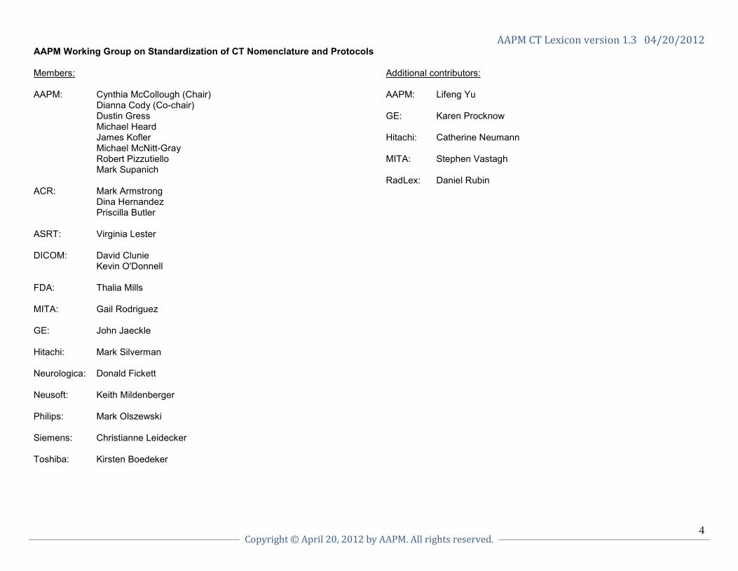
AAPM Working Group on Standardization of CT Nomenclature and Protocols Members: AAPM: Cynthia McCollough (Chair) Dianna Cody (Co-chair) Dustin Gress Michael Heard James Kofler Michael McNitt-Gray Robert Pizzutiello Mark Supanich ACR: Mark Armstrong Dina Hernandez Priscilla Butler ASRT: Virginia Lester DICOM: David Clunie Kevin O'Donnell FDA: Thalia Mills MITA: Gail Rodriguez GE: John Jaeckle Hitachi: Mark Silverman Neurologica: Donald Fickett Neusoft: Keith Mildenberger Philips: Mark Olszewski Siemens: Christianne Leidecker Toshiba: Kirsten Boedeker
Additional contributors: AAPM: Lifeng Yu GE: Karen Procknow Hitachi: Catherine Neumann MITA: Stephen Vastagh RadLex: Daniel Rubin

AAPM CT Lexicon version 1.3 04/20/2012
Copyright © April 20, 2012 by AAPM. All rights reserved.
5
1. Scan acquisition and user interface basics Generic description GE PHILIPS SIEMENS TOSHIBA HITACHI NEUSOFT NEUROLOGICA The portion of the user interface where scans are prescribed
Exam Rx Scan Procedure Examination eXam Plan Scan Protocol Scan Procedure (Neuviz 16); Main Scan Interface (Neuviz DUAL)
Scan protocol
Other portions of the user interface, such as were reconstructed images are viewed
Desktop Active viewer Various “task cards”, such as “Viewing”
Active display Image Viewer Image Display Area (Neuviz 16); Viewer (Neuviz DUAL)
Desktop
CT localizer radiograph (i.e. the scanned projection radiograph, often acquired by the CT system to allow the user to prescribe the start and end locations of the scan range)
Scout Surview Topogram Scanogram Scanogram Surview Scout
Axial scan mode: Data acquisition while the patient table remains stationary; the table position may be incremented between x-ray exposures to collect data over a longer z axis range.
Axial Axial Sequence Scan & View, Scan & Scan, Volume, Wide Volume (Aquilion One)
Normal Axial Axial
Helical or Spiral scan mode: Data acquisition while the patient table is continuously moving along the z axis.
Helical Helical Spiral Helical Volume Helical Helical
Dynamic scan mode - single detector width: Data acquisition at multiple time points over the same anatomic location(s) while the patient table remains stationary; x-ray exposure can be continuous or intermittent
Cine or zero interval Axial
CCT (Continuous CT)
Dynamic (continuous) or Serio (intermittent); scan mode name: DynMulti or DynSerio.
Dynamic (Continuous or Intermittent)
Dynamic CCT (Neuviz 16); N/A (Neuviz DUAL)
Dynamic
Dynamic scan mode - multiple detector widths: Data acquisition at multiple time points over the same anatomic location(s) while the patient table cycles back and forth between designated start and end locations in order image a region wider than the detector
Shuttle Jog Adaptive 4D Spiral; scan mode name: DynMulti4D or DynSerio4D (ECG triggered)
N/A N/A N/A Dynamic
Interventional CT - Intermittent x-ray exposures
SmartStep Single CCT Model dependent: Biopsy or Intervention (i-Sequence/i-Spiral)
CT Fluoro (CTF) guideShot Single CCT (Neuviz 16); N/A (Neuviz DUAL)
CT Fluoro (CTF)
Interventional CT - Continuous x-ray exposures
SmartView Continuous CCT Model dependent: CARE Vision or Intervention (i-Fluoro)
CT Fluoro (CTF) Not available Continuous CCT (Neuviz 16); N/A (Neuviz DUAL)
CT Fluoro (CTF)
Table increment (mm) per 360 degree rotation of the x-ray tube (axial scan mode)
Interval Increment (mm) Feed (mm) Couch movement (mm)
Table Feed (mm)
Increment (mm) Increment (mm)

AAPM CT Lexicon version 1.3 04/20/2012
Copyright © April 20, 2012 by AAPM. All rights reserved.
6
Scan acquisition and user interface basics, continued Generic description GE PHILIPS SIEMENS TOSHIBA HITACHI NEUSOFT NEUROLOGICA Table feed per 360 degree rotation of the x-ray tube (helical scan mode)
Speed (mm/rot)
Table speed (mm/rot)
Table Feed (mm/rot)* Couch speed (mm/Rot)
Table Speed (mm/rot)
Table speed (mm/rot) Pitch
Field of measurment: Diameter of the circular region within the scan plane over which projection data are collected. Nominally equal to the diameter of the primary beam at isocenter in the axial plane.
Scan Field of View (SFOV, cm)
Not determined by tech; built into protocol
Not determined by tech; built into protocol
CFOV (Calibrated Field of View)
Scan Field of View (SFOV, mm)
FOV Full Field of View (FFOV, cm)
Tube current: Number of electrons accelerated across an x-ray tube per unit time, expressed in units of milliampere (mA)
mA mA* mA* mA mA mA* mA
Tube current-time product: The product of tube current and exposure time per rotation, expressed in units of milliampere • seconds (mAs). In axial scan mode, this is equal to tube current × (scan angle ÷ 360) × rotation time. In helical scan mode, this is equal to tube current × rotation time.
Not used on this system
mAs mAs mAs Not used on this system
mAs mAs
Effective tube current-time product: In helical scan mode, this is the product of tube current and rotation time (expressed in units of milliampere • seconds (mAs) ÷ pitch)
Not used on this system
mAs per slice (= mAs/pitch)
Effective mAs (= mAs/pitch)
Effective mAs (= mAs/pitch)
Not used on this system
mAs per slice (= mAs/pitch)
mAs
Tube potential: The electric potential applied across an x-ray tube to accelerate electrons towards a target material, expressed in units of kilovolts (kV)
kV kVp kV kV kVp KV kV
Pitch: Unitless parameter used to describe the table travel during helical CT; equal to table travel (mm) per gantry rotation ÷ total nominal beam width (mm)
Pitch Pitch Pitch CT Pitch Factor Pitch pitch Pitch
Automated patient instructions AutoVoice Auto voice API (Automated Patient Instructions)
Breath Control Auto Voice Auto voice Audio
* Not able to be directly modified on the user interface. Value is calculated/determined by other settings.

AAPM CT Lexicon version 1.3 04/20/2012
Copyright © April 20, 2012 by AAPM. All rights reserved.
7
2. Dose modulation and reduction tools Generic description GE PHILIPS SIEMENS TOSHIBA HITACHI NEUSOFT NEUROLOGICA Automatic exposure control (AEC): A scanner feature that automatically adapts the x-ray tube current to the overall patient size to achieve a specified level of image quality
Available in AutomA and SmartmA
Available in DoseRight Automatic Current Selection (ACS)
Available in CARE Dose4D Available in SURE Exposure
Available in IntelliEC
DoseRight, ACS (automatic current selection) [Neuviz 16]; DoseSave, ACS (automatic current selection) [Neuviz DUAL]
N/A
Angular tube current modulation
SmartScan (CT/i only)
D-DOM (Dose Modulation)
CARE Dose not available as a separate item
Adaptive mA
D-DOM (Neuviz 16); N/A (Neuviz DUAL)
N/A
Longitudinal tube current modulation
AutomA Z-DOM not available as a separate item SURE Exposure n/a Z-DOM (Neuviz 16); N/A (Neuviz DUAL)
N/A
Angular and longitudinal tube current modulation
SmartmA (x, y, z)
Work in progress CARE Dose4D SURE Exposure 3D (X, Y and Z Modulation)
IntelliEC ACS+DOM (Neuviz 16); N/A (Neuviz DUAL)
N/A
ECG-based tube current modulation
ECG Modulated mA
DoseRight Cardiac
All features available in HeartView package (except (3), only available for SOMATOM Definition Flash) (1) Retrospective gated spiral mode: use “Pulsing” settings in Trigger card
(2) Prospective triggered sequence: use “Adaptive Cardio Seq.” and “Pulsing” settings in Trigger card.
(3) Prospectively triggered spiral (“Flash” mode)
ECG Modulation ECG Dose Modulation
N/A N/A
Image quality reference parameter for AEC
Noise Index Reference image Quality reference mAs Standard Deviation or standard, low-dose, or high-quality
Standard Deviation (% ) or standard, low-dose, or high-quality
reference image N/A

AAPM CT Lexicon version 1.3 04/20/2012
Copyright © April 20, 2012 by AAPM. All rights reserved.
8
3. Multi-Slice Detector Geometry Generic description GE PHILIPS SIEMENS TOSHIBA HITACHI NEUSOFT NEUROLOGICA
Multi-slice detector array design
Fixed Model dependent: Fixed or Asymmetric
Model dependent: Adaptive or Fixed
Fixed (32 row and above); Adaptive (16 row and below)
Asymmetric-16 slice; Fixed-64 slice
Asymmetric (Neuviz 16); Fixed (Neuviz DUAL)
Fixed
Detector configuration Detector Configuration
Collimation N x T (mm)
Detector Configuration or Aqu (Acquisition) on Exam Card
Detector Configuration Detector Configuration
Collimation N x T (mm) Detector Configuration

AAPM CT Lexicon version 1.3 04/20/2012
Copyright © April 20, 2012 by AAPM. All rights reserved.
9
4. Image Reconstruction and Display Generic description GE PHILIPS SIEMENS TOSHIBA HITACHI NEUSOFT NEUROLOGICA Window width: Range of CT numbers (maximum - minimum) that are distributed over the viewable grey scale of the display device or film
Window Width Window Width Window width Window width Window Width Window Width (Neuviz 16); WW Window Width (Neuviz DUAL)
Window width
Window center: The CT number in the center of the viewable grey scale
Window Level Window Center Window center Window level Window Level Window Center (Neuviz 16); WL Window Level (Neuviz DUAL)
Window level
Reconstruction field of view: Width of the square region mapped to the reconstructed image matrix
Display Field of View (DFOV) (cm)
DFOV (mm) FoV (mm) DFOV (mm) DFOV (mm) FOV (mm) (Neuviz 16); Rec FOV (Neuviz DUAL)
FOV (cm)
Prescribing the reconstruction parameters prior to scan acquisition
Prospective recon
Recon and Additional Recons
Recon Job Prospective recon
Multi Recon Axial or helical reconstruction
Protocol
Prescribing the reconstruction parameters after scan acquisition
Retrospective recon
Offline Recon or Re-Recon
Recon Job Retrospective or Raw data recon
Post Reconstruction
Offline reconstruction (Neuviz 16); Image Reconstruction (Neuviz DUAL)
Post Recon
Reconstruction property that determines sharpness or smoothness of image in the axial plane
Algorithm Reconstruction Filter
Kernel Filter convolution (FC)
Image Filter Reconstruction filter (Neuviz 16); Recon Filter (Neuviz DUAL)
Kernel
Helical interpolation options to achieve a wider or narrower section sensitivity profile
Full (narrower) or Plus (wider) mode
Slice width independent of pitch
Slice width independent of pitch
Slice width independent of pitch
Slice width independent of pitch
Slice width independent of pitch (Neuviz 16); Thickness (Neuviz DUAL)
Slice width
Nominal width of reconstructed image along the z axis
Thickness (mm)
Thickness (mm) Slice (mm) Image thickness Slice Thickness Thickness Slice thickness
Distance between two consecutive reconstructed images
Interval Increment Position increment
Reconstruction interval
Interval Increment Slice separation
Fast but lower-quality reconstructed images for rapid review of entire exam
QC Image Image Check
Evolving reconstructions
RT (Real-time reconstruction)
SUREScan Real Time Reconstruction
Evolving mode (Neuviz 16); N/A (Neuviz DUAL)
Image Preview Image Check
Off-center reconstruction coordinates are called
RL Center; AP Center
Center x, center y Center x, Center y Center Position; (Vari Area)
Center x, y Center x, center y Center x, center y
Flip or rotate the image orientation is called
Flip/rotate Flip/rotate Mirroring (Flip in Viewing card); Rotate
Rotate/Mirror Flip/Rotate Flip/rotate Flip/rotate
Image modifications to alter sharpness or smoothness (done in image space without reconstructing images)
Image Filters Image enhancement filter
Evaluation > Image Manipulation (Viewing card)
Filter, QDS Filter IMAGE ENHANCE FILTER (Neuviz 16); DISPLAY MODE (Neuviz DUAL)
N/A

AAPMCTLexiconversion1.304/20/2012
Copyright©April20,2012byAAPM.Allrightsreserved.
10
5. Contrast Media Tools Generic description GE PHILIPS SIEMENS TOSHIBA HITACHI NEUSOFT NEUROLOGICA Bolus tracking: Scanner feature to automatically initiate a prescribed axial, helical or dynamic scan when a threshold level of contrast enhancement is reached at a specified region of interest
Smart Prep Bolus Tracking
CARE Bolus (includes Test Bolus and Bolus Tracking)
SUREStart Predict Scan Bolus Tracking Bolus Tracking
Test Bolus: Scan mode used to measure the contrast transit time using a small injection of contrast media
Take axial scans at zero table feed and process with MIROI
Time Lapse Test Bolus Dynamic study Not available TIBT (Neuviz 16); Tracking layer (Neuviz DUAL)
Test Bolus
Time-attenuation curve (TAC): Graph of the contrast enhancement versus time
Smart Prep graph or MIROI graph
Time Lapse graph
Enhancement Curve
Time Density Curve
Monitoring Graph
Time Lapse Graph Contrast curve
Threshold: CT number (HU) where bolus tracking tool will trigger the system to begin the scan
Transition ROI Threshold
Threshold Level Threshold ROI (HU)
Threshold Threshold CT threshold
Scanner feature used to quantitatively evaluate the TAC
MIROI (multiple image region of interest)
Tracker ROI Tools
DynEva (dynamic evaluation)
Real Time Monitoring
No special name
N/A Algorithm
Monitoring delay: Time from injection to the start of monitoring scans (Time 1 in figure below)
Monitoring Delay Post Injection Delay
Delay (on monitoring scan)
Delay (on SUREStart)
Scan Delay (PIT) Post Injection Time
Time delay
Monitoring interval: Time between consecutive monitoring scans to (Time 2 in figure below)
Monitor ISD (InterScan Delay)
Cycle time Cycle time Real time monitoring or pulsed monitoring (seconds)
Monitoring Time
Cycle time (Neuviz 16); Bolus Timing (Neuviz DUAL)
Temporal resolution
Scan delay: Time from when threshold is reached and prescribed axial, helical or dynamic scan begins (Time 3 in figure below)
Diagnostic delay Post Threshold Delay
Delay (on scan) Delay (on helical) Scan Delay (PTD) Post Threshold Delay
Delay

AAPM CT Lexicon version 1.3 04/20/2012
Copyright © April 20, 2012 by AAPM. All rights reserved.
11
6. Multi-planar formats and 3-D Processing Generic description GE PHILIPS SIEMENS TOSHIBA HITACHI NEUSOFT NEUROLOGICA Reformatted image at an oblique plane (not an axial, coronal, or sagittal)
Oblique reformat Oblique Oblique Oblique Oblique MPR oblique/curved surface
Digital tilt
Saving images at various viewing angles about a volume or surface rendered object
Batch Loop Cine Radial Ranges Key Frame Movie Multi-Slice /Angle Cine Capture
Saving images at various planes through a volume
Batch Reformat Batch MPR Parallel Ranges Batch MPR Multi planar reformat
batch MPR Capture
Surface-rendered object 3D SSD 3D (Shaded Surface Display – 3D)
Shaded Surface Display (SSD)
ShadedVol (Shaded volume rendering (SVR))
Shaded Surface Display (SSD)
SSD (Shaded surface display)
3D
Volume-rendered object Volume Rendered image (VR)
Volume Rendering Volume Rendering Technique (VRT)
Shaded Vol Volume Rendering Volume Rendering (VR)
Volume Rendered image (VR)
7. Service and Application Tools Generic description GE PHILIPS SIEMENS TOSHIBA HITACHI NEUSOFT NEUROLOGICA X-ray tube warm up Tube Warm-up
(tube warm up) Tube conditioning Check-up (calibrate and
check values); Calibrate (part of Check-up, can be performed separately)
Warm up Warm up tube warm up Warmup
Daily calibrations Fast Cals (done in daily prep)
Not necessary to do daily calibrations
Quality Daily Selectable air calibrations can be scheduled after warm-up
Air cals built into Warm up
not required daily, recommend air calibration weekly
Daily Calibration
Application information Learning Solutions or User Manual
On-line Help On-line Help; CT Life (task card)
E-Learning Center Sentinel (Remote Service)
On-line Help On-line help
Application support assistance
Insite or Ilinq Customer Care Solutions Center
Uptime In Touch Center CT Applications Helpline
Applications Specialist or Field Service Engineer
Service center

AAPM CT Lexicon version 1.3 04/20/2012
Copyright © April 20, 2012 by AAPM. All rights reserved.
12
8. Workflow Generic description GE PHILIPS SIEMENS TOSHIBA HITACHI NEUSOFT NEUROLOGICA Scheduled (but not yet scanned) patient list is called
Patient Schedule Scheduled (HIS-RIS) and Catalog- (manual list)
Patient Browser – Scheduler
Modality Worklist Manager
MWM-modality worklist management
Schedule Modality Worklist Manager
Already scanned patient list is called
List/Select Archive Manager Patient Browser – Local Database
Directory Patient List Archive Manager (Neuviz 16); Archive Management Interface (Neuviz DUAL)
Patient Browser – Local Database
User comments or text added to an image is called
User annotation Label (series) and Annotate (image)
Comment Annotation Comment N/A Annotation
Filming tools are called Auto/manual film composer
Filming Film Sheet on Filming task card
Filming Filming Filming Printer
Data page summarizing scan parameters, CTDIvol and DLP
Exam Text Page or Series Text Page
Image Parameters Patient Protocol (series number 501)
Summary and Exposure Record
Text Page Information Display Bar on the right hand side of the Main Scan Interface & Dose Info series in Image Information List and Dose report at last series
Image Parameters
Sorting patient list Sort Click on sort field (name, date, etc.)
In Patient Browser: select “Sort” or “Filter” functions in menu bar
Click on sort field (name, date, etc.)
Click on sort field (name, date, etc.)
Select field to sort by (name, patient ID, etc.) and left click with mouse to sort.
Sort
Related Documents