Scaling-Up of Dental Pulp Stem Cells Isolated from Multiple Niches Nelson F. Lizier 1,2 *, Alexandre Kerkis 1 , Cı ´cera M. Gomes 1 , Josimeri Hebling 3 , Camila F. Oliveira 1 , Arnold I. Caplan 4 , Irina Kerkis 1,2 1 Laboratory of Genetics, Butantan Institute, Sao Paulo, SP, Brazil, 2 Departament of Morphology of Federal University of Sao Paulo (UNIFESP), Sao Paulo, SP, Brazil, 3 Department of Orthodontics and Pediatric Dentistry of State University of Sao Paulo (UNESP), Araraquara, SP, Brazil, 4 Skeletal Research Center, Department of Biology of Case Western Reserve University, Cleveland, Ohio, United States of America Abstract Dental pulp (DP) can be extracted from child’s primary teeth (deciduous), whose loss occurs spontaneously by about 5 to 12 years. Thus, DP presents an easy accessible source of stem cells without ethical concerns. Substantial quantities of stem cells of an excellent quality and at early (2–5) passages are necessary for clinical use, which currently is a problem for use of adult stem cells. Herein, DPs were cultured generating stem cells at least during six months through multiple mechanical transfers into a new culture dish every 3–4 days. We compared stem cells isolated from the same DP before (early population, EP) and six months after several mechanical transfers (late population, LP). No changes, in both EP and LP, were observed in morphology, expression of stem cells markers (nestin, vimentin, fibronectin, SH2, SH3 and Oct3/4), chondrogenic and myogenic differentiation potential, even after cryopreservation. Six hours after DP extraction and in vitro plating, rare 5- bromo-29-deoxyuridine (BrdU) positive cells were observed in pulp central part. After 72 hours, BrdU positive cells increased in number and were found in DP periphery, thus originating a multicellular population of stem cells of high purity. Multiple stem cell niches were identified in different zones of DP, because abundant expression of nestin, vimentin and Oct3/4 proteins was observed, while STRO-1 protein localization was restricted to perivascular niche. Our finding is of importance for the future of stem cell therapies, providing scaling-up of stem cells at early passages with minimum risk of losing their ‘‘stemness’’. Citation: Lizier NF, Kerkis A, Gomes CM, Hebling J, Oliveira CF, et al. (2012) Scaling-Up of Dental Pulp Stem Cells Isolated from Multiple Niches. PLoS ONE 7(6): e39885. doi:10.1371/journal.pone.0039885 Editor: Jan Pruszak, University of Freiburg, Germany Received February 28, 2012; Accepted May 28, 2012; Published June 29, 2012 Copyright: ß 2012 Lizier et al. This is an open-access article distributed under the terms of the Creative Commons Attribution License, which permits unrestricted use, distribution, and reproduction in any medium, provided the original author and source are credited. Funding: This work was supported by grant 2010/51051-6 from FAPESP (Sao Paulo Research Foundation). The funder had no role in study design, data collection and analysis, decision to publish, or preparation of the manuscript. Competing Interests: The authors have declared that no competing interests exist. * E-mail: [email protected] Introduction Isolation of stem cells (SC) from human adult and deciduous teeth has been reported in the last decade [1,2]. In this short period of time, considerable progress has been achieved, in particular, with deciduous teeth stem cells (DTSC) [3]. It has been demonstrated that the use of different handling methods of dental pulp (DP) can lead to the isolation of SC populations with distinct properties. These DTSC populations are similar to mesenchymal stem cells (MSCs) or epithelial SCs or they are composed by a mixed population of both cell types [3]. We previously isolated a population of multipotent DTSCs, which were referred to as ‘‘immature’’ ( Immature Dental Pulp Stem Cells, IDPSCs). Along with MSC markers, IDPSCs express embryonic stem (ES) cells markers (Oct3/4, Nanog and Sox2) and undergo spontaneous differentiation into a wide range of cell types in vitro [4]. These cells showed expressive capacity to contribute into multiple tissues in response to the cellular milieu during human/mouse pre-termed chimeras development [5]. After transplantation of IDPSCs into different adult animals, including mouse, rabbit and dog, neither immune rejection nor teratoma formation was observed [6,7,8]. IDPSCs and other dental stem/progenitor cells were recently used to obtain induced pluripotent SCs [9,10]. These cells demonstrat- ed higher efficiency of reprogramming than fibroblasts, providing a model for the study of pediatric diseases and disorders. Taken together, these data strongly suggest that IDPSCs are a hopeful source for the future of SC therapies [11]. Recent SC research studies revealed a promising potential of MSCs to treat at least ten human diseases: heart disease, diabetes, Crohn’s disease, deafness, autoimmune disorders, leukemia, cancers, sickle cell disease, amyotrophic lateral sclerosis and metabolic disorders. Since MSCs are present at low relative amounts in bone marrow and other adult tissues, significant in vitro expansion is necessary in order to generate sufficient quantities of these cells to treat human disease [12,13]. The expansion process itself induces senescence of MSCs and loss of their stemness as shown by a decline in proliferative and differentiation capacity [14,15]. In addition, prolonged culturing of MSCs increases the probability of genetic changes, which could affect their safe use in clinical trials and future therapies [16,17]. Therefore, studies which provide adequate production of SC of excellent quality at early passages derived from the same donor are of importance. Our group was the first to use explant culture of DP to obtain DTSCs, which in combination with appropriate cell culture conditions, provides isolation of a relatively pure (not homoge- neous) population of IDPSC [4]. Subsequent study demonstrated PLoS ONE | www.plosone.org 1 June 2012 | Volume 7 | Issue 6 | e39885

Welcome message from author
This document is posted to help you gain knowledge. Please leave a comment to let me know what you think about it! Share it to your friends and learn new things together.
Transcript
Scaling-Up of Dental Pulp Stem Cells Isolated fromMultiple NichesNelson F. Lizier1,2*, Alexandre Kerkis1, Cıcera M. Gomes1, Josimeri Hebling3, Camila F. Oliveira1,
Arnold I. Caplan4, Irina Kerkis1,2
1 Laboratory of Genetics, Butantan Institute, Sao Paulo, SP, Brazil, 2 Departament of Morphology of Federal University of Sao Paulo (UNIFESP), Sao Paulo, SP, Brazil,
3 Department of Orthodontics and Pediatric Dentistry of State University of Sao Paulo (UNESP), Araraquara, SP, Brazil, 4 Skeletal Research Center, Department of Biology of
Case Western Reserve University, Cleveland, Ohio, United States of America
Abstract
Dental pulp (DP) can be extracted from child’s primary teeth (deciduous), whose loss occurs spontaneously by about 5 to 12years. Thus, DP presents an easy accessible source of stem cells without ethical concerns. Substantial quantities of stem cellsof an excellent quality and at early (2–5) passages are necessary for clinical use, which currently is a problem for use of adultstem cells. Herein, DPs were cultured generating stem cells at least during six months through multiple mechanical transfersinto a new culture dish every 3–4 days. We compared stem cells isolated from the same DP before (early population, EP) andsix months after several mechanical transfers (late population, LP). No changes, in both EP and LP, were observed inmorphology, expression of stem cells markers (nestin, vimentin, fibronectin, SH2, SH3 and Oct3/4), chondrogenic andmyogenic differentiation potential, even after cryopreservation. Six hours after DP extraction and in vitro plating, rare 5-bromo-29-deoxyuridine (BrdU) positive cells were observed in pulp central part. After 72 hours, BrdU positive cells increasedin number and were found in DP periphery, thus originating a multicellular population of stem cells of high purity. Multiplestem cell niches were identified in different zones of DP, because abundant expression of nestin, vimentin and Oct3/4proteins was observed, while STRO-1 protein localization was restricted to perivascular niche. Our finding is of importancefor the future of stem cell therapies, providing scaling-up of stem cells at early passages with minimum risk of losing their‘‘stemness’’.
Citation: Lizier NF, Kerkis A, Gomes CM, Hebling J, Oliveira CF, et al. (2012) Scaling-Up of Dental Pulp Stem Cells Isolated from Multiple Niches. PLoS ONE 7(6):e39885. doi:10.1371/journal.pone.0039885
Editor: Jan Pruszak, University of Freiburg, Germany
Received February 28, 2012; Accepted May 28, 2012; Published June 29, 2012
Copyright: � 2012 Lizier et al. This is an open-access article distributed under the terms of the Creative Commons Attribution License, which permitsunrestricted use, distribution, and reproduction in any medium, provided the original author and source are credited.
Funding: This work was supported by grant 2010/51051-6 from FAPESP (Sao Paulo Research Foundation). The funder had no role in study design, data collectionand analysis, decision to publish, or preparation of the manuscript.
Competing Interests: The authors have declared that no competing interests exist.
* E-mail: [email protected]
Introduction
Isolation of stem cells (SC) from human adult and deciduous
teeth has been reported in the last decade [1,2]. In this short
period of time, considerable progress has been achieved, in
particular, with deciduous teeth stem cells (DTSC) [3]. It has been
demonstrated that the use of different handling methods of dental
pulp (DP) can lead to the isolation of SC populations with distinct
properties. These DTSC populations are similar to mesenchymal
stem cells (MSCs) or epithelial SCs or they are composed by a
mixed population of both cell types [3]. We previously isolated a
population of multipotent DTSCs, which were referred to as
‘‘immature’’ (Immature Dental Pulp Stem Cells, IDPSCs). Along
with MSC markers, IDPSCs express embryonic stem (ES) cells
markers (Oct3/4, Nanog and Sox2) and undergo spontaneous
differentiation into a wide range of cell types in vitro [4]. These cells
showed expressive capacity to contribute into multiple tissues in
response to the cellular milieu during human/mouse pre-termed
chimeras development [5]. After transplantation of IDPSCs into
different adult animals, including mouse, rabbit and dog, neither
immune rejection nor teratoma formation was observed [6,7,8].
IDPSCs and other dental stem/progenitor cells were recently used
to obtain induced pluripotent SCs [9,10]. These cells demonstrat-
ed higher efficiency of reprogramming than fibroblasts, providing
a model for the study of pediatric diseases and disorders. Taken
together, these data strongly suggest that IDPSCs are a hopeful
source for the future of SC therapies [11].
Recent SC research studies revealed a promising potential of
MSCs to treat at least ten human diseases: heart disease, diabetes,
Crohn’s disease, deafness, autoimmune disorders, leukemia,
cancers, sickle cell disease, amyotrophic lateral sclerosis and
metabolic disorders. Since MSCs are present at low relative
amounts in bone marrow and other adult tissues, significant in vitro
expansion is necessary in order to generate sufficient quantities of
these cells to treat human disease [12,13]. The expansion process
itself induces senescence of MSCs and loss of their stemness as
shown by a decline in proliferative and differentiation capacity
[14,15]. In addition, prolonged culturing of MSCs increases the
probability of genetic changes, which could affect their safe use in
clinical trials and future therapies [16,17]. Therefore, studies
which provide adequate production of SC of excellent quality at
early passages derived from the same donor are of importance.
Our group was the first to use explant culture of DP to obtain
DTSCs, which in combination with appropriate cell culture
conditions, provides isolation of a relatively pure (not homoge-
neous) population of IDPSC [4]. Subsequent study demonstrated
PLoS ONE | www.plosone.org 1 June 2012 | Volume 7 | Issue 6 | e39885
the advantages of DP explant culture for the differentiation and
proliferation potentials of SC [18]. Herein, we aimed to establish a
new method based on tissue explant culture and mechanical (non-
enzymatic) transfer in order to obtain a long-term culture of DP
providing substantial quantities of DTSCs without aberrant
genetic and biologic changes. DP was maintained in culture
following mechanical transfer during several months. We evalu-
ated such characteristics as: morphology, expression of specific
MSC-phenotypes and ES cell proteins and genes, karyotype,
growth rate and differentiation ability of IDPSCs just after DP
extraction (early population, EP) and after multiple DP transfer
(late population, LP). Some of these parameters were evaluated
after cryopreservation and with culturing IDPSCs in three distinct
culture media. The used of antibody against BrdU incorporated in
DP just after plating and three days after DP cultivation gave
insight into the mechanism of IDPSCs generation by explant
culture. Additionally, to distinguish SCs in DP, immunohisto-
chemical staining against nestin, vimentin, Oct3/4 and STRO-1
has been performed.
Results
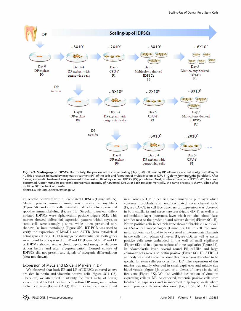
Long-term Culture of DPFreshly extracted DP is a tissue, which contains large nerve
trunks and blood vessels in the central region of the coronal and
radicular pulp (Figure 1A). First outgrowing fibroblast-like cells
appeared between three to four days after DP plating (Figure 1B).
Long-term culture was performed by mechanical transfer of DP
into new culture dish without using enzymatic treatment. After
each transfer, DP produces large numbers of outgrowing cells
approximately every three or four days, thus allowing constant
production of SCs at passage zero (P0) (Figure 2). We obtained
successful isolations with all samples (n = 10) of deciduous teeth.
We performed multiple DP transfers during, at least, six months
(LP of IDPSCs). Both, EP and LP of IDPSCs maintained their
morphology (Figure 1C). Transmission electron microscopy
revealed two types of IDPSCs morphology: ES-like cells with
low cytoplasm-to-nucleus ratio, low cytoplasm density, which are
poor of organelles (Figure 1D). IDPSCs of MSC-like cells have a
high number of stretched out pseudopodes, which serve to explore
substrate and more cytoplasm and organelles when compared with
IDPSCs of ES-like cells (Figure 1D, E). Figure 1F documents that
IDPSCs showed a relatively uniform population in respect of these
two cell types. IDPSCs karyotype was confirmed here to be
unchanged, suggesting that during culture, numerical and gross
structural chromosomal abnormalities did not occur as shown by
routine G-banding technique (Figure 1G). Further, in vitro
expansion of IDPSCs was performed using enzymatic treatment
and the passages was called P1 (Figure 2).
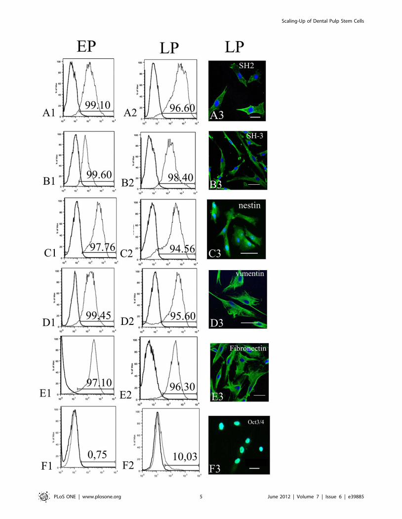
Immunophenotyping of EP and LP of IDPSCsEP and LP were characterized using several markers, which
were employed in our previous original study [4], such as SH2/
CD105, SH3/CD73 (MSCs markers) and Oct3/4 (ES cells
marker). Additionally, expression of MSCs markers such as
vimentin, nestin, fibronectin has been evaluated in both EP and
LP. The reason of choosing these markers will be explained in
discussion section. Representative Figure 3 (A1–E1, A2–E2)
showed that all MSC markers were expressed in both populations
(EP and LP) and slightly declined in LP after six month of multiple
DP transfer (Figure 3A2–E2). A percentage of IDPSCs, which
showed positive immunostaining for these markers, was evaluated
by flow cytometry and was 99.10% to EP and 96.60% to LP for
SH2/CD105; 99.60% to EP and 98.40% to LP for SH3/CD73;
97.76% to EP and 94.56% to LP for nestin; 99.45% to EP and
95.60% to LP for vimentin; 97.10% to EP and 96.30% to LP for
fibronectin (Figure 3A1–E1, A2–E2). Interestingly, that in this
particular IDPSC population (which is not a rule) a very low
percentage of Oct3/4 positive cells ,0.75% was observed in EP,
which increased to ,10.03% in LP (Figure 3F1, F2).
Flow cytometry data have been confirmed by cells immuno-
staining using antibodies against the same MSC and ES cell
markers. Their expression was observed in both EP and LP of
IDPSCs. In Figure 3, the expression of MSC markers in LP is
presented (Figure 3A3–E3). As expected, Oct-3/4 protein
expression was observed in the cell nuclei (Figure 3F3). The
expression of all these markers in LP was similar to EP (data not
shown).
Culture Media Influence EP and LP Growth Rate andGene Expression
Proliferative capacity of EP and LP of IDPSCs before and after
cryopreservation was studied using three different culture media:
DMEM/F12, DMEM-LG, and MEM-alpha. Starting from P2,
non-cryopreserved cells were harvested following enzymatic
dissociation and counted daily during 15 consecutive passages.
IDPSCs cultured in DMEM/F12 and MEM-alpha medium,
presented constant proliferative rate during initial passages, which
achieved their peak growth = 562 (Figure 4A, Table 1). Based on
growth curves presented in Figure 4, statistical analyses were
performed considering the cell number from passage 3 to 7
(Table 1). Using the same parameters, proliferative rate of EP and
LP cultivated in DMEM/F12 and MEM-alpha media were
evaluated after thawing and showed similar proliferative potential,
when compared with those before cryopreservation (Figure 4B,
Table 1). Non-cryopreserved EP and LP of IDPSCs cultured in
DMEM-LG presented spontaneous differentiation into osteogenic
lineage (data not shown) and demonstrated rapid decrease of
proliferative potential (Figure 4A, Table 1). Interestingly, EP and
LP of IDPSCs, cultured in DMEM-LG after thawing, maintained
their proliferative state (Figure 4B, Table 2). DMEM/F12 and
MEM-alpha media did not induce any spontaneous differentiation
in non-cryopreserved and cryopreserved EP and LP of IDPSCs.
The gene expression pattern of pluripotent ES cell and MSC
markers were analyzed by RT-PCR in EP and LP after thawing.
Both were cultured in different basal media (DMEM/F12, MEM-
alpha, and DMEM-LG) for seven passages. Overall, both EP and
LP showed similar expression pattern of vimentin, SH2/CD105
and SH3/CD73 (Figure 4C). Similar expression pattern was
observed to fibronectin, nestin and Oct3/4, when IDPSCs were
cultured in DMEM/F12 and DMEM-LG. However, it was
distinct when cultivated in MEM-alpha, in which three of these
genes (fibronectin, nestin and Oct3/4) did not show any
expression (Figure 4C).
Chondrogenic and Myogenic DifferentiationMultipotential capacity of IDPSCs was reported elsewhere [4].
Therefore, only two differentiation assays were chosen to
demonstrate their differentiation capacity.
At day 21 after induction of chondrogenic differentiation,
IDPSCs demonstrated the formation of an extracellular cartilage
matrix which was intensively stained by Massons trichrome
(Figure 5A, Inset). Toluidine blue staining was used to detect
essential cartilage matrix proteins such as proteoglycans
(Figure 5B). IDPSCs maintained in basal culture medium (control)
did not form any cell pellet (data not shown). Additionally,
chondrogenic differentiation was confirmed by the expression of
COMP (Cartilage Oligomeric Matrix Protein) gene, which
Scaling-Up of Dental Pulp Stem Cells
PLoS ONE | www.plosone.org 2 June 2012 | Volume 7 | Issue 6 | e39885

encodes a pentameric non-collagenous matrix protein that is
mainly expressed in articular cartilage. The expression of COMP
was observed in both EP and LP of IDPSCs (Figure 5C). It is
important to highlight that chondrogenic differentiation of
IDPSCs was uniform even in the absence of TGF-b, which is
known to be a strong inductor of chondrogenesis in bone marrow-
derived MSCs [19,20].
Following myogenic differentiation, IDPSCs showed typical
cells elongation and fusion leading to small myotubes formation at
day 7 (Figure 5D). At day 21, this cell fusion was obvious and most
of the cells formed small myofibers (Figure 5E). MyoD transcrip-
tion factor, which is a master regulatory gene of skeletal muscle
differentiation, as expected, was expressed in IDPSC-derived
myoblasts in nucleus or in perinuclear space following immunosta-
ing using anti-MyoD1 antibody (Figure 5F). These myoblasts
further form myosacs and MyoD1 protein was observed in the
cytoplasm of these more mature cells (Figure 5G). Titin is the third
most abundant skeletal muscle filamentous protein that forms a
separate myofilament system in both skeletal and cardiac muscle.
It was expressed in IDPSC-derived muscle cells at more advanced
stages of differentiation (Figure 5H). Some titin negative cells were
also observed (Figure 5I).
Troponin I is a protein responsible for immobilizing the actin-
tropomyosin complex in place. The expression of this protein was
visualized in more mature myofibers-derived from IDPSCs
(Figure 5J). Human specific anti-actinin and anti-myosin antibod-
Figure 1. Dental pulp and IDPSCs. A) Highly vascularized (black arrows) DP just after extraction. B) Explant culture of DP with outgrowing IDPSCs.C) Culture of IDPSCs at 1st passage. D) IDPSCs showing ES-like cells morphology with a large nucleus. E) IDPSCs showing MSC-like morphology withseveral pseudopodes. F) IDPSCs showing uniform morphology resembling ES cells and MSCs. G) Karyotype of IDPSCs (LP): chromosomes in pairs,ordered by size and position did not reveal any numerical changes in chromosome number; G-banding analysis. A–C, G) Light Microscopy; D–F)Transmission Electron Microscopy; A = 20X, G = 63X; Scale bars: B = 20 mm; C, F = 10 mm; D, E = 3 mm.doi:10.1371/journal.pone.0039885.g001
Scaling-Up of Dental Pulp Stem Cells
PLoS ONE | www.plosone.org 3 June 2012 | Volume 7 | Issue 6 | e39885
ies reacted positively with differentiated IDPSCs (Figure 5K–N).
Myosin positive immunostaining was observed in myofibers
(Figure 5K) and also in differentiated small cells, which presented
spot-like immunolabeling (Figure 5L). Singular binuclear differ-
entiated IDPSCs were alpha-actinin positive (Figure 5M). This
marker showed differential expression pattern within myosacs:
some cells were strongly positive, while others presented only
shadow-like immunostaining (Figure 5N). RT-PCR was used to
verify the expression of MyoD1 and ACTB (Beta cytoskeletal
actin) genes during IDPSCs myogenic differentiation. Both genes
were found to be expressed in EP and LP (Figure 5O). EP and LP
of IDPSCs showed similar chondrogenic and myogenic differen-
tiation before and after cryopreservation. Control culture of
IDPSCs did not present any signals of myogenic differentiation
(data not shown).
Expression of MSCs and ES Cells Markers in DPWe observed that both EP and LP of IDPSCs cultured in vitro
are rich in nestin and vimentin positive cells (Figure 3C1–C3).
Therefore, we attempted to identify the exact niche of nestin,
vimentin and Oct3/4 positive cells within DP using immunohis-
tochemical assay (Figure 6A–Q). Nestin positive cells were found
in all zones of DP: in cell rich zone (innermost pulp layer which
contains fibroblasts and undifferentiated mesenchymal cells)
(Figure 6A–C), in cell free zone, nestin expression was observed
in both capillaries and nerve networks (Figure 6D–F); as well as in
odontoblastic layer (outermost layer which contains odontoblasts
and lies next to the predentin and mature dentin) (Figure 6G, H).
Nestin positive cells in cell rich zone showed fibroblast-like as well
as ES-like cell morphologies (Figure 6B, C). In cell free zone,
nestin protein was found to be expressed in intermediate filaments
in the cells from plexus of nerves (Figure 6D), as well as nestin
positive cells were embedded in the wall of small capillaries
(Figure 6E) and in adjacent regions of these capillaries (Figure 6F).
In odontoblastic layer, several round ES cell-like and large
columnar cells were also nestin positive (Figure 6G, H). STRO-1
antibody was used as control, once this marker was described to be
specific for stem cells/pericytes from DP. The expression of this
marker was mainly observed in small capillaries and middle size
blood vessels (Figure 6J), as well as in plexus of nerves in the cell
free zone (Figure 6K). We also verified localization of vimentin
expressing cells in DP. As expected, vimentin positive cells were
localized in capillaries and in innermost pulp layer, locals where
nestin positive cells were also found (Figure 6L, M). Once low
Figure 2. Scaling-up of IDPSCs. Horizontally, the process of DP in vitro plating (Day 0, P0) followed by DP adherence and cells outgrowth (Day 3–4). This process is followed by enzymatic treatment (P1) of the cells and formation of multiple colonies (CFU-f - Colony Forming Units-fibroblast). After5 days, enzymatic treatment was performed to harvest multicolony-derived IDPSCs (P2) population. Next, in vitro expansion of IDPSCs (P3) has beenperformed. Upper numbers represent approximate quantity of harvested IDPSCs in each passage. Vertically, the same process is shown, albeit aftermultiple DP mechanical transfer.doi:10.1371/journal.pone.0039885.g002
Scaling-Up of Dental Pulp Stem Cells
PLoS ONE | www.plosone.org 4 June 2012 | Volume 7 | Issue 6 | e39885
Scaling-Up of Dental Pulp Stem Cells
PLoS ONE | www.plosone.org 5 June 2012 | Volume 7 | Issue 6 | e39885

percentage of Oct3/4 positive cells, which increased with time of
DP in vitro cultivation, was observed in IDPSCs, the expression of
this protein in DP was also checked. Strong expression of Oct3/4
in the cells nuclei, localized in DP capillaries and in innermost
pulp layer can be observed (Figure 6N–Q).
Localization of BrdU Positive Cells in DP during in vitroCultivation
In order to understand continuous process of IDPSCs
generation, DP was treated with BrdU just after extraction and
in vitro plating (Figure 6R–T). After 6 h, only few anti-BrdU
antibody positive cells were found in the central part of DP
(Figure 6R). After 48 h, BrdU positive cells were observed in the
periphery of DP (Figure 6S), while after 72 h, it seems, that BrdU
positive cells increased in number and were also found in the
periphery of DP in the apical part, close to IDPSCs outgrowing
zone (Figure 6T). Morphological aspect of DP with and without
enzymatic treatment (collagenase/dispase) was compared
(Figure 6U, V). DP, without any treatment, maintains their
integrity especially in the region where BrdU positive cells were
observed (Figure 6U), while after enzymatic digestion this region
was destroyed (Figure 6V).
Discussion
Stem cells reside in a quiescent state within all organs of
organism in their special niche and they start to proliferate and to
migrate when their niche experiences changes [21–24]. Thus,
culture of adult SC niche may provide harvesting of SCs at high
scale. We developed a method of long-term DP culture, which
allowed harvesting of large quantities of IDPSCs (Figure 2).
Mechanical transfer, before and after DP cryopreservation, can
also be performed as long as it is necessary and in our experience,
it may stop due to, e.g. occasional DP explant contamination.
IDPSCs are uniform in respect of morphology (light microscopy
and TEM analyzes) (Figure 1A–F) and karyotype of cells remained
unchanged (Figure 1G). These cells express high percentage of SC
markers such as SH2/CD105, SH3/CD73, nestin, vimentin,
fibronectin and low percentage of Oct3/4 (Figure 3A1–F3)
without losing their original properties [25].
To date, standardized protocol of SCs culture from DP is not
available. Therefore, we directed our study to optimization culture
medium conditions for scaling-up of IDPSCs. DMEM-LG, MEM-
alpha and DMEM/F12 are culture media which are commonly
used for the isolation and expansion of MSCs [26]. In the present
work, we verified the effect of DMEM-LG, MEM-alpha and
DMEM/F12 on proliferation rate and gene expression pattern of
IDPSCs (Figure 4). These analyses indicated that MEM-alpha and
DMEM/F12 were the most appropriate media for the isolation
and long-term expansion of IDPSCs (Figure 4A, B, Table 2);
distinct gene expression patterns were observed between these
media (Figure 4C). DMEM-LG was not efficient for the isolation,
but it was able to support long-term expansion of these cells after
their cryopreservation (Figure 4A, B, Table 1). It seems that our
present data (Figure 4, Table 1) are in contrast with previous
observations, which showed IDPSCs exponential growth following
multiple passages [4]. However, in the present study, cells were
counted daily, while in previous study, passages were performed
every 3–4 days. Therefore, enzymatic treatment used daily seems
to have hampered the IDPSCs.
Analysis of differentiation potential toward chondrogenic and
myogenic lineages evidenced high differentiation potential of LP
and EP of IDPSCs (Figure 5A–O) comparable to those described
previously [4]. IDPSCs showed similar chondrogenic and myo-
genic differentiation before and after cryopreservation (data not
shown) as was reported previously for other SCs from DP [27,28].
Cryopreservation process preserves the proliferative and differen-
tiation capacity of IDPSCs and, thus, allows the opportunity to
bank these valuable DTSCs [29].
Recently, new populations of DTSCs were isolated and were
shown to be distinct from DPSC (Dental Pulp Stem Cells from
permanent teeth)/SHED (Stem Cells from Human Exfoliated
Deciduous teeth) [1,2,4,30–35]. As reported in several original
publications, DPSC/SHED are supposed to be pericytes, which
are isolated from perivascular niche [36,37]. To delineate the
anatomic localization of IDPSCs inside the pulp, we performed in
Figure 3. Characterization of EP and LP of IDPSCs. A1–F1) Flow cytometry showing EP of IDPSCs, which highly expressed such markers asSH2/CD105 (A1); SH3/CD73 (B1); nestin (C1); vimentin (D1); fibronectin (E1). F1) Low expression of Oct3/4 in EP; A2–F2) Flow cytometry showing LPof IDPSCs, which expressed same markers as EP. F2) Higher expression of Oct3/4 in LP, than in F1. A3–F3) Immunofluorescence of LP of IDPSCs usingsame markers as in (A2–E2). F3) Nuclear localization of Oct3/4 can be observed. A3–F3) Epi-fluorescence, nuclei stained with DAPI (blue). Scale bars:A3, B3, E3, F3 = 5 mm; C3, D3 = 10 mm.doi:10.1371/journal.pone.0039885.g003
Table 1. Number of IDPSCs cultured in three different growth media and at different passages before and after cryopreservation.
Cell lineGrowthmedia Passage number
2 3 4 5 6 7 8 9 10 11 12 13 14 15
Beforecryopreservation
MEM-alpha 100 165.5 364 431.5 476.5 394 378.5 321 266.5 189 176.5 147.5 119.5 90.5
DMEM-LG 100 139.5 109.5 100 78 38.5 a a a a a a a a
DMEM/F12 100 125 262.5 302 333 305.5 273.5 218 193 161.5 149.5 124.5 110.5 89
Aftercryopreservation
MEM-alpha 100 153 329 433.5 345 325.25 245 191.5 175.5 172.5 166.5 112.5 64.6 60.5
DMEM-LG 100 115.5 265 314 266 268.50 209 186.5 165.5 162 154.5 50 64.5 35.75
DMEM/F12 100 99 251.5 296.5 250.5 232.5 191 180.5 166.5 142 128 117.5 59.5 57
aDMEM-LG did not support cell growth.doi:10.1371/journal.pone.0039885.t001
Scaling-Up of Dental Pulp Stem Cells
PLoS ONE | www.plosone.org 6 June 2012 | Volume 7 | Issue 6 | e39885

situ analysis using markers of MSCs and ES cells. Our study
suggested that DP has multiple SC niches, which are localized in
capillaries and nerve networks in cell free zone (Figure 6D–F); in
innermost pulp layer in cell rich zone (Figure 6A–C) and in
outermost layer, which contains odontoblasts (Figure 6G, H). All
these niches are rich of nestin positive cells [38], which can present
fibroblast-, epithelial- and odontoblast-like morphologies
(Figure 6A–I). In accordance with our finding, recently, rare
quiescent multipotent nestin positive MSCs were found in bone
marrow in association with hematopoietic SCs and adrenergic
nerve fibers. However, bone marrow derived colonies of MSCs
cultured in vitro exhibit a low percentage (,4%) of such nestin
positive cells [39]. Meanwhile, STRO-1 positive cells localization
in DP, as expected, is restricted to endothelium (Figure 6J), albeit
very weak positive immunostaining was observed in nervous
plexus (Figure 6K). Vimentin showed distribution within DP
similar to nestin, however, a smaller amount of cells positive for
this markers can be observed (Figure 6L, M). Overall, these data
suggest that IDPSCs constitute a mixed population of both MSCs
and epithelial SCs, among which the pericytes (SHED) are also
present. Surprisingly, multiple Oct3/4 positive cells were found in
DP (Figure 6N–Q), showing the appropriate nuclear localization
(Figure 6Q). After isolation, IDPSCs did not show as high as
expected percentage of Oct3/4 positive cells. Previously, we
succeeded to isolate IDPSCs population, which contained ,20%
of these cells. We supposed that in the pulp, Oct3/4 positive cells
are highly pluripotent and of epithelial type. When these cells start
to migrate, they undergo epithelial-mesenchymal transition, which
leads to a decrease or lost Oct3/4 expression. Thus, another
method can be developed in order to isolate naıve Oct3/4 positive
cells from their niche in DP.
DP is capable to produce long-term culture of SCs; however the
process of such ability is unknown. BrdU incorporation into DP
demonstrates that, as expected, only very rare BrdU positive cells
were observed just after DP extraction and plating. Following
further cultivation, such cells increased in number and are located
in the periphery of DP. It is plausible to suggest, that the isolation
of DP stimulates the mechanisms leading to SC proliferation and
migration (Figure 6R–T). In contrast to previous original report
[1], which used the method of DP enzymatic dissociation, we
choose DP explant as a main method of SCs isolation [4].
Comparative morphological analysis demonstrate that prior SCs
isolation enzymatic treatment is not recommended for DP which
may destroy future ‘‘niche’’ of SCs (Figure 6U, V).
In conclusion, our method provides the isolation of a high purity
SC population in substantial quantities. It is based on natural
(intrinsic) mechanisms of SCs activation similar which occur
during tissue trauma or injury, when in response to their damage,
quiescent SCs are activated. Our method can be applied to isolate
SCs from single and multiple niches from any type of adult tissues,
such as bone marrow, adipose tissue, umbilical cord, muscles, skin
and others. This protocol diminishes a probability of occurrence of
spontaneous genomic mutations and eventual karyotype abnor-
malities, which may arise during multiple passages in SCs. This
method is simple, does not requires long time DP preparation,
guarantees sterility (DP can be transferred using sterile instruments
or even pipette) and avoids any type of SCs selection, which is
undesirable for future clinical applications.
Materials and Methods
Human Dental Pulp Extraction and Cell CultureThe investigation was approved by the Ethical Committee of
the Federal University of Sao Paulo (Protocol Nu 0139/10).
Human DP was extracted from ten deciduous teeth of ten healthy
subjects (range 6–9 years) following previously established protocol
[4]. All patients agreed to participate of this study as well as, their
next of kin, carers or guardians on the behalf of the minors/
children participants signed a written informed consent. Next, DP
was gently rinsed in phosphate-buffered solution (PBS) (Invitrogen,
Carlsbad, CA, USA), slightly dissected and placed into 35 mm
plastic tissue culture dishes (Corning Inc., Corning, NY, USA).
Tissue explants were cultured in Dulbecco’s-modified Eagle’s
Figure 4. Proliferation rate and gene expression of IDPSCs after cultivation in three distinct culture media. A) Proliferation curve of LPbefore cryopreservation; B) Proliferation curve of LP after cryopreservation. C) Gene expression of LP after cryopreservation.doi:10.1371/journal.pone.0039885.g004
Table 2. Cell number (x103) of IDPSCs (passage range P3–P7)cultured in different growth media before and aftercryopreservation.
Cryopreservation Growth media
MEM-alpha DMEM-LG DMEM/F12
Before 317.156101.81a 245.80675.70a 226.00674.82a
After 366.306119.88a 93.10637.68b 265.60682.52a
aValues are mean6standard deviation, n = 5. Means followed by the same letterare not statistically different (Tukey, p.0.05).doi:10.1371/journal.pone.0039885.t002
Scaling-Up of Dental Pulp Stem Cells
PLoS ONE | www.plosone.org 7 June 2012 | Volume 7 | Issue 6 | e39885

medium (DMEM)/Ham’s F12 (DMEM/F12, Invitrogen Corpo-
ration - Carlsbad, CA, USA) supplemented with 15% fetal bovine
serum (FBS, Hyclone, Logan, Utah, USA), 100 units/ml penicil-
lin, 100 mg/ml streptomycin, 2 mM L-glutamine, and 2 mM
nonessential amino acids (all from Invitrogen) in a 5% CO2 humid
atmosphere at 37uC. After a period of 3 or 4 days, fibroblast-like
cells were generated from adherent explants. Explants were
transferred to another Petri dish under the same culture
conditions; this procedure was repeated several times (Figure 2).
Fibroblast-like cells growing in monolayer were further washed
twice with PBS and subjected to 0.5 g/L trypsin and 0.53 mmol/
L Ethylenediamine tetra-acetic acid (EDTA) (Invitrogen) for 3 to 5
minutes at 37uC. Passage 1 was counted after the first enzymatic
digestion. Trypsin action was inactivated by culture medium
supplemented with 10% FBS and cells (,56105) were placed into
25 cm2 cell culture flask (Corning). This subculturing was
performed each 3–4 days and the culture medium was changed
daily. For cryopreservation, 90% FBS and 10% dimethylsulfoxide
(DMSO) (Sigma, St. Louis, Mo., USA) were used as freezing
medium. Frozen cells were maintained in sealed vials at 2196uC.
Karyotype AnalysesKaryotyping of subconfluent EP and LP of IDPSCs cultured in
DMEM/F12 medium (Invitrogen) was performed at passage 3.
Figure 5. In vitro differentiation potential of IDPSCs. A–C) Chondrogenic differentiation. A) Pellet culture: collagen fibers intensively stained byMassons thrichrome. Inset: same as in (A) high magnification. B) The proteoglycans presence was revealed by Toloudine blue staining. C) RT-PCRshows the expression of COMP gene in EP and LP of IDPSCs. Housekeeping gene GAPDH is used as control. D–O) Myogenic differentiation. D, E)Morphological aspect showing stages of muscle fibers formation. F) Nuclear expression of MyoD1 protein in LP of IDPSCs-derived myocyte-like cells.G) Myosac composed by MyoD1 positive cells. H, I) Titin protein expression in LP of IDPSCs-derived myotubes. J) Expression of troponin I in Z-bandsof myofibers. K) Myosin protein expression. L) Very small, satellite-like cells, showing positive myosin immunostaining. M) Binuclear cell positive foralpha-actinin (spot-like labeling). N) Fused myotubes, which deferentially express alpha-actinin protein. O) RT-PCR shows the expression of MyoD1and ACTB genes in EP and LP of IDPSCs. Housekeeping gene GAPDH is used as control. A, B, D, E) Light Microscopy; F-N) Epi-fluorescence, nucleistained with DAPI (blue). Scale bars: A = 200 mm; B = 20 mm; D = 50 mm; E, N = 10 mm; F–M = 5 mm.doi:10.1371/journal.pone.0039885.g005
Scaling-Up of Dental Pulp Stem Cells
PLoS ONE | www.plosone.org 8 June 2012 | Volume 7 | Issue 6 | e39885

Figure 6. Expression of nestin, STRO-1, vimentin, Oct3/4 and BrdU in DP. A–H) Nestin expression. A–C) Cell rich zone. A) Multiple nestinpositive cells can be observed. Here and below black arrows indicate immunopositive, while white arrows - immunonegative cells. B) Supposedly
Scaling-Up of Dental Pulp Stem Cells
PLoS ONE | www.plosone.org 9 June 2012 | Volume 7 | Issue 6 | e39885

Before harvesting, demecolcine (Sigma) at a final concentration of
0.1 mg/ml was added for 1 hour. Cells were harvested, washed in
PBS and resuspended in 0.5 ml of medium and mixed with
0.075 M KCl (Sigma) to a volume of 10 ml. After incubation for
20 minutes at room temperature, cells were centrifuged at 400 g
for five minutes and the pellet fixed in 5 ml three times (3:1) of
cold methanol/acetic acid (Sigma). Three drops of cell suspension
were fixed per slide. For chromosome counting, slides were stained
in Giemsa for 15 minutes and; .200 cells were analyzed per cell
line and reported on a Zeiss II microscope (Zeiss, Jena, Germany)
according to the International System for Human Cytogenetic
Nomenclature.
Transmission Electron Microscopy (TEM)For TEM, EP and LP of IDPSCs were fixed in 2.5%
glutaraldehyde (Sigma) for 48 h, post-fixed in 1% phosphate-
buffered osmium tetroxide solution (pH 7.4) (Sigma) for 2 h at 4uCand embedded in Spurr’s Resin (Sigma). Ultrathin sections were
obtained using an automatic ultramicrotome (Ultracut R, Leica
Microsystems, Germany). Sections were double-stained with
uranyl acetate (Sigma) and lead citrate (Sigma) (2% and 0.5%,
respectively) and analyzed using TEM (Morgagni 268D, FEI
Company, The Netherlands; Mega).
Antibodies and ImmunophenotypingEP and LP of IDPSCs immunophenotyping was based on
immunofluorescence and flow-cytometry analyses performed by
using anti-human specific antibodies (vimentin, nestin, fibronectin,
Oct3/4 (all from Santa Cruz Biotechnology, Santa Cruz, CA,
USA), CD105/SH-2 and CD73/SH-3 (both from Case Western
Reserve University, OH, USA). FITC-conjugated secondary
antibodies (Chemicon, Temecula, CA, USA) were used and
respective isotype matched controls. Immunofluorescence were
analyzed using these aforementioned antibodies after cell fixation
in 4% paraformaldehyde (Sigma) in PBS and permeabilization in
0.1% Triton X-100 (Sigma) in PBS. IDPSCs were incubated with
5% bovine serum albumin (BSA, Sigma) diluted in PBS for 30
minutes and further incubated for 1 h at room temperature with
FITC-conjugated goat anti-mouse or anti-rabbit immunoglobulin
(Chemicon) at a final dilution of 1:500 in PBS (Invitrogen).
Microscope slides were mounted in Vectashield mounting medium
with 49,6-Diamidino-2-phenylindol (DAPI, Vector Laboratories,
Burlingame, CA) and immunofluorescence was detected using a
Carl Zeiss Axioplan fluoromicroscope (LSM 410, Zeiss, Jena,
Germany) or Nikon Eclipse E1000 (Nikon, Kanagawa, Japan).
Digital images were acquired with CCD camera (Applied Imaging
model ER 339) and the documentation system used was
Cytovision v. 2.8 (Applied Imaging Corp. - Santa Clara, CA,
USA). Flow-cytometry was performed using EP and LP of IDPSC
at passage 3. Cells were detached by using a 10 min treatment at
37uC with PBS 0.02% EDTA, pelleted (10 min at 400 g) and
washed in 0.1% BSA in 0.1 M PBS at 4uC. Next, cells at a
concentration of 105 cells/ml were stained with saturating
concentration of aforementioned antibodies (10 ml). After 45
minute incubation in the dark at room temperature, cells were
washed three times with PBS and resuspended in 0.25 ml of cold
PBS. Flow-cytometry analysis was performed on a fluorescence-
activated cell sorter (FACS; Becton, Dickinson, San Jose, CA)
using the CELL Quest program (Becton, Dickinson). The flow
cytometry and/or immunofluorescence analyses were repeated
with all samples (n = 10), and one representative experiment is
presented. All experiments have been done in triplicate and
furthermore were repeated several times.
Cell Growth RateTo evaluate the effect of different culture media on cell growth,
freshly isolated and the same IDPSC frozen–thawed were equally
divided in three groups (DMEM/F12, DMEM low-glucose
(1000 mg/ml; DMEM-LG) and Minimum Essential Medium
(MEM) Alpha Medium (MEM-alpha). All media (Invitrogen) were
supplemented with 15% FBS (Hyclone), 100 units/ml penicillin,
100 mg/ml streptomycin, 2 mM L-glutamine, and 2 mM nones-
sential amino acids (all from Invitrogen). Cells were seeded at a
density of 105/ cm2 counted for at least fifteen consecutive days to
evaluate the growth rate and the effect of cryopreservation. We
also verified the capacity of DP tissue explant to produce IDPSC
after consecutive rounds of cryopreservation and thawing. All
experiments were performed in triplicate.
Data and Statistical AnalysisGrowth curves were constructed using data from cell lines,
passage number (P2 to P15), cryopreservation and growth
medium. Cell number data were analyzed by using two-way
analysis of variance (‘‘cryopreservation’’ and ‘‘growth medium’’)
complemented by Tukey post hoc multiple comparison tests. The
significance level was set at 5% (SPSS 19.0, Chicago, IL, USA).
RNA Extraction and Reverse Transcription-polymeraseChain Reaction (RT-PCR)
EP and LP of IDPSCs were cultivated during seven passages in
three distinct media (DMEM/F12, MEM-alpha, and DMEM-
LG). To evaluate the effect of these different culture media on
gene expression, total RNA was extracted using Trizol (Invitro-
gen): IDPSCs were washed in PBS and RNA extraction was
performed according to manufactures instructions. cDNAs were
synthesized from 1 mg of total RNA reverse transcribed with the
RevertAid M-MuLV Reverse Transcriptase and oligo (dT)
(Fermentas Life Science, Amherst, NY, EUA) according to the
manufactures instructions. The final concentrations of reagents
were: 20 ml of PCR reactions were prepared with 2 ml cDNA,
0,2 mM of each primer, 1 unit of Taq DNA Polymerase, 0,2 mM of
dNTPs, 1,5 mM of magnesium chloride and buffer Taq DNA
Polymerase (Fermentas Life Science). Primer sequences (forward
and reverse), and the lengths of amplified products are summa-
rized: Nestin FW 59- CTCTGACCTGTCAGAAGAAT-39, and
RV 59-GACGCTGACACTTACAGAAT-39 (302 bp/54uC); Vi-
mentin FW 59-AAGCAGGAGTCCACTGAGTACC-39, and RV
undifferentiated MSC shows nestin cytoplasm localization. C) Nestin positive cells with two distinct morphologies round epithelial-like (ES-like) andfibroblast-like cells. D–F) Cell free zone. D) Nestin showing intermediate filament staining in nerve plexus. E) Small capillary with two intensivelystained nestin positive cells. F) Same as in (E) with nestin positive cells in lateral of capillary (arrow). G, H) Odontoblastic layer. Nestin positiveobontoblasts (G, H) can be observed. I) Negative control: only secondary antibody was used. J, K) Cell free zone. J) STRO-1 positive cells withincapillaries (perivascular niche). K) Very poor STRO-1 immunostaining was observed within nerve plexus. L, M) Vimentin positive (black arrows) cellslocalization in cell rich (L) and cell free (M) zones. N–Q) Oct3/4 positive cells localization in cell rich (N) and cell free (O–Q) zones. R–T) BrdUimmunostaining of DP. R) DP just after plating in culture medium. S) 48 hours after in vitro cultivation. T) 72 hours after in vitro cultivation. U–V) DPwithout (U) and with (V) enzymatic treatment. V) External cell layer of DP is destroyed by such treatment. A–V) Light Microscopy. Scale bars: A, D, F–P,R–T = 20 mm; B, C, Q = 5 mm; U, V = 50 mm.doi:10.1371/journal.pone.0039885.g006
Scaling-Up of Dental Pulp Stem Cells
PLoS ONE | www.plosone.org 10 June 2012 | Volume 7 | Issue 6 | e39885

59-GAAGGTGACGAGCCATTTCC-39 (205 bp/55uC); Fibro-
nectin FW 59-GGATCACTTACGGAGAAACAG-39, and RV
59-GATTGCATGCATTGTGTCCT-39 (386 bp/56uC); OCT3/
4 FW 59-ACCACAGTCCATGCCATCAC-39, and RV 59-
TCCACCACCCTGTTGCTGTA-39 (120 bp/61uC); SH2/
CD105 FW 59- TCTGGACCACTGGAGAATAC-39, and RV
59-GAGGCATGAAGTGAGACAAT-39 (171 bp/56uC); SH3/
CD73 FW 59-ACACGGCATTAGCTGTTATT-39, and RV 59-
AGTATTTGTTCTTTGGGCA-39 (391 bp/56uC). For chon-
drogenic and myogenic differentiation, following primer sequences
were used: COMP FW 59-CCGACAGCAACGTGGTCTT-39,
and RV 59-CAGGTTGGCCCAGATGATG-39 (91 bp/53uC);
ACTB FW 59-TGGCACCACACCTTCTACAATGAGC-39,
and RV 59 GCACAGCTTCTCCTTAATGTCACGC-39
(395 bp/59uC); MYOD1 FW 59-GCCGCCTGAGCAAAG-
TAAATGAGG-39, and RV 59-TAGTCCATCATGCCGTCG-
GAGC-39 (280 bp/53uC). GADPH gene FW 59- ACCACAGTC-
CATGCCATCAC-39, and RV 59-
TCCACCACCCTGTTGCTGTA-39 (463 bp/61uC) was used
as control. Undifferentiated IDPSC were examined as negative
control for differentiation specific primers. PCR reactions were
performed under the following conditions: 1 cycle at 94uC for 5
minutes, followed by 35 cycles at 94uC for 1 minute, annealing
temperature for 1 minute, and 72uC for 1 minute. Amplified
products were resolved by electrophoresis on a 1.5% agarose gel
(Sigma) and visualized using ethidium bromide (Sigma) staining.
Differentiation AssaysChondrogenic differentiation. The differentiation was
performed using pellet culture technique [19,20]. EP and LP of
IDPSCs populations from sub confluent cultures (passage 3) were
released by 0.5 g/L trypsin and 0.53 mmol/L EDTA, counted
and used to generate micromass culture. Briefly, 46106 cells were
centrifuged at 500 g in 15 ml polypropylene conical tubes
(Corning) and the resulting pellets were cultured for 4 weeks.
Control cultures were grown in a serum-free chemically defined
medium consisting of DMEM, high-glucose (4,500 mg/L;
DMEM-HG) (Invitrogen) supplemented with 6.25 mg/ml insulin,
6.2 mg/ml transferrin, 6.25 mg/ml selenious acid, 5.33 mg/ml
linoleic acid (ITS, Premix, BD, USA) and 1 mM sodium pyruvate
(Invitrogen). To induce chondrogenic differentiation, control
medium was supplemented with 0.1 mM dexamethasone and
50 mg/ml ascorbate-2-phosphate (both from Invitrogen), without
transforming growth factor beta (TGF-b). Cultures were incubated
for 4 weeks at 37uC in a humid atmosphere containing 5% CO2;
the medium was changed every day. Cell aggregates were
harvested at 4 weeks for RT-PCR, with primer sequences
aforementioned, and were also fixed for histology.
Muscle differentiation. EP and LP of IDPSCs at the
passage 3 were seeded at a concentration of 5000 cells/cm2.
Control cultures were grown in a serum-free chemically defined
medium consisting of DMEM-HG (Invitrogen) supplemented with
10% FBS (Hyclone). To induce myogenic differentiation, control
medium was supplemented with 50 mM hydrocortisone (Sigma)
and 5% horse serum (Invitrogen). Cultures were incubated for 21
days at 37uC in a humid atmosphere containing 5% CO2; the
medium was changed every three days. Then, cultured cells were
fixed for histology and for immunohistochemistry with antibodies
that recognize human muscle proteins. Aforementioned primers
sequences were used to detect myogenic differentiation of IDPSCs.
Histological AnalysesPellet culture analysis. Cell aggregates were fixed in 4%
formaldehyde in PBS for 40 minutes and embedded in paraffin
(Sigma). Paraffin sections were stained with Masson’s trichrome
(Sigma) and Toluidine Blue (Sigma) and analyzed using transmit-
ted and polarized light microscopy Nikon Eclipse E1000 (Nikon),
digital images were acquired by CCD camera (Applied Imaging).
Muscle analysis. Slices were fixed in 4% paraformaldehyde
diluted in PBS for 40 min and analyzed using transmitted and
polarized light microscopy Nikon Eclipse E1000 (Nikon), digital
images were acquired by CCD camera (Applied Imaging).
ImmunohistochemistryTroponin I (Chemicon), titin (Chemicon), alpha-smooth muscle
actinin (Sigma), MyoD1 (Chemicon) and sarcomeric myosin
(Chemicon) proteins were analyzed using specific antibodies. First,
cells were fixed in 4% paraformaldehyde diluted in PBS and
permeabilized in 1% Triton X-100 diluted in PBS. EP and LP of
IDPSC that were submitted to myogenic differentiation were
incubated in 5% BSA diluted in PBS for 30 min and incubated for
1 h at room temperature with FITC-conjugated goat anti-mouse
or anti-rabbit immunoglobulin (Chemicon) at a final dilution of
1:500 in PBS. Microscope slides were mounted in Vectashield
mounting medium with DAPI and immunofluorescence was
detected using a Carl Zeiss Axioplan fluoromicroscope (Zeiss) or
Nikon Eclipse E1000 (Nikon), digital images were acquired by
CCD camera (Applied Imaging) and the documentation system
used was Cytovision v. 2.8 (Applied Imaging Corp).
Pulp Tissue ExperimentsTissue specimens. Ten deciduous teeth of ten healthy
subjects (range 5–10 years), free of caries and restorations, were
extracted and initially rinsed in PBS. Seven pulps were gently
removed and fixed in 10% formalin solution for 48 h. The
specimens were embedded in paraffin blocks and sections of
10 mm were obtained. The other three pulps were gently rinsed in
PBS and sliced. Each slice was placed in different culture dish.
Next, 5-bromo-29-deoxyuridine (BrdU, Sigma) was added directly
into basal culture medium. First pulp slice was fixed and processed
after 6 h of treatment with BrdU. In the second culture dish with
pulp slice, BrdU was added after 42 h and in the third - after 66 h.
After 6 h of treatment with BrdU, all slices were fixed in 10%
formalin solution for 48 h. The specimens were embedded in
paraffin blocks and sections of 10 mm were obtained. All above
specimens were treated by immunohistochemical methods.
ImmunohistochemicalParaffin sections of 10 mm were deparaffinized and then
hydrated. Endogenous peroxidase activity was measured by
incubating the sections for 30 min in a 0.1% solution of hydrogen
peroxide (Sigma). For antigen retrieval, sections were incubated
with trypsin for 10 min at 37uC. To inhibit nonspecific antigen
binding, sections were incubated with blocking serum (5% fetal
calf serum, Invitrogen) for 10 min. Sections were then incubated
for 12–16 h with the primary antibody in a moist chamber at 4uC.
Primary antibodies were the same used in immunophenotyping of
IDPSCs and additionally anti-human STRO-1 (Santa Cruz) and
mouse anti-BrdU IGg (Chemicon). The optimal dilution of the
primary antibody was found to be 1:10. Slides were again rinsed
with PBS and then incubated with biotinylated secondary
antibody (DAKO, Glostrup, Denmark) in 1:200 dilution for
30 min. The samples were washed with PBST (PBS with 0.1% of
Tween 20) and incubated with StrepABComplex/HRP (DAKO)
at 1:100 dilution for 30 min. After one more wash with PBST, the
colour was revealed by the chromogen 3 (3-diaminobenzidine
DAB Kit, Zymed Laboratories, Inc.) for 5 min, followed by PBST
washing, nuclear counterstaining with Harris haematoxylin for
Scaling-Up of Dental Pulp Stem Cells
PLoS ONE | www.plosone.org 11 June 2012 | Volume 7 | Issue 6 | e39885

45 s, dehydrated and mounted in Permount. Observation of the
sections was conducted using a Carl Zeiss Axioplan fluoromicro-
scope (Zeiss). Negative control sections were treated identically,
except the primary antibody which was substituted by PBS.
Acknowledgments
We are grateful to Dr Adriana da Costa Neves for her expertise in
immunohistochemical assay and to Alexsander S. de Souza for his excellent
assistance in confocal microscopy analysis. We wish to thank Dr. Graciela
C. Pignatari for her assistance in molecular biology, Dr. Cristiane V.
Wenceslau and Lisley I. Mambelli for their help during the development of
this project.
Author Contributions
Conceived and designed the experiments: NFL IK AC. Performed the
experiments: NFL CG. Analyzed the data: NFL IK AK JH CO.
Contributed reagents/materials/analysis tools: NFL IK JH CO. Wrote
the paper: NFL IK AC.
References
1. Gronthos S, Mankani M, Brahim J, Robey PG, Shi S (2000) Postnatal human
DP stem cells (DPSCs) in vitro and in vivo. Proc Natl Acad Sci U S A 97: 13625–
13630.
2. Miura M, Gronthos S, Zhao M, Lu B, Fisher LW, et al. (2003) SHED: Stem
cells from human exfoliated deciduous teeth. Proc Natl Acad Sci U S A 100:
5807–5812.
3. Kerkis I, Caplan AI (2011) Stem cells in dental pulp of deciduous teeth. Tissue
Eng Part B Rev Dec 28 [Epub ahead of print].
4. Kerkis I, Kerkis A, Dozortsev D, Stukart-Parsons GC, Gomes Massironi SM, et
al. (2006) Isolation and characterization of a population of immature dental pulp
stem cells expressing OCT-4 and other embryonic stem cell markers. Cells
Tissues Organs 184: 105–116.
5. Fonseca SAS, Abdelmassih S, de Mello Cintra Lavagnolli T, Serafim RC,
Clemente Santos EJ, et al. (2009) Human immature dental pulp stem cells’
contribution to developing mouse embryos: production of human/mouse
preterm chimaeras. Cell Prolif 42: 132–140.
6. de Mendonca Costa A, Bueno DF, Martins MT, Kerkis I, Kerkis A, et al. (2008)
Reconstruction of large cranial defects in nonimmunosuppressed experimental
design with human dental pulp stem cells. J Craniofac Surg 19: 204–210.
7. Gomes JA, Geraldes Monteiro B, Melo GB, Smith RL, Cavenaghi Pereira da
Silva M, et al. (2010) Corneal reconstruction with tissue-engineered cell sheets
composed of human immature dental pulp stem cells. Invest Ophthalmol Vis Sci
51: 1408–1414.
8. Kerkis I, Ambrosio CE, Kerkis A, Martins DS, Zucconi E, et al. (2008) Early
transplantation of human immature DP stem cells from baby teeth to golden
retriever muscular dystrophy (GRMD) dogs: Local or systemic? J Transl Med 6:
35.
9. Beltrao-Braga PCB, Pignatari GC, Maiorka PC, Oliveira NAJ, Lizier NF, et al.
(2011) Feeder-free derivation of induced pluripotent stem cells from human
immature dental pulp stem cells. Cell Transplant [Epub ahead of print]; DOI:
10.3727/096368911x566235.
10. Yan X, Qin H, Qu C, Tuan RS, Shi S, et al. (2010) iPS cells reprogrammed
from human mesenchymal-like stem/progenitor cells of dental tissue origin.
Stem Cells Dev 19: 469–480.
11. Kerkis I, Lobo SE, Kerkis A (2009) Dental pulp stem cells and perspectives of
future application in cell therapy. In: Deb, K.D, and Totel, S.M., eds. Stem
Cells. Basic and Applications. New Delhi: Tata McGraw Hill. 426–438.
12. Caplan AI (2009) Why are MSCs therapeutic? New data: new insight. J Pathol
217: 318–324.
13. Wagner J, Kean T, Young R, Dennis JE, Caplan AI (2009) Optimizing
mesenchymal stem cell-based therapeutics. Curr Opin Biotechnol Oct;20(5):
531–536.
14. Stenderup K, Justesen J, Clausen C, Kassem M (2003) Aging is associated with
decreased maximal life span and accelerated senescence of bone marrow stromal
cells. Bone 33(6): 919–926.
15. Baxter MA, Wynn RF, Jowitt SN, Wraith JE, Fairbairn LJ, et al. (2004) Study of
telomere length reveals rapid aging of human marrow stromal cells following in
vitro expansion. Stem Cells 22(5): 675–682.
16. Rubio D, Garcia-Castro J, Martin MC, de la Fuente R, Cigudosa JC, et al.
(2005) Spontaneous human adult stem cell transformation. Cancer Res 65(8):
3035–3039.
17. Wang Y, Huso DL, Harrington J, Kellner J, Jeong DK, et al. (2005) Outgrowth
of a transformed cell population derived from normal human bone marrow
mesenchymal stem cell culture. Cytotherapy 7(6): 509–519.
18. Spath L, Rotilio V, Alessandrini M, Gambara G, De Angelis L, et al. (2010)
Explant-derived human dental pulp stem cells enhance differentiation and
proliferation potentials. J Cell Mol Med. 2010 Jun;14(6B): 1635–44. Epub 2009
Jul 7.
19. Johnstone B, Hering TM, Caplan AI, Goldberg VM, Yoo JU (1998) In vitrochondrogenesis of bone marrow-derived mesenchymal progenitor cells. Exp Cell
Res. Jan 10; 238(1): 265–272.20. Yoo JU, Barthel TS, Nishimura K, Solchaga L, Caplan AI, et al. (1998) The
chondrogenic potential of human bone-marrow-derived mesenchymal progen-
itor cells. J Bone Joint Surg Am. Dec; 80(12): 1745–1757.21. Schofield R (1978) The relationship between the spleen colony forming cell and
the hemopoietic stem cell. Blood Cells 4: 7–25.22. Mitsiadis TA, Barrandon O, Rochat A, Barrandon Y, De Bari C (2007) Stem
cell niches in mammals. Experimental Cell Research 313(16): 3377–3385.23. De Bari C, Pringle S, Pitzalis C, Dell’Accio F (2006) The stem cell niche: a new
target in medicine. Current Opinion in Orthopaedics 17(5): 398–404.
24. Abdallah BM, Boissy P, Tan Q, Dahlgaard J, Traustadottir GA, et al. (2007)dlk1/FA1 regulates the function of human bone marrow mesenchymal stem cells
by modulating gene expression of pro-inflammatory cytokines and immuneresponserelated factors. J Biol Chem 282: 7339–7351.
25. Gronthos S, Brahim J, Li W, Fisher LW, Cherman N, et al. (2002) Stem cell
properties of human dental pulp stem cells. J Dent Res 81: 531–535.26. Pal R, Hanwate M, Jan M, Totey S (2008) Phenotypic and Functional
comparison of optimum culture condition for upscaling of Bone marrow derivedMesenchymal stem cells. J Tissue Eng Regen Med 3: 163–174.
27. Papaccio G, Graziano A, d’Aquino R, Graziano MF, Pirozzi G, et al. (2006)Long-term cryopreservation of dental pulp stem cells (SBP-DPSCs) and their
differentiated osteoblasts: a cell source for tissue repair. J Cell Physiol 208: 319–
325.28. Zhang W, Walboomers XF, Shi H, Fan M, Jansen JA, et al. (2006) Multilineage
differentiation of stem cells derived from human dental pulp after cryopreser-vation. Tissue Eng 12: 2813–2823.
29. Arora V, Arora P, Munshi AK (2009) Banking stem cells from human exfoliated
deciduous teeth (SHED): Saving for the Future. J Clin Pediatr Dent 33: 289–294.
30. Laino G, d’Aquino R, Graziano A, Lanza V, Carinci F, et al. (2005) A newsource of human adult dental pulp stem cells: a useful source of living autologous
fibrous bone tissue. J Bone Miner Res 20: 1394–1402.
31. Pierdomenico L, Bonsi L, Calvitti M, Rondelli D, Arpinati M, et al. (2005)Multipotent mesenchymal stem cells with immunosuppressive activity can be
easily isolated from dental pulp. Transplantation 80: 836–842.32. Jo YY, Lee HJ, Kook SY, Choung HW, Park JY, et al. (2007) Isolation and
characterization of postnatal stem cells from human dental tissues. Tissue Eng13: 767–773.
33. Huang AH, Chen YK, Lin LM, Shieh TY, Chan AW (2008) Isolation and
characterization of dental pulp stem cells from supernumerary tooth. J OralPathol Med 37: 571–574.
34. Nam H, Lee G (2009) Identification of novel epithelial stem cell-like cells inhuman deciduous dental pulp. Biochem Biophys Res Commun 386: 135–139.
35. Waddington RJ, Youde SJ, Lee CP, Sloan AJ (2009) Isolation of distinct
progenitor stem cell populations from dental pulp. Cells Tissues Organs 189:268–274.
36. Shi S, Gronthos S (2003) Perivascular niche of post natal mesenchymal stem cellsin human bone marrow and dental pulp. J Bone Miner Res 189: 696–704.
37. Crisan M, Yap S, Casteilla L, Chen CW, Corselli M, et al. (2008) A perivascularorigin for mesenchymal stem cells in multiple human organs. Cell Stem Cell 3:
301–313.
38. About I, Laurent-Maquin D, Lendahl U, Mitsiadis TA (2000) Nestin expressionin embryonic and adult human teeth under normal and pathological conditions.
Am. J. Pathol. 157: 287–295.39. Mendez-Ferrer S, Michurina TV, Ferraro F, Mazloom AR, Macarthur BD, et
al. (2010) Mesenchymal and haematopoietic stem cells form a unique bone
marrow niche. Nature 12; 466 (7308): 829–834.
Scaling-Up of Dental Pulp Stem Cells
PLoS ONE | www.plosone.org 12 June 2012 | Volume 7 | Issue 6 | e39885
Related Documents