Available online at www.sciencedirect.com ScienceDirect European Journal of Protistology 50 (2014) 551–592 Scale evolution in Paraphysomonadida (Chrysophyceae): Sequence phylogeny and revised taxonomy of Paraphysomonas, new genus Clathromonas, and 25 new species Josephine Margaret Scoble ∗ , Thomas Cavalier-Smith Department of Zoology, University of Oxford, South Parks Road, Oxford OX1 3PS, UK Received 6 May 2014; received in revised form 31 July 2014; accepted 11 August 2014 Available online 19 August 2014 Abstract Heterotrophic chrysomonads of the genus Paraphysomonas are ubiquitous phagotrophs with diverse silica scale morphology. Over 50 named species have been described by electron microscopy from uncultured environmental samples. Sequence data exist for very few, but the literature reveals misidentification or lumping of most previously sequenced. For critically integrating scale and sequence data, 59 clonal cultures were studied light microscopically, by sequencing 18S ribosomal DNA, and recording scale morphology by transmission electron microscopy. We found strong congruence between variations in scale morphology and rDNA sequences, and unexpectedly deep genetic diversity. We now restrict Paraphysomonas to species with nail-like spine scales, establishing 23 new species and eight subspecies (Paraphysomonadidae). Species having base-plates with dense margins form three distinct subclades; those with a simple margin only two. We move 29 former Paraphysomonas species with basket scales into a new genus, Clathromonas, and describe two new species. Clathromonas belongs to a very distinct rDNA clade (Clathromonadidae fam. n.), possibly distantly sister to Paraphysomonas. Molecular and morphological data are mutually reinforcing; both are needed for evaluating paraphysomonad diversity and confirm excessive past lumping. Former Paraphysomonas species with neither nail-like nor basket scales are here excluded from Paraphysomonas and will be assigned to new genera elsewhere. © 2014 Published by Elsevier GmbH. Keywords: Clathromonas; Chrysophyte; 18S rDNA phylogeny; Heterokont; Paraphysomonas vestita; Scale ultrastructure Introduction Colourless chrysomonads of the genera Paraphysomonas and Spumella are major phagotrophs in freshwater and soil food webs, and Paraphysomonas is also widespread in marine environments (Charvet et al. 2011; del Campo ∗ Corresponding author. Tel.: +44 1865 281906; fax: +44 1865 281310. E-mail addresses: [email protected], [email protected] (J.M. Scoble). and Massana 2011; Massana et al., 2004, 2006, 2014; Richards and Bass 2005). These important feeders on bacte- ria have received considerable experimental study (Jürgens et al. 1997; Lim et al. 1999; Pfandl et al. 2004; Simek et al. 1997; Zwirglmaier et al. 2009), but their taxonomy is unsatisfactory and needs major revision. Ribosomal DNA phylogeny showed that Spumella is certainly polyphyletic; about five non-scaly chrysophyte lineages independently lost photosynthesis and thus became Spumella-like in mor- phology (Boenigk et al. 2005; Boenigk 2008); eventually http://dx.doi.org/10.1016/j.ejop.2014.08.001 0932-4739/© 2014 Published by Elsevier GmbH.

Welcome message from author
This document is posted to help you gain knowledge. Please leave a comment to let me know what you think about it! Share it to your friends and learn new things together.
Transcript
SSnJ
D
RA
A
OessasmwrmPt
©
K
I
asi
j
h0
Available online at www.sciencedirect.com
ScienceDirect
European Journal of Protistology 50 (2014) 551–592
cale evolution in Paraphysomonadida (Chrysophyceae):equence phylogeny and revised taxonomy of Paraphysomonas,ew genus Clathromonas, and 25 new species
osephine Margaret Scoble∗, Thomas Cavalier-Smith
epartment of Zoology, University of Oxford, South Parks Road, Oxford OX1 3PS, UK
eceived 6 May 2014; received in revised form 31 July 2014; accepted 11 August 2014vailable online 19 August 2014
bstract
Heterotrophic chrysomonads of the genus Paraphysomonas are ubiquitous phagotrophs with diverse silica scale morphology.ver 50 named species have been described by electron microscopy from uncultured environmental samples. Sequence data
xist for very few, but the literature reveals misidentification or lumping of most previously sequenced. For critically integratingcale and sequence data, 59 clonal cultures were studied light microscopically, by sequencing 18S ribosomal DNA, and recordingcale morphology by transmission electron microscopy. We found strong congruence between variations in scale morphologynd rDNA sequences, and unexpectedly deep genetic diversity. We now restrict Paraphysomonas to species with nail-likepine scales, establishing 23 new species and eight subspecies (Paraphysomonadidae). Species having base-plates with denseargins form three distinct subclades; those with a simple margin only two. We move 29 former Paraphysomonas speciesith basket scales into a new genus, Clathromonas, and describe two new species. Clathromonas belongs to a very distinct
DNA clade (Clathromonadidae fam. n.), possibly distantly sister to Paraphysomonas. Molecular and morphological data areutually reinforcing; both are needed for evaluating paraphysomonad diversity and confirm excessive past lumping. Formeraraphysomonas species with neither nail-like nor basket scales are here excluded from Paraphysomonas and will be assigned
o new genera elsewhere.
2014 Published by Elsevier GmbH.
teroko
a
eywords: Clathromonas; Chrysophyte; 18S rDNA phylogeny; He
ntroduction
Colourless chrysomonads of the genera Paraphysomonasnd Spumella are major phagotrophs in freshwater andoil food webs, and Paraphysomonas is also widespreadn marine environments (Charvet et al. 2011; del Campo
∗Corresponding author. Tel.: +44 1865 281906; fax: +44 1865 281310.E-mail addresses: [email protected],
[email protected] (J.M. Scoble).
Rreeipalp
ttp://dx.doi.org/10.1016/j.ejop.2014.08.001932-4739/© 2014 Published by Elsevier GmbH.
nt; Paraphysomonas vestita; Scale ultrastructure
nd Massana 2011; Massana et al., 2004, 2006, 2014;ichards and Bass 2005). These important feeders on bacte-
ia have received considerable experimental study (Jürgenst al. 1997; Lim et al. 1999; Pfandl et al. 2004; Simekt al. 1997; Zwirglmaier et al. 2009), but their taxonomys unsatisfactory and needs major revision. Ribosomal DNA
hylogeny showed that Spumella is certainly polyphyletic;bout five non-scaly chrysophyte lineages independentlyost photosynthesis and thus became Spumella-like in mor-hology (Boenigk et al. 2005; Boenigk 2008); eventually
5 an Jour
tdoms‘pCftdSshadt
d1sd(ctgcS(filtsssbtTePso2fcmts
n2stm‘o
isafsdgaaFatt(
a1siow8odmwfbidrdlC
o(awatPiPPstcaw
w
52 J.M. Scoble, T. Cavalier-Smith / Europe
hey must be divided into several genera. Paraphysomonasiffers from Spumella by having numerous silica scalesn its cell body, but it is easy to confuse them by lighticroscopy, which does not reveal the scales of most species;
o many strains and their sequences have merely been calledSpumella-like’ (Boenigk et al. 2005). Traditionally, Para-hysomonas was grouped with three photosynthetic generahrysosphaerella, Spiniferomonas, and Polylepidomonas in
amily Paraphysomonadaceae (Preisig 1995); more recentlyhese were excluded, Paraphysomonas alone constituting aistinct chrysomonad order Paraphysomonadales (Cavalier-mith and Chao 2006), which sequence trees often place asister to all other chrysophytes (Skaloud et al. 2013). We focusere on the biodiversity and taxonomy of Paraphysomonasnd show that several genera are needed to encompass theiriversity, and more species than hitherto realised can be dis-inguished from Spumella in the light microscope.
Ultrastructural differences in scale morphology currentlyistinguish 56 – 57 Paraphysomonas species (Lucas 1967,968; Preisig and Hibberd 1982a, 1982b, 1983). The typepecies, P. vestita, is the only one not originally thusefined, having been discovered before electron microscopyStokes 1885 as Physomonas vestita). De Saedeleer (1929)hanged its name to Paraphysomonas vestita because theype species (Physomonas socialis) was removed to anotherenus, Monas, now abandoned as a nomen dubium roughlyorresponding with Spumella (see Silva 1960); howeverpumella is itself polyphyletic and requires major revisionBoenigk 2008). Paraphysomonas vestita spine scales wererst drawn by Korshikov (1929) as ‘nails with relatively
arge flat heads’. Houwink (1952) published the first elec-ron micrographs of Paraphysomonas ‘vestita’ spine scales,howing their circular base-plate and long central pointedpine. Subsequent ultrastructural studies and environmentalurveys of silica-scaled protists have shown a great variety ofroadly similar, yet distinctly different, nail-like scales underhe umbrella name P. vestita (Manton and Leedale 1961;akahashi 1976; Cronberg and Kristiansen 1980; Thomsent al. 1981; Santos and Leedale 1993; Bergesch et al. 2008;etronio and Rivera 2010). It is unclear which, if any, of thesetructurally quite diverse scales are actually from P vestitar from undescribed species (see Scoble and Cavalier-Smith013). Hardly any Paraphysomonas species were describedrom clonal cultures, nearly all being named from a few cellsollected directly from the environment and dried on electronicroscope grids. There is therefore almost no knowledge of
he range of variation of scales within a strain, still less aingle species, causing identification problems.
Ribosomal DNA sequences are available for only fiveamed Paraphysomonas species (Scoble and Cavalier-Smith013). Unfortunately, some sequences labelled as the samepecies (P. vestita and P. foraminifera) are so far apart on
he trees and radically different that some sequenced strainsust have been seriously misidentified; moreover one P.foraminifera’ sequence (AB022864) is almost the same asne P. ‘vestita’ sequence (Z28335: Rice et al. 1997), differing
alsg
nal of Protistology 50 (2014) 551–592
n one inserted T. No ultrastructure was provided for mosttrains so their true identity is unknown and cultures no longervailable for study. Some Paraphysomonas sequences wereortunately published together with electron micrographs ofcales (Caron et al. 1999; Rice et al. 1997); in all cases theiretailed structure differs from that of the type strains, sug-esting that none was correctly identified. These mistakesnd the rarity of combined sequence and morphological datare totally confusing for Paraphysomonas scale evolution.rom environmental sequencing more different sequences arelready known in the Paraphysomonas spine-scale clade thanhe total number of named spine-scaled species, so the asser-ion that most Paraphysomonas species are already knownFinlay and Clarke 1999a) was overconfident.
It has been claimed that P. vestita is the commonestnd most widespread Paraphysomonas (Finlay and Clarke999b), but that could be an artefact of an excessively loosepecies definition (see Scoble and Cavalier-Smith 2013). Thedentity of the type species P. vestita is loosely defined: theriginal description tells us scarcely more than it was ∼15 �mith projecting spines, but strains under that name range from
to 26 �m and exhibit such a large range in scale morphol-gy that they probably represent numerous species. Looseefinition may also apply to some extent to the ‘second com-onest’ species P. imperforata (Finlay and Clarke 1999b),hose relatively non-descript spine scales differ obviously
rom those attributed to P. vestita only by lacking a densease-plate margin and from P. foraminifera merely by lack-ng holes on the base-plate, i.e. P. imperforata is negativelyefined. The literature has not been critically reviewed untilecently, but there are clearly subtle and some more obviousifferences in broadly similar scale types for both ‘P. vestita’-ike and P. imperforata-like scales, as noted by Scoble andavalier-Smith (2013).To clarify these problems, and put Paraphysomonas tax-
nomy on a sounder footing, we studied 59 clonal culturesmostly newly isolated) by light and electron microscopynd 18S rDNA sequencing; we describe 23 new speciesith spine scales (four based on previously published work),
nd show how differences in scale morphology map ontohe 18S rDNA tree. In addition to eight previously knownaraphysomonas species with spine scales (i.e. P. vestita, P.
mperforata, P. foraminifera, P. bandaiensis, P. antarctica,. circumforaminifera, P. porosa, P. oligocycla), we include. cylicophora, whose scales we regard as modified spinecales, and raise a former subspecies (P. vestita truncata)o species status. Thus spine-scale species now total 32 andonstitute Paraphysomonas sensu stricto, which we make
much more homogeneous genus by excluding all speciesith other scale types.Lucas (1968), in describing the first Paraphysomonas
ith latticed not spine scales, thought it might merit a sep-
rate genus, but unfortunately did not erect one. Othersater suggested that the large array of ‘Paraphysomonas’pecies with ever more diverse open-mesh scales may deserveeneric separation (Leadbeater 1972; Pennick and Clarke
an Jour
1iUspagassPnWho(C
u2nttrcmfcert
M
O
ssgchssficfetowlcft
ts(ltptPOtWk
D
PDwi
P
g5(p3rD9spat(ctei3ttfd−ASa(
J.M. Scoble, T. Cavalier-Smith / Europe
973; Takahashi 1976), but all conservatively left themn Paraphysomonas making it excessively heterogeneous.nlike Paraphysomonas sensu stricto, species with latticed
cales have two different scale types forming two layers: flatlate scales with perforations close to the plasma membranend tiered crown scales outside them. We establish a newenus Clathromonas for 31 such species; they are part ofn environmental DNA clade very distinct from the hugepine-scale clade (Paraphysomonas sensu stricto), thoughometimes weakly group with it; we therefore keep both inaraphysomonadida (=Paraphysomonadales; we use ICZNot IBN for this purely phagotrophic order of non-algae).e exclude all the numerous ‘Paraphysomonas’ species
aving yet other, very different, scale types (most with-ut spines, some with an open lattice as in P. butcheriPennick and Clarke, 1972)) from both Paraphysomonas andlathromonas, placing them in new genera in another paper.As many clades of chrysomonad DNA sequences of
nknown phenotype were recently discovered (Charvet et al.011; del Campo and Massana 2011), our trees includeumerous representatives of them all to clarify their rela-ionships to paraphysomonads and other chrysomonads, ando test the monophyly of Paraphysomonadida. We includeepresentatives of all major chrysophyte clades and signifi-ant ochrophyte outgroups to provide a more comprehensive,ore reliably rooted, chrysophyte tree than hitherto. We
ound seven deeply branching clades of Chrysophyceaeontaining known organisms, plus either one or two hugenvironmental clades of unknown phenotype, though 18SDNA trees do not robustly establish relationships amongsthese 8 – 9 major clades.
aterial and Methods
btaining Paraphysomonas isolates
Clonal cultures of Paraphysomonas were obtained fromoil, freshwater, and marine environments. Ten to 20 g ofoil, sand or sediment and water were collected and a fewrammes put into Petri dishes along with media (Artifi-ial Salt Water for Protists (ASWP CCAP media recipesttp://www.ccap.ac.uk/media/) or Volvic® for freshwateramples) and were enriched with barley grain juice (table-poon of barley grain in 100 ml Volvic® bring to boil andlter water through 0.22 �m filter – put a few drops in theulture to encourage general growth of protists via bacterialood bloom) and left at ambient temperature for 48 h. Thesenriched cultures were examined by phase microscopy forhe presence of Paraphysomonas-like cells; if present, 10 �lf the culture was serially diluted up to eight times in 96-ell Plates – 12 copies of each dilution. Fourty eight hours
ater the 96 wells were checked for Paraphysomonas-likeells, further serial dilutions were performed at least anotherour times (every two days), and once a well was thoughto contain a pure colony it was serially diluted twice more
P
n
nal of Protistology 50 (2014) 551–592 553
o give more chance of a pure clone being selected. Cellelection was initially based on size and basic features: large∼≥7 �m) completely round cells with two visible cilia (oneong one short), colourless and with a stalked stage. Afterhese preliminary efforts yielded 100% spine-scaled Para-hysomonas, smaller cells were then targeted, adhering tohe same other criteria as before, which is when P. lucasi,. aff. imperforata and Clathromonas butcheri were found.nly round cells were chosen, often mainly those stalked
o the substratum. Eight strains (JBM01, JBM02, WA20KP,I34KN, WA28KT, PR26KB, PR26KA and AU30KV) were
indly provided by Jens Boenigk.
NA extraction
As soon as the new clonal culture was established one 9 cmetri dish of the culture was extracted using UltraClean® SoilNA Isolation Kit. Whatman GF/F glass fibre (0.2 �m) filtersere used to filter the cells and the filter chopped up and put
nto the soil extraction bead tube of the kit.
CR and sequencing
The same eukaryote-wide primers, targeting the 18S rDNAene, were used in PCR and sequencing: 25F (forward:′-CATATGCTTGTCTCAAAGATTAAGCCA-3′), 1801Rreverse: 5′-TGATCCTTCTGCAGGTTCACCT-3′); theselus a third internal primer were used for sequencing:NDF (forward: 5′-GGCAAGTCTGGTGCCAG-3′). PCReactions were mixed in 25 �l (using InvitrogenTM reagents).enaturation (5 min at 95 ◦C) was followed by 35 cycles:5 ◦C for 32 s; 60 ◦C for 30 s; 72 ◦C for 2 min. Final exten-ion was for 7 min at 72 ◦C. Five microlitres of the PCRroduct was subjected to 1% agarose gel electrophoresis,nd after ethidium bromide staining viewed under UV. Ifhere were multiple bands the correct size PCR fragment∼1800 kbp) was cut out and cleaned using a GE Health-are GFXTM extraction kit. If there was a single band athe correct size, the PCR reaction was cleaned using poly-thylene glycol (PEG): 25 �l PEG and 1 �l of 3 �M NaCls added to each 25 �l reaction and mixed by vortex, kept0 min at ambient temperature, and pelleted by centrifuga-ion at 1500 RCF for 30 min. Supernatant was discarded,he pellet washed with 25 �l 70% EtOH, centrifuged againor 10 min before removing supernatant. Pellets were left tory before resuspending in deionised water and storage at20 ◦C. Sequencing used dye terminators and an automatedBI-377 sequencer. Editing was via free program Sequencecanner v. 1.0 (http://www.appliedbiosystems.com); contigssembly was by BioEdit, CAP Contig Assembly ProgramHall 1999).
hylogenetic analysis
All new 18S sequence fragments were blasted (http://www.cbi.nlm.nih.gov/BLAST) to determine whether they were
5 an Jour
fbeofS2ohpffsrtf2c2cc
F(
hfiwcdTtlfltbmdam
L
HswwtsFem
R
fsgtgbwplMYia(svl1cachnprP
P
oipaoD
moftewPsbas
54 J.M. Scoble, T. Cavalier-Smith / Europe
rom a Paraphysomonas culture or a non-scaly Spumellaefore spending time fixing cells for TEM. Unidentifiednvironmental sequences related to Paraphysomonas werebtained from GenBank both by BLAST-based selection androm published work on chrysophytes (Richards et al. 2005;hi et al. 2009; Charvet et al. 2011; del Campo and Massana011; Tarbe et al. 2011). A very extensive alignment ofver 500 18S rDNA sequences was made manually with theelp of Macgde (http://macgde.bio.cmich.edu) for chryso-hytes and representatives of all major heterokont outgroups,rom which we selected two representative taxon samplesor detailed analysis: an ochrophyte-wide alignment of 329equences and 1672 nucleotide positions and a smaller oneestricted to 239 chrysophyte sequences plus four belongingo their closest outgroup Picophagea (1681 positions). Treesor each were calculated by RAxML v.7.0.4 (Stamatakis006) using the GTRGAMMAMIX model with eight rateategories and by Mr Bayes (Ronquist and Huelsenbeck,003) and the covarion and adgamma options with four rateategories and five million generations (1 M generations dis-arded as burnin).
ixation and transmission electron microscopyTEM)
Fresh cultures of each strain were prepared for TEM as theyave less detritus than the stock cultures; excess medium wasltered out before fixation. EM grade 25% glutaraldehydeas added directly to the filtered culture to a working con-
entration of 2.5%. The fix was washed after 1 h, rinsed withistilled water, again by filtration, to a final volume of ∼2 �l.he entire sample was never allowed to pass completely
hrough the filter; vacuum pressure being released before alliquid passed through; the remaining concentrated-with-cellsuid was recovered by disposable pipette. Cells were allowed
o settle before pipetting ∼8 �l of the concentrate (from theottom of the Eppendorf tube) onto a formvar-coated 200esh copper grid. The sample was allowed to practically
ry-out, then washed in distilled water. Samples were vieweds unstained whole mounts with an FEI Tecnai 12 electronicroscope.
ight microscopy
All cultures were recorded live using Sony HDV 1080andycam® via an adapter fitted to Nikon Eclipse 80i micro-
cope and viewed using a differential interference contrastater immersion lens (X60 NA 1.0). Cell measurementsere all made by videoing live specimens and calibrating
he measurements by a micrometer scale videoed using the
ame settings. Video footage was uploaded to computer usinginalCut Express HD 3.5.1 from which still images werexported and transferred to Adobe Photoshop CS4 11.0.2 toake plates.A(gl
nal of Protistology 50 (2014) 551–592
esults
About 75 putative Paraphysomonas cultures were obtainedrom freshwater, soil, and marine environments. Weequenced 18S rDNA for phylogenetic analysis from 59enetically distinct cultures, measured cells and cilia inhe light microscope, and took transmission electron micro-raphs of scale structure using whole mounts. All culturesut one examined ultrastructurally had simple spine scalesith a broad, almost flat, usually circular unperforated baselate and relatively slender unbranched central spine broadlyike those of P. vestita as interpreted by Korshikov (1929) and
anton and Leedale (1961) or P. imperforata (Lucas 1967).et their genetic diversity was huge and scale structure differs
n fine details between strains of different sequence, so thesere not merely two species. We found just one Spumella sp.JQ967332 strain CH3). Relative dimensions and detailedtructure of Paraphysomonas scales’ base-plate and spinearied systematically amongst strains in ways that corre-ate with their position on the tree, enabling us to establish9 new species with spine scales from our observations onlonal cultures plus four more by reinterpreting existing liter-ture. The culture lacking spine scales had latticed plate andrown scales and was identified as Paraphysomonas butcheri,ere sequenced for the first time and transferred to theew genus Clathromonas along with 29 other (former Para-hysomonas) species, to which we add two further species byeinterpreting the literature that incorrectly identified them as. butcheri.
hylogeny
Phylogenetic analyses used a large alignment with 329chrophyte 18S rDNA sequences including 239 chrysophytesn the hope that we could not only see where ‘Para-hysomonas’ sequences branch within Chrysophyceae butlso clarify the uncertain relationships of the chrysophyterders and positions of chrysophyte-related environmentalNA sequences.Fig. 1 shows that the large Paraphysomonas clade isaximally supported as a clade on the Bayesian tree but
nly very weakly by maximum likelihood (ML); it hasour major subclades of distinctly different scale structure,reated here as subgenera, plus four sparsely representednvironmental lineages of unknown scale structure (threeith only one sequence) that branch outside them. Subgeneraaraphysomonas and Hebetomonas are each a consistentlytrongly supported clade in both Bayesian posterior proba-ility (PP) and ML bootstrap (BS) support (PP 1/BS 97%nd PP 0.92/BS 79%, respectively). They are sisters withtrong support (0.79/81); this joint clade is sister to subgenus
crospina, but this relationship is not strongly supportedPP 0.46/BS 21%). Subgenus Brevispina is the most diver-ent. Three of the four deeply branching environmentalineages are specifically related to subgenera Hebetomonas
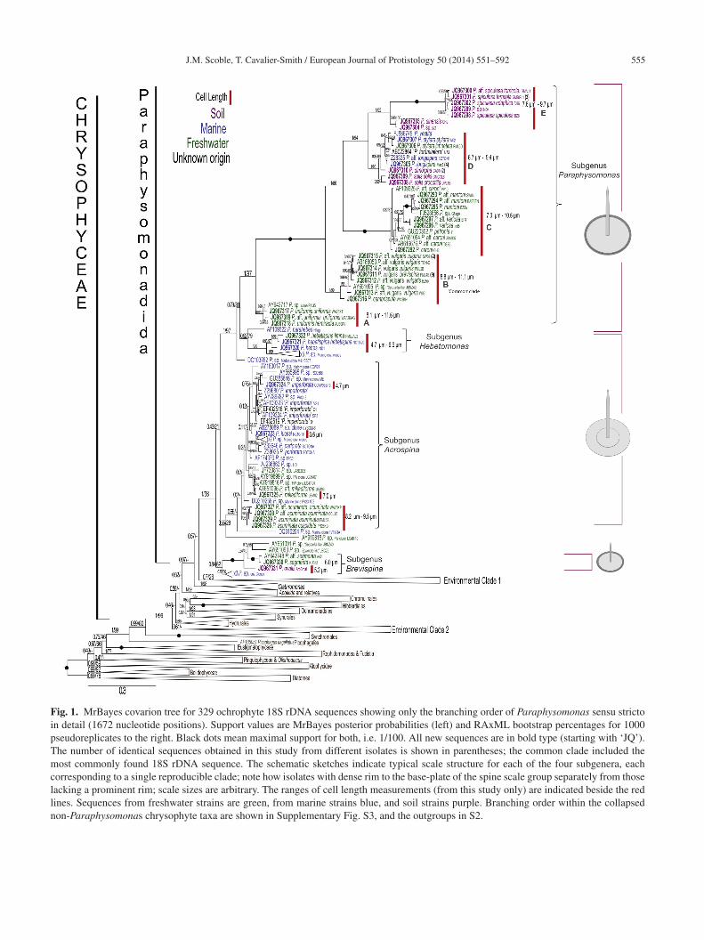
J.M. Scoble, T. Cavalier-Smith / European Journal of Protistology 50 (2014) 551–592 555
Fig. 1. MrBayes covarion tree for 329 ochrophyte 18S rDNA sequences showing only the branching order of Paraphysomonas sensu strictoin detail (1672 nucleotide positions). Support values are MrBayes posterior probabilities (left) and RAxML bootstrap percentages for 1000pseudoreplicates to the right. Black dots mean maximal support for both, i.e. 1/100. All new sequences are in bold type (starting with ‘JQ’).The number of identical sequences obtained in this study from different isolates is shown in parentheses; the common clade included themost commonly found 18S rDNA sequence. The schematic sketches indicate typical scale structure for each of the four subgenera, eachcorresponding to a single reproducible clade; note how isolates with dense rim to the base-plate of the spine scale group separately from thoselacking a prominent rim; scale sizes are arbitrary. The ranges of cell length measurements (from this study only) are indicated beside the redlines. Sequences from freshwater strains are green, from marine strains blue, and soil strains purple. Branching order within the collapsednon-Paraphysomonas chrysophyte taxa are shown in Supplementary Fig. S3, and the outgroups in S2.
5 an Jour
aop
nbsaLiocsctTssticTePsbbalutBtcemmPaft
garsfcmtbtp
ca
snTswtrbbbidtcTgaapp
tpae2isptre(pjHolEPacaSnr
etamp
56 J.M. Scoble, T. Cavalier-Smith / Europe
nd Acrospina and thus likely to be of similar phenotype;ne marine clone DQ103782 is robustly sister to the Para-hysomonas/Hebetomonas clade.The largest clade (subgenus Paraphysomonas) includes 13
ew named species that have spine scales with unperforatedase-plates with a dense margin and relatively long, typicallyimply pointed, spines similar to those lumped in the liter-ture as P. vestita from Houwink (1952) and Manton andeedale (1961) onwards. Clearly their genetic diversity is
mmensely greater than can reasonably be accommodated inne species. This large clade has five speciose major sub-lades (A – E), whose relative branching order is robust andtrongly supported by both methods; the three basal sub-lades (A – C) are all exclusively freshwater, suggestinghat was the ancestral habitat for subgenus Paraphysomonas.he two derived subclades (D, E) with somewhat shorterpines are robustly sisters; subclade E, itself with two robustubclades, is exclusively from soil and subclade D has a mix-ure of soil, freshwater and marine species. The two marinesolates in D previously identified as P. vestita are almostertainly misidentified (FJ886745/Z28335, see discussion).hey are genetically different from each other and bothxtremely distant from the third freshwater P. vestita, now. aff. caroni (subclade C); the authors did not specify whichtrain (PV10 or DB1) was used for TEM (Lim et al., 2001),ut their picture shows a scale with a spine of 2.9 �m and aase-plate of 1.4 �m. In subclade D “P. foraminifera” is prob-bly also misidentified as P. foraminifera scale base-platesack a dense margin and are multi-perforated (Lucas 1967)nlike any of the 13 species in subgenus Paraphysomonashat we sequenced and studied ultrastructurally. Subclades
– E with very long branches all share numerous inser-ions in 18S rDNA absent from other Paraphysomonas (andhrysophytes), exemplifying a common correlation betweenxtra-rapid sequence substitution and insertionally expandedolecules (von der Heyden et al., 2004); they share a com-on sequence signature AT (P. vulgaris brevispina (strainML4B pos. 762-763) where all other Chrysophyceae in thislignment have TC. 18S rDNA sequence signatures were alsoound for the two smaller Paraphysomonas subgenera (seeaxonomy section).
Sister to the major long-spine, dense-margin clade (sub-enus Paraphysomonas) is a small predominantly (probablyncestrally) marine clade (subgenus Hebetomonas) withelatively small cells and dramatically smaller scales, whosehorter spines are always blunt-ended and emerge centrallyrom comparatively narrower base-plates. The Hebetomonaslade has five marine environmental sequences and three newarine species plus a new freshwater subspecies of one of
hem); the P. hebes subclade of two new species lacks a densease-plate margin but sometimes has a faint annular fold onhe base-plate absent from subgenus Paraphysomonas or P.
arahebes.The second most speciose clade (subgenus Acrospina)omprises species lacking a dense base-plate margin,nd whose base-plate that is either imperforate (most
saPp
nal of Protistology 50 (2014) 551–592
pecies, formerly lumped as P. imperforata) or withumerous holes (species formerly lumped as P. foraminifera).he Acrospina clade is predominantly marine, but has twoubstantial phyletically distinct freshwater subclades. Strainsith a perforate base-plate form a small subclade within
he predominantly and almost certainly ancestrally imperfo-ate lineages. This large clade is mostly short-spined, spinesarely tapering with a short dull to rounded tip, but the deep-ranching P. acuminata subclade has characteristically longarely tapering spines with short very pointed tip (as long asn subgenus Paraphysomonas). The fourth morphologicallyefined subclade (subgenus Brevispina) consists of freshwa-er or soil lineages (e.g. P. ovalis, P. segmentata) with smallells and scales, short spines, and dense base-plate margins.hus, three clades have dense margined base-plates (sub-enera Paraphysomonas and Brevispina, and P. parahebes)nd two have plain base-plate margins (subgenus Acrospinand the main subclade of subgenus Hebetomonas). It is notossible to decide which of these states is ancestral for Para-hysomonas sensu stricto.
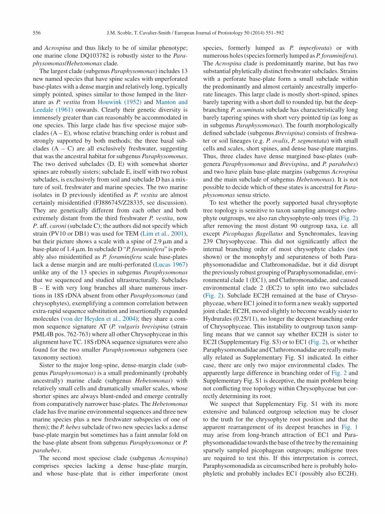
To test whether the poorly supported basal chrysophyteree topology is sensitive to taxon sampling amongst ochro-hyte outgroups, we also ran chrysophyte-only trees (Fig. 2)fter removing the most distant 90 outgroup taxa, i.e. allxcept Picophagus flagellatus and Synchromales, leaving39 Chrysophyceae. This did not significantly affect thenternal branching order of most chrysophyte clades (nothown) or the monophyly and separateness of both Para-hysomonadidae and Clathromonadidae, but it did disrupthe previously robust grouping of Paraphysomonadidae, envi-onmental clade 1 (EC1), and Clathromonadidae, and causednvironmental clade 2 (EC2) to split into two subcladesFig. 2). Subclade EC2H remained at the base of Chryso-hyceae, where EC1 joined it to form a new weakly supportedoint clade; EC2H, moved slightly to become weakly sister toydrurales (0.25/11), no longer the deepest branching orderf Chrysophyceae. This instability to outgroup taxon samp-ing means that we cannot say whether EC2H is sister toC2I (Supplementary Fig. S3) or to EC1 (Fig. 2), or whetheraraphysomonadidae and Clathromonadidae are really mutu-lly related as Supplementary Fig. S1 indicated. In eitherase, there are only two major environmental clades. Thepparently large difference in branching order of Fig. 2 andupplementary Fig. S1 is deceptive, the main problem beingot conflicting tree topology within Chrysophyceae but cor-ectly determining its root.
We suspect that Supplementary Fig. S1 with its morextensive and balanced outgroup selection may be closero the truth for the chrysophyte root position and that thepparent rearrangement of its deepest branches in Fig. 1ay arise from long-branch attraction of EC1 and Para-
hysomonadidae towards the base of the tree by the remaining
parsely sampled picophagean outgroups; multigene treesre required to test this. If this interpretation is correct,araphysomonadida as circumscribed here is probably holo-hyletic and probably includes EC1 (possibly also EC2H).
J.M. Scoble, T. Cavalier-Smith / European Journal of Protistology 50 (2014) 551–592 557
0.2
AF185051 Picophagus flagellatus
0.65/34
1/92
0.77/60
0.63/28
0.63/12
0.36/11
0.25/11
1/96
0.8/30
0.24/6
0.99/570.24/4
0.53/120.12/1
1/99
0.41/43
0.74/94
1/75
0.23/-
0.22/-
1/96Hibberdiales
Chromulinales
Ochromonadales (sensu sctrict o)
Hydrurales
EC2 I
Apoikia and re es
Clathromonadidae
Paraphysomonadidae
Synurales
EC1
EC2 H
SynchromalesPicophagea
Paraphysomonadida
Chrysophyceae
Fig. 2. MrBayes covarion tree for 239 chrysophyte 18S rDNA sequences (1681 nucleotide positions). To emphasize the tree’s main features,a collapsR ight. BP
Iaaedea(r
rt(ra
nd fit it onto one page, internal branches of all major clades are
AxML bootstrap percentages for 1000 pseudoreplicates to the ricophagea (Synchromales and Picophagus).
n both Fig. 2 and Supplementary Fig. S1 Hibberdialesnd Chromulinales are sisters and group with Ochromon-dales and Synurales as a weakly supported four-order clade;xclusion of both Paraphysomonadidae and Clathromona-idae from this reproducible clade is consistent with the
xclusion of Paraphysomonadida from both Chromulinalesnd Ochromonadales as a distinct non-photosynthetic orderCavalier-Smith and Chao 1996). The aforementioned sisterelationsip between Hibberdiales and Chromulinales is onlytes
ed. Support values are MrBayes posterior probabilities (left) andlack dots mean maximal support for both, i.e. 1/100. Rooted on
ecovered by Bayes and never ML methods in Fig. 1, sohese methods never agree even with more distant outgroupsSupplementary Fig. 3). And they remain contradictory withespect to the possible sister relationship between Synuralesnd Ochromonadales sensu stricto.
Supplementary Fig. S1 shows the large-scale structure ofhe chrysophyte tree: Chrysophyceae has 10 major clades,ight including known organisms and two only exclu-ively environmental sequences of unknown phenotype.
5 an Jour
SHtpdamibCbopaCttwbipCtItioSo
TP
SaptnrTPHneCaCmTspSnd
aaacoo
utlscHobtCieonas
ncwoubdjpsisctmg(pfacGbS
iag
58 J.M. Scoble, T. Cavalier-Smith / Europe
ix organismally defined clades are predominantly (forydrurales entirely) algal (i.e. photosynthetic), whereas
wo comprise purely heterotrophic scaly phagotrophs: Para-hysomonadidae and Clathromonadidae. Paraphysomona-idae, Clathromonadidae, and environmental clade 1 form
robust clade (Paraphysomonadida) in both Bayesian andaximum likelihood (ML) analyses, but the relative branch-
ng order of these three is essentially unresolved althoughoth methods weakly place environmental clade 1, notlathromonadidae, as sister to Paraphysomonadidae. Theranching order of Paraphysomonadida, the other sevenrders, and environmental DNA clade 2 is weakly sup-orted and inconsistent between methods. Thus, single-genenalysis is inadequate to establish the basal branching inhrysophyceae, even though seven orders were consis-
ently monophyletic, several with strong support. Moreover,he purely photosynthetic, scale-bearing Synurales branchesithin Chrysophyceae and is thus not sister to all the otherranches. Clathromonadidae is a strongly supported claden both Bayesian and ML trees. With this large taxon sam-le environmental clade 2 invariably groups strongly withhrysophyceae, and Chrysophyceae are consistently sisters
o Synchromales, with Picophagea apparently paraphyletic.ndividual clades are collapsed in Supplementary Fig. S1o emphasise overall tree structure. The internal branch-ng order of Paraphysomonadidae is shown in Fig. 1 andf Clathromonadidae and all other chrysophyte clades inupplementary Fig. S2; the internal branching order of allutgroups is in Supplementary Fig. S3.
axonomy: revised classification ofaraphysomonadida
The chrysomonad order Paraphysomonadales Cavalier-mith, 1996 was established to include both Paraphysomonasnd Spumella (Cavalier-Smith and Chao 1996), but the poly-hyletic Spumella was transferred to Ochromonadales inhe light of sequence evidence that all Spumella cladesest within photosynthetic Ochromonadales and none areelated to Paraphysomonas (Cavalier-Smith and Chao 2006).hereafter Paraphysomonadales included just the familyaraphysomonadidae (=Paraphysomonadaceae) Preisig andibberd, 1983. This family is often included in Chromuli-ales, but our trees reproducibly confirm earlier sequencevidence showing that to be incorrect (Andersen 2007;avalier-Smith and Chao 2006), and that Paraphysomonasnd Clathromonas are distinct deep-branching clades ofhrysophyceae, both genetically more distant from Chro-ulinales than is the photosynthetic scale-bearing Synurales.hus, Paraphysomonadales clearly merits its separate ordinaltatus. However, especially following the inclusion of more
urely phagotrophic phyla in kingdom Chromista (Cavalier-mith 2010), the convention of treating all Chromistaomenclaturally as plants (Cavalier-Smith 1981) must beiscontinued. As paraphysomonads are totally heterotrophicLTDW
nal of Protistology 50 (2014) 551–592
nd protozoan-like in phagotrophic nutrition and thus notlgae (Cavalier-Smith 2007), we here treat them under ICZNs order Paraphysomonadida, as phagotrophic supragenerichromist taxa that consist exclusively or almost exclusivelyf heterotrophs should be treated under the zoological codef nomenclature (Cavalier-Smith 2007).
The improved classification below is based on our newltrastructural data and sequence phylogeny jointly. Ourrees showed that every Paraphysomonas strain with nail-ike spine scales (i.e. a round or rarely oval base-plate andingle unbranched slender spine) is part of a very large robustlade devoid of any strains with contrasting scale types.ere we remove all former Paraphysomonas species with-ut plain spines centrally protruding from an oval or roundaseplate from the genus; those that have basket and addi-ional perforated plain plate scales are placed in a new genus,lathromonas. All former Paraphysomonas that do not fit
nto either genus as defined here are assigned to new gen-ra in a separate paper; some belong in Paraphysomonadida,thers do not (Cavalier-Smith and Scoble unpublished). Weow restrict family Paraphysomonadidae (=Paraphysomon-daceae) Preisig and Hibberd, 1983 to Paraphysomonasensu stricto:
Paraphysomonas De Saedeleer, 1929 em.: Revised diag-osis: biciliate, non-amoeboid, unicellular, heterotrophichrysomonads; cell body covered by numerous spine scalesith usually circular, rarely oval, base-plate approximatelyrthogonal to a long thin central spine; spine unbranched,nwinged, many times narrower than base-plate even at itsase; base-plate entire or with small perforations, of varyingistribution but no large lacunae; spine length varies fromust longer than to several times base-plate width; separatelate scales generally absent, but if present closely resemblepine-scale base-plate but with spine missing, usually largern diameter and no distinctive morphology; slender posteriortalk anchors cell to substratum or trails behind swimmingell. Plastid a colourless leucoplast without stigma. Contrac-ile vacuole in freshwater species. Posterior cilium lateral,
uch shorter than forward-directed anterior cilium. Four sin-le nucleotide 18S rDNA signatures: A (position 1387); Tposition 1465); C (position 1474); G (position 1476); allositions for reference strain ‘Arb’ P. ovalis (JQ967331)rom the deepest clade. These sequence signatures excludell other Chrysophyceae, except for position 1465 where onelone sequence ‘Marine Biosope T3′ (FJ537322) showed a
and all other chrysophytes A; this unique difference coulde a sequencing error. Type species P. vestita (Stokes) Deaedeleer, 1929.We make 23 new Paraphysomonas species below, includ-
ng raising P. vestita truncata sub-species to species Preisignd Hibberd (1982a), but retain only nine existing ones in theenus: P. vestita (Stokes) De Saedeleer, 1929, P. imperforata
ucas (1967), P. foraminifera Lucas (1967), P. bandaiensisakahashi (1976), P. antarctica Takahashi (1987), P. porosaürrschmidt and Cronberg (1989), P. circumforaminiferaujek (1983), P. oligocycla Takahashi (1987), and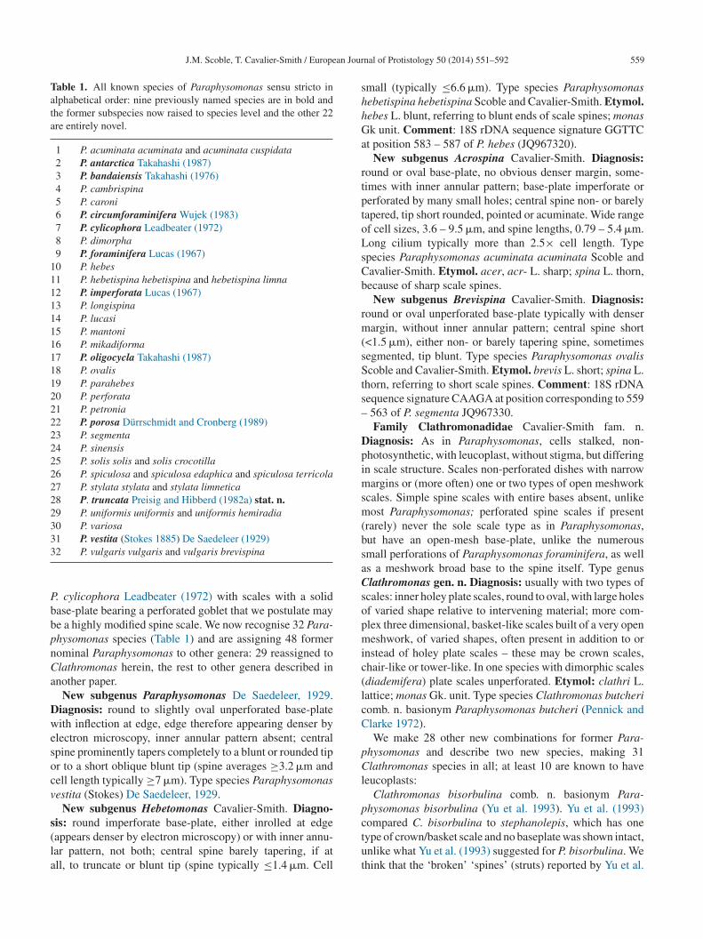
J.M. Scoble, T. Cavalier-Smith / European Jour
Table 1. All known species of Paraphysomonas sensu stricto inalphabetical order: nine previously named species are in bold andthe former subspecies now raised to species level and the other 22are entirely novel.
1 P. acuminata acuminata and acuminata cuspidata2 P. antarctica Takahashi (1987)3 P. bandaiensis Takahashi (1976)4 P. cambrispina5 P. caroni6 P. circumforaminifera Wujek (1983)7 P. cylicophora Leadbeater (1972)8 P. dimorpha9 P. foraminifera Lucas (1967)
10 P. hebes11 P. hebetispina hebetispina and hebetispina limna12 P. imperforata Lucas (1967)13 P. longispina14 P. lucasi15 P. mantoni16 P. mikadiforma17 P. oligocycla Takahashi (1987)18 P. ovalis19 P. parahebes20 P. perforata21 P. petronia22 P. porosa Dürrschmidt and Cronberg (1989)23 P. segmenta24 P. sinensis25 P. solis solis and solis crocotilla26 P. spiculosa and spiculosa edaphica and spiculosa terricola27 P. stylata stylata and stylata limnetica28 P. truncata Preisig and Hibberd (1982a) stat. n.29 P. uniformis uniformis and uniformis hemiradia30 P. variosa33
PbbpnCa
Dwesocv
s(la
shhGa
rtptoLsCb
rm(sSts–
Dpimsm(bsaCsopmic(lcC
pCl
p
1 P. vestita (Stokes 1885) De Saedeleer (1929)2 P. vulgaris vulgaris and vulgaris brevispina
. cylicophora Leadbeater (1972) with scales with a solidase-plate bearing a perforated goblet that we postulate maye a highly modified spine scale. We now recognise 32 Para-hysomonas species (Table 1) and are assigning 48 formerominal Paraphysomonas to other genera: 29 reassigned tolathromonas herein, the rest to other genera described innother paper.
New subgenus Paraphysomonas De Saedeleer, 1929.iagnosis: round to slightly oval unperforated base-plateith inflection at edge, edge therefore appearing denser by
lectron microscopy, inner annular pattern absent; centralpine prominently tapers completely to a blunt or rounded tipr to a short oblique blunt tip (spine averages ≥3.2 �m andell length typically ≥7 �m). Type species Paraphysomonasestita (Stokes) De Saedeleer, 1929.
New subgenus Hebetomonas Cavalier-Smith. Diagno-
is: round imperforate base-plate, either inrolled at edgeappears denser by electron microscopy) or with inner annu-ar pattern, not both; central spine barely tapering, if atll, to truncate or blunt tip (spine typically ≤1.4 �m. Cellctut
nal of Protistology 50 (2014) 551–592 559
mall (typically ≤6.6 �m). Type species Paraphysomonasebetispina hebetispina Scoble and Cavalier-Smith. Etymol.ebes L. blunt, referring to blunt ends of scale spines; monask unit. Comment: 18S rDNA sequence signature GGTTC
t position 583 – 587 of P. hebes (JQ967320).New subgenus Acrospina Cavalier-Smith. Diagnosis:
ound or oval base-plate, no obvious denser margin, some-imes with inner annular pattern; base-plate imperforate orerforated by many small holes; central spine non- or barelyapered, tip short rounded, pointed or acuminate. Wide rangef cell sizes, 3.6 – 9.5 �m, and spine lengths, 0.79 – 5.4 �m.ong cilium typically more than 2.5× cell length. Typepecies Paraphysomonas acuminata acuminata Scoble andavalier-Smith. Etymol. acer, acr- L. sharp; spina L. thorn,ecause of sharp scale spines.
New subgenus Brevispina Cavalier-Smith. Diagnosis:ound or oval unperforated base-plate typically with denserargin, without inner annular pattern; central spine short
<1.5 �m), either non- or barely tapering spine, sometimesegmented, tip blunt. Type species Paraphysomonas ovaliscoble and Cavalier-Smith. Etymol. brevis L. short; spina L.
horn, referring to short scale spines. Comment: 18S rDNAequence signature CAAGA at position corresponding to 559
563 of P. segmenta JQ967330.Family Clathromonadidae Cavalier-Smith fam. n.
iagnosis: As in Paraphysomonas, cells stalked, non-hotosynthetic, with leucoplast, without stigma, but differingn scale structure. Scales non-perforated dishes with narrow
argins or (more often) one or two types of open meshworkcales. Simple spine scales with entire bases absent, unlikeost Paraphysomonas; perforated spine scales if present
rarely) never the sole scale type as in Paraphysomonas,ut have an open-mesh base-plate, unlike the numerousmall perforations of Paraphysomonas foraminifera, as wells a meshwork broad base to the spine itself. Type genuslathromonas gen. n. Diagnosis: usually with two types of
cales: inner holey plate scales, round to oval, with large holesf varied shape relative to intervening material; more com-lex three dimensional, basket-like scales built of a very openeshwork, of varied shapes, often present in addition to or
nstead of holey plate scales – these may be crown scales,hair-like or tower-like. In one species with dimorphic scalesdiademifera) plate scales unperforated. Etymol: clathri L.attice; monas Gk. unit. Type species Clathromonas butcheriomb. n. basionym Paraphysomonas butcheri (Pennick andlarke 1972).We make 28 other new combinations for former Para-
hysomonas and describe two new species, making 31lathromonas species in all; at least 10 are known to have
eucoplasts:Clathromonas bisorbulina comb. n. basionym Para-
hysomonas bisorbulina (Yu et al. 1993). Yu et al. (1993)
ompared C. bisorbulina to stephanolepis, which has oneype of crown/basket scale and no baseplate was shown intact,nlike what Yu et al. (1993) suggested for P. bisorbulina. Wehink that the ‘broken’ ‘spines’ (struts) reported by Yu et al.
5 an Jour
(s‘ostow(iTbsCH
p
pL
p
p(
p
OLpL
p1
p
p
p
pL
pi1tdtfbHtP
(wadPh
pn
pL
pTsanrdtsLpas
p
p(
p
p
p
s
p
pL
P1
pL
p
60 J.M. Scoble, T. Cavalier-Smith / Europe
1993) are actually broken crown/basket scales fallen along-ide a distinct plate scale, which they misinterpreted as abase-plate’ of a spine scale. Plate 2D, E are poor imagesf scales, but F, G and H are clear and show a plate scaleeparate from a broken basket scale. Gao et al. (1993) misin-erpret the description of C. stephanolepis, stating ‘the scalesf P. stephanolepis have only base-plates and no apical plate’,hich is wrong because they are basket-like. In Yu et al.
1993) the schematic Fig. 2 legend is confused; Fig. 2Ks actually P. simplexocorbita and Fig. 2M is P. bisorbida.he TEM images of C. bisorbulina seem most similar to P.utcheri of Thomsen 1975 (their Figures 16 – 19), which haseparate plate scales and crown/basket scales. Plate scales of. bisorbulina resemble those of C. homolepis (Preisig andibberd 1982a, particularly Fig. 1E).Clathromonas cancellata comb. n. basionym Para-
hysomonas cancellata (Preisig and Hibberd 1982b)Clathromonas canistrum comb. n. basionym Para-
hysomonas canistrum (Preisig and Hibberd 1982b).eucoplast.Clathromonas corbidifera comb. n. basionym Para-
hysomonas corbidifera (Pennick and Clarke 1973)Clathromonas coronata comb. n. basionym Para-
hysomonas coronata Moestrup and Zimmerman inThomsen et al. 1981)
Clathromonas cribosa comb. n. basionym Para-hysomonas cribosa (Lucas 1968)Clathromonas diademifera comb. n. basionym
chromonas diademifera (Takahashi, 1972). Synonymsepidochromonas diademifera Kristiansen, 1980; Para-hysomonas diademifera (Preisig and Hibberd 1982a).eucoplast.Clathromonas eiffellii comb. n. basionym Para-
hysomonas eiffellii Thomsen in (Thomsen et al.981)Clathromonas elegantissima comb. n. basionym Para-
hysomonas elegantissima (Kling and Kristiansen 1983)Clathromonas faveolata comb. n. basionym Para-
hysomonas faveolata (Rees et al. 1974)Clathromonas homolepis comb. n. basionym Para-
hysomonas homolepis (Preisig and Hibberd 1982b)Clathromonas ignivoma comb. n. basionym Para-
hysomonas ignivoma (Preisig and Hibberd 1982b).eucoplast.Clathromonas inconspicua comb. n. basionym Para-
hysomonas inconspicua (Takahashi 1976). We do not acceptts synonymization with P. butcheri (Preisig and Hibberd982b), though agree that interpretation of crown scale struc-ure is not easy (they appear to differ); its plate scales are veryistinct, with much greater contrast between large holes andiny ones than in C. butcheri. Moreover, C. inconspicua isrom freshwater, not brackish like C. butcheri (however P.
utcheri from Cambridgeshire freshwater ponds (Preisig andibberd 1982b) seems correctly identified and is very similaro our brackish C. butcheri strain – see below). We agree withreisig and Hibberd (1982b) that P. butcheri of Takahashi
T
pc
nal of Protistology 50 (2014) 551–592
1976) was misidentified, as was his P. foraminifera; howevere do not accept that Takahashi’s ‘butcheri’ was P. morchella,
s the small-mesh holes of morchella were much less evi-ent; it may be an undescribed species somewhat similar to. morchella with a less evident chair-back and fewer smalloles.
Clathromonas manubriata comb. n. basionym Para-hysomonas manubriata (Preisig and Hibberd 1982b) stat.. (Vørs et al. 1990)
Clathromonas morchella comb. n. basionym Para-hysomonas morchella (Preisig and Hibberd 1982b).eucoplast.Clathromonas poteriophora comb. n. basionym Para-
hysomonas poteriophora Moestrup and Kristiansen inhomsen et al. (1981). We strongly disagree with its inclu-ion within C. coronata (Vørs et al. 1990), as their scalesre very distinct. We think Figures 6 – 10 of Vørs et al. areot coronata, but a third, undescribed species more closelyelated to coronata than to poteriophora, and are not interme-iate between coronata and poteriophora, and do not justifyheir merger. Their claim that Preisig and Hibberd (1982b)howed intermediates is disputable; in our view, Fig. 19 I,-O of Preisig and Hibberd (1982b) are neither C. poterio-hora, nor intermediates between poteriophora and coronatas Vørs et al. apparently assumed, but a fourth (undescribed)pecies closer to poteriophora than to coronata.
Clathromonas preisigii comb. n. basionym Para-hysomonas preisigii (Wujek 2013)Clathromonas quadrispina comb. n. basionym Para-
hysomonas quadrispina Thomsen and Kristiansen inThomsen et al. 1981). Leucoplast.
Clathromonas runcinifera comb. n. basionym Para-hysomonas runcinifera (Preisig and Hibberd 1982b)
Clathromonas sideriophora comb. n. basionym Para-hysomonas sideriophora (Thomsen 1975)Clathromonas sigillifera comb. n. basionym Para-
hysomonas sigillifera Moestrup in Thomsen et al. (1981)Clathromonas simplexocorbida comb. n. Paraphysomonas
implexocorbida (Yu et al. 1993)Clathromonas stelligera comb. n. basionym Para-
hysomonas stelligera (Preisig and Hibberd 1982b)Clathromonas stephanolepis comb. n. basionym Para-
hysomonas stephanolepis (Preisig and Hibberd 1982b).eucoplast.Clathromonas subquadrangularis comb. n. basionym
araphysomonas subquadrangularis (Preisig and Hibberd982b). Leucoplast.Clathromonas subrotacea comb. n. basionym Para-
hysomonas subrotacea Thomsen in Thomsen et al. (1981).eucoplast.Clathromonas takahashii comb. n. basionym Para-
hysomonas takahashii Cronberg and Kristiansen in
homsen et al. (1981)Clathromonas undulata comb. n. basionym Para-hysomonas undulata (Preisig and Hibberd 1982b). Leu-oplast.
an Jour
Te
cica(bElwbbsdtcaIlwwfsv
osstSbdgauefftidtnggac
sdasb
dwaigv1e(fwsHetpt
moaiaroi
S
nSsptdpt–1AsCcPipis
n
J.M. Scoble, T. Cavalier-Smith / Europe
axonomy: 23 new Paraphysomonas species,ight new subspecies, and strain descriptions
All new isolates described below are colourless biciliateells with tubular hairs on the long undulating anterior cil-um (LC) and a smooth shorter, largely passive, ‘posterior’ilium (SC). They all swim with anterior cilium leading and
trailing stalk used to attach to the substrate when feedingsessile). All new species had spine scales and imperforatease-plates. Diagnoses do not repeat these shared characters.xcept where stated otherwise all base-plates are round. Cell
ength (CL) measurements and estimates of cilium lengthere on live cells; mean cell length is given first followed inrackets by the range and number of cells measured. Scalease-plate diameter measured across the widest point, andpine-length to plate-width ratio (S/P ratio) is important inistinguishing species. For basally thicker spines we some-imes give spine-base widths above the base-plate (not to beonfused with the far greater width of the whole base-plate),verage values being followed by the range in parentheses.n some strains the scale spines are visible individually oniving cells in the light microscope (LM), mainly in thoseith unusually thick spines, but in most they are not. Evenhen one cannot see spines, the base-plates may collectively
orm a visible layer seen as a dense line around the main cellurface, which we refer to as a ‘scale-base layer’ since its LMisibility or not is constant for each strain.Diagnoses/descriptions are grouped by species positions
n the tree (Fig. 1), which usually placed those with moreimilar scales mutually closer. When we designate typeequences, strains, and illustrations, or any combination ofhese, all are to be regarded as part of a syntype (Cavalier-mith and Chao 2010). To save space we have not preparedoth comprehensive descriptions of new strains and separateiagnoses focusing solely on those characters that distin-uish each species from its closest relatives. Our decisionsbout species boundaries were made primarily using scaleltrastructural and rDNA sequence differences, which gen-rally mutually agree well; either or both these features (andor three species stomatocyst morphology) can be used inuture to reidentify reliably all new species and distinguishhem from close relatives. Features like cell size and cil-ary length are included as necessary features for properlyescribing most new species (summarised in Table 2), buthough they map in a meaningful way onto the phyloge-etic tree, and therefore are more stable evolutionarily andenetically than some might have anticipated, they cannotenerally be used to discriminate between close species, andre thus corroborative rather than diagnostic characters fororrect identification.
We cannot precisely compare new species with the typepecies P. vestita because its scale type is unknown. As the
iscussion explains more fully, cultures previously identifieds ‘P. vestita’ have been repeatedly studied ultrastructurallyince Houwink (1952) and Manton and Leedale (1961),ut their scale structure differs as greatly as many speciesLsf0
nal of Protistology 50 (2014) 551–592 561
escribed here that have radically different sequences, soe cannot know which if any are really the same species
s Stokes’ P. vestita. Either no data were given to enabledentification to be checked (e.g. Houwink 1952) or thoseiven strongly suggest that the organism studied was not P.estita but an undescribed species (e.g. Manton and Leedale961). Ideally we would have liked to establish a neotype tond that confusion, but no isolate was sufficiently similarby light microscopy) to Stokes’ (see discussion). There-ore it is unlikely that any new species described here forhich we give LM data can be vestita. We formally raise
ubspecies Paraphysomonas vestita truncata (Preisig andibberd 1982a) to a full species, as its spine scales are distinct
nough from other electron microscopically studied strainso merit that, its spines being too short for P. vestita: Para-hysomonas truncata Preisig and Hibberd, 1982a stat. n.;heir diagnosis and type applies.
For brevity, many additional comments and information onost of the following 22 new species, including descriptions
f separate isolates related to the type strains detailed below,re given only in Supplementary Information 1. Many stud-ed strains are described only in the Supplementary materials aff. plus a specific epithet to indicate their likely closestelative, even though a few of them are shown in the figuresr Table 2; some environmental sequences are also similarlydentified there.
ubgenus Brevispina: two new species
Paraphysomonas ovalis sp. n. Type Fig. 3A – F. Diag-osis: CL 5.2 �m (4.1 – 6.4 N = 17); LC 1.5 – 2 × CL;C 0.75 × CL. LC beats constantly. Attached cells round,ometimes flattened on one side. Swimming cells elongate toyriform, sometimes round, swim in slow spiral and directrajectories. Stalked cell close to substratum or attached toetritus. One type of spine scale with oval to irregular baselate. Spine 1.5 �m (1.1 – 1.9) tapers gently to roundedip, slightly flared out at very base; base-plate 0.8 �m (0.7
0.95) with prominent dense margin. S/P ratio 1.9 (range.4 – 2.4). Type strain ARB: CCAP 935/15. (2010; Harcourtrboretum, Oxfordshire, UK. JMS). Soil. Type 18S rDNA
equence GenBank JQ967331. Etymology: ovalis L. oval.omment: P. ovalis is most similar to P. bandaiensis, trun-
ata, and porosa; all have a base-plate with thickened margin.. ovalis differs from them all by its base-plate being oval torregular, not regularly circular; it is unperforated, unlike P.orosa. P. ovalis has a rounded spine tip; that of P. truncatas truncated. P. bandaiensis spine tip is also rounded but itshaft is non-tapered, unlike P. ovalis.
Paraphysomonas segmenta sp. n. Type Fig. 3G – I. Diag-osis: CL 6.0 �m (5 – 7.3 N = 22); LC 2 × CL; SC 0.5 × CL.
C sometimes static. Round to oval cell attached via shorttalk to substratum or detritus. Swimming cell oval to pyri-orm, stalk often trailing. One type of spine scale, spine.65 �m (0.52 – 0.73) usually in two non-tapering segments,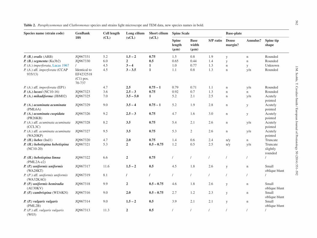
562
J.M.
Scoble, T.
Cavalier-Sm
ith /
European
Journal of
Protistology 50
(2014) 551–592
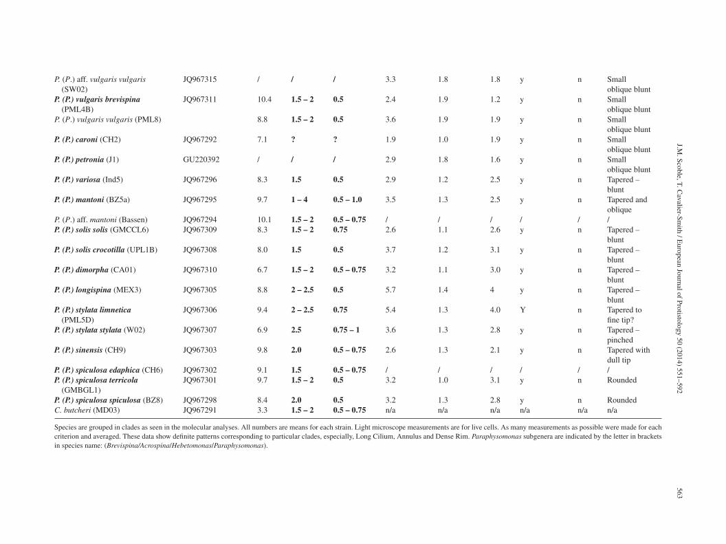
Table 2. Paraphysomonas and Clathromonas species and strains light microscope and TEM data, new species names in bold.
Species name (strain code) GenBank18S
Cell length(CL)
Long cilium(xCL)
Short cilium(xCL)
Spine Scale Base-plate
Spinelength(�m)
Basewidth(�m)
S/P ratio Densemargin?
Annulus? Spine tipshape
P. (B.) ovalis (ARB) JQ967331 5.2 1.5 – 2 0.75 1.5 0.8 1.9 y n RoundedP. (B.) segmenta (Ku3b2) JQ967330 6.0 2 0.5 0.65 0.44 1.4 y n RoundedP. (A.) imperforata, Lucas 1967 / 4.5 3 – 4 1 1.0 0.77 1.3 n y UnknownP. (A.) aff. imperforata (CCAP
935/13)Identical toEF4232518(C1) pos.70-737
4.5 3 – 3.5 1 1.1 0.8 1.3 n y/n Rounded
P. (A.) aff. imperforata (EP1) 4.7 2.5 0.75 – 1 0.79 0.71 1.1 n y/n RoundedP. (A.) lucasi (NC10-16) JQ967323 3.6 2.5 – 3 0.75 0.92 0.7 1.3 n n RoundedP. (A.) mikadiforma (JBM02) JQ967325 7.0 3.5 – 5.0 1 5.2 2.1 2.5 n y/n Acutely
pointedP. (A.) acuminata acuminata
(PML6A)JQ967329 9.0 3.5 – 4 0.75 – 1 5.2 1.9 2.8 n y Acutely
pointedP. (A.) acuminata cuspidata
(PR26KB)JQ967326 9.2 2.5 – 3 0.75 4.7 1.6 3.0 n y Acutely
pointedP. (A.) aff. acuminata acuminata
(CCL3C)JQ967328 8.2 3.5 0.75 5.4 2.1 2.6 n y/n Acutely
pointedP. (A.) aff. acuminata acuminata
(WA20KP)JQ967327 9.5 3.5 0.75 5.3 2 2.6 n y/n Acutely
pointedP. (H.) hebes (Ind1) JQ967320 4.7 2.0 0.75 1.4 0.6 2.4 n/y n TruncateP. (H.) hebetispina hebetispina
(NC10-20)JQ967321 5.3 2 0.5 – 0.75 1.2 0.5 2.5 n/y y/n Truncate
slightlyrounded
P. (H.) hebetispina limna(PML2A-e2)
JQ967322 6.6 2 0.75 / / / / / /
P. (P.) uniformis uniformis(WA28KT)
JQ967317 11.6 1.5 – 2 0.5 4.5 1.8 2.6 y n Smalloblique blunt
P. (P.) aff. uniformis uniformis(WA32KAG)
JQ967319 8.1 / / / / / / / /
P. (P.) uniformis hemiradia(AU30KV)
JQ967318 9.9 2 0.5 – 0.75 4.6 1.8 2.6 y n Smalloblique blunt
P. (P.) cambrispina (WI34KN) JQ967316 9.0 2.0 0.5 – 0.75 2.7 1.2 2.3 y n Smalloblique blunt
P. (P.) vulgaris vulgaris(PML2B)
JQ967314 9.0 1.5 – 2 0.5 3.9 2.1 2.1 y n Smalloblique blunt
P. (P.) aff. vulgaris vulgaris(W03)
JQ967313 11.3 2 0.5 / / / / / /
J.M.
Scoble, T.
Cavalier-Sm
ith /
European
Journal of
Protistology 50
(2014) 551–592
563
P. (P.) aff. vulgaris vulgaris(SW02)
JQ967315 / / / 3.3 1.8 1.8 y n Smalloblique blunt
P. (P.) vulgaris brevispina(PML4B)
JQ967311 10.4 1.5 – 2 0.5 2.4 1.9 1.2 y n Smalloblique blunt
P. (P.) vulgaris vulgaris (PML8) 8.8 1.5 – 2 0.5 3.6 1.9 1.9 y n Smalloblique blunt
P. (P.) caroni (CH2) JQ967292 7.1 ? ? 1.9 1.0 1.9 y n Smalloblique blunt
P. (P.) petronia (J1) GU220392 / / / 2.9 1.8 1.6 y n Smalloblique blunt
P. (P.) variosa (Ind5) JQ967296 8.3 1.5 0.5 2.9 1.2 2.5 y n Tapered –blunt
P. (P.) mantoni (BZ5a) JQ967295 9.7 1 – 4 0.5 – 1.0 3.5 1.3 2.5 y n Tapered andoblique
P. (P.) aff. mantoni (Bassen) JQ967294 10.1 1.5 – 2 0.5 – 0.75 / / / / / /P. (P.) solis solis (GMCCL6) JQ967309 8.3 1.5 – 2 0.75 2.6 1.1 2.6 y n Tapered –
bluntP. (P.) solis crocotilla (UPL1B) JQ967308 8.0 1.5 0.5 3.7 1.2 3.1 y n Tapered –
bluntP. (P.) dimorpha (CA01) JQ967310 6.7 1.5 – 2 0.5 – 0.75 3.2 1.1 3.0 y n Tapered –
bluntP. (P.) longispina (MEX3) JQ967305 8.8 2 – 2.5 0.5 5.7 1.4 4 y n Tapered –
bluntP. (P.) stylata limnetica
(PML5D)JQ967306 9.4 2 – 2.5 0.75 5.4 1.3 4.0 Y n Tapered to
fine tip?P. (P.) stylata stylata (W02) JQ967307 6.9 2.5 0.75 – 1 3.6 1.3 2.8 y n Tapered –
pinchedP. (P.) sinensis (CH9) JQ967303 9.8 2.0 0.5 – 0.75 2.6 1.3 2.1 y n Tapered with
dull tipP. (P.) spiculosa edaphica (CH6) JQ967302 9.1 1.5 0.5 – 0.75 / / / / / /P. (P.) spiculosa terricola
(GMBGL1)JQ967301 9.7 1.5 – 2 0.5 3.2 1.0 3.1 y n Rounded
P. (P.) spiculosa spiculosa (BZ8) JQ967298 8.4 2.0 0.5 3.2 1.3 2.8 y n RoundedC. butcheri (MD03) JQ967291 3.3 1.5 – 2 0.5 – 0.75 n/a n/a n/a n/a n/a n/a
Species are grouped in clades as seen in the molecular analyses. All numbers are means for each strain. Light microscope measurements are for live cells. As many measurements as possible were made for eachcriterion and averaged. These data show definite patterns corresponding to particular clades, especially, Long Cilium, Annulus and Dense Rim. Paraphysomonas subgenera are indicated by the letter in bracketsin species name: (Brevispina/Acrospina/Hebetomonas/Paraphysomonas).
564 J.M. Scoble, T. Cavalier-Smith / European Journal of Protistology 50 (2014) 551–592
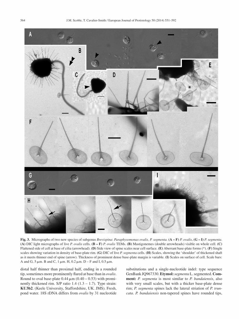
Fig. 3. Micrographs of two new species of subgenus Brevispina: Paraphysomonas ovalis, P. segmenta. (A – F) P. ovalis, (G – I) P. segmenta.(A) DIC light micrographs of live P. ovalis cells. (B – F) P. ovalis TEMs. (B) Mastigonemes (double arrowheads) visible on whole cell. (C)Flattened side of cell at base of cilia (arrowhead). (D) Side view of spine scales near cell surface. (E) Aberrant base-plate forms (*). (F) Singles e P. sea nse basA
dtRnKp
sGm
cales showing variation in density of base-plate rim. (G) DIC of livs it meets thinner end of spine (arrow). Thickness of prominent de
and G, 5 �m. B and C, 1 �m. H, 0.2 �m. D – F and I, 0.5 �m.
istal half thinner than proximal half, ending in a roundedip, sometimes more prominently flared at base than in ovalis.ound to oval base-plate 0.44 �m (0.40 – 0.53) with promi-
ently thickened rim. S/P ratio 1.4 (1.3 – 1.7). Type strain:U3b2. (Keele University, Staffordshire, UK. JMS). Fresh,ond water. 18S rDNA differs from ovalis by 31 nucleotide
wrc
gmenta cells. (H) Scales, showing the ‘shoulder’ of thickened shafte-plate margin is variable. (I) Scales on surface of cell. Scale bars:
ubstitutions and a single-nucleotide indel: type sequenceenBank JQ967330. Etymol: segmenta L. segmented. Com-ent: P. segmenta is most similar to P. bandaiensis, also
ith very small scales, but with a thicker base-plate denseim; P. segmenta spines lack the lateral striation of P. trun-ata. P. bandaiensis non-tapered spines have rounded tips,
an Jour
bs(
Ss
F–aato(o0avasTUJ
DSascvbw0
c(FifcmsntLd
nScsi0a(rscnsoJusmfba
owouwfdsSoot1
So
J.M. Scoble, T. Cavalier-Smith / Europe
ut its scales are much smaller; the spine is nearly a thirdhorter than in P. segmenta, base-plate diameter nearly halfTakahashi 1976).
ubgenus Acrospina: four new species and oneubspecies
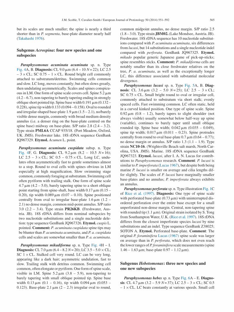
Paraphysomonas acuminata acuminata sp. n. Typeig. 4A, B. Diagnosis: CL 9.0 �m (6.4 – 10 N = 22); LC 2.5
3 × CL; SC 0.75 – 1 × CL. Round bright cell commonlyttached to substratum/detritus. Swimming cells commonnd slow. LC long, moves constantly; but often slows greatly,hen undulating asymmetrically. Scales and spines conspicu-us in LM. One form of spine scale covers cell. Spine 5.2 �m4.2 – 6.7), non-tapering to barely tapering ending in stronglyblique short pointed tip. Spine base width 0.191 �m (0.132 –.228), spine tip width 0.133 (0.094 – 0.158). Oval to roundednd irregular-shaped base-plate 1.9 �m (1.5 – 2.1), no/barelyisible dense margin, commonly with broad medium densitynnulus (i.e. a denser ring on the base-plate centred on thepine base) midway on base-plate. S/P ratio 2.8 (2.4 – 3.2).ype strain PML6A CCAP 935/18. (Port Meadow, Oxford,K. JMS). Freshwater lake. 18S rDNA sequence GenBank
Q967329. Etymol. Acumen L. point.
Paraphysomonas acuminata cuspidata subsp. n. TypeFig. 4F, G. Diagnosis: CL 9.2 �m (8.2 – 10.5 N = 16);LC 2.5 – 3 × CL; SC 0.5 – 0.75 × CL. Long LC, undu-lates often asymmetrically fast to gentle sometimes almostto a stop. Round to oval cells with spines obvious in LMespecially at high magnification. Slow swimming stagecommon, commonly foraging at substratum. Swimming celloften pyriform with trailing stalk. One form of spine scale4.7 �m (4.2 – 5.0), barely tapering spine to a short obliquepoint starting from spine-shaft, base width 0.17 �m (0.15 –0.20), tip width 0.089 �m (0.07 – 0.10). Spine positionedcentrally from oval to irregular base-plate 1.6 �m (1.2 –2.1) no dense margin, common mid-point annulus. S/P ratio3.0 (2.2 – 3.4). Type strain PR26KB. (Freshwater, Aus-tria. JB). 18S rDNA differs from nominal subspecies bytwo nucleotide substitutions and a single nucleotide dele-tion: type sequence GenBank JQ967326. Etymol. cuspis L.pointed. Comment: P. acuminata cuspidata spine tips maybe blunter than P. acuminata acuminata, and P. a. cuspidatacells and scales are somewhat smaller than P. a. acuminata.
Paraphysomonas mikadiforma sp. n. Type Fig. 4H – I.iagnosis: CL 7.0 �m (6.4 – 8.2 N = 20); LC 3.5 – 5.0 × CL;C 1 × CL. Stalked cell very round. LC can be very long,ppearing like a dark hair; asymmetric undulation, fast tolow. Trailing stalk with detritus common. Swimming cellommon, often elongate or pyriform. One form of spine scale,
isible in LM. Spine 5.2 �m (3.8 – 5.9), non-tapering toarely tapering with small oblique pointed tip. Spine baseidth 0.13 �m (0.1 – 0.16), tip width 0.094 �m (0.053 –.123). Base-plate 2.1 �m (2 – 2.3) irregular oval to round,s–
nal of Protistology 50 (2014) 551–592 565
ommon midpoint annulus, no dense margin. S/P ratio 2.51.8 – 3.0). Type strain JBM02. (Lake Mondsee, Austria. JB).reshwater. 18S rDNA sequence has 10 nucleotide substitut-
ons compared with P. acuminata acuminata, six differencesrom lucasi, but 14 substitutions and a single nucleotide indelompared with perforata. GenBank JQ967325. Etymol.ikado popular generic Japanese game of pick-up-sticks;
pine resembles sticks. Comment: P. mikadiforma cells areotably smaller than its close freshwater relatives on theree, P. a. acuminata, as well as the exceptionally longerC, this difference associated with substantial molecularivergence.
Paraphysomonas lucasi sp. n. Type Fig. 5A, B. Diag-osis: CL 3.6 �m (3.2 – 5.0 N = 25); LC 2.5 – 3 × CL;C 0.75 × CL. Small bright round to oval or irregular cell,ommonly attached to substratum via short stalk; evenlypaced cells. Fast swimming common. LC often static, heldn a curved kinked position. One type of spine scale. Spine.92 �m (0.8 – 1.2), barely tapers to slight shoulder (notlways visible) usually somewhat below half-way up spinevariable), continues to barely taper until small obliqueounded tip. Spine base width; 0.042 �m (0.035 – 0.049),pine tip width; 0.017 �m (0.011 – 0.23). Spine protrudesentrally from round to oval base-plate 0.7 �m (0.64 – 0.77);o dense margin or annulus. S/P ratio 1.3 (1.1 – 1.9). Typetrain NC10-16. (Wrightsville Beach salt marsh, North Car-lina, USA. JMS). Marine. 18S rDNA sequence GenBankQ967323. Etymol. lucasi, after I. A. N. Lucas for contrib-tions to Paraphysomonas research. Comment: P. lucasi isimilar to P. imperforata (Lucas 1967), but despite both beingarine P. lucasi is smaller on average and cilia lengths dif-
er slightly. The scales of P. lucasi have marginally smallerase-plates and no annulus; P. imperforata always exhibitsn annulus.
Paraphysomonas perforata sp. n. Type illustration Fig. 1Cf Rice et al. (1997). Diagnosis: One type of spine scaleith perforated base-plate (0.73 �m) with uninterrupted dis-rdered perforation over the entire base except for a smallnperforated non-dense margin. Central, non-tapering spineith rounded tip (1.1 �m). Original strain isolated by S. Tong
rom Southampton Water, U.K. (Rice et al. 1997). 18S rDNAifferes from the closest imperforate species lucasi by nineubstitutions and an indel. Type sequence GenBank Z38025;OTON A. Etymol. Perforated base-plate. Comment: Theriginal P. foraminifera Lucas (1967) spine scale was largern average than in P. perforata, which does not even reachhe lower ranges of P. foraminifera scale measurements (spine.46 – 1.63 �m; base-plate 0.97 – 1.12 �m).
ubgenus Hebetomonas: three new species andne new subspecies
Paraphysomonas hebes sp. n. Type Fig. 6A – E. Diagno-is: CL 4.7 �m (3.2 – 5.9 N = 37); LC 2.5 – 3 × CL; SC 0.5
1 × CL. LC beats constantly at various speeds. Small cell
566 J.M. Scoble, T. Cavalier-Smith / European Journal of Protistology 50 (2014) 551–592
Fig. 4. Micrographs of three new species of subgenus Acrospina: Paraphysomonas acuminata acuminata, P. acuminata cuspidata, P. mikad-iforma. (A,B) P. a. acuminata. (PML6A). (C,D) P. aff. a. acuminata (WA20KP). (E) P. aff. a. acuminata. (CCL3C). (F,G) P. a. cuspidata(PR26KB). (H,I) P. mikadiforma. (A) DIC live cells of P. a. acuminata, spines visible (arrowhead). (B) TEM of spine scales with obliquepointed tip and dense annulus on base-plate (arrows). (C) DIC live cell P. aff. a. acuminata (WA20KP), spines visible (arrowhead) and typicallong posterior cilium. (D) TEM of spine scales with no clear annulus visible. (E) P. aff. a. acuminata (CCL3C) TEM of scales, showingannulus (arrowhead). (F) DIC of P. a. cuspidata, long AF (double arrowhead) and spines visible (arrowhead). (G) TEM of spine scales withoblique pointed tip and annulus on base-plate (arrow). (H) TEM of three spine scales of P. mikadiforma with no dense base-plate rim andone contaminant scale (cnt.) with dense margin and different spine tip. Annulus clearly visible on two scales (arrows). (I) DIC of live P.mikadiforma with visible spines (arrowhead). Scale bars; A, C, F and I, 5 �m. B, D, E, G and H, 1 �m.
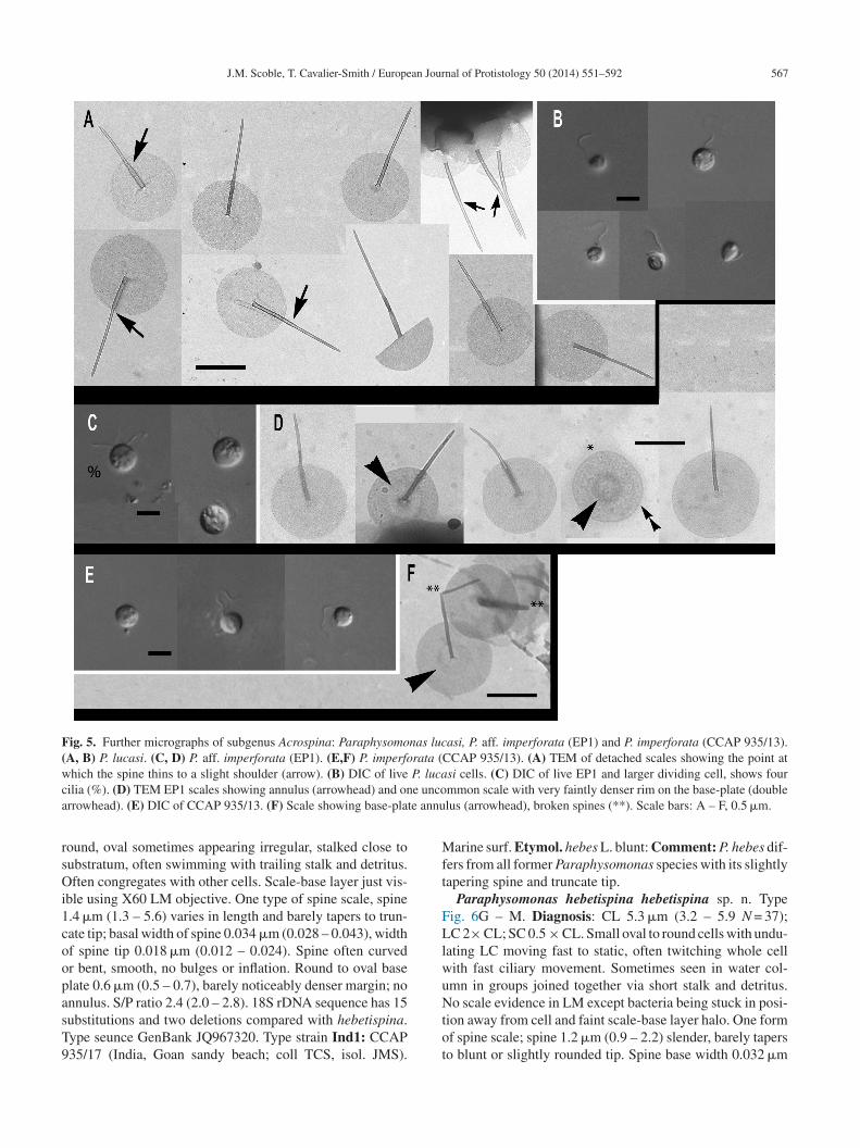
J.M. Scoble, T. Cavalier-Smith / European Journal of Protistology 50 (2014) 551–592 567
Fig. 5. Further micrographs of subgenus Acrospina: Paraphysomonas lucasi, P. aff. imperforata (EP1) and P. imperforata (CCAP 935/13).(A, B) P. lucasi. (C, D) P. aff. imperforata (EP1). (E,F) P. imperforata (CCAP 935/13). (A) TEM of detached scales showing the point atw P. lucac e uncoa e annu
rsOi1coopasT9
Mft
FLlwu
hich the spine thins to a slight shoulder (arrow). (B) DIC of liveilia (%). (D) TEM EP1 scales showing annulus (arrowhead) and onrrowhead). (E) DIC of CCAP 935/13. (F) Scale showing base-plat
ound, oval sometimes appearing irregular, stalked close toubstratum, often swimming with trailing stalk and detritus.ften congregates with other cells. Scale-base layer just vis-
ble using X60 LM objective. One type of spine scale, spine.4 �m (1.3 – 5.6) varies in length and barely tapers to trun-ate tip; basal width of spine 0.034 �m (0.028 – 0.043), widthf spine tip 0.018 �m (0.012 – 0.024). Spine often curvedr bent, smooth, no bulges or inflation. Round to oval baselate 0.6 �m (0.5 – 0.7), barely noticeably denser margin; no
nnulus. S/P ratio 2.4 (2.0 – 2.8). 18S rDNA sequence has 15ubstitutions and two deletions compared with hebetispina.ype seunce GenBank JQ967320. Type strain Ind1: CCAP35/17 (India, Goan sandy beach; coll TCS, isol. JMS).Ntot
si cells. (C) DIC of live EP1 and larger dividing cell, shows fourmmon scale with very faintly denser rim on the base-plate (doublelus (arrowhead), broken spines (**). Scale bars: A – F, 0.5 �m.
arine surf. Etymol. hebes L. blunt: Comment: P. hebes dif-ers from all former Paraphysomonas species with its slightlyapering spine and truncate tip.
Paraphysomonas hebetispina hebetispina sp. n. Typeig. 6G – M. Diagnosis: CL 5.3 �m (3.2 – 5.9 N = 37);C 2× CL; SC 0.5 × CL. Small oval to round cells with undu-
ating LC moving fast to static, often twitching whole cellith fast ciliary movement. Sometimes seen in water col-mn in groups joined together via short stalk and detritus.
o scale evidence in LM except bacteria being stuck in posi-ion away from cell and faint scale-base layer halo. One formf spine scale; spine 1.2 �m (0.9 – 2.2) slender, barely taperso blunt or slightly rounded tip. Spine base width 0.032 �m
568 J.M. Scoble, T. Cavalier-Smith / European Journal of Protistology 50 (2014) 551–592
Fig. 6. Micrographs of two new species of subgenus Hebetomonas: Paraphysomonas hebes, P. hebetispina limna, P. hebetispina hebetispina.(A – E) P. hebes. (F) P. hebetispina limna. (G – M) P. hebetispina hebetispina. (A) DIC of live P. hebes cells. (B – E) P. hebes TEMs. (B)Whole cell with both cilia and scales. (C) Blunt tips of scales (arrowhead). (D, E) Typical P. hebes scales. (F) DIC of live P. h. limna cells,one with obvious beating envelope (*). (G) DIC of live P. h. hebetispina cells, one with visible stalk (double arrowhead) and another withbeating envelope (*). (H – M) P. h. hebetispina TEMs. (H) Whole cell showing mastigonemes (small arrow), both cilia and scales. (I) Loosescales showing range of size, gentle taper of spine, and base-plate with no dense rim. (J) Close-up of blunt tip of spine. (K) A selection ofs er exam( �m. D
(Bl(
C
cales showing obvious base-plate annulus (large arrow). (L) Anothlarge arrow). Scale bars: A, F and G, 5 �m. B and H, 1 �m. C, 0.2
0.023 – 0.037), spine tip width 0.02 �m (0.015 – 0.028).
ase-plate width 0.5 �m (0.39 – 0.61), faint concentric annu-us in some scales only; slightly denser rim. S/P ratio 2.51.6 – 3.7). Type strain NC10-20. (Drainage ditch, North
–sv
ple of blunt tip of spine. (M) Single scale with base-plate annulus, E, I, J, K, L, and M, 0.5 �m.
arolina, Cape Fear River, Wilmington. JMS). Brackish
treated as marine. Etymol. hebes L. blunt: 18S rDNAequence GenBank JQ967321. Comment: Scale spine tipsary from rounded to flatly truncate; some spines appear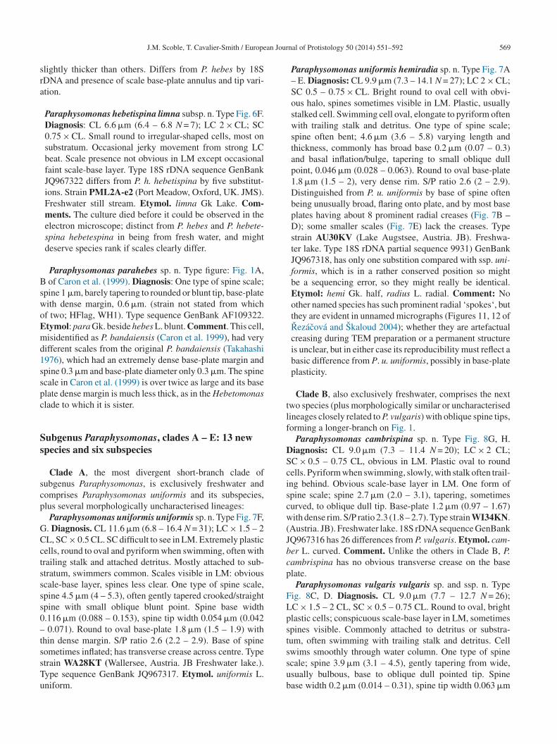
an Jour
sra
BswoEmd1sspc
Ss
scp
GCctssss0–tssTu
tlf
DSciscw(Jbcp
FLpst
J.M. Scoble, T. Cavalier-Smith / Europe
lightly thicker than others. Differs from P. hebes by 18SDNA and presence of scale base-plate annulus and tip vari-tion.
Paraphysomonas hebetispina limna subsp. n. Type Fig. 6F.Diagnosis: CL 6.6 �m (6.4 – 6.8 N = 7); LC 2 × CL; SC0.75 × CL. Small round to irregular-shaped cells, most onsubstratum. Occasional jerky movement from strong LCbeat. Scale presence not obvious in LM except occasionalfaint scale-base layer. Type 18S rDNA sequence GenBankJQ967322 differs from P. h. hebetispina by five substitut-ions. Strain PML2A-e2 (Port Meadow, Oxford, UK. JMS).Freshwater still stream. Etymol. limna Gk Lake. Com-ments. The culture died before it could be observed in theelectron microscope; distinct from P. hebes and P. hebete-spina hebetespina in being from fresh water, and mightdeserve species rank if scales clearly differ.
Paraphysomonas parahebes sp. n. Type figure: Fig. 1A, of Caron et al. (1999). Diagnosis: One type of spine scale;
pine 1 �m, barely tapering to rounded or blunt tip, base-plateith dense margin, 0.6 �m. (strain not stated from whichf two; HFlag, WH1). Type sequence GenBank AF109322.tymol: para Gk. beside hebes L. blunt. Comment. This cell,isidentified as P. bandaiensis (Caron et al. 1999), had very
ifferent scales from the original P. bandaiensis (Takahashi976), which had an extremely dense base-plate margin andpine 0.3 �m and base-plate diameter only 0.3 �m. The spinecale in Caron et al. (1999) is over twice as large and its baselate dense margin is much less thick, as in the Hebetomonaslade to which it is sister.
ubgenus Paraphysomonas, clades A – E: 13 newpecies and six subspecies
Clade A, the most divergent short-branch clade ofubgenus Paraphysomonas, is exclusively freshwater andomprises Paraphysomonas uniformis and its subspecies,lus several morphologically uncharacterised lineages:
Paraphysomonas uniformis uniformis sp. n. Type Fig. 7F,. Diagnosis. CL 11.6 �m (6.8 – 16.4 N = 31); LC × 1.5 – 2L, SC × 0.5 CL. SC difficult to see in LM. Extremely plasticells, round to oval and pyriform when swimming, often withrailing stalk and attached detritus. Mostly attached to sub-tratum, swimmers common. Scales visible in LM: obviouscale-base layer, spines less clear. One type of spine scale,pine 4.5 �m (4 – 5.3), often gently tapered crooked/straightpine with small oblique blunt point. Spine base width.116 �m (0.088 – 0.153), spine tip width 0.054 �m (0.042
0.071). Round to oval base-plate 1.8 �m (1.5 – 1.9) withhin dense margin. S/P ratio 2.6 (2.2 – 2.9). Base of spine
ometimes inflated; has transverse crease across centre. Typetrain WA28KT (Wallersee, Austria. JB Freshwater lake.).ype sequence GenBank JQ967317. Etymol. uniformis L.niform.ssub
nal of Protistology 50 (2014) 551–592 569
Paraphysomonas uniformis hemiradia sp. n. Type Fig. 7A– E. Diagnosis: CL 9.9 �m (7.3 – 14.1 N = 27); LC 2 × CL;SC 0.5 – 0.75 × CL. Bright round to oval cell with obvi-ous halo, spines sometimes visible in LM. Plastic, usuallystalked cell. Swimming cell oval, elongate to pyriform oftenwith trailing stalk and detritus. One type of spine scale;spine often bent; 4.6 �m (3.6 – 5.8) varying length andthickness, commonly has broad base 0.2 �m (0.07 – 0.3)and basal inflation/bulge, tapering to small oblique dullpoint, 0.046 �m (0.028 – 0.063). Round to oval base-plate1.8 �m (1.5 – 2), very dense rim. S/P ratio 2.6 (2 – 2.9).Distinguished from P. u. uniformis by base of spine oftenbeing unusually broad, flaring onto plate, and by most baseplates having about 8 prominent radial creases (Fig. 7B –D); some smaller scales (Fig. 7E) lack the creases. Typestrain AU30KV (Lake Augstsee, Austria. JB). Freshwa-ter lake. Type 18S rDNA partial sequence 9931) GenBankJQ967318, has only one substition compared with ssp. uni-formis, which is in a rather conserved position so mightbe a sequencing error, so they might really be identical.Etymol: hemi Gk. half, radius L. radial. Comment: Noother named species has such prominent radial ‘spokes‘, butthey are evident in unnamed micrographs (Figures 11, 12 ofRezácová and Skaloud 2004); whether they are artefactualcreasing during TEM preparation or a permanent structureis unclear, but in either case its reproducibility must reflect abasic difference from P. u. uniformis, possibly in base-plateplasticity.
Clade B, also exclusively freshwater, comprises the nextwo species (plus morphologically similar or uncharacterisedineages closely related to P. vulgaris) with oblique spine tips,orming a longer-branch on Fig. 1.
Paraphysomonas cambrispina sp. n. Type Fig. 8G, H.iagnosis: CL 9.0 �m (7.3 – 11.4 N = 20); LC × 2 CL;C × 0.5 – 0.75 CL, obvious in LM. Plastic oval to roundells. Pyriform when swimming, slowly, with stalk often trail-ng behind. Obvious scale-base layer in LM. One form ofpine scale; spine 2.7 �m (2.0 – 3.1), tapering, sometimesurved, to oblique dull tip. Base-plate 1.2 �m (0.97 – 1.67)ith dense rim. S/P ratio 2.3 (1.8 – 2.7). Type strain WI34KN.
Austria. JB). Freshwater lake. 18S rDNA sequence GenBankQ967316 has 26 differences from P. vulgaris. Etymol. cam-er L. curved. Comment. Unlike the others in Clade B, P.ambrispina has no obvious transverse crease on the baselate.
Paraphysomonas vulgaris vulgaris sp. and ssp. n. Typeig. 8C, D. Diagnosis. CL 9.0 �m (7.7 – 12.7 N = 26);C × 1.5 – 2 CL, SC × 0.5 – 0.75 CL. Round to oval, brightlastic cells; conspicuous scale-base layer in LM, sometimespines visible. Commonly attached to detritus or substra-um, often swimming with trailing stalk and detritus. Cell
wims smoothly through water column. One type of spinecale; spine 3.9 �m (3.1 – 4.5), gently tapering from wide,sually bulbous, base to oblique dull pointed tip. Spinease width 0.2 �m (0.014 – 0.31), spine tip width 0.063 �m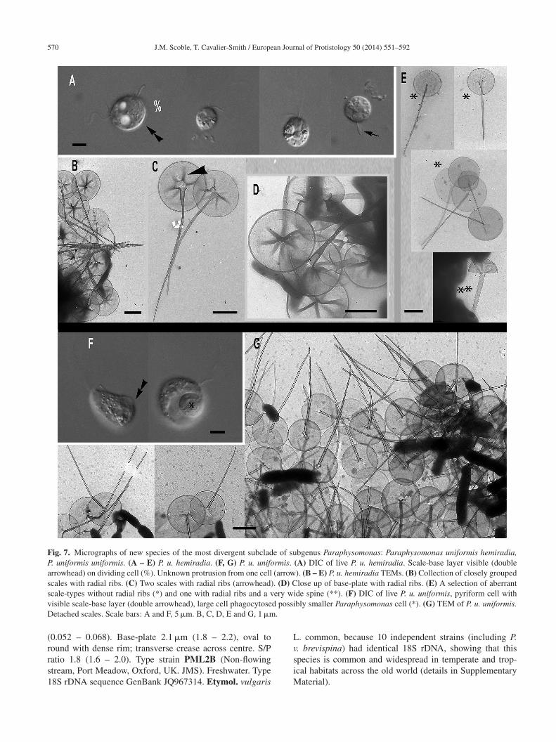
570 J.M. Scoble, T. Cavalier-Smith / European Journal of Protistology 50 (2014) 551–592
Fig. 7. Micrographs of new species of the most divergent subclade of subgenus Paraphysomonas: Paraphysomonas uniformis hemiradia,P. uniformis uniformis. (A – E) P. u. hemiradia. (F, G) P. u. uniformis. (A) DIC of live P. u. hemiradia. Scale-base layer visible (doublearrowhead) on dividing cell (%). Unknown protrusion from one cell (arrow). (B – E) P. u. hemiradia TEMs. (B) Collection of closely groupedscales with radial ribs. (C) Two scales with radial ribs (arrowhead). (D) Close up of base-plate with radial ribs. (E) A selection of aberrantscale-types without radial ribs (*) and one with radial ribs and a very wide spine (**). (F) DIC of live P. u. uniformis, pyriform cell withv d possiD .
(rrs1
Lv
isible scale-base layer (double arrowhead), large cell phagocytoseetached scales. Scale bars: A and F, 5 �m. B, C, D, E and G, 1 �m
0.052 – 0.068). Base-plate 2.1 �m (1.8 – 2.2), oval toound with dense rim; transverse crease across centre. S/P
atio 1.8 (1.6 – 2.0). Type strain PML2B (Non-flowingtream, Port Meadow, Oxford, UK. JMS). Freshwater. Type8S rDNA sequence GenBank JQ967314. Etymol. vulgarissiM
bly smaller Paraphysomonas cell (*). (G) TEM of P. u. uniformis.
. common, because 10 independent strains (including P.. brevispina) had identical 18S rDNA, showing that this
pecies is common and widespread in temperate and trop-cal habitats across the old world (details in Supplementaryaterial).
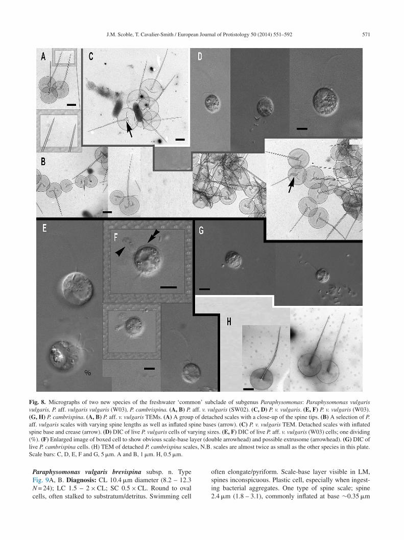
J.M. Scoble, T. Cavalier-Smith / European Journal of Protistology 50 (2014) 551–592 571
Fig. 8. Micrographs of two new species of the freshwater ‘common’ subclade of subgenus Paraphysomonas: Paraphysomonas vulgarisvulgaris, P. aff. vulgaris vulgaris (W03), P. cambrispina. (A, B) P. aff. v. vulgaris (SW02). (C, D) P. v. vulgaris. (E, F) P. v. vulgaris (W03).(G, H) P. cambrispina. (A, B) P. aff. v. vulgaris TEMs. (A) A group of detached scales with a close-up of the spine tips. (B) A selection of P.aff. vulgaris scales with varying spine lengths as well as inflated spine bases (arrow). (C) P. v. vulgaris TEM. Detached scales with inflatedspine base and crease (arrow). (D) DIC of live P. vulgaris cells of varying sizes. (E, F) DIC of live P. aff. v. vulgaris (W03) cells; one dividing( yer (dl es, N.BS
%). (F) Enlarged image of boxed cell to show obvious scale-base laive P. cambrispina cells. (H) TEM of detached P. cambrispina scalcale bars: C, D, E, F and G, 5 �m. A and B, 1 �m. H, 0.5 �m.
Paraphysomonas vulgaris brevispina subsp. n. TypeFig. 9A, B. Diagnosis: CL 10.4 �m diameter (8.2 – 12.3N = 24); LC 1.5 – 2 × CL; SC 0.5 × CL. Round to ovalcells, often stalked to substratum/detritus. Swimming cell
ouble arrowhead) and possible extrusome (arrowhead). (G) DIC of. scales are almost twice as small as the other species in this plate.
often elongate/pyriform. Scale-base layer visible in LM,spines inconspicuous. Plastic cell, especially when ingest-ing bacterial aggregates. One type of spine scale; spine2.4 �m (1.8 – 3.1), commonly inflated at base ∼0.35 �m
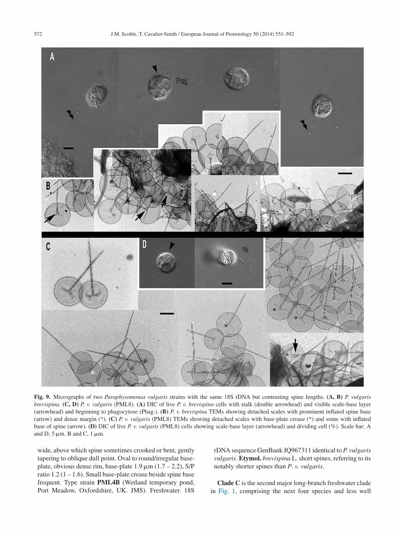
572 J.M. Scoble, T. Cavalier-Smith / European Journal of Protistology 50 (2014) 551–592
Fig. 9. Micrographs of two Paraphysomonas vulgaris strains with the same 18S rDNA but contrasting spine lengths. (A, B) P. vulgarisbrevispina. (C, D) P. v. vulgaris (PML8). (A) DIC of live P. v. brevispina cells with stalk (double arrowhead) and visible scale-base layer(arrowhead) and beginning to phagocytose (Phag.). (B) P. v. brevispina TEMs showing detached scales with prominent inflated spine base( owing
b howina
arrow) and dense margin (*). (C) P. v. vulgaris (PML8) TEMs shase of spine (arrow). (D) DIC of live P. v. vulgaris (PML8) cells snd D, 5 �m. B and C, 1 �m.
wide, above which spine sometimes crooked or bent, gentlytapering to oblique dull point. Oval to round/irregular base-
plate, obvious dense rim, base-plate 1.9 �m (1.7 – 2.2), S/Pratio 1.2 (1 – 1.6). Small base-plate crease beside spine basefrequent. Type strain PML4B (Wetland temporary pond,Port Meadow, Oxfordshire, UK. JMS). Freshwater. 18S idetached scales with base-plate crease (*) and some with inflatedg scale-base layer (arrowhead) and dividing cell (%). Scale bar: A
rDNA sequence GenBank JQ967311 identical to P. vulgarisvulgaris. Etymol. brevispina L. short spines, referring to its
notably shorter spines than P. v. vulgaris.Clade C is the second major long-branch freshwater claden Fig. 1, comprising the next four species and less well
an Jour
cAb
n0mccmw–(t3ssClt
n0vOsoTJEP
nA∼So1wawBGc
osoptaGP9po
nr
lmm
nSmsrmJhm
D0tCttrtfSo(c(JE
D
J.M. Scoble, T. Cavalier-Smith / Europe
haracterised lineages described in Supplementary Material.ll four are clearly different in rDNA and also distinguishabley scale dimensions and spine tip detail:Paraphysomonas variosa sp. n. Type Fig. 10A – F. Diag-
osis: CL 8.3 �m (7.3 – 9.1 N = 14); LC 1.5 × CL; SC.5 × CL. LC constant motion, although slows to an asym-etrical undulation. Difficult to see LC and SC on stationary
ells in LM because of orientation of cell. Round to ovalell, commonly attached to substratum or detritus. Swim-ing cells usually oval to elongate, swimming high inater column. One type of spine scale; spine 2.9 �m (1.4
3.7), tapering to blunt tip. Base width of spine 0.14 �m0.08 – 0.24); tip width 0.027 �m (0.015 – 0.049). Roundo oval base plate 1.2 �m (1 – 1.5); S/P ratio 2.5 (1.6 –.7), conspicuous to barely visible inflection at rim. Typetrain Ind5 (Freshwater, India. Coll. TCS. JMS). 18S rDNAequence, GenBank JQ967296. Etymol: variosa L. various.omment: P. variosa’s spine tip is most similar to P. sty-
ata stylata in that some have a more prominent pinchedip.
Paraphysomonas caroni sp. n. Type Fig. 10G – K. Diag-osis: CL 7.3 �m (5.9 – 9.6 N = 48); LC1.5 – 2 × CL; SC.5 × CL. Round to oval cell with scale-base layer sometimesisible on larger cells. Often trailing stalk when swimming.ne type of spined scale; spine 1.9 �m (1.3 – 2.1) tapering to
hort oblique blunt pointed tip. Round base-plate with obvi-us dense margin 1.0 �m (0.9 – 1.1); S/P ratio 1.9 (1.4 – 2.3).ype strain CH2. (Marsh in zoo, Beijing, China. Coll TC-S.MS). Freshwater. 18S rDNA sequence, GenBank JQ967292.tymol: named after D. A. Caron for his contribution toaraphysomonas research.Paraphysomonas mantoni sp. n. Type Fig. 10M – L. Diag-
osis: CL 6.8 – 20.5 �m; modal and median value, 8.6.verage, 9.7 �m: N = 31. LC, 1 – 4 ×CL smaller cells than8.6 �m appeared to have longer LC from 2.5 – 4 × CL.pines 3.5 �m (2.2 – 5.2) taper to a dull point (Fig. 10 M)r else to a small oblique tip (Fig. 10 N). Oval base-plate.3 �m (1 – 1.6) with dense rim. S/P 2.5 (1.9 – 3.6). Cellsith longer cilia seem to have more prominent spines in LM
s well as obvious scale-base layer. In very plastic large cellsith large vacuoles, scale presence not obvious. Type strainZ5a (Freshwater, Brazil. JMS). Type 18S rDNA sequenceenBank JQ967295. Etymol: named after I. Manton for her
ontribution to Paraphysomonas research.Paraphysomonas petronia sp. n. Type illustration Fig. 2A
f Petronio and Rivera (2010). Diagnosis: One type of spinecale covers cell; spine 2.9 �m (2.5 – 3.0), tapering to smallblique dull tip, centrally protruding from round to oval baselate 1.8 �m (1.6 – 2.0), with dense margin and no perfora-ions. Type strain J1 (Laguna de Bay, Philippines) (Petroniond Rivera 2010). Freshwater. Type 18S rDNA sequence:enBank GU220392. Etymol: named after first author, JAG
etronio. Comment: Its unique large 137 nt insert (position38 – 1075) in 18S rDNA was not seen in any other Para-hysomonas; when blasted against GenBank a match wasnly made to Paraphysomonas. The insert matches closely,Stv–
nal of Protistology 50 (2014) 551–592 573
ot exactly, an adjacent part of its sequence, so is a fairlyecent duplication.
Clade D. The next four long-branch species with relativelyong spines and each with very distinct rDNA have much
ore varied habitat than other clades: soil, freshwater, andarine:Paraphysomonas solis solis sp. n. Type Fig. 12A, B. Diag-
osis: CL 8.3 �m (6.4 – 11.4 N = 32); LC 1.5 – 2 × CL;C 7.5 × CL. Oval to round cells; often oval when swim-ing. One type of spine scale; spine 2.6 �m (1.5 – 4.4) tapers
trongly to dull point, base-plate 1.1 �m (0.8 – 1.3) oval toound with a varying dense rim. Type strain GMCCL6 (Wetud from end of stream, Christ Church Parks, Oxford, UK.
MS). Freshwater. 18S rDNA sequence GenBank JQ967309as 19 nucleotide differences from P. solis crocotilla. Ety-ol: solum L. soil.
Paraphysomonas solis crocotilla subsp. n. Type Fig. 11A –F. Diagnosis: CL 8 �m (6.8 – 9.1 N = 20) LC 1.5 × CL; SC0.5 × CL. Round to oval and elongate cells, swim fast up anddown water column. One form of spine scale; spine 3.7 �m(2.8 – 4.6) slender, sometimes slightly curved, gently tapersto a blunt tip. Spine base usually slightly inflated, width0.13 �m (0.103 – 0.188), spine tip width 0.017 �m (0.011– 0.021), larger inflation at the base rare (0.3 �m). Base-plate 1.2 �m (1.0 – 1.6 �m) with delicate but conspicuousdense rim. S/P ratio 3.1 (2.5 – 3.8). Type strain UPL1B (Soilfrom mole-hill under tree, University Parks, Oxford, UK.JMS). 18S rDNA sequence, GenBank JQ967308. Etymol:crocotilla L. slim. Comment: Some early micrographs sug-gest that it sometimes also has spineless scales; one roundto oval measured 1.4 �m (Fig. 11B). Spineless scales werenot observed when TEM was repeated.
Paraphysomonas dimorpha sp. n. Type Fig. 11 H – M.iagnosis: CL 6.7 �m (5 – 8.6 N = 22); LC 1.5 – 2 × CL; SC.5 – 7.5 × CL. LC moves constantly at varying rates, some-ime slowing almost to a stop, exhibiting slow undulation.ell round to oval, sometimes pyriform when swimming;
hick scale-base layer conspicuous in LM. Stalked cell closeo substratum/detritus. Two forms of scale; spine scales withound base-plate and dense rim; spine 3.2 �m (2.1 – 4.6)apers gently slightly curved to a blunt end and often startsrom base with slight bulge; base-plate 1.1 �m (1.0 – 1.2 �m),/P ratio 3.0 (2.0 – 4.2). Spineless scales round to slightlyval, 1.3 (1.1 – 1.6 �m), sometimes have central short line120 nm). Cell sometimes covered in one scale type but oftenovered in both forms. Type strain CA01 CCAP 935/16.Leaves, grass and mud, Monterey Bay, California, USA.MS). Freshwater. 18S rDNA sequence, GenBank JQ967310.tymol: dimorpha L. two forms, signifies two scale types.Paraphysomonas longispina sp. n. Type Fig. 13A, B.
iagnosis: CL 6.9 �m (6.8 – 13.6 N = 46); LC 2 – 2.5 × CL;
C 0.75 – 0.5 × CL. Plastic cells with large vacuoles, ovalo round. Readily ingests cells of own kind. Scale-base layerisible in LM. One form of spine scale; spine 5.7 �m (2.2
7.3) tapering to a dull point, often slightly curved along
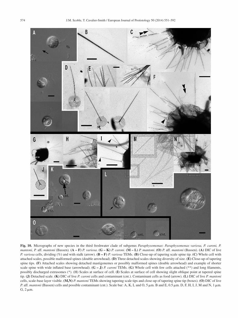
574 J.M. Scoble, T. Cavalier-Smith / European Journal of Protistology 50 (2014) 551–592
Fig. 10. Micrographs of new species in the third freshwater clade of subgenus Paraphysomonas: Paraphysomonas variosa, P. caroni, P.mantoni, P. aff. mantoni (Bassen). (A – F) P. variosa. (G – K) P. caroni. (M – L) P. mantoni. (O) P. aff. mantoni (Bassen). (A) DIC of liveP. variosa cells, dividing (%) and with stalk (arrow). (B – F) P. variosa TEMs. (B) Close-up of tapering scale spine tip. (C) Whole cell withattached scales, possible malformed spines (double arrowhead). (D) Three detached scales showing diversity of size. (E) Close-up of taperingspine tips. (F) Attached scales showing detached mastigonemes or possibly malformed spines (double arrowhead) and example of shorterscale spine with wide inflated base (arrowhead). (G – J) P. caroni TEMs. (G) Whole cell with few cells attached (**) and long filaments,possibly discharged extrusomes (*). (H) Scales at surface of cell. (I) Scales at surface of cell showing slight oblique point at tapered spinetip. (J) Detached scale. (K) DIC of live P. caroni cells and contaminant (cnt.). Contaminant cells as food (arrow). (L) DIC of live P. mantonicells, scale-base layer visible. (M,N) P. mantoni TEMs showing tapering scale tips and close-up of tapering spine tip (boxes). (O) DIC of liveP. aff. mantoni (Bassen) cells and possible contaminant (cnt.). Scale bar: A, K, L and O, 5 �m. B and E, 0.5 �m. D, F, H, I, J, M and N, 1 �m.G, 2 �m.
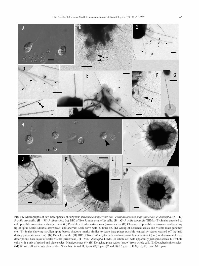
J.M. Scoble, T. Cavalier-Smith / European Journal of Protistology 50 (2014) 551–592 575
Fig. 11. Micrographs of two new species of subgenus Paraphysomonas from soil: Paraphysomonas solis crocotilla, P. dimorpha. (A – G)P. solis crocotilla. (H – M) P. dimorpha. (A) DIC of live P. solis crocotilla cells. (B – G) P. solis crocotilla TEMs. (B) Scales attached tocell, possible non-spine scales (arrows). (C) Possible extruded extrusomes (arrowheads). (D) Close-up of possible extrusomes and taperingtip of spine scales (double arrowhead) and aberrant scale form with bulbous tip. (E) Group of detached scales and visible mastigonemes(*). (F) Scales showing swollen spine bases; shadowy marks similar to scale base-plates possibly caused by scales washed off the gridduring preparation (arrow). (G) Detached scale. (H) DIC of live P. dimorpha cells and one possible contaminant (cnt.) or dormant cell (seedescription), base-layer of scales visible (arrowhead). (I – M) P. dimorpha TEMs. (I) Whole cell with apparently just spine scales. (J) Wholecells with a mix of spined and plate scales. Mastigonemes (*). (K) Detached plate scales (arrow) from whole cell. (L) Detached spine scales.(M) Whole cell with only plate scales. Scale bar: A and H, 5 �m. (B) 2 �m. (C and D) 0.5 �m. E, F, G, I, J, K, L and M, 1 �m.
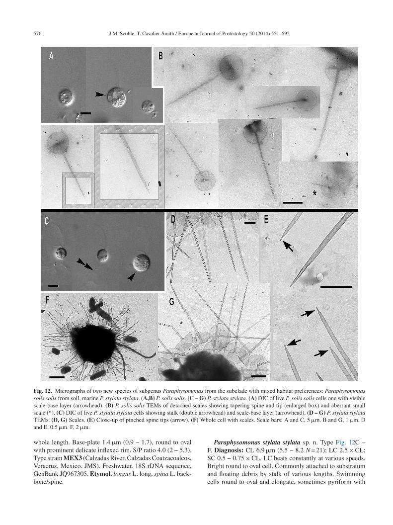
576 J.M. Scoble, T. Cavalier-Smith / European Journal of Protistology 50 (2014) 551–592
Fig. 12. Micrographs of two new species of subgenus Paraphysomonas from the subclade with mixed habitat preferences: Paraphysomonassolis solis from soil, marine P. stylata stylata. (A,B) P. solis solis. (C – G) P. stylata stylata. (A) DIC of live P. solis solis cells one with visiblescale-base layer (arrowhead). (B) P. solis solis TEMs of detached scales showing tapering spine and tip (enlarged box) and aberrant smalls le arroT F) Wha
wwTVGb
F
cale (*). (C) DIC of live P. stylata stylata cells showing stalk (doubEMs. (D, G) Scales. (E) Close-up of pinched spine tips (arrow). (nd E, 0.5 �m. F, 2 �m.
hole length. Base-plate 1.4 �m (0.9 – 1.7), round to ovalith prominent delicate inflexed rim. S/P ratio 4.0 (2 – 5.3).
ype strain MEX3 (Calzadas River, Calzadas Coatzacoalcos,eracruz, Mexico. JMS). Freshwater. 18S rDNA sequence,enBank JQ967305. Etymol. longus L. long, spina L. back-one/spine.SBac
whead) and scale-base layer (arrowhead). (D – G) P. stylata stylataole cell with scales. Scale bars: A and C, 5 �m. B and G, 1 �m. D
Paraphysomonas stylata stylata sp. n. Type Fig. 12C –. Diagnosis: CL 6.9 �m (5.5 – 8.2 N = 21); LC 2.5 × CL;
C 0.5 – 0.75 × CL. LC beats constantly at various speeds.right round to oval cell. Commonly attached to substratumnd floating debris by stalk of various lengths. Swimmingells round to oval and elongate, sometimes pyriform with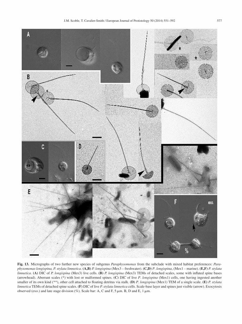
J.M. Scoble, T. Cavalier-Smith / European Journal of Protistology 50 (2014) 551–592 577
Fig. 13. Micrographs of two further new species of subgenus Paraphysomonas from the subclade with mixed habitat preferences: Para-physomonas longispina, P. stylata limnetica. (A,B) P. longispina (Mex3 – freshwater). (C,D) P. longispina, (Mex1 – marine). (E,F) P. stylatalimnetica. (A) DIC of P. longispina (Mex3) live cells. (B) P. longispina (Mex3) TEMs of detached scales, some with inflated spine bases(arrowhead). Aberrant scales (*) with lost or malformed spines. (C) DIC of live P. longispina (Mex1) cells, one having ingested anothersmaller of its own kind (**), other cell attached to floating detritus via stalk. (D) P. longispina (Mex1) TEM of a single scale. (E) P. stylatalimnetica TEMs of detached spine scales. (F) DIC of live P. stylata limnetica cells. Scale-base layer and spines just visible (arrow). Exocytosisobserved (exo.) and late stage division (%). Scale bar: A, C and F, 5 �m. B, D and E, 1 �m.
5 an Jour
ssb1oWTlisswsoso
sslrm
n–onw1rstrahnE
t(
–SusP∼t–or(Btasvi
F1b∼∼e(Gbg
78 J.M. Scoble, T. Cavalier-Smith / Europe
talk trailing. Scale-base layer visible in LM. One type ofcale; spine 3.6 �m (2.4 – 5.5) tapering to pinched tip. Spinease width, 0.079 �m (0.06 – 0.14). Round to oval base-plate.3 �m (1.0 – 1.6) with dense rim. Transverse central creasen base-plate often seen. S/P ratio 2.8 (1.7 – 3.7). Type strain:
02. (Wet beach sand, Pembrokeshire, UK. JMS). Marine.ype 18S rDNA sequence GenBank JQ967307. Etymol. sty-
us Gk writing instrument. Comment: The C at position 535n P. s. stylata 18S rDNA is a T in all other chrysomonads ando is probably a PCR or sequencing error in this strain. If nottudied carefully, the P. s. stylata spine tip could be confusedith the oblique tips of clade B, but the blunt end of P. s.
tylata is broader when unpinched, exhibiting little changer interruption in overall spine tapering. P. s. stylata is mostimilar to freshwater P. stylata limnetica, its sister sequencen Fig. 1, with much longer spines:
Paraphysomonas stylata limnetica subsp. n. Type Fig. 13E,F. Diagnosis: CL 9.4 �m (7.7 – 12.3 N = 14); LC 2.0 –2.5 × CL; SC 0.75 × CL. Plastic oval to round cell. Scale-base layer and nucleus conspicuous in LM. Swimming cellselongated and oval. One form of spine scale; spine 5.4 �m(3.8 – 7.2), gently tapering completely to a tip, spine basewidth 0.082 �m (0.06 – 0.12). Oval to round base-plate withdelicate thickened inflexed margin, 1.3 �m (0.9 – 1.7). S/Pratio 4.0 (3 – 6.8). Type strain: PML5D (from water floodedover grassy riverbank, Port Meadow, Oxford, UK. JMS).18S rDNA sequence GenBank JQ967306 differs from P.stylata stylata by two nucleotides (3 if position 535 of P. s.stylata is genuinely a C). Etymol: stylus Gk writing instru-ment. Comment: tapering is stronger and comes to a moreslender end in P. s. limnetica than in marine P. s. stylata,which commonly comes to a pinched tip; their spine basewidths match closely.
Clade E, exclusively from soil, comprises the next twopecies, distinctly different in rDNA, with a tendency to formhrunken smaller cells in culture. The subspecies of P. spicu-osa form a very distinct longer branch subclade (Fig. 1) andeadily encyst as collared stomatocysts of subspecies-specificorphology:Paraphysomonas sinensis sp. n. Type Fig. 14A – E. Diag-
osis: CL 9.8 �m (7.7 – 13.6 N = 25); LC 2.0 × CL; SC 0.5 0.75 × CL. Round to oval large plastic cell. Base layerf scales just visible in LM. Short stalk, sometimes thickerearer cell end. Scales heterogeneous, commonly spine scalesith rounded base-plate and dense rim; base-plate width.3 �m (1.1 – 1.5); spine length 2.62 �m (1.8 – 3.4) S/Patio 2.1 (1.5 – 2.7). Spineless larger plate scales present,ometime with a central stub or stump, usually much largerhan base-plate of spined scales, 2.1 �m (1.5 – 2.7). Aber-ant scale forms common; mostly spine is hyper-inflated
nd/or obscurely shaped. Large spineless scales commonlyave marks/scarring in the centre. Strain: CH9. (Soil, Yun-an, China. JMS). 18S rDNA sequence GenBank JQ967303.tymol. sinensis L. Chinese. Comment: Only pictures ofT
b
nal of Protistology 50 (2014) 551–592
he dormant-looking cells captured in the light microscopeFig. 16A) before the culture died.
Paraphysomonas spiculosa spiculosa sp. n. Type Fig. 15C E. Diagnosis: CL 8.4 �m (6.4 – 12.3 N = 36); LC 2.0 × CL:C 0.5 × CL. LC in constant motion, slows to symmetricalndulation. Round to oval cell, attached to substrate/detritus;wimming cells oval to elongate, trail stalk and detritus.lugged stomatocysts common, width ∼5.2 �m, collar height2.1 �m. One form of spine scale; spine 3.2 �m (2.4 – 3.6)
apers to subtly rounded tip, spine base width 0.11 �m (0.088 0.118), spine tip width 0.027 �m (0.022 – 0.029). Round toval base-plate 1.3 �m (1 – 1.8) delicate conspicuous denseim. S/P ratio 2.8 (1.33 – 3.6). Type strain BZ8 CCAP 935/19Soil and leaf litter, Brazil. JMS). 18S rDNA sequence, Gen-ank JQ967298. Etymol. spiculosa, as Stokes (1885) called
he radial projections of P. vestita spicules. Comment: Stom-tocyst similar size to P. spiculosa terricola but collar muchhorter, about half the height of terricola, and curved in sideiew (unlike P. spiculosa edaphica). Its closest sister on Fig. 1s P. s. edaphica.
Paraphysomonas spiculosa terricola subsp. n. TypeFig. 15A, B. Diagnosis: CL 9.7 �m (7.3 – 10.9 N = 11);LC 1.5 – 2.0 × CL: SC 0.5 × CL. Oval to round cells, some-times elongate when swimming, often trail stalk. Scale-baselayer, sometimes spines, visible in LM. Plugged stomato-cysts common (6.8 – 10 �m); refractile collar around stomatall (3.7 – 4.4 �m), almost as high as its basal width, slightlyundulating (less straight than in edaphica) and with thickermargin. One scale form. Spine 3.2 �m (2.2 – 5.4) taper-ing to a subtle rounded tip from a sometimes inflated spinebase, spine base width 0.083 �m (0.05 – 0.12), spine tipwidth 0.021 �m (0.02 – 0.03). Oval to round base-plate,1.0 �m (0.8 – 1.3) with obvious, sometimes delicate denserim. Type strain: GMBGL1 (Soil, Botanic Gardens, Oxford,UK. JMS). Freshwater. Type 18S rDNA sequence Gen-Bank JQ967301 differs from P. spiculosa spiculosa by 13substitutions and three indels. Etymol. terra L. earth colainhabit.
Paraphysomonas spiculosa edaphica subsp. n. Typeig. 15F. Diagnosis: CL 9.1 �m (9.1 – 9.1 N = 1); LC.5 × CL; SC 0.5 – 0.75 × CL. Round to oval cells. Scale-ase layer visible in LM. Plugged stomatocysts (diameter8 �m) common; collar around stoma straight sided, height3.1 �m, 4.4 �m diameter at base. Swimming cell often
longated with trailing stalk and detritus. Type strain: CH6Soil, Beijing, China. JMS). Type 18S rDNA sequenceenBank JQ967302 differs from P. spiculosa spiculosay 3 substitutions and four indels. Etymol: edaphos Gkround.
wo new Clathromonas species
We designated C. butcheri the type of Clathromonasecause it was the only species we were able to culture
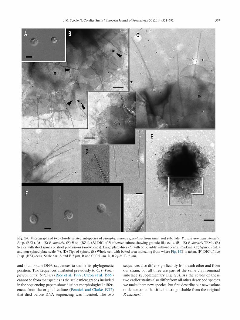
J.M. Scoble, T. Cavalier-Smith / European Journal of Protistology 50 (2014) 551–592 579
Fig. 14. Micrographs of two closely related subspecies of Paraphysomonas spiculosa from small soil subclade: Paraphysomonas sinensis,P. sp. (BZ1). (A – E) P. sinensis. (F) P. sp. (BZ1). (A) DIC of P. sinensis culture showing granule-like cells. (B – E) P. sinensis TEMs. (B)S late disa ith boP .2 �m.
appciet
sos
cales with short spines or short protrusions (arrowheads). Large pnd non-spined plate scale (*). (D) Tips of spines. (E) Whole cell w. sp. (BZ1) cells. Scale bar: A and F, 5 �m. B and C, 0.5 �m. D, 0
nd thus obtain DNA sequences to define its phylogeneticosition. Two sequences attributed previously to C. (=Para-hysomonas) butcheri (Rice et al. 1997; Caron et al. 1999)
annot be from that species as the scale micrographs includedn the sequencing papers show distinct morphological differ-nces from the original culture (Pennick and Clarke 1972)hat died before DNA sequencing was invented. The twotwtP
cs (*) with or possibly without central marking. (C) Spined scalesxed area indicating from where Fig. 16B is taken. (F) DIC of liveE, 2 �m.
equences also differ significantly from each other and fromur strain, but all three are part of the same clathromonadubclade (Supplementary Fig. S3). As the scales of those
wo earlier strains also differ from all other described speciese make them new species, but first describe our new isolateo demonstrate that it is indistinguishable from the original. butcheri.
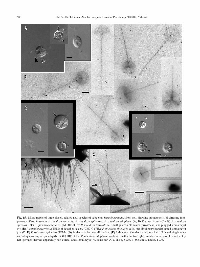
580 J.M. Scoble, T. Cavalier-Smith / European Journal of Protistology 50 (2014) 551–592
Fig. 15. Micrographs of three closely related new species of subgenus Paraphysomonas from soil, showing stomatocysts of differing mor-phology: Paraphysomonas spiculosa terricola, P. spiculosa spiculosa, P. spiculosa edaphica. (A, B) P. s. terricola. (C – E) P. spiculosaspiculosa. (F) P. spiculosa edaphica. (A) DIC of live P. spiculosa terricola cells with just visible scales (arrowhead) and plugged stomatocyst(*). (B) P. spiculosa terricola TEMs of detached scales. (C) DIC of live P. spiculosa spiculosa cells, one dividing (%) and plugged stomatocyst(*). (D, E) P. spiculosa spiculosa TEMs. (D) Scales attached to cell surface. (E) Side view of scales and cilium hairs (**) and single scaleincluding close-up of spine tip (box). (F) DIC of live P. spiculosa edaphica motile cell with cilia (on right), smaller more shrunken cell at topleft (perhaps starved, apparently non-ciliate) and stomatocyst (*). Scale bar: A, C and F, 5 �m. B, 0.5 �m. D and E, 1 �m.
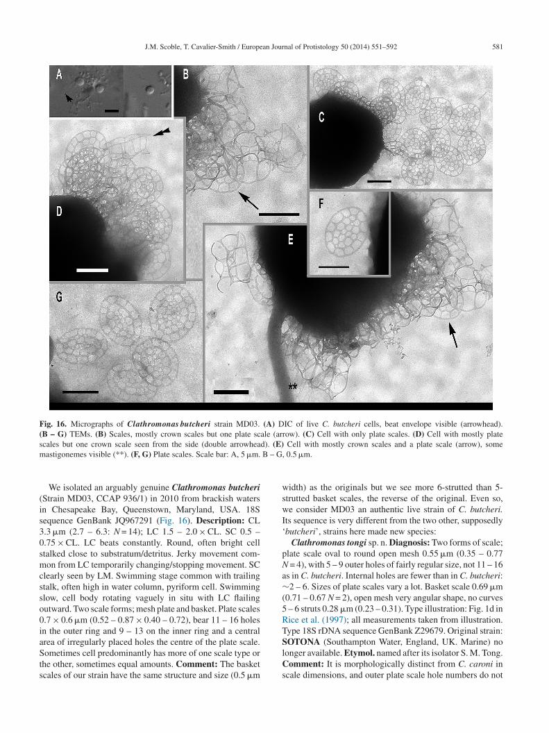
J.M. Scoble, T. Cavalier-Smith / European Journal of Protistology 50 (2014) 551–592 581
Fig. 16. Micrographs of Clathromonas butcheri strain MD03. (A) DIC of live C. butcheri cells, beat envelope visible (arrowhead).(B – G) TEMs. (B) Scales, mostly crown scales but one plate scale (arrow). (C) Cell with only plate scales. (D) Cell with mostly plates d). (E)m B – G,
(is30smcsso0iaSts
wswI‘
pNa∼(5RT
cales but one crown scale seen from the side (double arrowheaastigonemes visible (**). (F, G) Plate scales. Scale bar: A, 5 �m.
We isolated an arguably genuine Clathromonas butcheriStrain MD03, CCAP 936/1) in 2010 from brackish watersn Chesapeake Bay, Queenstown, Maryland, USA. 18Sequence GenBank JQ967291 (Fig. 16). Description: CL.3 �m (2.7 – 6.3: N = 14); LC 1.5 – 2.0 × CL. SC 0.5 –.75 × CL. LC beats constantly. Round, often bright celltalked close to substratum/detritus. Jerky movement com-on from LC temporarily changing/stopping movement. SC
learly seen by LM. Swimming stage common with trailingtalk, often high in water column, pyriform cell. Swimminglow, cell body rotating vaguely in situ with LC flailingutward. Two scale forms; mesh plate and basket. Plate scales.7 × 0.6 �m (0.52 – 0.87 × 0.40 – 0.72), bear 11 – 16 holesn the outer ring and 9 – 13 on the inner ring and a central
rea of irregularly placed holes the centre of the plate scale.ometimes cell predominantly has more of one scale type orhe other, sometimes equal amounts. Comment: The basketcales of our strain have the same structure and size (0.5 �m
SlCs
Cell with mostly crown scales and a plate scale (arrow), some 0.5 �m.
idth) as the originals but we see more 6-strutted than 5-trutted basket scales, the reverse of the original. Even so,e consider MD03 an authentic live strain of C. butcheri.
ts sequence is very different from the two other, supposedlybutcheri’, strains here made new species:
Clathromonas tongi sp. n. Diagnosis: Two forms of scale;late scale oval to round open mesh 0.55 �m (0.35 – 0.77
= 4), with 5 – 9 outer holes of fairly regular size, not 11 – 16s in C. butcheri. Internal holes are fewer than in C. butcheri:2 – 6. Sizes of plate scales vary a lot. Basket scale 0.69 �m
0.71 – 0.67 N = 2), open mesh very angular shape, no curves – 6 struts 0.28 �m (0.23 – 0.31). Type illustration: Fig. 1d inice et al. (1997); all measurements taken from illustration.ype 18S rDNA sequence GenBank Z29679. Original strain:
OTONA (Southampton Water, England, UK. Marine) noonger available. Etymol. named after its isolator S. M. Tong.omment: It is morphologically distinct from C. caroni in
cale dimensions, and outer plate scale hole numbers do not
5 an Jour
olbh
Lmel1ihowUiifMBokbtCsbf
D
mssgpicsPpdheai(tetep(f
sjteoHtr‘pr
Vs
sptrsossstsuspp‘aTscrsPrdpetifgwpph
82 J.M. Scoble, T. Cavalier-Smith / Europe
verlap with the range for C. butcheri; the basket scales areess angular. C. tongi has similar dimensions to C. butcheriut has plate scales more diverse in size with many feweroles.
Clathromonas caroni sp. n. Diagnosis: CL, at least 3.3 �mC = ×2 CL (N = 1, Lim et al., 2001). Two scale forms: openesh plate and basket scales; all descriptions based on Caron
t al. (1999) and Lim et al. (2001). Plate scale, oval to round,ength 0.92 �m (0.67 – 1.25); peripheral ring of holes (8 –2) sometimes with smaller perforations at joins as well asrregular holes in centre. One plate scale with inner ring of ∼9oles, but seven plate scales showed irregular size and shapef central holes. Basket scale (one) 0.9 �m wide, five strutsith curved upper tier on oval to round ring, strut ∼ 0.16 �m.nclear number of holes in upper tier, but appear irregular
n shape and size, similar to holes in plate scales. Typellustrations; Fig. 1f from Caron et al. (1999) and Fig. 3a,brom Lim et al. (2001). Type strain: DB4 (Patuxent River,
aryland, USA). Marine. Type 18S rDNA sequence Gen-ank AF109326. Etymol. caroni after D. A. Caron, authorf both papers on this species. Comment: The single bas-et scale looks more like C. inconspicua (Takahashi, 1976),ut plate scales are clearly different and sometimes resemblehe top tier of the basket scale in C. corbidifera (Pennick andlarke, 1973). The basket scale is larger than C. butcheri with
horter struts. The variety of hole-shapes and sizes is similarut scales are bigger than in C. butcheri and plate-scale holesewer on average, though ranges overlap.
iscussion
Our major conclusion is that Paraphysomonas was for-erly a heterogeneous repository for all non-photosynthetic
caly chrysophytes irrespective of their vastly differingcale types and was far too diverse morphologically andenetically to be accepted as a single genus. The revised Para-hysomonas and Clathromonas are now relatively uniformn scale structure. As we specifically targeted heterotrophicells for culturing it is unsurprising that we obtained repre-entatives only of two purely heterotrophic scaly families,araphysomonadidae and Clathromonadidae, both in Para-hysomonadida. Former Paraphysomonas species with veryifferent scales, now excluded from these morphologicallyomogenous and phylogenetically strongly supported gen-ra, are being reassigned to new genera in a separate paper,nd include some photosynthetic species unlikely to belongn Paraphysomonadida. Whether Clathromonadidae is sisterFig. 1) or not (Fig. 2) to Paraphysomonadidae in 18S rDNArees depends in part on taxon sampling; a recent tree omittingnvironmental sequences included here placed them consis-ently, sometimes strongly sisters by three methods (Skaloud
t al. 2013). Support for Paraphysomonadida being holo-hyletic is generally higher on trees showing it as a cladee.g. Skaloud et al. 2013) than the never significant supportor it contradictorily being paraphyletic (e.g. Fig. 2). We werePwt
nal of Protistology 50 (2014) 551–592
urprised that we did not isolate a lot of Spumella-like strains,ust one when we deviated from our earlier strict criterion ofargeting rigid round cells. This suggests that it is probablyasier to separate paraphysomonads and Spumella from eachther by light microscopy than was previously appreciated.owever, the limitations of light microscopy, especially in
he 19th century, mean that it may never be possible to toeidentify convincingly all ‘species’ originally described asMonas’ (see Boenigk 2008) or even assign them all to Para-hysomonas or Spumella, so some old species names mayemain for ever in limbo.
ast genetic diversity of Paraphysomonas sensutricto
There are now far more species of Paraphysomonasensu stricto (essentially those with only spine scales) thanreviously assumed (Finlay and Clarke 1999b). We increasedhe number from 9 to 32 and there are already >30 more envi-onmental DNA sequences on Fig. 1 for Paraphysomonasensu stricto distinct enough to be separate species, and somef genotypes labelled aff. And some treated here as only sub-pecies may prove to be worth making new species in future,o there are probably at least 80 genuine Paraphysomonaspecies, probably many more – perhaps several hundred ashere is no reason to think that sampling is anywhere nearaturation. Thus, excessive taxonomic lumping previouslynderestimated the number of spine-scaled Paraphysomonaspecies at least tenfold. Nearly all our new species wouldreviously have been lumped in just two ‘species’: Para-hysomonas vestita and imperforata. No wonder those twospecies’ were thought to be the most frequently encounterednd geographically ubiquitous (Finlay and Clarke 1999b).hey were not single species but swarms of separate species,ome at least as genetically different from each other as theomplete range of variation within the entire order Synu-ales. Supplementary Fig. S4 compares 15 of our 23 newpecies that would probably once have been lumped as either. vestita (10 with a dense base-plate rim) or P. imperfo-ata (five with plain base-plate rims). Yet these 15 speciesiffer obviously in cell size and shape, scale size and pro-ortions, and in spine tip structure; many are far distant fromach other on the tree (Fig. 1). Lee (1978) even argued forreating all as one species just because one strain can liven both marine and fresh water; the common assumption ofree movement between these habitats for Paraphysomonasenerally seems false: Fig. 1 shows that marine and fresh-ater lineages are phylogenetically rather stable in habitatreference; only one 18S rDNA genotype of the 82 Para-hysomonas sensu stricto in Fig. 1 was found in bothabitats.
As in many protist groups, there is no evidence whetheraraphysomonas is sexual or asexual, so we do not knowhether the biological species concept can be applied to
hem or not. It is therefore most reasonable to use a similar
an Jour
dphiewsrtstssafstdhstssHmtiiiipaoaoHslip(ilfs
Lw
iaeAt
rtooobocOocnsusssowoctfpcpsCmC
iLePhvhsvdcTi
Pcotat2
J.M. Scoble, T. Cavalier-Smith / Europe
egree of genetic differences for subdividing both sexualrotists and those whose population genetics is unknown, asas recently been done in other protist groups where sexual-ty is unknown but might exist (e.g. Bass et al. 2009; Howet al. 2011; Glücksman et al. 2013). As sexual eukaryotesith even a few differences in rDNA are invariably separate
pecies, placing those with genuinely non-identical 18SDNA in different named species is unlikely to be oversplit-ing. Placing those with identical 18S rDNA in the samepecies may often be correct, but could be undersplitting ifhey are really sexual. But if they are asexual the degree ofplitting appropriate for making nominal species is neces-arily arbitrary, so using this simple objective criterion asdopted here is not conceptually problematic. In general, weound that if 18S rDNA of closely related Paraphysomonastrains is unambiguously different by even just one orwo nucleotides, we can also reproducibly detect slightifferences in scale morphology. Conversely if two strainsave identical 18S rDNA their scales are generally extremelyimilar and in many cases indistinguishable. This meanshat the rate of divergence in 18S rDNA is approximatelyimilar to that of scale morphology in Paraphysomonas,o either can be a good criterion for species demarcation.owever, the discrete digital nature of rDNA sequencesakes them a simpler and less ambiguous criterion to apply
han the continuous qualitative and more statistical variationn scales. We found a few examples where strains withdentical 18S rDNA have detectably different scales or differn habitat (marine or freshwater) and therefore presumablyn physiology; we conservatively did not make these mor-hological or physiological variants separate species, but in
few instances felt it useful to make them subspecies. Asnly six genotypes were found more than once (one 10 andnother four times) there must still be gross undersamplingf Paraphysomonas and well over 100 species must exist.owever, our ability to find some genotypes repeatedly,
ometimes on different continents, means that variation is notimitless and the number of species globally worth namings unlikely to run into thousands as in diatoms, the hugerotist group where biological species have been best studiedAmato et al. 2007; Mann and Vanormelingen, 2013). Its possible that we have now identified most of the majorineages, so we would expect a similar future sampling effortor new strain isolation to result in proportionally fewer newpecies.
ight microscopy reveals systematic differencesithin Paraphysomonas
It is often said that most Paraphysomonas species arendistinguishable in the light microscope from Spumella,
phylogenetically heterogeneous array of non-scaly het-rotrophic chrysomonads abundant and diverse in freshwater.s some Spumella grow like weeds in culture, we were ini-
ially concerned that isolating colourless chrysomonads at
dssP
nal of Protistology 50 (2014) 551–592 583
andom would yield cultures that would mostly turn outo be Spumella (irrelevant to this project), and we wouldnly realise this after the efforts of purifying, sequencingr examining them by TEM. Spumella cells are elongatedr irregular in shape (perhaps because not so constrainedy scales) and some are very small, so we initially focusedn culturing large (∼≥7 �m) completely round colourlessells with two visible cilia (one long, one short: to avoidikomonas (Cavalier-Smith et al. 1996) which lacks the shortne), and with a stalked stage. To our surprise this suc-essfully biased the results against Spumella, despite oftenot being able to see scales clearly in the light micro-cope. This mode of selection has almost certainly givens a biased sample of paraphysomonad diversity – againstmaller, unstalked cells or irregular-shaped ones, or any (ifuch exist) without a short cilium, and could be one rea-on why we obtained so many Paraphysomonas and onlyne Clathromonas, which was found only later in the projecthen we targeted smaller cells to see if this would yieldther scale types. The rarity of Clathromonadidae in ourultures might in part be because they are harder to cul-ure under our conditions, possibly because they need algalood, but that is pure conjecture. In future it should beossible to obtain many more clathromonad cultures forombined TEM/sequencing studies similar to ours for Para-hysomonas, since Preisig and Hibberd (1982a,b) obtainedeveral uniprotist cultures of six species that we assign tolathromonas. Sequences are now desirable for a greaterorphological diversity of Clathromonas to test the unity oflathromonadidae.Very few Paraphysomonas had previously been stud-
ed live in the light microscope (Lucas, 1967, 1968;eadbeater, 1972; Pennick and Clarke, 1972, 1973; Reest al. 1974); most measurements of previously establishedaraphysomonas species were on fixed material likely toave shrunk in preparation. Therefore, cellular featuresisible in the light microscope were not previously used toelp distinguish species. Our results show that though notufficiently detailed to be diagnostic for individual species,ariations in cell size map sensibly onto the molecular tree asoes long cilium length (×CL). Cell size and cilium lengthan be characteristic of a set of species, not individual ones.hese features may help rule out certain species during initial
dentification.With respect to cilium length one can recognise three
araphysomonas clades with longer than average anteriorilium. First, subgenus Acrospina having non-dense marginn the scale base-plate with round or oblique pointed spineips (P. imperforata, P. lucasi, P. mikadiforma, P. acuminatacuminata, P. acuminata cuspidata) never has an LC shorterhan 2.5 × CL. Secondly, P. sp. (BZ1) and P. sinensis with.0 – 2.5 × CL. Thirdly, two species belonging to a larger
ense-margin base-plate clade with completely taperingpines, namely P. longispina, P. stylata limnetica and P.tylata stylata (a subclade of subclade D of subgenusaraphysomonas) have slightly longer LC than the rest
5 an Jour
idSHc
m(tssat–osos
soaPaiwtct
P
sotdw(oPp(dto(ttwiabap
b∼nu
P
taAccstfp
sfocsohvn(mwemcflnpasPtss(Gf‘tDio
84 J.M. Scoble, T. Cavalier-Smith / Europe
n their clade, 2.0 – 2.5 × CL (Table 2). All other speciesescribed here have a shorter long cilium (≤2.0 × CL).tokes’ P. vestita had LC 2 × CL like most of our isolates.owever, none of our strains exactly matches his other
ellular descriptions.Cell size is indicative of groups of species; formerlyisidentified P. foraminifera and P. imperforata sequences
subgenus Acrospina) all have cells below 5 �m, as well ashe new closely related species P. lucasi. Other small cellstudied form two distinct groups: P. ovalis and P. segmenta inubgenus Brevispina, and P. hebes, P. hebetispina hebetispinand P. hebetispina limna are a subclade of subgenus Hebe-omonas; all these species have average cell sizes between 4.7
6.6 �m. Subclade B of subgenus Paraphysomonas has somef the largest cells found, averaging 11.3 �m. So, average cellize, as well as scale dimensions and features like presencer absence of concentric annulus and spine tip shape, canometimes rule out certain species.
Cell behaviour can also differ among species. Somemaller, shorter stalked, species tend to grow as loose patchesn the substratum, fixed by a short stalk; some of these exhibit
twitching motion, particularly P. ovalis, whilst others like. aff. imperforata (EP1) and P. imperforata (CCAP 935/13)nd P. lucasi readily have static LC in a kinked/curved cil-ary position. Long cilium motion in the larger-celled speciesith very long LC is distinct from other species and motion
oo can be erratic. Other potentially describable behaviouralharacteristics, especially swimming style, were not noted inhis study.
araphysomonas spine scale conservatism
There were previously five species of Paraphysomonasensu stricto with holey spine-scale base-plates, but nonef our new cultures had base-plate holes or perforations,hough we described a sixth such species from publishedata (P. perforata). All other new species have spine scalesith entirely unperforated base-plates. Setting aside P. vestita
see next section), there were previously only three speciesf non-holey spine-scaled Paraphysomonas: P. imperforata,. bandaiensis, and P. antarctica. Almost all our 22 new non-erforated species would have been assigned to P. vestitaif with dense base-plate margin) or to P. imperforata (if noense margin) prior to our study. Yet we have shown thathese crudely defined morphotypes occupy the vast majorityf the genetically extremely diverse Paraphysomonas cladeFig. 1), whose genetic depth is comparable to that of any pho-osynthetic order of chrysophytes (Fig. 2). They are likelyo have diverged from each other in the Lower Cretaceoushen chrysophyte stomatocysts first appear (∼110 My ago)
n the fossil record (Siver and Wolfe 2005). Paraphysomonas
nd Clathromonas divergence seems somewhat later (possi-ly ∼90 My ago, estimated from Fig. 1 short branch taxa);similar crude estimate places the basal radiation of Para-hysomonas sensu stricto at ∼70 My ago. Therefore their
Asws
nal of Protistology 50 (2014) 551–592
asic nail-scale morphology has probably been stable for70 My. This remarkable morphological conservatism in
ail-scale structure led to species diversity being grosslynderestimated.
erforated-scale species diversity
It seems that the single definite perforated species onhe tree, P. perforata, is a relatively recently derived vari-nt within the large, predominantly unperforated subgenuscrospina (Fig. 1), which would otherwise all have beenalled P. imperforata previously; yet Acrospina showsomparable or greater genetic depth to most genera of photo-ynthetic chrysophytes (Fig. S1). Further research is neededo see if the other five perforate species group with per-orata or elsewhere; no convincing evidence indicates thaterforated base-plates evolved more than once.
Unfortunately, we did not isolate a P. foraminiferatrain. Two of the three sequences in GenBank labelled ‘P.oraminifera’ are so far apart that both cannot possibly be theriginal species. One was accompanied by a micrograph thatlearly shows that it was misidentified (Rice et al. 1997); itspine is only two thirds as long as P. foraminifera and tapersnly near its tip not along most of its length and base-plateoles are more regular and relatively larger than the inter-ening trabeculae in P. foraminifera. We therefore made it aew species, P. perforata. The other sequence AB022864strain MBI-HT3, unavailable) has no associated electronicrograph. It might be a genuine P. foraminifera, but if itas grown in freshwater medium DY-IV as stated (Andersen
t al. 1999), it was probably not P. foraminifera which wasarine (Lucas 1967), so we placed its name in inverted
ommas on Fig. 1. As all neighbouring strains to HT3 arereshwater vestita-like species, the true P. foraminifera mostikely will turn out to be related to the marine P. perforata,ot to HT3. If HT3 actually has a perforated scale base-late, such perforations must have evolved at least twice inncestrally unperforated Paraphysomonas lineages. The thirdequence AF174376 is marine and consistently groups with. perforata (P. foraminifera in GenBank; as does Fig. 1,he original paper more wisely labeled it Paraphysomonasp. (Atkins et al. 2000), noting that no scales were seen,o the species could not be identified); the stated cell size10 – 15 �m not 3.1 – 4.4 �m as in foraminifera) proves theenBank name to be wrong; we assume it was miscalled P.
oraminifera merely because it was closer to the Rice et al.P. foraminifera’ (actually perforata) on their trees than tohe only other Paraphysomonas (an unspecified ‘vestita’).alby et al. (2008) claimed that P. foraminifera is the dom-
nant phagotroph in oil-polluted microcosms, based purelyn 18S rDNA sequencing, its closest relative being said to be
F174376, which is not P. foraminifera; in fact their abundantequence PSX4-3 (AY789782) is not closest to AF174376;e found three P. imperforata strains and eight environmental
equences with stronger BLAST hits, and on Fig. 1 it groups
an Jour
whi
Ps
despf
stpPnsstbl(tpcrpeaod
v(ogssscmntd(
wsHBig
rodsbusiipcl
crotspwvas
thaiporImsiK
Poptocdljabtdtp
J.M. Scoble, T. Cavalier-Smith / Europe
ith P. imperforata VS1 (though P. imperforata C1 and D1ave the highest and second highest BLAST hits). Clearly its P. imperforata-like, not P. foraminifera.
hylogenetically significant Paraphysomonaspine scale variation
The many more imperforate species are all geneticallyifferent, often greatly, sometimes only slightly, and alsoxhibit subtly different ultrastructural features of their spinecales, whose evolutionary and taxonomic significance hadreviously been almost entirely overlooked. Nine spine-scaleeatures are useful for identification:
(1) Presence or absence of an inflated/bulbous base to thepine. (2) Degree of spine tapering. (3) Shape of the spineip: tapered to a blunt tip, rounded tip, oblique sharp, blunt orinched. (4) Shape of base-plate: oval, round or irregular. (5)resence or absence of a dense base-plate rim, whose thick-ess is characteristic within each species. Hibberd (1979)howed by sectioning a P. bandaiensis strain, which scaletructure suggests probably belongs to subgenus Brevispina,hat the dense rim is caused by a marginal inflection of thease-plate, but whether that is also true of the somewhatess dense rim of subgenus Paraphysomonas is unknown.6) Occurrence or not of a dense annulus on the base-plate;his is thought to be a slightly raised ring midway across thelate (Lucas 1968). For some strains it is not a consistentharacter, e.g., P. aff. imperforata strain EP1 and P imperfo-ata strain CCAP 935/14. (7) Crease at the base of the spineresent or not, thought to be relic of EM preparation. (8) Pres-nce or absence of radial ribs on the base-plate, possibly also
creasing artefact. (9) Size of scale: length of spine, widthf base-plate, and S/P ratio. All these characters are usefuliagnostic features.As shown on Fig. 1, one main feature is rather conser-
ative, and constant within three subgenera: the presencesubgenus Paraphysomonas; and thicker still in Brevispina)r absence (subgenus Acrospina) of the base-plate dense mar-in. However, the dense margin varies in prominence, somepecies have more subtle dense edges than others. The fourthubgenus Hebetomonas (only P. hebes, and P. hebetispinatudied ultrastructurally) has a distinct spine scale with a trun-ate/rounded spine tip and circular base-plate whose marginay be slightly dense or not. This somewhat intermediate
ature is probably not surprising as Hebetomonas is sistero subgenus Paraphysomonas, which might have evolved itsense base-plate edge independently of subgenus Brevispina.See Supplemenary Fig. S4 for direct comparison).
We cannot confidently deduce whether the base-plate rimas originally plain as in P. imperforata and the rest of
ubgenus Acrospina and the major subclade of subgenus
ebetomonas or dense as in subgenera Paraphysomonas andrevispina plus P. parahebes; but if the topology of Fig. 1s correct, assuming that a dense rim is ancestral wouldive only two origins of plain rims, whereas assuming plain
cPC
nal of Protistology 50 (2014) 551–592 585
ims were ancestral gives three independent origins of densenes, and so is marginally less parsimonious. Though theifference in rim structure is relatively small, it is fairly con-ervative and numerous clearly related lineages share similarase-plate margins. The margin is most dense and conspic-ous in P. bandaiensis, which led to its being separated as apecies even without sequence information. From our treest is likely that Paraphysomonas ancestrally had scales withmperforate base-plates; they became perforated in the smallerforata subclade near the base of the largest marine sub-lade of Acrospina, which is sister to P. imperforata plus P.ucasi.
Within subgenera Acrospina and Paraphysomonas spineharacters further define subsets (Table 2). There are dense-im (subgenus Paraphysomonas) species that all have a smallblique dull spine point. No spines ending in an oblique dullip are seen in any Acrospina. Conversely, there are Acrospinapecies with a sharp oblique tip not found in subgenus Para-hysomonas. In P. segmenta (subgenus Brevispina, to whiche suspect P. bandaiensis may belong) the dense margin isery wide, whereas others like P. uniformis hemiradia have
thinner dense edge – it would be interesting to study thetructural basis of these differences by TEM sections.
Variations in shape were more apparent in some specieshan others. Radial ribs on the base-plate, as in P. uniformisemiradia, have been noted before (Takahashi 1976; Preisignd Hibberd 1982a; Eloranta 1989); though it is unclearf they stem from artifactual regular wrinkling during EMreparation or are a natural rigid structure, their consistentccurrence in one subspecies only means that they musteflect an underlying structural difference from other species.t was impractical to do scanning electron microscopy for soany species, so we chose not to for any. Some studies con-
ider that it can sometimes be less useful and even confusedentification of silica-scaled chrysophytes (Boo et al. 2010;ynclová et al. 2010).Diversity of scale morphology within a species can vary.
. variosa is so named because of its unusually large rangef scale size and thickness. P. dimorpha is the only Para-hysomonas so far known with two qualitatively differentypes of scale: spine and plate. Some P. dimorpha cells hadnly spineless scales or mostly spine scales, perhaps indi-ating a life cycle stage of the organism. Varying ratios ofifferent scale types are also seen in Clathromonas butcheri;ike Pennick and Clarke (1973) we noted that some cells hadust plate or basket scales or a mixture of both. Both scale vari-tion within a species and the evidence that similar scales cane seen in several parts of the tree make species identifica-ion from single scales in environmental samples, sometimesone (Finlay and Clarke 1999a,b), necessarily less precisehan from whole cells, and sometime more ambiguous thanreviously realised.
Our study emphasises the importance of noting subtle butonsistent differences in scale structure when identifyingaraphysomonas or describing new species (see Scoble andavalier-Smith 2013). Their neglect led to their previous

5 an Jour
edtChad‘Pttrvtabar
tsonoihtpaaspcodmsggsErDb
P
eldtaa
ot
mtcaJn(PnmwsisTnsmbcTdatdwiStrtibcssvrstvtn
fE–tn
86 J.M. Scoble, T. Cavalier-Smith / Europe
xcessive lumping: predominantly just ‘vestita’ with aense base-plate border and imperforata lacking it, by farhe two most frequently encountered ‘species’ (Finlay andlarke 1999b); this difference actually characterises twouge species-rich groups that differ genetically as muchs do whole families in many eukaryotes. These crudelyifferentiated morphotypes cannot be real species; thevestita-like’ morphotype is a clade, the entire new subgenusaraphysomonas, and the imperforata morphotype consti-
utes the majority of the new subgenus Acrospina apart fromhe structurally derived P. perforata. This makes all previousecords of the distribution and ecological preferences of P.estita and P. imperforata meaningless at the species level;hey pertain only to the whole subgenus Paraphysomonasnd most of subgenus Acrospina respectively – too crude toe useful. Species with holey base-plates like P. foraminiferand perforata appear to be strictly marine and have not beeneported from freshwater or soil.
If variations in scale morphology are compared acrosshe tree it is clear that in some regions small variations incales correspond with small variations in sequences and canften be similarly resolving to DNA sequences, if properote is made of these small structural differences. We foundne case where scale differences seem to be more resolv-ng than sequences (P. vulgaris strains PML4B and PML8ave different spine lengths – see Supplementary Informa-ion 1), but several instances where quite similar scales wereresent on rather distant lineages. Thus, though there is often
broad (and sometimes quite close) correlation between scalend genetic differences, this is not a precise correlation andequences in general offer a more reliable picture of Para-hysomonas evolution and affinities that is less subject toonvergent evolution (e.g. with respect to the appearancef the base-plate margin and spine tips). The new speciesescribed here can probably all be distinguished by scaleorphology alone if whole cells are available (but not from
ingle scales). But as undescribed species might have indistin-uishable scales and the full range of variation for most rDNAenotypes is not yet known, more reliable identification needsequences in addition to or instead of scale morphology.lectron microscopy alone would be less resolving for envi-
onmental surveys of Paraphysomonas sensu stricto thanNA sequencing but would advantageously have differentiases.
araphysomonas vestita identification problem
We found a very large number of genetically oftenxtremely different species of Paraphysomonas having nail-ike scales with non-perforated entire round base-plate with a
ense margin and relatively long, more or less pointed, cen-ral spine. Previously all such species were lumped togethers Paraphysomonas vestita despite the different dimensionsnd differences in scale morphology we discovered. Are anynahf
nal of Protistology 50 (2014) 551–592
f our strains P. vestita as described by Stokes (1885) underhe name Physomonas vestita?
Physomonas vestita was described before electronicroscopy, therefore we can only use features visible in
he light microscope in identification: cell size, shape, andilium, stalk, and spine lengths. Stokes only observed sessilend stalked cells from shallow ponds and streams in Newersey, USA; 1/1666 inch in diameter, equal to ∼15.2 �m,ot 11 �m as incorrectly stated by Manton and Leedale1961), who made the first electron microscope study of aaraphysomonas in a mixed protist culture, but studied onlyon-sessile cells. Stokes did not say whether his measure-ent included or excluded the spines, but as the longer ciliumas said to be twice body diameter he must have excluded
pines for that to be true, if his drawing is accurate. His draw-ng depicts spines approximately 81% of the length of thehorter cilium, said to be ‘one-fourth’ that of the longer one.hese numbers may be approximations to simple fractions,ot precise measurements; recurring decimals in his diametereem likely to result from calculating from a starting measure-ent with perhaps only one significant figure, so may only
e approximate. Taking them literally makes P. vestita’s longilium ∼30 �m, short cilium ∼7.6 �m, and stalk ∼61 �m.he stalk in the drawing is actually ∼3.4 – 3.6 X the bodyiameter (width slightly less than length) not 4X, givingn idea of likely rounding approximations in his descrip-ions/drawings. From spine length/cell diameters in the 1885rawing, the spines would be ∼6.3 – 7.5 �m long, but ife compared them instead with the short cilium, assuming
ts length was accurately shown, we would get ∼9.4 �m.tokes’s (1885) text description is identical except for omit-
ing detail concerning contractile vacuoles, but the figure isedrawn with proportionally shorter posterior cilium (closero proportions in text) and a somewhat less circular cell; fromt similar calculations indicate spine lengths 4.9 – 6.4 �m);ecause of the discrepancy between the spine-length/longilium (LC) ratio in the two figures we base our vestitapine length estimates on cell diameter/spine lengths, notpine length/LC length. Given the excessive lumping as P.estita of different genetically unrelated strains that our treeseveal, and the comparably excessive lumping of strains withubstantially different scale morphology discussed below,he concept of what P. vestita is has clearly been far tooague in the past, making it desirable to establish a neo-ype noting precise cell and scale measurements to stabiliseomenclature.
For a neotype to be established, the type strain should berom fresh water (ideally from the USA as was Stokes’s, noturope) and should have a sessile cell size range around 13
17 �m with mean close to 15 �m and ∼5 – 7.5 �m spineshat are very conspicuous in the light microscope. We haveot made a neotype primarily because, as Table 2 shows,
one of our strains has a mean diameter as great as 15 �m,nd those with the largest most conspicuous spines did notave the largest cells. Our largest strain is P. uniformis uni-ormis with mean diameter 11.6 �m but its spines are only
an Jour
4o2vLLPfioLSwpathwo
BfCPiassbif(m(avsoecowsa1fsmsfd
cmu1m
BaiAitfcNrsgtv
db(taKaTrvaofteaoewba
Pm
splsiCiapf
J.M. Scoble, T. Cavalier-Smith / Europe
.5 �m, so it is probably not P. vestita. The freshwater strainf Manton and Leedale (1961) was 12 – 20 �m and its spines
– 10 �m, so might have been P. vestita, though its extremeariation in spine lengths makes us doubt that. Manton andeedale’s cilium measurements differ from Stokes’ in that theC is longer (40 – 45 �m not 30 �m). A better candidate for. vestita is that carefully studied by Korshikov (1929), whorst showed the nail-like morphology and siliceous naturef the scales, though as his strain and that of Manton andeedale (1961) had just one contractile vacuole, not two liketokes’, and a shorter short cilium and longer long cilium,e cannot be sure of that. Bec et al. (2010) figure a Para-hysomonas identified as vestita that is 22 �m in diameternd with much more conspicuous spine scales under DIChan most of those described here. Thus, some other authorsave found larger strains more like Stokes’ P. vestita than anye isolated, so there is no reason to doubt the accuracy of hisbservations.
Four 18S rDNA sequences are labelled P. vestita in Gen-ank, all probably from misidentified strains. AF109325 was
rom a strain recloned from a contaminated derivative ofCAP 935/14 isolated from the eutrophic freshwater pondriest Pot (Caron et al. 1999); as its scale spines were approx-
mately 3 �m long it cannot be P. vestita. This sequencend two ‘Spumella-like’ (AB616676, AY651094) sequenceshare a large (55 nt) insert and just differ by a few presumedequencing errors, we have made all of these P. aff. caroniecause they are so close to our new more deeply branch-ng species. A different, closely related sequence GU220392rom marine strain J1 with much thicker spines 3.0 – 4.3 �mPetronio and Rivera 2010), is also not P. vestita, so weade it new species P. petronia. GenBank sequence Z28335
Rice et al. 1997) is an extremely different sequence from marine strain with spines ∼6 �m long, but more like P.estita (Manton and Leedale, 1961); however, because of thelenderness of its spines, lack of light microscope evidencef its cell and ciliary dimensions, and its marine habitat (Ricet al. 1997), we do not accept it as P. vestita. Therefore, weall Z28335 P. aff. longispina because its 18S rDNA is justne nucleotide different from our new species P. longispinaith similar scale dimensions. P. longispina had an obvious
cale-base layer but spines were not obvious. We isolatednother freshwater and two marine strains with identical8S rDNA sequence to P. longispina, making it the firstound in both freshwater and marine environments. Eveno, we do not know if the same isolate can grow in botharine and freshwater environments. In principle, even two
trains with the same 18S rDNA sequence could be dif-erent species with contrasting ecology and other geneticifferences.
Until genuine fresh water vestita-like strains areultured clonally and shown to be genetically and scale-
orphologically indistinguishable from marine strains, it isnwise to assume (as sometimes done: Finlay and Clarke999a) that P. vestita can grow in seawater. A short frag-ent FJ886745 (348 nt), from a strain from the Marine
mtth
nal of Protistology 50 (2014) 551–592 587
iology Laboratory of Copenhagen University (Bochdanskynd Huang 2010) and therefore presumably marine, is nearlydentical (one T insert) to both Z28335 (P. aff. longispina) andB022864 (‘P. foraminifera’ of Andersen et al. 1999, whose
dentity we questioned above, but which differs elsewhere inhe molecule from P. aff. longispina). Because the AB022864ragment is not associated with evidence for spine length, weannot say whether it could be vestita, but is probably not.o evidence is published where we are sure a ‘vestita’ 18S
DNA sequence is from a correctly identified strain; this strainhould also not be treated as representing genuine P. vestita,iven that both 18S rDNA and scale structures give evidencehat almost every previously studied strain identified as P.estita is a different species.
Scales seen in the three just cited TEM studies are clearlyifferent from each other, and different from the (possi-ly correctly identified) ‘P. vestita’ of Manton and Leedale1961); those of 10 further publications are all different fromhe aforementioned as well as from each other (Dürrschmidtnd Croome 1985; Finlay and Clarke 1999a; Jacobsen 1985;ristiansen, 1989, 1992; Lee and Takahashi 1993; Preisig
nd Hibberd 1982a, 1982b; Stefanová and Kalina 1992;akahashi 1976). From these examples alone, spine lengthsange from 1.2 to 10 �m and most would not have beenisible in the light microscope because they are so small,nd are therefore not examples of P. vestita, e.g., in Fig. 1Df Finlay and Clarke (1999b) spines are only 2 �m. Exceptor Manton and Leedale (1961), none of the many publica-ions showing scales identified as P. vestita provides sufficientvidence, even from light microscopy, that these specimensre from a cell like the original P. vestita; the resemblancef these scales to Manton and Leedale’s are of a greatly gen-ralised likeness and too imprecise to be evidence that theyere from the same species. Past records of P. ‘vestita’ areest regarded as of the whole subgenus Paraphysomonas, notny one species.
ast lumping of imperforata-like spine-scaleorphotypes
P. imperforata (Lucas 1967) was a small (4.5 �m) marinetrain with very small scales with plain-rimmed circular base-late with lightly distinguished annulus. Because its scalesack strongly distinctive characters, almost all unperforatedpine scales without a thick rim have been identified as “P.mperforata”, a gross lumping as for P. vestita (Scoble andavalier-Smith 2013). These over-generalisations led to the
ncorrect belief that “P. imperforata” scales have an addedttribute, not in the original description, of an oblique sharplyointed tip (Preisig and Hibberd 1982a), as here describedor P. acuminata acuminata, which would have been for-
erly lumped in P. imperforata despite spines being morehan ten times longer than Lucas’s (1967) original descrip-ion. By using genetically characterised clonal cultures weave shown how finer ultrastructural details, e.g. overall size,
5 an Jour
socsai
Mr
mpSPoenaw(HetefarpsipcmoaitewtafoChmmcfsctww
csbmtOtowp
Eq
siSftelcttDiaeupa(apclwim
ifswttfohc
88 J.M. Scoble, T. Cavalier-Smith / Europe
pine length and tip shape, can be used to help define numer-us species and for identification. Without sequences andlonal cultures it would not have been possible to interpret theignificance of these subtle differences in scale morphologynd rectify the gross taxonomic lumping that predominatedn the past.
onophyly of Paraphysomonadida andelationship to major environmental clades
Our Bayesian analyses suggest that major environ-ental clade 1 as defined here is probably related to
araphysomonads and may even branch within them (Fig.2): in that Bayesian tree environmental clade 1 is sister toaraphysomonadidae alone. However, the precise branchingrder of the 10 major chrysomonad clades revealed here (twonvironmental and two paraphysomonad; six of predomi-antly phototrophic chrysomonads) varies with algorithmnd taxon sampling. Five of these clades only correspondith presently established ancestrally photosynthetic orders
Chromulinales, Hibberdiales, Ochromonadales, Synurales,ydrurales), of which at least two have secondarily het-
rotrophic derivatives not directly related to either of thewo purely heterotrophic paraphysomonad clades or toither major environmental clade. The two paraphysomonadamilies (Paraphysomonadidae, Clathromonadidae) arelmost as mutually divergent as are the four ordinallyanked primarily photosynthetic clades. The lack of knownhenotype for two major clades means that we can neitheray whether Paraphysomonadida as currently circumscribeds monophyletic or reconstruct the ancestral chrysomonadhenotype until the organismal character of both majorhrysomonad purely environmental DNA clades is deter-ined. In particular we need to know whether EC1 consists
f silica-scaled scaly heterotrophs like other paraphysomon-ds or of phototrophs, scaly or otherwise. If as is possiblet consists of scaly heterotrophs, it might include some ofhe species formerly included in Paraphysomonas, but herexcluded because of very different scale morphology, inhich case it might be appropriate to consider this clade a
hird paraphysomonad family. A major conclusion of ournalyses that was not previously apparent is that there areewer radically distinct chrysomonad clades comprisingnly environmental sequences than previously thought (delampo and Massana 2011; Charvet et al. 2011) – just the twoere called EC1 and 2. Our better heterokont sampling alsoakes it clearer than before that both clades are genuinelyore closely related to known chrysomonads that to the
losest outgroup (Picophagea). This conclusion may helputure interpretations of these unknown organisms as ituggests that a broad appreciation of their significance could
ome from culturing and sequencing just a few. Thoughhe nature of these clades is highly relevant to the questionhether Paraphysomonadida is itself a clade or polyphyletic,e have placed a more detailed discussion of our findingst(rc
nal of Protistology 50 (2014) 551–592
oncerning them in the supplementary material to savepace (see: supplementary information part two). The poorasal resolution of the chrysomonad rDNA tree means thatultigene analyses will probably be necessary to establish
he relationships amongst the 10 major clades more securely.ur trees revealed at least seven distinct losses of photosyn-
hesis in Chrysophyceae, so the class should not be thoughtf as typically algal, but as ancestrally phagophototrophicith independent multiple losses of photosynthesis orhagotrophy.
nvironmental, ecological and biogeographicuestions
No environmental clones appear in the large long-branchubclade of subgenus Paraphysomonas, but they are foundn all short-branch clades, exactly as in Heliozoa (Cavalier-mith and von der Heyden 2007), which may similarly stemrom PCR bias. PCR of environmental DNA may have missedhese clades of Paraphysomonas if primers were not specificnough for these cells, but more likely bias arises because theong-branch clade has 18S rDNA insertions making ampli-ons longer (as in long-branch Heliozoa). It is most unlikelyhat all strains in this clade were so rare in all environmentshat rarity alone explains their absence from environmentalNA data, especially as they include the ‘common’ clade
solated most often. This example of how culturing revealsn entire clade of Paraphysomonas never picked up by gen-ral environmental DNA cloning emphasizes the necessity ofsing both culturing as well as environmental PCR to assessrotist biodiversity, as previously found in Cercozoa (Bassnd Cavalier-Smith 2004; Howe et al. 2009) and HeliozoaCavalier-Smith and von der Heyden 2007). It would be valu-ble to create a primer specific to this clade for environmentalrobing to uncover its diversity, especially because culturingan be so cumbersome and limited to cells that can live inaboratory conditions; that culturing itself can be biased isell known – Lim et al. (1999) found that Paraphysomonas
mperforata is disproportionately represented in bacteria-richarine enrichment cultures.Present evidence for the named 32 morphospecies is
nsufficient to decide whether any are cosmopolitan, but weound the same 18S rDNA genotype in multiple countries foreveral, suggesting that these ones may be very common andidespread and perhaps distributed world-wide at least in
emperate zones. However, another source of bias in interpre-ing biogeographic data on protists arises because in the pastew hundred years human transport of soil (e.g. with plantsr on shoes or vehicles) and water (e.g. bulk ship ballast) willave sharply increased their rate of global mixing, so someases of protist cosmopolitanism will be as anthropogenic as
hat of agricultural weeds or deliberately introduced speciesAguilar et al. 2014). With only geographically sparseecords so far for specific Paraphysomonas genotypes weannot say whether these few widely distributed samples
an Jour
rtiw
po(sss2fpCj(sfgTossnvC
tctatcwtaGHipdmpi(e(iTmtp
be
Cspipciwa(spbtpvWcdrtsfnP
C
seafwfoiftuDmp
A
tpI
J.M. Scoble, T. Cavalier-Smith / Europe
eflect natural dispersal or are distorted by unwitting humanransport. Denser genetic sampling across several continentss essential for clarifying Paraphysomonas biogeography,hich remains an almost entirely open question.A claim to have deduced cosmopolitanism for Para-
hysomonas sensu lato (Finlay and Clarke 1999a,b) basedn finding a high proportion of named species in one pondPriest Pot, Cumbria) is invalidated by the previously exces-ively coarse taxonomy, as the previous gross lumping ofpecies revealed by our study necessarily biases conclu-ions in favour of cosmopolitanism over endemism (Foissner006; Patterson and Lee 2000). Similar studies are neededor Clathromonas, which make up the majority of the para-hysomonads recorded in Priest Pot. As discussed above,. butcheri, originally from salt marsh pools, has been sub-
ect to excessive lumping. We therefore doubt whether mostif any other than the original description) records for thispecies were correctly identified. We suspect that (as weound for Paraphysomonas sensu stricto) closely related butenetically distinct saline and freshwater species may exist.he same could be true of other Priest Pot Clathromonasriginally recorded from marine habitats. We suspect that aubstantial fraction of these records may be of still unde-cribed species. That was certainly true of the majority of theew species of Heliozoa described by Cavalier-Smith andon der Heyden (2007). Culturing/genetic/TEM studies onlathromonas would test this surmise.Fig. 1 shows that habitat preferences for marine, freshwa-
er and soil environments are phylogenetically quite stronglyonserved in Paraphysomonas, i.e. major subclades are habi-at specific and rather few evolutionary shifts between marinend freshwater/soil are evident. Supplementary Fig. S3 showshis to be equally true of both major environmental DNAlades of chrysophytes. Strong phylogenetic conservatismith respect to occurrence in marine versus freshwater habi-
ats exists in numerous other protist groups (Cavalier-Smithnd Chao 2012; Cavalier-Smith and von der Heyden 2007;lücksman et al. 2013; von der Heyden et al. 2004; von dereyden and Cavalier-Smith 2005), so it is unsurprising that it
s also true of Paraphysomonas. The largest subgenus Para-hysomonas was clearly ancestrally freshwater (as its twoeepest subclades exclusively are); later invasions of soil andarine environments were rather few. The whole genus Para-
hysomonas displays relatively few such habitat switches, butt is harder to decide whether their ancestor was freshwatersomewhat more likely) or marine. Invasions of soil seemven rarer, perhaps only one in subgenus Paraphysomonasassuming that the freshwater habitat of P. stylata limneticas a secondary reversion of an ancestrally marine subgroup).he literature has often stated that P. vestita can inhabitarine and freshwater environments, but this pertains only
o the excessively generalized morphotype that P. vestita had
reviously.Stokes (1885) did not say what his P. vestita ate,ut that of Manton and Leedale (1961) fed on a vari-ty of smaller microorganisms, especially the haptophyte
aUtf
nal of Protistology 50 (2014) 551–592 589
hrysochromulina. Korshikov’s strain ate the large photo-ynthetic bacterium Chromatium okeni. If P. vestita prefershotosynthetic prey, that might explain why we did not findt. Though seven of our cultures, all from the subgenus Para-hysomonas and mostly from soil, also included smallerolourless chrysomonads (whether Oikomonas-like contam-nants or odd shrunken forms of the Paraphysomonas itselfas sometimes unclear), none contained eukaryotic algae,
nd our strains subsisted solely on heterotrophic bacteriaor on these ‘contaminants’/reduced forms) (less likely asaprotrophs, as no organics were added). If smaller Para-hysomonas can survive on a diet of heterotrophic bacteriaut larger ones require a diet including eukaryotes or pho-osynthetic organisms, our culturing method would haverevented our isolating them and explain why we found no P.estita or any other similarly large but undescribed species.e cannot exclude the possibility that some of our strains
ould eat eukaryotes (a few were cannibals), or that if theyid their cells would be larger, but suggest that a broaderange of food organisms should be used in future in an efforto obtain P. vestita for sequencing and probably a differentet of species from those we found. Not supplying the rightood could be why we isolated only one Clathromonas, andone of the former Paraphysomonas now excluded from botharaphysomonas and Clathromonas.
onclusion
We have shown that Paraphysomonas taxonomy formerlyuffered from excessive lumping at species and generic lev-ls. Restriction of the genus to species with nail-like scales,nd demonstration that previously over-looked subtle dif-erences and finer details in scale ultrastructure correlateith robust sequence phylogeny, provide a sounder basis for
uture studies of the biodiversity, ecology, and biogeographyf Paraphysomonas sensu stricto. Similarly detailed stud-es are needed for Clathromonas and other genera segregatedrom Paraphysomonas: greater culturing efforts for them andhe two major environmental clades are essential to improvenderstanding of their large-scale evolution. EnvironmentalNA studies using group-specific primers are needed to esti-ate the true biodiversity of former Paraphysomonas mor-
hotypes, which might collectively have hundreds of species
cknowledgements
JS was supported by a BBSRC studentship and TC-Shanks NERC for past research grants and fellowship sup-ort. We thank the Canadian Institute for Advanced Researchntegrated Microbial Diversity Program for a travel grant that
llowed JS to visit the Simpson and Adl labs at Dalhousieniversity where some of the TEM data was obtained usinghe same TEM model as in Oxford. We thank Jens Boenigkor providing cultures and Mike Shaw for help with TEM.
5 an Jour
A
fj
R
A
A
A
A
A
B
B
B
B
B
B
B
B
C
C
C
C
C
C
C
C
C
C
C
C
D
d
D
D
D
E
F
90 J.M. Scoble, T. Cavalier-Smith / Europe
ppendix A. Supplementary data
Supplementary data associated with this article can beound, in the online version, at http://dx.doi.org/10.1016/.ejop.2014.08.001.
eferences
guilar, M., Fiore-Donno, A., Lado, C., Cavalier-Smith, T., 2014.Using environmental niche models to test the everything is every-where hypothesis for Badhamia. ISME J. 8, 737–745.
mato, A., Kooistra, W.H., Ghiron, J.H., Mann, D.G., Proschold, T.,Montresor, M., 2007. Reproductive isolation among sympatriccryptic species in marine diatoms. Protist 158, 193–207.
ndersen, R.A., 2007. Molecular systematics of Chrysophyceae andSynurophyceae. In: Brodie, J., Lewis, J. (Eds.), Unravelling theAlgae: the Past, Present and Future of Algal Systematics. CRC– Taylor and Francis, Boca Raton, pp. 285–353.
ndersen, R.A., van de Peer, Y.V., Potter, D., Sexton, J.P., Kawachi,M., LaJeunesse, T., 1999. Phylogenetic analysis of the SSUrRNA from members of the Chrysophyceae. Protist 150, 71–84.
tkins, M.S., Teske, A.P., Anderson, O.R., 2000. A survey of flagel-late diversity at four deep-sea hydrothermal vents in the EasternPacific Ocean using structural and molecular approaches. J.Eukaryot. Microbiol. 47, 400–411.
ass, D., Cavalier-Smith, T., 2004. Phylum-specific environmen-tal DNA analysis reveals remarkably high global biodiversityof Cercozoa (Protozoa). Int. J. Syst. Evol. Microbiol 54,2393–2404.
ass, D., Howe, A.T., Mylnikov, A.P., Vickerman, K., Chao, E.E.,Smallbone, J.E., Snell, J., Cabral Jr., C., Cavalier-Smith, T.,2009. Phylogeny and classification of Cercomonadidae: Cer-comonas, Eocercomonas, Paracercomonas, and Cavernomonasgen. n. Protist 160, 483–521.
ec, A., Martin-Creuzburg, D., Von Elert, E., 2010. Fatty acid com-position of the heterotrophic nanoflagellate Paraphysomonassp.: influence of diet and de novo biosynthesis. Aquat. Biol. 9,107–112.
ochdansky, A.B., Huang, L., 2010. Re-evaluation of the EUK516Probe for the domain Eukarya results in a suitable probe for thedetection of kinetoplastids, an important group of parasitic andfree-living flagellates. J. Eukaryot. Microbiol. 57, 229–235.
ergesch, M., Odebrecht, C., Moestrup, Ø., 2008. Nanoflagellatesfrom coastal waters of southern Brazil (32oS). Bot. Mar. 5,65–150.
oenigk, J., 2008. The past and present classification problem withnanoflagellates exemplified by the genus Monas. Protist 159,319–337.
oenigk, J., Pfandl, K., Stadler, P.F., Chatzinotas, A., 2005. Highdiversity of the ‘Spumella-like’ flagellates: an investigationbased on the SSU rRNA gene sequences of isolates from habitatslocated in six different geographic regions. Environ. Microbiol.7, 685–697.
oo, S.M., Kim, H.S., Shin, W., Boo, G.H., Cho, S.M., Jo, B.Y.,
Kim, J.-H., Kim, J.H., Yang, E.C., Siver, P.A., Wolfe, A.P.,Bhattacharya, D., Andersen, R.A., Yoon, H.S., 2010. Complexphylogeographic patterns in the freshwater alga Synura providenew insights into ubiquity vs. endemism in microbial eukaryotes.Mol. Ecol. 19, 4328–4338.F
F
nal of Protistology 50 (2014) 551–592
aron, D.A., Lim, E.L., Dennett, M.R., Gast, R.J., Kosman,C., DeLong, E.F., 1999. Molecular phylogenetic analysis ofthe heterotrophic chrysophyte genus Paraphysomonas (Chrys-ophyceae), and the design of rRNA-targeted oligonucleotideprobes for two species. J. Phycol. 35, 824–837.
avalier-Smith, T., 1981. Eukaryote kingdoms: seven or nine?Biosystems 14, 461–481.
avalier-Smith, T., 2007. Evolution and relationships of algae:major branches of the tree of life. In: Brodie, J., Lewis, J. (Eds.),Unravelling the Algae. CRC Press, Boca Raton, pp. 21–55.
avalier-Smith, T., 2010. Kingdoms Protozoa and Chromista andthe eozoan root of eukaryotes. Biol. Lett. 6, 342–345.
avalier-Smith, T., Chao, E.E., 1996. 18S rRNA sequence of Het-erosigma carterae (Raphidophyceae), and the phylogeny ofheterokont algae (Ochrophyta). Phycologia 35, 500–510.
avalier-Smith, T., Chao, E.E., 2006. Phylogeny and megasystem-atics of phagotrophic heterokonts (kingdom Chromista). J. Mol.Evol. 62, 388–420.
avalier-Smith, T., Chao, E.E., 2010. Phylogeny and evolution ofApusomonadida (Protozoa: Apusozoa): new genera and species.Protist 16, 549–576.
avalier-Smith, T., Chao, E.E., 2012. Oxnerella micra sp. n(Oxnerellidae fam. n.), a tiny naked centrohelid, and the diversityand evolution of Heliozoa. Protist 163, 574–601.
avalier-Smith, T., Chao, E.E., Thompson, C., Hourihane, S., 1996.Oikomonas, a distinctive zooflagellate related to chrysomonads.Arch. Protistenkd. 146, 273–279.
avalier-Smith, T., von der Heyden, S., 2007. Molecular phylogeny,scale evolution and taxonomy of centrohelid Heliozoa. Mol.Phylogenet. Evol. 44, 1186–1203.
harvet, S., Vincent, W., Lovejoy, C., 2011. Chrysophytes and otherprotists in High Arctic lakes: molecular gene surveys, pigmentsignatures and microscopy. Polar Biol. 35, 1–16.
ronberg, G., Kristiansen, J., 1980. Synuraceae and otherChrysophyceae from central Småland. Swed. Bot. Not. 133,515–618.
alby, A.P., Kormas, K.A., Christaki, U., Karayanni, H., 2008. Cos-mopolitan heterotrophic microeukaryotes are active bacterialgrazers in experimental oil-polluted systems. Environ. Micro-biol. 10, 47–56.
el Campo, J., Massana, R., 2011. Emerging diversity within chrys-ophytes, choanoflagellates and bicosoecids based on molecularsurveys. Protist 162, 435–448.
e Saedeleer, H., 1929. Notules systematiques. VI—Physomonas.Ann. Protistol. 2, 177–178.
ürrschmidt, M., Croome, R., 1985. Mallomonadaceae (Chryso-phyceae) from Malaysia and Australia. Nord. J. Bot. 5, 285–298.
ürrschmidt, M., Cronberg, G., 1989. Contribution to theknowledge of tropical chrysophytes: Mallomonadaceae andParaphysomonadaceae from Sri Lanka. Arch. Hydrobiol. 54,15–37.
loranta, P., 1989. Scaled chrysophytes (Chrysophyceae andSynurophyceae) from national parks in southern and centralFinland. Nord. J. Bot. 8, 671–681.
inlay, B.J., Clarke, K.J., 1999a. Ubiquitous dispersal of microbialspecies. Nature 400, 828.
inlay, B.J., Clarke, K.J., 1999b. Apparent global ubiquity of species
in the protist genus Paraphysomonas. Protist 150, 419–430.oissner, W., 2006. Biogeography and dispersal of micro-organisms: a review emphasizing protists. Acta Protozool. 45,111–136.
an Jour
G
G
H
H
H
H
H
J
J
K
K
K
K
K
L
L
L
L
L
L
L
M
M
M
M
M
P
P
P
P
P
P
P
P
P
R
meshwork body-scales. Br. Phycol. J. 9, 273–283.ˇ ˇ
J.M. Scoble, T. Cavalier-Smith / Europe
ao, Y., Tseng, C.K., Guo, Y., 1993. Some new species ofnanoplankton in Jiaozhou Bay, Shandong. Chin. J. Oceanol.Linnol. 1, 108–114.
lücksman, E., Snell, E.A., Cavalier-Smith, T., 2013. Phylogenyand evolution of Planomonadida (Apusozoa): eight new speciesand new genera Fabomonas and Nutomonas. Eur. J. Protistol.49, 179–200.
all, T.A., 1999. BioEdit: a user-friendly biological sequencealignment editor and analysis program for Windows 95/98/NT.Nucleic Acids Symp. Ser. 4, 95–98.
ibberd, D.J., 1979. Notes on the ultrastructure of the genusParaphysomonas (Chrysophyceae) with special reference to P.bandaiensis Takahashi. Arch. Protistenkd. 12, 146–154.
ouwink, A.L., 1952. Die Pellikularschuppen und die Geis-sel der Physomonas vestita Stokes. Z. Mikrosk. Tech. 60,402–462.
owe, A.T., Bass, D., Vickerman, K., Chao, E.E., Cavalier-Smith,T., 2009. Phylogeny, taxonomy, and astounding genetic diversityof Glissomonadida ord. nov., the dominant gliding zooflagellatesin soil (Protozoa: Cercozoa). Protist 160, 159–189.
owe, A.T., Bass, D., Chao, E.E., Cavalier-Smith, T., 2011. Newgenera, species, and improved phylogeny of Glissomonadida(Cercozoa). Protist 162, 710–722.
acobsen, B.A., 1985. Scale-bearing Chrysophyceae (Mallomon-adaceae and Paraphysomonadaceae) from West Greenland.Nord. J. Bot. 5, 381–398.
ürgens, K., Arndt, H., Zimmermann, H., 1997. Impact of meta-zoan and protozoan grazers on bacterial biomass distribution inmicrocosm experiments. Aquat. Microb. Ecol. 12, 131–138.
ling, H.J., Kristiansen, J., 1983. Scale bearing Chrysophyceae,Mallomonadaceae, from central and northern Canada. Nord. J.Bot. 3, 269–290.
orshikov, A.A., 1929. Studies on the chrysomonads I. Arch. Pro-tistenkd. 67, 253–290.
ristiansen, J., 1989. Silica-scaled chrysophytes from China. Nord.J. Bot. 8, 539–552.
ristiansen, J., 1992. Silica-scaled chrysophytes from West Green-land: Disko Island and the Søndre Strømfjord region. Nord. J.Bot. 12, 525–536.
ynclová, A., Skaloud, P., Skaloudová, M., 2010. Unveilinghidden diversity in the Synura petersenii species com-plex (Synurophyceae, Heterokontophyta). Nov. Hedwig. 136,283–298.
eadbeater, B.S.C., 1972. Paraphysomonas cylicophora sp. nov.,a marine species from the coast of Norway. Norw. J. Bot. 19,179–185.
ee, D.M., 1978. Formation of scales in Paraphysomonas vestitaand the inhibition of growth by germanium dioxide. J. Protozool.25, 163–166.
ee, K.S., Takahashi, E., 1993. Studies on Paraphysomonadaceae(Chrysophyceae) from Andong Lake in Korea. Nord. J. Bot 13,211–220.
im, E.L., Dennett, M.R., Caron, D.A., 1999. The ecologyof Paraphysomonas imperforata based on studies employingoligonucleotide probe identification in coastal water samples andenrichment cultures. Limnol. Oceanogr. 44, 37–51.
im, E.L., Dennett, M.R., Caron, D.A., 2001. Identification of
heterotrophic nanoflagellates by restriction fragment lengthpolymorphism analysis of small subunit ribosomal DNA. J.Eukaryot. Microbiol. 48, 247–257.R
nal of Protistology 50 (2014) 551–592 591
ucas, I.A.N., 1967. Two new marine species of Paraphysomonas.J. Mar. Biol. Assoc. UK 47, 329–334.
ucas, I.A.N., 1968. A new member of the Chrysophyceaebearing polymorphic scales. J. Mar. Biol. Assoc. UK 48,437–441.
ann, D.G., Vanormelingen, P., 2013. An inordinate fondness? Thenumber, distributions, and origins of diatom species. J. Eukaryot.Microbiol. 60, 414–420.
anton, I., Leedale, G.F., 1961. Observations on the fine structureof Paraphysomonas vestita, with special reference to the Golgiapparatus and the origin of scales. Phycologia 1, 37–57.
assana, R., Balague, V., Guillou, L., Pedros-Alio, C., 2004.Picoeukaryotic diversity in an oligotrophic coastal site studiedby molecular and culturing approaches. FEMS Microbiol. Ecol.50, 231–243.
assana, R., del Campo, J., Michael, E., Audic, S., Logares, R.,2014. Exploring the uncultured microeukaryote majority in theoceans: reevaluation of ribogroups within stramenopiles. ISMEJ. 8, 854–866.
assana, R., Terrado, R., Forn, I., Lovejoy, C., Pedros-Alio,C., 2006. Distribution and abundance of uncultured het-erotrophic flagellates in the world oceans. Environ. Microbiol.8, 1515–1522.
ennick, N.C., Clarke, K.J., 1972. Paraphysomonas butcheri sp.nov. a marine, colourless, scale-bearing member of the Chryso-phyceae. Br. Phycol. J. 7, 45–48.
ennick, N.C., Clarke, K.J., 1973. Paraphysomonas corbidifera sp.nov., a marine, colourless, scale-bearing member of the Chrys-ophyceae. Br. Phycol. J. 8, 147–151.
atterson, D.J., Lee, W.J., 2000. Geographic distribution and diver-sity of free-living heterotrophic flagellates. In: Leadbeater,B.S.C., Green, J.C. (Eds.), The Flagellates: Unity, Diversity andEvolution. Taylor and Francis, London, pp. 269–287.
etronio, J.A.G., Rivera, W.L., 2010. Ultrastructure and SSU rDNAphylogeny of Paraphysomonas vestita (Stokes) De Saedeleerisolated from Laguna de Bay, Philippines. Acta Protozool. 49,107–113.
fandl, K., Posch, T., Boenigk, J., 2004. Unexpected effects ofprey dimensions and morphologies on the size selective feedingby two bacterivorous flagellates (Ochromonas sp. and Spumellasp.). J. Eukaryot. Microbiol. 5, 626–633.
reisig, H.R., 1995. A modern concept of chrysophyte classification.In: Sandgren, C.D., Smol, J.P., Kristiansen, J. (Eds.), Chryso-phyte Algae: Ecology, Phylogeny and Development. CambridgeUniversity Press, Cambridge, UK, pp. 46–74.
reisig, H.R., Hibberd, D.J., 1982a. Ultrastructure and taxonomy ofParaphysomonas (Chrysophyceae) and related genera 1. Nord.J. Bot. 2, 397–420.
reisig, H.R., Hibberd, D.J., 1982b. Ultrastructure and taxonomy ofParaphysomonas (Chrysophyceae) and related genera 2. Nord.J. Bot. 2, 601–638.
reisig, H.R., Hibberd, D.J., 1983. Ultrastructure and taxonomy ofParaphysomonas (Chrysophyceae) and related genera 3. Nord.J. Bot. 3, 695–723.
ees, A.J.J., Leedale, G.F., Cmiech, H.A., 1974. Paraphysomonasfaveolata sp. nov. (Chrysophyceae), a fourth marine species with
ezácová, M., Skaloud, P., (Thesis) 2004. Silica-scaled Chryso-phytes of Ireland. Department of Phycology, Institute of Biology,University of Copenhagen, Denmark.
5 an Jour
R
R
R
R
S
S
S
S
S
S
S
S
S
ST
T
T
T
T
T
v
v
V
W
W
Y
92 J.M. Scoble, T. Cavalier-Smith / Europe
ice, J., O’Connor, C.D., Sleigh, M.A., Burkill, P.H.,Giles, I.G., Zubkov, M.V., 1997. Fluorescent oligonu-cleotide rDNA probes that specifically bind to a commonnanoflagellate, Paraphysomonas vestita. Microbiology 143,1717–1727.
ichards, T.A., Bass, D., 2005. Molecular screening of free-livingmicrobial eukaryotes: diversity and distribution using a meta-analysis. Curr. Opin. Microbiol. 8, 240–252.
ichards, T.A., Vepritskiy, A.A., Gouliamova, D.E., Nierzwicki-Bauer, S.A., 2005. The molecular diversity of freshwaterpicoeukaryotes from an oligotrophic lake reveals diverse, dis-tinctive and globally dispersed lineages. Environ. Microbiol. 7,1413–1425.
onquist, F., Huelsenbeck, J.P., 2003. MRBAYES 3: Bayesian phy-logenetic inference under mixed models. Bioinformatics 19,1572–1574.
antos, L.M.A., Leedale, G.F., 1993. Silica-scaled chrysophytesfrom Portugal. Nord. J. Bot 13, 707–716.
coble, J.M., Cavalier-Smith, T., 2013. Molecular and morpho-logical diversity of Paraphysomonas (Chrysophyceae, orderParaphysomonadida); a short review. Nov. Hedwig. 142,117–126.
iver, P.A., Wolfe, A.P., 2005. Scaled chrysophytes in MiddleEocene lake sediments from Northwestern Canada, includ-ing descriptions of six new species. Nov. Hedwig. 128,295–308.
hi, X.L., Marie, D., Jardillier, L., Scanlan, D.J., Vaulot,D., 2009. Groups without cultured representatives dominateeukaryotic picophytoplankton in the oligotrophic south eastPacific Ocean. PLoS One 4, e7657, http://dx.doi.org/10.1371/journal.pone.0007657.
ilva, P.C., 1960. Remarks on algal nomenclature. III. Taxon 9,18–25.
imek, K., Hartman, P., Nedoma, J., Pernthaler, J., Spring-mann, D., Vrba, J., Psenner, R., 1997. Community structure,picoplankton grazing and zooplankton control of heterotrophicnanoflagellates in a eutrophic reservoir during the sum-mer phytoplankton maximum. Aquat. Microb. Ecol. 12,49–63.
ˇkaloud, P., Kristiansen, J., Skaloudová, M., 2013. Developments inthe taxonomy of silica-scaled chrysophytes—from morphologi-cal and ultrastructural to molecular approaches. Nord. J. Bot. 3,385–402.
tamatakis, A., 2006. RAxML-VI-HPC: maximum likelihood-
based phylogenetic analyses with thousands of taxa and mixedmodels. Bioinformatics 22, 2688–2690.ˇ tefanová, V., Kalina, T., 1992. New and rare species of silica-scaled chrysophytes from eastern Bohemia (Czechoslovakia).Arch. Protistenkd. 142, 167–178.
Z
nal of Protistology 50 (2014) 551–592
tokes, A.C., 1885. Fresh-water Infusoria. Am. J. Sci. 29, 313–328.akahashi, E., 1972. Studies on genera Mallomonas and Synura,
and other plankton in freshwater with the electron microscopeVIII. On three new species of Chrysophyceae. J. Plant Res. 85,293–302.
akahashi, E., 1976. Studies on genera Mallomonas and Synura,and other plankton in freshwater with the electron microscopeX. The genus Paraphysomonas (Chrysophyceae) in Japan. Br.Phycol. J. 1, 39–48.
akahashi, E., 1987. Loricate and scale-bearing protists fromLützow Holm Bay, Antarctica II. Four marine speciesof Paraphysomonas (Chrysophyceae) including two newspecies from the fast-ice covered area. Jpn. J. Phycol. 35,155–166.
arbe, A.L., Stenuite, S., Balagué, V., Sinyinza, D., Descy, J.P.,Massana, R., 2011. Molecular characterisation of the small-eukaryote community in a tropical Great Lake (Lake Tanganyika,East Africa). Aquat. Microb. Ecol. 62, 177–190.
homsen, H.A., 1975. An ultrastructural survey of the chryso-phycean genus Paraphysomonas under natural conditions. Br.Phycol. J. 10, 113–128.
homsen, H.A., Zimmermann, B., Moestrup, Ø., Kristiansen, J.,1981. Some new freshwater species of Paraphysomonas (Chrys-ophyceae). Nord. J. Bot., 559–581.
on der Heyden, S., Cavalier-Smith, T., 2005. Culturing andenvironmental DNA sequencing uncover hidden kinetoplas-tid biodiversity and a major marine clade within ancestrallyfreshwater Neobodo designis. Int. J. Syst. Evol. Microbiol. 55,2605–2612.
on der Heyden, S., Chao, E.E., Cavalier-Smith, T., 2004. Geneticdiversity of goniomonads: an ancient divergence between marineand freshwater species. Eur. J. Phycol. 39, 343–350.
ørs, N., Johansen, B., Havskum, H., 1990. Electron microscopicalobservations on some species of Paraphysomonas (Chryso-phyceae) from Danish lakes and ponds. Nov. Hedwig. 50,337–354.
ujek, D.E., 1983. A new fresh-water species of Paraphysomonas(Chrysophyceae: Mallomonadaceae). Trans. Am. Microsc. Soc.102, 165–168.
ujek, D.E., 2013. Silica-scaled chrysophytes (Chrysophyceae andSynurophyceae) from New Zealand Freshwaters II. Additions tothe flora. Pacific Sci. 67, 113–118.
u, G., Tseng, C.K., Guo, Y., 1993. Some new species of nanno-plankton in Jiaozhou Bay, Shandon, China. Chin. J. Oceanol.
Limnol. 11, 108–114.wirglmaier, K., Spence, E., Zubkov, M.V., Scanlan, D.J., Mann,N.H., 2009. Differential grazing of two heterotrophic nanoflag-ellates on marine Synechococcus strains. Environ. Microbiol. 11,1767–1776.
Related Documents


