CHIEF EDITORS R. Baughman, A. Caminati, C. Ravaglia L. Richeldi, P. Rottoli, S. Tomassetti, A. Vancheri ISSN 1124-0490 Volume 36 N. 2 - 2019 Pages 91-174 http://www.sarcoidosis.it sarcoidosis vasculitis and diffuse lung diseases OFFICIAL JOURNAL OF WASOG Review Lung transplantation for pulmonary sarcoidosis: Keith C. Meyer .............................................................................. 92 Original Articles: Clinical Research Multidisciplinary management of interstitial lung diseases: A real-life study: Caroline Biglia, Benoît Ghaye, Gregory Reychler, Sandra Koenig, Halil Yildiz, Valérie Lacroix, Farah Tamirou, Delphine Hoton, Thierry Pieters, Antoine Froidure ......................................................................................................................................................... 108 Systemic glucocorticoids plus cyclophosphamide for acute exacerbation of idiopathic pulmonary fibrosis: a retrospective nationwide study: Shotaro Aso, Hiroki Matsui, Kiyohide Fushimi, Hideo Yasunaga .............................. 116 A new side of sarcoidosis: medication and hospitalization use in a privately insured patient population: Derek Low, Kit N. Simpson, Richard Rissmiller, Ennis James .......................................................................................................... 124 Ultrasonographic evaluation of lung parenchyma involvement in sarcoidosis: Coşkun Doğan, Nesrin Kıral, Elif Torun Parmaksız, Benan Çağlayan, Seda Beyhan Sağmen, Banu Salepçi, Ali Fidan, Sevda Şener Cömert .............. 130 Cathepsin S, a new serum biomarker of sarcoidosis discovered by transcriptome analysis of alveolar macrophages: Hiroyuki Tanaka, Etsuro Yamaguchi, Nobuhiro Asai, Toyoharu Yokoi, Masaki Nishimura, Haruhisa Nakao, Masashi Yoneta, Yoshinori Ohtsuka, Satoshi Konno, Noritake Yamada........................................................................... 141 The role of video-assisted thoracoscopic surgery in the diagnosis of interstitial lung disease: Keishi Sugino, Hajime Otsuka, Yusuke Matsumoto, Yasuhiko Nakamura, Keiko Matsumoto, Yoko Azuma, Takashi Makino, Akira Iyoda, Kazutoshi Shibuya, Sakae Homma ............................................................................................................................... 148 Cyclophosphamide pulse therapy as treatment for severe interstitial lung diseases: Arik Bernard Schulze, Georg Evers, Andreas Kümmel, Felix Rosenow, Jan Sackarnd, Jan Philipp Hering, Christoph Schülke, Jonas Andreas Engelbertz, Dennis Görlich, Peter J Barth, Georg Lenz, Heidemarie Becker, Michael Mohr, Lars Henning Schmidt ....... 157 Case reports Atypical presentation of isolated orbital Langerhans cell histiocytosis: Nikisha Q Richards, Matthew Young, Kasey Pierson, John Le, Yuan Rong .............................................................................................................................. 167 Letter to the Editor e mystery of Black Pete make-up: a sarcoid-like foreign-body reaction: Marjolein Drent, Marcel Veltkamp, Aalt Bast ..................................................................................................................................................................... 172 Mattioli 1885 for: AIPO - SIMER

Welcome message from author
This document is posted to help you gain knowledge. Please leave a comment to let me know what you think about it! Share it to your friends and learn new things together.
Transcript
chief editorsr. Baughman, A. caminati, c. ravagliaL. richeldi, P. rottoli, s. tomassetti, A. Vancheri
issN 1124-0490 Volume 36 N. 2 - 2019 Pages 91-174
http://www.sarcoidosis.it
s a r c o i d o s i sva s c u l i t i s a n d d i f f u s e l u n g d i s e a s e s
officiAL JourNAL of WAsog
ReviewLung transplantation for pulmonary sarcoidosis: Keith C. Meyer .............................................................................. 92
Original Articles: Clinical ResearchMultidisciplinary management of interstitial lung diseases: A real-life study: Caroline Biglia, Benoît Ghaye, Gregory Reychler, Sandra Koenig, Halil Yildiz, Valérie Lacroix, Farah Tamirou, Delphine Hoton, Thierry Pieters, Antoine Froidure ......................................................................................................................................................... 108
systemic glucocorticoids plus cyclophosphamide for acute exacerbation of idiopathic pulmonary fibrosis: a retrospective nationwide study: Shotaro Aso, Hiroki Matsui, Kiyohide Fushimi, Hideo Yasunaga .............................. 116
A new side of sarcoidosis: medication and hospitalization use in a privately insured patient population: Derek Low, Kit N. Simpson, Richard Rissmiller, Ennis James .......................................................................................................... 124
ultrasonographic evaluation of lung parenchyma involvement in sarcoidosis: Coşkun Doğan, Nesrin Kıral, Elif Torun Parmaksız, Benan Çağlayan, Seda Beyhan Sağmen, Banu Salepçi, Ali Fidan, Sevda Şener Cömert .............. 130
cathepsin s, a new serum biomarker of sarcoidosis discovered by transcriptome analysis of alveolar macrophages: Hiroyuki Tanaka, Etsuro Yamaguchi, Nobuhiro Asai, Toyoharu Yokoi, Masaki Nishimura, Haruhisa Nakao, Masashi Yoneta, Yoshinori Ohtsuka, Satoshi Konno, Noritake Yamada........................................................................... 141
The role of video-assisted thoracoscopic surgery in the diagnosis of interstitial lung disease: Keishi Sugino, Hajime Otsuka, Yusuke Matsumoto, Yasuhiko Nakamura, Keiko Matsumoto, Yoko Azuma, Takashi Makino, Akira Iyoda, Kazutoshi Shibuya, Sakae Homma ............................................................................................................................... 148
cyclophosphamide pulse therapy as treatment for severe interstitial lung diseases: Arik Bernard Schulze, Georg Evers, Andreas Kümmel, Felix Rosenow, Jan Sackarnd, Jan Philipp Hering, Christoph Schülke, Jonas Andreas Engelbertz, Dennis Görlich, Peter J Barth, Georg Lenz, Heidemarie Becker, Michael Mohr, Lars Henning Schmidt ....... 157
Case reportsAtypical presentation of isolated orbital Langerhans cell histiocytosis: Nikisha Q Richards, Matthew Young, Kasey Pierson, John Le, Yuan Rong .............................................................................................................................. 167
Letter to the EditorThe mystery of Black Pete make-up: a sarcoid-like foreign-body reaction: Marjolein Drent, Marcel Veltkamp, Aalt Bast ..................................................................................................................................................................... 172
Mattioli 1885 for: AIPO - SIMER
s a r c o i d o s i sva s c u l i t i s a n d d i f f u s e l u n g d i s e a s e s
Formerly “sarcoidosis” (up to 1995) Founded 1984 By GianFranco rizzato
editors in chieFr. Baughman (cincinnati)a. caminati (Forlì)c. ravaglia (Forlì)l. richeldi (roma)p. rottoli (siena)s. tomassetti (Forlì)a. Vancheri (catania)
associate editorsu. costabel (essen) d.a. culver (cleveland)m. dottorini (perugia)m. drent (maastricht) s. harari (milano)s. nagai (Kyoto) a. pesci (monza)V. poletti (Forlì)l. richeldi (modena)d. Valeyre (paris)a. Wells (london)
editorial Boardn. aggarwal ashutoshc. alberaK. antonioua. aratad. Birniem. Bonifazim. chilosiV. cottinh. daiW. drakea. dubaniewiczJ. GrunewaldJ.c. Gruttersm. humbertm.a. Judsond.s. Kimh. lie. lowerl.a. maierJ. mueller-Quernheimh. okamoto
G. raghum. rosenbachJ. rosenbaumW. sauerp. spagnolou. specksc. Vancheris. WalshW. Wimi. yoshikazum. zompatori
eXecutiVe manaGerse. Bargaglip. micheletti
editinG manaGerV. ceci
property and copyriGhtaipo - associazione italiana pneumologi ospedalieriVia antonio da recanate, 2 20124 milanotel. +39 02 36590367 Fax +39 02 [email protected]
s.i.me.r. - società italiana dimedicina respiratoriaVia privata a. antonelli, 3 20139 milanotel. +39 02 87387209 Fax +39 02 [email protected]
puBlishermattioli 1885 - strada di lodesana 649/sx, loc. Vaio - 43036 Fidenza (parma)tel. +39 0524 530383 - Fax +39 0524 82537 - www.mattioli1885.com - [email protected]
Bibliographic Indices:This journal is regularly listed in bibliographic services, including current contents/clinical medicine, the science citation index, sci search, research alert and emBase/excerpta medica (priority Journals)

Introduction
Sarcoidosis is a granulomatous, multi-system disease characterized by a wide variety of clinical presentations and phenotypes (1-4). While sarcoido-sis has a tendency to spontaneously remit, its clinical course is highly variable. Although up to 95% of pa-tients with sarcoidosis develop some form of lung dis-ease over the course of their lives, only approximately one-third of patients develop chronic or progressive disease. Advanced lung disease in sarcoidosis can be characterized by extensive fibrosis, vascular remod-eling with pulmonary hypertension, cyst formation,
airway involvement with loss of patency/stricture or dilatation due to bronchiectasis, or combinations thereof (1-3, 5). However, only approximately 5% of patients diagnosed with sarcoidosis will develop ad-vanced lung disease due to pulmonary fibrosis (5). The majority of these patients, however, eventually succumb to respiratory complications of chronic pul-monary sarcoidosis, although some patients can re-main clinically stable for long periods of time (5, 6).
Lung transplantation is a treatment option that can improve quality of life and prolong survival for patients with advanced lung disease refractory to other therapeutic interventions (7). Indeed, end-stage sarcoidosis with severe fibrocystic lung disease and/or the presence of World Health Association Group 5 pulmonary hypertension (PH) remains a difficult-to-treat form of advanced lung disease for which lung transplantation may be the only inter-vention that can improve survival and quality of life. Although the total number of lung transplants re-ported to the International Society for Lung Trans-plantation (ISHLT) for patients with sarcoidosis is
Lung transplantation for pulmonary sarcoidosis
Keith C. MeyerDepartment of Medicine, Section of Pulmonary and Critical Care Medicine, University of Wisconsin School of Medicine and Public Health, Madison, Wisconsin, United States
Abstract. Although relatively few patients with pulmonary sarcoidosis develop advanced disease that pro-gresses to respiratory insufficiency despite receiving best practice pharmacologic interventions, lung transplanta-tion may be the only therapeutic option for such patients to both prolong survival and provide improved quality of life. Lung transplant can be successfully performed for patients with end-stage pulmonary sarcoidosis, and post-transplant survival is similar to that for other transplant indications such as idiopathic pulmonary fibrosis. However, appropriate timing of referral, comprehensive assessment of potential candidates for lung transplant, placement of patients on the lung transplant waiting list when within the transplant window as appropriate, choosing the best procedure (bilateral versus single lung transplant), and optimal peri-operative and post-trans-plant management are key to successful lung transplant outcomes for patients with sarcoidosis. (Sarcoidosis Vasc Diffuse Lung Dis 2019; 36 (2): 92-107)
Key words: lung transplantation, sarcoidosis, interstitial lung disease, pulmonary fibrosis
SARCOIDOSIS VASCULITIS AND DIFFUSE LUNG DISEASES 2019; 36 (2); 92-107 © Mattioli 1885
Review
Received: 6 March 2018Accepted after revision: 4 February 2019Correspondence: Keith C. Meyer, MD, MS, FCCP, FACPDepartment of Medicine, Section of Allergy, Pulmonary and Critical Care MedicineUniversity of Wisconsin School of Medicine and Public HealthMadison, Wisconsin, United StatesTel. 608 263-6363 (office); 608 263-3035 (secretary)Fax: 608 263-3104E-mail: [email protected]
Lung transplantation for sarcoidosis 93
relatively low (approximately 2.5% of all transplants performed from 1995 through 2014) (8), actuarial post-transplant survival has been reported to be comparable to that for patients with other forms of pulmonary fibrosis (9-11), and median survival ac-cording to recent ISHLT data is 6.1 years follow-ing primary transplantation (8). An examination of United Network for Organ Sharing (UNOS) data for patients listed for transplant from 1995 through 2000 showed that waitlisted patients with sarcoidosis had a mortality rate that was similar to the high risk of mortality observed for patients diagnosed with id-iopathic pulmonary fibrosis (IPF) (12). Additionally, Shorr et al. (13) reported that African Americans ap-peared to face a significantly increased risk of death (odds ratio of 2.5) while waitlisted, even when the data were adjusted for potential confounding factors. Determining the right time for referral and trans-plantation of sarcoidosis patients with advanced lung disease presents a considerable challenge.
Respiratory tract manifestations and complications of chronic sarcoidosis
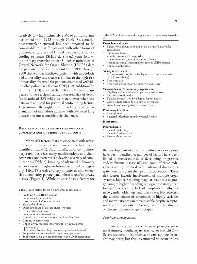
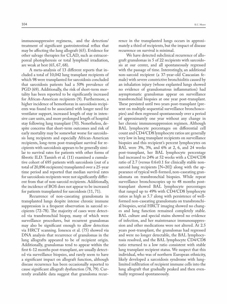
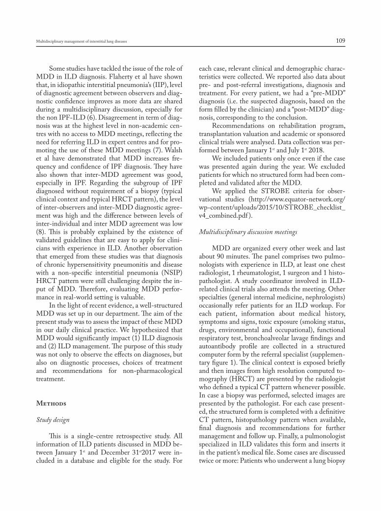
Many risk factors that are associated with worse outcomes in patients with sarcoidosis have been identified (Table 1). Additionally, advanced pulmo-nary sarcoidosis has many manifestations and char-acteristics, and patients can develop a variety of com-plications (Table 2). Imaging of advanced pulmonary sarcoidosis with high-resolution computed tomogra-phy (HRCT) reveals a variety of patterns with exten-sive adenopathy, parenchymal fibrosis, and/or airway disease (Figure 1). While no specific risk factors for
the development of advanced pulmonary sarcoidosis have been identified, a number of factors have been linked to increased risk of developing progressive and/or chronic disease (6), and some of these indi-viduals will go on to develop advanced disease de-spite non-transplant therapeutic interventions. These risk factors include involvement of multiple organ systems, higher Scadding stage at diagnosis or pro-gressing to higher Scadding radiographic stage, need for systemic therapy, lack of lymphadenopathy, fe-male gender, older age, and black race. Nonetheless, the clinical course of sarcoidosis is highly variable, and many patients can remain stable despite sympto-matic and/or persistent disease, even in the absence of chronic pharmacologic therapies.
Proximal airway disease
Sarcoidosis can involve the nasal passages, para-nasal sinuses, mouth, larynx, trachea, or bronchi (14). Severe stenosis of the trachea or cartilaginous bron-chi may occur, but this is estimated to occur in less
Table 1. Risk factors for worse outcomes in sarcoidosis
• ScaddingStageIII/IVdisease• Pulmonaryhypertension• Involvementof>3organsystems• Myocardialdisease• Olderage&ageofdiseaseonset>40years• AfricanAmericanrace• Presenceofneurosarcoidosis• Chronicrenaldysfunction(e.g.nephrocalcinosis)• Chronichypercalcemia• Upperairwaymucosalinvolvement(e.g.lupuspernio)• Splenomegaly• Skeletalinvolvement(e.g.extensivecysticbonelesions)• Progressiveand/orsustainedrespiratorysymptoms• Supplementaloxygenrequirement(especiallyifincreasing)
Table 2. Manifestations and complications of pulmonary sarcoido-sis
Parenchymal disease• Extensive/confluentgranulomatousdisease(e.g.alveolar
sarcoidosis)• Pulmonaryfibrosis -canbeextensive&progressive - most common cause of respiratory failure - can mimic usual interstitial pneumonia (UIP) pattern• Bullousemphysema
Airway involvement• Airflowobstruction(maydisplayreactivecomponentwith
partial reversibility)• Bronchiectasis• Bronchialstenosis(canbeextensiveandsevere)
Vascular disease & pulmonary hypertension• Capillaryobliterationduetoparenchymalfibrosis• Plexiformarteriopathy• Vascularcompressionbyenlargedlymphnodes• Cardiacdysfunctionduetocardiacsarcoidosis• Granulomatousangiitis(arterialorvenous)
Pulmonary infection• Aspergilloma• Bacterialinfectionslinkedtobronchiectasis
Hemoptysis
Pleural disease• Pleuralthickening• Pleuraleffusion(rare)• Pneumothorax(rare)
K.C. Meyer94
than one percent of patients (15). Although patients with extensive airway narrowing may be quite symp-tomatic, they usually do not have enough functional impairment to qualify them for lung transplant can-didacy.
Intrathoracic lymphadenopathy
Intrathoracic lymphadenopathy is observed in approximately 80% of patients during the course of their illness. Hilar adenopathy is bilateral in most cases, although unilateral hilar adenopathy can be seen in up to 5% of patients (16). Lymph node calci-fication can be seen at the time of diagnosis, and the likelihood of calcification increases later during the course of the disease (17).
Large nodules and alveolar consolidation
Nodules that follow a perilymphatic distribution and predominate in the mid to upper lung zones are seen in approximately 90% of patients (18). Sarcoid nodules can occasionally aggregate and form larger pulmonary nodules (up to 3 cm diameter) or large masses. Large nodules can remain stable for long pe-riods of time, show partial or complete regression, or cavitate. Massive consolidation may occur with coales-cent interstitial granulomas compressing alveoli (19).
Pulmonary fibrosis
Approximately 5% of patients have evidence of fibrotic changes on routine chest radiography at
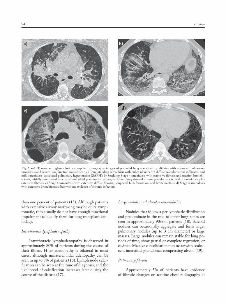
Fig. 1 a-d. Transverse high-resolution computed tomography images of potential lung transplant candidates with advanced pulmonary sarcoidosisandseverelungfunctionimpairment.a)Long-standingsarcoidosiswithbulkyadenopathy,diffusegranulomatousinfiltrates,andmild sarcoidosis-associated pulmonary hypertension (SAPH); b) Scadding Stage 4 sarcoidosis with extensive fibrosis and traction bronchi-ectasis,initiallyinterpretedasausualinterstitialpneumoniapattern;explantedlungshoweddiffusegranulomatatypicalofsarcoidosisplusextensivefibrosis;c)Stage4sarcoidosiswithextensivediffusefibrosis,peripheralblebformation,andbronchiectasis;d)Stage4sarcoidosiswith extensive bronchiectasis but without evidence of chronic infection.
a) b)
c) d)
Lung transplantation for sarcoidosis 95
presentation (5, 20). Fibrosis tends to predominate in the upper and mid lung regions, and conglomer-ate masses that surround and encompass vessels and bronchi with associated bronchial distortion is seen in over half of patients with fibrotic pulmonary sar-coidosis (21). Advanced fibrotic sarcoidosis is charac-terized by the presence of fibrotic cysts, bullae, trac-tion bronchiectasis, and paracicatricial emphysema, and cystic abnormalities are commonly seen in the upper lobes (22). Honeycomb change can be seen, and honeycomb-like cysts are usually found in an upper lobe distribution, but lower lobe honeycomb change that mimics a usual interstitial pneumonia (UIP) pattern that is typical of idiopathic pulmonary fibrosis (IPF) can also be seen (20, 23).
Pulmonary hypertension
The estimated prevalence of PH in sarcoidosis ranges from one to 28% (defined as mean pulmo-nary arterial pressure [mPAP] ≥25 mm Hg) at rest and as high as 43% if measured during exercise (24-28). Most patients with sarcoidosis-associated PH (SAPH) have radiographic changes of advanced dis-ease (Scadding stages III or IV), although extensive parenchymal abnormalities are not always present (24-26). One case series reported that 60% of patients with SAPH lacked evidence of significant fibrosis on chest radiography (27), and the extent of paren-chymal lung involvement may not correlate with the degreeofPHasreflectedbyrightheartcatheteriza-tion measurements. Up to 75% of patients who are
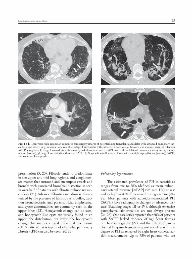
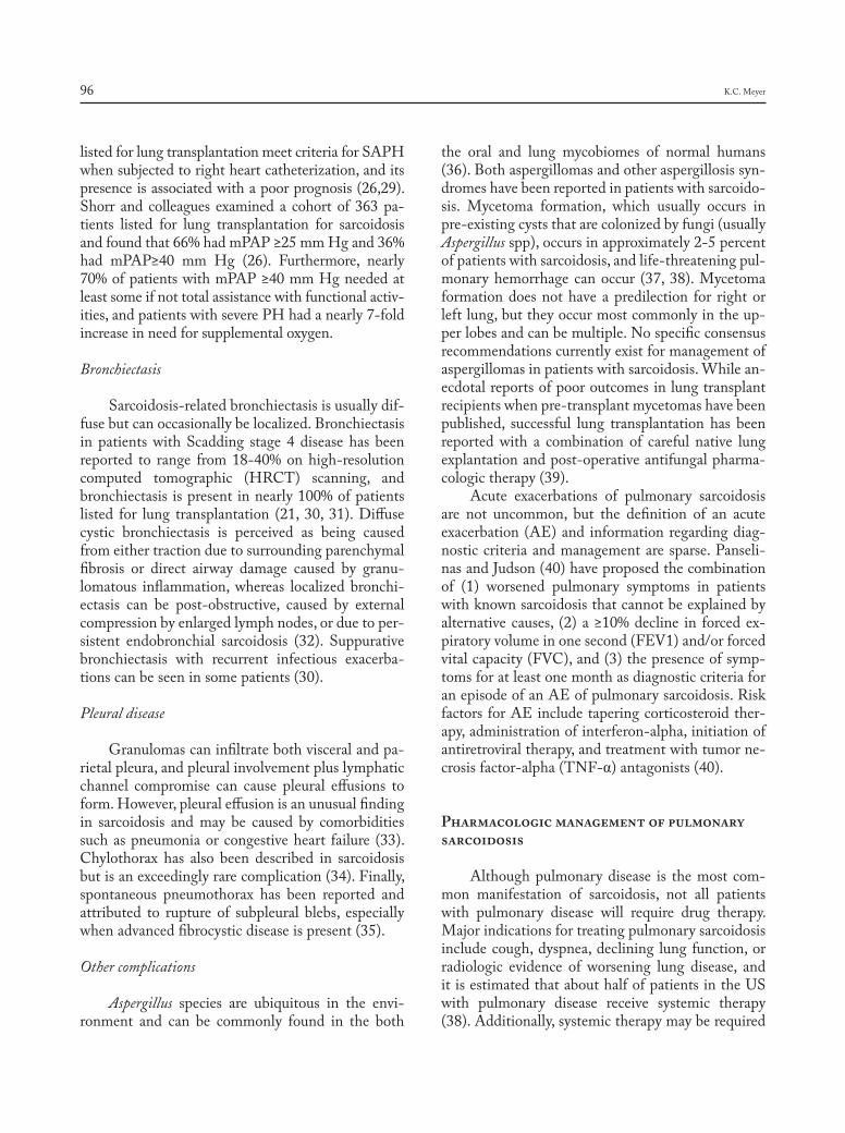
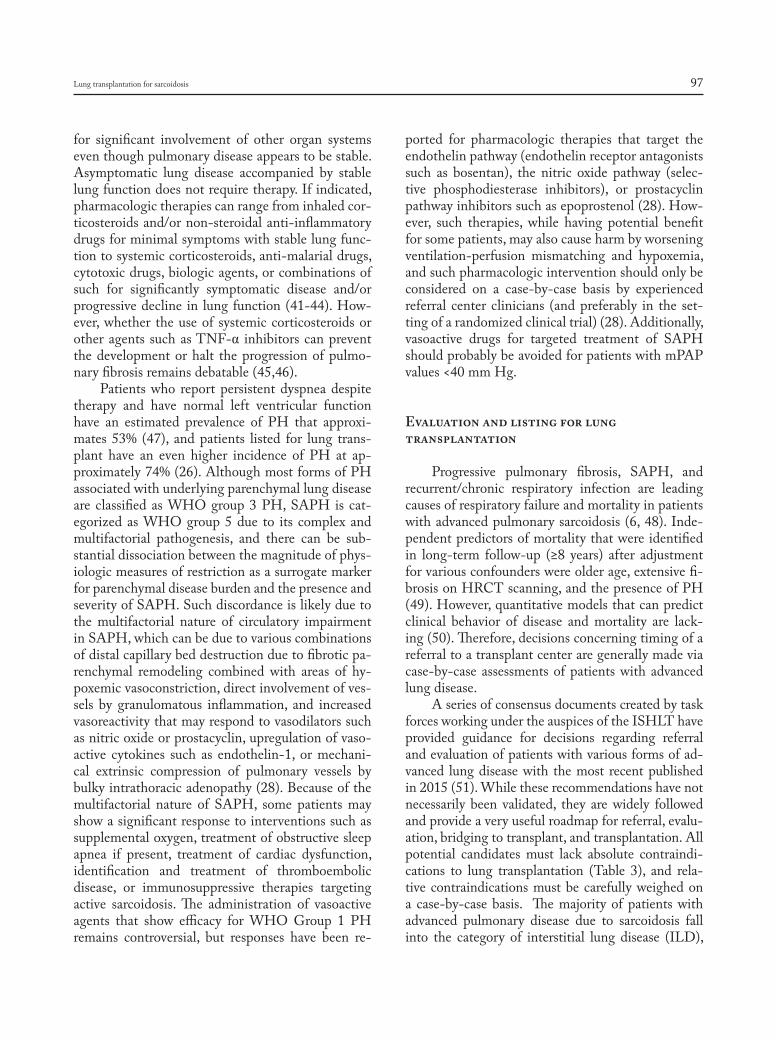
Fig. 1 e-h. Transverse high-resolution computed tomography images of potential lung transplant candidates with advanced pulmonary sar-coidosis and severe lung function impairment. e) Stage 4 sarcoidosis with extensive bronchiectasis (arrows) and chronic bacterial infection withP.aeruginosa;f )Stage4sarcoidosiswithparenchymalfibrosisandsevereSAPHwithdiffusebilateralpulmonaryarteryaneurysmfor-mation (arrows); g) Stage 3 sarcoidosis with severe SAPH; h) Stage 4 fibrobullous sarcoidosis with multiple aspergillomas (arrows), SAPH, and recurrent hemoptysis.
e) f )
g h)
K.C. Meyer96
listed for lung transplantation meet criteria for SAPH when subjected to right heart catheterization, and its presence is associated with a poor prognosis (26,29). Shorr and colleagues examined a cohort of 363 pa-tients listed for lung transplantation for sarcoidosis and found that 66% had mPAP ≥25 mm Hg and 36% had mPAP≥40 mm Hg (26). Furthermore, nearly 70% of patients with mPAP ≥40 mm Hg needed at least some if not total assistance with functional activ-ities, and patients with severe PH had a nearly 7-fold increase in need for supplemental oxygen.
Bronchiectasis
Sarcoidosis-related bronchiectasis is usually dif-fusebutcanoccasionallybelocalized.Bronchiectasisin patients with Scadding stage 4 disease has been reported to range from 18-40% on high-resolution computed tomographic (HRCT) scanning, and bronchiectasis is present in nearly 100% of patients listed for lung transplantation (21,30,31).Diffusecystic bronchiectasis is perceived as being caused from either traction due to surrounding parenchymal fibrosis or direct airway damage caused by granu-lomatous inflammation,whereas localizedbronchi-ectasis can be post-obstructive, caused by external compression by enlarged lymph nodes, or due to per-sistent endobronchial sarcoidosis (32). Suppurative bronchiectasis with recurrent infectious exacerba-tions can be seen in some patients (30).
Pleural disease
Granulomas can infiltrate both visceral and pa-rietal pleura, and pleural involvement plus lymphatic channel compromise can cause pleural effusions toform.However,pleuraleffusionisanunusualfindingin sarcoidosis and may be caused by comorbidities such as pneumonia or congestive heart failure (33). Chylothorax has also been described in sarcoidosis but is an exceedingly rare complication (34). Finally, spontaneous pneumothorax has been reported and attributed to rupture of subpleural blebs, especially when advanced fibrocystic disease is present (35).
Other complications
Aspergillus species are ubiquitous in the envi-ronment and can be commonly found in the both
the oral and lung mycobiomes of normal humans (36).Bothaspergillomasandotheraspergillosissyn-dromes have been reported in patients with sarcoido-sis. Mycetoma formation, which usually occurs in pre-existing cysts that are colonized by fungi (usually Aspergillus spp), occurs in approximately 2-5 percent of patients with sarcoidosis, and life-threatening pul-monary hemorrhage can occur (37, 38). Mycetoma formation does not have a predilection for right or left lung, but they occur most commonly in the up-per lobes and can be multiple. No specific consensus recommendations currently exist for management of aspergillomas in patients with sarcoidosis. While an-ecdotal reports of poor outcomes in lung transplant recipients when pre-transplant mycetomas have been published, successful lung transplantation has been reported with a combination of careful native lung explantation and post-operative antifungal pharma-cologic therapy (39).
Acute exacerbations of pulmonary sarcoidosis are not uncommon, but the definition of an acute exacerbation (AE) and information regarding diag-nostic criteria and management are sparse. Panseli-nas and Judson (40) have proposed the combination of (1) worsened pulmonary symptoms in patients with known sarcoidosis that cannot be explained by alternative causes, (2) a ≥10% decline in forced ex-piratory volume in one second (FEV1) and/or forced vital capacity (FVC), and (3) the presence of symp-toms for at least one month as diagnostic criteria for an episode of an AE of pulmonary sarcoidosis. Risk factors for AE include tapering corticosteroid ther-apy, administration of interferon-alpha, initiation of antiretroviral therapy, and treatment with tumor ne-crosis factor-alpha (TNF-α) antagonists (40).
Pharmacologic management of pulmonarysarcoidosis
Although pulmonary disease is the most com-mon manifestation of sarcoidosis, not all patients with pulmonary disease will require drug therapy. Major indications for treating pulmonary sarcoidosis include cough, dyspnea, declining lung function, or radiologic evidence of worsening lung disease, and it is estimated that about half of patients in the US with pulmonary disease receive systemic therapy (38). Additionally, systemic therapy may be required
Lung transplantation for sarcoidosis 97
for significant involvement of other organ systems even though pulmonary disease appears to be stable. Asymptomatic lung disease accompanied by stable lung function does not require therapy. If indicated, pharmacologic therapies can range from inhaled cor-ticosteroidsand/ornon-steroidalanti-inflammatorydrugs for minimal symptoms with stable lung func-tion to systemic corticosteroids, anti-malarial drugs, cytotoxic drugs, biologic agents, or combinations of such for significantly symptomatic disease and/or progressive decline in lung function (41-44). How-ever, whether the use of systemic corticosteroids or other agents such as TNF-α inhibitors can prevent the development or halt the progression of pulmo-nary fibrosis remains debatable (45,46).
Patients who report persistent dyspnea despite therapy and have normal left ventricular function have an estimated prevalence of PH that approxi-mates 53% (47), and patients listed for lung trans-plant have an even higher incidence of PH at ap-proximately 74% (26). Although most forms of PH associated with underlying parenchymal lung disease are classified as WHO group 3 PH, SAPH is cat-egorized as WHO group 5 due to its complex and multifactorial pathogenesis, and there can be sub-stantial dissociation between the magnitude of phys-iologic measures of restriction as a surrogate marker for parenchymal disease burden and the presence and severity of SAPH. Such discordance is likely due to the multifactorial nature of circulatory impairment in SAPH, which can be due to various combinations of distal capillary bed destruction due to fibrotic pa-renchymal remodeling combined with areas of hy-poxemic vasoconstriction, direct involvement of ves-sels by granulomatous inflammation, and increasedvasoreactivity that may respond to vasodilators such as nitric oxide or prostacyclin, upregulation of vaso-active cytokines such as endothelin-1, or mechani-cal extrinsic compression of pulmonary vessels by bulkyintrathoracicadenopathy(28).Becauseofthemultifactorial nature of SAPH, some patients may show a significant response to interventions such as supplemental oxygen, treatment of obstructive sleep apnea if present, treatment of cardiac dysfunction, identification and treatment of thromboembolic disease, or immunosuppressive therapies targeting active sarcoidosis. The administration of vasoactive agents that show efficacy for WHO Group 1 PH remains controversial, but responses have been re-
ported for pharmacologic therapies that target the endothelin pathway (endothelin receptor antagonists such as bosentan), the nitric oxide pathway (selec-tive phosphodiesterase inhibitors), or prostacyclin pathway inhibitors such as epoprostenol (28). How-ever, such therapies, while having potential benefit for some patients, may also cause harm by worsening ventilation-perfusion mismatching and hypoxemia, and such pharmacologic intervention should only be considered on a case-by-case basis by experienced referral center clinicians (and preferably in the set-ting of a randomized clinical trial) (28). Additionally, vasoactive drugs for targeted treatment of SAPH should probably be avoided for patients with mPAP values <40 mm Hg.
Evaluation and listing for lung transplantation
Progressive pulmonary fibrosis, SAPH, and recurrent/chronic respiratory infection are leading causes of respiratory failure and mortality in patients with advanced pulmonary sarcoidosis (6, 48). Inde-pendent predictors of mortality that were identified in long-term follow-up (≥8 years) after adjustment for various confounders were older age, extensive fi-brosis on HRCT scanning, and the presence of PH (49). However, quantitative models that can predict clinical behavior of disease and mortality are lack-ing (50). Therefore, decisions concerning timing of a referral to a transplant center are generally made via case-by-case assessments of patients with advanced lung disease.
A series of consensus documents created by task forces working under the auspices of the ISHLT have provided guidance for decisions regarding referral and evaluation of patients with various forms of ad-vanced lung disease with the most recent published in 2015 (51). While these recommendations have not necessarily been validated, they are widely followed and provide a very useful roadmap for referral, evalu-ation, bridging to transplant, and transplantation. All potential candidates must lack absolute contraindi-cations to lung transplantation (Table 3), and rela-tive contraindications must be carefully weighed on a case-by-case basis. The majority of patients with advanced pulmonary disease due to sarcoidosis fall into the category of interstitial lung disease (ILD),

K.C. Meyer98
for which guidance for timing of referral and timing of placing on the transplant waitlist are provided (Ta-ble 4). However, some patients with sarcoidosis may have predominantly vascular involvement but lack extensive pulmonary fibrosis, and ISHLT recommen-dations provided for pulmonary vascular diseases may bemoreappropriateforsuchpatients(Table4).Be-cause waitlist mortality is quite high among patients with pulmonary fibrosis, timely referral for transplant evaluation is essential for patients who have severe disease despite maximal therapy and wish to be con-sidered for lung transplantation.
Evaluation of potential candidates for lung transplant should include (1) an objective determi-nation of disease severity, (2) elucidation of the na-ture of disease characteristics that are causing the pa-tient’s symptoms, (3) a determination of whether the benefits (prolonged survival, improved quality of life)
of undergoing lung transplantation clearly outweigh the risks associated with receiving a lung transplant (Table 5). Many patients will unfortunately not be eligible for lung transplantation due to the presence of an absolute contraindication or combinations of relative contraindications and comorbidities that make the possibility of achieving a successful trans-plant unlikely (e.g. severe corticosteroid-induced diabetes and obesity). On the other hand, some pa-tients with sarcoidosis can be very symptomatic from their disease yet not have enough physiologic impair-ment to receive a high enough lung allocation score (LAS) value (for countries that use a LAS system to prioritize transplant candidates) to have a reasonable chanceof receivingadonor lungoffer ifplacedonthe waitlist.
The LAS system (Table 6) was implemented in the US in 2005 with the goals of (1) balancing the
Table 3. Contraindications to lung transplantation
Absolute• Recenthistoryofmalignancy(2-yeardisease-freeintervaliflowriskofrecurrence;5-yearintervalforhigherrisk;riskberemaintoo
high for some cancers beyond 5 years)• Severelylimitedfunctionalcapacitywithpoorrehabilitationpotential• Significantdysfunctionofothermajororgansystems(unlessmultiplecombinedorgantransplantationisfeasible)• Acutemedicalinstability(e.g.acutemyocardialinfarction,sepsis,hepaticfailure)• Uncorrectablebleedingdiathesis• Significant,uncorrectedatheroscleroticdiseasewithsuspected/confirmeddysfunction(orischemiaorsignificantcoronaryarterydisease
that cannot be revascularized)• Chronicinfectionwithhighlyvirulentand/orantibiotic-resistantmicrobeswithpoorcontrolpre-transplant• ActiveinfectionwithMycobacterium tuberculosis• Chestwallorspinaldeformitythatwouldcausesevereventilatorrestrictionpost-transplant• Bodymassindex(BMI)≥35kg/m2 (Class II/III obesity)• Non-adherencetorecommendedmedicaltherapies• Psychiatric/psychologicconditionscausinginabilitytocooperatewithhealthcareteaminteractionsand/oradherencetocomplex
medical therapies• Lackofanadequateand/orreliablesocialsupportsystem• Substanceabuseordependence(mustdemonstratemeaningful/persistentriskreductionbehaviorsandverifiedabstinencefrom
substances of concern (e.g. tobacco, alcohol, marijuana, or other illicit substances))
Relative• Age>65yearsifotherrelativecontraindicationsarepresentofphysiologicreserveissignificantlyimpaired• Malnutritionifprogressiveorsevere• Osteoporosisifsevere,symptomatic• BMI30.0-34.9kg/m2 (Class I obesity, especially if truncal/central obesity)• Extensivepriorthoracicsurgery(e.g.lungresection)• Receivingmechanicalventilation• Receivingextracorporeallifesupport• HepatitisBand/orCinfection• Humanimmunodeficiencyvirusinfection• Colonization/infectionwithhighlyvirulentand/orantibiotic-resistantbacteriaorfungi• Significantatheroscleroticdiseaseburden• Othersignificantmedicalconditions(e.g.diabetesmellitus,systemichypertension,gastroesophagealreflux)thathavenotcaused
advanced organ system damage (especially if not optimally treated/controlled)
Lung transplantation for sarcoidosis 99
urgency of need for transplantation due risk of death without receiving a transplant with the likelihood of an acceptable outcome following transplantation, (2) optimally placing organs according to LAS values combined with matching characteristics of potential recipients (e.g. blood type, thoracic cage dimensions), and (3) reducing the number of candidates on trans-plant center waitlists who die without the oppor-tunity to undergo a transplant (52,53). The LAS is weighted more by transplant urgency than likelihood of surviving for at least one year post-transplant, but its successful aspects have led to its adoption by a numberofcountriesoutsideoftheUS.Becausepa-tients with ILD (especially those with IPF) tend to have higher LAS values than candidates with other disease indications, the total number of transplants for ILD (mostly IPF) in the US surpassed that for other indications (e.g. chronic obstructive pulmonary disease, cystic fibrosis, PH due to pulmonary vascular disease) in 2007, and IPF is the leading indication for lung transplantation in the US at present (54). The major indications for lung transplantation for sarcoidosis are advanced fibrotic lung disease, severe pulmonary hypertension, or a combination of both.
A key question when evaluating a patient with sarcoidosis for potential lung transplantation
is whether their lung disease has been adequately treated. Our center has had candidates who were us-ing supplemental oxygen and very incapacitated but improvedmarkedlyandwereabletobeweanedoffsupplemental oxygen and achieve acceptable qual-ity of life when placed on adequate pharmacother-apy. Another key question is whether significant ex-trapulmonary sarcoidosis is present that may have an impact on post-transplant outcome, and appropriate screening should be performed to detect significant left ventricular dysfunction or sustained ventricular dysrhythmias.
Prior to placement on a transplant waitlist, co-morbidities should be aggressively and optimally managed. This includes angioplasty and/or stent placement for coronary artery disease if needed, anti-resorptive therapies to reduce fracture risk if osteoporosis is present, joining a weight loss pro-gram if overweight, and medical treatment of sys-temic hypertension or diabetes mellitus. Becausedyspnea may limit physical activity and promote de-conditioning, pulmonary rehabilitation with physi-cal training and breathing exercises should be pre-scribed, and pulmonary rehabilitation programs can provide educational and psychological support and optimize exercise tolerance and functional status
Table 4. Guidelines for timing of referral for potential lung transplantation
Referral to a transplant center (all patients with ILD)*• Impairedlungfunction - FVC <80% predicted - DLCO <40% predicted• Anydyspneaorfunctionallimitationduetolungdisease• Anyrequirementforsupplementaloxygen(evenifonlyrequiredduringexertion)• Failuretoimprovedyspnea,reduce/eliminaterequirementforsupplementaloxygen,and/orimprovelungfunctionwithaclinically
indicatedtrialofmedicaltherapyifinflammatoryILDispresent
Referral to a transplant center (all patients with PVD)* • NYHAFunctionalClassIIIorIVsymptomsdespiteescalatingtherapy• Rapidlyprogressivedisease(ruleoutbodyweightorrehabilitationconcerns)• UseofparenteraltargetedvasoactivetherapyregardlessofsymptomsorNYHAFunctionalClass
Suggested timing of referral to a transplant center for patients with sarcoidosis• Dyspneaorfunctionallimitationduetolungdisease• Significantlyimpairedlungfunction(e.g.FVC<80%predicted,DLCO<40%predicted)• Requirementforuseofsupplementaloxygen• EvidenceofSAPH• NYHAFunctionalClassIIIorIVsymptoms• Rapidlyprogressivedisease• Lackofresponsetoclinicallyindicatedpharmacologictherapies• Life-threateningcomplicationsofsuppurativebronchiectasis(e.g.episodeofrespiratoryfailurerequiringnon-invasiveventilation,poor
clinical recovery from exacerbations and/or increasing antibiotic resistance, pneumothorax, life-threatening hemoptysis)
K.C. Meyer100
prior to transplantation.Becauseof theprolongedand variable disease course for patients with sar-coidosis, the decision as to when to proceed with transplantation is challenging, even for experienced clinicians at referral/transplant centers. Guidance for timing the placement of lung transplant candi-dates on the waitlist has been provided by the ISH-LT (Table 7).
Surgical considerations
Previous thoracic surgical procedures are gen-erally not a contraindication to performing a lung transplant, but higher risk of hemorrhage, increased need for chest re-exploration, and renal dysfunction can be encountered in patients who have had previ-ous chest surgical procedures, especially if prolonged
Table 5. Evaluation of potential lung transplant candidates with sarcoidosis
Disease-specific considerations for patients with sarcoidosis• Isthediseaseadequatelytreated/managed?• Doestheriskofdeathfromthediseaseclearlyoutweighrisksassociatedwithlungtransplantation?• Issignificantinvolvementofotherorgansystemspresent?• Ischronicbacterialinfectionassociatedwithbronchiectasispresent?• Doesthepatienthavesarcoidosis-associatedpulmonaryhypertension?• Isfungaldiseaseanissue(especiallymycetomasduetoAspergillusspp)?
Evaluation and testing • Carefulphysicalexamination -Isdiaphragmaticmovementimpaired? -Isaxialskeletonandespeciallychestwallmobilitysignificantlyimpaired? -Isthereevidenceofsystemicdiseasewithsignificantorgansysteminvolvement(cardiac,nervoussystem,liver,spleen,skin)?• Thoracicimagingstudies - HRCT, routine chest x-ray - Quantitative nuclear medicine ventilation/perfusion scan -Bariumesophagram(?Esophagealdysfunction,significantreflux)• Lungfunctionassessment - Spirometry - Lung volumes -SinglebreathdiffusioncapacityforCO(DLCO) - Paranasal sinus imaging if pertinent• 6-minutewalktest - Walk distance - Oxyhemoglobin saturation at rest and with exertion - Quantification of supplemental oxygen requirements if significant desaturation present• Cardiacevaluation - Electrocardiogram - Echocardiography with bubble study to detect possible intracardiac shunt -Left&rightheartcatheterization - Ambulatory electrocardiography (e.g. 24-72 hrs to rule out significant dysrhythmia)• Laboratorytesting(completebloodcountwithdifferential,BUN,creatinine,electrolytes,liverfunctiontesting,fastinglipidprofile,viral
serologies[HIV,HBsAg,HBsAb,HCV,CMV,HSV,EBV,VZV],toxoplasmaandaspergillusantibodies,typeandscreen[bloodgroupandRhtype],prostate-specificantigen[males>40yearsofage],panelreactiveantibodytesting,anti-HLAantibodyscreening,urinalysis
• Bonedensitometry• PPDtesting• ScreeningforAspergillus (sputum culture, serum precipitins)• Ifsignificant/suppurativebronchiectasisispresent: - Sputum bacterial culture and sensitivities - Screen for non-tuberculous mycobacteria• Age-andgender-appropriatecancerscreening• Consultations: - Psychosocial evaluations - Nutritionist evaluation - Rehabilitation medicine - Dental evaluation - Others as indicated (e.g. ophthalmologic)
Lung transplantation for sarcoidosis 101
cardiopulmonary bypass times are required. The de-cision of whether to perform a single, bilateral, or heart-lung transplant involves consideration of the
nature of lung involvement and extent of physiologic impairment, whether significant extrapulmonary disease is an issue, what comorbid conditions are pre-
Table 6. Values/factors* used to calculate the lung allocation score**
• Lungdiagnosiscode• Age(years)• Bodymassindex(BMI)• Functionalstatus• Forcedvitalcapacity(FVC)percentpredicted• Requirementforsupplementaloxygen• 6-minutewalkdistance(feet)• Pulmonaryarterysystolicpressure(mmHg)• Meanpulmonaryarterypressure(mPAP;mmHg)• Cardiacindex(CI)inL/min/m2
• Centralvenouspressure(mmHg)• Ventilationstatus• pCO2 (current, highest, lowest) mm Hg• Presenceofdiabetes• Serumcreatinine(current,highest,lowest)inmg/dL• Totalbilirubin(current,highest,lowest)inmg/dL
* Some values are adjusted according to Disease Group (A-D); sarcoidosis is classified as Group A if mPAP is ≤30 mm Hg but switches to GroupDifmPAPis>30mmHg.**TheLAScalculationincorporatesthreedifferentmeasures(waitinglisturgency,post-transplantsurvival,andtransplantbenefit)toderivea Raw Allocation Score that is then normalized on a continuous scale of 0 to 100.For additional information see concerning LAS components and calculations see https://optn.transplant.hrsa.gov/media/1200/optn_poli-cies.pdf#nameddest=Policy_10. Organ Procurement and Transplantation Network Policies; Policy 10: Allocation of Lungs. Date accessed, 1/26/18.
Table 7. Guidelines for timing of waitlist placement for transplant candidates
Timing of placing a patient on the lung transplant waitlist (all patients with ILD)*• DeclineinFVC≥10%duringa6-monthfollow-upperiod(alesserdegreeofdeclinehasbeenassociatedwithapoorerprognosisand
may call for earlier listing) • DeclineinDLCO≥15%duringa6-monthfollow-upperiod• Oxyhemoglobindesaturationto<88%or6-MWTdistance<250metersor>50meterdeclinein6-MWTdistanceovera6-month
period
Timing of placing a patient on the lung transplant waitlist (all patients with PVD)*• NYHAFunctionalClassIIIorIVsymptomsdespite3monthsofcombinationvasoactivetherapies(includingprostanoids)• Cardiacindex<2L/min/m2
• Meanrightatrialpressure>15mmHg• 6-MWTdistance<350meters• Significanthemoptysis,pericardialeffusion,orprogressiverightheartfailure(asevidencedbyrenaldysfunction,increasingserum
bilirubin,increasingserumBNP,orrecurrentascites)
Suggested timing of waitlist placement for patients with sarcoidosis • DeclineinFVC≥10%duringa6-monthfollow-upperiod(alesserdegreeofdeclinehasbeenassociatedwithapoorerprognosisand
may call for earlier listing) • DeclineinDLCO≥15%duringa6-monthfollow-upperiod• Oxyhemoglobindesaturationto<88%or6-MWTdistance<250metersor>50meterdeclinein6-MWTdistanceovera6-month
period • NYHAFunctionalClassIIIorIVsymptomsdespite3monthsofcombinationvasoactivetherapies(includingprostanoids)• Cardiacindex<2L/min/m2
• Meanrightatrialpressure>15mmHg• 6-MWTdistance<350meters• Significanthemoptysis,pericardialeffusion,orprogressiverightheartfailure(asevidencedbyrenaldysfunction,increasingserum
bilirubin,increasingserumBNP,orrecurrentascites)
K.C. Meyer102
sent, and the likelihood of procuring a donor organ that matches a candidate’s thoracic cage dimensions andABObloodgroupstatus.Explantinglungsfrompatients with advanced sarcoidosis can be extremely challenging due to pleural adhesions and perihilar fibrosis, and substantial intraoperative bleeding is more likely to occur if resection of the native lung(s) proves to be difficult (55).
The presence of one or more mycetomas, es-pecially if abutting the pleura, increases the risk of seeding the pleural spaces during explantation. The risk and degree of pleural bleeding (especially if pa-tients require cardiopulmonary bypass) is likely to be increased, and a prolonged dissection to explant the native lungs may significantly increase donor lung cold ischemic time, thereby increasing the risk of significant reperfusion injury. One case series report-ed that post-transplant outcomes were significantly worse for patients with mycetomas (56), but aggres-sive pre-transplant antifungal therapy and prolonged post-transplant prophylaxis may successfully prevent post-transplant Aspergillus infection (57). Addition-ally, irrigation of the pleural space with an anti-fun-galagent(e.g.amphotericinB)whendonorlungsareimplanted should be considered. Patients with myce-tomas, even if apparently unilateral, should only be listedforbilaterallungtransplant(BLT).
If suppurative bronchiectasis is present, spu-tum cultures should be obtained as native lungs are explanted to identify all infecting organisms and their sensitivities to antibiotics. Peri-operative and post-operative antibiotics should be administered accordingtocultureandsensitivityresults.Becausea bronchiectatic native lung can serve as a reservoir of infection that places a transplanted single lung at risk for post-transplant infection, BLT is the pre-ferred approach for patients with bronchiectasis and chronic suppurative infection.
Bilateraltransplantmayalsobeabetterchoicethan single lung transplant (SLT) for patients with significant SAPH, although recipients can do well with SLT despite the presence of PH with mPAP values greater than 40 mm Hg (58). Indeed, a SLT may be a reasonable choice for patients in whom BLT is not required, and listing for SLTmay im-provechancesforadonororganofferandreduceriskof dying on the waitlist (59). A heart-lung transplant can be considered for patients with significant left ventricular dysfunction or cardiac dysrhythmias.
Post-transplant management, complications, and outcomes
Post-transplant management is multi-faceted and complicated, yet few randomized, prospec-tive controlled trials are available to provide robust evidence for optimal recipient management. Peri-operative care in the ICU requires both ventilator and circulatory support, and early surgical and/or medical complications must be promptly identified and addressed. Protocols should be in place to fa-cilitate prevention of infections (e.g. prophylaxis for cytomegalovirus and Pneumocystis jiroveci) as well as protocols to rapidly identify and treat infectious complications that may develop. Immunosuppressive regimens typically consist of pulse corticosteroid and an induction agent given at the time of surgery, and maintenance immunosuppression with a calcineurin inhibitor (tacrolimus or cyclosporine A), anti-metab-olite (usually mycophenolate or azathioprine), and a corticosteroid that is gradually weaned to a low dose.
Transplant recipients are at risk for a multitude of immediate, acute, and subacute/chronic complica-tions following successful transplantation (Table 8) (60,61). Approximately one third of lung transplant recipients will develop grade 3 primary graft dys-function (PGD) (62), but while a number of markers have been identified that correlate with increased risk of high-grade PGD (63), interventions other than providing supportive care have been relatively inef-fective in preventing or treating PGD.
Acute/subacute complications include anti-body-mediated rejection, acute cellular rejection, lymphocytic bronchiolitis, and infection. Becauseup to 90% of recipients have pre-formed anti-HLA antibodies of which approximately one third are donor-specificantibodies(DSAs)(64),effectiveandcarefully monitored immune suppression is essential to establish allograft immune tolerance. Monitoring recipients for evidence of lung function decline as well as monitoring for the appearance of numerous complications and co-morbidities is essential to op-timize post-transplant allograft function and recipi-ent quality of life and survival. Many centers subject recipients to surveillance bronchoscopy with BALandtransbronchialbiopsies(TBBs)atprotocol-de-termined intervals to detect occult infection and/or evidence of rejection, although other centers perform few if any protocol-driven surveillance bronchos-
Lung transplantation for sarcoidosis 103
copies and may only perform such when clinically indicated by deterioration in lung function with the suspicion that infection or allograft rejection may be the cause. Consensus guidelines for using or not us-ing post-transplant surveillance bronchoscopies have not yet become available.
For lung transplant recipients who survive be-yond the first year post-transplant, the development of chronic lung allograft dysfunction (CLAD) is the greatest threat to long-term allograft and recipient survival (65, 66). The ISHLT/American Thoracic Society (ATS)/European Respiratory Society (ERS) clinical practice guideline systematically examined available evidence for the prevention and treatment ofBOS/CLADandprovidedrecommendationsfor
thediagnosisandtreatmentofBOS/CLAD.Iden-tified risk factors included PGD, various forms of alloimmune rejection (acute cellular rejection, anti-body-mediated rejection, lymphocytic bronchiolitis), infections (viral, bacterial, fungal), pathologic GER, autoimmunity, and persistent bronchoalveolar lav-age (BAL) neutrophilia. Although evidence fromrandomized controlled trials (RCTs) for preventing and treating BOS/CLADwas found to be of lowor very low quality, a number of conditional recom-mendations were made by consensus among task force members following a comprehensive review of available publications. These include ruling out other causes of delayed, persistent allograft function decline, administering azithromycin, adjustment of
Table 8. Complications of lung transplantation
Pulmonary complicationsLung allograft complications• Primarygraftdysfunction(PGD) - Rejection (hyperacute, acute, chronic) - Anastomosis dysfunction (dehiscence, malacia, stricture)• Phrenicnervedysfunction• Chroniclungallograftdysfunction(CLAD) -Bronchiolitisobliteranssyndrome(obstructiveCLAD) - Restrictive allograft syndrome (restrictive CLAD)• Diseaserecurrence(e.g.sarcoidosis)• ComplicationsofbronchoscopywithtransbronchialbiopsyLung allograft and/or native lung complications• Infection(bacterial,fungal,viral)• Pleuralcomplications(empyema,effusion,hemothorax,fistula)• Pulmonaryembolicdisease• Malignancy(primarylungcancer,post-transplantlymphoproliferativedisease[PTLD])Native lung complications (single lung transplant recipients)• Hyperinflation(emphysematouslung)• Reactivatedand/orrefractoryinfection
Extrapulmonary/systemic complications (may impact lung function)• Adversedrugreactions(e.g.immunosuppressantsideeffects,drug-druginteractions)• Renaldysfunction• Infection(e.g.wound,sepsis)• Metabolic/endocrine - Hyperglycemia, diabetes, obesity - Electrolyte abnormalities - Dyslipidemia• Cardiovascular(e.g.systemichypertension,cardiacrhythmdisturbance,infarction)• Thromboembolism• Hematologic(anemia,leukopenia,thrombocytopenia,thromboticmicroangiopathy)• Gastrointestinal -GERD(canaffectlungallograft) -Biliarytractdisease - Gastroparesis, other bowel disorders• Musculoskeletal(osteopenia,osteoporosis,myopathy)• Neurologic - Tremor, headache, seizure, memory loss - Cerebrovascular accident, blindness, coma• Malignancy(skin,primarylungcancer,PTLD)
K.C. Meyer104
immunosuppressive regimens, and the detection/treatment of significant gastrointestinal reflux thatmaybeaffectingthelungallograft(65).Evidenceforother salvage therapies for CLAD, such as extracor-poreal photopheresis or total lymphoid irradiation, are weak at best (65, 67, 68).
Ameta-analysisof13differentreportsthatin-cluded a total of 10,042 lung transplant recipients of which 98 were transplanted for sarcoidosis concluded that sarcoidosis patients had a 50% prevalence of PGD (69). Additionally, the risk of short-term mor-tality has been reported to be significantly increased for African-American recipients (9). Furthermore, a higher incidence of hemothorax in sarcoidosis recipi-ents was found to be associated with longer need for ventilator support, increased length of stay in inten-sive care units, and more prolonged length of hospital stay following lung transplant (70). Nonetheless, de-spite concerns that short-term outcomes and risk of early mortality may be somewhat worse for sarcoido-sis lung recipients and especially African-American recipients, long-term post-transplant survival for re-cipients with sarcoidosis appears to be generally simi-lar to survival rates for patients with other forms of fibrotic ILD. Tamieh et al. (11) examined a cumula-tive cohort of 695 patients with sarcoidosis (out of a total of 20,896 recipients) transplanted over a 25-year time period and reported that median survival rates forsarcoidosisrecipientswerenotsignificantlydiffer-ent from that of non-sarcoid recipients. Additionally, theincidenceofBOSdoesnotappeartobeincreasedfor patients transplanted for sarcoidosis (11, 71).
Recurrence of non-caseating granulomata in transplanted lungs despite intense chronic immune suppression is a frequent observation in sarcoid re-cipients (72-78). The majority of cases were detect-ed via transbronchial biopsy, many of which were surveillance procedures, but recurrent granulomas may also be significant enough to allow detection via HRCT scanning. Ionescu et al. (75) showed via DNA analysis that recurrence of granulomas in the lung allografts appeared to be of recipient origin. Additionally, granulomas tend to appear within the first 6-12 months post-transplant, are usually detect-ed via surveillance biopsies, and rarely seem to have a significant impact on allograft function, although disease recurrence has been occasionally reported to cause significant allograft dysfunction (78, 79). Cur-rently available data suggest that granuloma recur-
rence in the transplanted lungs occurs in approxi-mately a third of recipients, but the impact of disease recurrence on survival is minimal.
We have detected subclinical recurrence of allo-graft granulomas in 5 of 22 recipients with sarcoido-sis at our center, and all spontaneously regressed with the passage of time. Interestingly, an additional non-sarcoid recipient (a 37-year-old Caucasian fe-male) with severe constrictive bronchiolitis caused by an inhalation injury (whose explanted lungs showed no evidence of granulomatous inflammation) hadasymptomatic granulomas appear on surveillance transbronchial biopsies at one year post-transplant. These persisted until two years post-transplant (pre-sent on multiple sequential surveillance bronchosco-pies) and then regressed spontaneously over a period of approximately one year without any change in her chronic immunosuppression regimen. Although BAL lymphocyte percentages on differential cellcount and CD4/CD8 lymphocyte ratios are generally very low in lung transplant recipients on surveillance biopsies and this recipient’s percent lymphocytes on BALwere 3%, 3%, and 6% at 2, 6, and 24weekspost-transplant, her BAL lymphocyte percentagehad increased to 24% at 52 weeks with a CD4/CD8 ratio of 2.7 (versus 0.6±0.1 for clinically stable non-sarcoid lung recipients [N=20]) along with the ap-pearance of typical well-formed, non-caseating gran-ulomata on transbronchial biopsies. While repeat surveillance bronchoscopies up to 48 months post-transplant showed BAL lymphocyte percentagesthat ranged up to 49% with CD4/CD8 lymphocyte ratios as high as 5.7 along with persistence of well-formed non-caseating granulomata on transbronchi-al biopsies, serial HRCT imaging showed no chang-es and lung function remained completely stable. BALcultureandspecialstainsshowednoevidenceof infection, and her maintenance immunosuppres-sion and other medications were not altered. At 2.5 years post-transplant, the granulomas had regressed andwerenolongerdetectable,theBALlymphocy-tosisresolved,andtheBALlymphocyteCD4/CD8ratio returned to a low ratio consistent with stable lung transplant recipient status. We suspect that this individual, who was of northern European ethnicity, likely developed a sarcoidosis syndrome with lung-limited infiltration of recipient immune cells into the lung allograft that gradually peaked and then even-tually regressed spontaneously.
Lung transplantation for sarcoidosis 105
Key Points
1. A small number of patients diagnosed with sar-coidosis develop advanced lung disease.
2. Advanced pulmonary disease phenotypes include extensive pulmonary fibrosis, pulmonary hyper-tension, and purulent bronchiectasis.
3. Lung transplantation is an appropriate treatment for sarcoidosis patients with advanced lung dis-ease that progresses to respiratory insufficiency despite other therapies.
4. Post-transplant survival is generally similar to that of recipients with other transplant indica-tions such as IPF.
5. Although bilateral lung transplantation is gener-ally a preferred procedure, single lung transplant may be an appropriate procedure for patients without complications of their lung disease such as purulent bronchiectasis, chronic fungal infec-tion, or severe pulmonary hypertension.
6. Although recurrence of granulomas in trans-planted lungs may occur, this rarely has a signifi-cant impact on lung allograft function or recipi-ent survival.
Acknowledgment
Supported in part by the George and Julie Mosher Pulmonary Research Fund.
Financial/nonfinancial disclosures:Within the past 3 years Dr. Meyer has been an investigator in clinicaltrialssponsoredbyBoehringer-Ingelheim,BristolMeyersSquibb, Genentech, National Institutes of Health, Nivalis, Parion, Promedior, Roche, and Vertex. Dr. Meyer does not report any other relevant affiliations or financial involvement with any organization orentitywithafinancialinterestinorfinancialconflictwiththesubject matter or materials discussed in this manuscript. No writ-ing assistance was utilized in the production of this manuscript.
References
1.HunninghakeGW,CostabelU,AndoM,BaughmanR,CordierJF,duBoisR,etal.ATS/ERS/WASOGstatementonsarcoidosis.AmericanThoracic Society/European Respiratory Society/World Association of Sarcoidosis and other Granulomatous Disorders. Sarcoidosis Vasc Dif-fuse Lung Dis 1999 Sep; 16(2): 149-73.
2. Newman LS, Rose CS, Maier LA. Sarcoidosis. N Engl J Med 1997 Apr 24; 336(17): 1224-34.
3.ValeyreD,PrasseA,NunesH,UzunhanY,BrilletPY,Müller-Quern-heim J. Sarcoidosis. Lancet 2014 Mar 29; 383(9923): 1155-67.
4.CulverDA,BaughmanRP.It’stimetoevolvefromScadding:pheno-typing sarcoidosis. Eur Respir J 2018 Jan 25; 51(1).
5.BaughmanRP,TeirsteinAS,JudsonMA,RossmanMD,YeagerHJr,BresnitzEA,etal.Clinicalcharacteristicsofpatientsinacasecon-trol study of sarcoidosis. Am J Respir Crit Care Med 2001 Nov 15; 164(10 Pt 1): 1885-9.
6.PatelDC,BudevM,CulverDA.Advanced(“end-stage”)pulmonarysarcoidosis. In Pulmonary Sarcoidosis: A Guide for the Practicing Clinician, Respiratory Medicine 17, Ed Judson M, 2014: pp 79-110.
7.KotloffRM,ThabutG.Lungtransplantation.AmJRespirCritCareMed 2011 Jul 15; 184(2): 159-71.
8.Yusen RD, Edwards LB, Kucheryavaya AY, Benden C, DipchandAI,GoldfarbSB,etal.TheRegistryoftheInternationalSocietyforHeart and Lung Transplantation: Thirty-second Official Adult Lung and Heart-Lung Transplantation Report--2015; Focus Theme: Early Graft Failure. J Heart Lung Transplant 2015 Oct; 34(10): 1264-77.
9.ShorrAF,HelmanDL,DaviesDB,NathanSD.Sarcoidosis, race,and short-term outcomes following lung transplantation. Chest 2004 Mar; 125(3): 990-6.
10. Shah L. Lung transplantation in sarcoidosis. Semin Respir Crit Care Med 2007; 28: 134-140.
11.TaimehZ,HertzMI, Shumway S, PritzkerM. Lung transplanta-tion for pulmonary sarcoidosis. Twenty-five years of experience in the USA. Thorax 2016 Apr; 71(4): 378-9.
12.ShorrAF,DaviesDB,NathanSD.Outcomesforpatientswithsar-coidosis awaiting lung transplantation. Chest 2002 Jul; 122(1): 233-8.
13.ShorrAF,DaviesDB,NathanSD.Predictingmortality inpatientswith sarcoidosis awaiting lung transplantation. Chest 2003 Sep; 124(3): 922-8.
14.BaughmanRP,LowerEE,TamiT.Upperairway.4:Sarcoidosisoftheupper respiratory tract (SURT). Thorax 2010; 65(2): 181-6.
15.ChambellanA,TurbieP,NunesH,BraunerM,BattestiJP,ValeyreD.Endoluminal stenosis of proximal bronchi in sarcoidosis: bronchos-copy, function, and evolution. Chest 2005 Feb; 127(2): 472-81.
16. Patil SN, Levin DL. Distribution of thoracic lymphadenopathy in sarcoidosis using computed tomography. J Thorac Imaging 1999 Apr; 14(2): 114-7.
17.MurdochJ,MüllerNL.Pulmonarysarcoidosis:changesonfollow-upCT examination. AJR Am J Roentgenol 1992 Sep; 159(3): 473-7.
18.McLoudTC,EplerGR,GaenslerEA,BurkeGW,CarringtonCB.A radiographic classification for sarcoidosis: physiologic correlation. Invest Radiol 1982 Mar-Apr; 17(2): 129-38.
19.BattestiJP,SaumonG,ValeyreD,etal.Pulmonarysarcoidosiswithan alveolar radiographic pattern. Thorax 1982 Jun; 37(6): 448-52.
20.NunesH,BrilletPY,ValeyreD,BraunerMW,WellsAU.Imagingin sarcoidosis. Semin Respir Crit Care Med 2007 Feb; 28(1): 102-20.
21.AbehseraM,ValeyreD,Grenier P, JailletH, Battesti JP, BraunerMW. Sarcoidosis with pulmonary fibrosis: CT patterns and corre-lation with pulmonary function. AJR Am J Roentgenol 2000 Jun; 174(6): 1751-7.
22.PrimackSL,HartmanTE,HansellDM,MüllerNL.End-stagelungdisease: CT findings in 61 patients. Radiology 1993 Dec; 189(3): 681-6.
23. Padley SP, Padhani AR, Nicholson A, Hansell DM. Pulmonary sar-coidosis mimicking cryptogenic fibrosing alveolitis on CT. Clin Ra-diol 1996 Nov; 51(11): 807-10.
24.HandaT,NagaiS,MikiS,FushimiY,OhtaK,MishimaM,IzumiT. Incidence of pulmonary hypertension and its clinical relevance in patients with sarcoidosis. Chest 2006 May; 129(5): 1246-52.
25.NunesH,HumbertM,CapronF,BraunerM,SitbonO,BattestiJP,Simonneau G, Valeyre D. Pulmonary hypertension associated with sarcoidosis: mechanisms, haemodynamics and prognosis. Thorax 2006 Jan; 61(1): 68-74.
26.ShorrAF,HelmanDL,DaviesDB,NathanSD.Pulmonaryhyper-tension in advanced sarcoidosis: epidemiology and clinical character-istics. Eur Respir J 2005 May; 25(5): 783-8.
K.C. Meyer106
27.SulicaR,TeirsteinAS,KakarlaS,NemaniN,BehnegarA,PadillaML. Distinctive clinical, radiographic, and functional characteristics of patients with sarcoidosis-related pulmonary hypertension. Chest 2005 Sep; 128(3): 1483-9.
28. Shlobin O, Nathan SD. Sarcoidosis-associated pulmonary hyperten-sion. In Pulmonary Sarcoidosis: A Guide for the Practicing Clinician, Respiratory Medicine 17, Ed Judson M, 2014: pp 111-128.
29.BaughmanRP,EngelPJ,TaylorL,LowerEE.Survivalinsarcoidosis-associated pulmonary hypertension: the importance of hemodynamic evaluation. Chest 2010 Nov; 138(5): 1078-85.
30.LewisMM,MortellitiMP,YeagerHJr,TsouE.Clinicalbronchiec-tasis complicating pulmonary sarcoidosis: case series of seven patients. SarcoidosisVascDiffuseLungDis2002Jun;19(2):154-9.
31.XuL,KligermanS,BurkeA.End-stagesarcoidlungdiseaseisdis-tinct from usual interstitial pneumonia. Am J Surg Pathol 2013 Apr; 37(4): 593-600.
32.UdwadiaZF,PillingJR,JenkinsPF,HarrisonBD.Bronchoscopicandbronchographic findings in 12 patients with sarcoidosis and severe or progressive airways obstruction. Thorax 1990 Apr; 45(4): 272-5.
33. Soskel NT, Sharma OP. Pleural involvement in sarcoidosis. Curr Opin Pulm Med 2000 Sep; 6(5): 455-68.
34.ParkerJM,TorringtonKG,PhillipsYY.Sarcoidosiscomplicatedbychylothorax. South Med J 1994 Aug; 87(8): 860-2.
35.FroudarakisME,BourosD,VoloudakiA,etal.Pneumothoraxasafirst manifestation of sarcoidosis. Chest 1997 Jul; 112(1): 278-80.
36. Nguyen LD, Viscogliosi E, Delhaes L. The lung mycobiome: an emerging field of the human respiratory microbiome. Front Micro-biol 2015 Feb 13; 6: 89.
37. Pena TA, Soubani AO, Samavati L. Aspergillus lung disease in pa-tients with sarcoidosis: a case series and review of the literature. Lung 2011 Apr; 189(2): 167-72.
38.JudsonMA,BoanAD,LacklandDT.Theclinicalcourseofsarcoido-sis: presentation, diagnosis, and treatment in a large white and black cohortintheUnitedStates.SarcoidosisVascDiffuseLungDis2012Oct; 29(2): 119-27.
39.MincesLR,BhamaJK,Abdel-MassihR,etal.Successfuldoublelungtransplantation in a patient with bilateral pulmonary and sinus asper-gillomas. Transpl Infect Dis 2011 Oct; 13(5): 485-8.
40. Panselinas E, Judson MA. Acute pulmonary exacerbation of sarcoido-sis. In Pulmonary Sarcoidosis: A Guide for the Practicing Clinician, Respiratory Medicine 17, Ed Judson M, 2014: pp 65-78.
41. Moller DR, Ho L. Pulmonary sarcoidosis. In: Clinical Handbook of Interstitial Lung Disease. Ed. Thillai M, Moller DR, Meyer KC.CRCPress,Taylor&FrancisGroup,BocaRaton2018:pp257-270.
42.SchuttAC,BullingtonWM,JudsonMA.Pharmacotherapyforpul-monary sarcoidosis: a Delphi consensus study. Respir Med 2010 May; 104(5): 717-23.
43.KorstenP,StrohmayerK,BaughmanRP,SweissNJ.Refractorypul-monary sarcoidosis - proposal of a definition and recommendations for the diagnostic and therapeutic approach. Clin Pulm Med 2016 Mar; 23(2): 67-75.
44.BaughmanRP,DrentM.Thetreatmentofpulmonarysarcoidosis.InPulmonary Sarcoidosis: A Guide for the Practicing Clinician, Res-piratory Medicine 17, Ed Judson M, 2014: pp 41-64.
45.GruttersJC,vandenBoschJM.Corticosteroidtreatmentinsarcoido-sis. Eur Respir J 2006 Sep; 28(3): 627-36.
46.BaughmanRP,DrentM,KavuruM,etal.Infliximabtherapyinpa-tients with chronic sarcoidosis and pulmonary involvement. Am J Respir Crit Care Med 2006 Oct 1; 174(7): 795-802.
47.BaughmanRP,EngelPJ,MeyerCA,BarrettAB,LowerEE.Pulmo-naryhypertensioninsarcoidosis.SarcoidosisVascDiffuseLungDis2006 Jun; 23(2): 108-16.
48.ValeyreD,NunesH,BernaudinJF.Advancedpulmonarysarcoidosis.Curr Opin Pulm Med 2014 Sep; 20(5): 488-95.
49.KirkilG,LowerEE,BaughmanRP.PredictorsofMortalityinPul-monary Sarcoidosis. Chest 2018 Jan; 153(1): 105-113.
50. Judson MA. Strategies for identifying pulmonary sarcoidosis patients at risk for severe or chronic disease. Expert Rev Respir Med 2017 Feb; 11(2): 111-118.
51.WeillD,BendenC,CorrisPA,etal.Aconsensusdocumentfortheselection of lung transplant candidates: 2014--an update from the Pulmonary Transplantation Council of the International Society for Heart and Lung Transplantation. J Heart Lung Transplant 2015 Jan; 34(1): 1-15.
52.EberleinM,GarrityER,Orens JB.Lung allocation in theUnitedStates. Clin Chest Med 2011 Jun; 32(2): 213-22.
53.EganTM,EdwardsLB.Effectofthelungallocationscoreonlungtransplantation in the United States. J Heart Lung Transplant 2016 Apr; 35(4): 433-9.
54. Lamas DJ, Lederer DJ. Lung transplantation for idiopathic pulmo-nary fibrosis. In; Idiopathic Pulmonary Fibrosis: A Comprehensive Clinical Guide. Eds Meyer KC, Nathan SD. Humana Press, Springer, NewYork2014,pp363-377.
55. Shlobin OA, Nathan SD. Management of end-stage sarcoidosis: pul-monary hypertension and lung transplantation.Eur Respir J 2012 Jun; 39(6): 1520-33.
56. Hadjiliadis D, Sporn TA, Perfect JR, Tapson VF, Davis RD, Palmer SM. Outcome of lung transplantation in patients with mycetomas. Chest 2002 Jan; 121(1): 128-34.
57.MincesLR,BhamaJK,Abdel-MassihR,etal.Successfuldoublelungtransplantation in a patient with bilateral pulmonary and sinus asper-gillomas. Transpl Infect Dis 2011 Oct; 13(5): 485-8.
58. Julliard WA, Meyer KC, De Oliveira NC, Osaki S, Cornwell RC, Sonetti DA, Maloney JD. The presence or severity of pulmonary hy-pertensiondoesnotaffectoutcomesforsingle-lungtransplantation.Thorax 2016 May; 71(5): 478-80.
59.NathanSD,ShlobinOA,AhmadS,BurtonNA,BarnettSD,Ed-wards E. Comparison of wait times and mortality for idiopathic pul-monary fibrosis patients listed for single or bilateral lung transplanta-tion. J Heart Lung Transplant 2010 Oct; 29(10): 1165-71.
60. Jaksch P, Koinig H, Klepetko W. Critical care management. In: Lung Transplantation.EdsVigneswaranWT,GarrityER Jr.LungBiol-ogy in Health and Disease, Volume 243. Informa, London, 2010: pp 224-236.
61. Meyer KC. Lung transplantation: chronic complications and man-agement. In: Lung Transplantation. Eds Vigneswaran WT, Garrity ERJr.LungBiology inHealthandDisease,Volume243.Informa,London, 2010: pp 357-374.
62. Shah RJ, Diamond JM, Cantu E, et al. Latent class analysis identifies distinct phenotypes of primary graft dysfunction after lung transplan-tation. Chest 2013; 144(2): 616-622.
63. Morrison MI, Pither TL, Fisher AJ. Pathophysiology and classifica-tion of primary graft dysfunction after lung transplantation. J Thorac Dis 2017 Oct; 9(10): 4084-4097.
64.BrugièreO,SuberbielleC,ThabutG,etal.Lungtransplantationinpatients with pretransplantation donor-specifi c antibodies detected by Luminex assay. Transplantation 2013; 95(5): 761-765.
65. Meyer KC, Raghu G, Verleden GM, et al. An international ISHLT/ATS/ERS clinical practice guideline: diagnosis and management of bronchiolitis obliterans syndrome. Eur Respir J 2014 Dec; 44(6): 1479-503.
66. Verleden GM, Raghu G, Meyer KC, Glanville AR, Corris P. A new classification system for chronic lung allograft dysfunction. J Heart Lung Transplant 2014 Feb; 33(2): 127-33.
67.BendenC,HaughtonM,LeonardS,HuberLC.Therapyoptionsforchronic lung allograft dysfunction-bronchiolitis obliterans syndrome following first-line immunosuppressive strategies: A systematic re-view. J Heart Lung Transplant 2017 Sep; 36(9): 921-933.
68. Meyer KC. Diagnosis and management of bronchiolitis obliterans

Lung transplantation for sarcoidosis 107
syndrome following lung or hematopoietic cell transplantation. Ex-pert Rev Respir Med 2016 Jun; 10(6): 599-602.
69.LiuY,LiuY,SuL, JiangSJ.Recipient-related clinical risk factorsfor primary graft dysfunction after lung transplantation: a systematic review and meta-analysis. PLoS One 2014 Mar 21; 9(3): e92773.
70.HongA,KingCS,BrownAW,AhmadS,ShlobinOA,KhandharS,BogarL,RongioneA,NathanSD.Hemothorax following lungtransplantation: incidence, risk factors, andeffectonmorbidityandmortality. Multidiscip Respir Med 2016 Nov 15; 11: 40.
71.WilleKM,GaggarA,HajariAS,etal.Bronchiolitisobliteranssyn-drome and survival following lung transplantation for patients with sarcoidosis.SarcoidosisVascDiffuseLungDis2008; 25(02):117–124.
72.WalkerS,MikhailG,BannerN,PartridgeJ,KhaghaniA,BurkeM,YacoubM.Mediumtermresultsoflungtransplantationforendstagepulmonary sarcoidosis. Thorax 1998 Apr; 53(4): 281-4.
73.NunleyDR,HattlerB,KeenanRJ,IaconoAT,YousemS,OhoriNP,Dauber JH. Lung transplantation for end-stage pulmonary sarcoido-sis.SarcoidosisVascDiffuseLungDis1999Mar;16(1):93-100.
74.CollinsJ,HartmanMJ,WarnerTF,MüllerNL,KazerooniEA,Mc-Adams HP, Slone RM, Parker LA. Frequency and CT findings of
recurrent disease after lung transplantation. Radiology 2001 May; 219(2): 503-9.
75.IonescuDN,HuntJL,LomagoD,YousemSA.Recurrentsarcoidosisin lung transplant allografts: granulomas are of recipient origin. Diagn Mol Pathol 2005 Sep; 14(3): 140-5.
76.SchultzHH,AndersenCB, SteinbruuchelD, PerchM,Carlsen J,Iversen M. Recurrence of sarcoid granulomas in lung transplant re-cipients iscommonanddoesnotaffectoverall survival.SarcoidosisVascDiffuseLungDis2014Jul8;31(2):149-53.
77.BangaA,SahooD,LaneCR,FarverCF,BudevMM.DiseaseRe-currence and Acute Cellular Rejection Episodes During the First YearAfterLungTransplantationAmongPatientsWithSarcoidosis.Transplantation 2015 Sep; 99(9): 1940-5.
78.BjørtuftO,FoersterA,BoeJ,GeiranO.Singlelungtransplantationas treatment for end-stage pulmonary sarcoidosis: recurrence of sar-coidosisintwodifferentlungallograftsinonepatient.JHeartLungTransplant 1994 Jan-Feb; 13(1 Pt 1): 24-9.
79.YserbytJ,WuytsWA,VerledenSE,VerledenGM,VanRaemdonckDE,VerbekenEK,VanaudenaerdeBM,VosR.SolidOrganTrans-plantation in Sarcoidosis. Semin Respir Crit Care Med 2017 Aug; 38(4): 538-545.
Introduction
The diagnostic and management of interstitial lung diseases (ILD) are complex, as this group of dis-orders encompasses a wide heterogeneity of diseases, presenting with different causes, requiring personal-
ized management and leading to variable outcomes. In 2001, The American Thoracic Society/European Respiratory Society (ATS/ERS) already highlighted the need for a multidisciplinary and dynamic pro-cess in diagnosing idiopathic interstitial pneumonias (IIP) (1). Few years later, the ATS/ERS guidelines recommended multidisciplinary discussion (MDD) among experts to diagnose idiopathic pulmonary fibrosis (IPF) (1). This recommendation was recon-ducted in the last 2018 guidelines (2, 3). The emer-gence of anti-fibrotic drugs and the potential danger of misused immunosuppressive therapy (4) makes discrimination between IPF and non IPF-ILD criti-cally important in clinical practice (4, 5).
Multidisciplinary management of interstitial lung diseases: A real-life study
Caroline Biglia1, Benoît Ghaye2, Gregory Reychler1, 2, Sandra Koenig1, Halil Yildiz3, Valérie Lacroix4, Farah Tamirou5, Delphine Hoton6, Thierry Pieters1, Antoine Froidure1, 2
1 Pneumology department, Cliniques universitaires Saint-Luc, Bruxelles, Belgium; 2 Institut de Recherche Expérimentale et Clinique, Uni-versité catholique de Louvain, Belgium; 3 Radiology department, Cliniques universitaires Saint-Luc, Bruxelles, Belgium; 4 General internal medicine department, Cliniques universitaires Saint-Luc, Bruxelles, Belgium; 5 Thoracic surgery department, Cliniques universitaires Saint-Luc, Bruxelles, Belgium; 6 Rheumatology department, Cliniques universitaires Saint-Luc, Bruxelles, Belgium; 7 Pathology department, Clin-iques universitaires Saint-Luc, Bruxelles, Belgium
Abstract. Background: The guidelines on idiopathic pulmonary fibrosis (IPF) diagnosis established the crucial role of multidisciplinary discussion (MDD) in the diagnosis of interstitial lung diseases (ILD). However, real-life evaluation of MDD remains scarce. Our aim was to study the impact of a well-structured MDD on etio-logical assessment, diagnosis, and management of ILD. Methods: We collected and analysed all relevant data on patients concerning diagnosis and treatment before and after MDD during the year 2017. Results: One hundred fifty patients were included in the analysis. MDD had a significant impact on management: 42% of diagnoses were revised and the number of unclassifiable ILD was significantly reduced. Lung biopsy was performed in 26 patients (12 cryobiopsies and 14 surgical biopsies). The most prevalent diagnoses were connective-tissue disease associated ILD (32%), idiopathic pulmonary fibrosis (23%), hypersensitivity pneumonitis (13%) and granulomatous ILD (7%). MDD led to a change or initiation of treatment in 55% of cases. Nine patients were evaluated for transplantation, 23 patients were screened for academic or sponsored clinical trials and an 8-fold increase in rehabilitation inclusion was observed. Conclusion: Our results confirm the benefits of MDD on ILD management and diagnosis. MDD also facilitates access to non-pharmacological therapies and clinical trials. (Sarcoidosis Vasc Diffuse Lung Dis 2019; 36 (2): 108-115)
Key words: interstitial lung diseases, multidisciplinary management
SARCOIDOSIS VASCULITIS AND DIFFUSE LUNG DISEASES 2019; 36 (2); 108-115 © Mattioli 1885
Original article: Clinical research
Received: 14 January 2019Accepted after revision: 11 May 2019Correspondence: Pr Antoine Froidure, MD, PhDService de pneumologieCliniques universitaires Saint-Luc, Bruxelles, BelgiumUniversité catholique de Louvain, Bruxelles, BelgiumAvenue Hippocrate, 10, 1200 Bruxelles - BelgiumTel. 0032 (0) 2/7642832E-mail: [email protected]
Multidisciplinary management of interstitial lung diseases 109
Some studies have tackled the issue of the role of MDD in ILD diagnosis. Flaherty et al have shown that, in idiopathic interstitial pneumonia’s (IIP), level of diagnostic agreement between observers and diag-nostic confidence improves as more data are shared during a multidisciplinary discussion, especially for the non IPF-ILD (6). Disagreement in term of diag-nosis was at the highest level in non-academic cen-tres with no access to MDD meetings, reflecting the need for referring ILD in expert centres and for pro-moting the use of these MDD meetings (7). Walsh et al have demonstrated that MDD increases fre-quency and confidence of IPF diagnosis. They have also shown that inter-MDD agreement was good, especially in IPF. Regarding the subgroup of IPF diagnosed without requirement of a biopsy (typical clinical context and typical HRCT pattern), the level of inter-observers and inter-MDD diagnostic agree-ment was high and the difference between levels of inter-individual and inter MDD agreement was low (8). This is probably explained by the existence of validated guidelines that are easy to apply for clini-cians with experience in ILD. Another observation that emerged from these studies was that diagnosis of chronic hypersensitivity pneumonitis and disease with a non-specific interstitial pneumonia (NSIP) HRCT pattern were still challenging despite the in-put of MDD. Therefore, evaluating MDD perfor-mance in real-world setting is valuable.
In the light of recent evidence, a well-structured MDD was set up in our department. The aim of the present study was to assess the impact of these MDD in our daily clinical practice. We hypothesized that MDD would significantly impact (1) ILD diagnosis and (2) ILD management. The purpose of this study was not only to observe the effects on diagnoses, but also on diagnostic processes, choices of treatment and recommendations for non-pharmacological treatment.
Methods
Study design
This is a single-centre retrospective study. All information of ILD patients discussed in MDD be-tween January 1st and December 31st2017 were in-cluded in a database and eligible for the study. For
each case, relevant clinical and demographic charac-teristics were collected. We reported also data about pre- and post-referral investigations, diagnosis and treatment. For every patient, we had a “pre-MDD” diagnosis (i.e. the suspected diagnosis, based on the form filled by the clinician) and a “post-MDD” diag-nosis, corresponding to the conclusion.
Recommendations on rehabilitation program, transplantation valuation and academic or sponsored clinical trials were analysed. Data collection was per-formed between January 1st and July 1st 2018.
We included patients only once even if the case was presented again during the year. We excluded patients for which no structured form had been com-pleted and validated after the MDD.
We applied the STROBE criteria for obser-vational studies (http://www.equator-network.org/wp-content/uploads/2015/10/STROBE_checklist_v4_combined.pdf ).
Multidisciplinary discussion meetings
MDD are organized every other week and last about 90 minutes. The panel comprises two pulmo-nologists with experience in ILD, at least one chest radiologist, 1 rheumatologist, 1 surgeon and 1 histo-pathologist. A study coordinator involved in ILD-related clinical trials also attends the meeting. Other specialties (general internal medicine, nephrologists) occasionally refer patients for an ILD workup. For each patient, information about medical history, symptoms and signs, toxic exposure (smoking status, drugs, environmental and occupational), functional respiratory test, bronchoalveolar lavage findings and autoantibody profile are collected in a structured computer form by the referral specialist (supplemen-tary figure 1). The clinical context is exposed briefly and then images from high resolution computed to-mography (HRCT) are presented by the radiologist who defined a typical CT pattern whenever possible. In case a biopsy was performed, selected images are presented by the pathologist. For each case present-ed, the structured form is completed with a definitive CT pattern, histopathology pattern when available, final diagnosis and recommendations for further management and follow up. Finally, a pulmonologist specialized in ILD validates this form and inserts it in the patient’s medical file. Some cases are discussed twice or more: Patients who underwent a lung biopsy

C. Biglia, B. Ghaye, G. Reychler, et al.110
and cases requiring treatment response assessment or a significant change in management.
Analysis and statistics
For every patient included, we compared sus-pected diagnosis at referral to final diagnosis estab-lished by the MDD. Chi-square test was used to es-timate the impact of MDD impact on the number of unclassifiable ILD after MDD.
Ethics
The present study was approved by our local ethics committee (study PNEU-ILD-02, approval number 2018/15MAR/116).
Results
Study population
One hundred fifty-three patients were discussed in MDD. A mean of 9 (range 7-16) patients are dis-cussed at every meeting. We excluded three cases for who the structured form had not been filled prop-erly and validated, meaning that one hundred fifty patients were included in the analysis (table 1). Sex ratio was 78/72 (M/F). The mean age was 63.1 years (SD=15.3). One third of the subjects were former or current smokers of at least 20 pack-years. Most
patients were referred by pulmonologists (n= 58, 38 %) and rheumatologists (n=46, 30%) working in our hospital. Others (19%) were addressed by pulmonol-ogists from primary and secondary centres. The oth-ers were referred by various department of our centre (internal medicine 10; intensive care 3; oncology 1; haematology 1; nephrology 1; geriatric service 1). At the time of analysis, follow-up ranged from 7 months to 19 months.
Diagnostic assessment
The most prevalent diagnosis was connective-tissue disease associated ILD (CTD-ILD) with 48 cases reported (32%). Other frequent diagno-ses were idiopathic pulmonary fibrosis (IPF, n=35, 23%), chronic hypersensitivity pneumonitis (HP, n=20, 13%) and granulomatosis (n=11, 7%). Despite MDD, 15 cases (10%) remained unclassifiable ILD (figure 1).
Ninety-five cases of ILD (63%) were from known causes: CTD-ILD, HP, granulomatosis and ILD from rarer causes (drugs n=3; hemopathy and lymphoproliferative disease n=8). While 55 cases (37%) remained idiopathic: unclassifiable ILD, IPF, cryptogenic organizing pneumonia (n=3) and pleu-roparenchymal fibroelastosis (n=2).
Sarcoidosis was the most common granuloma-tosis (n=9). Two cases of eosinophilic granulomatosis
Table 1. Demographic characteristics of the cohort
Subjects Sexfemale/male 78/72 Age mean±SD (years) 63.1±15.3
Smoking status Ex/currentsmokers 50 Non smokers 100
Environmental exposure Occupational/Environmental 37 Drugs 8
Comorbidities/pre-existing diseases Chronicrespiratorydisease 42 Rheumatologicdisease 61
Ethnic group Europeans 115 Africans 26 Asians 7 Americans 2
Fig. 1. Proportion of MDD final diagnoses
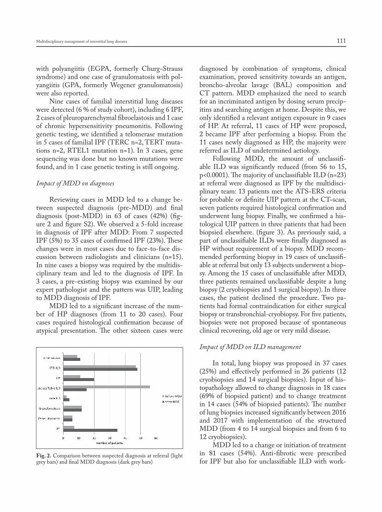
Multidisciplinary management of interstitial lung diseases 111
with polyangiitis (EGPA, formerly Churg-Strauss syndrome) and one case of granulomatosis with pol-yangiitis (GPA, formerly Wegener granulomatosis) were also reported.
Nine cases of familial interstitial lung diseases were detected (6 % of study cohort), including 6 IPF, 2 cases of pleuroparenchymal fibroelastosis and 1 case of chronic hypersensitivity pneumonitis. Following genetic testing, we identified a telomerase mutation in 5 cases of familial IPF (TERC n=2, TERT muta-tions n=2, RTEL1 mutation n=1). In 3 cases, gene sequencing was done but no known mutations were found, and in 1 case genetic testing is still ongoing.
Impact of MDD on diagnoses
Reviewing cases in MDD led to a change be-tween suspected diagnosis (pre-MDD) and final diagnosis (post-MDD) in 63 of cases (42%) (fig-ure 2 and figure S2). We observed a 5-fold increase in diagnosis of IPF after MDD: From 7 suspected IPF (5%) to 35 cases of confirmed IPF (23%). These changes were in most cases due to face-to-face dis-cussion between radiologists and clinicians (n=15). In nine cases a biopsy was required by the multidis-ciplinary team and led to the diagnosis of IPF. In 3 cases, a pre-existing biopsy was examined by our expert pathologist and the pattern was UIP, leading to MDD diagnosis of IPF.
MDD led to a significant increase of the num-ber of HP diagnoses (from 11 to 20 cases). Four cases required histological confirmation because of atypical presentation. The other sixteen cases were
diagnosed by combination of symptoms, clinical examination, proved sensitivity towards an antigen, broncho-alveolar lavage (BAL) composition and CT pattern. MDD emphasized the need to search for an incriminated antigen by dosing serum precip-itins and searching antigen at home. Despite this, we only identified a relevant antigen exposure in 9 cases of HP. At referral, 11 cases of HP were proposed, 2 became IPF after performing a biopsy. From the 11 cases newly diagnosed as HP, the majority were referred as ILD of undetermined aetiology.
Following MDD, the amount of unclassifi-able ILD was significantly reduced (from 56 to 15, p<0.0001). The majority of unclassifiable ILD (n=23) at referral were diagnosed as IPF by the multidisci-plinary team: 13 patients met the ATS-ERS criteria for probable or definite UIP pattern at the CT-scan, seven patients required histological confirmation and underwent lung biopsy. Finally, we confirmed a his-tological UIP pattern in three patients that had been biopsied elsewhere. (figure 3). As previously said, a part of unclassifiable ILDs were finally diagnosed as HP without requirement of a biopsy. MDD recom-mended performing biopsy in 19 cases of unclassifi-able at referral but only 13 subjects underwent a biop-sy. Among the 15 cases of unclassifiable after MDD, three patients remained unclassifiable despite a lung biopsy (2 cryobiopsies and 1 surgical biopsy). In three cases, the patient declined the procedure. Two pa-tients had formal contraindication for either surgical biopsy or transbronchial-cryobiopsy. For five patients, biopsies were not proposed because of spontaneous clinical recovering, old age or very mild disease.
Impact of MDD on ILD management
In total, lung biopsy was proposed in 37 cases (25%) and effectively performed in 26 patients (12 cryobiopsies and 14 surgical biopsies). Input of his-topathology allowed to change diagnosis in 18 cases (69% of biopsied patient) and to change treatment in 14 cases (54% of biopsied patients). The number of lung biopsies increased significantly between 2016 and 2017 with implementation of the structured MDD (from 4 to 14 surgical biopsies and from 6 to 12 cryobiopsies).
MDD led to a change or initiation of treatment in 81 cases (54%). Anti-fibrotic were prescribed for IPF but also for unclassifiable ILD with work-
Fig. 2. Comparison between suspected diagnosis at referral (light grey bars) and final MDD diagnosis (dark grey bars)
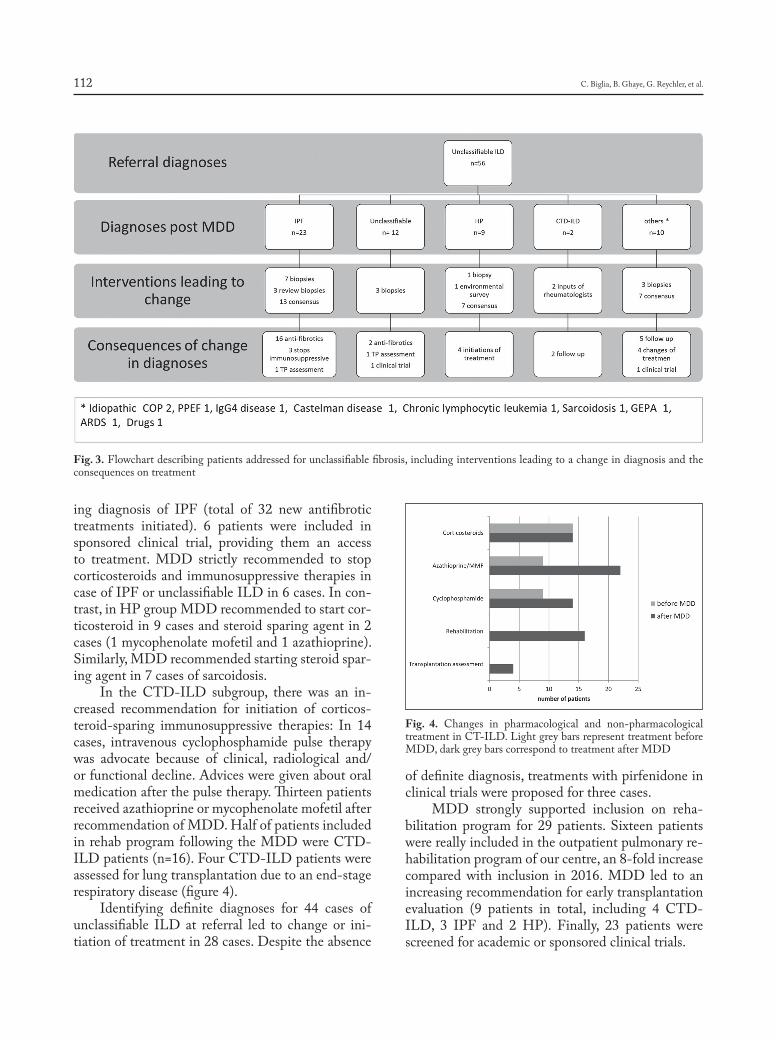
C. Biglia, B. Ghaye, G. Reychler, et al.112
ing diagnosis of IPF (total of 32 new antifibrotic treatments initiated). 6 patients were included in sponsored clinical trial, providing them an access to treatment. MDD strictly recommended to stop corticosteroids and immunosuppressive therapies in case of IPF or unclassifiable ILD in 6 cases. In con-trast, in HP group MDD recommended to start cor-ticosteroid in 9 cases and steroid sparing agent in 2 cases (1 mycophenolate mofetil and 1 azathioprine). Similarly, MDD recommended starting steroid spar-ing agent in 7 cases of sarcoidosis.
In the CTD-ILD subgroup, there was an in-creased recommendation for initiation of corticos-teroid-sparing immunosuppressive therapies: In 14 cases, intravenous cyclophosphamide pulse therapy was advocate because of clinical, radiological and/or functional decline. Advices were given about oral medication after the pulse therapy. Thirteen patients received azathioprine or mycophenolate mofetil after recommendation of MDD. Half of patients included in rehab program following the MDD were CTD-ILD patients (n=16). Four CTD-ILD patients were assessed for lung transplantation due to an end-stage respiratory disease (figure 4).
Identifying definite diagnoses for 44 cases of unclassifiable ILD at referral led to change or ini-tiation of treatment in 28 cases. Despite the absence
of definite diagnosis, treatments with pirfenidone in clinical trials were proposed for three cases.
MDD strongly supported inclusion on reha-bilitation program for 29 patients. Sixteen patients were really included in the outpatient pulmonary re-habilitation program of our centre, an 8-fold increase compared with inclusion in 2016. MDD led to an increasing recommendation for early transplantation evaluation (9 patients in total, including 4 CTD-ILD, 3 IPF and 2 HP). Finally, 23 patients were screened for academic or sponsored clinical trials.
Fig. 3. Flowchart describing patients addressed for unclassifiable fibrosis, including interventions leading to a change in diagnosis and the consequences on treatment
Fig. 4. Changes in pharmacological and non-pharmacological treatment in CT-ILD. Light grey bars represent treatment before MDD, dark grey bars correspond to treatment after MDD
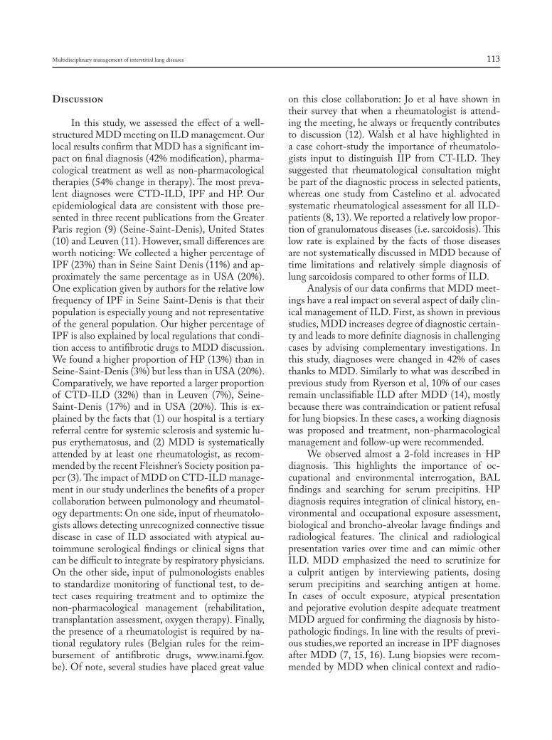
Multidisciplinary management of interstitial lung diseases 113
Discussion
In this study, we assessed the effect of a well-structured MDD meeting on ILD management. Our local results confirm that MDD has a significant im-pact on final diagnosis (42% modification), pharma-cological treatment as well as non-pharmacological therapies (54% change in therapy). The most preva-lent diagnoses were CTD-ILD, IPF and HP. Our epidemiological data are consistent with those pre-sented in three recent publications from the Greater Paris region (9) (Seine-Saint-Denis), United States (10) and Leuven (11). However, small differences are worth noticing: We collected a higher percentage of IPF (23%) than in Seine Saint Denis (11%) and ap-proximately the same percentage as in USA (20%). One explication given by authors for the relative low frequency of IPF in Seine Saint-Denis is that their population is especially young and not representative of the general population. Our higher percentage of IPF is also explained by local regulations that condi-tion access to antifibrotic drugs to MDD discussion. We found a higher proportion of HP (13%) than in Seine-Saint-Denis (3%) but less than in USA (20%). Comparatively, we have reported a larger proportion of CTD-ILD (32%) than in Leuven (7%), Seine-Saint-Denis (17%) and in USA (20%). This is ex-plained by the facts that (1) our hospital is a tertiary referral centre for systemic sclerosis and systemic lu-pus erythematosus, and (2) MDD is systematically attended by at least one rheumatologist, as recom-mended by the recent Fleishner’s Society position pa-per (3). The impact of MDD on CTD-ILD manage-ment in our study underlines the benefits of a proper collaboration between pulmonology and rheumatol-ogy departments: On one side, input of rheumatolo-gists allows detecting unrecognized connective tissue disease in case of ILD associated with atypical au-toimmune serological findings or clinical signs that can be difficult to integrate by respiratory physicians. On the other side, input of pulmonologists enables to standardize monitoring of functional test, to de-tect cases requiring treatment and to optimize the non-pharmacological management (rehabilitation, transplantation assessment, oxygen therapy). Finally, the presence of a rheumatologist is required by na-tional regulatory rules (Belgian rules for the reim-bursement of antifibrotic drugs, www.inami.fgov.be). Of note, several studies have placed great value
on this close collaboration: Jo et al have shown in their survey that when a rheumatologist is attend-ing the meeting, he always or frequently contributes to discussion (12). Walsh et al have highlighted in a case cohort-study the importance of rheumatolo-gists input to distinguish IIP from CT-ILD. They suggested that rheumatological consultation might be part of the diagnostic process in selected patients, whereas one study from Castelino et al. advocated systematic rheumatological assessment for all ILD-patients (8, 13). We reported a relatively low propor-tion of granulomatous diseases (i.e. sarcoidosis). This low rate is explained by the facts of those diseases are not systematically discussed in MDD because of time limitations and relatively simple diagnosis of lung sarcoidosis compared to other forms of ILD.
Analysis of our data confirms that MDD meet-ings have a real impact on several aspect of daily clin-ical management of ILD. First, as shown in previous studies, MDD increases degree of diagnostic certain-ty and leads to more definite diagnosis in challenging cases by advising complementary investigations. In this study, diagnoses were changed in 42% of cases thanks to MDD. Similarly to what was described in previous study from Ryerson et al, 10% of our cases remain unclassifiable ILD after MDD (14), mostly because there was contraindication or patient refusal for lung biopsies. In these cases, a working diagnosis was proposed and treatment, non-pharmacological management and follow-up were recommended.
We observed almost a 2-fold increases in HP diagnosis. This highlights the importance of oc-cupational and environmental interrogation, BAL findings and searching for serum precipitins. HP diagnosis requires integration of clinical history, en-vironmental and occupational exposure assessment, biological and broncho-alveolar lavage findings and radiological features. The clinical and radiological presentation varies over time and can mimic other ILD. MDD emphasized the need to scrutinize for a culprit antigen by interviewing patients, dosing serum precipitins and searching antigen at home. In cases of occult exposure, atypical presentation and pejorative evolution despite adequate treatment MDD argued for confirming the diagnosis by histo-pathologic findings. In line with the results of previ-ous studies,we reported an increase in IPF diagnoses after MDD (7, 15, 16). Lung biopsies were recom-mended by MDD when clinical context and radio-
C. Biglia, B. Ghaye, G. Reychler, et al.114
logic pattern were discordant. The number of lung biopsies increased with implementation of regular and standardized MDD, reflecting the obsession of the MDD to approach more accurately the diagnosis and adapt management.
Secondly, changing diagnosis led to changes in treatment in 54% cases. With the increase of IPF diagnoses we observed a significant increase in anti-fibrotic therapies prescription and recommendation against immunosuppressive therapies and corticos-teroids was made. In contrast, combination of cor-ticosteroids and immunosuppressive therapies were shown to increase risk of death and hospitalization (4). MDD helps to optimize immunosuppressive therapy in CTD-ILD.
Finally, MDD brought non-pharmacological measures that improved global management of ILD: Based on two controlled trials (17, 18), ATS/ERS guidelines for IPF management promoted inclu-sion in pulmonary rehabilitation program. These two studies have shown improvement in walked distance and symptoms or quality of life (18-20). Despite this, recommendations for pulmonary rehabilitation re-main weak in guidelines (21). Over the last few years, many studies were published and have strengthened the conviction that ILD patients benefits from ex-ercise training (17, 22). After implementation of structured MDD, recommendations for pulmonary rehabilitation attendance and effective participation increases. Efforts still need to be made to propose more systematically rehabilitation programs and to convince patients to participate.
Clinical trials are crucial in ILD to improve therapies and outcomes in this area where our thera-peutic action remains limited in certain cases. MDD allowed screening more patients for inclusion in clinical trial.
This study comprises several limitations: It is a retrospective study, so we could not compare MDD and absence of MDD face-to-face. In line with our inclusion criteria (files discussed between January and December 2017), we lack long-term follow up data that may provide hints regarding morbidity and mortality outcomes. Furthermore, our study was not designed for longitudinal evaluation of patients. Fi-nally, the implementation of a MDD per se is likely to have improved ILD management locally.
In conclusion, we report our experience on one-year use of a well-structured MDD. Our results
emphasize the multiple benefits related to MDD in ILD management. Furthermore, MDD manage-ment fosters collaboration between different depart-ments of the hospital, favouring integrated medicine and holistic care of patients.
References
1. Raghu G, Collard HR, Egan JJ, et al. An official ATS/ERS/JRS/ALAT statement: idiopathic pulmonary fibrosis: evidence-based guidelines for diagnosis and management. Am J Respir Crit Care Med 2011; 183(6): 788-824.
2. Raghu G, Remy-Jardin M, Myers JL, et al. Diagnosis of Idiopathic Pulmonary Fibrosis. An Official ATS/ERS/JRS/ALAT Clinical Practice Guideline. Am J Respir Crit Care Med 2018; 198(5): e44-e68.
3. Lynch DA, Sverzellati N, Travis WD, et al. Diagnostic criteria for idi-opathic pulmonary fibrosis: a Fleischner Society White Paper. Lancet Respir Med 2017.
4. Idiopathic Pulmonary Fibrosis Clinical Research N, Raghu G, An-strom KJ, et al. Prednisone, azathioprine, and N-acetylcysteine for pulmonary fibrosis. N Engl J Med 2012; 366(21): 1968-77.
5. Raghu G, Rochwerg B, Zhang Y, et al. An Official ATS/ERS/JRS/ALAT Clinical Practice Guideline: Treatment of Idiopathic Pulmo-nary Fibrosis. An Update of the 2011 Clinical Practice Guideline. Am J Respir Crit Care Med 2015; 192(2): e3-19.
6. Flaherty KR, King TE, Jr., Raghu G, et al. Idiopathic interstitial pneu-monia: what is the effect of a multidisciplinary approach to diagnosis? Am J Respir Crit Care Med 2004; 170(8): 904-10.
7. Flaherty KR, Andrei AC, King TE, Jr., et al. Idiopathic interstitial pneumonia: do community and academic physicians agree on diagno-sis? Am J Respir Crit Care Med 2007; 175(10): 1054-60.
8. Walsh SL, Wells AU, Desai SR, et al. Multicentre evaluation of mul-tidisciplinary team meeting agreement on diagnosis in diffuse paren-chymal lung disease: a case-cohort study. Lancet Respir Med 2016; 4(7): 557-65.
9. Duchemann B, Annesi-Maesano I, Jacobe de Naurois C, et al. Preva-lence and incidence of interstitial lung diseases in a multi-ethnic county of Greater Paris. Eur Respir J 2017; 50(2).
10. Lederer DJ, Martinez FJ. Idiopathic Pulmonary Fibrosis. N Engl J Med 2018; 378(19): 1811-23.
11. De Sadeleer LJ, Meert C, Yserbyt J, et al. Diagnostic Ability of a Dy-namic Multidisciplinary Discussion in Interstitial Lung Diseases: A Retrospective Observational Study of 938 Cases. Chest 2018; 153(6): 1416-23.
12. Jo HE, Corte TJ, Moodley Y, et al. Evaluating the interstitial lung disease multidisciplinary meeting: a survey of expert centres. BMC Pulm Med 2016; 16: 22.
13. Castelino FV, Goldberg H, Dellaripa PF. The impact of rheumato-logical evaluation in the management of patients with interstitial lung disease. Rheumatology (Oxford) 2011; 50(3): 489-93.
14. Ryerson CJ, Urbania TH, Richeldi L, et al. Prevalence and prognosis of unclassifiable interstitial lung disease. Eur Respir J 2013; 42(3): 750-7.
15. Jo HE, Glaspole IN, Levin KC, et al. Clinical impact of the intersti-tial lung disease multidisciplinary service. Respirology 2016; 21(8): 1438-44.
16. Chaudhuri N, Spencer L, Greaves M, et al. A Review of the Multi-disciplinary Diagnosis of Interstitial Lung Diseases: A Retrospective Analysis in a Single UK Specialist Centre. J Clin Med 2016; 5(8).
17. Ryerson CJ, Cayou C, Topp F, et al. Pulmonary rehabilitation im-

Multidisciplinary management of interstitial lung diseases 115
proves long-term outcomes in interstitial lung disease: a prospective cohort study. Respir Med 2014; 108(1): 203-10.
18. Dowman LM, McDonald CF, Hill CJ, et al. The evidence of ben-efits of exercise training in interstitial lung disease: a randomised con-trolled trial. Thorax 2017.
19. Nishiyama O, Kondoh Y, Kimura T, et al. Effects of pulmonary reha-bilitation in patients with idiopathic pulmonary fibrosis. Respirology 2008; 13(3): 394-9.
20. Holland AE, Hill CJ, Conron M, et al. Short term improvement in exercise capacity and symptoms following exercise training in intersti-tial lung disease. Thorax 2008; 63(6): 549-54.
21. Spruit MA. Pulmonary rehabilitation. Eur Respir Rev 2014; 23(131): 55-63.
22. Huppmann P, Sczepanski B, Boensch M, et al. Effects of inpatient pulmonary rehabilitation in patients with interstitial lung disease. Eur Respir J 2013; 42(2): 444-53.
Introduction
Idiopathic pulmonary fibrosis (IPF) is a spe-cific form of chronic, progressive fibrotic interstitial pneumonia of unknown cause. Acute exacerbation of idiopathic pulmonary fibrosis (AE-IPF) is defined as respiratory deterioration in less than 1 month (1).
Respiratory failure caused by AE-IPF is associated with high in-hospital mortality (55.6%-80%) (2-5). In particular, studies have shown that the mortality of patients with AE-IPF requiring mechanical ven-tilation is 81.8%-94% (6, 7).
Treatment of AE-IPF has not been established, and only anecdotal treatment reports exist. Inter-national evidenced-based guidelines weakly recom-mend a standard therapy for AE-IPF of administrat-ing systemic glucocorticoids, including methylpred-nisolone at a dosage of 1 g per day intravenously for 3 days (8). The international evidenced-base guidelines do not comment on the use of other immunosup-pressant agents combined with glucocorticoids ow-ing to a lack of conclusive results for the combina-
Systemic glucocorticoids plus cyclophosphamide for acute exacerbation of idiopathic pulmonary fibrosis: a retrospective nationwide study
Shotaro Aso1, Hiroki Matsui1, Kiyohide Fushimi2, Hideo Yasunaga1
1 Department of Clinical Epidemiology and Health Economics, School of Public Health, The University of Tokyo, Tokyo, Japan; 2 Depart-ment of Health Policy and Informatics, Tokyo Medical and Dental University Graduate School of Medicine, Tokyo, Japan
Abstract. Purpose: Mortality of acute exacerbation of idiopathic pulmonary fibrosis is high, and it remains un-known whether cyclophosphamide is an effective treatment for this condition. Objectives: This study compared the effects of cyclophosphamide combined with systemic glucocorticoids with those of systemic glucocorticoids alone. Methods: Using the Diagnosis Procedure Combination database in Japan, adult patients with idiopathic pulmonary fibrosis who had received high-dose methylprednisolone and mechanical ventilation at admission from July 1, 2010, to March 31, 2014, were identified. Instrumental variable analyses based on a hospital prefer-ence for cyclophosphamide were performed to compare in-hospital outcomes. Results: Eligible patients (n=1847) were divided into the methylprednisolone plus cyclophosphamide group (n=104) and the methylprednisolone alone group (n=1743). The results of an instrumental variable analysis detected no significant differences be-tween the groups with respect to in-hospital mortality (odds ratio, 1.11; 95% confidence interval, 0.19-6.43), ventilator-free days (difference, 2.2; 95% confidence interval, −2.6 to 7.0). Conclusions: In a Japanese inpatient database study analyzing outcomes from patients with acute exacerbation idiopathic pulmonary fibrosis receiv-ing systemic glucocorticoids, the addition of cyclophosphamide was not associated with improved in-hospital mortality and ventilator-free days. (Sarcoidosis Vasc Diffuse Lung Dis 2019; 36 (2): 116-123)
Key words: idiopathic pulmonary fibrosis, mortality, cyclophosphamide, glucocorticoids
SARCOIDOSIS VASCULITIS AND DIFFUSE LUNG DISEASES 2019; 36 (2); 116-123 © Mattioli 1885
Original article: Clinical research
Received: 14 March 2018Accepted after revision: 24 February 2019Correspondence: Shotaro Aso, MD, MPH, Department of Clinical Epidemiology and Health Economics, School of Public Health, The University of Tokyo, 7-3-1 Hongo, Bunkyo-ku, Tokyo, 113-0033, JapanTel. +81-3-5841-1887Fax +81-3-5841-1888E-mail: [email protected]
Cyclophosphamide for AE-IPF 117
tion treatment (8). It remains controversial whether cyclophosphamide combined with methylpredniso-lone is effective for patients with IPF (9-11). Pre-vious studies have reported that cyclophosphamide combined with high-dose methylprednisolone is potentially effective for patients with AE-IPF (12, 13). However, the patients in those studies had con-nective tissue diseases. To date, there has been no study comparing high-dose methylprednisolone plus cyclophosphamide with high-dose methylpredniso-lone alone as therapy for patients with AE-IPF, and thus it remains unknown whether cyclophosphamide would have an additive effect with high-dose meth-ylprednisolone in these patients.
Therefore, the aim of the present study was to use data from a national inpatient database in Japan to compare the effectiveness of the administration of cyclophosphamide combined with systemic gluco-corticoids to that of systemic glucocorticoids alone for reducing the mortality of AE-IPF.
Methods
Data source
Inpatient data were extracted from the Japanese Diagnosis Procedure Combination database. More than 1,000 hospitals voluntarily contribute to the database, which includes data from approximately 7 million inpatients, representing approximately 50% of all discharges from acute care hospitals in Japan. The data used in the present study included the hos-pital identification number; patient sex and age; body weight and height; consciousness level on admission; dates of hospitalization and discharge; main diag-noses, pre-existing comorbidities on admission, and complications that occurred during hospitalization, which were coded with the International Classifica-tion of Diseases, tenth revision (ICD-10) codes and text in Japanese; surgical and nonsurgical procedures and dates of procedures performed; dates and doses of drugs or blood products administered during the hospitalization; and discharge status.
Consciousness level on admission was evalu-ated using Japan Coma Scale scores (14, 15), which is widely used in Japan, and its assessment is well correlated with the Glasgow Coma Scale assessment (16).
The Institutional Review Board of The Univer-sity of Tokyo approved this study. Informed consent was waived because of the anonymous nature of the data.
Patient selection
This study used data from July 1, 2010, to March 31, 2014. The inclusion criteria were patients aged ≥15 years who were diagnosed as having idiopath-ic pulmonary fibrosis (ICD-10 codes: J84.1, J84.8, and J84.9) and who received mechanical ventilation within 1 day after admission. The patients were di-vided into two groups: (1) those who received cyclo-phosphamide 500 to 1,000 mg per day intravenously for 1 day and methylprednisolone 1 gram per day in-travenously for 3 days within 5 days after admission (termed the methylprednisolone plus cyclophospha-mide group); (2) those who received methylpredni-solone 500 to 1,000 mg per day intravenously for 3 days within 4 days after admission (methylpredniso-lone alone group).
Baseline characteristics
Baseline characteristics included the following: age; sex; Hugh-Jones classification on admission (17); consciousness level on admission; Charlson comorbidity index (CCI); smoking index (packs per year); past history of diabetes mellitus, chronic kid-ney disease, lung cancer, chronic obstructive pulmo-nary disease, and congestive heart failure; and use of cotrimoxazole, azithromycin (18), continuous renal replacement therapy, and noradrenaline within 1 day after admission. Patients were categorized into five age groups: 15-40, 41-60, 61-70, 71-80, and ≥81 years old. The CCI was classified into five groups: 0, 1, 2, 3-5, and ≥6 points. The smoking index was categorized into five groups: 0, 1-20, 21-40, 41-60, and ≥61 packs per year.
Outcomes
The primary outcome was in-hospital mortal-ity. The secondary outcome was ventilator-free days (VFDs) (19), incidence of sepsis (ICD-10 codes: A32.7, A39.4, A40.x, and A41.x), and incidence of mycosis (ICD-10 codes: B37.1, B37.5-B37.8, B44.0,B44.1, B45.0, B45.1, B45.7, B45.9, B48.7, B49, and J17.2).
S. Aso, H. Matsui, K. Fushimi, H. Yasunaga118
Statistical analysis
Because some values were missing for the Hugh-Jones classification, smoking index, and CCI, a mul-tiple imputation procedure was performed to replace each missing value with a set of submitted plausible values by creating 20 filled-in complete datasets us-ing a Markov chain Monte Carlo algorithm known as chained equations imputation (20). The multiple imputation method assumes that data are missing at random and that any systemic differences between the missing and observed values can be explained by differences in the observed data (21, 22).
An instrumental variable (IV) analysis was also performed. Unmeasured confounders can lead to in-correct inferences regarding the effectiveness of dif-ferent treatments. The IV analysis can theoretically balance both measured and unmeasured confounders between two groups (23, 24). A hospital preference for cyclophosphamide was selected as an IV, because use of cyclophosphamide depended on physician preference. When hospitals are strongly consistent in whether or not they use cyclophosphamide to treat AE-IPF, it is assumed that the decision to administer the drug may be made independently of an individual patient’s background. In such a situation, a hospital preference for cyclophosphamide may have acted as an IV, thereby setting the stage for a “natural experi-ment” that allowed an unbiased estimate of the risk of AE-IPF, even if unmeasured confounders existed (25, 26). An IV analysis assumes that patient hospital choice is made independently of the hospital’s choice of a specific drug, and the hospital’s use of the drug is independent of the outcomes. The number of pa-tients with AE-IPF who received cyclophosphamide in each hospital was counted, and then the average number of patients with AE-IPF who received cy-clophosphamide among all the hospitals was calcu-lated. Hospitals with more than the average number of cyclophosphamide users were defined as hospitals with a preference for cyclophosphamide. Hospitals with less than the average number of cyclophos-phamide users were defined as hospitals without a preference for cyclophosphamide. To assess the va-lidity of hospital preference as an IV, we confirmed that hospital preference was highly correlated to the receipt of cyclophosphamide (F statistic >10) (25). We examined hospital preference was not associated with outcomes.
A two-stage residual inclusion estimation framework of the IV analysis was used (27, 28). The residual inclusion approach has been shown to gen-erate more consistent and less biased estimates for a variety of nonlinear models. In the first stage model, the association between receipt of cyclophospha-mide and hospital preference for cyclophosphamide was measured, with adjustment for patient level co-variates. From this model, the raw residual for each patient was determined by calculating the difference between the model-predicted probability of receiving cyclophosphamide and the actual treatment received. The residuals were then included as an additional covariate in the second-stage model. In the second-stage model, the association between treatment and outcomes was estimated, adjusting for covariates. All IV analyses were performed using robust standard errors.
A sensitivity analysis was performed to confirm the correctness of the inclusion criteria for AE-IPF (1). First, patients who had not received a computed tomography (CT) scan within 1 day after admission were excluded. Second, patients who had not re-ceived a CT scan within 1 day after admission and with the use of furosemide within 1 day after admis-sion were excluded.
Continuous variables are presented as an aver-age along with the standard deviation or the median with the interquartile range. Categorical variables are presented as the number with a proportion. In the unadjusted comparisons, averages of continuous variables were compared using t-tests, and propor-tions of categorical variables using χ2 tests.
A P value <0.05 was considered to indicate statistical significance. All statistical analyses were performed using STATA/MP version 14.0 software (STATA Corp, College Station, TX, USA).
Results
During the analyzed period, we identified 12,992 patients who received methylprednisolone at a dose of 500 to 1,000 mg per day for 3 days within 4 days after admission (Figure 1). Among them, 1,847 patients were eligible for the present study, including 104 patients administered cyclophosphamide and 1,743 patients without cyclophosphamide adminis-tration.
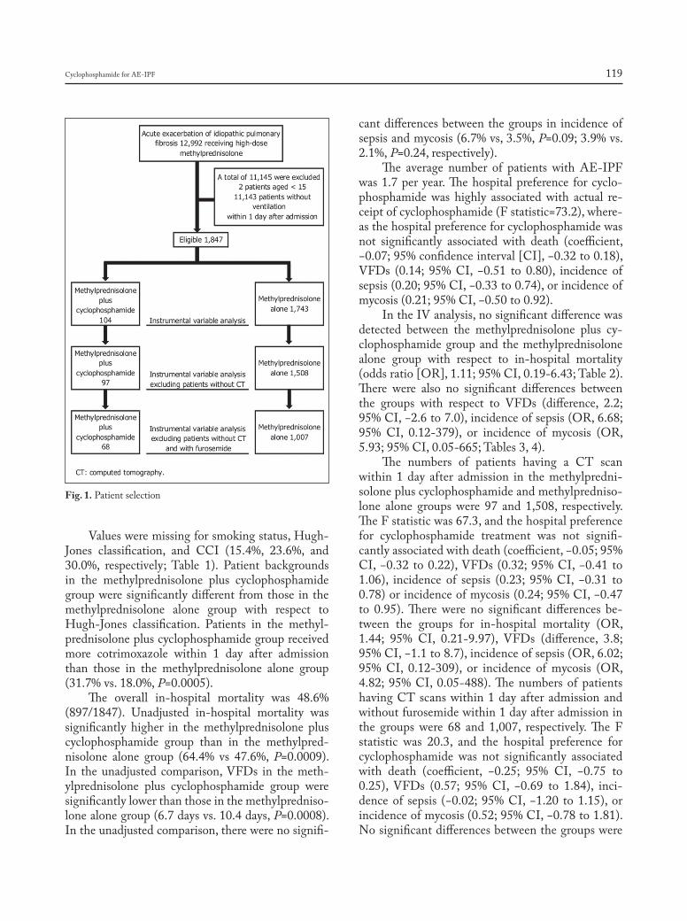
Cyclophosphamide for AE-IPF 119
Values were missing for smoking status, Hugh-Jones classification, and CCI (15.4%, 23.6%, and 30.0%, respectively; Table 1). Patient backgrounds in the methylprednisolone plus cyclophosphamide group were significantly different from those in the methylprednisolone alone group with respect to Hugh-Jones classification. Patients in the methyl-prednisolone plus cyclophosphamide group received more cotrimoxazole within 1 day after admission than those in the methylprednisolone alone group (31.7% vs. 18.0%, P=0.0005).
The overall in-hospital mortality was 48.6% (897/1847). Unadjusted in-hospital mortality was significantly higher in the methylprednisolone plus cyclophosphamide group than in the methylpred-nisolone alone group (64.4% vs 47.6%, P=0.0009). In the unadjusted comparison, VFDs in the meth-ylprednisolone plus cyclophosphamide group were significantly lower than those in the methylpredniso-lone alone group (6.7 days vs. 10.4 days, P=0.0008). In the unadjusted comparison, there were no signifi-
cant differences between the groups in incidence of sepsis and mycosis (6.7% vs, 3.5%, P=0.09; 3.9% vs. 2.1%, P=0.24, respectively).
The average number of patients with AE-IPF was 1.7 per year. The hospital preference for cyclo-phosphamide was highly associated with actual re-ceipt of cyclophosphamide (F statistic=73.2), where-as the hospital preference for cyclophosphamide was not significantly associated with death (coefficient, −0.07; 95% confidence interval [CI], −0.32 to 0.18), VFDs (0.14; 95% CI, −0.51 to 0.80), incidence of sepsis (0.20; 95% CI, −0.33 to 0.74), or incidence of mycosis (0.21; 95% CI, −0.50 to 0.92).
In the IV analysis, no significant difference was detected between the methylprednisolone plus cy-clophosphamide group and the methylprednisolone alone group with respect to in-hospital mortality (odds ratio [OR], 1.11; 95% CI, 0.19-6.43; Table 2). There were also no significant differences between the groups with respect to VFDs (difference, 2.2; 95% CI, −2.6 to 7.0), incidence of sepsis (OR, 6.68; 95% CI, 0.12-379), or incidence of mycosis (OR, 5.93; 95% CI, 0.05-665; Tables 3, 4).
The numbers of patients having a CT scan within 1 day after admission in the methylpredni-solone plus cyclophosphamide and methylpredniso-lone alone groups were 97 and 1,508, respectively. The F statistic was 67.3, and the hospital preference for cyclophosphamide treatment was not signifi-cantly associated with death (coefficient, −0.05; 95% CI, −0.32 to 0.22), VFDs (0.32; 95% CI, −0.41 to 1.06), incidence of sepsis (0.23; 95% CI, −0.31 to 0.78) or incidence of mycosis (0.24; 95% CI, −0.47 to 0.95). There were no significant differences be-tween the groups for in-hospital mortality (OR, 1.44; 95% CI, 0.21-9.97), VFDs (difference, 3.8; 95% CI, −1.1 to 8.7), incidence of sepsis (OR, 6.02; 95% CI, 0.12-309), or incidence of mycosis (OR, 4.82; 95% CI, 0.05-488). The numbers of patients having CT scans within 1 day after admission and without furosemide within 1 day after admission in the groups were 68 and 1,007, respectively. The F statistic was 20.3, and the hospital preference for cyclophosphamide was not significantly associated with death (coefficient, −0.25; 95% CI, −0.75 to 0.25), VFDs (0.57; 95% CI, −0.69 to 1.84), inci-dence of sepsis (−0.02; 95% CI, −1.20 to 1.15), or incidence of mycosis (0.52; 95% CI, −0.78 to 1.81). No significant differences between the groups were
Fig. 1. Patient selection
S. Aso, H. Matsui, K. Fushimi, H. Yasunaga120
Table 1. Baseline characteristics at admission
Variable Methylprednisolone Methylprednisolone plus alone cyclophosphamide P value (n=1,743) (n=104)
Sex, n (%) male 1,182 (67.8) 72 (69.2) 0.77Age, years, n (%) 15-40 9 (0.5) 0 0.0 0.30 41-60 112 (6.4) 9 (8.7) 61-70 384 (22.0) 29 (27.9) 71-80 751 (43.1) 45 (43.3) ≥81 487 (27.9) 21 (20.2) Smoking index (packs per year), n (%) 0 779 (44.7) 50 (48.1) 0.72 1-20 143 (8.2) 9 (8.7) 21-40 237 (13.6) 12 (11.5) 41-60 179 (10.3) 14 (13.5) ≥61 134 (7.7) 5 (4.8) missing 271 (15.5) 14 (13.5) Hugh-Jones classification, n (%) 1 54 (3.1) 3 (2.9) 0.04 2 82 (4.7) 5 (4.8) 3 107 (6.1) 2 (1.9) 4 198 (11.4) 6 (5.8) 5 903 (51.8) 52 (50.0) missing 399 (22.9) 36 (34.6) Charlson comorbidity index, n (%) 0 316 (18.1) 20 (19.2) 0.38 1 321 (18.4) 17 (16.3) 2 384 (22.0) 17 (16.3) 3-5 119 (6.8) 7 (6.7) ≥6 88 (5.0) 3 (2.9) missing 515 (29.5) 40 (38.5) Japan coma scale, n (%) 0 (alert) 1,273 (73.0) 85 (81.7) 0.17 1-digit (dizziness) 283 (16.2) 14 (13.5) 2-digit (somnolence) 87 (5.0) 3 (2.9) 3-digit (coma) 100 (5.7) 2 (1.9) Lung cancer, n (%) 72 (4.1) 5 (4.8) 0.73Chronic obstructive pulmonary disease, n (%) 140 (8.0) 7 (6.7) 0.63Congestive heart failure, n (%) 142 (8.1) 3 (2.9) 0.06Diabetes mellitus, n (%) 425 (24.4) 17 (16.3) 0.06Chronic kidney disease, n (%) 46 (2.6) 3 (2.9) 0.85Noradrenaline, n (%) 162 (9.3) 10 (9.6) 0.92Azithromycin, n (%) 272 (15.6) 9 (8.7) 0.06Cotrimoxazole, n (%) 313 (18.0) 33 (31.7) 0.0005Continuous renal replacement therapy, n (%) 24 (1.4) 3 (2.9) 0.22
Table 2. Comparison of in-hospital mortality between the methylprednisolone plus cyclophosphamide and methylprednisolone alone groups
OR* 95% CI† P value
Unadjusted 1.99 1.32 - 3.01 0.0009 Instrumental variable analysis 1.11 0.19 - 6.43 0.91 Instrumental variable analysis, patients with CT‡ 1.44 0.21 - 9.97 0.71 Instrumental variable analysis, patients with CT, without furosemide 0.95 0.04 - 23.97 0.97
*OR, odds ratio; †CI, confidence interval; ‡CT, computed tomography
Cyclophosphamide for AE-IPF 121
detected for in-hospital mortality (OR, 0.95; 95% CI, 0.04-23.97), VFDs (difference, 5.8; 95% CI, −5.2 to 16.8), incidence of sepsis (OR, 1.90; 95% CI, 0.00-1060), or incidence of mycosis (OR, 1.70; 95% CI, 0.01-532).
Discussion
This study used data obtained from a Japanese national inpatient database to compare the effective-ness of high-dose methylprednisolone plus cyclo-phosphamide with high-dose methylprednisolone alone for treating patients with AE-IPF. Our IV analysis showed no significant difference between the two treatment groups for in-hospital mortality, VFDs, incidence of sepsis, or incidence of mycosis.
Two previous studies showed that cyclophos-phamide was potentially effective for treating pa-tients with AE-IPF (12, 13). However, those studies were limited by having no control group and small sample sizes (n=11, 17, respectively). Despite the high mortality associated with AE-IPF, treatment of the condition remains uncertain. International evi-dence-based guidelines weakly recommend adminis-tration of systemic corticosteroids and do not judge whether other medications are effective for AE-IPF
because of a lack of evidence regarding the combined treatment (8). Furthermore, a recent review examin-ing AE-IPF reported that studies investigating treat-ment of AE-IPF were mostly small and uncontrolled and could not adjust for confounders (1).
The advantage of the present study was that we performed an IV analysis, and this analysis generated pseudo-randomization adjusting for unmeasured and measured confounders. We found no significant dif-ference for in-hospital mortality or VFDs between the methylprednisolone plus cyclophosphamide group and methylprednisolone alone group. One potential reason for this may be that cyclophospha-mide had no effect on AE-IPF. Previous studies have reported that cyclophosphamide has no effect on patients with IPF (9, 10). Another potential reason for the lack of differences is that no currently known medication, including corticosteroids and other im-munosuppressant agents, may be effective against AE-IPF because AE-IPF is a severe condition with a rapid progression. Although cyclophosphamide suppresses the immune system with depressing bone marrow, the present study showed that there were no significant differences in incidence of sepsis or mycosis between the cyclophosphamide users and non-users. One potential reason for this may be that patients receiving cyclophosphamide died before cy-
Table 3. Comparison of ventilator-free days between the methylprednisolone plus cyclophosphamide and methylprednisolone alone groups
Difference 95% CI* P value
Unadjusted −3.7 −5.9 - −1.6 0.0008 Instrumental variable analysis 2.2 −2.6 - 7.0 0.37 Instrumental variable analysis, patients with CT† 3.8 −1.1 - 8.7 0.13 Instrumental variable analysis, patients with CT, without furosemide 5.8 −5.2 - 16.8 0.30
*CI, confidence interval; †CT, computed tomography
Table 4. Comparison of incidence of sepsis or mycosis between the methylprednisolone plus cyclophosphamide and methylprednisolone alone groups
OR* 95% CI† P value
Sepsis Unadjusted 1.99 0.89 - 4.47 0.10 Instrumental variable analysis 6.68 0.12 - 379 0.36 Instrumental variable analysis, patients with CT‡ 6.02 0.12 - 309 0.37 Instrumental variable analysis, patients with CT, without furosemide 1.90 0.00 - 1060 0.84 Mycosis Unadjusted 1.84 0.64 - 5.28 0.25 Instrumental variable analysis 5.93 0.05 - 665 0.46 Instrumental variable analysis, patients with CT 4.82 0.05 - 488 0.50 Instrumental variable analysis, patients with CT, without furosemide 1.70 0.01 - 532 0.86
*OR, odds ratio; †CI, confidence interval; ‡CT, computed tomography
S. Aso, H. Matsui, K. Fushimi, H. Yasunaga122
clophosphamide had time to suppress their immune systems.
This study has several limitations. First, the database did not include detailed data on patients’ physical conditions, laboratory examinations, and other tests, such as respiratory rates, partial pressure of arterial oxygen/fraction of inspired oxygen ratios, lactate dehydrogenase levels, serum KL-6 levels, and CT imaging results (29). We could therefore not ob-tain information on the severity of IPF. Moreover, it is possible that patients receiving cyclophosphamide were treated more aggressively. We therefore used instrumental variable analysis to account for these unmeasured confounders. Furthermore, previous studies have shown that the prognosis of suspected AE-IPF is similar to that of AE-IPF (4, 5). The sec-ond limitation is that it cannot be proven that our IV analysis fully addressed unmeasured confound-ers (30). However, we conducted sensitivity analyses based on revised diagnostic criteria for AE-IPF, and the results of the sensitivity analysis were similar to those in the primary analysis. Furthermore, the Japa-nese Diagnosis Procedure Combination database has been well validated and can serve as a relatively accu-rate substitute for clinical data although any admin-istrative data have some limitations to the recorded data (31). Third, it is unknown whether our results can be applied to patients with AE-IPF who are not using mechanical ventilation.
Conclusions
Despite these limitations, our IV analysis using a Japanese inpatient database showed that the ad-ministration of cyclophosphamide to patients with AE-IPF who were also receiving systemic corticos-teroids was not associated with improved in-hospital mortality or VFDs. Further prospective studies will be required to confirm the effect of cyclophospha-mide in the treatment of AE-IPF.
Authorship: KF contributed to the study design and data acqui-sition. SA, HM, and HY performed the statistical analyses and produced the first draft of the manuscript. All authors commented on the manuscript and approved the final version.
Funding: This work was supported by grants for Research on Policy Planning and Evaluation from the Ministry of Health, Labour and Welfare, Japan (grant numbers H29-Policy-Desig-
nated-009, H27-Policy-Strategy-011); Ministry of Education, Culture, Sports, Science and Technology, Japan (grant numbers 17H04141); and the Japan Agency for Medical Research and De-velopment (AMED). The funders had no role in the execution of this study or interpretation of the results.
Disclosure Statement: HY and KF received grant support from the Japanese government. The funders had no role in the execu-tion of this study or interpretation of the results. All authors have disclosed that they do not have any potential conflicts of interest.
References
1. Collard HR, Ryerson CJ, Corte TJ, et al. Acute exacerbation of idi-opathic pulmonary fibrosis an international working group report. Am J Respir Crit Care Med 2016;1 94(3): 265-75.
2. Song JW, Hong S-B, Lim C-M, Koh Y, Kim DS. Acute exacerbation of idiopathic pulmonary fibrosis: incidence, risk factors and outcome. Eur Respir J 2011; 37(2): 356-63.
3. Usui Y, Kaga A, Sakai F, et al. A cohort study of mortality predic-tors in patients with acute exacerbation of chronic fibrosing interstitial pneumonia. BMJ Open 2013; 3: e002971.
4. Huie TJ, Olson AL, Cosgrove GP, et al. A detailed evaluation of acute respiratory decline in patients with fibrotic lung disease: Aetiology and outcomes. Respirology 2010; 15(6): 909-17.
5. Collard HR, Yow E, Richeldi L, Anstrom KJ, Glazer C. Suspected acute exacerbation of idiopathic pulmonary fibrosis as an outcome measure in clinical trials. Respir Res 2013; 14: 73.
6. Mallick S. Outcome of patients with idiopathic pulmonary fibrosis (IPF) ventilated in intensive care unit. Respir Med 2008; 102(10): 1355-9.
7. Kim DS, Park JH, Park BK, Lee JS, Nicholson AG, Colby T. Acute exacerbation of idiopathic pulmonary fibrosis: Frequency and clinical features. Eur Respir J 2006; 27: 143-50.
8. Raghu G, Collard HR, Egan JJ, et al. An Official ATS/ERS/JRS/ALAT Statement: Idiopathic pulmonary fibrosis: Evidence-based guidelines for diagnosis and management. Am J Respir Crit Care Med 2011; 183: 788-824.
9. Collard HR, Ryu JH, Douglas WW, et al. Combined corticosteroid and cyclophosphamide therapy does not alter survival in idiopathic pulmonary fibrosis. Chest 2004; 125(6): 2169-74.
10. Johnson MA, Kwan S, Snell NJ, Nunn AJ, Darbyshire JH, Turner-Warwick M. Randomised controlled trial comparing prednisolone alone with cyclophosphamide and low dose prednisolone in combi-nation in cryptogenic fibrosing alveolitis. Thorax 1989; 44( January): 280-8.
11. Pereira CAC, Malheiros T, Coletta EM, et al. Survival in idiopathic pulmonary fibrosis - Cytotoxic agents compared to corticosteroids. Respir Med 2006; 100(2): 340-7.
12. Novelli L, Ruggiero R, Giacomi F De, et al. Corticosteroid and cyclo-phosphamide in acute exacerbation of idiopathic pulmonary fibrosis : a single center experience and literature review. Sarcoidosis Vasc Dif-fus Lung Dis 2016; 33: 385-91.
13. Ota M, Iwasaki Y, Harada H, Sasaki O, Nagafuchi Y, Nakachi S, et al. Efficacy of intensive immunosuppression in exacerbated rheumatoid arthritis-associated interstitial lung disease. Mod Rheumatol 2016; 7595(May): 1-7.
14. Ohta T, Waga S, Handa H, et al. New grading of level of disordered consciousness (author’s translation). No shinkei geka 1974; 2: 623-7.
15. Shigematsu K, Nakano H, Watanabe Y. The eye response test alone is sufficient to predict stroke outcome—reintroduction of Japan Coma Scale: a cohort study. BMJ Open 2013; 3:e 002736.
16. Ono K, Wada K, Takahara T, Shirotani T. Indications for Computed
Cyclophosphamide for AE-IPF 123
Tomography in Patients With Mild Head Injury. Neurol Med Chir 2007; 47: 291-8.
17. Fletcher CM. The clinical diagnosis of pulmonary emphysema; an experimental study. Proc R Soc Med 1952; 45(9): 577-84.
18. Oda K, Yatera K, Fujino Y, et al. Efficacy of concurrent treatments in idiopathic pulmonary fibrosis patients with a rapid progression of respiratory failure: an analysis of a national administrative database in Japan. BMC Pulm Med 2016; 16(1): 91.
19. Schoenfeld D a, Bernard GR. Statistical evaluation of ventilator-free days as an efficacy measure in clinical trials of treatments for acute respiratory distress syndrome. Crit Care Med 2002; 30(8): 1772-7.
20. Rubin DB, Schenker N. Multiple imputation in health-care databas-es: an overview and some applications. Stat Med 1991; 10(4): 585-98.
21. Sterne JAC, White IR, Carlin JB, et al. Multiple imputation for miss-ing data in epidemiological and clinical research: potential and pit-falls. BMJ 2009; 338: b2393.
22. Li P, Stuart EA, Allison DB. Multiple Imputation: A Flexible Tool for Handling Missing Data. JAMA 2015; 314(18): 1966-7.
23. Newhouse JP, McClellan M. Econometrics in outcomes research: the use of instrumental variables. Annu Rev Public Health 1998; 19(1): 17-34.
24. Iwashyna TJ, Kennedy EH. Instrumental variable analyses: Exploit-
ing natural randomness to understand causal mechanisms. Ann Am Thorac Soc 2013; 10(3): 255-60.
25. Staiger D, Stock J. Instrumental Variables Regression with Weak In-struments. Econometrica 1997; 65(3): 557-86.
26. Stukel TA, Fisher ES, Wennberg DE, Alter DA, Vermeulen MJ. Analysis of Observational Studies in the Presence of Treatment Se-lection Bias. Jama 2007; 297(3): 278-85.
27. Terza J V, Basu A, Rathouz PJ. Two-stage residual inclusion esti-mation: Addressing endogeneity in health econometric modeling. J Health Econ 2008; 27(3): 531-43.
28. Terza J V., Bradford WD, Dismuke CE. The use of linear instrumen-tal variables methods in health services research and health econom-ics: A cautionary note. Health Serv Res 2008; 43(3): 1102-20.
29. Kishaba T, Tamaki H, Shimaoka Y, Fukuyama H, Yamashiro S. Stag-ing of acute exacerbation in patients with idiopathic pulmonary fibro-sis. Lung 2014; 192(1): 141-9.
30. Harris KM, Remler DK. Who is the marginal patient? Understanding instrumental variables estimates of treatment effects. Health Serv Res 1998; 33(5 Pt 1): 1337-60.
31. Yamana H, Moriwaki M, Horiguchi H, Kodan M, Fushimi K, Yasu-naga H. Validity of diagnoses, procedures, and laboratory data in Japa-nese administrative data. J Epidemiol 2017; 27(10): 476-82.
Background
Sarcoidosis is a multisystem inflammatory dis-ease characterized by the presence of noncaseating granulomas that affects 35.5 per 100,000 African Americans and 10.9 per 100,000 Caucasians in the United States (U.S.) (1, 2). Most sarcoidosis patients
experience spontaneous resolution of their disease (3) and do not require treatment, but a third of pa-tients will have chronic disease requiring prolonged treatment (3, 4). Corticosteroids are considered first line treatment for most forms of sarcoidosis, but can result in significant side effects and increased health-care use (4). Patients with refractory disease may re-quire treatment with second- or third-line medica-tions, including methotrexate (MTX), azathioprine (AZA), and monoclonal antibody therapy (5).
Previous studies evaluating sarcoidosis treatment options have primarily addressed sarcoidosis patients from large referral centers which have reported treat-ment rates of 55-65% (4, 6). This contrasts starkly
A new side of sarcoidosis: medication and hospitalization use in a privately insured patient population
Derek Low1, Kit N. Simpson2, Richard Rissmiller2, Ennis James2
1 University of Colorado, Denver CO; 2 Medical University of South Carolina, Charleston, SC
Abstract. Objective: This study describes patterns of medication prescriptions for sarcoidosis patients in a large commercially insured U.S. population, with specific focus on prescribing practices across medical specialties and their associated hospitalization risk. Methods: Using the Marketscan Database we selected adult patients with a diagnosis of sarcoidosis by ICD-9 code during the 2012 calendar year. Differences in prescribing practices were evaluated between provider types. A multivariate model controlling for age, sex, and region assessed hospitaliza-tion risk associated with provider type, prednisone dose, and use of non-steroid sarcoidosis medications. Results: Using the described criteria, 11,042 total patients were identified. A majority were female, mean age 49.3 years. Of these, 1,792 (16.2%) had one or more hospital admissions (mean 1.6, SD 1.3) with a mean length of stay of 8.1 days (SD 14.5). 25.5% of patients were prescribed prednisone with a 1 year mean cumulative dose of 250mg. Pulmonary/Rheumatology providers prescribed the highest cumulative prednisone dose (961 mg) and were more likely to prescribe methotrexate and monoclonal antibody medications. Sarcoidosis patients receiv-ing a cumulative prednisone dose >500 mg had an increased risk for hospitalization (OR 2.512, 2.210-2.855), while those prescribed methotrexate and azathioprine had decreased risk (OR 0.633, 0.481-0.833 and 0.460, 0.315-0.671). Monoclonal antibody use was associated with increased OR for hospitalization at 1.359. Conclu-sion: Sarcoidosis patients treated by subspecialists were more likely to receive higher doses of prednisone and non-steroid sarcoidosis medications. Higher doses of prednisone and monoclonal antibody use were associated with higher hospitalization risk while methotrexate and azathioprine were associated with lower hospitalization risk. (Sarcoidosis Vasc Diffuse Lung Dis 2019; 36 (2): 124-129)
Key words: sarcoidosis, epidemiology, hospitalization, corticosteroids
SARCOIDOSIS VASCULITIS AND DIFFUSE LUNG DISEASES 2019; 36 (2); 124-129 © Mattioli 1885
Original article: Clinical research
Received: 30 March 2018Accepted after revision: 11 March 2019Correspondence: Derek Matthew LowUniversity of Colorado, Denver COE-mail: [email protected]
Medication and hospitalization use in sarcoidosis 125
with a recent study which included a large, commu-nity population of patients with sarcoidosis in which only 22.8% of continuing cases received treatment (7). Scant data exists regarding prescribing patterns and outcomes in sarcoidosis patients from communi-ty-based practices. Large datasets from private insur-ers can provide insight into prescribing practices and associated outcomes across medical specialties from non-referral centers. The objectives of this study were to describe patterns of medication prescriptions for sarcoidosis patients in a large commercially insured U.S. population, with a specific focus on differences in prescribing practices across medical specialties and their associated hospitalization risk.
Methods
This retrospective analysis was conducted us-ing the Truven Health MarketScan® Research Da-tabases. These databases provide outcome measures including resource utilization and healthcare costs for inpatient and outpatient healthcare encounters of patients under employer-sponsored health insur-ance and Medicare-eligible individuals which covers approximately 143 million individuals. All database records were de-identified in compliance with the Health Insurance Portability and Accountability Act (HIPAA) of 1996.
Study Variables
Variables included in the analysis were demo-graphic data (age, sex), date and duration of hospi-talizations, geographic location, and International Classification of Diseases, Clinical Modification (ICD-9-CM) code details (specific code and date of code entry into record). The Marketscan database does not record race. Details of outpatient prescrip-tions included the specialty of the prescribing prac-titioner, medication name, daily dose and cumulative dose prescribed over the 12-month study period. Specific anti-sarcoidosis medications were selected for the analysis, including prednisone, MTX, AZA, hydroxychloroquine, and monoclonal antibody (MAB) which included infliximab and adalimumab. The prescriptions of other medications used in sar-coidosis, including Leflunomide and Mycopheno-late, were too infrequent to include in the analysis.
Study Definitions
Patients selected as having a diagnosis of sar-coidosis were those with an ICD-9-CM code of 135.xx on two separate outpatient encounters at least 7 days apart. Hospitalizations were defined as any in-patient admission at an acute care facility during the study period. The acute care facility designation is based on a UB04 claim indicating an acute care hos-pital admission or an emergency department claim record with a length of stay greater than 0 days. Pro-vider categories, as defined by billing label in the Marketscan Database, were divided into primary care providers (PCP), Pulmonology/Rheumatology (P/R), Dermatology/Immunology (D/I), multiple subspecialty providers (Multi), and “Other” sub-specialty (Other) which includes pediatric, surgical and other medical specialties. To ensure PCPs were representative of non-specialist prescribing practices, sarcoidosis patients receiving prescriptions in the PCP category were required to have no encounters with a specialty provider, while those seen by a spe-cialist could also be seen by a PCP.
Statistical Analysis
For characteristics of the sarcoidosis patients in the Marketscan database we used descritptive statis-tics reporting mean and SD for continuous variables. Medication prescriptions are described by mean dai-ly and cumulative dose. Differences between groups of providers where tested with univariate analysis us-ing gamma distributed log link models to account for skewed distribution of data. Multivariate logistic re-gression controlling for age, sex, and region was used to assess hospital risk by provider, prednisone use/cumulative dose, and use of non-steroid sarcoidosis medications. Regions were defined in the MarketS-can database as Northeast, North Central, South, West, and Unknown.
Results
Patient Characteristics
From January 1, 2012 to December 31, 2012, 11,042 patients met inclusion criteria (table 1). The cohort was predominately female (59.1%) with a mean age of 49.3 years.
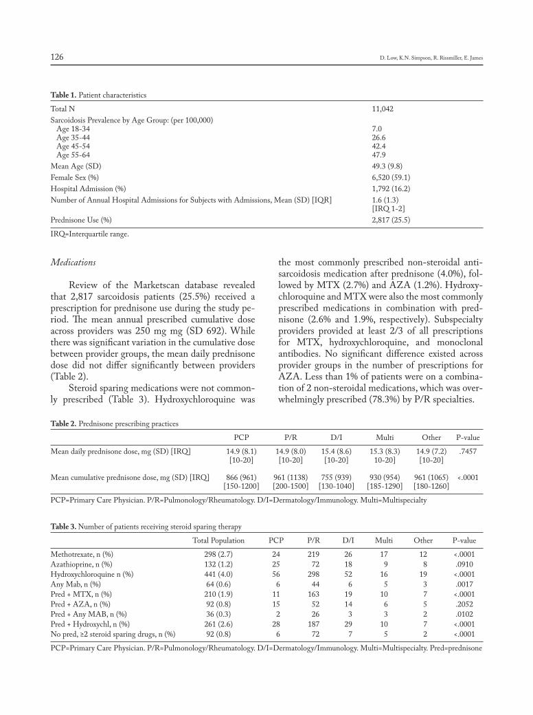
D. Low, K.N. Simpson, R. Rissmiller, E. James126
Medications
Review of the Marketscan database revealed that 2,817 sarcoidosis patients (25.5%) received a prescription for prednisone use during the study pe-riod. The mean annual prescribed cumulative dose across providers was 250 mg mg (SD 692). While there was significant variation in the cumulative dose between provider groups, the mean daily prednisone dose did not differ significantly between providers (Table 2).
Steroid sparing medications were not common-ly prescribed (Table 3). Hydroxychloroquine was
the most commonly prescribed non-steroidal anti-sarcoidosis medication after prednisone (4.0%), fol-lowed by MTX (2.7%) and AZA (1.2%). Hydroxy-chloroquine and MTX were also the most commonly prescribed medications in combination with pred-nisone (2.6% and 1.9%, respectively). Subspecialty providers provided at least 2/3 of all prescriptions for MTX, hydroxychloroquine, and monoclonal antibodies. No significant difference existed across provider groups in the number of prescriptions for AZA. Less than 1% of patients were on a combina-tion of 2 non-steroidal medications, which was over-whelmingly prescribed (78.3%) by P/R specialties.
Table 1. Patient characteristics
Total N 11,042Sarcoidosis Prevalence by Age Group: (per 100,000) Age 18-34 7.0 Age 35-44 26.6 Age 45-54 42.4 Age 55-64 47.9Mean Age (SD) 49.3 (9.8)Female Sex (%) 6,520 (59.1)Hospital Admission (%) 1,792 (16.2)Number of Annual Hospital Admissions for Subjects with Admissions, Mean (SD) [IQR] 1.6 (1.3) [IRQ 1-2]Prednisone Use (%) 2,817 (25.5)
IRQ=Interquartile range.
Table 2. Prednisone prescribing practices
PCP P/R D/I Multi Other P-value
Mean daily prednisone dose, mg (SD) [IRQ] 14.9 (8.1) 14.9 (8.0) 15.4 (8.6) 15.3 (8.3) 14.9 (7.2) .7457 [10-20] [10-20] [10-20] 10-20] [10-20]
Mean cumulative prednisone dose, mg (SD) [IRQ] 866 (961) 961 (1138) 755 (939) 930 (954) 961 (1065) <.0001 [150-1200] [200-1500] [130-1040] [185-1290] [180-1260]
PCP=Primary Care Physician. P/R=Pulmonology/Rheumatology. D/I=Dermatology/Immunology. Multi=Multispecialty
Table 3. Number of patients receiving steroid sparing therapy
Total Population PCP P/R D/I Multi Other P-value
Methotrexate, n (%) 298 (2.7) 24 219 26 17 12 <.0001Azathioprine, n (%) 132 (1.2) 25 72 18 9 8 .0910Hydroxychloroquine n (%) 441 (4.0) 56 298 52 16 19 <.0001Any Mab, n (%) 64 (0.6) 6 44 6 5 3 .0017Pred + MTX, n (%) 210 (1.9) 11 163 19 10 7 <.0001Pred + AZA, n (%) 92 (0.8) 15 52 14 6 5 .2052Pred + Any MAB, n (%) 36 (0.3) 2 26 3 3 2 .0102Pred + Hydroxychl, n (%) 261 (2.6) 28 187 29 10 7 <.0001No pred, ≥2 steroid sparing drugs, n (%) 92 (0.8) 6 72 7 5 2 <.0001
PCP=Primary Care Physician. P/R=Pulmonology/Rheumatology. D/I=Dermatology/Immunology. Multi=Multispecialty. Pred=prednisone
Medication and hospitalization use in sarcoidosis 127
Hospitalization risk
Patients seen in P/R clinics had an increased risk of hospitalization compared to those seen only by a PCP, D/I, or Other provider (Table 4). Medica-tion use was also correlated with hospitalization risk (Table 4). OR for prednisone use was grouped into three levels; no prednisone use, low-dose (1-500 mg cumulative yearly prednisone), and high-dose (>500 mg cumulative yearly prednisone). Low-dose pred-nisone correlated with a slightly higher risk for hos-pitalization compared to those who did not receive prednisone, while patients receiving high-dose pred-nisone had a substantial increase in hospitalization risk. MTX and AZA showed statistically significant decrease in hospitalization risk, while MAB showed an increased rate of hospitalization.
Discussion
The goal of this study was to describe a group of privately insured sarcoidosis patients with respect to medication prescribing practices across provider groups and associated hospitalization risk. As pre-vious studies have mainly described patient cohorts at large referral centers, this study evaluates a sub-set of patients who have not been well described historically. The recent study by Baughman, et al, evaluated a similar group using the Optum data-base, however their study centered around demo-graphic differences and health care costs (7). Our study further evaluated hospitalization risk in ref-erence to medication use and prescribing provider. Using the Marketscan dataset we demonstrated
differences in prescribing practices across pro-vider groups, with second- and third-line prescrip-tions being more common in subspecialty practices. Sarcoidosis referral center studies have often cited prednisone use in 55-65% of sarcoidosis patients, however this study found that only 25.5% of sar-coidosis patients in the Marketscan database were prescribed prednisone during the study period (4, 6). It should be noted, however, in the study by Judson, et al, 2012, their evaluation for prednisone use was described as 51% “at some point during the study pe-riod” which spanned 11 years of retrospective evalu-ation. This study does corroborate the findings of the Sarcoidosis in America study which found only 22.8% of patients in the Optum Database receiving therapy, of which 56% were prescribed prednisone (7). These disparities can partially be attributed to the increased severity of disease in patients seen in referral centers. In addition to prednisone, the rates of second line therapy use were more in line with those found in community studies as opposed to those observed at large referral centers.
We found the mean daily dose of prednisone per patient to be similar across provider groups, but the cumulative yearly dose of prednisone was significant-ly higher for P/R than PCP or D/I. This is consistent with the expectation that sarcoidosis patients seen by pulmonologists and rheumatologists are more likely to have refractory disease requiring a longer dura-tion of steroids. The hospitalization rate in the overall cohort was similar to that reported in other studies (8), and there was an increased risk of hospitaliza-tion in patients followed by P/R. While this could be related to greater disease severity, sarcoidosis patients followed by P/R also had higher cumulative doses of prednisone which has been previously been associ-ated with increased healthcare use in sarcoidosis pa-tients (9). We similarly found significant correlation between high cumulative doses of prednisone and increased risk of hospitalization. While this supports the concept that higher doses of prednisone are indic-ative of patients with more severe disease who have a higher hospitalization risk, an potential alternative explanation is that increased hospitalizations are a result of the adverse effects related to higher steroid exposure. This would agree with previous data pub-lished by Broos et. al. and others that found strong as-sociations between increased cumulative prednisone dose and a higher prevalence of comorbidities and
Table 4. Odds ratios for hospitalization
OR (CI)
Provider (ref: Pulmology/Rheumatology) Primary Care Physician .604 (.521-.699) Dermatology/Immunology .678 (.591-.777) Multi-Specialty .901 (.704-1.152) Other .776 (.638-.944)
Medication Pred: None vs 1-500 1.217 (1.035-1.431) Pred: None vs >500 2.512 (2.210-2.855) Any Monoclonal Antibody 1.359 (1.035-1.431) Methotrexate .633 (.481-.833) Azathioprine .460 (.315-.671)
*multivariable model controlling for effects of age, sex and region
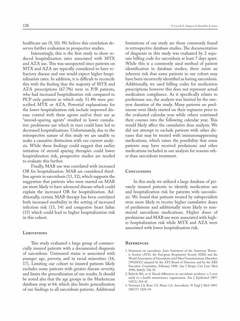
D. Low, K.N. Simpson, R. Rissmiller, E. James128
healthcare use (9, 10). We believe this correlation de-serves further evaluation in prospective studies.
Interestingly, this is the first study to show re-duced hospitalization rates associated with MTX and AZA use. This was unexpected since patients on MTX and AZA are typically considered to have re-fractory disease and one would expect higher hospi-talization rates. In addition, it is difficult to reconcile this with the finding that the majority of MTX and AZA prescriptions (67.7%) were in P/R patients, who had increased hospitalization risk compared to PCP-only patients in which only 11.4% were pre-scribed MTX or AZA. Potential explanations for the lower hospitalization risk include improved dis-ease control with these agents and/or their use as “steroid-sparing agents” resulted in lower cumula-tive prednisone use which in turn could have led to decreased hospitalizations. Unfortunately, due to the retrospective nature of this study we are unable to make a causative distinction with our current analy-sis. While these findings could suggest that earlier initiation of steroid sparing therapies could lower hospitalization risk, prospective studies are needed to evaluate this further.
Finally, MAB use was correlated with increased OR for hospitalization. MAB are considered third-line agents in sarcoidosis (11, 12), which supports the suggestion that patients who were started on MAB are more likely to have advanced disease which could explain the increased OR for hospitalization. Ad-ditionally, certain MAB therapy has been correlated both increased morbidity in the setting of increased infection risk (13, 14) and congestive heart failue (15) which could lead to higher hospitalization risk in this cohort.
Limitations
This study evaluated a large group of commer-cially insured patients with a documented diagnosis of sarcoidosis. Uninsured status is associated with younger age, poverty, and in racial minorities (16, 17). Limiting our cohort to insured patients likely excludes some patients with greater disease severity, and limits the generalization of our results. It should be noted also that the age groups in the Marketscan database stop at 64, which also limits generalization of our findings to all sarcoidosis patients. Additional
limitations of our study are those commonly found in retrospective database studies. The documentation of diagnosis in this study was evaluated by 2 sepa-rate billing code for sarcoidosis at least 7 days apart. While this is a commonly used method of patient identification in database studies, there exists an inherent risk that some patients in our cohort may have been incorrectly identified as having sarcoidosis. Additionally, we used billing codes for medication prescriptions however this does not represent actual medication compliance. As it specifically relates to prednisone use, the analysis was limited by the one-year duration of the study. Many patients on pred-nisone were likely started on their regimens prior to the evaluated calendar year while others continued their courses into the following calendar year. This would likely affect the cumulative dose analysis. We did not attempt to exclude patients with other dis-eases that may be treated with immunosuppressing medications, which raises the possibility that some patients may have received prednisone and other medications included in our analysis for reasons oth-er than sarcoidosis treatment.
Conclusions
In this study we utilized a large database of pri-vately insured patients to identify medication use and hospitalization risk for patients with sarcoido-sis. We found that patients treated by subspecialists were more likely to receive higher cumulative doses of prednisone and additionally more likely to non-steroid sarcoidosis medications. Higher doses of prednisone and MAB use were associated with high-er hospitalization risk while MTX and AZA were associated with lower hospitalization risk.
References
1. Statement on sarcoidosis. Joint Statement of the American Thorac-ic Society (ATS), the European Respiratory Society (ERS) and the World Association of Sarcoidosis and Other Granulomatous Disorders (WASOG) adopted by the ATS Board of Directors and by the ERS Executive Committee, February 1999. Am J Respir Crit Care Med 1999; 160(2): 736-55.
2. Rybicki BA, et al. Racial differences in sarcoidosis incidence: a 5-year study in a health maintenance organization. Am J Epidemiol 1997; 145(3): 234-41.
3. Newman LS, Rose CS, Maier LA. Sarcoidosis. N Engl J Med 1997; 336(17): 1224-34.
Medication and hospitalization use in sarcoidosis 129
4. Judson MA, Boan AD, Lackland DT. The clinical course of sarcoido-sis: presentation, diagnosis, and treatment in a large white and black cohort in the United States. Sarcoidosis Vasc Diffuse Lung Dis 2012; 29(2): 119-27.
5. Schutt AC, Bullington WM, Judson MA. Pharmacotherapy for pulmonary sarcoidosis: a Delphi consensus study. Respir Med 2010; 104(5): 717-23.
6. Baughman RP, et al. Clinical characteristics of patients in a case con-trol study of sarcoidosis. Am J Respir Crit Care Med 2001; 164(10 Pt 1): 1885-9.
7. Baughman RP, et al. Sarcoidosis in America. Analysis Based on Health Care Use. Ann Am Thorac Soc 2016; 13(8): 1244-52.
8. Gerke AK, et al. Increased hospitalizations among sarcoidosis patients from 1998 to 2008: a population-based cohort study. BMC Pulm Med 2012; 12: 19.
9. Ligon CB, Judson MA. Impact of systemic corticosteroids on health-care utilization in patients with sarcoidosis. Am J Med Sci 2011; 341(3): 196-201.
10. Broos CE, et al. No evidence found for an association between pred-nisone dose and FVC change in newly-treated pulmonary sarcoidosis. Respir Med 2018; 138s: S31-s37.
11. Baughman RP, et al. Infliximab therapy in patients with chronic sar-
coidosis and pulmonary involvement. Am J Respir Crit Care Med 2006; 174(7): 795-802.
12. Judson MA, et al. Efficacy of infliximab in extrapulmonary sarcoido-sis: results from a randomised trial. European Respiratory Journal 2008; 31(6): 1189-1196.
13. Koo S, Marty FM, Baden LR. Infectious complications associated with immunomodulating biologic agents. Infect Dis Clin North Am 2010; 24(2): 285-306.
14. Singh JA, et al. Adverse effects of biologics: a network meta-anal-ysis and Cochrane overview. Cochrane Database Syst Rev 2011(2): Cd008794.
15. Chung ES, et al. Randomized, double-blind, placebo-controlled, pilot trial of infliximab, a chimeric monoclonal antibody to tumor necrosis factor-alpha, in patients with moderate-to-severe heart failure: results of the anti-TNF Therapy Against Congestive Heart Failure (AT-TACH) trial. Circulation 2003; 107(25): 3133-40.
16. DeNavas-Walt CP, Smith BD, Income J. Poverty, and Health Insur-ance Coverage in the United States: 2012. US Census Bureau Current Population Reports, 2013: 60-245.
17. Grant SR, et al. Variation in insurance status by patient demographics and tumor site among nonelderly adult patients with cancer. Cancer 2015; 121(12): 2020-8.
Introduction
Sarcoidosis is a systemic granulomatous disease with an unclear etiology, which is frequently associ-
ated with pulmonary involvement and results in non-caseating granulomatous infiltration. The majority of cases are asymptomatic at the time of diagnosis, and many cases resolve spontaneously without treatment. Approximately 25% of cases are associated with pro-gressive pulmonary disease (1-3), and the pulmonary parenchymal involvement of sarcoidosis is crucial regarding disease staging and the initiation of treat-ment. Sarcoidosis is classified into five stages based on a lung X-ray findings (4). The disease is considered
Ultrasonographic evaluation of lung parenchyma involvement in sarcoidosis
Coşkun Doğan1, Nesrin Kıral1, Elif Torun Parmaksız1, Benan Çağlayan2, Seda Beyhan Sağmen1, Banu Salepçi1, Ali Fidan1, Sevda Şener Cömert1
1 Department of Chest Diseases, Dr. Lütfi Kırdar Kartal Training and Research Hospital, Istanbul, Turkey; 2 Department Of Chest Diseases, Koç University, Istanbul, Turkey
Abstract. Purpose: To use ultrasonography (USG) for the evaluation of lung parenchyma in patients with sarcoidosis, andto compare the USG findings with the results of a high-resolution computerized tomography (HRCT) and pulmonary function test-carbon monoxide diffusion test (PFT-DLCO), which are commonly used methods in the evaluation of parenchymal involvement in sarcoidosis. Material and Methods: Patients with sarcoidosis and healthy controls were enrolled in the study between January 2015 and December 2017. The clinical findings, HRCT and PFT-DLCO results of all subjects were recorded, and USG findings and comet tail artifact (CTA) measurements were recorded by another pulmonologist. The USG, HRCT and SFT-DLCO findings were compared between the two groups. Based on the findings of theclinical-radiologic investigations and PFT-DLCO, as the current gold standard in diagnosis, the sensitivity and specificity of USG in demon-strating lung parenchyma involvement in sarcoidosis patients were estimated. Findings: The sarcoidosis group consisted of 79 patients and the control group included 34 subjects. The mean number of CTAs in the sarcoido-sis and control groups was 33.4 and 25, respectively (p=0.001). In the sarcoidosis group, the number of CTAs in patients with DLCO% <80 and ≥80% was 37.4 and 29.7, respectively (p=0.011), and a negative correlation was identified between the number of CTAs and DLCO% (p=0.019 r=-0.267). The mean number of CTAs in patients with and without parenchymal involvement in HRCT was 36 and 25.5, respectively (p=0.001). The number of CTAs in the patients with sarcoidosis with a normal DLCO% value (≥80%) was higher than in the control group (p=0.014). The diagnostic sensitivity and specificity of thoracic USG were found to be 76% and 53%, respectively. Conclusion: The number of CTAs in patients with sarcoidosis was higher than that of the healthy controls. The number of CTAs in patients with sarcoidosis with parenchymal involvement in HRCT and/or a low DLCO (<80%) was also elevated. Thoracic USG has a high sensitivity (76%) in demonstrating parenchymal involvement in patients with sarcoidosis. (Sarcoidosis Vasc Diffuse Lung Dis 2019; 36 (2): 130-140)
Key words: B-lines, lung ultrasonography, sarcoidosis
SARCOIDOSIS VASCULITIS AND DIFFUSE LUNG DISEASES 2019; 36 (2); 130-140 © Mattioli 1885
Original article: Clinical research
Received: 30 April 2018Accepted after revision: 16 January 2019Correspondence: Coşkun Doğan, MD Neslişah sok. Teknik yapı Up city sitesi, B2 blok D 40 Uğurmumcu mah, Kartal Istanbul-TurkeyTel. +90 555 827 64 63E-mail: [email protected]
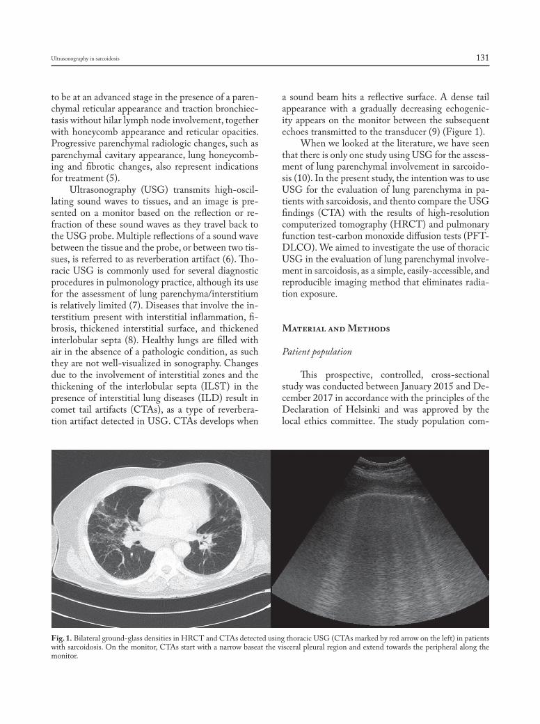
Ultrasonography in sarcoidosis 131
to be at an advanced stage in the presence of a paren-chymal reticular appearance and traction bronchiec-tasis without hilar lymph node involvement, together with honeycomb appearance and reticular opacities. Progressive parenchymal radiologic changes, such as parenchymal cavitary appearance, lung honeycomb-ing and fibrotic changes, also represent indications for treatment (5).
Ultrasonography (USG) transmits high-oscil-lating sound waves to tissues, and an image is pre-sented on a monitor based on the reflection or re-fraction of these sound waves as they travel back to the USG probe. Multiple reflections of a sound wave between the tissue and the probe, or between two tis-sues, is referred to as reverberation artifact (6). Tho-racic USG is commonly used for several diagnostic procedures in pulmonology practice, although its use for the assessment of lung parenchyma/interstitium is relatively limited (7). Diseases that involve the in-terstitium present with interstitial inflammation, fi-brosis, thickened interstitial surface, and thickened interlobular septa (8). Healthy lungs are filled with air in the absence of a pathologic condition, as such they are not well-visualized in sonography. Changes due to the involvement of interstitial zones and the thickening of the interlobular septa (ILST) in the presence of interstitial lung diseases (ILD) result in comet tail artifacts (CTAs), as a type of reverbera-tion artifact detected in USG. CTAs develops when
a sound beam hits a reflective surface. A dense tail appearance with a gradually decreasing echogenic-ity appears on the monitor between the subsequent echoes transmitted to the transducer (9) (Figure 1).
When we looked at the literature, we have seen that there is only one study using USG for the assess-ment of lung parenchymal involvement in sarcoido-sis (10). In the present study, the intention was to use USG for the evaluation of lung parenchyma in pa-tients with sarcoidosis, and thento compare the USG findings (CTA) with the results of high-resolution computerized tomography (HRCT) and pulmonary function test-carbon monoxide diffusion tests (PFT-DLCO). We aimed to investigate the use of thoracic USG in the evaluation of lung parenchymal involve-ment in sarcoidosis, as a simple, easily-accessible, and reproducible imaging method that eliminates radia-tion exposure.
Material and Methods
Patient population
This prospective, controlled, cross-sectional study was conducted between January 2015 and De-cember 2017 in accordance with the principles of the Declaration of Helsinki and was approved by the local ethics committee. The study population com-
Fig. 1. Bilateral ground-glass densities in HRCT and CTAs detected using thoracic USG (CTAs marked by red arrow on the left) in patients with sarcoidosis. On the monitor, CTAs start with a narrow baseat the visceral pleural region and extend towards the peripheral along the monitor.
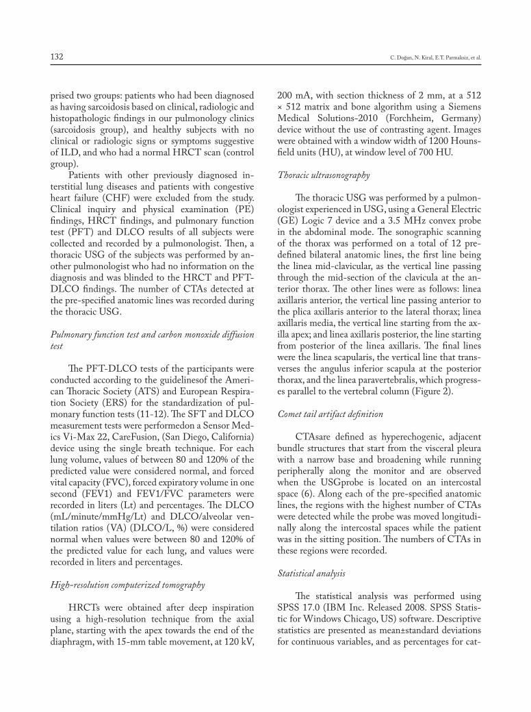
C. Doğan, N. Kiral, E.T. Parmaksiz, et al.132
prised two groups: patients who had been diagnosed as having sarcoidosis based on clinical, radiologic and histopathologic findings in our pulmonology clinics (sarcoidosis group), and healthy subjects with no clinical or radiologic signs or symptoms suggestive of ILD, and who had a normal HRCT scan (control group).
Patients with other previously diagnosed in-terstitial lung diseases and patients with congestive heart failure (CHF) were excluded from the study. Clinical inquiry and physical examination (PE) findings, HRCT findings, and pulmonary function test (PFT) and DLCO results of all subjects were collected and recorded by a pulmonologist. Then, a thoracic USG of the subjects was performed by an-other pulmonologist who had no information on the diagnosis and was blinded to the HRCT and PFT-DLCO findings. The number of CTAs detected at the pre-specified anatomic lines was recorded during the thoracic USG.
Pulmonary function test and carbon monoxide diffusion test
The PFT-DLCO tests of the participants were conducted according to the guidelinesof the Ameri-can Thoracic Society (ATS) and European Respira-tion Society (ERS) for the standardization of pul-monary function tests (11-12). The SFT and DLCO measurement tests were performedon a Sensor Med-ics Vi-Max 22, CareFusion, (San Diego, California) device using the single breath technique. For each lung volume, values of between 80 and 120% of the predicted value were considered normal, and forced vital capacity (FVC), forced expiratory volume in one second (FEV1) and FEV1/FVC parameters were recorded in liters (Lt) and percentages. The DLCO (mL/minute/mmHg/Lt) and DLCO/alveolar ven-tilation ratios (VA) (DLCO/L, %) were considered normal when values were between 80 and 120% of the predicted value for each lung, and values were recorded in liters and percentages.
High-resolution computerized tomography
HRCTs were obtained after deep inspiration using a high-resolution technique from the axial plane, starting with the apex towards the end of the diaphragm, with 15-mm table movement, at 120 kV,
200 mA, with section thickness of 2 mm, at a 512 × 512 matrix and bone algorithm using a Siemens Medical Solutions-2010 (Forchheim, Germany) device without the use of contrasting agent. Images were obtained with a window width of 1200 Houns-field units (HU), at window level of 700 HU.
Thoracic ultrasonography
The thoracic USG was performed by a pulmon-ologist experienced in USG, using a General Electric (GE) Logic 7 device and a 3.5 MHz convex probe in the abdominal mode. The sonographic scanning of the thorax was performed on a total of 12 pre-defined bilateral anatomic lines, the first line being the linea mid-clavicular, as the vertical line passing through the mid-section of the clavicula at the an-terior thorax. The other lines were as follows: linea axillaris anterior, the vertical line passing anterior to the plica axillaris anterior to the lateral thorax; linea axillaris media, the vertical line starting from the ax-illa apex; and linea axillaris posterior, the line starting from posterior of the linea axillaris. The final lines were the linea scapularis, the vertical line that trans-verses the angulus inferior scapula at the posterior thorax, and the linea paravertebralis, which progress-es parallel to the vertebral column (Figure 2).
Comet tail artifact definition
CTAsare defined as hyperechogenic, adjacent bundle structures that start from the visceral pleura with a narrow base and broadening while running peripherally along the monitor and are observed when the USGprobe is located on an intercostal space (6). Along each of the pre-specified anatomic lines, the regions with the highest number of CTAs were detected while the probe was moved longitudi-nally along the intercostal spaces while the patient was in the sitting position. The numbers of CTAs in these regions were recorded.
Statistical analysis
The statistical analysis was performed using SPSS 17.0 (IBM Inc. Released 2008. SPSS Statis-tic for Windows Chicago, US) software. Descriptive statistics are presented as mean±standard deviations for continuous variables, and as percentages for cat-
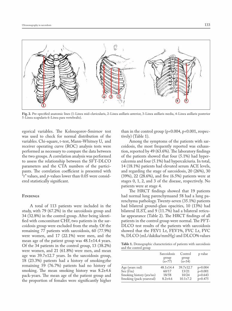
Ultrasonography in sarcoidosis 133
egorical variables. The Kolmogorov-Smirnov test was used to check for normal distribution of the variables. Chi-square, t-test, Mann-Whitney U, and receiver operating curve (ROC) analysis tests were performed as necessary to compare the data between the two groups. A correlation analysis was performed to assess the relationship between the SFT-DLCO parameters and the CTA numbers of the partici-pants. The correlation coefficient is presented with “r” values, and p-values lower than 0.05 were consid-ered statistically significant.
Findings
A total of 113 patients were included in the study, with 79 (67.2%) in the sarcoidosis group and 34 (32.8%) in the control group. After being identi-fied with concomitant CHF, two patients in the sar-coidosis group were excluded from the study. Of the remaining 77 patients with sarcoidosis, 60 (77.9%) were women, and 17 (22.1%) were men, and the mean age of the patient group was 48.1±14.4 years. Of the 34 patients in the control group, 13 (38.2%) were women, and 21 (61.8%) were men, and mean age was 39.7±12.7 years. In the sarcoidosis group, 18 (23.3%) patients had a history of smoking;the remaining 59 (76.7%) patients had no history of smoking. The mean smoking history was 8.2±4.6 pack-years. The mean age of the patient group and the proportion of females were significantly higher
than in the control group (p<0.004, p<0.001, respec-tively) (Table 1).
Among the symptoms of the patients with sar-coidosis, the most frequently reported was exhaus-tion, reported by 49 (63.6%). The laboratory findings of the patients showed that four (5.1%) had hyper-calcemia and four (5.1%) had hypercalciuria. In total, 14 (18.1%) patients had elevated serum ACE levels, and regarding the stage of sarcoidosis, 20 (26%), 30 (39%), 22 (28.6%), and five (6.5%) patients were at stages 0, 1, 2, and 3 of the disease, respectively. No patients were at stage 4.
The HRCT findings showed that 19 patients had normal lung parenchymaand 58 had a lung pa-renchyma pathology. Twenty-seven (35.1%) patients had bilateral ground-glass opacities, 10 (13%) had bilateral ILST, and 9 (11.7%) had a bilateral reticu-lar appearance (Table 2). The HRCT findings of all patients in the control group were normal. The PFT-DLCO test results of the patients with sarcoidosis showed that the FEV1 Lt, FEV1%, FVC Lt, FVC %, DLCO (mL/dakika/mmHg) and DLCO% values
Fig. 2. Pre-specified anatomic lines (1-Linea mid-clavicularis, 2-Linea axillaris anterior, 3-Linea axillaris media, 4-Linea axillaris posterior 5-Linea scapularis 6-Linea para vertebralis).
Table 1. Demographic characteristics of patients with sarcoidosis and the control group
Sarcoidosis Control p value group group (n=77) (n=34)
Age (years ±sd) 48.1±14.4 39.7±12.7 p=0.004Sex (f/m) 60/17 13/21 p<0.001Smoking history (yes/no) 18/58 10/24 p=0.643Smoking (pack-years±sd) 8.2±4.6 10.1±7.2 p=0.475
C. Doğan, N. Kiral, E.T. Parmaksiz, et al.134
were significantly lower than those of the controls (p<0.05) (Table 3).
The CTA numbers of patients with sarcoidosis were not significantly different to the those of the controls in the right and left linea axillaris anterior, right linea mid-clavicularis, and left linea axillaris media regions (p>0.05). The CTA numbers in all
other scanned regions were significantly higher in pa-tients with sarcoidosis than in the controls (p<0.05). The mean number of CTAs in the patient and con-trol groups was 33.4±13.1 and 25±6.5, respectively (p=0.001) (Table 4).
Analyses of the correlations between the PFT-DLCO parameters and the number of CTAs in the sarcoidosis group showed that DLCO% values were ≤80% and >80% in 37 (48%) and 40 (52%) of the patients, respectively. The mean number of CTAs in patients with DLCO% ≤80% was 37.4±15.8, where-as this figure was 29.7±8.7 in patients with DLCO% >80%, which represents a statistically significant difference (p=0.011) (Table 5). There was no sig-nificant relationship between the number of CTAs and FEV1% or FVC% in patients with sarcoidosis (p>0.05) (Table 5). The total number of CTAs was negatively correlated with the DLCO% of patients with sarcoidosis (p=0.019 r=-0.267) (Figure 3). No significant correlations were identified between FEV1 and the number of CTAs, FEV1% and the number of CTAs, FVC, and the number of CTAs, or FVC% and the number of CTAs (p>0.05).
The HRCT findings of the patients in the sar-coidosis group indicated that 58 patients had paren-chymal involvement, and the number of CTAs in pa-tients with parenchymal involvement in HRCT was 36±13.5, which was significantly higher than in pa-tients without parenchymal involvement (25.5±7.9) (p=0.001). The mean number of CTAs in 19 (24.6%) patients with sarcoidosis who had a normal HRCT (no parenchymal pathology seen) was 25.5±7.9,
Table 2. HRCT findings in patients withsarcoidosis
HRCT findings
Parenchymal abnormal findings (Yes/No) 58/19
Right lung nodule larger than 1 cm 30(39%)
Left lung nodule larger than 1 cm 25(32.5%)
Bilateral ground-glass appearance 27(35.1%)
Left lung sequela band appearance 22(28.6%)
Right lung sequela band appearance 19(24.7%)
Bilateral reticulo-nodular appearance 16(20.8%)
Bilateral peri-lymphatic, broncho-vascular, 1-2 mm 10 (13%)nodules located along ILS
Bilateral ILST 10 (13%)
Bilateral traction bronchiectasis / honeycombing 8 (10.4%)appearance
Bilateral mosaic perfusion appearance 5 (6.5%)
Other 12(15.6%)
ILST: Inter-lobular septal thickening
Table 3. PFT-DLCO findings in patients withsarcoidosis and the control group.
PFT-DLCO parameters Sarcoidosis group (n=77) Control group (n=34) p value
FEV1/FVC(Mean±sd) 76.8±7.4 78.4±8.6 p=0.309
FEV1 (Mean±sd)(Lt) 2.53±0.78 3.80±0.8 p<0.001
%FEV1 (Mean±sd) 91.2±17.8 99.6±12.4 p=0.014
FVC (Mean±sd)(Lt) 3.15±0.98 4.35±1 p<0.001
%FVC (Mean±sd) 98.7±17.8 106.2±12.5 p=0.028
DLCO (Mean±sd) (mL/minute/mmHg) 6.61±2.40 9.91±2 p<0.001
%DLCO (Mean±sd) 83.4±18.3 99.7±14.5 p<0.001
DLCO/VA(mL/minute/mmHg /Lt) (Mean/sd) 1.99±1.05 1.76±0.29 p=0.082
%DLCO/VA 101.43±16.9 109.18±16.9 p=0.029
DLCO:Carbonmonoxide diffusion capacity. FVC: Forced vital capacity. FEV1: Forced expiratory volume in one second. VA: Alveolar vol-ume. Mean: Mean SD: Standard deviation
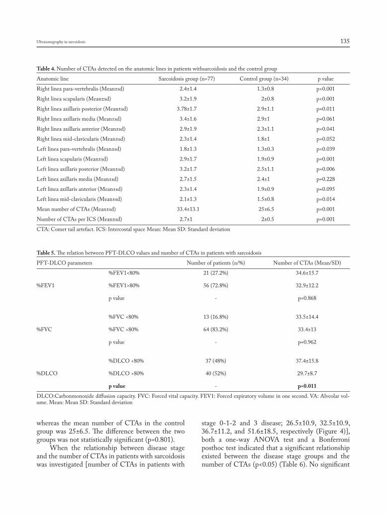
Ultrasonography in sarcoidosis 135
whereas the mean number of CTAs in the control group was 25±6.5. The difference between the two groups was not statistically significant (p=0.801).
When the relationship between disease stage and the number of CTAs in patients with sarcoidosis was investigated [number of CTAs in patients with
stage 0-1-2 and 3 disease; 26.5±10.9, 32.5±10.9, 36.7±11.2, and 51.6±18.5, respectively (Figure 4)], both a one-way ANOVA test and a Bonferroni posthoc test indicated that a significant relationship existed between the disease stage groups and the number of CTAs (p<0.05) (Table 6). No significant
Table 4. Number of CTAs detected on the anatomic lines in patients withsarcoidosis and the control group
Anatomic line Sarcoidosis group (n=77) Control group (n=34) p value
Right linea para-vertebralis (Mean±sd) 2.4±1.4 1.3±0.8 p<0.001
Right linea scapularis (Mean±sd) 3.2±1.9 2±0.8 p<0.001
Right linea axillaris posterior (Mean±sd) 3.78±1.7 2.9±1.1 p=0.011
Right linea axillaris media (Mean±sd) 3.4±1.6 2.9±1 p=0.061
Right linea axillaris anterior (Mean±sd) 2.9±1.9 2.3±1.1 p=0.041
Right linea mid-clavicularis (Mean±sd) 2.3±1.4 1.8±1 p=0.052
Left linea para-vertebralis (Mean±sd) 1.8±1.3 1.3±0.3 p=0.039
Left linea scapularis (Mean±sd) 2.9±1.7 1.9±0.9 p=0.001
Left linea axillaris posterior (Mean±sd) 3.2±1.7 2.5±1.1 p=0.006
Left linea axillaris media (Mean±sd) 2.7±1.5 2.4±1 p=0.228
Left linea axillaris anterior (Mean±sd) 2.3±1.4 1.9±0.9 p=0.095
Left linea mid-clavicularis (Mean±sd) 2.1±1.3 1.5±0.8 p=0.014
Mean number of CTAs (Mean±sd) 33.4±13.1 25±6.5 p=0.001
Number of CTAs per ICS (Mean±sd) 2.7±1 2±0.5 p=0.001
CTA: Comet tail artefact. ICS: Intercostal space Mean: Mean SD: Standard deviation
Table 5. The relation between PFT-DLCO values and number of CTAs in patients with sarcoidosis
PFT-DLCO parameters Number of patients (n/%) Number of CTAs (Mean/SD)
%FEV1<80% 21 (27.2%) 34.6±15.7 %FEV1 %FEV1>80% 56 (72.8%) 32.9±12.2 p value - p=0.868
%FVC <80% 13 (16.8%) 33.5±14.4 %FVC %FVC >80% 64 (83.2%) 33.4±13 p value - p=0.962
%DLCO <80% 37 (48%) 37.4±15.8 %DLCO %DLCO >80% 40 (52%) 29.7±8.7 p value - p=0.011
DLCO:Carbonmonoxide diffusion capacity. FVC: Forced vital capacity. FEV1: Forced expiratory volume in one second. VA: Alveolar vol-ume. Mean: Mean SD: Standard deviation
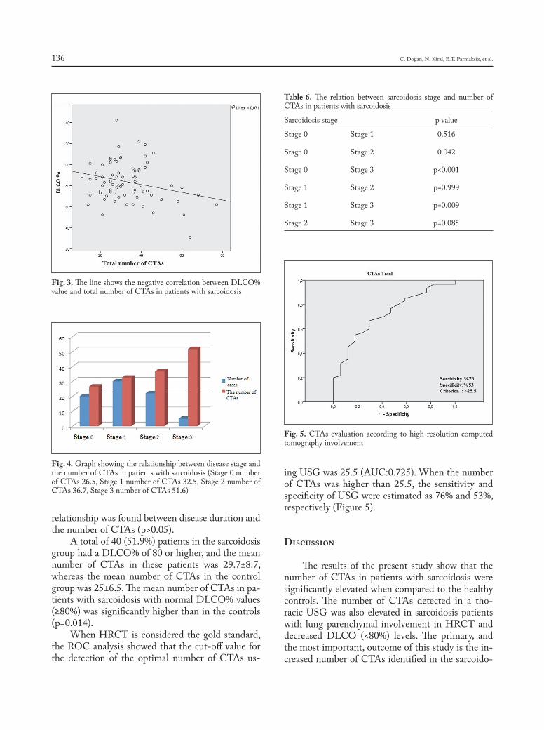
C. Doğan, N. Kiral, E.T. Parmaksiz, et al.136
relationship was found between disease duration and the number of CTAs (p>0.05).
A total of 40 (51.9%) patients in the sarcoidosis group had a DLCO% of 80 or higher, and the mean number of CTAs in these patients was 29.7±8.7, whereas the mean number of CTAs in the control group was 25±6.5. The mean number of CTAs in pa-tients with sarcoidosis with normal DLCO% values (≥80%) was significantly higher than in the controls (p=0.014).
When HRCT is considered the gold standard, the ROC analysis showed that the cut-off value for the detection of the optimal number of CTAs us-
ing USG was 25.5 (AUC:0.725). When the number of CTAs was higher than 25.5, the sensitivity and specificity of USG were estimated as 76% and 53%, respectively (Figure 5).
Discussion
The results of the present study show that the number of CTAs in patients with sarcoidosis were significantly elevated when compared to the healthy controls. The number of CTAs detected in a tho-racic USG was also elevated in sarcoidosis patients with lung parenchymal involvement in HRCT and decreased DLCO (<80%) levels. The primary, and the most important, outcome of this study is the in-creased number of CTAs identified in the sarcoido-
Fig. 3. The line shows the negative correlation between DLCO% value and total number of CTAs in patients with sarcoidosis
Fig. 4. Graph showing the relationship between disease stage and the number of CTAs in patients with sarcoidosis (Stage 0 number of CTAs 26.5, Stage 1 number of CTAs 32.5, Stage 2 number of CTAs 36.7, Stage 3 number of CTAs 51.6)
Table 6. The relation between sarcoidosis stage and number of CTAs in patients with sarcoidosis
Sarcoidosis stage p value
Stage 0 Stage 1 0.516
Stage 0 Stage 2 0.042
Stage 0 Stage 3 p<0.001
Stage 1 Stage 2 p=0.999
Stage 1 Stage 3 p=0.009
Stage 2 Stage 3 p=0.085
Fig. 5. CTAs evaluation according to high resolution computed tomography involvement
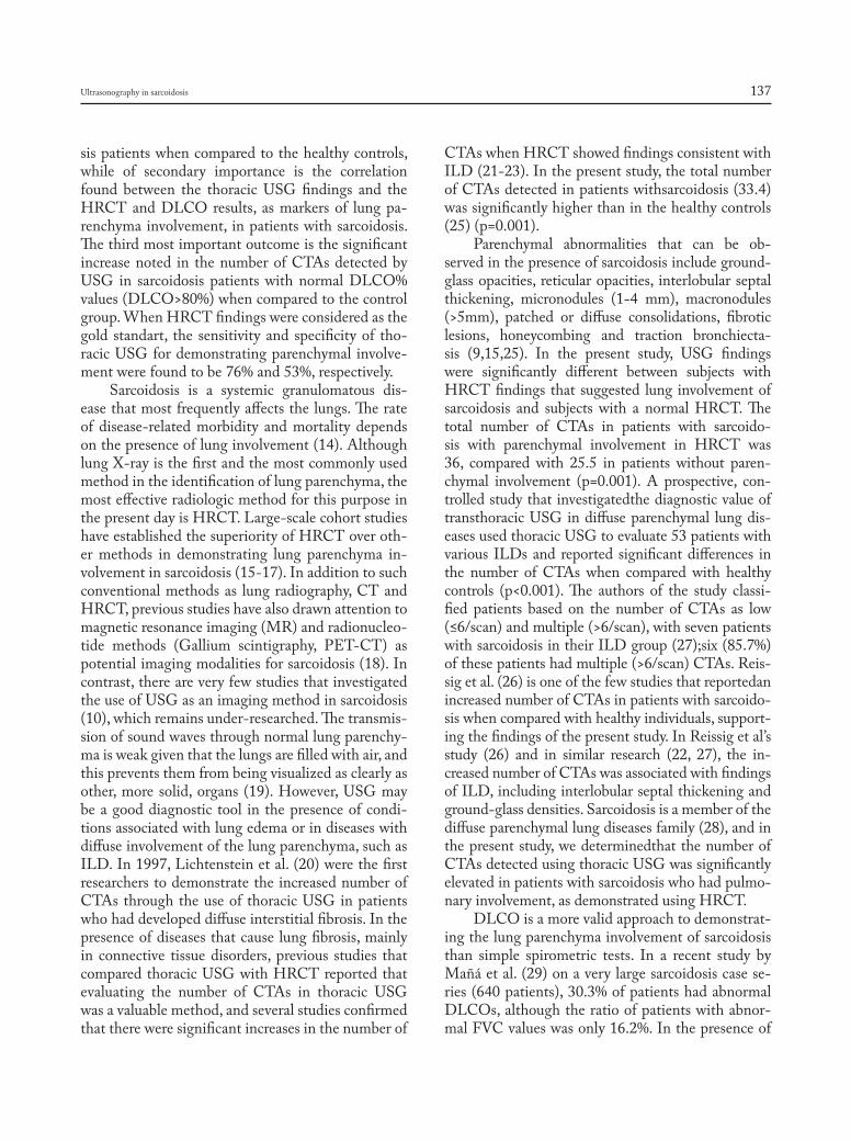
Ultrasonography in sarcoidosis 137
sis patients when compared to the healthy controls, while of secondary importance is the correlation found between the thoracic USG findings and the HRCT and DLCO results, as markers of lung pa-renchyma involvement, in patients with sarcoidosis. The third most important outcome is the significant increase noted in the number of CTAs detected by USG in sarcoidosis patients with normal DLCO% values (DLCO>80%) when compared to the control group. When HRCT findings were considered as the gold standart, the sensitivity and specificity of tho-racic USG for demonstrating parenchymal involve-ment were found to be 76% and 53%, respectively.
Sarcoidosis is a systemic granulomatous dis-ease that most frequently affects the lungs. The rate of disease-related morbidity and mortality depends on the presence of lung involvement (14). Although lung X-ray is the first and the most commonly used method in the identification of lung parenchyma, the most effective radiologic method for this purpose in the present day is HRCT. Large-scale cohort studies have established the superiority of HRCT over oth-er methods in demonstrating lung parenchyma in-volvement in sarcoidosis (15-17). In addition to such conventional methods as lung radiography, CT and HRCT, previous studies have also drawn attention to magnetic resonance imaging (MR) and radionucleo-tide methods (Gallium scintigraphy, PET-CT) as potential imaging modalities for sarcoidosis (18). In contrast, there are very few studies that investigated the use of USG as an imaging method in sarcoidosis (10), which remains under-researched. The transmis-sion of sound waves through normal lung parenchy-ma is weak given that the lungs are filled with air, and this prevents them from being visualized as clearly as other, more solid, organs (19). However, USG may be a good diagnostic tool in the presence of condi-tions associated with lung edema or in diseases with diffuse involvement of the lung parenchyma, such as ILD. In 1997, Lichtenstein et al. (20) were the first researchers to demonstrate the increased number of CTAs through the use of thoracic USG in patients who had developed diffuse interstitial fibrosis. In the presence of diseases that cause lung fibrosis, mainly in connective tissue disorders, previous studies that compared thoracic USG with HRCT reported that evaluating the number of CTAs in thoracic USG was a valuable method, and several studies confirmed that there were significant increases in the number of
CTAs when HRCT showed findings consistent with ILD (21-23). In the present study, the total number of CTAs detected in patients withsarcoidosis (33.4) was significantly higher than in the healthy controls (25) (p=0.001).
Parenchymal abnormalities that can be ob-served in the presence of sarcoidosis include ground-glass opacities, reticular opacities, interlobular septal thickening, micronodules (1-4 mm), macronodules (>5mm), patched or diffuse consolidations, fibrotic lesions, honeycombing and traction bronchiecta-sis (9,15,25). In the present study, USG findings were significantly different between subjects with HRCT findings that suggested lung involvement of sarcoidosis and subjects with a normal HRCT. The total number of CTAs in patients with sarcoido-sis with parenchymal involvement in HRCT was 36, compared with 25.5 in patients without paren-chymal involvement (p=0.001). A prospective, con-trolled study that investigatedthe diagnostic value of transthoracic USG in diffuse parenchymal lung dis-eases used thoracic USG to evaluate 53 patients with various ILDs and reported significant differences in the number of CTAs when compared with healthy controls (p<0.001). The authors of the study classi-fied patients based on the number of CTAs as low (≤6/scan) and multiple (>6/scan), with seven patients with sarcoidosis in their ILD group (27);six (85.7%) of these patients had multiple (>6/scan) CTAs. Reis-sig et al. (26) is one of the few studies that reportedan increased number of CTAs in patients with sarcoido-sis when compared with healthy individuals, support-ing the findings of the present study. In Reissig et al’s study (26) and in similar research (22, 27), the in-creased number of CTAs was associated with findings of ILD, including interlobular septal thickening and ground-glass densities. Sarcoidosis is a member of the diffuse parenchymal lung diseases family (28), and in the present study, we determinedthat the number of CTAs detected using thoracic USG was significantly elevated in patients with sarcoidosis who had pulmo-nary involvement, as demonstrated using HRCT.
DLCO is a more valid approach to demonstrat-ing the lung parenchyma involvement of sarcoidosis than simple spirometric tests. In a recent study by Mañá et al. (29) on a very large sarcoidosis case se-ries (640 patients), 30.3% of patients had abnormal DLCOs, although the ratio of patients with abnor-mal FVC values was only 16.2%. In the presence of
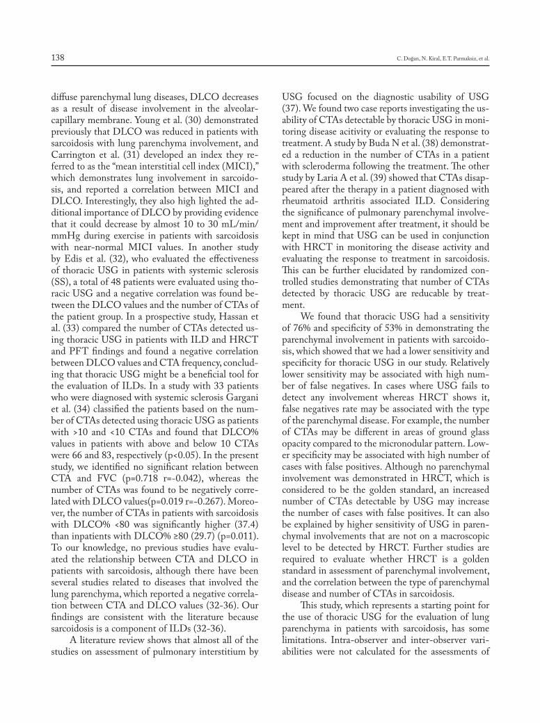
C. Doğan, N. Kiral, E.T. Parmaksiz, et al.138
diffuse parenchymal lung diseases, DLCO decreases as a result of disease involvement in the alveolar-capillary membrane. Young et al. (30) demonstrated previously that DLCO was reduced in patients with sarcoidosis with lung parenchyma involvement, and Carrington et al. (31) developed an index they re-ferred to as the “mean interstitial cell index (MICI),” which demonstrates lung involvement in sarcoido-sis, and reported a correlation between MICI and DLCO. Interestingly, they also high lighted the ad-ditional importance of DLCO by providing evidence that it could decrease by almost 10 to 30 mL/min/mmHg during exercise in patients with sarcoidosis with near-normal MICI values. In another study by Edis et al. (32), who evaluated the effectiveness of thoracic USG in patients with systemic sclerosis (SS), a total of 48 patients were evaluated using tho-racic USG and a negative correlation was found be-tween the DLCO values and the number of CTAs of the patient group. In a prospective study, Hassan et al. (33) compared the number of CTAs detected us-ing thoracic USG in patients with ILD and HRCT and PFT findings and found a negative correlation between DLCO values and CTA frequency, conclud-ing that thoracic USG might be a beneficial tool for the evaluation of ILDs. In a study with 33 patients who were diagnosed with systemic sclerosis Gargani et al. (34) classified the patients based on the num-ber of CTAs detected using thoracic USG as patients with >10 and <10 CTAs and found that DLCO% values in patients with above and below 10 CTAs were 66 and 83, respectively (p<0.05). In the present study, we identified no significant relation between CTA and FVC (p=0.718 r=-0.042), whereas the number of CTAs was found to be negatively corre-lated with DLCO values(p=0.019 r=-0.267). Moreo-ver, the number of CTAs in patients with sarcoidosis with DLCO% <80 was significantly higher (37.4) than inpatients with DLCO% ≥80 (29.7) (p=0.011). To our knowledge, no previous studies have evalu-ated the relationship between CTA and DLCO in patients with sarcoidosis, although there have been several studies related to diseases that involved the lung parenchyma, which reported a negative correla-tion between CTA and DLCO values (32-36). Our findings are consistent with the literature because sarcoidosis is a component of ILDs (32-36).
A literature review shows that almost all of the studies on assessment of pulmonary interstitium by
USG focused on the diagnostic usability of USG (37). We found two case reports investigating the us-ability of CTAs detectable by thoracic USG in moni-toring disease acitivity or evaluating the response to treatment. A study by Buda N et al. (38) demonstrat-ed a reduction in the number of CTAs in a patient with scleroderma following the treatment. The other study by Laria A et al. (39) showed that CTAs disap-peared after the therapy in a patient diagnosed with rheumatoid arthritis associated ILD. Considering the significance of pulmonary parenchymal involve-ment and improvement after treatment, it should be kept in mind that USG can be used in conjunction with HRCT in monitoring the disease activity and evaluating the response to treatment in sarcoidosis. This can be further elucidated by randomized con-trolled studies demonstrating that number of CTAs detected by thoracic USG are reducable by treat-ment.
We found that thoracic USG had a sensitivity of 76% and specificity of 53% in demonstrating the parenchymal involvement in patients with sarcoido-sis, which showed that we had a lower sensitivity and specificity for thoracic USG in our study. Relatively lower sensitivity may be associated with high num-ber of false negatives. In cases where USG fails to detect any involvement whereas HRCT shows it, false negatives rate may be associated with the type of the parenchymal disease. For example, the number of CTAs may be different in areas of ground glass opacity compared to the micronodular pattern. Low-er specificity may be associated with high number of cases with false positives. Although no parenchymal involvement was demonstrated in HRCT, which is considered to be the golden standard, an increased number of CTAs detectable by USG may increase the number of cases with false positives. It can also be explained by higher sensitivity of USG in paren-chymal involvements that are not on a macroscopic level to be detected by HRCT. Further studies are required to evaluate whether HRCT is a golden standard in assessment of parenchymal involvement, and the correlation between the type of parenchymal disease and number of CTAs in sarcoidosis.
This study, which represents a starting point for the use of thoracic USG for the evaluation of lung parenchyma in patients with sarcoidosis, has some limitations. Intra-observer and inter-observer vari-abilities were not calculated for the assessments of
Ultrasonography in sarcoidosis 139
thoracic USG and identification of CTAs, as well as for HRCT findings, which represents the most im-portant limitation of this study. Therefore, while in-terpreting the study results one should consider that thoracic USG is a highly user-dependent imaging technique. Another limitation is that this study was performed on a relatively low number of patients and reflected the experiences of a single center. Accord-ingly, the findings of this study cannot be generalized.
In conclusion, this study has shown that statisti-cally significant correlations exist between thoracic USG findings, which represent a novel method for the assessment of lung parenchyma involvement in patients with sarcoidosis, and the findings of HRCT, which is currently considered to be the most sensi-tive imaging method for the demonstration of lung parenchymal involvement of sarcoidosis, as well as DLCO, currently known to be the most effective test for showing the diffusion of gases through the alveo-lar-capillary membrane, and therefore, the potential involvement of the alveolar-capillary membrane. We believe that although it may not be the most pre-ferred method for the evaluation of parenchymal in-volvement in patients with sarcoidosis, thoracic USG may still have an area of use in the regular moni-toring and assessment of treatment response in such patients. Further studies are required to investigate methods of early detection of changes in lung pa-renchyma. We evaluated the use of USG for assess-ment of sarcoidosis. We believe that, as much as the healthy lung parenchyma cannot be evaluated using USG, it may be effective for the evaluation of lung parenchyma in the presence of such diseases as ILD, which extensively affects the lung parenchyma.
Acknowledgement
We thank MD ˙Ilker Akin for his contribution with his illustrations.
References
1. Wijsenbeek MS, Culver DA. Treatment of Sarcoidosis. Clin Chest Med 2015; 36: 751.
2. Iannuzzi MC, Rybicki BA, Teirstein AS. Sarcoidosis. N Engl J Med 2007; 357: 2153.
3. Baughman RP, Grutters JC. New treatment strategies for pulmonary sarcoidosis: antimetabolites, biological drugs, and other treatment ap-proaches. Lancet Respir Med 2015; 3: 813.
4. Bradley B, Branley HM, Egan JJ, et al. Interstitial lung disease guide-line: the British Thoracic Society in collaboration with the Thoracic Society of Australia and New Zealand and the Irish Thoracic Society. Thorax 2008; 63: v1-58.
5. Baughman RP, Culver DA, Judson MA. A concise review of pulmo-nary sarcoidosis. Am J Respir Crit Care Med 2011; 183: 573.
6. Mallamaci F, Benedetto FA, Tripepi R, et al. Detection of pulmonary congestion by chest ultrasound in dialysis patients. JACC Cardiovasc Imaging 2010; 3: 586-94.
7. Koenig SJ, Narasimhan M, Mayo PH. Thoracic ultrasonography forthe pulmonary specialist. Chest 2011; 140: 1332-1341.
8. Soldati G, Inchingolo R, Smargiassi A, et al. Ex vivo lung sonography: morphologic-ultrasound relationship. Ultrasound Med Biol 2012; 38: 1169-79.
9. Volpicelli G, Elbarbary M, Blaivas M, et al. International evidence-basedrecommendations for point-of-care lung ultrasound. Intensive Care Med 2012; 38: 577-91.
10. Targhetta R, Chavagneux R, Balmes P, et al. Sonographic lung surface evaluation in pulmonary sarcoidosis: preliminary results. J Ultrasound Med. 1994; 13: 381-8.
11. Miller MR, Hankinson J, Brusasco V, et al. ATS/ERS task force standardisation of lung function testing. Edited by V. Brusasco, R. Crapo and G. Viegi Number 2 in this Series Standardisation of spirometry. EurRespir J 2005; 26: 319-338.
12. Standardization of Spirometry, 1994 Update. American Thoracic So-ciety. Am J Respir Crit Care Med 1995; 152: 1107-1136.
13. Barskova T, Gargani L, Guiducci S, et al. Lung ultrasound for the screening of interstitial lung disease in very early systemic sclerosis. Ann Rheum Dis 2013; 72: 390-5.
14. Criado E, Sánchez M, Ramírez J, et al. Pulmonary sarcoidosis: typi-cal and atypical manifestations at high-resolution CT with pathologic correlation. Radiographics 2010; 30: 1567-86.
15. Dhagat PK, Singh S, Jain M, Singh SN, Sharma RK. Thoracic Sar-coidosis: Imaging with High Resolution Computed Tomography. J ClinDiagn Res. 2017; 11: 15-18.
16. Spagnolo P, Sverzellati N, Wells AU, Hansell DM. Imaging aspects of the diagnosis of sarcoidosis. EurRadiol 2014; 24: 807-16.
17. Walsh SL, Wells AU, Sverzellati N, et al. An integrated clinicoradio-logical staging system for pulmonary sarcoidosis: a case-cohort study. Lancet Respir Med 2014; 2: 123-30.
18. Silva M, Nunes H, Valeyre D, SverzellatiN. Imaging of Sarcoidosis. Clin Rev Allergy Immunol 2015; 49: 45-53.
19. Smargiassi A, Inchingolo R, Soldati G, et al. The role of chest ultra-sonography in the management of respiratory diseases: Document 2. MultidiscipRespir Med 2013; 8: 55.
20. Lichtenstein D, Mézière G, Biderman P, Gepner A, Barré O. The comet-tail artifact. An ultrasoundsign of alveolar-interstitial syn-drome. Am J RespirCrit Care Med 1997; 156: 1640–6.
21. Doveri M, Frassi F, Consensi A, et al. Ultrasound lung comets: New echographic sign of lung interstitial fibrosis in systemic sclerosis. Reu-matismo 2008; 60: 180–4.
22. Lim JH, Lee KS, Kim TS, Chung MP. Ring-down artifacts poste-rior to the right hemidiaphragm on abdominal sonography: sign of pulmonary parenchymal abnormalities. J Ultrasound Med 1999; 18: 403-10.
23. Sperandeo M, Varriale A, Sperandeo G, et al. Transthoracic ultra-sound inthe evaluation of pulmonary fibrosis: our experience. Ultra-sound Med Biol 2009; 35: 723-9.
24. Austin JH, Müller NL, Friedman PJ, et al. Glossary of terms for CT of the lungs: recommendations of the Nomenclature Committee of the Fleischner Society. Radiology 1996; 200: 327-31.
25. Al Jahdali H, Rajiah P, Koteyar SS. Atypical radiological manifesta-tions of thoracic sarcoidosis: A review and pictorial essay. Annals of Thoracic Medicine. 2013; 8: 186–96.
26. Reissig A, Kroegel C. Transthoracic sonography of diffuse parenchy-
C. Doğan, N. Kiral, E.T. Parmaksiz, et al.140
mal lung disease: the role of comet tail artifacts. J Ultrasound Med. 2003; 22: 173-80.
27. Gryminski J, Krakowka P, Lypacewicz G. The diagnosis of pleural effusion by ultrasonic and radiological techniques. Chest 1976; 70: 33-37.
28. American Thoracic Society, European Respiratory Society. Inter-national multidisciplinary consensus classification of the idiopathic interstitial pneumonias. Am J RespirCrit Care Med 2002; 165: 277-304.
29. Mañá J, Rubio-Rivas M, Villalba N, et al. Multidisciplinary approach and long-term follow-up in a series of 640 consecutive patients with sarcoidosis: Cohort study of a 40-year clinical experience at a tertiary referral center in Barcelona, Spain. Medicine (Baltimore) 2017; 96: e7595.
30. Young RL, Loudon RE, Krumholz RA, Harkleroad LE, Branam GE, Weg, JG. Pulmonary sarcoidosis: I. Pathophysiologic considerations. Am Rev Respir Dis 1968; 97: 997-1008.
31. Carrington CB. Structure and function in sarcoidosis. Ann N Y Acad Sci 1976; 278: 265-83.
32. Edis EÇ, Hatipoğlu ON, Pamuk ÖM, Eraslan RM, Aktöz M, Tuncel SA. Effectiveness of thoracic ultrasonography in the evaluation of the severity of pulmonary involvement in patients with systemic sclerosis. Arch Rheumatol 2016; 31: 364-370
33. Hasan AA, Makhlouf HA. B-lines: Transthoracic chest ultrasound signs useful in assessment of interstitial lung diseases. Ann Thorac Med 2014; 9: 99-103.
34. Gargani L, Doveri M, D‘Errico L, et al. Ultrasound lung comets in systemic sclerosis: a chest sonography hallmark of pulmonary intersti-tial fibrosis. Rheumatology (Oxford) 2009; 48: 1382-7.
35. Ostojić P, Damjanov N. Different clinical features in patients with limited and diffuse cutaneous systemic sclerosis. Clin Rheumatol 2006; 25: 453-7.
36. Gigante A, Rossi Fanelli F, Lucci S, et al. Lung ultrasound in sys-temic sclerosis: correlation with high-resolution computed tomogra-phy, pulmonary function tests and clinical variables of disease. Intern Emerg Med 2016; 11: 213-7.
37. Wang Y, Gargani L, Barskova T, et al. Usefulness of lung ultrasound B-lines in connective tissue disease-associated interstitial lung disease: a literature review. Arthritis Res Ther 2017; 19(1): 206.
38. Buda N, Kosiak W, Smoleńska Z, et al. Transthoracic lung ultrasound in the monitoring of interstitial lung disease: a case of scleroderma. Pol Arch Med Wewn 2013;123(12):721-2.
39. Laria A, Lurati A, Scarpellini M. Ultrasound in rheumatologic inter-stitial lung disease: a case report of nonspecific interstitial pneumonia in rheumatoid arthritis. Case Rep Rheumatol 2015;2015:107275. doi: 10.1155/2015/107275.
Introduction
Sarcoidosis is a widespread multisystem disease that preferentially involves the lungs, intra-thoracic lymph nodes, eyes, and skin. The gold standard of diagnosis includes pathological findings compatible with sarcoidosis and exclusion of other infectious and non-infectious granulomatous diseases along with the presence of compatible clinical features (1). The presence of multiple typical clinical manifestations
Cathepsin S, a new serum biomarker of sarcoidosis discovered by transcriptome analysis of alveolar macrophages
Hiroyuki Tanaka1, Etsuro Yamaguchi1, Nobuhiro Asai2, Toyoharu Yokoi3, Masaki Nishimura1, Haruhisa Nakao4, Masashi Yoneda4, Yoshinori Ohtsuka5, Satoshi Konno 6, Noritaka Yamada7
1 Division of Respiratory Medicine and Allergology, Department of Internal Medicine, School of Medicine, Aichi Medical University, Naga-kute, Aichi, Japan; 2 Department of Infection Control and Prevention, School of Medicine, Aichi Medical University, Nagakute, Aichi, Japan; 3 Department of Pathology, Nagoya Ekisaikai Hospital, Nagoya, Aichi, Japan; 4 Division of Hepatology and Pancreatology, Department of Internal Medicine, School of Medicine, Aichi Medical University, Nagakute, Aichi, Japan; 5 Department of Medicine, Hokkaido Chuo Rosai Hospita, Iwamizawa, Japan; 6 First Department of Medicine, Hokkaido University School of Medicine, Sapporo, Japan; 7 Department of Respiratory Medicine, National Hospital Organization, Higashinagoya Hospital, Nagoya, Aichi, Japan
Abstract. Background: Development of reliable new biomarkers remains crucial to improve diagnosis and assessing disease activity in sarcoidosis. The objective of this study was to seek such markers from the gene expression signature of alveolar macrophages by transcriptome analysis. Methods: Pooled RNA extracted from alveolar macrophages from patients with active sarcoidosis and control patients was subjected to transcriptome analysis using microarrays. Expressed gene intensity in sarcoidosis relative to that in control was calculated. We measured serum cathepsin S (CTSS) concentrations in 89 healthy volunteers, 107 patients with sarcoidosis, 26 with interstitial pneumonia, 150 with pneumoconiosis, and 76 with pulmonary mycobacteriosis by the enzyme-linked immunosorbent assay. Results: Among 12 genes with ratios higher than that of a housekeeping gene, we selected CTSS for scrutinizing protein expression in serum because of the feasibility of the protein assay. CTSS concentrations were significantly increased in sarcoidosis compared with not only controls but also all the other lung diseases. Receiver operating characteristics curve for sarcoidosis and parenchymal lung diseases revealed an area under the curve of 0.800 (95% confidence interval, 0.751-0.850; p=1.4 x 10⁻¹⁸) with 70% sensitivity and 78% specificity at a CTSS concentration of 15.5 ng/ml. A significant trend was identified between CTSS concen-trations and the number of affected organs. Serum CTSS concentrations varied in parallel with clinical courses both spontaneously and in response to corticosteroid therapy. Epithelioid cells in granulomas were positive for immunohistochemical staining with CTSS. Conclusions: CTSS has the potential to be a useful biomarker in sarcoidosis. (Sarcoidosis Vasc Diffuse Lung Dis 2019; 36 (2): 141-147)
Key words: biomarkers, cathepsins, macrophages, sarcoidosis, transcriptome
SARCOIDOSIS VASCULITIS AND DIFFUSE LUNG DISEASES 2019; 36 (2); 141-147 © Mattioli 1885
Original article: Clinical research
Received: 11 August 2018Accepted after revision: 20 August 2018Correspondence: Etsuro Yamaguchi, MD, PhD, Division of Respiratory Medicine and Allergology, Department of Internal Medicine, School of Medicine, Aichi Medical University, Karimata 1-1, Yazako, Nagakute, Aichi, Japan, 480-1195E-mail: [email protected]
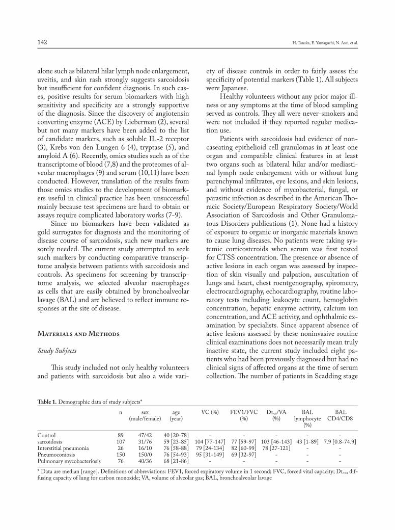
H. Tanaka, E. Yamaguchi, N. Asai, et al.142
alone such as bilateral hilar lymph node enlargement, uveitis, and skin rash strongly suggests sarcoidosis but insufficient for confident diagnosis. In such cas-es, positive results for serum biomarkers with high sensitivity and specificity are a strongly supportive of the diagnosis. Since the discovery of angiotensin converting enzyme (ACE) by Lieberman (2), several but not many markers have been added to the list of candidate markers, such as soluble IL-2 receptor (3), Krebs von den Lungen 6 (4), tryptase (5), and amyloid A (6). Recently, omics studies such as of the transcriptome of blood (7,8) and the proteomes of al-veolar macrophages (9) and serum (10,11) have been conducted. However, translation of the results from those omics studies to the development of biomark-ers useful in clinical practice has been unsuccessful mainly because test specimens are hard to obtain or assays require complicated laboratory works (7-9).
Since no biomarkers have been validated as gold surrogates for diagnosis and the monitoring of disease course of sarcoidosis, such new markers are sorely needed. The current study attempted to seek such markers by conducting comparative transcrip-tome analysis between patients with sarcoidosis and controls. As specimens for screening by transcrip-tome analysis, we selected alveolar macrophages as cells that are easily obtained by bronchoalveolar lavage (BAL) and are believed to reflect immune re-sponses at the site of disease.
Materials and Methods
Study Subjects
This study included not only healthy volunteers and patients with sarcoidosis but also a wide vari-
ety of disease controls in order to fairly assess the specificity of potential markers (Table 1). All subjects were Japanese.
Healthy volunteers without any prior major ill-ness or any symptoms at the time of blood sampling served as controls. They all were never-smokers and were not included if they reported regular medica-tion use.
Patients with sarcoidosis had evidence of non-caseating epithelioid cell granulomas in at least one organ and compatible clinical features in at least two organs such as bilateral hilar and/or mediasti-nal lymph node enlargement with or without lung parenchymal infiltrates, eye lesions, and skin lesions, and without evidence of mycobacterial, fungal, or parasitic infection as described in the American Tho-racic Society/European Respiratory Society/World Association of Sarcoidosis and Other Granuloma-tous Disorders publications (1). None had a history of exposure to organic or inorganic materials known to cause lung diseases. No patients were taking sys-temic corticosteroids when serum was first tested for CTSS concentration. The presence or absence of active lesions in each organ was assessed by inspec-tion of skin visually and palpation, auscultation of lungs and heart, chest roentgenography, spirometry, electrocardiography, echocardiography, routine labo-ratory tests including leukocyte count, hemoglobin concentration, hepatic enzyme activity, calcium ion concentration, and ACE activity, and ophthalmic ex-amination by specialists. Since apparent absence of active lesions assessed by these noninvasive routine clinical examinations does not necessarily mean truly inactive state, the current study included eight pa-tients who had been previously diagnosed but had no clinical signs of affected organs at the time of serum collection. The number of patients in Scadding stage
Table 1. Demographic data of study subjects*
n sex age VC (%) FEV1/FVC Dlco/VA BAL BAL (male/female) (year) (%) (%) lymphocyte CD4/CD8 (%)
Control 89 47/42 40 [20-78] - - - - -sarcoidosis 107 31/76 59 [23-85] 104 [77-147] 77 [59-97] 103 [46-143] 43 [1-89] 7.9 [0.8-74.9]Interstitial pneumonia 26 16/10 76 [58-88] 79 [24-134] 82 [60-99] 78 [27-121] - -Pneumoconiosis 150 150/0 76 [54-93] 95 [31-149] 69 [32-97] - - -Pulmonary mycobacteriosis 76 40/36 68 [21-86] - - - - -
* Data are median [range]. Definitions of abbreviations: FEV1, forced expiratory volume in 1 second; FVC, forced vital capacity; Dlco, dif-fusing capacity of lung for carbon monoxide; VA, volume of alveolar gas; BAL, bronchoalveolar lavage
Cathepsin S as a new biomarker of sarcoidosis 143
0, stage I, stage II, and stage III were 16 (15%), 56 (52%), 31 (29%), and 4 (4%), respectively. Clinical courses of sarcoidosis were judged by changes in the number of affected organs or apparent changes in the severity of lesions clinically assessed as described above.
Patients with idiopathic interstitial pneumonia had either a usual interstitial pneumonia pattern (n=14) or a nonspecific interstitial pneumonia pat-tern (n=12) on high-resolution computed tomog-raphy according to the ATS/ERS consensus clas-sification (12) or a published document (13) with no evidence of underlying diseases judging from occupational history, physical examination, and se-rological tests for collagen vascular diseases or other vasculitides. No patients were taking systemic corti-costeroids when sera were collected.
Pneumoconiosis was diagnosed based on long-standing occupational exposure to silica or coal, and discrete nodular shadows of variable sizes in predom-inantly upper lung areas on chest roentgenography or computed tomography. According to the manage-ment classification by the Ministry of Health, Labor and Welfare of Japan, 50 patients were classified into type 2 (a number of opacities smaller than 1 cm in diameter [ILO classification: p, q, r, s, t, u]), 50 into type 3 (numerous opacities smaller than 1 cm in di-ameter), and 50 into type 4 (nodular shadows larger than 1 cm in diameter).
Diagnosis of pulmonary mycobacteriosis was made based on compatible radiological findings and demonstration of mycobacteria in sputum and/or bronchoalveolar lavage fluid. Forty-six patients had tuberculosis and 30 had nontuberculous mycobacte-riosis.
Microarray
BAL was conducted in three nonsmoking pa-tients with active sarcoidosis with biopsy evidence and four nonsmoking controls who were suspected to have lung cancer. BAL was done in the middle lobe or lingular segments for patients with sarcoido-sis, or in segments unaffected by pulmonary le-sions for patients suspected of lung cancer. All were women. Since most lymphocytes of BAL cells are cluster of differentiation (CD) 2+ T cells (14) and the proportion of granulocytes was less than 3% in all the subjects studied, a cellular fraction of BAL
cells depleted of CD2+ cells by microbeads coated with anti-CD2 monoclonal antibody (MicroBeads, Miltenyi Biotec GmbH, Bergisch Gladbach, Ger-many) was used as alveolar macrophages. Total RNA from this cell fraction was extracted using BioRobot EZ1 (Qiagen GmbH, Hilden, Germany) and EZ1 RNA Cell Mini Kit (Qiagen). Equal amount of total RNA from each subject was pooled for the patient group or the control group, and 0.4 μg of RNA from each group was subjected to microarray analysis us-ing CodeLink Human Whole Genome Bioarray (Applied Microarrays, Tempe, AZ) and CodeLink Expression Bioarray System (Applied Microarrays). Array slides were hybridized with biotin-labelled cRNA, stained with streptoavidin-Cy5, washed ac-cording to the manufacturer’s protocol, and scanned by arrayWoRx (Applied Precision, Issaquah, WA).
Measurement of CTSS Protein and Immunostaining
Sera were obtained at the time of annual health check-up in healthy volunteers, at the time of initial visit or at the oldest time point in patients with sar-coidosis, and at variable time points in patients with other diseases. Concentrations of CTSS in sera di-luted 100-fold with assay diluent were measured us-ing a Human Total Cathepsin S DuoSet (R&D Sys-tems, Minneapolis, MN). Specific immunostaining for human CTSS was performed on formalin-fixed paraffin-embedded slides prepared from a mediasti-nal lymph node in a patient with sarcoidosis using goat anti-CTSS antibody (M-19, sc-6505; Santa Cruz Biotechnology, Santa Cruz, CA), and perox-idase-labelled anti-goat immunoglobulin (Histofine Simple Stain MAX-PO [G]; Nichirei Bioscience, Tokyo, Japan).
Statistical Analysis
Difference between two study groups was as-sessed by the Mann-Whitney test followed by Bon-ferroni’s correction to counteract multiple compari-sons. Receiver operating characteristics curve was used to determine sensitivity and specificity for discriminating sarcoidosis from control subjects or other lung diseases. The Youden index was used to determine the cut-off point. The Jonckheere-Terp-stra test was used to assess trend in CTSS concentra-tions across patient groups ranked by the number of
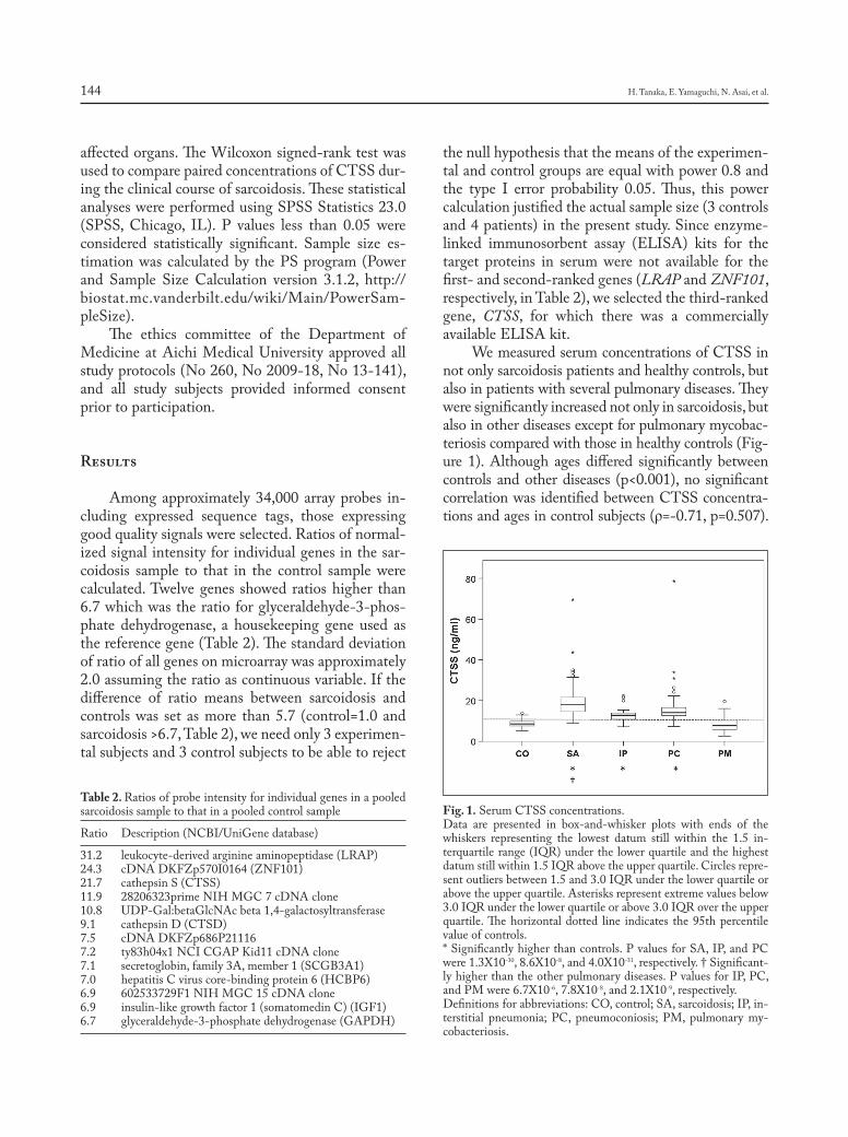
H. Tanaka, E. Yamaguchi, N. Asai, et al.144
affected organs. The Wilcoxon signed-rank test was used to compare paired concentrations of CTSS dur-ing the clinical course of sarcoidosis. These statistical analyses were performed using SPSS Statistics 23.0 (SPSS, Chicago, IL). P values less than 0.05 were considered statistically significant. Sample size es-timation was calculated by the PS program (Power and Sample Size Calculation version 3.1.2, http://biostat.mc.vanderbilt.edu/wiki/Main/PowerSam-pleSize).
The ethics committee of the Department of Medicine at Aichi Medical University approved all study protocols (No 260, No 2009-18, No 13-141), and all study subjects provided informed consent prior to participation.
Results
Among approximately 34,000 array probes in-cluding expressed sequence tags, those expressing good quality signals were selected. Ratios of normal-ized signal intensity for individual genes in the sar-coidosis sample to that in the control sample were calculated. Twelve genes showed ratios higher than 6.7 which was the ratio for glyceraldehyde-3-phos-phate dehydrogenase, a housekeeping gene used as the reference gene (Table 2). The standard deviation of ratio of all genes on microarray was approximately 2.0 assuming the ratio as continuous variable. If the difference of ratio means between sarcoidosis and controls was set as more than 5.7 (control=1.0 and sarcoidosis >6.7, Table 2), we need only 3 experimen-tal subjects and 3 control subjects to be able to reject
the null hypothesis that the means of the experimen-tal and control groups are equal with power 0.8 and the type I error probability 0.05. Thus, this power calculation justified the actual sample size (3 controls and 4 patients) in the present study. Since enzyme-linked immunosorbent assay (ELISA) kits for the target proteins in serum were not available for the first- and second-ranked genes (LRAP and ZNF101, respectively, in Table 2), we selected the third-ranked gene, CTSS, for which there was a commercially available ELISA kit.
We measured serum concentrations of CTSS in not only sarcoidosis patients and healthy controls, but also in patients with several pulmonary diseases. They were significantly increased not only in sarcoidosis, but also in other diseases except for pulmonary mycobac-teriosis compared with those in healthy controls (Fig-ure 1). Although ages differed significantly between controls and other diseases (p<0.001), no significant correlation was identified between CTSS concentra-tions and ages in control subjects (ρ=-0.71, p=0.507).
Table 2. Ratios of probe intensity for individual genes in a pooled sarcoidosis sample to that in a pooled control sample
Ratio Description (NCBI/UniGene database)
31.2 leukocyte-derived arginine aminopeptidase (LRAP)24.3 cDNA DKFZp570I0164 (ZNF101)21.7 cathepsin S (CTSS)11.9 28206323prime NIH MGC 7 cDNA clone10.8 UDP-Gal:betaGlcNAc beta 1,4-galactosyltransferase9.1 cathepsin D (CTSD)7.5 cDNA DKFZp686P21116 7.2 ty83h04x1 NCI CGAP Kid11 cDNA clone7.1 secretoglobin, family 3A, member 1 (SCGB3A1)7.0 hepatitis C virus core-binding protein 6 (HCBP6)6.9 602533729F1 NIH MGC 15 cDNA clone 6.9 insulin-like growth factor 1 (somatomedin C) (IGF1)6.7 glyceraldehyde-3-phosphate dehydrogenase (GAPDH)
Fig. 1. Serum CTSS concentrations.Data are presented in box-and-whisker plots with ends of the whiskers representing the lowest datum still within the 1.5 in-terquartile range (IQR) under the lower quartile and the highest datum still within 1.5 IQR above the upper quartile. Circles repre-sent outliers between 1.5 and 3.0 IQR under the lower quartile or above the upper quartile. Asterisks represent extreme values below 3.0 IQR under the lower quartile or above 3.0 IQR over the upper quartile. The horizontal dotted line indicates the 95th percentile value of controls. * Significantly higher than controls. P values for SA, IP, and PC were 1.3X10⁻³⁰, 8.6X10⁻⁸, and 4.0X10⁻³¹, respectively. † Significant-ly higher than the other pulmonary diseases. P values for IP, PC, and PM were 6.7X10⁻⁶, 7.8X10⁻⁸, and 2.1X10⁻⁹, respectively.Definitions for abbreviations: CO, control; SA, sarcoidosis; IP, in-terstitial pneumonia; PC, pneumoconiosis; PM, pulmonary my-cobacteriosis.
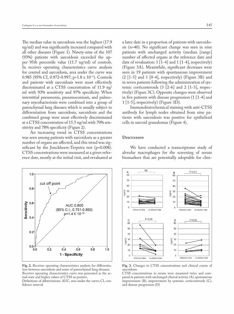
Cathepsin S as a new biomarker of sarcoidosis 145
The median value in sarcoidosis was the highest (17.9 ng/ml) and was significantly increased compared with all other diseases (Figure 1). Ninety-nine of the 107 (93%) patients with sarcoidosis exceeded the up-per 95th percentile value (11.7 ng/ml) of controls. In receiver operating characteristics curve analysis for control and sarcoidosis, area under the curve was 0.985 (95% CI, 0.972-0.997; p=1.8 x 10⁻³¹). Controls and patients with sarcoidosis were most effectively discriminated at a CTSS concentration of 11.9 ng/ml with 93% sensitivity and 97% specificity. When interstitial pneumonia, pneumoconiosis, and pulmo-nary mycobacteriosis were combined into a group of parenchymal lung diseases which is usually subject to differentiation from sarcoidosis, sarcoidosis and the combined group were most effectively discriminated at a CTSS concentration of 15.5 ng/ml with 70% sen-sitivity and 78% specificity (Figure 2).
An increasing trend in CTSS concentrations was seen among patients with sarcoidosis as a greater number of organs are affected, and this trend was sig-nificant by the Jonckheere-Terpstra test (p=0.008). CTSS concentrations were measured at a given refer-ence date, mostly at the initial visit, and revaluated at
a later date in a proportion of patients with sarcoido-sis (n=40). No significant change was seen in nine patients with unchanged activity (median [range] number of affected organs at the reference date and date of revaluation: 1 [1-4] and 1 [1-4], respectively) (Figure 3A). Meanwhile, significant decreases were seen in 19 patients with spontaneous improvement (2 [1-5] and 1 [0-4], respectively) (Figure 3B) and in seven patients following the administration of sys-temic corticosteroids (3 [2-6] and 2 [1-3], respec-tively) (Figure 3C). Opposite changes were observed in five patients with disease progression (1 [1-4] and 1 [1-5], respectively) (Figure 3D).
Immunohistochemical staining with anti-CTSS antibody for lymph nodes obtained from nine pa-tients with sarcoidosis was positive for epithelioid cells in sarcoid granulomas (Figure 4).
Discussion
We have conducted a transcriptome study of alveolar macrophages for the screening of serum biomarkers that are potentially adoptable for clini-
Fig. 2. Receiver operating characteristics analysis for differentia-tion between sarcoidosis and some of parenchymal lung diseases. Receiver operating characteristics curve was generated as the ac-tual state and higher values of CTSS as positive.Definitions of abbreviations: AUC, area under the curve; CI, con-fidence interval
Fig. 3. Changes in CTSS concentrations and clinical course of sarcoidosis.CTSS concentrations in serum were measured twice and com-pared in patients with unchanged clinical activity (A), spontaneous improvement (B), improvement by systemic corticosteroids (C), and disease progression (D)
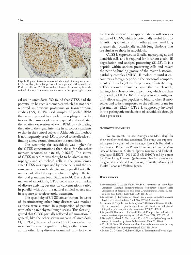
H. Tanaka, E. Yamaguchi, N. Asai, et al.146
cal use in sarcoidosis. We found that CTSS had the potential to be such a biomarker, which has not been reported in previous proteomic or transcriptomic studies (7-9,11). We used samples of pooled RNA that were expressed by alveolar macrophages in order to save the number of arrays required and evaluated the relative expression of each RNA by calculating the ratio of the signal intensity in sarcoidosis patients to that in the control subjects. Although this method is not frequently used (15), it proved to be effective in finding a new serum biomarker in sarcoidosis.
The sensitivity for sarcoidosis was higher for the CTSS concentrations than those for the other markers reported to date (6,10,16,17). The source of CTSS in serum was thought to be alveolar mac-rophages and epithelioid cells in the granulomas, since CTSS was expressed by these cells and the se-rum concentrations tended to rise in parallel with the number of affected organs, which roughly reflected the total granuloma load. Similar to ACE as a classic marker of sarcoidosis, CTSS could also be a marker of disease activity, because its concentrations varied in parallel with both the natural clinical course and in response to corticosteroid treatment.
The specificity of CTSS concentrations in terms of discriminating other lung diseases was modest, as these were elevated in a proportion of patients with other parenchymal lung diseases. This fact sug-gested that CTSS partially reflected inflammation in general, like the other serum markers of sarcoidosis (3,18,19,20). Nevertheless, the CTSS concentrations in sarcoidosis were significantly higher than those in all the other lung diseases examined. This fact ena-
bled establishment of an appropriate cut-off concen-tration of CTSS, which is potentially useful for dif-ferentiating sarcoidosis from other parenchymal lung diseases that occasionally exhibit lung shadows that are similar to those in sarcoidosis.
CTSS is expressed in B cells, macrophages, and dendritic cells and is required for invariant chain (Ii) degradation and antigen processing (21,22). Ii is a peptide within antigen-presenting cells and covers the peptide-binding groove of the major histocom-patibility complex (MHC) II molecules until it en-counters a foreign peptide in the lysosomal compart-ment of the cells (7). In the presence of interferon-γ, CTSS becomes the main enzyme that can cleave Ii, leaving class II-associated Ii peptides, which are then displaced by HLA-DM in the presence of antigens. This allows antigen peptides to bind to class II mol-ecules and to be transported to the cell membrane for presentation (22,23). CTSS is supposedly involved in the pathogenic mechanism of sarcoidosis through these processes.
Acknowledgments
We are grateful to Ms. Kamiya and Ms. Takagi for their excellent technical assistance.This study was support-ed in part by a grant of the Strategic Research Foundation Grant-aided Project for Private Universities from the Min-istry of Education, Culture, Sports, Science, and Technol-ogy, Japan (MEXT), 2011-2015 (S1101027) and by a grant for Rare Lung Diseases (pulmonary alveolar proteinosis, congenital interstitial lung disease) from the Ministry of Health Labor and Welfare, Japan.
References
1. Hunninghake GW. ATS/ERS/WASOG statement on sarcoidosis. American Thoracic Society/European Respiratory Society/World Association of Sarcoidosis and other Granulomatous Disorders. Sar-coidosis Vasc Diffuse Lung Dis 1999; 16: 149-73.
2. Lieberman J. Elevation of serum angiotensin-converting-enzyme (ACE) level in sarcoidosis. Am J Med 1975; 59: 365-72.
3. Tsutsumi T, Nagai S, Imai K, Setoyama Y, Uchiyama T, Izumi T. Solu-ble interleukin-2 receptor in blood from patients with sarcoidosis and idiopathic pulmonary fibrosis. Sarcoidosis 1994; 11: 102-9.
4. Miyoshi S, Hamada H, Kadowaki T, et al. Comparative evaluation of serum markers in pulmonary sarcoidosis. Chest 2010; 137: 1391-7.
5. Bargagli E, Mazzi A, Mezzasalma F, et al. The analysis of tryptase in serum of sarcoidosis patients. Inflammation 2009; 32: 310-4.
6. Eroglu H, Genc NS. Conventional markers in determination of activity of sarcoidosis. Int Immunopharmacol 2015; 25: 174-9.
7. Bloom CI, Graham CM, Berry MP, et al. Transcriptional blood signa-
Fig. 4. Representative immunohistochemical staining with anti-CTSS antibody for a lymph node from a patient with sarcoidosis. Positive cells for CTSS are stained brown. A hematoxylin-eosin stained picture of the same area is shown in the upper right corner.
Cathepsin S as a new biomarker of sarcoidosis 147
tures distinguish pulmonary tuberculosis, pulmonary sarcoidosis, pneumonias and lung cancers. PLoS One 2013; 8: e70630.
8. Su R, Li MM, Bhakta NR, et al. Longitudinal analysis of sarcoidosis blood transcriptomic signatures and disease outcomes. Eur Respir J 2014; 44: 985-93.
9. Silva E, Souchelnytsukyi S, Kasuga K, Eklund A, Grunewald J, Wheelock A. Quantitative intact proteomics investigations of alveo-lar macrophages in sarcoidosis. Eur Respir J 2013; 41: 1331-9.
10. Bons JA, Drent M, Bouwman FG, Mariman EC, van Dieijen-Visser MP, Wodzig WK. Potential biomarkers for diagnosis of sarcoidosis using proteomics in serum. Respir Med 2007; 101: 1687-95.
11. Zhang Y, Chen X, Hu Y, et al. Preliminary characterizations of a serum biomarker for sarcoidosis by comparative proteomic approach with tandem-mass spectrometry in ethnic Han Chinese patients. Respir Res 2013; 14: 18.
12. Raghu G, Collard HR, Egan JJ, et al. An official ATS/ERS/JRS/ALAT statement: idiopathic pulmonary fibrosis: evidence-based guidelines for diagnosis and management. Am J Respir Crit Care Med 2011; 183: 788-824.
13. Travis WD, Costabel U, Hansell DM, et al. An official American Thoracic Society/European Respiratory Society statement: Update of the international multidisciplinary classification of the idiopathic in-terstitial pneumonias. Am J Respir Crit Care Med 2013; 188: 733-48.
14. Yamaguchi E, Okazaki N, Itoh A, Furuya K, Abe S, Kawakami Y. Enhanced expression of CD2 antigen on lung T cells. Am Rev Respir Dis 1991; 143(4 Pt 1): 829-33.
15. Rego N, Bianchi S, Moreno P, et al. Search for an aetiological virus candidate in chronic lymphocytic leukaemia by extensive transcrip-tome analysis. Br J Haematol 2012; 157: 709-17.
16. Furuya K, Yamaguchi E, Itoh A, et al. Deletion polymorphism in the angiotensin I converting enzyme (ACE) gene as a genetic risk factor for sarcoidosis. Thorax 1996; 51: 777-80.
17. Grutters JC, Fellrath JM, Mulder L, Janssen R, van den Bosch JM, van Velzen-Blad H. Serum soluble interleukin-2 receptor measure-ment in patients with sarcoidosis: a clinical evaluation. Chest 2003; 124: 186-95.
18. Witkowska AM. On the role of sIL-2R measurements in rheumatoid arthritis and cancers. Mediators Inflamm 2005; 2005: 121-30.
19. Ishii H, Iwata A, Oka H, Sakamoto N, Ishimatsu Y, Kadota J. Elevat-ed serum levels of lysozyme in desquamative interstitial pneumonia. Intern Medicine 2010; 49: 847-51.
20. Sipeki N, Antal-Szalmas P, Lakatos PL, Papp M. Immune dysfunc-tion in cirrhosis. World J Gastroenterol 2014; 20: 2564-77.
21. Nakagawa TY, Rudensky AY. The role of lysosomal proteinases in MHC class II-mediated antigen processing and presentation. Immu-nol Rev 1999; 172: 121-9.
22. Honey K, Rudensky Y. Lysosomal cysteine proteases regulate antigen presentation. Nat Rev Immunol 2003; 3: 472-82.
23. Rocha N, Neefjes J. MHC class II molecules on the move for success-ful antigen presentation. EMBO J 2008; 27:1-5.
Abbreviations List:SLB: surgical lung biopsyVATS: video-assisted thoracoscopic surgery OLB: open lung biopsyHRCT: high resolution computed tomography
ILD: interstitial lung disease MDD: multidisciplinary discussion diagnosisIIPs: idiopathic interstitial pneumoniasIPF: idiopathic pulmonary fibrosisNSIP: nonspecific interstitial pneumoniaUIP: usual interstitial pneumoniaCHP: chronic hypersensitivity pneumoniaPPFE: pleuroparenchymal fibroelastosisAE: acute exacerbationKL-6: Krebs von den Lungen-6SP-D: surfactant protein-DFVC: forced vital capacityDLco: diffusing capacity for carbon monoxide
The role of video-assisted thoracoscopic surgery in the diagnosis of interstitial lung disease
Keishi Sugino1, 5, Hajime Otsuka2, Yusuke Matsumoto1, Yasuhiko Nakamura1, Keiko Matsumoto3, Yoko Azuma2, Takashi Makino2, Akira Iyoda2, Kazutoshi Shibuya4, Sakae Homma1
1Department of Respiratory Medicine, 2Department of Chest Surgery, 3Department of Diagnostic Radiology, 4Department of Pathology, Toho University Omori Medical Center, Tokyo, Japan; 5Department of Respiratory Medicine, Jizankai Medical Fundation Tsuboi Cancer Center Hospital, Asakamachi, Koriyama City, Fukushima, Japan
Abstract. Background: When a clinical context is indeterminate for idiopathic pulmonary fibrosis (IPF), or a chest high-resolution computed tomography (HRCT) pattern is not indicative of typical or probable usual inter-stitial pneumonia (UIP) in patients with interstitial lung disease (ILD), surgical lung biopsy should be considered to make a confident diagnosis on the basis of multidisciplinary diagnosis (MDD). Aim: The aim of this study was to evaluate the role and safety of video-assisted thoracoscopic surgery (VATS) in patients with ILD. Methods: A total of 143 patients with ILD underwent VATS at Toho University Medical Center Omori Hospital between March 2004 and April 2017. We conducted a retrospective study on the usefulness and safety of VATS in the diagnosis of ILD under MDD. Results: The 30-day mortality was 0%. The postoperative complication rate was 12.6%, which included 5 cases of pneumothorax after discharge (3.5%), 4 cases of prolonged air leakage (2.8%), and 2 cases of acute exacerbation (1.4%). Three of 9 cases (33.3%) complicated by pneumothorax after discharge or prolonged air leakage were resected specimens of pleuroparenchymal fibroelastosis (PPFE). Two patients had acute exacerbation, who were ultimately diagnosed as having idiopathic unclassifiable IP and had histologically significant irregular dense fibrosis and numerous fibroblastic foci. The comparison between chest HRCT and his-topathological findings revealed 55 cases of possible UIP [UIP (45%), NSIP (25%), and unclassifiable IP (29%)] and 21 cases of inconsistent with UIP [UIP (10%), NSIP (33%), organizing pneumonia (10%), unclassifiable IP (24%), and PPFE (24%)]. Conclusion: VATS can be safely performed to obtain a confident diagnosis for appropri-ate treatment strategies in patients with ILD. (Sarcoidosis Vasc Diffuse Lung Dis 2019; 36 (2): 148-156)
Key words: interstitial lung disease, video-assisted thoracoscopic surgery, complication, survival, multidiscipli-nary discussion diagnosise
SARCOIDOSIS VASCULITIS AND DIFFUSE LUNG DISEASES 2019; 36 (2); 148-156 © Mattioli 1885
Original article: Clinical research
Received: 3 November 2018Accepted after revision: 28 April 2019Correspondence: Keishi Sugino, MD. PhDDepartment of Respiratory Medicine, Toho University School of Medicine, 6-11-1, Omori-nishi, Ota-ku, Tokyo 143-8541, JapanTel: +81-3-3762-4151 - Fax: +81-3-3766-3551E-mail: [email protected]
The role of VATS in ILD 149
Introduction
In recent years, the necessity of surgical lung biopsy (SLB) for the purpose of diagnosis of inter-stitial lung disease (ILD) has been questioned not only because of the development of the chest high-resolution computed tomography (HRCT) but also because of the high morbidity and mortality associ-ated with the procedure. More recently, Lynch, et al. (1) emphasized in a Fleischner Society White Paper that a confident diagnosis of idiopathic pulmonary fibrosis (IPF) can be made in a correct clinical con-text without SLB when CT imaging shows a pattern of typical or probable usual interstitial pneumonia (UIP). However, it is very important and challeng-ing work-up for pulmonologist to make a correct diagnosis from over 100 different ILDs (2). When a clinical context is indeterminate for IPF, or a chest HRCT pattern is not indicative of typical or prob-able UIP in patients with ILD, surgical lung biopsy should be considered to make a confident diagnosis on the basis of multidisciplinary discussion diagnosis (MDD). Indeed, the chest HRCT findings do not always represent typical features of patients with ILD. Sverzellati et al. (3) reported that 34 out of 55 patients diagnosed as IPF on biopsy had received a diagnosis of NSIP, CHP, or sarcoidosis on SUGINO, The role VATS in ILD chest CT. In addition, Mor-ris et al. (4) described that only 54% of patients who received a consensus diagnosis of UIP after video-assisted thoracoscopic surgery (VATS) lung biopsy, had received a diagnosis of probable UIP on chest HRCT. Therefore, VATS can be considered as one of necessary tool for the accurate diagnosis of ILD.
It has been reported that in general, risk fac-tors of SLB are male sex, increasing age, increas-ing comorbidity, unstable condition such as rapidly progressive ILD requiring mechanical ventilation, severely impaired pulmonary function, coexisting of pulmonary hypertension in patients with ILD, un-dergoing open lung biopsy (OLB), and a provisional diagnosis of IPF or connective tissue disease–related ILD (5-8). VATS is generally considered as a safe procedure to provide adequate lung tissue samples for definitive histological diagnosis. However, post-operative complications may outweigh the potential benefits in patients with ILD because postoperative acute exacerbation (AE) or prolonged air leakage is one of the particularly critical and significant com-
plications. According to a comprehensive literature review by Nguyen and Meyer (9), the overall 30-day mortality for OLB was 4.3% versus 2.1% for VATS biopsy, and non-lethal complications appeared to oc-cur more frequently with OLB (18.1%) vs. VATS (9.6%) procedures. Given that VATS reduces risks of morbidity and mortality in this study, we aimed to assess the usefulness and postoperative complications of VATS in patients with ILD at our institution.
Methods
Patients
This study cohort included a total of 143 con-secutive patients underwent VATS for suspected ILD at Toho University Omori Medical Center be-tween March 2004 and April 2017. We conducted a retrospective review of the usefulness and postopera-tive complications of VATS in the diagnosis of ILD under MDD. All patients underwent a preoperative work-up including spirometry, blood gas analysis, electrocardiogram, and ultrasonic cardiogram. The data collected include: baseline patient characteris-tics, pulmonary function test findings, chest HRCT images, surgical parameters such as biopsy site, loca-tion, operation and anesthesia time, duration of chest drainage, postoperative complications, and 30-, 60-, and 90-day mortality. All scans were reviewed by consensus by 2 clinicians (S. H., K. Su.) and 1 radiol-ogist (K. M.) with vast experience in ILD, who were blinded to histopathology results and clinical infor-mation. All patients were reviewed and interpreted by 2 expert pulmonary pathologists (K. S., T. U.). The postoperative diagnosis of ILD was determined at MDD. Patients excluded for VATS had a definite UIP pattern on chest HRCT, which was determined in accordance with the 2011 American Thoracic So-ciety/European Respiratory Society/Japanese Res-piratory Society/Latin American Thoracic Associa-tion (ATS/ERS/JRS/ALAT) consensus statement (10). In addition, patients with diffusion capacity <30% predicted, AE-IPF and mechanical ventilation were excluded from VATS in the present study.
Informed consent for VATS was obtained from all patients. The study protocol was approved by the institutional ethics committee of Toho University Omori Medical Center (IRB No. M16266).
K. Sugino, H. Otsuka, Y. Matsumoto, et al.150
Surgical lung biopsy
A number of 2 or 3 biopsy sites, which were mild or moderate lesions adjacent to normal lungs avoid-ing severely affected areas, were determined through discussion with respiratory physicians, surgeons, and radiologists. Basically, VATS was performed with 3-port access under general anesthesia using a dou-ble-lumen endotracheal tube for single-lung ventila-tion as a standard procedure. In a case of severe pleu-ral adhesions, conversion to a minithoracotomy was performed. As a result, triangle-shaped specimens with 3 to 5 cm in each margin were obtained. All biopsy specimens were inflated with formalin using a syringe and a needle after removing staples. Tis-sue samples for microscopic analyses were embedded in paraffin after being fixed with 10% formaldehyde. Sections with a thickness of 4 μm were routinely stained with hematoxylin and eosin and elastic van Gieson stains.
Definitions of postoperative complications
All complication occurred within 30 days after VATS. Postoperative AE was diagnosed by modified criteria for AE of IPF proposed by Collard et al. (11)
and all of the following 3 conditions had to be ful-filled: i) worsening or development of dyspnea with-in 30 days after undergoing VATS; ii) chest HRCT scan with new bilateral ground-glass opacities and/or consolidation superimposed on a background ILD; iii) no evidence of pulmonary infection, heart fail-ure, pulmonary embolism, and alternative causes for acute lung injury. Prolonged air leak was defined as a status requiring chest tube placement for 5 days or more. In addition, pneumothorax after discharge in-dicated that there was no evidence of air leak during hospitalization.
Measurement of the levels of the serum markers
The serum level of Krebs von den Lungen (KL)-6 and surfactant protein (SP)-D were measured us-ing a KL-6 enzyme-linked immunosorbent assay (ELISA) kit (Eisai Co. Ltd., Tokyo, Japan) and a SP-D ELISA kit (Yamasa, Tokyo, Japan), respec-tively. These were both commercially available kits. The cut-off levels of serum KL-6 and SP-D were <500 U/ml and <110 ng/ml, respectively.
Chest CT scan
A helical CT scanner (Aquilion 16, Toshiba, Tokyo, Japan) was applied. Thin-section CT scans were obtained at full inspiration and CT images were reconstructed by 1-2-mm collimation sections with a high spatial frequency algorithm and photographed at window settings appropriate for viewing the lung parenchyma (window level from -600 Hounsfield Units (HU); width from 1600 HU).
Statistical analysis
Data were expressed as median value and range for continuous variables, and as number and per-centage for categorical variables. Data analyses were performed using statistical software ( JMP, version 10.0.0, SAS Institute, Cary, NC, USA).
Results
Baseline patient characteristics
Baseline patient characteristics of this study are summarized in Table 1.
The study population consisted of 143 patients. There were 69 male (48%) and 74 female (52%) pa-tients with a median age of 64 years (range 33-81 years). In 90 patients (62.9%), arterial blood gas analysis showed a ≥PaO2 of 80 Torr. The median se-rum KL-6 and SP-D were 743 U/mL and 127 ng/mL, respectively. The pulmonary function test re-vealed that a ≥ forced vital capacity (FVC) of pre-dicted of 60% and a ≥ diffusion capacity for carbon monoxide (DLco) of predicted of 50% were seen in 128 patients (89.5%) and 122 patients (89.1%), re-spectively (Table S1).
Surgical parameters in VATS
Characteristics of VATS lung biopsy procedure are summarized in Table S2.
While 3-port approach was carried out in 139 patients (97.2%), 4 patients were converted to minithoracotomy because of extensive pleural adhe-sion. The number of biopsy sites was 2 in 109 patients (76.2%). One biopsy was taken in 13 (9.0%) patients, and 3 biopsies were taken in 21 (14.7%) patients.
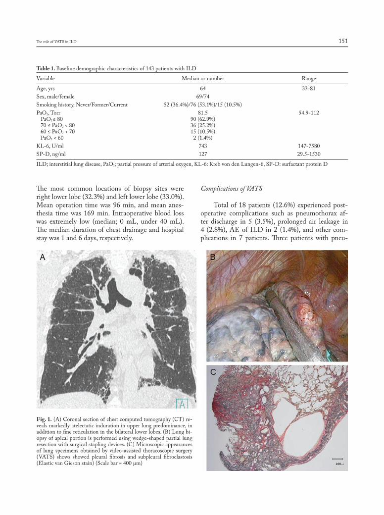
The role of VATS in ILD 151
The most common locations of biopsy sites were right lower lobe (32.3%) and left lower lobe (33.0%). Mean operation time was 96 min, and mean anes-thesia time was 169 min. Intraoperative blood loss was extremely low (median; 0 mL, under 40 mL). The median duration of chest drainage and hospital stay was 1 and 6 days, respectively.
Complications of VATS
Total of 18 patients (12.6%) experienced post-operative complications such as pneumothorax af-ter discharge in 5 (3.5%), prolonged air leakage in 4 (2.8%), AE of ILD in 2 (1.4%), and other com-plications in 7 patients. Three patients with pneu-
Table 1. Baseline demographic characteristics of 143 patients with ILD
Variable Median or number Range
Age, yrs 64 33-81Sex, male/female 69/74 Smoking history, Never/Former/Current 52 (36.4%)/76 (53.1%)/15 (10.5%) PaO2, Torr 81.5 54.9-112 PaO2 ≥ 80 90 (62.9%) 70 ≤ PaO2 < 80 36 (25.2%) 60 ≤ PaO2 < 70 15 (10.5%) PaO2 < 60 2 (1.4%) KL-6, U/ml 743 147-7580SP-D, ng/ml 127 29.5-1530
ILD; interstitial lung disease, PaO2; partial pressure of arterial oxygen, KL-6: Kreb von den Lungen-6, SP-D: surfactant protein D
Fig. 1. (A) Coronal section of chest computed tomography (CT) re-veals markedly atelectatic induration in upper lung predominance, in addition to fine reticulation in the bilateral lower lobes. (B) Lung bi-opsy of apical portion is performed using wedge-shaped partial lung resection with surgical stapling devices. (C) Microscopic appearances of lung specimens obtained by video-assisted thoracoscopic surgery (VATS) shows showed pleural fibrosis and subpleural fibroelastosis (Elastic van Gieson stain) (Scale bar = 400 µm)
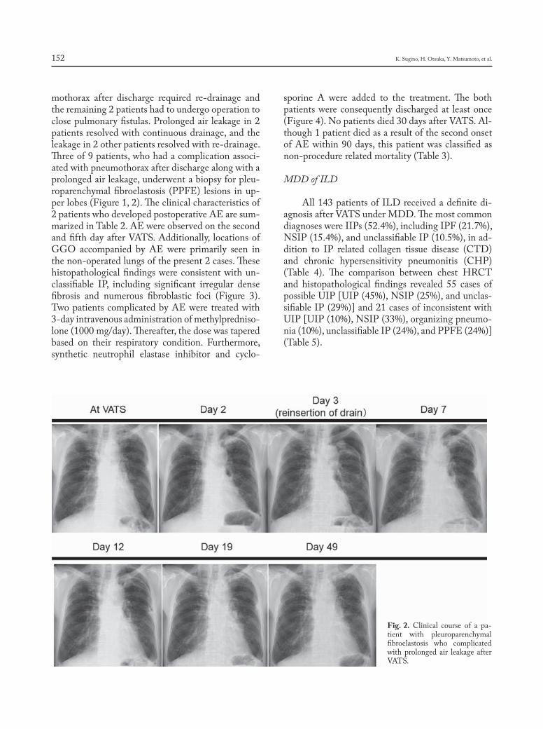
K. Sugino, H. Otsuka, Y. Matsumoto, et al.152
mothorax after discharge required re-drainage and the remaining 2 patients had to undergo operation to close pulmonary fistulas. Prolonged air leakage in 2 patients resolved with continuous drainage, and the leakage in 2 other patients resolved with re-drainage. Three of 9 patients, who had a complication associ-ated with pneumothorax after discharge along with a prolonged air leakage, underwent a biopsy for pleu-roparenchymal fibroelastosis (PPFE) lesions in up-per lobes (Figure 1, 2). The clinical characteristics of 2 patients who developed postoperative AE are sum-marized in Table 2. AE were observed on the second and fifth day after VATS. Additionally, locations of GGO accompanied by AE were primarily seen in the non-operated lungs of the present 2 cases. These histopathological findings were consistent with un-classifiable IP, including significant irregular dense fibrosis and numerous fibroblastic foci (Figure 3). Two patients complicated by AE were treated with 3-day intravenous administration of methylpredniso-lone (1000 mg/day). Thereafter, the dose was tapered based on their respiratory condition. Furthermore, synthetic neutrophil elastase inhibitor and cyclo-
sporine A were added to the treatment. The both patients were consequently discharged at least once (Figure 4). No patients died 30 days after VATS. Al-though 1 patient died as a result of the second onset of AE within 90 days, this patient was classified as non-procedure related mortality (Table 3).
MDD of ILD
All 143 patients of ILD received a definite di-agnosis after VATS under MDD. The most common diagnoses were IIPs (52.4%), including IPF (21.7%), NSIP (15.4%), and unclassifiable IP (10.5%), in ad-dition to IP related collagen tissue disease (CTD) and chronic hypersensitivity pneumonitis (CHP) (Table 4). The comparison between chest HRCT and histopathological findings revealed 55 cases of possible UIP [UIP (45%), NSIP (25%), and unclas-sifiable IP (29%)] and 21 cases of inconsistent with UIP [UIP (10%), NSIP (33%), organizing pneumo-nia (10%), unclassifiable IP (24%), and PPFE (24%)] (Table 5).
Fig. 2. Clinical course of a pa-tient with pleuroparenchymal fibroelastosis who complicated with prolonged air leakage after VATS.
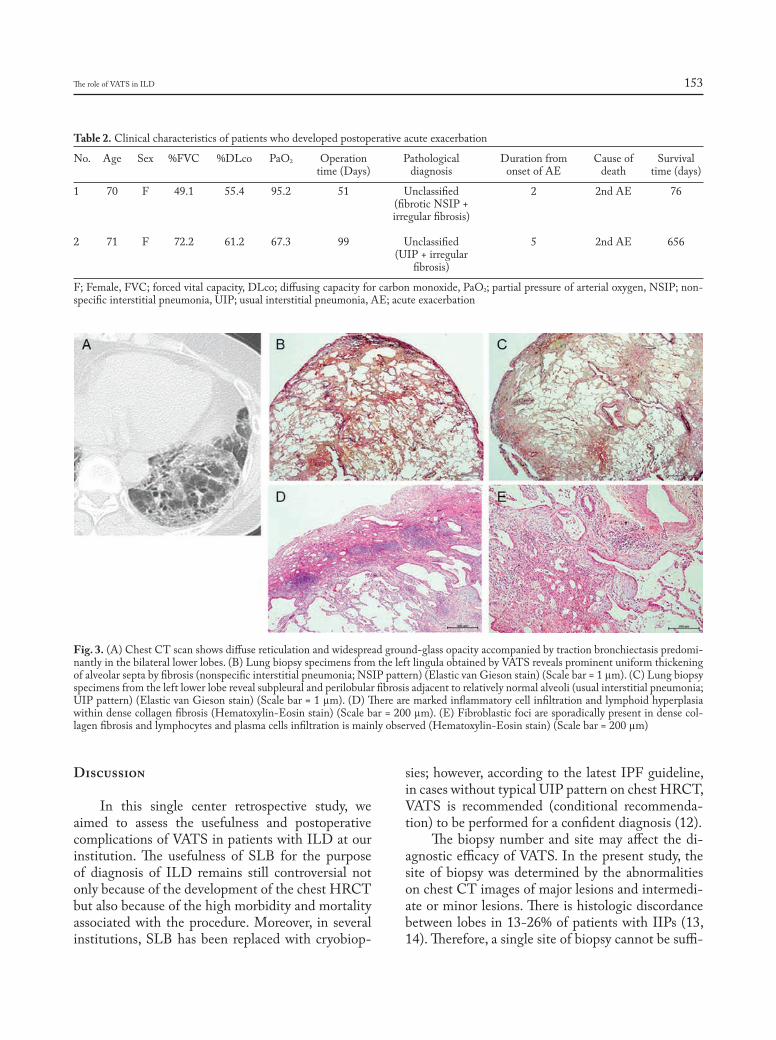
The role of VATS in ILD 153
Discussion
In this single center retrospective study, we aimed to assess the usefulness and postoperative complications of VATS in patients with ILD at our institution. The usefulness of SLB for the purpose of diagnosis of ILD remains still controversial not only because of the development of the chest HRCT but also because of the high morbidity and mortality associated with the procedure. Moreover, in several institutions, SLB has been replaced with cryobiop-
sies; however, according to the latest IPF guideline, in cases without typical UIP pattern on chest HRCT, VATS is recommended (conditional recommenda-tion) to be performed for a confident diagnosis (12).
The biopsy number and site may affect the di-agnostic efficacy of VATS. In the present study, the site of biopsy was determined by the abnormalities on chest CT images of major lesions and intermedi-ate or minor lesions. There is histologic discordance between lobes in 13-26% of patients with IIPs (13, 14). Therefore, a single site of biopsy cannot be suffi-
Table 2. Clinical characteristics of patients who developed postoperative acute exacerbation
No. Age Sex %FVC %DLco PaO2 Operation Pathological Duration from Cause of Survival time (Days) diagnosis onset of AE death time (days)
1 70 F 49.1 55.4 95.2 51 Unclassified 2 2nd AE 76 (fibrotic NSIP + irregular fibrosis)
2 71 F 72.2 61.2 67.3 99 Unclassified 5 2nd AE 656 (UIP + irregular fibrosis)
F; Female, FVC; forced vital capacity, DLco; diffusing capacity for carbon monoxide, PaO2; partial pressure of arterial oxygen, NSIP; non-specific interstitial pneumonia, UIP; usual interstitial pneumonia, AE; acute exacerbation
Fig. 3. (A) Chest CT scan shows diffuse reticulation and widespread ground-glass opacity accompanied by traction bronchiectasis predomi-nantly in the bilateral lower lobes. (B) Lung biopsy specimens from the left lingula obtained by VATS reveals prominent uniform thickening of alveolar septa by fibrosis (nonspecific interstitial pneumonia; NSIP pattern) (Elastic van Gieson stain) (Scale bar = 1 µm). (C) Lung biopsy specimens from the left lower lobe reveal subpleural and perilobular fibrosis adjacent to relatively normal alveoli (usual interstitial pneumonia; UIP pattern) (Elastic van Gieson stain) (Scale bar = 1 µm). (D) There are marked inflammatory cell infiltration and lymphoid hyperplasia within dense collagen fibrosis (Hematoxylin-Eosin stain) (Scale bar = 200 µm). (E) Fibroblastic foci are sporadically present in dense col-lagen fibrosis and lymphocytes and plasma cells infiltration is mainly observed (Hematoxylin-Eosin stain) (Scale bar = 200 µm)
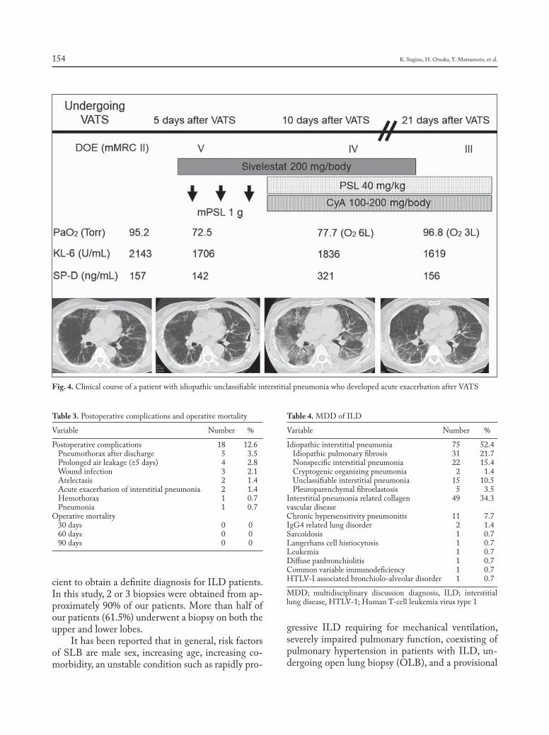
K. Sugino, H. Otsuka, Y. Matsumoto, et al.154
cient to obtain a definite diagnosis for ILD patients. In this study, 2 or 3 biopsies were obtained from ap-proximately 90% of our patients. More than half of our patients (61.5%) underwent a biopsy on both the upper and lower lobes.
It has been reported that in general, risk factors of SLB are male sex, increasing age, increasing co-morbidity, an unstable condition such as rapidly pro-
gressive ILD requiring for mechanical ventilation, severely impaired pulmonary function, coexisting of pulmonary hypertension in patients with ILD, un-dergoing open lung biopsy (OLB), and a provisional
Fig. 4. Clinical course of a patient with idiopathic unclassifiable interstitial pneumonia who developed acute exacerbation after VATS
Table 3. Postoperative complications and operative mortality
Variable Number %
Postoperative complications 18 12.6 Pneumothorax after discharge 5 3.5 Prolonged air leakage (≥5 days) 4 2.8 Wound infection 3 2.1 Atelectasis 2 1.4 Acute exacerbation of interstitial pneumonia 2 1.4 Hemothorax 1 0.7 Pneumonia 1 0.7Operative mortality 30 days 0 0 60 days 0 0 90 days 0 0
Table 4. MDD of ILD
Variable Number %
Idiopathic interstitial pneumonia 75 52.4 Idiopathic pulmonary fibrosis 31 21.7 Nonspecific interstitial pneumonia 22 15.4 Cryptogenic organizing pneumonia 2 1.4 Unclassifiable interstitial pneumonia 15 10.5 Pleuroparenchymal fibroelastosis 5 3.5Interstitial pneumonia related collagen 49 34.3vascular disease Chronic hypersensitivity pneumonitis 11 7.7IgG4 related lung disorder 2 1.4Sarcoidosis 1 0.7Langerhans cell histiocytosis 1 0.7Leukemia 1 0.7Diffuse panbronchiolitis 1 0.7Common variable immunodeficiency 1 0.7HTLV-I associated bronchiolo-alveolar disorder 1 0.7
MDD; multidisciplinary discussion diagnosis, ILD; interstitial lung disease, HTLV-1; Human T-cell leukemia virus type 1
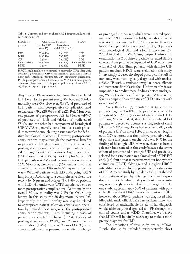
The role of VATS in ILD 155
diagnosis of IPF or connective tissue disease-related ILD (5-8). In the present study, 30-, 60-, and 90-day mortality were 0%. However, %FVC of predicted of ILD patients with postoperative complication tend to decrease (79.2±20.7% vs. 88.2±21.1%). Moreover, one patient of postoperative AE had lower %FVC of predicted of 49.1% and %DLco of predicted of 55.4%, and the other had component of histological UIP. VATS is generally considered as a safe proce-dure to provide enough lung tissue samples for defin-itive histological diagnosis. However, postoperative complications may outweigh the potential benefits in patients with ILD because postoperative AE or prolonged air leakage is one of the particularly criti-cal and significant complications. Sigurdsson et al. (15) reported that a 30-day mortality for SLB in 73 ILD patients was 2.7% and its complication rate was 16%. Moreover, Kreider et al. (16) demonstrated that a morbidity rate was 19% and a 60-day mortality rate was 4.4% in 68 patients with ILD undergoing VATS lung biopsy. According to a comprehensive literature review by Nguyen and Meyer (9), 9.6% of patients with ILD who underwent VATS experienced one or more postoperative complications. Additionally, the overall 30-day mortality was 2.1% for VATS lung biopsy. In this study, the 30-day mortality was 0%. Importantly, the low mortality rate may be related to appropriate patient selection criteria and opera-tion by trained chest surgeons. The postoperative complication rate was 12.6%, including 5 cases of pneumothorax after discharge (3.5%), 4 cases of prolonged air leakage (2.8%), and 2 cases of acute exacerbation (1.4%). Three of 9 cases (33.3%) were complicated by either pneumothorax after discharge
or prolonged air leakage, which were resected speci-mens of PPFE lesions. Probably, we should avoid resection of specimens of PPFE lesions in the upper lobes. As reported by Kreider et al. (16), 3 patients with pathological UIP and a low DLco value (19, 27, 30%) died after VATS lung biopsy. Postmortem examination in 2 of these 3 patients revealed diffuse alveolar damage on a background of UIP, consistent with AE of UIP. Thus, patients with definite UIP pattern on chest HRCT were excluded in this study. Interestingly, 2 cases developed postoperative AE in our study were histologically diagnosed with unclas-sifiable IP with significant irregular dense fibrosis and numerous fibroblastic foci. Unfortunately, it was impossible to predict these findings before undergo-ing VATS. Incidences of postoperative AE were too few to compare characteristics of ILD patients with or without AE.
Sverzellati et al. (3) reported that 34 out of 55 patients diagnosed as IPF on biopsy had received a di-agnosis of NSIP, CHP, or sarcoidosis on chest CT. In addition, Morris et al. (4) described that only 54% of patients who received a consensus diagnosis of usual UIP after VATS lung biopsy had received a diagnosis of probable UIP on chest HRCT. In contrast, Raghu et al. (17) reported that the positive predictive value of possible UIP pattern on chest CT was 94% for the finding of histologic UIP. However, there has been a selection bias noticed in this study because the entire cohort of patients had histologic UIP and previously selected for participation in a clinical trial of IPF. Fell et al. (18) found that in patients without honeycomb change on HRCT, older age and a higher HRCT interstitial score are highly predictive of a diagnosis of IPF. A recent study by Gruden et al. (19) showed that a pattern of patchy heterogeneous basilar-pre-dominant reticular abnormality without honeycomb-ing was strongly associated with histologic UIP. In our study, approximately 50% of patients with pos-sible UIP on chest HRCT was consistent with UIP; however, about 30% of patients was diagnosed with idiopathic unclassifiable IP. Some patients, who were considered as unclassifiable IP at initial diagnosis would ultimately be diagnosed as IPF through the clinical course under MDD. Therefore, we believe that MDD will be vitally necessary to make a more precise diagnosis for ILD.
The limitations of this study are as follows. Firstly, this study included retrospectively short
Table 5. Comparison between chest HRCT images and histologi-cal findings in IIPs
Histologic Chest HRCT pattern MDDpattern Possible UIP Inconsistent (n = 55) with UIP (n = 21)
UIP 25 (45%) 2 (10%) IPFNSIP 14 (25%) 7 (33%) NSIPOP 0 (0%) 2 (10%) COPUnclassifiable 16 (29%) 5 (24%) Unclassifiable IPPPFE 0 (0%) 5 (24%) PPFE
HRCT; high resolution computed tomography, IIPs; idiopathic interstitial pneumonias, UIP; usual interstitial pneumonia, NSIP; nonspecific interstitial pneumonia, OP; organizing pneumonia, PPFE; pleuroparenchymal fibroelastosis, MDD; multidisciplinary discussion diagnosis, IPF; idiopathic pulmonary fibrosis, COP; cryptogenic organizing pneumonia

K. Sugino, H. Otsuka, Y. Matsumoto, et al.156
follow-up period and small number of patients at a single center. Therefore, our results may not represent the usefulness and safety of VATS in the entire ILD population. In the future, longer and larger observa-tion prospective studies are needed to confirm our results. Secondly, patients with IP related to connec-tive tissue diseases usually do not need to undergo a biopsy under VATS in clinical setting. However, we suppose that this attempt is useful to select whether anti-fibrotic agents or anti-inflammatory agents such as corticosteroid or immunosuppressants should be applied. Thirdly, patients with a definite UIP pattern on chest HRCT were excluded from indication of VATS based on their risk of AE-IPF in this study. However, we should pay attention to referring pa-tients with possible UIP pattern for VATS, because many patients with possible UIP could have histo-logic UIP as reported by Raghu et al. (17) and be-cause patients with a provisional diagnosis of IPF including patients with possible UIP pattern also have an increased mortality after SLB, as described by Hutchinson et al. (8).
In conclusion, although VATS is not required in patients with suspected IPF who had typical UIP pattern on chest HRCT, in other ILD patients, VATS is safely performed to obtain a confident diag-nosis for appropriate treatment strategies.
Acknowledgments
We would like to thank Dr A. Kurosaki for interpre-tation of radiological findings (Department of Radiology, Fukujuji Hospital, Japan Anti-Tuberculosis Association, To-kyo, Japan), Dr T. Uekusa for analysis of pathological find-ings (Department of Pathology, Labor Health and Welfare Organization Kanto Rosai Hospital, Kanagawa, Japan).
This study is partially supported by a grant from Min-istry of Health, Labor and Welfare of Japan awarded to the Study Group on Diffuse Pulmonary Disorders, Scientific, Research/Research on intractable diseases.
Author Contributions: KSu, SH: study design, data analysis, manuscript preparation, guarantor of paperKSu, HO, TM, YM, YN, KM, KSi: data collection, data analysisKSu, HO, YM, YN, KM, YA, TM, AI, KSi, SH: manuscript prep-aration and reviewAll authors had full access to all of the data in the study and can take responsibility for the integrity of the data and the accuracy of the data analysis.
References
1. Lynch DA, Sverzellati N, Travis WD, et al. Diagnostic criteria for idi-opathic pulmonary fibrosis: a Fleischner Society White Paper. Lancet Respir Med 2018; 6: 138-53.
2. Jacob J, Hansell DM. HRCT of fibrosing lung disease. Respirology 2015; 20: 859-72.
3. Sverzellati N, Wells AU, Tomassetti S, et al. Biopsy-proved idiopathic pulmonary fibrosis: spectrum of nondiagnostic thin-section CT diag-noses. Radiology 2010; 254: 957-64.
4 . Morris D, Zamvar V. The efficacy of video-assisted thoracoscopic surgery lung biopsies in patients with Interstitial Lung Disease: a ret-rospective study of 66 patients. J Cardiothorac Surg 2014; 10; 9: 45.
5. Utz JP, Ryu JH, Douglas WW, et al. High short-term mortality fol-lowing lung biopsy for usual interstitial pneumonia. Eur Respir J 2001; 17: 175-9.
6. Park JH, Kim DK, Kim DS, et al. Mortality and risk factors for surgi-cal lung biopsy in patients with idiopathic interstitial pneumonia. Eur J Cardiothorac Surg 2007; 31: 1115-9.
7. Bando M, Ohno S, Hosono T, et al. Risk of acute exacerbation after video-assisted thoracoscopic lung biopsy for interstitial lung disease. J Bronchology Interv Pulmonol 2009; 16: 229-35.
8. Hutchinson JP, Fogarty AW, McKeever TM, et al. In-hospital mortal-ity after surgical lung biopsy for interstitial lung disease in the United States. 2000 to 2011. Am J Respir Crit Care Med 2016; 193: 1161-7.
9. Nguyen W, Meyer KC. Surgical lung biopsy for the diagnosis of in-terstitial lung disease: a review of the literature and recommendations for optimizing safety and efficacy. Sarcoidosis Vasc Diffuse Lung Dis 2013; 30: 3-16.
10. Raghu G, Collard HR, Egan JJ, et al; ATS/ERS/JRS/ALAT Com-mittee on Idiopathic Pulmonary Fibrosis. An official ATS/ERS/JRS/ALAT statement: idiopathic pulmonary fibrosis: evidence-based guidelines for diagnosis and management. Am J Respir Crit Care Med 2011; 183: 788-824.
11. Acute Exacerbation of Idiopathic Pulmonary Fibrosis. An Interna-tional Working Group Report. Collard HR, Ryerson CJ, Corte TJ, et al. Am J Respir Crit Care Med 2016; 194: 265-75.
12. Raghu G, Remy-Jardin M, Myers JL, et al. Diagnosis of idiopathic pulmonary fibrosis. An official ATS/ERS/JRS/ALAT clinical prac-tice guideline. Am J Respir Crit Care Med 2018; 198: e44-e68.
13. Sumikawa H, Johkoh T, Fujimoto K, et al. Usual interstitial pneumo-nia and nonspecific interstitial pneumonia: correlation between CT findings at the site of biopsy with pathological diagnoses. Eur J Radiol 2012; 81: 2919-24.
14. Flaherty KR, Travis WD, Colby TV, et al. Histopathologic variability in usual and nonspecific interstitial pneumonias. Am J Respir Crit Care Med 2001; 164: 1722-7.
15. Sigurdsson MI, Isaksson HJ, Gudmundsson G, Gudbjartsson T. Di-agnostic surgical lung biopsies for suspected interstitial lung diseases: a retrospective study. Ann Thorac Surg 2009; 88: 227-32.
16. Kreider ME, Hansen-Flaschen J, Ahmad NN, et al. Complications of video-assisted thoracoscopic lung biopsy in patients with interstitial lung disease. Ann Thorac Surg 2007; 83: 1140-4.
17. Raghu G, Lynch D, Godwin JD, et al. Diagnosis of idiopathic pul-monary fibrosis with high-resolution CT in patients with little or no radiological evidence of honeycombing: secondary analysis of a ran-domised, controlled trial. Lancet Respir Med 2014; 2: 277-84.
18. Fell CD, Martinez FJ, Liu LX, et al. Clinical predictors of a diagnosis of idiopathic pulmonary fibrosis. Am J Respir Crit Care Med 2010; 181: 832-7.
19. Gruden JF, Panse PM, Gotway MB, Jensen EA, Wellnitz CV, Wes-selius L. Diagnosis of usual interstitial pneumonitis in the absence of honeycombing: evaluation of specific CT criteria with clinical follow-up in 38 patients. AJR Am J Roentgenol 2016; 206: 472-80.
Cyclophosphamide pulse therapy as treatment for severe interstitial lung diseases
Arik Bernard Schulze1*, Georg Evers1*, Andreas Kümmel2, Felix Rosenow3, Jan Sackarnd3, Jan Philipp Hering4, Christoph Schülke4, Jonas Andreas Engelbertz5, Dennis Görlich6, Peter J. Barth7, Georg Lenz1,9, Heidemarie Becker8, Michael Mohr1#, Lars Henning Schmidt1#
1 Department of Medicine A, Hematology, Oncology and Pneumology, University Hospital Muenster, Muenster, Germany; 2 Department of Hematology, Medical Oncology and Pneumology, University Medical Center Mainz, Mainz, Germany; 3 Department of Cardiovascu-lar Medicine, Internal Intensive Care Medicine, University Hospital Muenster, Muenster, Germany; 4 Department of Clinical Radiology, University Hospital Muenster, Muenster, Germany; 5 Unit of Cytostatic Reconstitution, Hospital pharmacy, University Hospital Muenster, Muenster, Germany; 6 Institute of Biostatistics and Clinical Research, Westfaelische Wilhelms-Universitaet Muenster, Muenster, Germany; 7 Gerhard-Domagk-Institute of Pathology, University Hospital Muenster, Muenster, Germany; 8 Department of Medicine D, Nephrology, Rheumatology and Hypertensiology, University Hospital Muenster, Muenster, Germany; 9 Cluster of Excellence EXC 1003, Cells in Motion, Muenster, Germany
Abstract. Introduction: Besides invasive or non-invasive ventilation, treatment of severe forms of interstitial lung diseases (ILD) includes immunosuppressive medication. In case of refractory organ- or life-threatening courses of disease, cyclophosphamide pulse therapy can serve as a rescue treatment option. Objectives: To inves-tigate therapeutic and prognostic effects of cyclophosphamide for the treatment of severe forms of ILD on in-tensive care unit (ICU) we performed this analysis. Methods: Between 2009 and 2017 we identified 14 patients, who were treated on intensive care unit (ICU) with severe forms of ILD. Retrospectively, clinical, radiologic and prognostic data were collected and evaluated. Results: Our analysis demonstrated a prognostic impact of cyclophosphamide on the ILD in general. Whereas pulmonary manifestations of both systemic sclerosis (SSc) and ANCA-associated vasculitis had an improved outcome, a reduced overall survival was found for Goodpas-ture syndrome (GPS), dermatomyositis (DM), cryptogenic organizing pneumonia (COP) and drug reaction with eosinophilia and systemic symptoms (DRESS; p=0.040, logrank test). Besides, additional plasmapheresis and initiation of cyclophosphamide within ten days following initial diagnosis of ILD were associated with improved prognosis. Conclusion: Positive prognostic effects of cyclophosphamide pulse therapy in ICU treated patients suffering from severe respiratory failure due to pulmonary manifestations of both SSc and ANCA-associated-vasculitis were observed. Further prognostic and therapeutic data are needed for cyclophosphamide for this indication in order to prevent patients from its toxic side-effects, who most likely will not benefit from its application. (Sarcoidosis Vasc Diffuse Lung Dis 2019; 36 (2): 157-166)
Key words: intensive care medicine, interstitial lung dis-ease, treatment, chemotherapy, computed tomography, ex-acerbation
SARCOIDOSIS VASCULITIS AND DIFFUSE LUNG DISEASES 2019; 36 (2); 157-166 © Mattioli 1885
Original article: Clinical research
Received: 23 August 2018Accepted after revision: 24 February 2019Correspondence: Dr. med. Arik Bernard SchulzeDepartment of Medicine AUniversity Hospital MuensterAlbert-Schweitzer-Campus 1, Building A148149 Muenster, GermanyTel. +49-251-83-44827E-mail: [email protected]
* these authors are contributed equally to this work as first authors# these authors are contributed equally to this work as senior authors
A.B. Schulze, G. Evers, A. Kümmel, et al.158
Introduction
The term “interstitial lung diseases” (ILD) en-compasses both acute and chronic parenchymal lung diseases (1). The origin of ILD can be reactive to tox-ic agents, idiopathic (i.e. idiopathic interstitial pneu-monia) or associated with systemic diseases such as granulomatous disorders, connective tissue diseases (CTD) or vasculitis (2, 3). For the final diagnosis an-amnesis, clinical and functional data as well as radio-logic ILD patterns and histopathological results are taken into consideration (2, 3, 5-8).
In general, treatment of acute exacerbations and progressive courses of ILDs is difficult. Often, immu-nosuppressive regimens are initiated with corticos-teroids (3, 9). To intensify immunosuppressive treat-ment, addition of rituximab or cyclophosphamide is recommended only for progressive ILD forms due to either connective tissue disorders (CTD) or to vasculitides (10-12). As a rescue option, the British Thoracic Society (BTS) suggests the application of cyclophosphamide for the treatment of refractory and progressive ILD forms other than idiopathic pulmo-nary fibrosis (IPF) (13). However, only few data exist upon the prognostic and therapeutic effects of cyclo-phosphamide in critically ill patients.
For chronic ILD forms, Schupp et al. evaluated the impact of cyclophosphamide pulse therapy in n=26 patients. According to their analysis, prognos-tic outcome was improved for patients with lympho-cytic interstitial pneumonia (LIP) and non-specific interstitial pneumonia (NSIP) following cyclophos-phamide application. In contrast, patients with p-ANCA positive vasculitis had the worst prognosis. However, patients who had less than 3 infusions of cyclophosphamide and who were treated on ICU were not included in their study (14).
Since many ICU patients with severe ILD forms require invasive ventilation and sedation (15), it is often impossible to obtain patients’ consent. Consequently, considering toxic side effects (16), the indication to initiate additional cyclophosphamide is met by interdisciplinary teams (7, 8).
To investigate the impact of cyclophospha-mide pulse therapy in patients requiring ICU treat-ment due to respiratory failure caused by severe ILD forms, we performed this retrospective analysis with focus on radiologic ILD patterns and other clinical factors.
Material and Methods
Study population
First, approval of the ethical committee Muen-ster was obtained (Ref. 2017-599-f-S). In total, n=14,421 ICU patients were treated on our ICUs between 2009 and 2017. Among these patients, we identified n=14 patients suffering from differ-ent forms of ILD, who received at least one course of intravenous cyclophosphamide as rescue therapy (Table 1).
Data collection was performed retrospectively. Besides clinical data, therapeutic information (e.g. cyclophosphamide cycles, dosage, first-line immu-nosuppression, ventilation mode, ventilation dura-tion, P/F ratio [i.e. Horowitz index=arterial oxygen partial pressure (paO2 in mmHg)/fraction of inhaled oxygen (FiO2 in %)], additional antibiotics, extracor-poral membrane oxygenation and plasmapheresis) was gathered, too. Due to varying dosage schedules (mg/kg vs. mg/m2 vs. mg as absolute dosage), all analyses considered the absolute cyclophosphamide dosage. Of interest, cyclophosphamide was given exclusively to patients with severe respiratory failure (as defined by a partial oxygen pressure in [mmHg]/ oxygen saturation in inhaled gas [%]) <200) due to various forms of ILD, who required further immu-nosuppressive therapy. In contrast, if an infectious origin was suspected, cyclophosphamide was not applied. However, additional antibiotic prophylaxis was initiated before cyclophosphamide was given as prophylactic treatment in all patients.
Radiologic examination
With focus on the radiologic ILD patterns, tho-racic computed axial tomography (CAT) scans of the identified patients were categorized as non-specific interstitial pneumonia (NSIP), usual interstitial pneumonia (UIP), organizing pneumonia (OP), dif-fuse alveolar damage (DAD) and lymphocytic in-terstitial pneumonia (LIP) (17). To determine the morphologic extent of the underlying disease, the involvement of the affected lobes (i.e. upper left lobe including lingula, lower left lobe, upper right lobe, middle lobe and lower right lobe) was evaluated.
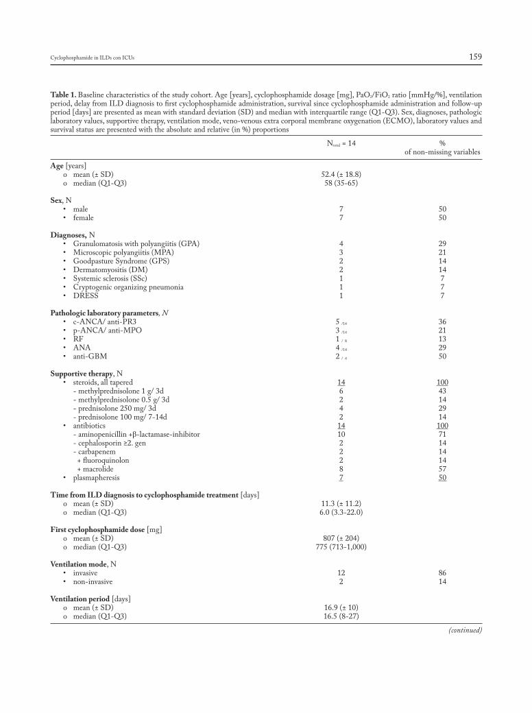
Cyclophosphamide in ILDs con ICUs 159
Table 1. Baseline characteristics of the study cohort. Age [years], cyclophosphamide dosage [mg], PaO2/FiO2 ratio [mmHg/%], ventilation period, delay from ILD diagnosis to first cyclophosphamide administration, survival since cyclophosphamide administration and follow-up period [days] are presented as mean with standard deviation (SD) and median with interquartile range (Q1-Q3). Sex, diagnoses, pathologic laboratory values, supportive therapy, ventilation mode, veno-venous extra corporal membrane oxygenation (ECMO), laboratory values and survival status are presented with the absolute and relative (in %) proportions
Ntotal = 14 % of non-missing variables
Age [years] o mean (± SD) 52.4 (± 18.8) o median (Q1-Q3) 58 (35-65)
Sex, N • male 7 50 • female 7 50
Diagnoses, N • Granulomatosiswithpolyangiitis(GPA) 4 29 • Microscopicpolyangiitis(MPA) 3 21 • GoodpastureSyndrome(GPS) 2 14 • Dermatomyositis(DM) 2 14 • Systemicsclerosis(SSc) 1 7 • Cryptogenicorganizingpneumonia 1 7 • DRESS 1 7
Pathologic laboratory parameters, N • c-ANCA/anti-PR3 5/14 36 • p-ANCA/anti-MPO 3/14 21 • RF 1/ 8 13 • ANA 4/14 29 • anti-GBM 2/ 4 50
Supportive therapy, N • steroids,alltapered 14 100 - methylprednisolone 1 g/ 3d 6 43 - methylprednisolone 0.5 g/ 3d 2 14 - prednisolone 250 mg/ 3d 4 29 - prednisolone 100 mg/ 7-14d 2 14 • antibiotics 14 100 - aminopenicillin +β-lactamase-inhibitor 10 71 - cephalosporin ≥2. gen 2 14 - carbapenem 2 14 + fluoroquinolon 2 14 + macrolide 8 57 • plasmapheresis 7 50
Time from ILD diagnosis to cyclophosphamide treatment [days] o mean (± SD) 11.3 (± 11.2) o median (Q1-Q3) 6.0 (3.3-22.0)
First cyclophosphamide dose [mg] o mean (± SD) 807 (± 204) o median (Q1-Q3) 775 (713-1,000)
Ventilation mode, N • invasive 12 86 • non-invasive 2 14
Ventilation period [days] o mean (± SD) 16.9 (± 10) o median (Q1-Q3) 16.5 (8-27)
(continued)
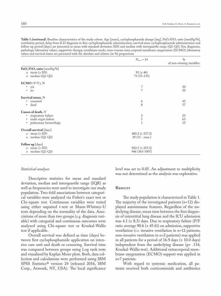
A.B. Schulze, G. Evers, A. Kümmel, et al.160
Statistical analyses
Descriptive statistics for mean and standard deviation, median and interquartile range (IQR) as well as frequencies were used to investigate our study population. Two-fold associations between categori-cal variables were analyzed via Fisher’s exact test or Chi-square test. Continuous variables were tested using either unpaired t-test or Mann-Whitney-U tests depending on the normality of the data. Asso-ciations of more than two groups (e.g. diagnosis vari-able) with categorial and continuous outcomes were analyzed using Chi-square test or Kruskal-Wallis test if applicable.
Overall survival was defined as time (days) be-tween first cyclophosphamide application on inten-sive care unit and death or censoring. Survival time was compared between groups using Log rank tests and visualized by Kaplan Meier plots. Both, data col-lection and calculations were performed using IBM SPSS Statistics® version 24 (released 2016, IBM Corp., Armonk, NY, USA). The local significance
level was set to 0.05. An adjustment to multiplicity was not determined as the analysis was explorative.
Results
The study population is characterized in Table 1. The majority of the investigated patients (n=12) dis-played autoimmune features. Regardless of the un-derlying disease, mean time between the first diagno-sis of interstitial lung disease and the ICU admission was 4.1 (± 8.3) days. Due to respiratory failure (P/F ratio average 90.6 (± 45.6)) on admission, supportive ventilation (i.e. invasive ventilation in n=12 patients; non-invasive ventilation in n=2 patients) was applied in all patients for a period of 16.9 days (± 10.0 days) independent from the underlying disease (p= .134, Kruskal-Wallis test). Additional extracorporal mem-brane oxygenation (ECMO) support was applied in n=7 patients.
With regard to systemic medication, all pa-tients received both corticosteroids and antibiotics
Table 1 (continued). Baseline characteristics of the study cohort. Age [years], cyclophosphamide dosage [mg], PaO2/FiO2 ratio [mmHg/%], ventilation period, delay from ILD diagnosis to first cyclophosphamide administration, survival since cyclophosphamide administration and follow-up period [days] are presented as mean with standard deviation (SD) and median with interquartile range (Q1-Q3). Sex, diagnoses, pathologic laboratory values, supportive therapy, ventilation mode, veno-venous extra corporal membrane oxygenation (ECMO), laboratory values and survival status are presented with the absolute and relative (in %) proportions
Ntotal = 14 % of non-missing variables
PaO2/FiO2 ratio [mmHg/%] o mean (± SD) 91 (± 46) o median (Q1-Q3) 73 (53-133)
ECMO (V/V), N • yes 7 50 • no 7 50
Survival status, N • censored 6 43 • dead 8 57
Causes of death, N • respiratoryfailure 2 25 • multiorganfailure 5 63 • pulmonaryhemorrhage 1 13
Overall survival [days] o mean (± SD) 682.2 (± 237.2) o median (Q1-Q3) 29 (15 - miss.)
Follow up [days] o mean (± SD) 822.5 (± 255.2) o median (Q1-Q3) 946 (363-1007)
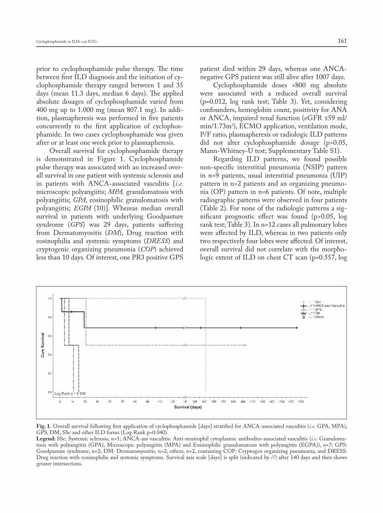
Cyclophosphamide in ILDs con ICUs 161
prior to cyclophosphamide pulse therapy. The time between first ILD diagnosis and the initiation of cy-clophosphamide therapy ranged between 1 and 35 days (mean 11.3 days, median 6 days). The applied absolute dosages of cyclophosphamide varied from 400 mg up to 1.000 mg (mean 807.1 mg). In addi-tion, plasmapheresis was performed in five patients concurrently to the first application of cyclophos-phamide. In two cases cyclophosphamide was given after or at least one week prior to plasmapheresis.
Overall survival for cyclophosphamide therapy is demonstrated in Figure 1. Cyclophosphamide pulse therapy was associated with an increased over-all survival in one patient with systemic sclerosis and in patients with ANCA-associated vasculitis [i.e. microscopic polyangiitis; MPA, granulomatosis with polyangiitis; GPA, eosinophilic granulomatosis with polyangiitis; EGPA (10)]. Whereas median overall survival in patients with underlying Goodpasture syndrome (GPS) was 29 days, patients suffering from Dermatomyositis (DM), Drug reaction with eosinophilia and systemic symptoms (DRESS) and cryptogenic organizing pneumonia (COP) achieved less than 10 days. Of interest, one PR3 positive GPS
patient died within 29 days, whereas one ANCA-negative GPS patient was still alive after 1007 days.
Cyclophosphamide doses >800 mg absolute were associated with a reduced overall survival (p=0.012, log rank test; Table 3). Yet, considering confounders, hemoglobin count, positivity for ANA or ANCA, impaired renal function (eGFR ≤59 ml/min/1.73m2), ECMO application, ventilation mode, P/F ratio, plasmapheresis or radiologic ILD patterns did not alter cyclophosphamide dosage (p>0.05, Mann-Whitney-U test; Supplementary Table S1).
Regarding ILD patterns, we found possible non-specific interstitial pneumonia (NSIP) pattern in n=9 patients, usual interstitial pneumonia (UIP) pattern in n=2 patients and an organizing pneumo-nia (OP) pattern in n=6 patients. Of note, multiple radiographic patterns were observed in four patients (Table 2). For none of the radiologic patterns a sig-nificant prognostic effect was found (p>0.05, log rank test; Table 3). In n=12 cases all pulmonary lobes were affected by ILD, whereas in two patients only two respectively four lobes were affected. Of interest, overall survival did not correlate with the morpho-logic extent of ILD on chest CT scan (p=0.557, log
Fig. 1. Overall survival following first application of cyclophosphamide [days] stratified for ANCA-associated vasculitis (i.e. GPA, MPA), GPS, DM, SSc and other ILD forms (Log Rank p=0.040). Legend: SSc: Systemic sclerosis, n=1; ANCA-ass vasculitis: Anti-neutrophil cytoplasmic antibodies-associated vasculitis (i.e. Granuloma-tosis with polyangiitis (GPA), Microscopic polyangiitis (MPA) and Eosinophilic granulomatosis with polyangiitis (EGPA)), n=7; GPS: Goodpasture syndrome, n=2; DM: Dermatomyositis, n=2; others, n=2, containing COP: Cryptogen organizing pneumonia, and DRESS: Drug reaction with eosinophilia and systemic symptoms. Survival axis scale [days] is split (indicated by //) after 140 days and then shows greater intersections.

A.B. Schulze, G. Evers, A. Kümmel, et al.162
rank test) or severity of pulmonary failure. Further-more no differences were found for anti-neutrophil cytoplasmic antibodies, anti-nuclear antibodies, rheumatoid factors, anti-glomerular basal membrane antibodies and blood count (p>0.05, log rank test). Impaired renal function (eGFR in ml/min/1.73 m2 cutoff >59 vs. ≤59) as well as anemia (hemoglobin level in g/dl cutoff ≥11 vs. < 11) or leukocytosis (in cells/µl cutoff >10.000 vs. ≤10.000) was not associ-ated with improved or reduced prognosis (p>0.050, log rank test). Among the investigated patients, n=7 patients died on ICU due to the fulminant course of disease. N=2 patients died due to respiratory failure, n=4 died due to multiple organ failure (MOF) and one patient died due to pulmonary hemorrhage. Of interest, one patient died 12 months later following initial ICU admission due to non-septic MOF.
Additional plasmapheresis (p=0.018, log rank test) and application of cyclophosphamide within ten days following first ILD diagnosis (p=0.028, log rank test) had a positive effect on prognosis (Table 3). Moreover, we observed that plasmapheresis was performed more often in patients with hemoglobin levels below 11 g/dl (p=0.029, Fisher’s exact test) independent from ANCA- or ANA-positivity, leu-kocyte count, renal impairment, ECMO use, venti-lation mode and radiographic ILD pattern (Fisher’s exact test p>0.05 in all calculations, data not shown).
Discussion
Treatment of acute respiratory failure due to various ILD forms is challenging. The therapeutic armamentarium includes supportive ventilation, ex-tracorporal membrane oxygenation, plasmapheresis, antibiotics and immunosuppressive medication. Cor-ticosteroids are often applied as first immunosup-pression (10, 13, 18-21). However, there are patients, who require more intensified immunosuppressive regimens to overcome refractory courses of disease. Here, cyclophosphamide or rituximab can serve as therapeutic alternatives especially for ANCA-asso-ciated vasculitis (10, 20, 21) or for CTDs (11). With regard to cyclophosphamide, the recommended dos-age is 15 mg/kg body weight every 2 to 3 weeks in combination with corticosteroids (22).
Schupp et al. evaluated the prognostic impact of intravenous cyclophosphamide pulsed therapy in n=26 patients with chronic ILD forms, who did not require ICU treatment. Patients were followed up for 18 months. According to their analysis, benefi-cial effects were demonstrated for patients with both LIP-patterns and NSIP-patterns, whereas p-ANCA associated ILDs did not benefit from additional cy-clophosphamide pulsed therapy (14).
In our study, we focused on those ILD patients who received cyclophosphamide as part of a rescue
Table 2. Study collective
No. Sex Age Diagn. Ventilation Ventilation P/F ECMO Plasma- CAT scan Survival Overall mode days ratio pheresis pattern status survival [days]
1 M 55 GPA IV 26 64,4 Yes Yes NSIP+ UIP- OP+ Alive 9462 F 61 DM IV 18 58,8 No No NSIP+ UIP- OP- Dead 213 M 80 GPS IV 30 141,6 Yes Yes NSIP+ UIP- OP+ Dead 294 M 64 MPA IV 2 69,7 No Yes NSIP+ UIP- OP+ Alive 3635 M 60 MPA IV 6 40,2 Yes Yes NSIP+ UIP- OP- Alive 1076 F 51 SSc NIV 15 46,1 No No NSIP- UIP- OP- Dead 4377 M 36 DRESS IV 15 54,7 Yes No NSIP- UIP- OP- Dead 158 F 67 DM NIV 18 99,7 No No NSIP+ UIP+ OP- Dead 109 M 61 COP IV 28 130,0 Yes No NSIP+ UIP- OP+ Dead 510 F 31 GPA IV 8 130,0 No No NSIP- UIP- OP- Alive 1311 M 19 GPS IV 9 169,2 No Yes NSIP- UIP- OP+ Alive 100712 F 56 MPA IV 24 152,8 Yes No NSIP+ UIP+ OP- Dead 2713 F 72 GPA IV 32 76,0 No Yes NSIP- UIP- OP+ Alive 176014 M 20 GPA IV 5 35,0 Yes Yes NSIP+ UIP- OP- Dead 2
Legend: Sex category (M = male, F= female), Age [years], ventilation mode (IV = invasive ventilation, NIV = non-invasive ventilation), absolute period of ventilation [days], the P/F ratio (i.e. partial oxygen pressure in [mmHg]/ oxygen saturation in inhaled gas [%]), the use of ECMO and Plasmapheresis, CAT scan patterns, regarding NSIP, UIP and OP as possible (+) or unlikely (-), survival status (alive/ dead) and survival since first cyclophosphamide administration [days]. Of interest, patient no. 10 was lost-to-follow-up after 13 days and discharge from hospital
Cyclophosphamide in ILDs con ICUs 163
immunosuppressive treatment on ICU to overcome respiratory failure. We found superior treatment ef-fects of cyclophosphamide in n=7 patients (50%) suffering from ANCA-associated ILDs compared to other ILDs. Similar observations were reported by Novack et al. in 1971 (23). A possible explana-tion for the results of Schupp et al. might be due to the fact, that in chronic (stable) ILD the possible life threatening side effects of cyclophosphamide exceed the benefit. Since plasmapheresis was applied in five out of seven patients (71%) with ANCA-associated
vasculitis, it is difficult to interpret the sole effect of cyclophosphamide in this context.
For sure, one major limitation of our study is its small study population. However, it is difficult to perform prospective studies with larger study popula-tions with focus on the risk-benefit analysis for cyclo-phosphamide for this indication. Among n=14,421 ICU patients treated between 2009 and 2017 on our ICUs, we identified only n=14 ILD patients, who were treated with cyclophosphamide due to this in-dication. To our knowledge, the underlying study is
Table 3. Univariate prognostic models depending on laboratory values, supportive therapies, CAT scan patterns and first absolute cyclo-phosphamide dosage. Additional plasmapheresis, early cyclophosphamide administration (≤10 days to ILD diagnosis) and an absolute first cyclophosphamide dose ≤800 mg were associated with improved overall survival in ILD patients treated with cyclophosphamide on ICU
Variable Comparator n Median OS§ 95% CI$ Log Rank (14) [days] p
Laboratory valuesa
ANCAb negative 6 15 1.8-28.2 0.156 positive 8 n. r.i ANAc negative 10 27 0.0-531.0 0.538 positive 4 29 n. e.k
Hemoglobin-level [g/dl] <11 7 n. r.i 0.090 ≥11 7 21 5.6-36.4 Leukocyte-count [/µl] ≤10.000 9 27 11.3-42.7 0.282 >10.000 5 n. r.i eGFRd [ml/min/1.73m2] >59 6 15 1.8-28.2 0.075 ≤59 8 n. r.i Supportive measures ECMOe not used 7 n. r.i 0.280 used 7 27 0.0-57.8 Ventilation invasive 12 29 n. e.k 0.419 non-invasive 2 10 n. e.k
P/F ratio <100 9 437 0.0-1261.4 0.773 ≥100 - 199 5 29 9.1-48.9 Plasmapheresis not used 7 21 9.1-33.0 0.018 used 7 n.r.i CAT scan patterns NSIPf unlikely 5 437 n. e.k 0.286 possible 9 27 9.5-44.5 UIPg unlikely 12 437 0.0-1383.2 0.148 possible 2 10 n.e.k
OPh unlikely 8 21 6.2-35.8 0.072 possible 6 n.r.i Treatment Cyclophosphamide dose [mg] ≤800 9 437 n. e.k 0.012 >800 5 15 7.2-22.8 Delay ILD diagnosis to Cyc. administration [days] ≤10 9 n.r.i 0.028 >10 5 15 4.3-25.7
Legend: § median overall survival in days since first cyclophosphamide administration, $ 95% confidence interval, a laboratory values on ICU admission, b Anti neutrophil cytoplasmic antibodies, c Anti nuclear antibodies, d estimated glomerular filtration rate (CKD-EPI method35) in ml/min/1.73m2, e extracorporal membrane oxygenation, f non-specific interstitial pneumonia, g usual interstitial pneumonia, h organizing pneumonia, i not reached, k not estimated
A.B. Schulze, G. Evers, A. Kümmel, et al.164
the first study, which investigates the impact of cyclo-phosphamide for critically ill ILD patients suffering severe respiratory failure and requiring ICU therapy.
Acute treatment of various ILD forms is sum-marized in Figure 2. As demonstrated, the applica-tion of cyclophosphamide represents a therapeutic alternative for ILD forms. Whereas cyclophospha-mide is recommended as first line treatment of SSc (20, 21, 24) and ANCA-associated vasculitis (10), there is less evidence for RA, DM and GPS. Here, cyclophosphamide could be an alternative option for refractory courses (19, 25, 26). Even though favora-ble effects of cyclophosphamide are reported for the treatment of pulmonary manifestations of GPS (18), there are still patients who died due to respiratory failure (27-29). Likewise, cyclophosphamide appli-cation had positive effects on lung function in some DM patients, still the prognosis in general is poor (19, 30). Even more evidence for the application of cyclophosphamide in COP is lacking. Whereas, cor-
ticosteroids are recommended for COP treatment (13, 31), less data exist upon the intensified immu-nosuppression with azathioprine, cyclosporine A or cyclophosphamide (13). With respect to the rather low incidence, one review recommended corticoster-oids and intravenous immunoglobulins following the withdrawal of potential causal agents as treatment option for DRESS syndrome (32), whereas addi-tional cyclophosphamide was only suggested by one study (33).
Against this background, we evaluated the therapeutic impact of cyclophosphamide for severe respiratory failure due to various ILD forms. Corre-sponding to the published recommendations, we ob-served positive effects of cyclophosphamide in those ILD patients with systemic sclerosis (SSc) (20) and ANCA-associated vasculitis (10). Especially in these patients, early administration of cyclophosphamide within ten days and a starting dosage of less than 800 mg had favorable effects. In consideration of the toxic
Fig. 2. Possible therapeutic options for the therapy of interstitial lung diseases (ILDs)
Cyclophosphamide in ILDs con ICUs 165
side effects (e.g. anemia, thrombocytopenia, leukocy-topenia, hemorrhage cystitis) of cyclophosphamide further studies should investigate the optimal dosage (34). Without doubt, additional plasmapheresis (10) might have improved the clinical status. Since pa-tients suffering from DM/PM did not benefit from additional cyclophosphamide therapy in our analysis, we cannot support its use for this specific indication.
In conclusion cyclophosphamide pulse therapy for the treatment of respiratory failure due to ILD can be a beneficial therapy option for some patients with underlying SSc and ANCA-associated vascu-litis. When considering cyclophosphamide in these patients, treatment should be initiated early, the dosage should be below 800 mg and plasmapheresis should be added. However, there is less evidence for the application of cyclophosphamide in those pa-tients with various ILD patterns due to GPS, DM, COP or DRESS. In consideration of the available literature and our analysis we cannot support the ap-plication of cyclophosphamide as first line treatment for ILD forms other than ANCA-positive vasculitis or SSc associated ILDs. Even though cyclophospha-mide is still applied for this indication, it is one of the most toxic routinely prescribed immunosuppres-sive agents (13). Consequently, the identification of those patients who might benefit from this therapy is crucial. Here, the investigation of larger study col-lectives with focus on ICU patients with severe ILD forms could serve for further therapeutic stratifica-tion. Whether less toxic agents or targeted therapies will be available in future is unclear.
Authorship Statement: LHS and MM designed the study. LHS, ABS and GE performed research, collected data and analyzed the data. LHS and ABS wrote the paper. FR, AK and JS collected data and gave relevant input upon treatment on ICU. JPH and CS performed retrospective CAT scan analysis. JAE detected cyclo-phosphamide treated patients on ICU and gave relevant input on cyclophosphamide dosage and treatment schedules. DG planned, controlled and supported the statistical analysis. PJB, GL, MM, LHS and HB gave relevant input on underlying disease. Every author read and approved the corrected manuscript.
References
1. Travis WD, et al. An official American Thoracic Society/European Respiratory Society statement: Update of the international multidis-ciplinary classification of the idiopathic interstitial pneumonias. Am J Respir Crit Care Med 2013; 188: 733-748.
2. Demedts M, Costabel U. ATS/ERS international multidisciplinary consensus classification of the idiopathic interstitial pneumonias. Eur Respir J 2002; 19: 794-6.
3. Fischer A, Du Bois R. Interstitial lung disease in connective tissue disorders. Lancet 2012; 380: 689-698.
4. Sambataro, G. et al. State of the art in interstitial pneumonia with autoimmune features: a systematic review on retrospective studies and suggestions for further advances. Eur Respir Rev an Off J Eur Respir Soc 27, 2018; 148: 170139
5. Fischer A, Lee JS, Cottin,V. Interstitial lung disease evaluation: De-tecting connective tissue disease. Respiration 2015; 90: 177-184.
6. Robles-Perez A, Molina-Molina M. Treatment considerations of lung involvement in rheumatologic disease. Respiration 2015; 90: 265-274.
7. Tomassetti S, Piciucchi S, Tantalocco P, Dubini A, Poletti V. The multidisciplinary approach in the diagnosis of idiopathic pulmonary fibrosis: A patient case-based review. Eur Respir Rev 2015; 24, 69-77.
8. Bonini M, Fiorenzano G. Exertional dyspnoea in interstitial lung diseases: the clinical utility of cardiopulmonary exercise testing. Eur Respir Rev 2017; 26: 235-8.
9. Mathai SC, Danoff SK. Management of interstitial lung disease asso-ciated with connective tissue disease. BMJ 2016, h6819. doi:10.1136/bmj.h6819
10. Yates M, et al. EULAR/ERA-EDTA recommendations for the man-agement of ANCA-associated vasculitis. Ann Rheum Dis 2016; 75: 1583-1594.
11. Saunders P, et al. Rituximab versus cyclophosphamide for the treat-ment of connective tissue disease-associated interstitial lung disease (RECITAL): study protocol for a randomised controlled trial. Trials 2017; 18: 275.
12. Stone JH, et al. Rituximab versus cyclophosphamide for ANCA-associated vasculitis. N Engl J Med 2010; 363: 221-32.
13. Wells AU, Hirani N. Interstitial lung disease guideline. Thorax 2008; 63: v1-v58.
14. Schupp JC, Köhler T, Müller-Quernheim J. Usefulness of Cyclophos-phamide Pulse Therapy in Interstitial Lung Diseases. Respiration 2016; 91: 296-301.
15. Xu Y. et al. Prone position ventilation support for acute exacerba-tion of interstitial lung disease? Clin Respir J 2017; 12: 1732-1380. doi:10.1111/crj.12665
16. Hengstler JG, et al. Induction of DNA crosslinks and DNA strand le-sions by cyclophosphamide after activation by cytochrome P450 2B1. Mutat Res - Fundam Mol Mech Mutagen 1997; 373: 215-223.
17. de Lauretis A, Veeraraghavan S, Renzoni E. Review Series: Aspects of Interstitial lung disease: Connective tissue disease-associated in-terstitial lung disease: How does it differ from IPF? How should the clinical approach differ? Chron Respir Dis 2011; 8: 53-82.
18. Salama AD, Levy JB, Lightstone L, Pusey CD. Goodpasture’s disease. Lancet 2001; 358: 917-920.
19. Marie I, Mouthon L. Therapy of polymyositis and dermatomyositis. Autoimmun Rev 2011; 11: 6-13.
20. Wallace B, Vummidi D, Khanna D. Management of connective tis-sue diseases associated interstitial lung disease. Curr Opin Rheumatol 2016; 28: 236-245.
21. Shenoy PD, Bavaliya M, Sashidharan S, Nalianda K, Sreenath S. Cy-clophosphamide versus mycophenolate mofetil in scleroderma inter-stitial lung disease (SSc-ILD) as induction therapy: a single-centre, retrospective analysis. Arthritis Res Ther 2016; 18: 123.
22. de Groot K, et al. Pulse versus daily oral cyclophosphamide for induc-tion of remission in antineutrophil cytoplasmic antibody-associated vasculitis: a randomized trial. Ann Intern Med 2009; 150: 670-80.
23. Novack SN, Pearson CM. Cyclophosphamide Therapy in Wegener’s Granulomatosis. N Engl J Med 1971; 284: 938-942.
24. Tashkin DP, et al. Cyclophosphamide versus Placebo in Scleroderma Lung Disease N Engl J Med 2006; 354: 2655-2666.
25. Kurita T, Yasuda S, Amengual O, Atsumi T. The efficacy of calcineurin inhibitors for the treatment of interstitial lung disease associated with polymyositis/dermatomyositis. Lupus 2015; 24: 3-9.
26. Shimojima Y, Ishii W, Matsuda M, Kishida D, Ikeda S. Effective
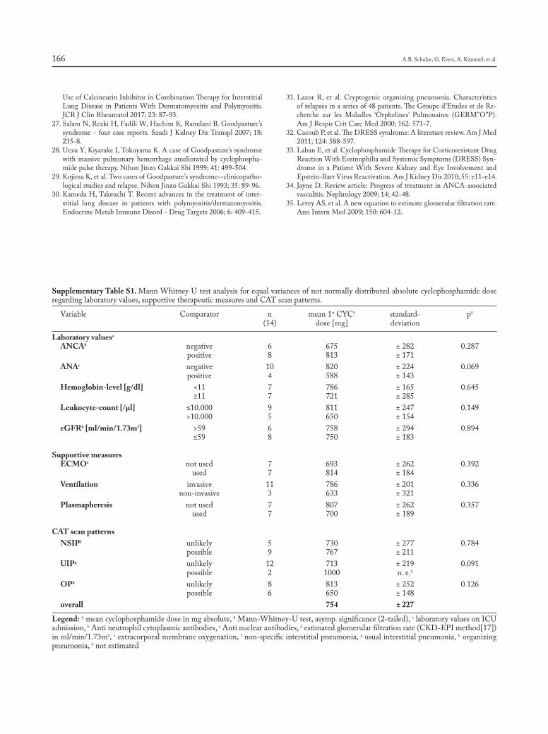
A.B. Schulze, G. Evers, A. Kümmel, et al.166
Use of Calcineurin Inhibitor in Combination Therapy for Interstitial Lung Disease in Patients With Dermatomyositis and Polymyositis. JCR J Clin Rheumatol 2017; 23: 87-93.
27. Salam N, Rezki H, Fadili W, Hachim K, Ramdani B. Goodpasture’s syndrome - four case reports. Saudi J Kidney Dis Transpl 2007; 18: 235-8.
28. Uezu Y, Kiyatake I, Tokuyama K. A case of Goodpasture’s syndrome with massive pulmonary hemorrhage ameliorated by cyclophospha-mide pulse therapy. Nihon Jinzo Gakkai Shi 1999; 41: 499-504.
29. Kojima K, et al. Two cases of Goodpasture’s syndrome--clinicopatho-logical studies and relapse. Nihon Jinzo Gakkai Shi 1993; 35: 89-96.
30. Kameda H, Takeuchi T. Recent advances in the treatment of inter-stitial lung disease in patients with polymyositis/dermatomyositis. Endocrine Metab Immune Disord - Drug Targets 2006; 6: 409-415.
31. Lazor R, et al. Cryptogenic organizing pneumonia. Characteristics of relapses in a series of 48 patients. The Groupe d’Etudes et de Re-cherche sur les Maladles ‘Orphelines’ Pulmonaires (GERM”O”P). Am J Respir Crit Care Med 2000; 162: 571-7.
32. Cacoub P, et al. The DRESS syndrome: A literature review. Am J Med 2011; 124: 588-597.
33. Laban E, et al. Cyclophosphamide Therapy for Corticoresistant Drug Reaction With Eosinophilia and Systemic Symptoms (DRESS) Syn-drome in a Patient With Severe Kidney and Eye Involvement and Epstein-Barr Virus Reactivation. Am J Kidney Dis 2010; 55: e11-e14.
34. Jayne D. Review article: Progress of treatment in ANCA-associated vasculitis. Nephrology 2009; 14; 42-48.
35. Levey AS, et al. A new equation to estimate glomerular filtration rate. Ann Intern Med 2009; 150: 604-12.
Supplementary Table S1. Mann Whitney U test analysis for equal variances of not normally distributed absolute cyclophosphamide dose regarding laboratory values, supportive therapeutic measures and CAT scan patterns.
Variable Comparator n mean 1st CYC§ standard- p$
(14) dose [mg] deviation
Laboratory valuesa
ANCAb negative 6 675 ± 282 0.287 positive 8 813 ± 171 ANAc negative 10 820 ± 224 0.069 positive 4 588 ± 143 Hemoglobin-level [g/dl] <11 7 786 ± 165 0.645 ≥11 7 721 ± 285 Leukocyte-count [/µl] ≤10.000 9 811 ± 247 0.149 >10.000 5 650 ± 154 eGFRd [ml/min/1.73m2] >59 6 758 ± 294 0.894 ≤59 8 750 ± 183
Supportive measures ECMOe not used 7 693 ± 262 0.392 used 7 814 ± 184 Ventilation invasive 11 786 ± 201 0.336 non-invasive 3 633 ± 321 Plasmapheresis not used 7 807 ± 262 0.357 used 7 700 ± 189
CAT scan patterns NSIPf unlikely 5 730 ± 277 0.784 possible 9 767 ± 211 UIPg unlikely 12 713 ± 219 0.091 possible 2 1000 n. e.k
OPh unlikely 8 813 ± 252 0.126 possible 6 650 ± 148 overall 754 ± 227
Legend: § mean cyclophosphamide dose in mg absolute, $ Mann-Whitney-U test, asymp. significance (2-tailed), a laboratory values on ICU admission, b Anti neutrophil cytoplasmic antibodies, c Anti nuclear antibodies, d estimated glomerular filtration rate (CKD-EPI method[17]) in ml/min/1.73m2, e extracorporal membrane oxygenation, f non-specific interstitial pneumonia, g usual interstitial pneumonia, h organizing pneumonia, k not estimated
Introduction
Langerhans cell histiocytosis (LCH) is a term used to describe a spectrum of diseases characterized by clonal proliferation of Langerhans cells which can manifest as either a unisystem or multisystem disease (1). It had previously been classified as a group of conditions under the general term "histiocytosis X". Due to improved histologic classification of LCH, it is now recognized as a disease which presents along a spectrum of severity ranging from benign isolated
bone lesions to aggressive multisystem disease (2). For instance, eosinophilic granuloma presents as a solitary or multifocal lesion predominantly in older children. When characterized by lytic bone lesions of the skull, proptosis, and diabetes insipidus, it is known as Hand-Schuller-Christian disease. In chil-dren, disease characterized by multiple organ in-volvement associated with rash, hepatosplenomegaly, anemia, and lymphadenopathy is known as Abt-Let-terer-Siwe disease and has the worst prognosis (3-5).
The typical age of onset of LCH is 1-4 years old with a median age at diagnosis of 3 years old (4), although it can be seen at any age (2, 6). The most common presenting symptom is bony pain (7). Pre-senting with primary symptoms arising exclusively from the orbit is uncommon (8-10). Those diagnosed when less than one year old have a worse prognosis (2). The incidence ranges from 2.6-8.9/1,000,000 children per year and 1-2/1,000,000 adults per year (1-5). It is more common in boys than girls, and
Atypical presentation of isolated orbital Langerhans cell histiocytosis
Nikisha Q. Richards1, Matthew Young1, Kasey Pierson1, John Le1, Yuan Rong2
1 Department of Ophthalmology, Virginia Commonwealth University, Richmond, VA; 2 Department of Pathology, Virginia Commonwealth University, Richmond, VA
Abstract. Background: A 9-year old female presented with one month of waxing and waning upper eyelid swelling. An excisional biopsy via anterior orbitotomy was performed. Objective: To describe a patient present-ing atypically with symptoms concerning for orbital cellulitis who was diagnosed with Langerhans cell histio-cytosis (LCH). Methods: Description of case report. Results: We report a case of a 9-year old female with one month of periorbital edema and erythema suspected to be orbital cellulitis. A complete ophthalmological exam, subsequent imaging, and an excisional biopsy revealed the diagnosis of LCH. With a confirmed diagnosis, the patient started chemotherapy indicated by the Histiocyte Society Evaluation and Treatment Guidelines. Conclu-sion: Langerhans cell histiocytosis (LCH) embodies a spectrum of diseases with the primary pathologic process being the abnormal proliferation of polyclonal Langerhans cells. In children with isolated bony involvement, the most common presenting symptom is pain. Rarely is orbital involvement with associated periorbital edema and erythema the primary presentation. (Sarcoidosis Vasc Diffuse Lung Dis 2019; 36 (2): 167-171)
Key words: Langerhans cell histiocytosis, orbital tumors, pediatric tumors, bone lesions
SARCOIDOSIS VASCULITIS AND DIFFUSE LUNG DISEASES 2019; 36 (2); 167-171 © Mattioli 1885
Case report
Received: 27 August 2018Accepted after revision: 5 November 2018Correspondence: Nikisha Q. Richards, MDDepartment of Ophthalmology, Virginia Commonwealth University401 N 11th St, Suite 439, Nelson Clinic 4th floorRichmond, VA 23298 USATel. (804) 828-9315Fax: (804) 828-1010E-mail: [email protected]

N.Q. Richards, M. Young, K. Pierson, et al.168
Caucasians are the most frequently affected. Multi-system involvement and hemophagocytic syndrome are associated with a less favorable outcome (11).
The decision regarding whether LCH represents a true neoplasm versus an inflammatory disease is controversial. There is evidence that it may represent an abnormal maturation of Langerhans cells and an aberrant immune system reaction, however no spe-cific infectious or autoimmune etiologies have been identified (1, 5). Support for LCH being a neoplas-tic process is provided by the co-occurrence of LCH with other malignancies such as myelodysplastic syn-drome (5, 12). Histologically, LCH is characterized by clonal proliferation of Langerhans cells that re-semble tissue macrophages rather than dendritic cells found in the skin (1). The infiltrate is often accompa-nied by eosinophils and, less frequently, lymphocytes, plasma cells, and polymorphonuclear leukocytes. Fo-cal necrosis and fibrosis may also be present in more chronic lesions. Immunohistochemistry is used to aid in the diagnosis of LCH, with Langerhans cells being immunopositive for S100, CD1a, and CD207 (langerin) (1, 4, 5, 13). On electron microscopy, rod and tennis racket shaped bodies, called Birbeck gran-ules, are found in the cytoplasm of Langerhans cells and are considered pathognomonic for the disease (1, 4, 12). Identification of the BRAF V600E gene mutation in LCH has become an effective tool to aid in diagnosis (1, 4, 14). Bone is the most frequently involved tissue, but skin, lymph nodes, spleen, lung, liver, brain and gastrointestinal tract involvement is also seen (2). Of all bony lesions, the most common site is the skull, followed by the femur, mandible, and pelvis (4). Here, we present a case report of LCH involving the orbit in a pediatric patient.
Case Report
All patient health information was collected and evaluated in a HIPAA-compliant manner, and this research adhered to the tenets of the Declaration of Helsinki.
A nine year old African-American female was transferred to the Virginia Commonwealth Uni-versity (VCU) Medical Center for further manage-ment of presumed orbital cellulitis. The patient and mother reported a one month history of waxing and waning left upper eyelid swelling that did not resolve
with warm compresses or a 10 day course of oral cef-dinir prescribed for preseptal cellulitis. The patient denied any pain or tenderness. The patient’s his-tory was significant for allergic rhinitis and a recent dental surgery with placement of a palate expander. Despite treatment with a bactericidal antibiotic, her upper eyelid edema did not resolve. She was there-fore brought to an outside hospital emergency room where computed tomography (CT) of the head was obtained and showed findings concerning for frontal sinusitis with extension of inflammation into the left superior orbit. CT imaging was repeated upon trans-fer to VCU and is presented in Figure 1.
At presentation, the patient was afebrile. Upon examination, her Snellen visual acuity without cor-rection at near was 20/20. Ishihara color plates were full in both eyes. No relative afferent pupillary defect was appreciated, and intraocular pressures obtained with tonopen were 15 mmHg on the right and 16 mmHg on the left. She had minimal periorbital swelling of the left upper eyelid with no erythema or tenderness to palpation. Hertel exophthalmometry measurements were equal at 16 mm bilaterally at a base of 92 mm indicating no proptosis. Her CRP was 0.5 mg/dl (reference range 0-0.5 mg/dl) and ESR was 3 mm in 1 hr (reference range 0-10 mm). No leukocytosis was present. Blood cultures were negative for bacteria or fungi.
Fig. 1. Coronal view of CT of the head showing left frontal sinusi-tis with superior orbital extension and bony erosion
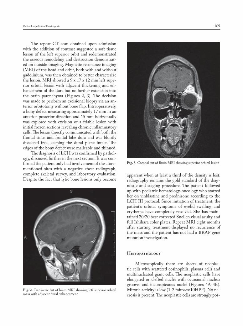
Orbital Langerhans cell histiocytosis 169
The repeat CT scan obtained upon admission with the addition of contrast suggested a soft tissue lesion of the left superior orbit and redemonstrated the osseous remodeling and destruction demonstrat-ed on outside imaging. Magnetic resonance imaging (MRI) of the head and orbit, both with and without gadolinium, was then obtained to better characterize the lesion. MRI showed a 9 x 17 x 12 mm left supe-rior orbital lesion with adjacent thickening and en-hancement of the dura but no further extension into the brain parenchyma (Figures 2, 3). The decision was made to perform an excisional biopsy via an an-terior orbitotomy without bone flap. Intraoperatively, a bony defect measuring approximately 17 mm in an anterior-posterior direction and 15 mm horizontally was explored with excision of a friable lesion with initial frozen sections revealing chronic inflammatory cells. The lesion directly communicated with both the frontal sinus and frontal lobe dura and was bluntly dissected free, keeping the dural plane intact. The edges of the bony defect were malleable and thinned.
The diagnosis of LCH was confirmed by pathol-ogy, discussed further in the next section. It was con-firmed the patient only had involvement of the afore-mentioned sites with a negative chest radiograph, complete skeletal survey, and laboratory evaluation. Despite the fact that lytic bone lesions only become
apparent when at least a third of the density is lost, radiography remains the gold standard of the diag-nostic and staging procedure. The patient followed up with pediatric hematology-oncology who started her on vinblastine and prednisone according to the LCH III protocol. Since initiation of treatment, the patient’s orbital symptoms of eyelid swelling and erythema have completely resolved. She has main-tained 20/20 best corrected Snellen visual acuity and full Ishihara color plates. Repeat MRI eight months after starting treatment displayed no recurrence of the mass and the patient has not had a BRAF gene mutation investigation.
Histopathology
Microscopically there are sheets of neoplas-tic cells with scattered eosinophils, plasma cells and multinucleated giant cells. The neoplastic cells have elongated or clefted nuclei with occasional nuclear grooves and inconspicuous nuclei (Figures 4A-4B). Mitotic activity is low (1-2 mitoses/10HPF). No ne-crosis is present. The neoplastic cells are strongly pos-
Fig. 2. Transverse cut of brain MRI showing left superior orbital mass with adjacent dural enhancement
Fig. 3. Coronal cut of Brain MRI showing superior orbital lesion
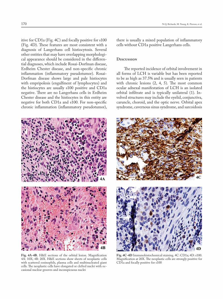
N.Q. Richards, M. Young, K. Pierson, et al.170
itive for CD1a (Fig. 4C) and focally positive for s100 (Fig. 4D). These features are most consistent with a diagnosis of Langerhans cell histiocytosis. Several other entities that may have overlapping morphologi-cal appearance should be considered in the differen-tial diagnoses, which include Rosai-Dorfman disease, Erdheim Chester disease, and non-specific chronic inflammation (inflammatory pseudotumor). Rosai-Dorfman disease shows large and pale histiocytes with empripolesis (engulfment of lymphocytes) and the histiocytes are usually s100 positive and CD1a negative. There are no Langerhans cells in Erdheim Chester disease and the histiocytes in this entity are negative for both CD1a and s100. For non-specific chronic inflammation (inflammatory pseudotumor),
there is usually a mixed population of inflammatory cells without CD1a positive Langerhans cells.
Discussion
The reported incidence of orbital involvement in all forms of LCH is variable but has been reported to be as high as 37.5% and is usually seen in patients with chronic lesions (2, 4, 5). The most common ocular adnexal manifestation of LCH is an isolated orbital infiltrate and is typically unilateral (1). In-volved structures may include the eyelid, conjunctiva, caruncle, choroid, and the optic nerve. Orbital apex syndrome, cavernous sinus syndrome, and sarcoidosis
Fig. 4A-4B. H&E sections of the orbital lesion. Magnification 4A: 10X; 4B: 20X. H&E sections show sheets of neoplastic cells with scattered eosinophils, plasma cells and multinucleated giant cells. The neoplastic cells have elongated or clefted nuclei with oc-casional nuclear grooves and inconspicuous nuclei
Fig. 4C-4D Immunohistochemical staining. 4C: CD1a; 4D: s100. Magnification at 20X. The neoplastic cells are strongly positive for CD1a and focally positive for s100
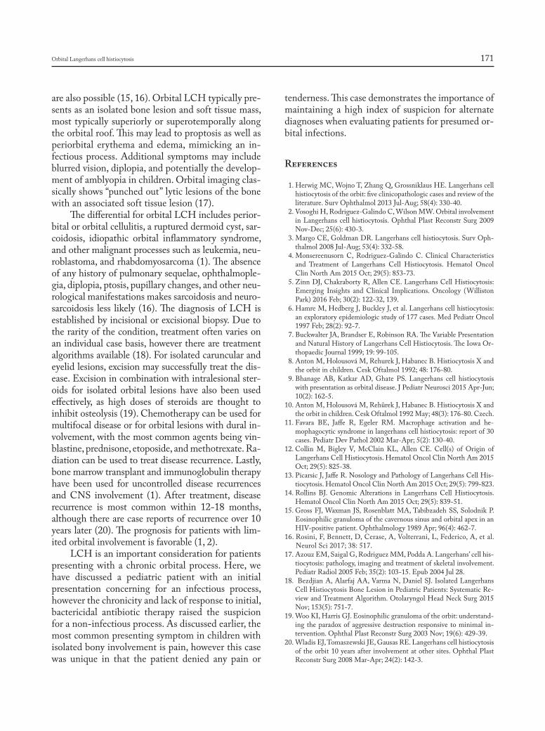
Orbital Langerhans cell histiocytosis 171
are also possible (15, 16). Orbital LCH typically pre-sents as an isolated bone lesion and soft tissue mass, most typically superiorly or superotemporally along the orbital roof. This may lead to proptosis as well as periorbital erythema and edema, mimicking an in-fectious process. Additional symptoms may include blurred vision, diplopia, and potentially the develop-ment of amblyopia in children. Orbital imaging clas-sically shows “punched out” lytic lesions of the bone with an associated soft tissue lesion (17).
The differential for orbital LCH includes perior-bital or orbital cellulitis, a ruptured dermoid cyst, sar-coidosis, idiopathic orbital inflammatory syndrome, and other malignant processes such as leukemia, neu-roblastoma, and rhabdomyosarcoma (1). The absence of any history of pulmonary sequelae, ophthalmople-gia, diplopia, ptosis, pupillary changes, and other neu-rological manifestations makes sarcoidosis and neuro-sarcoidosis less likely (16). The diagnosis of LCH is established by incisional or excisional biopsy. Due to the rarity of the condition, treatment often varies on an individual case basis, however there are treatment algorithms available (18). For isolated caruncular and eyelid lesions, excision may successfully treat the dis-ease. Excision in combination with intralesional ster-oids for isolated orbital lesions have also been used effectively, as high doses of steroids are thought to inhibit osteolysis (19). Chemotherapy can be used for multifocal disease or for orbital lesions with dural in-volvement, with the most common agents being vin-blastine, prednisone, etoposide, and methotrexate. Ra-diation can be used to treat disease recurrence. Lastly, bone marrow transplant and immunoglobulin therapy have been used for uncontrolled disease recurrences and CNS involvement (1). After treatment, disease recurrence is most common within 12-18 months, although there are case reports of recurrence over 10 years later (20). The prognosis for patients with lim-ited orbital involvement is favorable (1, 2).
LCH is an important consideration for patients presenting with a chronic orbital process. Here, we have discussed a pediatric patient with an initial presentation concerning for an infectious process, however the chronicity and lack of response to initial, bactericidal antibiotic therapy raised the suspicion for a non-infectious process. As discussed earlier, the most common presenting symptom in children with isolated bony involvement is pain, however this case was unique in that the patient denied any pain or
tenderness. This case demonstrates the importance of maintaining a high index of suspicion for alternate diagnoses when evaluating patients for presumed or-bital infections.
References
1. Herwig MC, Wojno T, Zhang Q, Grossniklaus HE. Langerhans cell histiocytosis of the orbit: five clinicopathologic cases and review of the literature. Surv Ophthalmol 2013 Jul-Aug; 58(4): 330-40.
2. Vosoghi H, Rodriguez-Galindo C, Wilson MW. Orbital involvement in Langerhans cell histiocytosis. Ophthal Plast Reconstr Surg 2009 Nov-Dec; 25(6): 430-3.
3. Margo CE, Goldman DR. Langerhans cell histiocytosis. Surv Oph-thalmol 2008 Jul-Aug; 53(4): 332-58.
4. Monsereenusorn C, Rodriguez-Galindo C. Clinical Characteristics and Treatment of Langerhans Cell Histiocytosis. Hematol Oncol Clin North Am 2015 Oct; 29(5): 853-73.
5. Zinn DJ, Chakraborty R, Allen CE. Langerhans Cell Histiocytosis: Emerging Insights and Clinical Implications. Oncology (Williston Park) 2016 Feb; 30(2): 122-32, 139.
6. Hamre M, Hedberg J, Buckley J, et al. Langerhans cell histiocytosis: an exploratory epidemiologic study of 177 cases. Med Pediatr Oncol 1997 Feb; 28(2): 92-7.
7. Buckwalter JA, Brandser E, Robinson RA. The Variable Presentation and Natural History of Langerhans Cell Histiocytosis. The Iowa Or-thopaedic Journal 1999; 19: 99-105.
8. Anton M, Holousová M, Rehurek J, Habanec B. Histiocytosis X and the orbit in children. Cesk Oftalmol 1992; 48: 176-80.
9. Bhanage AB, Katkar AD, Ghate PS. Langerhans cell histiocytosis with presentation as orbital disease. J Pediatr Neurosci 2015 Apr-Jun; 10(2): 162-5.
10. Anton M, Holousová M, Rehůrek J, Habanec B. Histiocytosis X and the orbit in children. Cesk Oftalmol 1992 May; 48(3): 176-80. Czech.
11. Favara BE, Jaffe R, Egeler RM. Macrophage activation and he-mophagocytic syndrome in langerhans cell histiocytosis: report of 30 cases. Pediatr Dev Pathol 2002 Mar-Apr; 5(2): 130-40.
12. Collin M, Bigley V, McClain KL, Allen CE. Cell(s) of Origin of Langerhans Cell Histiocytosis. Hematol Oncol Clin North Am 2015 Oct; 29(5): 825-38.
13. Picarsic J, Jaffe R. Nosology and Pathology of Langerhans Cell His-tiocytosis. Hematol Oncol Clin North Am 2015 Oct; 29(5): 799-823.
14. Rollins BJ. Genomic Alterations in Langerhans Cell Histiocytosis. Hematol Oncol Clin North Am 2015 Oct; 29(5): 839-51.
15. Gross FJ, Waxman JS, Rosenblatt MA, Tabibzadeh SS, Solodnik P. Eosinophilic granuloma of the cavernous sinus and orbital apex in an HIV-positive patient. Ophthalmology 1989 Apr; 96(4): 462-7.
16. Rosini, F, Bennett, D, Cerase, A, Volterrani, L, Federico, A, et al. Neurol Sci 2017; 38: 517.
17. Azouz EM, Saigal G, Rodriguez MM, Podda A. Langerhans’ cell his-tiocytosis: pathology, imaging and treatment of skeletal involvement. Pediatr Radiol 2005 Feb; 35(2): 103-15. Epub 2004 Jul 28.
18. Bezdjian A, Alarfaj AA, Varma N, Daniel SJ. Isolated Langerhans Cell Histiocytosis Bone Lesion in Pediatric Patients: Systematic Re-view and Treatment Algorithm. Otolaryngol Head Neck Surg 2015 Nov; 153(5): 751-7.
19. Woo KI, Harris GJ. Eosinophilic granuloma of the orbit: understand-ing the paradox of aggressive destruction responsive to minimal in-tervention. Ophthal Plast Reconstr Surg 2003 Nov; 19(6): 429-39.
20. Wladis EJ, Tomaszewski JE, Gausas RE. Langerhans cell histiocytosis of the orbit 10 years after involvement at other sites. Ophthal Plast Reconstr Surg 2008 Mar-Apr; 24(2): 142-3.
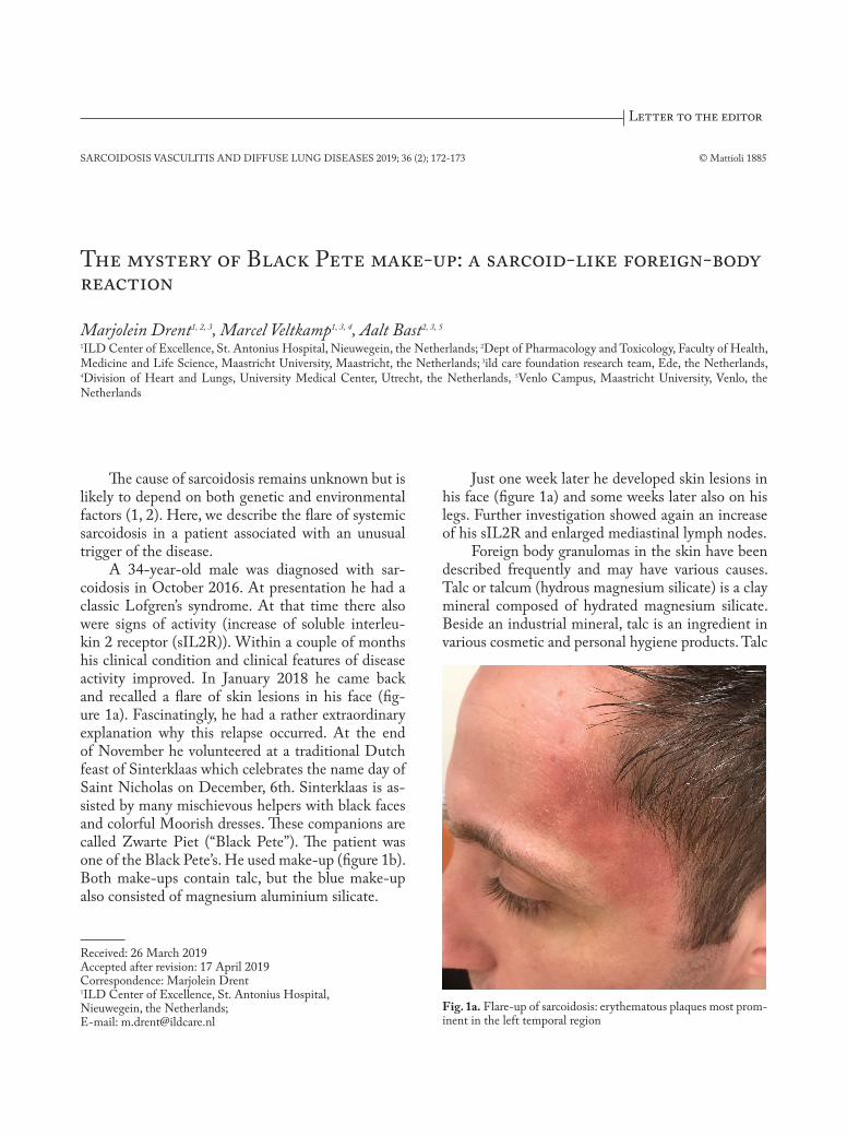
The cause of sarcoidosis remains unknown but is likely to depend on both genetic and environmental factors (1, 2). Here, we describe the flare of systemic sarcoidosis in a patient associated with an unusual trigger of the disease.
A 34-year-old male was diagnosed with sar-coidosis in October 2016. At presentation he had a classic Lofgren’s syndrome. At that time there also were signs of activity (increase of soluble interleu-kin 2 receptor (sIL2R)). Within a couple of months his clinical condition and clinical features of disease activity improved. In January 2018 he came back and recalled a flare of skin lesions in his face (fig-ure 1a). Fascinatingly, he had a rather extraordinary explanation why this relapse occurred. At the end of November he volunteered at a traditional Dutch feast of Sinterklaas which celebrates the name day of Saint Nicholas on December, 6th. Sinterklaas is as-sisted by many mischievous helpers with black faces and colorful Moorish dresses. These companions are called Zwarte Piet (“Black Pete”). The patient was one of the Black Pete’s. He used make-up (figure 1b). Both make-ups contain talc, but the blue make-up also consisted of magnesium aluminium silicate.
Just one week later he developed skin lesions in his face (figure 1a) and some weeks later also on his legs. Further investigation showed again an increase of his sIL2R and enlarged mediastinal lymph nodes.
Foreign body granulomas in the skin have been described frequently and may have various causes. Talc or talcum (hydrous magnesium silicate) is a clay mineral composed of hydrated magnesium silicate. Beside an industrial mineral, talc is an ingredient in various cosmetic and personal hygiene products. Talc
The mystery of Black Pete make-up: a sarcoid-like foreign-body reaction
Marjolein Drent1, 2, 3, Marcel Veltkamp1, 3, 4, Aalt Bast2, 3, 5
1ILD Center of Excellence, St. Antonius Hospital, Nieuwegein, the Netherlands; 2Dept of Pharmacology and Toxicology, Faculty of Health, Medicine and Life Science, Maastricht University, Maastricht, the Netherlands; 3ild care foundation research team, Ede, the Netherlands, 4Division of Heart and Lungs, University Medical Center, Utrecht, the Netherlands, 5Venlo Campus, Maastricht University, Venlo, the Netherlands
SARCOIDOSIS VASCULITIS AND DIFFUSE LUNG DISEASES 2019; 36 (2); 172-173 © Mattioli 1885
Letter to the editor
Received: 26 March 2019Accepted after revision: 17 April 2019Correspondence: Marjolein Drent1ILD Center of Excellence, St. Antonius Hospital, Nieuwegein, the Netherlands;E-mail: [email protected]
Fig. 1a. Flare-up of sarcoidosis: erythematous plaques most prom-inent in the left temporal region

The mystery of Black Pete make-up 173
may cause some adverse effects such as local inflam-mation, infection, and allergic reactions on the skin and even systemic adverse effects such as sarcoid like reactions. Most commonly, erythematous nodules are seen. Tattoo pigments may migrate to regional lymph nodes and may cause a sarcoid like granu-lomatous reaction (3).
Among others talc is believed to trigger an ex-aggerated immune response, with macrophages me-diating antigen processing and presentation, leading to an influx of T cells, a polarized T helper 1 (Th1) cytokine response, and ultimately, to granuloma for-mation (4). Several susceptibility genes have been identified in sarcoidosis, with the strongest associa-
tions to date in the major histocompatibility (MHC) class II gene alleles, which coordinate antigen pres-entation. These findings support the role of a dys-regulated response to antigens in development of sarcoidosis (5).
A flare up of systemic sarcoidosis in a 34-year-old male with a history of sarcoidosis first present-ing with Lofgren’s syndrome, who demonstrated spontaneously remission half a year after the first submission (April 2017), was presented. December 2017 he developed skin lesions in his face, just after he used make-up. This make-up contains talc. This undeniably exceptional case shows that the trigger of a sarcoid-like reaction might be make-up containing talc. It emphasizes that talc exposures should be con-sidered in case of skin lesions as well as other mani-festations of sarcoidosis or sarcoid-like reactions.
Acknowledgement
We wish to thank the patient for his consent to pub-lish the pictures and accompanying text.
References
1. Lodha S, Sanchez M, Prystowsky S. Sarcoidosis of the skin: a review for the pulmonologist. Chest 2009; 136: 583-596.
2. Valeyre D, Prasse A, Nunes H, et al. Sarcoidosis. Lancet 2014; 383: 1155-1167.
3. Anolik R, Mandal R, Franks AG, Jr. Sarcoidal tattoo granuloma. Der-matol Online J 2010; 16: 19.
4. Zissel G. Cellular activation in the immune response of sarcoidosis. Semin Respir Crit Care Med 2014; 35: 307-315.
5. Wolin A, Lahtela EL, Anttila V, et al. SNP Variants in major histo-compatibility complex are associated with sarcoidosis susceptibility: a joint analysis in four European populations. Front Immunol 2017; 8: 422.
Fig. 1b. Face of the same patient was colored with make-up one week before the skin lesions appeared (figure 1a)
Related Documents