Sample Dictations

Welcome message from author
This document is posted to help you gain knowledge. Please leave a comment to let me know what you think about it! Share it to your friends and learn new things together.
Transcript

SampleDictations
Every study must have an Impression!
Plain Films:
CXR: The CXR is arguably the most difficult of all studies to interpret. It is necessary to besystematic, evaluating each component in order. Evaluate the heart and mediastinum. Thehila, lungs, pleura, diaphragms (costophrenic and cardiophrenic angles), bones and softtissues. The order is personal style. Just be consistent and complete.Clinical Hx:Views: AP/PA and Lateral.Comparison:Findings: The heart and mediastinal contours are normal. The lungs are clear. Thevisualized bones are normal.Impression: Normal chest radiograph.
Portable CXR: Single view of ChestClinical Hx:Views: A portable upright CXR was obtained at 1900 h (must include time on all portablexrays).Comparison:Findings:Impression: Normal portable chest radiograph.ICU CXR: Same as above but make sure to mention lines, tubes, etc.
Abdominal Radiographs:Clinical Hx:Views: __ views: PA/AP Supine, Erect, Decubitus, etc. (the number of views must beincluded for billing purposes) Remember that multiple films may still only count as one view(e.g. large patient).Comparison:Findings: The bowel gas pattern is normal. There is no free air, ascites or mass effect. Thesoft tissue planes are normal. There are no suspicious calcifications. The visualized bonesare normal.Impression: Normal plain film of the abdomen.
Ankle:Clinical Hx:Views: AP, lateral, mortise views.
Comparisons:Findings: The bones and soft tissues are normal. The ankle mortise is intact. There is nojoint effusion.Impression: No fracture or dislocation.
CSpine:Clinical Hx:Views: Incollar lateral, AP, odontoid, obliques, swimmers, out of collar lateral, Flex/ExtComparisons:Findings: There is no fracture or subluxation. The prevertebral soft tissues are normal.Vertebral body height and alignment are maintained. The disk heights are maintained.There are no degenerative changes. The neural foramen are widely patent bilaterally [ifoblique views were obtained].Impression: No fracture or dislocation.
Elbow:Clinical Hx:Views: AP and LateralComparisons:Findings: The elbow joint is normal. There is no fracture or dislocation. There is no jointeffusion. There are no suspicious soft tissue calcifications.Impression: No fracture or dislocation
Knee:Clinical Hx:Comparisons:Views: AP, lateral, merchants, tunnel, standing, sunriseFindings: The bones and soft tissues are normal. There is no joint effusion. The jointspaces are preserved. There are no suspicious soft tissue calcifications.Impression: No fracture or dislocation.
Shoulder:Clinical Hx:Views: Internal/external rotation, scapular Y, axillary, lateral shoot throughComparisons:Findings: The glenohumeral joint is normal. The acromioclavicular joint is normal. There areno fractures or dislocations. There are no suspicious soft tissue calcifications.Impression: No fracture or dislocation.
Sinus:Clinical Indication: [...].Technique: Three views of the sinuses.Comparison: [...].Findings: There is no air fluid level within the visualized paranasal sinuses. No abonormalopacification is noted.Impression: No evidence of sinusitis.
GI/Fluoroscopy:Barium Swallow:Clinical Indication: [...].Technique: An air contrast esophagogram was performed.Comparison: [...].Findings: The esophagus is normal. There is no gastroesophageal reflux eitherspontaneously or with provocative maneuvers. The fundus of the stomach is normal.The 13 mm tablet passed easily down the esophagus into the stomach without difficulty.Impression: Normal esophagogram.
AC UGI (Upper GI):Clinical Hx:Comparisons:Technique: An aircontrast examination of the upper GI tract was performed. (oraircontrast upper GI series for small bowel series, add "with fluoroscopic small bowelexamination").Findings: The esophagus is normal. There was no gastroesophageal reflux eitherspontaneously or with provocative maneuvers. The stomach and duodenum were normal.Impression: Normal air contrast upper GI series.
Barium Enema:History: [...].Technique: An aircontrast barium enema was performed using the biphasic technique (orprimary double contrast technique).Findings: There is ….(findings). There was excellent visualized visualization of the cecum,ileocecal valve, terminal ileum, and appendix. There is no evidence of polyps greater than1 cm, polypoid masses or annular constrictions of the colon.Impression: [...]
Modified Barium Swallow (fluoroscopic swallowing study):Clinical Hx: e.g. Aspiration pneumonia.

Technique: The patient was given thin liquids, puree and soft solids and the oral andpharyngeal phases of swallowing were observed fluoroscopically.Findings: [Describe findings during the oral and pharyngeal phases for each consistencytested, each as a separate paragraph. Also, describe the use of maneuvers such as chintuck.] There is normal propulsion of the oral bolus into the oral pharynx. There is noevidence of penetration or aspiration of barium into the vocal cords or trachea. The oralmucosa, pharyngeal mucosa, pyrifrom sinuses, vallecula and epiglottis are normal. Thereis no prominent cricopharyngeus. The patient did well with both solids and liquids ofdifferent viscosities. A chin tuck maneuver did not significantly improve swallowingperformance.Impression: Normal swallowing with no aspiration or penetration with any consistencystudied. Please see the report by speech therapy for further details and dietaryrecommendations.
ERCP:Clinical Hx: RUQ pain and elevated LFTsTechnique: An ERCP of the bile duct (if pancreatic duct cannulated add and thepancreatic duct) was performed by Dr. Ruyman.Findings: The CBD was well opacified and there are no filling defects, dilatation orstrictures.The pancreatic duct was normal.Impression:1. Normal extrahepatic biliary ducts.2. Normal pancreatic duct.
Hysterosalpingogram:Clinical Hx: InfertilityTechnique: The hysterosalpingogram was performed by Dr. ____ using Omnipaque 300.Findings: The uterus is normal.Both fallopian tubes are patent with free spillage of contrast into the peritoneal cavity.Impression: Normal hysterosalpingogram.
Shoulder Arthrogram (will get MRI):Clinical Hx:Technique: The standard timeout procedure for patient and body part identification wasperformed. The risks and benefits of the procedure was explained to the patient and writteninformed consent was obtained. The patient was prepped and draped in the usual sterilefashion. After the administration of 4 mL of lidocaine 1%, a 22 gauge needle wasadvanced under fluoroscopic guidance into the glenohumeral joint space. 12 ml of a 3:1
mixture of 0.63 mL of Magnevist in 100 cc of Normal Saline and Omnipaque 300 was theninfused into the joint space. The patient tolerated the arthrogram well with no immediatecomplications. The patient was then taken to MRI.Findings: There is no fluoroscopic evidence of a rotator cuff tear. (There must be somedescription of findings for billing purposes.)Impression: Normal Fluroscopic arthorgram. Please refer to the MRI report to follow. (theMRI should be done within 1 hour of contrast administration).
Joint Steroid Injection:Clinical indication: [...].Technique: The timeout procedure was observed for proper identification of the patientand body part. The purpose, technique and risks of the procedure were explained to thepatient. [...] understood and signed a written consent form.The [...] hip was prepped in a sterile fashion. 4 ml of 1% lidocaine were injected into theoverlying soft tissues. A 22 gauge needle was introduced into the hip joint space underfluoroscopic guidance. 1 ml of Omnipaque300 were injected into the hip joint to establishintraarticular position of the needle tip. 5 ml of Sensorcaine (Marcaine) 0.5% and 80(sometimes 40) mg of DepoMedrol were injected into the hip joint.Findings: There is degenerative osteoarthritis of the [...] hip. The patient tolerated the [...]hip arthrocentesis without complication.Impression: Successful [...] hip arthrocentesis with injection of 5 ml of Sensorcaine(Marcaine) 0.5% and 80 mg of DepoMedrol.
Intravenous Pyelogram (Urogram):Clinical Hx:Technique: 100 ml of omnipaque 300 were injected intravenously and an IVP wasperformed.Findings: The preliminary film demonstrates …(findings, or “is normal.”).The kidneys are normal in size and position, (include length of both kidneys). Thepelvocalyceal systems and ureters are normal in appearance. The bladder is normal incontour. There is no postvoid residual.Impression: Normal IVP.
UltrasoundAbdominal Ultrasound:Clinical Hx:Technique: Transabdominal imaging was performed.Comparisons:
Findings: There is normal liver echogenicity without intrahepatic ductal dilatation. Thegallbladder is normal with no gallstones, wall thickening, pericholecystic fluid, distention ora positive ultrasonagraphic Murphy’s sign. The CBD measures __ mm, which is normal.Limited evaluation of the pancreas is normal. The spleen measures __ cm and is normal inappearance. There is no evidence of an abdominal aortic aneurysm.The kidneys measure _ and __ on the left and right, respectively and are normal inechogenicity. There is no hydronephrosis and no renal stones are identified.Impression: Normal abdominal ultrasound.
Renal Ultrasound:Clinical Hx:Comparisons:Findings: The kidneys are normal in size, shape and echogenicity measuring __ and __ onthe left and right, respectively. There is no hydronephrosis and no renal stones areidentified. There are no mass lesions or cysts. There is no evidence of an abdominal aorticaneurysm. The urinary bladder is normal and there is no postvoid residual.Impression: Normal renal ultrasound.
Obstetrical Ultrasound: (note there is a specific and detailed format for a fetalsurvey ask if you don't have it)Clinical Hx:Comparisons:Technique: Transabdominal (& transvaginal) imaging was performed of a gravid uterus.Findings: There is one fetus in variable lie. The heart rate in noted at __ bpm. Fetal surveyincluding visualization of the fourchambered view of the heart, stomach, kidneys, cordinsertion, bladder and spine are normal. (If not, mention here).Fetal measurements give a mean age of __, +/ __ weeks.If prior ultrasounds, then add the following: This correlates with the extrapolated age basedon the earlier ultrasound(s).If the fetus is in the 3rd TM, add: The estimated fetal weight is __ which is in the ___% forthe gestational age of ___ wks. If BPP performed give results here.The placenta is located anteriorly (fundally, posteriorly), above the lower uterine segment.The amniotic fluid volume is normal (or the AFI is __ cms).Impression: Single intrauterine pregnancy of __ weeks ___ days, +/ )___ days. Thiscorresponds/does not correspond to the dating by LMP with an EDD of ___. (If it doescorrespond, do not "correct" the due date as this drives patients and obstetricians crazy.)
Pelvic Ultrasound:Clinical Hx:
Technique: Transabdominal & transvaginal imaging were performed.(if done Dopplerevaluation of ovarian blood flow was included.)Comparisons:Findings: The uterus is homogenous in echogenicity measuring __ x __ x __ cm and isanteverted/retroverted. The endometrial stripe is uniform and measures __mm, which iswithin normal limits. There are no fibroids. The ovaries are identified by their follicles andmeasure __ x __ x __ cm trasvaginally on the right and __ x __ x __ cm transvaginally onthe left. There are no adnexal masses or cysts.The kidneys are normal in size, shape and echogenicity. There is no hydronephrosis andno renal stones are identified. No free fluid is seen. (If Doppler is used, e.g. to confirm flowto both ovaries, dicatate this as its own section.)Impression: Normal pelvic ultrasound.
Thyroid Ultrasound:Clinical Hx:Comparisons:Findings: Ultrasound evaluation of the thyroid gland demonstrates homogeneous glandulartissue. The right lobe measures __ x __ x __ cm and the left lobe measures __ x __ x __cm. The isthmus measures __. No nodules are seen.Impression: Normal thyroid ultrasound.
Testicular Ultrasound:Clinical Hx:Technique: Transscrotal ultrasound evaluation was performed.Comparisons:Findings: There is normal and symmetric testicular echogenicity with the right testismeasuring __ x __ x __ cm and the left testis measuring __ x __ x __ cm __. The head ofthe right epididymis measures <10mm: the head of the left epididymis measures <10mm.Testicular and epididymal perfusion is symmetric bilaterally. There are no varicoceles orhydroceles. The scrotal skin is not thickened. (If varicoceles are seen, both kidneys mustbe checked for hydronephrosis and this should be included in the report.)Impression: Normal testicular ultrasound.
Vascular Ultrasound, Lower Extremity Venous:Clinical Hx:Technique: Venous duplex imaging was performed of the __ lower extremity.Findings: The right common femoral, superficial femoral, popliteal, tibial and peronealveins demonstrate normal compressibility and color flow throughout. Maneuvers includingValsalva, augmentation and respiration demonstrate normal variation in flow in the vessels

studied. (Note to Residents: When dictating this study, it has been suggested to omit theword superficial from superficial femoral vein, because ordering doctors have been knownto not treat superficial femoral vein DVTs, confusing a true DVT with superficialthrombophlebitis)Impression: There is no evidence of a deep venous thrombosis.
First trimester ultrasound: use the system autotext.Fetal survey: use the system autotext.
Obstetrical Ultrasound: Biophysical ProfileCriteria for Coding Fetal Biophysical Variables as Normal or Abnormal
Biophysical
Variable
Normal(Score = 2)
Abnormal(Score = 0)
Fetalbreathingmovements
1 or more episodes of >20 s within30 min
Absent or no episode of >20 s within30 min
Grossbodymovements
2 or more discrete body/ limbmovements within 30 min (episodesof active continuous movementconsidered as a single movement)
<2 episodes of body/limbmovements within 30 min
Fetal tone 1 or more episodes of activeextension with return to flexion offetal limb(s) or trunk (opening andclosing of hand considered normaltone)
Slow extension with return to partialflexion, movement of limb in fullextension, absent fetal movement, orpartially open fetal hand
QualitativeAFV
1 or more pockets of fluidmeasuring >2 cm in vertical axis
Either no pockets or largest pocket<2 cm in vertical axis
Clinical Indication: Pregnancy of [...] weeks [...] days, [...], check biophysical profile andamniotic fluid index.Technique: Transabdominal imaging of the gravid uterus was performed, including Dopplerevaluation of the fetal heart rate.Findings: The fetus is in ____ presentation. Fetal breathing, movement and tone and theamniotic fluid index were assessed with a total biophysical profile score of 8/8. The
amniotic fluid index was measured to be [...] cm, which is within normal limits.The fetal heart rate was measured by Doppler to be [...] beats per minute.Impression: BPP of 8/8 with an AFI of [...]. This result was called to [...].
CT Scan:Head CT:Clinical Hx: Trauma, Headaches, Mental status changes, etc..Technique: Serial axial images were obtained from the foramen magnum to the vertex withoutthe use of intravenous contrast.Comparison:Findings: The ventricles, sulci and cisterns are normal for the patient's age. There is no masseffect, midline shift, hemorrhage, acute territorial infarct or extraaxial fluid collection. (You mayneed to mention mm of midline shift or type of hemiation.) The visualized paranasal sinuses,mastoid air cells, and bones are normal. The soft tissues are unremarkable.Impression:
Sinus CT:Clinical Hx:Technique: Helical 1.25 mm axial images were obtained through the paranasal sinuses andreconstructions were performed in the coronal plane.Comparison:Findings: There is no mucosal thickening in the frontal sinuses and the frontoethmoid recessesare clear. The ethmoid air cells, sphenoid and maxillary sinuses are normal. The visualizedportions of the mastoid air cells are well aerated. The osteomeatal complexes are widely patent,and the infundibulae are bordered laterally by the orbit (or by the Haller air cells). The nasalseptum is approximately midline in position without a nasal septal spur. The cribiform plate issymmetric and the lamina papryceae are intact. There is no evidence of dental disease. Thecarotid arteries are covered by bone as they pass superior to the sphenoid sinuses.Impression:
Temporal Bone CT:Clinical Hx: Hearing loss, pulsitile tinnitus, pain, etc.Technique: Serial axial images were obtained through the inner ear without the use ofintravenous contrast. Coronal and sagittal 2D reformatted images were also obtained.Comparisons: NoneFindings: The external auditory canals are normal bilaterally. No abnormalities are seen in eithermiddle ear space. Specifically, the ossicular chains are intact and there is no abnormal softtissue or bony erosion. The tympanic membranes are intact. The inner ear structures arenormal. The internal auditory canals are unremarkable. The tegmen is intact bilaterally, withnormal bony coverings of the lateral semicircular canal and facial nerve. The jugular veins andcarotid arteries follow their normal course.
Impression:
Facial bone CT:Clinical Hx: Trauma, etc..Technique: Serial axial images were obtained from the mandible through the frontal sinuseswithout the use of intravenous contrast. Coronal and sagittal 2D reformatted images were alsoobtained.Comparison:Findings: The visualized paranasal sinuses are normal. The orbits and surrounding soft tissuesare unremarkable. The medial wall and orbital floors are intact. There are no fractures in thenasal bones, maxilla, zygomatic arches or frontal bones. The nasal septum is midline. There isno evidence of dental disease.Impression:
Chest CT:Clinical Hx:Technique: Serial axial images were obtained from the apices of the lungs through the kidneysafter the administration of intravenous contrast.Orders (if applicable): The patient received 100 cc of Omnipaque 350 as ordered by...Comparisons:Findings: Include discussion of the following regions—neck, thyroid, axillae, lymph nodes:mediastinum including great vessels (always look for PE), mediastinal and hilar lymph nodes,airway, heart (mention coronary artery calcifications), pericardium, lungs, pleura, chest wall, softtissues/breasts, visualized abdomen.Impression: Prioritize findings, most important to least important.
CT Pulmonary Angiogram:Clinical Hx: Shortness of breath with elevated Ddimer.Technique: Serial axial images were obtained from the upper abdomen through the apices of thelungs during the administration of IV contrast. 3D and multiplanar CT reformations were thenperformed.Orders: The patient received 100 cc of Omnipaque 350 as ordered by...Comparisons:Findings: There is no filling defect within the main pulmonary arteries or their branches. Includediscussion of the following regionsneck, thyroid, axillae, lymph nodes: mediastinum includinggreat vessels, mediastinal and hilar lymph nodes, airway, heart, pericardium, lungs, pleura,chest wall, soft tissues/breasts, visualized abdomen.Impression:1. No pulmonary embolus (or PE seen…)2. Include any other findings.
Abdominal/Pelvis CT:
Clinical Hx:Technique: Serial axial images were obtained from the domes of the diaphragm through thepubic symphysis.Orders: The patient received 125 cc of Omnipaque 350 as ordered by ...Comparisons: There are no previous abdominal/pelvis CTs for comparison.Findings: The visualized lungs and mediastinal structures are normal.Abdomen: Describe the following: Liver, ascites, gallbladder, biliary tree, spleen, pancreas,kidneys & adrenals. Look at vascular structures (including aorta & its branches, IVC, portal vein,etc) & lymph nodes. Presence or absence of free air.The small and large bowel, always look for/at and commend on the appendix.Pelvis: Describe the following: The uterus and adnexae, prostate, seminal vesicles, bladder, iliacveins for DVT. Check for free fluid in pelvis.The visualized bones and soft tissues are normal.Impression:
Pancreatic Mass: use the system autotext.
Magnetic Resonance ImagingBrain MRI (without or with and without Gadolinium):Clinical Hx:Technique: Brain MR was performed without (or without and with) Gadolinium including thefollowing sequences: SagTl, Ax FLAIR, FSE Ax T2, Ax T1, Ax DWI, Ax GRE (If Post Gado: AxTl,CorTI, SagTl)Comparisons:Findings: The ventricles, sulci and cisterns are normal. There is no mass, mass effect,hemorrhage or abnormal extraaxial collection. Then answer the question i.e. There is noevidence of acute ischemic injury or traumatic intracranial injury or demyelinating disease ormetastatic disease or intracranial infection, etc. The flow voids of the major vessels at the baseof the brain are normal.The globes, orbits and paranasal sinuses are unremarkable.Impression:
Brain MRA and Neck MRA:Same as above except add the following to the Technique and BodyTechnique: AddBrain MRA: 3D time of flight imaging was performed of the Circle of Willis, with complexreformations.Neck MRA: 3D time of flight imaging post gadolinium was performed., with complexreformations.Body: AddBrain MRA: There is normal flow related enhancement in the visualized portion of the internal
carotid, middle cerebral and anterior cerebral arteries. The basilar artery and its branches areunremarkable.Neck MRA: The common, internal and external carotid arteries appear normal. The codominantvertebral arteries are unremarkable.
Internal Auditory Canal MRI (or Pituitary MRI):Clinical Hx:Technique:Findings:The ventricles, sulci and cisterns are normal. There is no mass, mass effect, hemorrhage orabnormal extraaxial collection. Thin section imaging through the internal auditory canals withand without contrast is normal. Specifically, there is no evidence of an acoustic schwannoma.(Or for pituitary MRIs: There is no sellar or suprasellar mass lesion. The infundibulum is midline.The cavernous sinus is unremarkable.) The globes, orbits and paranasal sinuses areunremarkable. The flow voids of the major vessels at the base of the brain are normal.Impression:
Cervical Spine MRI:Clinical Hx:Technique: MR Imaging of the cervical spine was performed without intravenous contrastincluding the following sequences: Sag FSE T2, Sag Tl, Ax FSE T2 and Ax Gradient Echo.Comparisons:Findings: The cervical vertebral bodies are normal in height, alignment, and signalcharacteristics. The visualized portion of the spinal cord and contents of the posterior fossaappear normal. The intervertebral disc heights are preserved. There is no significant central orforaminal narrowing at any of the imaged levels. (Be sure to look at the visualized brain)Impression:
Lumbar Spine MRI:Clinical Hx:Technique: MR imaging was performed of the lumbar spine without intravenous contrastincluding the following sequences: Sag T1 and FSE T2, Ax T1 and FSE T2 .Comparisons:Findings: The lumbar vertebral bodies are normal in height, alignment, and signalcharacteristics. The conus medullaris terminates normally at the T12/L1 level. The intervertebraldisc heights are preserved. There is no facet joint hypertrophy or ligamentum flavum infolding.There is no significant central or foraminal narrowing at any of the imaged levels.Impression:
Shoulder MRI:Clinical Hx:Technique: MR imaging was performed of the (right or left) shoulder without the use of
intravenous contrast including the following sequences: Triplanar proton density with fatsuppression, Axial Gradient Echo, Coronal Oblqieu T2, and Coronal Oblique STIR images.Comparisons:Findings: The bone marrow signal is normal. The supraspinatus tendon is normal inconfiguration and signal characteristics. The infraspinatus, teres minor and subscapularistendons are intact. The long head of the biceps tendon is normally located and attaches normallyon the superior labrum. The articular surface of the humeral head is intact. No displaced labraltear is seen. There is no significant acromioclavicular joint hypertrophy. The coracohumeral,coracoclavicular and coracoacromial ligaments are normal. There is no fluid in the subacromialbursa or subdeltoid bursa.
Knee MRI:Clinical Hx:Technique: MR Imaging of the knee was performed without intravenous contrast including thefollowing sequences: Sag PD, Sag PD with Fat Sat. Cor PD with fat saturation, Cor Tl, Ax PD,and Ax FSE T2 Fat Sat.Comparisons:Findings: The bone marrow signal is normal. The ACL, PCL, medial collateral ligament andlateral collateral ligamentous complex are intact. The quadriceps tendon and infrapatellar tendonare normal. There is no lateral or medial meniscal tear. The patellofemoral joint is normal with nochondromalacia patella. There is no joint effusion. No synovial cyst is seen.Impression:
Ankle MRI:Clinical Hx:Technique:Comparisons:Findings: The bone marrow signal is normal. The subtalar joints appear intact. No abnormalsignal is seen in the sinus tarsi. The posterior tibialis, flexor digitorum. flexor hallucis, peroneal.extensor and Achilles tendons all have homogeneous dark signal. There are no joint effusions.The anterior and posterior tibiofibular and talofibular ligaments are intact The deltoid ligamenthas normal signal characteristics.Impression:
Dr. Heffess instruction on protocoling special MRI studies:
MRI PELVIS: Cervical cancer. Must be posted by an MD.MRI PELVIS: Mullerian anomaly. Must be posted by an MD.MRI PELVIS: Rectal cancer. Must be posted by an MD.MRI PELVIS: Perianal fistula/abscess. Must be posted by an MD.
Failing to post the above studies properly interferes with their diagnostic quality.
MRI PELVIS: Prostate. Must be monitored by an MD.MRI ABDOMEN: Enterography. Must be monitored by an MD.MRI ABDOMEN: Urography. Must be monitored by an MD.
Please take care in protocoling these studies and with the EOVIST vs MAGNEVIST livers. Dr. Heffess isalways happy to help.
Nuclear Medicine: (many have autotexts available in SpeechQ)Note: use the autotexts for nuclear medicine, or get them from a fellow resident.Pulmonary V/Q Scan:Clinical Hx: Shortness of breathRadiopharmaceutical: Xe 133 and TcMAADose: __ mCi and __ mCiTechnique: Utilizing radioactive Xe, single breath, equilibrium and washout images of thelungs were obtained. Then after the intravenous administration of the perfusion agent, [sixor eight] images of the lungs were obtained.Findings: There was heterogenous ventilation of the lungs with trapping at the bases. Thereis homogeneous perfusion of the lungs without evidence of a pleural based, wedge shapeddefect.Impression: Normal V/Q scan [or very low prob, low prob, intermediate or high prob forPE].
Myocardial Perfusion study with LexiscanRadiopharmaceutical: Technetium 99m MyoviewDose: Rest dose [...] mCiStress dose [...] mCiTotal dose [...] mCiClinical indication: [...]Technique: A [...]day study was performed. The patient was injected with 0.4 mg IV ofregadenoscan and then with the radiopharmaceutical. Patient performed [...] min. of lowlevel exercise. Multiple tomographic views of the heart were then obtained and comparedwith similar images obtained at rest. The Lexiscan and rest examinations were gated. Theleft ventricular ejection fraction and wall motion were assessed.Comparison: [...]Findings: No significant focal reversible perfusion defect is seen. At pharmacologic stress,the left ventricular ejection fraction is [...] %, which is within normal limits. No significantregional wall motion abnormality is seen.Impression:
1. No evidence of focal jeopardized myocardium.2. Normal left ventricular ejection fraction without significant regional wall motionabnormality at stress.
Gastric emptying study:Radiopharmaceutical: Technetium 99m sulfur colloid eggsDose: [...] mCiClinical indication: [...]Technique: After the administration of the radioactive meal, sequential images of theabdomen were obtained every 10 minutes for 60 minutes. At 2, 3, and 4 hours, staticimages of the abdomen were obtained. Computer analysis was performed to determinethe amount of gastric retention at each hour.Findings: At 1, 2, 3, and 4 hours, the gastric contents remaining were [...]%, [...]%, [...]% and[...]% respectively. The upper limits of normal for gastric retention at 4 hours is 10%.Impression: [Normal] gastric emptying.
Triple Phase Bone Scan:Clinical Hx: History of cancer, bacteremia, etc.Radiopharmaceutical: Technecium99m MDPDose: __ mCiTechnique: Immediately after the intravenous administered of the radiopharmaceutical,blood flow and blood pool images were obtained. After a 3 hour delay, whole body andhigh resolution images of the area of interest were obtained.Findings: The blood flow and Blood pool images were symmetric and normal. On delayedimages, there was no abnormal uptake (at the area of interest).Impression: No evidence of metastatic disease, osteomyelitis, etc
Urea Breath Test:*Note regarding an indeterminate urea breath test:Findings: The resultof this study was indeterminate. Approximately 5% of all tests giveindeterminate results. Although there are many theories regarding the cause of this, theexact cause of an indeterminate result is not known.The indeterminate result should be evaluated by repeating the PY test or using analternative diagnostic method. If repeat breath testing is undertaken, careful history toexclude confounding factors should be obtained. If confounding factors are present, waitappropropriate time depending on the confounding factor before repeating the PY test. Inall cases, wait for at least six hours before repeating the PY test so that any 14CO2 in thebreath will be eliminated and not interfere with the results of a second test.The recommended time between confounding factor and retesting are as follows:

Antibiotics: 1 monthBismuth: 1 monthProton Pump Inhibitors: 2 weeksSucralfate: 2 weeksNonfasting: 6 hours
Specials (IR): (some have autotexts available in SpeechQ)Abscess Drainage, Seldinger TechniqueClinical history: [...]Operators: Drs. [...]Technique: Informed consent was obtained. CT confirm the presence of a fluid collectionwithin the [...]. After administration of 1% lidocaine into the superficial and deep tissues, aone step catheter was advanced into the collection with immediate aspiration of [...]. Awire was passed through the catheter, serial dilations were performed over the wire and a10.2 French drainage catheter was advanced into the collection. Then [...] mL of [...] fluidwas immediately aspirated. The drainage catheter was hooked up to a drainage bag.Followup CT imaging at the completion of the procedure revealed nearcompleteresolution of the fluid collection.The patient tolerated the procedure well with no immediate complications.Impression: Successful CTguided drainage of fluid collection with drainage of [...] mL offluid.
Abscess Drainage, Tandem Trocar Technique (JS often uses this technique)Clinical history: [...]Operators: Drs. [...]Technique: Informed consent was obtained. CT confirm the presence of a fluid collectionwithin the [...]. After administration of 1% lidocaine into the superficial and deep tissues, a20 gauge x 15 cm Temno Cheeba needle was advanced under CT guidance into the fluidcollection. Then, a 10.2 French drainage catheter was advanced into the collection usingtandem trocar technique. [...] mL of [...] fluid was immediately aspirated. The drainagecatheter was hooked up to a drainage bag. Followup CT imaging at the completion of theprocedure revealed nearcomplete resolution of the fluid collection.The patient tolerated the procedure well with no immediate complications.Impression: Successful CTguided drainage of fluid collection with near completeresolution of the fluid collection at the completion of the procedure.
Abscess Tube CheckClinical history: [...]Operators: Drs. [...]

Technique: 23 ml of Omnipaque 300 was injected into the patient's existing 10.2 Frenchallpurpose drainage catheter. The contrast was then aspirated through the drainagecatheter.Findings: The abscess drainage tube is in good position. There is a small residualabscess cavity. There is no fistula. The abscess drainage tube was reconnected to thecollection bag. The patient was instructed to call immediately if experiencing fevers, chills,nausea, vomiting or flank pain. Impression: Abscess drainage catheter in satisfactory position.
Angiogram, Lower ExtremitiesClinical history: [...]Operators: Drs. [...]Technique: Informed consent was obtained. The patient was placed in the supineposition, and prepped and draped in the usual sterile manner. 1% lidocaine was used foranesthesia at the groin. Using a Seldinger technique, the right groin was accessed in aretrograde manner. With the aid of a glide wire, a 4French Omniflush catheter wasadvanced into the aorta at the level of L1. An angiogram was then performed. The pigtailcatheter was then withdrawn to the level of the bifurcation. Subsequent angiograms werethen performed of the lower extremity runoff including a lateral view of the feet.Findings: There are single patent renal arteries bilaterally. The superficial femoral artery ispatent. The profunda femoris is patent. The popliteal artery is patent. There is a [...] vesselrunoff via [...] arteries.Angiogram of the foot demonstrates [...].Total contrast: [...] ml Omnipaque 350.Impression: Status post angiogram of the [...] lower extremity with [...] vessel runoff to thelower extremity.
Angiogram, GI BleedingClinical history: [...] year old with gastrointestinal bleeding.Operators: Drs. [...]Technique: Informed consent was obtained. The patient was placed in the supineposition, and prepped and draped in the usual sterile manner. 1% lidocaine was used foranesthesia at the groin. Using a Seldinger technique, the right groin was accessed in aretrograde manner. With the aid of a glide wire, a glide catheter was advanced into theaorta at the level of L1. The SMA was selected and an angiogram was then performed.The catheter was then exchanged for a Simmons catheter. A subsequent angiogram wasthen performed of the inferior mesenteric artery.Findings: There is no evidence of gastrointestinal bleeding from the superior or inferiormesenteric arteries.
Total contrast: [...] ml Omnipaque 350.Impression: Successful angiogram of the SMA and IMA without evidence ofgastrointestinal bleeding.
AVF Fistulogram/AngioplastyClinical history: [...]Operators: Drs. [...]Technique: Informed consent was obtained. The patient was placed supine upon theangiographic table. Sterile preparation and drape of the [...] arm was performed. 1%lidocaine was utilized within the superficial tissues overlying the [...] arterial venous graft.The arterial venous graft was accessed with a micropuncture access needle orientedtoward the venous anastomosis. A wire was placed and a micropuncture sheath wasplaced over the wire pointing toward the venous anastomosis. Venogram of the graft andcentral veins was then performed. The glide wire was then placed centrally and themicropuncture sheath was removed. Over the glide wire, a 7French working sheath wasplaced. Under fluoroscopic guidance, a [...] mm x [...] cm angioplasty balloon wasadvanced across the [...] stenosis. Angioplasty was then performed. The sheath wasremoved and hemostasis was achieved.Total contrast: [...] mL of Visipaque 320Findings: [...]Impression: Successful AV fistulogram and balloon angioplasty of stenosis of the [...] vein.
Axillary Lymph Node BiopsyClinical history: [...]Procedure: Axillary lymph node biopsyOperators: Drs. [...]Technique: Informed consent was obtained. An ultrasound was performed within the [...]axilla which revealed [...]. The patient was prepped and draped in the usual sterile fashion.Ultrasound guided needle biopsy using a Temno 17 gauge introducer needle and 18gauge biopsy needle system. Multiple specimens were obtained, which were placed infixative and sent to pathology. Pathology was in attendance for the procedure. The patienttolerated the procedure well and there were no immediate complications.Impression: Successful axillary lymph node biopsy.
Biliary Tube DrainageClinical indication: Choledocholithiasis.Operators: [...].Technique: The patient was explained the risks and benefits of the procedure and informedconsent was obtained. Conscious sedation was used for this procedure. The upper

abdomen was prepped and draped in the usual sterile fashion. Under ultrasound guidanceand after the administration of local anesthesia using 1% lidocaine, the [...] hepatic lobewas entered from an [anterior] approach with a Jeffrey’s needle set. Contrast was injectedthrough the needle under fluoroscopic observation to confirm that the tip was located in abile duct. A wire was advanced through the needle and was exchanged for a sheath.Approximately [...] ml of [...] were aspirated and sent for culture. A 10.2 French biliarydrainage tube was then placed and the drainage catheter was connected to a collectionbag. The patient tolerated the procedure well with no immediate complications.Impression: Successful placement of a percutaneous biliary drainage tube.
Biliary Tube Drainage/SphinceterotomyClinical indication: Choledocholithiasis.Operators: Drs. [...]Technique: The risks and benefits of the procedure were discussed with the patient andinformed consent was obtained. Conscious sedation was used for this procedure. Theupper abdomen was prepped and draped in the usual sterile fashion.Under ultrasound guidance and after the administration of local anesthesia using 1%lidocaine, the [...] hepatic lobe was entered from an [anterior] approach with a Jeffrey’sneedle set. Contrast was injected through the needle under fluoroscopic observation toconfirm that the tip was located in a bile duct. A wire was advanced through the needleand was exchanged for a sheath. Approximately [...] ml of [...] were aspirated and sent forculture.A cholangiogram was performed which revealed [...]. The ampulla was dilated with an 8 x4 mm balloon. A postdilataion cholangiogram revealed contrast passing into theduodenum. A 10.2 French biliary drainage tube was then placed and secured to the skinwith sutures. The drainage catheter was connected to a collection bag.The patient tolerated the procedure well with no immediate complications.Impression: Successful placement of a percutaneous biliary drainage tube. Successfulballoon sphincterotomy of stenosis at the distal common bile duct/ampulla.
Chest tube placement, Denver CathetherClinical history: Recurrent [...] pleural effusion.Operators: Drs. [...]Technique: Informed consent was obtained.Ultrasound examination demonstrated a [...]sided pleural fluid. Under sterile conditionsand local anesthesia, the [...] lateral chest wall and pleural space were entered from aposterolateral approach with a onestep catheter. The wire was passed through thecatheter, serial dilations were performed over the wire and a 16 French peelaway sheathwas placed. After administration of local anesthesia, a second incision was made of

several centimeters inferior to the first, and the Pleurx cajboolchtheter was tunneled fromthe incision to the first. The catheter was then advanced through the 16French peelawaysheath.Approximately [...] ml of [...]colored, serous fluid was aspirated. The patient toleratedprocedure well with no immediate complications.The patient was sent for followup chest xray.Impression: Successful [...] Pleurx catheter placement.
Chest tube placement, Denver CathetherClinical history: Recurrent [...] pleural effusion.Operators: Drs. [...]Technique: Informed consent was obtained.Ultrasound examination demonstrated a [...]sided pleural fluid. Under sterile conditionsand local anesthesia, the [...] lateral chest wall and pleural space were entered from aposterolateral approach with a onestep catheter. The wire was passed through thecatheter, serial dilations were performed over the wire and a 16 French peelaway sheathwas placed. After administration of local anesthesia, a second incision was made ofseveral centimeters inferior to the first, and the Pleurx catheter was tunneled from theincision to the first. The catheter was then advanced through the 16French peelawaysheath.Approximately [...] ml of [...]colored, serous fluid was aspirated. The patient toleratedprocedure well with no immediate complications.The patient was sent for followup chest xray.Impression: Successful [...] Pleurx catheter placement.
CT guided Bone BiopsyHistory: [...]Operators: Drs. [...]Procedure: Percutaneous CTguided biopsy of bone lesion.Technique: Informed consent was obtained from the patient. The patient was supine andscout CT images demonstrated a [...] lesion. A BB was placed on the skin marking a safetrajectory to the lesion. A sterile preparation and drape were applied to the patient. 1%lidocaine was administered subcutaneously and along the deeper tissues.Under CT guidance, a 10 gauge introducer needle was placed into the bone lesion.Multiple core biopsies were then obtained with a Murphy set bone biopsy needle.Pathology was in attendance for this procedure which confirmed the adequacy of thesamples. The needle was then removed. The tract was embolized with gelfoam.This procedure was uncomplicated.

Impression: Successful uncomplicated percutaneous CTguided biopsy of bone lesion.
CT guided liver biopsyHistory: [...]Operators: Drs. [...]Technique: Informed consent was obtained from the patient. The patient was supine andscout CT images demonstrated a [...] mass. A BB was placed on the skin marking a safetrajectory to the lesion. A sterile preparation and drape were applied to the patient. 1%lidocaine was administered subcutaneously and along the deeper tissues.Under CT guidance, a 19 gauge coaxial introducer needle was placed into the liver lesion.Multiple core biopsies were then obtained with a 20 gauge biopsy device. Pathology wasin attendance for this procedure which confirmed the adequacy of the samples. The needlewas then removed. The tract was embolized with gelfoam.This procedure was uncomplicated.Impression: Successful uncomplicated percutaneous CTguided biopsy of liver mass.
CT guided lung biopsyHistory: [...]Operators: [...]Procedure: Percutaneous CTguided biopsy of [...] lobe consolidation or mass.Technique: Informed consent was obtained from the patient.The patient was supine and scout CT images demonstrated [consolidation/mass] withinthe [...] lobe. A BB was placed on the skin marking a safe trajectory to the lung lesion. Asterile preparation and drape were applied to the patient. 1% lidocaine was administeredsubcutaneously and along the deeper tissues. Under CT guidance, a 19 gauge coaxialintroducer needle was placed into the lung lesion. Multiple 20 gauge core biopsies werethen obtained. Pathology was in attendance for this procedure which confirmed theadequacy of the samples. The needle was then removed.This procedure was uncomplicated.Impression: Successful uncomplicated percutaneous CTguided biopsy of [...] lobeconsolidation or mass.
CT guided percutaneous needle biopsy:Clinical History: Lung mass, tissue needed for diagnosis.Operators: Resident and attending names.Technique: The patient was explained the risks and benefits of the procedure and informedconsent was obtained. The patient was placed supine/prone on the CT gantry. The rightside of the back was then prepped and draped in the usual sterile fashion. 1 % lidocainewas used to anesthetize the skin and subcutaneous tissues. Under CT guidance, a
19gauge needle was placed adjacent to the lung mass and 6 biopsies were obtained witha 20guage biopsy gun without difficulty. The samples were sent to pathology for analysis.The patient tolerated the procedure well. There were no immediate complications.Followup chest xray did not demonstrate evidence of a pneumothorax.Impression: Successful biopsy of a right upper lobe lung mass.
CT guided renal biopsyHistory: [...]Operators: [...]Procedure: Percutaneous CTguided biopsy of [...] a mass within the [...] kidney.Technique: Informed consent was obtained from the patient. The patient was prone andscout CT images demonstrated a [...] renal mass. A BB was placed on the skin marking asafe trajectory to the renal lesion. A sterile preparation and drape were applied to thepatient. 1% lidocaine was administered subcutaneously and along the deeper tissues.Under CT guidance, a 17 gauge coaxial introducer needle was placed into the renal lesion.Multiple 18 gauge core biopsies were then obtained. Pathology was in attendance for thisprocedure which confirmed the adequacy of the samples. The needle was then removed.This procedure was uncomplicated.Impression: Successful uncomplicated percutaneous CTguided biopsy of [...] renal mass.
Femoral Arteriogram w/ or w/out balloon angioplasty:Clinical Indication: Rest pain/nonhealing ulcer, etc.Operators: [...]Technique: Possible complications of the procedure were discussed with the patient andinformed consent was obtained. Using aseptic technique and local anesthesia, the rightfemoral artery was cannulated with an arterial needle in an antegrade approach followed bya 5french sheath, glidewire, and straight measuring catheter. Views were obtained of theright lower extremity. After discussion with Dr. _________, angioplasty of the above theknee right popliteal artery occlusion was performed using a __ mm x __ cm balloon.Postprocedure, the catheter was removed, and hemostasis was achieved withoutimmediate complication.Findings: The right common femoral, profunda femoris and superficial femoral arteries areof good caliber. There is a discrete occlusion of the above the knee popliteal artery whichwas successfully angioplastied. There is three vessel runoff through the foot, etc.Impression:
Gastrostomy tube placement:Clinical Indication: [...].Operators: Drs. [...]

Technique: The patient was placed supine and the abdomen was prepped and draped inthe usual sterile fashion. The stomach was then insufflated with air, and utilizingfluoroscopy, the relationships of the stomach and anterior abdominal wall were localized.l% lidocaine was used to anesthetize the skin and subcutaneous tissues.Under direct fluoroscopic guidance, 2 T bars were used to tack the anterior wall of thestomach to the anterior abdominal wall. Between the 2 T bars, a trocar needle wasadvanced into the stomach and a guide wire was placed. The tract was then seriallydilated, and a [16French gastrostomy tube/14french Cope loop catheter] was placed intothe stomach without difficulty. A pigtail was formed on the distal end and the catheter wassecured into place. The patient tolerated the procedure well. There were no immediatecomplications.Impression: Successful percutaneous gastrostomy tube placement.
Gastrostomy (Transgastric Jejunostomy) tube placementClinical history: [...]Operators: Drs. [...].Technique: Informed consent was obtained. The stomach was insufflated with air. Usingfluoroscopy in the AP and lateral projections a spot was marked on the anterior abdominalwall overlying the stomach. This area was prepped and draped in usual sterile fashion. 1%lidocaine was used for local anesthesia.Under fluoroscopic guidance two T bars were used to tack the anterior wall the stomach tothe anterior abdominal wall. Between the two T bars a trocar needle was advanced into thestomach and a guidewire was placed. Over the guidewire a 7French seeking catheterwas advanced and access into the proximal jejunum was obtained. The tract was seriallydilated and a 16French peelaway sheath was placed. Through the peelaway sheath andover the guidewire a CareyAlzateCoons transgastric jejunostomy tube was placed so thatits tip lay within the proximal jejunum.Impression: Successful percutaneous transgastric jejunostomy tube placement.IVC Filter placement and venogram, basilic:Clinical history: [...].Operators: Drs. [...].Complications: None.Technique: Informed consent was obtained. The right arm was studied with ultrasoundwhich revealed the brachial and the basilic veins to be patent. The right arm was preppedand draped in usual sterile fashion. 1% lidocaine was used for local anesthesia. Underultrasound guidance the right basilic vein was entered with a micropuncture needle and amicropuncture sheath was placed. This was exchanged out over a guidewire for the7French sheath and diagnostic catheter of the Trapease IVC filter. Over a guidewire thiswas advanced into the distal right common iliac vein. An inferior vena cavagram was
performed. The renal veins were identified and the Trapease IVC filter was placed in theinfrarenal IVC without difficulty.Findings: There is standard caval anatomy with no evidence of thrombus seen. Reflux wasnoted down the left common iliac vein.The post placement images show good positioning of the IVC filter.Impression: Successful IVC filter placement.
IVC Filter placement and Venogram, groin:Clinical Indication: Pulmonary embolism, cannot be anticoagulatedOperators: Drs. [...].Technique: Possible complications of the procedure were discussed with the patient andinformed consent was obtained. The right groin was then prepped and draped in the usualsterile fashion. l% lidocaine was used to anesthetize the skin and subcutaneous tissues. Avenous needle was used to canulate the right femoral vein in an antegrade fashion. A guidewire was placed through the needle and a 5french measuring pigtail catheter was placedinto the IVC without difficulty. A cavogram was performed which demonstrated conventionalvenous anatomy. A Trapese IVC filter was then placed just below the renal veins withoutdifficulty. Hemostasis was then achieved using direct pressure. The patient tolerated theprocedure well. There were no immediate complications.Findings: There is standard caval anatomy with no evidence of thrombus seen. The postplacement images show good positioning of the IVC filter.Impression: Successful placement of an IVC filter.
Nephrostomy Tube CheckClinical history: Nephrostomy tube(s), originally placed [...], nephrostomy tube check.Technique: 25 ml of Omnipaque 300 were injected into the patient’s right nephrostomytube.Comparisons: [...]Findings: The [...] nephrostomy tube was injected. There were no apparent filling defectswith normal opacification of the [...] renal calyces, ureter and bladder. There was nohydronephrosis. The nephrostomy tube was clamped and the patient will return in 1 week.The patient was instructed to call immediately if experiencing fevers, chills, nausea,vomiting or flank pain.Impression: Normal opacification of the right renal calyces, ureter and bladder without fillingdefect to suggest calculi.
Nephrostomy Tube RemovalClinical history: Nephrostomy tube clamped one week ago, evaluate for removal.Operators: Drs. [...].
Technique: Approximately 25mL of Omnipaque 300 was injected into the patient’s [...]nephrostomy tube.Findings: The [...] nephrostomy tube was injected. There were no apparent filling defectswith normal opacification of the right renal calyces, ureter and bladder. There was nohydronephrosis. The nephrostomy tube was removedand a dressing applied.Impression: Normal opacification of the [...] renal calyces, ureter and bladder without fillingdefects to suggest calculi. Removal of [...] nephrostomy tube.Paracentesis:Clinical indication: Recurrent ascitesOperators: Drs. [...].Technique: Possible complications of the procedure were discussed with the patient andinformed consent was obtained. Ultrasound examination demonstrated accumulation ofascitic fluid. Under sterile conditions and local anesthesia, the right lower quadrant wasentered from an anterior approach with a 5 French TLA needle sheath without difficulty. Atotal of __cc of turbid/serous/serosanguinous fluid was drained without difficulty fortherapeutic effect. The patient tolerated the procedure well. The procedure wasuncomplicated.Impression: Successful paracentesis of__cc ascitic fluid.Percutaneous Cholecystostomy Tube Trocar TechniqueClinical history: Right upper quadrant pain, elevated liver function test, acute cholecystitison ultrasound.Operators: [...]Findings: Ultrasound was used to mark a spot on the right lateral upper quadrant overlyingthe inflamed gallbladder. This area was prepped and draped in the usual sterile fashion.1% lidocaine was used for local anesthesia.Under ultrasound utilizing the trocar technique an 8French APDL catheter was placed intothe gallbladder. The placement of the catheter was confirmed with fluoroscopy after theinjection of contrast. Multiple gallstones were noted within the gallbladder. A sample of bilewas obtained for culture and sensitivity.Impression: Successful percutaneous cholecystostomy placement.
Percutaneous Cholecystostomy Tube ChangeClinical History: Poorly functioning cholecystostomy tube.Operators: [...]Technique: The cholecystostomy tube was injected with contrast. The tube is positionedwithin the gallbladder which contains numerous calculi. Under sterile conditions andfluoroscopic guidance, the tube was removed over a guidewire and a new 8French APDtube placed for drainage. The position was confirmed with an injection of contrast material.Impression: Successful cholecystostomy tube change.

PICC line placement:Clinical indication: [Long term IV access for Antibiotics, Chemotherapy, etc...]Operators: Drs. [...].Technique: Possible complications of the procedure were discussed with the patient andinformed consent was obtained. The left/right arm was examined under ultrasound whichrevealed a patent basilic vein. The arm was then prepped and draped in the usual sterilefashion. After the administration of local anesthesia and under ultrasonic guidance, amicropuncture needle was used to enter the basilic vein and a 4.5 french/5.5 frenchpeelaway sheath was placed. Through the peelaway sheath and over a guide wire, a4French single lumen/5French dual lumen PICC was placed so that its tip lay within thesuperior vena cava. The internal length of the catheter was __cm. The patient tolerated theprocedure well. There were no immediate complicationsImpression: Successful placement of a single/dual lumen PICC in the left/right basilic vein.PortaCath placementClinical history: [...]Operators: [...]Complications: None.Technique: The patient's [...] neck was studied with ultrasound which revealed the internaljugular vein to be patent. The [...] neck and chest were prepped and draped in the usualsterile fashion. 1% lidocaine was used for local anesthesia.A small incision was made in the neck overlying the [...] internal jugular vein. Underultrasound guidance a micropuncture needle was advanced into the [...] internal jugular veinand a micropuncture sheath was placed.Fluoroscopy was used to mark a spot on the left chest overlying the anterior end of the leftsecond rib. The catheter was attached to the port of the PortACath. A subcutaneouspocket was created and the PortACath port was inserted into the pocket. Two 20 Vicrylsutures were used to secure the PortACath to the fascial tissues in the pocket. Thecatheter was tunneled subcutaneously from the port to the neck incision. A wire was usedto measure the desired length of the catheter and the catheter was trimmed. Themicropuncture sheath was exchanged out for a peelaway sheath. The catheter wasadvanced through the peelaway sheath so that its tip lay within the superior vena cava.This was confirmed fluoroscopically.Interrupted 20 Vicryl sutures were used to close the subcutaneous portion of the pocket.The skin incision was closed with a running 40 Vicryl subcuticular stitch. The neck incisionwas closed with two interrupted 30 nylon sutures.Impression: Successful [...]sided IJ port placement.
PortaCath placement, SchillerClinical history: [...]
Operators: Drs. [...].Complications: NoneTechnique: The patient's [...] neckwas studied with ultrasound which revealed the internaljugular vein to be patent. The [...] neck and chest were prepped and draped in the usualsterile fashion. 1% lidocaine was used for local anesthesia. Fluoroscopy was used tomark a spot on the [...] chest overlying the anterior end of the second rib.A small incision was made in the neck overlying the [...] internal jugular vein. Underultrasound guidance, a micropuncture needle was advanced into the [...] internal jugularvein and a micropuncture sheath was placed. The venotomy site was then dilated and apeelaway sheath was placed over a wire. Under fluoroscopic guidance, the catheter wasplaced into the proximal right atrium. Then a subcutaneous pocket was created at the spotmarked overlying the anterior chest using sharp and blunt dissection. The port wassecured to the pocket using 20 Vicryl sutures. The catheter was then tunneledsubcutaneously back through the subcutaenous tissues and connected to the port. ThePortACath was flushed. The port incision site was closed with 30 Vicryl sutures. Thevenotomy site was closed with 30 Ethilon sutures.Impression: Successful [...]sided IJ port placement.
Portacath RemovalClinical history: [...]Operator: [...].Complications: None.Technique: The [...] neck and chest were prepped and draped in the usual sterile fashion.1% lidocaine was used for local anesthesia within the subcutaneous and deep tissuesimmediately superior to the left port.An incision was made in the neck immediately superior to the port. The catheter wasremoved easily from the venotomy site. Then, the port was carefully dissected using thesuperficial and blunt dissection and removed from the subcutaneous pocket.Interrupted 20 Vicryl sutures were used to close the subcutaneous portion of the pocket.The skin incision was closed with a running 40 Vicryl subcuticular stitch.Impression: Successful PortACath removal.Renal Failure, HemostreamClinical history: Renal failure. Needs access for hemodialysis.Operators: Drs. [...]Complications: None.Technique: The [left/right] neck was studied with ultrasound which revealed a patent[left/right] internal jugular vein. The [left/right] neck and chest were prepped and draped inthe usual sterile fashion. 1% lidocaine was used for local anesthesia. A small incision wasmade overlying the [left/right] internal jugular vein. A micropuncture needle was used to
enter the [left/right] internal jugular vein under ultrasound guidance and a guidwire from theHemostream catheter kit was placed through the needle under fluoroscopic guidance. Thiswas exchanged for a micropuncture sheath. The initial guide wire was removed and aJwire was passed through the sheath into the IVC under fluoroscopic guidance. A 15.5French 24 cm long Hemostream catheter was tunneled subcutaneously to the incision inthe right neck. Sequential dilation was performed over the guidewire. Then, the tunneledHemostream catheter was placed over the guidewire with its tip terminating at thecavoatrial junction. The guide wire was removed.Impression: Successful uncomplicated Hemostream catheter placement into the [left/right]internal jugular vein.
Renal Failure, TessioClinical history: Renal failure. Needs access for hemodialysis.Operators: Drs. [...].Complications: None.Technique: The [left/right] neck was studied with ultrasound which revealed a patent[left/right] internal jugular. The [left/right] neck and chest were prepped and draped in theusual sterile fashion. 1% lidocaine was used for local anesthesia. A small incision wasmade overlying the [left/right] internal jugular vein. A micropuncture needle was used toenter the [left/right] internal jugular vein under ultrasound guidance and a micropuncturesheath was placed. This was exchanged out for a guidewire from the Tesio catheter kit. Asecond access with the micropuncture needle was used to allow placement of the secondTesio catheter wire. Peelaway sheaths were placed over the guidewires and the arterialand venous ports of the Tesio catheter were placed so that their tips lie at the cavoatrialjunction.The distal ends of both catheters were tunneled subcutaneously. The neck incision wasclosed with [...].Impression: Successful Tesio catheter placement into the [left/right] internal jugular vein.
Renal failure, Mahurkar IJClinical history: Renal failure. Needs access for hemodialysis.Operators: Drs. [...]Complications: None.Technique: Informed consent was performed.The [...] neck was studied with ultrasound which revealed the [...] internal jugular vein to bepatent. The [...] neck and chest were prepped and draped in the usual sterile fashion. 1%lidocaine was used for local anesthesia. A micropuncture needle was used to enter the [...]internal jugular vein under ultrasound guidance. Over a guidewire, serial dilations wereperformed. The Mahurkar was then placed with tip in satisfactory position at the SVCRA

junction. The patient tolerated the procedure well with no immediate complications.Impression: Successful Mahurkar catheter placement into the [...] internal jugular vein.
Renal failure, Mahurkar GroinClinical history: [...].Operators: Drs. [...]Technique: Informed consent was obtained.The patient’s [...] groin was then prepped and draped in the usual sterile fashion. Usingultrasound guidance, 1% lidocaine was administered subcutaneously. Using ultrasoundguidance, the micropuncture needle was used to access the [...] common femoral vein.Using fluoroscopic guidance, the wire was advanced into the [...] common iliac vein. Serialdilatation was performed and a Mahurkar catheter was then placed in the [...] common iliacvein.Impression: Successful, uncomplicated [...] groin Mahurkar catheter placement.RF Ablation, LungHistory: [...]Operators: Drs. [...].Procedure: CTguided radiofrequency ablation of [ ] mass.Technique: Informed consent was obtained from the patient. The patient was placed supineinto the CT gantry and localizing slices through the mass in the [...] lobe were obtained. Aspot on the [...] chest overlying the mass was marked. The area was prepped and drapedin the usual sterile fashion. 1% lidocaine was used for local anesthesia. Under CTguidance, a 15 cm Valley lab single probe (17 gauge) with a 3 cm active tip was advancedinto the [...] lung mass. The mass was treated with radiofrequency current for a 12 minutesession. A post ablation CT was performed.Findings: Following ablation, there is a halo of ground glass opacities surrounding theentire [...] lobe mass. There is no evidence for pneumothorax.This procedure was uncomplicated.Impression: Successful radiofrequency ablation of [...] lung mass.RF Ablation, RenalHistory: [...]Operators: Drs. [...].Procedure: CTguided radiofrequency ablation of [ ] renal mass.Technique: Informed consent was obtained from the patient.The patient was placed [supine/prone] into the CT gantry and localizing slices through themass in the [...] kidney were obtained. A spot on the [...] back overlying the mass wasmarked. The area was prepped and draped in the usual sterile fashion. 1% lidocaine wasused for local anesthesia.Under CT guidance, a 15 cm Valley lab single probe (17 gauge) with a 3 cm active tip was

advanced into the [...] renal mass. The mass was treated with radiofrequency current for a12 minute session. A post ablation CT was performed.Findings: Following ablation, there has been interval decrease in size of the [...] renalmass. There is a small low attenuation region surrounding the mass, consistent with theablation zone.This procedure was uncomplicated.Impression: Successful radiofrequency ablation of [...] renal mass.SacroplastyClinical history: Sacral insufficiency fracture.Procedure: Sacroplasty.Operator: Drs. [...]Complications: None.Technique: Informed consent was obtained. The patient was placed in the prone positionon the fluoroscopy table. Fluoroscopy in the AP and lateral projections were used to markspots on the back overlying the sacral ala bilaterally. These areas were prepped anddraped in the usual sterile fashion. 1% lidocaine was used for local anesthesia.Utilizing CTfluoroscopy (Xper CT), an [11 gauge] needle was advanced into the [...] sacralala. Then utilizing CTfluoroscopy, an 11 gauge needle was also advanced into the [...]sacral ala.The delivery cannula was then advanced into the left sacral ala under fluoroscopicguidance. Positioning was confirmed using CTfluoroscopy. Under direct fluoroscopicguidance approximately [...] ml of methylmethacrylate impregnated with barium wasinjected into the left sacral ala. There was no evidence for extravasation.The delivery cannula was then advanced into the right sacral ala under fluoroscopicguidance. Positioning was confirmed using CTfluoroscopy. Under direct fluoroscopicguidance approximately [...] ml of methylmethacrylate impregnated with barium wasinjected into the right sacral ala.Impression: Successful sacroplasty.
ThoracentesisClinical history: [left/right] pleural effusion.Operators: Drs. [...].Complications: None.Technique: Informed consent was obtained. Ultrasound was used to mark a spot on theposterior left chest overlying the left pleural effusion. This area was prepped and draped inthe usual sterile fashion. 1% lidocaine was used for local anesthesia.A sheath needle was advanced into the [left/right] pleural space and approximately [...] of[clear/cloudy/serous/serosanguinous] pleural fluid was aspirated. [...]Impression: Successful ultrasound guided [left/right] thoracentesis.
Thryoid FNAClinical history: Thyroid nodule.Procedure: Ultrasound guided thyroid biopsy.Operators: Drs. [...]Technique: Informed consent was obtained.Ultrasound revealed a [...] nodule within the [...] of the [...] thyroid lobe. The area wasmarked and 1% lidocaine was administered in the subcutaneous and deep tissuesoverlying the lesion. Multiple 20 gauge fine needle aspirates were obtained and placed incytolyte. These specimens were sent to pathology.The patient tolerated the procedure well with no immediate complications.Impression:Successful ultrasound guided biopsy of thyroid nodule.Ultrasound guided liver biopsyClinical history: [...]Operators: Drs. [...].Complications: None.Technique: Informed consent was obtained. Ultrasound was used to mark a spot on theanterior abdomen overlying the left lobe liver. This area was prepped and draped in theusual sterile fashion. 1% lidocaine was used for local anesthesia.Under ultrasound guidance a 17gauge coaxial needle was advanced into the periphery ofthe left lobe of liver. Three cores were obtained using an 18 gauge gun. The cores wereplaced in solution and sent pathology for analysis. The tract was embolized with Gelfoam.Impression: Successful ultrasound guided liver biopsy.
Uretral stent placement/ ureteral diliatlon/ nephrostomy tube change:Clinical History: Pyelonephritis/pyonephrosis secondary to obstruction.Operators: Resident and attendings names.Technique: The patient was placed supine and the right nephrostomy tube was preppedand draped in the usual sterile fashion. 1 % lidocaine was used to anesthetize the skin andsubcutaneous tissues around the nephrostomy tube.Contrast was then injected into the nephrostomy tube which demonstrated obstruction ofthe ureter. The nephrostomy tube was then removed over a guide wire and a 7frenchsheath was used to place a safety wire. A superstiff guide wire was then directed into theurinary bladder and a __ mm by __ cm balloon catheter was used to dilate the ureter underfluoroscopic guidance. A 10french 22 cm double pigtail ureteral stent was then placedover the guidewire without difficulty.A 10french Cope loop nephrostomy tube was then placed in the renal collecting system.The patient tolerated the procedure well. There were no immediate complications.
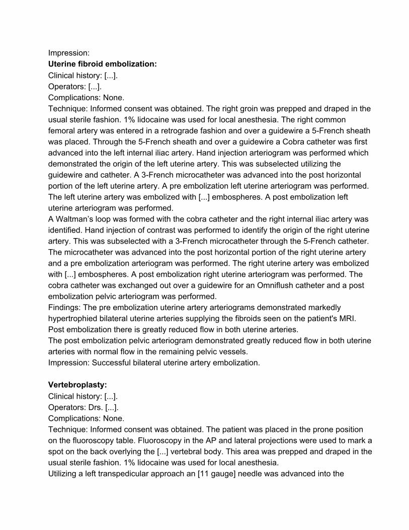
Impression:Uterine fibroid embolization:Clinical history: [...].Operators: [...].Complications: None.Technique: Informed consent was obtained. The right groin was prepped and draped in theusual sterile fashion. 1% lidocaine was used for local anesthesia. The right commonfemoral artery was entered in a retrograde fashion and over a guidewire a 5French sheathwas placed. Through the 5French sheath and over a guidewire a Cobra catheter was firstadvanced into the left internal iliac artery. Hand injection arteriogram was performed whichdemonstrated the origin of the left uterine artery. This was subselected utilizing theguidewire and catheter. A 3French microcatheter was advanced into the post horizontalportion of the left uterine artery. A pre embolization left uterine arteriogram was performed.The left uterine artery was embolized with [...] embospheres. A post embolization leftuterine arteriogram was performed.A Waltman’s loop was formed with the cobra catheter and the right internal iliac artery wasidentified. Hand injection of contrast was performed to identify the origin of the right uterineartery. This was subselected with a 3French microcatheter through the 5French catheter.The microcatheter was advanced into the post horizontal portion of the right uterine arteryand a pre embolization arteriogram was performed. The right uterine artery was embolizedwith [...] embospheres. A post embolization right uterine arteriogram was performed. Thecobra catheter was exchanged out over a guidewire for an Omniflush catheter and a postembolization pelvic arteriogram was performed.Findings: The pre embolization uterine artery arteriograms demonstrated markedlyhypertrophied bilateral uterine arteries supplying the fibroids seen on the patient's MRI.Post embolization there is greatly reduced flow in both uterine arteries.The post embolization pelvic arteriogram demonstrated greatly reduced flow in both uterinearteries with normal flow in the remaining pelvic vessels.Impression: Successful bilateral uterine artery embolization.
Vertebroplasty:Clinical history: [...].Operators: Drs. [...].Complications: None.Technique: Informed consent was obtained. The patient was placed in the prone positionon the fluoroscopy table. Fluoroscopy in the AP and lateral projections were used to mark aspot on the back overlying the [...] vertebral body. This area was prepped and draped in theusual sterile fashion. 1% lidocaine was used for local anesthesia.Utilizing a left transpedicular approach an [11 gauge] needle was advanced into the

posterior third of the [...] vertebral body under fluoroscopic guidance. The delivery cannulawas then advanced into the anterior third of the vertebral body under fluoroscopicguidance. Positioning was confirmed in the AP and lateral projections. Under directfluoroscopic guidance approximately [...] ml of methylmethacrylate impregnated withbarium was injected into the [...] vertebral body. There was no evidence for extravasation.On the AP projection there is evidence for cross filling to the right side.Impression: Successful percutaneous vertebroplasty of [...].
Women's Imaging:Many autotexts are available in Speech Q which are based on first describing thebreast tissue.Normal Mammogram Report:Begin by type of exam (screening vs. bilateral vs. unilateral), name, MR#Hx: routine, annual, screening, etc.Technique: Routine MLO and CC views of each breast (and any additional views) wereperformed and compared to previous mammograms from __ (or this is the patient’s baselineexam). Computer aided detection (CAD) was also utilized in this exam.Findings: Comparison is made with prior mammograms from ... (or this is the patient's baselineexam). Comment on the density of the breasts, and how this impacts on the sensitivity of themammography on this particular patient (E.g. The breasts are (quite) dense which (severely)limits the sensitivity of mammography to detect occult disease.)There is no evidence of skin thickening, suspicious clusters of microcalcification, nippleretraction or (new) dominant mass seen or any other secondary signs or malignancy. Mentionany significant normal findings such as fibroadenomas, skin calcifications, old surgery, orintramammary lymph nodes.Conclusion: There is no mammographic evidence of malignancy. Or Although mammographicinterpretation is quite limited in this case, there is no mammographic evidence of malignancy. OrPending comparison with the prior mammograms. there is no mammographic evidence ofmalignancy.ACR BIRADS Code 1, negative or 2, benign finding or 0, incomplete, additional imaging needed.The written results were given directly to the patient or the written results will be mailed to thepatient.Then in the bottom fields of the mammography report TYPE the Result Code which correspondswith the ACR Code you selected, e.g. 1 or 2 or 0 and then the Followup Code which determineshow the patient will be followed and whether the form/letter was given or mailed to the patient.
Needle Localization Report:Clinical Hx:Operators:Anesthesia: 13 cc of 1 % sodium bicarbonate buffered lidocaine.Technique: The risks, benefits and procedure were explained to the patient to her satisfaction.
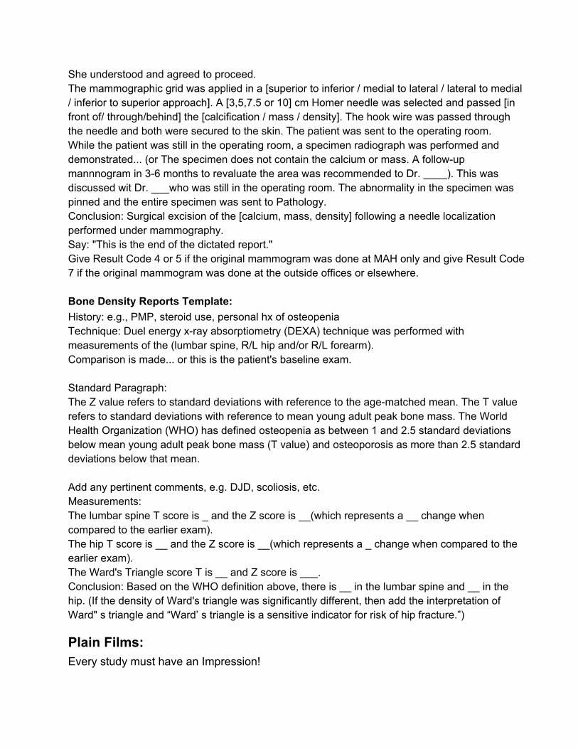
She understood and agreed to proceed.The mammographic grid was applied in a [superior to inferior / medial to lateral / lateral to medial/ inferior to superior approach]. A [3,5,7.5 or 10] cm Homer needle was selected and passed [infront of/ through/behind] the [calcification / mass / density]. The hook wire was passed throughthe needle and both were secured to the skin. The patient was sent to the operating room.While the patient was still in the operating room, a specimen radiograph was performed anddemonstrated... (or The specimen does not contain the calcium or mass. A followupmannnogram in 36 months to revaluate the area was recommended to Dr. ____). This wasdiscussed wit Dr. ___who was still in the operating room. The abnormality in the specimen waspinned and the entire specimen was sent to Pathology.Conclusion: Surgical excision of the [calcium, mass, density] following a needle localizationperformed under mammography.Say: "This is the end of the dictated report."Give Result Code 4 or 5 if the original mammogram was done at MAH only and give Result Code7 if the original mammogram was done at the outside offices or elsewhere.
Bone Density Reports Template:History: e.g., PMP, steroid use, personal hx of osteopeniaTechnique: Duel energy xray absorptiometry (DEXA) technique was performed withmeasurements of the (lumbar spine, R/L hip and/or R/L forearm).Comparison is made... or this is the patient's baseline exam.
Standard Paragraph:The Z value refers to standard deviations with reference to the agematched mean. The T valuerefers to standard deviations with reference to mean young adult peak bone mass. The WorldHealth Organization (WHO) has defined osteopenia as between 1 and 2.5 standard deviationsbelow mean young adult peak bone mass (T value) and osteoporosis as more than 2.5 standarddeviations below that mean.
Add any pertinent comments, e.g. DJD, scoliosis, etc.Measurements:The lumbar spine T score is _ and the Z score is __(which represents a __ change whencompared to the earlier exam).The hip T score is __ and the Z score is __(which represents a _ change when compared to theearlier exam).The Ward's Triangle score T is __ and Z score is ___.Conclusion: Based on the WHO definition above, there is __ in the lumbar spine and __ in thehip. (If the density of Ward's triangle was significantly different, then add the interpretation ofWard" s triangle and “Ward’ s triangle is a sensitive indicator for risk of hip fracture.”)
Plain Films:Every study must have an Impression!
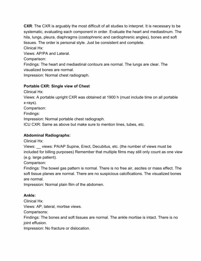
CXR: The CXR is arguably the most difficult of all studies to interpret. It is necessary to besystematic, evaluating each component in order. Evaluate the heart and mediastinum. Thehila, lungs, pleura, diaphragms (costophrenic and cardiophrenic angles), bones and softtissues. The order is personal style. Just be consistent and complete.Clinical Hx:Views: AP/PA and Lateral.Comparison:Findings: The heart and mediastinal contours are normal. The lungs are clear. Thevisualized bones are normal.Impression: Normal chest radiograph.
Portable CXR: Single view of ChestClinical Hx:Views: A portable upright CXR was obtained at 1900 h (must include time on all portablexrays).Comparison:Findings:Impression: Normal portable chest radiograph.ICU CXR: Same as above but make sure to mention lines, tubes, etc.
Abdominal Radiographs:Clinical Hx:Views: __ views: PA/AP Supine, Erect, Decubitus, etc. (the number of views must beincluded for billing purposes) Remember that multiple films may still only count as one view(e.g. large patient).Comparison:Findings: The bowel gas pattern is normal. There is no free air, ascites or mass effect. Thesoft tissue planes are normal. There are no suspicious calcifications. The visualized bonesare normal.Impression: Normal plain film of the abdomen.
Ankle:Clinical Hx:Views: AP, lateral, mortise views.Comparisons:Findings: The bones and soft tissues are normal. The ankle mortise is intact. There is nojoint effusion.Impression: No fracture or dislocation.
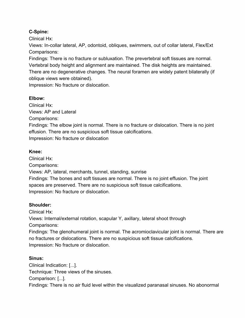
CSpine:Clinical Hx:Views: Incollar lateral, AP, odontoid, obliques, swimmers, out of collar lateral, Flex/ExtComparisons:Findings: There is no fracture or subluxation. The prevertebral soft tissues are normal.Vertebral body height and alignment are maintained. The disk heights are maintained.There are no degenerative changes. The neural foramen are widely patent bilaterally (ifoblique views were obtained).Impression: No fracture or dislocation.
Elbow:Clinical Hx:Views: AP and LateralComparisons:Findings: The elbow joint is normal. There is no fracture or dislocation. There is no jointeffusion. There are no suspicious soft tissue calcifications.Impression: No fracture or dislocation
Knee:Clinical Hx:Comparisons:Views: AP, lateral, merchants, tunnel, standing, sunriseFindings: The bones and soft tissues are normal. There is no joint effusion. The jointspaces are preserved. There are no suspicious soft tissue calcifications.Impression: No fracture or dislocation.
Shoulder:Clinical Hx:Views: Internal/external rotation, scapular Y, axillary, lateral shoot throughComparisons:Findings: The glenohumeral joint is normal. The acromioclavicular joint is normal. There areno fractures or dislocations. There are no suspicious soft tissue calcifications.Impression: No fracture or dislocation.
Sinus:Clinical Indication: [...].Technique: Three views of the sinuses.Comparison: [...].Findings: There is no air fluid level within the visualized paranasal sinuses. No abonormal
opacification is noted.Impression: No evidence of sinusitis.
GI/Fluoroscopy:Barium Swallow:Clinical Indication: [...].Technique: An air contrast esophagram was performed. The patient swallowed liquidbarium and EZ gas crystals as well as a 13 mm barium tablet without difficulty.Comparison: [...].Findings: There is normal esophageal mucosal detail, contour and motility. There is nogastroesophageal reflux either spontaneously or with provocative maneuvers. The fundusof the stomach is normal.Impression: Normal barium swallow.
UGI (Upper GI):Clinical Hx:Comparisons:Technique: An aircontrast esophagram (or aircontrast upper GI series for small bowelseries, add "with small bowel followthrough") was performed. The patient swallowed liquidbarium and EZ gas crystals as well as a l3mm barium tablet without difficulty.Findings: There is normal esophageal mucosal detail, contour and motility. There was nogastroesophageal reflux either spontaneously or with provocative maneuvers. Thestomach, duodenal bulb & small bowel are normal in position, motility, contour & mucosaldetail.Impression: Normal upper GI series.
Barium Enema:History: [...].Technique: An aircontrast barium enema was performed using the biphasic technique.Findings: There is good opacification of the colon to the cecum with filling of a normalappendix. There is no reflux into the ileum. There is no evidence of polyps greater than 1cm, polypoid masses or annular carcinomas.Impression: [...]
Modified Barium Swallow (fluoroscopic swallowing study):Clinical Hx: e.g. Aspiration pneumonia.Technique: The patient was given thin liquids, puree and soft solids and the oral andpharyngeal phases of swallowing were observed fluoroscopically.Findings: [Describe findings during the oral and pharyngeal phases for each consistency
tested, each as a separate paragraph. Also, describe the use of maneuvers such as chintuck.] There is normal propulsion of the oral bolus into the oral pharynx. There is noevidence of penetration or aspiration of barium into the vocal cords or trachea. The oralmucosa, pharyngeal mucosa, pyrifrom sinuses, vallecula and epiglottis are normal. Thereis no prominent cricopharyngeus. The patient did well with both solids and liquids ofdifferent viscosities. A chin tuck maneuver did not significantly improve swallowingperformance.Impression: Normal swallowing with no aspiration or penetration with any consistencystudied. Please see the report by speech therapy for further details and dietaryrecommendations.
ERCP:Clinical Hx: RUQ pain and elevated LFTsTechnique: The ampulla was cannulated by Dr. Fred Ruyman & contrast was injectedretrograde into the CBD.Findings: The CBD was well opacified and there are no filling defects, dilatation orstrictures. The pancreatic duct was also opacified and without filling defects or strictures.Impression:
Hysterosalpingogram:Clinical Hx: InfertilityTechnique: The cervix was cannulated by Dr. ____ and Omnipaque 300 was injectedretrograde into the uterus.Findings: There was good opacification of the uterus and bilateral fallopian tubes withquick spillage into the peritoneal cavity bilaterally.Impression: Normal hysterosalpingogram.
Shoulder Arthrogram (will get MRI):Clinical Hx:Technique: The standard timeout procedure for patient and body part identification wasperformed. The risks and benefits of the procedure was explained to the patient and writteninformed consent was obtained. The patient was prepped and draped in the usual sterilefashion. After the administration of local anesthesia, a 20 gauge needle was advancedunder fluoroscopic guidance into the glenohumeral joint space. Twelve cc of a 3:1 mixtureof 0.63 mL of Magnevist in 100 cc of Normal Saline and Omnipaque 300 was then infusedinto the joint space. The patient tolerated the arthrogram well with no immediatecomplications. The patient was then taken to MRI.Findings: There is no fluoroscopic evidence of a rotator cuff tear. (There must be somedescription of findings for billing purposes.)

Impression: Normal Fluroscopic arthorgram. Please refer to the MRI report to follow. (theMRI should be done within 1 hour of contrast administration).
Joint Steroid Injection:Clinical indication: [...].Technique: The timeout procedure was observed for proper identification of the patientand body part. The purpose, technique and risks of the procedure were explained to thepatient. [...] understood and signed a written consent form.The [...] hip was prepped in a sterile fashion. 7 ml of 1% lidocaine were injected into theoverlying soft tissues. A 20 gauge needle was introduced into the hip joint space underfluoroscopic guidance. Two ccs of Omnipaque300 were injected into the hip joint toestablish intraarticular position of the needle tip. 5 ml of Marcaine 0.75% and 80 mg ofDepoMedrol were injected into the hip joint.Findings: There is degenerative osteoarthritis of the [...] hip. The patient tolerated the [...]hip arthrocentesis without complication.Impression: Successful [...] hip arthrocentesis with injection of 5 ml of Marcaine 0.75% and80 mg of DepoMedrol.Intravenous Pyelogram (Urogram):Clinical Hx:Scout film findings: There are no calcific densities overlying the kidneys. The remainder ofthe abdominal radiograph is unremarkable.Comparison:Findings: The nephrograms and pyelograms are symmetric. The kidneys are normal insize, position and orientation (include length of both kidneys). The pelvocalyceal systemsand ureters are normal in appearance. The bladder is normal in contour. There is nopostvoid residual.Impression:
UltrasoundAbdominal Ultrasound:Clinical Hx:Technique: Transabdominal imaging was performed.Comparisons:Findings: There is normal liver echogenicity without intrahepatic ductal dilatation. Thegallbladder is normal with no gallstones, wall thickening, pericholecystic fluid, distention ora positive ultrasonagraphic Murphy’s sign. The CBD measures __ mm, which is normal.Limited evaluation of the pancreas is normal. The spleen measures __ cm and is normal inappearance. There is no evidence of an abdominal aortic aneurysm.
The kidneys measure _ and __ on the left and right, respectively and are normal inechogenicity. There is no hydronephrosis and no renal stones are identified.Impression: Normal abdominal ultrasound.
Renal Ultrasound:Clinical Hx:Comparisons:Findings: The kidneys are normal in size, shape and echogenicity measuring __ and __ onthe left and right, respectively. There is no hydronephrosis and no renal stones areidentified. There are no mass lesions or cysts. There is no evidence of an abdominal aorticaneurysm. The urinary bladder is normal and there is no postvoid residual.Impression: Normal renal ultrasound.
Obstetrical Ultrasound: (note there is a specific and detailed format for a fetalsurvey ask if you don't have it)Clinical Hx:Comparisons:Technique: Transabdominal (& transvaginal) imaging was performed of a gravid uterus.Findings: There is one fetus in variable lie. The heart rate in noted at __ bpm. Fetal surveyincluding visualization of the fourchambered view of the heart, stomach, kidneys, cordinsertion, bladder and spine are normal. (If not, mention here).Fetal measurements give a mean age of __, +/ __ weeks.If prior ultrasounds, then add the following: This correlates with the extrapolated age basedon the earlier ultrasound(s).If the fetus is in the 3rd TM, add: The estimated fetal weight is __ which is in the ___% forthe gestational age of ___ wks. If BPP performed give results here.The placenta is located anteriorly (fundally, posteriorly), above the lower uterine segment.The amniotic fluid volume is normal (or the AFI is __ cms).Impression: Single intrauterine pregnancy of __ weeks ___ days, +/ )___ days. Thiscorresponds/does not correspond to the dating by LMP with an EDD of ___. (If it doescorrespond, do not "correct" the due date as this drives patients and obstetricians crazy.)
Pelvic Ultrasound:Clinical Hx:Technique: Transabdominal & transvaginal imaging were performed.(if done Dopplerevaluation of ovarian blood flow was included.)Comparisons:Findings: The uterus is homogenous in echogenicity measuring __ x __ x __ cm and isanteverted/retroverted. The endometrial stripe is uniform and measures __mm, which is
within normal limits. There are no fibroids. The ovaries are identified by their follicles andmeasure __ x __ x __ cm trasvaginally on the right and __ x __ x __ cm transvaginally onthe left. There are no adnexal masses or cysts.The kidneys are normal in size, shape and echogenicity. There is no hydronephrosis andno renal stones are identified. No free fluid is seen. (If Doppler is used, e.g. to confirm flowto both ovaries, dicatate this as its own section.)Impression: Normal pelvic ultrasound.
Thyroid Ultrasound:Clinical Hx:Comparisons:Findings: Ultrasound evaluation of the thyroid gland demonstrates homogeneous glandulartissue. The right lobe measures __ x __ x __ cm and the left lobe measures __ x __ x __cm. The isthmus measures __. No nodules are seen.Impression: Normal thyroid ultrasound.
Testicular Ultrasound:Clinical Hx:Technique: Transscrotal ultrasound evaluation was performed.Comparisons:Findings: There is normal and symmetric testicular echogenicity with the right testismeasuring __ x __ x __ cm and the left testis measuring __ x __ x __ cm __. The head ofthe right epididymis measures <10mm: the head of the left epididymis measures <10mm.Testicular and epididymal perfusion is symmetric bilaterally. There are no varicoceles orhydroceles. The scrotal skin is not thickened. (If varicoceles are seen, both kidneys mustbe checked for hydronephrosis and this should be included in the report.)Impression: Normal testicular ultrasound.
Vascular Ultrasound, Lower Extremity Venous:Clinical Hx:Technique: Venous duplex imaging was performed of the __ lower extremity.Findings: The right common femoral, superficial femoral, popliteal, tibial and peronealveins demonstrate normal compressibility and color flow throughout. Maneuvers includingValsalva, augmentation and respiration demonstrate normal variation in flow in the vesselsstudied. (Note to Residents: When dictating this study, it has been suggested to omit theword superficial from superficial femoral vein, because ordering doctors have been knownto not treat superficial femoral vein DVTs, confusing a true DVT with superficialthrombophlebitis)Impression: There is no evidence of a deep venous thrombosis.

First trimester ultrasound: use the system autotext.Fetal survey: use the system autotext.
Obstetrical Ultrasound: Biophysical ProfileCriteria for Coding Fetal Biophysical Variables as Normal or Abnormal
Biophysical
Variable
Normal(Score = 2)
Abnormal(Score = 0)
Fetalbreathingmovements
1 or more episodes of >20 s within30 min
Absent or no episode of >20 s within30 min
Grossbodymovements
2 or more discrete body/ limbmovements within 30 min (episodesof active continuous movementconsidered as a single movement)
<2 episodes of body/limbmovements within 30 min
Fetal tone 1 or more episodes of activeextension with return to flexion offetal limb(s) or trunk (opening andclosing of hand considered normaltone)
Slow extension with return to partialflexion, movement of limb in fullextension, absent fetal movement, orpartially open fetal hand
QualitativeAFV
1 or more pockets of fluidmeasuring >2 cm in vertical axis
Either no pockets or largest pocket<2 cm in vertical axis
Clinical Indication: Pregnancy of [...] weeks [...] days, [...], check biophysical profile andamniotic fluid index.Technique: Transabdominal imaging of the gravid uterus was performed, including Dopplerevaluation of the fetal heart rate.Findings: The fetus is in ____ presentation. Fetal breathing, movement and tone and theamniotic fluid index were assessed with a total biophysical profile score of 8/8. Theamniotic fluid index was measured to be [...] cm, which is within normal limits.The fetal heart rate was measured by Doppler to be [...] beats per minute.Impression: BPP of 8/8 with an AFI of [...]. This result was called to [...].
CT Scan:Head CT:Clinical Hx: Trauma, Headaches, Mental status changes, etc..Technique: Serial axial images were obtained from the foramen magnum to the vertex withoutthe use of intravenous contrast.Comparison:Findings: The ventricles, sulci and cisterns are normal for the patient's age. There is no masseffect, midline shift, hemorrhage, acute territorial infarct or extraaxial fluid collection. (You mayneed to mention mm of midline shift or type of hemiation.) The visualized paranasal sinuses,mastoid air cells, and bones are normal. The soft tissues are unremarkable.Impression:
Sinus CT:Clinical Hx:Technique: Helical 1.25 mm axial images were obtained through the paranasal sinuses andreconstructions were performed in the coronal plane.Comparison:Findings: There is no mucosal thickening in the frontal sinuses and the frontoethmoid recessesare clear. The ethmoid air cells, sphenoid and maxillary sinuses are normal. The visualizedportions of the mastoid air cells are well aerated. The osteomeatal complexes are widely patent,and the infundibulae are bordered laterally by the orbit (or by the Haller air cells). The nasalseptum is approximately midline in position without a nasal septal spur. The cribiform plate issymmetric and the lamina papryceae are intact. There is no evidence of dental disease. Thecarotid arteries are covered by bone as they pass superior to the sphenoid sinuses.Impression:
Temporal Bone CT:Clinical Hx: Hearing loss, pulsitile tinnitus, pain, etc.Technique: Serial axial images were obtained through the inner ear without the use ofintravenous contrast. Coronal and sagittal 2D reformatted images were also obtained.Comparisons: NoneFindings: The external auditory canals are normal bilaterally. No abnormalities are seen in eithermiddle ear space. Specifically, the ossicular chains are intact and there is no abnormal softtissue or bony erosion. The tympanic membranes are intact. The inner ear structures arenormal. The internal auditory canals are unremarkable. The tegmen is intact bilaterally, withnormal bony coverings of the lateral semicircular canal and facial nerve. The jugular veins andcarotid arteries follow their normal course.Impression:
Facial bone CT:Clinical Hx: Trauma, etc..
Technique: Serial axial images were obtained from the mandible through the frontal sinuseswithout the use of intravenous contrast. Coronal and sagittal 2D reformatted images were alsoobtained.Comparison:Findings: The visualized paranasal sinuses are normal. The orbits and surrounding soft tissuesare unremarkable. The medial wall and orbital floors are intact. There are no fractures in thenasal bones, maxilla, zygomatic arches or frontal bones. The nasal septum is midline. There isno evidence of dental disease.Impression:
Chest CT:Clinical Hx:Technique: Serial axial images were obtained from the apices of the lungs through the kidneysafter the administration of intravenous contrast.Orders (if applicable): The patient received 100 cc of Omnipaque 350 as ordered by...Comparisons:Findings: Include discussion of the following regions—neck, thyroid, axillae, lymph nodes:mediastinum including great vessels (always look for PE), mediastinal and hilar lymph nodes,airway, heart (mention coronary artery calcifications), pericardium, lungs, pleura, chest wall, softtissues/breasts, visualized abdomen.Impression: Prioritize findings, most important to least important.
CT Pulmonary Angiogram:Clinical Hx: Shortness of breath with elevated Ddimer.Technique: Serial axial images were obtained from the upper abdomen through the apices of thelungs during the administration of IV contrast. 3D and multiplanar CT reformations were thenperformed.Orders: The patient received 100 cc of Omnipaque 350 as ordered by...Comparisons:Findings: There is no filling defect within the main pulmonary arteries or their branches. Includediscussion of the following regionsneck, thyroid, axillae, lymph nodes: mediastinum includinggreat vessels, mediastinal and hilar lymph nodes, airway, heart, pericardium, lungs, pleura,chest wall, soft tissues/breasts, visualized abdomen.Impression:1. No pulmonary embolus (or PE seen…)2. Include any other findings.
Abdominal/Pelvis CT:Clinical Hx:Technique: Serial axial images were obtained from the domes of the diaphragm through thepubic symphysis.Orders: The patient received 125 cc of Omnipaque 350 as ordered by ...
Comparisons: There are no previous abdominal/pelvis CTs for comparison.Findings: The visualized lungs and mediastinal structures are normal.Abdomen: Describe the following: Liver, ascites, gallbladder, biliary tree, spleen, pancreas,kidneys & adrenals. Look at vascular structures (including aorta & its branches, IVC, portal vein,etc) & lymph nodes. Presence or absence of free air.The small and large bowel, always look for/at and commend on the appendix.Pelvis: Describe the following: The uterus and adnexae, prostate, seminal vesicles, bladder, iliacveins for DVT. Check for free fluid in pelvis.The visualized bones and soft tissues are normal.Impression:
Pancreatic Mass: use the system autotext.
Magnetic Resonance ImagingBrain MRI (without or with and without Gadolinium):Clinical Hx:Technique: Brain MR was performed without (or without and with) Gadolinium including thefollowing sequences: SagTl, Ax FLAIR, FSE Ax T2, Ax T1, Ax DWI, Ax GRE (If Post Gado: AxTl,CorTI, SagTl)Comparisons:Findings: The ventricles, sulci and cisterns are normal. There is no mass, mass effect,hemorrhage or abnormal extraaxial collection. Then answer the question i.e. There is noevidence of acute ischemic injury or traumatic intracranial injury or demyelinating disease ormetastatic disease or intracranial infection, etc. The flow voids of the major vessels at the baseof the brain are normal.The globes, orbits and paranasal sinuses are unremarkable.Impression:
Brain MRA and Neck MRA:Same as above except add the following to the Technique and BodyTechnique: AddBrain MRA: 3D time of flight imaging was performed of the Circle of Willis, with complexreformations.Neck MRA: 3D time of flight imaging post gadolinium was performed., with complexreformations.Body: AddBrain MRA: There is normal flow related enhancement in the visualized portion of the internalcarotid, middle cerebral and anterior cerebral arteries. The basilar artery and its branches areunremarkable.Neck MRA: The common, internal and external carotid arteries appear normal. The codominantvertebral arteries are unremarkable.
Internal Auditory Canal MRI (or Pituitary MRI):Clinical Hx:Technique:Findings:The ventricles, sulci and cisterns are normal. There is no mass, mass effect, hemorrhage orabnormal extraaxial collection. Thin section imaging through the internal auditory canals withand without contrast is normal. Specifically, there is no evidence of an acoustic schwannoma.(Or for pituitary MRIs: There is no sellar or suprasellar mass lesion. The infundibulum is midline.The cavernous sinus is unremarkable.) The globes, orbits and paranasal sinuses areunremarkable. The flow voids of the major vessels at the base of the brain are normal.Impression:
Cervical Spine MRI:Clinical Hx:Technique: MR Imaging of the cervical spine was performed without intravenous contrastincluding the following sequences: Sag FSE T2, Sag Tl, Ax FSE T2 and Ax Gradient Echo.Comparisons:Findings: The cervical vertebral bodies are normal in height, alignment, and signalcharacteristics. The visualized portion of the spinal cord and contents of the posterior fossaappear normal. The intervertebral disc heights are preserved. There is no significant central orforaminal narrowing at any of the imaged levels. (Be sure to look at the visualized brain)Impression:
Lumbar Spine MRI:Clinical Hx:Technique: MR imaging was performed of the lumbar spine without intravenous contrastincluding the following sequences: Sag T1 and FSE T2, Ax T1 and FSE T2 .Comparisons:Findings: The lumbar vertebral bodies are normal in height, alignment, and signalcharacteristics. The conus medullaris terminates normally at the T12/L1 level. The intervertebraldisc heights are preserved. There is no facet joint hypertrophy or ligamentum flavum infolding.There is no significant central or foraminal narrowing at any of the imaged levels.Impression:
Shoulder MRI:Clinical Hx:Technique: MR imaging was performed of the (right or left) shoulder without the use ofintravenous contrast including the following sequences: Triplanar proton density with fatsuppression, Axial Gradient Echo, Coronal Oblqieu T2, and Coronal Oblique STIR images.Comparisons:Findings: The bone marrow signal is normal. The supraspinatus tendon is normal in

configuration and signal characteristics. The infraspinatus, teres minor and subscapularistendons are intact. The long head of the biceps tendon is normally located and attaches normallyon the superior labrum. The articular surface of the humeral head is intact. No displaced labraltear is seen. There is no significant acromioclavicular joint hypertrophy. The coracohumeral,coracoclavicular and coracoacromial ligaments are normal. There is no fluid in the subacromialbursa or subdeltoid bursa.
Knee MRI:Clinical Hx:Technique: MR Imaging of the knee was performed without intravenous contrast including thefollowing sequences: Sag PD, Sag PD with Fat Sat. Cor PD with fat saturation, Cor Tl, Ax PD,and Ax FSE T2 Fat Sat.Comparisons:Findings: The bone marrow signal is normal. The ACL, PCL, medial collateral ligament andlateral collateral ligamentous complex are intact. The quadriceps tendon and infrapatellar tendonare normal. There is no lateral or medial meniscal tear. The patellofemoral joint is normal with nochondromalacia patella. There is no joint effusion. No synovial cyst is seen.Impression:
Ankle MRI:Clinical Hx:Technique:Comparisons:Findings: The bone marrow signal is normal. The subtalar joints appear intact. No abnormalsignal is seen in the sinus tarsi. The posterior tibialis, flexor digitorum. flexor hallucis, peroneal.extensor and Achilles tendons all have homogeneous dark signal. There are no joint effusions.The anterior and posterior tibiofibular and talofibular ligaments are intact The deltoid ligamenthas normal signal characteristics.Impression:
Nuclear Medicine: (many have autotexts available in SpeechQ)Note: use the autotexts for nuclear medicine, or get them from a fellow resident.Pulmonary V/Q Scan:Clinical Hx: Shortness of breathRadiopharmaceutical: Xe 133 and TcMAADose: __ mCi and __ mCiTechnique: Utilizing radioactive Xe, single breath, equilibrium and washout images of thelungs were obtained. Then after the intravenous administration of the perfusion agent, [sixor eight] images of the lungs were obtained.Findings: There was heterogenous ventilation of the lungs with trapping at the bases. There

is homogeneous perfusion of the lungs without evidence of a pleural based, wedge shapeddefect.Impression: Normal V/Q scan [or very low prob, low prob, intermediate or high prob forPE].
Myocardial Perfusion study with LexiscanRadiopharmaceutical: Technetium 99m MyoviewDose: Rest dose [...] mCiStress dose [...] mCiTotal dose [...] mCiClinical indication: [...]Technique: A [...]day study was performed. The patient was injected with 0.4 mg IV ofregadenoscan and then with the radiopharmaceutical. Patient performed [...] min. of lowlevel exercise. Multiple tomographic views of the heart were then obtained and comparedwith similar images obtained at rest. The Lexiscan and rest examinations were gated. Theleft ventricular ejection fraction and wall motion were assessed.Comparison: [...]Findings: No significant focal reversible perfusion defect is seen. At pharmacologic stress,the left ventricular ejection fraction is [...] %, which is within normal limits. No significantregional wall motion abnormality is seen.Impression:1. No evidence of focal jeopardized myocardium.2. Normal left ventricular ejection fraction without significant regional wall motionabnormality at stress.
Gastric emptying study:Radiopharmaceutical: Technetium 99m sulfur colloid eggsDose: [...] mCiClinical indication: [...]Technique: After the administration of the radioactive meal, sequential images of theabdomen were obtained every 10 minutes for 60 minutes. At 2, 3, and 4 hours, staticimages of the abdomen were obtained. Computer analysis was performed to determinethe amount of gastric retention at each hour.Findings: At 1, 2, 3, and 4 hours, the gastric contents remaining were [...]%, [...]%, [...]% and[...]% respectively. The upper limits of normal for gastric retention at 4 hours is 10%.Impression: [Normal] gastric emptying.
Triple Phase Bone Scan:Clinical Hx: History of cancer, bacteremia, etc.

Radiopharmaceutical: Technecium99m MDPDose: __ mCiTechnique: Immediately after the intravenous administered of the radiopharmaceutical,blood flow and blood pool images were obtained. After a 3 hour delay, whole body andhigh resolution images of the area of interest were obtained.Findings: The blood flow and Blood pool images were symmetric and normal. On delayedimages, there was no abnormal uptake (at the area of interest).Impression: No evidence of metastatic disease, osteomyelitis, etc
Urea Breath Test:*Note regarding an indeterminate urea breath test:Findings: The resultof this study was indeterminate. Approximately 5% of all tests giveindeterminate results. Although there are many theories regarding the cause of this, theexact cause of an indeterminate result is not known.The indeterminate result should be evaluated by repeating the PY test or using analternative diagnostic method. If repeat breath testing is undertaken, careful history toexclude confounding factors should be obtained. If confounding factors are present, waitappropropriate time depending on the confounding factor before repeating the PY test. Inall cases, wait for at least six hours before repeating the PY test so that any 14CO2 in thebreath will be eliminated and not interfere with the results of a second test.The recommended time between confounding factor and retesting are as follows:Antibiotics: 1 monthBismuth: 1 monthProton Pump Inhibitors: 2 weeksSucralfate: 2 weeksNonfasting: 6 hours
Specials (IR): (some have autotexts available in SpeechQ)Abscess Drainage, Seldinger TechniqueClinical history: [...]Operators: Drs. [...]Technique: Informed consent was obtained. CT confirm the presence of a fluid collectionwithin the [...]. After administration of 1% lidocaine into the superficial and deep tissues, aone step catheter was advanced into the collection with immediate aspiration of [...]. Awire was passed through the catheter, serial dilations were performed over the wire and a10.2 French drainage catheter was advanced into the collection. Then [...] mL of [...] fluidwas immediately aspirated. The drainage catheter was hooked up to a drainage bag.Followup CT imaging at the completion of the procedure revealed nearcompleteresolution of the fluid collection.

The patient tolerated the procedure well with no immediate complications.Impression: Successful CTguided drainage of fluid collection with drainage of [...] mL offluid.
Abscess Drainage, Tandem Trocar Technique (JS often uses this technique)Clinical history: [...]Operators: Drs. [...]Technique: Informed consent was obtained. CT confirm the presence of a fluid collectionwithin the [...]. After administration of 1% lidocaine into the superficial and deep tissues, a20 gauge x 15 cm Temno Cheeba needle was advanced under CT guidance into the fluidcollection. Then, a 10.2 French drainage catheter was advanced into the collection usingtandem trocar technique. [...] mL of [...] fluid was immediately aspirated. The drainagecatheter was hooked up to a drainage bag. Followup CT imaging at the completion of theprocedure revealed nearcomplete resolution of the fluid collection.The patient tolerated the procedure well with no immediate complications.Impression: Successful CTguided drainage of fluid collection with near completeresolution of the fluid collection at the completion of the procedure.
Abscess Tube CheckClinical history: [...]Operators: Drs. [...]Technique: 23 ml of Omnipaque 300 was injected into the patient's existing 10.2 Frenchallpurpose drainage catheter. The contrast was then aspirated through the drainagecatheter.Findings: The abscess drainage tube is in good position. There is a small residualabscess cavity. There is no fistula. The abscess drainage tube was reconnected to thecollection bag. The patient was instructed to call immediately if experiencing fevers, chills,nausea, vomiting or flank pain. Impression: Abscess drainage catheter in satisfactory position.
Angiogram, Lower ExtremitiesClinical history: [...]Operators: Drs. [...]Technique: Informed consent was obtained. The patient was placed in the supineposition, and prepped and draped in the usual sterile manner. 1% lidocaine was used foranesthesia at the groin. Using a Seldinger technique, the right groin was accessed in aretrograde manner. With the aid of a glide wire, a 4French Omniflush catheter wasadvanced into the aorta at the level of L1. An angiogram was then performed. The pigtailcatheter was then withdrawn to the level of the bifurcation. Subsequent angiograms were
then performed of the lower extremity runoff including a lateral view of the feet.Findings: There are single patent renal arteries bilaterally. The superficial femoral artery ispatent. The profunda femoris is patent. The popliteal artery is patent. There is a [...] vesselrunoff via [...] arteries.Angiogram of the foot demonstrates [...].Total contrast: [...] ml Omnipaque 350.Impression: Status post angiogram of the [...] lower extremity with [...] vessel runoff to thelower extremity.
Angiogram, GI BleedingClinical history: [...] year old with gastrointestinal bleeding.Operators: Drs. [...]Technique: Informed consent was obtained. The patient was placed in the supineposition, and prepped and draped in the usual sterile manner. 1% lidocaine was used foranesthesia at the groin. Using a Seldinger technique, the right groin was accessed in aretrograde manner. With the aid of a glide wire, a glide catheter was advanced into theaorta at the level of L1. The SMA was selected and an angiogram was then performed.The catheter was then exchanged for a Simmons catheter. A subsequent angiogram wasthen performed of the inferior mesenteric artery.Findings: There is no evidence of gastrointestinal bleeding from the superior or inferiormesenteric arteries.Total contrast: [...] ml Omnipaque 350.Impression: Successful angiogram of the SMA and IMA without evidence ofgastrointestinal bleeding.
AVF Fistulogram/AngioplastyClinical history: [...]Operators: Drs. [...]Technique: Informed consent was obtained. The patient was placed supine upon theangiographic table. Sterile preparation and drape of the [...] arm was performed. 1%lidocaine was utilized within the superficial tissues overlying the [...] arterial venous graft.The arterial venous graft was accessed with a micropuncture access needle orientedtoward the venous anastomosis. A wire was placed and a micropuncture sheath wasplaced over the wire pointing toward the venous anastomosis. Venogram of the graft andcentral veins was then performed. The glide wire was then placed centrally and themicropuncture sheath was removed. Over the glide wire, a 7French working sheath wasplaced. Under fluoroscopic guidance, a [...] mm x [...] cm angioplasty balloon wasadvanced across the [...] stenosis. Angioplasty was then performed. The sheath wasremoved and hemostasis was achieved.

Total contrast: [...] mL of Visipaque 320Findings: [...]Impression: Successful AV fistulogram and balloon angioplasty of stenosis of the [...] vein.
Axillary Lymph Node BiopsyClinical history: [...]Procedure: Axillary lymph node biopsyOperators: Drs. [...]Technique: Informed consent was obtained. An ultrasound was performed within the [...]axilla which revealed [...]. The patient was prepped and draped in the usual sterile fashion.Ultrasound guided needle biopsy using a Temno 17 gauge introducer needle and 18gauge biopsy needle system. Multiple specimens were obtained, which were placed infixative and sent to pathology. Pathology was in attendance for the procedure. The patienttolerated the procedure well and there were no immediate complications.Impression: Successful axillary lymph node biopsy.
Biliary Tube DrainageClinical indication: Choledocholithiasis.Operators: [...].Technique: The patient was explained the risks and benefits of the procedure and informedconsent was obtained. Conscious sedation was used for this procedure. The upperabdomen was prepped and draped in the usual sterile fashion. Under ultrasound guidanceand after the administration of local anesthesia using 1% lidocaine, the [...] hepatic lobewas entered from an [anterior] approach with a Jeffrey’s needle set. Contrast was injectedthrough the needle under fluoroscopic observation to confirm that the tip was located in abile duct. A wire was advanced through the needle and was exchanged for a sheath.Approximately [...] ml of [...] were aspirated and sent for culture. A 10.2 French biliarydrainage tube was then placed and the drainage catheter was connected to a collectionbag. The patient tolerated the procedure well with no immediate complications.Impression: Successful placement of a percutaneous biliary drainage tube.
Biliary Tube Drainage/SphinceterotomyClinical indication: Choledocholithiasis.Operators: Drs. [...]Technique: The risks and benefits of the procedure were discussed with the patient andinformed consent was obtained. Conscious sedation was used for this procedure. Theupper abdomen was prepped and draped in the usual sterile fashion.Under ultrasound guidance and after the administration of local anesthesia using 1%lidocaine, the [...] hepatic lobe was entered from an [anterior] approach with a Jeffrey’s

needle set. Contrast was injected through the needle under fluoroscopic observation toconfirm that the tip was located in a bile duct. A wire was advanced through the needleand was exchanged for a sheath. Approximately [...] ml of [...] were aspirated and sent forculture.A cholangiogram was performed which revealed [...]. The ampulla was dilated with an 8 x4 mm balloon. A postdilataion cholangiogram revealed contrast passing into theduodenum. A 10.2 French biliary drainage tube was then placed and secured to the skinwith sutures. The drainage catheter was connected to a collection bag.The patient tolerated the procedure well with no immediate complications.Impression: Successful placement of a percutaneous biliary drainage tube. Successfulballoon sphincterotomy of stenosis at the distal common bile duct/ampulla.
Chest tube placement, Denver CathetherClinical history: Recurrent [...] pleural effusion.Operators: Drs. [...]Technique: Informed consent was obtained.Ultrasound examination demonstrated a [...]sided pleural fluid. Under sterile conditionsand local anesthesia, the [...] lateral chest wall and pleural space were entered from aposterolateral approach with a onestep catheter. The wire was passed through thecatheter, serial dilations were performed over the wire and a 16 French peelaway sheathwas placed. After administration of local anesthesia, a second incision was made ofseveral centimeters inferior to the first, and the Pleurx catheter was tunneled from theincision to the first. The catheter was then advanced through the 16French peelawaysheath.Approximately [...] ml of [...]colored, serous fluid was aspirated. The patient toleratedprocedure well with no immediate complications.The patient was sent for followup chest xray.Impression: Successful [...] Pleurx catheter placement.
Chest tube placement, Denver CathetherClinical history: Recurrent [...] pleural effusion.Operators: Drs. [...]Technique: Informed consent was obtained.Ultrasound examination demonstrated a [...]sided pleural fluid. Under sterile conditionsand local anesthesia, the [...] lateral chest wall and pleural space were entered from aposterolateral approach with a onestep catheter. The wire was passed through thecatheter, serial dilations were performed over the wire and a 16 French peelaway sheathwas placed. After administration of local anesthesia, a second incision was made of

several centimeters inferior to the first, and the Pleurx catheter was tunneled from theincision to the first. The catheter was then advanced through the 16French peelawaysheath.Approximately [...] ml of [...]colored, serous fluid was aspirated. The patient toleratedprocedure well with no immediate complications.The patient was sent for followup chest xray.Impression: Successful [...] Pleurx catheter placement.
CT guided Bone BiopsyHistory: [...]Operators: Drs. [...]Procedure: Percutaneous CTguided biopsy of bone lesion.Technique: Informed consent was obtained from the patient. The patient was supine andscout CT images demonstrated a [...] lesion. A BB was placed on the skin marking a safetrajectory to the lesion. A sterile preparation and drape were applied to the patient. 1%lidocaine was administered subcutaneously and along the deeper tissues.Under CT guidance, a 10 gauge introducer needle was placed into the bone lesion.Multiple core biopsies were then obtained with a Murphy set bone biopsy needle.Pathology was in attendance for this procedure which confirmed the adequacy of thesamples. The needle was then removed. The tract was embolized with gelfoam.This procedure was uncomplicated.Impression: Successful uncomplicated percutaneous CTguided biopsy of bone lesion.
CT guided liver biopsyHistory: [...]Operators: Drs. [...]Technique: Informed consent was obtained from the patient. The patient was supine andscout CT images demonstrated a [...] mass. A BB was placed on the skin marking a safetrajectory to the lesion. A sterile preparation and drape were applied to the patient. 1%lidocaine was administered subcutaneously and along the deeper tissues.Under CT guidance, a 19 gauge coaxial introducer needle was placed into the liver lesion.Multiple core biopsies were then obtained with a 20 gauge biopsy device. Pathology wasin attendance for this procedure which confirmed the adequacy of the samples. The needlewas then removed. The tract was embolized with gelfoam.This procedure was uncomplicated.Impression: Successful uncomplicated percutaneous CTguided biopsy of liver mass.
CT guided lung biopsyHistory: [...]
Operators: [...]Procedure: Percutaneous CTguided biopsy of [...] lobe consolidation or mass.Technique: Informed consent was obtained from the patient.The patient was supine and scout CT images demonstrated [consolidation/mass] withinthe [...] lobe. A BB was placed on the skin marking a safe trajectory to the lung lesion. Asterile preparation and drape were applied to the patient. 1% lidocaine was administeredsubcutaneously and along the deeper tissues. Under CT guidance, a 19 gauge coaxialintroducer needle was placed into the lung lesion. Multiple 20 gauge core biopsies werethen obtained. Pathology was in attendance for this procedure which confirmed theadequacy of the samples. The needle was then removed.This procedure was uncomplicated.Impression: Successful uncomplicated percutaneous CTguided biopsy of [...] lobeconsolidation or mass.
CT guided percutaneous needle biopsy:Clinical History: Lung mass, tissue needed for diagnosis.Operators: Resident and attending names.Technique: The patient was explained the risks and benefits of the procedure and informedconsent was obtained. The patient was placed supine/prone on the CT gantry. The rightside of the back was then prepped and draped in the usual sterile fashion. 1 % lidocainewas used to anesthetize the skin and subcutaneous tissues. Under CT guidance, a19gauge needle was placed adjacent to the lung mass and 6 biopsies were obtained witha 20guage biopsy gun without difficulty. The samples were sent to pathology for analysis.The patient tolerated the procedure well. There were no immediate complications.Followup chest xray did not demonstrate evidence of a pneumothorax.Impression: Successful biopsy of a right upper lobe lung mass.
CT guided renal biopsyHistory: [...]Operators: [...]Procedure: Percutaneous CTguided biopsy of [...] a mass within the [...] kidney.Technique: Informed consent was obtained from the patient. The patient was prone andscout CT images demonstrated a [...] renal mass. A BB was placed on the skin marking asafe trajectory to the renal lesion. A sterile preparation and drape were applied to thepatient. 1% lidocaine was administered subcutaneously and along the deeper tissues.Under CT guidance, a 17 gauge coaxial introducer needle was placed into the renal lesion.Multiple 18 gauge core biopsies were then obtained. Pathology was in attendance for thisprocedure which confirmed the adequacy of the samples. The needle was then removed.This procedure was uncomplicated.

Impression: Successful uncomplicated percutaneous CTguided biopsy of [...] renal mass.
Femoral Arteriogram w/ or w/out balloon angioplasty:Clinical Indication: Rest pain/nonhealing ulcer, etc.Operators: [...]Technique: Possible complications of the procedure were discussed with the patient andinformed consent was obtained. Using aseptic technique and local anesthesia, the rightfemoral artery was cannulated with an arterial needle in an antegrade approach followed bya 5french sheath, glidewire, and straight measuring catheter. Views were obtained of theright lower extremity. After discussion with Dr. _________, angioplasty of the above theknee right popliteal artery occlusion was performed using a __ mm x __ cm balloon.Postprocedure, the catheter was removed, and hemostasis was achieved withoutimmediate complication.Findings: The right common femoral, profunda femoris and superficial femoral arteries areof good caliber. There is a discrete occlusion of the above the knee popliteal artery whichwas successfully angioplastied. There is three vessel runoff through the foot, etc.Impression:
Gastrostomy tube placement:Clinical Indication: [...].Operators: Drs. [...]Technique: The patient was placed supine and the abdomen was prepped and draped inthe usual sterile fashion. The stomach was then insufflated with air, and utilizingfluoroscopy, the relationships of the stomach and anterior abdominal wall were localized.l% lidocaine was used to anesthetize the skin and subcutaneous tissues.Under direct fluoroscopic guidance, 2 T bars were used to tack the anterior wall of thestomach to the anterior abdominal wall. Between the 2 T bars, a trocar needle wasadvanced into the stomach and a guide wire was placed. The tract was then seriallydilated, and a [16French gastrostomy tube/14french Cope loop catheter] was placed intothe stomach without difficulty. A pigtail was formed on the distal end and the catheter wassecured into place. The patient tolerated the procedure well. There were no immediatecomplications.Impression: Successful percutaneous gastrostomy tube placement.
Gastrostomy (Transgastric Jejunostomy) tube placementClinical history: [...]Operators: Drs. [...].Technique: Informed consent was obtained. The stomach was insufflated with air. Usingfluoroscopy in the AP and lateral projections a spot was marked on the anterior abdominal
wall overlying the stomach. This area was prepped and draped in usual sterile fashion. 1%lidocaine was used for local anesthesia.Under fluoroscopic guidance two T bars were used to tack the anterior wall the stomach tothe anterior abdominal wall. Between the two T bars a trocar needle was advanced into thestomach and a guidewire was placed. Over the guidewire a 7French seeking catheterwas advanced and access into the proximal jejunum was obtained. The tract was seriallydilated and a 16French peelaway sheath was placed. Through the peelaway sheath andover the guidewire a CareyAlzateCoons transgastric jejunostomy tube was placed so thatits tip lay within the proximal jejunum.Impression: Successful percutaneous transgastric jejunostomy tube placement.IVC Filter placement and venogram, basilic:Clinical history: [...].Operators: Drs. [...].Complications: None.Technique: Informed consent was obtained. The right arm was studied with ultrasoundwhich revealed the brachial and the basilic veins to be patent. The right arm was preppedand draped in usual sterile fashion. 1% lidocaine was used for local anesthesia. Underultrasound guidance the right basilic vein was entered with a micropuncture needle and amicropuncture sheath was placed. This was exchanged out over a guidewire for the7French sheath and diagnostic catheter of the Trapease IVC filter. Over a guidewire thiswas advanced into the distal right common iliac vein. An inferior vena cavagram wasperformed. The renal veins were identified and the Trapease IVC filter was placed in theinfrarenal IVC without difficulty.Findings: There is standard caval anatomy with no evidence of thrombus seen. Reflux wasnoted down the left common iliac vein.The post placement images show good positioning of the IVC filter.Impression: Successful IVC filter placement.
IVC Filter placement and Venogram, groin:Clinical Indication: Pulmonary embolism, cannot be anticoagulatedOperators: Drs. [...].Technique: Possible complications of the procedure were discussed with the patient andinformed consent was obtained. The right groin was then prepped and draped in the usualsterile fashion. l% lidocaine was used to anesthetize the skin and subcutaneous tissues. Avenous needle was used to canulate the right femoral vein in an antegrade fashion. A guidewire was placed through the needle and a 5french measuring pigtail catheter was placedinto the IVC without difficulty. A cavogram was performed which demonstrated conventionalvenous anatomy. A Trapese IVC filter was then placed just below the renal veins withoutdifficulty. Hemostasis was then achieved using direct pressure. The patient tolerated the
procedure well. There were no immediate complications.Findings: There is standard caval anatomy with no evidence of thrombus seen. The postplacement images show good positioning of the IVC filter.Impression: Successful placement of an IVC filter.Nephrostomy Tube CheckClinical history: Nephrostomy tube(s), originally placed [...], nephrostomy tube check.Technique: 25 ml of Omnipaque 300 were injected into the patient’s right nephrostomytube.Comparisons: [...]Findings: The [...] nephrostomy tube was injected. There were no apparent filling defectswith normal opacification of the [...] renal calyces, ureter and bladder. There was nohydronephrosis. The nephrostomy tube was clamped and the patient will return in 1 week.The patient was instructed to call immediately if experiencing fevers, chills, nausea,vomiting or flank pain.Impression: Normal opacification of the right renal calyces, ureter and bladder without fillingdefect to suggest calculi.
Nephrostomy Tube RemovalClinical history: Nephrostomy tube clamped one week ago, evaluate for removal.Operators: Drs. [...].Technique: Approximately 25mL of Omnipaque 300 was injected into the patient’s [...]nephrostomy tube.Findings: The [...] nephrostomy tube was injected. There were no apparent filling defectswith normal opacification of the right renal calyces, ureter and bladder. There was nohydronephrosis. The nephrostomy tube was removedand a dressing applied.Impression: Normal opacification of the [...] renal calyces, ureter and bladder without fillingdefects to suggest calculi. Removal of [...] nephrostomy tube.Paracentesis:Clinical indication: Recurrent ascitesOperators: Drs. [...].Technique: Possible complications of the procedure were discussed with the patient andinformed consent was obtained. Ultrasound examination demonstrated accumulation ofascitic fluid. Under sterile conditions and local anesthesia, the right lower quadrant wasentered from an anterior approach with a 5 French TLA needle sheath without difficulty. Atotal of __cc of turbid/serous/serosanguinous fluid was drained without difficulty fortherapeutic effect. The patient tolerated the procedure well. The procedure wasuncomplicated.Impression: Successful paracentesis of__cc ascitic fluid.Percutaneous Cholecystostomy Tube Trocar Technique
Clinical history: Right upper quadrant pain, elevated liver function test, acute cholecystitison ultrasound.Operators: [...]Findings: Ultrasound was used to mark a spot on the right lateral upper quadrant overlyingthe inflamed gallbladder. This area was prepped and draped in the usual sterile fashion.1% lidocaine was used for local anesthesia.Under ultrasound utilizing the trocar technique an 8French APDL catheter was placed intothe gallbladder. The placement of the catheter was confirmed with fluoroscopy after theinjection of contrast. Multiple gallstones were noted within the gallbladder. A sample of bilewas obtained for culture and sensitivity.Impression: Successful percutaneous cholecystostomy placement.
Percutaneous Cholecystostomy Tube ChangeClinical History: Poorly functioning cholecystostomy tube.Operators: [...]Technique: The cholecystostomy tube was injected with contrast. The tube is positionedwithin the gallbladder which contains numerous calculi. Under sterile conditions andfluoroscopic guidance, the tube was removed over a guidewire and a new 8French APDtube placed for drainage. The position was confirmed with an injection of contrast material.Impression: Successful cholecystostomy tube change.PICC line placement:Clinical indication: [Long term IV access for Antibiotics, Chemotherapy, etc...]Operators: Drs. [...].Technique: Possible complications of the procedure were discussed with the patient andinformed consent was obtained. The left/right arm was examined under ultrasound whichrevealed a patent basilic vein. The arm was then prepped and draped in the usual sterilefashion. After the administration of local anesthesia and under ultrasonic guidance, amicropuncture needle was used to enter the basilic vein and a 4.5 french/5.5 frenchpeelaway sheath was placed. Through the peelaway sheath and over a guide wire, a4French single lumen/5French dual lumen PICC was placed so that its tip lay within thesuperior vena cava. The internal length of the catheter was __cm. The patient tolerated theprocedure well. There were no immediate complicationsImpression: Successful placement of a single/dual lumen PICC in the left/right basilic vein.PortaCath placementClinical history: [...]Operators: [...]Complications: None.Technique: The patient's [...] neck was studied with ultrasound which revealed the internaljugular vein to be patent. The [...] neck and chest were prepped and draped in the usual

sterile fashion. 1% lidocaine was used for local anesthesia.A small incision was made in the neck overlying the [...] internal jugular vein. Underultrasound guidance a micropuncture needle was advanced into the [...] internal jugular veinand a micropuncture sheath was placed.Fluoroscopy was used to mark a spot on the left chest overlying the anterior end of the leftsecond rib. The catheter was attached to the port of the PortACath. A subcutaneouspocket was created and the PortACath port was inserted into the pocket. Two 20 Vicrylsutures were used to secure the PortACath to the fascial tissues in the pocket. Thecatheter was tunneled subcutaneously from the port to the neck incision. A wire was usedto measure the desired length of the catheter and the catheter was trimmed. Themicropuncture sheath was exchanged out for a peelaway sheath. The catheter wasadvanced through the peelaway sheath so that its tip lay within the superior vena cava.This was confirmed fluoroscopically.Interrupted 20 Vicryl sutures were used to close the subcutaneous portion of the pocket.The skin incision was closed with a running 40 Vicryl subcuticular stitch. The neck incisionwas closed with two interrupted 30 nylon sutures.Impression: Successful [...]sided IJ port placement.
PortaCath placement, SchillerClinical history: [...]Operators: Drs. [...].Complications: NoneTechnique: The patient's [...] neckwas studied with ultrasound which revealed the internaljugular vein to be patent. The [...] neck and chest were prepped and draped in the usualsterile fashion. 1% lidocaine was used for local anesthesia. Fluoroscopy was used tomark a spot on the [...] chest overlying the anterior end of the second rib.A small incision was made in the neck overlying the [...] internal jugular vein. Underultrasound guidance, a micropuncture needle was advanced into the [...] internal jugularvein and a micropuncture sheath was placed. The venotomy site was then dilated and apeelaway sheath was placed over a wire. Under fluoroscopic guidance, the catheter wasplaced into the proximal right atrium. Then a subcutaneous pocket was created at the spotmarked overlying the anterior chest using sharp and blunt dissection. The port wassecured to the pocket using 20 Vicryl sutures. The catheter was then tunneledsubcutaneously back through the subcutaenous tissues and connected to the port. ThePortACath was flushed. The port incision site was closed with 30 Vicryl sutures. Thevenotomy site was closed with 30 Ethilon sutures.Impression: Successful [...]sided IJ port placement.
Portacath Removal
Clinical history: [...]Operator: [...].Complications: None.Technique: The [...] neck and chest were prepped and draped in the usual sterile fashion.1% lidocaine was used for local anesthesia within the subcutaneous and deep tissuesimmediately superior to the left port.An incision was made in the neck immediately superior to the port. The catheter wasremoved easily from the venotomy site. Then, the port was carefully dissected using thesuperficial and blunt dissection and removed from the subcutaneous pocket.Interrupted 20 Vicryl sutures were used to close the subcutaneous portion of the pocket.The skin incision was closed with a running 40 Vicryl subcuticular stitch.Impression: Successful PortACath removal.Renal Failure, HemostreamClinical history: Renal failure. Needs access for hemodialysis.Operators: Drs. [...]Complications: None.Technique: The [left/right] neck was studied with ultrasound which revealed a patent[left/right] internal jugular vein. The [left/right] neck and chest were prepped and draped inthe usual sterile fashion. 1% lidocaine was used for local anesthesia. A small incision wasmade overlying the [left/right] internal jugular vein. A micropuncture needle was used toenter the [left/right] internal jugular vein under ultrasound guidance and a guidwire from theHemostream catheter kit was placed through the needle under fluoroscopic guidance. Thiswas exchanged for a micropuncture sheath. The initial guide wire was removed and aJwire was passed through the sheath into the IVC under fluoroscopic guidance. A 15.5French 24 cm long Hemostream catheter was tunneled subcutaneously to the incision inthe right neck. Sequential dilation was performed over the guidewire. Then, the tunneledHemostream catheter was placed over the guidewire with its tip terminating at thecavoatrial junction. The guide wire was removed.Impression: Successful uncomplicated Hemostream catheter placement into the [left/right]internal jugular vein.
Renal Failure, TessioClinical history: Renal failure. Needs access for hemodialysis.Operators: Drs. [...].Complications: None.Technique: The [left/right] neck was studied with ultrasound which revealed a patent[left/right] internal jugular. The [left/right] neck and chest were prepped and draped in theusual sterile fashion. 1% lidocaine was used for local anesthesia. A small incision wasmade overlying the [left/right] internal jugular vein. A micropuncture needle was used to
enter the [left/right] internal jugular vein under ultrasound guidance and a micropuncturesheath was placed. This was exchanged out for a guidewire from the Tesio catheter kit. Asecond access with the micropuncture needle was used to allow placement of the secondTesio catheter wire. Peelaway sheaths were placed over the guidewires and the arterialand venous ports of the Tesio catheter were placed so that their tips lie at the cavoatrialjunction.The distal ends of both catheters were tunneled subcutaneously. The neck incision wasclosed with [...].Impression: Successful Tesio catheter placement into the [left/right] internal jugular vein.
Renal failure, Mahurkar IJClinical history: Renal failure. Needs access for hemodialysis.Operators: Drs. [...]Complications: None.Technique: Informed consent was performed.The [...] neck was studied with ultrasound which revealed the [...] internal jugular vein to bepatent. The [...] neck and chest were prepped and draped in the usual sterile fashion. 1%lidocaine was used for local anesthesia. A micropuncture needle was used to enter the [...]internal jugular vein under ultrasound guidance. Over a guidewire, serial dilations wereperformed. The Mahurkar was then placed with tip in satisfactory position at the SVCRAjunction. The patient tolerated the procedure well with no immediate complications.Impression: Successful Mahurkar catheter placement into the [...] internal jugular vein.
Renal failure, Mahurkar GroinClinical history: [...].Operators: Drs. [...]Technique: Informed consent was obtained.The patient’s [...] groin was then prepped and draped in the usual sterile fashion. Usingultrasound guidance, 1% lidocaine was administered subcutaneously. Using ultrasoundguidance, the micropuncture needle was used to access the [...] common femoral vein.Using fluoroscopic guidance, the wire was advanced into the [...] common iliac vein. Serialdilatation was performed and a Mahurkar catheter was then placed in the [...] common iliacvein.Impression: Successful, uncomplicated [...] groin Mahurkar catheter placement.RF Ablation, LungHistory: [...]Operators: Drs. [...].Procedure: CTguided radiofrequency ablation of [ ] mass.Technique: Informed consent was obtained from the patient. The patient was placed supine
into the CT gantry and localizing slices through the mass in the [...] lobe were obtained. Aspot on the [...] chest overlying the mass was marked. The area was prepped and drapedin the usual sterile fashion. 1% lidocaine was used for local anesthesia. Under CTguidance, a 15 cm Valley lab single probe (17 gauge) with a 3 cm active tip was advancedinto the [...] lung mass. The mass was treated with radiofrequency current for a 12 minutesession. A post ablation CT was performed.Findings: Following ablation, there is a halo of ground glass opacities surrounding theentire [...] lobe mass. There is no evidence for pneumothorax.This procedure was uncomplicated.Impression: Successful radiofrequency ablation of [...] lung mass.RF Ablation, RenalHistory: [...]Operators: Drs. [...].Procedure: CTguided radiofrequency ablation of [ ] renal mass.Technique: Informed consent was obtained from the patient.The patient was placed [supine/prone] into the CT gantry and localizing slices through themass in the [...] kidney were obtained. A spot on the [...] back overlying the mass wasmarked. The area was prepped and draped in the usual sterile fashion. 1% lidocaine wasused for local anesthesia.Under CT guidance, a 15 cm Valley lab single probe (17 gauge) with a 3 cm active tip wasadvanced into the [...] renal mass. The mass was treated with radiofrequency current for a12 minute session. A post ablation CT was performed.Findings: Following ablation, there has been interval decrease in size of the [...] renalmass. There is a small low attenuation region surrounding the mass, consistent with theablation zone.This procedure was uncomplicated.Impression: Successful radiofrequency ablation of [...] renal mass.SacroplastyClinical history: Sacral insufficiency fracture.Procedure: Sacroplasty.Operator: Drs. [...]Complications: None.Technique: Informed consent was obtained. The patient was placed in the prone positionon the fluoroscopy table. Fluoroscopy in the AP and lateral projections were used to markspots on the back overlying the sacral ala bilaterally. These areas were prepped anddraped in the usual sterile fashion. 1% lidocaine was used for local anesthesia.Utilizing CTfluoroscopy (Xper CT), an [11 gauge] needle was advanced into the [...] sacralala. Then utilizing CTfluoroscopy, an 11 gauge needle was also advanced into the [...]sacral ala.
The delivery cannula was then advanced into the left sacral ala under fluoroscopicguidance. Positioning was confirmed using CTfluoroscopy. Under direct fluoroscopicguidance approximately [...] ml of methylmethacrylate impregnated with barium wasinjected into the left sacral ala. There was no evidence for extravasation.The delivery cannula was then advanced into the right sacral ala under fluoroscopicguidance. Positioning was confirmed using CTfluoroscopy. Under direct fluoroscopicguidance approximately [...] ml of methylmethacrylate impregnated with barium wasinjected into the right sacral ala.Impression: Successful sacroplasty.
ThoracentesisClinical history: [left/right] pleural effusion.Operators: Drs. [...].Complications: None.Technique: Informed consent was obtained. Ultrasound was used to mark a spot on theposterior left chest overlying the left pleural effusion. This area was prepped and draped inthe usual sterile fashion. 1% lidocaine was used for local anesthesia.A sheath needle was advanced into the [left/right] pleural space and approximately [...] of[clear/cloudy/serous/serosanguinous] pleural fluid was aspirated. [...]Impression: Successful ultrasound guided [left/right] thoracentesis.
Thryoid FNAClinical history: Thyroid nodule.Procedure: Ultrasound guided thyroid biopsy.Operators: Drs. [...]Technique: Informed consent was obtained.Ultrasound revealed a [...] nodule within the [...] of the [...] thyroid lobe. The area wasmarked and 1% lidocaine was administered in the subcutaneous and deep tissuesoverlying the lesion. Multiple 20 gauge fine needle aspirates were obtained and placed incytolyte. These specimens were sent to pathology.The patient tolerated the procedure well with no immediate complications.Impression:Successful ultrasound guided biopsy of thyroid nodule.Ultrasound guided liver biopsyClinical history: [...]Operators: Drs. [...].Complications: None.Technique: Informed consent was obtained. Ultrasound was used to mark a spot on theanterior abdomen overlying the left lobe liver. This area was prepped and draped in the
usual sterile fashion. 1% lidocaine was used for local anesthesia.Under ultrasound guidance a 17gauge coaxial needle was advanced into the periphery ofthe left lobe of liver. Three cores were obtained using an 18 gauge gun. The cores wereplaced in solution and sent pathology for analysis. The tract was embolized with Gelfoam.Impression: Successful ultrasound guided liver biopsy.
Uretral stent placement/ ureteral diliatlon/ nephrostomy tube change:Clinical History: Pyelonephritis/pyonephrosis secondary to obstruction.Operators: Resident and attendings names.Technique: The patient was placed supine and the right nephrostomy tube was preppedand draped in the usual sterile fashion. 1 % lidocaine was used to anesthetize the skin andsubcutaneous tissues around the nephrostomy tube.Contrast was then injected into the nephrostomy tube which demonstrated obstruction ofthe ureter. The nephrostomy tube was then removed over a guide wire and a 7frenchsheath was used to place a safety wire. A superstiff guide wire was then directed into theurinary bladder and a __ mm by __ cm balloon catheter was used to dilate the ureter underfluoroscopic guidance. A 10french 22 cm double pigtail ureteral stent was then placedover the guidewire without difficulty.A 10french Cope loop nephrostomy tube was then placed in the renal collecting system.The patient tolerated the procedure well. There were no immediate complications.Impression:Uterine fibroid embolization:Clinical history: [...].Operators: [...].Complications: None.Technique: Informed consent was obtained. The right groin was prepped and draped in theusual sterile fashion. 1% lidocaine was used for local anesthesia. The right commonfemoral artery was entered in a retrograde fashion and over a guidewire a 5French sheathwas placed. Through the 5French sheath and over a guidewire a Cobra catheter was firstadvanced into the left internal iliac artery. Hand injection arteriogram was performed whichdemonstrated the origin of the left uterine artery. This was subselected utilizing theguidewire and catheter. A 3French microcatheter was advanced into the post horizontalportion of the left uterine artery. A pre embolization left uterine arteriogram was performed.The left uterine artery was embolized with [...] embospheres. A post embolization leftuterine arteriogram was performed.A Waltman’s loop was formed with the cobra catheter and the right internal iliac artery wasidentified. Hand injection of contrast was performed to identify the origin of the right uterineartery. This was subselected with a 3French microcatheter through the 5French catheter.The microcatheter was advanced into the post horizontal portion of the right uterine artery
and a pre embolization arteriogram was performed. The right uterine artery was embolizedwith [...] embospheres. A post embolization right uterine arteriogram was performed. Thecobra catheter was exchanged out over a guidewire for an Omniflush catheter and a postembolization pelvic arteriogram was performed.Findings: The pre embolization uterine artery arteriograms demonstrated markedlyhypertrophied bilateral uterine arteries supplying the fibroids seen on the patient's MRI.Post embolization there is greatly reduced flow in both uterine arteries.The post embolization pelvic arteriogram demonstrated greatly reduced flow in both uterinearteries with normal flow in the remaining pelvic vessels.Impression: Successful bilateral uterine artery embolization.
Vertebroplasty:Clinical history: [...].Operators: Drs. [...].Complications: None.Technique: Informed consent was obtained. The patient was placed in the prone positionon the fluoroscopy table. Fluoroscopy in the AP and lateral projections were used to mark aspot on the back overlying the [...] vertebral body. This area was prepped and draped in theusual sterile fashion. 1% lidocaine was used for local anesthesia.Utilizing a left transpedicular approach an [11 gauge] needle was advanced into theposterior third of the [...] vertebral body under fluoroscopic guidance. The delivery cannulawas then advanced into the anterior third of the vertebral body under fluoroscopicguidance. Positioning was confirmed in the AP and lateral projections. Under directfluoroscopic guidance approximately [...] ml of methylmethacrylate impregnated withbarium was injected into the [...] vertebral body. There was no evidence for extravasation.On the AP projection there is evidence for cross filling to the right side.Impression: Successful percutaneous vertebroplasty of [...].
Women's Imaging:Many autotexts are available in Speech Q which are based on first describing thebreast tissue.Normal Mammogram Report:Begin by type of exam (screening vs. bilateral vs. unilateral), name, MR#Hx: routine, annual, screening, etc.Technique: Routine MLO and CC views of each breast (and any additional views) wereperformed and compared to previous mammograms from __ (or this is the patient’s baselineexam). Computer aided detection (CAD) was also utilized in this exam.Findings: Comparison is made with prior mammograms from ... (or this is the patient's baselineexam). Comment on the density of the breasts, and how this impacts on the sensitivity of the
mammography on this particular patient (E.g. The breasts are (quite) dense which (severely)limits the sensitivity of mammography to detect occult disease.)There is no evidence of skin thickening, suspicious clusters of microcalcification, nippleretraction or (new) dominant mass seen or any other secondary signs or malignancy. Mentionany significant normal findings such as fibroadenomas, skin calcifications, old surgery, orintramammary lymph nodes.Conclusion: There is no mammographic evidence of malignancy. Or Although mammographicinterpretation is quite limited in this case, there is no mammographic evidence of malignancy. OrPending comparison with the prior mammograms. there is no mammographic evidence ofmalignancy.ACR BIRADS Code 1, negative or 2, benign finding or 0, incomplete, additional imaging needed.The written results were given directly to the patient or the written results will be mailed to thepatient.Then in the bottom fields of the mammography report TYPE the Result Code which correspondswith the ACR Code you selected, e.g. 1 or 2 or 0 and then the Followup Code which determineshow the patient will be followed and whether the form/letter was given or mailed to the patient.
Needle Localization Report:Clinical Hx:Operators:Anesthesia: 13 cc of 1 % sodium bicarbonate buffered lidocaine.Technique: The risks, benefits and procedure were explained to the patient to her satisfaction.She understood and agreed to proceed.The mammographic grid was applied in a [superior to inferior / medial to lateral / lateral to medial/ inferior to superior approach]. A [3,5,7.5 or 10] cm Homer needle was selected and passed [infront of/ through/behind] the [calcification / mass / density]. The hook wire was passed throughthe needle and both were secured to the skin. The patient was sent to the operating room.While the patient was still in the operating room, a specimen radiograph was performed anddemonstrated... (or The specimen does not contain the calcium or mass. A followupmannnogram in 36 months to revaluate the area was recommended to Dr. ____). This wasdiscussed wit Dr. ___who was still in the operating room. The abnormality in the specimen waspinned and the entire specimen was sent to Pathology.Conclusion: Surgical excision of the [calcium, mass, density] following a needle localizationperformed under mammography.Say: "This is the end of the dictated report."Give Result Code 4 or 5 if the original mammogram was done at MAH only and give Result Code7 if the original mammogram was done at the outside offices or elsewhere.
Bone Density Reports Template:History: e.g., PMP, steroid use, personal hx of osteopeniaTechnique: Duel energy xray absorptiometry (DEXA) technique was performed withmeasurements of the (lumbar spine, R/L hip and/or R/L forearm).Comparison is made... or this is the patient's baseline exam.
Standard Paragraph:The Z value refers to standard deviations with reference to the agematched mean. The T valuerefers to standard deviations with reference to mean young adult peak bone mass. The WorldHealth Organization (WHO) has defined osteopenia as between 1 and 2.5 standard deviationsbelow mean young adult peak bone mass (T value) and osteoporosis as more than 2.5 standarddeviations below that mean.
Add any pertinent comments, e.g. DJD, scoliosis, etc.Measurements:The lumbar spine T score is _ and the Z score is __(which represents a __ change whencompared to the earlier exam).The hip T score is __ and the Z score is __(which represents a _ change when compared to theearlier exam).The Ward's Triangle score T is __ and Z score is ___.Conclusion: Based on the WHO definition above, there is __ in the lumbar spine and __ in thehip. (If the density of Ward's triangle was significantly different, then add the interpretation ofWard" s triangle and “Ward’ s triangle is a sensitive indicator for risk of hip fracture.”)

Related Documents