Vol. 8: 189-197, 1990 DISEASES OF AQUATIC ORGANISMS Dis. aquat. Org. Published July 12 Hematopoietic intranuclear microsporidian infections with features of leukemia in chinook salmon Oncorhynchus tshawytscha ' Department of Medicine, School of Veterinary Medicine, University of California, Davis. California 95616, USA California Department of Fish and Game, Fish Disease Laboratory, Rancho Cordova, California 95670, USA ABSTRACT: Intranuclear infections of hematopoietic cells with characteristics of lymphoblasts were detected in juvenile chinook salmon Oncorhynchus tshawytscha with a leukemic condition. The rnicrosporidian infection was associated wlth an anemia secondary to the proliferat~on of hematopoietic cells in the kidney and spleen. Many of the nuclei of these lymphoid cells contained plasmod~a and sporogonic stages of the microsporidian. Infected cells occurred in the kidney and spleen but were also found in the blood, eye, brain, muscle, liver, pancreas, intestine, peritoneum and gill. Spores develop from multinucleated sporogonial plasmodia which contain polar tube precursors. Spores are ovoid (1.0 X 2.0 km), have a thln exospore and poorly developed endospore surrounding a complex of membranes (polaroplast),a posterior vacuole, nucleus and cytoplasm containing a polar tube with 4 to 5 turns. The characteristic sporogony and spore morphology of the salmonid microsporidian is found only in the genus Enterocytozoon. The microsporidian stimulates an abnormal proliferation of host lymphoblasts and the subsequent migration and invasion of these infected host cells into various tissues resulted in a leukemic condition. A similar disease has recently been described among adult chinook salmon reared in seawater net-pens in British Columbia, Canada. The microsporidlan was transmitted to prev~ously uninfected kokanee salmon 0. nerka by intraperitoneal injections of cells obtained from kidney homogenates of naturally-infected chinook salmon. These kokanee salmon also developed a similar leukemic condition to that observed in chinook salmon. INTRODUCTION Microsporidians are intracellular parasites of many animals and several genera are found in fish (Canning & Lom 1986). Several species have been detected in salmonids but there are only 3 reports of microsporida that are found within the nuclei of parasitized cells. Modin (1981) detected a microsporidian, Micro- sporidium rhabdophilia, in the nuclei of the rodlet cells of several salmonid species in California, USA. Although M. rhabdophilia was found to be widely distributed among salmonids, no pathological changes were reported among infected fish (Modin 1981). Elston et al. (1987) detected infections associated with an anemia in 3-yr-old chinook salmon Oncorhynchus tshawytscha reared in seawater net-pens in Washing- ton state, USA. An identical infection to that described by Elston et al. (1987) was later detected in juvenile chinook salmon reared in freshwater also from O Inter-Research/Printed in F. R. Germany Washington state (Morrison et al. 1990). In the latter 2 reports, the cell type infected by the microsporidian was clearly not a rodlet cell. Elston et al. (1987) thought the principal cell type involved was a blood-cell pre- cursor but in a more detailed examination of the stain- ing and structural characteristics, Morrison et al. (1990) determined the affected cell population most closely resembled lymphoblasts. In both of their reports the paucity of mature spores prevented further ultra- structural descriptions of the parasite found in affected lymphoblasts. The purposes of the following report are to (1) describe further characteristics of the disease induced by the microsporidian and it's similarities to a recent report of plasmacytoid leukemia in chinook salmon (Kent et al. 1990), (2) to provide further details on spore morphology of the parasite, and (3) to describe trans- mission of the microspondian and disease to kokanee salmon Oncorhynchus nerka.

Welcome message from author
This document is posted to help you gain knowledge. Please leave a comment to let me know what you think about it! Share it to your friends and learn new things together.
Transcript

Vol. 8: 189-197, 1990 DISEASES OF AQUATIC ORGANISMS Dis. aquat. Org.
Published July 12
Hematopoietic intranuclear microsporidian infections with features of leukemia in chinook
salmon Oncorhynchus tshawytscha
' Department of Medicine, School of Veterinary Medicine, University of California, Davis. California 95616, USA California Department of Fish and Game, Fish Disease Laboratory, Rancho Cordova, California 95670, USA
ABSTRACT: Intranuclear infections of hematopoietic cells with characteristics of lymphoblasts were detected in juvenile chinook salmon Oncorhynchus tshawytscha with a leukemic condition. The rnicrosporidian infection was associated wlth an anemia secondary to the proliferat~on of hematopoietic cells in the kidney and spleen. Many of the nuclei of these lymphoid cells contained plasmod~a and sporogonic stages of the microsporidian. Infected cells occurred in the kidney and spleen but were also found in the blood, eye, brain, muscle, liver, pancreas, intestine, peritoneum and gill. Spores develop from multinucleated sporogonial plasmodia which contain polar tube precursors. Spores are ovoid (1.0 X 2.0 km), have a thln exospore and poorly developed endospore surrounding a complex of membranes (polaroplast), a posterior vacuole, nucleus and cytoplasm containing a polar tube with 4 to 5 turns. The characteristic sporogony and spore morphology of the salmonid microsporidian is found only in the genus Enterocytozoon. The microsporidian stimulates an abnormal proliferation of host lymphoblasts and the subsequent migration and invasion of these infected host cells into various tissues resulted in a leukemic condition. A similar disease has recently been described among adult chinook salmon reared in seawater net-pens in British Columbia, Canada. The microsporidlan was transmitted to prev~ously uninfected kokanee salmon 0. nerka by intraperitoneal injections of cells obtained from kidney homogenates of naturally-infected chinook salmon. These kokanee salmon also developed a similar leukemic condition to that observed in chinook salmon.
INTRODUCTION
Microsporidians are intracellular parasites of many animals and several genera are found in fish (Canning & Lom 1986). Several species have been detected in salmonids but there are only 3 reports of microsporida that are found within the nuclei of parasitized cells. Modin (1981) detected a microsporidian, Micro- sporidium rhabdophilia, in the nuclei of the rodlet cells of several salmonid species in California, USA. Although M. rhabdophilia was found to be widely distributed among salmonids, no pathological changes were reported among infected fish (Modin 1981). Elston et al. (1987) detected infections associated with an anemia in 3-yr-old chinook salmon Oncorhynchus tshawytscha reared in seawater net-pens in Washing- ton state, USA. An identical infection to that described by Elston et al. (1987) was later detected in juvenile chinook salmon reared in freshwater also from
O Inter-Research/Printed in F. R. Germany
Washington state (Morrison et al. 1990). In the latter 2 reports, the cell type infected by the microsporidian was clearly not a rodlet cell. Elston et al. (1987) thought the principal cell type involved was a blood-cell pre- cursor but in a more detailed examination of the stain- ing and structural characteristics, Morrison et al. (1990) determined the affected cell population most closely resembled lymphoblasts. In both of their reports the paucity of mature spores prevented further ultra- structural descriptions of the parasite found in affected lymphoblasts.
The purposes of the following report are to (1) describe further characteristics of the disease induced by the microsporidian and it's similarities to a recent report of plasmacytoid leukemia in chinook salmon (Kent et al. 1990), (2) to provide further details on spore morphology of the parasite, and (3) to describe trans- mission of the microspondian and disease to kokanee salmon Oncorhynchus nerka.
190 Dis. aquat. Org. 8: 189-197, 1990
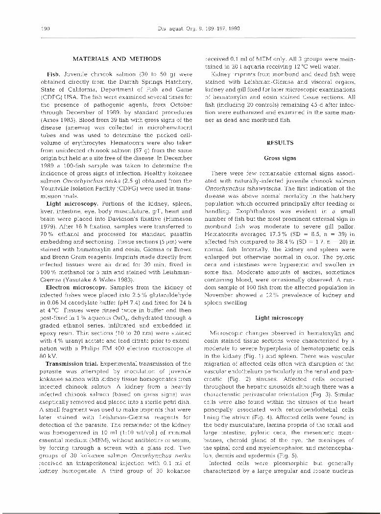
MATERIALS AND METHODS
Fish. Juvenile chinook salmon (30 to 50 g ) wele obtained directly from the Darrah Springs Hatchery, State of Cahfornia, Department of Fish and Game (CDFG) USA. The fish were examined several times for the presence of pathogenic agents, from October through December of 1989, by standard procedures (Amos 1985). Blood from 39 fish with gross signs of the disease (anemia) was collected in microhematocrit tubes and was used to determine the packed cell- volume of erythrocytes. Hematocrits were also taken from uninfected chinook salmon (57 g) from the same origin but held at a site free of the disease. In December 1989 a 100-fish sample was taken to determine the incidence of gross signs of infection. Healthy kokanee salmon Oncorhynchus nerka (2.5 g) obtained from the Yountville Isolation Facility (CDFG) were used in trans- mission trials.
Light rnicroscopy. Portions of the kidney, spleen, liver, intestine, eye, body musculature, gill, heart and brain were placed into Davidson's fixative (Humason 1979). After 16 h fixation, samples were transferred to 70% ethanol and processed for standard paraffin embedding and sectioning. Tissue sections (5 p,m) were stained with hematoxylin and eosin, Giemsa or Brown and Brenn Gram reagents. Imprints made directly from infected tissues were air dried for 30 min, fixed in 100 O/O methanol for 5 min and stained with Leishman- Giemsa (Yasutake & Wales 1983).
Electron microscopy. Samples from the kidney of infected fishes were placed into 2.5 % glutaraldehyde in 0.06 M cacodylate buffer (pH 7.4) and fixed for 24 h at 4°C. Tissues were rinsed twice in buffer and then post-fixed in 1 O/O aqueous Os04 , deh.ydrated through a graded ethanol series, infiltrated and embedded in epoxy resin. Thin sections (10 to 20 nm) were stained with 4 % uranyl acetate and lead citrate prior to exami- nation with a Philips EM 400 electron microscope at 80 kV.
Transmission trial. Experimental transmission of the parasite was attempted by inoculation of juvenile kokanee salmon with kidney tissue homogenates from infected chinook salmon. A kidney from a heavily infected chinook salmon (based on gross signs) was aseptically removed and placed into a sterlle petri dish. A small fragment was used to make imprints that were later stained with Leishman-Giemsa reagents for detection of the parasite. The remainder of the kidney was homogenized in 10 m1 (1:lO wt/vol.) of minimal essential medium (MEM), without antibiotics or serum. by forcing through a screen with a glass rod. Two groups of 30 kokanee salmon Oncorhynchus nerka received an intraperitoneal injection with 0.1 m1 of kidney homogenate. A third group of 30 kokanee
received 0.1 m1 of MEM only. All 3 groups were main- tained in 20 1 aquaria receiving 12°C well water.
Kidney imprints from moribund and dead fish were stained with Leishman-Giemsa and visceral organs, kidney and gill fixed for later microscopic examinations of hematoxylin and eosin stained tissue sections. All fish (including 20 controls) remaining 45-d after infec- tion were euthanized and examined in the same man- ner as dead and moribund fish.
RESULTS
Gross signs
There were few remarkable external signs associ- ated with naturally-infected juvenile chinook salmon Oncorhynchus tshawytscha. The first indication of the dlsease was above normal mortality in the hatchery population which occurred principally after feeding or handling. Exophthalmos was evident in a small number of fish but the most prominent external sign in moribund fish was moderate to severe gill pallor. Hematocrits averaged 17.5 O/O (SD = 8.5, n = 39) in affected fish compared to 38.4 % (SD = 1.7, n = 20) in normal fish. Internally, the kidney and spleen were enlarged but otherwise normal in color. The pyloric ceca and intestines were hyperemic and swollen in s0m.e fi.sh. Moderate amounts of ascites, sometimes containing blood, were occasionally observed. A ran- dom sample of 100 fish from the affected population in November showed a 12% prevalence of kidney and spleen swelling.
Light microscopy
Microscopic changes observed in hematoxylin and eosin stained tissue sections were characterized by a moderate to severe hyperplasia of hematopoietic cells in the ludney (Fig. 1) and spleen. There was vascular migration of affected cells often with disruption of the vascular endothelium particularly in the renal and pan- creatic (Fig. 2) sinuses. Affected cells occurred throughout the hepatic sinusoids although there was a characteristic perivascular orientation (Fig. 3). Similar cel1s were also found within the sinuses of the heart principally associated with reticuloendothelial cells lining the atrium (Fig. 4 ) . Affected cells were found in the body musculature, lamina propria of the small and large intestine, pyloric ceca, the mesenteric mem- branes, choroid gland of the eye, the meninges of the spinal cord and myelencephalon and metencepha- Ion, derrnis and epidermis ( h g . 5).
Infected cells were pleomorphic but generally characterized by a large irregular and lobate nucleus

Hednck et al.: Hematopoietic infection of chinook salmon 191
Figs 1 to 6. Oncorhynchus tshawytscha. Hematoxylin stained tissue sections from fish infected with an intranuclear micro- sporidian. Fig. l . Kdney with hyperplasia of hematopoietic cells; bar = 2 mm. Fig. 2. Large occluded vein in the pancreas containing infected lymphoblasts; bar = 1 mm. m.3. Vein, artery and bile duct of the liver with populations of affected cells; bar = 1 mm. Flg. Trabeculae of atrium with internalized affected cells; bar = 1 mm. Fig Accumulation of affected cells in the epidermis; bar = 2 mm. Fig& Infected cells in the interstitium of the kidney. Arrow shows intranuclear stage of the
microsporidian; bar = 10 pm

Dis. aquat. Org. 8: 189-197, 1990
and an increased nuclear to cytoplasmic ratio. The nucleus of affected cells contained eosinophilic bodies which ranged from circular to rod-shaped (Fig. 6). In certain nuclei, 2 to 3 of these intranuclear inclusions were observed. Apparent binucleate and mitotically active cells were common even in peripheral blood (Fig. 7). These infected cells had amphophilic to basophilic cytoplasm with irregular plasmalemma. Intranuclear stages were also found in many dead or smudged cell nuclei (Fig. 7). The infection was most easily detected by observation of prespore stages and spores within the nucleus of cells following Leishman- Giemsa staining of kidney imprints (Fig. 8). Although well developed spores were infrequent, some nuclei containcc! UP ts 8 or 16 spores (Fig. 8). The spores stained poorly and were ovoid, approximately 1.0 X 2.0 ILm (width by length) and contained a small centrally
located vacuole or polar body as measured from Leish- man-Giemsa stained preparations (Fig. 8).
Electron microscopy
Prespore and spore stages were observed by electron rnicroscopy in many cells in the kidney interstitium (Figs. 9 and 10). The host cell-type infected with the microsporidian was characterized by a large nucleus with dense chromatin, and a cytoplasm with abundant endoplasmic reticulum often with a concentric array (Fig. 11).
Plasrnodia within affected nuclei had a single simple plasmalemma and contained abundant endoplasmic reticulum and ribosomes but no mitochondria. Sporogonic plasmodia with lamellar precursors of the polar tube, vacuoles and pronounced endoplasmic
Figs. 7 and 8. Oncorhynchus tsha- wytscha Leishrnan-Giemsa stalned cells from the blood and kidney, re- spectively, of microsporidian infected fish. Fig. 7. Blood smear with infected lyrnphoblasts, one undergoing divison with intranuclear microsporidian stages (arrow) and a smudge cell with similar intranuclear forms; bar = 10 pm. E(ldney imprint showing an infected cell with 8 spores within the nucleus
(arrow); bar = 10 pm
Hedrick et al.: Hematopoiet :ic infection of chinook salmon 193
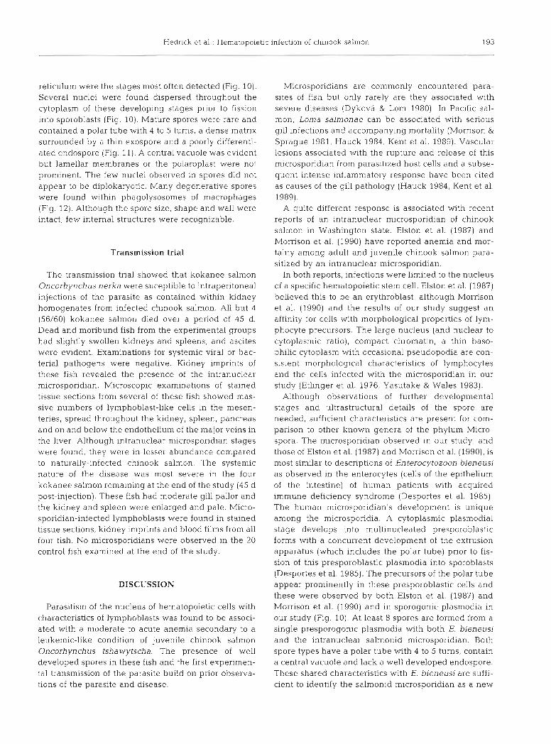
reticulum were the stages most often detected (Fig. 10). Several nuclei were found dispersed throughout the cytoplasm of these developing stages prior to fission into sporoblasts (Fig. 10). Mature spores were rare and contained a polar tube with 4 to 5 turns, a dense matrix surrounded by a thin exospore and a poorly differenti- ated endospore (Fig. 11). A central vacuole was evident but lamellar membranes or the polaroplast were not prominent. The few nuclei observed in spores did not appear to be diplokaryotic. Many degenerative spores were found within phagolysosomes of macrophages (Fig. 12). Although the spore size, shape and wall were intact, few internal structures were recognizable.
Transmission trial
The transmission trial showed that kokanee salmon Oncorhynchus nerka were suceptible to intraperitoneal injections of the parasite as contained within kidney homogenates from infected chinook salmon. All but 4 (56/60) kokanee salmon died over a period of 45 d. Dead and moribund fish from the experimental groups had slightly swollen kidneys and spleens, and ascites were evldent. Examinations for systemic viral or bac- terial pathogens were negative. Kidney imprints of these fish revealed the presence of the intranuclear microspondian. Microscopic examinations of stained tissue sections from several of these fish showed mas- sive numbers of lymphoblast-like cells in the mesen- teries, spread throughout the kidney, spleen, pancreas and on and below the endothelium of the major veins in the liver. Although intranuclear microsporidian stages were found, they were in lesser abundance compared to naturally-infected chinook salmon. The systemic nature of the disease was most severe in the four kokanee salmon remaining at the end of the study (45 d post-injection). These fish had moderate gill pallor and the kidney and spleen were enlarged and pale. Micro- sporidian-infected lymphoblasts were found in stained tissue sections, kidney imprints and blood films from all four fish. No microsporidians were observed in the 20 control fish examined a t the end of the study.
DISCUSSION
Parasitism of the nucleus of hematopoietic cells with characteristics of lymphoblasts was found to be associ- ated with a moderate to acute anemia secondary to a leukemic-like condition of juvenile chinook salmon Oncorhynchus tshawytscha. The presence of well developed spores in these fish and the first experimen- tal transmission of the parasite build on prior observa- tions of the parasite and disease.
Microsporidians are commonly encountered para- sites of fish but only rarely are they associated with severe diseases (Dykova & Lom 1980). In Pacific sal- mon, Loma salmonae can be associated with serious gill infections and accompanying mortality (Mornson & Sprague 1981, Hauck 1984, Kent et al. 1989). Vasculai- lesions associated with the rupture and release of this microsporidian from parasitized host cells and a subse- quent intense inflammatory response have been cited as causes of the gill pathology (Hauck 1984, Kent et al. 1989).
A quite different response is associated with recent reports of an intranuclear microsporidian of chinook salmon in Washington state. Elston et al. (1987) and Morrison et al. (1990) have reported anemia and mor- tality among adult and juvenile chinook salmon para- sitized by an intranuclear microsporidian.
In both reports, infections were limited to the nucleus of a specific hematopoietic stem cell. Elston et al. (1987) believed this to be a n erythroblast, although Morrison et al. (1990) and the results of our study suggest an affinity for cells with morphological properties of lym- phocyte precursors. The large nucleus (and nuclear to cytoplasmic ratio), compact chromatin, a thin baso- philic cytoplasm with occasional pseudopodia are con- sistent morphological characteristics of lymphocytes and the cells infected with the microsporidian in our study (Etlinger et al. 1976, Yasutake & Wales 1983).
Although observations of further developmental stages and ultrastructural details of the spore are needed, sufficient characteristics are present for com- parison to other known genera of the phylum Micro- spora. The microsporidian observed in our study, and those of Elston et al. (1987) and Morrison et al. (1990), is most similar to descriptions of Enterocytozoon bieneusi as observed in the enterocytes (cells of the epithelium of the intestine) of human patients with acquired immune deficiency syndrome (Desportes et al. 1985). The human microsporidian's development is unique among the microsporidia. A cytoplasmic plasmodial stage develops into multinucleated presporoblastic forms with a concurrent development of the extrusion apparatus (which includes the polar tube) prior to fis- sion of this presporoblastic plasmodia into sporoblasts (Desportes et al. 1985). The prec.ursors of the polar tube appear prominently in these presporoblastic cells and these were observed by both Elston et al. (1987) and Mornson et al. (1990) and in sporogonic plasmodia in our study (Fig. 10). At least 8 spores are formed from a single presporogonic plasmodia with both E. bieneusi and the intranuclear salmonid microsporidian. Both spore types have a polar tube with 4 to 5 turns, contain a central vacuole and lack a well developed endospore. These shared characteristics with E. bieneusi are suffi- cient to identify the salmonid microsporidian as a new


196 Dis. aquat. Org. 8: 189-197, 1990
Fig. 12. Oncorhync~~uo ror~aw~rscha. Electron-rnicroy~aph oi degenerative spores within a phagolysosome OL a macrophage of infected kidney; bar = 1 pm
Enterocytozoon sp. The intranuclear development, larger size and d~fferent host for the salmon~d micro- sporidian however, clearly separates it from E. bien e usi.
The possible relationship of the microsporidian we observed to Microspondium rhabdophllia needs to be further examined. Although no ultrastructural studies were conducted, Modin (1981) described a spore of similar size and intranuclear location in several sal- monid specl.es, including chinook sal.mon. He did not, however, observe the parasite other than in rodlet cells. The gross and microscopic pathology associated with M. rhabdophilia however, is clearly different to that observed in chinook salmon in our study. An ultrastruc- tural examination of M, rhabdophilia, as present in rodlet cells, is needed to determine whether possibly this microsporidian is related to the parasite we have found in lymphoblasts of chinook salmon.
The migration via the vasculature, adherence to endothelium of vessels and migration and establish- ment of microsporidian-infected cells in surrounding tissues are features of a neoplastic rather than pro-
liferative response (Cotran et al. 1989). Populations of these cells were found in nearly every tissue examined, although they were most abundant in the hematopoi- e t ~ c organs of chinook salmon (e .g . spleen and kidney). There was no evidence of tumor like growths in natur- ally or experimentally-infected fish.
The microscopic signs associated with microspor- idian infection in our study are nearly identical to a recently described plasmacytoid leukemia in chinook salmon reared in seawater net-pens In British Colum- bia (Kent et al. 1990). Kent & Dawe (1990) were able to transmit the disease by intraperitoneal injections of homogenized kidney from infected chinook salmon into previously healthy chinook, sockeye (Oncorhyn- chus nerka) and Atlantic salmon (Salmo salar). Development of the disease and mortality began 1 to 2 mo post-injection at water temperatures of 12°C. Although the microscopic signs of the plasmacytoid leukemia and the intranuclear microsporidian infec- tions are similar, microsporidian stages have not been observed in experimentally-induced plasmacytoid leukemia in chinook salmon in British Columbia (M. L.

Hedrick et al . : Hematopoietic infection of chinook salmon 197
Kent pers, comm.). Studies on the possible relatedness of the 2 conditions is in progress. The microsporidian may be acting as a factor stimulating a leukemic condi- tion perhaps in a fashion similar to the haemo- gregarina-induced lymphoma in cultured turbot (Scophthalmus maximus) as reported by Ferguson & Roberts (1976). Resolution of the microsporidian infec- tion in affected chinook with persistence of the pro- liferative/neoplastic condition may then result in a con- dition identical to the plasmacytoid leukemia described by Kent et al. (1990).
Acknowledgements. This work was supported in part by Dingell-Johnson/Wallop-Breaux Fish Restoration Act funds administered through the California Department of Fish and Game. Thanks to the technicians of the Veterinary Medicine Teaching Hospital for their preparations of stained tissue sections for light microscopy and Mr R . Munn for his electron microscopy expertise. We thank Mr J. Morrison for his valu- able insight in reviewing the microscopic materials.
LITERATURE CITED
Amos, K. (1985). Procedures for the detection of certain fish pathogens. Fish Health Section, blue book. American Fisheries Society, Bethesda. Maryland
Canning, E. M., Lom, J. (1986). The rnicrosporida of verte- brates. Academic Press, London
Cotran, R. S., Kumar, V., Robbins, S. L. (1989). Pathologic basis of disease, 4th edn. W. B. Saunders, Philadelphia
Desportes, I., Le Charpentier. Y., Galian. A., Bernard, F., Cochand-Priollet, B., Lavergen, A., Ravisse, P., Modigliani, R. (1985). Occurrence of a new microsporidian: Enterocy- tozoon bieneusi n.g.n.sp.. in the enterocytes of a human patient with AIDS. J. Protozool. 32: 250-253
Responsible Subject Editor: Professor W Korting, Hannover, F. R. Germany
Dykova, I., Lom, J . (1980). Tissue reactions to microsporidian infections in fish. J. Fish Dis. 3: 265-283
Elston, R. A., Kent, M. L., Harrell, L. H. (1987). An intranuclear microsporidium associated with acute anemia in the chinook salmon. J. Protozool. 34: 274-277
Etlinger, H. M., Hodgins, H. O., Chiller, J. M. (1976). Charac- terization of lymphocytes in a primitive teleost, Salmo gairdneri. In: Wright, R. K., Cooper, E. L. (eds.) Phylogeny of thymus and bone marrow-bursa cells. North-Holland Publishing Co., New York, p. 83-91
Ferguson, H. W., Roberts. R. J. (1976). A condition simulating lymphoma, associated with a sporozoan infection in cultured turbot (Scophthalmus maximus L.). Prog. exp. Tumor Res. 20: 212-216
Hauck, A. K. (1984). Amortality and associated tissue reactions of chinook salmon, Oncorhynchus tshawytscha (Walbaum), caused by a microsporidian Loma sp. J. Fish Dis. 7: 217-229
Humason, G . L. (1979). Animal tissue techniques. W. H. Free- man, San Francisco
Kent, M. L., Dawe, S. C. (1990). Experimental transn~ission of a plasmacytoid leukemia of chinook salmon (Oncorhyn- chus tshawytscha) Cancer Res. (in press)
Kent, M. L., Elliot, D. G , Groff, J M. , Hedrick, R . P. (1989). Lon~a salmonae (Protozoa: Microspora) infections in sea- water reared coho salmon (Oncorhynchus hsutch). Aquaculture 80: 211-222
Kent, M. L. , Groff, J.. Traxler. G. F.. Zinkl, J. G.. Bagshaw, J. W. (1990). Plasmacytoid leukemia of chinook salmon (Oncorhynchus tshawytscha). Dis. aquat. Org. 8: 199-209
Modin, J. C. (1981). Microsporidium rhabdophilia n. sp. from the rodlet cells of salmonid fishes. J. Fish Dis. 4: 203-211
Morrison, C. M., Sprague, V (1981). Microsporidian parasites in the gills of salmonid fishes. J. Fish Dis. 4: 371-386
Morrison, J. K., MacConnell, E.. Chapman, P. F., Westgard, R. L. (1990). A microsporidium-induced lymphoblastosis in chinook salmon (Oncorhynchus tshawytscha) in fresh water. Dis. aquat. Org. 8: 99-104
Yasutake, W T., Wales, J. H. (1983). Microscopic anatomy of salmonids: an atlas. United States Department of the Interior, Fish and Wildlife Service, Resource Publication 150, Washington D.C
Manuscript first received: February 8, 1990 Revised version accepted: May 3, 1990
Related Documents