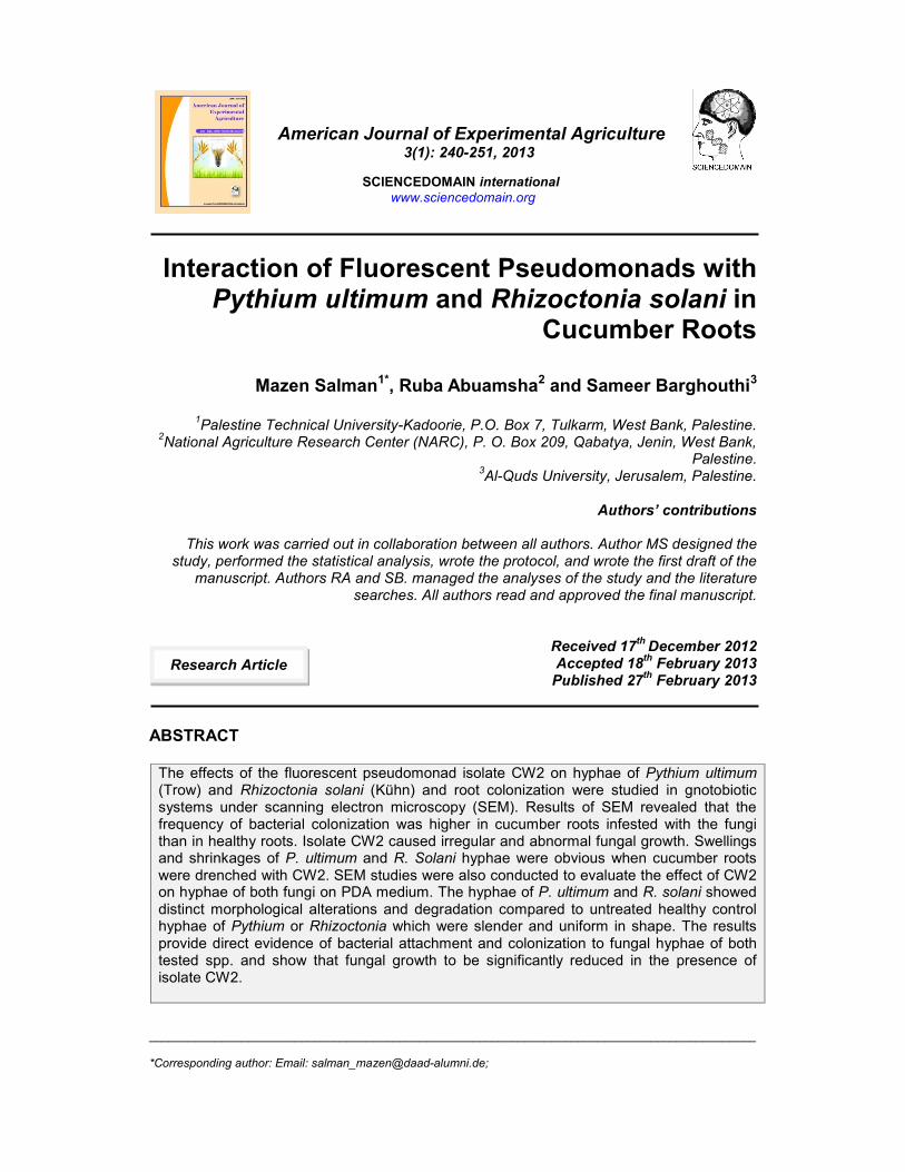
____________________________________________________________________________________________ *Corresponding author: Email: [email protected]; American Journal of Experimental Agriculture 3(1): 240-251, 2013 SCIENCEDOMAIN international www.sciencedomain.org Interaction of Fluorescent Pseudomonads with Pythium ultimum and Rhizoctonia solani in Cucumber Roots Mazen Salman 1* , Ruba Abuamsha 2 and Sameer Barghouthi 3 1 Palestine Technical University-Kadoorie, P.O. Box 7, Tulkarm, West Bank, Palestine. 2 National Agriculture Research Center (NARC), P. O. Box 209, Qabatya, Jenin, West Bank, Palestine. 3 Al-Quds University, Jerusalem, Palestine. Authors’ contributions This work was carried out in collaboration between all authors. Author MS designed the study, performed the statistical analysis, wrote the protocol, and wrote the first draft of the manuscript. Authors RA and SB. managed the analyses of the study and the literature searches. All authors read and approved the final manuscript. Received 17 th December 2012 Accepted 18 th February 2013 Published 27 th February 2013 ABSTRACT The effects of the fluorescent pseudomonad isolate CW2 on hyphae of Pythium ultimum (Trow) and Rhizoctonia solani (Kühn) and root colonization were studied in gnotobiotic systems under scanning electron microscopy (SEM). Results of SEM revealed that the frequency of bacterial colonization was higher in cucumber roots infested with the fungi than in healthy roots. Isolate CW2 caused irregular and abnormal fungal growth. Swellings and shrinkages of P. ultimum and R. Solani hyphae were obvious when cucumber roots were drenched with CW2. SEM studies were also conducted to evaluate the effect of CW2 on hyphae of both fungi on PDA medium. The hyphae of P. ultimum and R. solani showed distinct morphological alterations and degradation compared to untreated healthy control hyphae of Pythium or Rhizoctonia which were slender and uniform in shape. The results provide direct evidence of bacterial attachment and colonization to fungal hyphae of both tested spp. and show that fungal growth to be significantly reduced in the presence of isolate CW2. Research Article

Welcome message from author
This document is posted to help you gain knowledge. Please leave a comment to let me know what you think about it! Share it to your friends and learn new things together.
Transcript
____________________________________________________________________________________________
*Corresponding author: Email: [email protected];
American Journal of Experimental Agriculture3(1): 240-251, 2013
SCIENCEDOMAIN internationalwww.sciencedomain.org
Interaction of Fluorescent Pseudomonads withPythium ultimum and Rhizoctonia solani in
Cucumber Roots
Mazen Salman1*, Ruba Abuamsha2 and Sameer Barghouthi3
1Palestine Technical University-Kadoorie, P.O. Box 7, Tulkarm, West Bank, Palestine.2National Agriculture Research Center (NARC), P. O. Box 209, Qabatya, Jenin, West Bank,
Palestine.3Al-Quds University, Jerusalem, Palestine.
Authors’ contributions
This work was carried out in collaboration between all authors. Author MS designed thestudy, performed the statistical analysis, wrote the protocol, and wrote the first draft of the
manuscript. Authors RA and SB. managed the analyses of the study and the literaturesearches. All authors read and approved the final manuscript.
Received 17th December 2012Accepted 18th February 2013
Published 27th February 2013
ABSTRACT
The effects of the fluorescent pseudomonad isolate CW2 on hyphae of Pythium ultimum(Trow) and Rhizoctonia solani (Kühn) and root colonization were studied in gnotobioticsystems under scanning electron microscopy (SEM). Results of SEM revealed that thefrequency of bacterial colonization was higher in cucumber roots infested with the fungithan in healthy roots. Isolate CW2 caused irregular and abnormal fungal growth. Swellingsand shrinkages of P. ultimum and R. Solani hyphae were obvious when cucumber rootswere drenched with CW2. SEM studies were also conducted to evaluate the effect of CW2on hyphae of both fungi on PDA medium. The hyphae of P. ultimum and R. solani showeddistinct morphological alterations and degradation compared to untreated healthy controlhyphae of Pythium or Rhizoctonia which were slender and uniform in shape. The resultsprovide direct evidence of bacterial attachment and colonization to fungal hyphae of bothtested spp. and show that fungal growth to be significantly reduced in the presence ofisolate CW2.
Research Article
American Journal of Experimental Agriculture, 3(1): 240-251, 2013
241
Keywords: SEM; colonization; fluorescent pseudomonas; attachment; antifungal, biocontrol.
1. INTRODUCTION
The application of microorganisms as biocontrol agents has been known for several decades[1]. Plant growth-promoting rhizobacteria (PGPR) promote plant growth following inoculationonto seeds or subterranean plant parts [2]. Microorganisms that can grow in the rhizosphereare ideal for use as biocontrol agents, since the rhizosphere provides the front-line ofdefense against pathogenic microbes [3,4,5]. There are several species of micro-organismsthat have been tested as potential biocontrol agents of damping-off, including bacteria suchas Pseudomonas spp. [6,7], Bacillus spp. and Streptomyces griseoviridis as well as fungi, forinstance Trichoderma spp. (T. virens, T. harzinum), Gliocladium spp. and Pythiumoligandrum [8].
Fluorescent pseudomonads are one group of rhizospheric bacteria that have been describedas biological control agents and showed great promise with respect to protecting plant rootsby reducing the incidence of fungal-induced diseases [9,10]. Fluorescent Pseudomonadsproduce versatile catabolic and secondary metabolites which include antifungal compounds.They have excellent root-colonizing abilities allowing them to be effective in the vicinity ofplant roots. The soil-borne fluorescent pseudomonads have received special attention andemerged as the largest and potentially most promising group involved in biocontrol of plantdiseases [1,11,12].
Currently, Pseudomonas spp. are particularly suitable for applications as agriculturalbiocontrol agents since they: (1) can use many exudate compounds released by roots as asource of nutrients [13]; (2) are abundantly present in natural soils, in particular on plant rootsystems, which is indicative of their adaptive potential [14]; (3) have a high growth raterelative to many other rhizosphere bacteria; (4) possess diverse mechanisms by which theycan exert inhibitory activity towards phytopathogens and thereby mediate crop protection[13,14]; (5) are easy to grow in vitro; (6) can subsequently be reintroduced into therhizosphere by seed bacterization [14]; (7) are susceptible to mutation and molecularmanipulation techniques[15]; and (8) their growth temperatures are well suited to soiltemperatures ranging from 10 to 35ºC [16].
In recent years, several studies were conducted to evaluate the efficacy of fluorescentpseudomonad species against plant pathogens. The fluorescent pseudomonad isolate CW2proved to be a promising biocontrol agent against many plant pathogens including P.ultimum, R. solani [17], Fusarium oxysporum f. sp. Lycopersici [18]. The mechanism ofaction of the bacterium was studied intensively. The bacterium produces antibiotics such as2,4-diacetylphloroglucinol (DAPG) and other phenazine derivative [17]. In addition, it waspossible to apply the bacterium in combination with chemical fungicides to control P. ultimumdamping-off in tomato [19]. However little information is available about colonization of plantroots by this bacterium. The aim of this work was to provide direct evidence for theattachment and colonization of cucumber roots by the Pseudomonas fluorescens isolateCW2, in presence or absence of P. ultimum or R. solani. In addition, the study aimed atassessing the ability of the antagonistic bacterial isolates to control the hyphal growth of bothfungal spp. by the isolate CW2.
American Journal of Experimental Agriculture, 3(1): 240-251, 2013
242
2. MATERIALS AND METHODS
2.1 Cultivation and Maintenance of Antagonistic Fluorescent pseudomonads
The antagonistic P. fluorescens isolate CW2 was obtained from the Institute ofPhytomedicine, University of Hohenheim, Germany. Stock culture of the bacterium wasprepared by growing the bacterium in 125-ml Erlenmeyer flasks containing 25 ml King’s B[20] liquid medium on a rotary shaker (150 rpm) for 20 h at 28ºC. Isolates were stored as1ml-aliquots in 20% glycerol at - 80ºC.
2.2 Fungal Isolates
The oomycete P. ultimum (Trow) and R. solani (Kühn) were obtained from the Institute ofPhytomedicine, University of Hohenheim. The fungi were grown on potato dextrose agarmedium (PDA) at 22ºC for 4 days. Stock cultures were stored at 4ºC and sub culturedroutinely every 2 weeks.
2.3 Effect of Bacteria on Mycelial growth of P. ultimum and R. solani
Single 0.5 cm diameter agar disks grown with 4 days old P. ultimum or R. solani weretransferred into 125 ml Erlenmeyer flasks containing 25 ml PDB and incubated on a rotaryshaker at 110 rpm and 22ºC. After 3 days of incubation, 0.1 ml of CW2 (1 × 10⁹cfu ml-1) wasadded to the fungal cultures and incubated for an additional 3 days. Mycelial dry weightswere determined after filtration through preweighted Whatman # 1 filter paper and dryingovernight at 80ºC (modification of Nelson et al., 1986) [21].
2.4 Colonization of Cucumber roots by P. fluorescens
To study the population size of the P. fluorescens isolate CW2, cucumber seeds were pre-germinated between towel papers for two days. The germinated seedlings were thentransplanted in 9-cm diameter pots filled with humosoil®: sand mixtures (2:1 v/v) andincubated under greenhouse conditions. For inoculation of the seedlings, the bacterium wasgrown for 24 h in KB medium at 28ºC with shaking (150 rpm). Two methods of applicationwere used. For soil drenching, bacterial suspensions (1 × 10⁹cfu ml-1; 30 ml pot-1) werepoured over the soil of the pots of cucumber seedlings. For root dipping, the roots ofgerminated seedlings were soaked in 24 h old (1 × 10⁹cfu ml-1) bacterial suspensions for 30min before transplanting them in the pots. After different periods of time (1, 3, 7, 14 and 21days) roots of cucumber were washed gently under running tap water, cut into 1 cm longpieces and dipped in 100 ml KB liquid medium [18]. After incubation for two hours at 28ºC ona rotary shaker with 200 rpm, serial dilutions were made from 1 ml of the KB suspension.
100 µl of suspension were spread on solid NPC medium (KB medium plus Novobiocin 45.0mg L-1 Penicillin G 35.2 mg L-1 and Cycloheximide 75.0 mg L-1) prepared according to Sandsand Rovira (1970)[22] and incubated for 3 days at 28ºC. Bacterial colonies were visualizedunder UV light at 360 nm. Colonies showing green fluorescence were counted.
American Journal of Experimental Agriculture, 3(1): 240-251, 2013
243
2.5 Effect of Bacteria on P. ultimum and R. solani hyphae (scanning electronmicroscopy)
Dual cultures of P. fluorescens isolate CW2 and P. ultimum or R. solani were grown in 9 cmPetri dishes containing PDA medium for 3 days at 22ºC [17]. For scanning electronmicroscopy (SEM), 0.5 cm agar pieces with P. ultimum or R. solani were fixed withglutaraldehyde (4% w/v) in phosphate buffer (50 mM; pH 6.8) for 12 h at 4°C and rinsed 5times with the same buffer for 20 min. Samples were post fixed in osmium tetraoxide (1%) inphosphate buffer (50 mM; 6.8 pH) for 12 h at 4ºC and rinsed with phosphate buffer (4X, 15min each). After dehydration in graded acetone series, samples were critical-point dried,mounted on stubs, sputter-coated with gold-palladium, and observed using a Zeiss DSM100scanning electron microscope operating at 15 kV [23].
2.6 Interaction of the P. fluorescens isolate CW2 with P. ultimum and R. solaniin the Rhizosphere of Cucumber seedlings
A gnotobiotic system described by Simons et al. (1996) [24] was used in studying theinteraction of CW2 with P. ultimum and R. solani. Briefly, cucumber seeds were surfacesterilized by soaking in 1% (v/v) sodium hypochlorite for 5 min followed by five thoroughrinses with sterile water. Seeds were then germinated between autoclaved Whatman #1 filterpaper in 9 cm diameter Petri dishes in the dark at room temperature. Germinated seedlingswere planted in tubes filled with sand [24] before application of bacteria or fungi. One ml ofbacterial suspension (cells of isolate CW2 from a 24 h culture in KB medium) was washed inplant nutrient solution (PNS) and adjusted to a concentration of 1 × 10⁹ cfu ml-1. After twodays of incubation, the tubes were infested with PDA disks grown with 4 day old P. ultimumor R. solani. Control experiments included untreated seedlings, seedlings treated with CW2alone, and seedlings treated either with P. ultimum or R. solani. Tubes were then incubatedat room temperature for three days.
For SEM, plantlets were removed gently from the tubes and the roots were washed withwater and sectioned in about 1 cm pieces. Preparation of SEM samples was carried out asmentioned above.
2.7 Data Analysis
Statistical significances were computed with Xlstat (Adinosoft) using Tukey test aftersignificant F-test (P = 0.05) of the analysis of variance (ANOVA). Means indicated withdifferent letters are significantly different.
3. RESULTS
3.1 Root Colonization
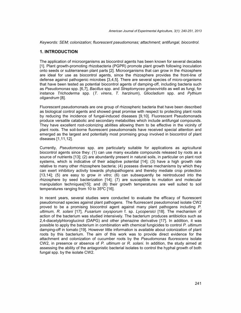
The population sizes of CW2 reisolated from the rhizosphere of cucumber seedlings wereslight higher but not significantly different (F= 9.74; df 9, 19; P< 0.001) between the rootdipping and soil drenching methods (Fig. 1). The determined bacterial concentrations ofCW2 were 6.6 and 5.9 log cfu g-1 root fresh weight, following root dipping and soil drenching,respectively. After 3 days of application as well as after 7 and 14 days, the bacterial numberdetermined on roots did not differ between soil drenching and root dipping methods. Afterthe third week of application, bacterial populations were significantly reduced in in cucumber
American Journal of Experimental Agriculture, 3(1): 240-251, 2013
244
roots after soil drenching and root dipping (Fig. 1). Significant reduction of bacterialconcentration was recorded after 21 days of the experiment.
Fig. 1. Comparison of root colonization of roots of cucumber plants by fluorescentpseudomonads isolates CW2. Bacterial suspensions (1×10⁹cfu ml-1) were appliedeither as root dipping or as soil drench. Data are represented as the log cfu/g root
fresh weight. Bars represent standard errors (SE)
3.2 Effect of Bacteria on Mycelial Growth
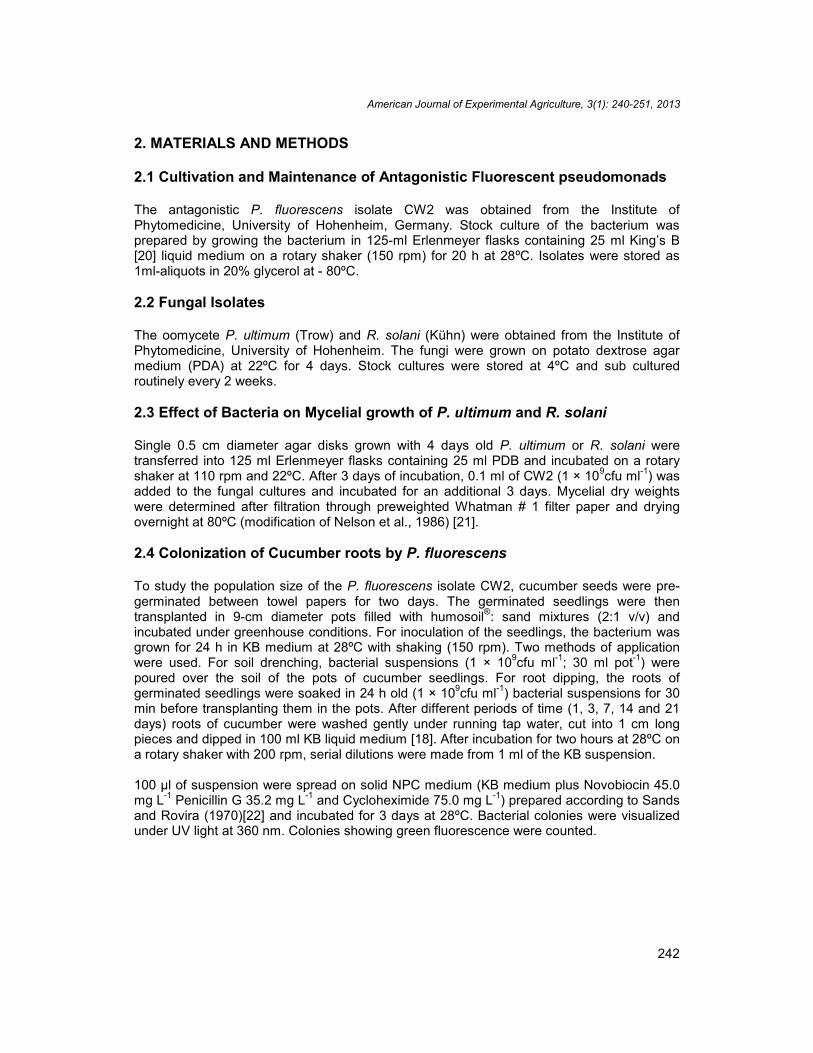
Growth rate of P. ultimum and R. solani was not significantly different (F= 71.99; df 3, 19; P< 0.0001) after 3days of incubation in PDB medium at 110 rpm and 24ºC (Fig. 2). However,in the presence of P. fluorescens isolate CW2; the growth rate was significantly reduced.The percent of reduction of mycelial weight was 60.5 and 63.6 % for P. ultimum and R.solani respectively.
Fig. 2. Effect of the fluorescent pseudomonads isolate CW2 (1 ×10⁹cfu ml-1) onmycelial growth of P. ultimum and R. solani in liquid PDB medium. The experiment
was done in triplicates and repeated three times. Data with different letters aresignificantly different after Tukeys HSD test using ANOVA at P < 0.05. Bars represent
standard errors (SE)
American Journal of Experimental Agriculture, 3(1): 240-251, 2013
245
3.3 Scanning Electron Microscope Studies (SEM)
These studies were carried out using the P. fluorescens isolate CW2. The effect of thebacterial isolate has been studied on mycelial growth of P. ultimum and, R. solani in dualcultures on PDA medium. The interaction of this bacterial isolate with P. ultimum and, R.solani in cucumber rhizosphere was conducted in a gnotobiotic system as described inSimons et al. [24]. Results are detailed below:
3.3.1 Effect of CW2 on P. ultimum
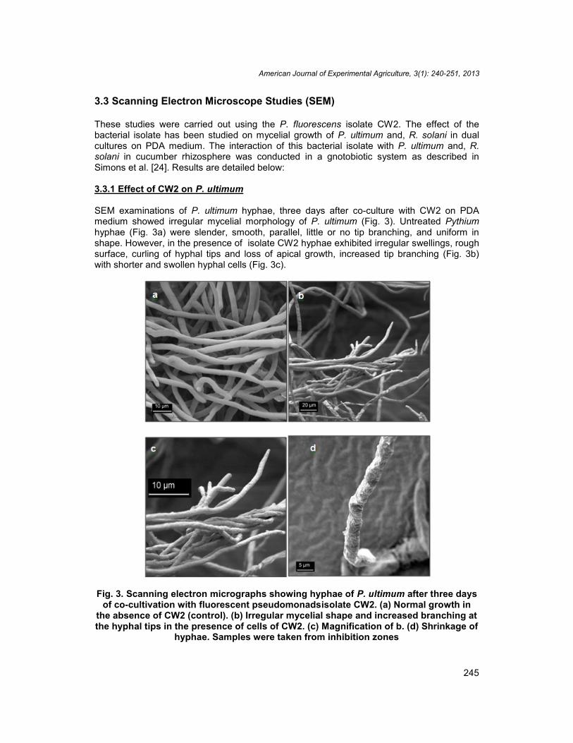
SEM examinations of P. ultimum hyphae, three days after co-culture with CW2 on PDAmedium showed irregular mycelial morphology of P. ultimum (Fig. 3). Untreated Pythiumhyphae (Fig. 3a) were slender, smooth, parallel, little or no tip branching, and uniform inshape. However, in the presence of isolate CW2 hyphae exhibited irregular swellings, roughsurface, curling of hyphal tips and loss of apical growth, increased tip branching (Fig. 3b)with shorter and swollen hyphal cells (Fig. 3c).
Fig. 3. Scanning electron micrographs showing hyphae of P. ultimum after three daysof co-cultivation with fluorescent pseudomonadsisolate CW2. (a) Normal growth in
the absence of CW2 (control). (b) Irregular mycelial shape and increased branching atthe hyphal tips in the presence of cells of CW2. (c) Magnification of b. (d) Shrinkage of
hyphae. Samples were taken from inhibition zones
American Journal of Experimental Agriculture, 3(1): 240-251, 2013
246
3.3.2 Effect of CW2 on R. solani
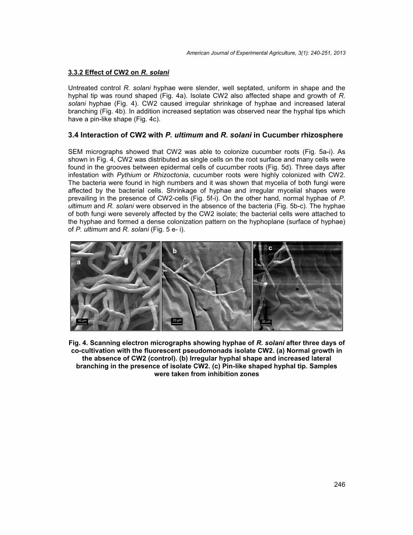
Untreated control R. solani hyphae were slender, well septated, uniform in shape and thehyphal tip was round shaped (Fig. 4a). Isolate CW2 also affected shape and growth of R.solani hyphae (Fig. 4). CW2 caused irregular shrinkage of hyphae and increased lateralbranching (Fig. 4b). In addition increased septation was observed near the hyphal tips whichhave a pin-like shape (Fig. 4c).
3.4 Interaction of CW2 with P. ultimum and R. solani in Cucumber rhizosphere
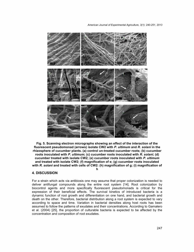
SEM micrographs showed that CW2 was able to colonize cucumber roots (Fig. 5a-i). Asshown in Fig. 4, CW2 was distributed as single cells on the root surface and many cells werefound in the grooves between epidermal cells of cucumber roots (Fig. 5d). Three days afterinfestation with Pythium or Rhizoctonia, cucumber roots were highly colonized with CW2.The bacteria were found in high numbers and it was shown that mycelia of both fungi wereaffected by the bacterial cells. Shrinkage of hyphae and irregular mycelial shapes wereprevailing in the presence of CW2-cells (Fig. 5f-i). On the other hand, normal hyphae of P.ultimum and R. solani were observed in the absence of the bacteria (Fig. 5b-c). The hyphaeof both fungi were severely affected by the CW2 isolate; the bacterial cells were attached tothe hyphae and formed a dense colonization pattern on the hyphoplane (surface of hyphae)of P. ultimum and R. solani (Fig. 5 e- i).
Fig. 4. Scanning electron micrographs showing hyphae of R. solani after three days ofco-cultivation with the fluorescent pseudomonads isolate CW2. (a) Normal growth in
the absence of CW2 (control). (b) Irregular hyphal shape and increased lateralbranching in the presence of isolate CW2. (c) Pin-like shaped hyphal tip. Samples
were taken from inhibition zones
ab c
American Journal of Experimental Agriculture, 3(1): 240-251, 2013
247
Fig. 5. Scanning electron micrographs showing an effect of the interaction of thefluorescent pseudomonad (arrows) isolate CW2 with P. ultimum and R. solani in the
rhizosphere of cucumber plants. (a) control un-treated cucumber roots; (b) cucumberroots inoculated with P. ultimum; (c) cucumber roots inoculated with R. solani; (d)cucumber treated with isolate CW2; (e) cucumber roots inoculated with P. ultimumand treated with isolate CW2; (f) magnification of e; (g) cucumber roots inoculated
with R. solani and treated with cells of CW2: (h) magnification of g; (i) magnification ofh
4. DISCUSSION
For a strain which acts via antibiosis one may assume that proper colonization is needed todeliver antifungal compounds along the entire root system [14]. Root colonization bybiocontrol agents and more specifically fluorescent pseudomonads is critical for theexpression of their beneficial effects. The survival kinetics of introduced bacteria is adynamic function of root growth and differentiation on one hand, and bacterial growth anddeath on the other. Therefore, bacterial distribution along a root system is expected to varyaccording to space and time. Variation in bacterial densities along host roots has beenassumed to follow the patterns of exudates and their concentrations. According to Gamaleroet al. (2004) [25], the proportion of culturable bacteria is expected to be affected by theconcentration and composition of root exudates.
American Journal of Experimental Agriculture, 3(1): 240-251, 2013
248
It was found that CW2 isolate was able to colonize cucumber roots as revealed byenumeration of the total cfu. Total cfu g-1 root fresh weights of cucumber were determined 1,3, 7, 14 and 21 days after treatment with bacteria either by soil drenching or seed treatment.However, the method is relatively elaborate, time consuming and does not provide arepresentative visual depiction of spatial colonization patterns relative to the host plant root[26]. These studies typically have been limited to a description of a given bacterial strain aspopulations per entire root system or as populations on root segments from specific regionsof the root system [27].
Colonization may involve simply root surface development but, endophytic colonization ofthe root is also known, and the degree of endophytic colonization depends on bacterial strainand plant type [4]. When colonizing a root environment, an organism is confronted with acomplex array of parameters such as water content, temperature, pH, soil types, andcomposition of root exudates, mineral content, microorganisms, and other factors [12].Evidence of colonization are provided by EM as seen in Fig. 5 where bacterial cells appearas colonies rather than being evenly and randomly distributed along the hyphae which offersa reasonable explanation for the increased bacterial numbers associated with cucumberroots in the presence of the fungal pathogen. It is also consistent with the views of Gamaleroet al. (2004) [25]; the proportion of culturable bacteria is expected to be affected by theconcentration and composition of root exudates.
For the detection of the pseudomonads on the surface of the cucumber roots, scanningelectron microscopy (SEM) was used. SEM offers the advantage to detect local interactionsand /or the adhesion places of the bacteria directly on the roots [28,29]. SEM investigationsshowed that the CW2 isolate formed micro-colonies at furrow-like site on the root surface.Similar observations were made also by Brown and Beringer (1983) [30] and Chin-A-Woenget al. (1997) [29] using other root-colonizing bacteria.
It is obvious from SEM studies that the bacteria may have more than one control mechanismagainst P. ultimum and R. solani. It was shown that the bacterium was able to colonizefungal hyphae. Lysis, swelling and increased branching of the fungal hyphae were noticed indual cultures of bacteria and fungi grown on PDB medium (light microscopy data are notshown).
Multiple mechanisms of biocontrol have been described in some fluorescent pseudomonadsstrains [9]. Modes of action include: inhibition of the pathogen by antimicrobial compounds(antibiosis), competition for iron through production of siderophores, competition forcolonization sites and nutrients supplied by seeds and roots; induction of plant resistancemechanisms, inactivation of pathogen germination factors present in seed or root exudates,degradation of pathogenicity factors of the pathogen such as toxins, parasitism that mayinvolve production of extracellular cell wall-degrading enzymes, for example, chitinase andß-1,3-glucanase that can lyse pathogen cell walls [4].
The most commonly reported mechanisms of biocontrol by fluorescent pseudomonadsspecies include production of antibiotics, hydrogen cyanide (HCN), lytic exoenzymes,competition for iron mediated by siderophores, competition for carbon and induced systemicresistance [9].
American Journal of Experimental Agriculture, 3(1): 240-251, 2013
249
5. CONCLUSION
In conclusion the capacity of P. fluorescens isolate CW2 to colonize the mycelia ofphytopathogens used in this study and possibly other similar phytopathogenic fungi, allowingthem to release their anti-fungal agents directly in the vicinity of the fungus maximizing itseffect. Therefore, plants benefit from the protective advantage imparted by pseudomonadssimilar to CW2. The efficacy of field applications of these biocontrol agents is beingdetermined in field applications on cucumber.
ACKNOWLEDGEMENTS
The financial support of the German Academic Exchange services is highly acknowledgedand appreciated. The authors deeply appreciate the efforts of Prof. H. Buchenauer, Instituteof Phytomedicine (360), University of Hohenheim, Otto-Sander-Str. 5, D-70599 Stuttgart,Germany for supervising this work.
COMPETING INTERESTS
Authors have declared that no competing interests exist.
REFERENCES
1. Weller DM. Biological control of soil borne plant pathogens in the rhizosphere withbacteria. Annu Rev Phytompathol. 1988;26:379–407.
2. Mavrodi OV, McSpadden GBB, Mavrodi DV, Bonsall RF, Weller DM, Thomashow LS.Genetic diversity of phlD from 2,4-diacetylphloroglucinol-producing fluorescentPseudomonads spp. Phytopathology. 2001;91:35–43.
3. Bolwerk A, Lagopod AL, Wijfje AHM, Lamers GEM, Chin-A-Woeng TFC, LugtenbergBJJ and Bloemberg GV. Interactions in the Tomato Rhizosphere of TwoPseudomonas Biocontrol Strains with the Phytopathogenic Fungus Fusariumoxysporum f. sp. radicis-lycopersici. Mol Plant Microbe In. 2003;16(11):983–993.
4. Whipps JM. Microbial interactions and biocontrol in the rhizosphere. J Exp Bot.2001;52:487-51.
5. Favrin RJ, Rahe JE, Mauza B. Pythium spp. associated with crown rot of cucumbersin British Columbia greenhouses. Plant Dis. 1988;72:683–687.
6. Benhamou N, Gagné S, Quéré DL, Dehbi L. Bacterial-mediated induced resistance incucumber: beneficial effect of the endophytic bacterium Serratia plymuthica on theprotection against infection by Pythium ultimum. Phytopathology. 2000;90:45–56.
7. Howell CR, Stipanovic RD. Suppression of Pythium ultimum-inducing damping-off ofcotton seedlings by Pseudomonas fluorecens and its antibiotic pyoluteorin.Phytopathology. 1980;70:712–715.
8. Georgakopoulos DG, Fildaman P, Leifert C, Malathrakis NE. Biological control ofcucumber and sugar beet damping-off caused by Pythium ultimum with bacterial andfungal antagonists. J Appl Microbiol. 2002;92:1078–1086.
9. DeLaFuente L, Tomashow L, Weller D, Bajsa N, Quagliotto L, Chernin L, Arias A.Pseudomonas fluorescens UP61 isolated from birds foot trefoil rhizosphere producesmultiple antibiotics extracts and exerts a broad spectrum of biocontrol activity. Eur JPlant Pathol. 2004;110:671–681.
American Journal of Experimental Agriculture, 3(1): 240-251, 2013
250
10. Andersen JB, Koch B, Nielsen TH, Sørensen D, Hansen M, Nybroe O, ChristophersenC, Sørensen J, Molin S, Givskov M. Surface motility in Pseudomonas sp. DSS73 isrequired for efficient biological containment of the root-pathogenic microfungiRhizoctonia solani and Pythium ultimum. Microbiology. 2003;149:37–46.
11. Walsh UF, Morrissey JP, O’Gara F. Pseudomonas for biocontrol of phytopathogens:from functional genomics to commercial exploitation. Curr Opin Biotech. 2001;12:289–295.
12. O’Sullivan DJ, O'Gara F. Traits of fluorescent Pseudomonas spp. involved insuppression of plant root pathogens. Microbiol Rev. 1992;56(4):662–676.
13. Lugtenberg BJJ, Kravchenko LV, Simons M. Tomato seed and root exudate sugars:composition, utilization by Pseudomonas biocontrol strains and role in rhizospherecolonization. Environ Microbiol. 1999;1(5):439–46.
14. Chin-A-Woeng TFC, Bloemberg GV, Lugtenberg BJJ. Phenazines and their role inbiocontrol by Pseudomonas bacteria. New Phytol. 2003;157:503–523.
15. Haas D, Keel C. Regulation of antibiotic production in root-colonizing pseudomonasspp and relevance for biological control of plant disease. Ann Rev Phytopathol.2003;41:117–153.
16. Polymenakou PN, Stephanou EG. Effect of temperature and additional carbonsources on phenol degradation by an indigenous soil Pseudomonad. Biodegradation.2005;16(5):403-13.
17. Salman M. Determination of Antibiotic Activity on Plasmids from fluorescentPseudomonads isolates CW2, WB15 and WB 52 against pre emergence damping-offcaused by Pythium ultimum and Rhizoctonia solani in cucumber. Biol Control.2010;53(2);161–167.
18. Fakhouri W, Buchenauer H. Characteristics of fluorescent pseudomonad isolatestowards controlling of tomato wilt caused by Fusarium oxysporum f. sp. lycopersici. JPlant Dis Protect. 2003;110:143–156.
19. Salman M, Abuamsha R. Potential for integrated biological and chemical control ofdamping-off disease caused by Pythium ultimum in tomato. BioControl2012;57(5):711-718.
20. King EO, Ward MK, Raney DE. Two simple media for the demonstration of pyocyninand fluorescin. J Lab Clin Med. 1954;44:301–307.
21. Nelson EB, Chao WL, Norton JM, Nash GT, Harman GE. Attachment of Enterobactercloacae to hyphae of Pythium ultimum: Possible role in the biological control ofPythium preemergence damping-off. Phytopathology. 1986;76:327–335.
22. Sands DC, Rovira AD. Isolation of Fluorescent Pseudomonads with a selectivemedium. Applied Microbiology. 1970;20(3):513-514.
23. Kang Z, Huang L, Buchenauer H. Ultrastructural and cytochemical studies off effectsof the fungicide metconazol on Fusarium culmorum in vitro. J Plant Dis Protect.2001;108(4):419–432.
24. Simons M, ven der Bij AJ, Brand I, de Weger AL, Wijffelman CA, Lugtenberg BJJ.Gnotobiotic system for studying rhizosphere colonization by plant growth-promotingPseudomonas bacteria. Mol Plant-Microbe In. 1996;9(7):600–607.
25. Gamalero E, Lingua G, Capri FG, Fusconi A, Berta G, Lemanceau P. Colonizationpattern of primary tomato roots by Pseudomonas fluorescens A6RI characterized bydilution plating, flow cytometry, fluorescence, confocal and scanning electronmicroscopy. Fems Microbiol Ecol. 2004;48(1):79–87.
26. Misaghi IJ. Screening bacteria for root colonizing ability by a rapid method. Soil. BiolBiochem. 1990;22(8):1085–1088.
American Journal of Experimental Agriculture, 3(1): 240-251, 2013
251
27. Roberts DP, Kobayashi DY, Dery PD, Short JNM An image analysis method fordetermination of spatial colonization patterns of bacteria in plant rhizosphere. ApplMicrobiol Biot. 1999;51:653–658.
28. Dandurand LM, Schotzko DJ, Knudsen R. Spatial patterns of rhizoplane populations ofPseudomonas fluorescens. Appl Environ Microb. 1997;63(8):3211–3217.
29. Chin-A-Woeng TFC, de Priester W, VanDerBij AJ, Lugtenberg BJJ. Description of thecolonization of a gnotobiotic tomato rhizosphere by Pseudomonas fluorescensbiocontrol strain WCS365, using scanning electron microscopy. Mol Plant Microbe In.1997;10(1):79–86.
30. Brown ME, Beringer JE. The potential of antagonists for fungal control. Agr EcosystEnviron.1983;10(2):127–141.
_________________________________________________________________________© 2013 Salman et al.; This is an Open Access article distributed under the terms of the Creative CommonsAttribution License (http://creativecommons.org/licenses/by/3.0), which permits unrestricted use, distribution, andreproduction in any medium, provided the original work is properly cited.
Peer-review history:The peer review history for this paper can be accessed here:
http://www.sciencedomain.org/review-history.php?iid=156&id=2&aid=1007
Related Documents