Safety Assessment of Transgenic Organisms OECD CONSENSUS DOCUMENTS BIOSAFETY AGRICULTURE ENVIRONMENT ENVIRONMENT AGRICULTURE BIOSAFETY AGRICULTURE ENVIRONMENT BIOSAFETY AGRICULTURE BIOSAFETY ENVIRONMENT AGRICULTURE BIOSAFETY ENVIRONMENT ENVIRONMENT BIOSAFETY AGRICULTURE ENVIRONMENT BIOSAFETY AGRICULTURE BIOSAFETY ENVIRONMENT AGRICULTURE BIOSAFETY ENVIRONMENT AGRICULTURE BIOSAFETY ENVIRONMENT AGRI BIOSAFETY AGRICULTURE ENVIRONMENT BIOSAFETY AGRICULTURE ENVIRONMENT BIOS ENVIRONMENT AGRICULTURE BIOSAFETY ENVIRONMENT AGRICULTURE BIOSAFETY ENVIRONMENT AGRICULTURE B AGRICULTURE ENVIRONMENT BIOSAFETY AGRICULTURE ENVIRONMENT BIOSAFETY AGR AGRICULTURE BIOSAFETY ENVIRONMENT AGRICULTURE BIOSAFETY ENVIRONMENT AGRICULTURE BIOSAFETY ENVIR ENVIRONMENT BIOSAFETY AGRICULTURE ENVIRONMENT BIOSAFETY AGRICULTURE ENV BIOSAFETY ENVIRONMENT AGRICULTURE BIOSAFETY ENVIRONMENT AGRICULTURE BIOSAFETY ENVIRONMENT AGR BIOSAFETY AGRICULTURE ENVIRONMENT BIOSAFETY AGRICULTURE ENVIRONMENT BIOS ENVIRONMENT AGRICULTURE BIOSAFETY AGRICULTURE BIOSAFETY ENVIRONMENT AGRICULTURE BIOSAFETY ENVIR AGRICULTURE ENVIRONMENT BIOSAFETY AGRICULTURE ENVIRONMENT BIOSAFETY AGR AGRICULTURE BIOSAFETY ENVIRONMENT AGRICULTURE BIOSAFETY ENVIRONMENT AGRICULTURE BIOSAFETY ENVIR ENVIRONMENT BIOSAFETY AGRICULTURE ENVIRONMENT BIOSAFETY AGRICULTURE ENV BIOSAFETY ENVIRONMENT AGRICULTURE BIOSAFETY ENVIRONMENT AGRICULTURE BIOSAFETY ENVIRONMENT AGR BIOSAFETY AGRICULTURE ENVIRONMENT BIOSAFETY AGRICULTURE ENVIRONMENT BIOS ENVIRONMENT AGRICULTURE BIOSAFETY ENVIRONMENT AGRICULTURE BIOSAFETY ENVIRONMENT AGRICULTURE B AGRICULTURE ENVIRONMENT BIOSAFETY AGRICULTURE ENVIRONMENT BIOSAFETY AGR GRICULTURE BIOSAFETY ENVIRONMENT AGRICULTURE BIOSAFETY ENVIRONMENT AGRICULTURE BIOSAFETY ENVIR BIOSAFETY AGRICULTURE ENVIRONMENT BIOSAFETY AGRICULTURE ENV ENT AGRICULTURE BIOSAFETY ENVIRONMENT AGRICULTURE BIOSAFET RICULTURE ENVIRONMENT BIOSAFETY A LTURE BIOSAFETY ENVIRO ENVIR Volume 2

Welcome message from author
This document is posted to help you gain knowledge. Please leave a comment to let me know what you think about it! Share it to your friends and learn new things together.
Transcript

Volum
e 2 S
AF
ET
Y A
SS
ES
SM
EN
T O
F T
RA
NS
GE
NIC
OR
GA
NIS
MS
OE
CD
Co
nse
nsu
s D
oc
um
en
ts
The full text of this book is available on line via these links:
http://www.sourceoecd.org/scienceIT/9264022589http://www.sourceoecd.org/agriculture/9264022589http://www.sourceoecd.org/environment/9264022589
Those with access to all OECD books on line should use this link:
http://www.sourceoecd.org/9264022589
SourceOECD is the OECD’s online library of books, periodicals and statistical databases. For more information about this award-winning service and free trials ask your librarian, or write to us at [email protected].
The goal of the OECD Biosafety Consensus Documents is to identify elements of scientific information used in the environmental safety and risk assessment of transgenic organisms which are common to OECD member countries. This is intended to encourage information sharing and prevent duplication of effort among countries.
This book offers ready access to those consensus documents which have been published thus far. As such, it should be of value to applicants for commercial uses of transgenic crops, regulators in national authorities as well as the wider scientific community.
More information on the OECD’s work related to the biosafety of transgenic organisms is found at BioTrack Online (www.oecd.org/biotrack).
Safety Assessment of Transgenic OrganismsOECD CONSENSUS DOCUMENTS
Volume 2
www.oecd.orgISBN 92-64-02258-9 97 2006 05 1 P-:HSTCQE=UWWZ]Y:
Safety Assessment of Transgenic Organisms OECD CONSENSUS DOCUMENTS
BIOSAFETY AGRICULTURE ENVIRONMENT
ENVIRONMENT AGRICULTURE BIOSAFETY
AGRICULTURE ENVIRONMENT BIOSAFETY
AGRICULTURE BIOSAFETY ENVIRONMENT AGRICULTURE BIOSAFETY ENVIRONMENT
ENVIRONMENT BIOSAFETY AGRICULTURE ENVIRONMENT BIOSAFETY AGRICULTURE
BIOSAFETY ENVIRONMENT AGRICULTURE BIOSAFETY ENVIRONMENT AGRICULTURE BIOSAFETY ENVIRONMENT AGRICULTURE
BIOSAFETY AGRICULTURE ENVIRONMENT BIOSAFETY AGRICULTURE ENVIRONMENT BIOSAFETY
ENVIRONMENT AGRICULTURE BIOSAFETY ENVIRONMENT AGRICULTURE BIOSAFETY ENVIRONMENT AGRICULTURE BIOSAFETY
AGRICULTURE ENVIRONMENT BIOSAFETY AGRICULTURE ENVIRONMENT BIOSAFETY AGRICULTURE
AGRICULTURE BIOSAFETY ENVIRONMENT AGRICULTURE BIOSAFETY ENVIRONMENT AGRICULTURE BIOSAFETY ENVIRONMENT
ENVIRONMENT BIOSAFETY AGRICULTURE ENVIRONMENT BIOSAFETY AGRICULTURE ENVIRONMENT
BIOSAFETY ENVIRONMENT AGRICULTURE BIOSAFETY ENVIRONMENT AGRICULTURE BIOSAFETY ENVIRONMENT AGRICULTURE
BIOSAFETY AGRICULTURE ENVIRONMENT BIOSAFETY AGRICULTURE ENVIRONMENT BIOSAFETY
ENVIRONMENT AGRICULTURE BIOSAFETY AGRICULTURE BIOSAFETY ENVIRONMENT AGRICULTURE BIOSAFETY ENVIRONMENT
AGRICULTURE ENVIRONMENT BIOSAFETY AGRICULTURE ENVIRONMENT BIOSAFETY AGRICULTURE
AGRICULTURE BIOSAFETY ENVIRONMENT AGRICULTURE BIOSAFETY ENVIRONMENT AGRICULTURE BIOSAFETY ENVIRONMENT
ENVIRONMENT BIOSAFETY AGRICULTURE ENVIRONMENT BIOSAFETY AGRICULTURE ENVIRONMENT
BIOSAFETY ENVIRONMENT AGRICULTURE BIOSAFETY ENVIRONMENT AGRICULTURE BIOSAFETY ENVIRONMENT AGRICULTURE
BIOSAFETY AGRICULTURE ENVIRONMENT BIOSAFETY AGRICULTURE ENVIRONMENT BIOSAFETY
ENVIRONMENT AGRICULTURE BIOSAFETY ENVIRONMENT AGRICULTURE BIOSAFETY ENVIRONMENT AGRICULTURE BIOSAFETY
AGRICULTURE ENVIRONMENT BIOSAFETY AGRICULTURE ENVIRONMENT BIOSAFETY AGRICULTURE
AGRICULTURE BIOSAFETY ENVIRONMENT AGRICULTURE BIOSAFETY ENVIRONMENT AGRICULTURE BIOSAFETY ENVIRONMENT
ENVIRONMENT BIOSAFETY AGRICULTURE ENVIRONMENT BIOSAFETY AGRICULTURE ENVIRONMENT
BIOSAFETY ENVIRONMENT AGRICULTURE BIOSAFETY ENVIRONMENT AGRICULTURE BIOSAFETY ENVIRONMENT
BIOSAFETY AGRICULTURE ENVIRONMENT BIOSAFETY AGRICULTURE
ENVIRONMENT AGRICULTURE BIOSAFETY ENVIRONMENT
AGRICULTURE ENVIRONMENT
Volume 2


ORGANISATION FOR ECONOMIC CO-OPERATION AND DEVELOPMENT
Safety Assessmentof Transgenic Organisms
OECD CONSENSUS DOCUMENTS
Volume 2
ORGANISATION FOR ECONOMIC CO-OPERATION AND DEVELOPMENT
The OECD is a unique forum where the governments of 30 democracies work together to
address the economic, social and environmental challenges of globalisation. The OECD is also at
the forefront of efforts to understand and to help governments respond to new developments and
concerns, such as corporate governance, the information economy and the challenges of an
ageing population. The Organisation provides a setting where governments can compare policy
experiences, seek answers to common problems, identify good practice and work to co-ordinate
domestic and international policies.
The OECD member countries are: Australia, Austria, Belgium, Canada, the Czech Republic,
Denmark, Finland, France, Germany, Greece, Hungary, Iceland, Ireland, Italy, Japan, Korea,
Luxembourg, Mexico, the Netherlands, New Zealand, Norway, Poland, Portugal, the Slovak Republic,
Spain, Sweden, Switzerland, Turkey, the United Kingdom and the United States. The Commission of
the European Communities takes part in the work of the OECD.
OECD Publishing disseminates widely the results of the Organisation’s statistics gathering and
research on economic, social and environmental issues, as well as the conventions, guidelines and
standards agreed by its members.
© OECD 2006
No reproduction, copy, transmission or translation of this publication may be made without written permission. Applications should be sent to
OECD Publishing: [email protected] or by fax (33 1) 45 24 13 91. Permission to photocopy a portion of this work should be addressed to the Centre
français d'exploitation du droit de copie, 20, rue des Grands-Augustins, 75006 Paris, France ([email protected]).
This book is published on the responsibility of the Working Group on Harmonisation of
Regulatory Oversight in Biotechnology, which is a subsidiary group of the ChemicalsCommittee and Working Party on Chemicals, Pesticide and Biotechnology of the OECD.
Foreword
3
FOREWORD
Genetically engineered crops (also known as transgenic crops) such as maize, soybean, rapeseed and cotton have been approved for commercial use in an increasing number of countries. During the period from 1996 to 2005, for example, there was more than fifty-fold increase in the area grown with transgenic crops worldwide, reaching 90 million hectares in 20051. Such approvals usually follows a science-based risk/ safety assessment.
The environmental safety/ risks of a transgenic organism have been assessed based on the information on the characteristics of the host organism, the introduced traits, the environment into which the organism is introduced, the interaction between these, and the intended application. The OECD’s Working Group on Harmonisation of Regulatory Oversight in Biotechnology decided at its first session, in June 1995, to focus its work on identifying parts of this information, which could be commonly used in countries for environmental safety/ risk assessment to encourage information sharing and prevent duplication of effort among countries. Biosafety Consensus Documents are one of the major outputs of its work.
Biosafety Consensus Documents are intended to be a “snapshot” of current information on a specific host organism or trait, for use during regulatory assessments. They are not intended to be a comprehensive source of information on everything that is known about a specific host or trait; but they do address the key or core set of issues that member countries believe are relevant to risk/ safety assessment. This information is said to be mutually acceptable among member countries. To date, 25 Biosafety Consensus Documents have been published. They include documents which address the biology of crops, trees and micro-organisms as well as those which address specific traits which are used in transgenic crops.
This book is a compilation of those Biosafety Consensus Documents published before February 2006. It also includes two recently published texts: the first, entitled An Introduction to the Biosafety Consensus Document of OECD’s Working Group for Harmonisation in Biotechnology, explains the purpose of the consensus documents and how they are relevant to risk/ safety assessment. It also describes the process by which the documents are drafted using a “lead country” approach. The second text is a Points to Consider for Consensus Documents on the Biology of Cultivated Plants. This is a structured checklist of “points to consider” for authors when drafting or for those evaluating a consensus document. Amongst other things, this text describes how each point is relevant to risk/ safety assessment.
This book offers ready access to those consensus documents which have been published thus far. As such, it should be of value to applicants for commercial uses of transgenic crops, regulators in national authorities as well as the wider scientific community. As each of the documents may be updated in the future as new knowledge becomes available, users of this book are encouraged to provide any information or opinions regarding the contents of this book or indeed, OECD’s other harmonisation activities. If needed, a short pre-addressed questionnaire is attached at the end of this book that can be used to provide such comments.
The published Consensus Documents are also available individually from OECD’s website (http://www.oecd.org/biotrack) at no cost.
1. Clive James (2005), International Service for the Acquisition of Agri-biotech Applications
(http://www.isaaa.org/)

Table of Contents
5
TABLE OF CONTENTS
CONSENSUS DOCUMENTS ON THE BIOLOGY OF TREES ................................................9
Section 1 Eastern White Pine (Pinus strobus L.) ..................................................................... 10 Section 2 European White Birch (Betula pendula Roth) ......................................................... 45 Section 3 Norway Spruce (Picea abies (L.) Karst).................................................................. 75 Section 4 Poplar (Populus L.) ................................................................................................ 100 Section 5 Sitka Spruce (Picea Sitchensis (Bong.) Carr.) ....................................................... 136 Section 6 Stone Fruits (Prunus spp.)...................................................................................... 175 Section 7 White Spruce (Picea glauca (Moench) Voss)........................................................ 204
CONSENSUS DOCUMENTS ON MICRO-ORGANISM....................................................... 237
Section 1 Baculoviruses ......................................................................................................... 238 Section 2 Pseudomonas.......................................................................................................... 312 Section 3 Acidithiobacillus .................................................................................................... 394
LEAD COUNTRY AND PUBLISHED YEAR OF EACH CONSENSUS DOCUMENT ...... 442
QUESTIONNAIRE TO RETURN TO THE OECD................................................................. 443
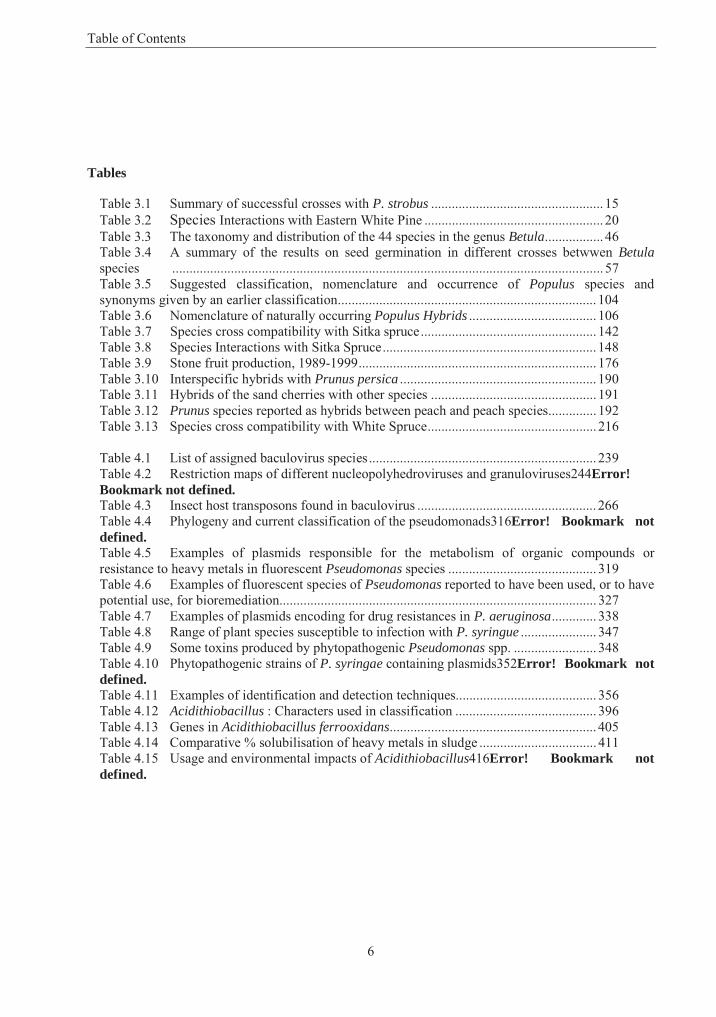
Table of Contents
6
Tables
Table 3.1 Summary of successful crosses with P. strobus .................................................. 15Table 3.2 Species Interactions with Eastern White Pine .................................................... 20Table 3.3 The taxonomy and distribution of the 44 species in the genus Betula................. 46Table 3.4 A summary of the results on seed germination in different crosses betwwen Betulaspecies ............................................................................................................................. 57Table 3.5 Suggested classification, nomenclature and occurrence of Populus species and synonyms given by an earlier classification........................................................................... 104Table 3.6 Nomenclature of naturally occurring Populus Hybrids ..................................... 106Table 3.7 Species cross compatibility with Sitka spruce................................................... 142Table 3.8 Species Interactions with Sitka Spruce.............................................................. 148Table 3.9 Stone fruit production, 1989-1999..................................................................... 176Table 3.10 Interspecific hybrids with Prunus persica ......................................................... 190Table 3.11 Hybrids of the sand cherries with other species ................................................ 191Table 3.12 Prunus species reported as hybrids between peach and peach species.............. 192Table 3.13 Species cross compatibility with White Spruce................................................. 216
Table 4.1 List of assigned baculovirus species.................................................................. 239Table 4.2 Restriction maps of different nucleopolyhedroviruses and granuloviruses244Error! Bookmark not defined.Table 4.3 Insect host transposons found in baculovirus .................................................... 266Table 4.4 Phylogeny and current classification of the pseudomonads316Error! Bookmark not defined.Table 4.5 Examples of plasmids responsible for the metabolism of organic compounds or resistance to heavy metals in fluorescent Pseudomonas species ........................................... 319Table 4.6 Examples of fluorescent species of Pseudomonas reported to have been used, or to have potential use, for bioremediation............................................................................................ 327Table 4.7 Examples of plasmids encoding for drug resistances in P. aeruginosa............. 338Table 4.8 Range of plant species susceptible to infection with P. syringue ...................... 347Table 4.9 Some toxins produced by phytopathogenic Pseudomonas spp. ........................ 348Table 4.10 Phytopathogenic strains of P. syringae containing plasmids352Error! Bookmark not defined.Table 4.11 Examples of identification and detection techniques......................................... 356Table 4.12 Acidithiobacillus : Characters used in classification ......................................... 396Table 4.13 Genes in Acidithiobacillus ferrooxidans............................................................ 405Table 4.14 Comparative % solubilisation of heavy metals in sludge .................................. 411Table 4.15 Usage and environmental impacts of Acidithiobacillus416Error! Bookmark not defined.
Table of Contents
7
Figures
Figure 3.1 The natural range of Eastern White Pine ......................................................... 12Figure 3.2 The leaf, scale, seed, stem, buds and female and male catkins of Betula pendula ......................................................................................................................... 47Figure 3.3 The natural distribution of Betula pendula ...................................................... 52Figure 3.4 Crossability of Populus species ..................................................................... 112Figure 3.5 The natural range of Sitka spruce .................................................................. 138Figure 3.6 The natural range of White Spruce................................................................ 208Figure 3.7 The reproductive cycle of White Spruce ....................................................... 210
Figure 4.1 Morphological characteristics of nucleopolyhedroviruses and granuloviruses ... ....................................................................................................................... 241

Part 3
CONSENSUS DOCUMENTS ON THE BIOLOGY OF TREES
10
SECTION 1
EASTERN WHITE PINE (PINUS STROBUS L.)
1. General Information
This consensus document addresses the biology of Eastern White Pine (Pinus strobus L.), referred to hereafter simply as Eastern White Pine (pin blanc in French Canada). Eastern White Pine is one of the most valuable tree species in eastern North America where its easily machined, uniform-textured wood is unsurpassed for doors, windows, panelling, mouldings and cabinet work (Mullins and McKnight, 1981; Farrar, 1995). The species played a major role in the settlement and economic development of New England and the Atlantic Provinces as England reserved all large Eastern White Pine suitable for masts under the "Broad Arrow" policy, starting in the late 1600's (Johnson, 1986). Eastern White Pine also responds well to nursery culture and is commonly used for reforestation, urban forestry and Christmas tree plantations.
The general biology of Eastern White Pine is described in the context of the species’ role in natural forests and its domestication in planted stands. Taxonomic and evolutionary relationships with other Pinusspecies are described. Reproductive biology is described with a focus on aspects of mating system, gene flow, seed production and natural stand establishment. The current knowledge of genetic variation within the species is reviewed, highlighting the importance of geographic variation patterns and the potential for improvement by means of recurrent selection breeding strategies. The tremendous biological diversity and the complexity of ecological interactions with higher and lower flora and fauna are discussed. While Eastern White Pine has been commonly planted within its natural range, the extent of reforestation has been limited by susceptibility to white pine weevil (Pissodes strobi) and blister rust (Cronartium ribicola).Domestication and operational breeding activities are also reviewed. Crossing with other related white pine species offers some promise of producing hybrids with increased resistance to both the weevil and blister rust. While white pine reforestation is currently based on seed propagation, vegetative propagation techniques are available and research continues into regeneration from somatic embryos.
Canada was the lead country in preparation of this document. It is intended for use by regulatory authorities and others who have responsibility for making assessments of transgenic plants proposed for commercialisation, and by those who are actively involved with genetic improvement and intensive management of this species.
2. Taxonomy and Natural Distribution
A. Taxonomy and nomenclature
The genus Pinus L. (family Pinaceae) is widely distributed throughout the Northern Hemisphere, from the arctic circle south to Guatemala, the West Indies, North Africa and Indonesia, with as many as 100 species being recognised (Krüssmann, 1985). The genus was first classified on evolutionary characteristics by Shaw (1914), and taxonomists have since followed his general separation of the genus into two groups: Haploxylon Koehne, and Diploxylon Koehne; commonly called the "soft" (or "white") and "hard" pines, respectively, based on the presence of one or two vascular bundles in the leaves. Shaw's original subdivision of these groups has been reworked by different authorities (e.g., Pilger, 1926; Duffield, 1952;
Section 1 – Eastern White Pine
11
de Ferré, 1965; Landry, 1974b, 1978), but botanists in recent years have generally recognised the classification described by Little and Critchfield (1969, 1986), who place Eastern White Pine, Pinus strobus L., within the subgenus Strobus Lemm. (equivalent to subgenus Haploxylon), section Strobus,subsection Strobi Loud. Also known as northern pine and, in parts of Europe, as Weymouth pine, after Lord Weymouth, the species nomenclature has remained virtually undisputed since the publication of the Species Plantarum (Linné, 1753), although Provancher later referred to it as Pinus alba Canadensis Prov. (Landry, 1974a).
Several horticultural forms have been named, although none are currently recognised with varietal status (Krüssmann, 1985). Only one variety has been commonly described, Pinus strobus L. var. chiapensis Martinez, the Chiapas white pine, occurring in the mountains of southern Mexico and Guatemala. While similar morphologically, it is physiologically quite different (Wright, 1970) and now generally recognised as a separate species, Pinus chiapensis (Martinez) Andresen (Griffiths, 1994; Perry, 1991).
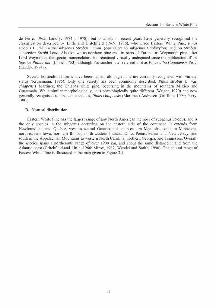
B. Natural distribution
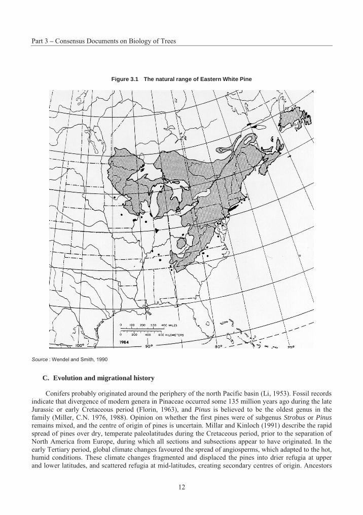
Eastern White Pine has the largest range of any North American member of subgenus Strobus, and is the only species in the subgenus occurring on the eastern side of the continent. It extends from Newfoundland and Quebec, west to central Ontario and south-eastern Manitoba, south to Minnesota, north-eastern Iowa, northern Illinois, north-western Indiana, Ohio, Pennsylvania, and New Jersey, and south in the Appalachian Mountains to western North Carolina, northern Georgia, and Tennessee. Overall, the species spans a north-south range of over 1900 km, and about the same distance inland from the Atlantic coast (Critchfield and Little, 1966; Mirov, 1967; Wendel and Smith, 1990). The natural range of Eastern White Pine is illustrated in the map given in Figure 3.1.
Part 3 – Consensus Documents on Biology of Trees
12
Figure 3.1 The natural range of Eastern White Pine
Source : Wendel and Smith, 1990
C. Evolution and migrational history
Conifers probably originated around the periphery of the north Pacific basin (Li, 1953). Fossil records indicate that divergence of modern genera in Pinaceae occurred some 135 million years ago during the late Jurassic or early Cretaceous period (Florin, 1963), and Pinus is believed to be the oldest genus in the family (Miller, C.N. 1976, 1988). Opinion on whether the first pines were of subgenus Strobus or Pinusremains mixed, and the centre of origin of pines is uncertain. Millar and Kinloch (1991) describe the rapid spread of pines over dry, temperate paleolatitudes during the Cretaceous period, prior to the separation of North America from Europe, during which all sections and subsections appear to have originated. In the early Tertiary period, global climate changes favoured the spread of angiosperms, which adapted to the hot, humid conditions. These climate changes fragmented and displaced the pines into drier refugia at upper and lower latitudes, and scattered refugia at mid-latitudes, creating secondary centres of origin. Ancestors
Section 1 – Eastern White Pine
13
of P. strobus and P. monticola were isolated in northern refugia from other species in section Strobi that were isolated in the south. The warm, tropical conditions changed rapidly at the end of the Eocene epoch and pines became re-established at middle latitudes. These abrupt changes in climate had drastic impacts on the gene structure of genetic variation of forest populations, with isolated populations continuing their short-term evolution (Critchfield, 1984).
3. Reproductive Biology
A. Reproductive development
Eastern White Pine is monoecious. Production of female strobili occurs as early as 4 years (Buckingham, 1963) while pollen production may not start for 10 to 20 years (Wright, 1970). As in other pines, development of the reproductive structures follows a 3-year cycle. Pollination occurs in the spring of the second year, with fertilisation delayed until the following spring, and seeds maturing in the fall of the third year (Owens and Blake, 1985). No other conifer genus has had its reproductive cycle described more often or more thoroughly, and Eastern White Pine was among the first pines to be studied in detail (Ferguson, 1901, 1904). Reproductive buds begin as axillary bud primordia within a complex long-shoot bud, consisting of a series of cataphylls initiated throughout the growing season. Many of the cataphylls support an axillary apex that first initiates a series of bud scales, then differentiates into a short (fascicular) shoot, seed or pollen cone, or lateral long shoot bud. Those axillary buds initiated at the base of the long shoot bud in the spring or early summer will differentiate into short-shoot or pollen-cone buds. Subsequent axillary buds differentiate only into short shoots. The distal axillary buds remain undetermined through winter dormancy of the long shoot bud, differentiating immediately the following spring into lateral long shoot or seed cone buds (Owston, 1969; Owens and Molder, 1977). While seed-cones generally develop on vigorous shoots in the upper portion of the crown, distribution of reproductive structures is often extremely variable.
Pollen development and meiosis does not occur until the spring of the second year as pollen cones resume their development. The ripening strobili turn light brown before releasing their pollen over a 1-week period. The seed cones also resume development in the spring, and are visible at the distal end of elongating long shoots. The developmental morphology of reproductive structures was well documented with colour photographs by Ho (1991). Wind-borne pollen grains landing on the receptive seed cone pass between the bracts and sift down to the surface of the micropylar arms, entering the micropyle by means of a pollen drop. The pollen germinates, but becomes dormant before the male gametes form (Owens and Molder, 1977). Fertilisation occurs about 13 months after pollination. Simple polyembryony in Eastern White Pine results from the fertilisation of 2 to 3 archegonia in each megagametophyte. The seed cones mature and seeds are dispersed in August or September of the same year (Krugman and Jenkinson, 1974).
B. Mating system and gene flow
Eastern White Pine is a wind-pollinated, monoecious species, and outcrossing is by far the most prevalent mating system, although there are relatively few detailed studies. Isozyme studies of populations in Québec indicated a high rate of outcrossing, with most loci in Hardy-Weinberg equilibrium (Beaulieu and Simon, 1994, 1995).
Gene flow in Pinus is mediated by very small pollen grains, 40-60 µm at their widest point (Eisenhut, 1961), whose two air sacs and low density make them well-adapted for aerial transport (Di-Giovanni and Kevan, 1991). Various studies of pollen dispersal in conifers indicate that over 90% of the pollen comes to rest less than 100 m from the source (Wright, 1976). Nevertheless, conifer pollen may remain viable for several days and a substantial quantity may travel great distances (Lindgren et al., 1995; Lindgren and Lindgren, 1996). Gregory (1973) cites reports that pollen of Pinus and Picea may travel as far as 600 to
Part 3 – Consensus Documents on Biology of Trees
14
1 000 km, and several authors have concluded that isolation distances of less than 1 km often have little impact on contamination rates in conifer seed orchards (see review by Di-Giovanni and Kevan, 1991).
C. Seed production
Eastern White Pine normally begins seed production at 5 to 10 years of age (Fowells, 1965; Sargent, 1965), although little pollen is produced during the early years of flowering (Wendel and Smith, 1990). The interval between heavy seed crops is usually 3 to 10 years (Krugman and Jenkinson, 1974; Wendel and Smith, 1990), becoming less frequent as trees become over mature (Horton and Bedell, 1960). A study in Germany recorded seed production as high as 73 kg/ha in a 90-year-old stand (Messer, 1956), while in Maine, a stand considered to be intermediate in density with a basal area of 28 m2/ha produced over 4.4 million seeds per hectare in a "bumper" year (Graber, 1970). This corresponds to 69kg/ha seed.
Initiation of seed dispersal is weather and site dependent, and may be delayed by cool, moist weather. Most of the seeds are dispersed in the fall during a 4 to 8 week period (Horton and Bedell, 1960; Graber, 1970). The seeds are mature when cone moisture content decreases below 200% on a dry-weight basis, but cone specific gravity is not a reliable indicator of maturity (Barnett, 1988). A short "artificial ripening" period can increase yield and quality of seed from immature cones (Bonner, 1986, 1991; Barnett, 1988).
The seeds are winged and dispersal distances depend greatly on local and prevailing wind patterns (Rudis et al., 1978). The seeds may travel more than 60 m within a closed stand, and over 200 m in the open (Wilson and McQuilckin, 1963), although most of the seed will fall within a distance equivalent to the height of the seed tree (Horton and Bedell, 1960). The seeds themselves are smaller than those of most other "soft" pines, but similar to those of P. monticola, with average cleaned seed weight of about 17 g/1000 seeds (Krugman and Jenkinson , 1974).
D. Natural regeneration
Eastern White Pine seeds exhibit varying degrees of embryo dormancy that may be broken by exposure to low temperatures under moist conditions, i.e., cold stratification (Krugman and Jenkinson, 1974; Nelson et al., 1980; Mittal et al., 1987; Downie and Bergsten, 1991). The recommended treatment for nursery sowing is stratification for 60 days at 1 to 5° C (Krugman and Jenkinson, 1974). Under natural conditions, over-winter stratification on the forest floor breaks seed dormancy and germination of most seeds occurs in late spring of the following year (Stiell, 1985).
Germination is epigeal. Moist mineral soil, polytrichum moss, and shortgrass cover of light to medium density are favourable seedbeds. Establishment on less favourable seedbeds, such as pine litter and lichen, will occur under partial shade and/or surface. Shelterwood harvesting systems provide good protection during initial establishment with sufficient light for subsequent growth of young stands (Wilson and McQuilckin, 1963; Corbett, 1994). Optimum conditions are provided when moist mineral seedbeds have greater than 20% of full sun, but where partial shade reduces surface temperatures and provides better moisture conditions (Lancaster and Leak, 1978). Low seedling densities are associated with competition from broad-leaved shrubs, herbaceous vegetation, tolerant conifer species and feather mosses (Carleton et al., 1996). White pine regeneration is usually associated with its proportion in the overstorey (Kittredge and Ashton, 1990), and under old-growth conditions is likely to become at least partially uneven-aged and self replacing, facilitated by local disturbances and continuous recruitment (Quinby, 1991; Ziegler, 1995). Older trees have increased their ability to recover from long periods of suppression (Abrams and Orwig, 1996).
Section 1 – Eastern White Pine
15
E. Vegetative reproduction in nature
Eastern White Pine does not regenerate vegetatively under natural conditions (Wendel and Smith, 1990).
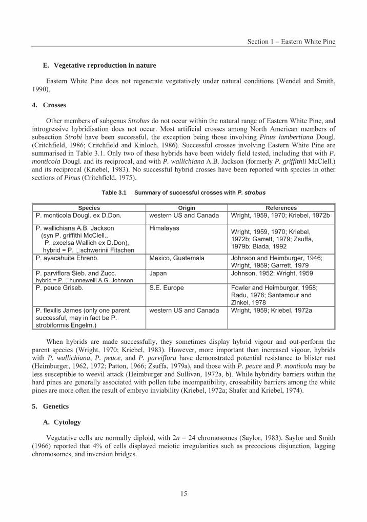
4. Crosses
Other members of subgenus Strobus do not occur within the natural range of Eastern White Pine, and introgressive hybridisation does not occur. Most artificial crosses among North American members of subsection Strobi have been successful, the exception being those involving Pinus lambertiana Dougl. (Critchfield, 1986; Critchfield and Kinloch, 1986). Successful crosses involving Eastern White Pine are summarised in Table 3.1. Only two of these hybrids have been widely field tested, including that with P. monticola Dougl. and its reciprocal, and with P. wallichiana A.B. Jackson (formerly P. griffithii McClell.) and its reciprocal (Kriebel, 1983). No successful hybrid crosses have been reported with species in other sections of Pinus (Critchfield, 1975).
Table 3.1 Summary of successful crosses with P. strobus
Species Origin References P. monticola Dougl. ex D.Don. western US and Canada Wright, 1959, 1970; Kriebel, 1972b
P. wallichiana A.B. Jackson (syn P. griffithi McClell., P. excelsa Wallich ex D.Don), hybrid = P. �schwerinii Fitschen
Himalayas Wright, 1959, 1970; Kriebel, 1972b; Garrett, 1979; Zsuffa, 1979b; Blada, 1992
P. ayacahuite Ehrenb. Mexico, Guatemala Johnson and Heimburger, 1946; Wright, 1959; Garrett, 1979
P. parviflora Sieb. and Zucc. hybrid = P. �hunnewelli A.G. Johnson
Japan Johnson, 1952; Wright, 1959
P. peuce Griseb. S.E. Europe Fowler and Heimburger, 1958; Radu, 1976; Santamour and Zinkel, 1978
P. flexilis James (only one parent successful, may in fact be P. strobiformis Engelm.)
western US and Canada Wright, 1959; Kriebel, 1972a
When hybrids are made successfully, they sometimes display hybrid vigour and out-perform the parent species (Wright, 1970; Kriebel, 1983). However, more important than increased vigour, hybrids with P. wallichiana, P. peuce, and P. parviflora have demonstrated potential resistance to blister rust (Heimburger, 1962, 1972; Patton, 1966; Zsuffa, 1979a), and those with P. peuce and P. monticola may be less susceptible to weevil attack (Heimburger and Sullivan, 1972a, b). While hybridity barriers within the hard pines are generally associated with pollen tube incompatibility, crossability barriers among the white pines are more often the result of embryo inviability (Kriebel, 1972a; Shafer and Kriebel, 1974).
5. Genetics
A. Cytology
Vegetative cells are normally diploid, with 2n = 24 chromosomes (Saylor, 1983). Saylor and Smith (1966) reported that 4% of cells displayed meiotic irregularities such as precocious disjunction, lagging chromosomes, and inversion bridges.
Part 3 – Consensus Documents on Biology of Trees
16
B. Genetic variation
Population-level variability
While seed source testing of Eastern White Pine began in the United States in 1937 (Pauley et al.,1955), provenance tests with range-wide sampling did not begin until the mid-1950's. Around this time, the USDA Forest Service initiated a large provenance test, in which 30 seed collections representing all parts of the natural range were established by co-operators in 13 test plantations in the United States and 2 in Ontario (Sluder, 1963; Wright et al., 1963; Funk, 1965; Fowler and Heimburger, 1969b; King and Nienstaedt, 1969; Genys, 1977). Shortly after, another provenance test involving more seedlots on fewer test sites was started by the University of Maryland (Genys, 1968; Genys et al,. 1978). Encouraging early results from these tests, indicating the superiority of sources from the South Appalachians, led to intensive testing of these sources under the leadership of Michigan State University (Roth and Carson, 1976; Wendel and Cech ,1976; Wright et al., 1976; Gall and Thor ,1977).
While correlations with latitude have sometimes been noted on a range-wide basis (Genys, 1987, 1991), relative differences in height and diameter between northern and southern sources diminish somewhat with age (Demeritt and Kettlewood, 1976; Demeritt and Garrett, 1996). Clinal patterns are often less distinct over shorter distances with the presence of non-clinal adapted ecotypes (Genys, 1968; Garrett et al., 1973; Thor, 1975; des Bordes and Thor, 1979; Funk, 1979). In Nebraska, seed sources from the southern Appalachians demonstrated correlations with latitude for needle length and reproductive phenology, a weak geographic pattern for variation in height, and none for survival (Sprackling and Read, 1976; Van Haverbeke, 1988). Ryu and Eckert (1983) investigated the genetic structure of 27 of these provenances for eight foliar enzymes coded by 12 loci and found four clusters of provenances, three of which may be representative of populations adapted to differing geographic and climatic conditions. The results of this study support the indication of ecotypic variation among three provenances in the southern Appalachians for growth performance and physiological variables, and suggest that these areas may have been isolated refugia during glaciation. Elsewhere in the northern part of the range, sources from the Atlantic coast outperformed those from further inland, while some exceptional sources originated from as far south as Georgia and Tennessee (Zsuffa, 1975; Abubaker and Zsuffa, 1991).
Southern provenances have heavier seeds (Genys, 1968), require longer periods of stratification before germinating (Fowler and Dwight, 1964; Graber, 1965) and longer chilling periods to break bud dormancy (Mergen, 1963), set bud later (Santamour, 1960) and are less cold hardy (Maronek and Flint, 1974). Wood specific gravity was negatively correlated with height and diameter, but differences among sources were small (Lee, 1974; Gilmore and Jokela, 1978; Olson et al., 1981). No variation could be detected for foliar monoterpene content, and no geographic pattern was evident for variation in cortical monoterpenes (Gilmore and Jokela, 1979).
Provenance tests have shown some variation in susceptibility to white pine weevil, but give little indication that resistant populations can be identified (Garrett, 1972, 1973; Connola and Beinkafner, 1976; Wilkinson, 1983b). Selective thinning of susceptible parents (dominant "wolf" trees) from a stand can increase the level of resistance in the progeny generation, and taller families tend to be more weevil resistant (Ledig and Smith, 1981). Although there is ample evidence of genetic control of susceptibility to weevil, the actual mechanism(s) of resistance remains uncertain.
Individual-level variability
While variation among provenances is important in determining the risks and benefits of transferring seed sources, genetic improvement from mass selection relies primarily on variation within-populations as the source of genetic gains. The partitioning of genetic variance among and within populations is greatly
Section 1 – Eastern White Pine
17
influenced by the range of adaptive variation sampled by the tested provenances and the age at which the test material is assessed. Range-wide and regional studies have typically demonstrated strong heritabilities, sufficient to predict moderate to high genetic gains, although heritability tends to be lower for older material (Thor, 1975; Adams and Jolly, 1978; des Bordes and Thor, 1979; Olson et al., 1981). Hierarchical sampling of populations over a more limited range in Québec and Ontario showed that population differences were greatest for allozyme markers, where 98% of the variation was within populations (Beaulieu et al., 1996). Growth traits, on the other hand, demonstrated variation within stands to be about half as great as that among populations (Li, P. et al., 1997). Individual heritability for height declined from 0.547 at age 4 in the nursery, to 0.187 at age 10 in the field (Beaulieu et al., 1996). In an incomplete diallele cross experiment among individuals of a local provenance, Kriebel et al. (1972) found that narrow-sense heritability for height growth declined from 0.59 at age 1 to 0.16 at age 3, and that while dominance effects were small, maternal effects were rather large. By age 13, it was still possible to achieve substantial gains by family selection (Kriebel, 1978).
Significant genotype-environment interactions have been reported in Eastern White Pine, but the magnitude of the interaction variance is generally low (less than 2%). Genetic correlations between sites tend to be high, indicating that family ranks are stable across sites (des Bordes and Thor, 1979; Beaulieu et al., 1996; Demeritt and Garrett, 1996).
The search for weevil-resistance has always been a driving force behind genetic testing in Eastern White Pine (Pauley et al., 1955; Wright and Gabriel, 1959). Early studies indicated that selection for weevil resistance might be done indirectly by assessment of bark thickness (Kriebel, 1954; Gerhold, 1962, 1966) and/or leader morphology (Stroh, 1964, 1965), but when these are corrected for tree size, they appear to be of little value for effective selection (Wilkinson, 1983a, 1984). Other studies have identified that concentrations of various cortical oleoresin compounds are correlated with weevil susceptibility, but even these criteria leave much of the variation in weevil susceptibility unexplained (van Buijtenen and Santamour, 1972; Santamour and Zinkel, 1976, 1978; Bridgen et al., 1979; Wilkinson, 1979, 1980, 1984, 1985).
C. Inbreeding depression and genetic load
Eastern White Pine is an outcrossing species that carries a fairly heavy load of deleterious recessive genes. Individuals are generally self-compatible, so that this genetic load is revealed by self-fertilisation (Fowler, 1965a; Fowler and Heimburger, 1969a). Although there is no reduction in numbers of filled seeds after selfing (Fowler, 1965b), selfed seedlings may be stunted, slow growing, chlorophyll-deficient and deformed (Johnson, 1945; Patton and Riker, 1958a; Fowler, 1965b). Simple polyembryony in Eastern White Pine results from 2 to 3 archegonia in each megagametophyte. As only one embryo normally germinates from the mature seed, it is likely that competition during seed development eliminates many weaker embryos, including those resulting from self-fertilisation (Willson and Burley, 1983). An isozyme study of populations in Quebec demonstrated a high outcrossing rate, with few loci deviating from Hardy-Weinberg equilibrium (Beaulieu and Simon, 1995). This study found evidence of family structure, with greater inbreeding in the filial than in the parental population, although few of the inbred genotypes were expected to reach reproductive age, due to natural selection.
D. Breeding programs
Eastern White Pine has been a candidate for tree breeding efforts throughout its native range. In the northern part of its range, throughout eastern Canada, the north-eastern US and the Lake States, planting programs have been limited by susceptibility to weevil and rust, so that seed orchards exist throughout this region (Zsuffa, 1985, 1986; Garrett, 1986; Miller, 1987; Eckert and Kuser, 1988; Lamontagne, 1992; Nielsen et al., 1995; Smith et al., 1997; pers. comm. R. Stine, Minnesota Tree Improvement Cooperative)
Part 3 – Consensus Documents on Biology of Trees
18
and, the level of effort reflects the restricted size of planting programmes. Pests are less of a problem for breeding programs in the Central States, where selection and hybrid breeding can focus on vigour (Kriebel, 1983). Outside of the natural range in Europe, selection within southern Appalachian provenances and crossing with other white pines, such as Pinus wallichiana, are used to develop fast-growing, rust-resistant hybrids (Kriebel, 1983).
Most seed orchards currently in production were established by grafting cuttings from plus-trees, and their establishment in cultivated field environments. Grafting success is usually very high. Flowering in field orchards can be enhanced by means of cultural treatments such as fertilisation (Hocker, 1962; Stephens, 1964). Flowering of young white pine grafts can also be stimulated by means of various cultural treatments, particularly those involving gibberellin A4/7, and this has facilitated the turnover of breeding cycles (Ho and Schnekenburger, 1992; Ho and Eng ,1995).
E. Conservation of genetic resources
Domestication of a key species such as Eastern White Pine can influence diversity of genetic resources (1) indirectly, by the method of seed collection, extraction, and storage, and by nursery and plantation culture; and (2) directly, by intentional selection to increase the frequency of genes for desirable traits (Morgenstern, 1996). The inadvertent loss of genes by natural processes and human activity can have negative consequences on the adaptability of populations and the potential for future gains from breeding.
A long history of exploitation has resulted in white pine forest fragmentation and reduction of population sizes, particularly at the northern limits of the species range (Buchert, 1994; Buchert et al.,1997). Throughout most of the range of white pine, in situ conservation of genetic resources is practised by protection of ecological reserves, special areas, and parks (Pollard, 1995), and integrated with domestication activities that control the movement of seed, active management of existing stands to maintain biological diversity, and protection of isolated, small populations (Mosseler, 1995; Nieman et al., 1995).
Ex situ conservation, by cryopreservation of germplasm, by off-site maintenance of populations in arboreta, seed orchards and clone banks, and by multi-population breeding strategies (Eriksson et al., 1993; Namkoong, 1995), has been practised to a much lesser extent, although many provenances and families of Eastern White Pine are now represented in field tests and seed bank collections (Plourde et al., 1995). Such "active" forms of gene management must be accelerated in preparation for response to rapid environmental and climate changes (Ledig and Kitzmiller, 1992).
6. Ecology and Associated Species
Much of the information in this section originates from the excellent monograph on silvics of the species by Wilson and McQuilkin (1963). Other citations are given when appropriate when specific information is attributable to other sources.
A. Habitat
Climate
Eastern White Pine's natural range is cool and humid. July average temperatures are between 18 to 25° C, and annual precipitation varies from about 510 mm in northern Minnesota to 2030 mm in north-western Georgia, with at least half occurring between April and November. Average snowfall varies from less than 15 cm in the southern portion of the range to over 250 cm in the northeast (Wendel and Smith, 1990). There is a surplus of moisture in all seasons.
Section 1 – Eastern White Pine
19
Soils and site type
Eastern White Pine grows on a wide variety of soils throughout its range, from dry sands and rocky ridges, to sphagnum bogs, although it grows best on moist sandy or loamy soils. Soils within the range are derived from granites, gneisses, schists, sandstones, and, to a lesser extent, phyllites, slates, shales and limestones. Eastern White Pine competes best on medium-textured, well-drained soils of moderate site quality, with surface pH between 4.0 and 7.5, and which are not sufficiently rich to support strong hardwood competition, or where competition is reduced during the establishment period, such as on old fields, burnt or blow-down areas (Horton and Bedell, 1960; Mader, 1986).
In the northeast portion of the range, Eastern White Pine generally occurs below 450 m above sea level, whereas in Pennsylvania, elevations vary between 150 and 600 m. In the southern Appalachians, stands generally occurs between 370 and 1070 m. Except in Pennsylvania and the southern Appalachians where stands are found on northerly aspects or in the shelter of stream bottoms. White pine sites are not generally restricted by slope or aspect.
B. Synecology and associated species
Eastern White Pine may form pure stands or occur as a major stand component of several stand types in association with other conifers and hardwoods such as: red pine (Pinus resinosa), balsam fir (Abies balsamea), black spruce (Picea mariana), White Spruce (P. glauca), red oak (Quercus rubra), sugar maple (Acer saccharum), red maple (Acer rubrum), hemlock (Tsuga canadensis), and chestnut oak (Quercus prinus). Eastern White Pine may also be found as a lesser stand component with jack pine (Pinus banksiana), pitch pine (P. rigida), shortleaf pine (P. echinata), sweet birch (Betula lenta), trembling aspen (Populus tremuloides), large-tooth aspen (P. grandidentata), black cherry (Prunus serotina), black oak (Quercus velutina), white oak (Quercus alba), and various hickories (Carya spp.) (Horton and Bedell, 1960; Eyre, 1980). The occurrence of associations depends on both site conditions and history of disturbance (Stiell, 1985).
Pure stands of Eastern White Pine usually support sparse cover of understory vegetation, but many species may be found under mixed stands, particularly those associated with hardwood associates. On drier sites, ground vegetation may consist of one or more species of blueberries (Vaccinium spp.), teaberry (Gaultheria procumbens), dwarf bush-honeysuckle (Diervilla lonicera), sweetfern (Comptonia peregrina)bracken fern (Pteridium aquilinum), clubmoss (Lycopodium spp.), and broom sedge (Andropogon virginicus). Richer, moist sites will often support ground cover of woodsorrels (Oxalis spp.), partridgeberry (Mitchella repens), wild sarsaparilla (Aralia nudicaulis), jack-in-the-pulpit (Arisaema spp.), and hay-scented fern (Dennstaedtia punctilobula). Intermediate sites may have varying amounts of the above species, together with dogwoods (Cornus spp.) and false lily-of-the-valley (Maianthemum canadense).
C. Competition and stand structure
Eastern White Pine is distributed over a larger area than any other North American white pine, and has demonstrated its capacity to grow and compete under a wide variety of environmental conditions (Stiell, 1978, 1985). While it is a long-lived successional species and may be a component of climax forest types, it is also well-known as a pioneering species on old fields in New England. Eastern White Pine is considered intermediate in its tolerance to shade, somewhat less tolerant than eastern spruces and more tolerant than its pine associates (Daniel et al., 1979). Vegetative competition for light and soil moisture is critical during seedling establishment, and remains important well into the life of the stand. Sites that have a high capability for productivity for pine tend to have greater competition. Competition problems are most severe on heavier, moist, rich soils, where Eastern White Pine will perform well, only if natural disturbance, such as fire, or silvicultural site treatments allow the pine to become established well ahead of
Part 3 – Consensus Documents on Biology of Trees
20
the hardwoods that normally occupy such sites (Horton and Bedell, 1960; Little et al., 1973; Stiell, 1985; Chapeskie et al., 1989).
D. Ecosystem dynamics
Several abiotic factors also interact with Eastern White Pine in forest ecosystems. While older trees have thick, heat-resistant bark, the thinner bark on exposed roots and younger stems is sensitive to fire. Even light fires can have a detrimental impact on seed supply, but may also reduce hardwood competition and leave a seedbed that is more conducive to the establishment of new germinants. Frost heaving can cause severe damage, particularly to container seedlings planted on finer-textured soils. Eastern White Pine is relatively wind firm, but may suffer storm breakage if the stand has been recently thinned. While it is widely held that Eastern White Pine is sensitive to ozone and sulphur dioxide pollution (Gerhold, 1977), recent data in the literature are somewhat contradictory and suggest that injury and growth losses may be strongly genotype and site dependant (Houston and Stairs, 1973; Genys and Heggestad, 1978, 1983; Townsend and Dochinger, 1982; Usher and Williams, 1982; Yang et al., 1982, 1983; Eberhardt et al.,1988; Rezabek et al. , 1989; Bartholomay et al., 1997; Hogsett et al., 1997).
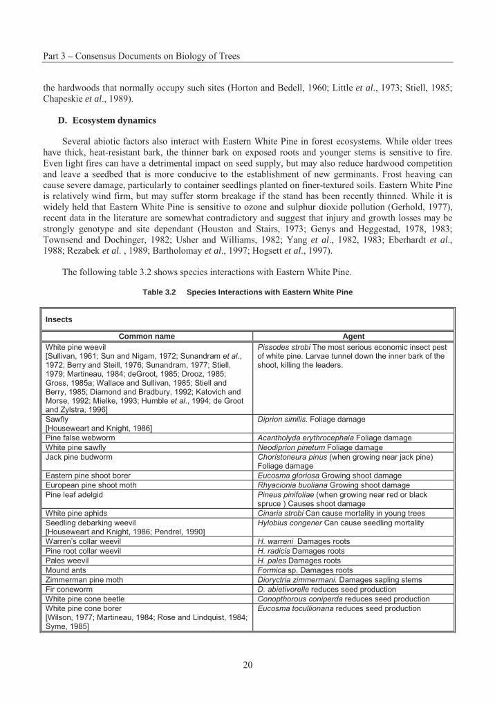
The following table 3.2 shows species interactions with Eastern White Pine.
Table 3.2 Species Interactions with Eastern White Pine
Insects
Common name Agent White pine weevil [Sullivan, 1961; Sun and Nigam, 1972; Sunandram et al., 1972; Berry and Steill, 1976; Sunandram, 1977; Stiell, 1979; Martineau, 1984; deGroot, 1985; Drooz, 1985; Gross, 1985a; Wallace and Sullivan, 1985; Stiell and Berry, 1985; Diamond and Bradbury, 1992; Katovich and Morse, 1992; Mielke, 1993; Humble et al., 1994; de Groot and Zylstra, 1996]
Pissodes strobi The most serious economic insect pest of white pine. Larvae tunnel down the inner bark of the shoot, killing the leaders.
Sawfly [Houseweart and Knight, 1986]
Diprion similis. Foliage damage
Pine false webworm Acantholyda erythrocephala Foliage damage
White pine sawfly Neodiprion pinetum Foliage damage
Jack pine budworm Choristoneura pinus (when growing near jack pine) Foliage damage
Eastern pine shoot borer Eucosma gloriosa Growing shoot damage
European pine shoot moth Rhyacionia buoliana Growing shoot damage
Pine leaf adelgid Pineus pinifoliae (when growing near red or black spruce ) Causes shoot damage
White pine aphids Cinaria strobi Can cause mortality in young trees
Seedling debarking weevil [Houseweart and Knight, 1986; Pendrel, 1990]
Hylobius congener Can cause seedling mortality
Warren’s collar weevil H. warreni Damages roots
Pine root collar weevil H. radicis Damages roots
Pales weevil H. pales Damages roots
Mound ants Formica sp. Damages roots
Zimmerman pine moth Dioryctria zimmermani. Damages sapling stems
Fir coneworm D. abietivorelle reduces seed production
White pine cone beetle Conopthorous coniperda reduces seed production
White pine cone borer [Wilson, 1977; Martineau, 1984; Rose and Lindquist, 1984; Syme, 1985]
Eucosma tocullionana reduces seed production
Section 1 – Eastern White Pine
21
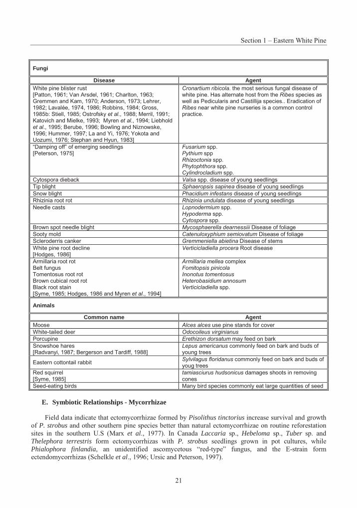
Fungi
Disease Agent White pine blister rust [Patton, 1961; Van Arsdel, 1961; Charlton, 1963; Gremmen and Kam, 1970; Anderson, 1973; Lehrer, 1982; Lavalée, 1974, 1986; Robbins, 1984; Gross, 1985b: Stiell, 1985; Ostrofsky et al., 1988; Merril, 1991; Katovich and Mielke, 1993; Myren et al., 1994; Liebhold et al., 1995; Berube, 1996; Bowling and Niznowske, 1996; Hummer, 1997; La and Yi, 1976; Yokota and Uozumi, 1976; Stephan and Hyun, 1983]
Cronartium ribicola. the most serious fungal disease of white pine. Has alternate host from the Ribes species as well as Pedicularis and Castillija species.. Eradication of Ribes near white pine nurseries is a common control practice.
“Damping off” of emerging seedlings [Peterson, 1975]
Fusarium spp. Pythium sppRhizoctonia spp.Phytophthora spp. Cylindrocladium spp.
Cytospora dieback Valsa spp. disease of young seedlings
Tip blight Sphaeropsis sapinea disease of young seedlings
Snow blight Phacidium infestans disease of young seedlings
Rhizinia root rot Rhizinia undulata disease of young seedlings
Needle casts Lopnodermium spp. Hypoderma spp. Cytospora spp.
Brown spot needle blight Mycosphaerella dearnessiii Disease of foliage
Sooty mold Catenuloxyphium semiovatum Disease of foliage
Scleroderris canker Gremmeniella abietina Disease of stems
White pine root decline [Hodges, 1986]
Verticicladiella procera Root disease
Armillaria root rot Belt fungus Tomentosus root rot Brown cubical root rot Black root stain [Syme, 1985; Hodges, 1986 and Myren et al., 1994]
Armillaria mellea complex Fomitopsis pinicola Inonotus tomentosus Heterobasidium annosum Verticicladiella spp.
Animals
Common name Agent Moose Alces alces use pine stands for cover
White-tailed deer Odocoileus virginianus
Porcupine Erethizon dorsatum may feed on bark
Snowshoe hares [Radvanyi, 1987; Bergerson and Tardiff, 1988]
Lepus americanus commonly feed on bark and buds of young trees
Eastern cottontail rabbit Sylvilagus floridanus commonly feed on bark and buds of youg trees
Red squirrel [Syme, 1985]
tamiasciurus hudsonicus damages shoots in removing cones
Seed-eating birds Many bird species commonly eat large quantities of seed
E. Symbiotic Relationships - Mycorrhizae
Field data indicate that ectomycorrhizae formed by Pisolithus tinctorius increase survival and growth of P. strobus and other southern pine species better than natural ectomycorrhizae on routine reforestation sites in the southern U.S (Marx et al., 1977). In Canada Laccaria sp., Hebeloma sp., Tuber sp. and Thelephora terrestris form ectomycorrhizas with P. strobus seedlings grown in pot cultures, while Phialophora finlandia, an unidentified ascomycetous “red-type” fungus, and the E-strain form ectendomycorrhizas (Schelkle et al., 1996; Ursic and Peterson, 1997).
Part 3 – Consensus Documents on Biology of Trees
22
Some ectomycorrhizal fungi can suppress root-rotting pathogens of conifers. A study of natural mycorrhizal colonisation and frequency of root rot on Eastern White Pine seedlings at a southern Canadian nursery revealed a negative correlation between T. terrestris and root rot. This suggested that the association of this ectomycorrhizal fungus with P. strobus roots might have some antipathogenic effects (Ursic et al., 1997). Additionally, removal of the basidiome of the ectomycorrizal fungus Laccaria bicolorassociated with container-grown Eastern White Pine seedlings induces a very rapid decrease in both net photosynthesis and stomatal conductance of the host plant (Lamhamedi et al., 1994).
7. Domestication
Eastern White Pine has been an attractive species for planting within its range, with up to 40 million seedlings shipped yearly for fibre production and Christmas trees (Eckert and Kuser, 1988). The species has also been used for shelter-belts and urban plantings, and has been used on a small scale in some European countries. Despite its very high timber value, management difficulties with control of white pine weevil and blister rust in planted stands have discouraged its use. Eastern White Pine is thus a rather minor reforestation species, particularly in the northern parts of its range in Canada, where annual nursery shipments in Ontario, Quebec and the Maritimes are now well below 5 million. Nevertheless, the potential value of white pine planting and breeding is well recognised, and tree improvement programs for the species are maintained at some level throughout most of its range.
A. Deployment of reforestation materials
White pine has a long history as a species for reforestation, and nursery production techniques are well-established. In the early years, most planting stock were produced as bareroot seedlings (Coons, 1978), with 2+0 shipped from southern nurseries and 3+0 in the north, although 2+2 transplants have demonstrated superior performance in the field (Mullin and Howard, 1973, Mullin and Christl, 1982). Following developments in nursery technology, Eastern White Pine is now commonly produced from seed in containerised systems, in soil-less growing media. A variety of containers are used and stock is raised in both heated and unheated greenhouse structures. Cultural techniques have become highly sophisticated, ensuring that high-quality planting stock can be produced reliably and efficiently (Landis et al., 1989, 1990a, b, 1992).
Eastern White Pine planting stock can also be produced by means of vegetative propagation. Much of the research in this area has been motivated by possible clonal deployment of individual genotypes with putative resistance to white pine weevil and blister rust. While older trees are often difficult to propagate using long-shoot cuttings, those from 2- to 3-year-old seedlings have long been known to root easily (Deuber, 1942; Patton and Riker, 1958b; Zsuffa, 1973; Kiang et al., 1974; Kiang and Garrett, 1975; Struve and Blazich, 1982). Propagation is also possible using fascicular shoots (Struve and Blazich, 1980, 1984). Growth and performance of rooted cuttings are comparable to planting stock raised from seed (Struve etal., 1984; Struve and McKeand, 1990).
Clonal propagation of Eastern White Pine can also be achieved through micropropagation of juvenile explant cultures derived from cotyledons, epicotyls and hypocotyls (Kaul, 1987, 1990; Webb et al., 1988). Techniques for the initiation of somatic embryos are also available, although whole plants have not yet been successfully recovered from these cultures (Becwar et al., 1988; Finer et al., 1989).
Some successful trials have demonstrated the potential of direct seeding as a regeneration technique for Eastern White Pine (Graber and Thompson, 1969; Horton and Wang, 1969; Graber, 1988), but stocking is often irregular (Torbet et al,. 1995). Operational use has generally been regarded as a failure and is not recommended (Waldron, 1974). Feeding losses to small mammals can be over 80%, unless the seeds are covered with soil at time of sowing (Graber, 1969).
Section 1 – Eastern White Pine
23
B. Provenance transfer
Local seed sources are often not the preferred provenance for planting, and northerly transfers are often beneficial, except in the extreme. Sources from the southern Appalachians perform well in all but the most northerly locations, with high volume production and reduced branchiness (Sluder, 1963; Funk, 1971, 1979; Sluder and Dorman, 1971; Funk et al., 1975; Wendel and Cech, 1976; Wright et al., 1976, 1979; Kriebel, 1978; Williams and Funk, 1978; Funk and Jokela, 1979). However, faster-growing southern sources are not sufficiently hardy to thrive in the harsher continental climates above 41°N (Fowler and Heimburger, 1969b; King and Nienstaedt, 1969; Jeffers, 1977). The use of seed zone controls to limit the transfers within regions of adaptation have been recommended for the northern part of the species range in Québec (Li et al., 1997).
Tests in Australia indicated that the best provenances are from the southern part of the natural range, although none are as productive are Pinus radiata (Matheson, 1977; Wright et al., 1979). In the Lower Saxony region of Germany, Appalachian Mountain sources below 39°N perform consistently well, while those from north of 45° perform poorly (Stephan, 1974; Genys et al., 1978).
In most of Europe, North American pines are considered to be fast growing tree species. In Romania, Pinus strobus is the second most productive species after Douglas fir, and has the least variation in annual radial increment and the lowest wood specific gravity of any commercial species. It is recommended on rotations of 40-60 years for pulpwood and 60-80 years for saw timber (Radu and Radu, 1972). In contrast, despite the extensive introduction and promising performance of P. strobus in Bohemia and Moravia, its wood has been grossly underrated by the woodworking industry, largely as a result of premature felling (Vytiskova, 1970). Of the 20 exotic Pines (9 from North America) growing in the central chernozem region of south central Russia, P. strobus has the fastest growth rate. However, exotic pines grown in Russia are significantly inferior in growth rate and yield to the local P. sylvestris (Lutkin et al., 1974). In the Lower Saxony region of the former German Federal Republic, P. strobus is not recommended for pure stands, partly because of the poor price paid for its timber and the unsaleability of thinnings; however, because of its fast growth, pleasing appearance, windfirmness, hardiness and general adaptability, it is strongly recommended for mixtures and particularly for the rehabilitation of recreation forests (Schumacher, 1974). As well, in provenance tests established in 1960 in Lower Saxony, growth of the best provenances of P. strobus was comparable or superior to that of local P. sylvestris, contrary to the situation in Russia (Stephan, 1981). Additionally, at two sites in Lower Saxony, differences between a rangewide sample of North American provenances were observed in height growth and mortality and attack by Chronartium ribicola (Stephan, 1974). P. strobus is recommended for wet or periodically waterlogged sites in the lowlands and hills of medium to low fertility in the former German Democratic Republic, especially those of extreme frost hazard (Thomasius and Hartig, 1979).
8. Summary
Eastern White Pine is one of the most important tree species in eastern North America. It has the largest range of any North American species in subsection Strobus, the "white pines", and is the only representative on the eastern side of the continent. It is an outcrossing, wind-pollinated species that can transfer genes rapidly to neighbouring populations and to other related species. Eastern White Pine is regarded as intermediate in its tolerance to shade, and natural regeneration is favoured by silvicultural systems that encourage partial shade during establishment and initial development.
Eastern White Pine exhibits clinal variation patterns, generally correlated with latitude, although local seed sources are often not the best performers. Heritability estimates are moderately high at young ages and, while typically decreasing at older ages, are sufficient to predict considerable gains from recurrent
Part 3 – Consensus Documents on Biology of Trees
24
selection. Significant genotype-environment interactions have been reported, but family ranks are generally stable across environments.
Best production is on medium-textured, well-drained soils, in cool, humid areas. White pine can occur as pure stands, or in mixture with several other conifer and hardwood associates, depending on site conditions and history of disturbance. It is a long-lived, successional species, but can be an aggressive pioneer on old fields. The white pine weevil and white pine blister are serious pests and are the major challenge for management of both natural and planted populations.
Eastern White Pine is well-suited to artificial regeneration and it has a long history as a planted species throughout its natural range, both in forestry and urban applications. Tree breeding efforts have been targeted primarily at selection and interspecific hybridisation, in an attempt to produce varieties with resistance to the weevil and blister rust. Management difficulties have limited planting of Eastern White Pine, particularly in the north of its range, although seed orchards are maintained in all regions. Meanwhile, a long history of economic exploitation has resulted in fragmentation and reduction of population sizes in some areas, making genetic conservation of this species a growing concern.
Section 1 – Eastern White Pine
25
REFERENCES
Abbott, H.G. 1961. White pine seed consumption by small animals. J. For. 59(3): 197-201.
Abrams, M.D., and Orwig, D.A. 1996. A 300-year history of disturbance and canopy recruitment for co-occurring white pine and hemlock on the Allegheny Plateau, USA. Ecology, 84: 353-363.
Abubaker, H.I., and Zsuffa, L. 1991. Provenance variation in Eastern White Pine (Pinus strobus L.): 28th-year results from two southern Ontario plantations. In Proceedings of a symposium on white pine provenances and breeding, 1990 August 5-11, Montreal, PQ. Edited by P.W. Garrett. General Technical Report NE-155, USDA Forest Service, Northeastern Forest Experiment Station, Radnor, PA. pp. 69-85.
Adams, W.T., and Jolly, R.J. 1978. Analysis of genetic variation for height growth and survival in open-pollinated progenies of Eastern White Pine. In Proceedings, 25th Northeastern Forest Tree Improvement Conference, Orono, ME, 1977. USDA Forest Service, Northeastern Forest Experiment Station, Durham, NH. pp. 117-131.
Alexander, L., Larson, B.C., and Olson, D.P. 1986. The influence of wildlife on Eastern White Pine regeneration in mixed hardwood-conifer forests. In Eastern White Pine: today and tomorrow, Symposium proceedings, June 12-14, 1985, Durham, New Hampshire. Edited by D.T. Funk. General Technical Report WO-51, USDA Forest Service, Northeastern Forest Experiment Station, Durham, NH. pp. 40-45.
Anderson, R.L. 1973. A summary of white pine blister rust research in the Lake States. General Technical Report NC-6, USDA Forest Service, North Central Forest Experiment Station.
Barnett, J.P. 1988. Eastern White Pine cone and seed maturity in the southern Appalachians. North. J. Appl. For. 5: 172-176.
Bartholomay, G.A., Eckert, R.T., and Smith, K.T. 1997. Reductions in tree-ring widths of white pine following ozone exposure at Acadia National Park, Maine, U.S.A. Can. J. For. Res. 27: 361-368.
Beaulieu, J., and Simon, J.P. 1994. Genetic structure and variability in Pinus strobus in Quebec. Can. J. For. Res. 24: 1726-1733.
Beaulieu, J., and Simon, J.P. 1995. Mating system in natural populations of Eastern White Pine in Quebec. Can. J. For. Res. 25: 1697-1703.
Beaulieu, J., Plourde, A., Daoust, G., and Lamontagne, L. 1996. Genetic variation in juvenile growth of Pinus strobus in replicated Quebec provenance-progeny tests. For. Genet. 3: 103-112.
Becwar, M.R., Wann, S.R., Johnson, M.A., Verhagen, S.A., Feirer, R.P., and Nagmani, R. 1988. Development and characterization of in vitro embryogenic systems in conifers. In Somatic cell genetics of woody plants: Proceedings IUFRO Working Party S2.04-07, held in Grosshansdorf, W.
Part 3 – Consensus Documents on Biology of Trees
26
Germany, 10-13 August 1987. Edited by M.R. Ahuja. Kluwer Academic Publishers, Dordrecht, Netherlands. pp. 1-18.
Bergeron, J.M., and Tardif, J. 1988. Winter browsing preferences of snowshoe hares for coniferous seedlings and its implication in large-scale reforestation programs. Can. J. For. Res. 18: 280-282.
Berry, A.B., and Stiell, W.M. 1976. Control of white pine weevil damage through manipulation of stand climate: preliminary results. Information Report PS-X-61, Canadian Forestry Service, Petawawa Forest Experiment Station, Chalk River, ON.
Berube, J.A. 1996. Use of triadimefon to control white pine blister rust. For. Chron. 72: 637-638.
Blada, I. 1992. Analysis of genetic variation in a Pinus strobus X P. griffithii F1 hybrid population. Silvae Geet. 41: 282-289.
Bolotov, N.A., Beljaev, A.B.: Usacev, A.J.1986: Sosnu Vejumutovu - v massovuj kulturu. Lesnoje Choszjajstvo (4), 35-37
Bonner, F.T. 1986. Cone storage and seed quality in Eastern White Pine (Pinus strobus L.). Tree Plant. Notes, 37: 3-6.
Bonner, F.T. 1991. Effect of cone storage on pine seed storage potential. South. J. Appl. For. 15: 216-221.
Bowling, C., and Niznowski, G. 1996. White pine in northwestern Ontario: Distribution, silviculture history and prospects. Technical Report TR-94, Ministry of Natural Resources, Northwest Science and Technology, Thunder Bay, ON.
Bridgen, M.R., Hanover, J.W., and Wilkinson, R.C. 1979. Oleoresin characteristics of Eastern White Pine seed sources and relationship to weevil resistance. For. Sci. 25: 175-183.
Buchert, G.P. 1994. Genetics of white pine and implications for management and conservation. For. Chron. 70: 427-434.
Buchert, G.P., Rajora, O.P., Hood, J.V., and Dancik, B.P. 1997. Effects of harvesting on genetic diversity in old-growth Eastern White Pine in Ontario, Canada. Conserv. Biol. 11: 747-758.
Buckingham, H.C. 1963. Early flowering seedlings of a plus tree. In Proceedings 10th Northeastern Forest Tree Improvement Conference. pp. 52-53.
Carleton, T.J., Maycock, P.F., Arnup, R., and Gordon, A.M. 1996. In situ regeneration of Pinus strobusand P. resinosa in the Great Lakes forest communities of Canada. J. Veg. Sci. 7(Special feature: plant functional types and climatic change, based on contributions presented at the GCTE workshop held at the Potsdam Institute for Climatic Impact Research, 26-30 October, 1994): 431-444.
Chapeskie, D.J., Galley, D.F., Mihell, J.R., Quinn, N.W., and Struik, H.H. 1989. A silvicultural guide for the white pine and red pine working groups in Ontario. Sci. and Tech. Ser. Vol. 6, Ontario Ministry of Natural Resources.
Charlton, J.W. 1963. Relating climate to Eastern White Pine blister rust infection hazard. USDA Forest Service, Eastern Region, State and Public Forestry, Upper Darby, PA.
Section 1 – Eastern White Pine
27
Connola, D.P., and Beinkafner, K. 1976. Large outdoor cage tests with Eastern White Pine being tested in field plots for white pine weevil resistance. In Proceedings 23rd Northeastern Forest Tree Improvement Conference, 1975. pp. 56-64.
Coons, C.F. 1978. Red and white pine planting. In Proceedings: White and red pine symposium. Edited byD.A. Cameron. Information Report O-P-6, Canadian Forestry Service, Great Lakes Forest Research Centre, Sault Ste. Marie, ON. pp. 103-111.
Corbett, C.M. 1994. White pine management and conservation in Algonquin Park. For. Chron. 70: 435-436.
Critchfield, W.B. 1975. Interspecific hybridization in Pinus: a summary review. In Proceedings 14th Canadian Tree Improvement Association, Part 2, Symposium on interspecific hybridization in forest trees, Fredericton, NB, 28-30 August 1973. Edited by D.P. Fowler and C.W. Yeatman. Canadian Forestry Service, Ottawa, ON. pp. 99-105.
Critchfield, W.B. 1984. Impact of the Pleistocene on the genetic structure of North American conifers. InProceedings of the 8th North American Forest Biology Workshop, July 30 - August 1, 1984, Utah State University, Logan, USA. Edited by R.M. Lanner. Department of Forest Resources, Utah State University, Logan. pp. 70-118.
Critchfield, W.B. 1986. Hybridization and classification of the white pines (Pinus section Strobus). Taxon, 35: 647-656.
Critchfield, W.B., and Kinloch, B.B. 1986. Sugar pine and its hybrids. Silvae Genet. 35: 138-145.
Critchfield, W.B., and Little, E.L., Jr. 1966. Geographic distribution of the pines of the world. Misc. Publ. 991, USDA Forest Service, Washington, D.C.
Daniel, T.W., Helms, J.A., and Baker, F.S. 1979. Principles of silviculture (2nd edition). McGraw-Hill, New York, NY.
de Ferré, Y. 1965. Structure des plantules et systématique du genre Pinus. Bull. Soc. His. Nat. (Toulouse) 100: 230-280.
de Groot, P. 1985. Chemical control of insect pests of white pine. Proc. Ent. Soc. Ont. 116(Supplement: White pine symposium, Petawawa National Forestry Institute, 14 September 1984. Edited by: C.R. Sullivan, C.A. Plexman, R.D. Whitney, W.M. Stiell and D.R. Wallace.): 67-71.
de Groot, P., and Zylstra, B.F. 1996. Control of white pine weevil in young plantations using a spring application of insecticides. Frontline, Technical Note No. 86, Canadian Forest Service, Great Lakes Forestry Centre, Sault Ste. Marie, ON.
Demeritt, M.E., Jr., and Garrett, P.W. 1996. Adaptation of Eastern White Pine provenances to planting sites. Research Paper NE-703, USDA Forest Service, Northeastern Forest Experiment Station, Radnor, PA.
Demeritt, M.E., Jr., and Kettlewood, H.C. 1976. Eastern White Pine seed source variation in the northeastern United States: 16-year results. In Proceedings of the 12th Lake States Forest Tree Improvement Conference, August 1975. Edited by D.W. Einspahr. General Technical Report NC-26, USDA Forest Service, North Central Forest Experiment Station. pp. 80-87.
Part 3 – Consensus Documents on Biology of Trees
28
des Bordes, W.K., and Thor, E. 1979. Estimates of heritabilities and gains from open pollinated progeny tests of Eastern White Pine. In Proceedings 1st North Central Tree Improvement Conference, Madison, Wisconsin, August 21-23, 1979. Edited by R.T. Guries. Department of Forestry, University of Wisconsin, Madison, WI. pp. 44-53.
Deuber, C.G. 1942. The vegetative propagation of Eastern White Pine and other five-needled pines. J. Arnold Arboretum 23: 198-215.
Di-Giovanni, F., and Kevan, P.G. 1991. Factors affecting pollen dynamics and its importance to pollen contamination: a review. Can. J. For. Res. 21: 1155-1170.
Dimond, J.B., and Bradbury, R.L. 1992. New approaches to chemical control of white pine weevil damage. Bull. No. 837, Maine Agricultural Experiment Station.
Downie, B., and Bergsten, U. 1991. An invigoration regime for Pinus strobus seeds. Can. J. For. Res. 21: 1343-1348.
Drooz, A.T. 1985. Insects of eastern forests. Misc. Publ. 1426, USDA Forest Service, Washington, D.C.
Duffield, J.W. 1952. Relationships and hybridization in the genus Pinus. Z. Forstgenet. Forstpflanzenzuecht. 1: 93-97.
Eberhardt, J., Brennan, E., Kuser, J., and Harkov, R. 1988. Ozone tolerance in New Jersey field-grown Eastern White Pine. J. Arbor. 14: 185-192.
Eckert, R.T., and Kuser, J.E. 1988. Eastern White Pine (Pinus strobus L.). In Tree improvement in the northeast: interim summary and recommendations for selected species. Technical Bulletin No. 131, University of Maine, Maine Agricultural Experiment Station. pp. 31-34.
Eisenhut, G. 1961. Untersuchungen über die Morphologie und Ökologie der Pollenkörner heimischer und fremdländischer Waldbäume. Forstwiss. Forsch. 15: 1-68.
Eriksson, G., Namkoong, G., and Roberds, J.H. 1993. Dynamic gene conservation for uncertain futures. For. Ecol. Manage. 62: 15-37.
Ernst, F. 1954: Die Bedeutung der Strobe für die Aufforstung von Kahlflächen - besonders in Spätfrostgebieten. Forstwirtschaftliches Centralblatt 73, 133-175
Eyre, F.H. 1980. Forest cover types of the United States and Canada. Society of American Foresters, Washington, D.C.
Farrar, J.L. 1995. Trees in Canada. Fitzhenry & Whiteside/Canadian Forest Service, Markham and Ottawa, ON.
Ferguson, M.C. 1901. The development of the egg and fertilisation in Pinus strobus L. Ann. Bot. 15: 435-479.
Ferguson, M.C. 1904. Contributions to the knowledge of the life history of Pinus with special reference to sporogenesis. The development of the gametophytes and fertilisation. Proc. Wash. Acad. Sci. 6: 1-202.
Section 1 – Eastern White Pine
29
Filer, T.H., Jr., and Peterson, G.W. 1975. Damping-off. In Forest nursery diseases in the United States. Edited by G.W. Peterson and R.S. Smith Jr. Agric. Handbook No. 470, USDA For. Serv., Washington, DC. pp. 6-8.
Finer, J.J., Kriebel, H.B., and Becwar, M.R. 1989. Initiation of embryogenic callus and suspension cultures of Eastern White Pine (Pinus strobus L.). Plant Cell Rep. 8: 203-206.
Florin, R. 1963. The distribution of conifer and taxad genera in time and space. Acta Horti Bergiani 20: 121-312.
Fowells, H.A. 1965. Silvics of forest trees of the United States. Agricultural Handbook No. 271, Forest Service, USDA, Washington, DC.
Fowler, D.P. 1965a. Effects of inbreeding in red pine, Pinus resinosa Ait. II. Pollination studies. Silvae Genet. 14: 12-23.
Fowler, D.P. 1965b. Effects of inbreeding in red pine, Pinus resinosa Ait. IV. Comparison with other northeastern Pinus species. Silvae Genet. 14: 76-81.
Fowler, D.P., and Dwight, T.W. 1964. Provenance differences in the stratification requirements of white pine. Can. J. Bot. 42: 669-675.
Fowler, D.P., and Heimburger, C.C. 1958. The hybrid Pinus peuce Griseb. x Pinus strobus L. Silvae Genet. 7: 81-86.
Fowler, D.P., and Heimburger, C.C. 1969a. Genetic improvement of red pine and Eastern White Pine. For. Chron. 45: 414-420.
Fowler, D.P., and Heimburger, C.C. 1969b. Geographic variation in Eastern White Pine, 7-year results in Ontario. Silvae Genet. 18: 123-129.
Funk, D.T. 1965. Southern Appalachian white pine off to a good start in the Midwest. In Proceedings 4th Central States Forest Tree Improvement Conference, Lincoln, Nebraska, October 1-3, 1964. pp. 26-28.
Funk, D.T. 1971. Eastern White Pine seed source trials: ten-year results from three midwestern plantations. Research Note NC-113, USDA Forest Service, North Central Forest Experiment Station, St. Paul, MN.
Funk, D.T. 1979. Genetic variation in volume growth of Eastern White Pine. For. Sci. 25: 2-6.
Funk, D.T., Allen, R., and Williams, R.D. 1975. Fifteen-year performance of Eastern White Pine seed source tests in the lower Ohio Valley. In Proceedings 9th Central States Forest Tree Improvement Conference, Ames, IA, October 10-11, 1974. pp. 153-158.
Funk, D.T., and Jokela, J.J. 1979. Eastern White Pine provenance tests in Illinois: twenty-year plantation performance. In Proceedings of the 1st North Central Tree Improvement Conference, Madison, Wisconsin, August 21-23, 1979. Edited by R.T. Guries. Department of Forestry, University of Wisconsin, Madison, WI. pp. 54-58.
Gall, W.R., and Thor, E. 1977. Effects of plot size on estimates of variance components in white pine open-pollinated heritability test plantations. In Proceedings 24th Northeastern Forest Tree
Part 3 – Consensus Documents on Biology of Trees
30
Improvement Conference, University of Maryland, Center for Environmental and Estuarine Studies, College Park, MD, July 26-29, 1976. USDA Forest Service, Northeastern Forest Experiment Station, Broomall, PA. pp. 29-40.
Garrett, P.W. 1972. Resistance of Eastern White Pine (Pinus strobus L.) provenances to the white-pine weevil (Pissodes strobi Peck.). Silvae Genet. 21: 119-121.
Garrett, P.W. 1973. Geographic variation in resistance to white pine weevil (Pissodes strobi) (Coleoptera: Curculionidae) by Eastern White Pine (Pinus strobus). Can. Ent. 105: 347-350.
Garrett, P.W. 1979. Species hybridization in the genus Pinus. Research Paper NE-436, USDA Forest Service, Northeastern Forest Experiment Station, Broomall, PA.
Garrett, P.W. 1986. Role of tree improvement in providing pest-resistant Eastern White Pine (Pinus strobus L.). In Eastern White Pine: today and tomorrow, Symposium proceedings, June 12-14, 1985, Durham, New Hampshire. Edited by D.T. Funk. General Technical Report WO-51, USDA Forest Service, Northeastern Forest Experiment Station, Durham, NH. pp. 75-88.
Garrett, P.W., Schreiner, E.J., and Kettlewood, H. 1973. Geographic variation of Eastern White Pine in the northeast. Research Paper NE-274, USDA Forest Service, Northeastern Forest Experiment Station, Broomall, PA.
Genys, J.B. 1968. Geographic variation in Eastern White Pine: two-year results of testing range-wide collections in Maryland. Silvae Genet. 17: 6-12.
Genys, J.B. 1977. Results of studies on sixteen geographic strains of Pinus strobus and four other white pine species in Maryland. In Proceedings 24th Northeastern Forest Tree Improvement Conference, University of Maryland, Center for Environmental and Estuarine Studies, College Park, MD, July 26-29, 1976. USDA Forest Service, Northeastern Forest Experiment Station, Broomall, PA. pp. 66-72.
Genys, J.B. 1987. Provenance variation among different populations of Pinus strobus from Canada and the United States. Can. J. For. Res. 17: 228-235.
Genys, J.B. 1991. Genetic diversity in Pinus strobus: results of range-wide provenance studies in Maryland, 1965-1990. In Proceedings of a symposium on white pine provenances and breeding, 1990 August 5-11, Montreal, PQ. Edited by P.W. Garrett. General Technical Report NE-155, USDA Forest Service, Northeastern Forest Experiment Station, Radnor, PA. pp. 100-104.
Genys, J.B., and Heggestad, H.E. 1978. Susceptibility of different species, clones and strains of pines to acute injury caused by ozone and sulfur dioxide. Plant Disease Reporter 62: 687-691.
Genys, J.B., and Heggestad, H.E. 1983. Relative sensitivity of various types of Eastern White Pine, Pinus strobus, to sulfur dioxide. Can. J. For. Res. 13: 1262-1265.
Genys, J.B., Canavera, D., Gerhold, H.D., Jokela, J.J., Stephan, B.R., Thulin, I.J., Westfall, R., and Wright, J.W. 1978. Interspecific variation of Eastern White Pine studied in USA, Germany, Australia and New Zealand. Technical Bulletin 189, Maryland Agricultural Experiment Station.
Gerhold, H.D. 1962. Testing white pine for weevil resistance. In Proceedings 9th Northeastern Forest Tree Improvement Conference. pp. 44-50.
Section 1 – Eastern White Pine
31
Gerhold, H.D. 1966. In quest of insect resistant forest trees. In Breeding pest-resistant trees. Edited by H.D. Gerhold, E.J. Schriener, R.E. McDermott and J.A. Winieski. Pergamon Press, Oxford, UK. pp. 305-318.
Gerhold, H.D. 1977. Effect of air pollution on Pinus strobus and genetic resistance: a literature review. EPA 600/3-77-002, US Environmental Protection Agency, Corvallis, OR.
Gilmore, A.R., and Jokela, J.J. 1978. Relationship of wood specific gravity, height, and diameter of white pine to geographic source of seed. Forestry Research Report No. 78-1, Department of Forestry, University of Illinois, Urbana-Champaign, IL.
Gilmore, A.R., and Jokela, J.J. 1979. Variation in monoterpene content among geographic sources of Eastern White Pine. In Proceedings 13th Lake States Forest Tree Improvement Conference. General Technical Report NC-50, USDA Forest Service, North Central Forest Experiment Station. pp. 158-165.
Graber, R.E. 1965. Germination of Eastern White Pine seed as influenced by stratification. Research Paper NE-36, USDA Forest Service, Northeastern Forest Experiment Station, Upper Darby, PA.
Graber, R.E. 1969. Seed losses to small mammals after fall sowing of pine seed. Research Paper NE-137, USDA Forest Service, Northeastern Forest Experiment Station, Broomall, PA.
Graber, R.E. 1970. Natural seed fall in white pine (Pinus strobus L.) stands of varying density. Research Note NE-119, USDA Forest Service, Northeastern Forest Experiment Station, Upper Darby, PA.
Graber, R.E. 1988. Stem quality of white pine established by seeding in furrows and by planting. North. J. Appl. For. 5: 128-129.
Graber, R.E., and Thompson, D.F. 1969. A furrow-seeder for the Northeast. Research Paper NE-150, USDA Forest Service, Northeastern Forest Experiment Station, Upper Darby, PA.
Gregory, P.H. 1973. The microbiology of the atmosphere (2nd edition). Leonard Hill, Aylesbury, UK.
Gremmen, J., and de Kam, M. 1970. Blister rust control in Pinus strobus nurseries. Nederlands Bosbouw Tijdschrift 42: 54-57.
Griffiths, M. 1994. Index of Garden Plants -- The New Royal Horticultural Society Dictionary. Timber Press, Portland, OR.
Gross, H.L. 1985a. Impact of pests on the white pine resource of Ontario. Proc. Ent. Soc. Ont. 116(Supplement: White pine symposium, Petawawa National Forestry Institute, 14 September 1984. Edited by: C.R. Sullivan, C.A. Plexman, R.D. Whitney, W.M. Stiell and D.R. Wallace.): 33-37.
Gross, H.L. 1985b. White pine blister rust: a discussion of the disease and hazard zones for Ontario. Proc. Ent. Soc. Ont. 116(Supplement: White pine symposium, Petawawa National Forestry Institute, 14 September 1984. Edited by: C.R. Sullivan, C.A. Plexman, R.D. Whitney, W.M. Stiell and D.R. Wallace.): 73-79.
Heimburger, C. 1962. Breeding for disease resistance in forest trees. For. Chron. 38: 356-362.
Heimburger, C.C. 1972. Relative blister rust resistance of native and introduced white pines in eastern North America. In Biology of rust resistance in forest trees: Proceedings NATO-IUFRO Advanced
Part 3 – Consensus Documents on Biology of Trees
32
Study Institute, 17-24 August 1969, Moscow, ID. Misc. Publ. 1221, USDA Forest Service. pp. 541-549.
Heimburger, C.C., and Sullivan, C.R. 1972a. Screening of Haploxylon pines for resistance to the white pine weevil. I. Pinus peuce and P. strobus grafted on Scots pine. Silvae Genet. 21: 93-95.
Heimburger, C.C., and Sullivan, C.R. 1972b. Screening of Haploxylon pines for resistance to the white pine weevil. II. Pinus strobus and other species and hybrids grafted on to white pine. Silvae Genet. 21: 210-215.
Ho, R.H. 1991. A guide to pollen- and seed-cone morphology of black spruce, White Spruce, jack pine and Eastern White Pine for controlled pollination. Forest Research Report No. 125, Ontario Min. Nat. Res., Sault Ste. Marie, ON.
Ho, R.H., and Eng, K. 1995. Promotion of cone production on field-grown Eastern White Pine grafts by gibberellin A4/7 application. For. Ecol. Manage. 75: 11-16.
Ho, R.H., and Schnekenburger, F. 1992. Gibberellin A4/7 promotes cone production on potted grafts of Eastern White Pine. Tree Physiol. 11: 197-203.
Hocker, H.W., Jr. 1962. Stimulating conelet production of Eastern White Pine;. Technical Bulletin 107, New Hampshire Agricultural Experiment Station.
Hodges, C.S. 1986. Diseases of Eastern White Pine. In Eastern White Pine: today and tomorrow, Symposium proceedings, June 12-14, 1985, Durham, New Hampshire. Edited by D.T. Funk. General Technical Report WO-51, USDA Forest Service, Northeastern Forest Experiment Station, Durham, NH. pp. 93-98.
Hogsett, W.E., Weber, J.E., Tingey, D., Herstrom, A., Lee, E.H., and Laurence, J.A. 1997. An approach for characterizing tropospheric ozone risk to forests. Environ. Manage. 21: 105-120.
Horton, K.W., and Bedell, G.H.D. 1960. Red and white pine - ecology, silviculture and management. Forestry Branch Bulletin 124, Canadian Department of Northern Affairs and Natural Resources, Ottawa, ON.
Horton, K.W., and Wang, B.S.P. 1969. Experimental seeding of conifers in scarified strips. For. Chron. 45: 22-29.
Houseweart, M.W., and Knight, F.B. 1986. Entomological problems in growing white pine. In Eastern White Pine: today and tomorrow, Symposium proceedings, June 12-14, 1985, Durham, New Hampshire. Edited by D.T. Funk. General Technical Report WO-51, USDA Forest Service, Northeastern Forest Experiment Station, Durham, NH. pp. 89-92.
Houston, D.B., and Stairs, G.R. 1973. Genetic control of sulfur dioxide and ozone tolerance of Eastern White Pine. For. Sci. 19: 267-271.
Humble, L.M., Humphreys, N., and Van Sickle, G.A. 1994. Distribution and hosts of the white pine weevil, Pissodes strobi (Peck), in Canada. In The white pine weevil: biology, damage and management. Proceedings of a symposium held January 19-21, 1994 in Richmond, British Columbia. Edited by R.I. Alfaro, G. Kiss and R.G. Fraser. FRDA Report No. 226, BC Ministry of Forests and Lands, Research Branch, Victoria, BC. pp. 68-75.
Section 1 – Eastern White Pine
33
Hummer, K.E. 1997. Diamonds in the rust: Ribes resistance to white pine blister rust. Fruit Var. J. 51: 112-117.
Jeffers, R.M. 1977. Eastern White Pine seed source variation in a northern Minnesota planting. InProceedings 24th Northeastern Forest Tree Improvement Conference, University of Maryland, Center for Environmental and Estuarine Studies, College Park, MD, July 26-29, 1976. USDA Forest Service, Northeastern Forest Experiment Station, Broomall, PA. pp. 50-54.
Jentsch J. 1955: Untersuchungen über die wuchsfördernden und bodenverbessernden Eigenschaften der Pinus strobus (Weymouthskiefer) als Mischholz auf sibirischen Böden. Archiv für Forstwesen 4, 97-169
Johnson, A.G. 1952. Spontaneous white pine hybrids. J. Arnold Arboretum 33: 179-185.
Johnson, L.P.V. 1945. Reduced vigor, chlorophyll deficiency and other effects of self-fertilisation in Pinus.Can. J. Res. (Sec. C) 23: 145-149.
Johnson, L.P.V., and Heimburger, C.C. 1946. Preliminary report on interspecific hybridization in forest trees. Can. J. Res. (Sec. C) 24: 308-312.
Johnson, R.S. 1986. Forests of Nova Scotia: a history. Four East Publications, Halifax, NS.
Katovich, S.A., and Mielke, M. 1993. How to manage Eastern White Pine to minimize damage from blister rust and white pine weevil. Report NA-FR-01-93, USDA Forest Service.
Katovich, S.A., and Morse, F.S. 1992. White pine weevil response to oak overstory girdling - results from a 16-year-old study. North. J. Appl. For. 9: 51-54.
Kaul, K. 1987. Plant regeneration from cotyledon-hypocotyl explants of Pinus strobus. Plant Cell Rep. 6: 5-7.
Kaul, K. 1990. Factors influencing in vitro micropropagation of Pinus strobus L. Biol. Plant. 32: 266-272.
Kiang, Y.T., and Garrett, P.W. 1975. Successful rooting of Eastern White Pine cuttings from a 17-year-old provenance planting. In Proceedings 22nd Northeastern Forest Tree Improvement Conference, 1974. pp. 24-34.
Kiang, Y.T., Rogers, O.M., and Pike, R.B. 1974. Vegetative propagation of Eastern White Pine by cuttings. N.Z. J. For. Sci. 4: 153-160.
King, J.P., and Nienstaedt, H. 1969. Variation in Eastern White Pine sources planted in the Lake States. Silvae Genet. 18: 83-86.
Kittredge, D.B., Jr., and Ashton, P.M.S. 1990. Natural regeneration patterns in even-aged mixed stands in southern New England. North. J. Appl. For. 7: 163-168.
Kriebel, H.B. 1954. Bark thickness as a factor in resistance to white-pine weevil injury. J. For. 52: 842-845.
Kriebel, H.B. 1972a. Embryo development and hybridity barriers in the white pines (section Strobus).Silvae Genet. 21: 39-44.
Part 3 – Consensus Documents on Biology of Trees
34
Kriebel, H.B. 1972b. White pines in North and Central America: Pinus strobus and introduced Asian and European species. In Biology of rust resistance in forest trees: Proceedings NATA-IUFRO Advanced Study Institute, 17-24 August 1969, Moscow, ID. Misc. Publ. 1221, USDA Forest Service. pp. 201-214.
Kriebel, H.B. 1978. Genetic selection for growth rate improvement in Pinus strobus. Genetika (Yugoslavia) 10: 269-276.
Kriebel, H.B. 1983. Breeding Eastern White Pine: a world-wide perspective. For. Ecol. Manage. 6: 263-279.
Kriebel, H.B., Namkoong, G., and Usanis, R.A. 1972. Analysis of genetic variation in 1-, 2-, and 3-year-old Eastern White Pine in incomplete diallel cross experiments. Silvae Genet. 21: 44-48.
Krugman, S.L., and Jenkinson, J.L. 1974. Pinus L. - Pine. In Seeds of woody plants in the United States. Edited by C.S. Schopmeyer. USDA Forest Service, Washington, D.C. pp. 598-638.
Krüssmann, G. 1985. Manual of cultivated conifers (2nd edition). Edited by H.-D. Warda. Timber Press, Portland, OR. [Translated by M.E. Epp from the 1983 German edition]
La,Y.J. Yi, C.K. 1976: New developments in the white pine blister rusts of Korea. XVI IUFRO World Congress, Oslo, Div. 2, 344-353.
Lamontagne, Y. 1992. Vergers à graines de première génération et tests de descendances implantés au Quebec pour les espèces résineuses. Bilan des réalisations. Memoire de Recherche Forestière No. 106, Ministère des Forêts, Quebec.
Lancaster, K.F., and Leak, W.B. 1978. A silvicultural guide for white pine in the Northeast. General Technical Report NE-41, USDA Forest Service, Northeastern Forest Experiment Station, Broomall, PA.
Landis, T.D., Tinus, R.W., McDonald, S.E., and Barnett, J.P. 1989. Seedling nutrition and irrigation, Vol. 4, The Container Tree Nursery Manual. Agric. Handbook 674, USDA Forest Service, Washington, DC.
Landis, T.D., Tinus, R.W., McDonald, S.E., and Barnett, J.P. 1990a. The biological component: nursery pests and mycorrhizae, Vol. 5, The Container Tree Nursery Manual. Agric. Handbook 674, USDA Forest Service, Washington, DC.
Landis, T.D., Tinus, R.W., McDonald, S.E., and Barnett, J.P. 1990b. Containers and growing media, Vol. 2, The Container Tree Nursery Manual. Agric. Handbook 674, USDA Forest Service, Washington, DC.
Landis, T.D., Tinus, R.W., McDonald, S.E., and Barnett, J.P. 1992. Atmospheric environment, Vol. 3, The Container Tree Nursery Manual. Agric. Handbook 674, USDA Forest Service, Washington, DC.
Landry, P. 1974a. Provancher et la nomenclature du pin blanc (Pinus strobus L.). Naturaliste Can. 101: 805-807.
Landry, P. 1974b. Les sous-genres et les sections du genre Pinus. Naturaliste Can. 101: 769-779.
Section 1 – Eastern White Pine
35
Landry, P. 1978. Réflexions sur la division et la subdivision taxonomiques d'un genre: l'exemple du genre Pinus. Bull. Soc. Bot. Fr. 125: 507-519.
Lavallée, A. 1974. Une réévaluation de la situation concernant la rouille vésiculeuse du pin blanc au Québec. For. Chron. 50: 228-232.
Lavallée, A. 1986. Zones de vulnérabilité du pin blanc à la rouille vésiculeuse au Québec. For. Chron. 62: 24-28.
Ledig, F.T., and Kitzmiller, J.H. 1992. Genetic strategies for reforestation in the face of global climate change. For. Ecol. Manage. 50: 153-169.
Ledig, F.T., and Smith, D.M. 1981. The influence of silvicultural practices on genetic improvement: height growth and weevil resistance in Eastern White Pine. Silvae Genet. 30: 30-36.
Lee, C.H. 1974. Geographic variation of growth and wood properties in Eastern White Pine: 15-year results. In Proceedings 21st Northeastern Forest Tree Improvement Conference, University of New Brunswick, Fredericton, NB, August 27-30, 1973. USDA Forest Service, Northeastern Forest Experiment Station, Upper Darby, PA. pp. 36-41.
Lehrer, G.F. 1982. Pathological pruning: a useful tool in white pine blister rust control. Plant Disease, 66: 1138-1139.
Lembcke, G. 1960: Der Anbau von Picea sitchensis und Pinus strobus im Diluvialgebiet der Deutschen Demokratischen Republik und im Harz. In Fragen der Ertragskunde und der Holzmeßkunde bei der Arbeit mit forstlichen Versuchsflächen. Tagungsbericht der AdL nr. 26, 123-137
Li, H.L. 1953. Present distribution and habitats of the conifers and taxads. Evolution, 7: 245-261.
Li, P., Beaulieu, J., Daoust, G., and Plourde, A. 1997. Patterns of adaptive genetic variation in Eastern White Pine (Pinus strobus) from Quebec. Can. J. For. Res. 27: 199-206.
Liebhold, A.M., MacDonald, W.L., Bergdahl, D., and Mastro, V.C. 1995. Invasion by exotic forest pests: a threat to forest ecosystems. For. Sci. 41(2, Monograph 30): 1-49.
Lindgren, D., and Lindgren, K. 1996. Long distance pollen transfer may make gene conservation difficult. In Conservation of forest genetic resources. Proceedings of Nordic Group for Forest Genetics and Tree Breeding Meeting in Estonia, June 3-7, 1996. Edited by M. Kurm and Y. Tamm. Estonia Agricultural University, Tartu. pp. 51-62.
Lindgren, D., Paule, L., Shen, X.-H., Yazdani, R., Segerström, U., Wallin, J.-E., and Lejdebro, M.L. 1995. Can viable pollen carry Scots pine genes over long distances? Grana, 34: 64-69.
Linné, C. 1753. Species Plantarum. Vol. 2 (1st edition). Stockholm.
Little, E.L., Jr., and Critchfield, W.B. 1969. Subdivisions of the genus Pinus (Pines). Misc. Publ. 1144, USDA Forest Service, Washington, D.C.
Little, S., Beck, D.E., and Della-Bianca, L. 1973. Eastern White Pine. In Silvicultural systems for the major forest types of the United States. Agricultural Handbook 445, USDA Forest Service, Washington, D.C. pp. 73-75.
Part 3 – Consensus Documents on Biology of Trees
36
Lukin, A.V., Dudetskaya, E.M., Kryukov, V.V., and Shiryaev, V.I. 1974. Results of the introduction of Pinus spp. in the central chernozem regions [of the RSFSR]. Lesovedenie 3: 43-48.
Mader, D.L. 1986. Soil-site relationships for white pine in the Northeast. In Eastern White Pine: today and tomorrow, Symposium proceedings, June 12-14, 1985, Durham, New Hampshire. Edited by D.T. Funk. General Technical Report WO-51, USDA Forest Service, Northeastern Forest Experiment Station, Durham, NH. pp. 28-31.
Maronek, D.M., and Flint, H.L. 1974. Cold hardiness of needles of Pinus strobus L. as a function of geographic source. For. Sci. 20: 135-141.
Martineau, R. 1984. Insects harmful to forest trees. Forestry Technical Report 32, Multiscience Publications Ltd.
Matheson, A.C. 1977. Field results from a provenance trial of Pinus strobus L. in Australia. Silvae Genet. 26: 158-162.
Mergen, F. 1963. Ecotypic variation in Pinus strobus. Ecology, 44: 716-727.
Merrill, W. 1991. Rate of development of white pine blister rust epidemics in North America. In Rusts of pine: Proceedings of the IUFRO Rusts of Pine Working Party Conference, September 18-22, 1989, Banff, Alberta, Canada. Edited by Y. Hiratsuka, J.K. Samoil, P.V. Blenis, P.E. Crane and B.L. Laishley. Information Report NOR-X-317, Forestry Canada - Northwest Region, Northern Forestry Centre, Edmonton, AB. pp. 164-169.
Messer, H. 1956. Untersuchungen über das Fruchten der Weymouths Kiefer (Pinus strobus L.) und der grünen Douglasie (Pseudotsuga taxifolia var. viridis. Z. Forstgenetik 5: 33-40.
Millar, C.I., and Kinloch, B.B. 1991. Taxonomy, phylogeny, and coevolution of pines and their stem rusts. In Rusts of pine: Proceedings of the IUFRO Rusts of Pine Working Party Conference, September 18-22, 1989, Banff, Alberta, Canada. Edited by Y. Hiratsuka, J.K. Samoil, P.V. Blenis, P.E. Crane and B.L. Laishley. Information Report NOR-X-317, Forestry Canada - Northwest Region, Northern Forestry Centre, Edmonton, AB. pp. 1-38.
Miller, C.N. 1976. Early evolution in the Pinaceae. Rev. Palaeobot. Palynol. 21: 101-117.
Miller, C.N. 1988. The origin of modern conifer families. In Origin and evolution of gymnosperms. Edited by C.B. Beck. Columbia University Press, New York. pp. 448-487.
Miller, L.K. 1987. Intensive seed orchard management - a Minnesota example. In Proceedings 5th North Central Tree Improvement Conference. pp. 178-186.
Mirov, N.T. 1967. The genus Pinus. Ronald Press, New York.
Mittal, R.K., Wang, B.S.P., and Harmsworth, D. 1987. Effects of extended prechilling on laboratory germination and fungal infection in seeds of White Spruce and Eastern White Pine. Tree Plant. Notes, 38: 6-9.
Morgenstern, E.K. 1996. Geographic variation in forest trees: genetic basis and application of knowledge in silviculture. UBC Press, Vancouver.
Section 1 – Eastern White Pine
37
Mosseler, A. 1995. Canada's forest genetic resources. Information Report PI-X-121, Canadian Forest Service, Petawawa National Forestry Institute, Chalk River, ON.
Mullin, R.E., and Christl, C. 1982. Morphological grading of white pine nursery stock. For. Chron. 58: 40-43.
Mullin, R.E., and Howard, C.P. 1973. Transplants do better than seedlings, and .... For. Chron. 49: 213-218.
Mullins, E.J., and McKnight, T.S. 1981. Canadian woods: their properties and uses (3rd edition). University of Toronto Press, Toronto, ON.
Myren, D.T., Laflamme, G., Singh, P., Magasi, L.P., and Lachance, D. 1994. Tree diseases of eastern Canada. Natural Resources Canada, Canadian Forest Service, Ottawa, ON.
Namkoong, G. 1995. Keynote address: Conservation of genetic resources. In Forest genetic resources conservation and management in Canada. Edited by T.C. Nieman, A. Mosseler and G. Murray. Information Report PI-X-119, Canadian Forest Service, Petawawa National Forestry Institute, Chalk River, ON. pp. vi-ix.
Nelson, S.O., Krugman, S.L., Stetson, L.E., Belcher, E.W., Jr., Works, D.W., Stone, R.B., Pettibone, C.A., and Goodenough, J.L. 1980. Germination responses of pine seed to radio frequency, infrared, and gas-plasma-radiation treatments. For. Sci. 26: 377-388.
Nielsen, C., Wild, J., Ford, R., Charrette, P., White, B., Joyce, D., Schnekenburger, F., and Nitschke, P. 1995. Ontario's genetic resources management program. In Proceedings 25th meeting Canadian Tree Improvement Association, Victoria, BC, August 28 - September 1, 1995, Part 1. Edited by J. Lavereau. Natural Resources Canada, Victoria, BC. pp. 91-96.
Nieman, T., Mosseler, A., and Murray, G. 1995. Forest genetic resource conservation and management in Canada. Information Report PI-X-119, Canadian Forest Service, Petawawa National Forestry Institute, Chalk River, ON.
Olson, R., Cech, F., and Wendel, G.W. 1981. Ten-year results of a limited range Eastern White Pine seed source study: variation in growth and specific gravity. In Proceedings 27th Northeastern Forest Tree Improvement Conference, 1980. pp. 138-144.
Ostrofsky, W.D., Rumpf, T., Struble, D., and Bradbury, R. 1988. Incidence of white pine blister rust in Maine after 70 years of a Ribes eradication program. Plant Disease, 72: 967-970.
Owens, J.N., and Blake, M.D. 1985. Forest tree seed production. Inform. Rep. PI-X-53, Petawawa National Forestry Institute, Can. For. Serv., Chalk River, ON.
Owens, J.N., and Molder, M. 1977. Development of long-shoot terminal buds of western white pine (Pinus monticola). Can. J. Bot. 55: 1308-1321.
Owston, P.W. 1969. The shoot apex of Eastern White Pine: its structure. seasonal development and variation within the crown. Can. J. Bot. 47: 1181-1188.
Patton, R.F. 1961. The effect of age upon the susceptibility of Eastern White Pine to infection by Cronartium ribicola. Phytopathology, 51: 429-434.
Part 3 – Consensus Documents on Biology of Trees
38
Patton, R.F. 1966. Interspecific hybridization in breeding for white pine blister rust resistance. In Breeding pest-resistant trees. Edited by H.D. Gerhold, E.J. Schriener, R.E. McDermott and J.A. Winieski. Pergamon Press, Oxford, UK. pp. 367-376.
Patton, R.F., and Riker, A.J. 1958a. Blister rust resistance in Eastern White Pine. In Proceedings 5th Northeastern Forest Tree Improvement Conference, Orono, ME, 22-23 August 1957. pp. 46-51.
Patton, R.F., and Riker, A.J. 1958b. Rooting cuttings of white pine. For. Sci. 4: 116-127.
Pauley, S.S., Spurr, S.H., and Whitmore, F.W. 1955. Seed source trials of Eastern White Pine. For. Sci. 1: 244-256.
Pendrel, B.A. 1990. Hazard from the seedling debarking weevil: a revised key to predicting damage on sites to be planted. Tech. Note No. 236, Forestry Canada - Maritimes Region, Fredericton, NB.
Perry, J.P. Jr. 1991: The pines of Mexico and central America. Timber Press, Portland Oregon 221pp.
Pilger, R. 1926. Pinaceae. In Naturlichen Pflanzenfamilien (2nd edition). Edited by A. Engler.
Plourde, A., Daoust, G., and Beaulieu, J. 1995. Forest tree genetic conservation activities at the Laurentian Forestry Centre. In Forest genetic resource conservation and management in Canada. Edited by T.C. Nieman, A. Mosseler and G. Murray. Information Report PI-X-119, Canadian Forest Service, Petawawa National Forestry Institute, Chalk River, ON. pp. 57-61.
Pollard, D.F.W. 1995. Ecological reserves. In Forest genetic resources conservation and management in Canada. Edited by T.C. Nieman, A. Mosseler and G. Murray. Information Report PI-X-119, Canadian Forest Service, Petawawa National Forestry Institute, Chalk River, ON. pp. 21-26.
Quinby, P.A. 1991. Self-replacement in old growth white pine forests of Temagami, Ontario. For. Ecol. Manage. 41: 95-109.
Radu, S. 1976. Breeding Haploxylon pines for timber production in rust-free regions. In Proceedings XVI IUFRO World Congress, 20 June - 2 July, 1976, Oslo, Norway. International Union of Forest Research Organizations. pp. 228-238.
Radu, S.I., and Radu, S. 1972. A silvicultural study of Pinus strobus. Summary of a doctoral thesis [Studiu silvicultural al pinului strob. Rezumatul tezei de doctorat.]. 1972, 87 pp.; Brasov, University of Brasov.; Rumania.
Radvanyi, A. 1974. Seed losses to small mammals and birds. In Direct seedling symposium, Timmins, Ontario, September 11, 12, 13, 1973. Edited by J.H. Cayford. Publ. No. 1339, Dep. Environ, Can. For. Serv., Ottawa, ON. pp. 67-75.
Radvanyi, A. 1987. Snowshoe hares and forest plantations: A literature review and problem analysis. Can. For. Serv., Inf. Rep. NOR-X-290.
Rezabek, C.L., Morton, J.A., Mosher, E.C., Prey, A.J., and Cummings-Carlson, J.E. 1989. Regional effects of sulfur dioxide and ozone on Eastern White Pine (Pinus strobus) in eastern Wisconsin. Plant Disease, 73: 70-73.
Robbins, K. 1984. How to select planting sites for Eastern White Pine in the Lake States. Leaflet NA-FB/M-8, USDA Forest Service, Northeastern Area, State and Private Forestry, Broomall, PA.
Section 1 – Eastern White Pine
39
Rose, A.H., and Lindquist, O.H. 1984. Insects of eastern pines. Publ. 1313, Department of Environment, Canadian Forestry Service, Ottawa, ON.
Roth, P.L., and Carson, W.H., Jr. 1976. An evaluation of southern Appalachian white pine (Pinus strobusL.) origins in southern Illinois. In Proceedings 10th Central States Forest Tree Improvement Conference, 22-23 September 1976. Edited by W.F. Beineke. Department of Forestry and Natural Resources, Purdue University, West Lafayette, IN. pp. 61-65.
Rudis, V.A., Ek, A.R., and Balsiger, J.W. 1978. Within-stand seedling dispersal for isolated Pinus strobuswithin hardwood stands. Can. J. For. Res. 8: 10-13.
Ryu, J.B., and Eckert, R.T. 1983. Foliar enzyme variation in twenty-seven provenances of Pinus strobusL.: genetic diversity and population structure. In Proceedings 28th Northeastern Forest Tree Improvement Conference, Durham, NH, July 7-9, 1982. Edited by R.T. Eckert. USDA Forest Service, Northeastern Forest Experiment Station, Durham, NH. pp. 249-261.
Santamour, F.S., Jr. 1960. Seasonal growth in white pine seedlings from different provenances. Research Note NE-105, USDA Forest Service, Northeastern Forest Experiment Station, Broomall, PA.
Santamour, F.S., Jr., and Zinkel, D.F. 1976. Inheritance of resin acids in an interspecific white pine cross. In Proceedings 23rd Northeastern Forest Tree Improvement Conference, 1975. pp. 46-51.
Santamour, F.S., Jr., and Zinkel, D.F. 1978. Resin acids, resin crystallization, and weeviling in Balkan X Eastern White Pine hybrids. In Proceedings 25th Northeastern Forest Tree Improvement Conference, July 1977, Orono, ME. USDA Forest Service, Northeastern Forest Experiment Station, Durham, NH. pp. 164-175.
Sargent, C.S. 1965. Manual of the trees of North America (exclusive of Mexico). Dover, New York.
Saylor, L.C. 1983. Karyotype analysis of the genus Pinus - subgenus Strobus. Silvae Genet. 32: 119-124.
Saylor, L.C., and Smith, B.W. 1966. Meiotic irregularity in species and interspecific hybrids of Pinus.Amer. J. Bot. 53: 453-468.
Schumacher, R. 1974. Experience in the cultivation, tending and utilization of Pinus strobus. Forst und Holzwirt 29(6): 118-122.
Shafer, T.H., and Kriebel, H.B. 1974. Histochemistry of RNA during pollen tube growth and early embryogenesis in Eastern White Pine. Can. J. Bot. 52: 1519-1523.
Shaw, G.R. 1914. The genus Pinus. Arnold Arboretum Publ. No, 5, Riverside Press, Cambridge, MA.
Sluder, E.R. 1963. A white pine provenance study. Research Paper SE-2, USDA Forest Service, Southern Forest Experiment Station, Asheville, NC.
Sluder, E.R., and Dorman, K.W. 1971. Performance in the southern Appalachians of Eastern White Pine seedlings from different provenances. Research Paper SE-90, USDA Forest Service, Southeastern Forest Experiment Station, Asheville, NC.
Smith, R.F., Yeates, L.D., and Hallett, R.D. 1997. Proceedings of the Ninth Annual Maritime Seed Orchards Managers' Workshop. Canadian Forest Service - Atlantic Forestry Centre, Fredericton, NB.
Part 3 – Consensus Documents on Biology of Trees
40
Sprackling, J.A., and Read, R.A. 1976. Eastern White Pine in eastern Nebraska: a provenance study of southern Appalachian origins. Research Paper RM-179, USDA Forest Service, Rocky Mountain Forest and Range Experiment Station, Fort Collins, CO.
Stephan, B. R. 1975: Erste Ergebnisse aus Provenienzversuchen mit Weymouthskiefer. Der Forst- und Holzwirt 30, 103-104
Stephan, B. R. 1985: Zur Blasenrostresistenz von fünfnadeligen Kiefernarten. Allg. Forstzeitung 26, 695-697
Stephan, B. R., Hyun, S.K. 1983: Studies on the specialisation of Cronartium ribicola and its differentiation on the alternate hosts Ribes and Pedicularis. Zeitschrift für Pflanzenkrankheiten und Pflanzenshutz 90, 670-678.
Stephan, B.R. 1974. Zur geographischen Variation von Pinus strobus auf Grund erster Ergebnisse von Versuchsflächen in Niedersachsen. Silvae Genet. 23: 214-220.
Stephan, B.R. 1981. Nordamerikanische Kiefernarten als schnellwachsende Baumarten [North American pines as fast growing tree species]. Holzzucht 35(1-2): 11-13. [Inst. For. Genetics & For. Tree Breeding, BFH, Schmalenbeck, German Federal Republic.]
Stephan, B.R. 1974: Zur geographishen Variation von Pinus strobus auf Grund erster Ergebnisse von Versuchsflächen in Niedersachsen. Silviae Genetica 23, 214-220
Stephens, G.R. 1964. Stimulation of flowering in Eastern White Pine. For. Sci. 10: 28-34.
Stiell, W.M. 1978. Characteristics of Eastern White Pine and red pine. In Proceedings: White and red pine symposium. Edited by D.A. Cameron. Information Report O-P-6, Canadian Forestry Service, Great Lakes Forest Research Centre, Sault Ste. Marie, ON. pp. 7-50.
Stiell, W.M. 1979. Releasing unweeviled white pine to ensure first-log quality of final crop. For. Chron. 55: 142-143.
Stiell, W.M. 1985. Silviculture of Eastern White Pine. Proc. Ent. Soc. Ont. 116(Supplement: White pine symposium, Petawawa National Forestry Institute, 14 September 1984. Edited by: C.R. Sullivan, C.A. Plexman, R.D. Whitney, W.M. Stiell and D.R. Wallace.): 95-107.
Stiell, W.M., and Berry, A.B. 1985. Limiting white pine weevil attacks by side shade. For. Chron. 61: 5-9.
Stratmann, J. 1988: Ausländeranbau in Niedersachsen und den angrenzenden Gebieten. Inventur und waldbaulich-ertragskundliche Untersuchungen. Schriften aus der Forstlichen Fakultät der Universität Göttingen und der Niedersächsischen Forstlichen Versuchsanstalt Band 91, J. D. Sauerländer´s Verlag, Frankfurt Main
Stroh, R.C. 1964. Racial variation of the leader characteristics of Pinus strobus L. correlated with feeding by the white pine weevil. In Proceedings 11th Northeastern Forest Tree Improvement Conference. pp. 41-48.
Stroh, R.C. 1965. Eastern White Pine characteristics related to weevil feeding. Silvae Genet. 14: 160-169.
Struve, D.K., and Blazich, F.A. 1980. Effects of stock pine treatments on the rooting of Eastern White Pine needle fascicles. HortScience, 15: 414-415.
Section 1 – Eastern White Pine
41
Struve, D.K., and Blazich, F.A. 1982. Comparison of three methods of auxin application on rooting of Eastern White Pine stem cuttings. For. Sci. 28: 337-344.
Struve, D.K., and Blazich, F.A. 1984. Pre-severance environmental conditions influence rooting response of Eastern White Pine needle fascicles. For. Sci. 30: 343-354.
Struve, D.K., and McKeand, S.E. 1990. Growth and development of Eastern White Pine rooted cuttings compared with seedlings through 8 years of age. Can. J. For. Res. 20: 365-368.
Struve, D.K., Talbert, J.T., and McKeand, S.E. 1984. Growth of rooted cuttings and seedlings in a 40-year-old plantation of Eastern White Pine. Can. J. For. Res. 14: 462-464.
Sullivan, C.R. 1961. The effects of weather and the physical attributes of white pine leaders on the behaviour and survival of the white pine weevil Pissodes strobi (Peck) in mixed stands. Can. Ent. 93: 721-741.
Sun, Y.P., and Nigam, P.C. 1972. Toxicity of Dursban, Gardona and seven other insecticides to white-pine weevil Pissodes strobi (Peck) and other components of the forest ecosystem. Proc. Ent. Soc. Ont. 103: 55-59.
Sundaram, K.M.S. 1977. A study on the comparative deposit levels and persistence of two methoxychlor formulations used in white pine weevil control. Information Report CC-X-142, Chemical Control Research Institute, Ottawa, ON.
Sundaram, K.M.S., Smith, G.G., O'Brien, W., and Bonnet, D. 1972. A preliminary report on the persistence of methoxychlor for the control of white pine weevil in plantations. Information Report CC-X-31, Chemical Control Research Institute, Ottawa, ON.
Syme, P.D. 1985. Eastern White Pine in Ontario: its entomological, pathological, physiological and other problems. Proc. Ent. Soc. Ont. 116(Supplement: White pine symposium, Petawawa National Forestry Institute, 14 September 1984. Edited by: C.R. Sullivan, C.A. Plexman, R.D. Whitney, W.M. Stiell and D.R. Wallace.): 21-31.
Thomasius, H., and Hartig, M. 1979. Recommendations for establishment and treatment of Pinus strobus stands. 1. Growth, development and establishment. 2. Treatment [Empfehlungen fur die Begrundung und Behandlung von Weymouths-Kiefernbestanden. I. Wachstum, Entwicklung und Begrundung. II. Bestandesbehandlung.]. Sozialistische Forstwirtschaft [Sekt. Forstw., Tharandt, German Democratic Republic.]
Thor, E. 1975. White pines from the southern Appalachians. In Proceedings 9th Central States Forest Tree Improvement Conference, Ames, IA, October 10-11, 1974. pp. 142-152.
Timmerman, H.R., and McNichol, J.G. 1988. Moose habitat needs. For. Chron. 64: 238-245.
Torbet, J.L., Burger, J.A., and Probert, T. 1995. Evaluation of techniques to improve white pine establishment on an Appalachian minesoil. Journal of Environmental Quality 24: 869-873.
Townsend, A.M., and Dochinger, L.S. 1982. Relative sensitivity of pine species to ozone. J. Arbor. 8: 186-188.
Usher, R.W., and Williams, W.T. 1982. Air pollution toxicity to Eastern White Pine in Indiana and Wisconsin. Plant Disease, 66: 199-204.
Part 3 – Consensus Documents on Biology of Trees
42
Van Arsdel, E.P. 1961. Growing white pine in the Lake States to avoid blister rust. Research Paper LS-92, USDA Forest Service, Lake States Forest Experiment Station.
van Buijtenen, J.P., and Santamour, J.S., Jr. 1972. Resin crystallization related to weevil resistance in white pine (Pinus strobus). Can. Ent. 104: 215-219.
Van Haverbeke, D.F. 1988. Genetic variation in Eastern White Pine: a 15-year test of provenances in eastern Nebraska. Research Paper RM-279, USDA Forest Service, Rocky Mountain Forest and Range Experiment Station.
Vytiskova, M. 1970-1971. The introduction of Pinus strobus [in Czechoslovakia]. Sbornik Vedeckeho Lesnickeho Ustavu Vysoke Skoly Zemedelske v Praze 13-14: 47-58.
Waldron, R.M. 1974. Direct seeding in Canada 1900-1972. In Direct seedling symposium, Timmins, Ontario, September 11, 12, 13, 1973. Edited by J.H. Cayford. Publication No. 1339, Canadian Forestry Service, Ottawa, ON. pp. 11-27.
Wallace, D.R., and Sullivan, C.R. 1985. The white pine weevil, Pissodes strobi (Coleoptera: Curculionidae): a review emphasizing behavior and development in relation to physical factors. Proc. Ent. Soc. Ont. 116(Supplement: White pine symposium, Petawawa National Forestry Institute, 14 September 1984. Edited by: C.R. Sullivan, C.A. Plexman, R.D. Whitney, W.M. Stiell and D.R. Wallace.): 39-62.
Webb, D.T., Flinn, B.S., and Georgis, W. 1988. Micropropagation of Eastern White Pine (Pinus strobusL.). Can. J. For. Res. 18: 1570-1580.
Wendel, G.W., and Cech, F. 1976. Six-year results of a white pine seed source test in West Virginia. Research Note NE-224, USDA Forest Service, Northeastern Forest Experiment Station, Parsons, WV.
Wendel, G.W., and Smith, H.C. 1990. Pinus strobus L. - Eastern White Pine. In Silvics of North America. Vol. 1, Conifers. Edited by R.M. Burns and B.H. Honkala. Agriculture Handbook 654, Forest Service, USDA, Washington, DC. pp. 476-488.
Wilkinson, R.C. 1979. Oleoresin crystallization in Eastern White Pine: relationships with chemical components of cortical oleoresin and resistance to the white-pine weevil. Research Paper NE-438, USDA Forest Service, Northeastern Forest Experiment Station, Durham, NH.
Wilkinson, R.C. 1980. Relationships between cortical monoterpenes and susceptibility of Eastern White Pine to white-pine weevil attack. For. Sci. 26: 581-589.
Wilkinson, R.C. 1983a. A re-examination of the relationship between bark thickness and susceptibility of Eastern White Pines to white-pine weevil attack. In Proceedings 28th Northeastern Forest Tree Improvement Conference, Durham, NH, July 7-9, 1982. Edited by R.T. Eckert. USDA Forest Service, Northeastern Forest Experiment Station, Durham, NH. pp. 134-139.
Wilkinson, R.C. 1983b. Seed source variation in susceptibility of Eastern White Pine to white-pine weevil attack. In Proceedings 28th Northeastern Forest Tree Improvement Conference, Durham, NH, July 7-9, 1982. Edited by R.T. Eckert. USDA Forest Service, Northeastern Forest Experiment Station, Durham, NH. pp. 126-133.
Section 1 – Eastern White Pine
43
Wilkinson, R.C. 1984. Leader and growth characteristics of Eastern White Pine associated with white pine weevil attack susceptibility. Can. J. For. Res. 13: 78-84.
Wilkinson, R.C. 1985. Comparative white-pine weevil attack susceptibility and cortical monoterpene composition of western and Eastern White Pines. For. Sci. 31: 39-42.
Williams, R.D., and Funk, D.T. 1978. Eighteen-year performance of an Eastern White Pine genetic test plantation in southern Indiana. Proc. Indiana Acad. Sci. 87: 116-119.
Willson, M.F., and Burley, N. 1983. Mate choice in plants: tactics, mechanisms, and consequences. Princeton University Press, Princeton, NJ.
Wilson, L.F. 1977. A guide to insect injury of conifers in the Lake States. Agricultural Handbook 501, USDA Forest Service, Washington, D.C.
Wilson, R.W., Jr., and McQuilckin, W.E. 1963. Silvical characteristics of Eastern White Pine. Research Note NE-13, USDA Forest Service, Northeastern Forest Experiment Station, Upper Darby, PA.
Wright, J.W. 1959. Species hybridization in the white pines. For. Sci. 5: 210-222.
Wright, J.W. 1970. Genetics of Eastern White Pine. Res. Pap. WO-9, USDA Forest Service, Washington, D.C.
Wright, J.W. 1976. Introduction to forest genetics. Academic Press, New York.
Wright, J.W., Amiel, R.J., Cech, F.C., Kriebel, H.B., Jokela, J.J., Lemmien, W.A., Matheson, A.C., Merrit, C., Read, R.A., Roth, P., Thor, E., and Thulin, I.J. 1979. Performance of Eastern White Pine from the southern Appalachians in eastern United States, New Zealand, and Australia. In Proceedings of the 26th Northeastern Forest Tree Improvement Conference, University Park, Pennsylvania, July 25-26, 1978. Edited by K.C. Steiner. Northeastern Forest Experiment Station, Durham, NH. pp. 203-217.
Wright, J.W., and Gabriel, W.J. 1959. Possibilities of breeding weevil-resistant white pine strains. Station Paper No. 115, USDA Forest Service, Northeastern Forest Experiment Station, Upper Darby, PA.
Wright, J.W., Lemmien, W.A., Bright, J.N., and Kowalewski, G. 1976. Rapid growth of southern Appalachian white pine in southern Michigan. Research Report 307, Michigan Agricultural Experiment Station.
Wright, J.W., Lemmien, W.L., and Bright, J. 1963. Geographic variation in Eastern White Pine: 6 year results. Mich. Agr. Exp. Sta., Quart. Bull. 45: 691-697.
Yang, Y.S., Skelly, J.M., and Chevone, B.I. 1982. Clonal response of Eastern White Pine to low doses of 03, SO2 and NO2, singly and in combination. Can. J. For. Res. 12: 803-808.
Yang, Y.S., Skelly, J.M., and Chevone, B.I. 1983. Sensitivity of Eastern White Pine clones to acute doses of ozone, sulfur dioxide or nitrogen dioxide. Phytopathology, 73: 1234-1237.
Yokota, S., Uozumi, T. 1976: New developments of white pine blister rust in Japan. XVI IUFRO World Congress, Oslo, Div. 2, 330-343.
Part 3 – Consensus Documents on Biology of Trees
44
Ziegler, S.S. 1995. Relict Eastern White Pine (Pinus strobus L.) stands in southwestern Wisconsin. Am. Midl. Nat. 133: 88-100.
Zsuffa, L. 1973. Variation in rooting of Pinus strobus L. and P. griffithii McClelland X P. strobus L. trees. Silvae Genet. 22: 119-121.
Zsuffa, L. 1975. Summary reports on poplar and pine breeding in 1971 and 1972. In Proceedings 14th Canadian Tree Improvement Association, Fredericton, NB, 28-30 August 1973, Part 1. Edited byD.P. Fowler and C.W. Yeatman. Canadian Forestry Service, Ottawa, ON. pp. 85-90.
Zsuffa, L. 1979a. The genetic improvement of Eastern White Pine in Ontario. In Tree improvement symposium : proceedings of a symposium / sponsored by the Ontario Ministry of Natural Resources and the Great Lakes Forest Research Centre, Toronto, Ontario, 19-21 September 1978. Edited byJ.B. Scarratt. Publ. O-P-7, Can. For. Serv., Sault Ste. Marie, Ont. pp. 153-160.
Zsuffa, L. 1979b. Poplar, white pine and ornamental tree breeding at Ontario Forest Research Centre, Maple in 1977 and 1978. In Proceedings 17th Meeting Canadian Tree Improvement Association, 27-30 August 1979, Gander, Newfoundland, Part 1. Edited by C.W. Yeatman. Canadian Forestry Service, Ottawa, ON. pp. 133-138.
Zsuffa, L. 1985. The genetic improvement of Eastern White Pine in Ontario. Proc. Ent. Soc. Ont. 116(Supplement: White pine symposium, Petawawa National Forestry Institute, 14 September 1984. Edited by: C.R. Sullivan, C.A. Plexman, R.D. Whitney, W.M. Stiell and D.R. Wallace.): 91-94.
Zsuffa, L. 1986. The genetic improvement of Eastern White Pine. In Eastern White Pine: today and tomorrow, Symposium proceedings, June 12-14, 1985, Durham, New Hampshire. Edited by D.T. Funk. General Technical Report WO-51, USDA Forest Service, Northeastern Forest Experiment Station, Durham, NH. pp. 32-39.
45
SECTION 2
EUROPEAN WHITE BIRCH (BETULA PENDULA ROTH)
1. General Description and Use in Forestry
A. Taxonomy
Taxonomy
European white birch, or silver birch (Betula pendula Roth), belongs to the genus Betula. There are some 40 Betula species, which are distributed throughout the northern temperate region. According to the systematics created by Regel, 1876 (in Natho 1957), the Betula genus is divided into two main sections, Eubetula and Betulaster (Table 3.3). The Betulaster section includes just a few birch species in Japan and China, in the subsection Acuminatae. The Eubetula section is further divided into three subsections, Costatae (yellow birches), Albae (white birches) and Nanae (dwarf birches). Betula pendula belongs to the Albae subsection, as does the other European treelike birch, B. pubescens.
In 1753 Carl von Linné defined the European arboriform birches as one species, B. alba. At the end of the century the German botanist, A. W. Roth, characterised the European white birch as its own species, B. pendula and this is the name used today, instead of B. verrucosa suggested by F. Ehrhardt (Raulo 1981). An Asian birch species, B. japonica is very close to B. pendula, since the hybrids of the two species produce fertile off-spring and the species are morphologically very similar (Johnsson 1945).
There are many variations and forms of B. pendula (Fontaine 1970). Most of the special forms of B.pendula var. pendula are grown as ornamental trees in parks and homesteads. An important variation economically is var. carelica, curly-birch. Its wood is strong and decorative and is used for making wooden ornaments. The wood is sold according to weight, and its price makes it more valuable than ordinary birch (Ryynänen and Ryynänen 1986).
Chemotaxonomy
The chemicals in birch stems are useful in recognition of different birch species. Julkunen-Tiitto et al.(1996) compared concentrations of 12 secondary metabolites in birch seedlings and saplings. Of phenolic compounds, dehydrosalidroside was found specifically in B. pendula. Platyphylloside was also a typical component in B. pendula and was found also in B. papyrifera but not in B. pubescens. Triterpenes are found in the resin glands on the surface of young birch stems. B. pendula contained mainly papyriferic acid (as did B. papyrifera and B. platyphylla) accompanied with deacetylpapyriferic acid. The secondary product composition in B. pendula differs markedly from that of, for example, the morphologically similar B. pubescens by the presence of platyphylloside and terpenoids. On the other hand, B. platyphylla(Japanese white birch) and B. resinifera show a moderately close phenolic and terpenoid relationship with B. pendula. Besides the chemical composition of the stem, foliar chemistry is also used for recognition of birch species. The end product of the ellagitannin pathway, 2,3-(S)-HHDP-glucose accumulates in the leaves of B. pubescens and B. nana, whereas it is present only in trace amounts in the leaves of B. pendula (Salminen et al. 2002). These results show that, together with exomorphic and cytological features, chemo-
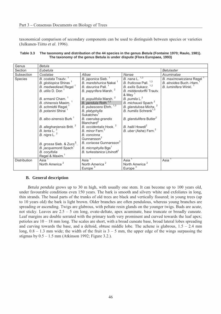
Part 3 – Consensus Documents on Biology of Trees
46
taxonomical comparison of secondary components can be used to distinguish between species or varieties (Julkunen-Tiitto et al. 1996).
Table 3.3 The taxonomy and distribution of the 44 species in the genus Betula (Fontaine 1970; Raulo, 1981). The taxonomy of the genus Betula is under dispute (Flora Europaea, 1993)
Genus Betula
Section Eubetula Betulaster
Subsection Costatae Albae Nanae Acuminatae
Species B. costata Trautv. 1 B. japonica Sieb.
1 B. nana L.
1,3B. maximowicziana Regel
1
B. globispica Shiras 1 B. mandshurica Nakai
1 B. fruticosa Pall.
1,3 B. alnoides Buch.-Ham.
1
B. medwediewii Regel 1 B. davurica Pall.
1 B. exilis Sukacz.
1,2 B. luminifera Winkl.
1
B. utilis D. Don 1 B. papyrifera Marsh.
2 B. middendorffii Trautv.
& Mey 1
B. ermanii Cham. 1 B. populifolia Marsh.
2 B. pumila L.
2
B. chinensis Maxim. 1
B. pendula Roth 1,3
B. michauxii Spach 2
B. schmidtii Regel 1 B. pubescens Ehrh.
1,3 B. glandulosa Michx.
2
B. potanini Shirai 1 B. platyphylla
Sukatchev1
B. humilis Schrenk1,3
B. albo-sinensis Burk 1 B. caerulea-grandis
Blanchard2
B. glandulifera Butler2
B. alleghaniensis Britt. 2 B. occidentalis Hook.
2 B. hallii Howell
2
B. lenta L. 2 B. minor Fern.
2 B. uber (Ashe) Fern.
2
B. nigra L. 2 B. concinna
Gunnarsson3
B. grossa Sieb. & Zucc1. B. coriacea Gunnarsson3
B. jacquemonti Spach1 B. microphylla Bge
1
B. coryifolia Regel & Maxim.
1B. turkestanica Litvinoff
1
Distribution Asia 1 Asia
1 Asia
1 Asia
1
North America 2 North America
2 North America
2
Europe 3 Europe
3
B. General description
Betula pendula grows up to 30 m high, with usually one stem. It can become up to 100 years old, under favourable conditions even 150 years. The bark is smooth and silvery white and exfoliates in long, thin strands. The basal parts of the trunks of old trees are black and vertically fissured; in young trees (up to 10 years old) the bark is light brown. Older branches are often pendulous, whereas young branches are spreading or ascending. Twigs are glabrous, with peltate resin glands on the younger twigs. Buds are acute, not sticky. Leaves are 2.5 – 5 cm long, ovate-deltate, apex acuminate, base truncate or broadly cuneate. Leaf margins are double serrated with the primary teeth very prominent and curved towards the leaf apex; petioles are 10 – 18 mm long. The scales are short, with a broad cuneate base, broad lateral lobes spreading and curving towards the base, and a deltoid, obtuse middle lobe. The achene is glabrous, 1.5 – 2.4 mm long, 0.8 – 1.3 mm wide; the width of the fruit is 3 – 5 mm, the upper edge of the wings surpassing the stigmas by 0.5 – 1.5 mm (Atkinson 1992; Figure 3.2.).
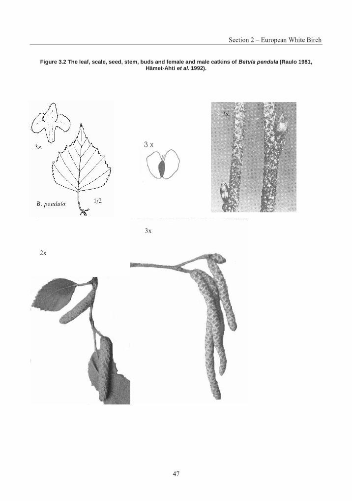
Section 2 – European White Birch
47
Figure 3.2 The leaf, scale, seed, stem, buds and female and male catkins of Betula pendula (Raulo 1981, Hämet-Ahti et al. 1992).
2x
3x
2x
Part 3 – Consensus Documents on Biology of Trees
48
C. Use of European white birch
Firewood
Birch has been an important source of energy, and earlier it was mostly used as firewood. The effective thermal value of birch is about 20 MJ/kg when absolutely dry, and about 15 MJ/kg with 30 % humidity. The high content of volatile oils makes the burning properties good (Salmi 1987).
Plywood production
In plywood production, the value of a birch log used for planks or veneer is influenced by its diameter, stem taper, and number and size of knots and branches. Logs of required dimensions can usually be produced with proper planting densities and thinning treatments.
Pulpwood
Birch fibres are relatively long and thin-walled and have a high content of hemicelluloses. As a result, birch pulp has good strength properties, low light-scattering ability and high density. The main product is bleached sulphate pulp. Coated and uncoated wood-free fine papers, which contain 50 to 90 % of bleached birch sulphate pulp, are especially important (Tammisola et al. 1995).
Curly-birch, Carelian birch
Curly-birch (B. pendula var. carelica) is a special form of B. pendula, and it occurs throughout Northern Europe and parts of Central and Eastern Europe. The wood is strong and decorative and is used in wooden ornaments and furniture. The genetic background of the curly-grained trait is not known. Curly-birch is self-sterile, and in controlled crossings between two individuals a maximum of 75 % of the progeny can be curly-birch. It has been suspected that the trait is homozygotically lethal, or that it is a genetical disease caused by a micro-organism or a virus (Ryynänen and Ryynänen 1986). The establishment of a plantation is an uncertain process, because it takes 10 years to see whether an individual will become a curly-birch. Normal birches have to be removed in order to prevent the curly-birches becoming suppressed. The propagation of curly-birches from cuttings has been attempted but the results have not been promising. Micro-propagation techniques have been developed for propagating curly-birches with the optimum stem form (Ryynänen and Ryynänen 1986).
2. Forestry Practices
A. Reproductive methods
Flower induction
In order to speed up the induction of flowering, and consequently, breeding, the Foundation for Forest Breeding in Finland built the first flower induction hall in 1976. It was equipped to provide good growing conditions for breeding material all year round. In the plastic greenhouses continuous illumination (24h day length) was kept at 10,000 – 20,000 lux by mercury and high pressure sodium lamps. Temperature was controlled by thermostats and fans. CO2 concentration was increased by burning propane gas and relative humidity was kept above 60 %. Required nutrients were provided automatically via continuous drip irrigation. The best results were obtained from birch: the daily growth was 7 cm per day, male flower buds appeared in 77 days and the first seed crop was collected 8 months after sowing (Holopainen and Pirttilä 1978).
Section 2 – European White Birch
49
B. Vegetative propagation
Birch can be propagated vegetatively, i.e. cloned, by grafting, rooting or by means of tissue culture. Production of grafts is quite expensive and grafts are used mainly for clonal collections and seed orchards (Ryynänen 1987). Also, rooting of birch cuttings is not much used in birch propagation as the percentage of rooted cuttings is low (10 – 50 %) for B. pendula, whereas Kling et al. (1985) found rooting rates of 80 % for a closely-related B. platyfylla.
Micro-propagation
Much research on tissue culture of birches has been done and effective micro-propagation techniques have been developed. Leaf callus from young seedlings can be used (Simola 1985) but with mature trees, vegetative buds are used as tissue material (Ryynänen and Ryynänen 1986). Shoots and roots can be specifically induced on different aseptic culture media, after which the plantlets can be potted on peat and moved into the greenhouse. The development of micro-propagation methods made an effective propagation of selected adult trees possible. Micro-propagated plants have proved viable in test plantings under field conditions (Meier-Dinkel 1992; Jones et al. 1996). A review on micro-propagation of juvenile and mature birches including results on field trials and cold storage was compiled by Meier-Dinkel (1992). Some clonally propagated birch plants have been used for practical forest cultivation in Finland (Viherä-Aarnio 1994a). Large-scale production of micro-propagated B. pendula seedlings was terminated in 1994, as it was considered unprofitable (Viherä-Aarnio and Velling 2001). Today, micro-propagation is used in commercial scale for curly-birch cloning. Micro-propagated plants can also be used instead of grafts in greenhouse seed orchards, when specific genotypes are required (Viherä-Aarnio and Ryynänen 1995).
Viherä-Aarnio (1994a) studied the field performance of micro-propagated birch plants in a small-scale field test with three clones and three seed-born lots. The best two lots of the experiment as regards the height and diameter growth at the age of six years were clones, but the weakest lot was also a clone. This indicates that the selection for clonal propagation should be done carefully. It is also important to test the clones in field trials before wide scale propagation. In fact, further studies revealed that micro-propagated clones do not differ from seed-born seedlings in terms of growth and resistance against pests and herbivores (Viherä-Aarnio and Velling 2001).
C. Reproductive materials used
Seed collection stands
Seed collection stands are natural birch stands of outstanding growth and quality. The best individuals in these stands have been selected as plus trees. A part of the seed used in nurseries is collected from seed collection stands (Viherä-Aarnio and Ryynänen 1994).
Seed orchards
As birch responds positively to intensive cultivation in a greenhouse, today most birch seed used for seedling production is obtained from polythene greenhouse seed orchards, a method which was developed by the Foundation for Forest Tree Breeding in Finland in 1972 (Huhtinen & Yahyaogly 1974). Most seed orchards are multi-clonal orchards with some 30 to 50 clones. Bi-clonal orchards have also been established in order to produce desired full-sib families (Viherä-Aarnio and Ryynänen 1994). Besides greenhouse orchards, open seed orchards in the field are used for seed production.
Seed production in greenhouse seed orchards begins three years after planting and continues for 5 to 7 years, until the trees grow too big to be kept inside. Flowering and seed ripening take place 1 to 2 weeks earlier in the greenhouse than in natural conditions. As a result, growing birches in greenhouses also
Part 3 – Consensus Documents on Biology of Trees
50
prevents unwanted background pollination. A seed orchard can be established with grafts of tested plus trees, seedlings selected from the best progenies or micro-propagated clones (Viherä-Aarnio and Ryynänen 1995).
Cultivation
In the Nordic countries and Europe, Finland has the longest experience in birch cultivation. The main emphasis in birch planting has been the afforestation of former agricultural land (Ferm et al. 1994). When a field is abandoned, it is first colonised by annual species, which then give way to perennial herbs and grasses. As field vegetation competes with tree seedlings for water and nutrients, success with field afforestation requires effective weed control during the establishment year. Soil preparation also affects the survival of trees. In 20-year-old experiments the best growth was gained when bare-rooted, large transplants of silver birch were planted on a ploughed and tilled field and weeds were properly controlled (Torpo 1991). Weed control improves the nutrient status of tree seedlings and increases growth. Birch seedlings with efficient weed control were 40 – 50 cm taller after two years than control seedlings, and they had bigger leaves (Ferm et al. 1994). Vegetation control reduces the risk of cicada (Cicadella viridis)wounding and consequently infection by pathogenic fungi, and is also effective against vole damage (Ferm et al. 1994).
D. Breeding
In the breeding programme for birch, outstanding stands for seed collection were selected and the best individual trees in these stands were selected as plus trees. The most important selection criteria were fast growth and good stem quality. After selection of plus trees, controlled crossings have been carried out, and progeny tests with both full-sib families from controlled crosses and half-sib families from open pollinated lots have been established (Koski 1991). Plus tree selection is still being continued in order to improve the geographical coverage of the breeding material (Viherä-Aarnio 1994b).
Planted birch stands of improved material can reach the size of final cutting by as early as 40 years and produce over 400 m3/ha (Viherä-Aarnio 1994b). Examples of realised genetic gain cannot yet be shown, but a potential gain of 20 to 30 % in volume growth has been suggested (Koski 1991) and in first generation seed orchards, the growth rate of the best selected family is 89 % higher than the control (Wang 1996).
Provenance transfers
Raulo and Koski (1977) reported on the first geographical transfers of silver birch. According to their study, seed transfers of 200 km northwards or southwards in central and southern Finland had no effect on the mortality or growth rate of the progenies; on the other hand they did not recommend long-distance transfers because of the risk of lower survival.
E. Conservation of genetic resources
Genetic diversity of forest trees
The cultivation of forest trees can be thought to endanger the genetic diversity in general. Maintaining genetic diversity in forest tree populations also means maintaining adaptability to changing environments. In naturally generated areas there is no concern about the loss of genetic diversity but large monocultures of a single clone would reduce genetic variability. However, forestry based on monoclonal blocks is not common, but cultivation of single clones is mostly applied to small plantations of special forms or variations, e.g. curly-birch (Viherä-Aarnio and Velling 2001). Moreover, real monocultures are impossible to maintain, as other trees can not be prevented from growing in the plantations.
Section 2 – European White Birch
51
Most forest trees are cross-fertilising and pollinated by wind and consequently there is crossing both between individuals in a stand and between stands. Even if small stands were to be cultivated with only one clone, effective wind-pollination will secure the mixing of genes between populations. Also, real monocultures are impossible to maintain, as other trees can not be prevented from growing in the plantations. Cultivation of single clones will probably mostly apply to small plantations of special forms or variations, e.g. curly-birch. The danger from planting large areas with a single clone arises when the adaptability of the clonal genotype is exceeded by adverse conditions. To avoid multiplication of poorly adapted or extremely susceptible genotypes, trees for micro-propagation are selected from old stands. Another option is the use of clonal mixtures which are buffering environmental risks (Kleinschmit 1998).
It has been estimated that a population of 500 trees is big enough to contain all possible alleles of different genes (Koski 1995), and this is the amount usually used in breeding populations. The seed from seed orchards is usually a result of cross-fertilisation of tens of plus tree clones from different localities, and it has been shown in studies with genetic markers that the genetic diversity is as large as that of natural seed collection stands (Koski 1995).
Gene reserve forests
Ongoing breeding activities may cause losses of genetic diversity, especially if population size is reduced from one generation to the next. Random allele loss is problematic for advanced-generation elite populations, and this has raised concerns about the maintenance of genetic diversity in forest-tree breeding programs.
Gene reserve forests have been established in order to conserve representative samples of the natural gene pool of forest trees. They have to be big enough (preferably tens of hectares) for the pollination to occur inside the forest and for the reserve to contain most of the local genetic diversity. Management and harvesting are allowed, but the forests are regenerated naturally or by sowing with seed from inside the forest or by planting seedlings grown from local seed. Gene reserve forests should consist of a network of forests in the area of the natural distribution of the species, in order to contain the diversity between provenances and localities. In Finland the gene reserve forests are situated in northern Finland and are part of a network of gene reserve forests covering all Europe (Parviainen et al. 2000).
Advanced breeding strategies
Two advanced models have been suggested in order to prevent random allele loss in breeding programs: HOPE (Hierarchical Open-Ended Breeding System) and MPBS (Multiple-Population Breeding Strategy) (Eriksson et al. 1993).
The HOPE system is composed of a hierarchy of breeding populations with successively higher performance levels. A large base population is maintained and is open to new material. Higher level populations are more stringently selected and from these, selections are made for commercial production. Under MPBS, a breeding population is divided into independent subpopulations which represent different sources or selection criteria. There is differentiation among subpopulations both in their source of germplasm and in their traits and environmental adaptabilities. There can be random allele loss in some subpopulations but, on average, gene frequencies remain fairly constant (Eriksson et al. 1993).
Of these, the MPBS seems to be a better choice for birch because it combines the highest possible genetic gains and the highest possible genetic diversity. It also gives the breeder more options for changing breeding goals with changing environment and markets (Eriksson et al. 1993).
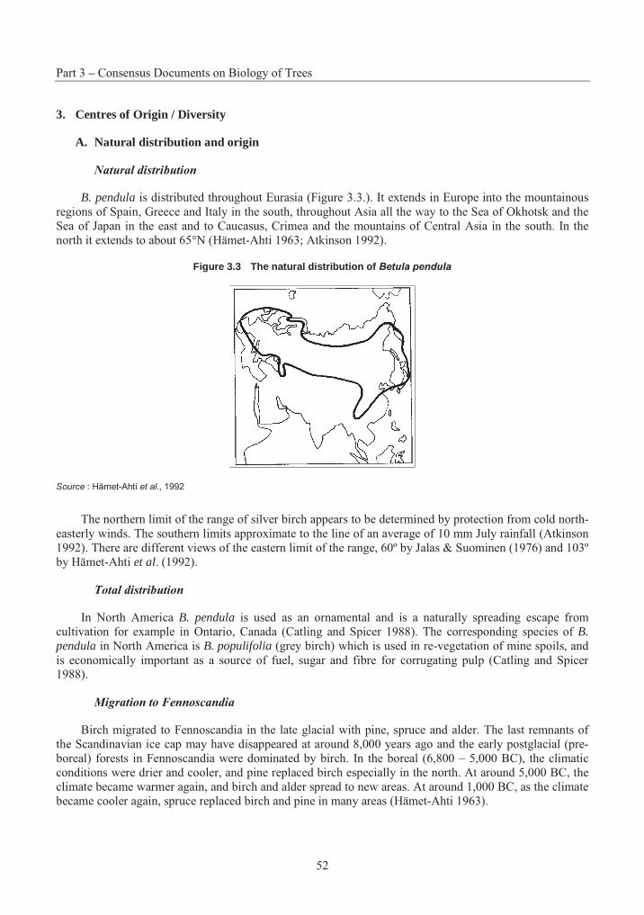
Part 3 – Consensus Documents on Biology of Trees
52
3. Centres of Origin / Diversity
A. Natural distribution and origin
Natural distribution
B. pendula is distributed throughout Eurasia (Figure 3.3.). It extends in Europe into the mountainous regions of Spain, Greece and Italy in the south, throughout Asia all the way to the Sea of Okhotsk and the Sea of Japan in the east and to Caucasus, Crimea and the mountains of Central Asia in the south. In the north it extends to about 65°N (Hämet-Ahti 1963; Atkinson 1992).
Figure 3.3 The natural distribution of Betula pendula
Source : Hämet-Ahti et al., 1992
The northern limit of the range of silver birch appears to be determined by protection from cold north-easterly winds. The southern limits approximate to the line of an average of 10 mm July rainfall (Atkinson 1992). There are different views of the eastern limit of the range, 60º by Jalas & Suominen (1976) and 103º by Hämet-Ahti et al. (1992).
Total distribution
In North America B. pendula is used as an ornamental and is a naturally spreading escape from cultivation for example in Ontario, Canada (Catling and Spicer 1988). The corresponding species of B. pendula in North America is B. populifolia (grey birch) which is used in re-vegetation of mine spoils, and is economically important as a source of fuel, sugar and fibre for corrugating pulp (Catling and Spicer 1988).
Migration to Fennoscandia
Birch migrated to Fennoscandia in the late glacial with pine, spruce and alder. The last remnants of the Scandinavian ice cap may have disappeared at around 8,000 years ago and the early postglacial (pre-boreal) forests in Fennoscandia were dominated by birch. In the boreal (6,800 – 5,000 BC), the climatic conditions were drier and cooler, and pine replaced birch especially in the north. At around 5,000 BC, the climate became warmer again, and birch and alder spread to new areas. At around 1,000 BC, as the climate became cooler again, spruce replaced birch and pine in many areas (Hämet-Ahti 1963).
Section 2 – European White Birch
53
4. Reproductive Biology
A. Sexual reproduction
Birches are monoecious (male and female flowers on the same tree) and diclinous (flowers unisexual). Male inflorescences are 1 –2 cm long, 4 mm wide while over-wintering; 2 – 6 cm long, 6 mm wide at anthesis. There are 2 – 4 male catkins together at the ends of small shoots. Female inflorescences are erect, 1.5 – 3.5 cm long, 7 mm wide (Atkinson 1992). They are pale green when immature, turning brown in the late autumn.
Flowering
Flowering usually starts when trees are between 5 and 10 years old but some single trees can be induced to flower within one year after germination (Elo et al. 2001). The female flowers emerge on the short-shoots at the same time as the buds open. The female flowers usually develop one day before male flowers on the same tree shed pollen. Flowers remain receptive for 4 days and after the sixth day are blackened and dead (Sarvas 1952). B. pubescens flowers about a week later than B. pendula.
Ripening of the flowers is closely related to the temperature conditions of the spring. The arrival of the first heat wave of daily temperatures above +10°C usually triggers flowering. Most pollen release occurs within 2 or 3 days of anthesis. Pollen shed is highest in the late afternoon and near zero from about midnight to early morning (Sarvas 1952). B. pendula pollen can remain viable for at least 20 days at room temperature in the dark. If the pollen is exposed to sunlight for 8 hours, however, ultraviolet rays and other factors reduce viability (Perala and Alm 1990).
Flowering is polygenically inherited and female and male flowers are governed by different sets of genes. Weather conditions have a strong influence on female flowering whereas they do not play a role in male flowering (Eriksson and Jonsson 1986).
Three cDNAs of B. pendula representing ADS-box genes BpMADS3, BpMADS4 and BpMADS5 are active during the development of both female and male inflorescence and the respective genes are involved in the determination of the identity of the inflorescence or flower meristem. BpMADS3 shows highest expression at late developmental stages. BpMADS4, besides regulating inflorescence, is expressed in roots and stem. Expression of BpMADS5 is inflorescence specific and continues during seed development. Ectopic expression of BpMADS3, BpMADS4 or BpMADS5 with CaMV 35S promotor in tobacco results in extremely early flowering (Elo et al. 2001).
B. Pollination
Male meiosis and pollen development
The male catkins develop in the autumn but do not release pollen until spring the following year. The principal events during micro-sporogenesis are the initiation and growth of the male buds, generation and growth of the sporogenous tissue, pre-meiotic maturation of the PMCs (pollen mother cells), meiosis, the tetrad stage, androgenesis and anthesis. The species in Betulaceae reach the tetrad phase from late July to mid-August (Luomajoki 1986). In Betulaceae the heat sums seem to have less connection with the development stage of micro-sporogenesis than in conifers, as the northernmost stands of Betula species develop first and the southernmost last. The timing of meiosis in Betula is more dependent on day length, and a hypothetical critical day length of 15 hours can quite accurately predict the onset of the tetrad phase (Luomajoki 1986).
Part 3 – Consensus Documents on Biology of Trees
54
Pollination
Pollination is anemophilous, i.e. birch is pollinated by wind. There is an intense maximum at the immediate start of flowering lasting 2 – 3 days during which 70 – 80 % of the total pollination takes place. Prolonged cold and wet conditions are likely to reduce the total amount of pollen (Sarvas 1948).
Pollen flow
As an evidence of long-distance pollen transport, Betula pollen concentrations in Fennoscandia can be relatively high before the local flowering period. The pollen is transported by south-eastern air-masses from eastern central Europe and the Baltic countries, with travelling times for pollen grains in the range of 9 – 20 hours (Hjelmroos 1991).
In northern Europe, birch pollen allergy is one of the most common reason for spring-time human rhinoconjunctivitis and asthma. In Sweden, about 8 – 9 % of the population suffers from allergies related to birch pollen (Hjelmroos and van Hage-Hamsten 1993). The major birch pollen allergen Bet v I is highly homologous to pathogenesis-related plant proteins. Another well described allergen is profilin, an actin binding protein, which is predominantly expressed in the pollen of different plant species. The birch profilin is recognised by IgE antibodies in 10 % of individuals allergic to birch pollen and seems to be an intermediate or major allergen in individuals allergic to pollens of grasses and weeds (Valenta et al. 1991). Another birch pollen allergen, Bet v III, representing a class of Ca 2+ binding proteins, has also been characterised (Seiberler et al. 1994).
C. Mating system
Cross-fertilisation
B. pendula is an outcrossing species. There is cross-fertilisation both between trees standing close in the stand and among distant individuals in the same or nearby stands. Hagman (1971) studied incompatibility in B. pendula and B. pubescens and found partial incompatibility in both cross-pollinated and self-pollinated trees. When trees from the same population were cross-pollinated some combinations were incompatible. Incompatibility occurs in the style as retarded growth of the pollen tube and is probably based on a one-locus multiple allele system. Incompatibility reaction is also influenced by environmental conditions: compatibility is increased at low temperatures.
Self-fertilisation and inbreeding depression
In the studies of Hagman (1971), self-incompatibility was observed after self-pollination. Self-incompatibility was not complete and the filled seed frequency was highest when the pollinations took place at low temperature.
Wang (1996) studied inbreeding depression in B. pendula and the possibility of using heterosis in breeding. He found that inbreeding depression in survival and in stem volume of progenies in three successive generations of selfing was significant. The mean survival rates were 88 %, 37 %, 28 % and 42 % for outcross controls, S1, S2 and S3 selfed progenies, respectively. Inbreeding depression for stem volume also increases with advancing generations of selfing. The mean stem volumes were 5.31, 3.15, 2.66 and 2.12 dm3 for outcross controls, S1, S2 and S3 selfed progenies, respectively.
Section 2 – European White Birch
55
D. Seed crop
Seed crops
The percent of germinating seed is directly related to the amount of pollen shed: seed crops with the highest viability will be produced during years of abundant flowering (Sarvas 1952). Each strobile contains about 450 seeds. In the average year in ordinary regeneration areas, seed crop is some 340 filled seeds m-2,with 2 300 seeds m-2 in a good seed year. In a good seed year in a pure birch stand the best annual result has been 53 200 seeds m-2, i.e. 128 kg per hectare. A crop amounting to 20 % of the best seed crop occurs only every third year, and intervening crops may be only 5 – 10 % of a good year (Sarvas 1948). Cold springs can lead to massive loss of male catkins. Birch produces empty fruits in the absence of pollination.
Dispersal range of birch seed is limited by the small seed size. The fall rate for silver birch seed is 0.52 m s-1 (Sarvas 1948). Most seeds fall within 40 – 50 m of the source, with a maximum amount of seeds at 10 – 12 m from the tree. The amount of seeds is still 5 % of the maximum at 100 m from the source, but after 70 m most seeds are empty. In B. pendula seeds are also dispersed throughout the autumn and winter, so there is probably also secondary dispersal of seed over the surface of snow by wind. With B. lenta,seeds were distributed over an area 3.3 times greater than by initial seed fall, and further dispersal by melting water in the spring could be some 50 km (Matlack 1989).
E. Natural regeneration
Germination and establishment in the field
A non-fluorescent water-soluble substance in the seed coat has germination-inhibiting properties. This inhibitor apparently increases the oxygen and light requirement of the embryo. Un-chilled, intact seeds require light for germination, but breaking the seed coat allows rapid entry of oxygen and hastens germination in darkness (Perala and Alm 1990). A long-day photoperiod or illumination with red light induces germination, whereas blue or far-red light inhibits germination. Stratification (chilling) or temperatures above 20°C override the photoperiod requirement. Fluctuating temperatures within the range of 20 – 30°C are more favourable for germination than steady temperatures (Perala and Alm 1990). Stratification also eliminates the effect of temperature on germination rate (Atkinson 1992). In natural conditions in the northern parts of the range of birch, the cold requirement is usually met.
In the autumn, after seed shed, short days and low temperatures inhibit germination and help seed survival over the winter. In the spring, after a long enough chilling period, dependence of germination on day length disappears and seeds germinate when temperature rises and there is enough moisture in the ground (Nygren 1987). Birch seeds may also germinate soon after seed deposition in the summer, if there is enough moisture in the ground. As the individual seeds are small and contain small amounts of reserve food, the radicle of the birch seedling remains short with the result that the plant frequently withers away and dies and seedlings only grow up on spots which contain enough moisture.
The percentage of germination of B. pendula seed in southern Finland (60° N) is about 61 % and it decreases towards the North, being about 53 % in central Finland (62° N). The opposite is true for B.pubescens with germination of 63 % and 70 %, respectively (Sarvas 1948).
In natural conditions the seeds are concentrated in the upper parts of the soil profile indicating a rapid turnover. Hill and Stevens (1981) showed that 80 % of the seeds were found in the litter layer and showed no long-term survival. In studies where B. pendula and B. pubescens seed were sown to two forest sites in northern Sweden, only 6 – 9 % of the seeds remained viable after one year. The depletion rate was slower in the two successive years (Granström and Fries 1985). This might be due to litter-fall and growth of mosses altering the environment, making the seeds less liable to germinate. When the seeds were buried
Part 3 – Consensus Documents on Biology of Trees
56
beneath the litter layer, the viability of the seeds did not fall consistently over a five-year period even though after three years the pericarps were more or less degraded exposing naked embryos (Granström 1987).
80 – 90 % of birch seedlings die within the first year, perhaps mainly as a result of drought. Establishment is best on bare mineral and humid soils and cushion Sphagnum. These surface types have the most favourable moisture conditions (Kinnaird 1974).
F. Vegetative reproduction in the field
Sprouting
Sprouting by basal buds occurs as a response to fire as well as other damage such as felling and grazing. The sprouting ability of B. pubescens is stronger than that of B. pendula. Sprouting ability differs seasonally, and is generally higher for dormant trees. Felled B. pendula produces the lowest frequency of stump sprouting in summer and the highest in fall and spring. The frequency of sprouting and shoot size (but not the number of shoots) depends on light intensity and temperature. No stumps were observed to sprout at temperature lower than 10°C (Perala and Alm 1990). Sprouting depends also on the soil type: sprouting is more frequent in moist forests and on peat land than on dry heath and woodlands. Sprouting also depends on the method of regeneration. It is most frequent on clear-cut areas and least frequent on group selection cutting areas, forest fire areas and burnt-over clear-cut areas because of damage to the basal buds (Sarvas 1948).
5. Crosses
A. Inter-specific crosses
Natho (1957) found several natural crosses between B. pendula and other birch species in Germany, i.e. B. pendula x B. pubescens, B. pendula x B. humilis and B. pendula x B. pubescens x B. humilis.Moreover, the author showed that there is a continuum from B. pendula via B. pubescens to B. humilis inleaf shape. This and the triple hybrid indicate that there is gene flow among the three species.
Johnsson (1945) found large variation among experimental crosses of B. pendula and five other birch species. The percentage of fruits containing seed were 53.2 %, 8.8 %, 0.9 % and 0.4 % for B. pendula x B. japonica, B. pendula x B. papyrifera, B. pendula x B. ermanii and B. pendula x B. maximowicziana,respectively. The hybrid B. pendula x B. pubescens was infertile in this experiment.
Clausen (1970) carried out inter-specific crosses between 12 Betula species (Table 3.4.). He observed that high-ploidy female x low-ploidy male usually gave low seed germinability. Crosses within a subsection did not give more germinable seeds than crosses between species from different subsections. Crosses with B. ermanii as female gave more germinable seeds than any other cross. Each species was represented by 1 – 13 individuals, so definite conclusions can not be drawn from these results. However, it is the most comprehensive study of its kind to date.
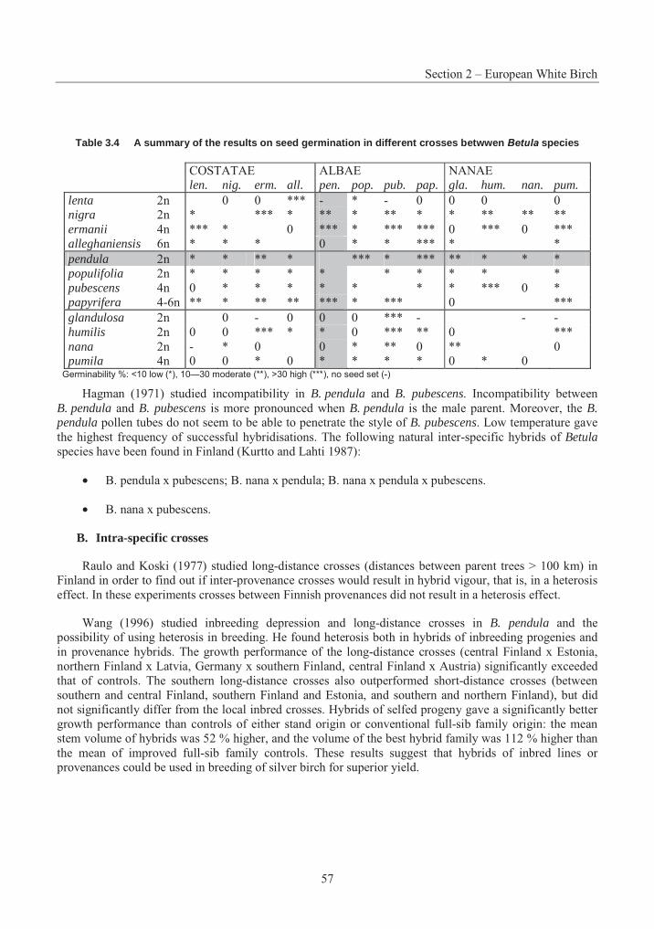
Section 2 – European White Birch
57
Table 3.4 A summary of the results on seed germination in different crosses betwwen Betula species
COSTATAE ALBAE NANAE len. nig. erm. all. pen. pop. pub. pap. gla. hum. nan. pum.
lenta 2n 0 0 *** - * - 0 0 0 0 nigra 2n * *** * ** * ** * * ** ** ** ermanii 4n *** * 0 *** * *** *** 0 *** 0 *** alleghaniensis 6n * * * 0 * * *** * * pendula 2n * * ** * *** * *** ** * * *populifolia 2n * * * * * * * * * * pubescens 4n 0 * * * * * * * *** 0 * papyrifera 4-6n ** * ** ** *** * *** 0 *** glandulosa 2n 0 - 0 0 0 *** - - - humilis 2n 0 0 *** * * 0 *** ** 0 *** nana 2n - * 0 0 * ** 0 ** 0 pumila 4n 0 0 * 0 * * * * 0 * 0
Germinability %: <10 low (*), 10—30 moderate (**), >30 high (***), no seed set (-)
Hagman (1971) studied incompatibility in B. pendula and B. pubescens. Incompatibility between B. pendula and B. pubescens is more pronounced when B. pendula is the male parent. Moreover, the B. pendula pollen tubes do not seem to be able to penetrate the style of B. pubescens. Low temperature gave the highest frequency of successful hybridisations. The following natural inter-specific hybrids of Betulaspecies have been found in Finland (Kurtto and Lahti 1987):
• B. pendula x pubescens; B. nana x pendula; B. nana x pendula x pubescens.
• B. nana x pubescens.
B. Intra-specific crosses
Raulo and Koski (1977) studied long-distance crosses (distances between parent trees > 100 km) in Finland in order to find out if inter-provenance crosses would result in hybrid vigour, that is, in a heterosis effect. In these experiments crosses between Finnish provenances did not result in a heterosis effect.
Wang (1996) studied inbreeding depression and long-distance crosses in B. pendula and the possibility of using heterosis in breeding. He found heterosis both in hybrids of inbreeding progenies and in provenance hybrids. The growth performance of the long-distance crosses (central Finland x Estonia, northern Finland x Latvia, Germany x southern Finland, central Finland x Austria) significantly exceeded that of controls. The southern long-distance crosses also outperformed short-distance crosses (between southern and central Finland, southern Finland and Estonia, and southern and northern Finland), but did not significantly differ from the local inbred crosses. Hybrids of selfed progeny gave a significantly better growth performance than controls of either stand origin or conventional full-sib family origin: the mean stem volume of hybrids was 52 % higher, and the volume of the best hybrid family was 112 % higher than the mean of improved full-sib family controls. These results suggest that hybrids of inbred lines or provenances could be used in breeding of silver birch for superior yield.
Part 3 – Consensus Documents on Biology of Trees
58
5. Genetic Variability
A. Genetic background
Chromosome number
The birches are characterised by their small chromosomes which are only a few micrometers long. A double-staining method is useful in counting the number of chromosomes in Betula and in other hardwood species with chromosomes of small size and large number (Hömmö and Särkilahti 1986).
Betula pendula is a diploid with 28 chromosomes; B. pubescens is a tetraploid with 56 chromosomes. Trees morphologically resembling tetraploid but with intermediate chromosome number (2n=42) are sometimes found. Because the chromosomes tend to lie in groups of seven and because of small numbers of quadrivalents in meiosis in the 28 and 56 chromosome plants, the original basic chromosome number is thought to be seven rather than fourteen (Eriksson and Jonsson 1986).
Natural polyploidy is very frequent in the genus Betula and the number of ploidy levels differ between the four subsections of the genus. Costatae has diploid (2X=28), tetraploid (4X=56) and hexaploid (6X=84) species and in addition to these, three ploidy levels. Albae has also a pentaploid (5X=70) species, Nanae has both diploid and tetraploid species and Acuminatae only diploid ones. Betulais a young species from the evolutionary point of view which explains the polyploid nature and the occurrence of various ploidy levels (Särkilahti and Valanne 1990).
The possibility of using induced polyploidy to speed up the natural evolution and breeding of Betulaarose in the 1960s (Särkilahti and Valanne 1990). The polyploidisation experiments were performed by treating seeds with colchicine during germination. Of the 687 polyploid trees produced by these experiments in Turku, Finland, 287 were still alive in 1990. Moreover, a series with ploidy levels ranging from diploid to dodecaploid (2X – 12 X) consisting of both natural and induced polyploids is available (Särkilahti and Valanne 1990).
Identification of polyploid trees of B. pendula is possible visually on the basis of leaf morphology, with polyploid trees having larger leaf blades, thicker leaf petioles and a rougher network structure on the abaxial leaf epidermis than normal trees. The mortality of colchicine polyploid trees is typically high in every growth phase, growth rate is slow and growth habit more or less abnormal. Thus, colchicine-induced autopolyploid trees are not of great value as such but they can be used for studying the effect of the ploidy level on growth, breeding, adaptability and evolution of Betula. In natural conditions polyploids are said to possess greater ecological and genetic amplitude and, therefore, exhibit greater variability than related diploids (Särkilahti 1990). Ploidy manipulation seems to produce mainly sterile trees but micro-propagation can be used for multiplication of polyploid material.
B. Variability within and between populations
Long-lived species such as forest trees are subject to conditions varying greatly from year to year. Thus, they have a large within population variation so that there are always genotypes well adapted to the varying conditions at regeneration. For example, in studies of five year heights of B. pendula progenies Velling (1985) stated that the within-population variation was as large as that between populations originating from latitudes 60 – 63° N. Raulo and Koski (1977) found that intra-group variation within provenances and between individuals was large compared to variation between stands or localities. Birch stands are not closed populations and gene exchange between stands is successful as a result of pollen dispersal by wind. A large within-population variation was also reported, for example, in two separate studies (Johnsson 1951 and Langhammer 1982, cited by Eriksson and Jonsson 1986).
Section 2 – European White Birch
59
Differences in qualitative traits; marker techniques
Raulo and Koski (1977) reported a large variation in growth and stem quality between progenies from different plus trees. Many progenies attained a stem volume that exceeded the mean value of the test by more than 40 %, and good progenies could be found among both open-pollinated and cross-pollinated families. Variation in wood density is generally smaller than that in growth and stem quality characteristics, but significant differences can be found between progenies (Velling 1979a). Nepveu and Velling (1983) studied the inheritance in wood quality characteristics. The inheritance in basic density and shrinkage was fairly strong, but volume growth and pulp yield showed low heritability.
As attention has focused on the external quality of the stem for plywood production, no active breeding or selection of wood quality has so far been applied. The properties of birch pulpwood could be improved if the bark content could be reduced, the size and/or number of branches reduced and the carbohydrate content of the wood increased. Increasing the number of fibres without affecting the fibre length would improve the optical properties and bulk of the pulp. Tammisola et al. (1995) studied tree-to-tree variation to determine if there is variation in the pulpwood properties of B. pendula that can be used in breeding. They found that significant variation occurred between individual trees in the properties studied, and high significance levels give support for underlying genetic differences.
If DNA markers closely associated with pulping and papermaking properties were found, it would be possible to select the most desirable trees within a progeny more quickly and cheaply compared to the more or less destructive analyses of mature trees (Tammisola et al. 1995). Altogether 157 nucleotide sequences and 177 proteins of B. pendula are listed in the database of NCBI. Molecular biology is the most active research area within biology and the number of known nucleotide sequences and proteins is increasing rapidly. The databases at http://www.ncbi.nlm.nih.gov/ have the most updated information about genetic markers. For an old review on genetic markers in Betula, see (Hattemer et al. 1990).
C. Adaptivity to climatic conditions
Growth cessation and winter hardening
In cool and temperate regions the annual temperature rhythm is the main regulating factor of the environment. Forest trees are adapted to the variation of the growing season between years as well as to the long-term average.
The timing of growth cessation and the subsequent process of winter hardening is determined by a joint effect of heat sum and night length (Koski and Sievänen 1985). In Punkaharju, Finland (61°48’ N), growth ceased at the cumulative temperature sum of 800 degree-days and night length of 7.5h. Koski and Sievänen (1985) predicted that an adapted provenance of B. pendula will cease growth by the accumulation of about two-thirds of the total heat sum for a normal growing season. If northern provenances were moved southwards, growth would cease with two thirds of the original local heat sum, but due to longer nights, growth would cease a few days earlier, that is, with a smaller heat sum than at the original locality. A northern provenance from Punkaharju (Finland) moved to Suwalki (Poland) would cease growth three weeks earlier than the local one. The opposite transfer northwards of southern provenances would lead to growth cessation three weeks later than the local ones. The heat sum characteristic of southern provenances would not be reached until the beginning of September, but long nights would induce growth cessation 10 days earlier, thus adapting the trees for the local growing season (Koski and Sievänen 1985).
Breaking the dormant state is affected by chilling temperatures during winter. The effect of chilling is cumulative increasing up to a threshold when the buds are released from dormancy. Bud burst and growth
Part 3 – Consensus Documents on Biology of Trees
60
start in spring occur after the accumulation of a certain heat sum above a specific base temperature (Myking and Heide 1995).
Growth start
As B. pendula is geographically widely distributed, it has by latitude and altitude determined ecotypes with different optimum and critical chilling temperatures and durations. The ecotypes of northern origin have the earliest bud burst with variation in the requirement for duration of chilling but not for chilling temperature. A longer chilling requirement is found in southern ecotypes. A clinal difference in the base temperature for growth among the ecotypes is also found. In the studies of Myking and Heide (1995) the north Norwegian ecotypes flushed 2.5 months earlier and developed faster and had a lower base temperature for growth than ecotypes from southern Scandinavia. The Danish birches have adapted to a milder and more variable winter climate by developing greater dormancy stability involving both a longer chilling requirement and a higher base temperature. Night length had no effect on bud burst after the chilling requirement was fully met. After full dormancy release, time of bud burst in birch depends solely on the temperature regime in late winter and spring (Myking and Heide 1995).
The upper temperature limit for normal dormancy release in birch is probably slightly above 12°C. Chilling deficit is thus unlikely to occur in Scandinavia and in other areas where the chilling requirement is far exceeded, even with a climatic warming of 7 – 8°C above the current normal winter temperature. The likely effects of a climatic warming include earlier bud burst, a longer growing season and increased risk of spring frost injury, especially in northern ecotypes (Myking and Heide 1995).
Growth capacity of different provenances
In provenance trials with B. pendula at four sites within the latitudinal range of 56 – 64°, Johnsson, 1977 (cited by Eriksson and Jonsson 1986) reported that long-distance transfers both southwards and northwards tend to result in growth reduction. The longer the transfer northwards, the more is the growth period prolonged for the material from the south and the higher is the risk for dieback of the leaders. Short-distance transfers (< 250 km) do not influence the height growth considerably. Kleinschmit and Svolba (1982) reported on three-year heights of populations from central Europe to Finland. The Finnish and Swedish populations moved to Germany performed poorly as a consequence of the long-distance transfer southwards. In a study of growth of seedlings (Velling 1979b) a dependence between height and latitude of the origins was found. Seedlings of different provenances (from Latvia, 56°31’N, to central Finland, 61°48’N) were grown in central Finland and the more southern the origin, the greater the height of seedlings. A correlation was also found between the degree of leaf yellowing and the origin of seedlings. While the Finnish origins had turned completely yellow in the autumn, the Latvian origins still retained their green colour. The colouring was connected with survival, with poor winter resistance causing increased mortality in the southern origins.
7. Ecology and Physiology
A. Dynamics of regeneration
Birch is a pioneer species, and it quickly colonises bare areas and does not tolerate shading. Young birches can not survive fire because of their thin bark, but some mature trees may, because the thin forest floor under birches can not support intense and persistent surface fires. Post-fire pioneer successions are often dominated by birch. Abundant seed production enhances the pioneering character of birch. Without fire or human intervention birches would be replaced in succession by more shade-tolerant and longer-lived species (Perala and Alm 1990).
Section 2 – European White Birch
61
Light
Seedling density is independent of canopy cover, i.e. germination is unaffected by light, but the ability of birch seedlings to penetrate the canopy is low and they cannot establish in even the lowest vegetation. Most birch seedlings are only about 5 – 12 cm tall after the first year, and competing vegetation on fertile sites can easily overgrow and subdue them. The birches are also sensitive to chemical interference (allelopathy) by other plants (Perala and Alm 1990). Moreover, shaded birches are a preferred host by insect herbivores (see e.g. Ruohomäki et al. 1996).
Shading by neighbouring trees has a profound effect on shoot growth. In a short-term experiment shaded seedlings were higher than those reared in the simulated sun-light (Aphalo and Lehto 1997). Fewer buds are initiated and a higher proportion die in zones of heaviest shading (Atkinson 1992). Optimum sunlight for silver birch height growth was found to be 43 % of full sunlight for weeded seedlings, but 24 % for seedlings competing with weeds. Silver birch has also been found to grow less when sunlight decreases from 56 to 16 %. In greenhouse experiments with conifers, birch was more sensitive to both its own canopy and root competition than to competition by conifers (Perala and Alm 1990).
The Eurasian birches can endure as much as 90 % shade by adapting leaf structure. As sunlight diminishes, the light intensity for photosynthetic saturation, maximum photosynthetic rate, leaf mesophyll thickness, and chlorophyll concentration all diminish. The maximum photosynthetic efficiency for silver birch is at 10 – 50 % full sunlight, much higher than for shade-tolerant plants (Perala and Alm 1990).
Temperature
The birches are adapted to cool climate and grow best at about room temperature. Once the soil temperature reaches 2 – 3°C, the growth of silver birch depends more on air temperature than on soil temperature. Seedling shoots grow in direct proportion to heat sums, gradually diminishing as photoperiod shortens. New shoots can tolerate growing season temperatures of -3 to -5°C (Perala and Alm 1990).
Water requirements
Assimilation of silver birch is fastest at about -5 x 10² kPa and water use diminishes in wet conditions. The birches are sensitive to both drought and flooding. Seedling mortality increases at water potentials below -1.6 x 10² kPa. However, adaptability of birches to anaerobic conditions by oxygen transfer from the shoots to the roots, reduces the effects of flooding. Fertilisation improves water uptake and drought resistance (Perala and Alm 1990).
The birches use water inefficiently. Silver birch seedlings maintain turgor at high soil water potential only by closing stomata, which partially close at about -15 x 102 kPa. In large trees daily transpiration per unit of foliage mass is about 514 kg water/kg foliage. Extreme transpiration demand reduces growth even on moist soil because transpiration and photosynthesis have partly separate control systems. Drought depresses photosynthesis more than it does transpiration (Perala and Alm 1990).
Nutrients
To achieve maximum productivity, white birch requires all necessary nutrients, an optimum ratio of nitrogen sources NO3
- and NH4+ in a rhizosphere and an optimal total nutrient solution (Ingestad 1971). In
general, B. pendula is a nitrogen-limited species. Low-nitrogen conditions reduce growth and increase amount of condensed tannins, whereas in high nitrogen conditions B. pendula grows faster and the content of flavonoids in foliage is higher (Keinänen et al. 1999). The effects of nitrogen fertilisation on herbivore resistance are not clear. Nitrogen fertilisation did not affect preference of mammalian herbivores on B.pendula seedlings. On the other hand, the autumnal moth grew larger on fertilised seedlings (Mutikainen et
Part 3 – Consensus Documents on Biology of Trees
62
al. 2000). The limiting effects of other nutrients on the birches are not as well defined. At optimum levels, both zinc and manganese stimulate seedling growth, but toxicity problems have also been reported. There is a wide genetic variation in zinc tolerance and uptake. B. pendula has a high requirement for sulphur and it readily takes up boron. It sometimes suffers from manganese toxicity on poorly drained peat. The pH optimum for B. pendula and B. pubescens is between 4 and 5. B. pendula can tolerate some soil salinity (Perala and Alm 1990).
Effects on soil
B. pendula grows on fertile mineral soils and on drier and lighter soils than B. pubescens, which grows commonly on both peat and mineral soils (Gimingham 1984). The birches usually improve soils by efficiently cycling nutrients. First generation birch stands on former Calluna vulgaris (heather) heathland have increased earthworm activity, higher soil pH, greater total P, higher base status, faster rates of N mineralisation and cellulose decomposition and more diverse ground flora. Surface soil N, P, K, Ca, Mg and Mn is increased. Silver birch on Sphagnum peat soils accelerates microbial decomposition, accumulates dead woody roots and increases soil bulk density (Perala and Alm 1990).
Photosynthesis
CO2 exchange variables showed considerable genetical variance in B. pendula. 54 % of variation in net photosynthesis, 36 % of variation in stomatal conductance and 45 % of variance in intercellular CO2
were assigned to family (Wang et al. 1995). Photosynthesis related traits are modified by environmental factors as well. Defoliation reduces leaf area and biomass, but not photosynthesis, since B. pendula is able to compensate the damage by increasing photosynthetic activity. In fact, in nitrogen-rich environments damaged leaves have higher photosynthetic activity than the undamaged controls (Ovaska et al. 1993). The degree of compensation is dependent on the source of damage. The compensative response of B. pendulaafter artificial damage does not differ from that of the autumnal moth (Ovaska 1993), whereas the response was relatively weak after the damage by the alder beetle Agelastica alni (Oleksyn et al. 1998).
Root development
Birch trees exploit soils efficiently by developing both an extensive and dense surface root system to intercept precipitation and sinker roots to penetrate dense ‘pans’ and exploit deep water. The tap-root becomes horizontal after about 30 cm or is overtaken by side roots. Narrower roots may penetrate to a considerable depth. The extreme length of horizontal roots can be 25 m on sandy soils. In anaerobic conditions, B. pendula and B. pubescens roots elongate more than in aerobic conditions. Birch roots penetrate deep into poorly drained soils such as peats, and benefit inter-grown conifers by oxygenating the soil. On the other hand, birch small-root biomass may be twice that of the conifers offering intense root competition (Perala and Alm 1990).
B. Mycorrhizae
Early mycorrhizal infection is an important factor in the successful establishment of birch seedlings especially on nutrient-poor soils. The rates of nitrogen mineralisation of many forest litters are so slow that nitrogen can become the key growth limiting element. Some ectomycorrhizal fungi (e.g. Amanita, Boletus,Paxillus, Suillus and Thelephora) have proteolytic activity and thus the potential to mobilise nitrogen from proteins, peptides and amino acids and make it available to the plant. Some fungi, like Laccaria laccata,lack this activity and are dependent upon mineralisation processes initiated by other organisms (Read 1991). The assimilation of mycorrhizal amino compounds also provides supplementary carbon as well as nitrogen to the host plant. Abuzinadah and Read (1989) showed that up to 9 % of the carbon assimilated by the host plant (B. pendula) over a period of 55 days was derived heterotrophically from the protein by
Section 2 – European White Birch
63
mycorrhizal fungi. This is thought to be an adaptation to shade stress in young trees which spend the early part of their lives under the canopy of mature trees and which need to supplement their carbon budgets by heterotrophic assimilation. Grellier et al. (1984) showed that association with a mycorrhizal fungus (Paxillus involutus) practically doubled the growth of in vitro grown birch seedlings compared to non-mycorrhizal seedlings. Soil fauna have also been found to positively influence nutrient uptake and net production of birch seedlings (Setälä and Huhta 1991). Mycorrhizae can be inhibited by acute phosphorous deficiency, but can be encouraged even by a light application of phosphorous. Mycorrhizae like Paxillus involutus increase zinc tolerance by adsorbing zinc to the hyphae and slowing its transport to the shoots (Perala and Alm 1990).
Because little growth is possible on the nutrient reserves available in the seed, the seedlings are dependent on an external nutrient supply by the production of the first pair of leaves. Newton and Pigott (1991) report that ectomycorrhizal infection was indeed recorded by the time the first pair of leaves had expanded. Two groups of mycorrhizal fungi have been recognised: "early-stage" fungi (Inocybe spp., Hebeloma spp. and Laccaria spp.) can inoculate roots of seedlings from basidiospores or from added inoculum whereas "late-stage" fungi (e.g. Lactarius pubescens and Leccinum roseofractum) infect by hyphal connection. In natural circumstances the "late-stage" fungi are the most vigorous colonists of birch seedlings (Read 1991).
C. Diseases
Firm Rot
Black coloured firm rot around the pith is a serious cause of concern in young cultivated birch stands. Several different pathogens (fungi and bacteria) are responsible for the defect. Their entrance is, obviously, facilitated by frost cracks, insect exit holes (possibly the exit holes of P. betulae) and especially vole or moose browsing. The defect is especially harmful in plywood industry if the black colouring spreads outside the peeler core (Uotila 1987).
Birch rust
Birch rust (Melampsoridium betulinum) is the most common leaf disease of B. pendula. The rust causes yellowing and premature falling of the leaves, but it does not affect buds or the wood. If epidemics appear in several consecutive years, the growth of young seedlings can be decreased because of the shortened assimilation period (Uotila 1987; Vuorinen 1992). There are clear genetic differences in susceptibility to rust among birch clones, and screening clones for resistance could be used in breeding. A leaf-disc bioassay was used for determining the field rust resistance of birch clones in the study of Poteri and Rousi (1996).
Stem spotting
Stem spot disease caused by a group of fungi (Godronia multispora, Botrytis cinerea, Fusarium avenaceum, Cylindrocarpon sp., Alternaria sp.) is common in nurseries and can be very detrimental to young birch seedlings. The small necrotic lesions in the bark produced in the autumn enlarge during winter, and seedlings may die before spring. These fungi infect trees through mechanical wounding sites, frost cracks or insect (especially cicada, Cicadella viridis) wounding sites (Juutinen et al. 1976). Stem spotting is common in birch trees grown in unsuitable sites, e.g. in waterlogged soils (Uotila 1987).
Rot fungi
B. pendula is mainly rotted by Polyporaceae fungi, which start the rotting in the heartwood in the inner part of the trunk, and then proceed to the living sapwood. Especially timber is susceptible to decay.
Part 3 – Consensus Documents on Biology of Trees
64
Fomes fomentarius and Inonotus obliquus are the most common rot fungi and they attack living trees through wounds. Ochroporus igniarius also infects living trees.
Piptoporus betulinus is less common and attacks old or dead trees, destroying both heartwood and sapwood at the same time. The best way to prevent rot damage of living trees is good forestry practice: thinning should be done in time and only dead branches pruned, since branch scars act as a route for rotting fungi (Uotila 1987). Other fungi that rot dead trees and timber are Stereum sanguinolentum, S. purpureum,Pycnoporus cinnabarinus, Cerrena unicolor, Trametes multicolor and T. hirsuta (Uotila 1987).
D. Insect herbivory
Insect herbivory affects birch growth. For instance, in 1965 – 1967 the autumnal moth (Epirrita autumnata) defoliated over 5000 km2 of birch forest in Finnish Lapland (Lehtonen and Heikkinen 1995). On the other hand, birches have a large community of invertebrate herbivores (Annila 1987), which may damage seedlings and trees locally, but at the stand scale birch is able to compensate the damage. Of geometrid defoliators living on B. pendula, Opheroptera brumata is among the most voracious species (Tikkanen et al. 1998, 1999, 2000). The species is common in Baltic countries, southern Scandinavia and central Europe. The damage may be severe after the insect outbreak but usually climatic conditions enable birch to compensate the damage by re-growth. Beetles may damage trunks of mature trees, and thereby reduce the economical value of the timber. For instance, Hylecoetus dermestoides and Trypodendron signatum bore tunnels in logs and timber. H. dermestoides can attack also living trees and a simultaneous infection by rot fungi can be fatal to the tree (Raulo 1981). Larvae of the birch cambium fly, Phytobia betulae, mine in the birch wood near the cambium layer and cause 1 – 4 mm wide brown streaks in the wood. The streaks decrease the value of birch wood used in plywood and furniture industry. The damage is aesthetic; the mechanical strength of the wood is not affected. The resistance mechanisms of birch and the biology of Phytobia betulae are not known. Ylioja et al. (1995) studied the susceptibility of European (B. pendula) and Japanese white birch (B. platyphylla) to Phytobia damage. Birch progenies which had B. platyphylla in their ancestry included more pith flecks than pure B. pendula progenies. Fast growing birches were also more susceptible to Phytobia attack. Differences in susceptibility to Phytobia attack between birch clones and progenies of plus trees could be used in resistance breeding.
Besides the genuine seasonal change in foliar phenolics (Salminen et al. 2002), insect grazing or artificial damage increases the level of phenolics in the leaves of B. pendula. In the study of Hartley (1988), increases in phenolic compounds did not affect further feeding, either by a natural birch-feeding herbivore (Apocheima pilosaria) or a polyphagous non-birch feeding insect (Spodoptera littoralis). Hence, there is no evidence that the tree’s responses were specific defences against further attack by insect herbivores.
E. Mammalian herbivores
In Scandinavia, trees of the genus Betula are important winter food for herbivorous animals, especially voles (Microtus, Clethrionomys), hares (Lepus) and moose (Alces alces) (Rousi et al. 1989, 1990; Jia et al. 1997). They usually destroy woody plants in wintertime when alternative food plants are under snow cover. The variation of resistance among origins and families and even among individual seedlings within a genus can be very large. The centres of origin of cultivated plants are thought to be the best places to find resistances to diseases and herbivores. In these centres, plants have been exposed to selective pressure from local pathogens and herbivores for a long time, and have consequently developed resistance to them. Bryant et al. (1989) indicated that Betula and Salix species from Pleistocene refuges (Alaska and Siberia) were more resistant to mountain hare than species from regions that were glaciated during the Pleistocene. Likewise, birches from Iceland, where there were no browsing mammals before the
Section 2 – European White Birch
65
Norse colonisation, were more susceptible than birches from regions with more browsing mammals, that is, Alaska, Siberia and Finland (Bryant et al. 1989).
The bark of seedlings, young shoots and twigs of B. pendula contain resin droplets that consist of papyriferic acid and other triterpenoids. The juvenile resistance is accounted for by the resin, which is synthesised in and excreted by glands that are active only during the season when the primary apical growth of the shoot takes place (Taipale et al. 1993). On the other hand, phenolic substances are present in winter-dormant birches of all growth stages, but are rapidly metabolised by the plant in the spring when leaves emerge. Of these, platyphylloside is shown to exhibit repellent and anti-nutritional effects in mountain hares (Palo et al. 1992).
In B. pendula there is a sharp decrease of resistance after the tree has reached certain dimensions and the tree is no longer within reach of the herbivores. In the feeding trials with hare, Rousi et al. (1989) found that 1-year-old seedlings were less palatable than twigs taken from 7-year-old saplings of the same origin.
Vole
Voles cause considerable damage to forest plantations, especially during the peak years of density fluctuations. Birch plantations are especially vulnerable, since they are often afforestations of old fields, which are habitats favoured by Microtus voles (Rousi et al. 1990). The vole destroys birch seedlings under snow cover and it can eat the bark of the seedlings until the basal diameter reaches 4 cm (when the seedling is about 5 years old). Triterpenes in young birch seedlings seem to be deterrents of vole feeding. However, as the resin droplets are mainly situated in the top parts of the seedlings, and voles usually feed at the base of the seedling, they avoid the deterrent substances of the resin droplets. In field tests of Rousi et al.(1990), there were no clear differences in resistance between European white birch families in field tests with voles, but the Japanese white birch (B. platyphylla) turned out to be especially resistant to vole feeding. Rousi et al. (1990) suggest that hybrids between B. pendula and B. platyphylla, or Finnish-Siberian crosses of B. pendula, could be used to increase vole resistance.
Hare
Hares feed on the upper branches of young birch seedlings (of 40 – 70 cm) and especially in winter the damage can be fatal for the seedlings. Hares are discriminating feeders and determination of palatability is guided to a large extent by olfactory stimuli, and the resistance of young birch seedlings is tied to the production of papyriferic acid. For example, in the feeding experiments of Rousi et al. (1991), for the mountain hare (Lepus timidus) the palatability of birch seedlings and saplings was strongly and negatively correlated with the number of resin droplets on the bark. The Japanese white birch (B. platyphylla) turned out to be the most resistant of different birch species.
Rousi et al. (1991, 1996) also tested how the growth of birch correlates with resistance and whether fertilisation lowers the resistance of seedlings to browsing. Contrary to predictions of growth-defence trade-off theories, no trade-offs were found in the resistance and growth rate. Fertilisation stimulated growth but did not affect the palatability of the seedlings. Consequently, fast growing birch species and families should not be more susceptible to damage by herbivores, and fast growth can be promoted parallel with herbivore resistance by means of breeding.
Moose
Moose browsing on young birch seedlings occurs throughout the year. During winter only twigs are browsed, but during summer both leaves and young twigs are browsed. In order to feed on the young twigs of the crown, moose often break the main stem of the saplings. Moose can cause serious damage to birch
Part 3 – Consensus Documents on Biology of Trees
66
plantations. In Finland, plantations established in 1976 – 1977 were studied in 1985 (Heikkilä and Raulo 1987). Only one third of the plantations were found to be undamaged. Half of the total area had been damaged slightly and 15 % seriously. At the time of the establishment, though, the moose density was very high, 3 – 8 animals/1 000 ha. With a density of 2 – 3 animals/1,000 ha moose are not a serious threat to growing birch. Also, establishing plantations close to built-up areas or main roads can considerably prevent the risk of moose damage.
Heikkilä et al. (1993) showed that after stem breakage, the recovery during the first two years was fairly good. If the stems were at the leader shoot of the previous year, re-growth was weaker than in unbroken trees, the angle of crookedness was stronger and wound healing weaker. Injuries to the wood commonly become discoloured, and there was discoloration in 80 % of the sample trees. The significance of discoloration and decay depends on their distance from the wood surface, and the effect of discoloration on the quality of logs, when used for saw timber or veneer, can only be determined after a longer growth period. Bergström and Danell (1987, 1995) simulated winter browsing and summer browsing of moose, and studied the effects on the morphology and biomass of B. pendula. The birches responded to simulated winter browsing by growing fewer but larger and more branched shoots. There was also a slight decrease in viable seed production (Bergström and Danell 1987). In the experiments of Danell and Huss-Danell (1985) birches browsed by moose had more ants, psyllids, leaf-galls, leaf-miners and other leaf-eating insects. The leaves of browsed trees were larger and heavier, appeared greener and contained more nitrogen and chlorophyll. A decrease in resin content was observed. The trees seemed to allocate most of the nutrients and energy to growth in order to grow above the browsing line (Danell and Huss-Danell 1985). No induced defence in juvenile trees has been found; those trees that had been browsed during the previous winter were more palatable than previously un-browsed trees (Danell et al. 1985). Defoliation (simulated summer browsing) resulted in an overall decline in biomass and reduction in height and diameter growth. The long-shoots produced on defoliated trees were smaller and suffered more from tip-drying than shoots on control trees (Bergström and Danell 1995).
F. Abiotic damage
Abiotic damages are caused by too high ground water, drought, frosts and frost cracks (Uotila 1987). Snow load can cause stem breakage or permanent bending in young trees (Hannelius et al. 1989). Mechanical wounding increases fungal infections. UV-radiation does not seem to affect the growth, morphology or specific leaf area of B. pendula seedlings. The absorption by the secondary metabolites (phenolic glucoside, phenolic acids and flavonoids) provides the main part of the total UV absorbance of birch leaves. Plant secondary metabolism responds to enhanced UV-radiation by increasing synthesis of the above mentioned compounds that are the most effective UV-protectors (Lavola et al. 1997). The growth of B. pendula does not seem to be adversely affected by acid rain (Ashenden and Bell 1988). Instead, there was a stimulation in the height of birch seedlings with increasing acidity. A slight chlorosis of leaf margins after exposure to 2.5 pH rainfall was observed. Soil characteristics might have an influence on the sensitivity to acid rainfall.
Ozone causes physical damage on leaf surface, i.e. chlorosis, decoloration, black spots and necrotic areas and finally, leaf shedding (Maurer et al. 1997). Moreover, it changes the balance between CO2
assimilation and stomatal conductance, which may severely limit plant's ability to repair ozone damage at the cellular level (Zhang et al. 2001). Ozone activates biosynthetic pathway, and thus the production of phytohormone ethylene (Kangasjärvi et al. 1997). Sequences of the cDNA of 1-aminocyclopropane-1-carboxylate oxidase (ACO), the enzyme catalysing the last step in ethylene biosynthetic pathway, as well other possible ACO-homologue fragments have been submitted to the Plant Gene Register (EMBL accession numbers X97993, X97992, X97994 and Y10749). Interestingly, B. pendula is among the most ozone-resistant woody plants in central Europe. The gas exchange traits differ by factor or two when compared to more vulnerable woody plants (Zhang et al. 2001).
Section 2 – European White Birch
67
REFERENCES
Abuzinadah, R. A. & Read, D. J. (1989): Carbon transfer associated with assimilation of organic nitrogen sources by silver birch (Betula pendula Roth). Trees 3: 17-23.
Annila, E. (1987): Rauduskoivulla (Betula pendula) elävät hyönteiset. (Summary: Insects on Silver Birch (Betula pendula)). Sorbifolia 18: 118-122.
Aphalo, P. J. & Lehto, T. (1997): Effects of light quality on growth and accumulation in birch seedlings. Tree Physiol. 17: 125-132.
Ashenden, T. W. & Bell, S. A. (1988): Growth responses of birch and Sitka spruce exposed to acidified rain. Environ. Pollut. 51: 153-162.
Atkinson, M. D. (1992): Betula pendula Roth (B. verrucosa Ehrh.) and B. pubescens Ehrh. J. Ecol. 80: 837-870.
Bergström, R. & Danell, K. (1987): Effects of simulated winter browsing by moose on morphology and biomass of two birch species. J. Ecol. 75: 533-544.
Bergström, R. & Danell, K. (1995): Effects of simulated summer browsing by moose on leaf and shoot biomass of birch, Betula pendula. Oikos 72: 132-138.
Bryant, J. P., Tahvanainen, J., Sulkinoja, M., Julkunen-Tiitto, R., Reichardt, P. & Green, T. (1989): Biogeographic evidence for the evolution of chemical defence by boreal birch and willow against mammalian browsing. Am. Nat. 134: 20-34.
Catling, P. M. & Spicer, K. W. (1988): The separation of Betula populifolia and Betula pendula and their status in Ontario. Can. J. For. Res. 18: 1017-1026.
Clausen, K.E. (1970): Interspecific crossability in Betula. Proceedings of the IUFRO Section 22 working group on sexual reproduction of forest trees, Varparanta, Finland. pp. 1-10.
Danell, K. & Huss-Danell, K. (1985): Feeding by insects and hares on birches earlier affected by moose browsing. Oikos 44: 75-81.
Danell, K., Huss-Danell, K. & Bergström, R. (1985): Interactions between browsing moose and two species of birch in Sweden. Ecology 66: 1867-1878.
Elo, A., Lemmetyinen, J., Turunen, M. L., Tikka, L. & Sopanen, T. (2001): Three MADS-box genes similar to APETALA1 and FRUITFULL from silver birch (Betula pendula). Physiol. Plant. 112: 95-103.
Eriksson, G. & Jonsson, A. (1986): A review of the genetics of Betula. Scand. J. For. Res 1: 421-434.
Eriksson, G., Namkoong, G. & Roberds, J. H. (1993): Dynamic gene conservation for uncertain futures. For. Ecol. Manage. 62:15-37.
Ferm, A., Hytönen, J., Lilja, S. & Jylhä, P. (1994): Effects of weed control on the early growth of Betulapendula seedlings established on an agricultural field. Scand. J. For. Res 9: 347-359.
Part 3 – Consensus Documents on Biology of Trees
68
Flora Europaea (1993): Vol. 1. Psilotaceae to Platanaceae. Cambrigde Univ. Press, Cambridge.
Fontaine, F.J. (1970): Het Geslacht Betula; bijdrage tot een monografie. Belmontia 13: 99-180.
Gimingham, C. H. (1984): Ecological aspects of birch. Proc. R. Soc. Edinburgh 85B: 65-72.
Granström, A. (1987): Seed viability of fourteen species during five years of storage in a forest soil. J. Ecol. 75: 321-331.
Granström, A. & Fries, C. (1985): Depletion of viable seeds of Betula pubescens and Betula verrucosa sown onto some north Swedish forest soils. Can. J. For. Res. 15: 1176-1180.
Grellier, B., Letouze, R. & Strullu, D. G. (1984): Micropropagation of birch and mycorrhizal formation in vitro. New Phytol. 97: 591-599.
Hagman, M. (1971): On self- and cross-incompatibility shown by Betula verrucosa Ehrh. and Betulapubescens Ehrh. Commun. Inst. For. Fenn. 73 (6): 1-125.
Hämet-Ahti, L. (1963): Zonation of the mountain birch forests in northernmost Fennoscandia. Ann. Soc. Bot. 'Vanamo', 34(4): 1-127.
Hämet-Ahti, L., Palmén, A., Alanko, P. & Tigerstedt, P. M. A. (1992): In Finnish (English title: Woody flora of Finland). Publications of the Finnish Dendrological Society 6: 1-373, 2nd edition, University Press, Helsinki.
Hannelius, S., Leikola, M. & Tuimala, A. (1989): In Finnish (English title: Forest Book). WSOY, Porvoo, Finland. 383 p.
Hartley, S. E. (1988): The inhibition of phenolic biosynthesis in damaged and undamaged birch foliage and its effect on insect herbivores. Oecologia 76: 65-70.
Hattemer, H.H., Steiner, W. & Kownatzki, D. (1990): Genetic markers in birch. Silvae Genetica, 39(2): 45-50.
Heikkilä, R. & Raulo, J. (1987): In Finnish (English summary: Moose damage in plantations of Betula pendula established in 1976-77). Research papers Finnish Forest Research Institute 261: 1-16.
Heikkilä, R., Lilja, A. & Härkönen, S. (1993): In Finnish (English summary: Recovery of young Betula pendula trees after stem breakage). Folia For. 809. 10 p.
Hill, M. O. & Stevens, P. A. (1981): The density of viable seed in soils of forest plantations in upland Britain. J. Ecol. 69: 693-709.
Hjelmroos, M. (1991): Evidence of long-distance transport of Betula pollen. Grana 30: 215-228.
Hjelmroos, M. & van Hage-Hamsten, M. (1993): Birch pollen dispersal on the Baltic island, Gotland. Grana Suppl. 2: 75-79.
Holopainen, L. & Pirttilä, V. (1978): In Finnish (English summary: The equipment in and use of the flower induction hall). The Foundation of Forest Tree Breeding in Finland. Information 1/1978.
Section 2 – European White Birch
69
Hömmö, L. M. & Särkilahti, E. M. (1986): A method of counting chromosomes of hardwood trees using root tips and young leaves. Can. J. For. Res. 16: 401-403.
Huhtinen, O. & Yahyaogly, Z. (1974): Das frühe Blühen von aus Kalluskulturen herangezogenen Pflänzchen bei der Birke (Betula pendula Roth.). Silvae Genet. 23: 32-34.
Ingestad, T. (1971): A definition of optimum nutrient requirements in birch seedlings. Physiol. Plant. 24: 118-125.
Jalas, J. & Suominen, J. (eds.) (1976): Atlas florae Europaeae: distribution of vascular plants in Europe. 3: Salicaceae to Balanophoraceae. Societas Biologica Fennica Vanamo, Helsinki, Finland. p. 128.
Jia, J., Niemelä, P., Rousi, M. & Härkönen, S. (1997): Selective browsing of moose (Alces alces) on birch (Betula pendula) clones. Scand. J. For. Res 12: 33-40.
Johnsson, H. (1945): Interspecific hybridization within genus Betula. Hereditas 31: 163-176.
Jones, O. P., Welander, M., Waller, B. J. & Ridout, M. S. (1996): Micropropagation of adult birch trees: production and field performance. Tree Physiol. 16: 521-525.
Julkunen-Tiitto, R., Rousi, M., Bryant, J., Sorsa, S., Keinänen, M. & Sikanen, H. (1996): Chemical diversity of several Betulaceae species: comparison of phenolics and terpenoids in northern birch stems. Trees 11: 16-22.
Juutinen, P., Kurkela, T. & Lilja, S. (1976): In Finnish (English summary: Cicadella viridis (L.) as a wounder of hardwood saplings and infection of wounds by pathogenic fungi). Folia For. 284. 12 p.
Kangasjärvi J., Tuomainen J., Betz C., Ernst D., Langebartels C. & Sandermann H.J. (1997): Ethylene synthesis in tomato plants exposed to ozone. The role of ethylene in ozone damage. In: The Biology and Biotechnology of the Plant Hormone Ethylene (Kanellis A.K., Chang C., Kende H. & Grierson D., eds.). Kluwer Academic Publishers, Dortrecht, pp. 259-265.
Keinänen, M., Julkunen-Tiitto, R., Mutikainen, P., Walls, M., Ovaska, J. & Vapaavuori, E. (1999): Trade-offs in phenolic metabolism of silver birch: effect of fertilisation, defoliation and genotype. Ecology 80: 1970-1986.
Kinnaird, J. W. (1974): Effect of site conditions on the regeneration of birch (Betula pendula Roth and B. pubescens Ehrh.). J. Ecol. 62: 467-473.
Kleinschmit, J. (1998): Die Birken – Standortansprühe und Möglickeiten der züchterischen Verbesserung. Forst und Holz 53: 99-104.
Kleinschmit, J. & Svolba, J. (1982): Prüfung von Birkenherkünften und Einzelbäumen – erste Ergebnisse der Feldverschuche. Der Holz- und Forstwirt 10: 257-263.
Kling, G. J., Perkins, L. M. & Nobles, R. (1985). Rooting Betula platyphylla var. szechuanica (Rehd.). Plant propagator 31(1): 9-10.
Koski, V. (1991): Experience with genetic improvement of birch in Scandinavia. In: The commercial potential of birch in Scotland (Lorrain-Smith, R. & Worrell, R., eds.). The Forestry Industry Committee of Great Britain, London. pp. 67-73.
Part 3 – Consensus Documents on Biology of Trees
70
Koski, V. (1995): In Finnish (English title: Genetical Diversity of Forest Trees). In: Monimuotoisuus metsien hoidossa (in Finnish, English translation Biodiversity in silviculture; Hannelius, S. & Niemelä, P., eds.). Research Papers Finnish Forest Research Institute 564. Finnish Forest Research Institute, Vantaa. pp. 85-88.
Koski, V. & Sievänen, R. (1985): Timing of growth cessation in relation to the variations in the growing season. In: Crop Physiology of Forest Trees (Tigerstsedt, P. M. A., Puttonen, P. & Smolander, H., eds.). Helsinki University Press, Helsinki. pp. 167-193.
Kurtto, A. & Lahti, T. (1987): In Finnish (English title: Checklist of the vascular plants in Finland). Pamphl. Bot. Mus. Univ. Helsinki II: I.-V.: 1-163.
Lavola, A., Julkunen-Tiitto, R., Aphalo, P., De La Rosa, T. & Lehto, T. (1997): The effect of u.v.-B radiation on u.v.-absorbing secondary metabolites in birch seedlings grown under simulated forest soil conditions. New Phytol. 137: 617-621.
Lehtonen, J. & Heikkinen, R. K. (1995): On the recovery of mountain birch after Epirrita damage in Finnish Lapland, with a particular emphasis on reindeer grazing. Ecoscience 2: 349-356.
Luomajoki, A. (1986): Timing of microsporogenesis in trees with reference to climatic adaptation. A review. Acta For. Fenn. 196: 1-33.
Matlack, G. R. (1989): Secondary dispersal of seed across snow in Betula lenta, a gap-colonizing tree species. J. Ecol. 77: 853-869.
Maurer, S., Matyssek, R., Günthardt-Goerg, S., Landolt, W. & Einig, W. (1997): Nutrition and the ozone sensitivity of birch (Betula pendula) I. Responses at the leaf level. Trees 12: 1-10.
Meier-Dinkel, A. (1992). Micropropagation of birches (Betula spp.). Biotechnology in Agriculture and Forestry 18: 40-81.
Mutikainen, P., Walls, M., Ovaska, J., Keinänen, M., Julkunen-Tiitto, R. & Vapaavuori, E. (2000): Herbivore resistance in Betula pendula: effect of fertilisation, defoliation, and plant genotype. Ecology 81: 49-65.
Myking, T. & Heide, O. M. (1995): Dormancy release and chilling requirement of buds of latitudinal ecotypes of Betula pendula and B. pubescens. Tree Physiol. 15: 697-704.
Natho, G. (1957): Variationsbreite und Bastardbildung bei mitteleuropäisischen Birkensippen. Feddes Repertorium 61: 211-273.
Nepveu, G. & Velling, P. (1983): In Finnish (English summary: Individual genetic variability of wood quality in Betula pendula). Folia For. 575: 1-21.
Newton, A. C. & Pigott, C. D. (1991): Mineral nutrition and mycorrhizal infection of seedling oak and birch. I. Nutrient uptake and the development of mycorrhizal infection during seedling establishment. New Phytol. 117: 37-44.
Nygren, M. (1987): In Finnish (English summary: Notes on the germination ecology of birch, Betula sp.) Sorbifolia 18: 17-20.
Section 2 – European White Birch
71
Oleksyn, J., Karolewski, P., Giertych, M.J., Zytkowiak, R., Reich, P.B. & Tjoelker, M.G. (1998): Primary and secondary host plants differ in leaf-level photosynthetic response to herbivory: evidence from Alnus and Betula grazed by the alder beetle, Agelastica alni. New Phytol. 140: 239-249.
OECD (Organisation for Economic Co-operation and Development) (1993a): Safety Consideration for Biotechnology: Scale-up of Crop Plants. OECD, Paris.
OECD (1993b): Traditional Crop Breeding Practices: An Historical Review to Serve as a Baseline for Assessing the Role of Modern Biotechnology. OECD, Paris.
OECD (1995a): Commercialisation of Agricultural Products Derived Through Modern Biotechnology: Survey Results. OECD, Paris.
OECD (1995b): Report of the OECD Workshop on the Commercialisation of Agricultural Products Derived Through Modern Biotechnology. OECD, Paris.
Ovaska, J. (1993): Compensatory photosynthesis as a response to foliage damage in birch (Betula L.). Rep. Dept. Biol. Univ. Turku 35: 1-22.
Ovaska, J., Ruuska, S., Rintamäki, E. & Vapaavuori, E. (1993). Combined effects of partial defoliation and nutrient availability on cloned Betula pendula saplings. II. Changes in net photosynthesis and related biochemical properties. J. Exp. Bot. 44:1395-1402.
Palo, R. T., Bergström, R. & Danell, K. (1992): Digestibility, distribution of phenols, and fiber at different twig diameters of birch in winter. Implication for browsers. Oikos 65: 450-454.
Parviainen, J., Kassioumis, K., Bücking, W., Hochbichler, E., Päivinen, R. & Little, D. (2000): Final report summary: Mission, goals, outputs, linkages, recommendations and partners. In: COST Action E4 Forest Reserves Research Network. Final report. European Commission, pp. 9-38.
Perala, D. A. & Alm, A. A. (1990): Reproductive ecology of birch: a review. For. Ecol. Manage. 32: 1-38.
Poteri, M. & Rousi, M. (1996): Variation in Melampsoridium resistance among European white birch clones grown in different fertilisation treatments. Eur. J. For. Path. 26: 171-181.
Raulo, J. (1981): In Finnish (English title: Birch book). Gummerus Oy, Jyväskylä. 130 p.
Raulo, J. & Koski, V. (1977): Growth of Betula pendula Roth progenies in southern and central Finland. Commun. Inst. For. Fenn. 90: 1-39.
Read, D. J. (1991): Mycorrhizas in ecosystems. Experientia 47: 376-391.
Regel, E. (1876): Monographische Bearbeitung der Betulaceen. Nouv. Mém. Soc. Moscow 13.
Rousi, M., Tahvanainen, J. & Uotila, I. (1989): Inter- and intraspecific variation in the resistance of winter-dormant birch (Betula spp.) against browsing by the mountain hare. Holarct. Ecol. 12: 187-192.
Rousi, M., Henttonen, H. & Kaikusalo, A. (1990): Resistance of birch (Betula pendula and B. platyphylla)seedlots to vole (Microtus agrestis) damage. Scand. J. For. Res 5: 427-436.
Rousi, M., Tahvanainen, J. & Uotila, I. (1991): A mechanism of resistance to hare browsing in winter-dormant European white birch (Betula pendula). Am. Nat. 137: 64-82.
Part 3 – Consensus Documents on Biology of Trees
72
Rousi, M., Mattson, W. J., Tahvanainen, J., Koike, T. & Uotila, I. (1996): Growth and hare resistance of birches: testing defence theories. Oikos 77: 20-30.
Ruohomäki, K., Chapin, F. S., Haukioja, E., Neuvonen, S. & Suomela, J. (1996): Delayed inducible resistance in mountain birch in response to fertilisation and shade. Ecology 77: 2302-2311.
Ryynänen, L. (1987): In Finnish (English summary: Cloning of Betula pendula). Sorbifolia 18: 109-114.
Ryynänen, L. & Ryynänen, M. (1986): Propagation of adult curly-birch succeeds with tissue culture. Silva Fenn. 20: 139-147.
Salmi, J. (1987): Koivun puuaineen ominaisuudet ja käyttö. (Abstract: Wood properties and uses of birch, Betula spp.) Sorbifolia 18: 123-132.
Salminen, J.-P., Ossipov, V. & Pihlaja, K. (2002): Distribution of hydrolysable tannins in the foliage of Finnish birch species. Z. Naturforsch., B: Chem. Sci. 57c: 248-256.
Särkilahti, E. (1990): Artificial polyploidy and in vitro culture of Betula spp. Thesis. Reports from the Department of Biology No. 26, University of Turku. 25 p.
Särkilahti, E. & Valanne, T. (1990): Induced polyploidy in Betula. Silva Fenn. 24: 227-234.
Sarvas, R. (1948): In Finnish (English summary: A research on the regeneration of birch in Southern Finland). Commun. Inst. For. Fenn. 35: 1-91.
Sarvas, R. (1952): On the flowering of birch and the quality of seed crop. Commun. Inst. For. Fenn. 40: 1-38.
Seiberler, S., Scheiner, O., Kraft, D., Lonsdale, D. & Valenta, R. (1994): Characterization of a birch pollen allergen, Bet v III, representing a novel class of Ca2+ binding proteins; specific expression in mature pollen and dependence of patients' IgE binding on protein-bound Ca2+. EMBO J. 13: 3481-3486.
Setälä, H. & Huhta, V. (1991): Soil fauna increase Betula pendula growth: laboratory experiments with coniferous forest floor. Ecology 72: 665-671.
Simola, L. (1985): Propagation of plantlets from leaf callus of Betula pendula f. purpurea. Scientia Horticulturae 26: 77-86.
Taipale, H. T., Vepsäläinen, J., Laatikainen, R., Reichardt, P. B. & Lapinjoki, S. P. (1993): Isolation and structure determination of three triterpenes from bark resin of juvenile European white birch. Phytochemistry 34: 755-758.
Tammisola, J., Varhimo, A., Velling, P., Viherä-Aarnio, A., Kauppinen, V., Sundquist, J. & Lapinjoki, S. (1995): Variation in the wood and pulping properties of European white birch. Paperi ja puu (Paper and Timber) 77: 648-654.
Tikkanen, O.-P., Roininen, H., Niemelä, P., Tahvanainen, J. & Zinojev, A. (1998): Use of host plants by Opheroptera brumata L. (Lep., Geometridae) during the first recorded outbreak in the subcontinental boreal zone of Fennoscandia. J. Appl. Ent. 122: 247-253.
Section 2 – European White Birch
73
Tikkanen, O.-P., Carr, T. G. & Roininen, H. (1999): Factors influencing the distribution of a generalist spring-feeding moth, Operophtera brumata (Lepidoptera: Geometridae), on host plant. Environ. Entomol. 28: 461-469.
Tikkanen, O.-P., Niemelä, P. & Keränen, J. (2000): Growth and development of a generalist insect herbivore, Operoptera brumata, on original and alternative host plants. Oecologia 122: 529-536.
Torpo (1991): In Finnish (English summary: Field afforestation methods in the light of 20-year-old experiments). Research papers Finnish Forest Research Institute 391: 39-49.
Uotila, A. (1987): In Finnish (English summary: Diseases of Betula pendula). Sorbifolia 18: 133-136.
Valenta, R., Duchêne, M., Pettenburger, K., Sillaber, C., Valent, P., Bettelheim, P., Breitenbach, M., Rumpold, H., Kraft, D. & Scheiner, O. (1991): Identification of profilin as a novel pollen allergen; IgE autoreactivity in sensitized individuals. Science 253: 557-560.
Velling, P. (1979a): In Finnish, English summary: Wood density in two Betula pendula Roth progeny trials. Folia For. 416: 1-24.
Velling, P. (1979b): In Finnish (English summary: Initial development in different Betula pendula Roth provenances in the seedling nursery and in field trials). Folia For. 379: 1-14.
Velling, P. (1985): Outlines of birch breeding in Finland. In: Crop Physiology of Forest Trees (Tigerstsedt, P. M. A., Puttonen, P. & Smolander, H., eds.). Helsinki University Press, Helsinki. pp. 315-320.
Viherä-Aarnio, A. (1994a): Performance of micropropagated plants of silver birch (Betula pendula) in a field trial. Silva Fenn. 28: 283-288.
Viherä-Aarnio, A. (1994b): Genetic variation and breeding strategy of birch in Finland. Nor. J. Agric. Sci., Suppl. 18: 19-26.
Viherä-Aarnio, A. & Ryynänen, L. (1994): Seed production of micropropagated plants, grafts and seedlings of birch in a seed orchard. Silva Fenn. 28: 257-263.
Viherä-Aarnio, A. & Ryynänen, L. (1995): Growth, crown structure and seed production of birch seedlings, grafts and micropropagated plants. Silva Fenn. 29: 3-12.
Viherä-Aarnio, A. & Velling, P. (2001): Micropropagated silver birches (Betula pendula) in the field - performance and clonal differences. Silva Fenn. 35: 385-401.
Vuorinen, M. (1992): Effects of acid rain treatments on the birch rust, Melampsoridium betulinum and birch, Betula pendula. Eur. J. For. Path. 22: 183-187.
Wang, T. (1996): Physiological and genetic basis of superior yield in silver birch (Betula pendula Roth). Thesis. The Department of Plant Biology, University of Helsinki. 51 p.
Wang, T., Hagqvist, R. & Tigerstedt, P. M. A. (1995): The relationship between yield and carbon fixation in selected hybrid families after crossing selfed lines of Betula pendula Roth. For. Gen. 2: 77-86.
Ylioja, T., Schulman, E., Rousi, M. & Velling, P. (1995): Susceptibility of white birch (Betula spp.) hybrids to Phytobia fly. Búvísindi, Icel. Agr. Sci. 9: 125-133.

Part 3 – Consensus Documents on Biology of Trees
74
Zhang, J., Ferdinand, J. A., Vanderheyden, D. J., Skelly, J. M. & Innes, J. L. (2001): Variation of gas exchange within native plant species of Switzerland and relationships with ozone injury: an open-top experiment. Environ. Pollut. 113: 177-185.
75
SECTION 3
NORWAY SPRUCE (PICEA ABIES (L.) KARST)
1. General Description and Use in Forestry
A. Taxonomy
Norway spruce (Picea abies (L.) Karst) belongs to the genus Picea, which includes at least 36 different species that all have their distribution in the northern hemisphere (Schmidt-Vogt 1977). The species P. abies has been designated by a large number of synonyms, partly caused by classification to different species or subspecies due to its great number of varieties and forms. The best known of these is the classification of the Siberian spruce in a separate species, P. obovata, based on the shape of its cone scales. These variations, however, should be considered as normal patterns of variation within a widespread species and should at most be used to designate different climatic varieties (Schmidt-Vogt 1977, 1978).
B. Use of Norway spruce
Norway spruce is the economically most important conifer tree species in Europe. It has a long history of cultivation in central Europe, and has been seeded and planted very intensely since the middle of the 19th century. This has changed natural forests into artificial ones and has led to the species’ introduction far outside its natural range, both in countries where it occurs naturally (e.g. Germany and Norway) and in new countries such as Denmark, Belgium and Ireland. To some extent, Norway spruce has also been planted in North America.
Norway spruce has shown good yield and quality performance under very different site conditions, and this favoured the species over a long period. In some areas, where maladapted provenances were used, damage and reduced yield have occurred. In the last two decades the species has suffered severely during the forest decline in central Europe, resulting in stands with high percentages of trees with needle loss (Wolf 1995) or in completely destroyed stands (Vancura 1995). The health problems of the central European spruce forest, and reduced possibilities for recreation in young spruce stands, have to some extent reduced the popularity of this species for reforestation, particularly outside its natural range.
2. Forest Practices for Norway Spruce
A. Breeding of Norway spruce
Usually the goal of breeding programmes is to produce superior material for reforestation by managing genetic variability. Breeding of Norway spruce was initiated in several European countries in the late 1940s (Danell 1991, Mikola 1993). In some countries the work started with the selection of phenotypically superior trees ("plus trees") in natural stands (Skrøppa 1982b, Mikola 1993, Gabrilavicius and Pliura 1993). Mature trees that had superior height and diameter growth, and stem and branch quality, compared to neighbouring trees in the stand were selected. They were grafted in multiple copies on rootstocks in clonal archives or seed orchards. Each grafted seed orchard is composed of a rather large number of selected clones (50-500), with the intention of seed production for one geographic region. The

Part 3 – Consensus Documents on Biology of Trees
76
seed orchard will generally start to flower 10-15 years after grafting. However, both the regularity and amount of flowering are very dependent on climatic conditions at the orchard site. To promote flowering, seed orchards have often been located to sites with a warmer climate than that from which the selected parents originate and where the orchard seed is intended for use.
It was soon realised that the selection of plus trees in natural Norway spruce stands is not an efficient method of selecting superior genotypes. It is necessary to test the genetic value of each parent, based on an evaluation of its offspring. In Norway this is done in progeny tests planted at several sites, where measurements are made of survival, height and diameter growth, and quality traits. The progeny tests are sometimes supplemented with tests in which seedlings are grown under controlled conditions in growth chambers and measurements are made of physiological traits. On the basis of several traits, a subset of the original parents is selected for further breeding. Seeds for practical planting can be collected in the orchard from the selected parents, the orchard can be thinned, or a new orchard can be established based on the selected group.
In other countries, breeding programmes were based on selected materials from populations with high adaptive potentials observed in comparative provenance trials. According to performances measured in progeny trials with families from these populations, the best individuals within progeny were selected either directly to produce seeds in seed orchards, or to create a breeding population through controlled crosses. Some of these programmes were also aimed at mass production of rooted cuttings of tested and selected clones (Birot 1982, van de Sype and Roman-Amat 1989, Kleinschmit 1993). Of major concern in the development of breeding strategies have been the breeding objectives; the sizes of the breeding and seed producing populations required to maintain genetic diversity; design and efficiency in testing; and identification of suitable regions where the orchard seed should be recommended for use. Based on test results, gains from the original plus tree selection have been estimated, at 6% genetic improvement in height growth at stand closure (Danell 1991). An additional 10% advance in the selected trait can be expected after progeny testing and further selection.
In Norway, Sweden and Finland several thousand selected spruce trees have been grafted in seed orchards or clonal archives. A large number of these have been progeny tested. Most seed orchards, however, consist of the original set of non-tested parents. In some orchards selective harvests have been made on the basis of progeny test results. The main selection criteria have been traits characterising the annual growth rhythm, height growth, and the avoidance of damage in field tests. Artificial freezing tests are being used to determine autumn frost hardiness, particularly for materials intended to be planted at high altitudes or far north. Each seed orchard is intended to produce seed for a specific region, which is determined by the origin of the parents and the growth rhythm and hardiness performance of the orchard offspring.
Seed orchard breeding is also the most common technique being used for spruce in central and eastern Europe (e.g. Dolgolikov 1993, Gabrilavicius and Pliura 1993, Kleinschmit 1993). Special types of orchards have been established, based on materials from provenance trials, in order to reconstitute provenances that probably no longer exist or are untraceable (Giertych 1993). Clonal breeding programmes based on rooted cuttings have been developed in Germany (Kleinschmit 1993) and Sweden (Karlsson 1993).
B. Reproductive methods
Flower induction
Application of growth regulators, primarily giberellins, has become an efficient method of regulating flowering in forest trees. In the case of Norway spruce grafts, treatments during the shoot growth period (with giberellic acid alone and in combination with heat) have been efficient in promoting female
Section 3 – Norway Spruce
77
flowering (Johnsen et al. 1994). Heat treatment alone will induce male flowering. Of mechanical flower stimulation techniques attempted in Norway spruce seed orchards, only thinning is useful on a larger scale (Schneck et al. 1995). The problem of shortening the juvenile phase and obtaining regular flowering at an early age remains unsolved (Chalupka 1991).
Vegetative propagation
Norway spruce has for decades been propagated by grafting. Scions to be grafted are taken from the crown of mature trees and grafted onto a rootstock, which is a young spruce seedling. This technique is being used to establish clonal seed orchards and clonal archives.
Vegetative propagation with rooted cuttings is easily achieved with young spruce seedlings and can be applied on a large scale (Kleinschmit et al. 1973). The propagation can be made either in early spring before bud flushing, or after shoot growth terminates at the end of the summer. Both the rooting capacity and the growth form of the cutting depend on the age of the cutting donor, but they can be improved in a proper rooting environment. A large genetic variation has been observed in rooting capacity and subsequent growth habit of the cutting (Johnsen and Skrøppa 1992). Several systems of propagation have been developed in order to keep the plant material at a juvenile stage (Roulund 1981, Dekker-Robertson and Kleinschmit 1991). Clonal tests with rooted cuttings have provided valuable information for Norway spruce genetics research, in addition to providing clonal material with high genetic value intended for commercial use (e.g. van de Sype 1989, Kleinschmit and Svolba 1991, Isik et al. 1995).
Cell and tissue culture techniques for micropropagation have recently been developed for conifers. The most promising method for Norway spruce appears to be somatic embryogenesis, obtained for the first time by Hakman et al. (1985). Somatic cells are stimulated to develop into somatic embryos, somewhat analogous to that of a zygotic embryo. The somatic embryo can be stimulated to mature and develop into a plant which will be of the same genotype as the zygotic embryo used for induction of this embryogenic cell line (Hakman 1993, von Arnold et al. 1995, von Arnold 1996, Egersdotter and von Arnold 1998). However, in P. abies somatic embryogenesis has also been initiated from explants other than zygotic embryos (Wescott 1994, Ruaud 1993). The somatic embryos can be propagated in unlimited numbers on a large scale and can be formed as artificial seeds. The method works only for a limited number of genotypes and requires controlling plant quality. Therefore, further developments are needed before somatic embryogenesis can be used for practical forestry purposes (Mo 1993). Norway spruce also belongs to the few spruce species with advanced in vitro regeneration, and thus could have possibilities for genetic transformation. There has recently been a breakthrough in production of stably transformed Norway spruce, based on Agrobacterium tumefaciens mediated transformation (Wenck et al. 1997) and on biolistic transformation (Walter et al. 1997). Also, genetic transformation of Norway spruce pollen and the use of transformed pollen in controlled pollinations is in progress (Häggman et al. 1997).
C. Reproductive materials used
The regeneration of Norway spruce forests is based on both natural regeneration and planting, with an emphasis in many countries on natural regeneration where that is a feasible method. The largest proportion of Norway spruce seeds being used world-wide are collected in natural or planted stands. Each seed lot be identified by the geographic origin of the stand. In several countries it is required that the seed stand be selected for superior performance (EEC 1966, Muhs 1986). The relative amounts of stand and seed orchard seed being used vary considerably between countries, and between regions within countries. Clonal forestry based on rooted cuttings, which was initiated to some extent in the 1970s in Germany and Sweden, occurs at present only on a small scale.
Part 3 – Consensus Documents on Biology of Trees
78
In the Nordic countries, stand seed accounts for the largest proportion of seed used. The percentages of stand seed used in 1994 were: 89% (Denmark), 84% (Sweden), 81% (Norway) and 80% (Finland) (personal communication Bjerne Ditlefsen, Lennart Ackzell, Gunnar Haug, Hannu Kukkonen). The percentage from seed orchards varied between 10 and 20%. Sweden is the greatest importer of spruce seed, 35% of which is stand seed. Rooted cuttings account for less than 1% of the total number of Norway spruce plants.
D. Provenance transfers
The first provenance transfers were more or less random and unrecorded, with the result that in large areas of central Europe and southern Scandinavia most spruce stands are of unknown and mixed origins. Subsequently, provenance trials have provided information about the effects of transferring provenances from different parts of the range of the species and have identified provenance areas which generally have both a high adaptability potential and high growth capacity. Examples are provenances from southern Poland and Romania, which have been planted with success in several countries (Lacaze 1969, Krutszch 1992, van de Sype 1998). Some other provenances present a low adaptation potential and exhibit huge variations in performance for survival rate and growth ability according to trial location (e.g. van de Sype 1998).
Provenance transfers have been used deliberately in several cases where materials with specific adaptive properties were sought. An example is the transfer of late-flushing Norway spruce provenances from Byelorussia and the Baltic countries to sites exposed to late spring frosts in southern Sweden. These provenance transfers have reduced frost damage and improved growth (Werner et al. 1991, Werner and Danell 1993). In other cases, however, maladaptation has occurred, particularly after transfers of provenances from a warm to a colder climate. In south-eastern Norway, spruce provenances from Austria and southern Germany were extensively planted during a 20-year period starting in the 1950s. A survey of 79 planted stands of central European origin and 21 of local origin, all at the age of 27 years, was recently made in Østfold County, Norway (Skrøppa et al. 1993). In the stands of local origin, 30% of trees were classified as having saw timber qualities, while only 7% of those in the central European stands obtained the same quality classification. This provenance transfer generally had a negative effect on timber quality and a small positive effect on volume production, even if a few of the stands planted with southern provenances had both high volume production and good stem quality. It is not known to what extent the pollen cloud from these stands may cause reduced fitness in offspring from the seed in neighbouring stands of local origin.
E. Conservation of Norway spruce genetic resources
In many countries the combination of natural regeneration and planting of materials with a broad genetic diversity is the basis for conservation of genetic resources of Norway spruce. In addition, several specific conservation measures are taken. In situ activities include national parks, nature reserves and specific conservation or gene reserve forests. The grafted clone banks and seed orchards and provenance and progeny tests constitute the ex situ gene conservation, together with long-term seed storage. Co-operation at the European level is achieved through the recently established European Forest Genetic Resources Programme (EUFORGEN). Norway spruce is one of the species for which a co-operative network has been formed (Turok et al. 1995, Turok and Koski 1997, Koski et al. 1997).
Section 3 – Norway Spruce
79
3. Centres of Origin and Immigration History
A. Natural distribution and origin
The natural distribution of P. abies can, according to Schmidt-Vogt (1977), be divided into three areas: the central and south-eastern European, north-eastern European and Siberian spruce areas. The first covers mainly mountainous and sub-alpine regions and is separated from the second by a spruceless area in Poland. The north-eastern European area includes the Baltic, Nordic and Russian spruce. The Siberian area covers all spruce east of the Urals.
The total natural distribution of P. abies covers 31 degrees of latitude from the Balkan Peninsula (latitude 41°27'N) to its northernmost extension near the Chatanga River, Siberia (latitude 72°15'N). Longitudinal range is from 5°27'E in the French Alps to 154°E at the Sea of Okhotsk in Eastern Siberia. The vertical distribution is from sea level to altitudes above 2 300 m in the Italian Alps. Outside this area the species has been widely planted, particularly in central Europe (Ellenberg 1988) and in Scandinavia.
It is thought likely that P. abies had its prehistoric origin in east Asia, from which it migrated to Europe through Siberia and the Urals (Schmidt-Vogt 1977). During the last Ice Age, the species is assumed to have survived in refugia in four European regions: north-central Russia, the Carpathians and Transylvanian Alps, the Dinaric Alps and the Apennine peninsula. From these refugia, P. abies migrated to its present natural areas. The central and south-eastern European spruce originate from the last three refugia, while the north-eastern European spruce migrated from Russia.
In central Europe, P. abies could probably be found only in two small areas in the present Czech Republic about 11,500 BC (Vancura 1995). It migrated from the Beskids over the Sudeten to the Ore Mountains in Germany, which were reached about 6500 BC. Spruce established in the Harz Mountains about 4000 BC, while the Black Forest in south-western Germany was reached about 1500 BC. In Switzerland, Norway spruce established in the eastern and south-eastern part of the country about 8000 years ago and expanded to the valleys of the Alps within 3500 years (Bonfils and Sperisen 1997). The expansion was much slower North of the Alps.
The migration of Norway spruce to Fennoscandia took place from the Russian refugium. It advanced through the forest area at the present border between Russia and Finland and passed through Finland and northern Sweden into Norway in the period 3500-500 BC (Moe 1970, Schmidt-Vogt 1977, Hafsten 1991, 1992a, b). The advance was quite rapid; it has been estimated at an average speed of 9 km every ten years (Moe 1970).
4. Reproductive Biology
A. Sexual reproduction
P. abies is monoecious, having both male and female flowers on the same individuals but on separate organs. The male flowers are in most cases located at the base of the preceding year's shoot, while the female strobili are at the tip of the shoot, directed upwards. The reproductive buds are initiated during the growth season the year before.
Male meiosis and pollen development occur after the winter dormancy in late winter or early spring and are regulated by the temperature conditions (Luomajoki 1993). A male flower may contain as many as 600 000 pollen grains, which are released at anthesis and dispersed by wind.
Female meiosis usually starts before the female strobili become receptive (Sarvas 1968) and occurs close to or during the period of pollination. Pollen grains are accommodated in a pollen chamber which has
Part 3 – Consensus Documents on Biology of Trees
80
a limited volume and, on average, contains three to five pollen grains. They germinate and pollen tubes grow towards the female gametophyte (egg cell) in the ovule, where fusion of the male and female gametes takes place. Under natural conditions this normally occurs five to six weeks after pollination. The fertilised egg develops rapidly into a seed. Several egg cells may be fertilised in each ovule, but only one will develop into a seed. Empty seeds may result from lack of pollination or from abortion.
Climatic conditions play an important role during several stages of the reproductive process. High temperature during the growth season is one of the main factors favouring floral initiation and the development of reproductive buds which will flower the next spring. Dry conditions and moderate to high temperatures during the flowering period the following year are necessary in order to obtain sufficient pollination. Seed development and maturation require high accumulated temperature sums during the summer and early autumn. Such specific weather patterns during two successive years occur rather seldom, particularly far north and at high latitude regions, which may explain why seed crops are both rare and irregular in these areas.
B. Mating system and gene flow
The mating system of Norway spruce falls into the mixed mating category. That is, the largest part of the seeds are produced through cross-fertilisation and the rest through self-fertilisation. Cross-fertilisations are both between trees that are close together in a stand, and among distant individuals in the same or nearby stands.
Spruce pollen is able to move over long distances. Andersson (1955) found that the amount of pollen at 2 500 m distance was 47% of the amount at the edge of the forest in one stand, and 15% in another stand. In two Norway spruce stands in Finland, Koski (1970) estimated the background pollination to be approximately 60% of the total pollen catch. Lindgren et al. (1991) reported results from studies of the dispersion of Norway spruce pollen artificially released in seed orchards outside the local pollen shedding season. These results showed a rapid drop-off in the pollen dispersion curve over distance from the source, with the largest amount of pollen being deposited less than 50 meters from the pollen source. However, large variation patterns most likely due to wind turbulence were found between pollen catches on individual days, indicating that a curve showing a smooth movement in the wind direction is not a good model for individual pollen dispersion events (Lindgren et al. 1991).
Both local and long-range pollen movements will determine the natural pollination patterns and actual gene flow of Norway spruce. On average, most pollinations will be with local pollen or pollen from nearby populations (Koski 1970), with exceptions in some years. In an allozyme marker genetic study in an experimental Norway spruce plantation, Xie and Knowles (1994) estimated the proportion of ovules fertilised with own pollen, pollen from trees within the plantation and that from trees outside the plantation to be 0.09, 0.75 and 0.16, respectively. The proportions varied considerably among parent trees. Wind-pollinated forest trees are known to have a high number of migrants per generation compared to other plant species (Govindaraju 1988, 1989). No good estimates of migration rates are available for Norway spruce, but Koski (1970) and Finkelday (1995) both concluded that the gene flow between nearby populations by means of pollen dispersal is effective and may play an essential role in the population genetics structure of Norway spruce.
The actual rate of self-fertilisations in natural populations may vary considerably between trees. It was found by Müller (1977) to vary between 7 and 18% in a study of five trees. Koski (1973) estimated the mean proportion of self-fertilisations at 10%, but concluded that only 1% of the filled seeds originate from self-fertilisations.
Section 3 – Norway Spruce
81
C. Inbreeding depression
Most spruce trees will produce some filled seed after self-pollinations, but the seed yield is much re-duced (Skrøppa and Tho 1990). The main reason for this is embryo abortions caused by lethal or deleterious genes that become harmful when they occur as recessive homozygotes in the selfed individuals (Koski 1971). Inbred spruce trees generally have reduced fitness compared to their outbred relatives. They have lower survival in the field, their growing season is shorter, and they grow more slowly (Langlet 1940, Eriksson et al. 1973, Skrøppa 1996). The inbreeding depression varies both among populations and among individuals within the same population (Skrøppa 1996). For a trait such as height growth at age ten years, it may vary in the range of 10 to 50% among selfed offspring of trees from the same population.
D. The seed crop
Norway spruce trees undergo a rather long juvenile period, during which they will not flower and set seeds. In the open stand, sexual maturity will generally be reached after 20-30 years, while it occurs later in the closed stand (Schmidt-Vogt 1978). However, a substantial flowering and seed set may occur con-siderably earlier than 20 years from planting (Skrøppa, unpublished) if the temperature conditions are favourable for floral induction. In exceptional years, female flowering has been observed on eight- to ten-year-old and male flowering on twelve- to 15-year-old Norway spruce trees.
Under central European conditions, Norway spruce will flower several times in a decade (Schmidt-Vogt 1978). In the boreal forest, cone harvests occur less frequently and at irregular intervals. There were no cone crops in the lowlands of southern Norway between 1976 and 1983. Since then, however, substan-tial flowering and seed crops have occurred in 1983, 1987, 1989, 1993 and 1995. In northern Norway, successful seed harvests have been obtained only three times during the last 40 years (1958, 1970 and 1981).
In central European seed orchards, flowering has occurred less frequently than was expected (Kleinschmit 1993).
E. Natural regeneration
The Norway spruce seeds are dispersed mainly by wind and partly by birds and animals (Sokolov etal. 1977). Most seeds will be dispersed close to the mother tree, but some may also be dispersed over larger distances.
Natural regeneration also very much depends on the species composition of the bottom and field layer. The most productive spruce forest appears to be the most problematic for natural regeneration, for example in “spruce forest with tall herbs” (Melico-Piceetum aconitetosum) and “spruce forest with tall ferns” (Eu-Piceetum athyrietosum). The optimal habitat for natural regeneration is the “spruce forest with small ferns” (Eu-Piceetum dryopteridetosum). Regeneration in our most common and widespread spruce community, “spruce forest with bilberry” (Eu-Piceetum myrtilletosum), is greatly hampered by a thick raw humus layer, especially at higher altitudes (Mork 1944, 1945, 1968).
The Norway spruce seedlings are very shade-tolerant and can survive for decades under a closed canopy (Siren 1955). They grow slowly during the first years, and the height growth increases after five to ten years (Sokolov et al. 1977, Nikolov and Helmisaari 1992). P. abies is often associated with grey alder (Alnus incana) on river plains in the boreal zone and may tolerate occasional flooding (Sokolov et al.1977).
Part 3 – Consensus Documents on Biology of Trees
82
F. Dynamics of regeneration
Boreal forests undisturbed by human activity have dynamic properties, with complex succession stages which influence habitats and create possibilities for regeneration. The most important natural disturbance factors are fire, storms, and pathogens such as bark beetles (Tømmerås 1994). The fire tolerance of P. abies is very poor (Drakenberg 1981). Mainly due to its shallow root system, spruce is intolerant to windthrow (Sokolov et al. 1977). Storms can blow down many trees, particularly in wind-exposed areas, where occasionally almost all the trees in a stand may blow down. Bark beetles (Ips typographus) can from time to time become a serious pest and kill trees (Christiansen and Bakke 1988). Together with gaps created by the death of individual old trees, these disturbances open up the canopy and lay the foundation for regeneration.
G. Vegetative reproduction in nature
Under certain conditions, Norway spruce will naturally reproduce vegetatively through the lowest branches, which may come into contact with the soil and differentiate roots and new shoots (layering). This occurs particularly in alpine areas, where the climatic conditions prohibit sexual reproduction, and is therefore important for species distribution (Skoklefald 1993). Examples of such vegetative reproduction can be seen above the timberline where scattered clonal groups occur.
5. Crosses
A. Crossability with other species
Crossability of spruce species can be judged both from introgressive hybridisation and from attempted controlled hybridisation. The only known natural hybrids involving Norway spruce are P. abies var.obovata with P. jezoensis and P. koraiensis (Schmidt-Vogt 1977) in eastern Asia. Successful artificial hybridisations are reported with eight other spruce species (P. asperata, P. glauca, P. mariana, P. montigena, P. omorica, P. orientalis, P. rubens and P. sitchensis) (Kleinschmit 1979). Rather few of the potential hybrid crossing combinations have been attempted.
B. Interprovenance hybridisation
No crossing barriers are present between spruce trees from different provenances. Provenance hybrids are therefore produced naturally in regions with both indigenous populations and introduced provenances or cultivars. The extent and implications of these hybridisations depend on many factors, such as the characteristics of the provenances discussed in section VI.
Experimental results with inter- and intraprovenance crosses have demonstrated that the hybrids are intermediate between the two parents for most traits (e.g. Ekberg et al. 1982, 1991, Kaya and Lindgren 1992). Hybrid vigour therefore does not appear to be present to any large extent.
6. Genetic Variability
The number of chromosomes of Norway spruce is 2n = 24 (Schmidt-Vogt 1977 and references cited therein). Tetraploidy has occasionally been observed in Swedish and German provenances (Kiellander 1950) and has also been induced by colchicine treatments (Johnsson 1975). Trees with irregular chromosome numbers appear in general to have reduced fitness.
The genetic variability of Norway spruce has been studied using a large number of methods and at different genetic levels. More than 100 years ago, experiments replicated at several locations with seed lots from different origins (provenances) were carried out in Austria, Germany and Switzerland (Langlet 1971).
Section 3 – Norway Spruce
83
In these first genecological experiments, traits such as height increment, needle morphology, growth habit, time of growth initiation and frost damage were studied. Later, both national and international provenance experiments were established, some of these organised by the International Union of Forest Research Organisations (IUFRO), and often including large numbers of provenances planted on multiple test sites in several countries (Krutzsch 1992). The traits studied are related, in particular, to forestry cultivation of Norway spruce. They characterise climatic adaptation, growth potential and quality. They are typically quantitative, having a continuous phenotypic distribution, and are strongly influenced by the environment. The same type of traits have been measured in numerous experiments with offspring from both natural and artificial populations, in most cases established with the intention of testing breeding materials. The field trials have been supplemented by tests under more controlled conditions, in which variability in specific physiological traits has been studied. Since biochemical markers became available, several population genetics studies have characterised the intra- and interpopulation variation and genetic structure of the species (e.g. Lagercrantz and Ryman 1990, Müller-Starck et al. 1992, Konnert and Maurer 1995). Recently, molecular DNA marker techniques have been developed for Norway spruce (Bucci and Menozzi 1993, Binelli and Bucci 1994).
A. Overall variability
Large genetic variability exists within the extensive range of the natural distribution of Norway spruce. The most pronounced adaptive patterns relate to populations’ responses to climatic conditions. Across the European range of the species these patterns of variability can often be related to latitude and altitude of origin, and with degree of continentality, and will sometimes vary clinally. In central Europe, however, differences among populations from the same geographic region are in many cases large and reflect several generations of Norway spruce cultivation. This fact often blurs patterns of variation of adaptive traits. Traits that characterise the annual growth cycle, particularly onset of growth in the spring and termination of growth and development of frost hardiness in late summer, show the most pronounced provenance variability (Langlet 1960, Krutzsch 1975, Dormling 1973, Beuker 1994, Beuker et al. 1998). However, observations of these and of growth performance traits are made in common garden field tests comparing provenances that are transferred unequal distances. Such provenance transfers may affect provenances differently. Provenance differences must therefore always be interpreted relative to the planting site conditions.
Populations with an early growth start, often expressed as bud burst or bud flushing, originate from high latitudes in northern Scandinavia, Finland and Siberia and from high altitudes in the central European Alps (Langlet 1960, Krutzsch 1975, Holzer 1993, Beuker 1994). The eastern and more continental provenances generally have a late growth start, and the latest flushing populations come from Byelorussia, north-eastern Poland and the interior of the Baltic Republics. The variation in bud flushing and initiation of shoot growth of Norway spruce provenances are assumed to be regulated both by differential responses to accumulated temperature sums in the spring and by conditions during acclimation the proceeding year (Heide 1974b, Schmidt-Vogt 1977, Dormling 1982).
Photoperiod is the environmental factor that initiates the cessation of growth and development of frost hardiness (Dormling 1973), but with some modifications caused by temperature (Heide 1974a). Under controlled growing conditions in growth chambers, seedlings of provenances from the northernmost latitudes will respond with a terminal bud set at a night length of two to three hours, compared to eight to nine hours of darkness for south-western European origins (Dormling 1973). The northern Scandinavian and Finnish provenances, and those from high altitudes in the Alps, have the earliest cessation of shoot growth (Skrøppa and Magnussen 1993). The latest growth cessation occurs in provenances from southern Poland and the eastern Carpathians.
Part 3 – Consensus Documents on Biology of Trees
84
At the provenance level, strong relationships are generally present between traits that characterise the timing and duration of the growth period, the lignification of the annual ring, and the development of autumn frost hardiness (Skrøppa and Magnussen 1993, Ekberg et al. 1994). These traits are components of an annual sequence of developmental events which describe the inherent annual rhythm of trees of the same provenance (Sarvas 1972, Skrøppa and Magnussen 1993).
Resistance to late spring frost is closely related to the time of growth start; the late flushing provenances from eastern Europe suffer less damage than native Nordic provenances during spring frost events in Scandinavia (Werner et al. 1991). A similar but somewhat weaker relationship is present between the timing of growth cessation and resistance to autumn frost. Provenances with early termination growth will normally enter dormancy (develop frost hardiness) earlier than those extending their growth period late in the summer, and thus be less damaged by early autumn frosts. In the Alps, high altitude provenances will be more resistant to early autumn frosts than those of lowland origins (Holzer 1993).
The growth capacity of different provenances is closely related to the duration of their growth period (Holzer 1993, Skrøppa and Magnussen 1993). Northern provenances, or those from high altitudes that are adapted to a short growth season, will therefore have a poorer growth potential than those adapted to a longer season. Two provenance regions with particularly high growth potential have been identified (Schmidt-Vogt 1978). One covers parts of the post-glacial advance of spruce from the Russian refugium, including the Baltic Republics, north-eastern Poland and northern parts of Byelorussia. The other region covers the eastern Carpathian and Bihor Mountains and parts of the Beskids.
Wood quality traits, such as basic wood density and its determining components, have been shown to vary considerably among different provenances (Mergen et al. 1964, Worrall 1970, Schmidt-Vogt 1986). They are also influenced by the extent of provenance transfer. Strong relationships exist between these traits and annual growth rhythm characteristics.
Provenance variation has been shown for a number of other traits, such as nutrient demands, respiration activity and shade tolerance (Schmidt-Vogt 1977). Provenance differences are present in crown form, related to snow and ice break resistance, with the resistance increasing according to altitude (Holzer 1964, Schmidt-Vogt 1977).
Enzyme genetic marker studies reveal a great genetic variability within Norway spruce populations (Müller-Starck et al. 1992, Goncharenko et al. 1995), and also in populations close to the climatic margin of the species (Tigerstedt 1973, 1979). In the most comprehensive isozyme study on Norway spruce (Lagercrantz and Ryman 1990), only 5% of the total genetic diversity was explained by differences among provenances. Some differentiation occurs among populations derived from different glacial refugia and appear to reflect their post-glacial evolutionary history (Lagercrantz and Ryman 1990). Populations from the same region show little genetic differentiation (Bergmann 1973, Lundkvist and Rudin 1977, Lundkvist 1979, Konnert and Franke 1991). Central European provenances appear to have reduced genetic diversity, expressed by a reduced level of average heterozygosity compared to those from eastern Europe and Scandinavia (Lagercrantz and Ryman 1990, Goncharenko et al. 1995).
Patterns of provenance variation show geographic variability on a large scale. However, genetic variation may be present between offspring from populations within the same provenance, and this variation is often larger than that between provenances. Dietrichson (1973) sampled three populations from the same altitude (620-750 m) in each of five provenances, covering an area of 200 km north-south and 250 km west-east in southern Norway. Measurements were made of heights at two and four years, growth initiation and cessation, and lignification of the annual ring. Several traits showed a larger variation among populations within a provenance than among provenances. In studies of clones sampled from several populations from the same provenance region and from different provenances, variation among clones has
Section 3 – Norway Spruce
85
been demonstrated for a large number of traits (Sauer et al. 1973, Sauer-Stegmann et al. 1978, Kleinschmit et al. 1981, 1981).
B. Variability within populations
The large within-population genetic variation demonstrated by genetic markers has been confirmed in studies of quantitative traits. Genetic variability has been found within all natural Norway spruce populations studied, as well as for traits that show clinal variation at the provenance level (Dietrichson 1971, 1973, Eriksson 1982, Skrøppa 1982a, Ekberg et al. 1985, 1991). Traits that show such variation characterise germination, early and later height and diameter growth, the timing and duration of the shoot growth period, autumn frost hardiness, survival in the field, and branch, crown form, stem and wood quality (Schmidt-Vogt 1977, Skrøppa 1991, 1993, Hylen 1997). The range of variation may sometimes be as large as that found between geographically distant provenances.
C. Resistance to fungi and insects
Studies investigating Norway spruce’s resistance to root rot (Heterobasidion annosum) have been carried out at both the provenance and clonal level (Dimitri and Kliefoth 1980). Treschow (1958) found no variation in growth of H. annosum among trees of different provenances. In an inoculation experiment with H. annosum on 98 Norway spruce clones, differences among clones were found in lesion length and fungal growth in sapwood (Swedjemark and Stenlid 1996). This indicates the presence of genetic variation among individual clones in degree of resistance to the fungus, and indicates that progress in resistance can be achieved through selection. Similar variation among clones has been found in resistance to bark beetle fungus infection (Ceratocystis polonica) (Christiansen and Berryman 1995, Brignolas et al. 1995).
Differences have been demonstrated among provenances in respect to infestation by spruce aphids (Adelges ssp.) (Balut and Sabor 1993), and also among families and clones from the same population (Skrøppa, unpublished). Little information is available on genetic variation in resistance to attacks by other insect species.
D. Factors influencing the genetic variability
The great genetic variability of the Norway spruce forests is influenced by a large number of factors: ancient origin and immigration history, natural selection, an extensive gene flow caused by pollen disper-sal, genetic drift due to small population size, and human activities.
Different factors may cause specific variation patterns to be present in parts of the range of the species. As an example, in some areas the existence of frost pockets or different slopes and exposure aspects may have resulted in selection of different annual growth rhythms. Patterns of spacial differentiation may therefore be the result of complex interactions of gene flow and selection (Finkelday 1995, Krutovskii and Bergmann 1995). In central Europe the species has been cultivated for more than 300 years, partly with seed material transferred from other regions. Differences in performance between provenances from the same region therefore may not exclusively express adaptational differences. Recent experimental results indicate that phenotypic provenance variation in traits characterising climatic adaptation is not only regulated by classical (Mendelian) gene frequency differences, but also by other mechanisms (e.g. gene regulation). These mechanisms appear to be triggered by environmental influences during the generative reproductive process (Skrøppa and Johnsen 1994, Johnsen and Skrøppa 1996, Johnsen et al. 1995, 1996).
Part 3 – Consensus Documents on Biology of Trees
86
7. Ecology
A. Synecology and associated species
Spruce forests are found in many different habitats. They usually belong to the acidophilous order Vaccinio-Piceetalia and to a lesser extent to the Fagetalia (Ellenberg 1988, Fremstad 1997).
Forests of P. abies play a dominating role in the boreal zone in Fennoscandia and northern Russia. They belong to three associations, according to Kielland-Lund (1981, 1994):
• Eu-Piceetum (EP). This is the most common forest association. EP is the typical climax com-munity on nutrient poor to medium rich, podzolic soil types. EP is subdivided into sub-associations, e.g. myrtilletosum (= “spruce forest with bilberry”), dryopteridetosum (= “spruce forest with small ferns”) and athyrietosum (= “spruce forest with tall ferns”), with increasing soil richness and soil humidity (Dahl et al. 1986, Kielland-Lund 1994).
• Melico-Piceetum (MP). According to Kielland-Lund (1994), MP (= “spruce forest with low herbs”) occurs in warmer localities and on more calcareous soils than Eu-Piceetum, mainly in the boreonemoral and south boreal zones. MP has three main sub-associations: pinetosum (= “calcareous low-herb woodland”) on dry limestone soils, typicum (= “spruce forest with low herbs”) and aconitetosum (= “spruce forest with tall herbs”). Most P. abies forests have a trivial vascular plant flora, except Melico-Piceetum pinetosum, which houses, among others, rare and in some areas threatened orchids, e.g. Ophrys insectifera and Cypripedium calceolus.
• Kielland-Lund (1981, 1994) has described the association Chamaemoro-Piceetum on clay or thin organic soils. This community is called “spruce swamp forest”.
Norway spruce forest communities in the superhumid parts of central Norway differ from those described (Kielland-Lund 1981, 1994) in having frequent oceanic species: in the field layer, e.g. Blechnum spicant and Cornus suecica; and in the bottom layer, the sub-oceanic bryophytes Plagiothecium undulatumand Rhytidiadelphus loreus.
The most common types of central European spruce forest are the montane and the sub-alpine. Spruce also occurs in wide areas in lowlands with mixed woodland communities. It can be dominant at both the sub-montane and planar levels where there is low competition, e.g. around the edges of raised bogs, in acid marshy ground and on waterlogged soils. Four montane and sub-alpine Vaccinio-Piceetalia associations are described:
• Piceetum montanum (PM). According to Ellenberg (1988), PM occurs in the montane zone of the valleys of the Alps. PM has two main sub-associations: galietosum and melicetosum (= Melico-Piceetum). The slightly humid galietosum is found where the substrate is rich in bases. It has many herbs, predominantly Galium rotundifolium. The dry montane melicetosum is poorer in species. This type also occurs on bedrock, which is poor in bases, and even on dry slopes with relatively base-rich and loamy soils.
• Veronico urticifoliae-Piceetum (VP). VP is a special association of a more productive montanespruce wood type (Ellenberg 1988). It develops on acid soil where the water supply is somewhat better than that where Piceetum montanum melicetosum woods are found.
• Piceetum subalpinum (PS). In contrast to the montane spruce woods, the sub-alpine woods are as a rule poorer in species (Ellenberg 1988). These are more constant and may appear in large
Section 3 – Norway Spruce
87
numbers, e.g. Oxalis acetosella, Vaccinium myrtillus, V. vitis-idaea, Calamagrostis villosa andHylocomium splendens. The spruce treees are mostly stunted because of severe winters. The type is common in central European highlands.
• Sphagno-Piceetum (SP) or Piceetum subalpinum sphagnetosum. This type, which has plants associated with wet conditions such as the genus Sphagnum, is found on waterlogged soils in all acidophilous spruce woods, especially in the sub-alpine region. It is frequent in high precipitation areas along the perimeter of the Alps.
The symbiotic relationship between the roots of Norway spruce and mycorrhiza fungi is important for spruce forest ecosystems. The importance specifically concerns Norway spruce in dry habitats, in habitats where soil moisture is variable, or generally in habitats with marginal growing conditions. In optimal growing conditions for Norway spruce, mycorrhizae are not so well developed. In dry habitats, mycorrhizae facilitate water uptake. Hundreds of species of mycorrhizae are described on Norway spruce.
B. Norway spruce as a key species
Norway spruce’s importance for a very large number of species is due to its ability to change the soil, and to create essential structures owing to the size of individuals and their distribution in large continuous forests, as well as its dominance in creating dynamics in the landscape. In addition, Norway spruce as growing tree and decaying wood provides “home and food” for hundreds of species. In Fennoscandia and northern Russia, it dominates the forest landscape as the region’s key species. In Norway, an estimated 20 000 species (~ half the number of species in the country) are associated with forests, a major part to spruce forests. Almost half the threatened species in Norway, Sweden and Finland (898, 695 and 717, respectively) live in forests (data from Nord 1994).
C. Special lichens
The spruce forest (called “boreal rain forest” or “coastal rain forest”) of the west central part of Norway is very rich in mosses, fungi and lichens. The latter group includes 40-50 species belonging to the so-called “Trøndelag element”. The lichen species either have their only known occurrence in Europe in this forest type, or have their main occurrence here (Holien 1996). A good indicator group for the occurrence of rare epiphytic lichens in the coastal rain forest is the Lobarion community, for which Lobaria pulmonaria is a characteristic species. In the alliance Lobarion, the epiphytes Pseudocyphellaria crocata and Ramalina thrausta are considered among the vulnerable Red List species of the coastal rain forest, whereas Pannaria ahlneri and “trønderlav” (Erioderma pedicellatum) are highly endangered. “Trønderlav” was classified as extinct in Europe until the summer of 1994, when it was found in two extremely small populations.
D. Special bryophytes
Norway spruce forests have a wide range of microclimatic and edaphic niches for bryophytes. Logs of Norway spruce at various stages of decay are habitats for many more or less specialised and very often rare communities and species of liverwort. Liverwort’s occurrence on Norway spruce logs depends mainly on two factors: (i) the stage of decay (or length of time since the tree was felled) and (ii) the size of the log. Different species have different preferences. Larger logs have more species and larger populations. Typical Red List species living on decaying logs are Lophozia ascendens and Calypogeia suecica. The former is a vulnerable species with a preference for large young logs, while the latter prefers large old (heavily decayed) ones. Both occur mainly in the superhumid spruce forest of central Norway.
Part 3 – Consensus Documents on Biology of Trees
88
E. Interaction between planted Norway spruce forests and other forest types
The general consequences of artificial introduction of P. abies into deciduous forests are well known, especially from western Norway and central Europe (Ellenberg 1988, Fylkesmannen i Rogaland 1993). The high shade tolerance of P. abies gives it a competitive advantage over nearly all deciduous species, and over ground flora if the temperature regimes are favourable for Norway spruce. The microclimate becomes more humid and oceanic after introduction of P. abies. In addition, the humus becomes rawer and more acid. Changes in microclimatic and edaphic conditions result in sparse ground flora and fauna (Børset 1985). Under natural conditions there is a balanced dynamic in the competition between P. abies and trees and communities. However, when spruce stands are introduced outside their natural range of occurrence, the species can show unpredictable invasiveness.
F. Elements of boreal spruce forest ecosystems
Due to Norway spruce’s dominant role in Fennoscandia and northern Russia, there should be a strong focus on the significance of the structure and dynamics of boreal spruce forests (Hansson 1992), especially in these areas.
G. Tree species
Different tree species are normally dominant at different succession stages. There are exceptions, however, in a few vegetation types in the boreal zone where the same tree species is both the pioneer and climax species. Spruce forests go through a deciduous stage (mainly birch, rowen, aspen, Salix sp. and alder in Fennoscandia) as the first step before the spruce becomes dominant. The biodiversity of the spruce forest depends on its succession stage (reviewed in Tømmerås 1994).
H. Deciduous trees in conifer forests
Deciduous trees lose their dominance in spruce forests after the pioneer period, but some of these trees are always part of the forest stand. The richer the vegetation type, the more deciduous trees are present. Old deciduous trees and large dead ones are very important for numerous lichens, insects and birds.
I. Period of rotation
Plant and animal species adapt to many niches, in a complicated pattern, during the forest’s successive stages. Species that require very specific conditions may be dependant on the presence of burned trees, dead wood, small seedlings, or old but living trees. The effects of disturbances in the rotation cycle on species diversity are not well known (Hansson 1992).
J. Layers
From an ecological point of view, the existence of more than one vertical layer in forests is a key factor in determining an area’s biodiversity. This layer structure is most dominant late in the pioneer and later stages.
K. Old trees
Spruce in natural forests may reach an age of 200-300 years. Usually some individuals become old and are of great size. These trees are habitats for many forms of life, such as woodpeckers, lichens, bryophytes and insects.
Section 3 – Norway Spruce
89
L. Dead trees
Dead wood results from various causes, including storm felling, fire, pathogens and normal ageing. This leads to a wide spectrum of types of dead wood being found in a spruce forest. Over a thousand plant and animal species take part in the process of decomposing dead spruce; a large proportion are specialists at a particular stage. It is estimated that more than 700 Norwegian beetle species are dependent on the dead wood of boreal forest tree species (Tømmerås, unpubl).
M. Continuity
Some areas in boreal forests are free from natural disturbances such as fire and storm felling. These forest areas can cover 20-40% of a forest landscape, and are often widespread on humid soil and in steep valley areas. Many cryptogams and invertebrates are dependent on continuous forest structure.
N. Importance of structural conditions and dynamic processes for plants and animals
The natural dynamics of spruce forests, in which a special mosaic landscape changes over time, leave some parts very little affected by disturbances (continuous forest) and other parts dominated by, for example, fires once every 100 years. Plant and animal communities adapt to these conditions. Many species are dependent on the stable structural conditions of a continuous forest, while others need disturbances such as fire (e.g. the threatened ortolan bunting Emberiza hortulane L., many insects and fungi) or storm felling. Finally, many species are dependent on the mosaic combination at the landscape level. Among these are the three species of forest hens: black grouse (Tetrao tetrix L.), willow grouse (Lagopus lagopus L.) and hazel grouse (Bonasa bonasia L.).
Part 3 – Consensus Documents on Biology of Trees
90
REFERENCES
Andersson, E. 1955. Pollenverbreitung und Abstandsisolierung von Forstsamenplantagen. - Z. Forstgenet. und Forstpflanzenz. 4: 150-152.
Balut, S. and Sabor, J. 1993. Current state of investigation in the international provenance test of Norwau spruce - IUFRO 1964/68 in Krynica. - In: Rhone, V. (ed.), Norway spruce provenances and breeding. - Proceedings of the IUFRO S2.2-11 Symposium Latvia 1993, 6-33.
Bergmann, F. 1973. Genetische Untersuchungen bei Picea abies mit Hilfe der Isoenzym-Identifisierung. III. Geographische Variation an 2 Esterase- und 2 Leucin-aminopeptidase-loci in der Schwedischen Fichtenpopulation. - Silvae Genetica 22: 63-66.
Beuker, E. 1994. Adaptation to climatic changes of the timing of bud burst in populations of Pinus sylvestris and Picea abies (L.) Karst. - Tree Physiol. 14: 961-97.
Beuker, E., Valtonen, E. and Repo, T. 1998. Seasonal variation in the frost hardiness of Scots pine and Norway spruce in old provenance experiments. - Forest Ecology and Management. (In press.)
Binelli, G. and Bucci, G. 1994. A genetic linkage map of Picea abies Karst, based on RAPD markers, as a tool in population genetics. - Theor. Appl. Genet. 88: 282-288.
Birot, Y. 1982. Breeding strategies with Norway spruce in France with particular reference to the criteria and methods of selection. - In: Proceedings of the IUFRO Joint Meeting of Working Parties on Genetics about Breeding Strategies including Multiclonal Varieties, Sensenstein, 10-21.
Bonfils, P. and Sperisen, C. 1997. Conservation of genetic resources of Norway spruce in Switzerland. - In: Turok, J. and Koski, V. (compilers), Picea abies Network. - Report of the second meeting, 5-7 September 1996, Hyytiälä, Finland, IPGRI, Rome, Italy, 52-59.
Brignolas, F., Lieutier, F., Sauvard, D., Yart, A., Drouet, A., Claudot, A.C. 1995. Changes in soluble-phenol content of Norway spruce (Picea abies) phloem in response to wounding and inoculation with Ophiostoma polonicum. - Eur. J. For. Path. 25: 253-265.
Bucci, G. and Menozzi, P. 1993. Segregation analysis of random amplified polymorphic DNA in Picea abies Karst. - Mol. Ecol. 2: 227-232.
Børset, O. 1985. Skogskjøtsel. I. Skogøkologi. - Landbruksforlaget, Oslo. 494 pp.
Chalupka, W. 1991. Usefulness of hormonal stimulation in the production of genetically improved seeds. - Silva Fennica 25: 235-240.
Christiansen, E. and Bakke, A. 1988. The spruce bark beetle of Eurasia. - Pages 479-503 in Berryman, A.A. (ed.), Dynamics of forest insect populations. Pattern, Causes, Implications. Plenum Press, NY, London.
Section 3 – Norway Spruce
91
Christiansen, E. and Berryman A.A. 1995. Studies of resistance in Norway spruce to bark beetle-fungus infection. - Aktuelt fra Skogforsk 6-95: 26-27.
Dahl, E., Elven, R., Moen, A. and Skogen, A. 1986. Nasjonalatlas for Norge. Vegetasjonsregioner. - Statens Kartverk.
Danell, Ö. 1991. Survey of past, current and future Swedish forest tree breeding. - Silva Fennica 25: 241-247.
Dekker-Robertson, D.L. and Kleinschmit, J. 1991. Serial propagation in Norway spruce (Picea abies (L.) Karst): Results from later propagation cycles. - Silvae Genetica 40: 202-214.
Dietrichson, J. 1971. A summary of studies on genetic variation in forest trees grown in Scandinavia with special reference to the adaptation problem. - Meddr norske SkogforVes. 29: 25-59.
Dietrichson, J. 1973. Genetic variation among trees, stands, and provenances of Norway spruce in alpine southern Norway. - IUFRO Meeting "Norway spruce provenances", Biri, Norway (unpublished), 12 pp.
Dimitri, L. and Kliefoth, R. 1980. Untersuchungen über die Widerstandsfähigkeit Verschiedener Fichtenklone gegenüber dem Fomes annosus. - In: Dimitri, L. (ed.), Proceedings of the Fifth International Conference on Problems of Root and Butt Rot in Conifers, Hessische Forstliche Versuchsanstalt, Hann. Münden, Germany, 85-93.
Dolgolikov, V. 1993. Norway spruce breeding in the north-west part of Russia. In: Rhone, V. (ed.), Norway spruce provenances and breeding. - Proceedings of the IUFRO S2.2-11 Symposium Latvia 1993, 171-173.
Dormling, I. 1973. Photoperiodic control of growth and growth cessation in Norway spruce seedlings. - Symposium on Dormancy in Trees. Kornik, Poland, September 1973, 16 pp.
Dormling, I. 1982. Frost resistance during bud flushing and shoot elongation in Picea abies. - Silva Fennica 16: 167-177.
Drakenberg, B. 1981. Kompendium i allmän dendrologi samt barrtrâds och barrvirkesegenskaper. - Umeå: SLU Inst. fôr skoglig ståndortslâra.
EEC. 1966. Council directives on the marketing of forest reproductive materials, 66/404/EEC.
Egertsdotter, U. and von Arnold, S. 1998. Development of somatic embryos in Norway spruce. - J. Exp. Bot. 49:155-162.
Ekberg, I., Eriksson, G. and Hadders G. 1982. Growth of intra- and inter provenance families of Picea abies (L.) Karst. - Silvae Genetica 31: 160-167.
Ekberg, I., Eriksson, G. and Weng, X. 1985. Between- and within-population variation in growth rhythm and plant height in four Picea abies populations. - Studia For. Suec. 167, 14 pp.
Ekberg, I., Eriksson, G. and Nilsson, C. 1991. Consistancy of phenology and growth of intra- and interprovenance families of Picea abies. - Scand. J. For. Res. 6: 323-333.
Part 3 – Consensus Documents on Biology of Trees
92
Ekberg, I., Eriksson, G., Namkoong, G., Nilsson, C. and Norell, L. 1994. Genetic correlations for growth rhythm and growth capacity at ages 3-8 years in provenance hybrids of Picea abies. - Scand. J. For. Res. 9: 25-33.
Ellenberg, H. 1988. Vegetation Ecology of Central Europe. - Cambridge University Press, Cambridge, UK, 731 pp.
Eriksson, G. 1982. Ecological genetics of conifers in Sweden. - Silva Fennica 16: 149-156.
Eriksson, G., Schelander, B. and Åkebrand, V. 1973. Inbreeding depression in an old experimental plantation of Picea abies. - Heriditas 73: 185-194.
Finkelday, R. 1995. Homogeneity of pollen allele frequencies of single trees in Picea abies (L.) Karst Plantations. - Heredity 74 : 451-463.
Fremstad, E. 1997. Vegetasjonstyper i Norge (Vegetation types in Norway). - NINA Temahefte 12: 280 pp.
Fylkesmannen i Rogaland 1993. Skogbruk og miljøvern på Vestlandet. - Miljørapport nr 3-1993.
Gabrilavicius, R. and Pliura, A. 1993. Breeding of Norway spruce in Lithuania. In: Rhone, V. (ed.), Norway spruce provenances and breeding. - Proceedings of the IUFRO S2.2-11 Symposium Latvia 1993, 189-192.
Giertych, M. 1993. Breeding of Norway spruce in Poland: from provenance tests to seed orchards. In: Rhone, V. (ed.), Norway spruce provenances and breeding. - Proceedings of the IUFRO S2.2-11 Symposium Latvia 1993, 193-199.
Goncharenko, G.G., Zadeika, I.V. and Birgelis, J.J. 1995. Genetic structure, diversity and differentiation of Norway spruce (Picea abies (L.) Karst) in natural populations of Latvia. - Forest Ecology and Management 72: 31-38.
Govindaraju, D.R. 1988. Relationship between dispersal ability and levels of gene flow in plants. - Oikos 52: 31-35.
Govindaraju, D.R. 1989. Estimates of gene flow in forest trees. - Biological Journal of the Linneean Society 37: 345-357.
Hafsten, U. 1991. Granskogens historie i Norge under opprulling. - Blyttia 49: 171-181.
Hafsten, U. 1992a. The immigration and spread of Norway spruce (Picea abies (L.) Karst) in Norway. - Norsk Geogr. Tidskr. 46: 121-158.
Hafsten, U. 1992b. Granskogens innvandring og spredning i Norge. - Norsk skogbruksmuseum. Årbok Nr. 13: 9-27.
Häggman, H., Aronen, T. and Nikkanen, T. 1997. Gene transfer by particle bombardment to Norway spruce and Scots pine pollen. - Canadian Journal of Forest Research 27: 928-935.
Hakman, I. 1993. Embryology in Norway spruce (Picea abies). An analysis of the composition of seed storage proteins and deposition of storage reserves during seed development and somatic embryogenesis. - Physiol. Plant. 87: 148-159.
Section 3 – Norway Spruce
93
Hakman, I., Fowke, L. C., von Arnold, S. and Eriksson, T. 1985. The development of somatic embryos in tissue cultures initiated from immature embryos of Picea abies (Norway spruce). - Plant Science 38: 53-59.
Hansson, L. (ed.) 1992. Ecological Principles of Nature Conservation. Elsevier Applied Science, London and New York.
Heide, O.M. 1974a. Growth and dormancy in Norway spruce ecotypes. I. Interaction of photoperiod and temperature. - Physiol. Plant. 30:1-12.
Heide, O.M. 1974b. Growth and dormancy in Norway spruce ecotypes. II. After-effects of photoperiod and temperature on growth and development in subsequent years. - Physiol. Plant. 31: 131-139.
Holien, H. 1996. The lichen flora on Picea abies in a suboceanic spruce forest area in central Norway with emphasis on the relationship to site and stand parameters - Nord. J. of Botany 17: 55-76.
Holzer, K. 1964. Die Seehöhengliederung der Fichentypen in den österreichischen Alpen. - Forstsamengewinnung und Pflanzenanzucht für das Hochgebirge. - BLV, München, 66-73.
Holzer, K. 1993. The evolution of alpine Norway spruce during immigration into high altitudes and its consequences. In: Rhone, V. (ed.), Norway spruce provenances and breeding. - Proceedings of the IUFRO S2.2-11 Symposium Latvia 1993, 68-78.
Hylen, G. 1997. Genetic variation of wood density and its relationship with growth traits in young Norway spruce. - Silvae Genetica 46: 55-60.
Isik, K., Kleinschmit, J. and Svolba, J. 1995. Survival, growth trends and genetic gains in 17-year-old Picea abies clones at seven test sites. - Silvae Genetica 44: 116-128.
Johnsen, Ø. and Skrøppa, T. 1992. Genetic variation in plagiotropic growth in a provenance hybrid cross with Picea abies. - Can. J. For. Res. 22: 355-361.
Johnsen, Ø. and Skrøppa, T. 1996 Adaptive properties of Picea abies progenies are influenced by environmental signals during sexual reproduction. - Euphytica 92: 67-71.
Johnsen, Ø., Haug, G., Dæhlen, O.G., Grønstad, B.S. and Rognstad, A.T. 1994. Effects of heat treatment, timing of heat treatment, and giberellin A4/7 on flowering in potted Picea abies grafts. - Scand. J. For. Res. 9: 333-340.
Johnsen, Ø., Skrøppa, T., Haug, G., Apeland, I. and Østreng, G. 1995. Sexual reproduction in a greenhouse reduced autumn frost hardiness of Picea abies progenies. - Tree Physiology 15: 551-555.
Johnsen, Ø., Skrøppa, T., Junttila, O. and Dæhlen, O.G. 1996. Influence of the female flowering environment on autumn frost hardiness of Picea abies progenies. - Theor. Appl. Genet. 92: 797-802.
Johnsson, H. 1975. Observations on induced polyploidy in some conifers (Pinus sylvestris, P. contorta, Picea abies and Larix sibirica). - Silvae Genetica 24: 62-68.
Karlsson, B. 1993. Twenty years of clonal forestry with Norway spruce in Sweden. In: Rhone, V. (ed.), Norway spruce provenances and breeding. - Proceedings of the IUFRO S2.2-11 Symposium Latvia 1993, 208-212.
Part 3 – Consensus Documents on Biology of Trees
94
Kaya, Z. and Lindgren, D. 1992. The genetic variation of inter-provenance hybrids of Picea abies and possible breeding consequences. - Scand. J. For. Res. 7: 15-26.
Kielland-Lund, J. 1981. Die Waldgesellschaften SO-Norwegens. - Phytocoenologia 9: 53-250.
Kielland-Lund, J. 1994. Syntaxonomy of Norwegian forest vegetation 1993. - Phytocoenologia 24: 299-310.
Kiellander, C.L. 1950. Polyploidy in Picea abies. - Hereditas 36: 513-516.
Kleinschmit, J. 1979. Present knowledge in spruce provenance and species hybridization potential. In: Proc. of the IUFRO Joint Working Parties on Norway Spruce Provenances and Norway Spruce Breeding, Lower Saxony Forest Research Institute, Escherode, 187-201.
Kleinschmit, J. 1993. 25 years Norway spruce breeding in lower Saxony, Germany. In: Rhone, V. (ed.), Norway spruce provenances and breeding. - Proceedings of the IUFRO S2.2-11 Symposium Latvia 1993, 213-218.
Kleinschmit, J., Müller, W., Schmidt, J. and Racz, J. 1973. Entwicklung der Stecklingsvermehrung von Fichte (Picea abies Karst) zur Praxisreife. - Silvae Genetica 22: 4-15.
Kleinschmit, J., Lunderstädt, J. and Svolba, J. 1981. Charakterisierung von Fichtenklonen (Picea abiesKarst) mit Hilfe morphlogischer, physiologischer und biochemischer Merkmale (III.). - Silvae Genetica 30: 122-127.
Kleinschmit, J., Sauer-Stegmann, A., Lunderstädt, J. and Svolba, J. 1981. Charakterisierung von Fichtenklonen (Picea abies Karst). II. Korrelation der Merkmale. - Silvae Genetica 30: 74-82.
Kleinschmit, J. and Svolba, J. 1991. Varation im Wachstum von Fichtenstecklingen (Picea abies (L.) Karst) in Niedersachsen. - Allg. Forst- und J. Ztg. 162: 7-11.
Konnert, M. and Franke, A. 1991. Die Fichte (Picea abies (L.) Karst) im Schwarzwald: Genetische Differenzierung von Beständen. - Allg. Forst- u. J. Ztg. 162: 100-106.
Konnert, M. and Maurer, W. 1995. Isozyme investigations on Norway Spruce (Picea abies (L.) Karst) and European Silver Fir (Abies alba Mill.). - Bayerische Landesanstalt für forstliche Saat- und Pflanzenzucht, 79 pp.
Koski, V. 1970. A study of pollen dispersal as a mechanism of gene flow in conifers. - Comm. Inst. For. Fenn. 70.4: 1-78.
Koski, V. 1971. Embryonic lethals of Picea abies and Pinus sylvestris. - Comm. Inst. For. Fenn. 75.3: 1-30.
Koski, V. 1973. On self-pollination, genetic load and subsequent inbreeding in some conifers. - Comm. Inst. For. Fenn. 78.10: 1-42.
Koski, V., Skroppa, T., Paule, L., Wolf, H., Turok, J. 1997. Technical guidelines for genetic conservation of Norway spruce (Picea abies (L.) Karst). - IPGRI (International Plant Genetic Resources Institute), Rome, 42 pp.
Section 3 – Norway Spruce
95
Krutovskii, K.K. and Bergmann, F. 1995. Introgressive hybridization and phylogenetic relationships between Norway, Picea abies (L.) Karst, and Siberian, P. obovata Ledeb, spruce species studied by isozyme loci. - Heredity 74: 464-480.
Krutzsch, P. 1975. Die Pflanzschulergebnisse eines inventierenden Fichtenher kunftversuches (Picea abies (L.) Karst and Picea obovata Ledeb.). Dept. of Forest Genetics, Royal Coll. of Forestry, Stockholm. - Res. Notes 14: 1-64 + appendix.
Krutzsch, P. 1992. IUFRO's role in coniferous tree improvement: Norway spruce (Picea abies (L.) Karst). - Silvae Genet. 41: 143-150.
Lacaze, J.F. 1969. Etude de la variabilité infraspécifique de l’épicéa (Picea abies Karst). Provenances françaises et provenances polonaises. Résultats au stade juvénile. - Ann. Sci. For. 26: 345-396.
Lagercrantz, U. and Ryman, N. 1990. Genetic structure of Norway spruce (Picea abies): Concordance of morphological and allozymic variation. - Evolution 44: 38-53.
Langlet, O. 1940. Om utvecklingen av granar ur frö efter sjölvbefruktning och efter fri vindpollinering. - Medd. Statens Skogförsöksanstalt 32: 1-22.
Langlet, O. 1960. Mellaneuropeiska granprovenienser i svenskt skogsbruk. - Kungl. Skogs- och Lantbruksakadamiens Tidsskrift 1960: 261-329.
Langlet, O. 1971. Two hundred years genecology. - Taxon 20: 653-722.
Lindgren, D., Yazdan, R., Lejdebro, M.L. and Lejdebro. L.G. 1991. The spread of conifer pollen from a point source. - Swedish University of Agricultural Sciences, Department of Forest Genetics and Plant Physiology, Report 10: 86-99.
Loumajoki, A. 1993. Climatic adaptation of Norway spruce (Picea abies (L.) Karsten) in Finland based on male flowering phenology. - Acta For. Fenn. 242: 1-28.
Lundkvist, K. 1979. Allozyme frequency distribution in four Swedish populations of Norway spruce (Picea abies K.). - Hereditas 90: 127-143.
Lundkvist, K. and Rudin, D. 1977. Genetic variation in eleven populations of Picea abies as determined by isozyme analyses. - Hereditas 85: 67-74.
Mergen, F., Burley, J. and Yeatman, C.W. 1964. Variation in wood characteristics and wood properties of Norway spruce. - Tappi 47: 499-504.
Mikola, J. 1993. Breeding of Norway spruce in Finland: problems and remedies. In: Rhone, V. (ed.), Norway spruce provenances and breeding. - Proceedings of the IUFRO S2.2-11 Symposium Latvia 1993, 231-239.
Mo, L.H. 1993. Somatic embryogenesis in Norway spruce (Picea abies). Dept. of Forest Genetics, Swedish University of Agricultural Sciences, Uppsala. - Res. Notes 49: 1-63 + Appendices.
Mo, L.H., von Arnold, S. and Lagercrantz, U. 1989. Morphogenic and genetic stability in longterm embryogenic cultures and somatic embryos of Norway spruce (Picea abies L. Karst). - Plant Cell Reports 8: 375-378.
Part 3 – Consensus Documents on Biology of Trees
96
Moe, D. 1970. The post-glacial immigration of Picea abies into Fennoscandia. - Bot. Notiser 123: 61-66.
Mork, E. 1944. Strøfallet i våre skoger. - Meddr norske SkogforsVes. 8: 297-365.
Mork, E. 1945. Om skogbunnens lyngvegetasjon. - Meddr norske SkogforsVes. 9: 269-356.
Mork, E. 1968. Økologiske undersøkelser i Hirkjølen forsøks-område.- Meddr norske SkogforsVes. 25: 463-614.
Muhs, H.-J. 1986. A synopsis of some international and national rules for forest reproductive material moving into trade - OECD-scheme, EEC-directives, and German law of forest reproductive material. - Mitt. Bundesforschungsanstalt für Forst- und Holzwirtschaft, Hamburg, No. 154: 25-37.
Müller, G. 1977. Short note: cross-fertilisation in a conifer stand inferred from enzyme gene-markers in seeds. - Silvae Genetica 26: 223-226.
Müller-Starck, G., Baradat, P. and Bergmann, F. 1992. Genetic variation within European tree species. - New Forest 6: 24-47.
Niklov, N. and Helmisaari, H. 1992. Silvics of the circumpolar boreal forest tree species. Pages 13-84 in: Shugart, H.H., Leemans, R. and Bonan, G.B. (eds.): A Systems Analysis of the Global Boreal Forest. - Cambridge University Press, 565 pp.
Nord 1994. Naturskogar i Norden. - Nordisk Ministerråd 1994:7.
Roulund, H. 1981. Problems of clonal forestry in spruce and their influence on breeding strategy. - Forestry Abstracts 42: 457-471.
Ruaud, J.N. 1993. Maturation and conversion into plantlets of somatic embryos derived from needles and cotyledons of 7-56 day-old Picea abies. - Plant Science 92: 213-220.
Sarvas, R. 1968. Investigations on the flowering and seed crop of Picea abies. - Comm. Inst. For. Fenn. 67.5: 1-84.
Sarvas, R. 1972. Investigations on the annual cycle of development of forest trees. Comm. - Inst. For. Fenn. 67.5: 1-84.
Sauer, A., Kleinschmit, J. and Lunderstädt, J. 1973. Charakterisierung von Fichten-Klonen (Picea abiesKarst) mit Hilfe morphologischer, physiologischer, und biochemischer Methoden. - Silvae Genetica 22: 173-182.
Sauer-Stegmann, A., Kleinschmit, J. and Lunderstädt, J. 1978. Methoden zur Charakterisierung von Fichtenklonen (Picea abies Karst). - Silvae Genetica 27: 085-172.
Schmidt-Vogt, H. 1977. Die Fichte. Band I. Taxomomie - Verbreitung - Morphologie - Ökologie -Waldgesellschaften. - Verlag Paul Parey, Hamburg and Berlin.
Schmidt-Vogt, H. 1978. Genetics of Picea abies (L.) Karst. - Annales Forestales 7/5: 147-186.
Schmidt-Vogt, H. 1986. Die Fichte. Band II/1. Wachstun-Züchtung-Boden-Umwelt-Holz. - Verlag Paul Parey, Hamburg and Berlin.
Section 3 – Norway Spruce
97
Schmidt-Vogt, H. 1989. Die Fichte. Band II/2. Krankheiten-Schäden-Fichtensterben. - Verlag Paul Parey, Hamburg and Berlin.
Schmidt-Vogt, H. 1991. Die Fichte. Band II/3. Waldbau-Ökosysteme-Urwald-Wirtschaftswald-Ernährung-Düngung-Ausblick. – Verlag Paul Parey, Hamburg and Berlin.
Schneck, V., Fiedler, H., Behm, A., Dimpelmeier, R., Dieterich, H., Eifler, I., Matschke, J., Kleinschmit, J. and Weisgerber, H. 1995. Versuche zur Blühstimulierung in Samenplantagen. - Allg. Forst- u. J.-Ztg. 166:48-60.
Siren, G. 1955. The development of spruce forest on raw humus site in northern Finland and its ecology. - Acta Forestalia Fennica 62: 1-363.
Skoklefald, S. 1993. Naturlig foryngelse av gran og furu.- Aktuelt fra Skogforsk Nr.5-93:3-6.
Skrøppa, T. 1982a. Genetic variation in growth rhythm characteristic within and between natural populations of Norway spruce. A preliminary report. - Silva Fennica 16: 160-167.
Skrøppa, T. 1982b. Breeding strategies with Norway spruce in south-eastern Norway. - In: Proceedings of the IUFRO Joint Meeting of Working Parties on Genetics about Breeding Strategies including Multiclonal Varieties, Sensenstein, 1-9.
Skrøppa, T. 1991. Within-population variation in autumn frost hardiness and its relationships to bud-set and height growth in Picea abies. - Scand. J. For. Res. 6: 353-363.
Skrøppa, T. 1993. Variation and inheritance in diallel crosses within natural populations of Norway spruce. In: Rhone, V. (ed.) Norway spruce provenances and breeding. - Proceedings of the IUFRO S2.2-11 Symposium Latvia 1993: 240.
Skrøppa, T. 1996. Diallel crosses in Picea abies. II. Performance and inbreeding depression of selfed families. - Forest Genetics 3: 69-79.
Skrøppa, T. and Johnsen, Ø. 1994. The genetic response of plant populations to a changing environment; the case of non-Mendelian processes. - Pages 183-199 in Boyle, T.J.B. and Boyle, C.E.B. (eds.), NATO ANSI Series, Vol. I20. Biodiversity, Temperate Ecosystems, and Global Change. Springer Verlag, Berlin, Heidelberg.
Skrøppa, T. and Magnussen, S. 1993. Provenance variation in shoot growth components of Norway spruce. - Silvae Genetica 42: 111-120.
Skrøppa, T. and Tho, T. 1990. Diallel crosses in Norway spruce. I. Variation in seed yield and seed weight. - Scand. J. For. Res. 5: 355-367.
Skrøppa, T., Martinsen, D.R. and Følstad, A. 1993. Vekst og kvalitet av mellomeuropeiske og norske granprovenienser plantet i Østfold. Summary: Growth and quality of Central European and native Norway spruce provenances planted in Østfold. - Research Paper of SKOGFORSK 7/93: 1-20.
Sokolov, S.Y., Svyvasev, O.A. and Kubly, V.A. 1977. Distribution Ranges of the USSR's Tree and Scrub Species - Vol. 1. - Leningrad: Nauka. (In Russian.)
Part 3 – Consensus Documents on Biology of Trees
98
Swedjemark, G. and Stenlid, J. 1996. Variation in spread of Heterobasidion annosum in clones of Picea abies grown at different vegetation phases under greenhouse conditions. - Scand. J. For. Res. 11: 137-144.
Tigerstedt, P.M.A. 1973. Studies on isozyme variation in marginal and central populations of Picea abies.Heriditas 75:47-60.
Tigerstedt, P.M.A. 1979. Genetic adaptation of plants in the subartic environment. - Holarctic Ecology 2:264268.
Treschow, C. 1958. Forsøg med rødgranracers resistens overfor angreb af Fomes annosus (fr.) Cke. (Experiments for determining the resistence of Norway spruce races to Fomes annosus attack.) - Forstl. Forsøgsv. Danm. XXV:1-23.
Turok, J., Koski, V., Paule, L. and Frison, E. (eds.). 1995. Picea abies Network. Report of the first meeting, 16-18 March 1995, Tatra National Park, Stara Lesna, Slovakia. - International Plant Genetic Resources Institute, Rome, Italy.
Turok, J. and Koski, V. (compilers). 1997. Picea abies Network. Report of the second meeting, 5-7 September 1996, Hyytiälä, Finland. - International Plant Genetic Resources Institute, Rome, Italy.
Tømmerås, B.Å. 1994. Skogens naturlige dynamikk. Elementer og prosesser i naturlig skogutvikling. - DN-rapport 1994-5: 1-48.
Tømmerås, B.Å., Johnsen, Ø., Skrøppa, T., Hindar, K., Holten, J. and Tufto, J. 1996. Long-term environmental impacts of release of transgenic Norway spruce (Picea abies). - NINA•NIKU Project Report 003: 1-48.
van de Sype, H. 1989. A preliminary study of a clonal test of Norway spruce (Picea abies (L.) Karst) originating from Poland. - The Institute for Forest Improvement, Uppsala. Report No. 11: 255-272.
van de Sype, H. 1998. Geographical structure of adaptive traits in the IUFRO 1964/68 experiment. In: Skrøppa et al. (eds.), Genetics and Breeding of Norway Spruce. - Arbora Publishers, Zvolen, Slovakia. (In press.)
van de Sype, H. and Roman-Amat, B. 1989. Analyse d’un test multiclonal d’épicéa commun (Picea abies(L.) Karst). Variabilité génétique. - Ann. Sci. For. 46: 15-29.
Vancura, K. 1995. Conservation of Norway spruce genetic resources in the Czech Republic. In: Turok et al. (eds.), Picea abies Network. - Report of the first meeting, 16-18 March 1995, Tatra National Park, Stara Lesna, Slovakia. IPGRI, Rome, Italy, 82-90.
von Arnold, S., Egertsdotter, U., Ekberg, I., Gupta, P., Mo, H. and Nörgaard, J. 1995. Somatic embryogenesis in Norway spruce (Picea abies). In: Jain, S., Gupta, P.K. and Newton, R.J. (eds.), Somatic Embryogenesis in Woody Plants, Vol. 3. - Gymnosperms, Kluwer Academic Publishers, Dordrecht, 17-36.
von Arnold, S., Clapham, D., Egertsdotter, U. and Mo, L. H. 1996. Somatic embryogenesis in conifers. A case study of induction and development of somatic embryos in Picea abies. - Plant Growth Regulation 20: 3-9.
Section 3 – Norway Spruce
99
Walter, C., Grace, L., Lonneborg, A., Kvaalen, H., Wagner, A., Mellerowicz, E. and Walden, A. 1997. Genetic engineering of conifer species: transformation and gene expression studies with transgenic tissues and trees. Program and Abstracts of the Joint Meeting of the IUFRO Working Parties 2.04-07 and 2.04-06 Somatic Cell Genetics and Molecular Genetics of Trees. Quebec City, Canada, 1997, 21.
Wenck, A., Whetten, R., Pullman, G.S. and Sederoff, R.R. 1997. High efficiency agrobacterium mediated transformation of Norway spruce (Picea abies (L.) Karst). Program and Abstracts of the Joint Meeting of the IUFRO Working Parties 2.04-07 and 2.04-06 Somatic Cell Genetics and Molecular Genetics of Trees. Quebec City, Canada, 1997, 27.
Werner, M. and Danell, Ö. 1993. On the problem of Picea abies breeding in Sweden. In: Rhone, V. (ed.), Norway spruce provenances and breeding. - Proceedings of the IUFRO S2.2-11 Symposium Latvia 1993, 247-253.
Werner, M., Karlsson, B. and Palmer, C.H. 1991. Domestic provenances of Norway spruce - a risky alternative for Southern Sweden. Institutet för skogsförbättring. - Information No. 4 1990/1991.
Wescott, R.J. 1994. Production of embryogenic callus from nonembryogenic explants of Norway spruce Picea abies (L.) Karst. Plant Cell Reports 14: 47-49.
Wolf, H. 1995. The conservation of Norway spruce genetic resources in the Federal Republic of Germany. In: Turok et al. (eds), Picea abies Network. - Report of the first meeting, 16-18 March 1995, Tatra National Park, Stara Lesna, Slovakia, IPGRI, Rome, Italy, 70-78.
Worrall, J. 1970. Interrelationships among some phenological and wood property variables in Norway spruce. - Tappi 53: 58-63.
Xie, C.Y. and Knowles. P. 1994. Mating system and effective pollen immigration in a Norway spruce (Picea abies (L.) Karst) plantation. - Silvae Genetica 43: 48-52.
100
SECTION 4POPLAR (POPULUS L.)
1. Forestry Practices
It has been estimated that more than 90% of poplar cultivation throughout the world is concentrated on species and hybrids in section Aigeiros (Thielges 1985). This is due to the ease of intrasectional hybridisation and intersectional crosses with species in the Tacamahaca section, the broad adaptability of these species and hybrids in the temperate and sub-tropical zones, and the ease of vegetative propagation. Section Turanga has been increasing in importance, and the scale of some planting programs is enormous: the Three North Shelterbelt System is a 35.6 million hectare shelterbelt project across the desert border in northern China, where poplars make up 60% of the effort, much of it P. euphratica and hybrids Populus simonii × P. nigra (Weisberger et al. 1995, Wang 1996).
A. Deployment of reforestation materials
Seedling propagation of reforestation stock is most commonly used for difficult-to-root species in section Populus, although there are no significant reforestation efforts with species in this section in Canada (PCC 1996b). When practised, primarily for growing out controlled crosses, seed propagation is easily carried out using standard greenhouse propagation techniques (Burr 1986, Stanton and Villar 1996).
For the most part, stem cuttings from species in the Tacamahaca and Aigeiros sections root easily, while rooting is usually poor for those in sections Populus, Leucoides and Turanga (Zsuffa 1975). The rooting ability of P. deltoides varies, while that of P. nigra and P. balsamifera is very high. These latter species transmit their better rooting ability to hybrids with P. deltoides (Zsuffa et al. 1993). Vegetative propagation techniques for poplars in nursery culture can be divided into two groupings: autovegetative propagation, including cuttings, layering, etc., and heterovegetative propagation, including grafting, budding, etc. (Fröhlich and van der Meiden 1979).
For easy-to-root varieties, stem cuttings are normally taken from ripened one-year-old shoots during the dormant season, usually from plants in nursery stool beds, but sometimes from epicormic branches of older trees. Such varieties are often planted directly as unrooted cuttings or as pre-rooted stock on well-prepared planting sites. For harder-to-root varieties, more intensive rooting procedures must normally be used, often using greenwood material, rooting hormones and mist chamber techniques. Poplars from section Populus, that are notoriously difficult to propagate from stem cuttings, are more commonly propagated from root suckers, root cuttings and layers (Benson and Schwalbach 1970, Zsuffa 1971, Dirr and Heuser 1987, Hall et al. 1989).
Grafting and budding of difficult-to-root varieties is possible, and some species combinations demonstrate that intersectional compatibility is present, for example P. tremula on P. trichocarpa (Dirr and Heuser 1987), and P. alba or P. × canescens on P. lasiocarpa (Fröhlich and van der Meiden 1979) is possible: the most extensive application of grafting as a propagation technique has likely been in China where P. × tomentosa (P. alba × P. adenopoda), a variety that roots poorly from stem cuttings, is grafted onto P. simonii or one of its hybrids. If the grafted plant is planted with the union below the soil surface, it is felt that the scion forms its own roots over time (Zsuffa et al. 1996).
Section 4 – Poplar
101
Poplars are also amenable to propagation by tissue culture and various protocols and explant materials have been used. The difficult-to-root species from section Populus have been propagated in vitro by enhanced axillary branching of shoots. Other cultivars of P. × canadensis (P. deltoides × P. nigra) and P. yunnanensis are propagated in vitro using dormant buds as the explant source (Dirr and Heuser 1987). In vitro techniques offer the most likely means for vegetative propagation of species and hybrids in section Populus (Fröhlich and Weisgerber 1985, Ahuja 1987). The use of somatic embryogenesis has proved successful with P. alba × P. grandidentata (Michler and Bauer 1991).
Embryo culture (also referred to as embryo rescue) techniques have been developed to improve the recovery rates of hybrid genotypes where crosses often exhibit high frequencies of aborted or immature embryos (Stanton and Villar 1996). While it has been possible to culture individual embryos or whole ovules removed several weeks after pollination (Kouider et al. 1984, Savka et al. 1987), subsequent developments have allowed the culture of half capsules or individual carpels, followed by subculture of germinated embryos (Raquin et al. 1993).
Poplars are deployed not only in production plantations, but are also important species for protection planting, especially as windbreaks on the plains for North America, and for other landscape use. Plantations may be deployed as monoclonal stands, mosaics of monoclonal blocks or clonal rows, and as intimate single-tree mixtures of various genotypes (Zsuffa 1993).
While Canada presently has no regulations governing the control and certification of planting stock, a certification service has been designed and introduced by the Poplar Council of Canada. The service provides certification of: (1) varietal (clonal) identity; (2) quality and type; and (3) sanitary condition. In addition the service maintains a Canadian register of clones and varieties not recommended for planting (Zsuffa 1993, PCC 1996a). In Europe, the 130 cultivars registered for commercial use in any country of the European Union (EU) can be marketed within the EU since 1966, and their circulation became free in 1993 with the opening of the single market (Pinon and Valadon, 1997). In Germany, Populus reproductive materials are regulated by the Gesetz über forstliches Saat-und Pflanzgut (FSaatG).
B. Provenance transfer
Although large geographic variation has been observed among provenances in morphology, growth and wood properties, the results to date indicate that possibilities for gain “vary from encouraging to confusing to bleak” (Farmer 1996). Limited studies in various Populus species do suggest that productivity gains might be achieved through provenance transfer (e.g., Nelson and Tauer 1987), but the focus on hybrid breeding and clonal selection has precluded serious consideration of provenance transfer as an improvement strategy.
C. Breeding programs
Poplar breeding has been characterised by two main features: interspecific hybridisation and clonal selection (Bisoffi and Gullberg 1996). The spontaneous occurrence of natural hybrids among sympatric and introduced species was recognised early this century and suggested a logical starting point for breeding programs. The more often assumed rather than proven superiority of F1 hybrids has been based on performance of selected clones usually attributed to heterosis, although recent studies have confirmed the existence of hybrid vigor (Stettler et al. 1988, Bradshaw and Stettler 1995). The incorporation of clonal selection as a characteristic of poplar breeding programs is largely due to the relative ease of vegetative propagation, compared with sexual reproduction (Mohrdiek 1983, Thielges 1985).
Interest in the breeding of polyploids followed the discovery of fast-growing triploid aspens in the 1930s (Einspahr et al. 1963, Einspahr and Winton 1976) but largely declined in the 1970s, with the
Part 3 – Consensus Documents on Biology of Trees
102
exception of some molecular genetics studies (Bradshaw and Stettler 1993). Renewed interest in aspen breeding in western Canada and the Lake States has focused on hybrid vigour of crosses between P.tremuloides and P. tremula, followed by clonal selection and deployment (Li and Wyckoff 1991, Li et al.1993, Li 1995).
Important long-term breeding programmes have a long history in Europe (Italy, France, Belgium, Netherlands). More recently, co-operation between universities and pulp industries has lead to a very intensive effort in the Pacific Northwest region of North America concentrating on P. trichocarpa × P.deltoides hybrids, and to a lesser extent those of P. trichocarpa × P. maximowiczii, P. trichocarpa × P. nigra, and P. deltoides × P. nigra (Stettler et al. 1996b, Zsuffa et al. 1996). In Bavaria in Germany cross breeding between P. maximowiczii × P. trichocarpa and P. maximowiczii × P. nigra has also been conducted. Intensively managed, short-rotation (5-8 years) plantations are geared toward pulp production, and over 30,000 ha are under cultivation as part of this programme between southern Oregon and British Columbia (Zsuffa et al. 1996).
Since the modern techniques of molecular biology are now being successfully applied to Populus it can be anticipated that novel traits, such as herbicide resistance, insect resistance and modified wood characteristics, will be introduced into these species.
D. Conservation of genetic resources
The history of poplar breeding and intensive culture spans a 70-year period. The International Poplar Commission (IPC) was established in 1947 to assist in the direction and co-ordination of this effort and to promote conservation and exchange of germplasm in 35 member nations (IPC 1996). In 1992, the IPC formally requested its member countries “to adopt appropriate measures to ensure that existing genetic resources of poplar and willow species, in natural and man-made stands, be properly preserved, stressing the role that fast-growing species may play in reducing pressure on delicate and endangered natural environments world-wide” (IPC 1992).
Some species of Populus are near the point of disappearing in some parts of their natural range, for example, P. nigra in Western Europe, while other species are still in full evolution, for example, P. deltoides. IPC nations have thus been encouraged to develop strategies for in situ conservation. Such efforts started in North China with the ex-situ conservation of Populus simonii due to three FAO co-ordinated poplar projects (Weisgerber et al. 1995), and in Europe with the conservation of P. nigra by the EUFORGEN network (Turok et al. 1997, Cagelli and Lefèvre, 1996). For broadly distributed species like P. deltoides, the recommended focus of in situ conservation is on small, isolated populations at the limits of the species range, as a source of adaptive variation. Once widely distributed, much of the original genetic resource for P. nigra was lost when natural regeneration was excluded by human activity (Steenackers 1996). The disappearance of P. nigra from middle Europe may also have resulted from the backcrossing of hybrids, especially of Populus × canadensis and P. nigra.
Other poplar species have a restricted natural range and require special protection. P. suaveolens (syn. P. maximowiczii) is well protected in natural reserves at different altitudes in the mountains of Hokkaido and can freely regenerate from seed in these areas. P. heterophylla is another species with restricted distribution, growing on soils that are too wet for P. deltoides; a special strategy is required to protect this species in situ (Steenackers 1996).
Despite the long history of domestication, ex situ conservation efforts have been limited and rarely involve seed materials. A notable exception is the P. trichocarpa seed bank created by the Netherlands in the 1970s. Promotion of ex situ conservation efforts has been recommended as an imperative to the IPC, as
Section 4 – Poplar
103
has the need for guidelines for the global management and conservation of poplar genetic resources (Steenackers 1996).
2. Taxonomy and Natural Distribution
Poplar species (peuplier in French) are members of the genus Populus L., in the family Salicaceae (willow family) and the order Salicales. The genus is traditionally subdivided taxonomically into sections. Five of these sections are widely recognised: Turanga, Leucoides, Aigeiros, Tacamahaca, and Populus (known synonymously as Leuce) (Zsuffa 1975). Periodically, taxonomists have been inclined to add a sixth single-species section to resolve classification problems. For example, Browicz (1966) proposed section Tsavo, to include the east African species P. ilicifolia, a species not even recognised by some taxonomists and included by others in section Turanga. Section Ciliata has been proposed to include the Himalayan species P. ciliata Wall. Ex Royle, formerly included in Leucoides (Khosla and Khurana 1982), an apparent mistake that others have suggested be resolved by classifying the species under Tacamahaca. Still another section, Abaso, has been proposed to accommodate P. mexicana that seems weakly related to other species of section Aigeiros, in which it has previously been placed (Eckenwalder 1996). The dispute over the sectional classification of poplars will no doubt continue; meanwhile, it is generally accepted that three of the sections are represented in Canada: Populus, Aigeiros, and Tacamahaca (Krüssmann 1985, Farrar 1995).
Disagreements over the species classification of poplars show no sign of abatement. The wide distribution of many poplar species, frequent introgressive hybridisation, a long history of cultivation and ease of vegetative propagation has led to much confusion in the nomenclature of poplars. Numerous synonyms exist, and hybrids and cultivated varieties have often been named as species (Zsuffa 1975). Thus, species counts for the genus range from the low 20's to over 80, depending on the authority. The classification suggested by Eckenwalder (1996), which enjoys the transitory advantage of being the most recently published, recognises 29. This classification is presented, with synonyms recognised by Zsuffa (1975), in Table 3.5. Polymorphisms of DNA add new data to this “conventional” classification (Cervera et al, 1997).
Section Turanga Bge
The three species in this section are native to northeast Africa and Asia. The most important is P.euphratica which, although not commonly cultivated in the past, can tolerate poor soils, extreme heat and soil salinity, and is now a key species for anti-desertification purposes in the large Three North Shelterbelt project in northern China (Wang 1996).
Section Leucoides Spach – Large-leaved Poplars
While no members of this section are native to Canada, swamp cottonwood (P. heterophylla) is a secondary species on wet sites in the central and eastern United States. Other examples of this section, P. lasiocarpa and P. glauca, are native to temperate regions of China.
Section Tacamahaca Spach – Balsam Poplars
North American members of this section, found in both Canada and the United States, are balsam poplar – peuplier baumier (P. balsamifera), black cottonwood – peuplier de l’Ouest (P.trichocarpa), and narrowleaf cottonwood – peuplier à feuilles étroites (P. angustifolia). This group includes the commonly planted Simon poplar – peuplier de Simon (P. simonii) from eastern Asia. Other important members from Asia include P. laurifolia, and P. suaveolens.
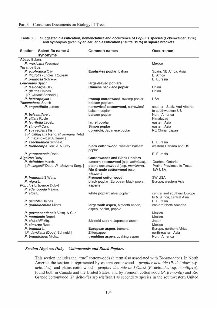
Part 3 – Consensus Documents on Biology of Trees
104
Table 3.5 Suggested classification, nomenclature and occurrence of Populus species (Eckenwalder, 1996) and synonyms given by an earlier classification (Zsuffa, 1975) in square brackets
Section Scientific name & synonyms
Common names Occurrence
Abaso Ecken.P. mexicana Wesmael Mexico
Turanga Bge. P. euphratica Oliv. Euphrates poplar, bahan Spain, NE Africa, Asia P. ilicifolia (Engler) Rouleau E. Africa P. pruinosa Schrenk E. Eurasia
Leucoides Spach large-leaved poplars P. lasiocarpa Oliv. Chinese necklace poplar China P. glauca Haines [P. wilsonii Schneid.]
China
P. heterophylla L. swamp cottonwood, swamp poplar, USA Tacamahaca Spach balsam poplars
P. angustifolia James narrowleaf cottonwood, narrowleaf balsam poplar
southern Sask. And Alberta to southwestern US
P. balsamifera L. balsam poplar North America P. ciliata Royle Himalayas P. laurifolia Ledeb. laurel poplar eastern Asia P. simonii Carr. Simon poplar eastern Asia P. suaveolens Fish. [ P. cathayana Rehd. P. koreana Rehd P. maximowiczii A.Henry ]
doronoki, Japanese poplar NE China, Japan
P. szechuanica Schneid. E. Eurasia P. trichocarpa Torr. & A.Gray black cottonwood, western balsam
poplar western Canada and US
P. yunnanensis Dode E. Eurasia Aigeiros Duby Cottonwoods and Black Poplars
P. deltoides Marsh. [ P. sargentii Dode, P. wislizenii Sarg. ]
eastern cottonwood (ssp. deltoides), plains cottonwood (ssp. monilifera),Rio Grande cottonwood (ssp. wislizenii
Quebec, Ontario Prairie Provinces to Texas SW USA
P. fremontii S.Wats. Fremont cottonwood SW USA P. nigra L. black poplar, European black poplar Europe, western Asia
Populus L. [Leuce Duby] aspens P. adenopoda Maxim.P. alba L. white poplar, silver poplar central and southern Europe
to N. Africa, central Asia P. gamblei Haines E. Eurasia P. grandidentata Michx. largetooth aspen, bigtooth aspen,
aspen, poplar, popple eastern North America
P. guzmanantlensis Vasq. & Cue. Mexico P. monticola Brand Mexico P. sieboldii Miq. Siebold aspen, Japanese aspen Japan P. simaroa Rzed. Mexico P. tremula L. [P. davidiana (Dode) Schneid.]
European aspen, tremble, Zitterpappel
Europe, northern Africa, north-eastern Asia
P. tremuloides Michx. trembling aspen, quaking aspen North America
Section Aigeiros Duby – Cottonwoods and Black Poplars.
This section includes the “true” cottonwoods (a term also associated with Tacamahaca). In North America the section is represented by eastern cottonwood – peuplier deltoïde (P. deltoides ssp. deltoides), and plains cottonwood – peuplier deltoïde de l’Ouest (P. deltoides ssp. monilifera), found both in Canada and the United States, and by Fremont cottonwood (P. fremontii) and Rio Grande cottonwood (P. deltoides ssp wislizeni) as secondary species in the southwestern United
Section 4 – Poplar
105
States. The black poplar – peuplier noir (P. nigra) is an important species native to North Africa, central and western Europe, and the cultivar known as Lombardy poplar – peuplier noir d’Italie(P. nigra cv. ‘Italica’) is commonly planted as a hardy ornamental in North America.
Section Populus L. (syn. Leuce Duby) – Aspens.
This section is further subdivided into two subsections, Albidae and Trepidae, containing the white poplars and aspens, respectively. The North American representatives of this section are both members of Trepidae: trembling aspen – peuplier faux-tremble (P. tremuloides), and largetooth aspen – peuplier à grandes dents (P. grandidentata). P. tremula is an important and highly variable aspen from Europe, while P. sieboldii is a recognised species from Japan. However, aspens throughout Eurasia are now thought to be races of a single, highly polymorphic species, viz. P. tremula (Barnes and Han 1993). While no white poplars are native to North America, the European white poplar – peuplier blanc (P. alba) was among the first species introduced from Europe.
Natural hybridisation has been reported between almost all sympatric poplar species, and between introduced and native poplars, both in North America and Europe (Schreiner 1974, Demeritt 1990). Natural hybridisation generally occurs between species in the same section to the limited extent that the parent species overlap (Brayshaw 1965, Eckenwalder 1977), although intersectional hybrids also occur. Species in different sections, though broadly sympatric, are ecologically isolated from one another, so that hybridisation occurs over large geographic areas but within a relatively narrow ecological range of overlap (Eckenwalder 1984a, c). Complicated natural hybrid populations may also form where three or more species are sympatric (Rood et al. 1986).
Eastern cottonwood was introduced into France from southeastern Canada in the late 1700’s. In southern Germany, since the early 1970’s, cross-breeding of P. × canadensis has been replaced by P.trichocarpa × P. deltoides or P. trichocarpa × P. Subsequent natural hybridisation with the native black poplar produced the hybrid which was named P. × canadensis in 1789 (Mühle Larsen 1960, Wright 1976). Clones of this hybrid are now widely planted across Europe. This hybrid was also the first poplar hybrid produced by controlled pollination, by Englishman A. Henry (Larsen 1956). Artificial hybridisation has been used in North America since the 1920s and 30s (Stout and Schreiner 1933, Heimburger 1936). Several of the more important hybrids occurring naturally in North America are listed, together with synonyms and common names, in Table 3.6.
3. Centres of Origin/Diversity
A. Natural distribution
The genus Populus is widely distributed throughout the Northern Hemisphere, in both the temperate and subtropical zones. Representative species are found from Alaska and Labrador south to northern Mexico, as well as Europe, North Africa, the Himalayas, mainland China and Japan (Schreiner 1974). Some species are very widely distributed. P. tremuloides, for example, is the most broadly distributed tree species in North America, spanning 110° of longitude and 47° of latitude, and the second most widely distributed in the world (Jones 1985, Barnes and Han 1993).
B. Evolution and migrational history
It was long felt that Populus was one of the oldest contemporary angiosperm genus, originating in China and Japan during the Triassic; however, these fossil records are now associated with other taxa. While the closest relatives in Flacourtiaceae are from tropical Asia, the fossil record now indicates that the genus Populus had tropical origins in North America during the late Paleocene, about 58 million years ago
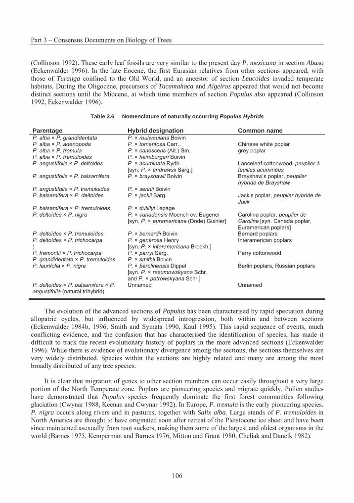
Part 3 – Consensus Documents on Biology of Trees
106
(Collinson 1992). These early leaf fossils are very similar to the present day P. mexicana in section Abaso(Eckenwalder 1996). In the late Eocene, the first Eurasian relatives from other sections appeared, with those of Turanga confined to the Old World, and an ancestor of section Leucoides invaded temperate habitats. During the Oligocene, precursors of Tacamahaca and Aigeiros appeared that would not become distinct sections until the Miocene, at which time members of section Populus also appeared (Collinson 1992, Eckenwalder 1996).
Table 3.6 Nomenclature of naturally occurring Populus Hybrids
Parentage Hybrid designation Common name P. alba × P. grandidentata P. × roulwauiana BoivinP. alba × P. adenopoda P. × tomentosa Carr.. Chinese white poplar P. alba × P. tremula P. × canescens (Ait.) Sm. grey poplar P. alba × P. tremuloides P. × heimburgeri BoivinP. angustifolia × P. deltoides P. × acuminata Rydb.
[syn. P. × andrewsii Sarg.] Lanceleaf cottonwood, peuplier à feuilles acuminées
P. angustifolia × P. balsamifera P. × brayshawii Boivin Brayshaw’s poplar, peuplier hybride de Brayshaw
P. angustifolia × P. tremuloides P. × sennii Boivin P. balsamifera × P. deltoides P. × jackii Sarg. Jack’s poplar, peuplier hybride de
JackP. balsamifera × P. tremuloides P. × dutillyi Lepage P. deltoides × P. nigra P. × canadensis Moench cv. Eugenei
[syn. P. × euramericana (Dode) Guinier] Carolina poplar, peuplier de Caroline [syn. Canada poplar, Euramerican poplars]
P. deltoides × P. tremuloides P. × bernardii Boivin Bernard poplars P. deltoides × P. trichocarpa )
P. × generosa Henry [syn. P. × interamericana Brockh.]
Interamerican poplars
P. fremontii × P. trichocarpa P. × parryi Sarg. Parry cottonwood P. grandidentata × P. tremuloides P. × smithii Boivin P. laurifolia × P. nigra P. × berolinensis Dippel
[syn. P. × rasumowskyana Schr. and P. × petrowskyana Schr.]
Berlin poplars, Russian poplars
P. deltoides × P. balsamifera × P. angustifolia (natural trihybrid)
Unnamed Unnamed
The evolution of the advanced sections of Populus has been characterised by rapid speciation during allopatric cycles, but influenced by widespread introgression, both within and between sections (Eckenwalder 1984b, 1996, Smith and Symata 1990, Kaul 1995). This rapid sequence of events, much conflicting evidence, and the confusion that has characterised the identification of species, has made it difficult to track the recent evolutionary history of poplars in the more advanced sections (Eckenwalder 1996). While there is evidence of evolutionary divergence among the sections, the sections themselves are very widely distributed. Species within the sections are highly related and many are among the most broadly distributed of any tree species.
It is clear that migration of genes to other section members can occur easily throughout a very large portion of the North Temperate zone. Poplars are pioneering species and migrate quickly. Pollen studies have demonstrated that Populus species frequently dominate the first forest communities following glaciation (Cwynar 1988, Keenan and Cwynar 1992). In Europe, P. tremula is the early pioneering species. P. nigra occurs along rivers and in pastures, together with Salix alba. Large stands of P. tremuloides in North America are thought to have originated soon after retreat of the Pleistocene ice sheet and have been since maintained asexually from root suckers, making them some of the largest and oldest organisms in the world (Barnes 1975, Kemperman and Barnes 1976, Mitton and Grant 1980, Cheliak and Dancik 1982).
Section 4 – Poplar
107
4. Reproductive Biology
A. Reproductive development
Poplars are normally dioecious and obligatory outcrossers; however, the occurrence of monoecious inflorescences and perfect flowers has been reported (Lester 1963a, b, Melchior 1967). Reproductive buds in P. tremuloides may develop into pistillate, staminate or perfect flowers, initiated at different times (Lester 1963a). P. lasiocarpa is a notable exception as it is normally monoecious and self-fertilising (Schreiner 1974). Overall sex ratios of 1:1 have been confirmed for P. tremuloides (Einspahr and Winton 1976, Grant and Mitton 1979) and for P. deltoides (Farmer 1964b), although an elevational gradient in sex ratio has been observed in P. tremuloides in the Rocky Mountains, with females more common at low elevations, while more than 90% are male above 3 200 m (Grant and Mitton 1979).
Reproductive buds are simple (Jackson and Sweet 1972). Their initiation and early development have been described in P. tremuloides and P. deltoides (Nagaraj 1952, Seitz 1958, Lester 1963a). Floral initiation takes place in buds, located in the axils of leaves on the current-year shoot. These buds are no more than small apices, each with a single bud scale when winter dormancy occurs (Owens and Blake 1985, Kaul 1995). Terminal buds burst in May, and axillary primordia initiate several bud scales during rapid shoot elongation. Reproductive apices are determined around mid-June (Lester 1963a). Pistillate primordia are first to begin development of floral parts by late June, followed by staminate primordia in early July. In P. deltoides, staminate flower buds can be readily distinguished from vegetative buds by midsummer, while identification of pistillate flowers requires dissection (Farmer 1976). Floral development continues within buds through September, so that anthers and ovules are well developed before winter dormancy (Owens and Blake 1985, Kaul 1995). For at least some Populus species, a chilling requirement must be satisfied before development will resume (Farmer 1964a). Megaspore mother cells differentiate in the spring, and microsporogenesis immediately precedes anthesis (Farmer and Pitcher 1981).
Flowers are borne in catkins (aments) early in the spring, prior to flushing of vegetative buds. When fully developed, both male and female catkins are 10 to 15 cm in length. Female flowers have from two to four, cap- or y-shaped stigmas, while the males have 30 to 80 stamens (Demeritt 1990). Each catkin bears a few dozen, one-celled capsules, each containing 10 to 30 seeds.
Male flowers ripen and shed pollen a few days before females, ensuring that pollen is in the air when the first females are receptive (Farmer and Pitcher 1981). Such pollen-pistil interactions have been largely documented in Populus nigra (Villar et al. 1987a, Villar et al.1993). Variation in flowering date is due to differences among trees and in P. deltoides is highly heritable (Farmer 1976). This variation in flowering date extends the pollination period from 2 to 3 weeks. Another study of natural variation in P. nigra over 111 sites in France revealed a fair level of diversity and a low overall differentiation, with an important intraregional gene diversity (Legionnet et al. 1997).
Pollen germinates within the first few hours after pollination. Fertilisation takes place several days later and is normally complete within two weeks (Farmer and Pitcher 1981). Seed development proceeds rapidly and dispersal occurs in most species by midsummer, before the full growth of the leaves (Schreiner 1974). In North American, the period of seed maturity in sections Populus and Tacamahaca is determined by temperature sums and is quite uniform within the limits of ecotypic zones (Pauley 1950). On the other hand, seed dispersal in the Aigeiros poplars may continue throughout the summer and early fall (Farmer 1966).
Part 3 – Consensus Documents on Biology of Trees
108
B. Mating system and gene flow
Two factors contribute to high gene flow and genetic diversity in poplars. Firstly, most are dioecious, and thus obligatory outcrossers. And secondly, in addition to being wind-pollinated, the long white, silky hairs attached to the short stalks of the seeds promote wind dispersal over great distances (Schreiner 1974), resulting in high rates of migration.
Electrophoretic studies in P. tremuloides suggest that gene flow is high, leading to a lack of differentiation among populations for putative neutral allozyme loci. However, the role of non-random mating in these same studies was variable, with no deviations from Hardy-Weinberg equilibrium detected in populations sampled in Minnesota (Lund et al. 1992), while an excess of heterozygotes were found in Alberta populations (Cheliak and Dancik 1982), and a deficiency of heterozygotes observed in Ontario populations (Hyun et al. 1987).
C. Seed production
Most poplars begin flowering between age 10 and 15 years (Schreiner 1974), although flowering in P.deltoides may occur as early as age four (Farmer and Pitcher 1981). Tacamahaca and Aigeiros poplars produce large annual seed crops. Those in section Populus produce some seeds each year, but bumper crops occur at intervals of three to five years. Poplars are prolific seed producers. A typical 12 m P.deltoides specimen was estimated to produce almost 28 million seeds in one season, and estimates for P. tremula have ranged as high as 54 million seeds. Poplar seeds are very small. Species in section Populuscan produce 6000 to 8000 seeds per gram, while North American Leucoides and Aigeiros produce from 300 to 450 seeds per gram (Schreiner 1974).
Typically, the longevity of poplar seeds under natural conditions is quite short – about two to four weeks. Under controlled low-temperature (-18 to 5° C) and stable moisture content (5 to 8%) conditions, storage time has been extended to 140 days for P. balsamifera (Hellum 1973), two years for P. tremuloides (Fechner et al. 1981), and five to six years for Aigeiros poplars (Tauer 1979, Muller and Tessier du Cros 1982).
D. Natural regeneration
Poplar seeds germinate or die within a few days after seedfall. Germination is epigeal. A fringe of hairs develops at the base of the hypocotyl, rendering the seedling upright and encouraging the root to grow down into the soil.
A favourable medium such as fine mineral soil is required for germination, together with light and continual moisture (McDonough 1979, Farrar 1995). Such conditions are rare, requiring fresh exposure of mineral soils, as found on shorelines, sandbars and old gravel pits. In North America, regeneration of section Populus from seed is confined to newly disturbed areas, whereas the primary mode of reproduction within stands is asexual (Barnes 1966, Schier 1973, Einspahr and Winton 1976).
E. Vegetative reproduction in nature
Except for members of section Populus, all poplars sprout vigorously from the stump and root collar. Coppicing occurs occasionally on young aspen (Zsuffa 1975). Reproduction from adventitious shoots on roots (root suckers) is common in many species, although less frequent in those in the Aigeiros and Leucoides sections.
Clonal groups of P. tremuloides in eastern North America are very common, but generally less than 0.1 ha in size, while in areas of Utah, groups as large as 80 ha have been observed (Kemperman and
Section 4 – Poplar
109
Barnes 1976). In the semi-arid western United States, some argue that widespread seedling establishment has not occurred since the last glaciation, some 10,000 years ago (Einspahr and Winton 1976, McDonough 1985). Indeed, some biologists feel that western clones could be as old as 1 million years (Barnes 1966, 1975). It has been claimed that a single clone, nicknamed "Pando" (Latin for I spread), covers 43 hectares, contains more than 47,000 stems and weighs in excess of 6 million kg, making it the largest known organism (Grant et al. 1992, Mitton and Grant 1996)
Studies have also demonstrated that both natural and vegetative propagation occur in nature, for example with P. nigra (Legionnet et al. 1997).
5. Genetics
A. Cytology
Poplars are normally found in the diploid condition with 2n = 38 chromosomes (Blackburn and Harrison 1924, Smith 1943). Polyploid individuals are rare and have only been reported in a half-dozen species (Darlington and Wylie 1956). While rare, the first discovery of a triploid forest tree was, in fact, a clone of P. tremula (Müntzing 1936). Several other natural triploid clones have since been found in both P. tremula and P. tremuloides, usually exhibiting larger leaves and exceptional growth (Einspahr et al. 1963, Heimburger 1968, Einspahr and Winton 1976).
Some reports suggest the sex determination in poplars is controlled by sex chromosomes (Peto 1938, Smith 1943, van Buijtenen and Einspahr 1959), however, this theory remains controversial. While published reports favour a genetic basis for gender, a linkage analysis of almost 2 500 PCR-based RAPD markers in a segregating family of F1 hybrid P. trichocarpa × P. deltoides failed to find any markers that were significantly associated with gender (McLetchie et al. 1994). The authors suggested that gender might be determined genetically by regions of the genome not sampled by the tested markers or by a complex of loci operating in an additive threshold manner or in an epistatic manner, or that gender is determined environmentally at an early zygote state.
B. Genetic variation
As already mentioned, the genus Populus is tremendously varied with species distributed throughout the Northern Hemisphere and the opportunities to generate novel genotypes through hybridisation are enormous. Breeding programs have not hesitated to exploit this genetic variability, although sound quantitative estimates of narrow-sense and broad-sense heritabilities, and covariances among selection criteria would undoubtedly have assisted in making breeding and selection strategies more efficient (Riemenschneider et al. 1996). Poplars are ideal species for quantitative genetics studies, as clonal replication can be readily accomplished to describe complex modes of gene action (Foster and Shaw 1988, Mullin and Park 1992, Bradshaw and Foster 1992). It is thus surprising that genetic variation has been studied in detail for only a few species and traits.
The true potential of poplar species can only be determined by genetic studies designed to resolve variation among and within stands. Unfortunately, the concentration of breeders on interspecific hybridisation has left this field largely ignored, and detailed studies of large natural populations are quite recent and only a few species are well documented (Mohrdiek 1983, Farmer 1991).
Population-level variability
Considerable clonal variation among populations may be expressed for growth traits and for Melampsora rust resistance, but for other characteristics there is often little geographic differentiation. Overall, data from molecular genetic studies suggest that gene flow through migration has been sufficient
Part 3 – Consensus Documents on Biology of Trees
110
to prevent genetic drift, inbreeding, and other processes that might give rise to geographic variation unrelated to adaptive selection (Farmer 1996).
Significant variation over a 10° latitudinal transect was observed for phenology (Farmer et al. 1988a), shoot/root allometric coefficients (Schnekenburger and Farmer 1989), and height growth among four provenances of P. balsamifera, with southern sources continuing to grow later in the season (Schnekenburger and Farmer 1989, Farmer 1993). These same populations exhibited very little geographic differentiation for isozyme characters (Farmer et al. 1988a), rooting ability (Farmer et al. 1989), and date of bud break (Farmer and Reinholt 1986). Another test series including a more restricted sampling of populations over a 3.5° range found significant population differences accounting for about 12% of the variation in two-year height, leaf morphology, sylleptic branching and pest resistance (Riemenschneider et al. 1992), with populations grouped into north-western, central, and south-eastern clusters (Riemenschneider and McMahon 1993).
Similarly, in P. tremuloides, isozyme and RAPD variation studies have shown little differentiation among populations (e.g., Hyun et al. 1987, Lund et al. 1992, Yeh et al. 1995), whereas considerable variation among populations is well-documented for morphology, growth and wood properties (van Buijtenen et al. 1959, Barnes 1969, Einspahr and Winton 1976). Variation among populations generally follows clinal trends, with wood density declining with increasing elevation (Valentine 1962) and from south to north (Einspahr and Benson 1967). A common-garden trial showed that northern and western provenances flushed and ceased growth first, with lower survival when grown in Michigan (Brissette and Barnes 1984), and another showed that better growing clones came from lower Michigan (Reighard and Hanover 1985). There is some evidence of a north-south increase in susceptibility of P. tremuloidespopulations to Hypoxylon mammatum (French and Hart 1978). Populations of P. tremuloides also vary with respect to ozone sensitivity, with tolerance correlated to maximum daily ozone levels, as well as annual precipitation and minimum temperature (Berrang et al. 1991).
Geographic variation is also well documented for P. deltoides. A wide range provenance trial established in Nebraska, included sources from Texas in the south, to Minnesota in the north and Pennsylvania in the east, and evaluated bark, stem, crown and leaf morphology, in addition to growth and survival (Ying and Bagley 1976). Clinal patterns of variation from north and west to south and east were observed for most traits. Cuttings from Nebraska, Minnesota and Wisconsin produced significantly higher numbers of roots than those from other sources (Ying and Bagley 1977). A similar clinal trend was found in a study of 40 populations in the southern Great Plains, where NW to SE patterns were observed for two-year height, diameter, branching and Melampsora rust resistance (Nelson and Tauer 1987). A study of nine populations in Ontario showed great variation in leaf morphology which was unrelated to latitude or longitude, and was not correlated with the moderate allozyme variation that suggested differences between eastern and western populations (Rajora et al. 1991).
A series of studies of P. trichocarpa populations in Washington have documented significant variation for leaf, branch and phenology characters (Weber et al. 1985), photosynthetic processes (Dunlap et al. 1993), survival height growth and biomass production (Heilman and Stettler 1985), volume production, Melampsora rust resistance and adaptation to arid sites (Dunlap et al. 1994), leaf and crown morphology (Dunlap et al. 1995). A sample of 10 populations over a 4.5° latitudinal range showed only weak clinal trends for three-year height and diameter (Rogers et al. 1989). Another sample of five riparian populations in Washington showed little difference among populations with respect to flood tolerance of young seedlings and rooted cuttings (Smit 1988).
Section 4 – Poplar
111
Individual-level variability
While the amount of genetic variation among populations differs greatly depending on the trait, the variation within populations is moderate to high for virtually all traits. Unfortunately, most genetic testing has focused on clonal materials without any particular family structure, and these studies have concentrated on the species and hybrids of sections Tacamahaca and Aigeiros. Generally, only estimates of broad-sense heritability (H2) are available, and genetic structure is rarely partitioned into additive and non-additive components (Riemenschneider et al. 1996). In the limited number of studies of seedling populations, narrow-sense heritability (h2) estimates for growth were similar to estimates of H2 in P. deltoides (Farmer 1970, Ying and Bagley 1976, Nelson and Tauer 1987), and much lower in the case of P. trichocarpa(Rogers et al. 1989).
A large number of studies of growth and yield characters in P. deltoides have produced consistent estimates of H2 between 0.20 and 0.50, with significant genotype-environment interactions, usually less than half as large as the corresponding genetic main effects (Wilcox and Farmer 1967, Farmer and Wilcox 1968, Mohn and Randall 1971, 1973, Randall and Cooper 1973, Foster 1986). A limited number of clonally replicated trials suggest that much of the genetic variance in yield is non-additive (Foster 1985, Foster and Shaw 1988). Heritability for stem growth in P. balsamifera was about H2 = 0.50 (Farmer et al.1988b). In both P. deltoides and P. balsamifera, C-effects (in sensu Lerner 1958) during the first year were often as large as that due to clones (Wilcox and Farmer 1968, Farmer et al. 1989), but appear to be less important after field planting (Farmer et al. 1988b). Detailed measurements of leaf, branch and phenological characteristics, which are known to affect tree productivity, have been employed to describe ideotypes (Dickmann and Keathley 1996) that may be useful in yield selection of P. balsamifera(Riemenschneider et al. 1992), P. trichocarpa (Riemenschneider et al. 1994), and hybrids involving P.deltoides, P. nigra and P. simonii (Wu 1994a, b). Indications of the utility of the ideotype concept for yield selection have been inconsistent.
Heritability of rooting and root characters is typically very high, with H2 estimates as high as 0.85 to 0.91 in P. deltoides (Wilcox and Farmer 1968, Ying and Bagley 1977). Other recent studies also indicate high heritability for rooting in P. trichocarpa (Riemenschneider et al. 1996).
Much effort has been concentrated on the inheritance of Melampsora rust resistance, due to its impact on poplar culture. Early studies of rust resistance in P. deltoides gave estimates of h2 between 0.38 and 0.66, and for H2 between 0.66 and 0.88 (Jokela 1966). Similarly high estimates for rust resistance or severity have since been reported for P. deltoides (Farmer and Wilcox 1968, Thielges and Adams 1975), P. tremula and P. tremuloides (Gallo et al. 1985), P. balsamifera (Riemenschneider et al. 1992), P. trichocarpa and its hybrids (Hsiang et al. 1993, Riemenschneider et al. 1994), and hybrids among P. deltoides, P. nigra, and P. maximowiczii (Rajora et al. 1994).
Molecular genetics
In recent years a great deal of research has been directed towards associating important traits with molecular markers, and in developing corresponding genetic maps (Bradshaw et al. 1994, Cervera et al.1997). The emphasis of this work has been directed towards adaptive traits (Bradshaw and Stettler, 1995) and to resistance to diseases (Villar et al. 1996), and suggests the role of a few quantitative trait loci (QTL’s) that have large effects on these quantitative traits.
C. Inbreeding depression and genetic load
Given the high rate of gene flow in poplars, one would expect low rates of inbreeding. A study of 200 clones of P. tremuloides in Ontario indicated a deficiency of heterozygotes and an average fixation index
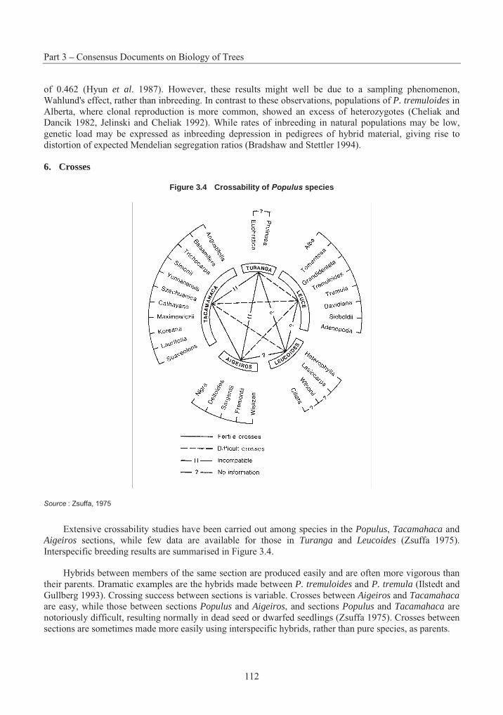
Part 3 – Consensus Documents on Biology of Trees
112
of 0.462 (Hyun et al. 1987). However, these results might well be due to a sampling phenomenon, Wahlund's effect, rather than inbreeding. In contrast to these observations, populations of P. tremuloides in Alberta, where clonal reproduction is more common, showed an excess of heterozygotes (Cheliak and Dancik 1982, Jelinski and Cheliak 1992). While rates of inbreeding in natural populations may be low, genetic load may be expressed as inbreeding depression in pedigrees of hybrid material, giving rise to distortion of expected Mendelian segregation ratios (Bradshaw and Stettler 1994).
6. Crosses
Figure 3.4 Crossability of Populus species
Source : Zsuffa, 1975
Extensive crossability studies have been carried out among species in the Populus, Tacamahaca and Aigeiros sections, while few data are available for those in Turanga and Leucoides (Zsuffa 1975). Interspecific breeding results are summarised in Figure 3.4.
Hybrids between members of the same section are produced easily and are often more vigorous than their parents. Dramatic examples are the hybrids made between P. tremuloides and P. tremula (Ilstedt and Gullberg 1993). Crossing success between sections is variable. Crosses between Aigeiros and Tacamahacaare easy, while those between sections Populus and Aigeiros, and sections Populus and Tacamahaca are notoriously difficult, resulting normally in dead seed or dwarfed seedlings (Zsuffa 1975). Crosses between sections are sometimes made more easily using interspecific hybrids, rather than pure species, as parents.
Section 4 – Poplar
113
The incompatibility of some species is characterised by arrested development of the pollen tube and failure to penetrate the stigma (Melchior and Seitz 1968, Guries and Stettler 1976, Stettler et al. 1980, Knox 1984, Villar et al. 1987b, Rougier et al. 1992, Villar et al. 1993). This block can be overcome in some cases by mixing the incompatible pollen with “mentor pollen”, i.e., compatible pollen that has been killed by means of gamma radiation (Stettler and Ager 1984, Knox et al. 1987, Villar and Gaget-Faurobert 1996), and by treatment of pollen and stigmas with solvents and extracts from compatible pollen (Whitecross and Willing 1975, Willing and Pryor 1976). This technique has made it possible to obtain crosses of section Populus with Aigeiros and Tacamahaca poplars that are otherwise difficult (Stettler 1968, Zufa 1971, Knox et al. 1972, Willing and Pryor 1976).
7. Ecology and Associated Species
A. Habitat
Poplars are found in a great variety of forest ecosystems, from boreal to sub-tropical, and from mountainous to riparian. In some environments such as boreal forests and in large river valleys, they form large stands, while in other situations they are found as small stands or groups of trees. Although there is some variation among species, essentially all poplars are very intolerant of shade. They are pioneering species and among the first to invade and re-colonise areas disturbed by harvesting, land clearing and fire. The Populus species and their hybrids vary greatly in their adaptability to climate, although all are nutrient demanding and perform best with an abundant and continuous supply of moisture (Heilman et al. 1996).
The members of section Populus are very broadly distributed and thus found over a tremendous range of climates. In P. tremuloides, the southern limit is roughly defined by the 24° C mean July temperature isotherm, while the northern limit corresponds to a mean annual degree-day sum of 700° C, at threshold temperature of 5.6° C (Fowells 1965). Within this range the species occurs where annual precipitation exceeds evapotranspiration. While P. tremuloides is found on a variety of soils, ranging from shallow and rocky to deep loamy sands and heavy clays, growth is strongly influenced by drainage and fertility (Perala 1990). Throughout the more restricted range of P. grandidentata, moisture is adequate in all seasons, with the least precipitation occurring at the north-west limit on the prairie border in Manitoba with only 510 mm. It is far less adaptable than P. tremuloides, developing best on moist, fertile sandy uplands with good aeration (Laidly 1990).
The members of the Tacamahaca and Aigeiros sections are referred to collectively as the riparian cottonwoods. Species in the Tacamahaca section are generally found at higher elevations and latitudes (high river systems of montane and young piedmont valley floodplains) (Braatne et al. 1996). In this section, P. balsamifera is the most broadly distributed, withstanding climatic extremes similar to that of P. tremuloides. It is usually restricted to moist, low-lying ground, and is one of the few boreal species associated with poorly drained clay soils having pH greater than 7.2 (Dix and Swan 1971, Zasada and Phipps 1990). P. trichocarpa is most commonly found in the humid coastal forests of the Pacific Northwest, performing best on deep alluvial soils with abundant moisture, nutrients, oxygen and pH 6.0 to 7.0 (Smith 1957, DeBell 1990). P. angustifolia has a very limited range where it is a pioneering species on gravel and sand bars near fast-flowing rivers (Brayshaw 1965).
Members of section Aigeiros are limited to lower elevations and latitudes (lower river systems of mature piedmont valley flood plains) (Braatne et al. 1996). The natural range of P. deltoides covers a wide southern range, with frost-free days ranging from less than 100 to more than 200, and rainfall from less than 380 mm in the northwest to more than 1,400 mm in the south. This species performs best on moist, well-drained, fine sandy or silt loams, and on sites that are rarely higher than 6 m above the average level of nearby streams (Cooper and Van Haverbeke 1990).
Part 3 – Consensus Documents on Biology of Trees
114
The only North American member of section Leucoides, P. heterophylla, is found in warm, humid areas with abundant rainfall. It performs best on deep moist soils of shallow swamps and low-lying areas near tidewater, and occupies sites that are too wet for P. deltoides (Johnson 1990).
B. Synecology and associated species
Poplars occur in the early successional stages over a wide range of forest ecosystems, so it is no surprise that ecological associations are tremendously diverse. This is particularly true for species in section Populus, as they are not restricted to riparian habitats. In Europe, P. tremula is the early pioneer. P.nigra occurs along rivers and in pastures, together with Salix alba, P. tremuloides is found in pure stands across its range, but also in mixed stands where it is commonly associated with White Spruce (Picea glauca), black spruce (Picea mariana), balsam fir (Abies balsamea), white birch (Betula papyrifera), P.balsamifera, and jack pine (Pinus banksiana). The associations with shrub and herbaceous species are even more numerous and varied (Perala 1990, Farrar 1995). P. grandidentata occurs in small pure stands, but more commonly as an associate in poplar stands with either P. tremuloides or P. balsamifera. It is a minor component of many other forest types and is thus associated with a wide range of shrubs and ground flora (Laidly 1990, Farrar 1995).
In section Tacamahaca, P. balsamifera reaches its best development on river flood plains where it occurs as pure stands and is associated with various willows and alders (Viereck et al. 1983). However, it also occurs in mixtures with boreal conifers and several hardwood species, and the associated shrub and herbaceous mixtures are numerous (Zasada and Phipps 1990). P. trichocarpa generally occurs in mixtures with larger willows, and to a lesser extent with several western conifers (DeBell 1990). Shrub and herbaceous associates are numerous, but better sites are characterised by beaked hazel (Corylus cornuta), elder (Sambucus spp.), salmonberry (Rubus spectabilis), nettles (Stachys spp.), swordfern (Polystichum munitum) and lady fern (Athyrium filix-femina) (Smith 1957).
On riparian sites, P. deltoides tends to grow as essentially pure stands or mixed in open stands with other riparian species. In the area where the species performs best, roughleaf dogwood (Cornus drummondii) and swamp-privet (Forestiera acuminata) are major shrub associates (Cooper and Van Haverbeke 1990).
C. Competition and stand structure
As mentioned earlier, all poplars are shade intolerant, early successional species, and disturbance is often essential in maintaining many poplar ecosystems. The availability of sites suitable for colonisation, particularly following fires, plays a major role in determining seedling establishment (DeByle and Winokur 1985, Jelinski and Cheliak 1992, Kay 1993). Once established, fire can also remove shade-tolerant competitors, allowing vigorous P. tremuloides sprouts to emerge from persistent root systems (Bailey et al. 1990), with densities in excess of 1 million per hectare (Schier et al. 1985). In the absence of disturbance, aspens are regarded as transient, and successional patterns are determined by soil water regime (Roberts and Richardson 1985). Intolerant associates will often out-live aspens, while tolerant hardwoods and conifers will also dominate by virtue of their ability to regenerate under shade.
The riparian cottonwoods are all very flood-tolerant, so that establishment and growth are promoted by the disturbances characteristic of alluvial habitats. In non-alluvial habitats, they take the opportunity to establish on moist agricultural fields, forest clearings and the margins of wetlands, but are eventually dominated by secondary forest species (Braatne et al. 1996). Willows and alders may precede the establishment of P. balsamifera, which is normally then replaced by White Spruce (Walker and Chapin 1986, Walker et al. 1986). P. deltoides is a very poor competitor, as it is very intolerant of shade, and
Section 4 – Poplar
115
competes well only with willows, owing to its faster growth except on very wet sites (Cooper and Van Haverbeke 1990).
When established as plantations, poplars and their hybrids must generally be established in pure stands. In addition to being highly intolerant of shade, young poplars cannot tolerate competition from grass, weeds or shrubs. Control of vegetation in the first few years is essential, although poplars are highly sensitive to many herbicides used for vegetation control (Demeritt 1990).
D. Ecosystem dynamics
Poplars coexist with a wide range of insects, but most pose a more serious threat only to artificial populations of planted species and their hybrids. In North America the most serious defoliator, particularly in hybrid plantings, is the cottonwood leaf beetle (Chrysomela scripta). Other foliage insects include the forest tent caterpillar (Malacosoma disstria), the poplar tent maker (Ichthyura inclusa), mourning cloak butterfly larvae (Nymphalis antiopa), the large aspen tortrix (Choristoneura conflictana), a leaf beetle (Zeugophora scutellaris), and the aspen blotch miner (Phyllocnistis populiella). The cottonwood twig borer (Gypsonoma haimbachiana) is particularly destructive, while several other borers may also do damage. Infestations of poplar gall midge (Prodiplosis morrisi) and many species aphids are also responsible for damage (Dickmann and Stuart 1983, Demeritt 1990). In China, in Ningzia Autonomous Region, it was reported that 24 million trees were destroyed due to attack by the longicorn, Anoplophora glabripennis (Chinese National Report, IPC 1996). Several mechanisms are thought to operate to give hybrids more or less resistance relative to their parental species, but as a rule hybrid populations are centres of insect abundance (Whitham et al. 1996).
Other insect pests include:
– Satin moth Stilpnoia salicis– Gluphisia septentrionis– Viceroy butterfly larvae Basilarchia archippus– Gypsy moth Lymantria dispar [European and Asian varieties] – Mourningcloak butterfly Nymphalis antiopa– Pandemis leafroller Pandemis pyrusana– Large aspen tortrix Christoneura conflictana– Forest tent caterpillar Malacosma dissitria– Poplar/willow borer Chryptorhyuchus lapthi– Clear-winger poplar borer Panthrene robinae– Poplar borer Saperda calcerata– Bronze poplar borer Agrilus grandulatus lirogus– Scented willow sawfly Nematus salicis odoratus– Phratora leaf Beetle Phratora californica– Flea beetle Altica sp. – Williamette balley Corythucha salicata– western willow lace bug – Cottonwood twig borer Gypsonoma haimbachiana– Aphids
Further information about insect pest species of poplar can be found in Peterson, et al. 1996, Hiratsuka, 1987, Furniss and Carolin, 1977, Hepting 1971 and USDA Forest Service 1979. A useful Internet website is “http://ww.cas.psu.edu/docs/CASDEPT/PLANText/poplar.html”.
Part 3 – Consensus Documents on Biology of Trees
116
The fungi associated with Populus species are tremendously diverse. More than 250 species are known to be associated with the decay of P. tremuloides alone (Lindsey and Gilbertson 1978). Only fungi associated with sections Populus, Aigeiros, and Tacamahaca have been studied to any extent, and “virtually nothing is known” of those associated with other sections (Newcombe 1996).
The five most damaging or potentially damaging diseases of Aigeiros and Tacamahaca are:
• Melampsora leaf rust (Melampsora spp.) which, while causing only moderate levels of mortality in plantations, can cause volume growth reduction of up to 65% (Widin and Schipper 1981). The situation is now becoming serious in Europe as variability in Melampsora larici-populina has been reported and interspecific hybrid trees selected for complete resistance to this fungus are now affected by new races of this pathogen (Pinon 1992a,b, Pinon, 1995, Pinon and Frey 1997).
• Marssonina anthracnose or leaf spot (Marssonina spp.), affecting Aigeiros species and some of their intersectional hybrids in particular, and causing an estimated 16% loss from production plantations in Italy (Thielges 1985).
• Bacterial canker (Xanthomonus populi Ridé), causing serious damage to non-native Aigeiros and Tacamahaca species planted in Europe (Thielges 1985).
• Dothiciza canker (Discosporium populeum), of minor importance in North America, but causing wide-spread, heavy losses to P. × canadensis clones in Europe (Waterman 1957, Thielges 1985).
• Septoria leaf spot and canker (Septoria musiva Peck), which is generally limited to a leaf spot in native stands but is particularly damaging to hybrids, and has prevented the general use of most P. × canadensis clones in Canada, the United States and Argentina (Thielges 1985).
Members of the Populus section are most likely to be affected by:
• Hypoxylon stem canker [Hypoxylon mammatum (Whal.) Miller] is broadly distributed on host species in section Populus, but only becomes a disease problem in certain areas (Manion and Griffin 1986, Newcombe 1996). It sometimes causes cankers on P. trichocarpa in Europe (Terrasson et al. 1988) and on various hybrid clones in North America (Ostry and McNabb 1986).
• White-rotting fungus (Phellinus tremulae (Bond.) Bond. & Borisov.) causes serious decay in aspens (Thomas et al. 1960), although the mechanism of resistance by members of Aigeiros and Tacamahaca to decay is unknown (Newcombe 1996).
Various poplar species and hybrids display well-developed adaptations to environmental stress, but in particular to drought, flooding, salinity, cold, and atmospheric pollutants such as ozone (Blake et al. 1996, Neuman et al. 1996). It is also noted that P. canescens shows stability against strong winds.
Many mammals feed on the bark, leaves and roots of Populus, notably snowshoe hares (Lepus americanus), beaver (Castor canadensis), porcupine (Erethizon dorsatum), pocket gophers (Thomomys bottae), and opossum (Trichosurus vulpecula) (Edwards 1978, Bryant 1981, Cantor and Whitham 1989, Basey et al. 1990). Ungulates such as deer, moose and elk (Cervus elaphus) not only browse on shoots and new sprouts, but also damage bark by chewing and rubbing with their antlers (Romme et al. 1995). Cattle and sheep also browse on regeneration and cause root damage to existing trees when allowed to range through stands (Cooper and Van Haverbeke 1990, Perala 1990). While little is known about the response of mammals to patterns of hybridisation in Populus, there is tremendous variation in feeding preference
Section 4 – Poplar
117
among hybrids and individual clones. It is suspected that this is due to variation in concentrations of phenolic glycosides which are known to be defensive toward mammals (Whitham et al. 1996).
Mice and voles can cause severe damage in young plantations (DeBell 1990). Many species of birds thrive in poplar forests, and a few can cause damage through their feeding. The ruffed grouse and the sharp-tailed grouse feed on aspen buds, and the ruffed grouse also feeds on the leaves during the summer months. Red-breasted and yellow-bellied sapsuckers may scar trees with drill holes as they forage for bark insects (Fowells 1965).
The riparian cottonwoods are one of the most productive and sensitive components of riparian ecosystems in western North America. The number of vertebrate species associated with these communities is four times higher than the numbers associated with spruce-fir, lodgepole pine, or Douglas-fir communities, yet human activities result in the loss of over 100 000 ha of riparian habitat each year (Finch and Ruggiero 1993). While riparian ecosystems occur on less than 1% of the western North American landscape, they provide habitat for more bird species than all other vegetation types combined (Knopf et al. 1988). Zones of hybridisation are thought to be centres of biodiversity (Whitham et al. 1996), representing a refugium for insect species (Whitham 1989), and thus a superior habitat for insectivorous birds (Martinsen and Whitham 1994, Dickson and Whitham 1996).
8. Summary
The wide distribution of Populus throughout the Northern Hemisphere represents an important and valuable component of many forest ecosystems and great potential for domestication. Evolution of the genus has been characterised by divergence into various sections, which offer even more opportunities for novel genetic combinations through hybridisation. Gene flow within the range of the individual species is usually very high, with populations distinguished only by their adaptive response to environmental selection pressures.
While initial establishment is by seeds, which may travel long distances to invade newly disturbed areas, the maintenance of populations often relies on poplar's ability to reproduce vegetatively. Plantation culture of poplars has exploited this trait, and most breeding programs are characterised by deployment of selected clones. Clonal variation is high for yield traits, as well as disease resistance and wood quality.
In North America, P. tremuloides is the most widely distributed tree species and an important component of many forest types. The riparian cottonwoods, from sections Aigeiros and Tacamahaca, play an important role in the maintenance of complex riparian communities, in addition to their importance in plantation culture. Although North American poplar communities are still largely intact, small populations on the edge of species' ranges are in need of conservation. Of even greater concern is the erosion of genetic resources, particularly for P. nigra, that has resulted from human activity in Europe.
Poplar is ideally suited as a model organism for understanding growth processes in forest trees. It has and will undoubtedly continue to be a target for domestication and forest management in many parts of the world.
Part 3 – Consensus Documents on Biology of Trees
118
REFERENCES
Ahuja, M.R. 1987. In vitro propagation of poplar and aspen. In Cell and tissue culture in forestry. Vol. 3. Edited by J.M. Bonga and D.J. Durzan. Nijhoff, The Hague. pp. 207-223.
Bailey, A.W., Irving, B.D., and Fitzgerald, R.D. 1990. Regeneration of woody species following burning and grazing in Aspen Parkland. Journal of Range Management 43: 212-215.
Barnes, B.V. 1966. The clonal growth habit of American aspens. Ecology, 47: 439-447.
Barnes, B.V. 1969. Natural variation and delineation of clones of Populus tremuloides and P.grandidentata in northern Lower Michigan. Silvae Genet. 18: 130-142.
Barnes, B.V. 1975. Phenotypic variation of trembling aspen in western North America. For. Sci. 21: 319-328.
Barnes, B.V., and Han, F.Q. 1993. Phenotypic variation of Chinese aspens and their relationship to similar taxa in Europe and North America. Can. J. Bot. 71: 799-815.
Basey, J.M., Jenkins, S.H., and Miller, G.C. 1990. Food selection by beavers in relation to inducible defenses of Populus tremuloides. Oikos 59: 57-62.
Benson, M.K., and Schwalbach, D.E. 1970. Techniques for rooting aspen root sprouts. Tree Plant. Notes, 21: 12-14.
Berrang, P., Karnosky, D.F., and Bennett, J.P. 1991. Natural selection for ozone tolerance in Populus tremuloides: an evaluation of nationwide trends. Can. J. For. Res. 21: 1091-1097.
Bisoffi, S., and Gullberg, U. 1996. Poplar breeding and selection strategies. In Biology of Populus and its implications for management and conservation. Edited by R.F. Stettler, H.D. Bradshaw Jr., P.E. Heilman and T.M. Hinkley. NRC Research Press, National Research Council Canada, Ottawa, ON. pp. 139-158.
Blackburn, K.B., and Harrison, J.W.H. 1924. A preliminary account of the chromosomes and chromosome behaviour in the Salicaceae. Ann. Bot. 38: 361-378.
Blake, T.J., Sperry, J.S., Tschaplinski, T.J., and Wang, S.S. 1996. Water relations. In Biology of Populusand its implications for management and conservation. Edited by R.F. Stettler, H.D. Bradshaw Jr., P.E. Heilman and T.M. Hinckley. NRC Research Press, National Research Council of Canada, Ottawa, ON. pp. 401-422.
Braatne, J.H., Rood, S.B., and Heilman, P.E. 1996. Life history, ecology and conservation of riparian cottonwoods in North America. In Biology of Populus and its implications for management and conservation. Edited by R.F. Stettler, H.D. Bradshaw Jr., P.E. Heilman and T.M. Hinkley. NRC Research Press, National Research Council Canada, Ottawa, ON. pp. 57-85.
Section 4 – Poplar
119
Bradshaw, H.D. and Foster, G.S. 1992. Marker-aided selection and propagation systems in trees: advantages of cloning for studying quantitative inheritance. Can. J. For. Res. 22: 1044-1049.
Bradshaw, H.D., Jr., and Stettler, R.F. 1993. Molecular genetics of growth and development in Populus. I. Triploidy in hybrid poplars. Theor. Appl. Genet. 86: 301-307.
Bradshaw, H.D., Jr., and Stettler, R.F. 1994. Molecular genetics of growth and development in Populus. II. Segregation distortion due to genetic load. Theor. Appl. Genet. 89: 551-558.
Bradshaw, H.D., Villar, M., Watson, B., Otto, K., Stewart, S., and Stettler, R. F. 1994. Molecular genetics of growth and development in Populus. III. A genetic linkage map of a hybrid poplar composed of RFLP, STS and RAPD markers. Theor. Appl. genet. 89: 167-178.
Bradshaw, H.D., Jr., and Stettler, R.F. 1995. Molecular genetics of growth and development in Populus.IV. Mapping QTLs with large effects on growth, form, and phenology traits in a forest tree. Genetics 139: 963-973.
Brayshaw, T.C. 1965. Native poplars of southern Alberta and their hybrids. Publ. No. 1109, Department of Forestry, Ottawa. 40 pp.
Brissette, J.C., and Barnes, B.V. 1984. Comparisons of phenology and growth of Michigan and western North American sources of Populus tremuloides. Can. J. For. Res. 14: 789-793.
Browicz, K. 1966. Populus ilicifolia (Engler) Rouleau and its taxonomic position. Acta Soc. Bot. Poloniae, 35: 325-335.
Bryant, J.P. 1981. Phytochemical deterrence of snowshow hare browsing by adventitious shoots of four Alaskan trees. Science, 313: 889-890.
Burns, R.M., and Honkala, B.H. 1990. Silvics of North America. Agriculture Handbook 654, Forest Service, USDA, Washington, DC. 877 pp.
Burr, K.E. 1986. Greenhouse production of quaking aspen seedlings. In Gen. Tech. Rep. RM-125. Rocky Mtm. For. Range Exp. Sta., USDA For. Serv. pp. 31-37.
Cagelli, L., and Lefèvre, F. 1996. The conservation of Populus nigra L. and gene flow with cultivated poplars in Europe. Forest Genetics, 2:135-144
Cantor, L.F., and Whitham, T.G. 1989. Importance of belowground herbivory: pocket gophers may limit aspen to rock outcrop refugia. Ecology, 70: 962-970.
Cervera, M. T., Villar, M. Faivre-Rampant, P., Goué, M. C., Van Montagu, M. and Boerjan, W. 1997, Applications of molecular marker technologies in Populus breeding. In Microporopatation, Genetic Engineering, and Molecular Biology of Populus. Gen. tech. Rep. RM-GTR-297, Fort Collins, CO:USDA, Forest Service, : Klopfenstein, N. B. Chun, Y. W., Kim, M. S. and Ahuja, M. R. Edited by, Dillon, M. C., Carman, R. C. and Eskew, L. G.(tech. eds.) pp 101-116.
Cheliak, W.M., and Dancik, B.P. 1982. Genic diversity of natural populations of a clone-forming tree Populus tremuloides. Can. J. Genet. Cytol. 24: 611-616.
Collinson, M.E. 1992. The early fossil history of the Salicaceae. Proc. Roy. Soc. Edinburgh Sect. B (Biol. Sci.), 98: 155-177.
Part 3 – Consensus Documents on Biology of Trees
120
Cooper, D.T., and Van Haverbeke, D.F. 1990. Populus deltoides Bartr. ex Marsh. Eastern cottonwood. InSilvics of North America. Vol. 2, Hardwoods. Edited by R.M. Burns and B.H. Honkala. Agriculture Handbook 654, Forest Service, USDA, Washington, DC. pp. 530-543.
Cwynar, L.C. 1988. Late Quaternary vegetation history of Kettlehole Pond, southwestern Yukon. Can. J. For. Res. 18: 1270-1279.
Darlington, C.D., and Wylie, A.P. 1956. Chromosome atlas of flowering plants. MacMillan Co., New York.
DeBell, D.S. 1990. Populus trichocarpa Torr. & Gray. Black cottonwood. In Silvics of North America. Vol. 2, Hardwoods. Edited by R.M. Burns and B.H. Honkala. Agriculture Handbook 654, Forest Service, USDA, Washington, DC. pp. 570-576.
DeByle, N.V., and Winokur, R.P. 1985. Aspen: ecology and management in the western United States. Gen. Tech. Rep. RM-119, USDA Forest Service, Fort Collins, CO. 283 pp.
Demeritt, M.E., Jr. 1990. Populus L. Poplar hybrids. In Silvics of North America. Vol. 2, Hardwoods. Edited by R.M. Burns and B.H. Honkala. Agriculture Handbook 654, Forest Service, USDA, Washington, DC. pp. 577-582.
Dickmann, D.I., and Keathley, D.E. 1996. Linking physiology, molecular genetics, and the Populusideotype. In Biology of Populus and its implications for management and conservation. Edited byR.F. Stettler, H.D. Bradshaw Jr., P.E. Heilman and T.M. Hinkley. NRC Research Press, National Research Council of Canada, Ottawa, ON. pp. 491-514.
Dickmann, D.I., and Stuart, K.W. 1983. Culture of hybrid poplars in northeastern North America. Michigan State University, Department of Forestry, East Lansing, MI. 168 pp.
Dickson, L.L., and Whitham, T.G. 1996. Genetically-based plant resistance traits affect arthropods, fungi, and birds. Oecolgia, 106: 400-406.
Dirr, M.A., and Heuser, C.W., Jr. 1987. The reference manual of woody plant propagation: from seed to tissue culture. Varsity Press, Athens, GA. 239 pp.
Dix, R.L., and Swan, H.M.A. 1971. The roles of disturbance and succession in upland forest at Candle Lake, Saskatchewan. Can. J. Bot. 49: 657-676.
Dunlap, J.M., Braatne, J.H., Hinckley, T.M., and Stettler, R.F. 1993. Intraspecific variation in photosynthetic traits of Populus trichocarpa. Can. J. Bot. 71: 1304-1311.
Dunlap, J.M., Heilman, P.E., and Stettler, R.F. 1994. Genetic variation and productivity of Populus trichocarpa and its hybrids. VII. Two-year survival and growth of native black cottonwood clones from four river valleys in Washington. Can. J. For. Res. 24: 1539-1549.
Dunlap, J.M., Heilman, P.E., and Stettler, R.F. 1995. Genetic variation and productivity of Populus trichocarpa and its hybrids. VIII. Leaf and crown morphology of native P. trichocarpa clones from four river valleys in Washington. Can. J. For. Res. 25: 1710-1724.
Eckenwalder, J.E. 1977. North American cottonwoods (Populus, Salicaceae) of sections Abaso and Aigeriros. J. Arnold Arboretum 58: 193-208.
Section 4 – Poplar
121
Eckenwalder, J.E. 1984a. Natural intersectional hybridization between North American species of Populus(Salicaceae) in sections Aigeiros and Tacamahaca. I. Population studies of P. X parryi. Can. J. Bot. 62: 317-324.
Eckenwalder, J.E. 1984b. Natural intersectional hybridization between North American species of Populus(Salicaceae) in sections Aigeiros and Tacamahaca. III. Paleobotany and evolution. Can. J. Bot. 62: 336-342.
Eckenwalder, J.E. 1984c. Natural intersectional hybridization between North American species of Populus(Salicaceae) in sections Aigerios and Tacamahaca. II. Taxonomy. Can. J. Bot. 62: 325-335.
Eckenwalder, J.E. 1996. Systematics and evolution of Populus. In Biology of Populus and its implications for management and conservation. Edited by R.F. Stettler, H.D. Bradshaw Jr., P.E. Heilman and T.M. Hinkley. NRC Research Press, National Research Council Canada, Ottawa, ON. pp. 7-32.
Edwards, W.R.N. 1978. Effect of salicin content on palatibility of Populus foliage to opossum (Trichosurus vulpecula). N. Z. J. Sci. 21: 103-106.
Einspahr, D.W., and Benson, M.K. 1967. Geographic variation of quaking aspen in Wisconsin and Upper Michigan. Silvae Genet. 16: 106-112.
Einspahr, D.W., and Winton, L.L. 1976. Genetics of quaking aspen. Res. Pap. WO-25, USDA For. Serv. 23 pp.
Einspahr, D.W., van Buijtenen, J.P., and Peckham, J.R. 1963. Natural variation and heritability in triploid aspen. Silvae Genet. 12: 51-58.
Farmer, R.E., Jr. 1964a. Cottonwood flowering as related to cold requirements of flower buds. For. Sci. 10: 296-299.
Farmer, R.E., Jr. 1964b. Sex ratio and sex-related characteristics in eastern cottonwood. Silvae Genet. 13: 116-118.
Farmer, R.E., Jr. 1966. Variation in time of flowering and seed dispersal of eastern cottonwood in the lower Mississippi Valley. For. Sci. 12: 343-347.
Farmer, R.E., Jr. 1970. Genetic variation among open-pollinated progeny of eastern cottonwood. Silvae Genet. 19: 149-151.
Farmer, R.E., Jr. 1976. Sexual reproduction of eastern cottonwood. In Proceedings of Symposium on Eastern Cottonwood and Related Species, Greenville, Mississippi. Louisiana State Univ., Baton Rouge, LA. pp. 89-98.
Farmer, R.E., Jr. 1991. Genetic improvement of poplar in western Canada: alternatives, opportunities, and pitfalls. In Aspen management for the 21st century. Edited by S. Navratil and P.B. Chapman. pp. 129-134.
Farmer, R.E., Jr. 1993. Latitudinal variation in height and phenology of balsam poplar. Silvae Genet. 42: 148-153.
Part 3 – Consensus Documents on Biology of Trees
122
Farmer, R.E., Jr. 1996. Genecology of Populus. In Biology of Populus and its implications for management and conservation. Edited by R.F. Stettler, H.D. Bradshaw Jr., P.E. Heilman and T.M. Hinkley. NRC Research Press, National Research Council Canada, Ottawa, ON. pp. 33-55.
Farmer, R.E., Jr., and Pitcher, J.A. 1981. Pollen handling for Southern hardwoods. In Pollen management handbook. Edited by E.C. Franklin. Agriculture Handbook 587, USDA For. Serv., Washington, DC. pp. 77-83.
Farmer, R.E., Jr., and Reinholt, R.W. 1986. Genetic variation in dormancy relations of balsam poplar along a latitudinal transect in northwestern Ontario. Silvae Genet. 35: 38-42.
Farmer, R.E., Jr., and Wilcox, J.R. 1968. Preliminary testing of eastern cottonwood clones. Theor. Appl. Genet. 38: 197-201.
Farmer, R.E., Jr., Cheliak, W.M., Perry, D.J., Knowles, P., Barrett, J., and Pitel, J.A. 1988a. Isozyme variation in balsam poplar along a latitudinal transect in northwestern Ontario. Can. J. For. Res. 18: 1078-1081.
Farmer, R.E., Jr., Garlick, K., and Watson, S. 1988b. Heritability and C effects in a 3-year-old balsam poplar clonal test. Can. J. For. Res. 18: 1059-1062.
Farmer, R.E., Jr., Freitag, M., and Garlick, K. 1989. Genetic variance and 'C' effects in balsam poplar rooting. Silvae Genet. 38: 62-65.
Farrar, J.L. 1995. Trees in Canada. Fitzhenry & Whiteside/Canadian Forest Service, Markham and Ottawa.
Fechner, G.H., Burr, K.E., and Myers, J.F. 1981. Effects of storage, temperature, and moisture stress on seed germination and early seedlings development of trembling aspen. Can. J. For. Res. 11: 718-722.
Finch, D.M., and Ruggiero, L.F. 1993. Wildlife habitats and biological diversity in the Rocky Mountains and the northern Great Plains. Natural Areas Journal 13: 191-203.
Foster, G.S. 1985. Genetic parameters for two eastern cottonwood populations in the lower Mississippi Valley. In Proceedings of the 18th Southern Forest Tree Improvement Conference, 21-23 May 1985, Long Beach, MS. Edited by R.C. Schmidtling and M.M. Griggs. pp. 258-266.
Foster, G.S. 1986. Provenance variation of eastern cottonwood in the lower Mississippi Valley. Silvae Genet. 35: 32-38.
Foster, G.S., and Shaw, D.V. 1988. Using clonal replicates to explore genetic variation in a perennial plant species. Theor. Appl. Genet. 76: 788-794.
Fowells, H.A. 1965. Silvics of forest trees of the United States. Agricultural Handbook No. 271, Forest Service, USDA, Washington, DC. 762 pp.
French, J.R., and Hart, J.H. 1978. Variation in resistance of trembling aspen to Hypoxylon mammatumidentified by inoculating naturally occurring clones. Phytopathology, 68: 485-489.
Fröhlich, H., and van der Meiden, H.A. 1979. Propagation of poplars: nursery techniques. In Poplars and willows in wood production and land use. Food and Agriculture Organization of the United Nations, Rome. pp. 88-106.
Section 4 – Poplar
123
Fröhlich, H.J., and Weisgerber, H. 1985. Research on in vitro techniques within the framework of poplar breeding - results and future trends. Silvae Genet. 34: 132-137.
Furniss, R. L. and Carolin, V. M. 1977. Western Forest Insects. USDA Forest Service Miscellaneous Publication No. 1339.
Gallo, L.A., Stephan, B.R., and Krusche, D. 1985. Genetic variation of Melampsora leaf rust resistance in progenies of crossings between and within Populus tremula and P. tremuloides clones. Silvae Genet. 34: 208-214.
Grant, M.C., and Mitton, J.B. 1979. Elevational gradients in adult sex ratios and sexual differentiation in vegetative growth rates of Populus tremuloides Michx. Evolution, 33: 914-918.
Grant, M.C., Mitton, J.B., and Linhart, Y.B. 1992. Even larger organisms. Nature, 360: 216.
Guries, R.P., and Stettler, R.F. 1976. Prefertilisation barriers to hybridization in the poplars. Silvae Genet. 25: 37-43.
Hall, R.B., Colletti, J.P., Schultz, R.C., Faltonson, R.R., Kolison, S.H., Jr., Hanna, R.D., Hillson, T.D., and Morrison, J.W. 1989. Commercial-scale vegetative propagation of aspens. In Proceedings of Aspen Symposium, 25-27 July 1989, Duluth, MN. Gen. Tech. Rep. NC-140, USDA Forest Service, Duluth, MN. pp. 211-219.
Heilman, P.E., and Stettler, R.F. 1985. Genetic variation and productivity of Populus trichocarpa and its hybrids. II. Biomass production in a 4-year plantation. Can. J. For. Res. 15: 384-388.
Heilman, P.E., Hinckley, T.M., Roberts, D.A., and Ceulemans, R. 1996. Production physiology. In Biology of Populus and its implications for management and conservation. Edited by R.F. Stettler, H.D. Bradshaw Jr., P.E. Heilman and T.M. Hinkley. NRC Research Press, National Research Council of Canada, Ottawa, ON. pp. 459-489.
Heimburger, C. 1936. Report on poplar hybridization. For. Chron. 12: 285-290.
Heimburger, C. 1968. Poplar breeding in Canada. In Growth and utilization of poplars in Canada. Publ. No. 1205, Can. For. Bra. pp. 88-100.
Hellum, A.K. 1973. Seed storage and germination of balsam poplar. Can. J. Plant Sci. 53: 227-228.
Hepting, G. H. 1971. Diseases of Forest Shade Trees of the United States. USDA Forest Service Agricultural Handbook No. 386.
Hiratsuka, Y. 1987. Forest Tree Diseases of the Prairie Provinces. Can. For. Serv., North For. Cent., Edmonton, Alberta. Inf. Rep. NOR-X-286. ISBN 0-662-15281-6, ISSN 0704-7673.
Hsiang, T., Chastagner, G.A., Dunlap, J.M., and Stettler, R.F. 1993. Genetic variation and productivity of Populus trichocarpa and its hybrids. VI. Field susceptibility of seedlings to Melampsora occidentalis leaf rust. Can. J. For. Res. 23: 436-441.
Hyun, J.O., Rajora, O.P., and Zsuffa, L. 1987. Genetic variation in trembling aspen in Ontario based on isozyme studies. Can. J. For. Res. 17: 1134-1138.

Part 3 – Consensus Documents on Biology of Trees
124
Ilstedt, B., and Gullberg, U. 1993. Genetic variation in a 26-year-old hybrid aspen trial in southern Sweden. Scand. J. For. Res. 8: 185-192.
[IPC] International Poplar Commission. 1992. Proceedings Working Party on Breeding and Selection of Poplars and Willows, 19th Session IPC, Zaragoza, Spain, 22-25 September 1992. International Poplar Commission.
[IPC] International Poplar Commission. 1996. Synthesis of national reports on activities related to poplar and willow areas, production, consumption and the functioning of the National Poplar Commissions. Secretariat Note, 20th Session, Budapest, Hungary, 2-4 October 1996, International Poplar Commission. 10 pp.
Jackson, D.I., and Sweet, G.B. 1972. Flower initiation in temperate wood plants: a review based largely on the literature of conifers and deciduous fruit trees. Horticult. Abstr. 42: 9-24.
Jelinski, D.E., and Cheliak, W.M. 1992. Genetic diversity and spatial subdivision of Populus tremuloides(Salicaceae) in a heterogeneous landscape. Amer. J. Bot. 79: 728-736.
Johnson, R.L. 1990. Populus heterophylla L. Swamp cottonwood. In Silvics of North America. Vol. 2, Hardwoods. Edited by R.M. Burns and B. H. Honkala. Agriculture Handbook 654, Forest Service, USDA, Washington, DC. pp. 551-554.
Jokela, J.J. 1966. Incidence and heritability of Melampsora rust in a seedling plantation of hybrid poplar. In Breeding pest resistant trees. Edited by H.D. Gerhold, E.J. Schriener, R.E. McDermott and J.A. Winieski. Pergamon Press, Oxford, UK. pp. 111-117.
Jones, J.R. 1985. Distribution. In Aspen: ecology and management in the western United States. Edited byN.V. DeByle and R.P. Winokur. Gen. Tech. Rep. RM-119, USDA Forest Service, Fort Collins, CO. pp. 9-10.
Kaul, R.B. 1995. Reproductive structure and organogenesis in a cottonwood, Populus deltoides(Salicaceae). Int. J. Plant Sci. 156: 172-180.
Kay, C.E. 1993. Aspen seedlings in recently burned areas of Grand Teton and Yellowstone National Parks. Northwest Sci. 67: 94-104.
Keenan, T.J., and Cwynar, L.C. 1992. Late Quaternary history of black spruce and grasslands in southwest Yukon Territory. Can. J. Bot. 70: 1336-1345.
Kemperman, J.A., and Barnes, B.V. 1976. Clone size in American aspens. Can. J. Bot. 54: 2603-2607.
Khosla, P.K., and Khurana, K.D. 1982. Evolution of genus Populus L. and systematic placement of P. ciliata Wall. ex Royle. J. Tree Sci. 1: 81-87.
Knopf, F.L., Johnson, R.R., Rich, T., Samson, F.B., and Szaro, R.C. 1988. Conservation of riparian ecosystems in the United States. Wilson Bull. 100: 272-284.
Knox, R.B. 1984. Pollen-pistil interactions. In Cellular interactions. Edited by H.F. Linskens and J. Heslop-Harrison. Springer Verlag, Berlin. pp. 508-608.
Knox, R.B., Willing, R.R., and Pryor, L.D. 1972. Interspecific hybridization in poplars using recognition pollen. Silvae Genet. 21: 65-69.
Section 4 – Poplar
125
Knox, R.B., Gaget, M., and Dumas, C. 1987. Mentor pollen techniques. Int. Rev. Cytol. 107: 315-332.
Kouider, M., Skirvin, R.M., Saladin, K.P., Dawson, J.O., and Jokela, J.J. 1984. A method to culture immature embryos of Populus deltoides in vitro. Can. J. For. Res. 14: 956-958.
Krüssmann, G. 1985. Manual of cultivated broad-leaved trees and shrubs. Vol. II, E-PRO. Timber Press, Portland, OR. 445 pp. [Translated by M.E. Epp from the 1977 German edition]
Laidly, P.R. 1990. Populus grandidentata Michx. Bigtooth aspen. In Silvics of North America. Vol. 2, Hardwoods. Edited by R.M. Burns and B.H. Honkala. Agriculture Handbook 654, Forest Service, USDA, Washington, DC. pp. 544-550.
Legionnet, A., Faivre-Rampant, P. Villar, M. and Lefèvre, F. 1997. Sexual and asexual reproduction in natural stands of Populus nigra. Bot. Acta 110: 257-263.
Larsen, C.S. 1956. Genetics in silviculture. Oliver and Boyd, Edinburgh. [Translated by M.L. Anderson]
Lerner, I.M. 1958. The genetic basis of selection. John Wiley & Sons, New York. 298 pp.
Lester, D.T. 1963a. Floral initiation and development in quaking aspen. For. Sci. 9: 323-329.
Lester, D.T. 1963b. Variation in sex expression in Populus tremuloides Michx. Silvae Genet. 12: 141-151.
Li, B. 1995. Aspen improvement strategies for western Canada - Alberta and Saskatchewan. For. Chron. 71: 720-724.
Li, B., and Wyckoff, G.W. 1991. A breeding strategy to improve aspen hybrids for the University of Minnesota Aspen/Larch Genetics Cooperative. In Proceedings of the International Energy Agency 1991 Joint Meeting of the Task V Activity Groups on Exchange of Genetic Material, Disease/Management, and Joint Trials of Alnus, Populus, and Salix, 22-27 August, 1991. Iowa State University, Ames, Iowa. pp. 33-41.
Li, B., Wyckoff, G.W., and Einspahr, D.W. 1993. Hybrid aspen performance and genetic gains. North. J. Appl. For. 10: 117-122.
Lindsey, J.P., and Gilbertson, R.L. 1978. Basidiomycetes that decay aspen in North America. Bibl. Mycol. 63: 1-406.
Lund, S.T., Furnier, G.R., and Mohn, C.A. 1992. Isozyme variation in quaking aspen in Minnesota. Can. J. For. Res. 22: 521-524.
Manion, P.D., and Griffin, D.H. 1986. Sixty-five years of research on Hypoxylon canker of aspen. Plant Disease, 70: 803-808.
Martinsen, G.D., and Whitham, T.G. 1994. More birds nest in hybrid cottonwoods. Wilson Bull. 106: 474-481.
McDonough, W.T. 1979. Quaking aspen - seed germination and early seedling growth. Res. Pap. INT-234, USDA For. Serv., Ogden, UT. 13 pp.
Part 3 – Consensus Documents on Biology of Trees
126
McDonough, W.T. 1985. Sexual reproduction, seeds and seedlings. In Aspen: ecology and management in the western United States. Edited by N.V. DeByle and R.P. Winokur. Gen. Tech. Rep. RM-119, USDA Forest Service, Fort Collins, CO.
McLetchie, D.N., Tuskan, G.A., and Dietrichson, J. 1994. Gender determination in Populus. Norweigan J. Agric. Sci. 18: 57-66.
Melchior, G.H. 1967. Zwei Funde von Zwittrigkeit an Pappeln der Sektion Aigeiros. Silvae Genet. 16: 77-80.
Melchior, G.H., and Seitz, F.W. 1968. Interspezifische KreuzungssterilitÄt innerhalb der Pappelsektion Aigeiros. Silvae Genet. 17: 88-93.
Michler, C. H. and Bauer, E.O. 1991. Highr frequency comatic embryogenesis from leaf tissue fo Populusspp. Plant Science 77: 111-118.
Mitton, J.B., and Grant, M.C. 1980. Observations on the ecology and evolution of quaking aspen Populus tremuloides, in the Colorado front range. Amer. J. Bot. 67: 202-209.
Mitton, J.B., and Grant, M.C. 1996. Genetic variation and the natural history of quaking aspen. BioScience, 46: 25-31.
Mohn, C.A., and Randall, W.K. 1971. Inheritance and correlation of growth characters in Populus deltoides. Silvae Genet. 20: 182-184.
Mohn, C.A., and Randall, W.K. 1973. Interaction of cottonwood clones with site and planting year. Can. J. For. Res. 3: 329-332.
Mohrdiek, O. 1983. Discussion: future possibilities for poplar breeding. Can. J. For. Res. 13: 465-471.
Morley, P.M., and Balatinecz, J.J. 1993. Poplar utilization in Canada: past, present and future. For. Chron. 69: 46-52.
Mühle Larsen, C. 1960. L'Amélioration du peuplier par voie génétique. Extrait de bulletin de la Société Royale Forestière de Belgique, Mars-Avril: 48 pp.
Müntzing, A. 1936. The chromosomes of a giant Populus tremula. Hereditas, 21: 383-393.
Muller, C., and Tessier du Cros, E. 1982. Storage of Populus nigra seed for five years. Ann. Sci. For. 39: 179-185.
Mullin, T.J., and Park, Y.S. 1992. Estimating genetic gains from alternative breeding strategies for clonal forestry. Can. J. For. Res. 22: 14-23.
Nagaraj, M. 1952. Floral morphology of Populus deltoides and P. tremuloides. Bot. Gaz. 114: 222-243.
Nelson, C.D., and Tauer, C.G. 1987. Genetic variation in juvenile characters of Populus deltoides Bartr. from the southern Great Plains. Silvae Genet. 36: 216-221.
Neuman, D.S., Wagner, M., Braatne, J.H., and Howe, J. 1996. Stress physiology -- abiotic. In Biology of Populus and its implications for management and conservation. Edited by R.F. Stettler, H.D.
Section 4 – Poplar
127
Bradshaw Jr., P.E. Heilman and T.M. Hinkley. NRC Research Press, National Research Council of Canada, Ottawa, ON. pp. 423-458.
Newcombe, G. 1996. The specificity of fungal pathogens of Populus. In Biology of Populus and its implications for management and conservation. Edited by R.F. Stettler, H.D. Bradshaw Jr., P.E. Heilman and T.M. Hinkley. NRC Research Press, National Research Council of Canada, Ottawa, ON. pp. 223-246.
Ostry, M.E., and McNabb, H.S., Jr. 1986. Populus species and hybrid clones resistant to Melampsora,Marssonina, and Septoria. Res. Pap. NC-272, North Central Forest Experiment Station, USDA Forest Service. 6 pp.
Owens, J.N., and Blake, M.D. 1985. Forest tree seed production. Inform. Rep. PI-X-53, Petawawa National Forestry Institute, Can. For. Serv., Chalk River, ON. 161 pp.
Pauley, S.S. 1950. Flowering habits in Populus. Genetics, 35: 684. [abstract]
Perala, D.A. 1990. Populus tremuloides Michx. Quaking aspen. In Silvics of North America. Vol. 2, Hardwoods. Edited by R.M. Burns and B.H. Honkala. Agriculture Handbook 654, Forest Service, USDA, Washington, DC. pp. 555-569.
Peterson, E.B., Peterson, N. M., and McLennan, D. S. 1996 (March).Black Cottonwood and Balsam Poplar Manger’s Handbook for British Columbia. FRDA Report #250. ISSN 0-7726-2952-8.
Peto, F.H. 1938. Cytology of poplar species and natural hybrids. Can. J. Res. (Sec. C) 16: 445-455.
[PCC] Poplar Council of Canada. 1996a. Activities related to poplar and willow cultivation, exploitation and utilization. Canadian report to the 20th session of the International Poplar Commission, Canadian Forest Service, Poplar Council of Canada, Ottawa, ON. 12 pp.
[PCC] Poplar Council of Canada. 1996b. Environmental and social issues in poplar and willow growing and utilization. Canadian report on the theme of the 20th session of the International Poplar Commission, Canadian Forest Service, Poplar Council of Canada, Ottawa, ON. 12 pp.
Pinon, J. 1992a. Variability in the genus Populus in sensitivity to rusts. Silvae Genet. 41: 25-34.
Pinon, J. 1992b. Frequency and evolution of Melampsora larici-populina Klebahn races in North-Western France. Ann. Sci. For. 49: 1-15.
Pinon, J. 1995. Présence en france dúne nouvelle race de Melampsora larici-populina, agent de la rouille foliaire des peupleirs cultivés. Rev. For. Fr. 47: 230-234.
Pinon, J. and Frey, P. 1997. structure of Melampsora larici-populina populations on wild and cultivated poplar. Eur. J. Plant. Pathol. 103: 159-173.
Pinon, J. and Valadon, A. 1997. Comportement des cultivars de peupliers commercialisables dans lÚnion Européenne vis-à-vis de quelques parasites majeurs. Ann. Sci. For. 54: 19-38.
Rajora, O.P., Zsuffa, L., and Dancik, B.P. 1991. Allozyme and leaf morphological variation of eastern cottonwood at the northern limits of its range in Ontario. For. Sci. 37: 688-702.
Part 3 – Consensus Documents on Biology of Trees
128
Rajora, O.P., Zsuffa, L., and Yeh, F.C. 1994. Variation, inheritance and correlations of growth characters and Melampsora leaf rust resistance in full-sib families of Populus. Silvae Genet. 43: 219-226.
Randall, W.K., and Cooper, D.T. 1973. Predicted genotypic gain from cottonwood clonal tests. Silvae Genet. 22: 165-167.
Raquin, C., Troussard, L., and Villar, M. 1993. In-ovary embryo culture as a tool for poplar hybridization. Can. J. Bot. 71: 1271-1275.
Reighard, G.W., and Hanover, W. 1985. Progeny testing of native aspens and their hybrids for biomass production in Michigan. In Proceedings 29th Northeastern Forest Tree Improvement Conference, Morgantown, WV, 1984. pp. 5-22.
Riemenschneider, D.E., and McMahon, B.G. 1993. Genetic variation among Lake States balsam poplar populations is associated with geographic origin. For. Sci. 39: 130-136.
Riemenschneider, D.E., McMahon, B.G., and Ostry, M.E. 1992. Use of selection indices to increase tree height and to control damaging agents in 2-year-old balsam poplar. Can. J. For. Res. 22: 561-567.
Riemenschneider, D.E., McMahon, B.G., and Ostry, M.E. 1994. Population-dependent selection strategies needed for 2-year-old black cottonwood clones. Can. J. For. Res. 24: 1704-1710.
Riemenschneider, D.E., Stelzer, H.E., and Foster, G.S. 1996. Quantitative genetics of poplars and poplar hybrids. In Biology of Populus and its implications for management and conservation. Edited byR.F. Stettler, H.D. Bradshaw Jr., P.E. Heilman and T.M. Hinkley. NRC Research Press, National Research Council Canada, Ottawa, ON. pp. 159-181.
Roberts, M.R., and Richardson, C.J. 1985. Forty-one years of population change and community succession in aspen forests on four soil types, northern lower Michigan, USA. Can. J. Bot. 63: 1641-1651.
Rogers, D.L., Stettler, R.F., and Heilmann, P.E. 1989. Genetic variation and productivity of Populustrichocarpa and its hybrids. III. Structure and pattern of variation in a 3-year field test. Can. J. For. Res. 19: 372-377.
Romme, W.H., Turner, M.G., Wallace, L.L., and Walker, J.S. 1995. Aspen, elk, and fire in northern Yellowstone National Park. Ecology, 76: 2097-2106.
Rood, S.B., Campbell, J.S., and Despins, T. 1986. Natural poplar hybrids from southern Alberta. I. Continuous variation for foliar characteristics. Can. J. Bot. 64: 1382-1388.
Rougier, M., Jnoud, N., Said, C., Russell, S., and Dumas, C. 1992. Interspecific incompatibility in Populus: inhibition of tube growth and behaviour of the male germ unit in P. deltoides X P. alba.Protoplasma, 168: 107-112.
Sargent, C.S. 1965. Manual of the trees of North America (exclusive of Mexico). Dover, New York. 934 pp.
Savka, M.A., Dawson, J.O., Jokela, J.J., and Skirvin, R.M. 1987. A liquid culture method for rescuing immature embryos of eastern cottonwood. Plant Cell Tissue Org. Cult. 10: 221-226.
Schier, G.A. 1973. Origin and development of aspen root suckers. Can. J. For. Res. 3: 45-53.
Section 4 – Poplar
129
Schier, G.A., Jones, J.R., and Winokur, R.P. 1985. Vegetative regeneration. In Aspen: ecology and management in the western United States. Edited by N.V. DeByle and R.P. Winokur. Gen. Tech. Rep. RM-119, USDA Forest Service, Fort Collins, CO.
Schnekenburger, F., and Farmer, R.E., Jr. 1989. Genetic variance in growth of balsam poplar under 16- and 8-hour photosynthetic periods. For. Sci. 35: 903-919.
Schreiner, E.J. 1974. Populus L. -- Poplar. In Seeds of woody plants in the United States. Edited by C.S. Schopmeyer. Agricultural Hanbook No. 450, Forest Service, USDA, Washington, DC. pp. 645-655.
Seitz, F.W. 1958. Frühtreibversuche mit Blühreisern der Aspe. Silvae Genet. 7: 102-105.
Smit, B.A. 1988. Selection of flood-resistant and susceptible seedlings of Populus trichocarpa Torr. & Gray. Can. J. For. Res. 18: 271-275.
Smith, E.C. 1943. A study of the cytology and speciation in the genus Populus. J. Arnold Arboretum 24: 275-304.
Smith, J.H.G. 1957. Some factors indicative of site quality for black cottonwood (Populus trichocarpaTorr. and Gray). J. For. 55: 578-580.
Smith, R.L., and Symata, K.J. 1990. Evolution of Populus nigra (sect. Aigeiros): introgressive hybridization and the chloroplast contribution of Populus alba (sect. Populus). Amer. J. Bot. 77: 1176-1187.
Stanton, B.J., and Villar, M. 1996. Controlled reproduction of Populus. In Biology of Populus and its implications for management and conservation. Edited by R.F. Stettler, H.D. Bradshaw Jr., P.E. Heilman and T.M. Hinkley. NRC Research Press, National Research Council Canada, Ottawa, ON. pp. 113-138.
Steenackers, V. 1996. Towards a global management of poplar genetic resources. Presentation at 20th Session of the International Poplar Commission, Budapest, Hungary, 1-4 October 1996, International Poplar Commission. 11 pp.
Stettler, R.F. 1968. Irradiated mentor pollen: its use in remote hybridization of black cottonwood. Nature, 219: 746-747.
Stettler, R.F., and Ager, A.A. 1984. Mentor effects in pollen interactions. In Cellular interactions. Edited by H.F. Linskens and J. Heslop-Harrison. Springer Verlag, Berlin. pp. 609-623.
Stettler, R.F., Koster, R., and Steenackers, V. 1980. Interspecific crossability studies in poplars. Theor. Appl. Genet. 58: 273-282.
Stettler, R.F., Fenn, R.C., Heilman, P.E., and Stanton, B.J. 1988. Populus trichocarpa X Populus deltoideshybrids for short rotation culture: variation patterns and 4-year field performance. Can. J. For. Res. 18: 745-753.
Stettler, R.F., Bradshaw, H.D., Jr., Heilman, P.E., and Hinkley, T.M. 1996a. Biology of Populus and its implications for management and conservation. NRC Research Press, National Research Council Canada, Ottawa, ON. 539 pp.
Part 3 – Consensus Documents on Biology of Trees
130
Stettler, R.F., Zsuffa, L., and Wu, R.L. 1996b. The role of hybridization in the genetic manipulation of Populus. In Biology of Populus and its implications for management and conservation. Edited byR.F. Stettler, H.D. Bradshaw Jr., P.E. Heilman and T.M. Hinkley. NRC Research Press, National Research Council Canada, Ottawa, ON. pp. 87-112.
Stout, A.B., and Schreiner, E.J. 1933. Results of a project in hybridizing poplars. J. Hered. 24: 217-227.
Tauer, C.G. 1979. Seed tree, vacuum, and temperature effects on eastern cottonwood seed viability during extended storage. For. Sci. 25: 112-114.
Terrasson, D., Pinon, J., and Ridé, M. 1988. L'infection naturelle de Populus trichocarpa par Hypoxylon mammatum. Rev. For. Fr. 40: 126-130.
Thielges, B.A. 1985. Breeding poplars for disease resistance. Forestry Paper 56, FAO, Rome. 66 pp.
Thielges, B.A., and Adams, J.C. 1975. Genetic variation and heritability of Melampsora leaf rust resistance in eastern cottonwood. For. Sci. 21: 278-282.
Thomas, G.P., Etheridge, D.E., and Paul, G. 1960. Fungi and decay in aspen and balsam poplar in the boreal forest region, Alberta. Can. J. Bot. 38: 459-466.
Turok, J., Lefèvre, F., de Vries, S. and Toth, B. 1997 Populus nigra network. Report of the third meeting, 5-7 October 1996, Saver, Hungary, IPGRI, Rome, Italy. 27p.
USDA Forest Service, 1979. A Guide to Common Insects and Diseases of Forest Trees in the Northeastern United States. USDA Forest Service, Northeast. Area State Priv. For. Publ. NA-FR-4 (Address: Northeastern Area, State and Private Forestry, 370 Reed Road, Broomal, PA 19008).
Valentine, F.A. 1962. Natural variation in specific gravity in Populus tremuloides in northern New York. In Proceedings 9th Northeastern Forest Tree Improvement Conference, 1961. pp. 17-24.
van Buijtenen, J.P., and Einspahr, D.W. 1959. Note on the presence of sex chromosomes in Populus tremuloides. Bot. Gaz. 121: 60-61.
van Buijtenen, J.P., Einspahr, D.W., and Joranson, P.N. 1959. Natural variation in Populus tremuloidesMichx. Tappi 42: 819-823.
Viereck, L.A., Dyrness, C.T., van Cleve, K., and Foote, M.J. 1983. Vegetation, soils, and forest productivity in selected forest types in interior Alaska. Can. J. For. Res. 13: 703-720.
Villar, M., Gaget, M. and Dumas, C. 1987a The route of pollent tube from stigma to ovule in Populus nigra: a new look. Ann. Sci For. 44:259-264.
Villar, M. Gaget, M. Said, C., Knox, R. B., and Dumas, C. 1987b Incompatibility in Populus : structura and cytochemical characteristics of the receptive stigmas of Populus alba and Populus nigra. J. Cell. Sci. 87:483-490.
Villar, M., Gaget, M., Rougier, M., and Dumas, C. 1993. Pollen-pistil interactions in Populus : beta galactosidase activity associated with pollen tube growth in the crosses P. nigra x P. nigra and P. nigra .x P.alba. Sex. Plant Reprod. 65:249-256.
Section 4 – Poplar
131
Villar, M., Lefèvre, F., Bradshaw, H. D., Teissier du Cros, E. 1996. Molecular genetics of rust resistance in poplars (Melaampsora larici-populina Kleb/Populus sp.) by bulked segregant analysis in a 2 x 2 factorial mating design. Genetics 143: 531-536.
Villar, M., Gaget-Faurobert, M. 1996. Mentor Effects in pistil-mediated pollen-pollen interactions. In“Pollen biotechnology for Crop Production and Improvement.” K. R. Shivanna and W. K. Sawhney Edited by Cambridge University Press, new York, pp. 315-322.
Walker, L.R., and Chapin, F.S., III. 1986. Physiological controls over seedling growth in primary succession on an Alaskan floodplain. Ecology, 67: 1508-1523.
Walker, L.R., Zasada, J.C., and Chapin, F.S., III. 1986. The role of life history processes in primary succession on an Alaskan floodplain. Ecology, 67: 1243-1253.
Wang, S. 1996. Approaching poplar's functions in the improvement of environment in China's Tree North Shelterbelt System. In Environmental and social issues in poplar and willow cultivation and utilization. Proceedings 20th Session of the International Poplar Commission, Budapest, 1-4 October 1996. Vol. 1. Edited by I. Bach. International Poplar Commission, FAO, Budapest. pp. 500-503.
Waterman, A.M. 1957. Canker and dieback of poplars caused by Dothichiza populea. For. Sci. 3: 175-183.
Weber, J.C., Stettler, R.F., and Heilman, P.E. 1985. Genetic variation and productivity of Populus trichocarpa and its hybrids. I. Morphology and phenology of 50 native clones. Can. J. For. Res. 15: 376-383.
Weisgerber, H., Kowanatzki, D., and Mussong, M. 1995. Natural poplar resources in China and their significance for breeding and afforestation. Silvae Genetica, 44:298-303.
Whitecross, M.I., and Willing, R.R. 1975. Hybridization of incompatible poplars following solvent treatment of stigmas. Experimentia, 31: 651-653.
Whitham, T.G. 1989. Plant hybrid zones as sinks for pests. Science, 244: 1490-1493.
Whitham, T.G., Floate, K.D., Martinsen, G.D., Driebe, E.M., and Keim, P. 1996. Ecological and evolutionary implications of hybridization: Populus-herbivore interactions. In Biology of Populusand its implications for management and conservation. Edited by R.F. Stettler, H.D. Bradshaw Jr., P.E. Heilman and T.M. Hinkley. NRC Research Press, National Research Council of Canada, Ottawa, ON. pp. 247-275.
Widin, K.D., and Schipper, A.L., Jr. 1981. Epidemiology and impact of Melampsora medusae leaf rust infection on yield of hybrid poplars in the north-central United States. Eur. J. For. Path. 11: 438-448.
Wilcox, J.R., and Farmer, R.E., Jr. 1967. Variation and inheritance of juvenile characters of eastern cottonwood. Silvae Genet. 16: 162-165.
Wilcox, J.R., and Farmer, R.E., Jr. 1968. Heritability and C effects in early root growth of eastern cottonwood cuttings. Heredity, 23: 239-245.
Willing, R.R., and Pryor, L.D. 1976. Interspecific hybridization in poplar. Theor. Appl. Genet. 47: 141-151.
Wright, J.W. 1976. Introduction to forest genetics. Academic Press, New York. 463 pp.
Part 3 – Consensus Documents on Biology of Trees
132
Wu, R.-L. 1994a. Quantitative genetics of yield breeding for Populus short-rotation culture. II. Genetic determination and expected selection response of tree geometry. Can. J. For. Res. 24: 155-165.
Wu, R.-L. 1994b. Quantitative genetics of yield breeding for Populus short rotation culture. III. Efficiency of indirect selection on tree geometry. Theor. Appl. Genet. 88: 803-811.
Yeh, F.C., Chong, D.K.X., and Yang, R.C. 1995. RAPD variation within and among natural populations of trembling aspen (Populus tremuloides Michx.) from Alberta. J. Hered. 86: 454-460.
Ying, C.C., and Bagley, W.T. 1976. Genetic variation in eastern cottonwood in an eastern Nebraska provenance study. Silvae Genet. 25: 67-73.
Ying, C.C., and Bagley, W.T. 1977. Variation in rooting capacity of Populus deltoides. Silvae Genet. 26: 204-207.
Zasada, J.C., and Phipps, H.M. 1990. Populus balsamifera L. Balsam poplar. In Silvics of North America. Vol. 2, Hardwoods. Edited by R.M. Burns and B.H. Honkala. Agriculture Handbook 654, Forest Service, USDA, Washington, DC. pp. 518-529.
Zsuffa, L. 1971. A rapid method for vegetative propagation of aspens and their hybrids. For. Chron. 47: 36-39.
Zsuffa, L. 1975. A summary review of interspecific breeding in the genus Populus L. In Proceedings 14th meeting of the Canadian Tree Improvement Association, part 2. Dept. Environment, Canadian Forestry Service, Ottawa. pp. 107-123.
Zsuffa, L. 1993. Proposal for establishment and implementation of a certification service for poplar and willow planting stool. Poplar Council of Canada Newsletter 4: 4-6.
Zsuffa, L., Sennerby-Forsse, L., Weisgerber, H., and Hall, R.B. 1993. Strategies for clonal forestry with poplars, aspens and willows. In Clonal forestry II: Conservation and application. Edited by M.R. Ahuja and W.J. Libby. Springer-Verlag, Berlin. pp. 91-119.
Zsuffa, L., Giordano, E., Pryor, L.D., and Stettler, R.F. 1996. Trends in poplar culture: some global and regional perspectives. In Biology of Populus and its implications for management and conservation. Edited by R.F. Stettler, H.D. Bradshaw Jr., P.E. Heilman and T.M. Hinkley. NRC Research Press, National Research Council of Canada, Ottawa, ON. pp. 515-539.
Zufa, L. 1971. Summary report on poplar and pine breeding in 1968 and 1969. In Proc. 12th Meet. Comm. Forest Tree Breed. Can., Part 2. pp. 53-63.
Section 4 – Poplar
133
APPENDIX
Genetic Transformation of Poplars
Poplar is the first forest tree species to have been transformed (Fillatti et al. 1987). Since that time, a number of different transformation procedures have been optimised on poplar species in many laboratories. These allow for the easy recovery of transgenic poplar plants (Jouanin et al. 1993). Agrobacterium-mediated transformation appears to be the most widely used vector for DNA transfer in poplar (Leplé et al.1992). The first reports on poplar transformation deal primarily with the introduction of marker genes: the GUS gene or genes conferring selectable traits such as resistance to antibiotics or to herbicides (Brasiliero et al. 1992). Since 1990, an increasing number of studies have focused on the expression of genes potentially able to modify agronomic traits, for example: tolerance to insect attack (Robinson et al. 1994, Leplé et al. 1995, 1998), control of flowering (Weigel and Nilsson, 1995), modification of wood quality through altered lignin content and composition (Baucher et al. 1996, Van Doorsselaere et al. 1995), improvement to oxidative stress tolerance (Strohm et al. 1995). Until recently, most of the results have been obtained using juvenile material grown under controlled conditions, however, an increasing number of field trials are now being set up with transgenic poplars in order to validate results obtained in the greenhouse. Moreover, these field evaluations will answer a number of questions concerning the spatial and temporal stability of transgene expression in mature trees that are subject to a natural changing environment (Pilate et al. 1997).
Part 3 – Consensus Documents on Biology of Trees
134
REFERENCES
Baucher, M., Chabbert, B., Pilate, G., Van Doorsselaere, J., Trollier, M-T., Petit-Conil, M. Cornu, D., Monties, B., Van Montagu, M., Inzé, D., Jouanin, L., and Boerjan, W. 1996. Red xylem and higher lignin extractability by doen-regulating a cinnamyl alcohol dehydrogenase in poplar. Plant Physiol. 112: 1479-1490
Brasiliero, A. C. M., Tourneur, C., Leplé, J. C. Combes, V. and Jouanin, L. 1992. Expression of the mutant Arabidopsis thaliana acetolactate synthase gene confers chlorsulfuron resistanct to transgenic poplar plants. Transgenic Res. 1: 133-141.
Fillatti, J. J., Sellmer, J., McCown, B., Haissig, B. and Comai, L. 1987 Agrobacterium-mediated transformation and regeneration of Populus. Mol. Gen. genet. 206:192-199.
Jouanin, L., Brasiliero, A. C. M., Leplé, J. C., Pilate, G. and Cornu, D. 1993. Genetic transformation. A short review of methods and their applications, results and perspectives for forest trees. Ann. Sci. For. 50: 325-336.
Leplé, J. C., Brasiliero, A. C. M., Michel, M. F., Delmotte, F. and Jouanin, L. 1992. Transgenic poplars: expression of chimeric genes using four different constructs. Plant Cell Rep. 11: 137-141.
Leplé, J. C., Bonade-Bottino, M., Augustin, S., Pilate, G. Letan, V. D., Delplanque, A. Cornu, D. and Jouanin, L. 1995. Toxicity to Chrysomela tremulae (coleoprtera: chrsomelidae) of transgenic poplars expressing a cysteine proteinase inhibitor. Mol. Breeding 1: 319-328.
Leplé, J. C., Pilate, G. and Jouanin, L. 1998. Genetic transformation of poplar. In Biotechnology in Agriculture and Forestry, vol. 44, Edited by Bajaj, Springer Verlag, Berlin, New-York (in press).
Pilate, G., Ellis, D. and Hawkins, S. 1997. Transgene expression in field-grown poplar. In Klopfenstein, N. B. Chun, Y. W., Kim, M. S. and Ahuja, M. R. Edited by Microporopatation, Genetic Engineering, and Molecular Biology of Populus. USDA Forest Service - in press.
Robinson, D. J., McCown, B. H. and Raffa, K. F. 1994. responses of gypsy moth (Lepidoptera Lymantriidae) and forest tent caterpillar (Lepidoptera: Lasiocampidae) to transgenic poplar, Populusspp., containing a Bacillus thuringiensis delta-endotoxin gene. Env. Entomol. 23: 1030-1041
Strohm, M. Jouanin, L. Kunert, K. J., Pruvost, C. Polle, A., Foyer, C. H. and Rennenberg, H. 1995. Regulation of glutathione systhesis in leaves of transgenic poplar (Populus tremula x P. alba)overexpressing glutathione synthetase. Plan J. 7: 141-145
Van Doorsselaere, J. Baucher, M. Chognot, E. Chabbert, B. Tollier, M. T., Petitconil, M. Leplé, J. C., Pilate, G., Cornu, D., Monties, B., Vanmontagu, M. Inze, D., Boerjan, W. and Jouanin, L. 1995. A novle lignin in poplar trees with a reduced caffeic acid 5-hydroxyferulic acid O-methyltransferase activity. Plant J. 8: 855-864.

Section 4 – Poplar
135
Weigel, D. and Nilsson, O. 1995. a developmental switch sufficient for flower initiation in diverse plants. Nature 377: 495-500.
136
SECTION 5 SITKA SPRUCE (PICEA SITCHENSIS (BONG.) CARR.)
1. General Information
This consensus document addresses the biology of Sitka spruce (Picea sitchensis (Bong.) Carr.). Sitka spruce is an ecologically important species of the north temperate coastal rain forest of western North America. It is a valuable species for both pulp and lumber. Its wood offers unique qualities for manufacture of the highest quality sounding boards and tops for many musical instruments. As well, an outstanding strength-to-weight ratio made Sitka spruce strategically important during both World Wars for construction of aircraft (Brazier, 1987). While its natural range is not extensive and the species' economic importance ranks far below that of other western conifers, it is a keystone species in some of the most productive ecosystems of North America, particularly in the Queen Charlotte Islands of British Columbia (Peterson etal., 1997). Outside its natural range, Sitka spruce has played an important role in plantation forestry, particularly in Northern Europe (Hermann, 1987). In Great Britain, the species now accounts for almost 70% of the annual conifer planting stock (Malcolm, 1997) and plantations cover over 20% of the forest/woodland area (Cannell and Milne, 1995). Sitka spruce is also a primary plantation species in Brittany, where productivity of stands is similar to that in Britain (Vaudelet, 1982; Serrière-Chadoeuf, 1986; Guyon, 1995).
The general biology of Sitka spruce is described in the context of the species' role in natural forests and its domestication in planted stands. Taxonomic and evolutionary relationships with other Picea species are described. Reproductive biology is described with a focus on aspects of mating system, gene flow, seed production and natural stand establishment. The current knowledge of genetic variation within the species is reviewed, highlighting the importance of variation patterns and the potential for improvement by means of recurrent selection breeding strategies. Biological diversity and ecological interactions with higher and lower flora and fauna are discussed. Domestication and operational breeding activities are reviewed. While Sitka spruce reforestation is currently based on seed propagation, vegetative propagation of rooted cuttings is well advanced, and somatic-embryogenesis techniques are available making it a logical target for implementation of transgenic biotechnologies and the use of cloning in both breeding and deployment strategies.
This document was prepared by the lead country, Canada. It is intended for use by regulatory authorities and others who have a responsibility for conducting assessments of transgenic plants proposed for commercialisation, and by those who are actively involved with genetic improvement and intensive management of this species.
2. Taxonomy and Natural Distribution
A. Taxonomy and nomenclature
Sitka spruce (épinette Sitka in French Canada, épicéa Sitka in France, Sitkafichte in Germany) is one of about 40 species of the genus Picea A. Dietr. (family Pinaceae) distributed throughout the cooler parts of the North Temperate Zone and higher elevations in the south. It is also one of 7 species native to North America and 5 native to Canada (Farrar, 1995). There is lack of agreement among taxonomists regarding
Section 5 – Sitka Spruce
137
the subdivision of the genus Picea (Schmidt-Vogt, 1977). Most early taxonomists suggested dividing the genus into three sections: Eupicea (or Morinda), Casicta, and Omorika. Mikkola (1969) recommended recognition of only two sections: Abies and Omorika. After extensive crossability studies, Fowler (1983, 1987) has suggested that the section Omorika be further divided into two subsections: Omorikoides and Glaucoides, with Sitka spruce assigned to the latter, together with White Spruce and Engelmann spruce.
Originally introduced to Great Britain by David Douglas in the early 1800s as Pinus menziesii, the species was described soon after by the French botanist Bongard as Pinus sitchensis, referring to the origin of his specimens, Sitka Island, Alaska (now known as Baranoff Island) (Peterson et al., 1997). Carrière later recognised the species as a member of genus Picea, and the species is now well recognised as Picea sitchensis (Bong.) Carr. Common names are numerous, including airplane spruce, coast spruce, Menzies spruce, silver spruce, tideland spruce, western spruce, and yellow spruce. A famous rare mutant form found on the Queen Charlottes is known as golden spruce (Peterson et al., 1997).
Introgressive hybridisation between Sitka and White Spruce (Picea glauca (Moench) Voss) occurs in sympatric areas in north-western British Columbia and Alaska, with the hybrid known as Picea × lutziiLittle (Little, 1953; Daubenmire, 1968; Roche, 1969; Hanover and Wilkinson, 1970; Copes and Beckwith, 1977; Yeh and Arnott, 1986; Woods, 1988). Introgressive hybridisation between white and Englemann spruce (Picea englemannii Parry ex Engelm.) is common where the two are sympatric in western Canada, Montana and Wyoming, and the hybrids have given rise to the variety Picea glauca var. albertiana (S. Brown) Sarg., commonly known as "interior spruce" (Roche, 1969; Roche et al., 1969; Daubenmire, 1974). Sitka spruce hybridises with Englemann spruce through controlled crosses (Johnson, 1939; Roche, 1969; Jeffers, 1971; Fowler and Roche, 1977; Kiss, 1989), and there is evidence suggesting that hybrids among Sitka, white and interior spruce also occur naturally (Woods, 1988; Sutton et al., 1991a, b, 1994; Coates et al., 1994; Grossnickle et al., 1996a, b). Several horticultural varieties, most of them dwarf phenotypes, have been recognised (Krüssmann, 1985; Griffiths, 1994).
B Natural distribution
The natural range of Sitka spruce spans a narrow strip on the north Pacific coast of North America, extending for 2 900 km from 61°N latitude in south-central Alaska to 39°N in northern California. Throughout this tremendous north-south range, Sitka spruce is a coastal species, occupying islands of the Alexander Archipelago in Alaska and the Queen Charlotte Islands in British Columbia, and, with the exception of river valleys, they rarely reach more than a few kilometers from the coast along a narrow mainland strip. The southern limit of the species is an isolated population in Mendocino County, California (Harris, 1978). The natural range of Sitka spruce is illustrated in the map given in Figure 3.5.
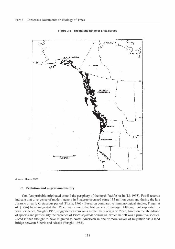
Part 3 – Consensus Documents on Biology of Trees
138
Figure 3.5 The natural range of Sitka spruce
Source : Harris, 1978
C. Evolution and migrational history
Conifers probably originated around the periphery of the north Pacific basin (Li, 1953). Fossil records indicate that divergence of modern genera in Pinaceae occurred some 135 million years ago during the late Jurassic or early Cretaceous period (Florin, 1963). Based on comparative immunological studies, Prager etal. (1976) have suggested that Picea was among the first genera to emerge. Although not supported by fossil evidence, Wright (1955) suggested eastern Asia as the likely origin of Picea, based on the abundance of species and particularly the presence of Picea koyamai Shirasawa, which he felt was a primitive species. Picea is then thought to have migrated to North American in one or more waves of migration via a land bridge between Siberia and Alaska (Wright, 1955).
Section 5 – Sitka Spruce
139
Phylogenetic relationships within coniferous genera are commonly interpreted from species crossability studies, where it is assumed that the more related are two species, the more easily they can be crossed (Wright, 1955; Critchfield, 1975). The close phylogenetic relationship between the north-western American “white” spruces (Sitka, white, and Engelmann spruce) and the eastern Asiatic Picea jezoensis(Sieb et Zucc.) Carr. (Wright, 1955; Roche and Fowler, 1975) supports this theory, at least for the members of the subsection Glaucoides in section Omorika, and suggests that speciation occurred after their arrival in North America.
The fossil record of Picea during the Pleistocene era in North America is incomplete, but it is believed that many conifer populations were fragmented and isolated in various refugia during the glacial period (Critchfield, 1984). Sitka spruce probably occupied roughly its present-day range before glaciation, surviving at higher elevations on hills and mountains, and reoccupying lower areas of the long coastal strip in British Columbia and Alaska soon after (Daubenmire, 1968; Page and Hollands, 1987).
3. Reproductive Biology
A. Reproductive development
Sitka spruce is monoecious. Development of the reproductive structures follows a 2-year cycle typical of most conifers in the northern hemisphere, other than Pinus species and members of the Cupressaceae family (Owens and Blake, 1985). Bud scales are initiated at the terminal apex and at newly initiated axillary apices within the enlarging vegetative buds, from about mid-April (Owens and Molder, 1976a; Cannell and Bowler, 1978). Apices differentiate as vegetative, pollen cone, or seed cone buds around mid-July, at the cessation of shoot elongation. Pollen cones typically develop from small axillary apices on vigorous distal shoots or terminal apices on less vigorous, proximal shoots. Seed cones usually develop on distal axillary positions on vigorous shoots or from smaller terminal apices on less vigorous shoots (Moir and Fox, 1975a; Owens and Molder, 1976b).
Pollen cone bud development is complete, although meiosis has not occurred before they become dormant at the end of October. Seed cones also do not undergo meiosis prior to becoming dormant in late November (Owens and Molder, 1976b). By the time buds become dormant, all microsporophylls, microsporangia, bracts and functional ovuliferous scales, and leaves have been initiated. The overwintering seed cone, pollen cone, and vegetative buds are small and similar in shape: broadly conical, greenish-brown and covered in a bloom of light grey resin (Moir and Fox, 1975a; Eis and Craigdallie, 1981).
Reproductive and vegetative buds break dormancy at about the same time, in response to photoperiod, while subsequent development is regulated by temperature. Meiosis and subsequent development of pollen occur immediately, followed by maturation of the megagametophyte (Moir and Fox, 1975b; Owens and Molder, 1980). Flushing of reproductive buds precedes that of vegetative buds, and pollen is released over a one-week period, in late-April on Vancouver Island (Owens and Molder, 1980) and by mid-May in Scotland (Moir and Fox, 1975a). The pollen enters receptive seed cones and adheres to the sticky micropylar arms. A week later, a "pollination drop" draws the pollen into the micropyle (Owens and Blake, 1984). Fertilisation occurs 4 to 5 weeks later, and embryo development is completed in mid-August. Without fertilisation, no embryo is formed and the megagametophyte tissue degenerates, leaving a normal-sized, but empty seed (Owens and Molder, 1980).
B. Mating system and gene flow
Sitka spruce is a wind-pollinated, monoecious species, and outcrossing is by far the most prevalent mating system (Cottrell and White, 1995). Self pollination occurs to some degree, as the period of pollen release and female receptivity coincide for an individual tree (Owens and Molder, 1980; El-Kassaby and
Part 3 – Consensus Documents on Biology of Trees
140
Reynolds 1990). Female strobili are usually found at the ends of primary branches in the mid- to upper-crown, while males are more prevalent at the ends of secondary branches lower in the crown (Tompsett, 1978; Philipson, 1997), although the effectiveness of this zonation against selfing is questionable (Nienstaedt and Teich, 1972). In the open-grown conditions in a seed orchard, the outcrossing rate was greatly reduced for seeds produced in the lower crown (Chaisurisri et al., 1994). The two-step pollination mechanism, whereby pollen is collected in the sticky micropyllar arms over the receptive period, and only then drawn en masse by the pollination drop, ensures that pollen from many sources has a chance to fertilise any given ovule (Owens and Blake, 1984; Runions et al., 1995).
Gene flow in Picea is mediated by small pollen grains, 70-85 µm at their widest point (Eisenhut, 1961), whose bladdery wings make them well-adapted for aerial transport (Di-Giovanni and Kevan, 1991). Various studies of pollen dispersal in conifers indicate that over 90% of the pollen comes to rest less than 100 m from the source (Wright, 1976). Nevertheless, conifer pollen may remain viable for several days and a substantial quantity may travel great distances (Lindgren et al., 1995; Lindgren and Lindgren, 1996). Gregory (1973) cites reports that pollen of Pinus and Picea may travel as far as 600 to 1 000 km, and several authors have concluded that isolation distances of less than 1 km often have little impact on contamination rates in conifer seed orchards (see review by Di-Giovanni and Kevan, 1991). While pollen dynamics are not well known in Sitka spruce, a recent study of pollen dispersal dynamics in a black spruce seed orchard indicated that "large amounts" of pollen rose to a height of 300 m above ground level (Di-Giovanni et al. 1996). At a steady wind speed of 5 m·s-1, the authors calculated that spruce pollen reaching this altitude would drift about 47 km.
C. Seed production
Sitka spruce begins to produce seed at 20 to 25 years of age, with heavy crops occurring at intervals of 3 to 5 years (Malcolm, 1987; Philipson, 1987b). Crop intervals are somewhat longer, 5 to 8 years, in the northern part of the range (Harris, 1969). It is a prolific seed producer and, in a good seed year, an old-growth stand may produce as much as 14.5 kg of seed per hectare (Peterson et al., 1997). The seeds themselves are small, and average cleaned seed weight is about 2.2 g/1000 seeds (Safford, 1974).
Initiation and duration of seed dispersal are weather and site dependent. The mature cones open as they lose moisture and the scales flex in dry weather, re-closing during wet periods. Seed dispersal begins in the fall, with over 70% of the seeds dispersed within the first 6 weeks, 90% by February, and the remainder released over the next growing season (Ruth, 1958; Harris, 1969). The seeds are winged and wind-dispersed. The actual distance reached from the source depends on several factors, including height and position of the seed source, local topography and wind conditions (Harris, 1967, 1978). While 80% of the seed usually falls within 30 metres the parent tree, some may travel up to several hundred metres (Mair, 1973).
D. Natural regeneration
Sitka spruce seeds exhibit weak dormancy, and both the rate and total amount of germination can be increased by exposure to low temperatures under moist conditions, i.e., cold stratification (Pintaric, 1972; Gordon et al., 1976; Gosling, 1988; Chaisurisri et al., 1992; Jinks and Jones, 1996). While not always employed in nursery practice, moist-chilling of Sitka spruce seed is often beneficial, particularly when moisture content and temperature are carefully controlled (Gosling and Rigg, 1990; Jones et al., 1993; Jones and Gosling, 1994), and stratified seed will tolerate redrying (Jones and Gosling, 1990; Jinks and Jones, 1996; Poulsen, 1996). In the wild, Sitka spruce seeds normally germinate the following spring, as soon as soil surface temperatures are warm enough and provided there is adequate moisture.
Section 5 – Sitka Spruce
141
Unlike its highly shade tolerant associates western hemlock and western red cedar that can germinate and survive on organic seedbeds, Sitka spruce is more restricted in its seedbed and light requirements and tends to be more disturbance dependent (Taylor, A.H., 1990; Peterson et al., 1997). Sitka spruce regenerates naturally on landslides, newly exposed alluvial sites, and openings created by windthrow. Regeneration cutting systems in Sitka spruce must generate sufficient ground exposure and disturbance, by clear cutting, shelterwood or seed-tree methods (Harris and Johnson, 1983; Weetman and Vyse, 1990). Regarding thinning operations in mixed western hemlock, Sitka spruce stands will normally favour regeneration of the hemlock over the spruce, particularly in older stands where thinning intensity is light (Deal and Farr, 1994). Dense natural regeneration has been more commonly observed after harvesting of first-generation planted stands in Britain, with up to several hundreds of thousands per hectare and requiring spacing (Nelson, 1991; Adam and Berg, 1996).
E. Vegetative reproduction in nature
Vegetative reproduction of Sitka spruce is rare under natural conditions or in plantations, although layering can occur on moist sites (Cooper, 1931; Roche and Fowler, 1975). Rooting is most likely to occur when lower branches of open-grown trees come in contact with the ground and become covered by soil or organic materials on the edges of bogs or near the timber line (Harris, 1978).
4. Crosses
Potential crosses with Sitka spruce are summarised in Table 3.7. Natural hybrids between Sitka and White Spruce were first collected in North America by H.J. Lutz on the Kenai Peninsula, Alaska, and given the name Picea × lutzii by E.L. Little (1953). Before this, white-Sitka spruce hybrids had been observed in Denmark as a result of natural crossing between adjacent plantations (Thaarup, 1945; Bornebusch, 1946). Populations resulting from introgressive hybridisation have since been documented in the Skeena, Nass, and Bulkley river valleys in British Columbia where the two species are sympatric (Daubenmire, 1968; Roche, 1969). The hybrid has frequently been made artificially with parents from outside the sympatric area (Fowler, 1987), often in the hope of imparting the resistance of White Spruce to the white pine weevil. The degree of cold hardiness of the hybrid is related to the proportion of White Spruce germplasm (Ying and Morgenstern, 1982), and growth performance of the hybrid depends greatly on the origin of the parents (Sheppard and Cannell, 1985). Though Picea breweriana and Picea sitchensis can cross successfully, P. breweriana has a very small range that is rarely, if ever, sympatric with P. sitchensis given that the populations are separated by elevation. The category of “easily crossed, probably occurring naturally” indicates species that readily cross with P. sitchensis if grown together in artificial plantations, but are not naturally sympatric.
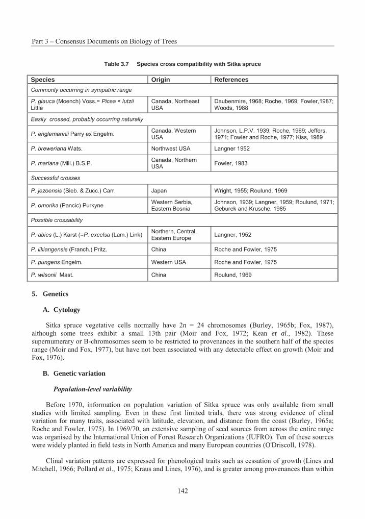
Part 3 – Consensus Documents on Biology of Trees
142
Table 3.7 Species cross compatibility with Sitka spruce
Species Origin References
Commonly occurring in sympatric range
P. glauca (Moench) Voss.= Picea × lutzii Little
Canada, Northeast USA
Daubenmire, 1968; Roche, 1969; Fowler,1987; Woods, 1988
Easily crossed, probably occurring naturally
P. englemannii Parry ex Engelm. Canada, Western USA
Johnson, L.P.V. 1939; Roche, 1969; Jeffers, 1971; Fowler and Roche, 1977; Kiss, 1989
P. breweriana Wats. Northwest USA Langner 1952
P. mariana (Mill.) B.S.P.Canada, Northern USA
Fowler, 1983
Successful crosses
P. jezoensis (Sieb. & Zucc.) Carr. Japan Wright, 1955; Roulund, 1969
P. omorika (Pancic) Purkyne Western Serbia, Eastern Bosnia
Johnson, 1939; Langner, 1959; Roulund, 1971; Geburek and Krusche, 1985
Possible crossability
P. abies (L.) Karst (=P. excelsa (Lam.) Link) Northern, Central, Eastern Europe
Langner, 1952
P. likiangensis (Franch.) Pritz. China Roche and Fowler, 1975
P. pungens Engelm. Western USA Roche and Fowler, 1975
P. wilsonii Mast. China Roulund, 1969
5. Genetics
A. Cytology
Sitka spruce vegetative cells normally have 2n = 24 chromosomes (Burley, 1965b; Fox, 1987), although some trees exhibit a small 13th pair (Moir and Fox, 1972; Kean et al., 1982). These supernumerary or B-chromosomes seem to be restricted to provenances in the southern half of the species range (Moir and Fox, 1977), but have not been associated with any detectable effect on growth (Moir and Fox, 1976).
B. Genetic variation
Population-level variability
Before 1970, information on population variation of Sitka spruce was only available from small studies with limited sampling. Even in these first limited trials, there was strong evidence of clinal variation for many traits, associated with latitude, elevation, and distance from the coast (Burley, 1965a; Roche and Fowler, 1975). In 1969/70, an extensive sampling of seed sources from across the entire range was organised by the International Union of Forest Research Organizations (IUFRO). Ten of these sources were widely planted in field tests in North America and many European countries (O'Driscoll, 1978).
Clinal variation patterns are expressed for phenological traits such as cessation of growth (Lines and Mitchell, 1966; Pollard et al., 1975; Kraus and Lines, 1976), and is greater among provenances than within
Section 5 – Sitka Spruce
143
(Falkenhagen, 1977; Deleporte, 1984). Southern coastal provenances produce up to 100% more height growth than northern inland sources (Cannell, 1974; Cannell and Willett, 1975; Cannell and Willet, 1976). While southern sources grow faster, they are more susceptible to frost damage, particularly in the nursery (Magnesen, 1986; Lines, 1987b; McKay, 1994). Provenance trials in the former Federal Republic of Germany showed a north-south trend in growth, with latitude accounting for over 80% of the among-provenance variation (Kleinschmit, 1984). Results of a 19 year provenance trial in Ireland demonstrated that the most productive provenances of Sitka Spruce for the mild, coastal conditions in Ireland originated from southern Washington and northern Oregon (Thompson and Pfeifer, 1995).
Ecotypic variation related to bioclimatic and physiographic factors has been demonstrated among provenances for seed and cone traits (Falkenhagen, 1978; Falkenhagen and Nash, 1978). Even for growth traits that normally exhibit clinal variation patterns, substantial variation may be present at the microgeographic level, attributable to such local site factors as slope and aspect (Campbell et al., 1989). Variation in biochemical composition appears to be clinal for sources from Alaska to north Washington, while more southerly sources show no geographic trends, perhaps reflecting the post-glacial recolonisation of northern parts of the range (Forrest, 1975b, 1980; Wellendorf and Kaufmann, 1977).
Population differences have also been demonstrated for susceptibility to insect attack. Provenances from Kitwamga (inland Skeena River) and Big Qualicum (SE Vancouver Island) suffer less damage from the white pine weevil (Alfaro and Ying, 1990; Tomlin and Borden, 1994; Ying and Ebata, 1994). Density of green spruce aphids attacking a provenance test in northern Ireland was related to latitude of seed origin, with southern provenances especially susceptible (Day, 1984). Lignified stone cell masses in spruce bark are considered an important physical defence against insects and fungi, and there is a clinal increase in bark lignin with increasing latitude of provenance origin (Wainhouse and Ashburner, 1996).
In contrast to many other characters, geographic variation at polymorphic allozyme loci appears to be weak. In a study of the 10 IUFRO provenances, only 8% of the diversity at polymorphic loci was due to differences among populations, whereas 92% resided within populations (Yeh and El-Kassaby, 1980).
Individual-level variability
While variation among provenances is important in determining the risks and benefits of transferring seed sources, genetic improvement from mass selection relies primarily on variation within-populations as the source of genetic gains. For productivity traits, on average, 40% of the genetic variation for Sitka spruce is at the provenance level, while 60% is within provenance (Fletcher, 1992).
Estimates of narrow-sense heritabilities for height growth during the first six years was around 25% for open-pollinated progeny of randomly selected trees in a single population (Samuel and Johnstone, 1979), while another study estimated heritability at over 40% for height after eight years (Birot and Christophe, 1983). Other quantitative estimates of additive genetic variation in growth traits have also varied widely (Falkenhagen, 1977; Yeh and Rasmussen, 1985; Samuel, 1991), but narrow-sense heritability is normally more than sufficient for individual tree selection and breeding to be effective, particularly when provenance and family performance are combined in a multiple-trait selection index (Christophe and Birot, 1983). Height superiority of individual trees is not necessarily associated with production of "free growth", although progeny of plus-trees have been noted to improve their height rankings over the first six years on better sites by production of free growth (Cannell and Johnstone, 1978).
Perhaps the most precise estimates of genetic variance for Sitka spruce have been reported for a 7-tree diallel cross, planted at two test sites in Scotland and Wales (Samuel et al., 1972; Samuel, 1991). In this experiment, genetic variation for height growth, although under some additive genetic control, was predominantly controlled by non-additive effects, whereas diameter was only controlled by additive
Part 3 – Consensus Documents on Biology of Trees
144
effects. Practically all the variation in monoterpene composition for these crosses was attributable to additive genetic effects, and reflected the differences in parental means in additive combination with little significant deviation due to specific combining ability or reciprocal effects (Forrest and Samuel, 1988).
The ease of vegetative propagation of Sitka spruce offers opportunities to capture additional gains earlier in the breeding cycle. Clonal selection has been demonstrated to be highly effective in Sitka spruce, for characters such as planting stock phenology, frost resistance (Nielsen and Roulund, 1996), root-growth potential (Deans et al., 1992), early height and diameter growth, and branching habit (Cahalan, 1981) . Clonal testing has also demonstrated that it is possible to select clones that combine good growth with high wood density (Costa e Silva et al., 1994).
A provenance test in British Columbia demonstrated significant difference in susceptibility to white pine weevil, both among provenances and among families within provenances (Alfaro and Ying, 1990; Ying, 1991). Unfortunately, a study of variation within a resistant provenance found that taller families were more likely to be attacked (Alfaro et al., 1993).
Variation in biochemical composition for trees within provenances is high (Forrest, 1975a, b; Wellendorf and Kaufmann, 1977), a trend that is also found for variation at polymorphic enzyme loci (Yeh and El-Kassaby, 1980). While southern provenances are generally less resistant to frost, there is sufficient variation within populations to select frost resistant, fast-growing individuals (Nicoll et al,. 1996).
Genotype-environment interactions are of concern to tree breeders who generally seek broad adaptability within bred material. A seedling test of provenances grown under controlled environments found that provenances near the centre of the range were more broadly adapted (Mergen et al., 1974). A test of families originating from the latitudinal range of Sitka spruce and planted at eight locations in Britain found highly significant family-site interaction for six-year height, but found that above-average families could be selected that were broadly adapted (Johnstone and Samuel, 1978). A progeny test established at 3 locations in Denmark demonstrated genetic control of growth, stem form, wood density (pilodyn penetration), frost resistance and resistance to aphids, in addition to substantial genotype–environment interaction for growth characters (Jensen et al., 1996). Another clonal test in Denmark found that 15% of the clones contributed over 50% of the GE interaction variance (Nielsen and Roulund, 1996).
C. Inbreeding depression and genetic load
Self pollination in Sitka spruce has severe effects on seed set, early growth, and survival (Samuel et al., 1972). Among those that survive, strong inbreeding depression continues with selfed individuals only 68% as tall as outcrossed trees at 15 years of age (Samuel, 1991). Inbreeding depression is also exhibited by progeny originating from seed collections in small stands (Phillips, 1984).
D. Breeding programs
Breeding strategies for Sitka spruce generally utilise a system of progeny testing and recurrent selection for generation advancement, combined with clonal seed orchards for production of improved seed. Flowering of Sitka spruce grafts can be stimulated by means of various cultural treatments, particularly those involving gibberellin A4/7, and this has facilitated the turnover of breeding cycles (Philipson, 1985a, b, 1987a, 1992; Philipson et al., 1990; Ross, 1991; Owens et al., 1992). While most seed orchards currently in production were established by grafting cuttings from plus-trees, and their placement in cultivated field environments, some programs have also experimented with the management of containerised Sitka spruce orchards, with the possibility of vegetative multiplication of small quantities of seed by rooted cuttings (John and Mason, 1987; Philipson and Fletcher, 1990).
Section 5 – Sitka Spruce
145
Breeding programs have been established in all the areas where Sitka spruce is an important plantation species. In British Columbia, where planting of Sitka spruce is severely limited due to risk of damage by the white pine weevil, the breeding plan emphasises weevil resistance. Currently, more than 250 open-pollinated families and 300 clones are included in weevil-resistance screening trials (King, 1994; King et al,. 1998). The intensive breeding program in Britain began in the early 70's (Fletcher and Faulkner, 1972), and is by far the most ambitious. From an initial 2 800 plus-tree selections, the breeding population now consists of 200 tested parents, subdivided into sub-populations targeted for different geographic areas (Faulkner, 1987; Fletcher, 1992; Malcolm, 1997). Breeding programs are also carried out in Denmark (Roulund, 1990) and in northern France (Deleporte and Roman-Amat, 1986), where Sitka spruce is an important component of plantation forestry operations.
E. Conservation of genetic resources
Domestication of a key species such as Sitka spruce can influence diversity of genetic resources: (1) indirectly, by the method of seed collection, extraction, and storage, and by nursery and plantation culture; and (2) directly, by intentional selection to increase the frequency of genes for desirable traits (Chaisurisri and El-Kassaby, 1994; Morgenstern, 1996). The inadvertent loss of genes by natural processes and human activity can have negative consequences on the adaptability of populations and the potential for future gains from breeding. The need for gene conservation for a species can be assessed by evaluating (1) its current status of protection, (2) its frequency of occurrence, (3) the extent of its botanical range, (4) ease of natural regeneration, and (5) its representation in genetic testing and breeding programs (Lester, 1996). Using this approach, Yanchuk and Lester (1996) ranked the need of Sitka spruce for gene conservation as higher than some of its associates (Douglas-fir, western red cedar and western hemlock), but lower than others (mountain hemlock, amabilis fir, yellow-cedar and western white pine).
In the case of Sitka spruce, in situ conservation of genetic resources is practised by protection of ecological reserves, special areas, and parks (Pollard, 1995), and integrated with domestication activities that control the movement of seed, active management of existing stands to maintain biological diversity, and protection of isolated, small populations (Yanchuk, 1995). Ex situ conservation, by cryopreservation of germplasm, by off-site maintenance of populations in arboreta and clone banks, and by multi-population breeding strategies (Eriksson et al., 1993; Namkoong, 1995), has been practised to a much lesser extent, although many provenances and families are now represented in field tests and seed bank collections (Edwards and El-Kassaby, 1993). Such "active" forms of gene management must be accelerated in preparation for response to rapid environmental and climate changes (Ledig and Kitzmiller, 1992; Yanchuk and Lester, 1996).
6. Ecology and associated species
A. Habitat
In its natural range, Sitka spruce is primarily a coastal species, but may extend well inland where optimum environmental conditions occur (Harris, 1990). It is primarily a low elevation species and is uncommon at elevations above 500 m. It is, however, much less restricted by edaphic factors than by climate and physiography (Roche and Haddock, 1987).
Climate
The natural range of Sitka spruce is restricted to an area of maritime climate with abundant moisture throughout the year, relatively mild winters and cool summers. Annual precipitation is high throughout the range, but somewhat greater in the north where summer precipitation as light drizzle and fog are frequent. Summer temperatures in these coastal habitats lack the extremes of more continental areas and, while
Part 3 – Consensus Documents on Biology of Trees
146
moderated by the ocean current of the north Pacific, decrease northward. Although winters are mild, accumulated heat input varies with latitude and probably accounts for much of the variation in productivity, soil development, and associated species (Harris, 1978).
Soils and site type
While Sitka spruce grows on soil derived from a variety of parent materials, their best development is on deep, moist, well-aerated soils. Growth is poor on swampy sites. Sitka spruce commonly occupies alluvial soils along streams, coarse-textured soils, or soils with a thick accumulation of organic matter. In Alaska, it is a pioneering species on immature soils exposed by glacial retreat or uplift from the sea. Soils in the natural range are acidic, with pH values varying from 4.0 to 5.7 (Harris, 1978).
Sitka spruce is relatively nutrient demanding, particularly at young ages prior to crown closure (Miller and Miller, 1987). It is most productive on nitrogen rich soils, but also requires relatively large amounts of calcium, magnesium and phosphorus (Krajina, 1969). Nitrogen and phosphorus have been found to be limiting on sites in both British Columbia and Britain, and applications of fertilisers may be necessary for successful plantation establishment (McIntosh, 1981, 1983; Miller and Miller, 1987; Taylor, C.M.A., 1990; Taylor and Tabbush, 1990; Prescott and Weetman, 1994).
B. Synecology and associated species
Sitka spruce occurs most commonly in mixed stands, usually associated with western hemlock (Tsugaheterophylla) (Harris and Johnson, 1983). Red alder (Alnus rubra) and black cottonwood (Populus trichocarpa) are associated throughout the range. Other associates vary with latitude: Douglas-fir (Pseudotsuga menziesii), Port-Orford-cedar (Chamaecyparis lawsoniana), western white pine (Pinus monticola), redwood (Sequoia sempervirens), and bigleaf maple (Acer macrophyllum) are limited to the south; shore pine (Pinus contorta var. contorta) and western red cedar (Thjua plicata) extend into south-east Alaska; while yellow cedar (Chamaecyparis nootkatensis), mountain hemlock (Tsuga mertensiana),subalpine fir (Abies lasiocarpa), and Sitka alder (Alnus sinuata) are limited to northern sites and higher elevations in the south (Harris, 1978). In Alaska and the Skeena, Nass, and Bulkley river valleys of British Columbia, Sitka spruce is associated with White Spruce (Picea glauca), and hybrid populations are found (Daubenmire, 1968; Roche, 1969). Pure stands of Sitka spruce are common on tidewater areas that receive quantities of salt spray, and in early succession situations following disturbance. Sitka spruce is an aggressive pioneer and, by itself or together with intolerant associates such as alder or cottonwood, will invade landslides, dunes, uplifted beaches and glaciated terrain.
Due to the latitudinal spread of the Sitka spruce range and the variation in precipitation and exposure, the species is a component of several ecological associations, characterised by available moisture and nutrient regimes. On the mid- to upper-slopes of the Queen Charlotte Islands, the western hemlock – Sitka spruce forest is typically associated with Alaska blueberry (Vaccinium alaskaense), red huckleberry (V.parifolium), ovalleaf huckleberry (V. ovalifolium), and several mosses (Rhytidiadelphus loreus,Hylocomium splendens, and Mnium glabrescens). On exposed coastal locations, pure stands of salt-tolerant Sitka spruce are associated with reed grass (Calamagrostis nutkaensis), salal (Gaultheria shallon) and finger moss (Stokesiella oregana). Sheltered alluvial sites find Sitka spruce and its associates western hemlock and red alder on grass meadows with Trisetum cernum, Gymnocarpium dryopteris, Hylocomium splendens and Leucolepis menziesii (Roche and Haddock, 1987; Hanley and Hoel, 1996; Hanley and Brady, 1997).
Inland valleys and eastern slopes of coastal mountains tend to be drier. It is on these areas that Sitka spruce is most likely to be sympatric with White Spruce and hybridisation can occur. Within this drier zone, valley bottoms and mountain slopes typically support mixed conifer forests with ground vegetation
Section 5 – Sitka Spruce
147
dominated by Alaska blueberry, ovalleaf huckleberry, red huckleberry, and rustyleaf menziesii (Menziesia ferruginea). Devil's club (Oplopanax horridus) appears within this zone on fluvial sites of the Skeena and Nass Rivers, and skunk cabbage (Lysichiton americanum) and salmonberry (Rubus spectabilis) are found on the driest sites in flat areas (Roche and Haddock, 1987; Harris, 1990).
C. Competition and stand structure
While Sitka spruce is rated as tolerant to shade, it is less tolerant than its usual associate, western hemlock (Daniel et al., 1979; Minore, 1979; Kobe and Coates, 1997), so that the general successional tendency is toward a western hemlock climax type, although few climax stands proceed to pure hemlock. As Sitka spruce is physically large, long-lived, and able to invade small openings resulting from windthrow, it is commonly maintained as a stand component, even under climax conditions. In south-east Alaska, mixed stands of hemlock and spruce are regarded as the climax stand type, with Sitka spruce regenerating on mineral soil mixtures exposed by windthrow and other disturbance, and hemlock seeding in on organic substrates (Harris, 1990; Deal et al., 1991; Peterson et al., 1997).
Sitka spruce is one of few conifer species that produce epicormic shoots along the stem. These shoots may originate from either dormant or adventitious buds (Stone and Stone, 1943) in response to light intensity (Isaac, 1940; Herman, 1964). Increasing exposure of stems to sunlight by thinning of stands will stimulate epicormic branching and affect the future quality of the trees (Farr and Harris, 1971).
D. Ecosystem dynamics
Many abiotic factors interact with Sitka spruce in natural and planted forests, and some may cause significant damage. Windthrow is probably the most serious damaging agent, particularly in plantations of Sitka spruce that are established in Great Britain where shallow rooting on unfavourable soils and exposure to strong winds results is risk of instability (Miller, 1986; Coutts and Philipson, 1987; Mason and Quine, 1995; Malcolm, 1997). Elsewhere in Europe, planted Sitka spruce has suffered significant wind damage, but has proven more wind-firm than other conifers such as Picea abies, Abies spp. and Pinus sylvestris in France (de Champs et al,. 1983; Touzet, 1983), Denmark (Neckelmann, 1981) and Norway (Lohmander and Helles, 1987). In North America, Sitka spruce is considered less wind-resistant than Pseudotsuga menziesii and Thuja plicata, but more-so than Tsuga heterophylla and Abies amabilis (Minore, 1979).
While Sitka spruce is among the least fire-resistant species in coastal forests, wild fires are not a major cause of damage within the native range (Minore, 1979; Agee, 1990). On the other hand, Sitka spruce regeneration benefits rather more from slash burning than several of its conifer associates (Hawkes et al., 1990; Otchere-Boateng and Herring, 1990). Frost heaving can cause severe damage, particularly to container seedlings planted on finer-textured soils (Shaw et al,. 1987; Goulet, 1995). Autumn frost is a particular problem in Great Britain, where faster-growing southern provenances are particularly susceptible (Redfern and Cannell, 1982; Lines 1987b; Nicoll et al,. 1996).
In the following table, other species interactions with Sitka Spruce are shown.
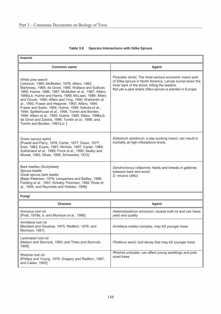
Part 3 – Consensus Documents on Biology of Trees
148
Table 3.8 Species Interactions with Sitka Spruce
Insects
Common name Agent
White pine weevil [Johnson, 1965; McMullen, 1976; Alfaro, 1982; Martineau, 1984; de Groot, 1985; Wallace and Sullivan, 1985; Hulme, 1986, 1987; McMullen et al,. 1987; Alfaro, 1989a,b; Hulme and Harris, 1989; McLean, 1989; Alfaro and Omule, 1990; Alfaro and Ying, 1990; Warkentin et al., 1992; Fraser and Heppner, 1993; Alfaro, 1994; Fraser and Szeto, 1994; Hulme, 1994; Sahota et al.,1994; Spittlehouse et al., 1994; Tomlin and Borden, 1994; Alfaro et al., 1995; Hulme, 1995; Alfaro, 1996a,b; de Groot and Zylstra, 1996; Tomlin et al., 1996; and, Tomlin and Borden, 1997a,b: ]
Pissodes strobi; The most serious economic insect pest of Sitka spruce in North America. Larvae tunnel down the inner bark of the shoot, killing the leaders. Not yet a pest where Sitka spruce is planted in Europe.
Green spruce aphid [Powell and Parry, 1976; Carter, 1977; Dixon, 1977; Koot, 1983; Evans, 1987; Nichols, 1987; Carter, 1989; Sutherland et al., 1989; Finck et al., 1990; Seaby and Mowat, 1993; Straw, 1995; Schwenke, 1972]
Elatobium abietinum; a sap sucking insect, can result in mortality at high infestations levels
Bark beetles (Scolytidae) Spruce beetle Great spruce bark beetle [Bejer-Petersen, 1976; Lemperiere and Bailley, 1986; Fielding et al., 1991; Kirkeby-Thomsen, 1992; Rose et al,. 1994, and Reynolds and Holsten, 1996]
Dendroctonus rufipennis; feeds and breeds in galleries between bark and wood. D. micans (ditto)
Fungi
Disease Agent
Annosus root rot [Pratt, 1979a, b; and Morrison et al., 1986]
Heterobasidoion annosum; causes butt-rot and can lower yield and quality
Armillaria root rot [Boullard and Gaudray, 1975; Redfern, 1978; and Morrison, 1981]
Armillaria mellea complex; may kill younger trees
Laminated root rot [Nelson and Sturrock, 1993; and Thies and Sturrock, 1995]
Phellinus weirii; butt decay that may kill younger trees
Rhizinia root rot [Phillips and Young, 1976; Gregory and Redfern, 1987; and Callan, 1993]
Rhizinia undulata; can affect young seedlings and pole sized trees
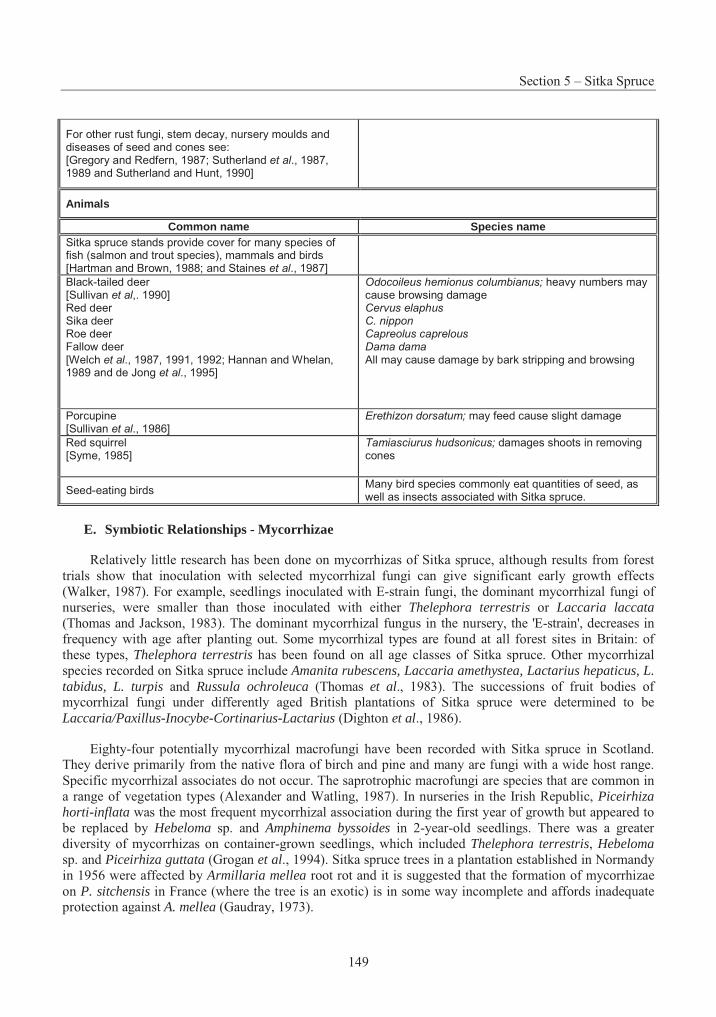
Section 5 – Sitka Spruce
149
For other rust fungi, stem decay, nursery moulds and diseases of seed and cones see: [Gregory and Redfern, 1987; Sutherland et al., 1987, 1989 and Sutherland and Hunt, 1990]
Animals
Common name Species name Sitka spruce stands provide cover for many species of fish (salmon and trout species), mammals and birds [Hartman and Brown, 1988; and Staines et al., 1987]
Black-tailed deer [Sullivan et al,. 1990] Red deer Sika deer Roe deer Fallow deer [Welch et al., 1987, 1991, 1992; Hannan and Whelan, 1989 and de Jong et al., 1995]
Odocoileus hemionus columbianus; heavy numbers may cause browsing damage Cervus elaphus C. nippon Capreolus caprelous Dama dama All may cause damage by bark stripping and browsing
Porcupine [Sullivan et al., 1986]
Erethizon dorsatum; may feed cause slight damage
Red squirrel [Syme, 1985]
Tamiasciurus hudsonicus; damages shoots in removing cones
Seed-eating birds Many bird species commonly eat quantities of seed, as well as insects associated with Sitka spruce.
E. Symbiotic Relationships - Mycorrhizae
Relatively little research has been done on mycorrhizas of Sitka spruce, although results from forest trials show that inoculation with selected mycorrhizal fungi can give significant early growth effects (Walker, 1987). For example, seedlings inoculated with E-strain fungi, the dominant mycorrhizal fungi of nurseries, were smaller than those inoculated with either Thelephora terrestris or Laccaria laccata(Thomas and Jackson, 1983). The dominant mycorrhizal fungus in the nursery, the 'E-strain', decreases in frequency with age after planting out. Some mycorrhizal types are found at all forest sites in Britain: of these types, Thelephora terrestris has been found on all age classes of Sitka spruce. Other mycorrhizal species recorded on Sitka spruce include Amanita rubescens, Laccaria amethystea, Lactarius hepaticus, L. tabidus, L. turpis and Russula ochroleuca (Thomas et al., 1983). The successions of fruit bodies of mycorrhizal fungi under differently aged British plantations of Sitka spruce were determined to be Laccaria/Paxillus-Inocybe-Cortinarius-Lactarius (Dighton et al., 1986).
Eighty-four potentially mycorrhizal macrofungi have been recorded with Sitka spruce in Scotland. They derive primarily from the native flora of birch and pine and many are fungi with a wide host range. Specific mycorrhizal associates do not occur. The saprotrophic macrofungi are species that are common in a range of vegetation types (Alexander and Watling, 1987). In nurseries in the Irish Republic, Piceirhiza horti-inflata was the most frequent mycorrhizal association during the first year of growth but appeared to be replaced by Hebeloma sp. and Amphinema byssoides in 2-year-old seedlings. There was a greater diversity of mycorrhizas on container-grown seedlings, which included Thelephora terrestris, Hebeloma sp. and Piceirhiza guttata (Grogan et al., 1994). Sitka spruce trees in a plantation established in Normandy in 1956 were affected by Armillaria mellea root rot and it is suggested that the formation of mycorrhizae on P. sitchensis in France (where the tree is an exotic) is in some way incomplete and affords inadequate protection against A. mellea (Gaudray, 1973).
Part 3 – Consensus Documents on Biology of Trees
150
The ability of six ectomycorrhizal fungi (Thelephora terrestris, Hebeloma crustuliniforme strains Siv and 81a, Paxillus involutus, Laccaria laccata and Lactarius rufus) to form mycorrhizas on plantlets of Sitka spruce derived from somatic embryos was investigated by Sasa and Krogstrup (1991). Mycorrhizal synthesis was achieved only on the oldest plantlets during the third week after inoculation. The rate and development of mycorrhizal formation varied according to the fungal species, with infection by T. terrestris the highest (92% of the total number of root tips), and Lactarius rufus failing to form any mycorrhizas.
7. Domestication
In 1930, Sitka spruce seedlings were among those planted in British Columbia's first reforestation project in the Fraser River Valley, near Vancouver (Young, 1989). While previously planted at a level of about 10 million seedlings per year in British Columbia, Sitka spruce has been all but eliminated from reforestation programs in North America, due to damage from the white pine weevil (King et al., 1998). Today, less than 2 million seedlings are planted each year, primarily on cool, coastal areas of the Queen Charlotte Islands. Meanwhile, Sitka spruce is the backbone of plantation forestry in Great Britain, accounting for about 70% of the seedlings planted (Malcolm, 1997), and is a commonly planted species in other European countries such as France and Denmark (Hermann, 1987). In spite of the good growth potential of Sitka spruce in the former Federal Republic of Germany, planting has been drastically reduced due to frost damage, drought and storm damage, and foraging by deer (Kleinschmit, 1978).
A. Deployment of reforestation materials
While Sitka spruce planting stock has traditionally been produced in bare root nurseries as 2+0 seedlings or 1+1 transplants, an increasing proportion is now produced in containerised growing systems, particularly in North America (Daniels and Simpson, 1990; Van Eerden and Gates, 1990; Aldhous and Mason, 1994). A variety of containers are used and stock is raised in both heated and unheated greenhouse structures. Cultural techniques have become highly sophisticated, ensuring that high-quality planting stock can be produced reliably and efficiently.
Sitka spruce planting stock can also be produced by means of vegetative propagation. Practical propagation systems have been developed (Kleinschmidt, 1992). Juvenile cuttings are easily rooted to produce planting stock as "stecklings" (Mason, 1984, 1992; John and Mason, 1987; Mason and Keenleyside, 1987) whose performance after planting is comparable to that of conventional transplant stock (Roulund, 1978; Roulund and Bergstedt, 1982; Baldwin and Mason, 1986; Mason et al., 1989). While steckling planting stock has been actively promoted in Great Britain, higher nursery production costs have kept annual production to a few million (Mason, 1991, 1992; Mason and Sharpe, 1992). Despite the higher production costs for steckling stock, the ability to bulk-up (vegetatively multiply) scarce seed sources and tested crosses between selected individuals is expected to more than compensate by realising potential genetic gains earlier (Gill, 1983; Mason and Gill, 1986; Mason and Harper, 1987; Lee, 1992).
Techniques for the initiation and regeneration of somatic embryos are available (Krogstrup et al., 1988), and embryogenic lines can be successfully regenerated after cryostorage (Find et al., 1993; Kristensen et al., 1994), making it possible to maintain genotypes in a completely juvenile condition during clonal testing. Sitka spruce plantlets derived from tissue culture propagation systems are also being automated further through the application of bioreactor technology (Moorhouse et al., 1996).
Sitka spruce has been established by direct seeding on an experimental basis, where it has been shown that the seedlings produce a much smaller amount of adventitious roots, compared with bare root transplants (Coutts et al., 1990). However, direct seeding has not been used operationally as a regeneration
Section 5 – Sitka Spruce
151
technique for Sitka spruce. Its use in British Columbia is considered a poor option, due to the very slow growth of germinants, which predisposes them to drought and competition (Mitchell et al,. 1990).
B. Provenance transfer
Within the native range, plantations established on sites with a strong maritime climate will be faster growing if seeds are transferred from more southerly latitudes. Conversely, transfers of seed from coastal origins to planting sites further inland involve higher risk (Lester et al., 1990; Ying, 1990).
Sitka spruce seedlots from British Columbia have also been certified under the OECD scheme for sale in Europe (Pollard and Portlock, 1990; Portlock, 1996). In Britain, southern provenances (below 47°N latitude) grow fastest, but are susceptible to spring and autumn frosts (Lines, 1987a, b), although clonal testing has demonstrated substantial variation in frost hardiness within provenances and potential for selecting southern genotypes with low risk of frost damage (Nicoll et al., 1996). Material from the Queen Charlotte Islands is generally recommended over much of Britain, although origins further south in Washington are better for south-west England, Wales, and parts of west Scotland (Fletcher, 1992). In the north of Germany, provenances from Washington are recommended, while fast-growing sources from Oregon are deemed to be too susceptible to frost damage (Stratmann and Tegeler, 1987). Provenances have been recommended for use in France, where plots have been established at four locations to demonstrate seed source differences (Bastien and Lemoine, 1986; François, 1986; Steinmetz, 1986). In Denmark, naturalised seed sources of the second and subsequent generations have grown faster and shown better adaptation than trees from seed imported directly from North America (Nielsen, 1994). In Germany, provenances from British Columbia (Canada) are also recommended.
8. Summary
Sitka spruce is an economically important species of the north temperate coastal rain forest of western North America. While not as commercially important as other conifers within its native range, it is a keystone species in some of the most productive ecosystems in North America. Sitka spruce is now widely planted in North Europe, where it forms the backbone of plantation forestry and is of enormous economic value in some regions. It is closely related to the other North American "white" spruces, Picea glauca and P. englemannii. As an outcrossing, wind-pollinated species and prolific seed producer, it can transfer its genes rapidly to neighbouring populations and to other related spruces.
Sitka spruce exhibits clinal variation for many growth traits, associated with latitude, elevation, and distance from the coast. Population differences are also demonstrated for resistance to insect attack. While there is great variation among populations, more than half of the genetic variation in many growth traits is found among individuals within populations. Heritabilities for growth and quality traits are sufficiently high to expect substantial genetic gain from conventional recurrent-selection breeding programs. The species is readily propagated by rooted cuttings, offering potential to capture non-additive genetic variance and to accelerate the pace of genetic improvement. Statistically significant genotype-environment interactions have been observed, but broadly adapted individuals are rather common.
The distribution of Sitka spruce is limited to an area of maritime climate with abundant moisture. It may occur as pure stands, particularly on exposed coastal sites, but more commonly occurs in mixtures with western hemlock. While Sitka spruce is tolerant to shade and may occur in climax forest types, it is dependent on disturbance for regeneration and can be an aggressive pioneer in earlier stages of succession. The white pine weevil is by far the most serious threat to stands in North America, killing the leader and seriously affecting growth and merchantability. While the weevil does not affect planted stands in Europe, the green spruce aphid, various species of deer, and windthrow can cause significant damage.

Part 3 – Consensus Documents on Biology of Trees
152
Sitka spruce is well suited to artificial regeneration. While constituting a minor component of the reforestation effort within its native range, Sitka spruce plantation programs are well developed in some parts of Europe. Genetically improved materials from local seed orchards now constitute a significant portion of deployed planting stock. While most Sitka spruce reforestation is currently based on seed propagation, vegetative propagation techniques for cuttings and regeneration of somatic embryos are well advanced, making it a logical target for implementation of transgenic biotechnologies and the use of cloning in both breeding and deployment strategies.
Section 5 – Sitka Spruce
153
REFERENCES
Adam, G.A. and Berg, S. 1996. Mechanized respacing naturally regenerated Sitka spruce in Great Britain. In Site preparation and stand treatment - impact on biology, economy and labour. Proceedings of S3.02-00 technical sessions during IUFRO XX World Congress 1995, held in Tampere, Finland. Selected papers, SkogsForsk (Forestry Research Institute of Sweden), Uppsala, Sweden. pp. 122-129.
Agee, J.K. 1990. The historical role of fire in Pacific Northwest forests. In Natural and prescribed fire in Pacific Northwest forests. Edited by J.D. Walstad, S.R. Radosevich and D.V. Sandberg. Oregon State University Press, Corvallis, OR. pp. 25-38.
Aldhous, J.R. and Mason, W.L. 1994. Forest nursery practice. Forestry Commission Bulletin No. 111, UK Forestry Commission, HMSO, London, UK.
Alexander, I. and Watling, R. 1987. Macrofungi of Sitka spruce in Scotland. Proc. Roy. Soc. Edinburgh Sect. B (Biol. Sci.) 93: 107-115.
Alfaro, R.I. 1982. Fifty year-old Sitka spruce plantations with a history of intense weevil attack. J. Entomol. Soc. B.C. 79: 62-65.
Alfaro, R.I. 1989a. Probability of damage to Sitka spruce by the Sitka spruce weevil, Pissodes strobi. J. Entomol. Soc. B.C. 86: 48-54.
Alfaro, R.I. 1989b. Stem defects in Sitka spruce induced by Sitka spruce weevil, Pissodes strobi (Peck.). InInsects affecting reforestation: biology and damage. Proceedings of a Meeting of the IUFRO Working Group on Insects Affecting Reforestation (S2.07-03), held under the auspices of the 18th International Congress of Entomology, 3-9 July 1988, Vancouver, BC. Edited by R.I. Alfaro and S.G. Glover. Forestry Canada, Pacific and Yukon Region, Victoria, Canada. pp. 177-185.
Alfaro, R.I. 1994. The white pine weevil in British Columbia. In The white pine weevil: biology, damage and management. Proceedings of a symposium held January 19-21, 1994 in Richmond, British Columbia. Edited by R.I. Alfaro, G. Kiss and R.G. Fraser. FRDA Report No. 226, BC Ministry of Forests and Lands, Research Branch, Victoria, BC. pp. 7-22.
Alfaro, R.I. 1996a. Feeding and oviposition preferences of white pine weevil (Coleoptera: Curculionidae) on resistant and susceptible Sitka spruce clones in laboratory bioassays. Environ. Entomol. 25: 1012-1019.
Alfaro, R.I. 1996b. Role of genetic resistance in managing ecosystems susceptible to white pine weevil. For. Chron. 72: 374-380.
Alfaro, R.I. and Omule, S.A.Y. 1990. The effect of spacing on Sitka spruce weevil damage to Sitka spruce. Can. J. For. Res. 20: 179-184.
Part 3 – Consensus Documents on Biology of Trees
154
Alfaro, R.I. and Ying, C.C. 1990. Levels of Sitka spruce weevil, Pissodes strobi (Peck), damage among Sitka spruce provenances and families near Sayward, British Columbia. Can. Ent. 122: 607-615.
Alfaro, R.I., Borden, J.H., Fraser, R.G. and, Yanchuk, A. 1995. The white pine weevil in British Columbia: basis for an integrated pest management system. For. Chron. 71: 66-73.
Alfaro, R.I., Hulme, M. and, Ying, C. 1993. Variation in attack by Sitka spruce weevil, Pissodes strobi(Peck), within a resistant provenance of Sitka spruce. J. Entomol. Soc. B.C. 90: 24-30.
Baldwin, E. and Mason, W.L. 1986. An early trial of Sitka spruce cuttings. Scottish For. 40: 176-184.
Bastien, J.C. and Lemoine, M. 1986. Les plantations comparatives. Rev. For. Fr. 38(Amélioration génétique des arbres forestiers): 101-102.
Bejer-Petersen, B. 1976. Dendroctonus micans Kug. in Denmark: the situation 25 years after a 'catastrophe'. Z. Pflanzenkrankheiten und Pflanzenschutz 83: 16-21.
Birot, Y. and Christophe, C. 1983. Genetic structures and expected genetic gains from multitrait selection in wild populations of Douglas fir and Sitka spruce. I. Genetic variation between and within populations. Silvae Genet. 32: 141-151.
Bornebusch, C.H. 1946. Sitka-hvidgran-bastarden. Dansk Skovforen. Tidsskr. 31: 42-46.
Boullard, B. and Gaudray, D. 1975. À propos d'une attaque de résineux hauts-normands par l'Armillaire couleur de miel (Armillariella mellea (Fr.) Karsten) --- ou l'écologie au service de la phytopathologie. Botaniste, 57: 119-151.
Brazier, J.D. 1987. Man's use of Sitka spruce. Proc. Roy. Soc. Edinburgh Sect. B (Biol. Sci.) 93(Sitka spruce, proceedings of a symposium, Royal Botanic Garden, Edinburgh, 3-6 Oct. 1986. Edited by:D.M. Henderson and R. Faulkner.): 213-221.
Burley, J. 1965b. Karotype analysis of Sitka spruce, Picea sitchensis (Bong.) Carr. Silvae Genet. 14: 127-132.
Burley, J.F. 1965a. Genetic variation in Picea sitchensis (Bong.) Carr.: a literature review. Commonw. For. Rev. 44: 47-58.
Cahalan, C.M. 1981. Provenance and clonal variation in growth, branching and phenology in Picea sitchensis and Pinus contorta. Silvae Genet. 30: 40-46.
Callan, B.E. 1993. Rhizina root rot of conifers. Pest Leaflet No. 56, Forestry Canada, Pacific Forestry Centre, Victoria, BC.
Campbell, R.K., Pawuk, W.A. and, Harris, A.S. 1989. Microgeographic genetic variation of Sitka spruce in southeastern Alaska. Can. J. For. Res. 19: 1004-1013.
Cannell, M.G.R. 1974. Production of branches and foliage by young trees of Pinus contorta and Picea sitchensis: provenance differences and their simulation. J. Appl. Ecol. 11: 1091-1115.
Cannell, M.G.R. and Bowler, K.C. 1978. Spatial arrangement of lateral buds at the time that they form on leaders of Picea and Larix. Can. J. For. Res. 8: 129-137.
Section 5 – Sitka Spruce
155
Cannell, M.G.R. and Johnstone, R.C.B. 1978. Free or lammas growth and progeny performance in Picea sitchensis. Silvae Genet. 27: 248-254.
Cannell, M.G.R. and Milne, R. 1995. Carbon pools and sequestration in forest ecosystems in Britain. Forestry, 68(Special issue: Forestry Research Coordination Committee Conference 'Greenhouse gas balance in forestry' held 9 November 1994): 361-378.
Cannell, M.G.R. and Willet, S.C. 1976. Shoot growth phenology, dry matter distribution and root:shoot ratios of provenances of Populus trichocarpa, Picea sitchensis and Pinus contorta growing in Scotland. Silvae Genet. 25: 49-59.
Cannell, M.G.R. and Willett, S.C. 1975. Rates and times at which needles are initiated in buds on differing provenances of Pinus contorta and Picea sitchensis in Scotland. Can. J. For. Res. 5: 367-380.
Carter, C.I. 1977. Impact of green spruce aphid on growth: can a tree forget its past? Research and Development Paper No. 116, UK Forestry Commission.
Carter, C.I. 1989. The 1989 outbreak of the green spruce aphid, Elatobium abietinum. Research Information Note 161, Research Division, UK Forestry Commission.
Chaisurisri, K. and El-Kassaby, Y.A. 1994. Domestication and genetic diversity in Sitka spruce. InProceedings: International symposium on genetic conservation and production of tropical forest tree seed, 14-16 June 1993, Chiang Mai, Thailand. Edited by R.M. Drysdale, S.E.T. John and A.C. Yapa. ASEAN-Canada Forest Tree Seed Centre, Saraburi, Thailand. pp. 144-153.
Chaisurisri, K., Edwards, D.G.W. and, El-Kassaby, Y.A. 1992. Genetic control of seed size and germination in Sitka spruce. Silvae Genet. 41: 348-355.
Chaisurisri, K., Mitton, J.B. and, El-Kassaby, Y.A. 1994. Variation in the mating system of Sitka spruce (Picea sitchensis): evidence for partial assortative mating. Amer. J. Bot. 81: 1410-1415.
Christophe, C. and Birot, Y. 1983. Genetic structures and expected genetic gains from multitrait selection in wild populations of Douglas fir an (sic) Sitka spruce. II. Practical application of index selection on several populations. Silvae Genet. 32: 173-181.
Coates, K.D., Haeussler, S., Lindeburgh, S., Pojar, R. and, Stock, A.J. 1994. Ecology and silviculture of interior spruce in British Columbia. FRDA Report 220, BC Ministry of Forests, Victoria, BC.
Cooper, W.S. 1931. The layering habit in Sitka spruce and the two western hemlocks. Bot. Gaz. 91: 441-451.
Copes, D.L. and Beckwith, R.C. 1977. Isoenzyme identification of Picea glauca, P. sitchensis, and P. lutzii. Bot. Gaz. 138: 512-521.
Costa e Silva, J., Nielsen, U.B. and, Roulund, H. 1994. Sitka spruce clonal performance with special reference to basic density: 23 years' results of a clonal trial. Silvae Genet. 43: 82-91.
Cottrell, J.E. and White, I.M.S. 1995. The use of isozyme genetic markers to estimate the rate of outcrossing in a Sitka spruce (Picea sitchensis (Bong.) Carr.) seed orchard in Scotland. New Forests 10: 111-122.
Part 3 – Consensus Documents on Biology of Trees
156
Coutts, M.P. and Philipson, J.J. 1987. Structure and physiology of Sitka spruce roots. Proc. Roy. Soc. Edinburgh Sect. B (Biol. Sci.) 93(Sitka spruce, proceedings of a symposium, Royal Botanic Garden, Edinburgh, 3-6 Oct. 1986. Edited by: D.M. Henderson and R. Faulkner.): 131-144.
Coutts, M.P., Walker, C. and, Burnand, A.C. 1990. Effects of establishment method on root form of lodgepole pine and Sitka spruce and on the production of adventitious roots. Forestry, 63: 143-159.
Critchfield, W.B. 1975. Interspecific hybridization in Pinus: a summary review. In Proceedings 14th Canadian Tree Improvement Association, Part 2, Symposium on interspecific hybridization in forest trees, Fredericton, NB, 28-30 August 1973. Edited by D.P. Fowler and C.W. Yeatman. Canadian Forestry Service, Ottawa, ON. pp. 99-105.
Critchfield, W.B. 1984. Impact of the Pleistocene on the genetic structure of North American conifers. InProceedings of the 8th North American Forest Biology Workshop, July 30 - August 1, 1984, Utah State University, Logan, USA. Edited by R.M. Lanner. Department of Forest Resources, Utah State University, Logan. pp. 70-118.
Daniel, T.W., Helms, J.A. and, Baker, F.S. 1979. Principles of silviculture (2nd edition). McGraw-Hill, New York, NY.
Daniels, T.G. and Simpson, D.G. 1990. Seedling production and processing: bareroot. In Regenerating British Columbia's forests. Edited by D.P. Lavender, R. Parish, C.M. Johnson, G. Montgomery, A. Vyse, R.A. Willis and D. Winston. University of British Columbia Press, Vancouver, BC. pp. 206-225.
Daubenmire, R. 1968. Some geographic variations in Picea sitchensis and their ecologic interpretation. Can. J. Bot. 46: 787-797.
Daubenmire, R. 1974. Taxonomic and ecological relationships between Picea glauca and Picea engelmannii. Can. J. Bot. 52: 1545-1560.
Day, K. 1984. Systematic differences in the population density of green spruce aphid, Elatobium abietinumin a provenance trial of Sitka spruce, Picea sitchensis. Ann. Appl. Biol. 105: 405-412.
de Champs, J., Ferron, J.L., Michaud, D. and, Savatier, N. 1983. Leçons à tirer de la tempête des 6-8 novembre 1982. In Annales de Recherches Sylvicoles, AFOCEL, France, 1982. Association Forêt-Cellulose, Paris, France. pp. 4-101.
de Groot, P. 1985. Chemical control of insect pests of white pine. Proc. Ent. Soc. Ont. 116(Supplement: White pine symposium, Petawawa National Forestry Institute, 14 September 1984. Edited by: C.R. Sullivan, C.A. Plexman, R.D. Whitney, W.M. Stiell and D.R. Wallace.): 67-71.
de Groot, P. and Zylstra, B.F. 1996. Control of white pine weevil in young plantations using a spring application of insecticides. Frontline, Technical Note No. 86, Canadian Forest Service, Great Lakes Forestry Centre, Sault Ste. Marie, ON.
de Jong, C.B., Gill, R.M.A., van Wieren, S.E. and, Burlton, F.W.E. 1995. Diet selection by roe deer Capreolus capreolus in Kielder Forest in relation to plant cover. For. Ecol. Manage. 79(Special issue: Kielder - the ecology of a man-made spruce forest. Papers presented at a symposium held at the University of Newcastle upon Tyne, UK, 20-21 September 1994): 91-97.
Section 5 – Sitka Spruce
157
Deal, R.L. and Farr, W.A. 1994. Composition and development of conifer regeneration in thinned and unthinned natural stands of western hemlock and Sitka spruce in southeast Alaska. Can. J. For. Res. 24: 976-984.
Deal, R.L., Oliver, C.D. and, Bormann, B.T. 1991. Reconstruction of mixed hemlock-spruce stands in coastal southeast Alaska. Can. J. For. Res. 21: 643-654.
Deans, J.D., Mason, W.L. and, Harvey, F.J. 1992. Clonal differences in planting stock quality of Sitka spruce. For. Ecol. Manage. 49: 101-107.
Deleporte, P. 1984. Épicéa de Sitka: resultats et méthodologie d'une sélection multicritère. In Annales de Recherches Sylvicoles, AFOCEL, 1983. Association Forêt-Cellulose, Paris, France. pp. 282-337.
Deleporte, P. and Roman-Amat, B. 1986. Épicéa de Sitka, Picea sitchensis (Bong.) Carr. Rev. For. Fr. 38(Amélioration génétique des arbres forestiers): 149-152.
Dighton, J., Poskitt, J.M. and, Howard, D.M. 1986. Changes in occurrence of basidiomycete fruit bodies during forest stand development: with specific reference to mycorrhizal species. Transactions of the British Mycological Society 87(1): 163-171.
Di-Giovanni, F. and Kevan, P.G. 1991. Factors affecting pollen dynamics and its importance to pollen contamination: a review. Can. J. For. Res. 21: 1155-1170.
Di-Giovanni, F., Kevan, P.G., and Arnold, J. 1996. Lower planetary boundary layer profiles of atmospheric conifer pollen above a seed orchard in northern Ontario, Canada. For. Ecol. Manage. 83: 87-97.
Dixon, A.F.G. 1977. Aphid ecology: life-cycles, polymorphism, and population regulation. Ann. Rev. Ecol. Syst. 8: 329-353.
Edwards, D.G.W. and El-Kassaby, Y.A. 1993. Ex situ conservation of forest biodiversity in British Columbia. In Proceedings of the Forest Ecosystem Dynamics Workshop, February 10-11, 1993. Edited by V. Marshall. FRDA Report No. 210, Forestry Canada and B.C. Ministry of Forests, Victoria, BC. pp. 65-67.
Eis, S. and Craigdallie, D. 1981. Reproduction of conifers. A handbook for cone crops assessment. Information Report BC-X-219, Canadian Forestry Service, Pacific Forest Research Centre, Victoria, BC.
Eisenhut, G. 1961. Untersuchungen über die Morphologie und Ökologie der Pollenkörner heimischer und fremdländischer Waldbäume. Forstwiss. Forsch. 15: 1-68.
El-Kassaby, Y.A. and, Reynolds, S. 1990. Reproductive phenology, parental balance, and supplemental mass pollination in a Sitka spruce seed orchard. For. Ecol. Manage. 31: 45-54.
Eriksson, G., Namkoong, G. and, Roberds, J.H. 1993. Dynamic gene conservation for uncertain futures. For. Ecol. Manage. 62: 15-37.
Evans, H.F. 1987. Sitka spruce insects: past, present and future. Proc. Roy. Soc. Edinburgh Sect. B (Biol. Sci.) 93(Sitka spruce, proceedings of a symposium, Royal Botanic Garden, Edinburgh, 3-6 Oct. 1986. Edited by: D.M. Henderson and R. Faulkner.): 157-167.
Part 3 – Consensus Documents on Biology of Trees
158
Falkenhagen, E.R. 1977. Genetic variation in 38 provenances of Sitka spruce. Silvae Genet. 26: 67-75.
Falkenhagen, E.R. 1978. Parent tree variation in Sitka spruce provenances, an example of fine geographic variation. Silvae Genet. 27: 24-29.
Falkenhagen, E.R. and Nash, S.W. 1978. Multivariate classification in provenance research. A comparison of two statistical techniques. Silvae Genet. 27: 14-23.
Farr, W.A. and Harris, A.S. 1971. Partial cutting of western hemlock and Sitka spruce in southeast Alaska. Research Paper PNW-124, USDA Forest Service, Pacific Northwest Forest and Range Experiment Station.
Farrar, J.L. 1995. Trees in Canada. Fitzhenry & Whiteside/Canadian Forest Service, Markham and Ottawa, ON.
Faulkner, R. 1987. Genetics and breeding of Sitka spruce. Proc. Roy. Soc. Edinburgh Sect. B (Biol. Sci.) 93(Sitka spruce, proceedings of a symposium, Royal Botanic Garden, Edinburgh, 3-6 October 1986. Edited by: D.M. Henderson and R. Faulkner.): 41-50.
Fielding, N.J., Evans, H.F., Williams, J.M. and, Evans, B. 1991. Distribution and spread of the great European spruce bark beetle, Dendroctonus micans, in Britain - 1982 to 1989. Forestry, 64: 345-358.
Finck, K.E., Shrimpton, G.M. and, Summers, D.W. 1990. Insect pests in reforestation. In Regenerating British Columbia's forests. Edited by D.P. Lavender, R. Parish, C.M. Johnson, G. Montgomery, A. Vyse, R.A. Willis and D. Winston. University of British Columbia Press, Vancouver, BC. pp. 279-301.
Find, J.I., Floto, F., Krogstrup, P., Moller, J.D., Norgaard, J.V. and, Kristensen, M.M.H. 1993. Cryopreservation of an embryogenic suspension culture of Picea sitchensis and subsequent plant regeneration. Scand. J. For. Res. 8: 156-162.
Fletcher, A.M. 1992. Breeding improved Sitka spruce. In Super Sitka for the 90s. Proceedings of a meeting held in Elgin, Scotland, 2-4 October 1990, organized by the UK Forestry Commission Research Division. Edited by D.A. Rook. Forestry Commission Bulletin 103, HMSO, London, UK. pp. 11-24.
Fletcher, A.M. and Faulkner, R. 1972. A plan for the improvement of Sitka spruce by selection and breeding. Research and Development Paper No. 85, Forestry Commission, London, UK.
Florin, R. 1963. The distribution of conifer and taxad genera in time and space. Acta Horti Bergiani 20: 121-312.
Forrest, G.I. 1975a. Polyphenol variation in Sitka spruce. Can. J. For. Res. 5: 26-37.
Forrest, G.I. 1975b. Variation in polyphenol content within and between Sitka spruce provenances at different sites. Can. J. For. Res. 5: 46-54.
Forrest, G.I. 1980. Geographic variation in the monoterpene composition of Sitka spruce cortical oleoresin. Can. J. For. Res. 10: 458-463.
Forrest, G.I. and Samuel, C.J.A. 1988. Monoterpene analysis of a diallel cross in Sitka spruce. Silvae Genet. 37: 100-104.
Section 5 – Sitka Spruce
159
Fowler, D.P. 1983. The hybrid black X Sitka spruce, implications to phylogeny of the genus Picea. Can. J. For. Res. 13: 108-115.
Fowler, D.P. 1987. The hybrid white X Sitka spruce: species crossability. Can. J. For. Res. 17: 413-417.
Fowler, D.P. and Roche, L. 1977. Genetics of Engelmann spruce. Research Paper WO-30, USDA Forest Service, Washington, D.C.
Fox, D.P. 1987. The chromosomes of Picea sitchensis (Bong.) Carr. and its relatives. Proc. Roy. Soc. Edinburgh Sect. B (Biol. Sci.) 93(Sitka spruce, proceedings of a symposium, Royal Botanic Garden, Edinburgh, 3-6 Oct. 1986. Edited by: D.M. Henderson and R. Faulkner.): 51-59.
François, J.M. 1986. Pour le choix des meilleures provenances: un réseau de démonstration à l'intention des sylviculteurs. Forêt Entreprise 38: 33-45.
Fraser, R.G. and Heppner, D.G. 1993. Control of white pine weevil, Pissodes strobi, on Sitka spruce using implants containing systemic insecticide. For. Chron. 69: 600-603.
Fraser, R.G. and Szeto, S. 1994. Insecticides applied with Ezject will protect young Sitka spruce plantations from white pine weevil attack. In The white pine weevil: biology, damage and management. Proceedings of a symposium held January 19-21, 1994 in Richmond, British Columbia. Edited by R.I. Alfaro, G. Kiss and R.G. Fraser. FRDA Report No. 226, BC Ministry of Forests and Lands, Research Branch, Victoria, BC. pp. 239-247.
Gaudray, D. 1973. Observations on the mycorrhizae of Sitka Spruce and the role they can play during an attack by Armillaria mellea. Comptes Rendus des Seances de la Societe de Biologie et de ses Filiales 167(6-7): 1023-1026.
Geburek, T. and Krusche, D. 1985. Wachstum von Hybriden zwischen Picea omorika und P. sitchensis im Vergleich zu den Elternarten. Allg. For. Jagdzeitg. 156: 47-54.
Gill, J.G.S. 1983. Comparisons of production costs and genetic benefits of transplants and rooted cuttings of Picea sitchensis. Forestry, 56: 61-73.
Gordon, A.G., Salt, G.A. and, Brown, R.M. 1976. Effect of pre-sowing moist-chilling treatments on seedbed emergence of Sitka spruce seed infected by Geniculodendron pyriforme Salt. Forestry, 49: 143-151.
Gosling, P.G. 1988. The effect of moist chilling on the subsequent germination of some temperate conifer seeds over a range of temperatures. J. Seed Tech. 12: 90-98.
Gosling, P.G. and Rigg, P. 1990. The effect of moisture content and prechill duration on the efficiency of dormancy breakage in Sitka spruce (Picea sitchensis) seed. Seed Sci. & Technol. 18: 337-343.
Goulet, F. 1995. Frost heaving of forest tree seedlings: a review. New Forests 9: 67-94.
Gregory, P.H. 1973. The microbiology of the atmosphere (2nd edition). Leonard Hill, Aylesbury, UK.
Gregory, S.C. and Redfern, D.B. 1987. The pathology of Sitka spruce in northern Britain. Proc. Roy. Soc. Edinburgh Sect. B (Biol. Sci.) 93(Sitka spruce, proceedings of a symposium, Royal Botanic Garden, Edinburgh, 3-6 Oct. 1986. Edited by: D.M. Henderson and R. Faulkner.): 145-156.
Part 3 – Consensus Documents on Biology of Trees
160
Griffiths, M. 1994. Index of Garden Plants -- The New Royal Horticultural Society Dictionary. Timber Press, Portland, OR.
Grogan, H.M., O'-Neill, J.J.M. and, Mitchell, D.T. 1994. Mycorrhizal associations of Sitka-spruce seedlings propagated in Irish tree nurseries. European Journal of Forest Pathology 24(6-7): 335-344.
Grossnickle, S.C., Sutton, B.C.S. and, Holcomb, R.W. 1996b. Genetics and seedling biology of Sitka spruce hybrids. FRDA Memo 232, BC Ministry of Forests, Victoria, BC.
Grossnickle, S.C., Sutton, B.C.S., Folk, R.S. and, Gawley, R.J. 1996a. Relationship between nuclear DNA markers and physiological parameters in Sitka X interior spruce populations. Tree Physiol. 16: 547-555.
Guyon, A. 1995. Peuplements forestiers littoraux de Bretagne: description et gestion. Rev. For. Fr. 47: 255-262.
Hanley, T.A. and Brady, W.W. 1997. Understory species composition and production in old-growth western hemlock-Sitka spruce forests of southeastern Alaska. Can. J. Bot. 75: 574-580.
Hanley, T.A. and Hoel, T. 1996. Species composition of old-growth and riparian Sitka spruce - western hemlock forests in southeastern Alaska. Can. J. For. Res. 26: 1703-1708.
Hannan, M.J. and Whelan, J. 1989. Deer and habitat relations in managed forests. In Mammals as pests. Papers presented at a symposium of the Mammal Society, London, November 1987. Edited by R.J. Putman. Chapman and Hall Ltd, London, UK. pp. 116-127.
Hanover, J.W. and Wilkinson, R.C. 1970. Chemical evidence for introgressive hybridization in Picea.Silvae Genet. 19: 17-22.
Harris, A.S. 1967. Natural reforestation on a mile-square clearcut in southwest Alaska. Research Paper PNW-52, USDA Forest Service, Pacific Northwest Forest and Range Experiment Station, Portland, OR.
Harris, A.S. 1969. Ripening and dispersal of a bumper western hemlock-Sitka spruce seed crop in southeastern Alaska. Research Note PNW-105, USDA Forest Service, Pacific Northwest Forest and Range Experiment Station, Portland, OR.
Harris, A.S. 1978. Distribution, genetics, and silvical characteristics of Sitka spruce. In Proceedings of the IUFRO Joint Meeting of Working Parties, Vancouver, Canada 1978. Vol. 1. B.C. Ministry of Forests, Victoria, BC. pp. 95-122.
Harris, A.S. 1990. Picea sitchensis (Bong.) Carr. - Sitka spruce. In Silvics of North America. Vol. 1, Conifers. Edited by R.M. Burns and B.H. Honkala. Agriculture Handbook 654, Forest Service, USDA, Washington, DC. pp. 260-267.
Harris, A.S. and Johnson, D.L. 1983. Western hemlock-Sitka spruce. In Silvicultural systems for the major forest types of the United States. Edited by R.M. Burns. Agricultural Handbook 445, USDA Forest Service, Washington, D.C. pp. 5-8.
Hartman, G.F. and Brown, T.G. 1988. Forestry-fisheries planning considerations on coastal floodplains. For. Chron. 64: 47-51.
Section 5 – Sitka Spruce
161
Hawkes, B.C., Feller, M.C. and, Meehan, D. 1990. Site preparation: fire. In Regenerating British Columbia's forests. Edited by D.P. Lavender, R. Parish, C.M. Johnson, G. Montgomery, A. Vyse, R.A. Willis and D. Winston. University of British Columbia Press, Vancouver, BC. pp. 131-149.
Herman, F.R. 1964. Epicormic branching of Sitka spruce. Research Paper PNW-18, USDA Forest Service, Pacific Northwest Forest and Range Experiment Station.
Hermann, R.K. 1987. North American tree species in Europe: transplanted species offer good growth potential on suitable sites. J. For. 85(12): 27-32.
Hulme, M.A. 1994. The potential of Allodorus crassigaster for the biological control of Pissodes strobi. InThe white pine weevil: biology, damage and management. Proceedings of a symposium held January 19-21, 1994 in Richmond, British Columbia. Edited by R.I. Alfaro, G. Kiss and R.G. Fraser. FRDA Report No. 226, BC Ministry of Forests and Lands, Research Branch, Victoria, BC. pp. 294-300.
Hulme, M.A. 1995. Resistance by translocated Sitka spruce to damage by Pissodes strobi (Coleoptera: Curculionidae) related to tree phenology. J. Econ. Entomol. 88: 1525-1530.
Hulme, M.A. and Harris, J.W.E. 1989. How Lonchaea corticis Taylor may impact broods of Pissodes strobi (Peck) in Picea sitchensis (Bong.) Carr. In Insects affecting reforestation: biology and damage. Proceedings of a Meeting of the IUFRO Working Group on Insects Affecting Reforestation (S2.07-03), held under the auspices of the 18th International Congress of Entomology, 3-9 July 1988, Vancouver, BC. Edited by R.I. Alfaro and S.G. Glover. Forestry Canada, Pacific and Yukon Region, Victoria, Canada. pp. 161-166.
Hulme, M.A., Dawson, A.F. and, Harris, J.W.E. 1986. Exploiting cold-hardiness to separate Pissodes strobi (Peck) (Coleoptera: Curculionidae) from associated insects in leaders of Picea sitchensis(Bong.) Carr. Can. Ent. 118: 1115-1122.
Hulme, M.A., Harris, J.W.E. and, Dawson, A.F. 1987. Exploiting adult girth to separate Pissodes strobi(Peck) (Coleoptera: Curculionidae) from associated insects in leaders of Picea sitchensis (Bong.) Carr. Can. Ent. 119: 751-753.
Isaac, L.A. 1940. "Water sprouts" on Sitka spruce. In Research Note 31, USDA Forest Service, Pacific Northwest Forest and Range Experiment Station. pp. 6-7.
Jeffers, R.M. 1971. Research at the Institute of Forest Genetics. Res. Pap. NC-67, USDA For. Serv. North Central For. Exp. Sta., St. Paul, MN.
Jensen, J.S., Kjær, E.D. and, Roulund, H. 1996. A progeny trial with domesticated Picea sitchensis (Bong.) in Denmark. Silvae Genet. 45: 85-90.
Jinks, R.L. and Jones, S.K. 1996. The effect of seed pretreatment and sowing date on the nursery emergence of Sitka spruce (Picea sitchensis (Bong.) Carr.) seedlings. Forestry, 69: 335-345.
John, A. and Mason, B. 1987. Vegetative propagation of Sitka spruce. Proc. Roy. Soc. Edinburgh Sect. B (Biol. Sci.) 93(Sitka spruce, proceedings of a symposium, Royal Botanic Garden, Edinburgh, 3-6 Oct. 1986. Edited by: D.M. Henderson and R. Faulkner.): 197-203.
Johnson, L.P.V. 1939. A descriptive list of natural and artificial interspecific hybrids in North American forest-tree genera. Can. J. Res. (Sec. C) 17: 411-444.
Part 3 – Consensus Documents on Biology of Trees
162
Johnson, N.E. 1965. A test of 12 insecticides for the control of the Sitka spruce weevil, Pissodes sitchensisHopkins. J. Econ. Entomol. 58: 572-574.
Johnstone, R.C.B. and Samuel, C.J.A. 1978. The interaction between genotype and site: its influence on tree selection in programmes in Great Britain (Paper presented at the Eighth World Forestry Congress, Jakarta, Indonesia). Research and Development Paper 122, UK Forestry Commission, Edinburgh, UK.
Jones, S.K. and Gosling, P.G. 1990. The successful redrying of imbibed, or imbibed plus prechilled Sitka spruce seeds. Seed Sci. & Technol. 18: 541-547.
Jones, S.K. and Gosling, P.G. 1994. "Target moisture content" prechill overcomes the dormancy of temperate conifer seeds. New Forests 8: 309-321.
Jones, S.K., Bergsten, U. and, Gosling, P.G. 1993. A comparison of 5°C and 15°C as dormancy breakage treatments for Sitka spruce seeds (Picea sitchensis). In Dormancy and barriers to germination: Proceedings of an international symposium of IUFRO Project Group P2.04-00 (Seed problems) Victoria, British Columbia, Canada, 23-26 April, 1991. Edited by D.G.W. Edwards. Canadian Forest Service - Pacific Forestry Centre, Victoria, BC. pp. 51-55.
Kean, V.M., Fox, D.P. and, Faulkner, R. 1982. The accumulation mechanism of the supernumerary (B-) chromosome in Picea sitchensis (Bong.) Carr. and the effect of this chromosome on male and female flowering. Silvae Genet. 31: 126-131.
King, J.N. 1994. Delivering durable resistant Sitka spruce for plantations. In The white pine weevil: biology, damage and management. Proceedings of a symposium held January 19-21, 1994 in Richmond, British Columbia. Edited by R.I. Alfaro, G. Kiss and R.G. Fraser. FRDA Report No. 226, BC Ministry of Forests and Lands, Research Branch, Victoria, BC. pp. 134-149.
King, J.N., Alfaro, R., Brown, G. and, Cartwright, C. 1998. Genetic and environmental dynamics of an artificial infestation of a Sitka spruce plantation with the white pine weevil. In Proceedings of the 25th Meeting of the Canadian Tree Improvement Association, Québec, QC, August 1977, Part 2. Edited by J.D. Simpson. Natural Resources Canada, Fredericton, NB. [Abstract in press]
Kirkeby-Thomsen, A. 1992. Dendroctonus micans i Danmark. Dansk Skovforen. Tidsskr. 77: 55-65.
Kiss, G.K. 1989. Engelmann X Sitka spruce hybrids in central British Columbia. Can. J. For. Res. 19: 1190-1193.
Kleinschmidt, J. (1978): Sitka spruce in Germany. Proceedings of the IUFRO joint meeting of working parties. Vancouver, Canada 1978, S.183-191 (see Appendix I)
Kleinschmidt, J. (1992): The use of spruce cuttings in plantations. Forestry Comission Bulletin 103: Super Sitka for the 90`s. Ed. D. A. Rook (see Appendix I)
Kleinschmidt,J. (1984): IUFRO Sitka spruce ten provenance experiment. Proceedings of the IUFRO Sitka Spruce Provenance Experiment. Edinburgh, Scotland 1984 (see Appendix I)
Kobe, R.K. and Coates, K.D. 1997. Models of sapling mortality as a function of growth to characterize interspecific variation in shade tolerance of eight tree species of northwestern British Columbia. Can. J. For. Res. 27: 227-236.
Section 5 – Sitka Spruce
163
Koot, H.P. 1983. Spruce aphid in British Columbia. Forest Pest Leaflet FPL 16, Canadian Forestry Service, Pacific Forest Research Centre, Victoria, BC.
Krajina, V.J. 1969. Ecology of forest trees in British Columbia. In Ecology of western North America. Vol. 1. Edited by V.J. Krajina. University of British Columbia, Vancouver, BC. pp. 1-147.
Kraus, J.F. and Lines, R. 1976. Patterns of shoot growth, growth cessation and bud set in a nursery test of Sitka spruce provenances. Scottish For. 30: 16-24.
Kristensen, M.M.H., Find, J.I., Floto, F., Moller, J.D., Norgaard, J.V. and, Krogstrup, P. 1994. The origin and development of somatic embryos following cryopreservation of an embryogenic suspension culture of Picea sitchensis. Protoplasma, 182: 65-70.
Krogstrup, P., Eriksen, E.N., Moller, J.D. and, Roulund, H. 1988. Somatic embryogenesis in Sitka spruce (Picea sitchensis (Bong.) Carr.). Plant Cell Rep. 7: 594-597.
Krüssmann, G. 1985. Manual of cultivated conifers (2nd edition). Edited by H.-D. Warda. Timber Press, Portland, OR. [Translated by M.E. Epp from the 1983 German edition]
Langner, W. 1952. Die Forschungsstatte fur Forstgenetik und Forstpflanzenzuchtung in Schmalenbeck. Z. Weltforstwirtsch. 15: 15-18. [cited by Roche and Fowler 1975]
Langner, W. 1959. Ergebnisse Einiger Hybridisierrungsversuche Zwischen Picea sitchensis (Bong.) Carr. und Picea omorika (Pancic) Purkyne. Silvae Genet. 8: 138-143.
Ledig, F.T. and Kitzmiller, J.H. 1992. Genetic strategies for reforestation in the face of global climate change. For. Ecol. Manage. 50: 153-169.
Lee, S.J. 1992. Likely increases in volume and revenue from planting genetically improved Sitka spruce. In Super Sitka for the 90s. Proceedings of a meeting held in Elgin, Scotland, 2-4 October 1990, organized by the UK Forestry Commission Research Division. Edited by D.A. Rook. Forestry Commission Bulletin 103, HMSO, London, UK. pp. 61-74.
Lemperiere, G. and Bailley, D. 1986. Limousin: observation sur les manifestations du dendroctone de l'épicéa. Forêts de France 290: 18-22.
Lester, D. 1996. Gene conservation methods and an application in British Columbia. In Forest gene conservation - principles to practice. Workshop proceedings. Edited by G. McVey and C. Nielsen. Workshop proceedings WP-008, Ontario Ministry of Natural Resources, Southern Region Science & Technology Transfer Unit, Kemptville, ON. pp. 35-45.
Lester, D.T., Ying, C.C. and, Konishi, J.D. 1990. Genetic control and improvement of planting stock. InRegenerating British Columbia's forests. Edited by D.P. Lavender, R. Parish, C.M. Johnson, G. Montgomery, A. Vyse, R.A. Willis and D. Winston. University of British Columbia Press, Vancouver, BC. pp. 180-192.
Li, H.L. 1953. Present distribution and habitats of the conifers and taxads. Evolution, 7: 245-261.
Lindgren, D. and Lindgren, K. 1996. Long distance pollen transfer may make gene conservation difficult. In Conservation of forest genetic resources. Proceedings of Nordic Group for Forest Genetics and Tree Breeding Meeting in Estonia, June 3-7, 1996. Edited by M. Kurm and Y. Tamm. Estonia Agricultural University, Tartu. pp. 51-62.
Part 3 – Consensus Documents on Biology of Trees
164
Lindgren, D., Paule, L., Shen, X.-H., Yazdani, R., Segerström, U., Wallin, J.-E. and, Lejdebro, M.L. 1995. Can viable pollen carry Scots pine genes over long distances? Grana, 34: 64-69.
Lines, R. 1987a. Choice of seed origins for the main forest species in Britain. Forestry Commission Bulletin No. 66, UK Forestry Commission, London, UK.
Lines, R. 1987b. Seed origin variation in Sitka spruce. Proc. Roy. Soc. Edinburgh Sect. B (Biol. Sci.) 93(Sitka spruce, proceedings of a symposium, Royal Botanic Garden, Edinburgh, 3-6 October 1986. Edited by: D.M. Henderson and R. Faulkner.): 25-39.
Lines, R. and Mitchell, A.F. 1966. Differences in phenology of Sitka spruce provenances. In Report on research for year ended March 1965. UK Forestry Commission, London, UK. pp. 173-184.
Little, E.L., Jr. 1953. A natural hybrid spruce in Alaska. J. For. 51: 745-747.
Lohmander, P. and Helles, F. 1987. Windthrow probability as a function of stand characteristics and shelter. Scand. J. For. Res. 2: 227-238.
Magnesen, S. 1986. Det internasjonale Sitkagran-proveniensforsoket pa Vestlandet. Rapport No. 1/86, Norsk Institutt for Skogforskning, Fana, Norway. [English summary]
Mair, A.R. 1973. Dissemination of tree seed : Sitka spruce, western hemlock and Douglas fir. Scottish For. 27: 308-314.
Malcolm, D.C. 1987. Some ecological aspects of Sitka spruce. Proc. Roy. Soc. Edinburgh Sect. B (Biol. Sci.) 93(Sitka spruce, proceedings of a symposium, Royal Botanic Garden, Edinburgh, 3-6 Oct. 1986. Edited by: D.M. Henderson and R. Faulkner.): 85-92.
Malcolm, D.C. 1997. The silviculture of conifers in Great Britain. Forestry, 70: 293-307.
Martineau, R. 1984. Insects harmful to forest trees. Forestry Technical Report 32, Multiscience Publications Ltd.
Mason, W.L. 1984. Vegetative propagation of conifers using stem cuttings. I. Sitka spruce. Research Information Note 90, Forestry Commission, HMSO, London, UK.
Mason, W.L. 1991. Commercial development of vegetative propagation of genetically improved Sitka spruce (Picea sitchensis (Bong.) Carr.) in Great Britain. In Proceedings of the IUFRO Symposium on the Efficiency of Stand Establishment Operations. Edited by M.I. Menzies, G. Parrott and L.J. Whitehouse. FRI Bulletin No. 156, Forest Research Institute, Rotorua, NZ.
Mason, W.L. 1992. Reducing the cost of Sitka spruce cuttings. In Super Sitka for the 90s. Proceedings of a meeting held in Elgin, Scotland, 2-4 October 1990, organized by the UK Forestry Commission Research Division. Edited by D.A. Rook. Forestry Commission Bulletin 103, HMSO, London, UK. pp. 25-41.
Mason, W.L. and Gill, J.G.S. 1986. Vegetative propagation of conifers as a means of intensifying wood production in Britain. Forestry, 59: 155-172.
Mason, W.L. and Harper, W.G.C. 1987. Forest use of improved Sitka spruce cuttings. Research Information Note 119/87/SILN, UK Forestry Commission, Edinburgh, UK.
Section 5 – Sitka Spruce
165
Mason, W.L. and Keenleyside, J.C. 1987. Propagating Sitka spruce under intermittent mist and other systems. Comb. Proc. Int. Plant Propagators' Soc. 37: 294-303.
Mason, W.L. and Quine, C.P. 1995. Silvicultural possibilities for increasing structural diversity in British spruce forests: the case of Kielder Forest. For. Ecol. Manage. 79(Special issue: Kielder - the ecology of a man-made spruce forest. Papers presented at a symposium held at the University of Newcastle upon Tyne, UK, 20-21 September 1994): 13-28.
Mason, W.L. and Sharpe, A.L. 1992. The establishment and silviculture of Sitka spruce cuttings. In Super Sitka for the 90s. Proceedings of a meeting held in Elgin, Scotland, 2-4 October 1990, organized by the UK Forestry Commission Research Division. Edited by D.A. Rook. Forestry Commission Bulletin 103, HMSO, London, UK. pp. 42-53.
Mason, W.L., Biggin, P. and, McCavish, W.J. 1989. Early forest performance of Sitka spruce planting stock raised from cuttings. Research Information Note 143, UK Forestry Commission, HMSO, London, UK.
McIntosh, R. 1981. Fertiliser treatment of Sitka spruce in the establishment phase in upland Britain. Scottish For. 35: 3-13.
McIntosh, R. 1983. Nitrogen deficiency in establishment phase Sitka spruce in upland Britain. Scottish For. 37: 185-193.
McKay, H.M. 1994. Frost hardiness and cold-storage tolerance of the root system of Picea sitchensis,Pseudotsuga menziesii, Larix kaempferi and Pinus sylvestris bare-root seedlings. Scand. J. For. Res. 9: 203-213.
McLean, J.A. 1989. Effect of red alder overstory on the occurrence of Pissodes strobi (Peck) during the establishment of a Sitka spruce plot. In Insects affecting reforestation: biology and damage. Proceedings of a Meeting of the IUFRO Working Group on Insects Affecting Reforestation (S2.07-03), held under the auspices of the 18th International Congress of Entomology, 3-9 July 1988, Vancouver, BC. Edited by R.I. Alfaro and S.G. Glover. Forestry Canada, Pacific and Yukon Region, Victoria, BC. pp. 167-176.
McMullen, L.H. 1976. Spruce weevil damage: ecological basis and hazard rating for Vancouver Island. Information Report BC-X-141, Canadian Forestry Service, Pacific Forest Research Centre, Victoria, BC.
McMullen, L.H., Thomson, A.J. and, Quenet, R. 1987. Sitka spruce weevil (Pissodes strobi) population dynamics and control: a simulation model based on field relationships. Information Report BC-X-288, Canadian Forestry Service, Pacific Forestry Centre, Victoria, BC.
Mergen, F., Burley, J. and, Furnival, G.M. 1974. Provenance-temperature interactions in four coniferous species. Silvae Genet. 23: 200-210.
Mikkola, L. 1969. Observations on interspecific sterility in Picea. Ann. Bot. Fenn. 6: 285-339.
Miller, H.G. and Miller, J.D. 1987. Nutritional requirements of Sitka spruce. Proc. Roy. Soc. Edinburgh Sect. B (Biol. Sci.) 93(Sitka spruce, proceedings of a symposium, Royal Botanic Garden, Edinburgh, 3-6 Oct. 1986. Edited by: D.M. Henderson and R. Faulkner.): 75-83.
Miller, K.F. 1986. Windthrow hazard in conifer plantations. Irish For. 43: 66-78.
Part 3 – Consensus Documents on Biology of Trees
166
Minore, D. 1979. Comparative autecological characteristics of northwestern tree species: a literature review. General Technical Report PNW-87, USDA Forest Service, Pacific Northwest Forest and Range Experiment Station, Portland, OR.
Mitchell, W.K., Dunsworth, G., Simpson, D.G. and, Vyse, A. 1990. Planting and seeding. In Regenerating British Columbia's forests. Edited by D.P. Lavender, R. Parish, C.M. Johnson, G. Montgomery, A. Vyse, R.A. Willis and D. Winston. University of British Columbia Press, Vancouver, BC. pp. 235-253.
Moir, R.B. and Fox, D.P. 1972. Supernumerary chromosomes in Picea sitchensis (Bong.) Carr. Silvae Genet. 21: 182-186.
Moir, R.B. and Fox, D.P. 1975a. Bud differentiation in Sitka spruce, Picea sitchensis (Bong.) Carr. Silvae Genet. 24: 193-196.
Moir, R.B. and Fox, D.P. 1975b. Male meiosis in Sitka spruce, Picea sitchensis (Bong.) Carr. Silvae Genet. 25: 187-192.
Moir, R.B. and Fox, D.P. 1976. Supernumerary chromosomes and growth rate in Picea sitchensis (Bong.) Carr. Silvae Genet. 25: 139-141.
Moir, R.B., and Fox, D.P. 1977. Supernumerary chromosome distribution in provenances of Picea sitchensis (Bong.) Carr. Silvae Genet. 26: 26-33.
Moorhouse, S.D., Wilson, G., Hennerty, M.J., Selby, C. and, Mac-An-tSaoir, S. 1996. A plant cell bioreactor with medium-perfusion for control of somatic embryogenesis in liquid cell suspensions. Plant Growth Regulation 20: 53-56.
Morgenstern, E.K. 1996. Geographic variation in forest trees: genetic basis and application of knowledge in silviculture. UBC Press, Vancouver.
Morrison, D.J. 1981. Armillaria root disease: a guide to disease diagnosis, development and management in British Columbia. Information Report BC-X-203, Canadian Forestry Service, Pacific Forestry Centre, Victoria, BC.
Morrison, D.J., Larock, M.D. and, Waters, A.J. 1986. Stump infection by Fomes annosus in spaced stands in the Prince Rupert Forest Region of British Columbia. Information Report BC-X-285, Canadian Forestry Service, Pacific Forestry Centre, Victoria, BC.
Namkoong, G. 1995. Keynote address: Conservation of genetic resources. In Forest genetic resources conservation and management in Canada. Edited by T.C. Nieman, A. Mosseler and G. Murray. Inform. Rep. PI-X-119, Can. For. Serv., Petawawa Nat. For. Inst., Chalk River, ON. pp. vi-ix.
Neckelmann, J. 1981. Stabilisering af rande og interne laebaelter i rodgranbevoksninger pa sandjord. Dansk Skovforen. Tidsskr. 66: 296-314. [English summary]
Nelson, D.G. 1991. Management of Sitka spruce natural regeneration. Research Information Note No. 204, UK Forestry Commission, Research Division.
Nelson, E.E. and Sturrock, R.N. 1993. Susceptibility of western conifers to laminated root rot (Phellinus weirii) in Oregon and British Columbia field tests. West. J. Appl. For. 8: 67-70.
Section 5 – Sitka Spruce
167
Nichols, J.F.A. 1987. Damage and performance of the green spruce aphid, Elatobium abietinum on twenty spruce species. Entomol. Exper. Appl. 45: 211-217.
Nicoll, B.C., Redfern, D.B., and McKay, H.M. 1996. Autumn frost damage: clonal variation in Sitka spruce. For. Ecol. Manage. 80: 107-112.
Nielsen, U.B. 1994. Genetisk variation i sitkagran (Picea sitchensis (Bong.) Carr.) i hojdevaekst, stammeform og frosthaerdighed - vurderet ud fra danske proveniens-, afkoms- og klonforsog. Forskningsserien No. 9, Danish Forest and Landscape Research Institute, Lyngby, Denmark. [English summary]
Nielsen, U.B. and Roulund, H. 1996. Genetic variation in characters of importance for stand establishment in Sitka spruce (Picea sitchensis (Bong.) Carr.). Silvae Genet. 45: 197-204.
Nienstaedt, H. and Teich, A. 1972. The genetics of White Spruce. Research Paper WO-15, USDA Forest Service, Washington, D.C.
O'Driscoll, J. 1978. Sitka spruce international ten provenance experiment: results to end of nursery stage. In Food and Agriculture Organization: Forest genetic resources information - No. 7. FAO Forestry Occasional Paper No. 1, Food and Agriculture Organization - UN, Rome. pp. 35-46.
Otchere-Boateng, J. and Herring, L.J. 1990. Site preparation: chemical. In Regenerating British Columbia's forests. Edited by D.P. Lavender, R. Parish, C.M. Johnson, G. Montgomery, A. Vyse, R.A. Willis and D. Winston. University of British Columbia Press, Vancouver, BC. pp. 165-178.
Owens, J.N. and Blake, M.D. 1984. The pollination mechanism of Sitka spruce (Picea sitchensis). Can. J. Bot. 62: 1136-1148.
Owens, J.N. and Blake, M.D. 1985. Forest tree seed production. Inform. Rep. PI-X-53, Petawawa National Forestry Institute, Can. For. Serv., Chalk River, ON.
Owens, J.N. and Molder, M. 1976a. Bud development in Sitka spruce. I. Annual growth cycle of the vegetative buds and shoots. Can. J. Bot. 54: 313-325.
Owens, J.N. and Molder, M. 1976b. Bud development in Sitka spruce. II. Cone differentation and early development. Can. J. Bot. 54: 766-779.
Owens, J.N. and Molder, M. 1980. Sexual reproduction of Sitka spruce (Picea sitchensis). Can. J. Bot. 58: 886-901.
Owens, J.N., Philipson, J.J. and, Harrison, D.L.S. 1992. The effects of the duration and timing of drought plus heat plus gibberellin A4/7 on apical meristem development and coning in Sitka spruce. New Phytologist 122: 515-528.
Page, C.N. and Hollands, R.C. 1987. The taxonomic and biogeographic position of Sitka spruce. Proc. Roy. Soc. Edinburgh Sect. B (Biol. Sci.) 93(Sitka spruce, proceedings of a symposium, Royal Botanic Garden, Edinburgh, 3-6 October 1986. Edited by: D.M. Henderson and R. Faulkner.): 13-24.
Peterson, E.B., Peterson, N.M., Weetman, G.F. and, Martin, P.J. 1997. Ecology and management of Sitka spruce, emphasizing its natural range in British Columbia. UBC Press, Vancouver, BC.
Part 3 – Consensus Documents on Biology of Trees
168
Philipson, J.J. 1985a. The effect of top pruning, girdling, and gibberellin A4/7 application on the production and distribution of pollen and seed cones in Sitka spruce. Can. J. For. Res. 15: 1125-1128.
Philipson, J.J. 1985b. The promotion of flowering in large field-grown Sitka spruce by girdling and stem injections of gibberellin A4/7. Can. J. For. Res. 15: 166-170.
Philipson, J.J. 1987a. Promotion of cone and seed production by gibberellin A4/7 and distribution of pollen and seed cones on Sitka spruce grafts in a clone bank. For. Ecol. Manage. 19(Flowering and seed-bearing in forest seed orchards. Proc. Int. Symp. IUFRO S2.01.00 and S2.01.05, 2-7 Sep. 1985, Kornik, Poland): 147-154.
Philipson, J.J. 1987b. A review of coning and seed production in Picea sitchensis. Proc. Roy. Soc. Edinburgh Sect. B (Biol. Sci.) 93(Sitka spruce, proceedings of a symposium, Royal Botanic Garden, Edinburgh, 3-6 Oct. 1986. Edited by: D.M. Henderson and R. Faulkner.): 183-195.
Philipson, J.J. 1992. Optimal conditions for inducing coning of container-grown Picea sitchensis grafts: effects of applying different quantities of GA4/7, timing and duration of heat and drought treatment, and girdling. For. Ecol. Manage. 53: 39-52.
Philipson, J.J. 1997. Predicting cone crop potential in conifers by assessment of developing cone buds and cones. Forestry, 70: 87-96.
Philipson, J.J. and Fletcher, A.M. 1990. Implications of cone induction techniques for breeding strategies and production of improved seed. For. Tree Improv. 23(Proceedings from the Nordic Tree Breeders meeting, September 1990): 69-80.
Philipson, J.J., Owens, J.N. and, O'Donnell, M.A. 1990. Production and development of seed and pollen in grafts of Picea sitchensis (Bong.) Carr. treated with gibberellin A4/7 to induce coning and the effect of forcing treatments. New Phytologist 116: 695-706.
Phillips, D.H. and Young, C.W.T. 1976. Group dying of conifers. Leaflet No. 65, UK Forestry Commission, Farnham, UK.
Phillips, M.T.T. 1984. Small-scale seed collections can cause problems. Forestry and British Timber 13: 26-27.
Pintaric, K. 1972. Effect of stratification on the germination of seeds of Picea sitchensis. Sumarski List 96: 63-68. [English summary]
Pollard, D.F.W. 1995. Ecological reserves. In Forest genetic resources conservation and management in Canada. Edited by T.C. Nieman, A. Mosseler and G. Murray. Inform. Rep. PI-X-119, Can. For. Serv., Petawawa Nat. For. Inst., Chalk River, ON. pp. 21-26.
Pollard, D.F.W. and, Portlock, F.T. 1990. Certification of tree seed for export from Canada, 1980-1989: lodgepole pine, Sitka spruce, Douglas-fir, alpine fir and grand fir. In Proceedings of the Joint Meeting of Western Forest Genetics Association and IUFRO Working Parties S2.02-05, 06, 12, and 14, 20-24 August 1990, Olympia, WA. pp. 247-254.
Pollard, D.F.W., Teich, A.H. and, Logan, K.T. 1975. Seedling shoot and bud development in provenances of Sitka spruce, Picea sitchensis (Bong.) Carr. Can. J. For. Res. 5: 18-25.
Section 5 – Sitka Spruce
169
Portlock, F.T. 1996. Forest tree seed certification in Canada under the OECD scheme and ISTA rules: summary report for 1991-1995. Information Report BC-X-361, Canadian Forest Service, Pacific Forestry Centre, Victoria, BC.
Poulsen, K.M. 1996. Prolonged cold, moist pretreatment of conifer seeds at controlled moisture content. Seed Sci. & Technol. 24: 75-87.
Powell, W. and Parry, W.H. 1976. Effects of temperature on overwintering populations of the green spruce aphid Elatobium abietinum. Ann. Appl. Biol. 82: 209-219.
Prager, E.M., Fowler, D.P. and, Wilson, A.C. 1976. Rates of evolution in conifers (Pinaceae). Evolution, 30: 637-649.
Pratt, J.E. 1979a. Fomes annosus butt-rot of Sitka spruce. II. Loss of strength of wood in various categories of rot. Forestry, 52: 31-45.
Pratt, J.E. 1979b. Fomes annosus butt rot of Sitka spruce. III. Losses in yield and value of timber in diseased trees and stands. Forestry, 52: 113-127.
Prescott, C.E. and Weetman, G.F. 1994. Salal Cedar Hemlock Integrated Research Program: a Synthesis. Faculty of Forestry, University of British Columbia, Vancouver, BC.
Redfern, D.B. 1978. Infection by Armillaria mellea and some factors affecting host resistance and the severity of disease. Forestry, 51: 121-135.
Redfern, D.B. and Cannell, M.G.R. 1982. Needle damage in Sitka spruce caused by early autumn frosts. Forestry, 55: 38-45.
Reynolds, K.M. and Holsten, E.H. 1996. Classification of spruce beetle hazard in Lutz and Sitka spruce stands on the Kenai Peninsula, Alaska. For. Ecol. Manage. 84: 251-262.
Roche, L. 1969. A genecological study of the genus Picea in British Columbia. New Phytol. 68: 505-554.
Roche, L. and Fowler, D.P. 1975. Genetics of Sitka spruce. Research Paper WO-26, USDA Forest Service, Washington, D.C.
Roche, L. and Haddock, P.G. 1987. Sitka spruce (Picea sitchensis) in North America with special reference to its role in British forestry. Proc. Roy. Soc. Edinburgh Sect. B (Biol. Sci.) 93(Sitka spruce, proceedings of a symposium, Royal Botanic Garden, Edinburgh, 3-6 October 1986. Edited by: D.M. Henderson and R. Faulkner.): 1-12.
Roche, L., Holst, M.J. and, Teich, A.H. 1969. Genetic variation and its exploitation in white and Engelmann spruce. For. Chron. 45: 445-448.
Rose, A.H., Lindquist, O.H., and Syme, P. 1994. Insects of eastern spruces, fir and hemlock. Nat. Res. Can., Can. For. Serv., Ottawa, ON.
Ross, S.D. 1991. Promotion of flowering in a Sitka spruce seed orchard by stem injections of gibberellin A4/7. Research Note No. 107, BC Ministry of Forests, Victoria, BC.
Roulund, H. 1969. Artskrydsningsforsog i slaegten Picea. Dansk Skovforen. Tidsskr. 54: 22-233. [English summary]
Part 3 – Consensus Documents on Biology of Trees
170
Roulund, H. 1971. Observations on spontaneous hybridization in Picea omorika (Pancic) Purkyne. For. Tree Improv. 2: 2-17.
Roulund, H. 1978. A comparison of seedlings and clonal cuttings of Sitka spruce (Picea sitchensis (Bong.) Carr.). Silvae Genet. 27: 104-108.
Roulund, H. 1990. Outline for a revision of the Sitka spruce breeding plan in Denmark. For. Tree Improv. 23(Proceedings from the Nordic Tree Breeders meeting, September 1990): 131-144.
Roulund, H. and Bergstedt, A. 1982. Sammenligning mellem froplanter og stiklingsformerede kloner af sitkagran (Picea sitchensis (Bong.) Carr.): 10 ars resultater. Dansk Skovforen. Tidsskr. 67: 218-235. [English summary]
Runions, C.J., Catalano, G.L. and, Owens, J.N. 1995. Pollination mechanism of seed orchard interior spruce. Can. J. For. Res. 25: 1434-1444.
Ruth, R.H. 1958. Silvical characteristics of Sitka spruce. Silvical Series 8, USDA Forest Service, Pacific Northwest Forest and Range Experiment Station, Portland, OR.
Safford, L.O. 1974. Picea A. Dietr. Spruce. In Seeds of woody plants in the United States. Edited by C.S. Schopmeyer. Agricultural Handbook 450, USDA For. Serv., Washington, DC. pp. 587-597.
Sahota, T.S., Manville, J.F. and, White, E. 1994. Interaction between Sitka spruce weevil and its host, Picea sitchensis (Bong) Carr.: a new mechanism for resistance. Can. Ent. 126: 1067-1074.
Samuel, C.J.A. 1991. The estimation of genetic parameters for growth and stem-form over 15 years in a diallel cross of Sitka spruce. Silvae Genet. 40: 67-72.
Samuel, C.J.A. and Johnstone, R.C.B. 1979. A study of population variation and inheritance in Sitka spruce. I. Results of glasshouse, nursery and early forest progeny tests. Silvae Genet. 28: 26-32.
Samuel, C.J.A., Johnstone, R.C.B. and, Fletcher, A.M. 1972. A diallel cross in Sitka spruce: assessment of first year characters in an early glasshouse test. Theor. Appl. Genet. 42: 53-61.
Sasa, M. and Krogstrup, P. 1991. Ectomycorrhizal formation in plantlets derived from somatic embryos of Sitka spruce. Scand. J. For. Res. 6(1): 129-136.
Schmidt-Vogt, H. 1977. Die Fichte. Band I. [The spruces. Vol. 1]. Paul Parey, Hamburg.
Schober, R. 1962: Die Sitka-Fichte. Schriften aus der Forstlichen Fakultät der Universität Göttingen und der Niedersächsischen Forstlichen Versuchsanstalt, Band 24/25, Sauerländer`s Verlag ´Frankfurt/Main, 230 S.
Schütt, P. and Lang, U.M. 1995: Picea sitchensis (Bong.) Carr. In Schütt, P. et al (Hrsg.): Enzyklopädie der Holzgewächse. Ecomed Verlag, Landsberg. 14 S.
Schwenke, W. 1972: Die Forstschädlinge Europas. Band I. Verlag P. Parey, Hamburg, Berlin. 474 S.
Seaby, D.A. and Mowat, D.J. 1993. Growth changes in 20-year-old Sitka spruce Picea sitchensis after attack by the green spruce aphid Elatobium abietinum. Forestry, 66: 371-379.
Section 5 – Sitka Spruce
171
Serrière-Chadoeuf, I. 1986. Production et sylviculture de l'épicéa de Sitka en France. Rev. For. Fr. 38(Amélioration génétique des arbres forestiers): 140-148.
Shaw, C.G., III, Sidle, R.C. and, Harris, A.S. 1987. Evaluation of planting sites common to a southeast Alaska clear-cut. III. Effects of microsite type and ectomycorrhizal inoculation on growth and survival of Sitka spruce seedlings. Can. J. For. Res. 17: 334-339.
Sheppard, L.J., and Cannell, M.G.R. 1985. Performance and frost hardiness of Picea sitchensis X Picea glauca hybrids in Scotland. Forestry, 58: 67-74.
Spittlehouse, D.L., Sieben, B.G. and, Taylor, S.P. 1994. Spruce weevil hazard mapping based on climate and ground survey data. In The white pine weevil: biology, damage and management. Proceedings of a symposium held January 19-21, 1994 in Richmond, British Columbia. Edited by R.I. Alfaro, G. Kiss and R.G. Fraser. FRDA Report No. 226, BC Ministry of Forests and Lands, Research Branch, Victoria, BC. pp. 23-32.
Staines, B.W., Petty, S.J. and, Ratcliffe, P.R. 1987. Sitka spruce (Picea sitchensis (Bong.) Carr.) forests as a habitat for birds and mammals. Proc. Roy. Soc. Edinburgh Sect. B (Biol. Sci.) 93(Sitka spruce, proceedings of a symposium, Royal Botanic Garden, Edinburgh, 3-6 Oct. 1986. Edited by: D.M. Henderson and R. Faulkner.): 169-181.
Steinmetz, G. 1986. Le choix des provenances et le classement des peuplements porte-graines. Rev. For. Fr. 38(Amélioration génétique des arbres forestiers): 69-73.
Stone, E.L. and Stone, M.H. 1943. "Dormant" versus "adventitious" buds. Science, 98: 62.
Stratmann, J. 1988: Ausländeranbau in Niedersachsen und den angrenzenden Gebieten. Schriften aus der Forstlichen Fakultät der Universität Göttingen und der Niedersächsischen ForstlichenVersuchsanstalt, Band 91, Sauerländer`s Verlag Frankfurt /Main, 131 S.
Stratmann, J. and Tegeler, R. 1987. Die Sitkafichten-Provenienzversuche der Niedersachsischen Forstlichen Versuchsanstalt von 1956 und 1961 in Nordwestdeutschland. Allg. For. Jagdzeitg. 158: 11-18.
Straw, N.A. 1995. Climate change and the impact of green spruce aphid, Elatobium abietinum (Walker), in the UK. Scottish For. 49: 134-145.
Sullivan, T.P., Harestad, A.S. and, Wikeem, B.M. 1990. Control of mammal damage. In Regenerating British Columbia's forests. Edited by D.P. Lavender, R. Parish, C.M. Johnson, G. Montgomery, A. Vyse, R.A. Willis and D. Winston. University of British Columbia Press, Vancouver, BC. pp. 302-318.
Sullivan, T.P., Jackson, W.T., Pojar, J. and, Banner, A. 1986. Impact of feeding damage by the porcupine on western hemlock - Sitka spruce forests of north-coastal British Columbia. Can. J. For. Res. 16: 642-647.
Sutherland, J.R. and Hunt, R.S. 1990. Diseases in reforestation. In Regenerating British Columbia's forests. Edited by D.P. Lavender, R. Parish, C.M. Johnson, G. Montgomery, A. Vyse, R.A. Willis and D. Winston. University of British Columbia Press, Vancouver, BC. pp. 266-278.
Sutherland, J.R., Miller, T. and, Quinard, R.S. 1987. Cone and seed diseases of North American conifers. North American Forestry Commission Publ. No. 1, Canadian Forestry Service, Ottawa, ON.
Part 3 – Consensus Documents on Biology of Trees
172
Sutherland, J.R., Shrimpton, G.M. and, Sturrock, R.N. 1989. Diseases and insects in British Columbia forest seedling nurseries. FRDA Report No. 65, Forestry Canada and British Columbia Ministry of Forests, Victoria, BC.
Sutton, B.C.S., Flanagan, D.J. and, El-Kassaby, Y.A. 1991a. A simple and rapid method for estimating representation of species in spruce seedlots using chloroplast DNA restriction fragment length polymorphism. Silvae Genet. 40: 119-123.
Sutton, B.C.S., Flanagan, D.J., Gawley, J.R., Newton, C.H., Lester, D.T. and, El-Kassaby, Y.A. 1991b. Inheritance of chloroplast and mitochondrial DNA in Picea and composition of hybrids from introgression zones. Theor. Appl. Genet. 82: 242-248.
Sutton, B.C.S., Pritchard, S.C., Gawley, J.R., Newton, C.H. and, Kiss, G.K. 1994. Analysis of Sitka spruce - interior spruce introgression in British Columbia using cytoplasmic and nuclear DNA probes. Can. J. For. Res. 24: 278-285.
Taylor, A.H. 1990. Disturbance and persistence of Sitka spruce (Picea sitchensis (Bong) Carr.) in coastal forests of the Pacific Northwest, North America. J. Biogeography 17: 47-58.
Taylor, C.M.A. 1990. Survey of forest fertiliser prescriptions in Scotland. Scottish For. 44: 3-9.
Taylor, C.M.A. and Tabbush, P.M. 1990. Nitrogen deficiency in Sitka spruce plantations. Bulletin No. 89, UK Forestry Commission, Farnham, UK.
Thaarup, P. 1945. Bastarden sitkagran X hvidgran. Dansk Skovforen. Tidsskr. 30: 381-384.
Thies, E.G. and Sturrock, R.N. 1995. Laminated root rot in western North America. General Technical Report PNW-GTR-349, USDA Forest Service, Pacific Northwest Forest and Range Experiment Station.
Thomas, G.W. and Jackson, R.M. 1983. Growth responses of sitka spruce seedlings to mycorrhizal inoculation. New Phytologist 95(2): 223-229.
Thomas, G.W., Rogers, D. and, Jackson, R.M. 1983. Changes in the mycorrhizal status of Sitka spruce following outplanting. Pp. 319-323 In: Tree root systems and their mycorrhizas [edited by Atkinson, D.; et al.]. Nijhoff/Junk, The Hague, Netherlands.
Thompson, D.G. and Pfeifer,A.R. 1995. Sitka Spruce IUFRO Provenance Trial - 19 Year Irish Results. In: Evolution of Breeding strategies for conifers from the Pacific Northwest. Proc.Joint Meeting of the IUFRO Working parties S2.02.05.,06, 12 and 14. Limoges (France). SS.1,6 S.
Tomlin, E.S. and Borden, J.H. 1994. Relationship between leader morphology and resistance or susceptibility of Sitka spruce to the white pine weevil. Can. J. For. Res. 24: 810-816.
Tomlin, E.S. and Borden, J.H. 1997a. Multicomponent index for evaluating resistance by Sitka spruce to the white pine weevil (Coleoptera: Curculionidae). J. Econ. Entomol. 90: 704-714.
Tomlin, E.S. and Borden, J.H. 1997b. Thin bark and high density of outer resin ducts: interrelated resistance traits in Sitka spruce against the white pine weevil (Coleoptera: Curculionidae). J. Econ. Entomol. 90: 235-239.
Section 5 – Sitka Spruce
173
Tomlin, E.S., Borden, J.H. and, Pierce, H.D., Jr. 1996. Relationship between cortical resin acids and resistance of Sitka spruce to the white pine weevil. Can. J. Bot. 74: 599-606.
Tompsett, P.B. 1978. Studies of growth and flowering in Picea sitchensis (Bong.) Carr. II. Initiation and development of male, female and vegetative buds. Ann. Bot. 42: 889-900.
Touzet, G. 1983. Les chablis des 6-7-8 novembre 1982 dans le Massif Central. Comptes Rendus des Séances de l'Académie d'Agriculture de France 69: 722-732.
Van Eerden, E. and Gates, J.W. 1990. Seedling production and processing: container. In Regenerating British Columbia's forests. Edited by D.P. Lavender, R. Parish, C.M. Johnson, G. Montgomery, A. Vyse, R.A. Willis and D. Winston. University of British Columbia Press, Vancouver, BC. pp. 226-234.
Vaudelet, J.C. 1982. Conseils aux reboiseurs de la Bretagne septentrionale. Inform. For. 1: 33-52.
Wainhouse, D. and Ashburner, R. 1996. The influence of genetic and environmental factors on a quantitative defensive trait in spruce. Functional Ecology 10: 137-143.
Walker, C. 1987. Sitka spruce mycorrhizas. Proc. Roy. Soc. Edinburgh Sect. B (Biol. Sci.) 93: 117-129.
Wallace, D.R. and Sullivan, C.R. 1985. The white pine weevil, Pissodes strobi (Coleoptera: Curculionidae): a review emphasizing behavior and development in relation to physical factors. Proc. Ent. Soc. Ont. 116(Supplement: White pine symposium, Petawawa National Forestry Institute, 14 September 1984. Edited by: C.R. Sullivan, C.A. Plexman, R.D. Whitney, W.M. Stiell and D.R. Wallace.): 39-62.
Warkentin, D.L., Overhulser, D.L., Gara, R.I. and, Hinckley, T.M. 1992. Relationships between weather patterns, Sitka spruce (Picea sitchensis) stress, and possible tip weevil (Pissodes strobi) infestation levels. Can. J. For. Res. 22: 667-673.
Weetman, G.F. and Vyse, A. 1990. Natural regeneration. In Regenerating British Columbia's forests. Edited by D.P. Lavender, R. Parish, C.M. Johnson, G. Montgomery, A. Vyse, R.A. Willis and D. Winston. University of British Columbia Press, Vancouver, BC. pp. 118-130.
Welch, D., Staines, B.W., Scott, D. and, Catt, D.C. 1987. Bark stripping damage by red deer in a Sitka spruce forest in western Scotland. I. Incidence. Forestry, 60: 249-262.
Welch, D., Staines, B.W., Scott, D. and, French, D.D. 1992. Leader browsing by red and roe deer on young Sitka spruce trees in western Scotland. II. Effects on growth and tree form. Forestry, 65: 309-330.
Welch, D., Staines, B.W., Scott, D., French, D.D. and, Catt, D.C. 1991. Leader browsing by red and roe deer on young Sitka spruce trees in western Scotland. I. Damage rates and the influence of habitat factors. Forestry, 64: 61-82.
Wellendorf, H. and Kaufmann, U. 1977. Thin layer chromatography of fluorescent phenolic compounds in needles. Geographic variation in Picea sitchensis (Bong.) Carr. For. Tree Improv. 11: 1-21.
Woods, J.H. 1988. Nursery trials of Sitka-interior spruce hybrids. FRDA Memo 59, BC Ministry of Forests, Victoria, BC.
Part 3 – Consensus Documents on Biology of Trees
174
Wright, J.W. 1955. Species crossability in spruce in relation to distribution and taxonomy. For. Sci. 1: 319-349.
Wright, J.W. 1976. Introduction to forest genetics. Academic Press, New York.
Yanchuk, A.D. 1995. A gene conservation strategy for B.C. conifers: a summary of current approaches. InForest genetic resources conservation and management in Canada. Edited by T.C. Nieman, A. Mosseler and G. Murray. Informormation Report PI-X-119, Canadian Forest Service, Petawawa National Forestry Institute, Chalk River, ON. pp. 3-14.
Yanchuk, A.D. and Lester, D.T. 1996. Setting priorities for conservation of the conifer genetic resources of British Columbia. For. Chron. 72: 406-415.
Yeh, F.C. and Arnott, J.T. 1986. Electrophoretic and morphological differentiation of Picea sitchensis,Picea glauca, and their hybrids. Can. J. For. Res. 16: 791-798.
Yeh, F.C. and El-Kassaby, Y.A. 1980. Enzyme variation in natural populations of Sitka spruce (Picea sitchensis). 1. Genetic variation patterns among trees from 10 IUFRO provenances. Can. J. For. Res. 10: 415-422.
Yeh, F.C. and Rasmussen, S. 1985. Heritability of height growth in 10-year-old Sitka spruce. Can. J. Genet. Cytol. 27: 729-735.
Ying, C.C. 1990. Adaptive variation in Douglas-fir, Sitka spruce, and true fir: a summary of provenance research in coastal British Columbia. In Proceedings of the Joint Meeting of Western Forest Genetics Association and IUFRO Working Parties S2.02-05, 06, 12, and 14, 20-24 August 1990, Olympia, WA. pp. 378-379.
Ying, C.C. 1991. Genetic resistance to the white pine weevil in Sitka spruce. Research Note No. 106, B.C. Ministry of Forests, Research Branch, Victoria, BC.
Ying, C.C. and Ebata, T. 1994. Provenance variation in weevil attack in Sitka spruce. In The white pine weevil: biology, damage and management. Proceedings of a symposium held January 19-21, 1994 in Richmond, British Columbia. Edited by R.I. Alfaro, G. Kiss and R.G. Fraser. FRDA Report No. 226, BC Ministry of Forests and Lands, Research Branch, Victoria, BC. pp. 98-109.
Ying, C.C. and Morgenstern, E.K. 1982. Hardiness and growth of western spruce species and hybrids in Ontario. Can. J. For. Res. 12: 1017-1020.
Young, W. 1989. The Green Timbers plantations: a British Columbia forest heritage. For. Chron. 65: 183-184.
175
SECTION 6
STONE FRUITS (PRUNUS SPP.)
1. Introduction
A. General Background
The genus of Prunus sensu latu comprises more domesticated (also cultivated) species of temperate fruits than the other genera in the family of Rosaceae (Malus, Pyrus, Sorbus, Cydonia, Rubus, Fragaria). One of the obvious reasons for the abundant domestication might have been the coincidence between the location of the centre of variability of Prunus and the site of human evolution and/or of the first ancient high civilisations of human history.
Improvement of fruit trees through traditional breeding methods is a long-term effort because of their lengthy generation time. Thus, new approaches are researched to attain the envisaged breeding goals in a reasonable time frame. Genetic transformation is potentially useful, because specific genetic changes can be made. In the last few years successful examples of resistance breeding against viruses from different plant virus families have been reported, using the coat protein-mediated cross protection approach (Beachy et al., 1990). However, only very few fruit trees have been among these experiments due to the difficulties in transformation protocols.
“Cross protection” was originally described as the phenomenon of protection of a plant against the invasion of a severe disease-causing virus due to prior inoculation of the plant with an attenuated virus strain (McKinney, 1929). Hamilton postulated in 1980 that the expression of sequences from the viral genome, if expressed in transgenic plants, could possibly cause a protection against viruses. In fact by the expression of the viral coat protein gene in transgenic plants, similar effects could be obtained, and it was therefore distinguished as coat protein mediated protection (Beachy et al., 1990).
This still continues to be a difficult task among fruit trees, as can be seen from the limited number of successful reports of regeneration in woody species (McGranahan et al., 1988; Laimer da Câmara Machado et al., 1989; James and Dandekar, 1991; Oliveira and Pais, 1992; Mante et al., 1991) and in Prunus species, in particular plum (Scorza et al., 1994; Ravelonandro et al., 1997) peach (Hammerschlag et al., 1989), apricot and cherry rootstocks (Laimer da Câmara Machado et al., 1992; da Câmara Machado et al., 1995a,b).
B. Topics for this case study
One subject of the present study is to assess tools intended to cope with the new plagues, such as the currently incurable Plum Pox Virus (PPV) disease, which harass the growth of Prunus species and endanger the mere existence of the most precious, highly esteemed ancient, as well as, new cultivars.
Attempts at developing GMOs have been initiated by several research teams around the world and on such crops as plums (Scorza et al., 1994) and apricots (Laimer da Câmara Machado et al., 1992; da Câmara Machado et al., 1995c). Similar research is currently sponsored in Hungary, especially, to save the traditional plum variety, Besztercei. This precious local variety found fame in the last century in Hungarian
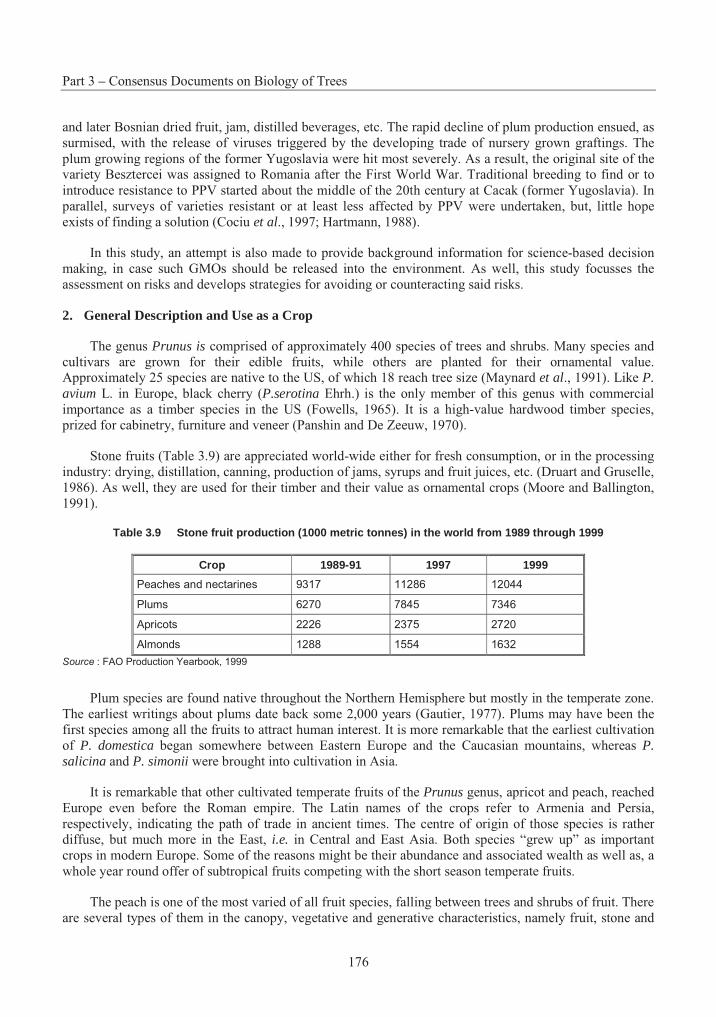
Part 3 – Consensus Documents on Biology of Trees
176
and later Bosnian dried fruit, jam, distilled beverages, etc. The rapid decline of plum production ensued, as surmised, with the release of viruses triggered by the developing trade of nursery grown graftings. The plum growing regions of the former Yugoslavia were hit most severely. As a result, the original site of the variety Besztercei was assigned to Romania after the First World War. Traditional breeding to find or to introduce resistance to PPV started about the middle of the 20th century at Cacak (former Yugoslavia). In parallel, surveys of varieties resistant or at least less affected by PPV were undertaken, but, little hope exists of finding a solution (Cociu et al., 1997; Hartmann, 1988).
In this study, an attempt is also made to provide background information for science-based decision making, in case such GMOs should be released into the environment. As well, this study focusses the assessment on risks and develops strategies for avoiding or counteracting said risks.
2. General Description and Use as a Crop
The genus Prunus is comprised of approximately 400 species of trees and shrubs. Many species and cultivars are grown for their edible fruits, while others are planted for their ornamental value. Approximately 25 species are native to the US, of which 18 reach tree size (Maynard et al., 1991). Like P.avium L. in Europe, black cherry (P.serotina Ehrh.) is the only member of this genus with commercial importance as a timber species in the US (Fowells, 1965). It is a high-value hardwood timber species, prized for cabinetry, furniture and veneer (Panshin and De Zeeuw, 1970).
Stone fruits (Table 3.9) are appreciated world-wide either for fresh consumption, or in the processing industry: drying, distillation, canning, production of jams, syrups and fruit juices, etc. (Druart and Gruselle, 1986). As well, they are used for their timber and their value as ornamental crops (Moore and Ballington, 1991).
Table 3.9 Stone fruit production (1000 metric tonnes) in the world from 1989 through 1999
Crop 1989-91 1997 1999
Peaches and nectarines 9317 11286 12044
Plums 6270 7845 7346
Apricots 2226 2375 2720
Almonds 1288 1554 1632
Source : FAO Production Yearbook, 1999
Plum species are found native throughout the Northern Hemisphere but mostly in the temperate zone. The earliest writings about plums date back some 2,000 years (Gautier, 1977). Plums may have been the first species among all the fruits to attract human interest. It is more remarkable that the earliest cultivation of P. domestica began somewhere between Eastern Europe and the Caucasian mountains, whereas P. salicina and P. simonii were brought into cultivation in Asia.
It is remarkable that other cultivated temperate fruits of the Prunus genus, apricot and peach, reached Europe even before the Roman empire. The Latin names of the crops refer to Armenia and Persia, respectively, indicating the path of trade in ancient times. The centre of origin of those species is rather diffuse, but much more in the East, i.e. in Central and East Asia. Both species “grew up” as important crops in modern Europe. Some of the reasons might be their abundance and associated wealth as well as, a whole year round offer of subtropical fruits competing with the short season temperate fruits.
The peach is one of the most varied of all fruit species, falling between trees and shrubs of fruit. There are several types of them in the canopy, vegetative and generative characteristics, namely fruit, stone and
Section 6 – Stone Fruits
177
seed traits. All commercial cultivars belong to P. persica L. Batsch, and are primarily grown in temperate zones between latitudes 30° and 45° N and S, and in the tropics and subtropics at higher elevations (Hammerschlag, 1986).
A close relative of the peach, the almond, represents an entirely different food quality. Its cultivation in generally dry, if not marginal habitats, is as extensive as an almost semi-domesticated fruit, less subject to phytosanitary problems. Interspecific hybrids of P. amygdalus and P. persica are well known in fruit growing as an important rootstock for peach production, e.g., GF 677.
A somewhat detached subgenus of the genus Prunus includes the cherry and sour cherry which are, equally, ancient cultivated fruits, one of them being diploid, the other, tetraploid.
P. avium is primarily a European species, which occurs abundantly in wild form on the forest slopes of Southern, Central and Western Europe. Pomologically, according to fruit firmness, cherry cultivars are divided into the Heart cherry group, with mainly early ripening cultivars that have a soft flesh, and the Bigarreau group. The Bigarreau group includes late cultivars with firm flesh, such as Lambert, Stella, Bing, Van, Windsor, Schmidt, Hedelfingen, Napoleon, and Gold that have dark red, black, yellowish or light-coloured fruits. The major portion of the harvest is processed into solid, liquid or frozen products, and part is kept for direct consumption in the fresh state (Ivanicka and Pretová, 1986).
Sour cherry is widely naturalised and its distribution area covers almost all European countries and SW Asia. However, it is cultivated in many other parts of the world, mainly in North America. Sour cherry production is about one-third that of sweet cherry (FAO Yearbook, 1975, data not included in later editions). More recent data (in thousands tons) indicate that the most important producers are the USSR with 450 (Kramer, 1985) and the USA with 119 (Westwood, 1978). Other countries with great productions are: Germany (91), Former Yugoslavia (47), and Hungary (41) (Christensen, 1985; Kramer, 1985). Although the most important cultivars used are Schattenmorelle and Montmorency, the list of cultivars reaches a great number. Thus, in the USSR it runs up to 80 sour cherry varieties listed in various district catalogs (Kramer, 1985). The use of Stockton Morello in North America as a cherry rootstock is very minor (Tukey, 1964). The predominant root stocks in North America are mazzard (Prunus avium) or P.mahaleb.
The predominance of one or two major apricot cultivars in each production area is partly responsible for large fluctuations in yield and makes this crop species vulnerable to adverse environmental conditions, diseases and pests (Mehlenbacher et al., 1991). Moreover, the major cultivars of the main apricot producing countries (Spain, Italy, the United States, Greece, France, Morocco, Hungary, Romania, South Africa, Bulgaria, Australia, Algeria) belong to the European group, which by their origin are known to have a very narrow genetic background (Kostina, 1969).
Apricot production is rapidly changing in Europe. Spain, the main producer keeps its production constant, while France is increasing production and Italy and Greece are decreasing their production levels.
Especially drastic is the situation in Greece, where the annual production of 100,000 tonnes about 10 years ago has decreased to 30-50,000 tonnes, mainly due to damage caused by late frosts and the Sharka virus.
The same holds true for Hungary, where at the beginning of the Seventies 60-130,000 tons were produced on an area of 13-14,000 ha, while in the early 90s the orchard area decreased to 2,500 – 3,000 ha and production dropped to 20-40,000 tons/a (Pedryc, Budapest, pers. comm).
Part 3 – Consensus Documents on Biology of Trees
178
3. Taxonomic Situation
A. Taxonomy
In the past different approaches were chosen to present the phylogeny of the subfamily of Prunoideaebelonging to the family of Rosaceae. There were two main contrasting conceptions, i.e. all stone fruits belong to the genus Prunus, or the genus Prunus contains only plums and prunes. Here the classification is presented according to Strasburger et al. (1991).
The seven subgenera in Prunus are determined basically by how the leaves are rolled up in the bud, whether the flowers are organised in cymes or in racemes and finally by morphological characteristics of the generative organs, i.e. the size and colour of flowers, fruit, stone and seed traits.
− AMYGDALUS (almonds): P. amygdalus, P. bucharica, P. fenzliana, P. kuramica, P. nana, P. orientalis, P. webbii
− PERSICA (peaches): P. davidiana, P. ferganensis, P. kansuensis, P. mira, P. persica
− ARMENIACA (apricots): P. ansu, P. armeniaca, P. brigantiaca, P. x dasycarpa, P. holosericea, P. mandshurica, P. mume, P. sibirica
− PRUNUS (plums and prunes): P. cerasifera, P. divaricata, P. domestica, P. insititia, P. italica, P. spinosa, P. syriaca, P. salicina, P. simonii, P. ussuriensis, P. americana, P. angustifolia, P. hortulana, P. maritima, P. mexicana, P. munsoniana, P. nigra, P. rivularis, P. subcordata
− CERASUS (sweet and sour cherries): P. avium, P. cerasus, P. fruticosa, P. japonica, P. maackii, P. mahaleb, P. pseudocerasus, P. pumila, P. serrulata, P. tomentosa
− PADUS (bird cherries) P. padus, P. serotina
− LAUROCERASUS (bay-cherries)
This study will focus on essential data about the species in bold (P. amygdalus, P. persica, P. armeniaca, P. domestica, P. avium, P. cerasus and P. salicina), since they are the most widely grown species with horticultural interest. However, interactions with wild or escaped relatives will also be considered.
B. Number of chromosomes
The phenomenon of polyploidy is a widespread occurrence and of great importance in the evolution of new species or forms. For example, many genera of flowering plants contain a series of species characterised by varying degrees of ploidy. Polyploidy is important, too, from a practical point of view, since plants with this character are often very vigorous, and may be more resistant to frost and the attacks of parasitic fungi. Moreover, changes of flower structure and self-fertility according to the number of chromosomes have been observed. In Prunus, the basic number in vegetative cells is eight chromosomes. Polyploidy, due to interspecific hybridisation, took place during the phylogeny of the genus and is responsible for self-sterility and intersterility. The C-value is the DNA amount in the unreplicated haploid nucleus (pg/cell). The DNA amount in the unreplicated haploid or gametic nucleus of an organismis referred to as its C-value (Swift, 1950), irrespective of the ploidy level of the taxon. C-value equals genome size in diploid species, but always exceeds genome size in polyploid species. Nuclear DNA C-value and genome size are important biodiversity characters with fundamental biological significance and many uses (Bennett and Leitch, 1995).
The following different number of chromosomes and degrees of ploidy have been reported:
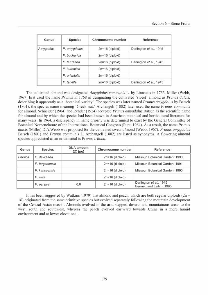
Section 6 – Stone Fruits
179
Genus Species Chromosome number Reference
Amygdalus P. amygdalus 2n=16 (diploid) Darlington et al., 1945
P. bucharica 2n=16 (diploid)
P. fenzliana 2n=16 (diploid) Darlington et al., 1945
P. kuramica 2n=16 (diploid)
P. orientalis 2n=16 (diploid)
P. tenella 2n=16 (diploid) Darlington et al., 1945
The cultivated almond was designated Amygdalus communis L. by Linnaeus in 1753. Miller (Webb, 1967) first used the name Prunus in 1768 in designating the cultivated ‘sweet’ almond as Prunus dulcis,describing it apparently as a ‘botanical variety’. The species was later named Prunus amygdalus by Batsch (1801), the species name meaning ‘Greek nut.’ Archangeli (1882) later used the name Prunus communisfor almond. Schneider (1904) and Rehder (1924) accepted Prunus amygdalus Batsch as the scientific name for almond and by which the species had been known in American botanical and horticultural literature for many years. In 1964, a discrepancy in name priority was determined to exist by the General Committee of Botanical Nomenclature of the International Botanical Congress (Punt, 1964). As a result, the name Prunus dulcis (Miller) D.A.Webb was proposed for the cultivated sweet almond (Webb, 1967). Prunus amygdalusBatsch (1801) and Prunus communis L. Archangeli (1882) are listed as synonyms. A flowering almond species appreciated as an ornamental is Prunus triloba.
Genus Species DNA amount 2C (pg) Chromosome number Reference
Persica P. davidiana 2n=16 (diploid) Missouri Botanical Garden, 1990
P. ferganensis 2n=16 (diploid) Missouri Botanical Garden, 1991
P. kansuensis 2n=16 (diploid) Missouri Botanical Garden, 1990
P. mira 2n=16 (diploid)
P. persica 0.6 2n=16 (diploid) Darlington et al., 1945 Bennett and Leitch, 1995
It has been suggested by Watkins (1979) that almond and peach, which are both regular diploids (2n = 16) originated from the same primitive species but evolved separately following the mountain development of the Central Asian massif. Almonds evolved in the arid steppes, deserts and mountainous areas to the west, south and southwest, whereas the peach evolved eastward towards China in a more humid environment and at lower elevations.
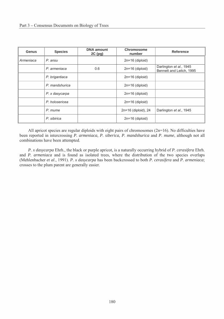
Part 3 – Consensus Documents on Biology of Trees
180
Genus Species DNA amount 2C (pg)
Chromosome number Reference
Armeniaca P. ansu 2n=16 (diploid)
P. armeniaca 0.6 2n=16 (diploid) Darlington et al., 1945 Bennett and Leitch, 1995
P. brigantiaca 2n=16 (diploid)
P. mandshurica 2n=16 (diploid)
P. x dasycarpa 2n=16 (diploid)
P. holosericea 2n=16 (diploid)
P. mume 2n=16 (diploid), 24 Darlington et al., 1945
P. sibirica 2n=16 (diploid)
All apricot species are regular diploids with eight pairs of chromosomes (2n=16). No difficulties have been reported in intercrossing P. armeniaca, P. siberica, P. mandshurica and P. mume, although not all combinations have been attempted.
P. x dasycarpa Ehrh., the black or purple apricot, is a naturally occurring hybrid of P. cerasifera Ehrh. and P. armeniaca and is found as isolated trees, where the distribution of the two species overlaps (Mehlenbacher et al., 1991). P. x dasycarpa has been backcrossed to both P. cerasifera and P. armeniaca;crosses to the plum parent are generally easier.
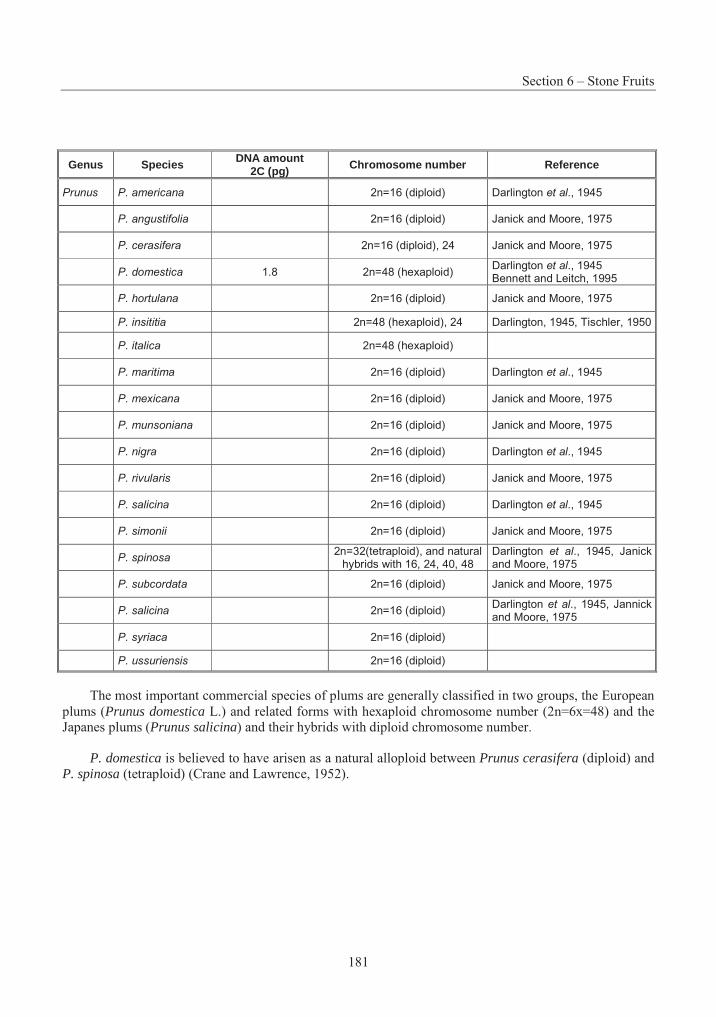
Section 6 – Stone Fruits
181
Genus Species DNA amount 2C (pg) Chromosome number Reference
Prunus P. americana 2n=16 (diploid) Darlington et al., 1945
P. angustifolia 2n=16 (diploid) Janick and Moore, 1975
P. cerasifera 2n=16 (diploid), 24 Janick and Moore, 1975
P. domestica 1.8 2n=48 (hexaploid) Darlington et al., 1945 Bennett and Leitch, 1995
P. hortulana 2n=16 (diploid) Janick and Moore, 1975
P. insititia 2n=48 (hexaploid), 24 Darlington, 1945, Tischler, 1950
P. italica 2n=48 (hexaploid)
P. maritima 2n=16 (diploid) Darlington et al., 1945
P. mexicana 2n=16 (diploid) Janick and Moore, 1975
P. munsoniana 2n=16 (diploid) Janick and Moore, 1975
P. nigra 2n=16 (diploid) Darlington et al., 1945
P. rivularis 2n=16 (diploid) Janick and Moore, 1975
P. salicina 2n=16 (diploid) Darlington et al., 1945
P. simonii 2n=16 (diploid) Janick and Moore, 1975
P. spinosa 2n=32(tetraploid), and natural
hybrids with 16, 24, 40, 48 Darlington et al., 1945, Janick and Moore, 1975
P. subcordata 2n=16 (diploid) Janick and Moore, 1975
P. salicina 2n=16 (diploid) Darlington et al., 1945, Jannick and Moore, 1975
P. syriaca 2n=16 (diploid)
P. ussuriensis 2n=16 (diploid)
The most important commercial species of plums are generally classified in two groups, the European plums (Prunus domestica L.) and related forms with hexaploid chromosome number (2n=6x=48) and the Japanes plums (Prunus salicina) and their hybrids with diploid chromosome number.
P. domestica is believed to have arisen as a natural alloploid between Prunus cerasifera (diploid) and P. spinosa (tetraploid) (Crane and Lawrence, 1952).
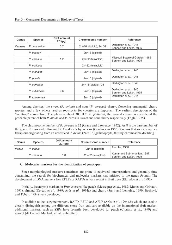
Part 3 – Consensus Documents on Biology of Trees
182
Genus Species DNA amount 2C (pg) Chromosome number Reference
Cerasus Prunus avium 0.7 2n=16 (diploid), 24, 32 Darlington et al., 1945 Bennett and Leitch, 1995
P. besseyi 2n=16 (diploid)
P. cerasus 1.2 2n=32 (tetraploid) Missouri Botanical Garden, 1985 Bennett and Leitch, 1995
P. fruticosa 2n=32 (tetraploid)
P. mahaleb 2n=16 (diploid) Darlington et al., 1945
P. pumila 2n=16 (diploid) Darlington et al., 1945
P. serrulata 2n=16 (diploid), 24 Darlington et al., 1945
P. subhirtella 0.6 2n=16 (diploid) Darlington et al., 1945 Bennett and Leitch, 1995
P. tomentosa 2n=16 (diploid) Darlington et al., 1945
Among cherries, the sweet (P. avium) and sour (P. cerasus) cherry, flowering ornamental cherry species, and a few others used as rootstocks for cherries are important. The earliest description of the “keration” comes from Theophrastus about 300 B.C. P. fruticosa, the ground cherry, is considered the probable parent of both P. avium and P. cerasus, sweet and sour cherry respectively (Fogle, 1975).
The chromosome number of P. cerasus is 32 (Crane and Lawrence, 1952). As 8 is the base number of the genus Prunus and following De Candolle´s hypothesis (Coutanceau 1953) it seems that sour cherry is a tetraploid originating from an unreduced P. avium (2n = 16) gametophyte, thus by chromosome doubling.
Genus Species DNA amount 2C (pg) Chromosome number Reference
Padus P. padus 2n=16 (diploid) Tischler, 1950
P. serotina 1.0 2n=32 (tetraploid) Kumar and Subramanian, 1987 Bennett and Leitch, 1995
C. Molecular markers for the identification of genotypes
Since morphoplogical markers sometimes are prone to equivocal interpretations and generally time consuming, the search for biochemical and molecular markers was initiated in the genus Prunus. The development of DNA markers like RFLPs or RAPDs is very recent in fruit trees (Eldredge et al., 1992).
Initially, isoenzyme markers in Prunus crops like peach (Messeguer et al., 1987; Monet and Gribault, 1991), almond (Cerezo et al., 1989; Arús et al., 1994a) and cherry (Santi and Lemoine, 1990; Boskovic and Tobutt, 1994) were developed.
In addition to the isozyme markers, RAPD, RFLP and AFLP (Arús et al., 1994a,b) which are used to clearly distinguish among the different stone fruit cultivars available on the international fruit market, additional markers, such as SSRs have recently been developed for peach (Cipriani et al., 1999) and apricot (da Camara Machado et. al., submitted).
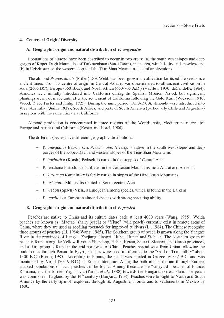
Section 6 – Stone Fruits
183
4. Centres of Origin/ Diversity
A. Geographic origin and natural distribution of P. amygdalus
Populations of almond have been described to occur in two areas: (a) the south west slopes and deep gorges of Kopet-Dagh Mountains of Turkmenistan (800-1700m), in an area, which is dry and snowless and (b) in Uzbekistan on the western slopes of the Tian Shan Mountains at similar elevations.
The almond Prunus dulcis (Miller) D.A Webb has been grown in cultivation for its edible seed since ancient times. From its centre of origin in Central Asia, it was disseminated to all ancient civilisation in Asia (2000 BC), Europe (350 B.C.), and North Africa (600-700 A.D.) (Vavilov, 1930; deCandolle, 1964). Almonds were initially introduced into California during the Spanish Mission Period, but significant plantings were not made until after the settlement of California following the Gold Rush (Wickson, 1910; Wood, 1925; Taylor and Philip, 1925). During the same period (1850-1900), almonds were introduced into West Australia (Quinn, 1928), South Africa, and parts of South America (particularly Chile and Argentina) in regions with the same climate as California.
Almond production is concentrated in three regions of the World: Asia, Mediterranean area (of Europe and Africa) and California (Kester and Horel, 1980).
The different species have different geographic distributions:
− P. amygdalus Batsch. syn. P. communis Arcang. is native in the south west slopes and deep gorges of the Kopet-Dagh and western slopes of the Tien-Shan Mountains
− P. bucharica (Korsh.) Fedtsch. is native in the steppes of Central Asia
− P. fenzliana Fritsch. is distributed in the Caucasian Mountains, near Ararat and Armenia
− P. kuramica Korchinsky is feraly native in slopes of the Hindukush Mountains
− P. orientalis Mill. is distributed in South-central Asia
− P. webbii (Spach) Vieh., a European almond species, which is found in the Balkans
− P. tenella is a European almond species with strong sprouting ability
B. Geographic origin and natural distribution of P. persica
Peaches are native to China and its culture dates back at least 4000 years (Wang, 1985). Wolda peaches are known as “Maotao” (hairy peach) or “Yitao” (wild peach) currently exist in remote areas of China, where they are used as seedling rootstock for improved cultivars (Li, 1984). The Chinese recognise three groups of peaches (Li, 1984; Wang, 1985). The Southern group of peach is grown along the Yangtze River in the provinces of Jiangsu, Zhejiang, Jiangxi, Hubei, Hunan and Sichuan. The Northern group of peach is found along the Yellow River in Shandong, Hebei, Henan, Shanxi, Shaanxi, and Gansu provinces, and a third group is found in the arid northwest of China. Peaches spread west from China following the trade routes through Persia. In Egypt, peaches were used in offerings to the “God of Tranquillity” about 1400 B.C. (Roach, 1985). According to Plinius, the peach was planted in Greece by 332 B.C. and was mentioned by Virgil (70-19 B.C.) in Roman literature. Along the path of distribution through Europe, adapted populations of local peaches can be found. Among these are the “vineyard” peaches of France, Romania, and the former Yugoslavia (Parnia et al., 1988) towards the Hungarian Great Plain. The peach was common in England by the 14th century (Bunyard, 1938). Peaches were brought to North and South America by the early Spanish explorers through St. Augustine, Florida and to settlements in Mexico by 1600.
Part 3 – Consensus Documents on Biology of Trees
184
Five species are considered as peaches:
− P. davidiana (Carr.) Franch. is native of North China
− P. ferganensis (Kost. et Rjyb.) Kov. et Kost., which is found in Western China
− P. kansuensis Rehd., which is native in North-western China
− P. mira Koehne, which is found in the Himalayan mountains and along the Yellow and Yangtze Rivers
− P. persica (L.) Batsch. which contains most of the cultivated peaches and nectarines (convarietas leavis), be it freestone (provar. glabra) or clingstone (provar. nudicarpa).
C. Geographic origin and natural distribution of P. armeniaca
Prunus armeniaca L., the cultivated apricot, is believed to have originated in the mountains of Northern and North-eastern China in the same area as the Great Wall and overlapping the southern branch of the distribution of P.sibirica L. (Mehlenbacher et al., 1991). The apricot was brought via Armenia and Asia Minor into Italy over 2000 years ago, to England in the 13th century and to North America only by 1720 (Westwood, 1978).
Wild apricots also occur in the Tien Shan Mountains in the Xinijang autonomous region (Wang, 1985) and Dzhungar and Zailing Mountains in Soviet Central Asia. This is believed to be the secondary centre of origin (Zeven and de Wet, 1982). The area of distribution of the cultivated apricot is much larger and includes areas where seedling orchards are common such as Central Asia, Afghanistan, Kashmir, Iran, Turkey, and Trans-Caucasia (Kostina, 1936; Mehlenbacher et al., 1991). All of these areas are valuable sources of germplasm.
Apricot production is severely restricted by ecological conditions. The gene pool of apricot contains only few species and varieties, which range in areas of adaptation from the cold winters of Siberia to the subtropical climate of North Africa and from California, the deserts of Central Asia and the humid areas of Japan and Eastern China. However, commercial production areas are still very limited (Mehlenbacher et al., 1991; Faust - Surányi and Nyujtó, 1998).
In China, the Yu’s order (2200 B.C.) refers to apricot growing and there are also documents from the 7th century (Löschnig and Passecker, 1954; Nyujtó and Surányi, 1981; Faust et al., 1998). Kostina (1969) presented an excellent eco-geographical grouping for apricot cultivars and species. There are Central Asian, Irano-Caucasian, European, Northern Chinese, Tibetan, North-eastern Chinese, Eastern Chinese and Dshungar-Zailij groups (Faust et al., 1998; Mehlenbacher et al., 1991). Basic species are identified, as follows:
− P. ansu Maxim. is distributed in Eastern China, South Korea and Japan
− P. armeniaca L. is native of Northern and North-eastern China
− P. brigantiaca Vill. (Alpine apricot) is distributed in the region of the Alps, in South-eastern France
− P. mandshurica (Maxim.) Koehne is native in north east of China
− P. x dasycarpa, a hybrid between P. cerasifera and P. armeniaca Ehrh.
− P. holosericea (Batal) Kost. (Tibetian apricot), which is native of Tibetian Mountains
− P. mume (Sieb.) Sieb. et Zucc., which is native in south of China
Section 6 – Stone Fruits
185
− P. sibirica L., which is distributed along Baikal Lake, Mandshuria, North Korea
D. Geographic origin and natural distribution of P. domestica and P. salicina
Plum species are found native throughout the Northern Hemisphere but mostly in the temperate zone. The earliest writings about plums date back some 2000 years (Cullinan, 1937) and De Candolle assumes that plums have been known for 2000-4000 years (Banegal, 1954).
Prunus domestica seems to have originated in Southern Europe or Western Asia around the Caucasus Mountains and the Caspian sea (Cullinan, 1937). However, it is also widespread in the Balkans and Mediterranean countries.
Prunus salicina originated in China and was introduced into Japan 200-400 years ago. In China, it has been cultivated since ancient times where it is thought to occur in the wild in the Tsunglin range in Shensi and Kansu. Recently, it reached Europe by way of California and Italy.
Plums are a diverse group of plants with many botanical species, that have been cultivated for the last 3000 years. The most important species of Prunus are generally classified into three groups, the European, the Asian and the American plums. Plums may have been the first species among all the fruits to attract human interest. Six of the most important species of plums, P. domestica, P. italica, P. syriaca, P. salicina, P. simonii and P. americana are not known in the wild and presumably were selected and cultivated very early by humans. It is more remarkable that the earliest cultivation of P. domestica began somewhere between Eastern Europe and the Caucasian mountains, whereas P. salicina and P. simonii were brought into cultivation in Asia.
Regarding the Krieche/Haferpflaume (P. insistitia var. juliana), repeatedly described as “wild”, Körber-Grohne (1996) mentions, that this is not the case in SW-Germany. As a welcome fruiting shrub, it has served as a shrubby hedge (Hag) around farm gardens or as a division between fruit orchards. It is not present in hedges or open fields nor is it found in woods or wood margins, as is the case with the crab apple. The oldest subfossil fruitstones have been found in Neolithic settlements in Germany and Switzerland (Ehrenstein, Robenhausen). The Krieche/Haferpflaume (P. insistitia var. juliana) is a typical example of the continuity of domestication from the Neolithic until the present, which has been attributed to the propagation by grafting from Roman times onwards, or in the case of plum cultivars through root suckers.
The classification of plums is divided into geographic groups:
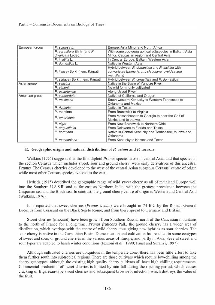
Part 3 – Consensus Documents on Biology of Trees
186
P. spinosa L. Europe, Asia Minor and North Africa
P. cerasifera Ehrh. (and P. divaricata Ledeb.)
With some eco-geographical subspecies in Balkan, Asia Minor, Caucasian region and Central Asia
P. insititia L. In Central Europe, Balkan, Western Asia
P. domestica L. Native in Western Asia
P. italica (Borkh.) em. Kárpáti Hybrid between P. domestica and P. insititia with convarietas (pomariarum, claudiana, ovoidea and mamillaris)
European group
P. syriaca (Borkh.) em. Kárpáti Hybrid between P. cerasifera and P. domestica
P. salicina Native in the Basin of Yangtze River
P. simonii No wild form, only cultivated
Asian group
P. ussuriensis Along Ussuri River
P. subcordata Native of California and Oregon
P. mexicana South-western Kentucky to Western Tennessee to Oklahoma and Mexico
P. rivularis Native in Texas
P. maritima From Brunswick to Virginia
P. americanaFrom Massachusetts to Georgia to near the Golf of Mexico and to the west
P. nigra From New Brunswick to Northern Ohio
P. angustifolia From Delaware to Florida and Texas
P. hortulana Native in Central Kentucky and Tennessee, to Iowa and Oklahoma
American group
P. munsoniana From Kentucky to Kansas and Texas
E. Geographic origin and natural distribution of P. avium and P. cerasus
Watkins (1976) suggests that the first diploid Prunus species arose in central Asia, and that species in the section Cerasus which includes sweet, sour and ground cherry, were early derivatives of this ancestral Prunus. The Cerasus cherries developed to the west of the central Asian subgenus Cerasus’ centre of origin while most other Cerasus species evolved to the east.
Hedrick (1915) described the geographic range of wild sweet cherry as all of mainland Europe well into the Southern U.S.S.R. and as far east as Northern India, with the greatest prevalence between the Casparian sea and the Black sea. In contrast, the ground cherry centre of origin is Western and Central Asia (Watkins, 1976).
It is reported that sweet cherries (Prunus avium) were brought in 74 B.C by the Roman General Lucullus from Cerasunt on the Black Sea to Rome, and from there spread to Germany and Britain.
Sweet cherries (mazzard) have been grown from Southern Russia, north of the Caucasian mountains to the north of France for a long time. Prunus fruticosa Pall., the ground cherry, has a wider area of distribution, which overlaps with the centre of wild cherry, thus giving new hybrids as sour cherries. The sour cherry is native in the Carpathian Basin. Domestication and cultivation has resulted in some ecotypes of sweet and sour, or ground cherries in the various areas of Europe, and partly in Asia. Several sweet and sour types are adapted to harsh winter conditions (Iezzoni et al., 1990; Faust and Surányi, 1997).
Although cultivated cherries are ubiquitous in the temperate zone, there has been little effort to take them further south into subtropical regions. There are those cultivars which require low-chilling among the cherry genotypes, although the existing high quality cherry cultivars all have high chilling requirements. Commercial production of sweet cherries is limited by rain fall during the ripening period, which causes cracking of Bigarreau-type sweet cherries and subsequent brown-rot infection, which destroys the value of the fruit.
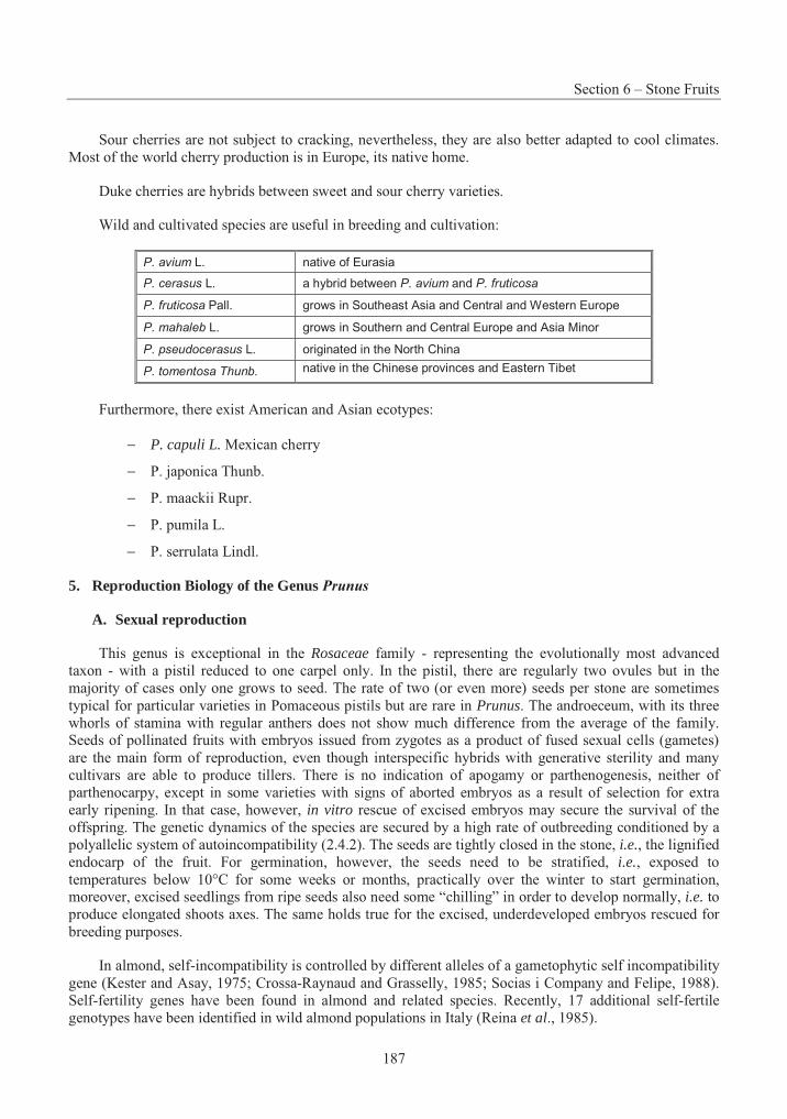
Section 6 – Stone Fruits
187
Sour cherries are not subject to cracking, nevertheless, they are also better adapted to cool climates. Most of the world cherry production is in Europe, its native home.
Duke cherries are hybrids between sweet and sour cherry varieties.
Wild and cultivated species are useful in breeding and cultivation:
P. avium L. native of Eurasia
P. cerasus L. a hybrid between P. avium and P. fruticosa
P. fruticosa Pall. grows in Southeast Asia and Central and Western Europe
P. mahaleb L. grows in Southern and Central Europe and Asia Minor
P. pseudocerasus L. originated in the North China
P. tomentosa Thunb. native in the Chinese provinces and Eastern Tibet
Furthermore, there exist American and Asian ecotypes:
− P. capuli L. Mexican cherry
− P. japonica Thunb.
− P. maackii Rupr.
− P. pumila L.
− P. serrulata Lindl.
5. Reproduction Biology of the Genus Prunus
A. Sexual reproduction
This genus is exceptional in the Rosaceae family - representing the evolutionally most advanced taxon - with a pistil reduced to one carpel only. In the pistil, there are regularly two ovules but in the majority of cases only one grows to seed. The rate of two (or even more) seeds per stone are sometimes typical for particular varieties in Pomaceous pistils but are rare in Prunus. The androeceum, with its three whorls of stamina with regular anthers does not show much difference from the average of the family. Seeds of pollinated fruits with embryos issued from zygotes as a product of fused sexual cells (gametes) are the main form of reproduction, even though interspecific hybrids with generative sterility and many cultivars are able to produce tillers. There is no indication of apogamy or parthenogenesis, neither of parthenocarpy, except in some varieties with signs of aborted embryos as a result of selection for extra early ripening. In that case, however, in vitro rescue of excised embryos may secure the survival of the offspring. The genetic dynamics of the species are secured by a high rate of outbreeding conditioned by a polyallelic system of autoincompatibility (2.4.2). The seeds are tightly closed in the stone, i.e., the lignified endocarp of the fruit. For germination, however, the seeds need to be stratified, i.e., exposed to temperatures below 10°C for some weeks or months, practically over the winter to start germination, moreover, excised seedlings from ripe seeds also need some “chilling” in order to develop normally, i.e. to produce elongated shoots axes. The same holds true for the excised, underdeveloped embryos rescued for breeding purposes.
In almond, self-incompatibility is controlled by different alleles of a gametophytic self incompatibility gene (Kester and Asay, 1975; Crossa-Raynaud and Grasselly, 1985; Socias i Company and Felipe, 1988). Self-fertility genes have been found in almond and related species. Recently, 17 additional self-fertile genotypes have been identified in wild almond populations in Italy (Reina et al., 1985).
Part 3 – Consensus Documents on Biology of Trees
188
Prunus persica is self-fertile. However, pollen sterility in P. persica could be useful for interspecific hybridisation.
In apricot, most Central Asian cultivars are self-incompatible, while most European cultivars are self-compatible (Mehlenbacher et al., 1991).
Most sweet cherry cultivars presently being grown are self-incompatible. Self-incompatibility in sweet cherry was first identified by East and Mangelsdorf (1925) and later determined to be of the monofactorial gametophytic type with multiple allelic S locus (Lewis, 1948).
Self-fertility in sweet cherry has been obtained through X-ray radiation applied to flower buds at the pollen mother cell stage (Lewis, 1948; Lewis and Crowe, 1954) and by spontaneously occurring mutations (Lewis, 1951).
B. The mating system of most cultivated Prunus species
The mating system of most cultivated Prunus species is determined by the clearly defined system of auto-incompatibility, which is inherently combined with inter-incompatibility. However, the high number (20-40) of known alleles of the single (Sx) locus with gametophytic determination allows little chance of meeting incompatible mates amongst the varieties cultivated. Those incompatible combinations are registered according to experimental proofs accumulated during the last, nearly seventy years since the phenomenon has been detected. Auto-incompatibility is expressed at different degrees between total, intermediate and scarcely identified cases. The apparent lack of such a mechanism is found in peach, although male sterility occurs at a relatively high frequency, e.g., the cultivar J.H. Hale, and in some plums, e.g., the cvs Tuleu gras, Pitestan and Carpentin (Silbereisen et al., 1996). The evolutionary role of male sterility is highly analogous to that of incompatibility. As a general tendency of the domestication process, the increased rate of self-fertility is derived from the selection pressure for high and regular yields in crops grown for their generative organs. This can be seen in the Prunus species as many, mainly recently developed, varieties are self-fertile. In modern times, self-fertility has been consciously favoured. Another condition of self-fertility is polyploidy appearing on the margin of the spreading species like the European plum and sour cherry as amphiploids of interspecific hybrids in which activity of the S alleles is impaired. No doubt that some of those clones are also tillering spontaneously. Conditions of cultivation, introduction to new habitats included, are similar to the marginal areas of a species where rare mutations enjoy better chances to survive. All those reasons enhance the chances of self-fertile genotypes. Some exceptions in European plum and sour cherry prove that auto-incompatibility did not disappear entirely with amphiploidy, so a conscious effort in the research for high productivity gave rise to new, self-fertile sour cherry cultivars.
Search for radiation-induced mutants in cherry populations has spotted the Sf allele, which proved to be dominant in relation to the rest of the sterility alleles. This phenomena is employed in breeding programmes in order to produce new self-fertile cherry cultivars. The first self-fertile sweet cherry cultivar was released from a breeding program in 1968 and named Stella (Lapins, 1971). It was the result of a cross between Lambert and JI 2420. The cross was made in 1956. JI 2420 came from the John Innes Institute in the UK and was the result of a cross between Emperor Francis and irradiated pollen from Napoleon. At the moment all self-fertile cultivars have Stella in its pedigree somewhere. The John Innes Institute had a couple of other selections that were self-fertile but they have not yet made it into a named cultivar.
Bees play a major role as pollinating agents, as the pollen of Prunus species cannot be carried by wind and even self-pollination requires the mechanical intervention of insects.
Section 6 – Stone Fruits
189
C. Natural vegetative multiplication
Natural vegetative multiplication is rather exceptional in the genus of Prunus, but all subgenera have members which build up extended colonies by tillering, and some successful cultivars have been maintained by tillers since ancient times, e.g., European (hexaploid) plums in the NE-Hungary (in the riverside of the Tisza river) and there are clones of semi-wild cultigens, blackthorn and sour cherry. Most of the existing cultivars are, however, multiplied by grafting and trading, except, some old varieties, such as the sour cherry variety Cigánymeggy (Hungary) and Oblacinska (Former Yugoslavia). In the group of almonds the only tillering wild species, P. tenella, is eligible as a potentially dwarfing stock.
More attempts have been invested into the vegetative propagation of Prunus rootstocks. As very few are inclined to develop tillers spontaneously, the tools of micropropagation in vitro gained particularly high interest, for example, in the rapid multiplication of GF 677. In fact, millions of plants are produced world-wide by in vitro techniques (Rosati and de Paoli, 1992). The production of self-rooted plantlets by green cuttings under a mist curtain seems to be less favoured mainly because of the questionable value of the self-rooted trees and/or the low efficiency of the technique. Grafting techniques, on the other hand, became routine several centuries ago. In addition, some stocks are propagated with cuttings.
6. Crossability
A. Interspecific and intergeneric hybrids
Interspecific (and intergeneric) relations in Prunus are not clear, perhaps due to the widespread presence of auto-incompatibility and the relative fertility of interspecific hybrids. The prolonged time period for flowering in the Prunus species and even within varieties of one species, substantially influences the possibility of mutual pollination between different cultivars, as well as different species. This is due to the different phases in flowering during the blooming season of the cultivated Prunus species.
The physiological or ecological diversity of the species caused sufficient isolation of their individual habitat. Consequently, they were scarcely sympatric in their natural environment, whereas, some Prunus species were grown, regularly, in home gardens if not in larger orchards. Interspecific barriers did not develop during natural evolution, but by the appearance of cultigenous hybrids, which triggered their development from the first steps of domestication up to the limits set by taxonomic divergence. Although the cherries are perhaps the most distant from the rest of the species, we find bridging species between plums and cherries as documented for P. salicina. Less difficult seems to be the gene flow between plum-apricot-peach and almond as documented by the list of successful interspecific crosses (Table 3.10).
Part 3 – Consensus Documents on Biology of Trees
190
Table 3.10 Interspecific hybrids with Prunus persica
x P. persica P. amygdalus
x (P. amygdalus x P. davidiana)
x P. davidiana P. armeniaca
x P. persica
P. besseyi x P. persica
P. cerasus x P. persica
P. hortulana x P. persica
P. nana x P. persica
x P. amygdalus
x P. davidiana
x P. cerasifera var. divaricata
x cherry (sps?)
x P. kansuensis
x P. mira
x P. nana
x P. besseyi
x P. salicina
P. persica
x P. spinosa
P. salicina x P. persica
P. spinosa x P. persica
x P. davidianaP. tenella
x P. persica
Source : after Janick and Moore, 1975
All the species mentioned have been intercrossed with various degrees of difficulty, and grafted on each other within reasonable limits, which is proof of their genetic and physiological affinities. The use of rootstocks enlarges, dramatically, the possibilities of occupying ecological niches previously inaccessible with species on their own roots.
Hybridisation readily takes place between Prunus amygdalus and Prunus persica (Kester and Asay, 1975, 1988). Naturally interspecific hybrid rootstocks, ‘GF 667’, are common, where the two species are grown together.
North-American species and their interspecific hybrids, created between 1907 and 1965, represent a distinct group of cultivated Prunus species, the “cherry plums” (different from the species P. cerasifera,the cherry plum in the traditional sense). They are derived essentially from P. besseyi and P. pumila, with the western and the eastern sand cherry as a common parent (Janick and Moore, 1975) (Table 3.11).
Section 6 – Stone Fruits
191
Table 3.11 Hybrids of the sand cherries (P. besseyi and P. pumila) with other species
Sand Cherry x P. americana
Sand Cherry x P. salicina
Sand Cherry x P. salicina
Sand Cherry x P. simoni
Sand Cherry x P. armeniaca
Sand Cherry x P. persica
Source : Janick and Moore, 1975)
It is remarkable that peach (P. persica) is one of the most flexible species of Prunus regarding its use in interspecific crosses for breeding purposes. The documented products of these breeding efforts are divided into two distinct groups: Hybrids mostly fertile (A) and mostly sterile (B). A complicated polyhybrid background is surmised in most cases from taxa P. davidiana (d), ferganensis (f), kansuensis (k), mira (m) and persica (p) according to Scorza and Okie (1990) and Janick and Moore (1996) (Table 3.12). In group (A), we may consider the hybrid products as potentially new fruits, i.e. distinct commodities, as it happened with the sand cherry derivatives in the Midwest of the United States.
Part 3 – Consensus Documents on Biology of Trees
192
Table 3.12 Prunus species reported as hybrids between peach and peach species
Species Hybrid Common name Origin
P. amygdalus (A) d.m.p. almond SW Asia P. davidiana (A) k.p. mountain peach, shan tao N China
P. ferganensis (A) p. xinjiang tao NE China, S Russia
P. kansuensis (A) d.p. wild peach, kansu tao NW China
P. mira (A) p. Tibetan peach, xizang tao, smooth-pit W China-Himalayas
P. persica (A) d.f.k.m. peach, maotao China
P. americana (B) p. American plum USA
P. armeniaca (B) d.p. Apricot Asia
P. besseyi (B) d.p. western sand cherry N USA, Canada
P. brigantina (B) p. Briancon apricot France
P. cerasifera (B) d.p. myrabolan plum W Asia
P. cerasus (B) p. sour cherry W Asia, SE Europe
P. domestica (B) p. European plum W Asia, Europe
P. hortulana (B) p. wild plum C USA
P. japonica (B) p. Chinese bush cherry, Korean b.c. China
Species Hybrid Common name Origin
P. nigra (B) p. Canadian plum N USA, Canada
P. pumila (B) p. eastern sand cherry N USA
P. salicina (B) f.p. Japanese plum China
P. simmonii (B) p. Simon’s plum N China
P. spinosa (B) p. sloe or blackthorn Europe, W Asia, N Africa
P. tenella = nana (B) d.p. Siberian almond SE Europe, W Asia
P. tomentosa (B) p. Chinese bush cherry, Manchu cherry N&W China, Japan
P. virginiana (B) p. choke cherry N USA, Canada (A) Closely related to peach producing fertile hybrids (B) Hybrids mostly sterile. The codes of species used as parent in the ancestry: P. davidiana (d), ferganensis (f), kansuensis (k), mira (m) and persica (p)
The only valid example of commercially recommended interspecific pollinations is between sweet and sour cherry (Nyéki and Soltész, 1996). As parthenocarpy, understood as seedless fruit, does not exist in Prunus, it seems obvious that seed abortion might have little chance in fruit production, however, some extra early ripening cherry and peach varieties are used to develop unviable seeds. It was proved that the excision of the embryos before the fruit have ripened facilitates the rescue of plantlets under in vitroconditions. Thus an efficacious technique has been developed for use by breeders in combining genes of those extra early varieties, e.g., Bailey and Hough.
Because of the ease of natural hybridisation of P. fruticosa with P. cerasus and P. avium, some P. fruticosa rootstocks under testing may be interspecific hybrids.
Prunus tomentosa has been hybridised with cherry (Fisher and Schmidt, 1938; Noznikov, 1951). Prunus salicina hybridises easily with P. simonii, P. armeniaca, and American plum species.
Section 6 – Stone Fruits
193
No difficulties have been reported in intercrossing P. armeniaca, P.sibirica, P. mandshurica and P.mume, although not all combinations have been attempted. P.x dasycarpa has been backcrossed to both P. cerasifera and P. armeniaca; crosses to the plum parent are generally easier.
Results to date indicate that crosses between true apricot species (P. armeniaca, P. mandshurica, P. sibirica and P. mume) are successful when made in either direction and resulting hybrids are viable and fertile.
A large number of crosses between various plum and apricot species have been reported. Listed in order of flowering date, they are P. salicina Lindl., P.X dasycarpa, P. cerasifera Ehrh., P. domestica L., P.besseyi Bailey, and P. maritima Marsh. The initial cross is generally more successful when plums are used as the female parent. P.cerasifera x P. armeniaca produced hybrids resembling the natural interspecific hybrid species P.X dasycarpa.
Hybrids of the Asian plum species P. salicina with P. armeniaca have also been generated with little difficulty. Fertility of the hybrids varies; pollen fertility is generally quite low.
Several authors also report successful hybridisation of the hexaploid plum P. domestica with apricot. Resulting hybrids are tetraploid.
The beach plum, P. maritima has also been hybridised successfully with common apricot. More distant hybrids of apricots with peach and almond have been reported. These crosses are quite difficult to make and the resulting hybrids are often weak and sterile. The incorporation of genes from P. persicacould conceivably greatly expand the areas in which apricots could be grown.
B. Introgression into wild relatives
The introgression between cultivated and wild species is scarcely documented. There is no doubt concerning the physical possibility. Escapes of cultivated varieties are frequently found in woods, pastures, abandoned orchards, ruderal, suburban, and marginal areas. Intercrosses with really wild populations have very little chance, as blackthorn, hedge cherry and dwarf almond (P. tenella) are extremely different in morphology, as well as in adaptation, i.e. eventual hybrids could only survive in a much protected environment. Cherries may have more chances as far as introgression into the wild populations is concerned. It is worthwhile to consider the escapes of varieties and species introduced as rootstocks to nurseries and grown out from the roots and stumps of destroyed grafts in abandoned orchards. That is how a high diversity of cherry plums have been naturalised recently. As a result, the cherry plum has become much more tolerant than the European plum and apricot to the destructive effect of Plum Pox Virus (PPV). Escaped rootstock varieties and spontaneous hybrids of ancient, as well as, recently introduced varieties are a general phenomena found in neglected orchards, and escapes of no immediate relation to fruits growing in the area are found. For example, P. serotina, P. mahaleb, P. padus, bitter almonds.
In Central Europe, the possibility of introgression is much more limited to the Near East, Caucasus, Iran, Central Asia and the Chinese subcontinent, where a huge wealth of intermediate and semi-cultivated forms reside.
7. Domestication of Prunus sp.
A. Breeding of Prunus
One of the obvious reasons for the abundant domestication of the Prunus species might have been the coincidence between the centre of variability of Prunus and the site of human evolution and/or of the first ancient high civilisations of human history. The easily fossilised stone of the fruit proved that fruit of
Part 3 – Consensus Documents on Biology of Trees
194
considerable size existed long before the appearance of man. Plums “offered themselves” to man to be domesticated. According to ecological arguments, it is highly probable that the today despised species, e.g., blackthorn (P.spinosa) followed men as a “secondary crop” which is reflected in its occurrence as a witness of the ancient Neolithic culture on the outskirts of the villages, roadsides and pastures as hedges. The ancestors of that tetraploid species are unknown. It survived adversities of severe pasturage but benefited from deforestation during the spreading of primitive agriculture in the Near East and Europe, where its more vigorous relative, the favoured fruit tree, the diploid cherry plum (P. cerasifera) presented a permanent temptation for crossbreeding. The appearance of the European plum (P. domestica and insititiaincluded) was not an unique and endemic event in the history of the Eurasian region. The hexaploid, (amphiploid) species has been reproduced at several instances by purposeful breeding, according to the model of bread wheat, triticale, tobacco, oilseed rape, garden strawberry, and other cultivated species, first perhaps by Rybin, a disciple of Vavilov in the 1930s. Since ancient and medieval times, the European plum made an important carrier, first owing to its ability of producing tillers as its alleged ancestor, the blackthorn also did, and secondly, because it became naturalised in mesophytic marginal cultivated areas and some river flats (e.g., of Felsö-Tisza). As the most important fruit and almost staple food, it served the well being of poor people for centuries. However, later on, it became one of the first horticultural products to be exported and became of this, was seen as a symbol of wealth (as the greater number of plum trees on the manor of a member of the gentry, the more wealthy he was). The distilled drink achieved its fame as the national drink in S-E Europe, replacing gin. Recently, the fate of the European plum has been severely impaired, by the Plum Pox Virus, which was identified during the first part of the 20th century.
In the rich choice of plums, a host of species originating from East Asia and North America are diploid. These species have been inter-crossed with the cherry plum with considerable success. As a result, in the last few decades there has been an impetus of the so-called Japanese Plums. The first documented attempts are due to Luther Burbank, a Californian breeder at the end of the 19th century. Those interspecific hybrids, however, well represented in the list of cultivars, only represent a small fragment of the huge gene reserves of the Northern Hemisphere. As a source of precious genes, especially for resistance and special qualities lacking from the traditional European plum, these varieties are to be kept in mind.
B. Conservation of Prunus genetic resources
The International Plant Genetic Resources Institute (IPGRI), formerly known as the International Board for Plant Genetic Resources (IBPGR) has elaborated a descriptor list for plum, peach, cherry and apricot (IBPGR 1984a,b,c, 1995) and developed recommendations for the safe movement of germplasm of stone fruits (Diekmann and Putter, 1996).
The European Information Platform on Crop Genetic Resources has been established under the umbrella of the European Co-operative Programme for Crop Genetic Resources Networks (ECP/GR) to facilitate access to information about genetic resources conserved in genebanks throughout the Region. The European Prunus database has been maintained by Dr. Anne Zanetto at the “Institut National de la Recherche Agronomique” (INRA) in Bordeaux, France, under the initiative of the European Co-operative Programme for Crop Genetic Resources Networks (ECP/GR) since 1994. The database includes, in 26 European countries, the collections of all Prunus species, cultivated stone fruit and their related species (even the wild ones). The database is comprised of 19 passport data from the IPGRI/FAO Multicrop descriptors list, 13 descriptors common for all the different species and 3 to 7 specific descriptors depending on the species of the accession. These descriptors are mainly morphological. The possibility of including more agronomic or physiological descriptors is under consideration by the ECP/GR PrunusWorking Group. The database has been supported for three years by the European Union in the “European Programme on the conservation, characterisation, collection and utilisation of genetic resources in
Section 6 – Stone Fruits
195
agriculture”, under the title “International Network on Prunus genetic resources” (GENRES61). In addition, there are gene banks in China and Japan.
C. Synecology
In Europe, some wild, native and escape species of P. nana, P. avium, P. fruticosa, P. mahaleb and P. spinosa can be found in natural and cultivated forests or ruderals.
D. Interaction with pathogens
Rosaceae, in general, and Prunus species, in particular, are prone, in varying degrees, to infections bya range of pathogens, e.g. fungi such as Monilia laxa, Taphrina deformans, bacteria, such as Pseudomonas,and Xanthomonas, viruses, such as PPV, PNRSV and PDV and phytoplasmas such as European Stone Fruit Yellows (ESFY) (http://www.boku.ac.at/pbiotech/phytopath).
The main problems associated with apricot growing include: die-back or apoplexy, sensitivity to viruses, frost damage in winter and spring, fungi cankers and alternate bearing.
In recent years, a viral pathogen became the major threat to stone fruit cultivations in large areas of Central and Southern Europe and other Mediterranean countries. This pathogen was responsible for considerable economic losses and reduction of production areas. The Plum Pox Virus (PPV), causal agent of Sharka disease and a member of the potyvirus family, was classified by US and EC plant quarantine agencies as the most important pathogen in apricots, plums and peaches (the only plant pathogen for which an APHIS plan exists (Scorza, 1991). Sharka infection data from Spain, Greece, France and Italy clearly demonstrates the economic consequences of this threat (COST 88 Plum Pox Virus Workshop -Potyvirus Group, Valencia June 1993), as apricot cultures are increasingly being replaced. Apricot appears to be the most sensitive stone fruit towards infection with PPV. Apricot production was practically erased in the late 70s in some valleys of Northern Italy, e.g., Vintschgau (Eynard et al., 1991), and is seriously threatened in some Austrian valleys, e.g. Wachau (Pieber, pers. comm). After its appearance in the South American Continent (Herrera et al., 1997) in 1999, it has been confirmed for the first time in the US (http://aphis.usda. gov/lpa/press/1999/10/plumpox.txt). This prompted Canada to close the entry of Prunus material from the US (http://www.cfia-acia.agr.ca/english/corpaffr/ newsrelease/19991122e.shtml).
Considering the severity of the disease, the difficulty to control its spread, and the lack of resistant cultivars, the necessity of resistant cultivars is evident and a straight-forward strategy is required. In fact there are no species resistant to Plum Pox Virus among the species sexually compatible to crop cultivars. This means it is not possible to obtain resistant cultivars by conventional hybridisation.
The information concerning the interrelation of Prunus species gives an opportunity of considering their pathosystems. Most pathogens and pests have developed along evolutionary pathways in parallel with the domestication and evolution of new species and hybrids. Because of geographical continuity, most species coming from the East carried their parasites freely. One of the best examples is the green peach aphid, Myzus persicae, which proved its fidelity to the peach tree in spite of being polyphagous; neither related species substituted the peach as a primary host. It is remarkable that even though the peach was one of the last oriental species to arrive in Europe, the green peach aphid became one of the most efficient vectors of viral diseases in Northern Europe. On the contrary, a pathogen Taphrina deformans did not harm either of the relatives of peach. Although, another adapted disease, Polystigma rubrum kept to the European plum (and blackthorn) as host, in spite of the permanent sympatric presence of relatives of the Prunus genus. Different susceptibilities to other diseases and pests indicates the existence of genetic resistance in spite of the possibly, small genetic divergence, e.g. the reduced proliferation of aphids on apricots in relation to peach and plum.
Part 3 – Consensus Documents on Biology of Trees
196
The increasing threat of stone fruit production in Mediterranean countries by phytoplasma diseases has been recognised in recent years. Although severe decline of European stone fruits was reported as early as 1924 on apricots in France and in 1933 on Japanese plums, in Italy it was only in 1973 that their phytoplasma aetiology was discovered. At that time, phytoplasmas were called mycoplasma-like organisms (MLOs). As different Prunus species were affected, different disease names were given: apricot chlorotic leaf roll (ACLR) on apricots, plum leptonecrosis (PLN) on Japanese plums, peach yellows, peach rosette and peach vein clearing (PVC) on peach and several other decline diseases on European plum, almond and flowering cherry. Prunus rootstocks are also severely affected by similar disorders. Common symptoms are yellowing and leaf roll in summer, off-season growth in winter, die-back and a more or less rapid decline. Up to now these diseases have been restricted to the southern half of Europe with their northern border in Germany. In the past few decades they have been of increasing economic importance, e.g. ACLR and PLN are especially devastating for apricots and Japanese plums. Molecular analysis of the pathogen revealed that only one type of phytoplasma, the European stone fruit yellows (ESFY) phytoplasma, is associated with all these diseases (Jarausch et al., 2000). ESFY phytoplasmas are genetically different from phytoplasmas infecting Prunus species in North America. ESFY phytoplasmas are classified as quarantine organisms by European legislation (Laimer da Câmara Machado et al., 2001; Heinrich et al., 2001). Upon experimental inoculation, apricot, peach and Japanese plum are the most susceptible stone fruits whereas European plum and almond are more tolerant and cherries appear to be resistant (Jarausch et al., 2000).
Section 6 – Stone Fruits
197
REFERENCES
Archangeli. 1882. Comp. Fl. Ital. 209.
Arús P., Vlarte C., Romero M and Vargas F. 1994a. Linkage analysis of ten isozyme genes in F1 segregating progenies of almond. J. Amer. Soc. Hort. Sci. 119: 339 –344.
Arús, P., Messeguer, R., Viruel, F., Tobutt, K., Dirlewanger, E., Santi, F., Quarta, R. and Ritter, E. 1994. The European Prunus mapping Project 1994b. In. Progress in temperate Furit Breeding. Schmidt H, and Kellerhals M. (eds.) Kluwer Acad. Publ. The Netherlands 305-308.
Banegal, N.B. 1954. History and development of the cultivated fruits. Ass. Agric. Rev. 24:21-28.
Batsch, A.S. 1801. Beytr. Gesch. Naturr. 1:30.
Beachy, R.N., Loesch-Fries, L.S. and Tumer, N.E. 1990. Coat Protein-Mediated Resistance against Virus Infection. Annual Rev. of Phytopath. 451-474.
Bennett, M.D. and Leitch, I.J. 1995. Nuclear DNA Amounts in Angiosperms. Annals of Botany 76: 113 -176.
Bolkhovskikh, Z., Grif, V., Matvejeva, T., Zakharyeva, O.1969. Chromosome Numbers of Fowering Plants. In Russian. Lenjingrad. Academy of sciences of the USSR. V.L. Komarov Botanical Institute. 629 pp.
Boskovic, R. and Tobutt, K. 1994. Inheritance and likange of isoenzymes in two interspecific cherry progenies. Acta Hort.
Bunyard, E.A. 1938. The history and cultivation of the peach and nectarine. J. Royal Hort. Soc. 63:114-121.
Cerezo, M., Socias i Company and Arús, P. 1989. Identification of almond cultivars by pollen isoenzymes. J. Amer. Soc. Hort. Sci. 114: 164-169.
Christensen, J.V. (1985) Production of cherries in Western Europe. Acta Hort. 169:15-26
Cipriani, G., Lot, G., Huang ,W.G., Marrazzo, M.T. Peterlunger, E. and Testolin, R. 1999. AC/GT and AG/CT microsatellite repeats in peach (Prunus persica L. Batsch): isolation, characterisation and cross-species amplification in Prunus. TAG 99:65-72.
Cociu, V., Dragoi, D., Popescu, A.N., 1997: Gene sources for breeding new plum (Prunus domestica L.) varieties with tolerance to plum pox virus (sharka). Horticultural Science (Budapest) 29, 52-56.
Coutanceau, M. 1953. Arboriculture fruitiere: technique et écononomie des cultures de rosaceés fruitiéres ligneuses. Bailliére. Paris.
Part 3 – Consensus Documents on Biology of Trees
198
Crane, M.B. and Lawrence, W.J. 1952. The genetics of garden plants. 3rd ed. Macmillan. London.
Crossa-Raynaud, P. and Grasselly, C. 1985. Existence de groupes d´intersterilité chez l´amandier . GREMPA Coll. Thessaloniki. Options Méditerranéennes 1985. I: 43-45.
Cullinan, F.P. 1937. Improvement of stone fruits. Yearbook Agric. USDA 665-748.
da Câmara Machado, A. and Laimer da Câmara Machado, M. 1995a. Genetic transformation in Prunus armeniaca L. (apricot). in Biotechnology in Agriculture and Forestry. Plant protoplasts and genetic engineering VI. ed. Bajaj Y.P.S. Vol. 34: 246-260.
da Câmara Machado, A., Puschmann, M., Pühringer, H., Kremen, R., Katinger, H. and Laimer da Câmara Machado, M. 1995b. Somatic embryogenesis of Prunus subhirtella autumno-rosa and regeneration of transgenic plants after Agrobacterium-mediated transformation. Plant Cell Reports 14: 335-340.
da Câmara Machado, A., Knapp, E., Seifert, G., Pühringer, H., Hanzer, V., Weiss, H., Wang, Q., Katinger, H. and Laimer da Câmara, M. 1995c. Gene transfer methods for the pathogen mediated resistance breeding in fruit trees. XXIV ISHS Congress, Kyoto, 1994. Acta Hort 392:193-202.
da Câmara Machado, A., Lopes, M.S., Mendonça, D. and Laimer da Câmara Machado, M. 2001. Microsatellites for the characterisation of european cultivars of Prunus armeniaca (submitted).
Darlington, C.D., Ammal Janaki, E. K. 1945. Chromosome Atlas of Flowering Plants. London. Allen § Unwin LTD. 149 pp.
Darlington, C.D., Wylie,A.P. 1955. Chromosome Atlas of Flowering Plants. 2nd ed. London. Allen § Unwin LTD.
De Candolle, A. 1964. Origin of cultivated plants. Hafner Publ. Co. N.Y.
Dickson, E.E., Arumuganathan, K, Kresovich, S. and Doyle, J.J. 1992. Nuclear DNA content variation within the Rosaceae. Amer. J. of Botany. 79(9): 1081-1086
Diekmann, M. and Putter, C.A. 1996. FAO/IPGRI Technical Guidelines for the Safe Movement of Stone Fruits. Nr. 16.
Druart, Ph. and Gruselle, R. 1986. Plum (Prunus domestica) in: Biotechnology in Agriculture and Forestry Trees I. ed. Y.P.S. Bajaj, Springer Vlg Berlin. Heidelberg. Vol. 1: 130-156.
ECP/GR http://www.cgiar.org/ecpgr/platform/index.htm
East, E.M. and Mangelsdorf, A.J. 1925.A new interpretation of the heredity behaviour. Proc. Nat. Acad. Sci. 11:166-171.
Eldridge, L., Ballard, R., Baird, W.V., Abbot, A., Morgens, P., Callahan, A., Scorza, R. and Monet, R. 1992. Application of RFLP analysis to genetic linkage mapping in peaches. Hort. Sci. 27:160-163.
Eynard, A., Ruggero, P., Renzi, R., Conti, M and Milne, R.G. 1991. Test for pollen and seed transmission of Plum Pox Virus (Sharka) in two apricot cultivars. Adv. In Hort. Sci. 3: 104 -106.
Fisher, A. and Schmidt, M. 1938. Wild species of pome and stone fruit, their homeland and their importance for the origin of cultivated varieties and for breeding. Züchter. 10: 157-167.
Section 6 – Stone Fruits
199
FAO (ed) (1975) Production Yearbook, FAO, Rome, Vol. 29:196
FAO (ed) (1999) Production Yearbook, FAO, Rome, Vol. 53:171 - 183.
Fogle, H.W. 1975. Cherries. In: Janick J. and Moore J.N. eds. Advances in Fruit Breeding. Purdue University Press, West Lafayette: 348-366.
Fowells, H.A. 1965. Prunus (cherries, plums). Silvics of forest trees of the USDA. Agriculture Handbook 271.538 - 545.
Gautier, M. 1977. Le prunier et sa culture. Arboric. Fruit. 284:285-286
GENRES61 http://europa.eu.int/comm/dg06/res/gen/61a.htm
Grzyb, Z.S., Zmarlicki, K., Sitarek, M., 1998: Plum and Prune Genetics, Breeding and Pomology. VI. International Symposium of ISHS, Warszawa-Skierniewice 1997. Acta Hort. 478
Hamilton, R.I. 1980. Defenses triggered by previous invaders: viruses. In: Plant disease: an advanced treatise. Horsfall JG, Cowling EB (eds) Academic Press, N.Y. Vol 5. p. 270-303
Hammerschlag, F.A., Owens, L.D. and Smigocki, A.C. 1989. Agrobacterium-mediated transformation of peach cells derived from mature plants that were propagated in vitro. J. Americ. Soc. Hort. Sci. 114: 508-510.
Hammerschlag, F.A. 1986. Peach (Prunus persica L. Batsch). In: Bajaj Y.P.S. (ed.) Biotechnology in Agriculture and Forestry, Vol. 1 Trees I. Springer Vg. Berlin 170-183.
Hartmann, W. 1998. Breeding of plums and Prunes resistant to Plum Pox Virus. Acta Virologica 42: 230-232.
Hedrich, U.P. 1915. The cherries of New York. Albany. N.Y: J.B. Lyon
Heinrich, M., Botti, S., Caprara, L., Arthofer, W., Strommer, S., Hanzer, V., Paltrinieri, S., Martini, M., Katinger, H., Bertaccini, A. and Laimer da Câmara Machado, M. 2001. Improved detection methods for fruit tree phytoplasmas. Plant Mol. Biol. Rep. accepted.
Herrera, G., Rosales, M. and Hinrichsen, P. 1997. Detection of Sharka Disease (plum Pox Virus) in Chile. Proceedings of the Middle European Meeting 1996 on Plum Pox. Budapest, October 1996: 87-90
Iezzoni, A.F., Schmidt, H. and Albertini, A. 1991. Cherries (Prunus) In: Moore J.N., Ballington Jr. J.R. (eds.) Genetic resources of temperate fruit and nut crops. ISHS Wageningen: Vol. 1: 109- 173.
IBPGR 1994a. Descriptor List for Peach (Prunus persica). Sellini E., Watkins R. and Pomarici E. (eds.) IBPGR /CEC 84/90 Rome, Brussels.
IBPGR 1984b. Revised Descriptor List for Apricot (Prunus armeniaca). Guerriero R. and Watkins R. (eds.) IBPGR /CEC 84/91 Rome, Brussels.
IBPGR 1984c. Descriptor List for Plum and allied species. Cobianchi D. and Watkins R. (eds.) IBPGR /CEC 84/92 Rome, Brussels.
Part 3 – Consensus Documents on Biology of Trees
200
IBPGR 1985. Cherry Descriptor List. Schmidt H., Vittrup-Christensen J., Watkins R. and Smith R.A. (eds.) IBPGR /CEC 85/37 Rome, Brussels.
Ivanicka, J. and Pretova, A. 1986. Cherry (Prunus avium L.) In: Bajaj Y.P.S. (ed.) Biotechnology in Agriculture and Forestry, Vol. 1 Trees I. Springer Vg. Berlin 154-169.
James, D.J. and Dandekar, A.M. 1991. Regeneration and transformation of apple (Malus pumila Mill.) In: Lindsey K. (ed.) Plant Tissue Culture Manual: Fundamentals and Applications. Kluwer Academic Publishers, Dordrecht. B8/1-B8/18.
Janick, J. and Moore, J.N., 1975: Advances in fruit breeding. Purdue University Press, West Lafayette, Indiana.
Janick, J. and Moore, J.N., 1996: Fruit Breeding, Volume 1. Tree and Tropical Fruits. John Wiley & Sons, Inc.
Jarausch, W., Saillard, C., Broquaire, J.M., Garnier, M. & Dosba, F. 2000. PCR-RFLP and sequence analysis of a non-ribosomal fragment for genetic characterization of European stone fruit yellows phytoplasmas infecting various Prunus species. Molecular and Cellular Probes 14: 171-179, (2000).
Kester, D.E. and Asay, R. 1975. Almonds. In: Janick K. and Moore J.N. (eds.) Advances in fruit breeding. Purdue Univerity Press; West Lafayette: 387-419.
Kester, D.E. and Asay, R. 1988. Hybridization between cultivated almond (Prunus dulcis ) and almond species germplasm from europe and Asia. In: Abstracts. of Intl. Symp. on Hort. Germplasm, cultivated and wild. Chin. Soc. Hort. Sci. (ed.) Beijing, China.
Kester, D.E. and Horel, L.A. 1980. Almond production. In: Handbook of Energy Utilization in Agriculture. D. Pimentel (ed.) CRC Press. Inc. Boca Raton, Fla.
Kostina, K.F. 1936. The apricot. (in Russian) Bul. Appl. Bot. Gen. Plant Brdg. Suppl. 83; Institute of Plant Industry, Leningrad.
Kostina, K.F. 1969. The use of varietal resources of apricots for breeding (in Russian). Trudy Nikit. Bot. Sada. 40:45-63.
Kramer, S. 1985. Production of cherries in the European socialist countries. Acta Hort. 69:27-34
Körber-Grohne, U. 1996. Pflaumen, Kirschpflaumen, Schlehen: heutige Pflanzen und ihre Geschichte seit der Fühzeit. K. Theiss Vg. Stuttgart.
Kumar, V., Subramaniam, B. 1987. Chromosome Atlas of Flowering Plants of the Indian Subcontinent.Volume I. Dicotyledons. Botanical survey of India. 342 pp.
Laimer, M., da Câmara Machado, A., Hanzer, V., Himmler, G., Mattanovich, D. and Katinger, H.W.D. 1989. Regeneration of shoots from leaf discs of fruit trees as a tool for transformation. Acta Hort. 235, 85-92
Laimer da Câmara Machado, M., da Câmara Machado, A., Hanzer, V., Weib, H., Regner, F., Steinkellner, H., Mattanovich, D., Plail, R., Knapp, E., Kalthoff, B., Katinger, H. 1992. Regeneration of transgenic plants of Prunus armeniaca containing the coat protein gene of Plum Pox Virus. Plant Cell Reports 11(1): 25-29.
Section 6 – Stone Fruits
201
Laimer da Câmara Machado, M., Paltrinieri, S., Hanzer, V., Arthofer, W., Strommer, S., Martini, M., Pondrelli, M. and Bertaccini, A. 2001. Presence of European stone fruit (ESFY or 16SrX-B) phytoplasmas in apricots in Austria. Plant Pathology 50 (1) 130 – 135.
Lapins, K.O. 1971. Stella, a self-fruitful sweet cherry. Can. J. Plant Sci. 51: 252-253.
Lewis, D. 1948. Structure of the incompatibility gene I. Sponaneous mutation rate. Heredity 2:221.
Lewis, D. 1951. Structure of the incompatibility gene III: Types of spontaneous and induced mutation. Heredity 5:399.
Lewis, D. and Cowe, C.K. 1954. The induction of self-fertility in tree fruits. J. Hort. Sci. 29: 220-225.
Li, Z. 1984. Peach germplasm and breeding in China. HortScience 19: 348-351.
Lichou, J. 1998. Abricot, les variétés, mode d´emploi. CTIFL, France 11-17.
Löschnig, J. and Passecker, F. 1954. Die Marille (Aprikose) und ihre Kultur. (in German) Österr. Agrarverlag, Wien.
Mante, S., Morgens, P.H., Scorza, R., Cordts, J.M. and Callahan, A.M. (1991) Agrobacterium-mediated transformation of plum (Prunus domestica) hypocotyl slices and regeneration of transgenic plants. Bio/Techn. 9: 853-857.
Marin, J.A. and Gella, R 1991. Sour cherry (P. cerasus). in: Biotechnology in Agriculture and Forestry Trees III. ed. Y.P.S. Bajaj, Springer Vlg Berlin. Heidelberg. Vol. 16:23-43.
Mansfeld, R. 1965. Verzeichnis landwirtschaftlicher und gärtnerischer Kulturpflanzen (ohne Zierpflanzen). Band 1. Herausgegeben von Jürgen Schultze-Motel. Springer-Verlag.Berlin, Heidelberg, New York, Tokyo.
Maynard, C.A., Kavanagh, K., Fuernkranz, H. and Drew, A.P. 1991. Black Cherry (Prunus serotina Ehrh.) in Biotechnology in Agriculture and Forestry. Trees III. ed. Y.P.S. Bajaj. Springer Vlg Berlin, Heidelberg Vol. 16: 3-22.
Mc Granahan, G.H., Leslie, C.A., Uratsu, S.l., Martin, L.A. and Dandekar, A.M. 1988. Agrobacterium-mediated transformation of walnut somatic embryos and regeneration of transgenic plants. Bio/Techn. 6:800-804
McKinney, H.H. 1929. Mosaic disease in the Canary Islands, West Africa, and Gibraltar. Journal of Agricultural Research, 39:557-578.
Mehlenbacher, S.A., Cociu, V and Hough, L.F. 1991. Apricots. In: Moore J.N. and Ballington R.J. (eds) Genetic resources of temperate fruit and nut crops. ISHS Wageningen pp. 65-107.
Messeguer, R., Arús, P. and Carrera, M. 1987. Identification of peach cultivars with pollen isozymes. Scientia Hort. 31:107-117.
Missouri Botanical Garden. 1985. Index to plant chromosome numbers 1982-1983. Monographs in Systematic Botany. Vol 13: 175
Part 3 – Consensus Documents on Biology of Trees
202
Missouri Botanical Garden. 1990. Index to plant chromosome numbers 1986 - 1987. Monographs in Systematic Botany. Vol 30: 169
Missouri Botanical Garden. 1991. Index to plant chromosome numbers 1988 - 1989. Monographs in Systematic Botany. Vol 40: 172
Monet, R. and Gibault, B. 1991. Polymorphisme de l´ a-amylase chez le pêcher. Ètude génétique. Agronomie 11: 353-358.
Moore, J.N. and Ballington, R.J. 1991. Genetic resources of temperate fruit and nut crops. ISHS Wageningen pp. 65-107.
Nyéki, J. and Soltész, M. 1996: Floral biology of temperate zone fruit trees and small fruits. Akadéniai Kiadó Budapest
Oliveira, M.M. and Pais, M.S. 1992. Somatic embryogenesis in leaves and leaf-derived protoplasts of Actinidia deliciosa var. deliciosa cv. Hayward. Plant Cell Rep. 11:314-317.
Parnia, P., Mladin, G.H., Dutu, I. and Stanciu, N. 1988. Progress in breeding rootstocks in Romania. HortScience 23:107-109.
Panshin, A.J. and De Zeeuw, C. 1970. Textbook of Wood Technology. 3rd ed. Vol. 1. Mac Graw-Hill. New York.
Punt, W. 1964. Names found to be incorrect for which no legitimate alternative name is available. Regn. Vegetabile 36:9.
Quinn, N.R. 1928. Almond culture in south Australia. Dept. of Agr. Australia Bull. 367.
Ravelonandro, M., Scorza, R., Bachelier, J-C., Labonne, G., Levy, L., Damsteegt, V., Callahan, A.M. and Dunez, J. 1997. Resistance of transgenic Prunus domestica to plum pox virus infection. Plant Dis. 81: 1231-1235.
Rehder, A. 1947. Manual of cultivated trees and shrubs. 2nd ed. Macmillan Comp. N.Y.
Reina, A., Giorgio, V., and Godini, A. 1985. Autres types autocompatibles parmi la population d´amandiers des Pouilles. Options Méditerranéennes CIHEAM/IAMZ 85/I: 25- 29.
Roach, F.A. 1985. Cultivated fruits of Britain: Their origin and history. Basil Blackwell Inc. N.Y.
Rosati, P. and de Paoli, G. 1992. Micropropagation industrielle des arbres fruitiers et plants auto-enracines en Italie.Fruit Belge 60: 318-328.
Santi, F. and Lemoine, M. 1990. Genetic markers for Prunus avium L.: Inheritance and linkage of isozyme loci. Ann. Sci. For. 47: 131 – 139.
Schneider, K. 1904. Handbuch der Laubholzkunde, Jena. Gustav Fischer Vg.
Scorza, R. 1991. Gene transfer for the genetic improvement of perennial fruit and nut crops. HortScience 26:1033-1035.
Section 6 – Stone Fruits
203
Scorza, R. and Okie, W.R. 1990. Peaches (Prunus). In: Moore J.N., Ballington Jr. J.R. (eds.) Genetic resources of temperate fruit and nut crops. ISHS Wageningen: Vol. 1: 177-231.
Scorza, R., Ravelonandro, M., Callahan, A.M., Cordts, J.M., Fuchs, M. and Dunez, J. 1994. Transgenic plums (Prunus domestica L.) express the plum pox virus coat protein gene. Plant Cell Rep. 14: 18-22.
Silbereisen, R., Götz, G. and Hartmann, W. 1996. Obstsorten-Atlas: Kernobst, Steinobst, Beerenobst, Schalenobst. Eugen Ulmer Vg. 2. Aufl.: 247-344.
Socias i Company R. and Felipe, A.J. 1988. Self-compatibility in almond: transmission and recent advances in breeding. Acta Hort. 244: 307-317.
Strasburger, E., Noll, F., Schenck, H. and Schimper, A.F. 1991 Lehrbuch der Botanik für Hochschulen. Sitte P., Ziegler H., Ehrendorfer F. and Bresinsky A. (eds.) 33. Ed. Gustav Fischer Vg. Stuttgart, Jena, N.Y. 778-780.
Swift, H. 1950. The constancy of deoxyribose nucleic acid in plant nuclei. Proceedings of the National Academy of Sciences 36: 643-654. Royal Botanic Gardens Kew (http://www.rbgkew.org.uk/cval/pteridophytes/intro.html)
Taylor, R.H. and Philip, G.L. 1925. The almond in California. Univ. Calif. Agr. Expt. Sta. Circ. Berkeley 184.
Tischler, G. 1950. Die Chromosomenzahlen der Gefässpflanzen Mitteleuropas.Uitgeverij Dr W.Junk. S-Gravenhage.43pp.
Tukey, H.B. 1964. Dwarfed fruit trees. Macmillan, New York
Watkins, R. 1976. Cherry, Plum, peach, apricot and almond. Prunus ssp. In: Evolution of crop plants. N.W. Simmonds (ed.) Logman, London.242-247.
Wang, Y. 1985. Peach growing and germplasm in China. Acta Hort. 173:51-55.
Webb, D.A. 1967. Prunus dulcis (Miller). D.A. Webb, Comb. Nov. Repertorium Speciarum novarum Regni vegetabilis, Berlin 74:24.
Westwood, M.N. 1978. Temperate-zone pomology. Freeman, San Francisco
Wickson, E.J. 1910. The California fruits and how to grow them. Pacific Rural Press. San Francisco.
Wood, M.N. 1925. Almond varieties in the United States. USDA Bul. 1282.
Vavilov, N.I. 1930. Wild progenitors of the fruit trees of Turkestan and the Caucasus and the problem of the origin of fruit trees. Proc. 9.th Intl. Hort. Congr. London: 271-286.
Zeven, A.C. and de Wet, J.M. 1982. Dictionary of cultivated plants and their regions of diversity. Pudoc: Wageningen.
SECTION 7 WHITE SPRUCE (PICEA GLAUCA (MOENCH) VOSS)
1. Forestry Practices
White Spruce is the most commonly planted tree species in Canada, accounting for more than one-third of all reforestation throughout the (Kuhnke 1989). Active tree breeding and orchard programmes exist in every Canadian province, a distinction not shared by any other species (Fowler and Morgenstern 1990; Lavereau 1995). Breeding programmes in the United States are active, with productive White Spruce seed orchards in New England, New York and the Lake States (Carter and Simpson 1985; Carter 1988; Stine et al. 1995). ). White Spruce is the dominant reforestation species in the upper Lake States (Rauscher 1987). In Germany, Picea glauca has had limited use as a species with potential for reclaiming badly polluted industrial areas (Weiss 1986). Elsewhere in the world, White Spruce is of much less importance. With the exception of limited ornamental use, it is not generally planted outside its North American range.
A. Deployment of reforestation materials
White Spruce has a long history as a preferred species for reforestation across its range. In the early years, most planting stock were produced as seedlings or transplants in bareroot nurseries (Stiell 1976). Following developments in nursery technology, most planting stock for White Spruce are now produced from seed in containerised systems, in soil-less growing media. A variety of containers are used and stock is raised in both heated and unheated greenhouse structures. Cultural techniques have become highly sophisticated, ensuring that high-quality planting stock can be produced reliably and efficiently (Landis et al. 1989, 1990a, b, 1992, 1995).
White Spruce planting stock can also be produced by means of vegetative propagation. The simplest approach is to bulk-up (vegetatively multiply) tested crosses between selected individuals. White Spruce cuttings taken from the seedlings can then be rooted to produce stecklings for deployment (Russell and Ferguson 1990).
Techniques for the initiation and regeneration of somatic embryos have been available for White Spruce for about 10 years (e.g., Hakman and Fowke 1987; Lu and Thorpe 1987; Hakman and von Arnold 1988). In fact, work on Picea glauca has been responsible for many in vitro technologies now used with coniferous plants. Since then, technical progress has been rapid, and the production of White Spruce planting stock is also now possible by means of somatic embryogenesis. In addition to P.glauca somatic embryogenesis has also been achieved in P. engelmanni and in P. glauca engelmannii complex (Wilson and Thorpe, 1995). Work by Lulsdorf et al. (1993) also describes the development of encapsulation of somatic embryos. While embling production systems have not yet achieved operational status in White Spruce, nursery and field testing has demonstrated that performance of emblings is comparable to that of seedling stock (Grossnickle and Major 1994a, b; Grossnickle et al.1994). Embryogenic lines can be successfully regenerated after cryostorage (Cyr et al. 1994; Park etal. 1994), making it possible to maintain genotypes in a completely juvenile condition during clonal
Section 7 – White Spruce
205
testing. White Spruce embling propagation systems are also being automated further through the application of bioreactor technology to produce "synthetic" seeds (Attree et al. 1994).
The advancement of in vitro propagation of P. glauca has played an important role in the more recent success in genetic transformation of the species. P. glauca was the second coniferous species to be stably transformed (Ellis et al. 1993). The number of stably transformed coniferous species remains rather low (five to ten species).
The use of direct seeding as a regeneration technique for White Spruce has fluctuated, and results have been erratic (Waldron 1974; Stiell 1976). Its operational use has been largely restricted to Alberta, where direct seeding of White Spruce is often performed as a species mixture with Pinus contorta (Kuhnke 1989).
B. Provenance transfer
A long history of provenance tests has demonstrated the general wisdom of using local White Spruce seed sources in the absence of tested alternatives. While some movement of genetic material from point of collection to site of establishment is inevitable, these transfers are normally controlled either a set of transfer rules, regulating distance of movement, or seed zones, where it is assumed that adaptation of populations has been shaped by climate and other ecological factors (Morgenstern 1996). Both are appropriate for a species like White Spruce, where genetic variation is predominantly clinal. Seed zones have been most commonly used throughout the range of White Spruce, where they are normally based on ecological classification schemes (e.g., Fowler and MacGillivray 1967; Konishi 1979). With provenance test data in hand, White Spruce seed zones may be revised to recognise the amplitude of genetic variation and stability over regions (Govindaraju 1990).
While local seed sources are generally recommended for White Spruce, some provenance transfers have been demonstrated to be particularly promising. Provenances from the Ottawa Valley region have continued to perform better than local sources in several field experiments, to the point that breeding programmes based on this material have been recommended in the Lake States (Nienstaedt and Kang 1983), New Brunswick (Fowler 1986) and Quebec (Beaulieu 1996).
C. Breeding programmes
With experimental evidence that substantial genetic variation was to be found within populations, selection of plus-trees has been a common starting point for most improvement programmes. The actual improvement realised through plus-tree selection systems may vary considerably, depending on the techniques used and the stand situations, all of which affect selection intensity, genetic variance and heritability for traits of interest (Morgenstern and Mullin 1988; Cornelius 1994). In White Spruce it is particularly important that sampling of the founder population by selection be balanced, and that breeding strategies maintain this balance to avoid rapid loss of genetic diversity in the breeding population (Nienstaedt and Kang 1987).
Breeding programmes are now well established throughout the range where White Spruce is planted. Regional breeding strategies have been prepared which generally utilise a system of progeny testing and recurrent selection for generation advancement, combined with clonal seed orchards for production of improved seed and usually involving multi-agency co-operation (e.g., Carter and Simpson 1985; Fowler 1986; Dojack 1991; Lamontagne 1992; Stine et al. 1995). Flowering of young White Spruce grafts can be stimulated by means of various cultural treatments, particularly those involving gibberellin A4/7, and this has facilitated the turnover of breeding cycles (Greenwood et al.1991; Daoust et al. 1995).

Part 3 – Consensus Documents on Biology of Trees
206
Most seed orchards currently in production were established by grafting cuttings from plus-trees, and establishment in cultivated field environments. Early data indicated that such orchards would average over 1 million viable seeds per hectare by the time they entered their productive period (Nienstaedt and Jeffers 1970; McPherson et al. 1982). Many of these first-generation orchards are now in production, and some regional nursery requirements are now met completely by orchard seed. Some programmes have also experimented with the management of containerised White Spruce orchards. While container orchards can be conveniently managed to maximise genetic value and to promote flower production, the yield has seldom been more than 10-15 filled seed per cone and requires further development of cultural protocols (Webber and Stoehr 1995).
D. Conservation of genetic resources
Domestication of a key species such as White Spruce can influence diversity of genetic resources (1) indirectly, through the method of seed collection, extraction and storage, and through nursery and plantation culture; and (2) directly, through intentional selection to increase the frequency of genes for desirable traits (Morgenstern 1996). The inadvertent loss of genes through natural processes and human activity can have negative consequences for the adaptability of populations and the potential for future gains from breeding.
Throughout most of the range of White Spruce, in situ conservation of genetic resources is practised by protecting of ecological reserves, special areas and parks (Pollard 1995), and is integrated with domestication activities that control the movement of seed, active management of existing stands to maintain biological diversity, and protection of small isolated populations (VanBorrendam 1984; Dhir and Barnhardt 1995; Villeneuve 1995; Yanchuk 1995). As outcrossing rates in White Spruce stands can be lower than those of other conifers, inbreeding depression related to population size is a concern for in situ conservation efforts. Studies have been initiated to develop guidelines on minimum viable population size (Mosseler et al. 1995).
Ex situ conservation, through cryopreservation of germplasm, off-site maintenance of populations in arboreta and clone banks, and multi-population breeding strategies (Eriksson et al. 1993; Namkoong 1995), has been practised to a much lesser extent, although many White Spruce provenances and families are now represented in field tests and seed bank collections (Plourde et al.1995). Such "active" forms of gene management must be accelerated in preparation for response to rapid environmental and climate changes (Ledig and Kitzmiller 1992).
2. Taxonomy
White Spruce (épinette blanche in French Canada) is one of about 40 species of the genus Picea A. Dietr. (family Pinaceae) distributed throughout the cooler parts of the North Temperate Zone and higher elevations in the south, and one of seven species native to North America and five native to Canada (Farrar 1995). Its scientific name is now well recognised as Picea glauca (Moench) Voss, although it has also been referred to in the literature under an array of botanical synonyms including Picea canadensis B.S.P. and Picea alba Link. (Sutton, 1970; Krüssmann, 1985). Its colloquial synonyms are even more numerous. They include cat spruce, skunk spruce and Canadian spruce in English, and épinette à bière, épinette des champs and sapinette blanche in French (Sutton 1970).
A variety is generally recognised as Porsild spruce (Picea glauca var. porsildii Raup) in northern Alberta, the Yukon and Alaska (Farrar 1995). Introgressive hybridisation between white and Englemann spruce (Picea englemannii Parry ex Engelm.) is common where the two are sympatric in western Canada, Montana and Wyoming, and the hybrids have given rise to a variety known as Picea glauca var. albertiana (S. Brown) Sarg. (Roche 1969; Roche et al. 1969; Daubenmire 1974).
Section 7 – White Spruce
207
Introgressive hybridisation between white and Sitka spruce (Picea sitchensis (Bong.) Carr.) also occurs in sympatric areas in northwestern British Columbia and Alaska, with the hybrid known as Picea × lutzii Little (Roche 1969; Copes and Beckwith 1977; Yeh and Arnott 1986). A rare natural hybrid between white and black spruce (Picea mariana (Mill.) B.S.P.), known as “Rosendahl” spruce, has been recognised in the southern part of the range (Little and Pauley 1958; Riemenschneider and Mohn 1975) and has been reported as occurring commonly in northwestern Canada (Larsen 1965; Roche 1969), but its F1 hybrid status has been questioned (Parker and McLachlan 1978). Many named horticultural varieties are recognised (Krüssmann 1985; Griffiths 1994).
There is lack of agreement among taxonomists regarding the subdivision of the genus Picea(Schmidt-Vogt 1977). Most early taxonomists suggested dividing the genus into three sections: Eupicea (or Morinda), Casicta and Omorika. Mikkola (1969) recommended recognition of only two sections: Abies and Omorika. After extensive crossability studies, Fowler (1983, 1987a) has suggested that the section Omorika be further divided into two subsections, Omorikoides and Glaucoides, with White Spruce assigned to the latter together with Sitka spruce (Picea sitchensis (Bong.) Carr.) and Engelmann spruce (Picea engelmanni Parry).
3. Centres of Origin/ Diversity
A. Natural distribution
The natural range of White Spruce extends from the Atlantic to within 100 km of the Pacific Ocean, and from the northern tree limit across North America south into northern New England, New York and the Lake States (Sutton 1970). Over this tremendous range it is found at elevations ranging from sea level to 1 520 m (Nienstaedt and Zasada 1990). Alone or with black spruce and tamarack (Larix laricina (Du Roi) K. Koch), White Spruce forms the northern limit of tree-form growth. Outlier populations have been reported as far south as the Black Hills in Wyoming and South Dakota (Sutton 1970).
Several range maps have been prepared for White Spruce, but that drawn by E.L. Little, Jr. and presented in Fowells (1965) has formed the basis for maps found in current reference publications (Nienstaedt and Zasada 1990; Farrar 1995). Little’s map is shown in Figure 3.6.
B. Evolution and migrational history
Fossil records indicate that divergence of genera in Pinaceae occurred some 135 million years ago during the late Jurassic or early Cretaceous period (Florin 1963). Based on comparative immunological studies, Prager et al. (1976) have suggested that Picea was among the first genera to emerge.
Although not supported by fossil evidence, Wright (1955) suggested eastern Asia as the likely origin of Picea, based on the abundance of species and particularly the presence of Picea koyamaiShirasawa, which he felt is a primitive species. Picea is then thought to have migrated to North America in one or more waves via a land bridge between Siberia and Alaska (Wright 1955). Critchfield (1984) cites fossil evidence that the White Spruce extended in a broad, shifting pattern across much of North America by the Late Pleistocene.
Phylogenetic relationships within coniferous genera are commonly interpreted from species crossability studies, where it is assumed that the more related are two species, the more easily they can be crossed (Wright 1955; Critchfield 1973). The close phylogenetic relationship between the northwestern American “white” spruces (white, Sitka and Engelmann spruce) and the eastern Asiatic
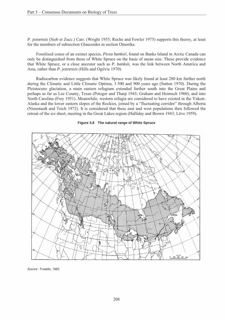
Part 3 – Consensus Documents on Biology of Trees
208
P. jezoensis (Sieb et Zucc.) Carr. (Wright 1955; Roche and Fowler 1975) supports this theory, at least for the members of subsection Glaucoides in section Omorika.
Fossilised cones of an extinct species, Picea banksii, found on Banks Island in Arctic Canada can only be distinguished from those of White Spruce on the basis of mean size. These provide evidence that White Spruce, or a close ancestor such as P. banksii, was the link between North America and Asia, rather than P. jezoensis (Hills and Ogilvie 1970).
Radiocarbon evidence suggests that White Spruce was likely found at least 280 km further north during the Climatic and Little Climatic Optima, 3 500 and 900 years ago (Sutton 1970). During the Pleistocene glaciation, a main eastern refugium extended further south into the Great Plains and perhaps as far as Lee County, Texas (Potzger and Tharp 1943; Graham and Heimsch 1960), and into North Carolina (Frey 1951). Meanwhile, western refugia are considered to have existed in the Yukon-Alaska and the lower eastern slopes of the Rockies, joined by a “fluctuating corridor” through Alberta (Nienstaedt and Teich 1972). It is considered that these east and west populations then followed the retreat of the ice sheet, meeting in the Great Lakes region (Halliday and Brown 1943; Löve 1959).
Figure 3.6 The natural range of White Spruce
Source : Fowells, 1965
Section 7 – White Spruce
209
4. Reproductive Biology
A. Reproductive development
White Spruce is monoecious. Development of the reproductive structures follows a two-year cycle typical of most conifers in the northern hemisphere (Figure 3.7), other than Pinus species and members of the Cupressaceae family (Owens and Blake 1985). Bud scales are initiated at the terminal apex, and at newly initiated axillary apices within the enlarging vegetative buds, from about late April (Owens et al. 1977). Apices differentiate as vegetative, pollen cone or seed cone buds around mid-July, at the cessation of shoot elongation. The proportion of apices differentiating as reproductive buds may be increased by hot, dry weather at the time of differentiation, particularly if preceding cone crops have been poor (Owens and Blake 1985; Nienstaedt and Zasada 1990).
Pollen cone bud development is complete by early October, when they become dormant. Completion of seed cone and vegetative bud development follows shortly thereafter. By the time buds become dormant, all microsporophylls, microsporangia, bracts and functional ovuliferous scales, and leaves have been initiated. Megaspore mother cells are present in the dormant seed cone buds, although meiosis has not begun (Owens and Molder 1977, 1984). Overwintering vegetative buds are small and dome-shaped. Reproductive buds, usually terminal or subterminal, may be identified by their larger size and ovate to obovate shape. Distinguishing between male and female buds may be difficult without dissection, but males are generally found in the middle to lower crown (Eis 1967a; Eis and Inkster 1972).
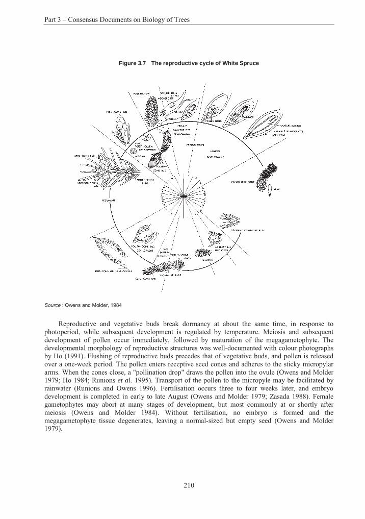
Part 3 – Consensus Documents on Biology of Trees
210
Figure 3.7 The reproductive cycle of White Spruce
Source : Owens and Molder, 1984
Reproductive and vegetative buds break dormancy at about the same time, in response to photoperiod, while subsequent development is regulated by temperature. Meiosis and subsequent development of pollen occur immediately, followed by maturation of the megagametophyte. The developmental morphology of reproductive structures was well-documented with colour photographs by Ho (1991). Flushing of reproductive buds precedes that of vegetative buds, and pollen is released over a one-week period. The pollen enters receptive seed cones and adheres to the sticky micropylar arms. When the cones close, a "pollination drop" draws the pollen into the ovule (Owens and Molder 1979; Ho 1984; Runions et al. 1995). Transport of the pollen to the micropyle may be facilitated by rainwater (Runions and Owens 1996). Fertilisation occurs three to four weeks later, and embryo development is completed in early to late August (Owens and Molder 1979; Zasada 1988). Female gametophytes may abort at many stages of development, but most commonly at or shortly after meiosis (Owens and Molder 1984). Without fertilisation, no embryo is formed and the megagametophyte tissue degenerates, leaving a normal-sized but empty seed (Owens and Molder 1979).
Section 7 – White Spruce
211
B. Mating system and gene flow
White Spruce is a wind-pollinated, monoecious species, and outcrossing is by far the most prevalent mating system. Self-pollination occurs to some degree, as the period of pollen release and female receptivity coincide for an individual tree (Nienstaedt 1958). Female strobili are concentrated in the top quarter of the crown, while males are more prevalent in the mid to lower crown, but the effectiveness of this zonation against selfing is questionable (Nienstaedt and Teich 1972). The two-step pollination mechanism, whereby pollen is collected in the sticky micropyllar arms over the receptive period, and only then drawn en masse by the pollination drop, ensures that pollen from many sources has a chance to fertilise any given ovule (Runions et al. 1995). Under controlled pollination, where large quantities of pollen are applied at one time, the micropylar arms become fully occupied and it is more likely that the first-on become first-in (Franklin 1974; Ho 1985). The nucellus itself can accommodate two or three pollen grains, and there is potential of some postzygotic selection to reduce the number of selfed embryos (Fowler 1987b). The incidence of selfing also varies greatly as a function of population size and structure. Selfing rates in a White Spruce seed orchard varied from 0 to 22% (Denti and Schoen 1988). However, the presence of several ramets of the same clone might generate a pollen cloud quite unlike that in a natural stand. Although outcrossing pollen has no advantage over self-pollen prior to fertilisation and self-fertilisation, can occur, most selfed embryos fail to develop, likely as a result of homozygous lethal recessive genes (Mergen et al. 1965; Nienstaedt and Teich 1972).
Natural inbreeding among related trees is common in small populations. In two such stands in New Brunswick, the average inbreeding coefficient was estimated as F = 0.145 (Coles and Fowler 1976; Park et al. 1984). An electrophoretic study of six stands near the limit of the range in subarctic Quebec displayed deficiencies of heterozygotes at 60% of the loci analysed and suggested rapid accumulation of inbreeding. Differences among these isolated populations were large (Fst = 0.113), suggesting that genetic drift might be important (Tremblay and Simon 1989). Substantial deviations from random mating and high rates of inbreeding were also observed within stands in Newfoundland (Innes and Ringius 1990) and in a seed production area in Alberta (King et al. 1984). Another electrophoretic study of trees throughout a 19 ha stand in eastern Ontario demonstrated that, while selfing was not a major component of the mating system and there was an excess of heterozygotes in both the parental and filial populations, other forms of non-random mating among relatives and restricted transmission distances of effective pollen gametes were important (Cheliak et al. 1985).
Gene flow in Picea is mediated by small pollen grains, 70-85 nm at their widest point (Eisenhut 1961), whose bladdery wings make them well-adapted for aerial transport (Di-Giovanni and Kevan 1991). Various studies of pollen dispersal in conifers indicate that over 90% of the pollen comes to rest less than 100 m from the source (Wright 1976). Nevertheless, a substantial quantity may travel great distances; Gregory (1973) cites reports that pollen of Pinus and Picea may travel as far as 600 to 1 000 km, and several authors have concluded that isolation distances of less than 1 km often have little impact on contamination rates in conifer seed orchards (see review by Di-Giovanni and Kevan 1991). A recent study of pollen dispersal dynamics in a black spruce seed orchard indicated that "large amounts" of pollen rose to a height of 300 m above ground level (Di-Giovanni et al. 1996). At a steady wind speed of 5 m s-1, the authors calculated that spruce pollen reaching this altitude would drift about 47 km. Another study examined pollen contamination within a White Spruce seed orchard that a forest fire had isolated from native stands by as much as 3 km upwind (Caron et al. 1994; Mercier et al. 1994). In a heavy pollen year, contamination levels within this well-isolated orchard were estimated at 93%.
Part 3 – Consensus Documents on Biology of Trees
212
C. Seed production
Cones and seeds may be produced on White Spruce as young as four years (Sutton 1970), but most trees do not produce seed until 10 to 15 years. Significant seed production normally occurs on trees that are at least 30 years of age (Nienstaedt and Teich 1972). Periodicity of seed production and crop size are extremely variable. Good cone crops are borne irregularly, but on average every four years (Stiell 1976). In a "heavy" crop year, Waldron (1965) estimated that a mature stand in Manitoba produced 13.8 million seeds/ha, but that only 59% were sound. In a "moderate" seed year, the stand produced 2.5 million seeds/ha. Maximum seed production over a 13-year period in a stand in Alaska was 40 million seeds/ha, and in three years exceeded 10 million seeds/ha (Nienstaedt and Zasada 1990).
Initiation and duration of seed dispersal are weather and site dependent (Zasada 1988). The mature cones open as they lose moisture and the scales flex in dry weather, reclosing during wet periods. Seed dispersal begins in mid- to late-August, with most seeds released in September (Crossley 1953; Waldron 1965; Dobbs 1976). The interval between seed ripening and beginning of dispersal can be less than two weeks, which creates problems in determining the best time to collect cones (Smith 1983; Mercier and Langlois 1992). At some northern sites, seed dispersal may begin before seeds are fully mature (Mercier 1994). In practice, cone collection can begin a couple of weeks earlier if seeds are allowed to "artificially ripen" by storing the cones under cool, moist conditions (Winston and Haddon 1981; Caron et al. 1990, 1993).
The seeds are winged and wind-dispersed. The actual distance reached from the source varies from site to site (Dobbs 1976), but in one study less than 5% of the seeds were dispersed more than 100 m from the source (Zasada and Lovig 1983). The seeds themselves are small, and average cleaned seed weight is about 2.0 g/1 000 seeds (Safford 1974).
D. Natural regeneration
White Spruce seeds exhibit varying degrees of dormancy that may be broken by exposure to low temperatures under moist conditions, i.e., cold stratification (Wang 1974). Dormancy results from inhibition of embryo development, induced by the seed coat and/or megagametophyte tissue (Downie and Bewley 1996). Seed dormancy may vary greatly among stands, individual trees and crop year (Hellum 1968; Wang 1976; Caron et al. 1990). In the wild, White Spruce seeds normally germinate the following spring, as soon as soil surface temperatures are warm enough and provided there is adequate moisture (Nienstaedt and Zasada 1990).
Natural regeneration of White Spruce can be difficult to predict and is not easily established under most harvesting systems. As a shade-tolerant species, White Spruce is able to regenerate under mature stands of spruce and early successional species, but advance regeneration stocking is often poor (Jablanczy 1967, 1979; Krasny et al. 1984; Walker et al. 1986; Nienstaedt and Zasada 1990). Freshly disturbed areas with exposed mineral soils offer the best conditions for germination and establishment (Eis 1967b; Lees 1970; Dobbs 1976; Zasada et al. 1978). Thick organic layers are common under mature stands, but such surfaces restrict germination success and shallow root penetration leads to mortality if the canopy is opened suddenly (Jablanczy 1967). Where advance regeneration is established on thick moss, survival after logging is often poor and seedlings are soon replaced by other more aggressive species. Under the more open canopies of stands growing on alluvium sites, the sudden increase of competing vegetation after harvesting prevents seedling establishment and causes severe mortality to advance regeneration (Eis 1981). Advance regeneration that does establish under closed canopies will not survive suppression as long as in other more tolerant
Section 7 – White Spruce
213
species such as balsam fir (Jablanczy 1967, 1979). Allelopathic effects of Cladonia lichens may inhibit the establishment of regeneration (Fisher 1979).
E. Vegetative reproduction in nature
While vegetative reproduction is rare over much of the range of White Spruce, layering is common at the northern limit where regeneration from seed is limited because of climatic conditions (Nienstaedt and Zasada 1990; Fayle and Scott 1995). Rooting occurs when lower branches of open-grown trees come in contact with the ground and are covered by soil or organic materials. Populations in these arctic areas likely originated from seed at a time when climatic conditions were warmer, and vegetative propagation is now the only possible means of regeneration (Elliott 1979).
5. Genetics
A. Cytology
Vegetative cells are normally diploid, with 2n = 24 chromosomes (Mehra and Khoshoo 1956; Santamour 1960). Aneuploidy and polyploidy are very rare (De Torok and White 1960); about 1 in 13 000 seedlings have been observed, mostly tetraploid, and most of them stunted (Winton 1964).
B. Genetic variation
Population-level variability
White Spruce was an early candidate for provenance research, and evidence of clinal variation for height growth related to latitude and elevation of origin appeared as early as 1950 (Morgenstern 1996). North-south adaptive variation has also been observed for such characters as cold hardiness of buds, foliage and stems (Simpson 1994), optimal and threshold germination temperature (Fraser 1971), germination rate (Roche 1969), seed quality (Khalil 1986), juvenile growth (Dunsworth and Dancik 1983; Khalil 1986), date of bud flush (Blum 1988), and various other seedling morphological and phenological traits (Nienstaedt and Teich 1972).
There is evidence of east-west variation patterns in such taxonomic characteristics as needle colour, number of stomata and branch pubescence (Nienstaedt and Teich 1972), cortical monoterpenes (Wilkinson et al. 1971), DNA content (Miksche 1968) and cpDNA allele frequencies (Furnier and Stine 1995). These data are consistent with the two-refugia theory of White Spruce remigration, following the Pleistocene glaciation, with a major division at about 95°W, with latitudinal clines within each division (Nienstaedt and Teich 1972).
While White Spruce generally exhibits clinal variation for adaptive traits, edaphic ecotypes have been identified in eastern Ontario that produce superior height growth on granitic and limestone sites (Teich and Holst 1974; Murray and Skeates 1985). White Spruce populations from moist-warm habitats of the sub-boreal spruce biogeoclimatic zone in the interior of British Columbia have displayed greater resistance to white pine weevil (Pissodes strobi) attack (Alfaro et al. 1996). In a 20-year-old trial in the badly polluted industrial Erzgebirge region of East Germany, there was great variation in performance of 16 tested White Spruce provenances. The best of these (from Sundridge, Ontario) was superior to the best of 17 tested provenances of Norway spruce (Weiss et al. 1988).
Local provenances are generally well-adapted and grow well, but it is not uncommon for provenances from more southerly locations to exhibit better growth (Nienstaedt and Teich 1972). Some particular sources have demonstrated superior performance over a wide range of sites. A
Part 3 – Consensus Documents on Biology of Trees
214
provenance from Birch Island, British Columbia, has proven exceptional and in coastal nurseries will match the growth of Sitka spruce (Nienstaedt and Zasada 1990). Provenances from the Ottawa Valley have performed well at many locations from the Lakes States through to Newfoundland (Nienstaedt 1969; Corriveau and Boudoux 1971; Teich et al. 1975; Fowler and Coles 1977; Radsliff et al. 1983; Khalil 1985). Although these sources grew well in Newfoundland, survival was sometimes poor (Hall 1986). In Nova Scotia, Ottawa Valley sources were surpassed in height growth by provenances from Prince Edward Island (Bailey 1987). In a range-wide provenance test in Alberta, the 10 best provenances included sources from Saskatchewan, Manitoba, Ontario and Quebec, and had 15% greater height and only slightly lower survival after 15 years than did the local seed sources (Hansen et al. 1995).
When provenances of diverse geographic origin are tested, population differences may explain 10 to 15% of the phenotypic variation in wood relative density (Stellrecht et al. 1974; Beaulieu and Corriveau 1985; Corriveau et al. 1987). However, population differences within a smaller geographic area can be negligible, even while family differences within stands can account for 16% of the variation in relative density of outer wood (Corriveau et al. 1991).
In contrast to many other characters, geographic variation at polymorphic allozyme loci appears to be weak. A 19-year-old test in Minnesota of 22 provenances from across the range of White Spruce demonstrated that while 48.0% and 54.1% of the variation in height at ages nine and 19, respectively, was due to differences among populations, an average of only 3.8% of the allozyme variation was due to population differences (Furnier et al. 1991). The variance among enzyme systems at 13 loci in four populations in Alaska, on an altitudinal gradient from 120 to 750 m, was such that only 2% of the variance was among populations, while 97% of the genetic diversity was within-stand, suggesting that the allozyme systems studied were selectively neutral (Alden and Loopstra 1987).
Individual-level variability
While variation among provenances is important in determining the risks and benefits of transferring seed sources, genetic improvement from mass selection relies primarily on variation within populations as the source of genetic gains. The partitioning of genetic variance among and within populations is greatly influenced by the range of adaptive variation sampled by the tested provenances. A wide-range sample of provenances tested in Wisconsin estimated population variance to be two to three times the family-within-population variance for height at nine and 15 years (Nienstaedt and Riemenschneider 1985). Another sample drawn from across Quebec and Ontario indicated that population variance was as large as that of families-within-populations (Li et al. 1993). Field trials using hierarchal sampling over a limited area of southeastern Ontario showed high within-stand variation for height growth and phenology, while variation among stands was low (Dhir 1976; Pollard and Ying 1979a, b).
The oldest White Spruce progeny tests were established on four sites at the Petawawa Forest Experiment Station in 1958 (Holst and Teich 1969). Narrow-sense heritabilities for this material were reported at age eight to 11 years in the range of 0.15 to 0.35 for height, and at three of the four sites was similar at age 22 (Ying and Morgenstern 1979). A similar progeny test in Minnesota produced heritability estimates for height of 0.27 at age nine, increasing to 0.35 at age 12 (Mohn et al. 1976), and another in Wisconsin produced estimates of 0.16 and 0.25 at ages nine and 15, respectively (Nienstaedt and Riemenschneider 1985).
Heritability estimates for diameter have typically been lower: from 0.05 to 0.10 at age 22 in the Ontario test (Ying and Morgenstern 1979), and 0.14 in the Minnesota test (Merrill and Mohn 1985).
Section 7 – White Spruce
215
The Petawawa trial was revisited by Magnussen (1993), who found that heritability estimates were much higher when only the "crop" trees were considered, as would be the case during selection in older stands. Stem analysis of almost 300 trees from 18 open-pollinated families at 36 years of age showed strong heritability of height growth, in the range of 0.3 to 0.6. Heritability for volume was also moderately strong, peaking at about 0.3 at age 20 and declining rapidly thereafter.
White Spruce in the western part of the range is frequently deformed by the white pine weevil (Pissodes strobi). Resistance to this pest varies substantially among individuals within a population, and the genetic basis has been demonstrated (Kiss and Yanchuk 1991; Alfaro et al. 1996).
While significant variation in wood specific gravity exists among populations, there are few correlations with environmental gradients or growth, and most of the variability exists among individuals within stands (Beaulieu and Corriveau 1985). Corriveau et al. (1991) studied the variation of wood quality characters in 19-year-old open-pollinated progenies from eight populations of White Spruce in the Upper Ottawa Valley. Their results indicated that the relative density of outer wood in White Spruce is under strong genetic control, with 16% of the variation explained by family differences and narrow-sense heritability estimated to be 0.63. An open-pollinated family test in British Columbia produced a similarly high estimate of heritability for wood specific gravity, 0.47, at age 15 (Yanchuk and Kiss 1993).
Substantial genetic variation has also been demonstrated in the initiation, maturation and germination of somatic White Spruce embryos from zygotic embryonic tissue (Park et al. 1993, 1994). Of particular concern to clonal selection programmes, a substantial portion of the genetic variance in the response to cultural treatments and the maturation and germination of somatic embryos was due to non-additive genetic variance.
C. Inbreeding depression and genetic load
Strong inbreeding depression has been reported in White Spruce (Mergen et al. 1965; Fowler and Park 1983; Park et al. 1984), and height growth losses as great as 33% have been reported (Ying 1978). Compared with other conifers, the number of lethal equivalents per zygote, 12.6, is high, and selfing has severe effects on seed set, early growth and survival (Fowler and Park 1983). Selection likely acts to remove selfed and highly inbred individuals early in the life-cycle, prior to the age of reproduction (Furnier et al. 1991). Nevertheless, natural inbreeding among related trees is common in small populations. In two such stands in New Brunswick, the average inbreeding coefficient was estimated as F = 0.145 (Coles and Fowler 1976; Park et al. 1984).
6. Crosses
Potential crosses with White Spruce are summarised in Table 3.13 (modified from Nienstaedt and Teich 1972). Introgressive hybridisation between white and Englemann spruce is widespread where the species are sympatric over large areas of British Columbia and Alberta (Nienstaedt and Teich 1972). In these areas breeding programmes simply treat the hybrid complex as a single species, "interior spruce".
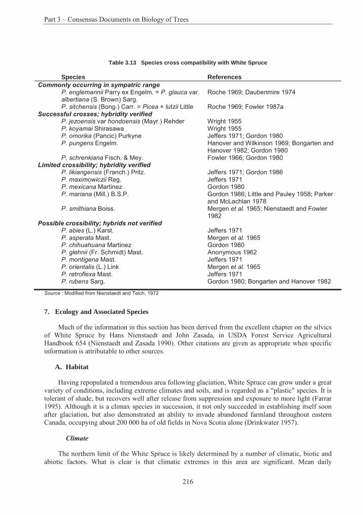
Part 3 – Consensus Documents on Biology of Trees
216
Table 3.13 Species cross compatibility with White Spruce
Species References Commonly occurring in sympatric range
P. englemannii Parry ex Engelm. = P. glauca var. albertiana (S. Brown) Sarg.
Roche 1969; Daubenmire 1974
P. sitchensis (Bong.) Carr. = Picea × lutzii Little Roche 1969; Fowler 1987a Successful crosses; hybridity verified
P. jezoensis var hondoensis (Mayr.) Rehder Wright 1955 P. koyamai Shirasawa Wright 1955 P. omorika (Pancic) Purkyne Jeffers 1971; Gordon 1980 P. pungens Engelm. Hanover and Wilkinson 1969; Bongarten and
Hanover 1982; Gordon 1980 P. schrenkiana Fisch. & Mey. Fowler 1966; Gordon 1980
Limited crossibility; hybridity verifiedP. likiangensis (Franch.) Pritz. Jeffers 1971; Gordon 1986 P. maximowiczii Reg. Jeffers 1971 P. mexicana Martinez Gordon 1980 P. mariana (Mill.) B.S.P. Gordon 1986; Little and Pauley 1958; Parker
and McLachlan 1978 P. smithiana Boiss. Mergen et al. 1965; Nienstaedt and Fowler
1982 Possible crossibility; hybrids not verified
P. abies (L.) Karst. Jeffers 1971 P. asperata Mast. Mergen et al. 1965 P. chihuahuana Martinez Gordon 1980 P. glehnii (Fr. Schmidt) Mast. Anonymous 1962 P. montigena Mast. Jeffers 1971 P. orientalis (L.) Link Mergen et al. 1965 P. retroflexa Mast. Jeffers 1971 P. rubens Sarg. Gordon 1980; Bongarten and Hanover 1982
Source : Modified from Nienstaedt and Teich, 1972
7. Ecology and Associated Species
Much of the information in this section has been derived from the excellent chapter on the silvics of White Spruce by Hans Nienstaedt and John Zasada, in USDA Forest Service Agricultural Handbook 654 (Nienstaedt and Zasada 1990). Other citations are given as appropriate when specific information is attributable to other sources.
A. Habitat
Having repopulated a tremendous area following glaciation, White Spruce can grow under a great variety of conditions, including extreme climates and soils, and is regarded as a "plastic" species. It is tolerant of shade, but recovers well after release from suppression and exposure to more light (Farrar 1995). Although it is a climax species in succession, it not only succeeded in establishing itself soon after glaciation, but also demonstrated an ability to invade abandoned farmland throughout eastern Canada, occupying about 200 000 ha of old fields in Nova Scotia alone (Drinkwater 1957).
Climate
The northern limit of the White Spruce is likely determined by a number of climatic, biotic and abiotic factors. What is clear is that climatic extremes in this area are significant. Mean daily
Section 7 – White Spruce
217
temperatures for January throughout much of the species range in Alaska, the Yukon and the Northwest Territories are in the vicinity of -29°C, whereas those is July reach only 13°C. Moisture is also limited in this area, with mean annual precipitation of only 250 mm. While photoperiod north of the Arctic Circle is 24 hours at the summer solstice, the length of the growing season at the northern limit is only about 60 days and may be as short as 20 days.
The southern limit of White Spruce’s dominance as a species in forest stands roughly follows the 18°C July isotherm, except in the Prairie Provinces where it swings somewhat north. Maximum summer temperatures as high as 43°C have been recorded within the range in Manitoba, and mean annual precipitation can be as high as 1 270 mm in Nova Scotia and Newfoundland. Low mean annual precipitation in the range of 380 to 510 mm combines with mean high temperatures in July of over 24°C to produce the most severe conditions along the southern edge of the range in the Prairie Provinces.
Soils and site type
A wide range of soils and site conditions support White Spruce, although the diversity of sites becomes more limited in northern areas with increasing severity of climate (Sutton 1970). Within its range, it is found on soils of glacial, lacustrine, marine and alluvial origin derived from geologically diverse substrata, including granites, gneisses, sedimentaries, slates, schists, shales and conglomerates.
Podzolic soils are most common, but White Spruce also grows on brunisolic, luvisolic, gleysolic and regisolic soils. It can also be found as a minor species on sand flats and other coarse-textured soils, on shallow mesic organic soils in Saskatchewan, and on organic soils with black spruce in the central Yukon.
While White Spruce can occupy extremely harsh site conditions, it is generally regarded as more demanding than other associated conifers, requiring higher moisture and fertility to achieve best development on moderately well-drained soils. Optimum pH values are probably in the range of 4.7 to 7.0 (Sutton 1970; Stiell 1976), but White Spruce stands are found on strongly acidic soils at pH 4.0, as well as alkaline soils as high as pH 8.0. Ecotypic variation has been observed in White Spruce, with some ecotypes adapted to limestone sites (Teich and Holst 1974; Murray and Skeates 1985). White Spruce stand development itself can have an impact on organic layers and on properties of the mineral soil. Brand et al. (1986) found that soil pH decreased by 1.2 units in plantations established on abandoned farmland in Ontario.
B. Synecology and associated species
Distributed over such a wide range, it is no surprise that White Spruce is an important component of several different forest types. In the eastern part of its range, it occurs in pure stands on abandoned fields in New England and the Maritime Provinces (Drinkwater 1957; Sutton 1970) and in moist boreal regions in the north. It more commonly occurs as a major stand component in association with black spruce (Picea mariana), red spruce (Picea rubens), balsam fir (Abies balsamea), white birch (Betula papyrifera) and trembling aspen (Populus tremuloides), and to a lesser extent with yellow birch (Betula alleghaniensis) and sugar maple (Acer saccharum). When White Spruce occurs in communities with intolerant species such as trembling aspen, white birch or red pine (Pinus resinosa),its greater shade tolerance leads to its assuming increasing importance as succession progresses. In northern Quebec, White Spruce is associated with lichen (Cladonia), feathermosses (e.g., Hylocomium splendens, Pleurozium schreberi, Ptilium cristacastrensis, and Dicranum spp.), dwarf birch (Betula nana) and many ericaceous plants.
Part 3 – Consensus Documents on Biology of Trees
218
Pure stands are more common in the western part of the range. Associated species in such stands in Alaska include white birch, trembling aspen, black spruce and balsam poplar (Populus balsamifera), whereas in western Canada the pure White Spruce type is associated with subalpine fir (Abies lasiocarpa), balsam fir, Douglas fir (Pseudotsuga menziesii), jack pine (Pinus banksiana) and lodgepole pine (P. contorta). In northwestern Canada and Alaska, closed White Spruce stands occur in communities with willows (Salix spp.) and buffalo berry (Shepherdia spp.), combined either with northern goldenrod (Solidago multiradiata) and crowberry (Empetrum spp.), or with huckleberry (Gaylussacia spp.), dewberry (Rubus spp.) and peavine (Lathyrus spp.).
In low elevations of western Canada and throughout interior Alaska, White Spruce is found in mixed-wood stands with trembling aspen. Common understorey shrubs found under such canopies in Alaska include green alder (Alnus crispa), willows, common bearberry (Arctostaphylos uva-ursi),highbush cranberry (Viburnum edule) and mountain cranberry (Vaccinium vitis-idaea). In the Prairie Provinces, the White Spruce-aspen type is associated with common snowberry (Symphoricarpos albus), red osier dogwood (Cornus stolonifera), western serviceberry (Amelanchier alnifolia) and western chokecherry (Prunus virginiana var. demissa).
Mixed White Spruce-paper birch stands are also common in western Canada and parts of Alaska. In this stand type, the understory vegetation usually includes willows, green alder, highbush cranberry, prickly rose (Rosa acicularis), mountain cranberry, bunchberry (Cornus canadensis) and Labrador-tea (Ledum groenlandicum).
Both the White Spruce-aspen and White Spruce-white birch stand types are successional stages leading to the pure White Spruce type or, in alpine treeline communities, the black spruce-White Spruce type. The latter occurs as open stands that, depending on moisture availability, may also support resin birch (Betula glandulosa), alders, willows, feathermosses and Cladonia lichens, together with Labrador-tea, bog blueberry (Vaccinium uliginosum), mountain cranberry and black crowberry (Empetrum nigrum).
Where White Spruce occurs as an important component of the boreal spruce-fir forest, green alder is the most commonly associated tall shrub, with willows important in western areas, and mountain maple (Acer spicatum), showy mountain ash (Sorbus decora) and American mountain ash (S. americana) important in the east. Common medium to low shrubs are highbush cranberry, red currant (Ribes triste), prickly rose and raspberry (Rubus idaeus). Ground vegetation commonly includes fireweed (Epilobium angustifolium), one-sided wintergreen (Pyrola secunda), one-flowered wintergreen (Moneses uniflora), northern twinflower (Linnaea borealis), naked bishop’s cap (Mitella nuda), bunchberry, dwarf rattlesnake plantain (Goodyera repens), stiff club moss (Lycopodium annotinum) and horsetail (Equisetum spp.) (la Roi 1967). Many bryophytes occur in these boreal spruce-fir stands. The most common mosses are Pleurozium schreberi, Hylocomium splendens,Ptilium cristacastrensis, Dicranum fuscescens and Drepanocladus uncinatus. Common liverworts are Ptilidium pulcherrimum, P. ciliare, Lophozia spp. and Blepharostoma trichophyllum. Common lichens include Peltigera apthosa, P. canina, Cladonia rangiferina, C. sylvatica, C. alpestris, C. gracilis and Cetraria islandica (la Roi and Stringer 1976).
C. Competition and stand structure
White Spruce can exist in various stand types and various stages of succession. Under shade, it is classified as intermediate to tolerant (Nienstaedt and Zasada 1990; Farrar 1995). It will compete with, but not necessarily outperform, other shade-tolerant conifers such as hemlock, black and red spruce, balsam fir, sugar maple and beech. In association with less tolerant early-successional species such as aspen, white birch and lodgepole pine, it may remain a suppressed, understorey component, becoming
Section 7 – White Spruce
219
more prominent at later successional stages. White Spruce competes poorly against the dense growth of perennials, bracken fern and understorey shrubs (Fowells 1965).
While White Spruce can form pure stands, particularly in the northwestern part of its range and in the New England States and Maritime Provinces, these stands are not always self-sustaining climax types. Where White Spruce pioneers to form even-aged stands on old fields in the Maritimes, advance regeneration is often outnumbered and outperformed by balsam fir seedlings that become a larger component after release (Jablanczy 1979). In mixtures, particularly with less tolerant species, the response to release by disturbance or cutting can be much more successful (Crossley 1976; Berry 1982; Nienstaedt and Zasada 1990).
White Spruce can be a component of multi-aged stands, either as pure stands or mixed with other tolerant late-successional conifers and hardwoods. Older age classes in such stands can be as high as 200 to 250 years in Alberta (Day 1972; Nienstaedt and Zasada 1990). As establishment is facilitated by disturbance, the age distribution in such stands is not continuous, but rather grouped according to periods of successful establishment.
D. Ecosystem dynamics
Many abiotic factors interact with White Spruce in forest ecosystems; some pose a direct threat to the species or cause significant damage. Wild fires can eliminate seed supply and leave a seedbed that is more conducive to the establishment of other species such as lodgepole pine, intolerant hardwoods and even black spruce. Stands established on flood plains may benefit from deposit of seedbed materials or suffer from disturbance to young regeneration. Frost heaving can cause severe damage, particularly to container seedlings planted on finer-textured soils. Root form and depth of White Spruce can vary greatly depending on site conditions (Strong and la Roi 1983), and shallow-rooted stands may be prone to windthrow. Periodic storms may cause considerable damage from hail, ice and snow (Dobbs and McMinn 1973; Gill 1974; Sampson and Wurtz 1994). Late-spring frosts can cause significant damage to flushing vegetative and reproductive buds.
While a great number of insects are a natural component of White Spruce forest types, few are responsible for large losses. Of these, the eastern spruce budworm (Choristoneura fumiferana) is the most destructive. Massive epidemics of this defoliator occur periodically, resulting in heavy mortality and loss of growth, particularly where White Spruce is associated with balsam fir (Rose et al. 1994). Several other defoliators cause damage or weaken trees on a smaller scale, including the yellow-headed spruce sawfly (Pikonema alaskensis), European spruce sawfly (Diprion hercyniae),needleminers, needleworms, loopers, tussock moths and the spruce harlequin. Other groups of insects attack buds and shoots of White Spruce, including gall-forming adelgids (Adelges spp.), spruce bud moths (Zeiraphera spp.) and the white pine weevil (Pissodes strobi).
Several species of bark beetles, Scolytidae, feed and breed in galleries between the bark and wood. The spruce beetle (Dendroctonus rufipennis) may attack trees of normal vigour, particularly those which are large-diameter and slow-growing, and has killed large areas (Ostaff and Newell 1981; Hard et al. 1983; Werner and Holsten 1984). Generally, though, bark beetles are considered secondary pests, attacking trees weakened by other means such as budworm epidemics, and may be thought of as beneficial in that their feeding hastens the return of wood to the humus (Rose et al. 1994).
Warren's collar weevil (Hylobius warreni) causes significant damage in scattered areas, girdling smaller trees and making larger trees susceptible to root rots such as Inonotus tomentosus (Merler and van der Kamp 1984; Rose et al. 1994). The strawberry root weevil (Otiorhynchus ovatus) can cause injury to young seedlings, and the root-collar weevil Hylobius congener can cause significant
Part 3 – Consensus Documents on Biology of Trees
220
mortality to White Spruce seedlings planted on recently cut softwood sites (Pendrel 1990; Eidt and Weaver 1993).
Many insect species inhabit or feed on spruce cones and seed, as part of their life-cycle. Significant losses in natural stands and seed orchards are caused in particular by the White Spruce cone maggot (Strobilomyia neanthracina) and the spruce seed moth (Cydia strobilella), and to a lesser extent by the spruce budworm and spruce coneworm (Dioryctria reniculelloides), among others (Hedlin et al. 1980; Turgeon 1994). Only a few pathogens cause problems with cone and seed production. The spruce cone rust (Chrysomyxa pirolata) can cause abnormal development of the cones, reduced seed production, and decreased viability of seeds (Sutherland et al. 1987; Myren et al.1994).
Emerging seedlings, particularly in bareroot nurseries, are commonly affected by damping-off fungi, primarily Fusarium but also Pythium, Rhizoctonia, Phytophthora and Cylindrocladium (Filer and Peterson 1975). Young seedlings may also suffer from Sirococcus blight (Sirococcus strobilinus)and infestation by the nematode Xiphinema bakeri (Sutherland and Van Eerden 1980).
Spruce needle rust (Chrysomyxa ledi and C. ledicola) is common wherever the alternate host, Labrador-tea, is found, but extensive damage from the fungus is rare. Spruce broom rust is common, causing abnormal proliferation of shoots to form "witches'-broom", but rarely causes death. Witches'-broom on White Spruce is sometimes caused by eastern dwarf mistletoe (Arceuthobium pusillum), although black spruce is more susceptible to this parasite. Scleroderris canker (Gremmeniella abietina)and cytospora canker (Leucostoma kunzei) both affect White Spruce, but cause little damage (Myren et al. 1994). Massive tumour-like growths are commonly observed on stems and branches in some White Spruce populations, particularly near coastal areas, but their etiology is not known (De Torok and White 1960).
Many rot fungi produce stem, butt and root rot in White Spruce, including red ring rot (Phellinus pini), red belt fungus (Fomitopsis pinicola) and Armillaria root rot (Armillaria mellea complex). Tomentosus root rot (Inonotus tomentosus) and brown cubical rot (Phaeolus schweinitzii) infect root systems and can reduce quality and growth, even if direct mortality is often light (Myren et al. 1994).
White Spruce forest stands commonly provide cover for many species of animals. Some, like moose, deer, black bear and many other fur-bearers, seek shelter in forest habitats but rarely feed on White Spruce. Porcupines (Erethizon dorsatum) also seek shelter in White Spruce forests and may kill small numbers of trees by feeding on the bark (Rose et al. 1994). Snowshoe hares (Lepus americanus),which commonly feed on foliage of young trees, tend to favour many other conifer species over White Spruce (Bergeron and Tardif 1988; Rangen et al. 1994), although planted White Spruce seedlings are preferred over natural regeneration (Sampson and Wurtz 1994). Many small mammals such as squirrels, mice, voles, chipmunks and shrews are heavy consumers of White Spruce seed and can have a major impact on regeneration, while the impact of seed-eating birds, including chickadees, grossbeaks, crossbills, juncos and sparrows, is relatively small (Radvanyi 1974). Many more bird species feed on the many species of insects that inhabit or feed on White Spruce trees and associated species.
The hybrid between white and Sitka spruce, Picea × lutzi, also occurs naturally where these species are sympatric. The hybrid has frequently been made artificially with parents from outside the sympatric area (Fowler 1987a), often in the hope of imparting the resistance of White Spruce to the white pine weevil (Pissodes strobi Peck). The degree of cold hardiness of the hybrid is related to the proportion of White Spruce germplasm (Ying and Morgenstern 1982), and growth performance of the hybrid depends greatly on the origin of the parents (Sheppard and Cannell 1985).
Section 7 – White Spruce
221
Many other artificial hybrids have been made successfully (e.g. Wright 1955; Jeffers 1971; Bongarten and Hanover 1982). The hybrid with Himalayan spruce (P. smithiana) was inferior to native White Spruce when field tested in New Brunswick, but superior in Wisconsin (Nienstaedt and Fowler 1982). Generally speaking, few of these hybrids have shown promise and none has achieved commercial importance (Nienstaedt and Zasada 1990).
8. Summary
White Spruce is an enormously important tree species in North America. It occupies a dominant role in several forest types that span the breadth of the continent, from the northern tree limit south to the Lake States and New England. The species has been successful as both a pioneering and climax type, and is genetically broadly adapted and highly variable. It is an outcrossing, wind-pollinated species that can transfer genes rapidly, and yet it tolerates higher levels of inbreeding when found in small populations.
The ecology of White Spruce is extremely diverse, given its tremendous geographic distribution and its genetic plasticity. The typical White Spruce ecosystem has a diverse mixture of associated tree species, vascular flora, bryophytes, insects, fungi, birds and animals. Only a very small number of these associated species pose a major threat by competition or direct damage, and White Spruce is well-adapted to this complex coexistence.
White Spruce is well-suited to artificial regeneration. It is the most commonly planted forest species throughout its natural range. Tree breeding programmes have a long history, and improved material from seed orchards now constitutes a significant portion of deployed reforestation material in some areas. While White Spruce reforestation is currently based on seed propagation, vegetative propagation techniques for cuttings and regeneration of somatic embryos are well-advanced, making it a logical target for implementation of transgenic biotechnologies and the use of cloning in both breeding and deployment strategies.
Part 3 – Consensus Documents on Biology of Trees
222
REFERENCES
Alden, J., and Loopstra, C. 1987. Genetic diversity and population structure of Picea glauca on an altitudinal gradient in interior Alaska. Can. J. For. Res. 17: 1519-1526.
Alfaro, R.I., He, F.L., Kiss, G., King, J., and Yanchuk, A. 1996. Resistance of White Spruce to white pine weevil: development of a resistance index. For. Ecol. Manage. 81: 51-62.
Anonymous. 1962. Progress report – 1962 forest tree improvement at State University College of Forestry, Syracuse, NY. 126 pp. (cited in Nienstaedt and Teich 1972)
Attree, S.M., Pomeroy, M.K., and Fowke, L.C. 1994. Production of vigorous, desiccation tolerant White Spruce (Picea glauca [Moench.] Voss.) synthetic seeds in a bioreactor. Plant Cell Reports 13: 601-606.
Bailey, T.A. 1987. Five year results of a White Spruce provenance experiment at three locations in Nova Scotia. B.Sc.F. thesis, Univ. of New Brunswick, Fredericton, NB, 39 pp.
Beaulieu, J. 1996. Breeding program and strategy for White Spruce in Quebec. Inform. Rep. LAU-X-117E, Nat. Res. Can., Can. For. Serv. - Quebec, Sainte-Foy, Quebec. 25 pp.
Beaulieu, J., and Corriveau, A. 1985. Variabilité de la densité du bois et de la production des provenances d'épinette blanche, 20 ans après plantation. Can. J. For. Res. 15: 833-838.
Bergeron, J.M., and Tardif, J. 1988. Winter browsing preferences of snowshoe hares for coniferous seedlings and its implication in large-scale reforestation programs. Can. J. For. Res. 18: 280-282.
Berry, A.B. 1982. Response of suppressed conifer seedlings to release from an aspen-pine overstorey. For. Chron. 58: 91-92.
Blum, B.M. 1988. Variation in the phenology of bud flushing in white and red spruce. Can. J. For. Res. 18: 315-319.
Bongarten, B.C., and Hanover, J.W. 1982. Hybridization among white, red, blue and white X blue spruces. For. Sci. 28: 129-134.
Brand, D.G., Kehoe, P., and Connors, M. 1986. Coniferous afforestation leads to soil acidification in central Ontario. Can. J. For. Res. 16: 1389-1391.
Caron, G.E., Wang, B.S.P., and Schooley, H.O. 1990. Effect of tree spacing, cone storage, and prechilling on germination of Picea glauca seed. For. Chron. 66: 388-392.
Caron, G.E., Wang, B.S.P., and Schooley, H.O. 1993. Variation in Picea glauca seed germination associated with the year of cone collection. Can. J. For. Res. 23: 1306-1313.
Section 7 – White Spruce
223
Caron, G.-É., Mercier, S., and Rainville, A. 1994. White Spruce pollen dispersal near an orchard with a large pollen dilution zone. In Proceedings of Palynological Symposium "Trabajos de Palinologia Básica y Aplicada," September 1994, University of Valencia. Edited by I.M. Andrés, M.D. Ollivier, J.G. Heras and M.E. Burgaz-Moreno. University of Valencia, Valencia, Spain.
Carter, K.K. 1988. White Spruce (Picea glauca (Moench) Voss). In Tree improvement in the northeast: interim summary and recommendations for selected species. Tech. Bull. No. 131, Maine Agric. Exp. Sta. pp. 47-50.
Carter, K.K., and Simpson, J.D. 1985. Status and outlook for tree improvement programs in the Northeast. North. J. Appl. For. 2: 127-131.
Cheliak, W.M., Pitel, J.A., and Murray, G. 1985. Population structure and the mating system of White Spruce. Can. J. For. Res. 15: 301-308.
Coles, J.F., and Fowler, D.P. 1976. Inbreeding in neighbouring trees in two White Spruce populations. Silvae Genet. 25: 29-34.
Copes, D.L., and Beckwith, R.C. 1977. Isoenzyme identification of Picea glauca, P. sitchensis, and P. lutzii. Bot. Gaz. 138: 512-521.
Cornelius, J. 1994. The effectiveness of plus-tree selection for yield. For. Ecol. Manage. 67: 23-34.
Corriveau, A., and Boudoux, M. 1971. Le développement des provenances d'épinette blanche de la région forestière des Grands-Lacs et du St-Laurent au Québec. Inform. Rep. Q-F-X-15, Can. For. Serv., For. Res. Lab., Sainte-Foy, Québec.
Corriveau, A., Beaulieu, J., and Mothe, F. 1987. Wood density of natural White Spruce populations in Quebec. Can. J. For. Res. 17: 675-682.
Corriveau, A., Beaulieu, J., and Daoust, G. 1991. Heritability and genetic correlations of wood characters of Upper Ottawa Valley White Spruce populations grown in Quebec. For. Chron. 67: 698-705.
Critchfield, W.B. 1973. Interspecific hybridization in Pinus: a summary review. In Proceedings 14th Can. Tree Improve. Assoc., Part 2. pp. 99-105.
Critchfield, W.B. 1984. Impact of the Pleistocene on the genetic structure of North American conifers. In Proceedings of the 8th North American Forest Biology Workshop, July 30 - August 1, 1984, Utah State University, Logan, USA. Edited by R.M. Lanner. Department of Forest Resources, Utah State University, Logan. pp. 70-118.
Crossley, D.I. 1953. Seed maturity in White Spruce (Picea glauca). Silv. Res. Note 104, Can. Forest Res. Div., Ottawa. 16 pp.
Crossley, D.I. 1976. Growth response of spruce and fir to release from suppression. For. Chron. 52: 189-193.
Part 3 – Consensus Documents on Biology of Trees
224
Cyr, D.R., Lazaroff, W.R., Grimes, S.M.A., Quan, G., Bethune, T.D., Dunstan, D.I., and Roberts, D.R. 1994. Cryopreservation of interior spruce embryogenic cultures. Plant Cell Reports 13: 574-577.
Daoust, G., Plourde, A., and Beaulieu, J. 1995. Influences of crown size and maturation on flower production and sex expression in Picea glauca treated with gibberellin A4/7. Tree Physiol. 15: 471-475.
Daubenmire, R. 1974. Taxonomic and ecological relationships between Picea glauca and Picea engelmannii. Can. J. Bot. 52: 1545-1560.
Day, R.J. 1972. Stand structure, succession and use of southern Alberta's Rocky Mountain Forest. Ecology, 53: 472-478.
Denti, D., and Schoen, D.J. 1988. Self-fertilisation rates in White Spruce: effect of pollen and seed production. J. Hered. 79: 284-288.
De Torok, D., and White, P.R. 1960. Cytological instability of tumors of Picea glauca. Science, 131: 730-732.
Dhir, N.K. 1976. Stand, family and site effects in Upper Ottawa Valley White Spruce. In Proceedings of the Twelfth Lake States Forest Tree Improvement Conference, August 1975. Edited by D.W. Einspahr. Gen. Tech. Rep. No. NC-26, USDA For. Serv, North Central For. Exp. Sta. pp. 88-97.
Dhir, N.K., and Barnhardt, L.K. 1995. Forest genetic resources and conservation in Alberta. In Forest genetic resources conservation and management in Canada. Edited by T.C. Nieman, A. Mosseler and G. Murray. Inform. Rep. PI-X-119, Can. For. Serv., Petawawa Nat. For. Inst., Chalk River, ON. pp. 32-39.
Di-Giovanni, F., and Kevan, P.G. 1991. Factors affecting pollen dynamics and its importance to pollen contamination: a review. Can. J. For. Res. 21: 1155-1170.
Di-Giovanni, F., Kevan, P.G., and Arnold, J. 1996. Lower planetary boundary layer profiles of atmospheric conifer pollen above a seed orchard in northern Ontario, Canada. For. Ecol. Manage. 83: 87-97.
Dobbs, R.C. 1976. White Spruce seed dispersal in central British Columbia. For. Chron. 52: 225-228.
Dobbs, R.C., and McMinn, R.G. 1973. Hail damage to a new White Spruce and lodgepole pine plantation in central British Columbia. For. Chron. 49: 174-175.
Dojack, J. 1991. Manitoba's tree improvement strategy. Manitoba Nat. Res., Winnipeg, MB. 79 pp.
Downie, B., and Bewley, J.D. 1996. Dormancy in White Spruce (Picea glauca (Moench.) Voss.) seeds is imposed by tissues surrounding the embryo. Seed Science Research 6: 9-15.
Drinkwater, M.H. 1957. Field spruce in Nova Scotia. Nat. Res. Tech. Note No. 65, Department of Northern Affairs, Ottawa.
Dunsworth, G.B., and Dancik, B.P. 1983. Seedling variation among Alberta populations of White Spruce (Picea glauca (Moench) Voss) in controlled environments. Northwest Sci. 57: 74-83.
Section 7 – White Spruce
225
Eidt, D.C., and Weaver, C.A.A. 1993. Control damage by seedling debarking weevil. Tech. Note No. 271, Forestry Canada - Maritimes Region, Fredericton, NB. 5 pp.
Eis, S. 1967a. Cone crops of white and black spruce are predictable. For. Chron. 43: 247-252.
Eis, S. 1967b. Establishment and early development of White Spruce in the interior of British Columbia. For. Chron. 43: 174-177.
Eis, S. 1981. Effect of vegetative competition on regeneration of White Spruce. Can. J. For. Res. 11: 1-8.
Eis, S., and Inkster, J. 1972. White Spruce cone production and prediction of cone crops. Can. J. For. Res. 2: 460-466.
Eisenhut, G. 1961. Untersuchungen über die Morphologie und Ökologie der Pollenkörner heimischer und fremdländischer Waldbäume. Forstwiss. Forsch. 15: 1-68.
Elliott, D.L. 1979. The current regenerative capacity of the northern Canadian trees, Keewatin, N.W.T., Canada: some preliminary observations. Arctic and Alpine Research 11: 243-251.
Ellis, D., McCabe, D., Mcinnis, S., Ramachandran, R., Russell, D., Wallace, K., Martinell, B., Roberts, D., Raffa, K. and McCown, B. 1993. Stable transformation of Picea glauca by particle acceleration. Bio/Technology 11: 84-89.
Eriksson, G., Namkoong, G., and Roberds, J.H. 1993. Dynamic gene conservation for uncertain futures. For. Ecol. Manage. 62: 15-37.
Farrar, J.L. 1995. Trees in Canada. Fitzhenry & Whiteside/Canadian Forest Service, Markham and Ottawa.
Fayle, D.C., and Scott, P.A. 1995. Woody root system development of White Spruce layerings in a tundra environment. Ecoscience, 2: 408-414.
Filer, T.H., Jr., and Peterson, G.W. 1975. Damping-off. In Forest nursery diseases in the United States. Edited by G.W. Peterson and R.S. Smith Jr. Agric. Handbook No. 470, USDA For. Serv., Washington, DC. pp. 6-8.
Fisher, R.F. 1979. Possible allelopathic effects of reindeer moss (Cladonia) on jack pine and White Spruce. For. Sci. 25: 256-260.
Florin, R. 1963. The distribution of conifer and taxad genera in time and space. Acta Horti Bergiani 20: 121-312.
Fowells, H.A. 1965. Silvics of forest trees of the United States. Agricultural Handbook No. 271, Forest Service, USDA, Washington, DC. 762 pp.
Fowler, D.P. 1966. A new spruce hybrid – Picea schrenkiana × P. glauca. In Proceedings Second Genetics Workshop, 7th Lake States Forest Tree Improvement Conference, Hickory Corners, MI, 1965. pp. 44-47.
Part 3 – Consensus Documents on Biology of Trees
226
Fowler, D.P. 1983. The hybrid black X Sitka spruce, implications to phylogeny of the genus Picea.Can. J. For. Res. 13: 108-115.
Fowler, D.P. 1986. Strategies for the genetic improvement of important tree species in the Maritimes. Inf. Rep. M-X-156, Can. For. Serv. - Maritimes, Fredericton, NB. 30 pp.
Fowler, D.P. 1987a. The hybrid white X Sitka spruce: species crossability. Can. J. For. Res. 17: 413-417.
Fowler, D.P. 1987b. In defense of the polycross. Can. J. For. Res. 17: 1624-1627.
Fowler, D.P., and Coles, J.F. 1977. Seedling seed orchards of Ottawa Valley White Spruce for the Maritimes. Inform. Rep. M-X-73, Can. For. Serv., Fredericton, NB. 46 pp.
Fowler, D.P., and MacGillivray, H.G. 1967. Seed zones for the Maritime Provinces. Inform. Rep. M-X-12, Maritimes For. Res. Lab., Fredericton, NB. 23 pp.
Fowler, D.P., and Morgenstern, E.K. 1990. Tree improvement research and development in Canada. For. Chron. 66: 97-101.
Fowler, D.P., and Park, Y.S. 1983. Population studies of White Spruce. I. Effects of self-pollination. Can. J. For. Res. 13: 1133-1138.
Fowler, D.P., MacGillivray, H.G., Manley, S.A.M., and Bonga, J.M. 1971. Tree breeding in the Maritimes Region, 1968-69. In Proceedings 12th Meeting of the Committee for Forest Tree Breeding Canada, Quebec, PQ, 1970.
Franklin, E.C. 1974. Pollination in slash pine: first come, first served. In Proceedings of the Seed Yield from Southern Pine Seed Orchards Conference, Macon, Georgia. Edited by J. Kraus. Georgia Forest Research Council, Macon, GA. pp. 15-20.
Fraser, J.W. 1971. Cardinal temperatures for germination of six provenances of White Spruce seed. Publ. No. 1290, Dep. Fisheries For., Can. For. Serv., Ottawa. 10 pp.
Frey, D.G. 1951. Pollen succession in the sediments of Singletary Lake, North Carolina. Ecology, 32: 518-533.
Furnier, G.R., and Stine, M. 1995. Interpopulation differentiation of nuclear and chloroplast loci in White Spruce. Can. J. For. Res. 25: 736-742.
Furnier, G.R., Stine, M., Mohn, C.A., and Clyde, M.A. 1991. Geographic patterns of variation in allozymes and height growth in White Spruce. Can. J. For. Res. 21: 707-712.
Gill, D. 1974. Snow damage to boreal mixedwood stands in northern Alberta. For. Chron. 50: 70-74.
Gordon, A.G. 1980. Spruce genetics, Sault Ste. Marie in 1977 and 1978. In Proceedings of the 17th
Meeting of the Canadian Tree Improvement Association, Gander, NF, Aug. 27-30, 1979, Part 1. Edited by C.W. Yeatman. pp. 117-121.
Gordon, A.G. 1986. Breeding, genetics and genecological studies in spruce for tree improvement in 1983 and 1984, Sault Ste. Marie, Ontario. In Proceedings of the 20th Meeting of the Canadian
Section 7 – White Spruce
227
Tree Improvement Association, Quebec City, QC, 1985, Part 1. Edited by C.W. Yeatman and T.J.B. Boyle. pp. 112-116.
Govindaraju, D.R. 1990. Gene flow, spatial patterns and seed-collection zones. For. Ecol. Manage. 35: 291-302.
Graham, A., and Heimsch, C. 1960. Pollen studies of some Texas peat deposits. Ecology, 41: 751-763.
Greenwood, M.S., Adams, G.W., and Gillespie, M. 1991. Stimulation of flowering by grafted black spruce and White Spruce: a comparative study of the effects of gibberellin A4/7, cultural treatments, and environment. Can. J. For. Res. 21: 395-400.
Gregory, P.H. 1973. The microbiology of the Atmosphere. 2nd ed. Leonard Hill, Aylesbury, UK.
Griffiths, M. 1994. Index of Garden Plants -- The New Royal Horticultural Society Dictionary. Timber Press, Portland, OR. 1234 pp.
Grossnickle, S.C., and Major, J.E. 1994a. Interior spruce seedlings compared with emblings produced from somatic embryogenesis. II. Stock quality assessment prior to field planting. Can. J. For. Res. 24: 1385-1396.
Grossnickle, S.C., and Major, J.E. 1994b. Interior spruce seedlings compared with emblings produced from somatic embryogenesis. III. Physiological response and morphological development on a reforestation site. Can. J. For. Res. 24: 1397-1407.
Grossnickle, S.C., Major, J.E., and Folk, R.S. 1994. Interior spruce seedlings compared with emblings produced from somatic embryogenesis. I. Nursery development, fall acclimation, and over-winter storage. Can. J. For. Res. 24: 1376-1384.
Hakman, I.C., and von Arnold, S. 1988. Somatic embryogenesis and plant regeneration from suspension cultures of Picea glauca (White Spruce). Physiol. Plant. 72: 579-587.
Hakman, I., and Fowke, L.C. 1987. Somatic embryogenesis in Picea glauca (White Spruce) and Picea mariana (black spruce). Can. J. Bot. 65: 656-659.
Hall, J.P. 1986. A provenance trial of White Spruce in Newfoundland: twenty-five years from seed. Inform. Rep. N-X-247, Can. For. Serv., Nfld. For. Cen. 33 pp.
Halliday, W.E.D., and Brown, A.W.A. 1943. The distribution of some important forest trees in Canada. Ecology, 24: 353-373.
Hanover, J.W., and Wilkinson, R.C. 1969. A new hybrid between blue spruce and White Spruce. Can. J. Bot. 47: 1693-1700.
Hansen, C.R., Dhir, N.K., Yakimchuk, K., Schilf, J.M., Barnhardt, L., Andriuk, C., Antoniuk, N.W., Hoberg, S., and Mochulski, M.A. 1995. Tree improvement programme Alberta Land and Forest Service. In Proceedings 25th Can. Tree Improve, Assoc., Part 1. Edited by J. Lavereau. Nat. Res. Can., Victoria, BC. pp. 50-64.
Hard, J.S., Werner, R.A., and Holsten, E.H. 1983. Susceptibility of White Spruce to attack by spruce beetles during the early years of an outbreak in Alaska. Can. J. For. Res. 13: 678-684.
Part 3 – Consensus Documents on Biology of Trees
228
Hedlin, A.F., Yates, H.O., III, Tovar, D.C., Ebel, B.H., Koerber, T.W., and Merkel, E.P. 1980. Cone and seed insects of North American conifers. Canadian Forestry Service, Ottawa; USDA Forest Service, Washington; and Secretaría de Agricultura y Recursos Hidráulicos, Mexico. 122 pp.
Hellum, A.K. 1968. A case against cold stratification of White Spruce seed prior to nursery seeding. For. Bra. Dep. Publ. 1243, Can. Dep. For. Rural Devel.
Hills, L.V., and Ogilvie, R.T. 1970. Picea banksii n. sp. Beaufort Formation (Tertiary), northwestern Banks Island, Arctic Canada. Can. J. Bot. 48: 457-464.
Ho, R.H. 1984. Seed-cone receptivity and seed production potential in White Spruce. For. Ecol. Manage. 9: 161-171.
Ho, R.H. 1985. Effect of repeated pollination upon filled seed in White Spruce. Can. J. For. Res. 15: 1195-1197.
Ho, R.H. 1991. A guide to pollen- and seed-cone morphology of black spruce, White Spruce, jack pine and Eastern White Pine for controlled pollination. Forest Research Report No. 125, Ontario Min. Nat. Res., Sault Ste. Marie, ON. 31 pp.
Holst, M.J., and Teich, A.H. 1969. Heritability estimates in Ontario White Spruce. Silvae Genet. 18: 23-27.
Innes, D.J., and Ringius, G.G. 1990. Mating system and genetic structure of two populations of White Spruce (Picea glauca) in eastern Newfoundland. Can. J. Bot. 68: 1661-1666.
Jablanczy, A. 1967. A generalized scheme for the natural regeneration of old-field spruce. Inform. Rep. M-X-11, Can. Dep. For. Rural Develop., For. Res. Lab., Maritimes Region, Fredericton, NB. 6 pp.
Jablanczy, A. 1979. Regeneration on White Spruce cutovers in eastern Nova Scotia. Unnumbered Rep., Nova Scotia Dep. Lands and Forests. 17 pp.
Jeffers, R.M. 1971. Research at the Institute of Forest Genetics. Res. Pap. NC-67, USDA For. Serv. North Central For. Exp. Sta., St. Paul, MN. 31 pp.
Khalil, M.A.K. 1985. Genetic variation in eastern White Spruce (Picea glauca (Moench) Voss) populations. Can. J. For. Res. 15: 444-452.
Khalil, M.A.K. 1986. Variation in seed quality and some juvenile characters of White Spruce (Picea glauca (Moench) Voss). Silvae Genet. 35: 78-85.
King, J.N., Dancik, B.P., and Dhir, N.K. 1984. Genetic structure and mating system of White Spruce (Picea glauca) in a seed production area. Can. J. For. Res. 14: 639-643.
Kiss, G.K., and Yanchuk, A.D. 1991. Preliminary evaluation of genetic variation of weevil resistance in interior spruce in British Columbia. Can. J. For. Res. 21: 230-234.
Konishi, J. 1979. Seed registration and zoning in Canada with particular reference to British Columbia. In Tree seed production and tree improvement in Canada - research and development
Section 7 – White Spruce
229
needs 1977-1987. Inform. Rep. PS-X-74, Can. For. Serv., Petawawa Nat. For. Inst., Chalk River, ON. pp. 35-48.
Krasny, M.E., Vogt, K.A., and Zasada, J.C. 1984. Root and shoot biomass and mycorrhizal development of White Spruce seedlings naturally regenerating in interior Alaskan floodplain communities. Can. J. For. Res. 14: 554-558.
Krüssmann, G. 1985. Manual of cultivated conifers. 2nd ed. (Edited by H.-D. Warda.) Timber Press, Portland, OR. 361 pp. [Translated by M.E. Epp from the 1983 German edition]
Kuhnke, D.H. 1989. Silviculture statistics for Canada: an 11-year summary. For. Can. - Nor. For. Cen., Inf. Rep NOR-X-301. 81 pp.
Lamontagne, Y. 1992. Vergers à graines de première génération et tests de descendances implantés au Quebec pour les espèces résineuses. Bilan des réalisations. Memoire de Recherche Forestière No. 106, Ministère des Forêts, Quebec. 39 pp.
Landis, T.D., Tinus, R.W., McDonald, S.E., and Barnett, J.P. 1989. Seedling nutrition and irrigation, Vol. 4, The Container Tree Nursery Manual. Agric. Handbook 674, USDA Forest Service, Washington, DC. 119 pp.
Landis, T.D., Tinus, R.W., McDonald, S.E., and Barnett, J.P. 1990a. The biological component: nursery pests and mycorrhizae, Vol. 5, The Container Tree Nursery Manual. Agric. Handbook 674, USDA Forest Service, Washington, DC. 171 pp.
Landis, T.D., Tinus, R.W., McDonald, S.E., and Barnett, J.P. 1990b. Containers and growing media, Vol. 2, The Container Tree Nursery Manual. Agric. Handbook 674, USDA Forest Service, Washington, DC. 87 pp.
Landis, T.D., Tinus, R.W., McDonald, S.E., and Barnett, J.P. 1992. Atmospheric environment, Vol. 3, The Container Tree Nursery Manual. Agric. Handbook 674, USDA Forest Service, Washington, DC. 145 pp.
Landis, T.D., Tinus, R.W., McDonald, S.E., and Barnett, J.P. 1995. Nursery planning, development, and management, Vol. 1, The Container Tree Nursery Manual. Agric. Handbook 674, USDA Forest Service, Washington, DC. 188 pp.
la Roi, G.H. 1967. Ecological studies in the boreal spruce-fir forests of the North American taiga. I. Analysis of the vascular flora. Ecological Monographs 37: 229-253.
la Roi, G.H., and Stringer, M.H. 1976. Ecological studies in the boreal spruce-fir forests of the North American taiga. II. Analysis of the bryophyte flora. Can. J. Bot. 54: 619-643.
Larsen, J.A. 1965. The vegetation of the Ennadai Lake area N.W.T. Studies in subarctic and arctic bioclimatology. Ecological Monographs 35: 37-59.
Lavereau, J. 1995. Activity reports from active CTIA members. Proceedings 25th Meeting Can. Tree Improve. Assoc. Part 1 (Minutes and member's reports), Nat. Res. Can., Victoria, BC. 137 pp.
Ledig, F.T., and Kitzmiller, J.H. 1992. Genetic strategies for reforestation in the face of global climate change. For. Ecol. Manage. 50: 153-169.
Part 3 – Consensus Documents on Biology of Trees
230
Lees, J.C. 1970. Natural regeneration of White Spruce under spruce-aspen shelterwood, B-18a forest section, Alberta. Publ. No. 1274, Can. For. Serv., Ottawa, ON. 14 pp.
Li, P., Beaulieu, J., Corriveau, A., and Bousquet, J. 1993. Genetic variation in juvenile growth and phenology of White Spruce provenance-progeny test. Silvae Genet. 42: 52-60.
Little, E.L., Jr., and Pauley, S.S. 1958. A natural hybrid between white and black spruce in Minnesota. Am. Midl. Nat. 60: 202-211.
Löve, D. 1959. The postglacial development of the flora of Manitoba: a discussion. Can. J. Bot. 37: 547-585.
Lu, C.-Y., and Thorpe, T.A. 1987. Somatic embryogenesis and plantlet regeneration in cultured immature embryos of Picea glauca. J. Plant Physiol. 128: 297-302.
Lulsdorf, M. M., Tautorus, T. E., Kikcio, S. I., Bethune, T. D. and Dunstan, D. I. 1993. Germination of encapsulated embryos of interior spruce (Picea glauca engelmannii complex) and black spruce (Picea mariana Mill) Plant Cell Reports 12: 385-389.
Magnussen, S. 1993. Growth differentiation in White Spruce crop tree progenies. Silvae Genet. 42: 258-266.
McPherson, J.A., Morgenstern, E.K., and Wang, B.S.P. 1982. Seed production in grafted clonal orchards at Longlac, Ontario. For. Chron. 58: 31-34.
Mehra, P.N., and Khoshoo, T.N. 1956. Cytology of conifers. J. Genet. 54: 165-180.
Mercier, S. 1994. Maturité et dormance des graines de l'épinette blanche en milieu nordique. Note de Recherche Forestière No. 59, Ministère des Forêts du Québec, Sainte-Foy, Qc. 11 pp.
Mercier, S., and Langlois, C.-G. 1992. Indice de maturité et conservation en fonction de la période de récolte des semences d'épinette blanche du Québec. Can. J. For. Res. 22: 1516-1523.
Mercier, S., Rainville, A., and Caron, G.-É. 1994. Contamination pollinique potentielle de quatre vergers à graines au Québec. Memoire de Recherche Forestière No. 113, Ministère des Forêts, Québec. 78 pp.
Mergen, F., Burley, J., and Furnival, G.M. 1965. Embryo and seedling development in Picea glauca(Moench) Voss after self-, cross-, and wind-pollination. Silvae Genet. 14: 188-194.
Merler, H., and van der Kamp, B.J. 1984. Root disease of White Spruce in central British Columbia. [Abstract]. Can. J. Plant Path. 6: 265.
Merrill, R.E., and Mohn, C.A. 1985. Heritability and genetic correlations for stem diameter and branch characteristics in White Spruce. Can. J. For. Res. 15: 494-497.
Mikkola, L. 1969. Observations on interspecific sterility in Picea. Ann. Bot. Fenn. 6: 285-339.
Miksche, J.P. 1968. Quantitative study of intraspecific variation of DNA per cell in Picea glauca and Pinus banksiana. Can. J. Genet. Cytol. 10: 590-600.
Section 7 – White Spruce
231
Mohn, C.A., Riemenschneider, D.E., Cromell, W., and Peterson, L.C. 1976. A White Spruce progeny test - Seedling seed orchard: 12th year progress report. In Proceedings of the Twelfth Lake States Forest Tree Improvement Conference, August 1975. Edited by D.W. Einspahr. Gen. Tech. Rep. No. NC-26, USDA For. Serv, North Central For. Exp. Sta. pp. 98-107.
Morgenstern, E.K. 1996. Geographic variation in forest trees: genetic basis and application of knowledge in silviculture. UBC Press, Vancouver. 209 pp.
Morgenstern, E.K., and Mullin, T.J. 1988. Plus-tree selection: controlling its cost. In Proceedings 21st Canadian Tree Improvement Association, Part 2. Edited by E.K. Morgenstern and T.J.B. Boyle. Can. For. Serv., Ottawa, ON. pp. 108-116.
Mosseler, A., De Verno, L., and Lavereau, J. 1995. Genetic diversity and reproductive success. InProceedings 25th Can. Tree Improve, Assoc., Part 1. Edited by J. Lavereau. Nat. Res. Can., Victoria, BC. pp. 84-86.
Murray, G., and Skeates, D.A. 1985. Variation in height of White Spruce provenances after 10 and 20 years in five field tests. In Proc. 29th Northeastern For. Tree Improve. Conf., 1984. pp. 82-89.
Myren, D.T., Laflamme, G., Singh, P., Magasi, L.P., and Lachance, D. 1994. Tree diseases of eastern Canada. Nat. Res. Can., Can. For. Serv., Ottawa, ON. 159 pp.
Namkoong, G. 1995. Keynote address: Conservation of genetic resources. In Forest genetic resources conservation and management in Canada. Edited by T.C. Nieman, A. Mosseler and G. Murray. Inform. Rep. PI-X-119, Can. For. Serv., Petawawa Nat. For. Inst., Chalk River, ON. pp. vi-ix.
Nienstaedt, H. 1958. Receptivity of female strobili of White Spruce. For. Sci. 4: 110-115.
Nienstaedt, H. 1969. White Spruce seed source variation and adaptation to 14 planting sites in northeastern United States and Canada. In Proceedings 11th Meeting of the Committee on Forest Tree Breeding in Canada, MacDonald College, Quebec, 1968, Part 2. pp. 183-194.
Nienstaedt, H., and Fowler, D.P. 1982. The hybrid White Spruce X Himalayan spruce. Res. Note NC-284, North Central For. Exp. Sta., USDA For. Serv. 5 pp.
Nienstaedt, H., and Jeffers, R.M. 1970. Potential seed production from a White Spruce clonal seed orchard. Tree Plant. Notes, 21: 15-17.
Nienstaedt, H., and Kang, H. 1983. A budget tree improvement program. Res. Note NC-294, USDA For. Serv., North Central For.Exp. Sta., Rhinelander, WI. 3 pp.
Nienstaedt, H., and Kang, H. 1987. Establishing a Picea glauca (Moench) Voss base breeding population for the Lake States region of the United States. Silvae Genet. 36: 21-30.
Nienstaedt, H., and Riemenschneider, D.E. 1985. Changes in heritability estimates with age and site in White Spruce, Picea glauca (Moench) Voss. Silvae Genet. 34: 34-41.
Nienstaedt, H., and Teich, A. 1972. The genetics of White Spruce. Res. Pap. WO-15, Forest Service, USDA. 24 pp.
Part 3 – Consensus Documents on Biology of Trees
232
Nienstaedt, H., and Zasada, J.C. 1990. Picea glauca (Moench) Voss. White Spruce. In Silvics of North America. Vol. 1, Conifers. Edited by R.M. Burns and B.H. Honkala. Agriculture Handbook 654, Forest Service, USDA, Washington, DC. pp. 204-226.
Ostaff, D.P., and Newell, W.R. 1981. Spruce mortality in Nova Scotia caused by the spruce beetle, Dendroctonus rufipennis Kby. Inform. Rep. M-X-122, Can. For. Serv., Maritimes For. Res. Cen., Fredericton, NB. 8 pp.
Owens, J.N., and Blake, M.D. 1985. Forest tree seed production. Inform. Rep. PI-X-53, Petawawa National Forestry Institute, Can. For. Serv., Chalk River, ON. 161 pp.
Owens, J.N., and Molder, M. 1977. Bud development in Picea glauca. II. Cone differentiation and early development. Can. J. Bot. 55: 2746-2760.
Owens, J.N., and Molder, M. 1979. Sexual reproduction of White Spruce (Picea glauca). Can. J. Bot. 57: 152-169.
Owens, J.N., and Molder, M. 1984. The reproductive cycle of interior spruce [Picea glauca and P.engelmannii]. Ministry of Forests, Inform. Serv. Bra., Victoria, BC. 30 pp.
Owens, J.N., Molder, M., and Langer, H. 1977. Bud development in Picea glauca. I. Annual growth cycle of vegetative buds and shoot elongation as they relate to date and temperature sums. Can. J. Bot. 55: 2728-2745.
Park, Y.S., Fowler, D.P., and Coles, J.F. 1984. Population studies of White Spruce. II. Natural inbreeding and relatedness among neighboring trees. Can. J. For. Res. 14: 909-913.
Park, Y.S., Pond, S.E., and Bonga, J.M. 1993. Initiation of somatic embryogenesis in White Spruce (Picea glauca): genetic control, culture treatment effects, and implications for tree breeding. Theor. Appl. Genet. 86: 427-436.
Park, Y.S., Pond, S.E., and Bonga, J.M. 1994. Somatic embryogenesis in White Spruce (Picea glauca): genetic control in somatic embryos exposed to storage, maturation treatments, germination, and cryopreservation. Theor. Appl. Genet. 89: 742-750.
Parker, W.H., and McLachlan, D.G. 1978. Morphological variation in white and black spruce: investigation of natural hybridization between Picea glauca and P. mariana. Can. J. Bot. 56: 2512-2520.
Pendrel, B.A. 1990. Hazard from the seedling debarking weevil: a revised key to predicting damage on sites to be planted. Tech. Note No. 236, Forestry Canada - Maritimes Region, Fredericton, NB. 4 pp.
Plourde, A., Daoust, G., and Beaulieu, J. 1995. Forest tree genetic conservation activities at the Laurentian Forestry Centre. In Forest genetic resource conservation and management in Canada. Edited by T.C. Nieman, A. Mosseler and G. Murray. Inform. Rep. PI-X-119, Can. For. Serv., Petawawa National Forestry Institute, Chalk River. pp. 57-61.
Pollard, D.F.W. 1995. Ecological reserves. In Forest genetic resources conservation and management in Canada. Edited by T.C. Nieman, A. Mosseler and G. Murray. Inform. Rep. PI-X-119, Can. For. Serv., Petawawa Nat. For. Inst., Chalk River, ON. pp. 21-26.
Section 7 – White Spruce
233
Pollard, D.F.W., and Ying, C.C. 1979a. Variance in flushing among and within stands of seedling White Spruce. Can. J. For. Res. 9: 517-521.
Pollard, D.F.W., and Ying, C.C. 1979b. Variation in response to declining photoperiod among families and stands of White Spruce in southeastern Ontario. Can. J. For. Res. 9: 443-448.
Potzger, J.E., and Tharp, B.C. 1943. Pollen record of Canadian spruce and fir from Texas bog. Science, 98: 584.
Prager, E.M., Fowler, D.P., and Wilson, A.C. 1976. Rates of evolution in conifers (Pinaceae). Evolution, 30: 637-649.
Radsliff, W., Mohn, C.A., and Cromwell, W. 1983. Variation among White Spruce provenances in Minnesota test plantings. Minn. For. Res. Notes No. 283. 4 pp.
Radvanyi, A. 1974. Seed losses to small mammals and birds. In Direct seedling symposium, Timmins, Ontario, September 11, 12, 13, 1973. Edited by J.H. Cayford. Publ. No. 1339, Dep. Environ, Can. For. Serv., Ottawa, ON. pp. 67-75.
Rangen, S.A., Hawley, A.W.L., and Hudson, R.J. 1994. Relationship of snowshoe hare feeding preferences to nutrient and tannin content of four conifers. Can. J. For. Res. 24: 240-245.
Rauscher, H.M. 1987. White Spruce plantations in the Upper Great Lakes region: status and problem-solving needs. North. J. Appl. For. 4: 146-149.
Riemenschneider, D., and Mohn, C.A. 1975. Chromatographic analysis of an open-pollinated Rosendahl spruce progeny. Can. J. For. Res. 5: 414-418.
Roche, L. 1969. A genecological study of the genus Picea in British Columbia. New Phytol. 68: 505-554.
Roche, L., and Fowler, D.P. 1975. Genetics of Sitka spruce. Res. Pap. WO-26, Forest Service, USDA. 15 pp.
Roche, L., Holst, M.J., and Teich, A.H. 1969. Genetic variation and its exploitation in white and Engelmann spruce. For. Chron. 45: 445-448.
Rose, A.H., Lindquist, O.H., and Syme, P. 1994. Insects of eastern spruces, fir and hemlock. Nat. Res. Can., Can. For. Serv., Ottawa, ON. 159 pp.
Runions, C.J., and Owens, J.N. 1996. Pollen scavenging and rain involvement in the pollination mechanism of interior spruce. Can. J. Bot. 74: 115-124.
Runions, C.J., Catalano, G.L., and Owens, J.N. 1995. Pollination mechanism of seed orchard interior spruce. Can. J. For. Res. 25: 1434-1444.
Russell, J., and Ferguson, C. 1990. Production of genetically improved stecklings of interior spruce: a grower's manual. FRDA Report 110, BC Min. For., Victoria, BC. 15 pp.
Part 3 – Consensus Documents on Biology of Trees
234
Safford, L.O. 1974. Picea A. Dietr. Spruce. In Seeds of woody plants in the United States. Edited byC.S. Schopmeyer. Agricultural Handbook 450, USDA For. Serv., Washington, DC. pp. 587-597.
Sampson, G.R., and Wurtz, T.L. 1994. Record interior Alaska snowfall effect on tree breakage. North. J. Appl. For. 11: 138-140.
Santamour, F.S., Jr. 1960. New chromosome counts in Pinus and Picea. Silvae Genet. 9: 87-88.
Schmidt-Vogt, H. 1977. Die Fichte. Band I. [The spruces. Vol. 1]. Paul Parey, Hamburg.
Sheppard, L.J., and Cannell, M.G.R. 1985. Performance and frost hardiness of Picea sitchensis X Picea glauca hybrids in Scotland. Forestry 58: 67-74.
Simpson, D.G. 1994. Seasonal and geographic origin effects on cold hardiness of White Spruce buds, foliage, and stems. Can. J. For. Res. 24: 1066-1070.
Smith, R.F. 1983. How early can spruce cones be collected? Tech. Note No. 92, Maritimes For. Res. Cen., Can. For. Serv., Fredericton, NB. 2 pp.
Stellrecht, J.W., Mohn, C.A., and Cromwell, W.M. 1974. Productivity of White Spruce seed sources in a Minnesota test planting. Minnesota For. Res. Notes No. 251.
Stiell, W.M. 1976. White Spruce: artificial regeneration in Canada. Inform. Rep. FMR-X-85, Can. For. Serv., For. Manage. Inst., Ottawa. 275 pp.
Stine, R.A., Miller, L.K., Wyckoff, G., and Li, B.L. 1995. A comprehensive tree improvement plan for Minnesota. Staff Paper Series No. 102, Dep. For. Res., Univ. Minn. 24 pp.
Strong, W.L., and la Roi, G.H. 1983. Root-system morphology of common boreal forest trees in Alberta, Canada. Can. J. For. Res. 13: 1164-1173.
Sutherland, J.R., and Van Eerden, E. 1980. Diseases and insect pests in British Columbia forest nurseries. Joint Rep No. 12, B.C. Min. For. and Can. For. Serv., Victoria, BC. 55 pp.
Sutherland, J.R., Miller, T., and Quinard, R.S. 1987. Cone and seed diseases of North American conifers. North Amer. For. Commission Publ. No. 1, Can. For. Serv., Ottawa, ON. 77 pp.
Sutton, R.F. 1970. Silvics of White Spruce (Picea glauca (Moench) Voss). For. Bra. Publ. No. 1250, Dep. Fisheries For., Ottawa, ON. 57 pp.
Teich, A.H., and Holst, M.J. 1974. White Spruce limestone ecotypes. For. Chron. 50: 110-111.
Teich, A.H., Morgenstern, E.K., and Skeates, D.A. 1975. Performance of White Spruce provenances in Ontario. Special Joint Rep. No. 1, Environ. Can. For. Serv., Petawawa For. Exp. Sta. and Ontario Min. Nat. Res., For. Res. Bra. 19 pp.
Tremblay, M., and Simon, J.-P. 1989. Genetic structure of marginal populations of White Spruce (Picea glauca) at its northern limit of distribution in Nouveau-Québec. Can. J. For. Res. 19: 1371-1379.
Section 7 – White Spruce
235
Turgeon, J.J. 1994. Insects exploiting seed cones of Larix spp., Picea spp. and Pinus spp.: species richness and patterns of exploitation. In Biology, damage and management of seed orchard pests. Edited by J.J. Turgeon and P. de Groot. Inform. Rep. FPM-X-89, Can. For. Serv., For. Pest Manage. Inst., Sault Ste. Marie, ON. pp. 15-30.
VanBorrendam, W. 1984. Identification of remnant White Spruce in southern Ontario: a basis for gene conservation. For. Res. Rep. No. 109, Ont. Min. Nat. Res. 20 pp.
Villeneuve, M. 1995. La conservation des ressources génétiques dans les forêts du Québec. In Forest genetic resources conservation and management in Canada. Edited by T.C. Nieman, A. Mosseler and G. Murray. Inform. Rep. PI-X-119, Can. For. Serv., Petawawa Nat. For. Inst., Chalk River, ON. pp. 62-73.
Waldron, R.M. 1965. Cone production and seedfall in a mature White Spruce stand. For. Chron. 41: 314-329.
Waldron, R.M. 1974. Direct seeding in Canada 1900-1972. In Direct seedling symposium, Timmins, Ontario, September 11, 12, 13, 1973. Edited by J.H. Cayford. Publ. No. 1339, Dep. Environ, Can. For. Serv., Ottawa, ON. pp. 11-27.
Walker, L.R., Zasada, J.C., and Chapin, F.S., III. 1986. The role of life history processes in primary succession on an Alaskan floodplain. Ecology, 67: 1243-1253.
Wang, B.S.P. 1974. Testing and treatment of Canadian White Spruce seed to overcome dormancy. Proc. Assoc. Off. Seed Anal. 64: 72-79.
Wang, B.S.P. 1976. Dormancy and laboratory germination criteria of White Spruce seed. InProceedings of the 2nd International Symposium on Physiology of Seed Germination, IUFRO Working Party S2.01.06, Dec. 1976, Tokyo, Japan. Government Forest Experiment Station, Ministry of Agriculture and Forestry, Tokyo, Japan. pp. 179-188.
Webber, J., and Stoehr, M. 1995. Seed production research - BC Ministry of Forests. In Proceedings 25th Can. Tree Improve, Assoc., Part 1. Edited by J. Lavereau. Nat. Res. Can., Victoria, BC. pp. 48-51.
Weiss, M. 1986. Vorschlag einer Herkunftssorte von picea glauca (Moench) Voss fur den Anbau im immissionsgeschadigten fichtengebiet der Mittelgebirge. Institut fur Forstwirtschaften Eberswalde, Forschungsbericht E4.
Weiss, M., Braun, H., and Weissleder, S. 1988. Anbauwürdigkeit von Provenienzen der Kanadischen Weiss - oder Schimmelfichte (Picea glauca [Moench] Voss) im immissionsgeschädigten Fichtengebiet der Mittelgebirge. Beit. Forstwirtschaft 22: 7-13.
Werner, R.A., and Holsten, E.H. 1984. Mortality of White Spruce during a spruce beetle outbreak on the Kenai Peninsula in Alaska. Can. J. For. Res. 13: 96-101.
Wilkinson, R.C., Hanover, J.W., Wright, J.W., and Flake, R.H. 1971. Genetic variation in monoterpene components of White Spruce (Picea glauca). For. Sci. 17: 83-90.
Wilson, S. M. and Thorpe, T. A. 1995. Somatic embryogenesis in picea glauca (White Spruce), P. engelmannii (Engelmann spruce) and P. glauca engelmannii complex (interior spruce). In
Part 3 – Consensus Documents on Biology of Trees
236
Somatic Embryogenesis in Woody Plants, Vol 3 - Gymnosperms (pp 37-53). Edited by Jain, S., Gupta, P. K. and Newton, R. J. Kluwer Academic Publishers, Dordrecht.
Winston, D.A., and Haddon, B.D. 1981. Effects of early cone collection and artificial ripening on White Spruce and red pine germination. Can. J. For. Res. 11: 817-826.
Winton, L.L. 1964. Natural polyploidy in juvenile white and black spruce. Minnesota Forestry Notes No. 144. 2 pp.
Wright, J.W. 1955. Species crossability in spruce in relation to distribution and taxonomy. For. Sci. 1: 319-349.
Wright, J.W. 1976. Introduction to forest genetics. Academic Press, New York. 463 pp.
Yanchuk, A.D. 1995. A gene conservation strategy for B.C. conifers: a summary of current approaches. In Forest genetic resources conservation and management in Canada. Edited byT.C. Nieman, A. Mosseler and G. Murray. Inform. Rep. PI-X-119, Can. For. Serv., Petawawa Nat. For. Inst., Chalk River, ON. pp. 3-14.
Yanchuk, A.D., and Kiss, G.K. 1993. Genetic variation in growth and wood specific gravity and its utility in the improvement of interior spruce in British Columbia. Silvae Genet. 42: 141-148.
Yeh, F.C., and Arnott, J.T. 1986. Electrophoretic and morphological differentiation of Picea sitchensis, Picea glauca, and their hybrids. Can. J. For. Res. 16: 791-798.
Ying, C.C. 1978. Performance of White Spruce (Picea glauca [Moench] Voss) progenies after selfing. Silvae Genet. 27: 214-215.
Ying, C.C., and Morgenstern, E.K. 1979. Correlations of height growth and heritabilities at different ages in White Spruce. Silvae Genet. 28: 181-185.
Ying, C.C., and Morgenstern, E.K. 1982. Hardiness and growth of western spruce species and hybrids in Ontario. Can. J. For. Res. 12: 1017-1020.
Zasada, J.C. 1988. Embryo growth in Alaskan White Spruce seeds. Can. J. For. Res. 18: 64-67.
Zasada, J.C., and Lovig, D. 1983. Observations on primary dispersal of White Spruce, Picea glauca.Can. Field Nat. 97: 104-106.
Zasada, J.C., Foote, M.J., Deneke, F.J., and Parkerson, P.H. 1978. Case history of an excellent White Spruce cone and seed crop in interior Alaska: cone and seed production, germination and seedling survival. Gen. Tech. Rep. PNW-65, USDA For. Serv., N.W. For. Range Exp. Sta., Portland, OR. 53 pp.
237
Part 4
CONSENSUS DOCUMENTS ON MICRO-ORGANISMS
238
SECTION 1 BACULOVIRUSES
I. General Considerations
1. Subject of Document: Species Included and Taxonomic Considerations
A. Taxonomic considerations
Baculoviuses are a family of arthropod-specific, rod-shaped (baculum = rod), enveloped viruses with a circular double-stranded DNA genome. Until recently, the family Baculoviridae was divided into two subfamilies, the Eubaculovirinae and the Nudibaculovirinae (Francki et al., 1991). Based on the type of virion occlusion (see below) the Eubaculovirinae comprised the genera (i) nuclear polyhedrosis virus (NPV) and (ii) granulovirus (GV). The subfamily Nudibaculovirinae contained the only genus non-occluded baculovirus (NOB) which differed from the Eubaculovirinae in the lack of occlusion body formation and virion morphology (for review see Burand, 1991).
Recently, the International Committee on Taxonomy of Viruses (ICTV) revised the classification of baculoviruses (Murphy et al., 1995). The family Baculoviridae is now divided into two genera (i) Nucleopolyhedrovirus (formerly nuclear polyhedrosis virus) and (ii) Granulovirus (formerly granulosis virus). The NOB including the Oryctes rhinoceros virus (OrV) and the Heliothis (=Helicoverpa) zea virus 1 (HzV-1) has been removed from this family and are not assigned to any virus family.
Some properties used for the taxonomy and classification criteria for baculoviruses are summarised as follows (Murphy et al., 1995) (Fig. 1 in A 2.1).
Baculoviruses exclusively have been isolated from arthropods, primarily from 4 insect orders as Lepidoptera, Hymenoptera, Diptera and Coleoptera (Martignoni and Iwai, 1986b; Adams and Bonami, 1991).
During the replicative cycle of baculovirus, two virion phenotypes are produced. One virion phenotype, called occlusion derived virus (ODV), is embeded into a crystalline protein matrix, the occlusion body. Occlusion bodies are polyhedral and contain numerous virions (genus Nucleopoly-hedrovirus) or ovicylindrical and contain only one (rarely two) virions (genus Granulovirus). The ODVs of granuloviruses contain only one nucleocapsid within the viral envelope, whereas NPV ODVs can harbour a single nucleocapsid (SNPV) or multiple nucleocapsids (MNPV) per virion. A second virus phenotype, called budded virus (BV), is generated during early stages of infection. BV consist of single nucleocapsids which bud through the plasma membrane of infected cells into the extracellular fluid. Their membrane envelopes are loose-fitting and contain peplomers of a viral encoded glycoprotein (Fig. 1 in A 2.1).
The rod-shaped nucleocapsids are 30-55 nm in diameter and 250-300 nm in length and contain a single supercoiled, closed circular doublestranded DNA of 90-160 kb.
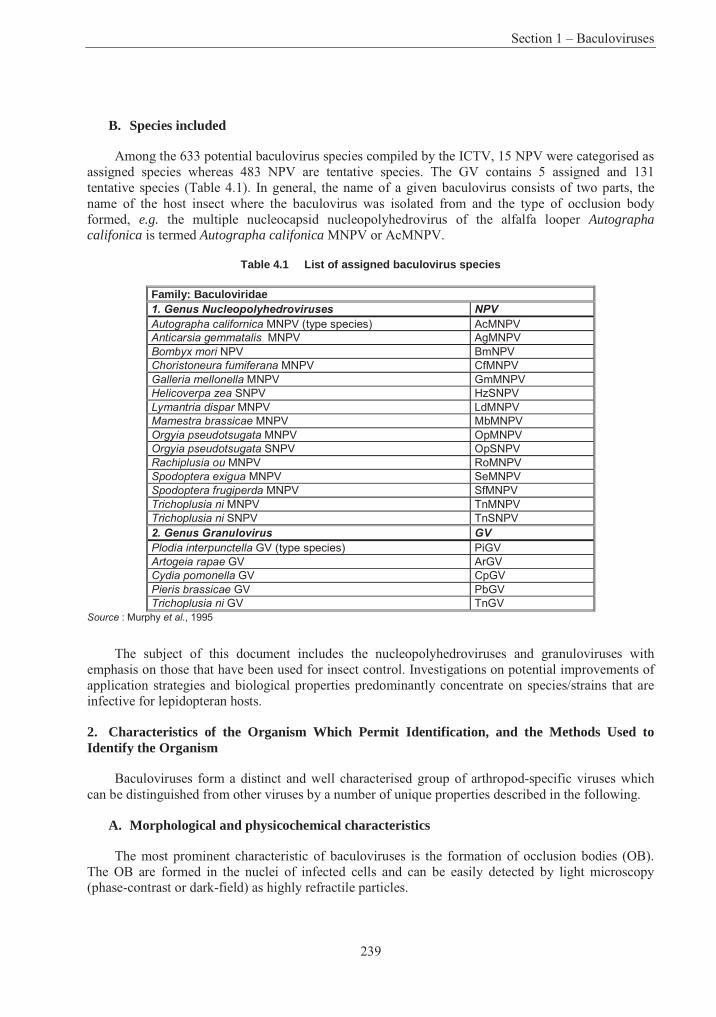
Section 1 – Baculoviruses
239
B. Species included
Among the 633 potential baculovirus species compiled by the ICTV, 15 NPV were categorised as assigned species whereas 483 NPV are tentative species. The GV contains 5 assigned and 131 tentative species (Table 4.1). In general, the name of a given baculovirus consists of two parts, the name of the host insect where the baculovirus was isolated from and the type of occlusion body formed, e.g. the multiple nucleocapsid nucleopolyhedrovirus of the alfalfa looper Autographa califonica is termed Autographa califonica MNPV or AcMNPV.
Table 4.1 List of assigned baculovirus species
Family: Baculoviridae 1. Genus Nucleopolyhedroviruses NPV
Autographa californica MNPV (type species) AcMNPV
Anticarsia gemmatalis MNPV AgMNPV
Bombyx mori NPV BmNPV
Choristoneura fumiferana MNPV CfMNPV
Galleria mellonella MNPV GmMNPV
Helicoverpa zea SNPV HzSNPV
Lymantria dispar MNPV LdMNPV
Mamestra brassicae MNPV MbMNPV
Orgyia pseudotsugata MNPV OpMNPV
Orgyia pseudotsugata SNPV OpSNPV
Rachiplusia ou MNPV RoMNPV
Spodoptera exigua MNPV SeMNPV
Spodoptera frugiperda MNPV SfMNPV
Trichoplusia ni MNPV TnMNPV
Trichoplusia ni SNPV TnSNPV
2. Genus Granulovirus GV
Plodia interpunctella GV (type species) PiGV
Artogeia rapae GV ArGV
Cydia pomonella GV CpGV
Pieris brassicae GV PbGV
Trichoplusia ni GV TnGV
Source : Murphy et al., 1995
The subject of this document includes the nucleopolyhedroviruses and granuloviruses with emphasis on those that have been used for insect control. Investigations on potential improvements of application strategies and biological properties predominantly concentrate on species/strains that are infective for lepidopteran hosts.
2. Characteristics of the Organism Which Permit Identification, and the Methods Used to Identify the Organism
Baculoviruses form a distinct and well characterised group of arthropod-specific viruses which can be distinguished from other viruses by a number of unique properties described in the following.
A. Morphological and physicochemical characteristics
The most prominent characteristic of baculoviruses is the formation of occlusion bodies (OB). The OB are formed in the nuclei of infected cells and can be easily detected by light microscopy (phase-contrast or dark-field) as highly refractile particles.
Part 4 – Consensus Documents on Micro-Organisms
240
Nucleopolyhedroviruses form polyhedra-like occlusion bodies of 0.15 to 15µm in size and contain many enveloped virions. The major component of the occlusion body is a single, viral encoded protein of Mr 25-33 x 103, called polyhedrin (Hooft van Iddekinge et al., 1983). Polyhedral occlusion bodies normally band at 54-56% sucrose on 40-65% w/w sucrose gradients at 100,000 g. The buoyant density of ODVs in CsCl is 1.18-1.25 g/cm3, that of BV in sucrose is 1.17-1.18 g/ cm3.
Electron microscopic observation of polyhedral inclusion bodies reveal two morphotypes: (i) single nucleocapsid nucleopolyhedroviruses (SNPV) contain only a single nucleocapsid within a virion, whereas the virions of (ii) multiple nucleocapsid nucleopolyhedroviruses (MNPV) harbour few to many nucleocapsids. Factors determining and regulating the formation of SNPV or MNPV have not been elucidated.
Granuloviruses generally form ovicylindrical (granule-like) occlusion bodies of 120-300 nm in width and 300-500 nm in length (Crook, 1991). The matrix protein, called granulin, is genetically and serologically closely related to the NPV polyhedrin.
SDS-polyacrylamide gel electrophoresis and serological techniques such as immunodiffusion, immunoelectrophoresis, radioimmunassay (RIA), enzyme-linked immunosorbent assay (ELISA) and Western-blotting have been used to identify particular NPV and GV and to study their relationship (Summers and Smith, 1975; Reinganum, 1984). ELISA was demonstrated to be a rapid, specific and sensitive method for detecting and quantifying baculoviruses (reviewed by Harrap and Payne, 1979). Polyclonal and monoclonal antisera specific against occlusion body and capsid proteins revealed a high degree of cross-reactivity among NPV and GV (Smith and Summers, 1981). These traits allowed the identification of a single baculovirus species and were used for the first phylogenetic studies of different baculoviruses.
By using monoclonal antibodies in ELISA, it was possible to detect virus antigens in NPV-infected Helicoverpa armigera and Choristoneura fumiferana larvae at about 6-9 hours after virus exposure, whereas disease symptoms of the larvae could only be observed after 5-6 days (Zhang and Kaupp, 1988; Lu et al., 1995). With an antiserum against the polyhedrin component of the NPV ofMamestra brassicae, it was possible to detect polyhedra at a concentration of 3.13 x 10⁴ polyhedra/ml by means of immunoelectrophoresis, and as low as 2.44 x 10² polyhedra/ml by means of ELISA. Even though this antiserum was specific to MbMNPV, it also cross-reacted with the polyhedrin of Agrotis segetum NPV, Lymantria monocha NPV and Neodiprion sertifer NPV (Riechenbacher and Schliephake, 1988). Similarly, polyhedrin specific antisera were developed for detection of LdMNPV and Borrelina bombycis NPV by ELISA in infected host larvae or cultured insect cells (Ma et al.,1984; Shamim et al., 1994). Alternatively, a monoclonal antibody against the 42K protein of AcMNPV was used for virus detection in dead larvae and for safety investigations (Naser and Miltenburger, 1983).
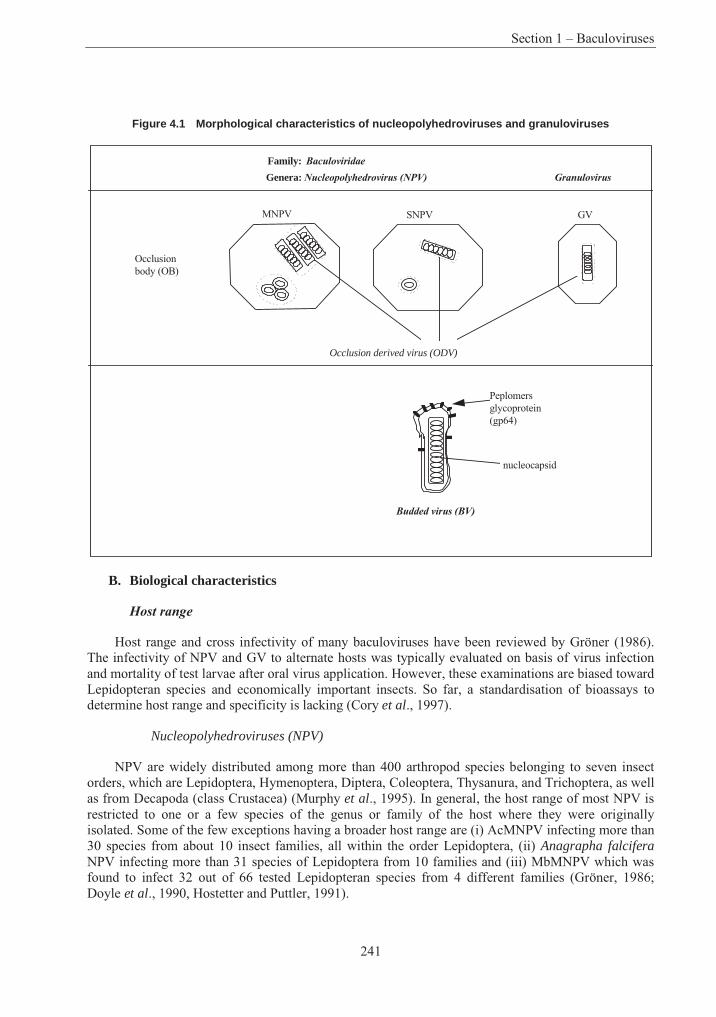
Section 1 – Baculoviruses
241
Figure 4.1 Morphological characteristics of nucleopolyhedroviruses and granuloviruses
Family: Baculoviridae
Genera: Nucleopolyhedrovirus (NPV) Granulovirus
Occlusionbody (OB)
Peplomersglycoprotein (gp64)
Budded virus (BV)
MNPV SNPV GV
Occlusion derived virus (ODV)
nucleocapsid
B. Biological characteristics
Host range
Host range and cross infectivity of many baculoviruses have been reviewed by Gröner (1986). The infectivity of NPV and GV to alternate hosts was typically evaluated on basis of virus infection and mortality of test larvae after oral virus application. However, these examinations are biased toward Lepidopteran species and economically important insects. So far, a standardisation of bioassays to determine host range and specificity is lacking (Cory et al., 1997).
Nucleopolyhedroviruses (NPV)
NPV are widely distributed among more than 400 arthropod species belonging to seven insect orders, which are Lepidoptera, Hymenoptera, Diptera, Coleoptera, Thysanura, and Trichoptera, as well as from Decapoda (class Crustacea) (Murphy et al., 1995). In general, the host range of most NPV is restricted to one or a few species of the genus or family of the host where they were originally isolated. Some of the few exceptions having a broader host range are (i) AcMNPV infecting more than 30 species from about 10 insect families, all within the order Lepidoptera, (ii) Anagrapha falciferaNPV infecting more than 31 species of Lepidoptera from 10 families and (iii) MbMNPV which was found to infect 32 out of 66 tested Lepidopteran species from 4 different families (Gröner, 1986; Doyle et al., 1990, Hostetter and Puttler, 1991).
Part 4 – Consensus Documents on Micro-Organisms
242
Granuloviruses (GV)
GV infections have been reported for more than 100 insect species, however they appear to infect only members of the order Lepidoptera (Murphy et al., 1995). In contrast to NPV, the host range of GV appears to be even more narrow and mostly restricted to a single species. (see also C 23.3)
Gross pathology
Nucleopolyhedroviruses
The gross pathology post infection (p.i.) of NPV infecting Lepidopteran larvae can be summarised as follows:
• Day 1 - 3 p. i.: Infected larvae normally do not show obvious signs of disease.
• Day 4 - 6 p. i.: Diseased larvae only react slowly to tactile stimuli. The larvae start to appear swollen, glossy and moribund.
• Day 6 - 7 p. i.: Diseased larvae stop feeding and begin to die. Diseased larvae of some species, e.g. Lymantria spec., crawl to the top of the twigs (negative geotropism) on which they were feeding.
• Day 7-10 p. i.: Diseased larvae die and may liquefy, the cuticle ruptures and polyhedra arereleased.
Granuloviruses
In general three different types of gross pathology with GV can be distinguished (Federici, 1997).
• Many GV such as CpGV show a similar gross pathology in infected larvae as described for NPV.
• Some GV, esp. many Noctuid-infecting GV also pass the midgut epithelium but then infect only fat body tissue. Infected larvae do not stop feeding and grow even bigger than healthy larvae. Larval death occurs at 10-15 days.
• The third type of gross pathology has to date only been observed with Harrisina brilliansGV. Infection is restricted to the midgut epithelium which causes heavy diarrhetic disorder and death within 4 to 7 days.
C. Pathological characteristics
Baculovirus pathogenesis has been most extensively studied for its type species AcMNPV, but appears to be similar in all other known baculoviruses (see below).
Most typically, virus replication of NPV occurs in the nuclei of infected host cells, whereas in GV-infected cells the nuclear membrane disrupts during the replication process and loses its integrity. Upon infection, the nuclei appear to become hypertrophic. The occlusion bodies produced by infected cells can be detected by light microscopy.
Section 1 – Baculoviruses
243
Divergent tissue tropism is observed with different viruses in their respective hosts. Most NPV specific for lepidopteran species as well as most GV establish a transient infection of the midgut epithelium and then invade other tissues such as fat body, epidermis, tracheal matrix, muscle, nerve, malphigian tubules, and reproductive and glandular tissues. In contrast, NPV specific for Hymenoptera, most Diptera, Trichoptera, Thysanura, and Crustacea as well as the Harrisina brilliansGV were only found to infect midgut epithelium cells but not any other larval tissue (Federici, 1997).
D. Genetic characteristics
Restriction mapping
Early studies of the size and GV content of baculovirus DNA were based on a number of different methods, such as electron microscopy, melting point determination and reassociation kinetics, ultra-centrifugation in sucrose gradients and chemical analyses.
Restriction endonuclease analysis of isolated DNA has been for many years one of the most important and powerful tools for characterisation and identification of DNA viruses including baculoviruses (Rohrmann et al., 1978). This method allows investigators (i) to identify unequivocally the infecting agent, e.g. when a latent baculovirus is activated (Jurcovicova, 1979), (ii) to distinguish among different viruses or virus isolates, which infect the same host species and show similar biological properties (Vlak, 1980), and (iii) to recognise the identity of a virus infecting different host species (Miller and Dawes, 1978). Furthermore, restriction endonuclease analysis is indispensable for the construction of physical maps, which assign the position of the restriction fragments to each other. By convention, the smallest restriction fragment containing the gene encoding polyhedrin or granulin was chosen as the zero point of baculovirus physical maps (Vlak and Smith, 1982). To date restriction maps have been constructed for 18 NPV and 5 GV (Table 4.2).
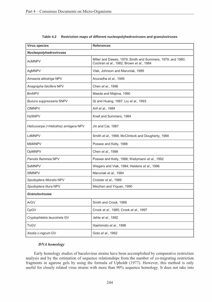
Part 4 – Consensus Documents on Micro-Organisms
244
Table 4.2 Restriction maps of different nucleopolyhedroviruses and granuloviruses
Virus species References
Nucleopolyhedroviruses
AcMNPV Miller and Dawes, 1979; Smith and Summers, 1979; and 1980; Cochran et al., 1982; Brown et al., 1984
AgMNPV Vlak, Johnson and Maruniak, 1989
Amsacta albistriga NPV Anuradha et al., 1995
Anagrapha falcifera NPV Chen et al., 1996
BmNPV Maeda and Majima, 1990
Buzura suppressaria SNPV Qi and Huang, 1987; Liu et al., 1993
CfMNPV Arif et al., 1984
HzSNPV Knell and Summers, 1984
Helicoverpa (=Heliothis) armigera NPV Jin and Cai, 1987
LdMNPV Smith et al., 1988; McClintock and Dougherty, 1988
MbMNPV Possee and Kelly, 1988
OpMNPV Chen et al., 1988
Panolis flammea NPV Possee and Kelly, 1988; Weitzmann et al., 1992
SeMNPV Wiegers and Vlak, 1984; Heldens et al., 1996
SfMNPV Maruniak et al., 1984
Spodoptera littoralis NPV Croizier et al., 1989
Spodoptera litura NPV Meizhen and Yiquan, 1990
Granuloviruses
ArGV Smith and Crook, 1988
CpGV Crook et al., 1985; Crook et al., 1997
Cryptophlebia leucotreta GV Jehle et al., 1992
TnGV Hashimoto et al., 1996
Xestia c-nigrum GV Goto et al., 1992
DNA homology
Early homology studies of baculovirus strains have been accomplished by comparative restriction analysis and by the estimation of sequence relationships from the number of co-migrating restriction fragments in agarose gels by using the formula of Upholdt (1977). However, this method is only useful for closely related virus strains with more than 90% sequence homology. It does not take into
Section 1 – Baculoviruses
245
consideration that many genomic variations are due to small sequence insertions and deletions which result in differences of the restriction profiles even of very closely related virus strains.
DNA hybridisation techniques, such as Southern blot, dot blot and cross blot analysis, have been widely applied for identification and quantification of the intergenomic relationship of many baculoviruses. These methods permit the identification of heteroduplex formation of two different DNAs if they show more than 67% sequence identity (low stringency) or more than 85% identity (high stringency) (Howley et al., 1979).
Smith and Summers (1982) analysed the genomic interrelationship of 18 baculoviruses by restriction analysis, Southern hybridisation and semi-quantitative dot blot hybridisation. They found that under low stringency conditions all viral DNAs showed detectable cross hybridisation. Their results also corroborated the earlier classification of baculoviruses into three subgroups (nuclear polyhedrosis viruses, granulosis viruses and non-occluded baculoviruses, see A 1.1).
The combination of physical mapping and cross hybridisation indicated that the genomes of baculoviruses are similarly arranged. Leisy et al. (1984) showed that OpMNPV and AcMNPV are two distinct viruses with a colinear genomic arrangement. Similar observations were made for the genomes of CfMNPV and AcMNPV (Arif et al., 1984), MbMNPV and Panolis flammea NPV (Possee and Kelly, 1988) as well as for ArGV and CpGV (Crook et al., 1997) and for Cryptophlebia leucotreta GV and CpGV (Jehle et al., 1992).
3. Information on the Recipient Organism’s Reproductive Cycle
A. In vivo and in vitro replication of baculovirus in permissive hosts
Initial stages of infection
The replication of AcMNPV has been most extensively studied in larvae of Trichoplusia ni and in cultured cells of Spodoptera frugiperda and serves as a model for NPV and GV replication in Lepidoptera (reviewed by Granados and Williams, 1986; Federici, 1997; Williams and Faulkner, 1997).
The natural route of infection is the peroral ingestion of viral occlusion bodies by larvae. In the alkaline environment of the midgut (pH > 9.5), the occlusion bodies dissolve rapidly and occlusion-derived virions (ODVs) are released. There is evidence that the dissolution of the occlusion body matrix might be facilitated by an insect derived alkaline protease which is associated with the occlusion body matrix. The ODVs pass through the peritrophic membrane (PM), a proteinaceous-chitinaceous layer which is secreted by the midgut cells to protect the midgut epithelium from direct contact with ingested material. After attachment to the microvilli of the midgut epithelium, the nucleocapsids enter the cell lumen either via fusion of the virion envelope with the epithelial membrane or by viropexis. The nucleocapsids are transported, most likely under involvement of the cellular microtubular structures, to the nucleus and become uncoated at the nuclear pore or within the nucleus where the viral DNA is released and DNA expression and replication is initiated.
The different temporal phases of gene expression
The following stages and secondary infections initiated by budded virions (BV) of host tissues or cultured cells are thought to be similar. In the early stages (8 hr p. i.) of infection the nucleus becomes hypertrophic and a virogenic stroma is formed where DNA replication and nucleocapsid assembly take place. Host cell protein synthesis is completely shut off by 24 hr p.i.
Part 4 – Consensus Documents on Micro-Organisms
246
Viral gene transcription and expression follows a temporally co-ordinated cascade. Early and delayed early genes (α and β genes) are transcribed by a host dependent RNA polymerase II, which is sensitive to alpha-amanitin. These genes are necessary for regulation of viral gene transcription, for viral DNA replication, and late gene expression. Their promoters resemble those of host genes in having a CAGT transcription initiation site which is 25-31 bp downstream of TATA box and are recognized by nuclear extracts of uninfected cells.
Transcription and expression of late genes (or γ genes) involved in DNA replication, production of structural proteins and budded virions (BV) occurs between 8 and 24 hr p.i.
These genes as well as the very late (or δ genes) (see below) have a universal and invariant (A/G/T)TAAG transcription initiation site and are transcribed by a viral encoded RNA polymerae insensitive to alpha-amanitin. Five essential (p143, ie-1, lef-1, lef-2, and lef-3) and five stimulatory genes (dnapol, p35, ie-2, lef7, and pe38) were identified to be involved in AcMNPV DNA replication. In addition, transient expression studies using the chloramphenicol acetyl transferase (cat) reporter gene under the control of the late and very late promoters (vp39, p6.9, polyhedrin, p10) showed that eighteen AcMNPV genes (those necessary for DNA replication and lef-4, lef-5, lef-6, lef-8, lef-9, lef-10, lef-11, p47, and 39K) were essential for optimal expression from these promoters (Lu and Miller, 1995a). DNA replication and late viral gene transcription can be blocked by aphidicolin (Miller et al., 1981; Rice and Miller, 1986).
Expression of very late genes (or δ genes) begins about 18 - 24 hr p.i. and is characterised by a dramatic increase of polh and p10 transcription and expression. Polh encodes the polyhedrin (its homologue in granuloviruses is called granulin gene), which is the major component of the occlusion body matrix. The p10 gene product is associated with the formation of extensive fibrillar structures in the nucleus and cytoplasm of infected cells (Quant-Russell et al., 1987; van der Wilk, et al., 1987). Although the biological function of p10 has not been convincingly elucidated so far, it was suggested that it might contribute to the disruption of the nuclear membrane and to the release of occlusion bodies from infected cells (van Oers et al., 1993; Williams et al., 1989).
Since both polh and p10 are not required for DNA replication and are hyperexpressed during cell infection, their strong promoters were exploited for the development of the baculovirus expression vector systems. The coding region of these genes can be replaced by exogenous genes resulting in the production of high levels of the foreign protein (Smith et al., 1983; Vlak et al., 1990).
Production of budded viruses (BVs) and occlusion derived viruses (ODVs)
The replicative cycle of baculoviruses is biphasic and generates two distinct viral phenotypes, the budded virus (BV) and the occlusion derived virus (ODV). The two phenotypes are structurally distinct and destined for two different functions, both of which are essential for virus survival in nature. ODVs are released from the inclusion bodies and infect midgut epithelium cells, where a first round of virus replication takes place. The newly produced nucleocapsids traverse the nuclear membrane, the cytosol and bud through the basal lamina of the midgut cells into the hemolymph. These budded virions (BV) acquire a new envelope which consists of plasma membrane containing peplomers of a viral encoded glycoprotein, termed gp64 (see Fig. 1 in A 2.1). Gp64 appears to be pivotal for the interaction between the BV envelope and susceptible host cells through a possible interaction with a cell membrane receptor molecule and then a final fusion with the endosomal membrane (for review see Blissard, 1996). In cultured cells the production of BV peaks during the late phase of gene expression, between 10 - 20 hr p.i., whereas the very late occlusion phase can be observed between 16 - 72 hr p.i.
Section 1 – Baculoviruses
247
For most NPV and GV infecting lepidopteran host larvae, virus occlusion is not observed in midgut epithelial cells. These cells release BV into the hemolymph which then systemically spreads the virus infection among susceptible cells and tissues. In contrast, NPV of Hymenoptera, Diptera and Crustacea and the H. brillians GV infect only midgut cells where occluded viruses are produced (for review see Federici, 1997). However, there is also evidence that some nucleocapsids might traverse the midgut epithelial cells without replication and bud directly into the hemolymph (Granados and Lawler, 1981).
Engelhard et al. (1994) used a recombinant AcMNPV mutant expressing lacZ reporter gene (beta-galactosidase) to study the infection pathway in fourth instars of Trichoplusia ni. Based on the observation of early infection of midgut tracheoblast and the tracheal matrix, they postulated that the tracheol system might directly contribute to the systemic spread of BV. This finding, however, is not supported by others who used a similar approach (Flipsen et al., 1993; 1995).
Altogether, the spread of BV and systemic infection starts from the midgut and continues to hemocytes, tracheal cells, fat body, muscle and nerve cells as well as reproductive and glandular tissues. In the final step of infection, occlusion bodies are formed and the nuclei are packed with occlusion bodies which causes the cellular hypertrophy and swollen appearance of the infected larvae.
The different role of BVs and ODVs in the reproduction of baculoviruses can be summarised as follows: ODVs transmit infection from one larvae to another within an insect population, whereas the budded virus (BV) spreads the infection within susceptible larval tissues.
B. Behaviour in semi- and non-permissive insect cells
Baculovirus gene expression, replication and reproduction is only possible if one of the virion phenotypes (BV or ODV) is able to enter a permissive host cell. There are a number of viral factors and host responses which are necessary for a productive infection. In a non-compatible baculovirus-host cell interaction, the baculovirus gene expression and replication is blocked at a early stage. In order to analyse the genetic factors involved in host specificity and baculovirus-host cell interaction, a number of studies on the effect of virus on semi- and non-permissive insect cell lines were conducted. Infection studies with vertebrate and mammalian cells will be discussed in Section B.
When T. ni cells (TN-368) were infected with SfMNPV, typical early cytopathological effects such as nuclear hypertrophy and the formation of a virogenic stroma could be observed. However, only a few early transcripts and two proteins, an early 97 kD and a late 29 kD polypeptide, were synthesised. Virion assembly could not be detected. This result suggested that the block of infection occurred during the early phase of gene expression (Carpenter and Bilimoria, 1983; Bilimoria, 1991).
AcMNPV infection of Choristoneura fumiferana cell lines Cf124T and CF-203 is blocked after DNA replication and causes apoptotic cell death, respectively (Liu and Carstens, 1993; Palli et al.,1996). An apoptotic response to AcMNPV infection has also been observed for Spodoptera littoraliscell line SL2 (Chejanovsky and Gershburg, 1995).
By using recombinant AcMNPV expressing chloramphenicol acetyltransferase (cat) reporter gene under control of different temporally regulated promoters such as early, late and very late promoters, Morris and Miller (1992; 1993) showed that the activity of late and very late promoters is significantly reduced in semi- and nonpermissive cell lines of Bombyx mori (BmN-4), Choristoneura fumiferana (CF-1), Lymantria dispar (Ld652Y), Mamestra brassicae (MaBr-3) and Drosophila melanogaster (Dm) compared to permissive cell lines of Spodoptera frugiperda (Sf-21). Since there was some evidence for viral DNA replication but not for virus assembly, it was suggested that in these
Part 4 – Consensus Documents on Micro-Organisms
248
cell lines progression of infection is blocked during DNA replication. By infecting different cell lines with AcMNPV expressing cat reporter gene controlled by the Rous sarcoma terminal repeat promoter and β-galactosidase controlled by the very late polyhedrin promoter, it was shown that nonpermissive Drosophila cells and permissive Sf-21 had a similar cat expression, whereas very late promoter activity was only observed with Sf-21 (Carbonell et al., 1985, Carbonell and Miller, 1987).
C. Genes involved in host range determination
So far, a number of baculovirus genes involved in differential host cell and host larval specificity have been identified. There are several examples where the expression or deletion of these host range determinants by recombinant NPV allowed the specific extension or restriction of host specificity. These results, mainly obtained with AcMNPV, provide evidence that a baculovirus which is infective to different host species relies on specific genes to establish infection and virus replication and that these sets of genes might differ slightly from host species to host species.
Although genetically closely related, AcMNPV and BmNPV have distinct host range specificities. Kondo and Maeda (1991) demonstrated that an AcMNPV mutant containing part of the BmNPV p143 gene became infective to the normally refractile BmN cell line of Bombyx mori. This mutant, called eh2-AcMNPV, was obtained after coinfection of Sf-21 cells with AcMNPV (OT2) and BmNPV(T3), which are not infective for BmN and Sf21 cells, respectively. The progeny virus was subsequently passaged through BmN and Sf-21 cells thereby isolating eh2-AcMNPV which was found to be infective for both cell lines. In further experiments, the genome region responsible for host range extension was more precisely localised on a 572-bp fragment of BmNPV p143, which differed in 14 out of 109 amino acids as compared to AcMNPV (OT2) (Maeda et al., 1993). Similar results were obtained by Mori et al. (1992) who performed the co-transfection experiments with fragments of the BmNPV genome instead of BmNPV virions. This finding was corroborated by studies of Croizier etal. (1994) who demonstrated that the exchange of only three amino acids within p143 was sufficient to expand the host range of AcMNPV to B. mori cells. It appears that the substitution of a single serine residue to an asparagine residue of AcMNPV p143 is sufficient for this host range extension (Kamita and Maeda, 1997).
Infection of the Lymantria dispar cell line Ld652Y with AcMNPV is characterised by early cyto-pathic effects, transcription from all temporal classes of promoters and DNA replication but with a very low level of protein translation and no formation of infectious virions (McClintock et al., 1986; Guzo et al., 1991; 1992). However, when Ld625Y cells were coinfected with AcMNPV and LdMNPV, replication and production of AcMNPV was observed suggesting that LdMNPV encodes a trans-acting factor which rescues an abortive AcMNPV infection (McClintock and Dougherty, 1987). This trans-acting factor encoded by LdMNPV was finally mapped and identified by co-transfecting Ld625Y cells with AcMNPV genomic DNA and single cosmids of the LdMNPV genome. It was called host range factor 1 (hrf-1) and was shown to extend the host range of recombinant AcMNPV expressing the factor to Ld652Y cells and to L. dispar larvae. This indicates that hrf-1 determines the host range at the cell culture and larval levels (Thiem et al., 1996).
Transient expression studies using the cat reporter gene revealed that eighteen AcMNPV genes, so-called lef genes, were essential for optimal expression of late and very late genes in Sf21 cells (see also Chapter 3.1.2). For the permissive T. ni cell line TN-368, it was found that in addition to these 18 lef genes an additional gene, called host cell-specific factor-1 (hcf-1) was required for efficient late gene expression. AcMNPV mutants lacking hcf-1 replicate normally in Sf21 cells and S. frugiperdalarvae but are unable to productively infect TN-368 cells and T. ni larvae (Lu and Miller, 1995b; 1996; reviewed by Miller and Lu, 1997). This observation clearly indicated that hcf-1 has tissue specific as
Section 1 – Baculoviruses
249
well as species-specific effects on the replication of AcMNPV in cultured insect cells and in insect larvae.
Abortive replication in the permissive cell lines SF-21 (and SF-9) was observed with AcMNPV mutants lacking a functional p35 gene. p35 is an inhibitor of programmed cell death (apoptosis) of insect cells, which might be a defence reaction of insects against baculovirus infection at the organismal level (Clem et al., 1991; for review Friesen, 1997). It appears that p35 also exerts host range function since this gene is essential for replication in SF-21-cells and S. frugiperda larvae but not in TN-368 and T ni larvae. AcMNPV p35 mutants were 1000-fold less infective for S. frugiperdalarvae than wildtype-AcMNPV when the virus is injected into the hemocoel and they were about 25-fold less infective in peroral infections (Clem and Miller, 1993; Clem et al., 1994).
4. Biological Features and Environmental Conditions Which Affect Survival, Reproduction, Growth, Multiplication or Dissemination
A. Formation of occlusion body
NPV and GV form polyhedral and granular occlusion bodies, respectively, into which the occlusion derived virions (ODV) are embedded. The occlusion bodies serve to protect the embedded virions against damaging environmental conditions and allow the virions to remain viable for many years. The occlusion bodies are solubilised in alkaline conditions of the midgut and thus deliver the ODV to susceptible columnar epithelial cells.
The crystalline matrix of the occlusion body mainly consists of a single protein, called polyhedrin and granulin, respectively. These proteins of about 245 amino acids (29 kDa) are hyperexpressed during the very late phase of virus infection and are not required for virus replication (for review see Rohrmann, 1992; Funk et al., 1997). To date, polyhedrin and granulin genes of about 35 different lepidopteran- and one hymenopteran-specific baculovirus species have been sequenced. The polyhedrin and granulin genes are highly conserved with at least 70% amino acid identity among lepidopteran NPV and about 50% amino acid sequence identity between NPV polyhedrins and GV granulins.
The importance of the occlusion body for the stability and maintenance of infectivity of baculoviruses in the environment has been clearly demonstrated by field tests using polyhedrin deficient AcMNPV mutants. In 1987, a field test was performed where insect larvae were infected with an AcMNPV polyhedrin minus mutant and then released into enclosed field plots. The non-occluded virus progeny rapidly lost its activity in the decaying larval carcass and no virus activity could be detected on cabbage leaves or in soil samples within two weeks after the release (Bishop etal., 1988a; 1988b). Wood and co-workers applied a slightly different approach by co-occluding polyhedrin-minus AcMNPV into the polyhedrin matrix of wild-type virus (Hamblin et al., 1990). In laboratory experiments it was shown that the persistence of such a co-occluded polyhedrin-minus mutant was significantly reduced at inoculum levels below a 100% dose. This observation was corroborated in a three-year field test in which AcMNPV polyhedra containing polyhedrin minus and wild-type AcMNPV in a ratio of 48:52 were applied on T. ni larvae, and the amount of polyhedrin minus mutants was analysed in the following years. It was found that the amount of polyhedrin minus mutant dropped below 20% of the virus progeny in the second and third year (Hamblin et al., 1990; Wood et al., 1994).
The occlusion bodies are not solely composed of polyhedrin. They are surrounded by an envelope, called a polyhedron calyx or polyhedron envelope (PE). Minion et al. (1979) reported that the PE of Helicoverpa (=Heliothis) virescens NPV was composed of hexose and pentose
Part 4 – Consensus Documents on Micro-Organisms
250
carbohydrates. Whitt and Mannig (1988) showed that the PE of AcMNPV consisted of a phosphorylated protein which might be covalently linked to the carbohydrate component. The PE protein is encoded by a late and very late gene (pp34) which has been sequenced for several NPV (Gombart et al., 1989; Bjornson and Rohrmann, 1992). OpMNPV mutants lacking the PE gene produced unstable polyhedra with a rugged and pitted surface (Gross et al., 1994a).
Another protein which is normally associated with polyhedra in the infected cells is the p10protein. Evidence suggests that it is involved in the formation of PE and lysis of infected nuclei. AcMNPV mutants with inactivated p10 genes failed to release the polyhedra from infected cells. p10negative OpMNPV and AcMNPV mutants also failed to form an intact polyhedron envelope gene-rating fragile polyhedra with significantly reduced stability but with the peculiarity to form polyhedral aggregates (Williams et al., 1989; van Oers et al., 1993). Based on these observations, it was suggested that the function of p10 and PE is twofold: to protect the polyhedra from mechanical damage by sealing their surface and to prevent their aggregation. Hence, these properties could be important to maximise the number of intact virions per occlusion body and to optimise virus dissemination (Gross et al., 1994a).
B. Enhancing of host susceptibility
Synergism between Pseudaletia unipuncta NPV and GV, which resulted in an increased susceptibility of P. unipuncta larvae for the NPV, has been described (Tanada, 1959). A protein component in the granule of PsunGV, termed synergistic factor, which interacts with the microvillar membrane of midgut cells and facilitates adsorption of the NPV virions was identified (Tanada et al., 1975; Zhu et al., 1989).
Similar factors have been found in TnGV, Xestia c-nigrum GV and Helicoverpa (= Heliothis) armigera GV (Derksen and Granados, 1988; Goto, 1990; Roelvink et al., 1995). These proteins are commonly known as enhancing factors or enhancin. TnGV enhancin was shown to increase the infectivity of AcMNPV to different noctuid larvae. The predicted amino acid sequences of TnGV enhancin and the synergistic factor of PSunGV-H are almost identical. TnGV enhancin is a protein of 104 kDa which most probably functions as a metalloprotease, since it can be reversibly inactivated by metal chelators (Lepore et al., 1996). TnGV enhancin is located in the occlusion body and causes specific degradation of the intestinal mucin component of the peritrophic membrane (Wang and Granados, 1997). The disruption of the peritrophic membrane of the larval midgut facilitates the virions access to the midgut columnar cells.
Although such a function was also found with a component of NPV polyhedra, no gene homologue to the TnGV enhancin could be identified in NPV (Derksen and Granados, 1988; Hashimoto et al., 1991). Observations of the synergistic enhancement of infectivity by the activity of components from different NPV, and the isolation of another synergistic factor from Pseudaletia unipuncta GV indicate that a somewhat heterogeneous set of genes and functions may contribute to this modulation of the infectious process (Arne and Nordin, 1995; Ding et al., 1995).
C. Inhibition of cellular apoptosis
Programmed cell death or apoptosis is a cellular pathway during which a cascade of responses is activated resulting in a well regulated cellular suicide. Apoptosis is the normal fate of many cells during development and metamorphosis, cellular turnover of renewing tissues and other dynamic cellular processes. There are also many examples of viral infections whose progression is blocked during early stages of replication by an apoptotic response of the infected host cell. It appears that apoptosis is a very powerful mechanism of many vertebrate and invertebrate cells to prevent viruses
Section 1 – Baculoviruses
251
from replicating and becoming persistent. On the other hand, viruses have evolved mechanisms that overcome or block this apoptotic response and so establish an infection (for review see Clem, 1997).
In baculoviruses two classes of proteins, the p35 protein and the IAP (inhibitor of apoptosis) proteins, have been identified and characterised as anti-apoptotic agents. Expression of p35 was found to be essential for AcMNPV replication in S. frugiperda cells (Clem et al., 1991). SF-21 cells infected with an AcMNPV ‘annihilator’ mutant, which was shown to contain a deletion in p35 gene, underwent apoptotic cell death within 24 hours. The antiapoptotic effect of p35 has been demonstrated not only in insect cells but also in many heterologous systems, such as Caenorhabditis elegans, Drosophila melanogaster, mammalian and human cells (Sugimoto et al., 1994; Hay et al., 1994; Rabizadeh et al., 1993; Beidler et al., 1995). The competence of p35 to prevent apoptosis in many different cell types pointed to the possibility that p35 acts on a universal step in the apoptotic cascade. Xue and Horvitz (1995) and Bump et al. (1995) demonstrated that p35 is able to inhibit cystein proteases, so-called ICE-like proteases, which play a key role in the highly conserved effector pathway of apoptotic response.
Compared to wild-type viruses, the infectivity of p35 minus AcMNPV mutants to S. frugiperdalarvae was dramatically reduced (by a factor of 1000 if injected into the hemocoel and by a factor of 25 if perorally applied). The yield of virus progeny from infected larvae was decreased 900-fold suggesting that the anti-apoptotic effect of p35 may play an important role during in vivo infection of host larvae. In contrast to SF-21 cells, p35 is not essential for AcMNPV to establish infection in TN-368 nor is there any difference between p35 minus and wildtype AcMNPV in the infectivity for T. nilarvae. This suggests that p35 is also involved in determining a baculovirus host range (Clem and Miller, 1993; Clem et al., 1994). A p35 homologue has been also identified in BmNPV but is not present in OpMNPV (Maeda, 1994; Ahrens et al., 1997).
A second class of antiapoptotic genes of baculoviruses are iap genes, which were identified in CpGV and OpMNPV because of their ability to rescue replication and polyhedra formation of p35 minus AcMNPV mutants in SF-21 cells (Crook et al., 1993; Birnbaum et al., 1994). All baculovirus iap proteins contain two cysteine-rich repeats known as BIR (baculovirus iap repeat) at their N-termini and a RING finger motif at the C-terminus. Further iap genes were identified upon genome sequencing in AcMNPV (iap1, iap2), BmNPV (iap1, iap2) and OpMNPV (iap1-iap4) (Ayres et al., 1994; Maeda, 1994; Ahrens et al., 1997). Cellular iap homologues were identified in humans and insects and might be ubiquitous in a wide variety of organisms (for review see Clem, 1997). Although the mode of action of iap is not well understood, there is strong evidence that both iap and p35 inhibit defence mechanisms at the cellular and organismal levels, thus allowing baculoviruses to replicate and reproduce.
D. Arresting host development by ecdysteroid-UDP-glucosyltransferase
One of the most remarkable features of baculoviruses is their ability to impair larval moulting. In normal development, larval moulting is a physiological process regulated by ecdysteroid hormones. It was found that AcMNPV is able to inhibit larval moulting by expressing the enzyme ecdysteroid-UDP-glucosyltransferase (EGT). EGT inactivates insect ecdysteroid hormones by conjugation with glucose or galactose residues (O´Reilly and Miller, 1989). Whereas, the moulting process in healthy larvae is characterised by a cessation of feeding, baculovirus infected larvae do not show this behaviour. By generating an EGT minus AcMNPV mutant and comparing its pathogenicity with wild-type virus, O’Reilly and Miller (1989) found that EGT expression is responsible for the suppression of the host development during infection. Molecular studies showed that EGT is transcribed and expressed early during infection (O’Reilly and Miller, 1990). To date, homologous EGT genes have
Part 4 – Consensus Documents on Micro-Organisms
252
been identified and sequenced from 12 nucleopolyhedroviruses and it seems very likely that it is a conserved ancestral gene present in all baculoviruses (Chen et al., 1997).
Since EGT minus AcMNPV mutants yielded about one fourth less virus progeny in late S. frugiperda instars than wild-type AcMNPV, it was proposed that EGT expression might have an important role in maximising the amount of virus progeny from an infected larva (O’Reilly and Miller, 1991, for review see O’Reilly, 1997). A further intriguing observation was that egt minus AcMNPV mutants killed host larvae faster than wild-type virus. The mean survival time (ST50) of EGT minus AcMNPV-infected S. frugiperda and T. ni larvae was about 20-30% shorter than that of wild-type AcMNPV (O’Reilly and Miller, 1991; Eldridge et al., 1992). Although the reason for this increased virulence is not yet elucidated, there is apparently a correlation with the decreased amount of virus progeny. In the faster dying insects, the virus does not have enough time to produce the maximum amount of virus progeny.
The accelerated mortality of the EGT minus AcMNPV mutant accompanied by reduced feeding of the infected larvae led to a new concept of genetically engineered baculoviruses with improved insecticidal properties (O’Reilly and Miller, 1991). In recent years, EGT minus baculoviruses have been tested in the field and they are the most likely candidates to become the first registered genetically engineered baculovirus control agents.
E. Facilitating the release of occlusion bodies from larval cadavers
The final step of baculovirus infection is the breakdown of the larval cuticle and the release of the occlusion bodies into the environment. The cuticles of insect larvae consist mainly of chitin fibres embedded into a proteinacous matrix. Two baculovirus genes, encoding a chitinase (chiA) and cathepsin (cath), have been described to contribute to the liquefaction of the larval carcass and the release of occlusion bodies. Chitinase is a chitin-degrading enzyme with endo- and exomolecular specificity, whereas cathepsin has cysteine proteinase activity (Slack et al., 1995; Hawtin et al., 1995).
The chiA and cath genes appear to be located next to each other in many baculovirus genomes and are expressed late in infection (for review see O’Reilly, 1997). The functional role of chiA and cath was elucidated by constructing AcMNPV and BmNPV mutants lacking either chiA or cath or both. Both single chiA- or cath-minus mutants were not able to cause liquefaction of infected larvae (Slack et al., 1995; Hawtin et al., 1995). Hence, the co-operative action of chitinase and cathepsin appears to exert an effect on the release of viruses from succumbed larvae and to facilitate the dissemination of occlusion bodies in the environment.
An additional role of cath was proposed by Lanier et al. (1996) who found that cathepsin is associated with BV of AcMNPV and is able to cleave actin in an in vitro assay. Whether cathepsin is also involved in the rearrangement of actin filaments during early stages of infection needs further investigation
F. Latent virus infections
Latency is the ability of a virus to persist in a host without causing disease symptoms, thereby providing the virus the possibility of vertical transmission from one generation to another. There are a number of early publications reporting circumstantial evidence of latent baculovirus infections. UV light and treatment with chemicals, rearing conditions, or superinfection with a different virus have been described as reasons for activating latent infections (Krieg, 1956; David and Gardiner, 1965; Longworth and Cunningham, 1968; Biever and Wilkinson, 1978; Podgwaite and Mazzone, 1986). However, most of these early studies were made when molecular tools for identification such as
Section 1 – Baculoviruses
253
hybridisation techniques or restriction endonuclease analysis were not available. Hence, it cannot be excluded that at least some of these reports were biased by cross-infections and contamination. Ponsen and de Jong (1964) and Jurcovicova (1979) reported activation of latent Adoxophyes orana SNPV by infecting A. orana larvae with Barathra brassicae MNPV and vice versa activation of latent B. brassicae MNPV by infection of B. brassicae larvae with A. orana SNPV. An increase in larval deaths from granulovirus infection correlated to stress from dehydrated diet or low temperature was observed in Pieris rapae (Biever and Wilkinson, 1978). Virulence testing indicated the identity of the stress induced virus with laboratory grown virus preparations. It was noted that larvae demonstrating symptoms of stress induced virus infection could recover to a healthy condition after relief of the stress by providing a fresh diet.
More recently, Hughes et al. (1993) provided first molecular evidence for activation of a latent baculovirus in M. brassicae. Activation of a MbMNPV was observed after feeding Panolis flammeaNPV or AcMNPV to the laboratory culture of M. brassicae termed MbLC. Other (environmental) stress factors such as high and low temperatures, starvation or crowding were not effective in triggering apparent infections in this culture (Goulson unpublished data, Goulson and Cory, 1995a). Contamination of the virus inoculum was excluded by control infections of another M. brassicaeculture (MbWS). Challenging this insect culture, established from a novel environmental isolate and adapted to laboratory growth conditions, did not result in multiplication of MbMNPV when infected with the same inoculum. Furthermore, MbMNPV-specific sequences were detected by PCR in each stage of insect development, i.e. in eggs, larvae, pupae and adults. When dissected larval tissues were analysed, latent virus sequences were only detected in the fat body of MbLC larvae. Cell lines established from fat body cells also harboured MbMNPV-specific sequences.
The occult state of the virus in the MbLC culture was further characterised by analysis of m-RNA in larvae, demonstrating the presence of polyhedrin specific m-RNA. Assays of transient reporter gene expression (CAT, chloramphenicol acetyl transferase) after transfection of primary cultures of MbLC fat body cells with constructs containing early, late and very late promoters, demonstrated the presence of expression factors for all of these, albeit at low levels. Furthermore, inoculation of MbWS larvae with MbLV fat body cells resulting in larval deaths from MbMNPV infection, strongly suggested the presence of viable virus particles in these cells (Hughes et al., 1997). The latent or occult status of MbMNPV in the insect can thus be described as a persistent infection, with a continuous production of virus proteins at a low level. Whether this state of the virus is controlled by host functions exclusively or to which degree viral regulatory functions are engaged in its establishment and support, remains an intriguing and challenging question.
A more detailed knowledge of the possibility and extent of virus persistence as a latent infection with vertical transmission to insect progeny and the potential to induce an infectious cycle through environmental conditions or stress factors, is crucial for an improved understanding of baculovirus ecology and population dynamics. So far, the contribution of this feature to the persistence of baculoviruses in natural populations, and its occurrence among insect and virus species and genotypes, is largely unknown. The perception of baculovirus population dynamics as, for example, represented in the modelling of natural and induced epizootics, almost exclusively considers the multiplication of viruses as a result of infections initiated by horizontal virus transmission.
G. Environmental factors influencing virus persistence
Virus persistence is an important factor affecting the potential of baculoviruses to interact with their hosts in the environment. Biotic and abiotic persistence mechanisms allow the virus to overcome situations of varying host density without running the risk of extinction (Evans, 1986). Baculoviruses may persist in the host population through vertical transmission of disease or by a latent infection (see
Part 4 – Consensus Documents on Micro-Organisms
254
A 4.6) or in non-target organisms which may serve as vectors of virus dispersal (see C 26.2). This biotic persistence complements the maintenance of virus activity outside living organisms (abiotic persistence). In the open environment UV radiation of the sunlight has the most significant impact on abiotic persistence which also may be influenced by temperature, relative humidity and precipitation (for review, see Jacques, 1975). Half life time may be as short as 1.3 days for a purified preparation of the Phthorimaea operculella granulovirus (Kroschel et al., 1996) or, more typically, about 5 - 10 days for many NPV and GV (Bell and Hayes, 1994; Kolodny-Hirsch et al., 1993). Hence, a variety of formulations and additives have been tested and applied to improve the life time of preparations for pest control applications (Ignoffo et al., 1989; Ignoffo and Garcia, 1996; see also A.6.3). On the other hand, after spray applications of Panolis flammea NPV on pine foliage, only a slight decrease of infectivity was monitored over several months (Carruthers et al., 1988). This observation suggests that the application time of PaflNPV for control of the pine beauty moth, Panolis flammea, does not necessarily need a precise optimisation (Cory and Entwistle, 1990). Agricultural operations can affect the distribution of the baculoviruses in soil. Following A. gemmatalis NPV spraying on soybeans and subsequent epizootics, it is transported into the soil and decreases rapidly due to overwinter weathering, but enough remains near the soil surface at the beginning of the next growing season to initiate new epizootics. Persistence of the NPV in soil is not adversely affected by discing, cultivating, or other agricultural soil operations (Fuxa and Richter, 1996).
Furthermore, plant surfaces can have a distinct influence on viability and virulence as discussed in section C. 23.4.1. In soil, which can also be a potential reservoir of baculoviruses, the inactivation rate not only depends on soil type and pH but also on microbial activity (Thompson and Scott, 1979; Ignoffo and Garcia, 1966; Jaques and Huston, 1969; Undorf, 1991). Baculoviruses may persist in soil for long periods. Soil samples taken from a field treated with Trichoplusia ni NPV showed 15% of the original virus activity 6 years after application (Jacques, 1964). Similar results were obtained for other GVs and NPVs (for review see Jaques, 1975).
In summary, these observations denote that the physical environment, chemical conditions and the (micro)-biological composition of micro-habitats have a very significant impact on the persistence and activity of baculoviruses. The validity of comparisons of the persistence of different virus types depends on the degree of control of experimental variables. In particular cases the determination of the abundance of genotypes in artificial mixtures could be used as an approach. Parameters of persistence used in calculations of virus spread and modelling have either to account for the detailed structure of the respective environment or must include wide margins for the variation of median values. Biotic and abiotic persistence are prerequisites for mechanisms and routes of dispersal (summarised in section C.26).
5. Behaviour in Simulated Natural Environment Such as Microcosms, Growth Rooms, Greenhouses, Insectaries, etc
A. Monitoring of baculoviruses in closed environments and ecological modelling
Experiments such as bioassays (C.23.1) test well defined characteristics of baculovirus phenotypes in the laboratory. The use of microcosms or similar facilities may supplement these investigations by monitoring virus interactions in more complex environments. Microcosms may represent the variables of natural settings to a widely divergent degree, depending on their particular objective. Fraser and Keddy (1997) describe the “manipulation of an individual environmental axis” to explore its function in structuring the community as a common factor of microcosm research in ecology. Research of this kind may contribute to an improved understanding of baculovirus ecology by investigating mechanisms of fate and interactions in the environment in some detail (Cory et al.,1997; Cory and Hails, 1997). Increased knowledge of ecological behaviour will also generate a better
Section 1 – Baculoviruses
255
predictability and may improve safety assessments. Perspectives of safety may also trigger microcosm experimentation because some degree of confinement from the open environment is provided.
Traditionally, such testing was not usually performed with natural strains during the development of baculovirus insecticides. Baculovirus behaviour (e.g. pathogenicity, virulence, host range, and toxicity for particular non-target organism) is inferred from laboratory testing on one side. On the other, the world-wide application of baculoviruses for pest control together with scientific investigations on baculovirus population dynamics in the open environment contribute to the present understanding of baculovirus ecology. This background of knowledge about the mechanisms driving the dynamics of effects on insect species and about the ability to manipulate it, mostly is considered to be adequate for judgements about applications and safety in the environment. To some extent this view is reflected in the criteria of registration procedures (e.g. Andersen et al., 1989).
Conventional testing procedures using soil columns as a section of the natural environment were used to test the leaching behaviour of baculovirus preparations as a component of environmental fate in order to predict potential exposition rates of the groundwater. Rates were tested under conditions similar to pesticide registration procedures (C.26).
The perspective of using genetically modified baculoviruses seems to have changed the perception with respect to testing in "closed" environments to some degree. The first "deliberate release" of a genetically engineered baculovirus can be viewed as an experiment performed in a confined "simulated natural environment" (Bishop et al., 1988). Modified versions of microcosms introduced for environmental transport and fate studies in chemical risk assessment were used in this context to monitor essentially similar parameters of baculovirus fate. Modelsystems of this kind also provided some simulation of meteorological conditions (sunlight, rainfall, and air movement). Investigations included the comparison of modelsystem and field data, and of the persistence and spread of natural and genetically modified virus types. The potential of monitoring individual genotypes was demonstrated to some extent. A reduced spread and multiplication of the modified genotype in some contrast to similar biotest data was noted (Undorf, 1991). However, systematic comparisons of biotest, modelsystem, and field data, in order to analyse their correspondence, are not well documented in the scientific literature. The potentials as well as the limitations of experimentation in an intermediate scale of complexity have not been explored in great detail. This is in contrast to its use for the evaluation and modelling of the fate and effects of chemicals in the environment. The interrelationship of an improved understanding of baculovirus ecology on one side and their biosafety on the other is stressed in recent publications (Cory et al., 1997). Limited relevant modelling supported by experimentation is cited there to exemplify the need to analyse the correspondence between theory, closed environment experimentation and field data for an improved understanding of baculovirus population dynamics (Begon and Bowers, 1994; Begon et al., 1996).
For inferences, empirical data must be interpreted by theoretical structuring of knowledge in some kind of model, representing the essential components and the relations assumed to exist between them. Explicit modelling of baculovirus ecology given a mathematical form tries to calculate the dynamics of viruses and of their arthropod hosts under a given set of parameter variations. Results of such calculations are then compared with empirical data from modelsystem experimentation, field testing, or natural epizootics, to explore their present contribution to an understanding of mechanisms and to a limited degree of predictive potential, eventually. Simulated natural environments have been employed to a limited extent for testing environmental variables, to verify the formulation of interactions, and to analyse parameter values. This type of research is described in the following to account for common experimental approaches and for the correspondence with objectives to improve (predictive) knowledge of baculovirus ecology.
Part 4 – Consensus Documents on Micro-Organisms
256
The work of Anderson and May (1980, 1981), cited in reviews of baculovirus ecology with some emphasis, is the key reference for the modelling of the dynamics between pathogens and their invertebrate hosts (Evans, 1986; Cory et al., 1997). In the Anderson and May model, the variations of forest insect pest abundances with low densities between regular extensive population increases were triggered exclusively by the dynamic relations between an insect pest population and pathogens persisting in the environment. Their model assumptions, describing the rate of disease transmission by a mass-action law with a linear relationship between pathogen/host densities and productive infection, have been criticised for incorrectness, oversimplification and lack of correspondence to reality (Bowers et al., 1993; D´Amico et al., 1996; Berryman, 1997). Whereas the statement that "it is no longer possible to claim that mathematical models provide a basis for believing that forest insect cycles might be generated by host-pathogen interactions alone" (Bowers et al., 1993) may be accepted or be viewed as a matter of discussion, the basic structure of the model is further employed as a starting point for modifications and adaptations of parameters and interactions. Cory et al. (1997) describe in some detail the modifications of the terms of transmission, virulence, yield, and persistence that are introduced or should be incorporated to generate correspondence with actual knowledge about baculovirus biology, as summarised in different sections of this paper.
Modelsystems, not intending to simulate natural environments to any significant degree, were used by Sait et al. (1994) and Begon et al. (1996) to study fluctuations in population densities of the Indian meal moth Plodia interpunctella including a baculovirus pathogen and a parasite. Systems varied with respect to composition: virus-free populations, infected populations continuously exposed to a granulovirus, or three-species systems including the interaction with the ichneumonid parasitoid Venturia canescens were used. A cyclic dynamic of abundance in all systems was recorded, with a very significant dependence of cycle periods upon the composition of the system. The correspondence of the details of the dynamical patterns with predictions from mathematical modelling was assessed only qualitatively.
The variation of viral disease transmission was analysed using different larval instars in laboratory and field studies. Modelling was used to account for different susceptibilities, feeding habits and times of larval death after infection (Goulson et al., 1995). The influence of the parameter of larval population density on transmission of gypsy moth NPV was investigated by D´Amico et al.(1996). In order to control the "environmental axis" under investigation, meshs were used to prevent the movement of larvae from their place on red oak foliage. Different numbers of larvae were used to investigate the influence on the transmission coefficient. Its density dependent variability did not correspond to predictions of different modelling approaches, demonstrating the need to redefine this process. A correspondence with data of large scale investigations (Woods and Elkinton, 1987) suggested a common driving force for the type of variability. It was concluded that the large-scale dynamics of the virus may be determined by interactions at the small scale. Further experimentation at a large scale was considered necessary in order to test this hypothesis.
Observations of virus distribution following artificially induced epizootics failed to correspond with any of the predictions included in mathematical modelling of spatial spread (Dwyer and Elkinton, 1995). The mechanism of dispersal in addition to larval movement, including ballooning of first instars, remained a matter of speculation.
In the type of research as described above, field experimentation or observations in the open environment were sometimes used directly to compare model predictions with data. The need for particular data and the convenience of their generation were the main factors determining the level of complexity for investigations. A special role of microcosm experimentation was not appreciated explicitly. Altogether, empirical testing challenges theoretical modelling, to improve its accuracy and
Section 1 – Baculoviruses
257
correspondence to reality, in this way increasing its predictive value. In general, the correspondence with data from the open environment is considered the crucial test for the validity of models.
Whereas theoretical investigations have their own merits - in particular by shaping the reflection about essential components and relations, their value for the description of real systems is that of an untested hypothesis. This character compromises the direct applicability for microbial pest control or the prediction of the fate of non-target organisms with a low susceptibility for virus infection (Bowers and Begon, 1991; Begon and Bowers, 1994).
The interrelationship of an improved understanding of baculovirus ecology on one side and their biosafety on the other is stressed in recent publications (Cory et al., 1997; Cory and Hails, 1997). The importance of ecological testing and modelling is highlighted and its use for inferences on the fate of novel baculovirus genotypes (is the genotype prone to extinction in time? or will it be established as a member of natural populations?) and on the fate of hosts with varying sensitivities is briefly considered. Molecular biology techniques not only offer new insights into the mechanisms of virus host interactions, they also provide methods of its modification with some impact on safety perception. The potential of strain detection and identification also provides for novel details of monitoring in space and time suitable for direct comparisons of different genotypes, differing in subtle biological parameters.
The generation of a novel quality of (predictive) knowledge about baculovirus fate and effects seems to represent a significant scientific challenge in baculovirus research. The potentials and inherent limits of an intermediate stage of microcosm research still have to be explored in response to such a challenge. There is no doubt that strategies of baculovirus use as insecticides will profit from improvements in ecological knowledge in the long run.
B. Experimentation and uses in greenhouses
Experimentation in closed environments is not generally used to improve control over experimental parameters, to confine experimentation from the open environment, or to generate predictive knowledge about the environmental behaviour of viruses. The development of a virus application in greenhouses is cited here to illustrate the stepwise extension of experience and knowledge during the conventional steps for the commercialisation as a biocide product. In this case, greenhouses merely represent the intended localisation for the application and do not constitute “simulated environments”. The design of experimentation thus is not adapted to generate novel potentials of inferences about the fate and behaviour of the virus in the open environment.
The intended use in greenhouse cultures triggered experiments studying the efficacy of a nucleopolyhedrovirus, SeMNPV, for controlling larval populations of the noctuid Spodoptera exigua.Application of 1x10⁸ OBs per m2 on ornamental plants and tomatoes resulted in 95% - 100% larval mortality. Mortality of early and late instar larvae was similar, but feeding damage was more pronounced when larvae were in late instars at the time of application. This is referred to as “maturation resistance“. It was concluded that the virus was an effective potential control agent for S. exigua in greenhouses also in comparison with a use of chemical insecticides (Smits et al., 1987b). Superior activity of baculoviruses against pests of ornamental plants in comparison to chemical treatments has also been reported by others (Geissler and Schiephake, 1991).
Different virus strains, known or suspected to be infective to specific target insects, have been screened with respect to their virulence (lethal dose, LD50). Selection of a strain for application may then include other criteria like the knowledge about host range and the possibility of cost effective production (Smits and Vlak, 1988a). Monitoring included the observation of movement and feeding
Part 4 – Consensus Documents on Micro-Organisms
258
behaviour of the target organism to adapt application techniques (Smits et al., 1987a). After optimisation of production conditions, the product was characterised for its biological activity and its microbiological composition and purity (Smits and Vlak, 1988b). After the accumulated information was considered to be satisfactory by all parties, registration of the product was the outcome (Smits and Vlak, 1994). Data of virus dispersal, virulence, and replication together with features of the host organism (development, susceptibility of instar stages, movement) provided input for modelling. Understanding viral population dynamics in generating predictive knowledge was considered to be useful for safety assessment in general and to contribute to the optimisation of application strategies (Moed et al., 1990; van der Werf et al. 1991).
6. History of Use (Examples of Environmental Applications of the Organisms and Information Derived from These Examples)
A. Brief historical account of baculovirus detection and application
Observations of silkworm (Bombyx mori) cultures with descriptions of jaundiced larvae due to paralysis, as described in ancient Chinese literature, appear to be the earliest accounts describing the effect of baculovirus infections. In western literature, this is credited to the verses of the poem “De Bombyce” by the Italian bishop Marco Vida of Cremona, 1527 (Benz, 1986). Liquefaction of diseased larvae occurs in the process of converting insect biomass into progeny virus and the older literature refers to these symptoms as “melting” or “wilting”.
The breakdown of the European silk industry and virus infections threatening the emerging shrimp aquaculture industry provide two of the very few examples of negative consequences from baculovirus infectivity as perceived by mankind (Chen et al., 1989; Chou et al., 1995; Momoyama and Sano, 1996). Favourable conditions for epizootics in artificial mass cultures of arthropods contribute to this type of observation. Considering present knowledge of the widespread occurrence of baculoviruses in insects, their persistence and spread by a variety of vectors which exposes soil, crop plants and plant debris with great numbers of virus particles (Heimpel et al., 1973), all day life can be regarded as natural “nontarget-species testing”. This occurence demonstrates to some extent the lack of infectivity and pathogenicity of baculoviruses for organisms other than arthropods, in particular vertebrates including humans.
The first attempts to use baculoviruses for biological control can be dated back to the year 1892. During massive population increases of nun moths (Lymantria monocha, L.), a severe pine pest in Europe, the use of the infectious agent causing the so called “Wipfelkrankheit” was intended to combat the insect pest. But the “insecticide” was prepared in a manner corresponding to its identification as a bacterium at that time. Thus, the observation of diseased larvae and a final collapse of the population short after application must be attributed to a common natural outbreak of an epizootic, not to a successful application strategy (Huber, 1986).
The history of integrating novel experimental techniques and increasing knowledge in biology to finally identify the nature and features of the viruses did not proceed in a continuous and straightforward manner (Benz, 1986). The systematic exploration of the potential of baculoviruses for the control of insect pests is described by Benz (1986) and Huber (1986). Yearian and Young (1982) and Cunningham (1982) documented the use of about 50 different baculoviruses. The successful biological control of insect pests in field crops, plantation, orchard crops, and forests was demonstrated by field experimentation and applications of widely different scales all over the world. These experimentations and applications resulted in the development of fully registered baculovirus insecticides in several countries. However, registration and commercialisation of viral pesticides did
Section 1 – Baculoviruses
259
not keep pace with the development, although, it was estimated that about 30% of the major insect pest species could potentially be controlled by baculoviruses (Falcon, 1978).
A substantial increase in the use world-wide did not fundamentally change the situation regarding the registration and commercial production of baculovirus insecticides and their economic success. Although environmental policy and public awareness demand an increasingly rigorous testing for the registration of chemicals, a trend that should generally favour biologicals, the situation regarding the registration and commercial production of baculovirus insecticides has not changed fundamentally. Baculoviruses, due to their character as biologicals, are endowed with a variety of application constraints. Among these, the narrow host range, a limited life time and the slow speed of action resulting in demands on application strategies, are important factors in their failure to effectively compete with chemicals (Huber, 1986; Bohmfalk, 1986). Two noticeable developments might, in the future, contribute to a more favourable situation for baculovirus insecticides on the market.
• The progress in the development of techniques for virus production may significantly improve their economic competitiveness. Supported by the development of cell culturing techniques and a widespread use of baculovirus systems for the expression and production of a multitude of heterologous proteins, a cost effective multiplication of viruses by insect cell cultures in fermenters has become a realistic perspective (Rhodes, 1996).
• Principles and methods of molecular biology contribute to an enhanced understanding of the interaction of baculoviruses with their hosts and offer a variety of potentials to modify this interaction with the objective to relieve some of the (biological) constraints on their convenient applicability (Bonning et al., 1992; Crook and Winstanley, 1995).
The first trend can clearly be seen in an increasing number of publications on technical aspects of baculovirus insecticide production. Limitations on maintaining infectivity following growth in insect cell culture are becoming better understood and overcome. The exclusive emphasis given to the registration of genetically modified baculoviruses in a recent contribution on commercialisation of baculovirus insecticides, appears to underpin the importance given to the latter trend internationally (Black et al., 1997).
B. Types of pest control and strategies of virus selection for application
Any intended use of baculoviruses for insect pest management includes the screening for a virus strain virulent for the particular species. Isolates from diseased insects in the application area frequently are the first choice and are used as a promising and convenient starting point. If available, such isolates are in general included in the screening, but the testing for suitable viruses is not conventionally limited to those indigenous agents (Smits and Vlak, 1988a; Shapiro and Robertson, 1991). Both, Benz and Huber (1986) give a special credit to the control of the European spruce sawfly, Gilpinia hercyniae, introduced in the US and Canada long before the 1930s, when it became a serious pest of spruce. Population gradations frequently collapsed after the dissemination of a NPV which was probably accidentally introduced with imported parasites. Later, in the following, planned applications successfully supported the natural epizootics and the establishment of the virus finally controlled areas of minor infestations. The CpGV-isolate which is registered and commonly used for the control of the codling moth in different European countries, was originally isolated from diseased insects found in Mexico (Tanada, 1964).
This instance can be taken as an example of two types of strategies of biological control: the introduction and establishment of a novel agent from a different geographic region, however not planned in this case, and inoculative augmentation, used to enhance a natural antagonist, in order to
Part 4 – Consensus Documents on Micro-Organisms
260
achieve control of the pest species. The history of baculovirus introductions or uses does not contain any documented observations of negative or unintended consequences. This is in contrast to other introductions for biological control (Ehler, 1991) and reflects the biological properties of these viruses to some extent.
The application of baculoviruses in forests can most often be characterised as an augmentation of an indigenous pathogen reservoir to improve natural control over high rates of insect multiplication (radiations). This type of application represents the most impressive record of the use of baculoviruses with respect to extent and success (Huber, 1986). The control of insect pests in orchards, greenhouses and annual crops, in general, demands a third type of application, inundative augmentation, by inoculating these areas with an amount of viruses that productively infects the number of pest organisms that must be controlled to keep the damage below economic threshold. Such applications do not rely on virus multiplication and spread to achieve the desired effect - and thus are more comparable to the use of topically applied chemicals. However, the slope of the rate effect curve and economic reasons will not allow management of the efficacy of applications by modifying (increasing) the application dose in the same way as is sometimes the case with chemicals (Huber, 1986).
The screening for virus isolates suitable for a pest management objective is generally not adapted to the intended type of control as categorised above. Infectivity and virulence, encompassing the dose-response function of the virus interaction with the pest species (C.23) make up the most important and frequently exclusive criteria for the selection of a virus and the planning of field application. Whereas bioassays serve in the analysis of the potency of a baculovirus preparation, field testing is essential to evaluate whether its use is feasible and to investigate the effect of formulations and the application conditions of the efficacy of the insecticide. World-wide, many improvements and modifications in production, formulation and application of baculovirus insecticides have been achieved and tested in recent years.
Before the era of molecular biology, the potential to modify infectivity and persistence of baculoviruses was frequently tested by conventional means of selection, eventually including mutagenesis (e.g. Reichelderfer and Benton, 1973). Eventually, selection resulted in a virus mutant analogous to genetically modified strains carrying a deletion of a gene for host interaction with the same objective. The description of a mutant, selected by Wood and co-workers (1981), which demonstrated a significantly reduced mean lethal time, suggests such a coherence (with genetically modified viruses lacking the egt-gene, see A 4.4 ). From experiments intending to modify host range or to adapt baculoviruses to alternate hosts, no clear cut picture has yet emerged. Experimentation before the introduction of molecular methods suffered from the difficulty of differentiating inoculum viruses from viruses persistent in the insect culture and potentially activated during the experiment. An adaptation to novel hosts has been described in several cases and/or an enhanced virulence was observed after virus multiplication in an alternative host (Shapiro et al., 1982; Martignoni and Iwai, 1986a; Stairs, 1990).
The testing of virus multiplication in alternative hosts may also be triggered by the objective to improve virus production with respect to cost effectiveness. For example, salt marsh caterpillars (Estigmene acraea) appeared to be a suitable production host for several NPVs of forest pests (Orygia pseudotsugata SNPV, Choristoneura fumiferana MNPV) by not only improving virus yield per larvae, but also by enhancing virulence (Shapiro et al., 1982). From experiments analysing the virulence of viruses obtained from different instar stages of larvae, it was recommended the virulence in and yield of occlusion bodies (OB) per weight of larvae for the optimisation of production yields be observed (Shapiro et al., 1986). Considering the potential of virus evolution during multiplication in an unnatural environment with novel selective constraints, an observation of the biological activity of production lots obviously is advisable, especially after propagation in alternative hosts (Reiser et al.,
Section 1 – Baculoviruses
261
1993; Maracaja et al., 1994). The optimisation of production in cell culture may represent an additional challenge in this respect (Lynn et al., 1993; Tompkins et al., 1988).
Apart from the modification or selection for host range or infectivity parameters, another biological feature of baculoviruses compromising their applicability in the environment seems to be prone to direct selection: that is their sensitivity for UV-radiation, being the most significant factor for the limited life time of baculovirus insecticides. Although the mechanism of improved UV adaptation is not elucidated, some reports of successful selections were published (e.g. Brassel and Benz, 1979; Witt and Hink, 1979; Shapiro and Bell, 1984). However, the use of such variants in field experimentation was not reported. Selective procedures were also used to isolate variants of Spodoptera frugiperda MNPV with an enhanced rate of vertical transmission of baculovirus infections from adults to progeny, for example, by egg contamination (Fuxa and Richter, 1991). Such variants are likely to modify the dynamics of the spread of infections in natural populations and might improve the control of pests in particular situations.
Baculoviruses have also been used in combinations of different virus strains, in order to achieve control of different pest species (Harper, 1986; Dhandapini et al., 1992). The synergistic effect known from certain double infections (Arne and Nordin, 1995; Ding et al., 1995, see section A.4.2) has not been exploited in field experimentation.
C. Modifications of chemical composition of formulations and of application techniques
In addition to approaches intending to improve the use of baculoviruses by modifying some of their biological parameters, formulations were adapted to achieve an economically feasible control. In competition with chemical insecticides, field testing and applications world-wide are evaluated for the optimisation of pest control.
The limited lifetime of baculovirus insecticides in the environment, to a great extent caused by their sensitivity for sunlight (UV), can be increased by the addition of a variety of UV-protectants. Optical brighteners, originally only tested as radioprotectants also proved to very significantly enhance virulence of virus host interactions in the laboratory (Shapiro, 1992; Shapiro and Robertson, 1992). The median lethal dose was reduced up to 4 orders of magnitude, the median lethal time was reduced for about 50% (7 instead of 14 days), and non-susceptibility was converted to susceptibility in some cases (Shapiro and Dougherty, 1994; Zou and Young, 1996). Laboratory testing was also supplemented by field testing with qualitatively corresponding results (Webb et al., 1994; Zou and Young, 1996). The phenomenon obviously requires the combined action of brighteners and viruses. At the highest dosage, the application of brighteners alone does not induce larval death. In field tests the increase of mortality of Lymantria dispar caused by the indigenous natural virus was observed (Webb et al., 1994).
In order to enhance the frequency and effectiveness of exposition of insect larvae, which predominantly become infected by feeding on plant material, spraying techniques have to be adapted and optimised (Smits et al., 1988; Payne et al., 1996). The addition of detergents and stickers serves for modifications of the distribution of spray droplets, and feeding attractants (e.g. crude sugar) are tested in order to enhance productive encounters of larvae with this distribution. The requirement to carefully test the environmental safety of formulation additives is highlighted by a field observation that molasses added as an UV protectant proved to be phytotoxic for soybean plants (Im et al., 1990).
A variety of approaches describe the combined use of baculoviruses and chemical insecticides at significantly reduced application dosages (1/3 - 1/10). Apparently, the chemicals reduce the tolerance of insects for other stress factors, thereby enhancing their susceptibility for baculovirus infection.
Part 4 – Consensus Documents on Micro-Organisms
262
Alternatively, an impact of the virus infection on the insects ability to degrade the chemical has also been suggested as a mechanism (Huang and Dai, 1991). A synergistic (more than additive) enhancement in effectiveness of a combined use has been described (Jacques et al., 1988, 1989; Salama and Moawed, 1988; Peters and Coaker, 1993). This strategy was particularly recommended to compensate unsatisfactory control due to heavy insect infestations or climatic conditions (Moscardi and Corso, 1988; Ding et al., 1989).
D. Field testing and commercial use of baculovirus insecticides
A detailed continent-by-continent survey on the developmental, experimental and commercial use of baculovirus insecticides was recently compiled in “Insect Viruses and Pest Management” (Hunter-Fujita et al., 1998). Some examples of the most important baculovirus insecticides tested and used in the field are:
• Adoxophyes orana GV has been extensively field tested and finally registered in Switzerland for control of the summerfruit tortix (Andermatt, 1991). This virus is also registered in Germany.
• Agrotis segetum GV and NPV have been tested in many countries for control of the common cutworm (A. segetum) and the greasy cutworm (A. ipsilon). They showed superior efficacy compared to chemical insecticides. Agrotis segetum GV-based bioinsecticides were registered in Denmark and the former Soviet Union (Huber, 1998; Lipa, 1998).
• Anticarsia gemmatalis MNPV has been used for control of the velvetbean caterpillar in soybean on a large scale in Brazil (Moscardi, 1990; Da Silva, 1992). The application of AgMNPV has increased from 2000 ha treated in 1982/ 1983 to 1 million ha in 1989/90. At a dose of 50 LE/ha (LE=Larval equivalents, the amount of virus prepared from one larvae) one application of the baculovirus was as efficient as chemical control (frequently requiring several applications) to maintain population densities of Anticarsia gemmatalis below economic threshold. The combination of the virus preparation with low dosages of chemical insecticides has also been tested and proved to enhance effectivity. It was recommended to be used in situations of heavy infestations when control cannot be achieved with the virus preparation alone (Moscardi and Corso, 1988; Da Silva, 1995). The widespread use of the viral insecticide was the reason to initiate the special monitoring of Brazilian Anticarsia populations for the selection of phenotypes with enhanced resistance to the virus (see C.24).
• Cydia pomonella GV has been extensively tested for control of the codling moth in apples, pears and walnut in the last 25 years. Meanwhile CpGV based products are registered in many European countries, e.g. Austria, France, Germany, Spain, Switzerland and the Netherlands and have been registered in the US since 1995. Field testing has also been conducted in Australia, Canada, Chile, the USA and other countries. Best results were obtained in cooler climates where the codling moth produces only one annual generation (Audemard et al., 1992, Huber, 1998).
• Autographa californica MNPV has a broad host range and has been tested for control of different pest insects, esp. of H. virescens and T. ni. AcMNPV has been registered in the USA since 1994. Experimental and commercial use of AcMNPV is reported from Central America, where it has been applied on several thousand hectares of cabbage and broccoli (Hunter-Fujita et al., 1998). In recent years, field tests have been performed using genetically engineered AcMNPV recombinants with improved speed of kill. These recombinants, which
Section 1 – Baculoviruses
263
express insecticidal toxins and/or lack the egt gene, are the prototypes for other genetically engineered baculovirus insecticides (Cory et al., 1994, Black, 1997).
• Heliothis (Helicoverpa) sp. NPV. The cotton bollworm (Helicoverpa zea) and the tobacco budworm (Heliothis virescens) are major pests of cotton in southern/south-eastern regions of the US and serious pests of many food, fibre, and forage crops world-wide. Heliothis SNPV based insecticides were developed during the late 1960s - 70s, registered in 1975, and were a commercial success until the early 1980s when synthetic pyrethroids were introduced. The total area in the USA sprayed with this virus was estimated to be more than 1 million hectares (C. M. Ignoffo cited in Cunningham, 1998). A novel strategy for control of Helicoverpa ssp. using Heliothis SNPV was investigated when it was found that introduced and native early season host plants (in particular the wild geranium, Geranium dissectum),occupying only 5% of the Mississippi rural area, support the first generation of bollworm/budworm populations which subsequently invade the cotton fields. A control of the first and possibly second generation by applying HzSNPV to alternate hosts had been tested in small field tests as an effective potential management strategy. Large area testing followed these first experiments (Bell and Hardee, 1994; Bell and Hayes, 1994; Hayes and Bell, 1994).
• Helicoverpa armigera NPV has a great potential for the control of African cotton bollworm,H. armigera, which is one of the most deleterious insect pest in warmer climates of the Old World. Extensive studies including combinations with another biological, Bacillus thuringiensis (Bt), have been performed in China, India and African countries (Zhang et al., 1996a; 1996b, Kunjeku et al., 1998). A significantly reduced mean lethal time and a high efficacy (97 % of larval deaths) were observed for the combination with Bt, which also had an effect on pupal mortality in the following year. The effectiveness was found to be at least equivalent to that of recommended chemical insecticides.
• Lymantria dispar NPV has been widely applied for control of gypsy moth L. dispar in the USA, where it is registered since 1978 and where more than 11 000 ha have been treated (for review see Lewis, 1981; Podgwaite, 1985; Cunningham, 1998). It also became the most important viral insecticide in the former Soviet Union (for review see Lipa, 1998). In order to improve the control of divergent densities of the gypsy moth, the commercial product Gypchek was modified with sunlight protectants, feeding stimulants and a sticker. Two applications of 1.25 x 10¹² OB/ha resulted in a reduction of egg masses by 98% and 80% in comparison with control woodlots at different sites (Podgwaite et al., 1992). This application dose is currently recommended for the use of LdMNPV preparations in the USA.
• Mamestra brassicae NPV has been tested for efficacy in a number field trials for control of the cabbage looper, M. brassicae. This virus has a considerably broad host range and more than 30 susceptible insect species have been identified (Doyle et al., 1990). Reasonable success was reported for the control of P. xylostella, Heliothis spp, Spodoptera spp., Trichoplusia ni and others. Commercial products were developed and registered in France and the former Soviet Union (Hunter-Fujita et al., 1998).
• Neodiprion sertifer NPV has been successfully applied for control of the European pine sawfly, which is a serious pest on pine plantations (for review see Cunningham and Entwistle, 1981). Field work with NeseNPV has been conducted in Canada, Scandinavia, Poland, UK, the former Soviet Union and USA. Commercial products were registered in Finland (1983), UK (1985), USA (1983, but discontinued by the company in 1991) and the former Soviet Union.
Part 4 – Consensus Documents on Micro-Organisms
264
• psuedotsugata NPV is used and has been registered since 1976 in the US and Canada for control of the Douglas fir tussock moth. It is recommended particularly for early control when population densities are relatively low because it takes five to eight weeks before the larvae stop feeding, during which time further defoliation occurs. (Defoliator Management Guidebook, Ministry of Forests, British Colombia, Canada http://www.for.gov.bc.ca/tasb/legsregs/fpc/fpcguide/guidetoc.htm).
• Spodoptera spp. NPV. Several specific viruses have been isolated from different Spodopteraspecies, such as SeMNPV from the beet army worm (S. exigua), SpexNPV from the African armyworm (S. exempta), SfMNPV from the fall army worm (S. frugiperda), SpltNPV from the Tobacco cut worm (S. litura), SpliNPV from the Egyptian cotton leaf worm (S. littoralis) and others. Extensive research on these viruses and field tests resulted in the development of several commercial products (Hunter-Fujita et al., 1998). Field isolates of SeMNPV were found in the USA, Thailand and Spain (Caballero et al., 1992). A registered product, based on the US isolate, is registered and widely used in Dutch greenhouses and in the USA, where S. exigua was found to be tolerant to many chemical insecticides. SfMNPV is being used in Central and South America, where S. frugiperda occurs as a major pest of corn and rice. It was reported that more than 20 000 ha were treated with SfMNPV in Brazil by 1992 (Oliveira et al., 1998).
7. Characterisation of the Genomes (e.g. Open Reading Frames, Insertion Sequences), and Stability of These Characteristics
A. The structure of the baculovirus genome
The baculovirus genome is a double-stranded, circular DNA which varies between 90 - 160 kilo base pairs (kbp) for different members of the family Baculoviridae (Murphy et al., 1995). Based on the analysis of the entire genome sequence of AcMNPV, it was estimated that the genome contain between 140 - 160 genes. About half of these genes have been transcriptionally and functionally characterised (for review see Kool and Vlak, 1993, Possee and Rohrmann, 1997). The immense variation of genome size among different baculovirus species suggests that some baculovirus genomes contain significantly less genes than others. Apart of these interspecific differences of genome size, an intraspecific variation caused by natural variation (Chapter 7.2) and host transposon insertion (Chapter 7.3) can be observed. The ability of the baculovirus nucleocapsid to expand and to harbour additional genetic information was exploited and led to the development of the baculovirus expression vector system.
To date, the genomes of three baculoviruses isolated from noctuid hosts have been completely sequenced. These are AcMNPV (with a genome of 133,894 bp), BmNPV (128,413 bp) and OpMNPV (131,990 bp) (Ayres et al., 1994, Maeda, 1994; Ahrens et al., 1997). Other complete baculovirus genome sequences can be expected in the future. Partial sequence information of about 35 other baculovirus genomes is available on the GenBank database (National Center for Biotechnology Information, http://www.ncbi.nlm.nih.gov/).
In general, baculovirus genomes contain non-overlapping open reading frames (ORFs) with short intergenic regions. However, a few ORFs which partially overlapped adjacent ORFs were found. For example, in the genome of several NPV and that of Cryptophlebia leucotreta GV, the 5’ end of the adjacent, bidirectionally orientated ORFs encoding p38 and lef-5 overlap to a considerable extent (Ayres et al., 1994, Jehle and Backhaus, 1994). Gene splicing has only been observed with the early activator ie-0 of AcMNPV. Its coding region consists of a small exon and the ORF of ie-1, which are spliced together and expressed early in infection (Kovacs et al., 1991).
Section 1 – Baculoviruses
265
Interspersed homologous regions, known as hr regions, have been identified in many baculovirus genomes. The hr regions in the AcMNPV genome consist of multiple copies of 28-30-bp palindromic sequences flanked on either side by direct repeats of 20 bp (Cochran and Faulkner, 1983; Ayres et al.,1994). These regions function as cis-acting enhancing factors for the transcription of early genes by RNA polymerase II (Theilmann and Stewart, 1992, for review see Friesen 1997) and as origins of DNA replication in transient DNA replication assays (Pearson et al., 1992; for review see Kool et al., 1995).
B. Natural variability of baculovirus genomes
In the 1970s, progress of the analysis of baculovirus genomes was accelerated: (i) the development of in vitro and in vivo cloning procedures, which allowed researchers to isolate and to propagate single virus genotypes, and (ii) the introduction of restriction endonuclease analysis, which allowed isolated virus DNA to be cleaved into fragments of specific length.
The occurrence of submolar bands in the restriction enzyme profiles of isolated DNA of baculovirus field isolates provided first evidence of substantial genetic variation within the natural baculovirus population. When single genotypes were isolated using plaque purification assays, these differences were commonly mapped to point mutations, acquisition or deletion of restriction sites or to insertions or deletions of hundreds or thousands basepairs (Lee and Miller, 1978; Crook et al., 1985; Smith and Crook, 1993).
C. Transposon insertion into baculovirus genomes
Horizontal transfer of insect host transposable elements (TE) into baculovirus genomes is an occasionally observed event that contributes to genetic heterogeneity. Most TE insertion of AcMNPV or GmMNPV were associated with a specific alteration of plaque morphology of infected insect cells and characterised by a significantly reduced number of polyhedra per infected cell (Fraser and Hink, 1982; Fraser, 1986). These so-called “few polyhedra” or FP mutants were generated during serial passage and harboured transposon insertion within the 25K gene commonly known as FP locus (map unit 36.0 - 37.0). The role of the 25K gene has not yet been completely elucidated but it might be involved in the temporal regulation of BV and ODV production (Jarvis et al., 1992). AcMNPV with a mutated 25K gene was frequently impaired in virion occlusion and intranuclear nucleocapsid envelopment but released up to five-times more BV from infected SF-9 cells (Harrison and Summers, 1995). Disruption of the 25K gene apparently favours the production of budded viruses (BV). Since BV are about three orders of magnitude more infectious to cultured cell lines than ODVs, it is likely that transposon carrying FP-mutants have a strong selection advantage over wild-type viruses during the specific conditions of serial passaging in cultured cells.
A number of FP mutants of AcMNPV and GmMNPV harbouring transposons which belong to the class of (DNA)-transposons have been described (Table 4.3). These transposons originate from T. ni or S. frugiperda host cells. Although their sizes vary from 0.3 - 2.5 kbp, they share common structural characteristics as short terminal inverted repeats of 13-15 bp and a duplication of a TTAA target sequence (for review see Fraser, 1986; Friesen, 1993; Jehle, 1996). Most of these transposons lack open reading frames to encode a transposase suggesting that they are defective copies that cannot transpose autonomously. Only transposon piggyBac (formerly termed IFP2) was shown to contain an ORF which supports insertion and excision of this element (Fraser et al., 1995; Elick et al., 1996).
The conformation that transposon insertion into baculovirus genomes is not restricted to the specific conditions of cell culture but also occurs during normal infection of host larvae was provided
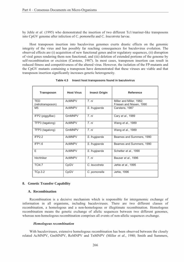
Part 4 – Consensus Documents on Micro-Organisms
266
by Jehle et al. (1995) who demonstrated the insertion of two different Tc1/mariner-like transposons into CpGV genome after infection of C. pomonella and C. leucotreta larvae.
Host transposon insertion into baculovirus genomes exerts drastic effects on the genomic integrity of the virus and has possibly far reaching consequences for baculovirus evolution. The observed effects are (i) acquisition of new functional genes and/or regulatory sequences, (ii) disruption of viral genes rendering them non functional, and (iii) deletion of extended portions of the genome by self-recombination or excision (Carstens, 1987). In most cases, transposon insertion can result in reduced fitness and competitiveness of the altered virus. However, the isolation of the FP-mutants and the CpGV mutants containing a transposon have demonstrated that these viruses are viable and that transposon insertion significantly increases genetic heterogeneity.
Table 4.3 Insect host transposons found in baculovirus
Transposon Host Virus Insect Origin Reference
TED(retrotransposon)
AcMNPV T. ni Miller and Miller, 1982; Friesen and Nissen, 1990
M5 AcMNPV S. frugiperda Carstens, 1987
IFP2 (piggyBac) GmMNPV T. ni Cary et al., 1989
TFP3 (tagalong) AcMNPV T. ni Wang et al., 1989
TFP3 (tagalong) GmMNPV T. ni Wang et al., 1989
IFP2.2 AcMNPV S. frugiperda Beames and Summers, 1990
IFP1.6 AcMNPV S. frugiperda Beames and Summers, 1990
E AcMNPV S. frugiperda Schetter et al., 1990
hitchhiker AcMNPV T. ni Bauser et al., 1996
TCl4.7 CpGV C. leucotreta Jehle et al., 1995
TCp.3.2 CpGV C. pomonella Jehle, 1996
8. Genetic Transfer Capability
A. Recombination
Recombination is a decisive mechanism which is responsible for intergenomic exchange of information in all organisms, including baculoviruses. There are two different classes of recombination, a homologous and a non-homologous or illegitimate recombination. Homologous recombination means the genetic exchange of allelic sequences between two different genomes, whereas non-homologous recombination comprises all events of non-allelic sequences exchange.
Homologous recombination
With baculoviruses, extensive homologous recombination has been observed between the closely related AcMNPV, GmMNPV, RoMNPV and TnMNPV (Miller et al., 1980; Smith and Summers,
Section 1 – Baculoviruses
267
1980; Croizier and Quiot, 1981). After coinfection of TN-368 cells of T. ni with AcMNPV and RoMNPV seven out of 100 isolated plaques where identified as recombinants, each with at least one crossing over event (Summers et al., 1980). Intraspecific recombination was also observed when SF-9 cells were co-infected with different pairs of AgMNPV genotypes (Croizier and Ribeiro, 1992). These results indicated that the frequency of recombination among highly homologous baculoviruses is considerable.
When Croizier et al. (1988) co-transfected larvae of Galleria mellonella with isolated DNA of RoMNPV and restriction fragments of AcMNPV, a very high number of RoMNPV recombinants but no parental RoMNPV genotypes were recovered. Since replication of AcMNPV in G. mellonella is much more effective than that of RoMNPV, it was suggested that the RoMNPV might have acquired an unknown replication advantage by recombination with AcMNPV DNA fragments.
A high recombination rate was also found when T. ni larvae were co-infected with wild-type AcMNPV and a polyhedrin-negative, lacZ-expressing AcMNPV mutant. A recombination frequency of at least 6.6% was estimated by plaque assays of hemolymph of co-infected larvae and scoring for recombinant plaques (Merrywheather et al., 1994).
Further evidence for extensive homologous recombination among different variants of the same virus species is based on the isolation and restriction mapping of wild-type virus mixtures. By the method of in vivo cloning Smith and Crook (1988; 1993) isolated a number of different ArGV genotypes which differed in a few restriction fragments. Based on comparative physical mapping, it was suggested that the diversity of these variants was partly caused by recombination during natural coinfections. A rapid replacement of two closely related parental SeMNPV variants (Se-US and Se-SP2) by a recombinant (Se-SUR1) which differed from the parental strains by several cross over events was observed after coinfection of S. exigua larvae. This result suggested selection advantage of the new recombinant over the parental strains, though significant differences in the biological activities (LD50 and LT50) were not observed (Munoz et al., 1997).
These findings underscore the fact that homologous recombination among virus variants is a normal process of genetic exchange which occurrs at a considerable frequency during in vitro and in vivo replication. The process of homologous recombination appears to be linked to and driven by the viral replicative machinery. Although the genetic factors involved in recombination have not been identified, it was suggested that genes involved in DNA replication and possibly hr regions may play a central role in the events (Martin and Weber, 1997).
Heterologous recombination
The probability of recombination between baculovirus sequences decreases drastically when the baculoviruses are less closely related. So far, recombination between different baculovirus species has only been observed in laboratory experiments where the generation of recombinants was biased by selection or replication advantages. For example, the identification of genes encoding host range factors or apoptosis inhibitor was achieved using this approach (see A 3.3 and A 4.3).
Heterologous recombination has been forced by co-transfection of restricted DNA or cloned DNA fragments with intact viruses or virus DNA. In an experiment addressing the possibility of interspecific recombination Roosien et al. (1986) co-transfected S. frugiperda cells with a polyhedrin negative AcMNPV mutant and a plasmid containing the polyhedrin gene of MbMNPV. AcMNPV mutants were isolated and contained the MbMNPV polyhedrin gene insertion at different, non-specific sites. However, these recombinants contained less virions and showed reduced infectivity to S. frugiperda cells than wild-type AcMNPV. Similar results were obtained when a polyhedrin minus
Part 4 – Consensus Documents on Micro-Organisms
268
AcMNPV mutant was co-transfected with a cloned polyhedrin gene of SfMNPV (Gonzales et al., 1989). The recombinant isolated from this experiment expressed less than 25% of the level of wild-type AcMNPV polyhedrin gene.
In general, the competitiveness and biological fitness of heterologous recombinants is lower than that of wild-type viruses. One reason is that by non-specific recombination events, coding or regulatory sequence functions might become impaired resulting in a less viable virus.
II. Human Health Considerations
Baculoviruses are naturally occuring pathogens of arthropods. Their host range is exclusively restricted to arthropods. No member of this virus family is infective to plants or vertebrates. Baculoviruses are ubiquitously present in the environment and have been used for biological insect control for more than 100 years. Circumstantial evidence for the safety of baculoviruses emerges from the history of contact between baculoviruses and humans without any detrimental effect.
During the 1970s, the US Environmental Protection Agency established the Guidance for Safety Testing of Baculoviruses, which also became a guideline of baculovirus safety tests in many other countries (Anonymous, 1975; Summers et al., 1975). This guidance included in vivo and in vitrosafety studies and was applied for commercial baculovirus insecticides, such as Helicoverpa zea SNPV (Elcar ), Orgyia pseudotsugata NPV (TM Biocontrol-1) and many others. Helicoverpa zeaSNPV was the first commercial baculovirus insecticide and is one of the most extensively tested entomopathogenic viruses (Ignoffo, 1975). Safety tests of more than 51 entomopathogenic viruses including more than 30 baculoviruses resulted in a long and complete safety record (extensively reviewed by Ignoffo, 1973; Burges et al.1980a, 1980b; Gröner, 1986). No adverse effect on human health has been observed in any of these investigations indicating that the use of baculovirus is safe and does not cause any health hazards.
A. Safety tests of baculoviruses included
In vitro and in vivo replication of baculoviruses in vertebrate and mammals
The possibility of replication of baculoviruses in vertebrates and mammals was investigated by challenging many vertebrate and human cell lines with OB and BV of many baculoviruses. Although virus uptake of these cells was frequently reported, no evidences of virus replication or cytopathological effects were observed. The few early reports, which stated baculovirus replication in vertebrate cell lines (Himeno et al., 1967; McIntosh and Shamy, 1980) could never be demonstrated or confirmed in other laboratories. After oral uptake of baculoviruses by man, mice, chickens, rabbit, pigs, and other mammals, no specific antibody production, which would indicate replication of the virus used to challenge the host, was observed (reviewed by Gröner, 1986). In contrast, a specific immunological response against CpGV was observed in woodmice (Apodemus sylvaticus) which were trapped in an apple orchard sprayed with CpGV. It is conceivable that an antigenic challenge may have occurred via the nasal mucous membrane, virus replication or a negative effect to the animals was not observed (Bailey and Hunter Fujita, 1987).
Using a recombinant AcMNPV containing the cat gene under the control of the Rous sarcoma virus terminal repeat promoter and the β-galactosidase gene under the control of the very late polyhedrin promoter reporter gene, expression was analysed in different invertebrate and vertebrate cell lines (Carbonell et al., 1985; Carbonell and Miller, 1987). No cat or β-galactosidase activity was detected in transfected mouse or human carcinoma cells. On the other hand, recent reports showed that recombinant AcMNPV virus is efficiently taken up by human hepatocytes via an endosomal pathway.
Section 1 – Baculoviruses
269
Recombinant AcMNPV carrying the Escherichia coli lacZ reporter gene under control of the Rous sarcoma virus promoter and mammalian RNA processing signals showed considerable expression levels in the human liver cell line HepG2, but at very low levels, or not at all, in cell lines from other tissues (Hofmann et al., 1995; Boyce and Bucher, 1996). Based on these findings it was suggested that baculovirus might be exploited for liver-directed gene therapy. From the view of baculovirus safety this results also show that careful attention has to be paid to the promoters used to control heterologous gene expression in recombinant baculoviruses.
Acute oral and intraperitoneal toxicity of mammal
Acute toxicity of Helicoverpa zea NPV has been tested in many mammals, e.g. rat, mouse, rabbit, guinea pig and man, at doses from 6 x 10⁹ to 3 x 10¹² OB/kg, which is up to 1000 times the average field rate per acre. Similar tests were conducted with TnSNPV, SeMNPV, AcMNPV, LdMNPV, CpGV and others (Ignoffo, 1975).
Subacute dietary-administration to mammals
In order to test potential subacute toxicity or pathogenicity Helicoverpa zea NPV was fed or subcutaneously injected to mice (about 5 x 10¹⁰ OB/kg of animal), rats (4 x 10⁹ to 4 x 10¹¹ OB/kg), beagle dogs (7 x 10⁹ OB/kg), rhesus monkeys (108 to 1.6 1010 OB/kg) and man (109 OB/day for 5 days). Similar tests were performed with rats for OpMNPV, LdMNPV and others (Ignoffo, 1975). Furthermore, health monitoring of workers who were involved in production of HzSNPV (for the viral pesticide) for extended time periods did not show any clinical symptomatology, nor any serological response or any indications that the virus is allergenic (Rogoff, 1975).
Eye- or skin-irritation to mammals
Eye irritation tests were negative, when 1 x 10⁵ to 20 x 105 OB/eye were applied to rabbit eyes. Skin irritation sensitivity tests were conducted with Helicoverpa zea NPV in rabbits, guinea pigs and man at doses of 103 to 106 OB/mm2 of skin. Dermal and eye applications have been also conducted with Neodiprion sertifer NPV, AcMNPV, SeMNPV and others without any adverse reactions (Ignoffo, 1975).
Cytogenetical implications as chromosomal aberrations or sister chromatid exchange to
mammalian cell lines
In studies on the activation of endogenous C-type retrovirus by baculoviruses in three mammalian cell lines (mouse, rat, and man) no activation of C-type retrovirus could be detected (Schmidt, 1981). When cultured frog cells (ICT-2A) were challenged with TnSNPV no virus multiplication and no chromosome aberrations were observed over a 4-week period of time (McIntosh, 1975). No chromosome aberrations in Chinese hamster cells, mouse cells after oral uptake of BV or OB of AcMNPV and MbMNPV was observed (Miltenburger, 1978).
Carcinogenicity, teratogenicity, mutagenicity to mammals.
Potential carcinogenicity of Helicoverpa NPV were conducted in mice 10 x 109 to 4 x 10¹¹
OB/kg) or rats (3.5 x 10¹² OB/kg), teratogenicity tests were performed in rats at a dose of 109 OB/kg. No evidence of carcinogenic or teratogenic effects was found (Ignoffo, 1975).
Part 4 – Consensus Documents on Micro-Organisms
270
III. Environmental and Agricultural Considerations
1. Natural Habitat and Geographic Distribution. Climatic Characteristics of Original Habitats
Baculoviruses are ubiquitous in the environment, their prevalence depending on the frequency of occurrence of their arthropod hosts that inhabit terrestrial and marine ecosystems. By 1986, about 1100 viruses known to infect insects had been described, more than 60% of them being baculoviruses (Martignoni and Iwai, 1986b). Natural epizootics and their survival under a variety of environmental / transport conditions provides for particularly high densities in diverse locations in the environment which may act as infection sources for pathogen multiplication (cabbage leafs, dust: Heimpel et al.,1973; Olofsson, 1988). Viruses from the same arthropod species from different geographic regions of the world may be as closely related as their hosts as demonstrated by the similarity of their restriction pattern (Vickers et al., 1991). However, different baculoviruses can also be isolated from the same species in one geographical region. Restriction endonuclease digest patterns can be used to analyse the genetic heterogeneity of baculovirus strains and of strains obtained from different geographical regions (Laitinen et al., 1996).
2. Significant Involvement in Environmental Processes, Including Biogeochemical Cycles and Potential for Production of Toxic Metabolites
The ecological significance of baculoviruses is characterised by their impact on the population dynamics of their arthropod hosts. As deduced from experiences with epizootics of insects they can be regarded as a regulator of their host population density and multiplication. No report on a potential contribution of baculoviruses to the extinction of insect species is available. This feature does not seem to depend only on the usual evolutionary adaptation of the interaction of pathogens with their hosts, but might be a more fundamental property of their life style. Through international trade, and in the course of applications of baculoviruses for pest control to a considerable extent, strains of baculoviruses have been moved around the globe without negative observations on local faunas. The ecological traits of baculoviruses (in particular virulence and dispersal characteristics) in their combination are an apparent biological constraint on excessive population increases, potentially endangering the basis of their existence, and the survival of their arthropod hosts.
With the impact on population densities of their hosts, a secondary effect on predators, parasites and hyperparasites is evidently connected.
3. Pathogenicity – Host range, Infectivity, Toxigenicity, Virulence, Vectors
A. Bioassays for the testing of infectivity and virulence
Parameters of baculovirus host range, infectivity, and virulence are tested by bioassays which measure the response (e.g. mortality) of test species (“subjects”) to different doses of defined virus preparations (“stimulus”). The in vivo bioassay is the only means by which the combined effect of all factors determining the potency of a baculovirus preparation can be measured. It is also used as a very sensitive method of monitoring the fate of baculoviruses in the field (see C.28). The titration of occlusion bodies in the light microscope offers a convenient method to compare different virus preparations with respect to the concentration of their (potentially) active structures. In principle, any comparisons between two virus preparations (e.g. a test substance with a standard preparation) requires that the dose response curve for both have the same shape, i.e. the bioactive component of the preparations is essentially similar. An understanding of the basic biological and statistical concepts of bioassays is important to fully realise their potential for analysing baculovirus interactions with other organisms and for improving their application (Hughes and Wood, 1986).
Section 1 – Baculoviruses
271
To obtain statistically meaningful results with the desired precision bioassays have to be carefully designed with respect to the number of subjects challenged. Optimised strategies depending on previous knowledge and model assumptions have been described (e.g. Hughes and Wood, 1986). Bioassays determining the yes or no (quantal) response of larval death through virus infection result in dose response relationships with the median effective dose ED50 (or EC50 if expressed in virus particles per ml) and the slope of the response curve as characteristic parameters. In addition to counting dead individuals, response data may also include the determination of particle titers in the cadavers. If the time course of larval death is recorded (time-mortality assays), as conveniently done in one experimental approach, monitoring results yield median lethal time LT50 (continuous exposure to virus) or median survival time ST50 (one inoculum at start of experiment), respectively.
To obtain meaningful and reproducible results other conditions of bioassays must be carefully observed. With respect to the test organism, uniform instar stages of larvae are used and reared under identical conditions (temperature, light regime, diet, group size). The ability to hatch insect larvae reproducibly with minor premature deaths (e.g. by bacterial infections) is one major problem for testing a great number of different test species, as required for host spectrum analysis.
Infection is frequently performed by applying the virus preparation to the surface of the natural or artificial diet - or by incorporating virus suspensions to the components of the artificial diet. Depending on the experimental design, the test species are kept on inoculated material for the duration of the assay - or the regime includes a transfer to a noninoculated diet, in order to better control inoculum and infection time. A large variety of experimental protocols for controlled virus inoculation have been described. They all suffer from inaccuracies, to which the variation of feeding behaviour of the insects also contributes (Hughes and Wood, 1986). Direct administering of virus suspensions by microinjection into the mouth of larvae, or the “measured drop feeding” method are ways to improve control over dose and inoculation time (Kunimi and Fuxa, 1996). The incubation of eggs with virus suspensions to infect neonate larvae seems to be a method for a well controlled infection of large numbers of insects at an identical developmental stage. Some of the latter methods are time consuming and have other limits (size of larvae for injection) which prevent their widespread use. Experimental protocols in general have to seek a sensible compromise between accuracy and practicability. Modifications of the assays have to accommodate the habits and peculiarities of the test species.
Data interpretation follows model assumptions. If the only interest is an estimate of the median effective dose and no inferences with respect to the absolute number of virus particles can be made, the probit model may be used. It assumes a log-normal distribution of individual effective doses (IED) for each host which invariably induces the response. The model refers to mechanistic interpretation of chemical interactions and assumes some co-operativity to be necessary for infection. The necessity to overcome host defenses can be considered as an interpretation of such assumptions in light of present knowledge. However, unlike chemicals, replicating entities such as bacteria and viruses can act independently to produce a response. In these cases, and if the hosts are reasonably uniform in their susceptibility to the pathogen, the dose response curve can be described and interpreted more adequately by an exponential model. The model describes infection as a stochastic process in which the lethal dose is the same for all individuals whereas the response rates reflect the chance to receive this dose. Essentially, one infectious virus entering the replicative stage is able to manifest the response according to such a model. Predictions of quantitative features of the infection process according to the model can be tested experimentally and results do not conflict with its interpretation, stressing the general applicability. (Hughes et al., 1984; Hughes and Wood, 1986; Ridout et al., 1993).
The method of “in vivo-cloning” takes advantage from the capacity of single viruses to initiate a productive infection. Administering virus titers for infection far below the LD50 results in a relatively large fraction of surviving larvae. The fraction of larvae, which died from replication of a single
Part 4 – Consensus Documents on Micro-Organisms
272
infectious unit can then be calculated (Huber and Hughes, 1984). This method, eventually used in two consecutive steps to enhance probability of the selection, is conveniently used to obtain genetically homogeneous virus preparations (Smith and Crook, 1988).
B. Survey of non-target effects
The specificity of the interactions of baculoviruses with arthropod and the corresponding narrow range of species which are susceptible to productive infection by a particular virus, is the basis of their innocuousness for a large spectrum of nontarget organisms (Gröner, 1986). In the course of safety assessments toxicity and pathology studies have been performed on mammals, birds and other wildlife animals including beneficial insects such as the honeybee and silkworm. The studies with animals other than arthropods up to 1986 were extensively reviewed by Gröner (1986). In the following, the studies are only briefly summarised because they conclusively demonstrated the absence of any adverse effects.
• Toxicity studies on mammals with a variety of NPVs, using the spectrum of application routes as conventionally tested for chemical pesticides, never resulted in any indications of toxicity or pathogenicity using doses 10 to 100 times the per-acre field rate equated to a 70-kg man. Also, no indications of teratogenic or carcinogenic effects in mammals were found with challenges of NPVs (section B).
• No side effects on birds after oral application and on aquatic vertebrate and invertebrate animals could be observed. Such laboratory studies were supplemented by some extensive monitoring for pathological effects of wildlife birds and mammals after (aerial) applications of different NPVs.
Recently, toxic effects were observed with a larval test which was considered to be useful for assessing adverse effects of microbial pest control agents on nontarget bivalves because of its simplicity, precision, and sensitivity. Larvae of the coot clam Mulinia lateralis were challenged for 48 h during the straight hinged stage of development with the LdMNPV at a density of 106 OB/ml. Mortalities observed were significantly higher than those obtained with a heat killed control. Similar mortality was observed with a 10-4 dilution of a commercial mosquito larvicide based on Bacillus thuringiensis ssp. israelensis (Bti). No effect was observed with a molluscidal strain of Bacillus alveior a broad-host-range fungal insect pathogen, Metarhizium anisopliae. Sodium dodecyl sulfate and a watersoluble fraction of a fuel oil were tested as a reference for comparison (Gormly et al., 1996).
• Consistent with their restriction of infectivity to the family or at least order of their original host, no infectivity or adverse effects on beneficial insects like pollinators (bees) have been observed. Baculovirus infection interferes with the multiplication of parasitoids within the same host. This interaction seems to be described most adequately as a competition for the same resource. No productive infection of the parasitoids is observed (see C.23/24).
• No genotoxic effect was observed by cytological studies after challenging mammals or cell cultures.
Similar studies with granuloviruses are smaller in number but gave the same results.
Small mammals or birds and also parasitoids feeding on insects infected by a baculovirus may take up and transport intact baculoviruses (e.g. in their digestive tract). Excretion of infective viruses may contribute to virus dispersal (C.26).
Section 1 – Baculoviruses
273
One experimental approach to use vectorised transport by honey bees as a nonintrusive means for virus dispersal has been field tested and indicates the perception of safety and non-target-innocuousness of baculoviruses. An applicator in a specifically designed substructure of a conventional beehive caused honey bees to take up (by surface contamination) and disseminate a talc formulation of HzSNPV into fields of Trifolium incarnatum. Increased HzSNPV induced mortality was observed in the clover fields foraged by the bees. A good persistence of baculovirus infectivity in honey was noted. An increased knowledge about the intersection of bee and target organism behaviour determining the virus transmission was considered to be essential, in order to further investigate the feasibility of the approach (Gross et al., 1994b).
C. Host range
The range of arthropods that can productively be infected by a given baculovirus can be regarded as its host range in a narrower sense. On one side, a limited host range is a feature of a rationally targeted biocontrol programme and an issue of biological safety. The control of defined pest species avoids side effects on the environment in general, and adverse effects on non-target insects including predatory and parasitoid species which provide additional control, in particular. On the other side, host range restrictions are also frequently regarded as an important application constraint if a complex of pest insects has to be managed. They determine limitations for the potential market volume of viral insecticides. Reports on particular host range testing and the selection strategies on virus strains for applications reflect this ambivalent view on host range restriction.
Among the approaches to relief application constraints for virus insecticides, prospects of modifying - and enhancing - host range by the use of molecular biology tools are being developed (Doyle et al., 1990; Thiem, 1997). The realisation of this objective will challenge the validity of host range testing procedures and the interpretation of their results even more than the use of genetically modified viruses does in general. An increasing knowledge about the molecular interactions involved in host range determination and limitations is the basis for such an approach. In comparison to the use of selective strategies on natural viruses such a basis may enable a design of modifications for more targeted and predictable effects on virus phenotype (Miller and Lu, 1997; see section A.3.3).
The genetic information of the virus genome, conserving evolutionary processes for perpetuation of its existence in time, is realised in the sequence of processes during an infectious cycle typically resulting in the release of high numbers of progeny virus from larval carcasses (see section A.3.1). Abortive or persistent infections indicate potential alternative strategies for persistence. A productive infection requires the specific interaction of host components with virus structure and virus encoded functions within a temporal and local arrangement of “key-lock fittings” in order to overcome host barriers and defenses and to exploit and transform host functions for viral replication. Overall, these specificities correlate with phylogenetic relationships, and the systematics of baculoviruses seems to represent the common evolutionary origin and concerted evolution with their arthropod hosts to some degree.
In detail however, evolution - and the variation and distribution of “keys” and “lockers” - follow different pathways in virus populations and in sexually reproducing animals. Genome modifications by mutations and genetic exchange are governed by different mechanisms and constraints. Thus, the degree of adaptation of a virus strain or “species” to a set of host organisms providing the requirements for productive interactions (specificities) presently is not easily discernible from its systematic position (which is defined by the degree of homology of (parts of) his genome to other viruses). In the future, the molecular analysis of the determinants (genes) for virus-host interactions and of their variability in natural populations may provide more power of inference with respect to the host range of a particular virus.
Part 4 – Consensus Documents on Micro-Organisms
274
So far, the testing with host species in vivo provides the relevant empirical basis for judgements about host ranges. Methods of data generation with respect to the following cannot follow harmonised and accepted internationally standardised procedures:
• The control of the homogeneity of the virus inoculum.
• The administration of the virus and the range of concentrations to be used.
• The spectrum of insect or other arthropod species and cell types to be included.
• The selection and control of end points (e.g. larval death, virus multiplication and yield, transcription of genes, DNA replication).
Parameters of influence on testing results include:
• The particular genotype of test species.
• The developmental stage of larvae during infection.
• The diet for host.
• Other conditions challenging the general stress tolerance of the test species (e.g. temperature, moisture).
Descriptions of a host range spectrum based on empirical testing will always suffer from ambiguities and incompleteness, due to apparent limitations in test species selection and experimental parameter variations. Studies of virus entry, DNA replication, transcription of genes and assembly of virus progeny together with the observation of cytopathic effects in cell cultures do substantially contribute to an understanding of host specificities at the level of molecular interactions (Miller and Lu, 1997). They complement but do not supplement, so far, in vivo testing of host range limitations (Danyluk and Maruniak, 1987; Castro et al., 1997). It may be stressed again, that an understanding of mechanisms is a prerequisite to improve inferences from genotype to host specificity phenotype.
Host range and parameters of virulence may be correlated to a different extent. An increase in virulence may appear as a host range extension in conventional testing using fixed doses of virus challenge. However, the spectrum of susceptible host species may not have changed to any extent. A maintenance of the order and quantitative relations of infectivity parameters (LD50s) would indicate an unchanged host range. A precise definition of experimental methods and end points clearly is a prerequisite for a common understanding of host range and virulence parameters.
The modification of virulence/host range by a selective procedure in the laboratory, cited by Federici and Maddox (1996) to exemplify a natural ability to manipulate host range, can be used to illustrate the importance of both, virulence and host range determinations. Martignoni and Iwai (1986a) used large doses of the MNPV of Orygia pseudotsugata (tussock moth, family: Lymantriidae) to sequentially infect Trichoplusia ni (cabbage looper, family: Noctuidae) over a period of 12 generations. In the course of these experiments, representing a selective pressure which in its extent will not occur in natural settings with any realistic probability, the virus adapted to the novel host, the cabbage looper. Low mortalities and minor levels of infection in most tissues at the beginning were converted to increasing levels of tissue infection and a complete adaptation of the virus to the novel host by the seventh generation. The passaged virus strain, if not selected from a heterogeneous inoculum, apparently accumulated some (set of) unknown mutation(s) and also had increased its
Section 1 – Baculoviruses
275
virulence for its original host, the tussock moth, by a factor of ten. This increase in virulence, however, would not in itself necessarily result in the expansion of the host range, as observed.
An overview of the host ranges of baculoviruses, as determined in cross infectivity studies in the laboratory is given by Gröner (1986). Results presented in tables are cited with the reservation that most studies did not control the identity with the inoculum virus after challenging of different hosts. Thus, the activation of a latent virus infection followed by its replication or the selective propagation of a minor contaminating virus type cannot be excluded in most cases. In general, NPVs infect only members of the genus, or in some cases the family, of their original host. A tendency that multiple embedded NPVs have a somewhat broader host range than SNPVs and an even more limited host range of granuloviruses was noted. Among the MNPVs, the prototype AcMNPV is known for its relatively broad host range and infectivity for more than ten families within the order Lepidoptera.
The identification of virus progeny by molecular methods is recommended and used as a standard tool for host range determinations in other publications as well. Predominantly a restricted range of infectivity is corroborated (Barber et al., 1993). Comprehensive studies on a broad selection of potential host insects were induced by the necessity to generate an adequate background for respective studies with genetically modified baculoviruses (Doyle et al., 1990; Doyle and Hirst, 1991). These studies resulted in the finding that MbMNPV is infective for both, butterflies and moths (Doyle and Hirst, 1991). MbMNPV of the cabbage moth Mamestra brassicae (Lepidoptera: Noctuidae) also infected the small tortoiseshell butterfly, Aglais urticae. However, a very high virus inoculum (106 OBs) was necessary for virus multiplication and larval death. MbMNPV also infects Cynthia cardui(painted lady) and Vanessa indica (Indian red admiral), also of the Nymphalidae, but does not seem to infect other butterflies of the families Satyridae, Lycaenidae, or Pieridae (Doyle et al., 1990). The cross infection studies showed a somewhat broader host range of MbMNPV than previously demonstrated. Other baculoviruses are also infective for Nymphalid butterflies.
The (quantitative) interpretation of biotest data with respect to potential consequences from exposing natural populations, has not been investigated in any detail but could be regarded as a key area of judgements about baculovirus biosafety (Doyle et al., 1990; Cory et al., 1997). It is conceivable that the non-susceptibility at lower doses might normally prevent epizootics from being initiated from weak cross infectivities, e.g. after spray applications for biological control. But an improved potential of extrapolation from infectivity studies with respect to a potential of virus transmissions in heterologous populations is highly desirable, to more clearly define relevant borders of host range. Data from laboratory bioassays might be poor estimates for the prediction of nontarget impacts.
A multiple-embedded nucleopolyhedrovirus, AnfaMNPV, isolated from the celery looper, Anagrapha falcipera in central Missouri seems to have the broadest host range reported so far (Hostetter and Puttler, 1991). 31 susceptible species belong to ten lepidopteran families, and in addition to Helicoverpa sp., Heliothis sp., and Spodoptera sp. include important pests like pink bollworm (Pectinophora gossypiella), fall webworm (Hyphantria cunea), cutworms (Noctuidae), velvet bean caterpillar (Anticarsia gemmatalis), codling moth (Cydia pomonella), navel orangeworm (Amyelois transitella), and the diamondback moth (Plutella xylostella). As compared to AcMNPV, known for its relatively broad host range, the virus demonstrated equal virulence for species differing in susceptibility for AcMNPV by one order of magnitude. Relatively narrow criteria were used for differentiation of susceptibility (max. 250 OB/mm2 of diet). The list of nonsusceptible species as published (containing also Coleoptera and Diptera) might thus include some species which are susceptible, if challenged at higher doses. Propagation of virulent AfMNPV in tobacco hornworm (Manduca sexta) as untypical host demonstrated extensive possibilities for production in a variety of hosts and systems. Virulence against a great number of cosmopolitan economically important insect
Part 4 – Consensus Documents on Micro-Organisms
276
species was considered an encouraging result in this report and the virus became the first baculovirus patented by the US Government Patent and Trademark Office.
D. Variation of virulence and host range
By describing interactions between biological entities, virulence and host range are prone to modification by natural variability and by a variety of external “environmental” factors. Mutagenesis, recombination, and the (selective) propagation in different hosts have been analysed as causes of shifts in host range and virulence of baculoviruses, in the laboratory (sections A.3.3; A.6-8.). The same mechanisms are acting in natural or managed environments, however to an extent unknown in any detail and responding to different selection conditions. In the following, emphasis is given to some factors modulating insect responses to viral challenges that have not been described in other sections.
External (environmental) factors modulating insect response to baculoviruses
Chemicals
An additive or synergistic effect of chemicals on baculovirus infectivity has been exploited for control strategies, for example, by the use of combinations with insecticides or the addition of particular radiation protectants (section A.6).
Host plants
The plant material on which larvae are feeding may have a significant influence on their susceptibility for viral infections. Gypsy moths, feeding on Quercus rubra (red oak) or Acer rubrum (red maple) demonstrated a higher level of tolerance when infected with LdMNPV than larvae feeding on Populus tremuloides (quaking aspen) or Pinus rigida (pitch pine). The enhanced tolerance was correlated with an increased acidity and hydrolysable tannin content of leaf material (Keating and Yendol, 1987; Keating et al., 1988). Different tolerances for Spodoptera littoralis NPV were also noted in the cotton leaf worm on castor bean, alfalfa, mulberry, cotton and potato. LD50 values differed by a factor of about 3 between castor bean (the most “protective” feeding source) and potatoes, and the LT50 was also reduced by more than 10% on the latter plant (Santiago-Alvarez and Ortiz-Garcia, 1992).
A different approach was followed by Rabindra et al. (1994), who investigated the influence of plant surface environments on the virulence of Heliothis armigera SNPV. The larvae of the American bollworm were challenged with identical (microscopically controlled) titers of virus suspensions which had been exposed to the surfaces of 5 host plants (chickpea (Cicer arietinum), pigeon pea([syn. dhal] Cajanus cajan), lablab bean (Dolichos lablab), sunflower, and cotton). Bioassays used the leaf dip method with shoots of chickpea as identical feeding material for the test species. The cotton leaf surface was the most detrimental for virus activity resulting in an increase of more than three orders of magnitude in the LC50 value in comparison to lablab bean -exposed viruses (5 x 10⁶ OB/ml and 1.4 x 10³ OB/ml, respectively). Lablab bean even had some protective or stimulating effect on virulence in comparison to untreated NPV preparations (LC50 = 8 x 10⁴ OB/ml). Median lethal time measurements corresponded with these observations. A mortality time expanded by 25% after cotton leaf - exposure in comparison to lablab bean exposed virus was observed. This experimental approach was also used to determine the effect of adjuvants added to the virus preparations because the study together with others indicates the need to develop suitable formulations which could protect the virus from inactivating factors on plant surfaces.
Section 1 – Baculoviruses
277
These examples demonstrate the modulation of baculovirus virulence by the plant environment on which target insect larvae are feeding. This environment either indirectly alters insect response and tolerance, or interferes with the first steps of virus infection through variation of the milieu of the midgut lumen (e.g. pH), and/or directly modulates the specific activity (the probability of an OB in the light microscope to enter the replicative state) of virus preparations. An understanding of these kinds of interferences is valuable to improve formulations for insect control and to adapt methods of in vivobaculovirus production. It can also be used to reassess control strategies if applied to novel plant varieties (Beach and Todd, 1988). In an advanced stage of development of modelling the variability introduced by the respective plant canopy may be included. (Foster et al., 1992).
Population density
A variety of environmental factors such as temperature, light, nutrition, and humidity may compromise the capability of insects to resist baculovirus infections (Briese, 1986). Among these, rearing density has a distinct effect on their susceptibility to viral infection. Together with other phenotypic modifications (development time, weight at moulting, degree of melanisation), enhanced susceptibility is observed as a reaction to the stress of high population density of lepidopteran larvae (Goulson and Cory, 1995b). This was interpreted as a consequence of the adaptive response of accelerated development at the expense of larval weight, which compromised the ability to express resistance functions. Interestingly, larvae which were reared singly exhibited many of the same characteristics, including enhanced susceptibility to virus infection. The benefit of spending resources for resistance development may be low, if a low density of conspecifics reduces infection risk. This phenotypic variation is among the factors modulating the relationship between virus density and disease transmission.
Other infectious agents, parasitoids
The exposition of insects with different infectious agents or parasitoids is a different kind of stress, eventually enhancing baculovirus induced mortality (C.24). Superinfection of insects with a latent or persistent infection may result in larval death due to the propagation of the latent virus (see A.4.6).
Insect resistance and immunity
The varying degree of tolerance of insect larvae for baculovirus infections at different instar stages, usually increasing with age and stage of development, is but one observation suggesting the existence of particular mechanisms of pathogen tolerance (Mikhailov et al., 1992; Engelhard and Volkman, 1995). To assess the short-term and long-term effectivity of control strategies with baculovirus insecticides an understanding of the adaptive potential of insect populations to baculovirus infections is of paramount importance. In a review Briese gives an account on developmental and environmental factors affecting resistance (1986), the knowledge about defence mechanisms and the genetic factors conferring increased virus tolerance. Dominant and recessive autosomal genes or a multigene family may form the genetic basis of tolerance in insects. E.g. McIntosh and Ignoffo (1989) demonstrated that the resistance of Helicoverpa subflexa against Helicoverpa zea NPV (HzSNPV) appears to be controlled by a single non sex-linked gene.
Observations of markedly expressed virus resistance are restricted to laboratory observations of insect populations obtained under high artificial selective pressure of virus challenges, for example, by propagating the surviving fraction of infections with an LD80 in several subsequent generations. Somewhat conflicting results with respect to the success of selection strategies may either be caused by the general potentials of the insect species under investigation or by the particular genotype of the
Part 4 – Consensus Documents on Micro-Organisms
278
strain as most obvious reasons (e.g. Kaomini and Roush, 1988). It is a general objection against the predictive value of laboratory selections, that insect cultures in the laboratory represent a minor fraction of the genetic heterogeneity of natural populations. The lack of documented cases of resistance in field populations of insects under control of a virus insecticide may reflect their less frequent and extensive use in comparison to chemicals, which have induced an exponentially increasing rate of resistance developments in insect populations.
There is some circumstantial evidence that the spectrum of tolerance and respective gene frequencies in natural populations may have been biased by natural virus epizootics or the application of virus insecticides (Fuxa et al., 1988). Laboratory selections in colonies of velvetbean caterpillar (Anticarsia gemmatalis) from the US and Brazil demonstrated the achievement of a significantly higher level (1000x) of resistance to AgMNPV after 13 - 15 generations in the Brazilian colonies than in the colony from Lousiana, the resistance of which levelled off at a ratio of 5x after 4 generations (Abot et al., 1995). AgMNPV, is used for biological control of the velvetbean caterpillar in soybean on a large scale in Brazil (see A.6.4).
Alleviation of the selection pressure by virus challenges seems to favour more competitive sensitive wild-type genotypes of insects, which are superior in their reproduction without the virus selection pressure (Fuxa and Richter, 1989). Such observations correspond to a more general perception that some cost in reduced fitness has to be paid for the expression of disease resistance. This hypothesis however, has not been tested rigorously in many cases (Gemmill and Read, 1998). Genes for virus resistance confer pleiotropic phenotypic effects including increased susceptibility to chemical insecticides (Fuxa and Richter, 1990). The exclusive restriction to the oral application route of the increased virus tolerance and enhanced insecticide susceptibility phenotypes indicates a correlation of modifications to components of the midgut interfering with early stages of virus infection.
Using the genetically modified virus AcMNPV-hsp70/lacZ for the monitoring of virus activity by the lac-Z reporter gene, the physiological basis of resistance of Helicoverpa zea against AcMNPV was analysed by Washburn et al. (1996). The larval cells were actually very susceptible to AcMNPV infection, but infected cells were encapsulated by hemocytes and subsequently cleared from the midgut lumen. Cellular immune responses of larvae seem to be a significant factor in preventing the spread of the infection and a determinant of the functional host range of baculoviruses. Based on further experiments including chemical and biological immune suppression, it was suggested to use modified viruses to express immunosuppressive genes in order to compensate for this defence mechanism.
4. Interactions with and Effects on Other Organisms in the Environment
Direct effects of baculoviruses on other organisms in the environment are restricted to their host range, and most have been observed only in their original host or in the target species of biocontrol applications. Baculovirus infection interferes with other pathogens or parasitoids feeding on a shared host. Observations on the synergistic or additive effect of particular crossinfections with other pathogens as well as interferences with parasitoids have been summarised by Harper (1986). Section A 4.2 gives some account to the synergistic co-operation between some baculoviruses. Some of such pathogen combinations have been used in field experiments to improve control of pest species.
The analysis of the interference with parasitoid developments is used to assess the possibility of adverse effects on populations providing additional natural control of pest species. The observed detrimental effect on parasitoid development, fitness or reproductive success as well as an enhanced sensitivity of parasitised insect larvae for baculovirus infection can most adequately be described with
Section 1 – Baculoviruses
279
the interspecific competition for a limited resource (Cossentine and Lewis, 1988; Hochberg, 1991; Al Fazairy et al., 1993; Nakai and Kunimi, 1997). The relative timings of baculovirus infection and parasitoid emergence determine the degree of interference to a large degree. No productive infection of parasitoids was observed but frequently a large number of viral occlusion bodies is present in their midgut lumen, indicating a potential mechanism of virus dispersal. A reduced level of a parasitoid population has been observed after aerial LdMNPV application (Webb et al., 1989).
5. Ability to Form Survival Structures
The structure of baculovirus occlusion bodies provides for their potential to persist in the environment in a variety of abiotic and biotic conditions (A.4.1/4.7). This feature is described as a key factor dominating the dynamics of virus transmission and a prerequisite of baculovirus epizootiology by Evans (1986). Viability may extend for more than 40 years in forest soil. No special survival structures are formed.
6. Routes of Dissemination, Physical or Biological
A. Physical dispersal
Transport and dispersal of baculoviruses by wind or water is considered to be of minor importance for the natural spread of baculovirus infections. Its contribution to mechanisms of primary dispersal in natural epizootics is not known in detail (Briese, 1986). In field experiments, testing the spatial spread of a baculovirus, ballooning of first instar larvae of the gypsy moth Lymantria disparwas a good predictor of viral spread in the first few weeks. The scale of spread and its lack of directionality at later times however, did not match the results of a mathematical modelling even when primary dispersal by ballooning and short distance larval dispersal with a high rate of disease transmission were included. Circumstantial evidence suggested that autonomous dispersal by wind of occlusion bodies did not significantly contribute to dispersal. Parasitoid vectoring of viruses was discussed as the potential relevant additional dispersal mechanism (whereas vectoring by other animals was not considered (Dwyer and Elkinton, 1995). Evidence for a dispersal of NPV from soil to pine foliage in dust was presented as a likely explanation for the observed distribution of diseased larval colonies near a forest road by Olofsson (1988).
Aerial dispersal is the predominant mechanism of distribution for insect control applications; techniques and timing have to consider meteorological conditions and other agricultural procedures like irrigation for optimal distribution (Young, 1990; Payne et al., 1996).
Reminiscent of traditional testing procedures for the prediction of exposition rates with chemicals, the leaching behaviour of baculoviruses in natural soil cores and columns filled with soil or sand has been tested to evaluate eventual rates of exposition of the groundwater table. Consistently, some experiments demonstrated the retention of baculoviruses in soil to be comparable or superior to that of other Viruses (Polio virus and bacteriophage f2 of Escherichia coli were used for comparison). Soil with higher levels of organic matter was less efficient than sand for the reference viruses but not for the baculoviruses (NPVs and GVs). Results were independent from the particular composition of percolating water. Lysimeter studies with baculoviruses did not result in any observation of positive samples of leachate taken at a depth of 1.5 m during 7 months of monitoring. The concentration of viruses applied in this experiment resulted in a surface exposition 3 - 4 orders of magnitude above concentrations used in agricultural applications (1014 particles of a Granulovirus in 20 L of tap water applied to a surface of 0.8 m2). A good retention of baculoviruses by soils was concluded and this property was tentatively attributed to the particular protein envelope of virus particles consisting of polyhedrin and granulin, respectively (Lopez-Pila, 1988).
Part 4 – Consensus Documents on Micro-Organisms
280
The behavioural shift of some insect larvae following baculovirus infection (i.e. the movement to higher and exposed positions on host plants: “Wipfelkrankheit”) effects the physical distribution of baculovirus particles following death. It might be an evolutionary adaptation of the virus to increase horizontal transmission, e.g. by contaminating more foliage following rainfall (Vasconcelos et al.,1996a; Goulson, 1997).
B. Vectorised dispersal by diseased larvae, predators and parasitoids
Virus infected larvae are an effective dispersal agent even before larval death and carcass lysis, which results in very high inoculum density. Viable virus particles are also dispersed in the environment through either defecation or regurgitation during late stages of infection (Vasconcelos, 1996). Cannibalism of infected larvae may be another possibility if it occurs at any relevant frequency. Transport and dispersal of virus particles also occurs by predators feeding on diseased larvae or by parasitoids ovipositioning on and developing within larvae (Briese, 1986; Boucias et al., 1987; Vasconcelos et al., 1996b). The viruses remain viable after passage of the alimentary canal of vertebrates and invertebrates, however to a widely divergent extent.
With respect to the speed and distance of the transfer of infectious viruses, the dispersal by birds deserves some special interest. Birds may take up infected or dead larvae directly or feed on non vertebrate or small vertebrate predators. Droppings may contain up to some 107 polyhedra. But the contribution of particular dispersal routes and mechanisms to virus epizootics is difficult to assess conclusively. Even a coincidence of novel areas with infected larvae and viruses in bird droppings is only circumstantial evidence of a significant contribution of avian transport to virus epizootics (Entwistle et al., 1977; Buse, 1977; Cory et al., 1988; Entwistle et al., 1993). Novel baculovirus infections appearing in Gilpinia hercyniae (European spruce sawfly) in a previously unexposed geographical area at a time, before any virus could be monitored in avian droppings, suggests a different mode of dispersal (Buse, 1977). Even more recent experiments including trials with different virus applications at different locations and the identification of virus types in bird droppings by molecular restriction-hybridisation methods must close with the statement, that “the actual role of birds and many other biotic dispersal agents in the spread of baculoviruses and other micro-organisms remains to be demonstrated” (Entwistle et al., 1993). Clearly, the knowledge of the intersection in space and time of insect behaviour and development and the ecological habit of particular bird species (including feeding - and defecation - behaviour and territoriality) must be included in such investigations.
Such intersections are also determinants of success for any approaches using insects as biotic dispersal agents for insect control. These experiments also can be regarded as a demonstration of a potential natural dispersal role of the engaged species (Biever et al., 1982; Young and Yearian, 1990, 1992; Gross et al., 1994b - see C.23.2). Any intentions to surmount the demonstrative character of these experiments in order to biologise application strategies for baculovirus insecticides would require the input of increased knowledge. It remains speculative whether such a manipulation of insect and baculovirus ecology for efficient pest control can be achieved in a practicable and effective manner.
7. Containment and Decontamination
Special containment conditions in order to protect workers and the environment from baculoviruses are not usually required. The establishment of confinement measures is determined by the need to protect insect and cell cultures and the experiment from unintended infections. In part, depending on laboratory experiences with outbreaks of virosis in insectaries, the extension of technical installations and working procedures is adapted to these protection objectives. Small particle size and
Section 1 – Baculoviruses
281
persistence of baculoviruses represent a challenge for effective measures. The performance of work with baculoviruses has to follow the principles of good microbiological practice. Local separation of cell culture and insect work is a minimum requirement. To change laboratory coats when beginning or leaving work with baculoviruses seems to be advisable.
Heating can be used for decontamination of laboratory material and media. Baculoviruses are inactivated at temperatures and times significantly below conventional microbiological sterilisation conditions (Martignoni and Iwai, 1977). Sodium hypochlorite and formaldehyde can be used for chemical decontamination. Sodium dodecyl sulfate was also tested for egg surface sterilisation to prevent ex ovarial (vertical) transmission. A lower impact on moth rearing and sufficient effectiveness of inactivation were noted (Ilsley et al., 1980).
No decontamination procedure seems to be recommendable for environmental contaminations, e.g. by spills from baculovirus insecticide containers. In fact, any procedure would compromise environmental quality to a greater extent than the baculovirus preparation. Soil decontamination by formaldehyde treatment as used after the first field trial with a genetically modified baculovirus or similar treatments will eventually be used according to the respective risk perception.
8. Description of Detection and Monitoring Techniques, Including Specificity, Sensitivity, and Reliability
A. Detection
Detection of baculoviruses in the environment is frequently performed by collecting (diseased or dead) larvae and making microscopic observations of stained baculoviruses. While this technique is adequate to monitor an infectious process in an insect population, other techniques are required to analyse the abundance and activity of infectious particles in the environment. Methods may consist of biotests by feeding target larvae with potentially exposed (plant) material. In order to quantify virus particles and to additionally determine their infectivity, virus particles are separated from the habitat matrix by washing or sonication. Virus particles collected by centrifugation are stained and counted by microscopy (Taverner and Connor, 1992; Carruthers et al., 1988). Subsequent biotesting is then performed by applying an aquaous suspension to the insect diet.
Biotesting may amplify rather low concentrations of viruses, and does not seem to generally be inferior in its sensitivity in comparison to molecular methods which can be employed to directly detect baculovirus sequences by probe hybridisations or by amplifying specific sequences via PCR. The potential of these methods has not been evaluated for the monitoring of field trials in any significant extent. A method to detect and identify baculoviruses by a combination of PCR (amplifying a homologous region of the polyhedrin gene) and subsequent differentiation by restriction analysis has been described for 8 baculoviruses (De Moraes and Maruniak, 1997). However, PCR does not differentiate between viable and non-viable viruses.
B. Identification
For the identification and differentiation of Baculovirus species/strains, the restriction analysis and/or probe hybridisations has become an established method (Trzebitzky et al., 1988; Bensimon etal., 1987; Doyle et al., 1990; Barber et al., 1993; Entwistle et al., 1993; Hughes et al., 1997). The feasibility of the approach for ecological investigations was conclusively demonstrated by Laitinen etal. (1996) who used heterogeneities in restriction endonuclease patterns among geographic isolates to assess the origin and spread of genotypes. The number of restriction enzymes used and the cutting frequency determine the degree of differentiation of divergent genotypes. A quantification of the
Part 4 – Consensus Documents on Micro-Organisms
282
composition of heterogeneous mixtures is difficult to achieve by the analysis of banding heterogeneities and submolar bands. In vivo cloning of the genotypes of the mixture and quantification of their frequency would be a labour intensive solution.
In recent years, the PCR became an efficient tool with high sensitivity and specificity for the identification and diagnostics of micro-organisms. Primer specificities and reaction conditions determine the amplification of specific DNA fragments. De Moraes and Maruniak (1997) adopted this technique to amplify a highly conserved region of 575 bp within the polyhedrin gene of different NPVs with a specifically designed pair of primers. Restriction endonuclease digestion of the PCR product resulted in specific restriction patterns of the 8 analysed nucleopolyhedroviruses (Anagrapha falcifera NPV, AcMNPV, AgMNPV, BmNPV, and HzSNPV, OpMNPV, SfMNPV, and SeMNPV).
A similar approach was chosen to detect low levels of Spodoptera littoralis NPV from viral occlusion bodies and from infected host larvae and to differentiate between S. littoralis NPV and AcMNPV (Faktor and Raviv, 1996). These experiments demonstrate that PCR-based methods are useful for rapid identification and allow cost effective and sensitive monitoring of wild-type as well as genetically engineered baculoviruses.
Section 1 – Baculoviruses
283
REFERENCES
Abot, A.R., Moscardi, F., Fuxa, J.R., Sosa Gomez, D.R., and Richter, A.R. 1995. Susceptibility of populations of Anticarsia gemmatalis (Lepidoptera: Noctuidae) from Brazil and the United States to a nuclear polyhedrosis virus. Journal of Entomological Science, 30, 62-69.
Adams, J.R. and Bonami, J.R. 1991. “Atlas of Invertebrate Viruses.”, Boca Raton, Florida.
Ahrens, C.H., Russell, R., Funk, C.J., Evans, J.T., Harwood, S.H. and, Rohrmann, G.F. 1997. The sequence of Orgyia pseudotsugata multinucleocapsid nuclear polyhedrosis virus genome. Virology 229, 381-399.
Al Fazairy, A.A.M., Hegazi, E.M., Karam, H.H. and, Abo Abd Allah, L.M. 1993. Differential susceptibility of parasitized and unparasitized larvae of Spodoptera littoralis (Boisd.) (Lepidoptera: Noctuidae) to its nuclear polyhedrosis virus. Insect Science and its Application 14, 361-365.
Andermatt, M. 1986. Field efficacy and environmental persistence of the granulosis viruses of the summer fruit tortrix, Adoxophyes orana. IOBC/WPRS Bulletin. 14, 73-74.
Andersen, J., Edwards, D.F., Hazel, W.J., Levin, M.A., Pilsucki R.W., Schneider, W.R. and, Sjoblad, R.D. (WR Schneider, Ed.). 1989. Subdivision M of the Pesticide Testing Guidelines. Microbial and Biochemical Pest Control Agents. US Environmental Protection Agency: Office of Pesticide and Toxic Substances
Anderson, R.M. and May, R.M. 1980. Infectious cycles of forest insects. Science 210, 658-661
Anderson, R.M. and May, R.M. 1981. The population dynamics of microparasites and their invertebrate hosts. Philosophical Transactions of the Royal Society of London, B 291, 451-524
Anonymous. 1975. Guidance for safety testing of baculoviruses. pp. 179-184. In: Baculoviruses for Insect Pest Control: Safety Considerations, (M Summers, R Engler, LA Falcon, P Vail, Eds.). American Society of Microbiology, Washington, D. C.
Anuradha, S., Ramakrishnan, N., Srinivasan. 1995. Restriction map of genomic DNA of a nuclear polyhedrosis virus isolated from Amsacta albistriga (Walker). Proceedings of the Indian National Science Academy. Part B, Biological Sciences 61, 119-125.
Arif, B.M., Kuzio, J., Faulkner, P. and, Doerfler, W. 1984. The genome of Choristoneura fumiferananuclear polyhedrosis virus: molecular cloning and mapping of the EcoRI, BamHI, SmaI, XbaIand BglI restriction sites. Virus Research 1, 605-614.
Arne, C.N. and Nordin, G.L. 1995. Enhancement of indices of viral infection by simultaneously administering Helicoverpa zea and Autographa californica nuclear polyhedrosis viruses to larval Helicoverpa zea (Boddie). Journal of Invertebrate Pathology 66, 18-24.
Part 4 – Consensus Documents on Micro-Organisms
284
Audemard, H., Burgerjon, A., Baudry, O., Bergere, D., Breniaux, D., Delay, JC., Desvaux, R., Formantin, C., Gendrier, J.P. and, Tarbouriech, M.F. 1992. Evaluation of 100 trials of carpovirusine, a granulosis virus preparation to control codling moth Cydia pomonella L. in apple orchards. Acta Phytopathologica et Entomologica Hungarica 27, 45-49.
Ayres, M.D., Howard, S.C., Kuzio, J., Lopez Ferber, M. and, Possee, R.D. 1994. The complete DNA sequence of Autographa californica nuclear polyhedrosis virus. Virology 202, 586-605.
Bailley, M.J. and Hunter Fujita, F.R. 1987. Specific immunological response against the granulosis virus of the codling moth (Cydia pomonella) in woodmice (Apodemus sylvaticus): field observations. Annales of applied Biology 111, 649-660.
Barber, K.N., Kaupp, W.J. and, Holmes, S.B. 1993. Specificity testing of the nuclear polyhedrosis virus of the gypsy moth, Lymantria dispar (L.) (Lepidoptera: Lymantriidae). Canadian Entomologist 125, 1055-1066.
Bauser, C.A., Elick, T.A. and, Fraser, M.J. 1996. Characterization of hitchhiker, a transposon insertion frequently associated with baculovirus FP mutants derived upon passage in the TN- 368 cell line. Virology 216, 235-237.
Beach, R.M. and Todd, J.W. 1988. Discrete and interactive effects of plant resistance and nuclear polyhedrosis viruses for suppression of soybean looper and velvetbean caterpillar (Lepidoptera: Noctuidae) on soybean. Journal of Economic Entomology 81(2), 684-691.
Beames, B., and Summers, M.D. 1990. Sequence comparison of cellular and viral copies of host cell DNA insertions found in Autographa californica nuclear polyhedrosis virus. Virology 174, 354-363.
Begon, M. and Bowers, R.G. 1994. Host-host-pathogen models and microbial pest control: the effect of host self-regulation. Journal of Theoretical Biology 169, 275-287.
Begon, M., Sait, S.M. and, Thompson, D.J. 1996. Predator-prey cycles with period shifts between two- and three-species systems. Nature 381, 311-315.
Beidler, D.R., Tewari, M., Friesen, P.D., Poirier, G. and, Dixit, V.M. 1995. The baculovirus p35 protein inhibits Fas- and tumor necrosis factor- induced apoptosis. Journal of Biological Chemistry 270, 16526-16528.
Bell, M.R. and Hayes, J.L. 1994. Areawide management of cotton bollworm and tobacco budworm (Lepidoptera: Noctuidae) through application of a nuclear polyhedrosis virus on early-season alternate hosts. Journal of Economic Entomology 87, 53-57.
Bensimon, A., Zinger, S., Gerassi, E., Hauschner, A., Harpaz, I. and, Sela, I. 1987. "Dark cheeks," a lethal disease of locust provoked by a lepidopterous baculovirus. Journal of Invertebrate Pathology 50(3) 254-260.
Benz, G.A. 1986. Introduction: Historical Perspectives. pp. 1-36. In: The Biology of Baculoviruses. Vol I: Biological Properties and Molecular Biology, (RR Granados, BA Federici, Eds). CRC Press, Boca Raton, USA.
Section 1 – Baculoviruses
285
Berryman, A.A. 1997. What causes population cycles of forest Lepidoptera? Trends in Ecology and Evolution 11, 28-32
Biever, K.D., Andrews, P.L. and, Andrews, P.A. 1982. Use of a predator, Podisus maculiventris, to distribute virus and initiate epizootics. Journal of Economic Entomology 75, 150-152.
Biever, K.D. and Wilkinson, J.D. 1978. A stress-induced granulosis virus of Pieris rapae.Environmental Entomology 7, 572-573.
Bilimoria, S.L. 1991. The biology of nuclear polyhedrosis viruses. pp. 1-72. In: Viruses of Invertebrates, (E Kurstak, Ed.). Marcel Dekker Inc., New York.
Birnbaum, M.J., Clem, R.J. and, Miller, L.K. 1994. An apoptosis-inhibiting gene from a nuclear polyhedrosis virus encoding a polypeptide with Cys/His sequence motifs. Journal of Virology 68, 2521-2528.
Bishop, D.H.L., Entwistle, P.F., Cameron, I.R., Allen, C.J. and, Possee, R.D. 1988a. Genetically engineered baculovirus insecticides. Aspects of Applied Biology 17, 385-395.
Bishop, D.H.L., Entwistle, P.F., Cameron, I.R., Allen, C.J. and, Possee, R.D. 1988b. Field trials of genetically-engineered baculovirus insecticides. pp. 143-179. In: The Release of Genetically Engineered Micro-Organisms, (M Sussman, CH Collins, FA Skinner, DE Stewart-Tull, Eds.). Academic Press, London.
Bjornson, R.M. and Rohrmann, G.F. 1992. Nucleotide sequence of the polyhedron envelope protein gene region of the Lymantria dispar nuclear polyhdrosis virus. Journal of General Virology 73, 1499-1504.
Black, B.C., Brennan, L.A., Dierks, P.M. and Gard I.E. 1997. Commercialization of Baculoviral Insecticides. pp. 341-387. In: The Baculoviruses, (LK Miller, Ed.). Plenum Press, New York and London.
Blissard, G.W. 1996. Baculovirus-insect cell interactions. Cytotechnology 20, 73-93.
Bohmfalk, G.T. 1986. Practical factors influencing the utilization of baculoviruses as pesticides. pp. 223-235. In: The Biology of Baculoviruses. Vol. II: Practical Applications for Insect Control, (RR Granados, BA Federici, Eds.). CRC Press, Boca Raton, USA.
Bonning, B.C., Hammock, B.D. and, Tombs, M.P. 1992. Development and potential of genetically engineered viral insecticides. Biotechnology and genetic engineering reviews 10, 455-489.
Boucias, D.G., Abbas, M.S.T., Rathbone, L. and, Hostettler, N. 1987. Predators as potential dispersal agents of the nuclear polyhedrosis virus of Anticarsia gemmatalis (Lep.: Noctuidae) in soybean. Entomophaga 32(1), 97-108.
Bowers, R.G. and Begon, M. 1991. A host-host-pathogen model with free-living infective stages, applicable to microbial pest control. J.theor.Biol. 148, 305-329
Bowers, R.G., Begon, M. and, Hodgkinson, D.E. 1993. Host-pathogen population cycles in forest insects? Lessons from simple models reconsidered. Oikos 67, 529-538
Part 4 – Consensus Documents on Micro-Organisms
286
Boyce, F.M. and Bucher, N.L. 1996. Baculovirus-mediated gene transfer into mammalian cells. Proceedings of the National Academy of Sciences USA 93, 2348-52.
Brassel, J. and Benz, G. 1979. Selection of a strain of the granulosis virus of the codling moth with improved resistance against artificial ultraviolet radiation and sunlight. Journal of Invertebrate Pathology 33, 358-363.
Briese, D.T. 1986. Insect resistance to Baculoviruses. pp. 237-264. In: The Biology of Baculoviruses. Vol. II: Practical Applications for Insect Control, (RR Granados, BA Federici, Eds.). CRC Press, Boca Raton, USA.
Bump, N.J., Hackett, M., Hugunin, M., Seshagiri, S., Brady, K., Chen, P., Ferenz, C., Franklin, S., Ghayur, T., Ping, L., Licari, P., Mankovich, J., Shi, L., Greenberg, A.H., Miller, L.K;. and, Wong, W.W. 1995. Inhibition of ICE family proteases by baculovirus antiapoptotic protein p35. Science 269, 1885-1888.
Burand, J.P. 1991. Molecular biology of the Hz-1 and Oryctes Nonoccluded baculovirus. pp. 111-126. In: Viruses of Invertebrates, (E Kurstak, Ed.). Marcel Dekker, New York.
Burges, H.D., Croizier, G., Huber, J. 1980a. A review of safety tests on baculoviruses. Entomophaga 25, 329-339.
Burges, H.D., Huber, J. and, Croizier, G. 1980b. Guidelines for safety tests on insect viruses. Entomophaga 25, 341-347.
Buse, A. 1977. The importance of birds in the dispersal of nuclear polyhedrosis virus of European spruce sawfly, Gilpinia hercyniae (Hymenoptera: Diprionidae) in mid-Wales. Entomologia Experimentalis et Applicata 22, 191-199.
Caballero, P., Zuidema, D., Santiago Alvarez, C. and, Vlak, J.M. 1992. Biochemical and biological characterization of four isolates of Spodoptera exigua nuclear polyhedrosis virus. Biocontrol Science and Technology. 2, 145-157.
Carbonell, L.F., Klowden, M.J. and, Miller, L.K. 1985. Baculovirus-mediated expression of bacterial genes in dipteran and mammalian cells. Journal of Virology 56, 153-160.
Carbonell, L.F. and Miller, L.K. 1987. Baculovirus interaction with nontarget organisms: a virus-borne reporter gene is not expressed in two mammalian cell lines. Applied and Environmental Microbiology 53, 1412-1417.
Carpenter, W.M. and Bilimoria, S.L. 1983. A semipermissive nuclear polyhedrosis virus infection: characterization of infection kinetics and morphogenesis. Virology 130, 227-231.
Carruthers W.R., Cory J.S. and, Entwistle P.F. 1988. Recovery of pine beauty moth (Panolis flammea)nuclear polyhedrosis virus from pine foliage. Journal of Invertebrate Pathology 52(1), 27-32.
Carstens, E.B. 1987. Identification and nucleotide sequence of the regions of Autographa californicanuclear polyhedrosis virus genome carrying insertion elements derived from Spodoptera frugiperda. Virology 161, 8-17.
Section 1 – Baculoviruses
287
Cary, L.C., Goebel, M., Corsaro, B.G., Wang, H.G., Rosen, E. and, Fraser, M.J. 1989. Transposon mutagenesis of baculoviruses: analysis of Trichoplusia ni transposon IFP 2 insertions within the FP-locus of nuclear polyhedrosis viruses. Virology 172, 156-169.
Castro, M.E.B., Souza, M.L., Araujo, S. and, Bilimoria, S.L. 1997. Replication of Anticarsia gemmatalis nuclear polyhedrosis virus in four lepidopteran cell lines. Journal of Invertebrate Pathology 69, 40-45.
Chejanovsky, N. and Gershburg, E. 1995. The wild-type Autographa californica nuclear polyhedrosis virus induces apoptosis of Spodoptera littoralis cells. Virology 209, 519-525.
Chen, C., Leisy, D.J., Thiem, S.M. and, Chen, C.J. 1996. Physical map of Anagrapha falciferamultinucleocapsid nuclear polyhedrosis virus. Journal of General Virology 77, 167-171.
Chen, D.D., Nesson, M.H., Rohrmann, G.F. and, Beaudreau, G.S. 1988. The genome of the multicapsid baculovirus of Orgyia pseudotsugata: restriction map and analysis of two sets of GC-rich repeated sequences. Journal of General Virology 69, 1375-1381.
Chen, S.N., Chang, P.S. and, Kou, G.H. 1989. Observation on pathogenicity and epizootiology of Penaeus monodon baculovirus (MBV) in cultured shrimp in Taiwan. Fish Pathology 24, 189-195.
Chen, X., Hu, Z-H., Jehle, J.A. and, Vlak, J.M. 1997. Characterization of the ecdysteroid UDP-glycosyltransferase gene of Heliothis armigera single-nucleocapsid baculovirus. Virus Genes 15, 219-225.
Chou, H., Huang, C., Wang, C., Chiang, H., Lo, C., Chou, H.Y., Huang, C.Y., Wang, C.H., Chiang, H.C. and, Lo, C.F. 1995. Pathogenicity of a baculovirus infection causing white spot syndrome in cultured penaeid shrimp in Taiwan. Diseases of Aquatic Organisms 23, 165-173.
Clem, R.J. 1997. Regulation of programmed cell death by baculoviruses. pp. 237-266. In: The Baculoviruses, (LK Miller, Ed.). Plenum Press, New York.
Clem, R.J., Fechheimer, M. and, Miller, LK. 1991. Prevention of apoptosis by a baculovirus gene during infection of insect cells. Science 254, 1388-1390.
Clem, R.J. and Miller, LK. 1993. Apoptosis reduces both the in vitro replication and the in vivoinfectivity of a baculovirus. Journal of Virology 67, 3730-2738.
Clem, R.J., Robson, M. and, Miller, L.K. 1994. Influence of infection route on the infectivity of baculovirus mutants lacking the apoptosis-inhibiting gene p35 and the adjacent gene p94. Journal of Virology 68, 6759-6762.
Cochran, M.A. and Faulkner, P. 1983. Location of homologous DNA sequences interspersed at five regions in the baculovirus AcMNPV genome. Journal of Virology 45, 961-967.
Cory, J.S., Hirst, M.L., Williams, T., Hails, R.S., Goulson, D., Green, B.M., Carty, T.M., Possee, R.D., Cayley, P.J. and, Bishop, D.H.L. 1994. Field trial of a genetically improved baculovirus insecticide. Nature 370, 138-140.
Part 4 – Consensus Documents on Micro-Organisms
288
Cory, J.S. and Entwistle, P.F. 1990. The effect of time of spray application on infection of the pine beauty moth, Panolis flammea (Den. & Schiff.) (Lep., Noctuidae), with nuclear polyhedrosis virus. Journal of Applied Entomology 110, 235-241.
Cory, J.S., Entwistle, P.F., Killick, H.J., Doyle, C.J. and, Forkner, A.C. 1988 Persistence and dispersal of pine beauty moth, Panolis flammea, nuclear polyhedrosis virus in pine forests in Scotland. Aspects of Applied Biology 17, 249-250.
Cory, J.S. and Hails, R.S. 1997. The ecology and biosafety of baculoviruses. Current Opinion in Biotechnology 8, 323-327
Cory, J.S., Hails, R.S. and, Sait, S.M. 1997. Baculovirus Ecology. pp. 301-339. In: The Baculoviruses, (LK Miller, Ed.). Plenum Press, New York and London.
Cossentine, J.E. and Lewis, L.C. 1988. Impact of Nosema pyrausta, Nosema sp., and a nuclear polyhodrosis virus on Lydella thompsoni within infected Ostrinia nubilalis hosts. Journal of Invertebrate Pathology 51, 126-132.
Croizier, G., Boukhoudmi-Amiri, K. and, Croizier, L. 1989. A physical map of Spodoptera littoralisB-type nuclear polyhedrosis virus genome. Archives of Virology 104, 145-151.
Croizier, G., Croizier, L., Quiot, J.M. and, Lereclus, D. 1988. Recombination of Autographa californica and Rachiplusia ou nuclear polyhedrosis viruses in Galleria mellonella L. Journal of General Virology 69, 177-185.
Croizier, G. and Quiot, J.M. 1981. Obtention and analysis of two genetic recombinants of baculoviruses of Lepidoptera, Autographa californica and Rachiplusia ou nuclear polyhedrosis viruses in Galleria mellonella L. Annales de Virologie 132, 3-18.
Croizier, G. and Ribeiro, H.C.T. 1992. Recombination as a possible major cause of genetic heterogeneity in Anticarsia gemmatalis nuclear polyhedrosis virus wild populations. Virus Research 26, 183-196.
Croizier, G., Croizier, L., Argaud, O. and, Poudevigne, D. 1994. Extension of Autographa californicanuclear polyhedrosis virus host range by interspecific replacement of a short DNA sequence in the p143 helicase gene. Proceedings of the National Academy of Sciences of the United States of America 91, 48-52.
Crook N.E. and Winstanley, D. 1995. Use of genetically modified organisms. Benefits and risks of using genetically engineered baculoviruses as insecticides. pp. 223-230 In: Biological Control: Benefits and Risks. Plant and Microbial Biotechnology, (HMT Hokkanen, JM Lynch, Eds.). Research Series No. 4, United Kingdom.
Crook, N.E. 1991. Baculoviridae: subgroup B. Comparative aspects of granulosis viruses. pp. 73-110. In: Viruses of Invertebrates, (E Kurstak, Ed.). Marcel Dekker Inc., New York.
Crook, N.E., Clem, R.J. and, Miller, L.K. 1993. An apoptosis-inhibiting baculovirus gene with a zinc finger-like motif. Journal of Virology 67, 2168-2174.
Section 1 – Baculoviruses
289
Crook, N.E., James, J.D., Smith, I.R.L. and, Winstanley, D. 1997. Comprehensive physical map of the Cydia pomonella granulovirus genome and sequence analysis of the granulin gene region. Journal of General Virology 78, 965-974.
Crook, N.E., Spencer, R.A., Payne, C.C. and, Leisy, D.J. 1985. Variation in Cydia pomonella granulosis virus isolates and physical maps of the DNA from three variants. Journal of General Virology 66, 2423-2430.
Cunningham, J.C. and Entwistle, P.F. 1981. Control of sawflies by baculovirus, pp. 379-407. In: Microbial control of pests and plant diseases 1970-1980, (HD Burges ,Ed.). Academic Press, Inc., London.
Cunningham, J.C. 1982. Field trials with baculoviruses: control of forest insect pests. pp. 335. In: Microbial and viral pesticides, (E Kurstak, Ed.). Marcel Dekker, Inc., New York.
Cunningham, J.C. 1998. North America, pp. 313-331, Insect viruses and pest management. (FR Hunter-Fujita, PF Entwistle; HF Evans; NE Crook, Eds.). John Wiley & Sons, Inc., New York.
D’Amico, V., Elkinton, J.S., Dwyer, G., Burand, J.P. and, Buonaccorsi, J.P. 1996. Virus transmission in gypsy moths is not a simple mass action process. Ecology 77, 201-206
Da Silva, M.T.B. 1992. Control of soyabean caterpillar (Anticarsia gemmatalis Hubner, 1818 - Lepidoptera: Noctuidae) II. Baculovirus anticarsia. Ciencia Rural 22, 261-265.
Da Silva, M.T.B. 1995. Baculovirus anticarsia associated with dosage reduction of insecticides for the control of Anticarsia gemmatalis (Hubner, 1818). Ciencia Rural 25, 353-358.
Danyluk, G.M. and Maruniak, J.E. 1987. In vivo and in vitro host range of Autographa californicanuclear polyhedrosis virus and Spodoptera frugiperda nuclear polyhedrosis virus. Journal of Invertebrate Pathology 50(3), 207-212.
David, W.A.L. and Gardiner, B.O.C. 1965. The incidence of granulosis deaths in susceptible and resistant Pieris brassicae (Linnaeus) larvae following changes of population density, food and temperature. Journal of Invertebrate Pathology. 7, 347-355.
de Moraes, R.R. and Maruniak, J.E. 1997. Detection and identification of multiple baculoviruses using the polymerase chain reaction (PCR) and restriction endonuclease analysis. Journal of Virological Methods 63, 209-217.
de Oliveira, M.R.V. 1998. South America, pp. 339-356, Insect viruses and pest management. (FR Hunter-Fujita, PF Entwistle; HF Evans; NE Crook, Eds.). John Wiley & Sons, Inc., New York.
Derksen, A.C.G. and Granados, R.R. 1988. Alteration of a lepidopteran peritrophic membrane by baculoviruses and enhancement of viral infectivity. Virology 167, 242-250.
Ding, Y.G., Chen, J.X., Xu, D.J., Shao, J.G., Deng, Y. and, Ren, J.F. 1989. Combined application of a granulosis virus with low dose chemical insecticides against tea bunch caterpillar. Chinese Journal of Biological Control 5(2), 79-81.
Ding, T., Deng, T. and, Tsai, X.Y. 1995. Enhancement of baculovirus infection by the synergistic factor of a granulosis virus of the armyworm. Acta Entomologica Sinica 38, 407-413.
Part 4 – Consensus Documents on Micro-Organisms
290
Doyle, C.J. and Hirst, M.L. 1991. Cross infection of nymphalid butterfly larvae with cabbage moth (Mamestra brassicae) nuclear polyhedrosis virus. Journal of Invertebrate Pathology. 57, 131-133.
Doyle, C.J., Hirst, M.L., Cory, J.S. and, Entwistle, P.F. 1990. Risk assessment studies: detailed host range testing of wild-type cabbage moth, Mamestra brassicae (Lepidoptera: Noctuidae) nuclear polyhedrosis virus. Applied and Environmental Microbiology 56, 2704-2710.
Dwyer, G. and Elkinton, J.S. 1995. Host dispersal and the spatial spread of insect pathogens. Ecology 76, 1262-1275.
Ehler, L.E. 1991. Planned Introductions in Biological Control. pp. 21-40 In: Assessing Ecological Risks of Biotechnology, (LR Ginzburg, Ed.). Butterworth-Heinemann, Boston.
Eldrige, R., O'Reilly, D.H., Hammock, B.D. and, Miller, L.K. 1992. Insecticidal properties of genetically engineered baculovirus expressing an insect juvenile hormone esterase gene. Applied and Environmental Microbiology 58, 1583-1591.
Elick, T.A., Bauser, C.A. and, Fraser, M.J. 1996. Excision of the piggyBac transposable element in vitro is a precise event that is enhanced by the expression of its encoded transposase. Genetica 98, 33-41.
Engelhard, E.K., Kam Morgan, L.N.W., Washburn, J.O. and, Volkman, L.E. 1994. The insect tracheal system: a conduit for the systemic spread of Autographa californica M nuclear polyhedrosis virus. Proceedings of the National Academy of Sciences of the United States of America 91, 3224-3227.
Engelhard, E.K. and Volkman, L.E. 1995. Developmental resistance in fourth instar Trichoplusia niorally inoculated with Autographa californica M nuclear polyhedrosis virus. Virology 209(2), 384-389.
Entwistle, P.F., Adams, P.H.W. and, Evans, H.F. 1977. Epizootiology of a nuclear-polyhedrosis virus in European spruce sawfly, Gilpinia hercyniae: birds as dispersal agents of the virus during winter. Journal of Invertebrate Pathology 30, 15-19.
Entwistle, P.F., Forkner, A.C., Green, B.M. and, Cory, J.S. 1993. Avian dispersal of nuclear polyhedrosis viruses after induced epizootics in the pine beauty moth, Panolis flammea (Lepidoptera: Noctuidae). Biological Control 3(1), 61-69.
Evans, H.F. 1986. Ecology and epizootiology of baculoviruses. pp. 89-132. In: The Biology of Baculoviruses. Vol. II: Practical Applications for Insect Control, (RR Granados, BA Federici, Eds.). CRC Press, Boca Raton, USA.
Falcon, L.A. 1978. Viruses as alternatives to chemical pesticides in the Western Hemisphere. pp. 11 ff. In: Proc. Symp. Viral Pesticides: Present Knowledge and Potential Effects on Public and Environmental Health, Myrtle Beach, 1977 (MD Summers, CY Kawanishi, Eds.), US Environmental Protection Agency, Washington D.C.
Faktor, O. and Raviv, D. 1996. A polymerase chain reaction for the detection of nucleopolyhedroviruses in infected insects: the fate of the Spodoptera littoralis virus in Locusta migratoria. Journal of Virological Methods 61, 95-101.
Section 1 – Baculoviruses
291
Federici B.A. and Maddox J.V. 1996. Host Specificity in Microbe-Insect Interactions. Insect control by bacterial, fungal, and viral pathogens. Bio Science 46(6), 410-421.
Federici, B.A. 1997. Baculovirus Pathogenesis. pp. 33-59. In: The Baculoviruses, (LK Miller, Ed.). Plenum Press, New York.
Flipsen, J.T.M., van Lent, J.W.M., van Oers, M.M. and, Vlak, J.M. 1993. Primary infection in Spodoptera exigua larvae using a recombinant Autographa californica nuclear polyhedrosis virus. Proceedings of the Section Experimental and Applied Entomology of the Netherlands Entomological Society.
Flipsen, J.T.M., Martens, J.W.M., van Oers, M.M., Vlak, J.M. and, van Lent, J.W.M. 1995. Passage of Autographa californica nuclear polyhedrosis virus through the midgut epithelium of Spodoptera exigua larvae. Virology 208, 328-335.
Foster, M.A., Schultz, J.C. and, Hunter, M.D. 1992. Modelling gypsy moth - virus - leaf chemistry interactions: implications of plant quality for pest and pathogen dynamics. Journal of Animal Ecology 61, 509-520.
Francki, R.I.B., Fauquet, C.M., Knudson, D. and, Brown, F. Eds. 1991. Classification and nomenclature of viruses. Fifth Report of the International Commitee on Taxonomy of Viruses, Archives of Virology, Supplementum 2.
Fraser, M.J. 1986. Transposon-mediated mutagenesis of baculoviruses: transposon shuttling and implications for speciation. Annals of the Entomological Society of America 79, 773-783.
Fraser, M.J., Cary, L., Boonvisudhi, K. and, Wang, H.G.H. 1995. Assay for movement of lepidopteran transposon IFP2 in insect cells using a baculovirus genome as target DNA. Virology 211, 397-407.
Fraser, M.J. and Hink, W.F. 1982. The isolation and characterization of the MP and FP plaque variants of Galleria mellonella nuclear polyhedrosis virus. Virology 117, 366-378.
Fraser, L.H and Keddy, P. 1997. The role of experimental microcosms in ecological research. Trends Ecol. Evol. 12(12), 478-481
Friesen, P.D. 1993. Invertebrate transposable elements in the baculovirus genome: characterization and significance. pp. 147-178. Parasites and Pathogens of Insects, Vol. 2: Parasites, (NE Beckage, SN Thompson, BA Federici, Eds.). Academic Press, London.
Friesen, P.D. 1997. Regulation of baculovirus early gene expression. pp. 141-170. In: The Baculoviruses, (LK Miller, Ed.). Plenum Press, New York.
Friesen, P.D. and Nissen, M.S. 1990. Gene organization and transcription of TED, a lepidopteran retrotransposon integrated within the baculovirus genome. Molecular and Cellular Biology 10, 3067-3077.
Funk, C.J., Braunagel, S.C. and, Rohrmann, G.F. 1997. Baculovirus Structure. pp. 7-32. In: The Baculoviruses, (LK Miller, Ed.). Plenum Press, New York.
Part 4 – Consensus Documents on Micro-Organisms
292
Fuxa, J.R., Mitchell, F.L. and, Richter, A.R. 1988. Resistance of Spodoptera frugiperda (Lep.: Noctuidae) to a nuclear polyhedrosis virus in the field and laboratory. Entomophaga 33(1), 55-63.
Fuxa, J.R. and Richter, A.R. 1989. Reversion of resistance by Spodoptera frugiperda to nuclear polyhedrosis virus. Journal of Invertebrate Pathology 53, 52-56.
Fuxa, J.R. and Richter, A.R. 1990. Response of nuclear polyhedrosis virus-resistant Spodoptera frugiperda larvae to other pathogens and to chemical insecticides. Journal of Invertebrate Pathology, 55, 272-277.
Fuxa, J.R. and Richter, A.R. 1991. Selection for an increased rate of vertical transmission of Spodoptera frugiperda (Lepidoptera: Noctuidae) nuclear polyhedrosis virus. Environmental Entomology 20, 603-609.
Fuxa, J. R. and A. R. Richter. 1996. Effect of agricultural operations and precipitation on vertical distribution of a nuclear polyhedrosis virus in soil. Biol. Control. 6, 324-329.
Geissler, K. and Schliephake, E. 1991. First results of field trials with the granulosis virus of the winter seed noctuid (Agrotis segetum Schiff.) for control of the pest in ornamental plants. Archiv fuer Phytopathologie und Pflanzenschutz 27, 79-80.
Gemmill, A.W. and Read, A.F. 1998. Counting the cost of desease resistance. Trends in Ecology and Evolution 13(1), 8-9.
Gombart, A.F., Pearson, M.N., Rohrmann, G.F. and, Beaudreaau, G.S. 1989. A baculovirus polyhedral envelope-associated protein: genetic location, nucleotide sequence, and immunocytochemical characterization. Virology 169, 182-193.
Gonzales, M.A. Smith, G.E. and, Summers, M.D. 1989. Insertion of the SfMNPV polyhedrin gene into an AcMNPV polyhedrin deletion mutant during viral infection. Virology 170, 160-175.
Gormly, N.M., Singer, S. and, Genthner, F.J. 1996. Nontarget testing of microbial pest control agents using larvae of the coot clam Mulinia lateralis. Diseases of Aquatic Organisms 26, 229-235.
Goto, C. 1990. Enhancement of a nuclear polyhedrosis virus (NPV) infection by a granulosis virus (GV) isolated from the spotted cutworm, Xestia c-nigrum L. (Lepidoptera: Noctuidae). Applied Entomology and Zoology 25, 135-137.
Goto, C., Minobe, Y. and, Iizuka, T. 1992. Restriction endonuclease analysis and mapping of the genomes of granulosis viruses isolated from Xestia c-nigrum and five other noctuid species. Journal of General Virology 73, 1491-1497.
Goulson, D. 1997. Wipfelkrankheit: modification of host behavior during baculoviral infection. Oecologia (Berlin) 109(2), 219-228.
Goulson, D. and Cory, J.S. 1995a. Sublethal effects of baculovirus in the cabbage moth, Mamestra brassicae. Biological Control 5, 361-367.
Section 1 – Baculoviruses
293
Goulson, D. and Cory, J.S. 1995b. Responses of Mamestra brassicae (Lepidoptera: Noctuidae) to crowding: interactions with disease resistance, colour phase and growth. Oecologia 104, 416-423.
Goulson, D., Hails, R.S., Williams, T., Hirst, M.L., Vasconcelos, S.D., Green, B.M., Carty, T.M. and, Cory, J.S. 1995. Transmission dynamics of a virus in a stage-structured insect population. Ecology 76, 392-401
Granados, R.R. and Lawler, K.A. 1981. In vivo pathway of Autographa californica baculovirus invasion and infection. Virology 108, 297-308.
Granados, R.R. and Williams, K.A. 1986. In vivo infection and replication of baculoviruses. pp. 89-108. InThe Biology of Baculoviruses, Vol.I, Biological Properties and Molecular Biology, (RR Granados, and BA Federici, Eds.) CRC Press, Boca Raton.
Gröner, A. 1986. Specificity and safety of baculoviruses. pp. 177-202. In: The Biology of Baculoviruses, Vol. I, Biological Properties and Molecular Biology, (RR Granados, BA Federici, Eds.) CRC Press, Boca Raton.
Gross, C.H., Russell, R.L. and, Rohrmann, G.F. 1994a. Orgyia pseudotsugata baculovirus p10 and polyhedron envelope protein genes: analysis of their relative expression levels and role in polyhedron structure. Journal of General Virology 75, 1115-1123.
Gross, H.R., Hamm, J.J. and, Carpenter, J.E. 1994b. Design and application of a hive-mounted device that uses honey bees (Hymenoptera: Apidae) to disseminate Heliothis nuclear polyhedrosis virus. Environmental Entomology 23, 492-501.
Guzo, D., Dougherty, E.M., Lynn, D.E., Braun, S.K. and, Weiner, R.M. 1991. Changes in macromolecular synthesis of gypsy moth cell line IPLB-Ld652Y induced by Autographa californica nuclear polyhedrosis virus infection. Journal of General Virology 72, 1021-1029.
Guzo, D., Rathburn, H., Guthrie, K. and, Dougherty, E.M. 1992. Viral and host cellular transcription in Autographa californica nuclear polyhedrosis virus-infected gypsy moth cell lines. Journal of Virology 66, 2966-2972.
Hamblin, M., van Beek, N.A.M., Hughes, P.R. and, Wood, H.A. 1990. Co-occlusion and persistence of a baculovirus mutant lacking the polyhedrin gene. Applied and Environmental Microbiology 56, 3057-3062.
Harper, J.D. 1986. Interactions between baculoviruses and other entomopathogen, chemical pesticides, and parasitoids. pp. 133-155. In: The Biology of Baculoviruses. Vol. II: Practical Applications for Insect Control, (RR Granados, BA Federici, Eds.). CRC Press, Boca Raton, USA.
Harrap, K.A. and Payne, C.C. 1979. The structural properties and identification of insect viruses. pp. 273-355. In: Advance in Virus Research (Vol. 25). Academic Press, Inc., Oxford, U.K.
Harrison, R.L. and Summers, M.D. 1995. Mutations in the Autographa californica multinucleocapsid nuclear polyhedrosis virus 25 kDa protein gene result in reduced virion occlusion, altered intranuclear envelopment and enhanced virus production. Journal of General Virology 76, 1451-1459.
Part 4 – Consensus Documents on Micro-Organisms
294
Hashimoto, Y., Corsaro, B.G. and, Granados, R.R. 1991. Location and nucleotide sequence of the gene encoding the viral enhancing factor of the Trichoplusia ni granulosis virus. Journal of General Virology 72, 2645-2651.
Hashimoto, Y., Hayashi, K., Okuno, Y., Hayakawa, T., Saimoto, A., Granados, R.R. and, Matsumoto, T. 1996. Physical mapping and identification of interspersed homologous sequences in the Trichoplusia ni granulosis virus genome. Journal of General Virology 77, 555-563.
Hawtin, R.E., Arnold, K., Ayres, M.D., Zanotto, P.M., Howard, S.C., Gooday, G.W., Chappell, L.H., Kitts, P.A., King, L.A. and, Possee, R.D. 1995. Identification and preliminary characterization of a chitinase gene in the Autographa californica nuclear polyhedrosis virus genome. Virology 212, 673-85.
Hay, B., Wolff, T. and, Rubin, G. 1994. Expression of baculovirus P35 prevents cell death in Drosophila. Dev Suppl 120, 2121-2129.
Hayes, J.L. and Bell, M. 1994. Evaluation of early-season baculovirus treatment for suppression of Heliothis virescens and Helicoverpa zea (Lepidoptera: Noctuidae) over a wide area. Journal of Economic Entomology 87(1), 58-66.
Heimpel, A.M., Thomas, E.D., Adams, J.R. and, Smith, L.J. 1973. The presence of nuclear polyhedrosis viruses of Trichoplusia ni on cabbage from the market shelf. Environmental Entomology 2, 72-75.
Heldens, J.G., van Strien, E.A., Feldmann, A.M., Kulcsar, P., Munoz, D., Leisy, D.J., Zuidema, D., Goldbach, R.W. and, Vlak, J.M. 1996. Spodoptera exigua multicapsid nucleopolyhedrovirus deletion mutants generated in cell culture lack virulence in vivo. Journal of General Virology 77, 3127-3134.
Himeno, M., Sakai, F., Onodera, K., Nakai, H., Fukada, T. and, Kawade, Y. 1967. Formation of nuclear polyhedral bodies and nuclear polyhedrosis virus of silkworm in mammalian cells infected with viral DNA. Virology 33, 507-512.
Hochberg, M.E. 1991. Intra-host interactions between a braconid endoparasitoid, Apanteles glomeratus, and a baculovirus for larvae of Pieris brassicae. Journal of Animal Ecology, 60, 51-63.
Hofmann, C., Sandig, V., Jennings, G., Rudolph, M., Schlag, P. and, Strauss, M. 1995. Efficient gene transfer into human hepatocytes by baculovirus vectors. Proceedings of the National Academy of Sciences USA 92(22), 10099-10103.
Hooft van Iddekinge, B.J.L., Smith, G.E. and, Summers, M.D. 1983. Nucleotide sequence of the polyhedrin gene of Autographa californica nuclear polyhedrosis virus. Virology 131, 561-565.
Hostetter, D.L. and Puttler, B. 1991. A new broad host spectrum nuclear polyhedrosis virus isolated from a celery looper, Anagrapha falcifera (Kirby), (Lepidoptera: Noctuidae). Environmental Entomology 20(5), 1480-1488.
Howley, P.M., Irael, M.A., Law, M-F. and, Martin, M.A. 1979. A rapid method for detecting and mapping homology between heterologous DNAs. Journal of Biological Chemistry 254, 4876-4883.
Section 1 – Baculoviruses
295
Huang, H. and Dai, G.Q. 1991. Studies on the synergism of Pieris rapae granulosis virus and insecticides. Journal of South China Agricultural University 12(2), 96-103.
Huber, J., and Hughes, P.R. 1984. The quantitative bioassay in insect pathology. Bulletin of the Entomological Society of America 30 (3), 31-35.
Huber, J. 1986. Use of Baculoviruses in Pest Management Programs. pp. 181-202. In: The Biology of Baculoviruses. Vol. II: Practical Applications for Insect Control, (RR Granados, BA Federici, Eds.). CRC Press, Boca Raton, USA.
Huber, J. 1998. Western Europe, pp. 201-215, Insect viruses and pest management. (FR Hunter-Fujita, PF Entwistle; HF Evans; NE Crook, Eds.). John Wiley & Sons, Inc., New York.
Hughes, P.R. and Wood, H.A. 1986. In Vivo and in Vitro Bioassay Methods for Baculoviruses. pp. 2-30. In: The Biology of Baculoviruses. Vol. II: Practical Applications for Insect Control, (RR Granados, BA Federici, Eds.). CRC Press, Boca Raton, USA.
Hughes, P.R., Wood, H.A., Burand, J.P. and, Granados, R.R. 1984. Quantification of the dose-mortality response of Trichoplusia ni, Heliothis zea, and Spodoptera frugiperda to nuclear polyhedrosis viruses: applicability of an exponential model. Journal of Invertebrate Pathology 43, 343-350.
Hughes, D.S., Possee, R.D. and, King, L.A. 1993. Activation and detection of a latent baculovirus resembling Mamestra brassicae nuclear polyhedrosis virus in M. brassicae insects. Virology 194, 608-615.
Hughes, D.S., Possee, R.D. and, King, L.A. 1997. Evidence for the presence of a low-level, persistent baculovirus infection of Mamestra brassicae insects. Journal of General Virology 78, 1801-1805.
Hunter-Fujita, F.R., Entwistle, P.F., Evans, H.F. and, Crook, N.E. 1998. Insect viruses and pest management. John Wiley & Sons, Inc., New York.
Ignoffo, C.M. 1973. Effects of entomopathogens on vertebrates. Annales of the New York Academy of Sciences 217, 141-172.
Ignoffo, C.M. 1975. Evaluation of in vivo specificity of insect viruses. pp. 52-57. In: Baculovirus for Insect Pest Control: Safety Considerations, (MD Summers, R Engler, LA Falcon, PV Vail, Eds.) ASM, Washington.
Ignoffo, C.M. and Garcia, C. 1966. The relation of pH to the activity of inclusion bodies of Heliothisnuclear polyhedrosis virus. Journal of Invertebrate Pathology 8, 426-427.
Ignoffo, C.M. and Garcia, C. 1996. Simulated sunlight-UV sensitivity of experimental dust formulations of the nuclear polyhedrosis virus of Helicoverpa/Heliothis. Journal of Invertebrate Pathology 67(2), 192-194.
Ignoffo, C.M., Rice, W.C. and, McIntosh, A.H. 1989. Inactivation of nonoccluded and occluded baculoviruses and baculovirus-DNA exposed to simulated sunlight. Environmental Entomology 18, 177-183.
Part 4 – Consensus Documents on Micro-Organisms
296
Ilsley, C.J. Jr., Padhi, S.B., Chase, T. Jr. and, McIntosh, A.H. 1980. Inactivation of nuclear polyhedrosis virus on egg surfaces of the gypsy moth, Lymantria dispar. Hatching efficiency and virus infection of treated eggs and second generation larvae. Entomologia Experimentalis et Applicata, 27, 201-203.
Im, D.J., Jin, B.R., Choi, K.M. and, Kong, S.K. 1990. Microbial control of the tobacco cutworm, Spodoptera litura (Fab.), using S. litura nuclear polyhedrosis virus. III. Field evaluation of the viral insecticides. Korean Journal of Applied Entomology 29, 252-256.
Jaques, R.P. 1964. The persistence of a nuclear polyhedrosis virus in soil. Journal of Insect Pathology 5, 251-254.
Jacques, R.P. and Huston, F.J. 1969. Tests on microbial decomposition of polyhedra of the nuclear polyhedrosis virus of the cabbage looper, Trichoplusia ni. Journal of Invertebrate Pathology 14, 289-290.
Jaques, R.P. 1975. Persistance, accumulation, and denaturation of nuclear polyhedosis and granulosis viruses. pp 90-99. In: Baculoviruses for Insect Pest Control: Safety Considerations, (M Summers, R Engler, LA Falcon, P Vail, Eds.). American Society of Microbiology, Washington, D. C.
Jaques, R.P. 1988. Field tests on control of the imported cabbageworm (Lepidoptera: Pieridae) and the cabbage looper (Lepidoptera: Noctuidae) by mixtures of microbial and chemical insecticides. Canadian Entomologist 120, 575-580.
Jaques, R.P., Laing, D.R. and, Maw, H.E.L. 1989. Efficacy of mixtures of microbial insecticides and permethrin against the cabbage looper (Lepidoptera: Noctuidae) and the imported cabbageworm (Lepidoptera: Pieridae). Canadian Entomologist 121(9), 809-820.
Jarvis, D.L., Bohlmeyer, D.A. and, Garcia, A. Jr. 1992. Enhancement of polyhedrin nuclear localization during baculovirus infection. Journal of Virology 66, 6903-6911.
Jehle, J.A. 1996. Transmission of insect transposons into baculovirus genomes: An unusual host-pathogen interaction. pp. 81-97. In: Transgenic Organsims: Biological and Social Implications, (K Wöhrmann, J Tomiuk, A Sentker, Eds.). Birkhaeuser Verlag, Basel.
Jehle, J.A. and Backhaus, H. 1994. Genome organization of the DNA-binding protein gene region of Cryptophlebia leucotreta granulosis virus is closely related to that of nuclear polyhedrosis viruses. Journal of General Virology 75, 1815-1820.
Jehle, J.A., Backhaus, H., Fritsch, E. and, Huber, J. 1992. Physical map of the Cryptophlebia leucotreta granulosis virus genome and its relationship to the genome of Cydia pomonella granulosis virus. Journal of General Virology 73, 1621-1626.
Jehle, J.A., Fritsch, E., Nickel, A., Huber, J. and, Backhaus, H. 1995. TCl4.7: a novel lepidopteran transposon found in Cydia pomonella granulosis virus. Virology 207, 369-379.
Jin, M.J. and Cai, N.Q. 1987. Physical maps of the genome in nuclear polyhedrosis virus of Heliothis armigera. Acta Entomologica Sinica 30, 377-386.
Section 1 – Baculoviruses
297
Johnson, D.W. and Maruniak, J.E. 1989. Physical map of Anticarsia gemmatalis nuclear polyhedrosis virus (AgMNPV-2) DNA. Journal of General Virology 70, 1877-1883.
Jurkovicova, M. 1979. Activation of latent virus infections in larvae of Adoxophyes orana(Lepidoptera: Tortricidae) and Barathra brassicae (Lepidoptera: Noctuidae) by foreign polyhedra. Journal of Invertebrate Pathology 34, 213-223.
Kamita, S.G. and Maeda, S. 1997. Sequencing of the putative DNA helicase-encoding gene of the Bombyx mori nuclear polyhedrosis virus and fine-mapping of a region involved in host range expansion. Gene 190, 173-179.
Kaomini, M. and Roush, R.T. 1988. Absence of response to selection for resistance to nucleopolyhedrosis virus in Heliothis virescens (F.) (Lepidoptera: Noctuidae). Journal of Entomological Science 23, 379-382.
Keating, S.T. and Yendol, W.G. 1987. Influence of selected host plants on gypsy moth (Lepidoptera: Lymantriidae) larval mortality caused by a baculovirus. Environmental Entomology 16, 459-462.
Keating, S.T., Yendol, W.G. and, Schultz, J.C. 1988. Relationship between susceptibility of gypsy moth larvae (Lepidoptera: Lymantriidae) to a baculovirus and host plant foliage constituents. Environmental Entomology 17(6), 952-958.
Knell, J.D. and Summers, M.D. 1984. A physical map for the Heliothis zea singly envelopped nuclear polyhedrosis virus. Journal of General Virology 65, 445-450.
Kolodny Hirsch, D.M., Warkentin, D.L., Alvarado Rodriguez, B. and, Kirkland, R. 1993. Spodoptera exigua nuclear polyhedrosis virus as a candidate viral insecticide for the beet armyworm (Lepidoptera: Noctuidae). Journal of Economic Entomology 86(2), 314-321.
Kondo, A. and Maeda, S. 1991. Host range expansion by recombination of the baculoviruses Bombyx mori nuclear polyhedrosis virus and Autographa californica nuclear polyhedrosis virus. Journal of Virology 65, 3625-3632.
Kool, M., Ahrens, C.H., Vlak, J.M. and, Rohrmann, G.F. 1995. Replication of baculovirus DNA. Journal of General Virology 76, 2103-2118.
Kool, M. and Vlak, J.M. 1993. The structural and functional organization of the Autographa californica nuclear polyhedrosis virus genome. Archives of Virology 130, 1-16.
Kovacs, G.R., Guarino, L.A. and, Summers, M.D. 1991. Novel regulatory properties of the IE1 and IE0 transactivators encoded by the baculovirus Autographa californica multicapsid nuclear polyhedrosis virus. Journal of Virology 65, 5281-5288.
Krieg, A. 1956. "Endogene Virusentstehung" und Latenz-problem bei Insektenviren. Untersuchungen der Polyedrose von Neodiprion setifer (Geoffr.). Mitteilung. Arch. ges. Virusforschung 6, 472-481.
Kroschel, J., Fritsch, E. and, Huber, J. 1996. Biological control of the potato tuber moth (Phthorimaea operculella Zeller) in the Republic of Yemen using granulosis virus: biochemical
Part 4 – Consensus Documents on Micro-Organisms
298
characterization, pathogenicity and stability of the virus. Biocontrol Science and Technology 6, 207-216.
Kunimi, Y. and Fuxa, J.R 1996. Volumes ingested by four species of Noctuids with reference to peroral droplet bioassay of Baculoviruses. Journal of Invertebrate Pathology 68, 310-311.
Kunjeku, E., Jones, K.A. and, Moawad, G.M. 1998. Africa, the Near and Middle East, pp. 280-302, Insect viruses and pest management. (FR Hunter-Fujita, PF Entwistle; HF Evans; NE Crook, Eds.). John Wiley & Sons, Inc., New York.
Laitinen, A.M., Otvos, I.S. and, Levin, D.B. 1996. Genotypic variation among wild isolates of Douglas Fir Tussock Moth (Lepidoptera: Lymantriidae) nuclear polyhedrosis virus. Journal of Economic Entomology 89(3), 640-647.
Lanier, L.M., Slack, J.M. and, Volkman, L.E. 1996. Actin binding and proteolysis by the baculovirus AcMNPV: the role of virion-associated V-CATH. Virology 216, 380-388.
Lee, H.H. and Miller, L.K. 1978. Isolation of genotypic variantsof Autographa californica nuclear polyhedrosis virus. Journal of Virology 27, 754-755.
Leisy, D.J., Rohrmann, G.F. and, Beaudreau, G.S. 1984. Conservation of genome organization in two multicapsid nuclear polyhedrosis viruses. Journal of Virology 52, 699-702.
Lepore, L.S., Roelvink, P.R. and, Granados, R.R. 1996. Enhancin, the granulosis virus protein that facilitates nucleopolyhedrovirus (NPV) infections, is a metalloprotease. Journal of Invertebrate Pathology 68, 131-40.
Lewis, F.B. 1981. Control of gypsy moth by a baculovirus, pp. 363-377. In: Microbial control of pests and plant diseases 1970-1980, (HD Burges, Ed). Academic Press, Inc., London.
Lipa, J.J. 1998. Eastern europe and the former Soviet Union, pp. 216-231, Insect viruses and pest management. (FR Hunter-Fujita, PF Entwistle; HF Evans; NE Crook, Eds.). John Wiley & Sons, Inc., New York.
Liu, J.J. and Carstens, E.B. 1993. Infection of Spodoptera frugiperda and Choristoneura fumiferanacell lines with the baculovirus Choristoneura fumiferana nuclear polyhedrosis virus. Canadian Journal of Microbiology 39, 932-940.
Liu, M., Hu, Z., Liang, B., Jin, F., Li, M. and, Xie, T. 1993. Physical mapping of Buzura suppressarianuclear polyhedrosis virus genome. Archives of Virology. 128, 357-362.
Longworth, J.F. and Cunningham, J.C. 1968. The activation of occult nuclear polyhedrosis viruses by foreign nuclear polyhedra. Journal of Invertebrate Pathology. 10, 361-367.
Lopez-Pila, J.M. 1988. Auswirkungen von Baculoviren auf das Grundwasser (Effect of Baculoviruses on Groundwater and Drinking Water). pp. 178-190. In: Umwelthygienische Aspekte bei der Bewertung von Pflanzenschutzmitteln aus Mikroorganismen und Viren im Zulassungsverfahren (Evaluating the Impact on the environment during Clearance Procedure for Microorganisms and Viruses used as Pesticides), (E Bode, Ed.). Mitteilungen aus der Biologischen Bundesanstalt für Land- und Forstwirtschaft Berlin-Dahlem. P. Parey, Berlin (In German).
Section 1 – Baculoviruses
299
Lu, A. and Miller, L.K. 1995a. Differential requirements for baculovirus late expression factor genes in two cell lines. Journal of Virology 69, 6265-6272.
Lu, A. and Miller, L.K. 1995b. The roles of eighteen baculovirus late expression factor genes in transcription and DNA replication. Journal of Virology 69, 975-982.
Lu, A. and Miller, L.K. 1996. Species-specific effects of the hcf-1 gene on baculovirus virulence. Journal of Virology 70, 5123-5130.
Lu, Z.Q., Hu, G.G., Chen, L.F., Yang, F., Hang, S.B., Le, Y.X. and, Su, D.M. 1995. Use of monoclonal antibodies and ELISA to detect nuclear polyhedrosis virus (NPV) in the larvae of Helicoverpa armigera. Chinese Journal of Biological Control 11, 36-39.
Lynn, D.E., Shapiro, M. and, Dougherty, E.M. 1993. Selection and screening of clonal isolates of the Abington strain of gypsy moth nuclear polyhedrosis virus. Journal of Invertebrate Pathology 62, 191-195.
Ma, M., Burkholder, J.K., Webb, R.E. and, Hsu, H.T. 1984. Plastic-bead ELISA: an inexpensive epidemiological tool for detecting gypsy moth (Lepidoptera: Lymantriidae) nuclear polyhedrosis virus. Journal of Economic Entomology 77, 537-540.
Maeda, S. 1994. Genbank Submission L33180.
Maeda, S., Kamita, S.G. and, Kondo, A. 1993. Host range expansion of Autographa californicanuclear polyhedrosis virus (NPV) following recombination of a 0.6-kilobase-pair DNA fragment originating from Bombyx mori NPV. Journal of Virology 67, 6234-6238.
Maeda, S. and Majima, K. 1990. Molecular cloning and physical mapping of the genome of Bombyx mori nuclear polyhedrosis virus. Journal of General Virology 71, 1851-1855.
Maracaja, P.B., Vargas Osuna, E. and, Santiago Alvarez, C. 1994. Biological activity of Spodopteralittoralis (Boisd.) nuclear polyhedrosis virus obtained from the natural host and the alternative host Spodoptera exigua (Hubner). Boletin de Sanidad Vegetal, Plagas 20, 495-499.
Martignoni, M.E. and Iwai, P.J. 1977. Thermal inactivation characteristics of two strains of nucleopolyhedrosis virus (Baculovirus subgroup A) pathogenic for Orgyia pseudotsugata.Journal of Invertebrate Pathology 30, 255-262.
Martignoni, M.E. and Iwai, P.J. 1986a. Propagation of multinucleocapsid nuclear polyhedrosis virus of Orygia pseudotsugata in larvae of Trichoplusia ni. Journal of Invertebrate Pathology 47, 32-41.
Martignoni, M.E. and Iwai, P.J. 1986b. A Catalogue of Viral Diseases of Insects, Mites and Ticks, (4th ed.) General technical Report PNW-195 US Department of Agriculture, Forest Service, Pacific Northwest Research Station, Portland, OR.
Martin, D.W. and Weber, P.C. 1997. DNA replication promotes high-frequency homologous recombination during Autographa californica multiple nuclear polyhedrosis virus infection. Virology 232, 300-309.
Part 4 – Consensus Documents on Micro-Organisms
300
Maruniak, J.E., Brown, S.E. and, Knudson, D.L. 1984. Physical maps of SfMNPV baculovirus DNA and its genomic variants. Virology 136, 221-234.
McClintock, J.T. and Dougherty, E.M. 1987. Superinfection of baculovirus-infected gypsy moth cells with the nuclear polyhedrosis viruses of Autographa californica and Lymantria dispar. Virus Research 7, 351-357.
McClintock, J.T. and Dougherty, E.M. 1988. Restriction mapping of Lymantria dispar nuclear polyhedrosis virus DNA: localization of the polyhedrin gene and identification of four homologous regions. Journal of General Virology 69, 2303-2312.
McClintock, J.T., Dougherty, E.M. and, Weiner, R.M. 1986. Semipermissive replication of a nuclear polyhedrosis virus of Autographa californica in a gypsy moth cell line. Journal of Virology 57, 197-204.
McIntosh, A.H. 1975. In vitro specificity and mechanism of infection. pp. 63-69. In: Baculoviruses for Insect Pest Control: Safety Considerations, (M Summers, R Engler, LA Falcon, P Vail, Eds.). American Society of Microbiology, Washington, D. C.
McIntosh, A.H. and Ignoffo, C.M. 1989. Genetic studies of resistance to baculovirus infection in Heliothis. Acta Phytopathologica et Entomologica Hungarica 24(1-2) 161-163.
McIntosh, A.H. and Shamy, R. 1980. Biological studies of a baculovirus in a mammalian cell line. Intervirology 13(6), 331-41.
Meizhen, F. and Yiquan, C. 1990. A physical map of Prodenia litura nuclear polyhedrosis virus genome. Chinese Journal of Virology 6, 56-61.
Merryweather Clarke, A.T., Hirst, M. and, Possee, R.D. 1994. Recombination between genetically modified and unmodified Autographa californica nuclear polyhedrosis virus in Trichoplusia nilarvae. Acta Virologica 38, 311-315.
Mikhailov, V.S., Zemskov, E.A. and, Abramova, E.B. 1992. Protein synthesis in pupae of the silkworm Bombyx mori after infection with nuclear polyhedrosis virus: resistance to viral infection acquired during pupal period. Journal of General Virology 73(12), 3195-3202.
Miller, D.W. and Miller, L.K. 1982. A virus mutant with an insertion of a copia-like transposable element. Nature 299, 562-564.
Miller, L.K. and Dawes, K.P. 1978. Restriction endonuclease analysis for the identification of baculovirus pesticides. Appl Environ Microbiol 35, 411-421.
Miller, L.K. and Dawes, K.P. 1979. Physical map of the DNA genome of Autographa californicanuclear polyhedrosis virus. Journal of Virology 29, 1044-1055.
Miller, L.K., Franzblau, S.G., Homan, H.W., and, Kish, L.P. 1980. A new variant of Autographa californica nuclear polyhedrosis virus. Journal of Invertebrate Pathology 36, 159-165.
Miller, L.K., Jewell, J.E. and, Browne, D. 1981. Baculovirus induction of a DNA polymerase. Journal of Virology 40, 305-308.
Section 1 – Baculoviruses
301
Miller, L.K. and Lu, A. 1997. The molecular basis of baculovirus host range. pp.217-235. In: The Baculoviruses, (LK Miller, Ed.).Plenum Press, New York and London.
Miltenburger, H.G. 1978. No effect of NPV on mammalian cells in vivo and in vitro (cell proliferation; chromosome structure). In “Safety aspects of baculoviruses as biological insecticides” (HG. Miltenburger, Ed.), pp. 185-202. Bundesministerium für Forschung und Technologie, Bonn, Darmstadt.
Minion, F.C., Coons, L.B. and, Broome, J.R. 1979. Characterization of the polyhedral envelope of the nuclear polyhedrosis virus of Heliothis virescens. Journal of Invertebrate Pathology 34, 303-307.
Moed, G.H. de, Werf, W. van der, Smits, P.H. 1990. Modelling the epizootiology of Spodoptera exigua nuclear polyhedrosis virus in a spatially distributed population of Spodoptera exigua in greenhouse chrysanthemums. Bulletin SROP 13(5), 135-141.
Momoyama, K. and Sano T. 1996. Infectivity of baculoviral mid-gut gland necrosis virus (BMNV) to larvae of five crustacean species. Fish Pathology 31(2), 81-85.
Mori, H., Nakazawa, H., Shirai, N., Shibata, N., Sumida, M. and, Matsubara, F. 1992. Foreign gene expression by a baculovirus vector with an expanded host range. Journal of General Virology 73, 1877-1880.
Morris, T.D. and Miller, L.K. 1992. Promoter influence on baculovirus-mediated gene expression in permissive and nonpermissive insect cell lines. Journal of Virology 66, 7397-7405.
Morris, T.D. and Miller, L.K. 1993. Characterization of productive and non-productive ACMNPV infection in selected insect cell lines. Virology 197, 339-348.
Moscardi, F. 1990. Development and use of soybean caterpillar baculovirus in Brazil. pp. 184-187 In: Proceedings and Abstracts, Vth International Colloquium on Invertebrate Pathology and Microbial Control, Adelaide, Australia, 20-24 August 1990.
Moscardi, F. and Corso, I.C. 1988. Control of the soyabean caterpillar using mixtures of Baculovirus anticarsia with reduced dosages of insecticides. Documentos, Centro Nacional de Pesquisa de Soja, EMBRAPA No. 36, 35-37.
Munoz, D., Vlak, J.M. and, Caballero, P. 1997. In vivo recombination between two strains of the genus Nucleopolyhedrovirus in its natural host, Spodoptera exigua. Appl Environ Microbiol 63, 3025-3031.
Murphy, F.A., Fauquet, C.M., Bishop, D.H.L., Ghabrial, S.A., Jarvis, A.W., Martelli, G.P., Mayo, M.A. and, Summers, M.D. (Eds.). 1995. Virus Taxonomy - The Classification and Nomenclature of viruses: Sixth Report of the International Committee on Taxonomy of Viruses. Springer-Verlag, New York.
Nakai M. and Kunimi Y. 1997. Granulosis virus infection of the smaller tea tortrix (Lepidoptera: Tortricidae): Effect on the development of the endoparasitoid, Ascogaster reticulatus(Hymenoptera: Braconidae). Biological Control 8 (1), 74-80.
Part 4 – Consensus Documents on Micro-Organisms
302
Naser, W.L. and Miltenburger, H.G. 1983. A rapid, sensitive and specific detection system for the nuclear polyhedrosis virus of Autographa californica using a monoclonal antibody. Mitteilungen der Deutschen Gesellschaft fur Allgemeine und Angewandte Entomologie 4, 48-50.
O'Reilly, D.R. 1997. Auxiliary genes of baculoviruses. pp. 267-300. In: The Baculoviruses, (LK Miller, Ed.). Plenum Press, New York.
O'Reilly, D.R. and Miller, L.K. 1989. A baculovirus blocks insect molting by producing ecdysteroid UDP-glucosyl transferase. Science 245, 1110-1112.
O'Reilly, D.R. and Miller, L.K. 1990. Regulation of expression of a baculovirus ecdysteroid UDPglucosyltransferase gene. Journal of Virology 64, 1321-1328.
O'Reilly, D.R. and Miller, L.K. 1991. Improvement of a baculovirus pesticide by deletion of the egt gene. Bio Technology 9, 1086-1089.
Olofsson, E. 1988. Dispersal of the nuclear polyhedrosis virus of Neodiprion sertifer from soil to pine foliage with dust. Entomologia Experimentalis et Applicata 46(2), 181-186.
Palli, S.R., Caputo, G.F., Sohi, S.S., Brownwright, A.J., Ladd, T.R., Cook, B.J., Primavera, M., Arif, B.M. and, Retnakaran, A. 1996. CfMNPV blocks AcMNPV-induced apoptosis in a continuous midgut cell line. Virology 222, 201-213.
Payne, N.J., Cunningham, J.C., Curry, R.D., Brown, K.W. and, Mickle, R.E. 1996. Spray deposits in a mature oak canopy from aerial applications of nuclear polyhedrosis virus and Bacillus thuringiensis to control gypsy moth, Lymantria dispar (L.). Crop Protection 15, 425-431.
Pearson, M., Bjornson, R., Pearson, G. and, Rohrmann, G. 1992. The Autographa californicabaculovirus genome: evidence for multiple replication origins. Science 257, 1382-1384.
Peters, S.E.O. and Coaker, T.H. 1993. The enhancement of Pieris brassicae (L.) (Lep., Pieridae) granulosis virus infection by microbial and synthetic insecticides. Journal of Applied Entomology 116, 72-79.
Podgwaite, J.D., Dubois, N.R., Reardon, R.C. and, Witcosky, J. 1993. Retarding outbreak of low-density gypsy moth (Lepidoptera: Lymantriidae) populations with aerial applications of Gypchek and Bacillus thuringiensis. Journal of Economic Entomology 86, 730-734.
Podgwaite, J.D. and Mazzone, H.M. 1986. Latency of insect viruses. Advances in Virus Research 31, 293-320.
Podgwaite, J.D., Reardon, R.C., Walton, G.S., Venables, L. and, Kolodny Hirsch, D.M. 1992. Effects of aerially applied Gypchek on gypsy moth (Lepidoptera: Lymantriidae) populations in Maryland woodlots. Journal of Economic Entomology 84, 1136-1139.
Ponsen, M.B. and De Jong, D.J. 1964. A nuclear polyhedrosis of Orthosia incerta (Hufnagel) (Lepidoptera, Noctuidae). Journal of Insect Pathology 6, 376-378.
Section 1 – Baculoviruses
303
Possee, R.D. and Kelly, D.C. 1988. Physical maps and comparative DNA hybridisation of Mamestra brassicae and Panolis flammea nuclear polyhedrosis virus genomes. Journal of General Virology 69, 1285-1298.
Possee, R.D. and Rohrmann, G.F. 1997. Baculovirus genome organization and Evolution. pp. 109-140. In: The Baculoviruses. (LK Miller, Ed.) Plenum Press, New York.
Qi, Y.P. and Huang, X.X. 1987. Restriction digestion and physical map of the insect baculovirus Buzura suppressaria nuclear polyhedrosis virus genome DNA. Chinese Journal of Virology 3, 264-269.
Quant-Russell, R.L., Pearson, M.N., Rohrmann, G.F. and Beaudreau, G.S. 1987. Characterization of baculovirus p10 synthesis using monoclonal antibodies. Virology 160, 9-19.
Rabindra, R.J., Muthuswami, M. and, Jayaraj, S. 1994. Influence of host plant surface environment on the virulence of nuclear polyhedrosis virus against Helicoverpa armigera (Hbn.) (Lep., Noctuidae) larvae. Journal of Applied Entomology 118, 453-460.
Rabizadeh, S., LaCount, D.J., Friesen, P.D. and, Bredesen, D.E. 1993. Expression of the baculovirus p35 gene inhibits mammalian neural cell death. Journal of Neurochemistry 61, 2318-2321.
Reichelderfer, C.F. and Benton, C.V. 1973. The effect of 3-methylcholanthrene treatment on the virulence of a nuclear polyhedrosis virus of Spodoptera frugiperda. Journal of Invertebrate Pathology 22, 38-41.
Reinganum, C. 1984. Polyacrylamide gel electrophoresis of baculovirus proteins: a simplified and sensitive modification for differentiating between isolates. Journal of Invertebrate Pathology 44, 134-139.
Reiser, M., Groener, A. and, Sander, E. 1993. Cryptophlebia leucotreta (Lep.: Tortricidae) - a promising alternate host for mass production of the Cydia pomonella granulosis virus (CpGV) for biological pest control. Zeitschrift fur Pflanzenkrankheiten und Pflanzenschutz 100(6), 586-598.
Rhodes, D.J. 1996. Economics of baculovirus - insect cell production systems. Cytotechnology 20(1/3), 291-297.
Rice, W.C. and Miller, L.K. 1986. Baculovirus transcription in the presence of inhibitors and in nonpermissive Drosophila cells. Virus Research 6, 155-72.
Ridout, M.S., Fenlon, J.S. and, Hughes, P.R. 1993. A generalized one-hit model for bioassays of insect viruses. Biometrics 49, 1136-1141.
Riechenbacher, D. and Schliephake, E. 1988. Possiblities for the serological detection of the nuclear polyhedrosis virus of the cabbage moth Mamestra brassicae L. Archiv für Phytopathologie und Pflanzenschutz 24, 229-235.
Roelvink, P.W., Corsaro, B.G. and, Granados, R.R. 1995. Characterization of the Helicoverpa armigera and Pseudaletia unipuncta granulovirus enhancin genes. Journal of General Virology 76, 2693-2705.
Part 4 – Consensus Documents on Micro-Organisms
304
Rogoff, M.H. 1975. Exposure of humans to nuclear polyhedrosis virus during industrial production. pp. 102-103. In: Baculoviruses for Insect Pest Control: Safety Considerations, (MD Summers, R Engler, LA Falcon, P Vail, Eds.). American Society of Microbiology, Washington, D. C.
Rohrmann, G.F. 1992. Baculovirus structural proteins. Journal of General Virology 73, 749-761.
Rohrmann, G.F., McParland, R.H., Martignoni, M.E. and, Beaudreau, G.S. 1978. Genetic relatedness of two nucleopolyhedrosis viruses pathogenic for Orgyia pseudotsugata. Virology 84, 213-217.
Roosien, J., Usmany, M., Klinge-Roode, E.C., Meijerink, P.H.S. and, Vlak, J.M. 1986. Heterologous recombination between baculoviruses. pp. 389-392. In: Fundamental and applied aspects of invertebrate pathology. Proceedings of the Fourth International Colloquium of Invertebrate Pathology, (RA Samson, JM Vlak, and D Peters, Eds.). ICIP, Wageningen, The Netherlands.
Sait, S.M., Begon, M. and, Thompson, D.J. 1994. Long-term population dynamics of the Indian meal moth Plodia interpunctella and its granulosis virus. Journal of Animal Ecology 63, 861-870
Salama, H.S. and Moawed, S.M. 1988. Joint action of nuclear polyhedrosis virus and chemical insecticides against the black cutworm, Agrotis ipsilon (Hubn). Acta Biologica Hungarica 39, 99-107.
Santiago-Alvarez, C. and Ortiz-Garcia, R. 1992. The influence of host plant on the susceptibility of Spodoptera littoralis (Boisd.) (Lep., Noctuidae) larvae to Spodoptera littoralis NPV (Baculoviridae, Baculovirus). Journal of applied entomology 114(2), 124-130.
Schetter, C., Oellig, C. and, Doerfler, W. 1990. An insertion of insect cell DNA in the 81-map-unit segment of Autographa californica nuclear polyhedrosis virus DNA. Journal of Virology 64, 1844-1850.
Schmidt, J. 1981. Untersuchungen zur Aktivierung endogener C-Typ Retroviren in Säugetierzellkulturen durch Baculo-Insektenviren. Mitteilungen der deutschen Gesellschaft fuer allgemeine und angewandte Entomologie 2, 126-135.
Shamim, M., Baig, M., Datta, R.K. and, Gupta, S.K. 1994. Development of monoclonal antibody-based sandwich ELISA for the detection of nuclear polyhedra of nuclear polyhedrosis virus infection in Bombyx mori L. Journal of Invertebrate Pathology 63, 151-156.
Shapiro, M. 1992. Use of optical brighteners as radiation protectants for gypsy moth (Lepidoptera: Lymantriidae) nuclear polyhedrosis virus. Journal of Economic Entomology 85, 1682-1686.
Shapiro, M. and Bell, R.A. 1984. Selection of UV-tolerance strain of the gypsy moth, Lymantria dispar (L.) (Lepidoptera: Lymantriidae), nucleopolyhedrosis virus. Environmental Entomology 13, 1522-1526.
Shapiro, M. and Dougherty, E.M. 1994. Enhancement in activity of homologous and heterologous viruses against the gypsy moth (Lepidoptera: Lymantriidae) by an optical brightener. Journal of Economic Entomology 87, 361-365.
Shapiro, M., Martignoni, M.E., Cunningham, J.C. and, Goodwin, R.H. 1982. Potential use of the saltmarsh caterpillar as a production host for nucleopolyhedrosis viruses. Journal of Economic Entomology 75, 69-71.
Section 1 – Baculoviruses
305
Shapiro, M. and Robertson, J.L. 1991. Natural variability of three geographic isolates of gypsy moth (Lepidoptera: Lymantriidae) nuclear polyhedrosis virus. Journal of Economic Entomology 84(1), 71-75.
Shapiro, M. and Robertson, J.L. 1992. Enhancement of gypsy moth (Lepidoptera: Lymantriidae) baculovirus activity by optical brighteners. Journal of Economic Entomology 84, 1120-1124.
Shapiro, M., Robertson, J.L. and, Bell, R.A. 1986. Quantitative and qualitative differences in gypsy moth (Lepidoptera: Lymantriidae) nucleopolyhedrosis virus produced in different-aged larvae. Journal of Economic Entomology 79, 1174-1177.
Slack, J.M., Kuzio, J. and, Faulkner, P. 1995. Characterization of v-cath, a cathepsin L-like proteinase expressed by the baculovirus Autographa californica multiple nuclear polyhedrosis virus. Journal of General Virology 76, 1091-1098.
Smith, G.E. and Summers, M.D. 1979. Restriction maps of five Autographa californica MNPV variants, Trichoplusia ni MNPV, and Galleria mellonella MNPV DNAs with endonucleases SmaI, KpnI, BamHI, SacI, XhoI, and EcoRI. Journal of Virology 30, 828-838.
Smith, G.E. and Summers, M.D. 1980. Restriction map of Rachiplusia ou and Rachiplusia ou-Autographa californica baculovirus recombinants. Journal of Virology 33, 311-319.
Smith, G.E. and Summers, M.D. 1981. Application of a novel radioimmunoassay to identify baculovirus structural proteins that share interspecies antigenic determinants. Journal of Virology 39, 125.
Smith, G.E. and Summers, M.D. 1982. DNA homology among subgroup A, B, and C baculoviruses. Virology 123, 393-406.
Smith, G.E., Summers, M.D. and, Fraser, M.J. 1983. Production of human beta interferon in insect cells infected with a baculovirus expression vector. Molecular and Cellular Biology 3, 2156-2165.
Smith, I.R.L., Beek, N.A.M., Podgwaite, J.D., Wood, H.A. and, Van Beek, N.A.M. 1988. Physical map and polyhedrin gene sequence of Lymantria dispar nuclear polyhedrosis virus. Gene 71, 97-105.
Smith, I.R.L. and Crook, N.E. 1988. In vivo isolation of baculovius genotypes. Virology 166, 240-244.
Smith, I.R.L. and Crook, N.E. 1993. Characterization of new baculovirus genotypes arising from inoculation of Pieris brassicae with granulosis viruses. Journal of General Virology 74, 415-424.
Smits, P.H. and Vlak, J.M. 1994. Control of insects with viruses, fungi and nematodes; Registration of the first viral insecticide in the Netherlands: the development of spod-X, based on Spodoptera exigua nuclear polyhedrosisvirus. Mededelingen van de Faculteit Landbouwkundige en Toegepaste Biologische Wetenschappen (Belgium), 59(2, pt.1).
Smits, P.H., Rietstra, I.P. and, Vlak, J.M. 1988. Influence of application techniques on the control of beet armyworm larvae (Lepidoptera: Noctuidae) with nuclear polyhedrosis virus. Journal of Economic Entomology 81, 470-475.
Part 4 – Consensus Documents on Micro-Organisms
306
Smits, P.H., van Velden, M.C., van den Vrie, M. and, Vlak, J.M. 1987a. Feeding and dispersion of Spodoptera exigua larvae and its relevance for control with a nuclear polyhedrosis virus. Entomologia Experimentalis et Applicata 43(1), 67-72.
Smits, P.H. and Vlak, J.M. 1988a. Selection of nuclear polyhedrosis viruses as biological control agents of Spodoptera exigua (Lep.: Noctuidae). Entomophaga 33, 299-308.
Smits, P.H. and Vlak, J.M. 1988b. Quantitative and qualitative aspects in the production of a nuclear polyhedrosis virus in Spodoptera exigua larvae. Ann.Appl.Biol. 112(2), 249-257.
Smits, P.H., van den Vrie, M.V.D. and, Vlak, J.M. 1987b. Nuclear polyhedrosis virus for control of Spodoptera exigua on greenhouse crops. Entomologia Experimentalis et Applicata 43, 73-80.
Stairs, G.R. 1990. Effects of Galleria mellonella nuclear polyhedrosis virus passaged once in Manduca sexta on growth and survival of young M. sexta larvae. Journal of Invertebrate Pathology 56, 139-141.
Sugimoto, A., Friesen, P.D. and, Rothman, J.H. 1994. Baculovirus p35 prevents developmentally programmed cell death and rescues a ced-9 mutant in the nematode Caenorhabditis elegans.EMBO Journal 13, 2023-2028.
Summers, M.D., Engler, R., Falcon L.A. and, Vail, P.V. Eds. 1975. Baculoviruses for Insect Pest Control: Safety Considerations. American Society of Microbiology, Washington.
Summers, M.D. and Smith, G.E. 1975. Comparative studies of baculovirus granulins and polyhedrins. Intervirology 6, 168-80.
Summers, M.D., Smith, G.E., Knell, J.D. and, Burand, J.P. 1980. Physical maps of Autographa californica and Rachiplusia ou nuclear polyhedrosis virus recombinants. Journal of Virology 34, 693-703.
Tanada, Y. 1964. A granulosis virus of the codling moth, Carpacapsa pomonella, (Linnaeus) (Olethreutidae, Lepidoptera). Journal of Insect Pathology 6, 378.
Tanada, Y. 1959. Synergism between two viruses of the armyworm, Pseudaletia unipuncta (Haworth) (Lepidoptera, Noctuidae). Journal of Insect Pathology 1, 215-231.
Tanada, Y., Hess, R.T. and, Omi, E.M. 1975. Invasion of a nuclear polyhedrosis virus in midgut of the armyworm, Pseudaletia unipuncta, and enhancement of a synergistic enzyme. Journal of Invertebrate Pathology 26, 99-104.
Taverner, M.P. and Connor, E.F. 1992. Optical enumeration technique for detection of baculoviruses in the environment. Environmental Entomology 21, 307-313.
Theilmann, D.A. and Stewart, S. 1992. Tandemly repeated sequence at the 3' end of the IE-2 gene of the baculovirus Orgyia pseudotsugata multicapsid nuclear polyhedrosis virus is an enhancer element. Virology 187, 97-106.
Thiem, S.M. 1997. Prospects for altering host range for baculovirus bioinsecticides. Current Opinion in Biotechnology 8, 317-322.
Section 1 – Baculoviruses
307
Thiem, S.M., Xianlin, D., Quentin, M.E. and, Berner, M.M. 1996. Identification of a baculovirus gene that promotes Autographa californica nuclear polyhedrosis virus replication in a nonpermissive insect cell line. Journal of Virology 70, 2221-2229.
Thompson, C.G. and Scott, D.W. 1979. Production and persistence of the nuclear polyhedrosis virus of the Douglas-fir tussock moth, Orygia pseudotsugata (Lepidoptera: Lymantriidae), in the forest ecosystem. Journal of Invertebrate Pathology 33, 57-65.
Tompkins, G.J., Dougherty, E.M., Adams, J.R. and, Diggs, D. 1988. Changes in the virulence of nuclear polyhedrosis viruses when propagated in alternate noctuid (Lepidoptera: Noctuidae) cell lines and hosts. Journal of Economic Entomology, 81, 1027-1032.
Trzebitzky, C., Bogenschuetz, H., Huber, J. and, Lohmann, K. 1988. Control of gypsy moth, Lymantria dispar L. (Lepidoptera: Lymantriidae), with a nuclear polyhedrosis virus by treatment of egg masses, and identification of the effective NPV strain by REN analysis. Mitteilungen der Deutschen Gesellschaft fuer Allgemeine und Angewandte Entomologie 6(4-6) 525-530.
Undorf, K. 1991. Beitrag zur Risikoabschätzung bei der Freisetzung von gentechnisch veränderten Baculoviren. (Studies contributing to the risk assessment of genetically modified Baculoviruses). Dissertation: Technical University Darmstadt (in German).
Upholt, W.B. 1977. Estimation of DNA sequence divergence from comparison of restriction endonuclease digests. Nucleic Acids Research 4, 1257-1260.
van der Werf, W., de Moed, G.H. and, Smits, P.H. 1991. A comprehensive simulation model of the epidemiology of Spodoptera exigua nuclear polyhedrosis virus on beet armyworm in glasshouse chrysanthemums. Bulletin SROP (Italy). 14(7), 108-117.
van der Wilk, F., van Lent, J.W.M. and, Vlak, J.M. 1987. Immunogold detection of polyhedrin, p10and virion antigens in Autographa californica nuclear polyhedrosis virus-infected Spodoptera frugiperda cells. Journal of General Virology 68, 2615-2623.
van Oers, M.M., Flipsen, J.T.M., Reusken, C., Sliwinsky, E.L., Goldbach, R.W. and, Vlak, J.M. 1993. Functional domains of the p10 protein of Autographa californica nuclear polyhedrosis virus. Journal of General Virology 74, 563-574.
Vasconcelos, S.D., Cory, J.S., Wilson, K.R., Sait, S.M. and, Hails, R.S. 1996a. Modified behavior in baculovirus-infected lepidopteran larvae and its impact on the spatial distribution of inoculum. Biological Control 7 (3), 299-306.
Vasconcelos, S.D. 1996. Alternative routes for the horizontal transmission of a Nucleopolyhedrovirus. Journal of Invertebrate Pathology 68, 269-274.
Vasconcelos, S.D., Williams, T., Hails, R.S. and, Cory, J.S. 1996b. Prey selection and baculovirus dissemination by carabid predators of Lepidoptera. Ecological Entomology 21, 98-104.
Vickers, J.M., Cory, J.S. and, Entwistle, P.F. 1991. DNA characterization of eight geographic isolates of granulosis virus from the potato tuber moth (Phthorimaea operculella) (Lepidoptera, Gelechiidae). Journal of Invertebrate Pathology 57, 334-342.
Part 4 – Consensus Documents on Micro-Organisms
308
Vlak, J.M. 1980. Identification of two nuclear polyhedrosis viruses from the cabbage moth, Mamestra brassicae (Lepidoptera:Noctuidae). Journal of Invertebrate Pathology 35, 269-278.
Vlak, J.M., Schouten, A., Usmany, M., Belsham, G.J., Klinge-Roode, E.C., Maule, A.J., Van Lent, J.W. and, Zuidema, D. 1990. Expression of cauliflower mosaic virus gene I using a baculovirus vector based upon the p10 gene and a novel selection method. Virology 179, 312-320.
Vlak, J.M. and Smith, G.E. 1982. Orientation of the genome of Autographa californica nuclear polyhedrosis virus: a proposal. Journal of Virology 41, 1118-1121.
Wang, H.H., Fraser, M.J. and, Cary, L.C. 1989. Transposon mutagenesis of baculoviruses: analysis of TFP3 lepidopteran transposon insertions at the FP locus of nuclear polyhedrosis viruses. Gene 81, 97-108.
Wang, P. and Granados, R.R. 1997. An intestinal mucin is the target substrate for a baculovirus enhancin. Proceedings of the National Academy of Sciences USA 94, 6977-82.
Washburn, J.O., Kirkpatrick, B.A. and, Volkman, L.E. 1996. Insect protection against viruses Nature 383, 767.
Webb, R.E., Shapiro, M., Podgwaite, J.D., Reardon, R.C., Tatman, K.M., Venables, L. and, Kolodny Hirsch, D.M. 1989. Effect of aerial spraying with Dimilin, Dipel, or Gypchek on two natural enemies of the gypsy moth (Lepidoptera: Lymantriidae). Journal of Economic Entomology 82, 1695-1701.
Webb, R.E., Shapiro, M., Podgwaite, J.D., Ridgway, R.L., Venables, L., White, G.B., Argauer, R.J., Cohen, D.L., Witcosky, J., Kester, K.M. and, Thorpe, K.W. 1994. Effect of optical brighteners on the efficacy of gypsy moth (Lepidoptera: Lymantriidae) nuclear polyhedrosis virus in forest plots with high or low levels of natural virus. Journal of Economic Entomology 87(1), 134-143.
Weitzman, MD; Possee, RD; King, LA. 1992. Characterization of two variants of Panolis flammea multiple nucleocapsid nuclear polyhedrosis virus. Journal of General Virology 73, 1881-1886.
Whitt, M.A. and Manning, J.S. 1988. A phosphorylated 34-kDa protein and a subpopulation of polyhedrin are thiol linked to the carbohydrate layer surrounding a baculovirus occlusion body. Virology 163, 33-42.
Wiegers, F.P. and Vlak, J.M. 1984. Physical map of the DNA of a Mamestra brassicae nuclear polyhedrosis virus variant isolated from Spodoptera exigua. Journal of General Virology 65, 2011-2019.
Williams, G.V. and Faulkner, P. 1997. Cytological changes and viral morphogenesis during baculovirus infection. pp. 61-107. In: The Baculoviruses, (LK Miller, Ed.) Plenum Press, New York.
Williams, G.V., Rohel, D.Z., Kuzio, J. and, Faulkner, P. 1989. A cytopathological investigation of Autographa californica nuclear polyhedrosis virus p10 gene function using insertion/deletion mutants. Journal of General Virology 70, 187-202.
Section 1 – Baculoviruses
309
Witt, D.J. and Hink, W.F. 1979. Selection of Autographa californica nuclear polyhedrosis virus for resistance to inactivation by near ultraviolet, far ultraviolet, and thermal radiation. Journal of Invertebrate Pathology 33, 222-232.
Wood, H.A., Hughes, P.R., Johnston, L.B. and, Langridge, W.H.R. 1981. Increased virulence of Autographa californica nuclear polyhedrosis virus by mutagenesis. Journal of Invertebrate Pathology 38, 236-241.
Wood, H.A., Hughes, P.R. and, Shelton, A. 1994. Field studies of the co-occlusion strategy with a genetically altered isolate of the Autographa californica nuclear polyhedrosis virus. Environmental Entomology 23, 211-219.
Woods, S.A. and Elkinton, J.S. 1987. Bimodal patterns of mortality from nuclear polyhedrosis virus in gypsy moth (Lymantria dispar) populations. Journal of Invertebrate Pathology 50(2), 151-157
Xue, D. and Horvitz, H.R. 1995. Inhibition of the Caenorhabditis elegans cell-death protease CED-3 by a CED-3 cleavage site in baculovirus p35 protein. Nature 377, 248-251.
Yearian, W.C. and Young, S.Y. 1982. Control of insect pests of agricultural importance by viral insecticides. pp. 387. In: Microbial and viral pesticides, (E Kurstak, Ed). Marcel Dekker, Inc., New York.
Young, S.Y. 1990. Influence of sprinkler irrigation on dispersal of nuclear polyhedrosis virus from host cadavers on soybean. Environmental Entomology 19(3), 717-720.
Young, S.Y. and Yearian, W.C. 1990. Contamination of arthropod predators with Heliothis nuclear polyhedrosis virus after Elcar applications to soybean for control of Heliothis spp. (Lepidoptera: Noctuidae). Journal of Entomological Science 25, 486-492.
Young, S.Y. and Yearian, W.C. 1992. Movement of nuclear polyhedrosis virus into velvetbean caterpillar (Lepidoptera: Noctuidae) larval populations on soybean by Nabis roseipennis(Heteroptera: Nabidae) nymphs. Journal of Entomological Science 27, 126-134.
Zhang, G. and Kaupp, W.J. 1988. Use of monoclonal antibodies in an enzyme-linked immunosorbent assay to detect the Choristoneura fumiferana nuclear polyhedrosis virus. Chinese Journal of Virology 4, 61-64.
Zhang, G.Y., Zhang, Z.X. and, Sun, X.L. 1996a. Evaluation of the application effectiveness of a new formulation of Helicoverpa NPV insecticide - an emulsifiable suspension. Chinese Journal of Biological Control 12(1), 24-28.
Zhang, X.H., Zhang, C.Z., Ma, Z.Q., Gao, F.K. and, Meng, F.S. 1996b. The application of Junduwei, a mixture of Bt and HaNPV, against the cotton bollworm. Chinese Journal of Biological Control 12(1), 1-4.
Zhu, Y.F., Hukuhara, T. and, Tamura, K. 1989. Location of a synergistic factor in the capsule of a granulosis virus of the armyworm, Pseudaletia unipuncta. Journal of Invertebrate Pathology 54, 49-56.

Part 4 – Consensus Documents on Micro-Organisms
310
Zou, Y. and Young, S.Y. 1996. Use of a fluorescent brightener to improve Pseudoplusia includens(Lepidoptera: Noctuidae) nuclear polyhedrosis virus activity in the laboratory and field. Journal of Economic Entomology (USA) 89(1), 92-96.
Section 1 – Baculoviruses
311
APPENDIX
Abbreviations
BV budded virus/virion cat chloramphenicol acetyl transferase EGT ecdysterorid-UDP-gluceoployhedrovirus ELISA enzyme linked immunosorbent assay FP few polyhedra GV granulovirus hr homologus regions IAP inhibitor of apoptosis ICTV International Committee on Taxonomy of Viruses IED individual effective dose LC50 lethal concentration (50%) LD50 lethal dose (50%) LT50 lethal time (50%) MNPV multiple nucleocapsid nucleopolyhedrovirus NPV nucleopolyhedrovirus NOB non-occluded baculovirus OB occlusion body ODV occlusion derived virus ORF open reading frame PCR polymerase chain reaction PE polyhedron envelope p. i. post-infection SNPV single nucleocapsid nucleopolyhedrovirus TE transposable element
312
SECTION 2
PSEUDOMONAS
I. General Introduction
This document presents information that is accepted in the scientific literature concerning the known characteristics of fluorescent members of the genus Pseudomonas (rRNA group I pseudomonads). Regulatory officials may find this information useful in evaluating and establishing the properties of environmental applications of biotechnology which involve those micro-organisms which are the focus of this document. Consequently, a wide range of information is provided without prescribing when the information would or would not be relevant to a specific risk assessment. This document represents a “snapshot” of current information that may potentially be relevant to such assessments. However, Member countries have not yet attempted to put together an exhaustive literature review on all aspects of these organisms.
The genus Pseudomonas may potentially be utilised in a number of different engineering applications. These include in situ applications such as groundwater reinjection, air sparging, and bioventing. They also include ex situ applications such as landfarming, slurry phase remediation, and biopiles. Many of the potential uses under development or envisioned for the genus Pseudomonas involve improvement of air, soil or water quality, or cleanup of otherwise intractable environmental contaminants.
In considering information that should be presented on this taxonomic grouping, the Task Group for Environmental Applications of Modern Biotechnology discussed the list of topics developed in the “Blue Book”, Recombinant DNA Safety Considerations (OECD, 1986), and attempted to pare down that list to eliminate duplications, as well as those topics whose meaning is unclear, and to rearrange the presentation of the topics covered so they would be easier to understand and use (see Section III).
This effort at refining the exposition of safety considerations in the “Blue Book” for application to the genus Pseudomonas has also recognized the importance of a thorough understanding of the characteristics of the particular application for which these organisms will be used. Worker and other human exposures, and environmental exposures, will differ depending on the method of application. This knowledge is likely to affect the types of information on particular taxa that regulatory officials deem relevant in specific risk assessments. Group I pseudomonads are known to display a range of pathogenic and toxicological characteristics in regard to humans, animals and plants. However, even though some of the rRNA group I pseudomonads are known to exhibit pathogenic properties, exposures of and potential impacts on humans, animals and plants may be relatively limited in some circumstances, e.g. when the micro-organisms are used in bioreactors of various sorts that have suitable controls on liquid and gaseous emissions, or when other specific mitigation or containment measures are in place. The factors discussed in this document may, therefore, have varying levels of impact on individual risk assessments, depending upon how and where the particular micro-organisms are used, i.e. depending on the likely exposures presented by the application.
Given the breadth of information contained in this document, it is hoped that it will be useful not only to regulatory officials as a general guide and reference source, but also to industry and to scientists involved in research.
Section 2 – Pseudomonas
313
This document is a consensus document for environmental applications involving fluorescent members of the genus Pseudomonas (rRNA group I). Section II is an introduction to the genus Pseudomonas and to the species which are the subject of the document. The format of the information is described in Section III, and the information is presented in Section IV. Section V contains the References.
II. Introduction to the Genus Pseudomonas
1. Taxonomy
Pseudomonas is part of a large, heterogeneous and ubiquitous group of micro-organisms generally referred to as pseudomonads. The pseudomonads are characterised as being highly metabolically versatile, bioactive, and prolific colonisers of surfaces. Pseudomonads are gram-negative, straight or slightly curved rods with polar flagella; they are chemo-organotrophs with a respiratory, non-fermentative type of metabolism and are usually catalase and oxidase-positive. The taxonomy of the group has been clarified using 16S ribosomal RNA sequence analysis (Table 4.4).
The genus Pseudomonas corresponds to rRNA group I (Table 4.4). The type species for the genus is Pseudomonas aeruginosa. Strains are metabolically diverse, as well as having the capacity for denitrification and arginine degradation under anaerobic conditions. P. aeruginosa has been studied in more detail than any other pseudomonad using genetic techniques. Physical and genetic chromosome maps have been described (Romling et al., 1989; Ratnaningsih et al., 1990).
2. Applications
Pseudomonads have been identified to be of importance in bioremediation as a result of their tremendous capacity for biodegradation. They also offer considerable promise in agronomic applications, since many strains are bioactive, fast-growing, prolific colonisers of plant surfaces and are able to suppress or out-compete pathogenic and other deleterious micro-organisms.
A. Pseudomonads as candidates for bioremediation
Nutritional versatility is exhibited widely amongst the pseudomonads. Combined with the presence or acquisition of catabolic plasmids by large numbers of strains, pseudomonads have the potential to mineralise a wide range of natural organic compounds, including aromatic hydrocarbons. This versatility allows for the rapid evolution of new metabolic pathways for the degradation of synthetic compounds (xenobiotics), leading to their complete oxidation and mineralisation. The complexity of the catabolic routes indicates sophisticated systems of regulation to control the expression and achieve the co-ordination of catabolic activities. Although the degradative pathways of pseudomonads vary considerably, the metabolic routes are convergent and lead to a limited number of common intermediates such as catechols. These represent key intermediates for aromatic compound degradation.
It is also anticipated that the nutritional versatility of pseudomonads and the application of molecular genetic techniques will be harnessed in the design of catabolic pathways for environmental purposes (Ensley, 1994; Timmis, 1994). For example, a Pseudomonas strain was recently isolated that can utilise TNT (2,4,6-trinitrotoluene) as a sole nitrogen source, producing toluene, aminotoluene and nitrotoluenes as end products. This organism was, however, unable to utilise toluene as a carbon source for growth. By introducing the entire toluene degradation pathway carried on the TOL plasmid pWWO-Km, an organism was produced that could potentially completely mineralise TNT (Ensley, 1994). Despite some of the TNT being completely mineralised, the formation of some dead-end metabolites by reduction of the nitrotoluenes to aminotoluenes remains a problem.
Part 4 – Consensus Documents on Micro-Organisms
314
B. Agronomic applications
Pseudomonads also have great potential in agronomic applications, since they are prolific colonisers of plant surfaces and represent a significant component of plant microflora. Furthermore, they have been identified to possess traits that make them suitable as agents for biological pest control (O’Sullivan and O’Gara, 1992). These traits include an ability to produce antimicrobial molecules (antibiotics, antifungals and siderophores) and a capacity to compete aggressively with other micro-organisms for niches and to exclude phytopathogens.
III. General Considerations
1. Subject of the Document: Species Included and Taxonomic Considerations
A. Species included
The subject of this document is a subset of seven species within the genus Pseudomonas, most of which produce fluorescent pigments. Many members of this set have been, or are likely to be, employed in various biotechnological applications in the environment. The seven species are: P. aeruginosa,P. chlororaphis (including P. aureofaciens), P. fluorescens (including P. marginalis), P. fragi, P. putida, P. syringae, and P. tolaasii.
B. Taxonomic considerations
The genus Pseudomonas
Prior to 1973, Pseudomonas was seen as one large heterogenous genus with members sharing a few phenotypic features. Palleroni et al. (1973) concluded that five groups approximating genera, which were established on the basis of rRNA sequence homologies (Table 4.4), appeared phylogenetically distant from each other. Though these groupings were confirmed through DNA hybridisation experiments (Johnson and Palleroni, 1989), it nonetheless took a decade to transform them into discrete taxonomic units based on both phenotypic and genotypic associations. The groupings now comprise units of larger than genus rank. Species once called Pseudomonas are now classified as members of at least a dozen genera found within the original five homology groups (Table 4.4; Yabuuchi and De Vos, 1995a; Yabuuchi and De Vos, 1995b). The genus Pseudomonas is now strictly confined to members of the rRNA group I (Table 4.4).
The members of this genus still represent a somewhat heterogenous collection of bacteria, but they are far more closely allied to each other than they are to species formerly having the genus name Pseudomonas. The type species for the genus is Pseudomonas aeruginosa. Strains of P. aeruginosa can be isolated from many environmental substrates, and appear uniform in a number of diagnostic characters (Palleroni, 1992b). It can be argued that P. fluorescens is more “typical” of the genus than is P. aeruginosa, but due to the difficulty of establishing defining characteristics for P. fluorescens, P. aeruginosa remains the choice for the type species (Palleroni, 1992c).
Common characteristics of the genus Pseudomonas:
A. Gram-negative. B. Rod-shaped (straight, asporogenous, 0.5-1.0 X 1.5-4.0 µm). C. Motile due to polar flagella. D. Oxidase-positive (except for P. syringae).E. Oxidative metabolism (mostly saccharolytic, some non-saccharolytic species, no gas. F. Formation from sugars). G. Chemo-organotrophs.
Section 2 – Pseudomonas
315
H. Catalase-positive. I. Growth with acetate as sole carbon source, most non-fastidious, few require growth factors. J. NO3 reduced to NO2 or molecular N2.
K. Accumulate longer-chained polyhydroxyalkanoates. L. Produce pigments. M. Indole-negative.
The “fluorescent” subgroup
The seven species considered in this document [P. aeruginosa, P. chlororaphis (includingP. aureofaciens), P. fluorescens (including P. marginalis), P. fragi, P. putida, P. syringae, and P. tolaasii]are considered to be closely related to each other except for P. aeruginosa and P. syringae (Molin and Ternström, 1986; Janse et al., 1992). These seven species are considered as the fluorescent subgroup of the rRNA group I, although P. fragi includes non-fluorescent strains.
P. fluorescens, P. putida and P. chlororaphis are seen as forming a complex, intertwined by a continuum of transitional strains (Molin and Ternström, 1986; Barrett et al., 1986). Complicating the classification scheme is the observation that both P. fluorescens and P. putida comprise several biovars, each of which may deserve species rank, but which are so interconnected that adequate methods have not been devised to clearly separate the member strains for each biovar. P. chlororaphis, which also encompasses the strains formerly called P. aureofaciens and P. lundensis, a recently described species, were once considered to belong to separate biovars of P. fluorescens.
Also closely associated with the fluorescens-chlororaphis-putida complex is P. fragi. This species has some fluorescent strains, but is primarily non-fluorescent. P. fragi is also a complex of different phenotypes, many of which are closely allied with some biovars of P. fluorescens and also could be misidentified as P. putida (Molin and Ternström, 1986).
P. aeruginosa, the type species and most clearly defined member of the genus, is seen as separate from the fluorescens-chlororaphis-putida complex.
P. syringae and P. tolaasii are pathogens in a group that also includes other pathogenic species (e.g. P. cichorii and P. viridiflava). However, P. tolaasii is an oxidase-positive mushroom pathogen related to, and potentially confused with, members of the P. fluorescens supercluster (Janse et al., 1992). P. syringaeis an oxidase-negative plant pathogen comprising many pathovars derived from taxa that previously had species rank (Palleroni, 1984).
Fluorescence
Pigments often provide valuable diagnostic characters, since their production invariably correlates well with other group properties. Fluorescent pigments are produced abundantly in media with a low iron content; fluorescence varies from white to blue-green upon excitation with ultraviolet radiation. King’s medium B is frequently used for the isolation of pseudomonads, especially by plant pathologists (King et al., 1954). Fluorescent species of Pseudomonas produce pyoverdin and/or phenazine pigments. Pyoverdin production is characteristic of most species. Palleroni (1984) indicates that P. fluorescens biovars II and V, along with P. chlororaphis and P. putida biovar B, have variable (11-89% positive) pyoveridin production. Although positive pyocyanin production is diagnostic for P. aeruginosa, the reverse is not necessarily true.
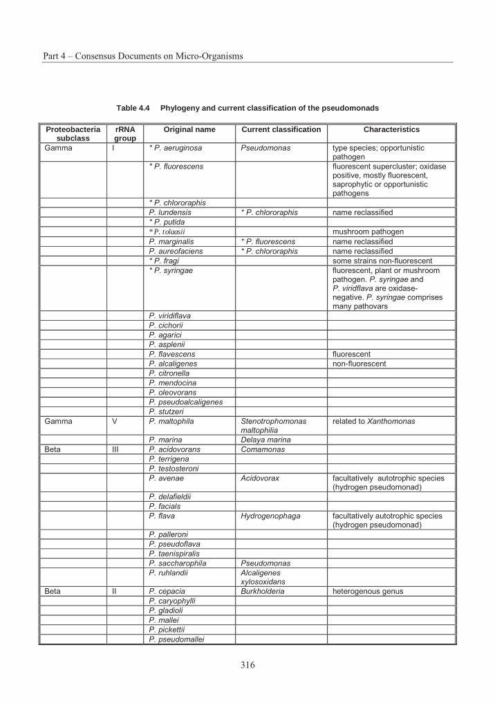
Part 4 – Consensus Documents on Micro-Organisms
316
Table 4.4 Phylogeny and current classification of the pseudomonads
Proteobacteria subclass
rRNA group
Original name Current classification Characteristics
Gamma I * P. aeruginosa Pseudomonas type species; opportunistic pathogen
* P. fluorescens fluorescent supercluster; oxidase positive, mostly fluorescent, saprophytic or opportunistic pathogens
* P. chlororaphis
P. lundensis * P. chlororaphis name reclassified
* P. putida
* P. tolaasii mushroom pathogen
P. marginalis * P. fluorescens name reclassified
P. aureofaciens * P. chlororaphis name reclassified
* P. fragi some strains non-fluorescent
* P. syringae fluorescent, plant or mushroom pathogen. P. syringae and P. viridflava are oxidase- negative. P. syringae comprises many pathovars
P. viridiflava
P. cichorii
P. agarici
P. asplenii
P. flavescens fluorescent
P. alcaligenes non-fluorescent
P. citronella
P. mendocina
P. oleovorans
P. pseudoalcaligenes
P. stutzeri
Gamma V P. maltophila Stenotrophomonas maltophilia
related to Xanthomonas
P. marina Delaya marina
Beta III P. acidovorans Comamonas
P. terrigena
P. testosteroni
P. avenae Acidovorax facultatively autotrophic species (hydrogen pseudomonad)
P. delafieldii
P. facials
P. flava Hydrogenophaga facultatively autotrophic species (hydrogen pseudomonad)
P. palleroni
P. pseudoflava
P. taenispiralis
P. saccharophila Pseudomonas
P. ruhlandii Alcaligenes xylosoxidans
Beta II P. cepacia Burkholderia heterogenous genus
P. caryophylli
P. gladioli
P. mallei
P. pickettii
P. pseudomallei
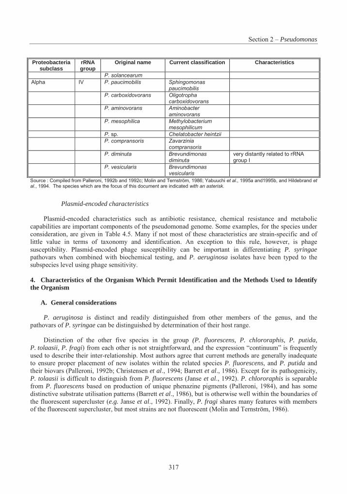
Section 2 – Pseudomonas
317
Proteobacteria subclass
rRNA group
Original name Current classification Characteristics
P. solancearum
Alpha IV P. paucimobilis Sphingomonas paucimobilis
P. carboxidovorans Oligotropha carboxidovorans
P. aminovorans Aminobacter aminovorans
P. mesophilica Methylobacterium mesophilicum
P. sp. Chelatobacter heintzii
P. compransoris Zavarzinia compransoris
P. diminuta Brevundimonas diminuta
very distantly related to rRNA group I
P. vesicularis Brevundimonas vesicularis
Source : Compiled from Palleroni, 1992b and 1992c; Molin and Ternström, 1986; Yabuuchi et al., 1995a and1995b, and Hildebrand et al., 1994. The species which are the focus of this document are indicated with an asterisk.
Plasmid-encoded characteristics
Plasmid-encoded characteristics such as antibiotic resistance, chemical resistance and metabolic capabilities are important components of the pseudomonad genome. Some examples, for the species under consideration, are given in Table 4.5. Many if not most of these characteristics are strain-specific and of little value in terms of taxonomy and identification. An exception to this rule, however, is phage susceptibility. Plasmid-encoded phage susceptibility can be important in differentiating P. syringaepathovars when combined with biochemical testing, and P. aeruginosa isolates have been typed to the subspecies level using phage sensitivity.
4. Characteristics of the Organism Which Permit Identification and the Methods Used to Identify the Organism
A. General considerations
P. aeruginosa is distinct and readily distinguished from other members of the genus, and the pathovars of P. syringae can be distinguished by determination of their host range.
Distinction of the other five species in the group (P. fluorescens, P. chlororaphis, P. putida, P. tolaasii, P. fragi) from each other is not straightforward, and the expression “continuum” is frequently used to describe their inter-relationship. Most authors agree that current methods are generally inadequate to ensure proper placement of new isolates within the related species P. fluorescens, and P. putida and their biovars (Palleroni, 1992b; Christensen et al., 1994; Barrett et al., 1986). Except for its pathogenicity, P. tolaasii is difficult to distinguish from P. fluorescens (Janse et al., 1992). P. chlororaphis is separable from P. fluorescens based on production of unique phenazine pigments (Palleroni, 1984), and has some distinctive substrate utilisation patterns (Barrett et al., 1986), but is otherwise well within the boundaries of the fluorescent supercluster (e.g. Janse et al., 1992). Finally, P. fragi shares many features with members of the fluorescent supercluster, but most strains are not fluorescent (Molin and Ternström, 1986).
Part 4 – Consensus Documents on Micro-Organisms
318
B. Methods used for identification and classification
Numerical taxonomy
Numerical taxonomy has become the “traditional” method for classifying members of the genus Pseudomonas. This approach compares multiple features of the isolate, for which there is substantial discriminatory power, with a database of features of well-described members of the taxon. The accuracy of this type of approach will depend upon the quality and quantity of the data for strains comprising the reference database.
In order to achieve valid results, identical laboratory techniques need to be used for analysis of the isolate and the strains used to construct the reference database. The success of numerical taxonomy is also affected by the complexity of the relationships among the taxa being evaluated.
Use of a broad spectrum of substrates in numeric taxonomic evaluations has had some success for fluorescent species of Pseudomonas (e.g. Barrett et al., 1986). These techniques have permitted some assignment of strains to species and biovars within the fluorescent supercluster (a term applied to all of the species and biovars of P. fluorescens, P. putida and their allies, Table 4.4). However, Molin and Ternström (1986) and Janse et al. (1992) both reported many unclassifiable strains among those they subjected to classical numeric taxonomic analyses.
Commercial suppliers have devised simplified, automated versions of this technique. Examples of commercial kits available for identification of Pseudomonas on the basis of carbon source utilisation patterns, and physiological and morphological characters, are the API20E (API, 200 Express Street, Plainview, New York 11803, USA; BioMerieux, F-69280 Marcy-L’Etoile, France) and the BIOLOG (BIOLOG Inc., Hayward, California, USA) systems. For these kits, the database for Pseudomonas is based on mainly clinical, not environmental, strains. As a result, the kits may fail to identify all environmental isolates.
Use of these kits requires experience. In addition, most of them are designed to determine the membership of the isolate within a taxon and not to distinguish strains within a species. That is, the test profile in most cases is not unique to a particular strain. So, in most cases, test profiles will not be sufficient to distinguish the isolate from other strains of the same species. If such a distinction is being made, it must be based on the detection of properties unique within the taxon.
Details of the test methodologies and profiles of the species can be found in Palleroni (1981; 1984; 1992c).
Genotypic approaches
The current classification of the pseudomonads is based on rRNA homologies. The variable and conserved regions of the RNA molecule are both important for identification purposes. The conserved regions serve as targets for polymerase chain reaction (PCR) primer binding sites and universal hybridisation probes. The variable regions are the targets of the hybridisation probes and primers that are taxon-specific. Probes and PCR primers directed at diagnostic rRNA sequences have facilitated the classification of pseudomonads into the five rRNA groups (Table 4.4).
Strong selection pressure for the conservation of 16S and 23S rRNA molecular structure and sequence has meant that rRNA molecules are powerful evolutionary clocks for describing phylogenetic relationships between rRNA groups of pseudomonads. At present, however, they are unable to position individual strains into species groups. This is particularly true for the fluorescent rRNA group I pseudomonads (Christensen et al., 1994). Using 23S rDNA methods, Christensen et al. (1994) found that “the method
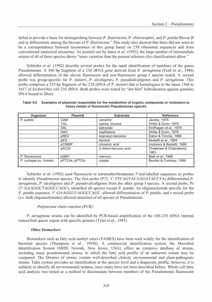
Section 2 – Pseudomonas
319
failed to provide a basis for distinguishing between P. fluorescens, P. chlororaphis, and P. putida Biovar B and to differentiate among the biovars of P. fluorescens.” This study also showed that there did not seem to be a correspondence between taxonomies of this group based on 23S ribosomal sequences and from conventional numerical taxonomy. As pointed out by Janse et al. (1992), the large number of intermediate strains of all of these species shows “more variation than the present schemes (for classification) allow.”
Schleifer et al. (1992) describe several probes for the rapid identification of members of the genus Pseudomonas. A 360 bp fragment of a 23S rRNA gene derived from P. aeruginosa (Festl et al., 1986) allowed differentiation of the eleven fluorescent and non-fluorescent group I species tested. A second probe was group-specific for P. stutzeri, P. alcaligenes, P. pseudoalcaligenes and P. aeruginosa. This probe comprises a 255 bp fragment of the 23S rDNA of P. stutzeri that is homologous to the bases 1366 to 1617 of Escherichia coli 23S rRNA. Both probes were tested by “dot-blot” hybridisation against genomic DNA bound to filters.
Table 4.5 Examples of plasmids responsible for the metabolism of organic compounds or resistance to heavy metals in fluorescent Pseudomonas species
Organism Plasmid Substrate Reference P. putida CAM
TOL camphor xylene, toluene
Jacoby, 1975 White & Dunn, 1978
SAL salicylate Korfhagen et al., 1978
NAH napthalene White & Dunn, 1978
pRE4 isopropyl-benzene Eaton & Timmis, 1986
pEG styrene Bestetti et al., 1984
pCINNP cinnamic acid Andreoni & Bestetti, 1986
pAC25 3-chloro-benzoic acid Chatterjee & Chakrabarty, 1983
P. fluorescens pQM1 mercury Bale et al., 1988
P. syringae pv. tomato pPT23a; pPT23c copper Bender & Cooksey, 1986
Schleifer et al. (1992) used fluorescein or tetramethylrhodamine 5’end-labelled sequences as probes to identify Pseudomonas species. The first probe (P72; 5’-TTCAGTACAAGATACCTA) differentiated P. aeruginosa, P. alcaligenes and P. pseudo-alcaligenes from the other group I species. A second probe, Ps (5’-GAAGGCTAGGCCAGC), identified all species except P. putida. An oligonucleotide specific for the P. putida sequence, 5’-GAAGGUUAGGCCAGC, allowed differentiation of P. putida, and a mixed probe (i.e. both oligonucleotides) allowed detection of all species of Pseudomonas.
Polymerase chain reaction (PCR)
P. aeruginosa strains can be identified by PCR-based amplification of the 16S-23S rDNA internal transcribed spacer region with specific primers (Tyler et al., 1995).
Other biomarkers
Biomarkers such as fatty acid methyl esters (FAMES) have been used widely for the identification of bacterial species (Thompson et al., 1993b). A commercial identification system, the Microbial Identification System (MIDI, Newark, New Jersey, USA), offers an extensive database of strains, including many pseudomonad strains, to which the fatty acid profile of an unknown isolate may be compared. The libraries of strains contain well-described clinical, environmental and plant-pathogenic strains. Tahis system provides an identification at the species level and a diagnostic profile; however, it is unlikely to identify all environmental isolates, since many have not been described before. Whole cell fatty acid analysis was tested as a method to discriminate between members of the Pseudomonas fluorescent
Part 4 – Consensus Documents on Micro-Organisms
320
supercluster (Janse et al., 1992). This analysis resulted in recognition of a large supercluster that included most P. fluorescens and related strains (P. chlororaphis, P. putida and P. tolaasii). In the supercluster there were no separate clusters discriminating biovars of P. fluorescens, the other related species, or strains received as P. marginalis (a name formerly applied to plant-pathogenic members of the supercluster). Thus, the resolution of this technique appears to have limitations.
Diagnostic profiles for micro-organisms may also be obtained using polyacrylamide electrophoresis of whole cell protein extracts, or DNA fingerprints produced via restriction endonuclease digestion of genomic DNA. Pseudomonads have genomes that are rich in GC DNA bases. Enzymes like SpeI that cut at sites with a high AT base composition will digest the DNA at only a few sites, producing large fragments which may be separated and analysed using pulse field gel electrophoresis.
The species of rRNA group I synthesize a ubiqionone with nine isoprene units (Q-9) in the side chain, whereas members of rRNA groups II, III, and V contain Q-8, and those of rRNA group IV a Q-10 (Oyaizu and Komgata, 1983).
Polyamine patterns are of similar utility: rRNA group I species have a high putrescine and spermidine content, rRNA group II and III species have 2-hydroxuputrescine and a high content of putrescine, rRNA group IV species only contain significant amounts of spermidine and sym-homospermidine, and rRNA group V species are characterised by high concentrations of cadaverine and spermidine (Busse and Auling, 1988; Auling, 1992; Yang et al., 1993).
In summary, the results of any of the methods of identification described above are only as good as the database of strains and isolates to which they are referenced. There are numerous techniques that, if applied at the optimum taxonomic level, may prove useful in identifying Pseudomonas and its species. Ribosomal RNA sequencing seems useful at the genus or higher level, and methods like fatty acid analysis can work at the strain and isolate level.
3. Information on the Reproductive Cycle (Sexual/ Asexual)
Pseudomonas species reproduce by cell growth and binary cell division.
4. Biological Features and Environmental Conditions Which Affect Survival, Reproduction, Growth, Multiplication or Dissemination
A. General considerations
Pseudomonas species are efficient saprophytic chemo-organotrophs which grow at neutral pH and at temperatures in the mesophilic range (optimal growth between 20° and 45°C). Some species will grow at 4°C (P. fluorescens, P. putida) but not at the elevated temperature of 41°C. Optimal temperatures are 25-30°C for P. fluorescens, P. putida and P. syringae, 30°C for P. chlororaphis, and 37°C for P. aeruginosa. Most, if not all, species fail to grow under acid conditions (< pH 4.5) (Palleroni, 1984).
Pseudomonas species thrive under moist conditions in soil (particularly in association with plants), and in sewage sediments and the aquatic environments. Environmental conditions which will affect their growth include nutrient availability, moisture, temperature, predation, competition, UV irradiation, oxygen availability, salinity, and the presence of inhibitory or toxic compounds. As nutritional demands are modest, pseudomonads can survive and multiply for months in moist environments such as tap water, sink drains, etc. (Palleroni, 1981; Bergen, 1981).
Section 2 – Pseudomonas
321
Competition and competitive niche exclusion are likely to limit the growth of introduced pseudomonad inoculants. Competitors are likely to include closely related pseudomonads and other bacteria able to compete for the same ecological niches with similar nutritional requirements (Lindow, 1992).
A number of environmental conditions may affect the dissemination of Pseudomonas species including surface water runoff, wind currents, and rain splash. It is likely that insects and other animals may also serve as vectors for dispersal. For example, P. aeruginosa can inhabit part of the normal gut or skin microflora of humans and animals. Thus dissemination would be associated with these vectors.
B. P. aeruginosa
P. aeruginosa is capable of growing in conditions of extremely low nutrient content (Palleroni 1984). The species was found to survive and proliferate in water for up to 100 days or longer (Warburton et al.,1994). Conditions of high humidity and temperature (80-90% humidity, 27°C) favoured the colonisation of lettuce and bean plants (Green et al., 1974).
C. P. chlororaphis
P. aureofaciens (P. chlororaphis) is an important coloniser of the rhizosphere and phyllosphere of plants (Thompson et al., 1993a; Legard et al., 1994). Kluepfel et al. (1991b) reported the colonisation of wheat roots in a field release of a recombinant P. aureofaciens (lacZY). This population of P. aureofaciensreached a maximum of 2x10⁶ cfu/g root two weeks after inoculation and declined steadily to reach a level below detection (<100 cfu/g root) by 38 weeks post-inoculation. Angle et al. (1995) found that inoculations of recombinant P. aureofaciens (lacZY) survived approximately twice as long in wheat rhizosphere as in non-rhizosphere bulk soil.
England et al. (1993) compared the survival and respiratory activity of P. aureofaciens in sterile and non-sterile loam and sandy loam soil microcosms. Recovery of P. aureofaciens was greater in sterile than non-sterile soils. Respiratory activity was higher in sandy loam soil than in loam soil, but soil type had no effect on survival.
The growth of P. aureofaciens in the spermosphere of seed-inoculated sugarbeets exhibited long lag phases (8-12 h) and their populations increased mainly between 12 and 24 hours (Fukui et al., 1994). The doubling time during the exponential growth phases was 2-3 h (Fukui et al., 1994).
D. P. fluorescens
P. fluorescens is commonly found inhabiting plant rhizosphere or phyllosphere environments. The plant rhizosphere provides an environment in which the species may show improved survival and growth. P. fluorescens distributed homogeneously in soil can result in significantly higher numbers in the rhizosphere of young wheat plants than in non-rhizosphere soil (Trevors et al., 1990).
The survival of P. fluorescens is affected by a variety of abiotic and biotic factors. Rattray et al.(1993) found that temperature and soil bulk density had a significant effect on lux-marked P. fluorescenscolonisation of wheat rhizospheres. The greatest rates of colonisation occurred at the highest temperature (22°C) and lower bulk density (0.82 g/cm3), and 100-fold higher numbers were found in the ectorhizosphere than in the endorhizosphere. Van Elsas et al. (1991) found P. fluorescens cells were able to withstand low temperatures, and could survive better at 4°C than at 15 or 27°C following introduction into natural soil, possibly due to an inhibition of the activity of the indigenous microflora. Van Elsas et al.(1986) found that P. fluorescens cell numbers declined slowly in both silt loam and loamy sand, but
Part 4 – Consensus Documents on Micro-Organisms
322
survival was better in the silt loam. Heijnen et al. (1993) found that P. fluorescens survived better in unplanted soils in the presence of bentonite clay. Stutz et al. (1989) demonstrated that survival of P. fluorescens in vermiculite clay was better than in montmorillonite, which was better than in illite.
Van Elsas et al. (1992) found P. fluorescens R2f survived above 107 cfu/g dry soil for up to 84 days in Ede loamy sand microcosms when encapsulated in alginate with skim milk and bentonite clay, while free cells declined below 105 cells/g dry soil after 21 days. Vandenhove et al. (1991) studied the survival of P. fluorescens inocula of different physiological stages in soil. Introduction of a late exponential phase inoculum into soil brought about a lower death rate compared to exponential or stationary phase inocula.
Handley and Webster (1993) studied the effect of relative humidity (RH at 20, 40, 60, and 80%) on airborne survival of P. fluorescens indoors. They found that P. fluorescens, suspended in distilled water, survived best at mid humidities and least at 80% relative humidity.
Boelens et al. (1994) and Bowers and Parke (1993) determined that motility of P. fluorescens did not affect its spread through soil. A non-motile mutant strain promoted plant growth and colonised roots as effectively as the motile strain. Water flow rates were more important than motility for dispersal through soil and rhizospheres. Knudsen (1989) developed a mathematical model for predicting aerial dispersal of bacteria during environmental release which predicted off-site dispersal patterns that were in qualitative agreement with results from a field release of a genetically engineered P. fluorescens in California.
Thompson et al. (1992) studied dissemination of P. fluorescens by placing bacterial populations on apple or pear pollen in the entrances of hives of honey bees. In a pear orchard, 72% of the flowers within 7.6 m of the hive were colonised with P. fluorescens eight days after the start of the study.
E. P. fragi
P. fragi is commonly found on refrigerated meat and dairy products (Jay, 1992). Psychrotrophs such as P. fragi generally have a lower metabolic rate than mesophiles (lower Q10 for the same substrate) and have membranes that transport solutes more efficiently (Jay, 1992). In addition, there is a correlation between the maximum growth temperature and the temperature at which respiratory enzymes are destroyed in psychrotrophs. Nashif and Nelson (1953) reported that extracellular lipase synthesis in P. fragi was inactivated at 30°C. The lipase of P. fragi is reported active at temperatures as low as -29°C (Alford and Pierce, 1961).
P. fragi has the ability to colonise stainless steel surfaces in food processing establishments to form “biofilms” (Hood and Zottola, 1995); attachment may involve a polysaccharide and protein matrix surrounding the cells (Herald and Zottola, 1989). Attachment of P. fragi to stainless steel surfaces occurred in 0.5 h at 25°C and in 2 h at 4°C through the development of attachment fibrils (Stone and Zottola, 1985).
F. P. putida
A variety of environmental factors can affect the survival of P. putida. For example, plant rhizospheres can provide an environment for improved survival. Gamliel and Katan (1992) studied the chemotaxis response of P. putida towards seed exudates and germinating tomato seeds and suggested this may contribute to its rapid establishment in plant rhizospheres. Temperature is also an important factor. Hartel et al. (1994) found that P. putida (lacZY) declined from about 108 to 103 cfu/g of soil after 35 days at 35°C, while it did not survive after three days at 40°C.
Macnaughton et al. (1992), using pLV1013 as a marker plasmid in P. putida PaW8, investigated the effect of soil texture on survival and found that introduced bacteria survived better in soils with higher clay
Section 2 – Pseudomonas
323
content. Compeau et al. (1988) studied survival of P. fluorescens and P. putida strains in sterile and non-sterile soil. Colonisation of sterile soil by one strain precluded normal colonisation of the second added strain and suggested there were limited sites for colonisation of Pseudomonas species in these soils.
Lynch (1990) found that P. putida WCS 358 survived in distilled water (without substrate input) for over a year. It was suggested that this could result from the utilisation of dead cells within the population, and from viable cells having a very low maintenance energy requirement in a state of arrested metabolism.
Madsen and Alexander (1982) found that cells of P. putida were not transported below 2.7 cm in moist soil in the absence of some transporting agent or in the presence of developing plant roots. Percolating water and a burrowing earthworm enhanced the vertical transport of P. putida.
G. P. syringae
Foliar plant pathogens such as P. syringae are generally not adapted to survival in the soil (Lindow et al., 1988). However, P. syringae has been isolated from plant debris in the soil and can overwinter in temperate climates (Hirano and Upper, 1983). P. syringae pv. syringae R32 have pili that function as adhesions anchoring the cell to the surface of plants, thereby enhancing epiphytic colonisation. Wild-type bacteria became virtually resistant to displacement by rinsing within one day after inoculation, whereas non-pilated mutants were only partially resistant within three days (Suoniemi et al., 1995).
P. syringae metabolises a broad range of substrates, thus demonstrating flexibility in nutrient utilisation (Hirano and Upper, 1990). Criteria for viability of cells have been modified as the result of starvation experiments with P. syringae; it was determined that respiration of acetate and glycerol were more accurate determinants of viability than respiration of succinate (Cabral, 1995). The use of bactericides in agriculture (streptomycin and copper) has resulted in selection for strains resistant to these compounds; the resistance is often encoded on plasmids (Cooksey, 1990).
Plant-pathogenic strains grow to larger population sizes on susceptible plant hosts than on resistant ones (Stadt and Saettler, 1981), and therefore pathovars of P. syringae will grow to greater numbers on their respective hosts than on non-hosts. The presence of free water may be the most important factor contributing to the increase in population of P. syringae pv. syringae to infectious levels on bean leaves (Hirano and Upper, 1983; 1990; Beattie and Lindow, 1994). Immediately after rainfall, there is an initial decrease in population as bacteria are washed off the leaf surface, followed by a rapid increase in the population within 12 to 24 hours. Ambient temperature appears to have little effect on field populations of P. syringae pv. syringae on leaves but the age of annual crops does have an effect, with many more cells found on older leaves than on younger ones (Hirano and Upper, 1990; Jacques et al., 1995). P. syringaepv. savastanoi causes tumors on olive and oleander by producing the plant growth regulators indoleacetic acid (IAA) and cytokinins following infection; mutants deficient in IAA production grew as well as the wild type in culture and on plants, but the wild type reached a higher population density and maintained its maximum density at least nine weeks longer than the mutant populations.
Rainfall plays an important role in redistributing P. syringae within the plant canopy by washing bacteria from upper leaves onto lower ones, and by allowing individual bacterial cells to move using their flagella and find protected micro-sites on the surface of the leaf (Beattie and Lindow, 1994). Rainfall efficiently removed bacteria from foliar surfaces, but most of the cells were washed onto the soil; only a small portion were washed a relatively short distance from the source (Butterworth and McCartney, 1991).
P. syringae is also dispersed on seeds (Hirano and Upper, 1983). When cells of P. syringae were applied as a spray to plots, an exponential decrease in numbers of cells was observed; some cells were detected 9.1 m downwind within 20 minutes of the spray application. When applied to oat plants (a non-
Part 4 – Consensus Documents on Micro-Organisms
324
host), viable cells could be detected for up to 16 days and were detected on plants up to 27 m downwind. In contrast to the plants, viable cells could not be detected in the upper layers of soil after two days (Lindow et al., 1988).
H. P. tolaasii
In the production of commercial mushrooms, P. tolaasii probably survives between crops on structural surfaces, in debris, and on equipment. It can be moved readily from one crop to another on the hands of pickers, on materials or equipment used in harvesting, and by insects, mites, water droplets and mushroom spores.
Conditions of high relative humidity and surface wetness encourage the expression of symptoms of brown blotch, an important mushroom disease, caused by P. tolaasii. Dispersal of the micro-organisms occurs readily upon watering once the disease is established (Howard et al., 1994).
5. Behaviour in Simulated Natural Environments Such as Microcosms, Growth Rooms, Greenhouses, Insectaries, etc.
A. P. aeruginosa
In a study by Sturman et al. (1994), the growth rate of P. aeruginosa appeared to be important in determining interspecies competition within packed-bed bioreactors filled with diatomaceous earth pellets.
B. P. chlororaphis
Angle et al. (1995) found that an intact soil core microcosm closely simulated survival results obtained from a field release of a recombinant P. aureofaciens (lacZY). The strain of P. aureofacienssurvived approximately 63 days in the bulk soil microcosm and 96 days in the rhizosphere microcosm.
C. P. fluorescens
Binnerup et al. (1993) found that kanamycin-resistant cells of P. fluorescens DF57-3 (Tn5 modified) inoculated in soil microcosms rapidly lost their culturability, as defined by visible colony formation on Kings B agar supplemented with kanamycin. After 40 days, only 0.02 to 0.35% of the initial inoculum was culturable. It was determined that about 20% of the initial inoculum represented viable, but non-culturable cells.
Compeau et al. (1988) studied survival of P. fluorescens and P. putida in sterile and non-sterile soil. Colonisation of sterile soil by one strain precluded normal colonisation of the second added strain and suggested there are limited sites for colonisation of Pseudomonas species in these soils. Similarly, Al-Achi et al. (1991) found that, when introduced as pairs into irradiated, sterile soils, a P. fluorescens strain prevented optimum colonisation by a P. putida strain. The addition of P. putida to sterile soil already populated by P. fluorescens impeded growth of P. putida in that soil. However, adding P. fluorescens to soil populated by P. putida did not prevent growth of P. fluorescens and caused a decrease in P. putida.These results suggested that there was competition for similar niches in soils, and that P. fluorescens was the more competitive species studied.
D. P. fragi
No information was found regarding the behaviour of this species in microcosms, greenhouses, insectaries, etc.

Section 2 – Pseudomonas
325
E. P. putida
Winstanley et al. (1993) studied the survival of P. putida, with an xy/E marker insert, in soil and lake water microcosms. When released into these microcosms, populations of the marked P. putida had a steady decline with little or no apparent division of cells. The rate of decline of P. putida in soil microcosms was significantly greater at 35% than at 50% field capacity water content, indicating that water content of the soil had an effect on survival. Similarly, Iwasaki et al. (1993; 1994) reported that the density of P. putidadecreased rapidly to less than 102 to 103 level within five days in lake water and soil microcosms. The survival was influenced by protozoa density, light intensity, and soil water content. The addition of P.putida (107 cfu/ml) into natural water and soil had no effect on the density of indigenous micro-organisms and enzyme activities.
Williams et al. (1992) studied the fate and effects of P. putida PPO200 genetically engineered for both nalidixic acid and kanamycin resistance in freshwater and marine microcosms. The freshwater microcosm contained fish (Poecilia latipinna), annelid worms (Tubifex tubifex), snails (Gyraulus sp.), freshwater mussels (Margaritifera margaritifera), freshwater shrimps (Palaemonetes kadiakensis), and plants (Elodea canadensis). The marine microcosm contained fish (Cyprinodon variegatus), sea anenomae (Bunodosoma californica), snails (Turbo fluctosus), oysters (Crassostrea gigas), estuarine shrimp (Palaemonetes pugio), shorefly larvae (Ephydra sp.), and plants (Salicornia biglovii). P. putida could be detected in the tissues of some non-target organisms, i.e. the bacterium survived. However, gross signs, survival, and the histological study of control and exposed non-target organisms indicated that there were no adverse effects.
Doyle et al. (1991) and Short et al. (1991) observed reductions in CO2 evolution and the number of fungal propagules, as well as the enhancement of dehydrogenase activity in soil amended with 2,4-dichlorophenoxyacetate (2,4-D) and inoculated with P. putida PPO301(pRO103) genetically engineered to degrade 2,4-D. These unanticipated effects were not observed: (a) in uninoculated soil; (b) when the homologous, plasmidless parent P. putida PPO301 was inoculated; or (c) in the presence of the genetically engineered P. putida when 2,4-D was not added. Moreover, the effects were not predictable from the phenotype of this genetically engineered P. putida. While long-term, statistically significant differences were detected in some microbial populations and processes, the majority of the differences were transient.
The effects of P. putida, on nitrogen transformations and nitrogen-transforming microbial populations were studied in a soil perfusion system by Jones et al. (1991). Neither the genetically engineered strain nor its homologous plasmidless host had a significant effect on ammonification, nitrification or denitrification in the soil, or on the population dynamics of the micro-organisms responsible for these processes.
F. P. syringae
Wendtpotthoff et al. (1994) monitored the fate of a genetically engineered strain of P. syringaeapplied to the leaves of bush beans in a planted soil microcosm. P. syringae established on the bean leaves at between 5 x 10³ and 4 x 10⁶ cfu/gm-1 fresh weight. During senescence of the bean plants, the strain was no longer detectable by selective cultivation and subsequent colony hybridisation.
Significant differences within P. syringae strain MF714R were detected when the bacterium was cultured on agar or in broth or collected from colonised leaves and subsequently inoculated onto greenhouse-grown plants in growth chambers or in the field or onto field grown plants. Bacterial cells cultured in liquid medium survived the least well after inoculation under all conditions, whereas cells cultured on solid media exhibited the highest percent survival and desiccation tolerance in the growth chamber but survived less well in the field than did cells harvested from plants. Cells harvested from plants
Part 4 – Consensus Documents on Micro-Organisms
326
and inoculated onto plants in the field usually had the highest percent survival, started to increase in numbers earlier, and reached a higher number than did cells cultured in vitro (Wilson and Lindow, 1993a).
Wilson and Lindow (1993b) indicated that greenhouse-grown plants support larger epiphytic populations of an inoculated strain of P. syringae than do field-grown plants.
G. P. tolaasii
No information was found regarding the behaviour of this species in microcosms, greenhouses, insectaries, etc.
6. History of Use (Examples of Environmental Applications of the Organism and Information Derived from These Examples)
A. General considerations
Pseudomonads have been identified to be of importance in bioremediation as a result of their metabolic versatility. This metabolic versatility, and the ability to acquire additional versatility via plasmids, provides the potential for the rapid evolution of novel metabolic ability in Pseudomonas species. Examples of useful, or potentially useful, environmental applications of Pseudomonas isolates are given in Table 4.6. Some pseudomonad species have been introduced into the environment in bioremediation studies and have provided valuable information pertaining to characteristics such as survival. For example, Thiem et al. (1994) injected Pseudomonas sp. strain B13, a chlorobenzoate degrader, into a subsurface aquifer and found they could detect the strain 14.5 months after its environmental introduction.
Pseudomonads also have great potential in agronomic applications, since they are prolific colonisers of plant surfaces and represent a significant component of plant microflora. For example, they have been identified to possess traits that make them suitable as agents for biological pest control (O’Sullivan and O’Gara, 1992). These include an ability to produce antimicrobial molecules (antibiotics, antifungals and siderophores) and a capacity to aggressively compete with other micro-organisms for niches and exclude phytopathogens. The possibility of the environmental application of strains to minimise frost damage on crop plants has also been investigated (Lindow and Panopoulos, 1988).
B. P. aeruginosa
Some strains of P. aeruginosa have been shown to produce biosurfactants which have potential uses in bioremediation for washing hydrocarbons from soil (Van Dyke et al., 1993). Jain et al. (1992) found that biosurfactants produced by P. aeruginosa, when added to soil, significantly enhanced the degradation of tetradecane, hexadecane and pristane.
Degradation of pentachlorophenol by P. aeruginosa has been investigated in shake-cultures. The bacteria were able to completely degrade pentachlorophenol up to 800 mg/l in six days with glucose as a co-substrate (Premalatha and Rajakumar, 1994). P. aeruginosa has also been found to degrade styrene in a continuous reactor at a rate of 293 mg g-1h-1. This could be applied to the industrial treatment of waste gas or polluted water (El Aalam et al., 1993).
C. P. chlororaphis
A strain of P. chlororaphis, genetically engineered to contain the lacZY genes, was introduced into the environment in a field trial in the United States in 1987, and its behaviour compared to the non-engineered strain (Kluepfel et al., 1991a, b, c). The non-engineered strain increased in number for two weeks, then declined to at or near the detection limit by 31 weeks.
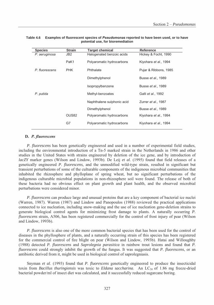
Section 2 – Pseudomonas
327
Table 4.6 Examples of fluorescent species of Pseudomonas reported to have been used, or to have potential use, for bioremediation
Species Strain Target chemical Reference P. aeruginosa JB2 Halogenated benzoic acids Hickey & Focht, 1990
PaK1 Polyaromatic hydrocarbons Kiyohara et al., 1994
P. fluorescens PHK Phthalate Pujar & Ribbons, 1985
Dimethylphenol Busse et al., 1989
Isopropylbenzene Busse et al., 1989
P. putida Methyl-benzoates Galli et al., 1992
Naphthalene sulphonic acid Zurrer et al., 1987
Dimethylphenol Busse et al., 1989
OUS82 Polyaromatic hydrocarbons Kiyohara et al., 1994
G7 Polyaromatic hydrocarbons Kiyohara et al., 1994
D. P. fluorescens
P. fluorescens has been genetically engineered and used in a number of experimental field studies, including the environmental introduction of a Tn-5 marked strain in the Netherlands in 1986 and other studies in the United States with strains engineered by deletion of the ice gene, and by introduction of lacZY marker genes (Wilson and Lindow, 1993b). De Leij et al. (1995) found that field releases of a genetically engineered P. fluorescens, and the unmodified wild-type strain, resulted in significant but transient perturbations of some of the culturable components of the indigenous microbial communities that inhabited the rhizosphere and phylloplane of spring wheat, but no significant perturbations of the indigenous culturable microbial populations in non-rhizosphere soil were found. The release of both of these bacteria had no obvious effect on plant growth and plant health, and the observed microbial perturbations were considered minor.
P. fluorescens can produce large and unusual proteins that are a key component of bacterial ice nuclei (Warren, 1987). Warren (1987) and Lindow and Panopoulos (1988) reviewed the practical applications connected to ice nucleation, including snow-making and the use of ice nucleation gene-deletion strains to generate biological control agents for minimizing frost damage to plants. A naturally occurring P. fluorescens strain, A506, has been registered commercially for the control of frost injury of pear (Wilson and Lindow, 1993b).
P. fluorescens is also one of the more common bacterial species that has been used for the control of diseases in the phyllosphere of plants, and a naturally occurring strain of this species has been registered for the commercial control of fire blight on pear (Wilson and Lindow, 1993b). Hatai and Willoughby (1988) detected P. fluorescens and Saprolegnia parasitica in rainbow trout lesions and found that P.fluorescens could strongly inhibit the growth of the fungus. It was suggested that P. fluorescens, or an antibiotic derived from it, might be used in biological control of saprolegniasis.
Snyman et al. (1993) found that P. fluorescens genetically engineered to produce the insecticidal toxin from Bacillus thuringiensis was toxic to Eldana saccharina. An LC50 of 1.86 mg freeze-dried bacterial powder/ml of insect diet was calculated, and it successfully reduced sugarcane boring.
Part 4 – Consensus Documents on Micro-Organisms
328
P. fluorescens has been shown to have the ability to degrade a wide variety of compounds, including: 3-chlorobenzoic acid (Fava et al., 1993); naphthalene, phenathrene, fluorene and fluoranthene (Weissenfels et al., 1990); chlorinated aliphatic hydrocarbons (Vandenbergh and Kunka, 1988); styrene (Baggi et al., 1983); and pure hydrocarbons and crude oil (Janiyani et al., 1993). P. fluorescens can also be used in biosensor applications. For example, the recombinant P. fluorescens strain HK9, which lights up in the presence of contaminants such as PAHs (due to the insertion of lux genes), allows easy detection of bioavailable fractions of pollutants in soils and sediments (King et al., 1990).
E. P. fragi
No information was found on the use of P. fragi in environmental applications.
F. P. putida
P. putida is capable of eliminating phytopathogenic micro-organisms and stimulating plant growth (Vancura, 1988; Kloepper et al., 1988; Freitas and Germida, 1990). P. putida is also capable of degrading many unusual compounds by means of enzymatic systems encoded in plasmids. Chemicals degraded include polychlorinated biphenyls (PCBs) (Boyle et al., 1993; Lajoie et al., 1994); trichloroethylene (TCE) (Fujita et al., 1995); acetonitrile and sodium cyanide (Babu et al., 1994). P. putida has also shown the ability to remediate non-ionic sewage (Turkovskaya et al., 1993), pulp mill waste (black liquor) (Jain et al., 1993), waste gases using a biofilter (Zilli et al., 1993), electroplating effluent with high concentrations of copper (Cu(II)) (Wong et al., 1993), and high-sulphur coal (Khalid and Aleem, 1991).
G. P. syringae
Lindow et al. (1988) monitored the fate of a strain of P. syringae in experimental field trials in the United States. They found an exponential decrease in numbers of viable cells deposited at increasing distances from sprayed field plots. The relative rate of survival of cells sprayed directly on plants was more than ten times higher than that of cells dispersed through the air to similar adjacent plants.
Use of P. syringae has been proposed to enhance snowmaking and to delay frost damage in plants (Lindow, 1983; Wilson and Lindow, 1993b). P. syringae has also been shown to incorporate aluminium, chromium and manganese, so the bioremediation of sites contaminated with these chemicals may be a potential use (Alaoukaty et al., 1992).
H. P. tolaasii
No information was found on the use of P. tolaasii in environmental applications.
7. Characterisation of the Genome (e.g. Presence of Large Plasmids, Insertion Sequences) and Stability of These Characteristics
Members of the genus Pseudomonas are known for their metabolic versatility. They are capable of degrading many recalcitrant xenobiotics due to their ability to recruit new genes and alter the expression of existing ones. An understanding of the relative chromosomal position of relevant genes, the diversity of mobile genetic elements found within this genus, and the role these mobile genetic elements play in the stability and metabolic adaptation of individual isolates, can be helpful for regulatory assessments.
The chromosomes of P. putida and P. aeruginosa have been described in detail by Holloway and Morgan (1986) and by Ratnaningsih et al. (1990), Romling et al. (1989) and Holloway et al. (1994). Holloway et al. (1990a) provide genetic maps of these two species, which are useful in locating the relative positions of important genes and provide a good summary of other chromosomal and extrachromosomal
Section 2 – Pseudomonas
329
features. The sizes of the chromosomes for P. putida and P. aeruginosa vary from approximately 4,400 to 5,400 kb, with P. aeruginosa strain PAO having a genome size (5,400 kb) significantly larger than the 4,700 kb E. coli chromosome. Analysis of the distribution of chromosomal genes in pseudomonads shows that those involved in biosynthesis are not contiguous as with the enterobacteria. The genes for catabolic functions tend to be clustered on the chromosome, but are also not contiguous. Many catabolic functions are located on plasmids (e.g. Table 4.5); these genes, such as TOL (toluene degradation) and NAH (napthalene degradation), tend to be contiguous. This genome configuration allows for many diverse substrains within a species, each adapted to a particular environment.
Pseudomonas species contain a large variety of plasmids, insertion sequences, and transposons. The diversity of plasmids involved in degradation of organic compounds, drug resistance, and phytopathogenicity is indicated in Tables 5.4, 5.7 and 5.10. Insertion sequences (IS elements) and transposons are mobile within the genome of gram-negative bacteria, and can act as new promoters or as terminators, causing polar mutations. If two IS elements are located near each other in the appropriate orientation, they can be transposed to a second genome as a unit along with any intervening genes.
These three classes of mobile genetic elements (plasmids, insertion sequences, and transposons) can potentially interact within the same isolate, causing shifts in the positions of key catabolic genes. An example is the NAH plasmid napthalene degradative genes, which are nested within a defective but mobilizable transposon on the plasmid (Tsuda and Iino, 1990). Such shifts can result in a variable stability for some traits. For example, P. syringae pv. savasatoni mutations causing IAA deficiency were identified to have resulted from the action of two IS elements. In another case, a 150 kb plasmid (able to integrate into the chromosome) from P. syringae pv phaseolicola, when excised from the chromosome, resulted in the formation of a series of plasmids that either contained chromosomal DNA or were deletion mutants of the plasmid. These events were associated with a common repeated sequence (RS) (Coplin, 1989). In a reverse situation, components of the TOL plasmid have been shown to integrate into both the P. putida and P. aeruginosa chromosomes (Holloway et al., 1990b), thereby potentially stabilizing degradative genes in the genome of the isolates.
Besides affecting the stability of certain traits, mobile genetic elements allow pseudomonads to recruit new genes from replicons such as plasmids, which can lead to new metabolic capabilities. Specific examples have been given by Chakrabarty (1995) of P. putida’s ability to recruit new degradative genes on a transposable element. These new genes allow the organism to degrade new chemicals without the need to evolve completely new degradative pathways. This species has been able to acquire the genes needed to degrade 3-chlorobenzoate to the intermediate protocatechuate, which then is further degraded by resident chromosomal genes. In a similar fashion, the same species has been able to degrade phenol by acquisition of two genes, pheA and pheB, whose products can convert phenol to intermediates which are metabolized by a chromosomally-encoded ortho pathway (Chakrabarty, 1995). A transposon-like mobile element encoding a dehalogenase function has also been recently described in P. putida (Thomas et al., 1992). In the well-characterised P. putida mt-2 plasmid pWW0, the TOL-degradative enzymes are encoded on a 56 kb transposon which is itself part of a 70 kb transposon (Tsuda et al., 1989), giving rise to a family of TOL plasmids (Assinder and Williams, 1990). In addition to acquisition of degradative genes, pseudomonads can also acquire genes whose products aid in waste degradation.
The chlorosis-inducing phytotoxin coronatine, produced by P. syringae pvs. tomato and atropurpurea is plasmid encoded (Coplin, 1989). Other toxins (e.g. phaseolotoxin, syringomycin and tabtoxin) have been shown to be chromosomally encoded. P. syringae pv. savasatoni produces abnormal growths due to an imbalance of cytokinin and auxin plant hormones. The genes for their biosynthesis are plasmid encoded in oleander, but not olive pathovars. The majority of P. solanacearum strains contain a large (700-1000 kb) megaplasmid that contains genes for host range and pathogenicity.
Part 4 – Consensus Documents on Micro-Organisms
330
8. Genetic Transfer Capability
The ability of pseudomonads to develop new metabolic pathway capabilities is often dependent on an isolate’s ability to acquire DNA from other bacteria, which is then integrated into the genome in a manner dependent on the organism’s environment. The three common systems for gene transfer in bacteria, namely conjugation, transduction and transformation, have been observed among members of the genus Pseudomonas. All three gene transfer mechanisms have been observed under laboratory and natural conditions. Gene transfer by all three mechanisms is affected by biological factors such as the nature and host range of the mobile genetic element, its transfer frequency, the concentrations of recipient and donor organisms, and the presence of other organisms which prey on donors and recipients. Abiotic factors such as temperature, moisture, and the presence of physical substrates which allow survival and/or gene transfer also affect the transfer frequency.
Even if the DNA is transferred to a new recipient, it may not be expressed. Sayre and Miller (1990) provide a detailed summary of factors associated with transposons and plasmids, the donors and recipients, and other biotic and abiotic conditions which affect gene transfer rates.
A. Conjugation
The acquisition of genetic material via conjugative plasmids represents an important evolutionary mechanism in the production of strains resistant to antibiotics and heavy metals, and with the ability to mineralise xenobiotics in selective environments. Gene transfer events may even affect the pathology of certain phytopathogens. Changes in cultivar-specificity and a loss of ability to produce fluorescent pigments of P. syringae pv. pisi were found to result upon the acquisition of IncP1 replicons such as plasmid RP4. Curing the RP4 plasmid from the strain maintained the new phenotype (Moulton et al.,1993). Walter et al. (1987) developed a combined mating technique to measure the conjugal transfer potential of conjugative plasmids that uses four different standard mating techniques (colony cross streak, broth mating, combined spread plate, and membrane filtration), since no one technique worked best for the tested combinations of plasmids and recipients.
Conjugation between pseudomonads has been detected in both soil and aquatic environments. The transfer of conjugative plasmids has been demonstrated to occur between pseudomonads in a number of non-rhizosphere and rhizosphere soil environments, both in microcosms and in situ (van Elsas et al., 1988; Trevors and Berg, 1989; Lilley et al., 1994).
Transfer frequencies were found to be enhanced by two orders of magnitude, that is, up to 10-2 per recipient organism, on the rhizoplane of sugarbeet in situ (Lilley et al., 1994). Soil components (such as clay, silt, organic matter and plant roots) provide excellent surfaces for the cell-to-cell contact required for bacterial conjugation (Trevors and Berg, 1989; Stotzky et al., 1991). In wheat plant root (van Elsas et al.,1988) and sugarbeet (Lilley et al., 1994) conjugation studies, survival of the donor and recipient, as well as frequency of plasmid transfer, decreased with increasing distance from the plant root. Transfer frequencies are also affected by soil moisture, with frequencies for R-plasmid transfer between E. coli isolates shown to be optimal at 60 to 80% soil moisture holding capacity (Trevors and Starodub, 1987). Conjugal transfer of broad host range plasmids between P. aeruginosa donor and recipient strains in lake water has been observed to occur at a lower rate in the presence of the natural microbial community (O’Morchoe et al., 1988). Plasmids incapable of conjugation themselves have been shown to be mobilised from a laboratory strain of E. coli in a laboratory-scale wastewater treatment facility by mobilizer and recipient E. coli strains of both laboratory and wastewater origin (Mancini et al., 1987).
The TOL plasmid pWWO can be transferred to other micro-organisms, and its catabolic functions for the metabolism of alkylbenzoates are expressed in a limited number of gram-negative bacteria, including
Section 2 – Pseudomonas
331
members of the rRNA group I pseudomonads and E. coli (Ramos-Gonzalez et al., 1991). Transfer of the recombinant plasmid to Erwinia chrysanthemi was observed, but transconjugants failed to grow on alkylbenzoates because they lost catabolic functions. Pseudomonads belonging to rRNA groups II, III, and IV, Acinetobacter calcoaceticus, and Alcaligenes sp. could not act as recipients for TOL, either because the plasmid was not transferred or because it was not stably maintained. Under optimal laboratory conditions, the frequency of transfer of pWWO from P. putida as a donor to pseudomonads belonging to rRNA group I was on the order of 1 to 10-2 transconjugants per recipient, whereas the frequency of intergeneric transfer ranged from 10-3 to 10-7 transconjugants per recipient. Intra-species, but not inter-species transfer of TOL in soils has been reported (Ramos et al., 1991), but it was affected by the type of soil used, the initial inoculum size, and the presence of chemicals that could affect the survival of the donor or recipient bacteria (Ramos-Gonzalez et al., 1991).
The P. putida TOL plasmid pWWO and the wide host range RP4 plasmid are able to mediate chromosomal mobilisation in the canonical unidirectional way (i.e. from donor to recipient cells) and bi-directionally [i.e. donor to recipient to donor (retrotransfer)] (Lejeune and Mergeay, 1980; Mergeay et al.,1987; Top et al., 1992; Ramos-Gonzales et al., 1994). Transconjugants are recipient cells that have received DNA from donor cells, whereas retrotransconjugants are donor bacteria that have received DNA from a recipient. The TOL plasmid pWWO and the pRP4 plasmid are able to directly mobilise and retromobilise a chromosomal marker integrated into the chromosome of the other Pseudomonas strains, and this process probably involves a single conjugational event. The rate of retrotransfer (as well as direct transfer) of chromosomal markers is influenced by the location of the marker on the chromosome, and it ranges from 10-3 to less than 10-8 retrotransconjugants per donor (transconjugants per recipient). The mobilised DNA is incorporated into the chromosome of the retrotransconjugants (transconjugants) in a process that seems to occur through recombination of highly homologous flanking regions. No interspecific mobilisation of the chromosomal marker in matings involving P. putida and the closely related P. fluorescens was observed.
It seems clear that pseudomonads can acquire plasmids from other bacteria in the environment. This premise is supported by the array of plasmids that have been recovered from members of Pseudomonas,some of which are listed in Tables 5.5, 5.7 and 5.10. The boundaries to gene transfer events are illustrated by plasmid RP4, originally isolated in P. aeruginosa, which has been shown to be transmissible to all gram-negative bacteria tested (Riley, 1989). E. coli has been shown to transfer plasmid-borne genetic information to over 40 genera (Stotzky et al., 1991). Direct evidence of pseudomonad isolate acquisition of plasmids from other bacteria in the environment is also available: Bale et al. (1988) showed that an introduced P. putida recipient acquired mercury resistance plasmids from an intact lotic epilithic bacterial community at frequencies up to 3.75 X 10⁻⁶ per recipient.
B. Transduction/bacteriophage mediated gene transfer
Two characteristics of a bacteriophage (phage) which are important in determining its ability to broadly distribute DNA were summarised in Sayre and Miller (1990). First, the host range of most phages is restricted to one species or a small number of related taxa, although broad host range phages such as phages P1 and Mu are known. Second, phages which undergo specialised transduction are likely to transfer chromosomal genes which are in close proximity to the phage integration site, while generalised transducing phages can transfer any of the bacterial genome’s sequences with approximately equal frequency.
Many different lytic and temperate phages have been identified in Pseudomonas, and the morphological diversity among phages is at least as great as for any other bacterial genera. Transduction by temperate phage of P. aeruginosa chromosomal DNA has been demonstrated in fresh water microcosms
Part 4 – Consensus Documents on Micro-Organisms
332
(Morrison et al., 1979; Saye et al., 1987; 1990) and the phylloplane of bean and soy bean plants (Kidambiet al., 1994).
P. aeruginosa has been frequently reported as subject to lysogeny, the process by which the phage chromosome becomes integrated into the bacterial host chromosome and is stably replicated with it, as a prophage. Lysogeny may lead to increased fitness of bacterial strains in the natural environment, by increasing the size and flexibility of the gene pool available to natural populations of bacteria via horizontal gene transfer. Approximately 45% of Pseudomonas field isolates tested positive in colony hybridisations when probed with phage isolated from the same area (Miller et al., 1990a). The prophage appears to contribute a major source of phage in the natural environment. In addition to mediating the transfer of genetic material within and between species, the induction of certain prophages results in transposition and mutagenesis events within the host genome.
C. Transformation
Both chromosomal and plasmid DNA are subject to natural transformation in the environment, a natural physiological process which is different from the artificial transformation techniques used in the laboratory (Stewart, 1990). In order for transformation of a cell to result in expression of the new DNA sequence, DNA must: 1) be excreted or lost from a donor cell; 2) persist in the environment; 3) be present in sufficient concentrations for efficient transformation to occur; 4) come in contact with a recipient cell which is naturally competent to receive the donor DNA; 5) be able to evade any recipient cell defences which degrade foreign DNA; and 6) integrate into a stably-maintained replicon in the recipient. Marine and soil environments have been shown to contain biologically significant levels of dissolved DNA (Paul et al.,1987; Lorenz et al., 1988). Soil environments offer protection from nuclease digestion for chromosomal and plasmid DNA (otherwise available for transformation) when bound to clay and sediment matter (Lorenz and Wackernagel, 1991; Romanowski et al., 1991; Khanna and Stotzky, 1992).
Natural transformation was found for P. stutzeri, P. mendocina, P. alcaligenes and P. pseudoalcaligenes, but not for P. aeruginosa, P. putida, P. fluorescens, and P. syringae strains (Carlson etal., 1983).
IV. Human Health Considerations
1. Diseases caused and mechanism of pathogenicity including invasiveness and virulence
General considerations
Included amongst the rRNA group I fluorescent pseudomonads are species pathogenic to humans. Those that do cause infection are generally opportunists, promoting a variety of conditions ranging from endocarditis to dermatitis (Pollack, 1992; Artenstein and Cross, 1993; Berger et al. 1995; Jarvis and Skipper, 1994). Individuals most at risk from Pseudomonas infection are the immunocompromised, in particular individuals with AIDS, patients with cystic fibrosis, and those suffering major trauma or burns (Artenstein and Cross, 1993; Neu, 1985; Bodey et al., 1983; Moayyedi et al., 1994; Schuster and Norris, 1994).
It should be stressed that only some of the Pseudomonads have been implicated in human disease and that proponents should not base assessment criteria on P. aeruginosa, the most problematic member of the genus. Proponents should also be aware that not all strains of a given species have been shown to promote disease, and that assessment criteria should be based upon the strain being reported rather than the species as a whole.
Section 2 – Pseudomonas
333
P. aeruginosa
The predominant pseudomonad isolated from clinical sources is P. aeruginosa. Evaluation of the pathogens causing nosocomial infections in hospitals in the United States (Jarvis and Martone, 1992) indicated that P. aeruginosa was the fourth most common pathogen isolated (10.1%) in hospital-wide surveillance and the most common (12.4%) in intensive care units. In more recent surveys, P. aeruginosawas found to be the fifth most common pathogen (9%) (Emori and Gaynes, 1993) and increased to 29% in intensive care facilities [European Prevalence of Infection in Intensive Care Study (EPIC) (Spencer, 1994)]. The ability of P. aeruginosa to persist in a number of hospital disinfectants and pharmaceuticals (Dominik et al., 1995; Gilardi, 1991) and in sanitary facilities (Bobhammer et al., 1996; Döring et al.,1991; Zembrzuska-Sadkowska et al., 1995) probably contributes to its prevalence in the hospital environment. P. aeruginosa causes a wide range of syndromes, involving nearly all body systems, that vary from sub-acute to chronic (Artenstein and Cross, 1993; Pollack, 1992).
Bacteremia associated with P. aeruginosa is restricted mainly to immunocompromised individuals with significant underlying disease (Artenstein and Cross, 1993; Askamit, 1993; Spencer, 1994). Symptoms are indistinguishable from gram-negative sepsis caused by other bacteria (Pollack, 1993). Factors associated with bacteremia include malignancy, transplants (solid organ, bone marrow), diabetes, cirrhosis of the liver, renal failure, burns, trauma, intravenous drug abuse, corticosteroid therapy, cytotoxic chemotherapy, cardiopulmonary resuscitation, immunoglobulin deficiency, AIDS, broad spectrum antibiotics, and invasive instrumentation (IV catheters, endoscopes, mechanical ventilation, etc.) (Askamit, 1993; Artenstein and Cross, 1993; Dropulic et al., 1995; Mallolas et al., 1990; Nensey et al., 1990). Major entry portals for P. aeruginosa bacteremia appear to be following infection (or colonisation) of the respiratory tract (Gallagher and Watanakunakorn, 1990; Artenstein and Cross, 1993) and the genitourinary tract (Gallagher and Watanakunakorn, 1990; Aksamit, 1993).
P. aeruginosa septicemia is primarily a condition of debilitated, immunocompromised adults and of infants. It is usually acquired in hospital, with prior broad-spectrum antibiotic therapy as a predisposing factor (Bodey et al., 1983; Richet et al., 1989), although community-acquired infections in apparently healthy children (Ros, 1989) and adults (Ishihara et al., 1995) have been reported. Mortality is high in immunocompromised patients (up to 50%) (Artenstein and Cross, 1993; Bisbe et al., 1988), with a better prognosis in “normal healthy” individuals (Ishihara et al., 1995).
Endocarditis due to P. aeruginosa has been associated with two major predisposing factors (Artenstein and Cross, 1993): the use of prosthetic heart valves and the use of illicit parenteral drugs. In drug abusers, endocarditis usually occurs in the right side of the heart and is sub-acute, although a complication, septic pulmonary emboli involving the tricuspid valve, can occur (Pollack, 1992).
Nosocomial pneumonia is the second most common hospital-acquired infection in the United States (Aksamit, 1993; Emori and Gaynes, 1993). P. aeruginosa is the most prevalent etiological agent for both poly- and mono-microbial pneumonia (Emori and Gaynes, 1993). Community-acquired pneumonia has also been attributed to this organism; its occurrence is rare (Artenstein and Cross, 1993), but it is more frequently recognised in AIDS patients (Dropulic et al., 1995; Schuster and Norris, 1994). Colonisation of the oropharyngeal and/or the upper gastrointestinal tracts is an important precursor to nosocomial pneumonia, although colonisation does not always imply infection. However, susceptibility to pneumonia is inversely related to a patient’s basic health (Aksamit, 1993; Artenstein and Cross, 1993, Dick et al., 1988; Dropulic et al., 1995). Conditions predisposing to P. aeruginosa pneumonia are similar to those mentioned for bacteremia.
Bacteremic pneumonia, with organisms isolated from both the lung and bloodstream, resembles bacteremia and pneumonia in clinical presentation. Prognosis is bleak with this syndrome. Mortality rates
Part 4 – Consensus Documents on Micro-Organisms
334
of 80-100% are observed, compared to 27-50% for bacteremia and 30-60% for pneumonia (Aksamit, 1993).
Chronic pulmonary colonisation by P. aeruginosa in the lungs of patients with cystic fibrosis results in frequent acute episodes of pneumonia and chronic bronchiectasis, but rarely in bacteremic pneumonia (Aksamit, 1993). Chronic infection leads to the obstruction of the airways, respiratory distress, and eventually death (Gilligan, 1991; Romling et al., 1994).
Otolaryngologic infections due to P. aeruginosa range from superficial and self-limiting to life-threatening (Artenstein and Cross, 1993). The most serious ear infection due to this organism is malignant otitis externa, usually resulting from a failure of topical therapy, and resulting in an invasive disease- destroying tissue which may progress to osteomyelitis at the base of the skull and possible cranial nerve abnormalities (Artenstein and Cross, 1993). Other ear infections associated with P. aeruginosa include external otitis (swimmer’s ear), otitis media, chronic suppurative otitis media, and mastoiditis (Artenstein and Cross, 1993; Legent et al., 1994; Kenna, 1994; Pollack, 1992).
P. aeruginosa is the leading cause of gram-negative ocular infections, presenting as keratitis or endophthalmitis (Holland et al., 1993; Chatterjee et al., 1995; Bukanov et al., 1994). Predisposing factors include the use of contact lenses (in particular their cleaning and storage solutions), trauma, burns, ocular irradiation, compromised host defences, and systemic infections (Holland et al., 1993; Pollack, 1992; Imayasu et al., 1994; Stapleton et al., 1995).
Moisture is the paramount defining factor in P. aeruginosa growth. Normal dry skin does not support growth, whereas moist skin enables the organism to flourish. For this reason, dermatologic infections with P. aeruginosa tend to be more prevalent in moist tropical and subtropical climates (Bodey et al., 1983) or to be associated with the use of swimming pools, hot tubs or whirlpools (Gustafson et al., 1983; Trueb etal., 1994; Vesaluoma et al., 1995). The use of contaminated “loofah” cosmetic sponges is another source of P. aeruginosa infection (Bottone and Perez, 1993; 1994; Fisher, 1994). Folliculitis, pyoderma, cellulitis and ecthyma gangrenosum are all dermatologic infections in which P. aeruginosa has been implicated (Pollack, 1992; Artenstein and Cross, 1993; Gustafson et al., 1983; Fisher, 1994; Noble. 1993).
P. aeruginosa is a frequent isolate from wounds, particularly those contaminated with soil, plant material or water. Its presence may reflect colonisation as opposed to infection, which is a consequence of its ubiquitous distribution in nature (Artenstein and Cross, 1993; Pollack, 1992). Puncture wounds, particularly those penetrating to bone, may result in osteomyelitis or osteochondritis. The former is common in intravenous drug abusers (Artenstein and Cross, 1993) and the latter in puncture wounds to the foot in children and diabetics (Lavery et al., 1994; Pollack, 1992; Jarvis and Skipper, 1994). The wearing of tennis shoes (sneakers) at the time of puncture injury increases the chance of P. aeruginosa infection (Pollack, 1992; Lavery et al., 1994; Fisher et al., 1985).
In rare cases, P. aeruginosa has been associated with meningitis or brain abscess (Pollack, 1992) and infection of the gastro-intestinal tract (Artestein and Cross, 1993). Both conditions are nosocomially acquired, occurring in patients suffering from malignancies, invasive procedures or neutropenia (Pollack, 1992; Artenstein and Cross, 1993).
P. fluorescens
P. fluorescens has occasionally been associated with human infection. The inability of most strains to grow at normal human body temperature (Palleroni, 1992a) restricts invasion and subsequent disease promotion. This organism has the ability to grow at 4oC (Gilardi, 1991). This characteristic, along with the observation that it is isolated from the skin of a small percentage of blood donors, makes it an occasional
Section 2 – Pseudomonas
335
contaminant of whole blood and blood products (Puckett et al., 1992; Stenhouse and Milner, 1992). Pseudobacteremia may result from the infusion of contaminated products (Scott et al., 1988; Simor et al.,1985; Gottlieb et al., 1991; Foreman et al., 1991) or from the use of contaminated equipment (Anderson and Davey, 1994).
P. fluorescens has been occasionally isolated from patients with AIDS (Franzetti et al., 1992; Roilides et al., 1992), where it caused bacteremia and urinary tract, ocular and soft tissue infections. Chamberland et al. (1992), in their across-Canada survey of septicemia, found that 1.5% of isolates were P. fluorescens.It is apparent that P. fluorescens can be an opportunistic pathogen in cancer patients and in others who are severely immunocompromised, but that it is of little concern to immunocompetent individuals. P.fluorescens is occasionally found in sputa of patients with cystic fibrosis, although its role as a pathogenic factor has yet to be resolved.
P. fragi
P. fragi is one of the pseudomonads associated with food spoilage (Barrett et al., 1986; Drosinos and Board, 1995; Greer, 1989) and is commonly isolated from milk products, pork and lamb. A search of the literature dating back to 1966 failed to reveal any association between P. fragi and human disease.
P. putida
P. putida is a rare opportunistic pathogen in immunocompromised individuals. Like P. fluorescens,this organism can grow at 4oC in whole blood and blood products and is consequently an occasional source of pseudobacteremia (Pitt, 1990; Taylor et al., 1984; Tabor and Gerety, 1984). Septicaemia and septic arthritis due to P. putida in immunocompromised patients have been reported (MacFarlane et al., 1991; Madhavan et al., 1973) and bacteremia in AIDS patients can occur at low frequency (Roilides et al., 1992). All syndromes appear to be associated with breaching of the patient’s mechanical defences, either associated with transfusion or following placement of in-dwelling catheters.
P. chlororaphis, P. syringae, P. tolaasii
A search of the literature dating back to 1966 failed to reveal any association between these species and human disease. The possibility does exist that an incomplete identification has failed to speciate these organisms, and that they are reported in the literature as Pseudomonas sp.
2. Communicability
P. aeruginosa, the species of most concern in the rRNA group I pseudomonads, has a ubiquitous distribution at a low frequency in nature (Romling et al., 1994a). Outside of the hospital environment, 20 to 30% of people harbour faecal P. aeruginosa. This frequency increases during hospitalisation as a result of contact with an environment in which the organism is more common. Both healthy individuals and patients with P. aeruginosa infections may serve as reservoirs for infection in hospitals.
P. aeruginosa is an important cause of nosocomial infections. It is particularly a problem in burn units, neonatal units, and wards housing leukemia and other cancer patients (Bergen, 1981). Nosocomial infections may spread by transmission 1) directly between patients; 2) via medical personnel; 3) via inanimate objects which may serve as reservoirs or vectors; and 4) from the normal flora of the patient (i.e.autoinfection).
Most types of hospital equipment or utensils can serve as a source of infection, including pharmaceutical products, disinfectants, water jugs, table tops, trays, urine bottles, urethral catheters, anaesthetic equipment, and respiratory apparatus. Transmission may also occur via food stuffs such as
Part 4 – Consensus Documents on Micro-Organisms
336
strawberries, plums and other fruit, vegetables, frozen poultry, refrigerated eggs, lemonade, raw milk, and any equipment or utensil involved in the preparation or serving the food.
3. Infective dose
Infective dose for the fluorescent pseudomonads is not really relevant, since infection usually occurs in immunosuppressed individuals. Most patients suffering from cystic fibrosis acquire a P. aeruginosainfection at some stage of their lives, resulting in frequent, recurrent bouts of pneumonia. Mortality in such cases may reach 100%.
4. Host range, possibility of alteration
P. aeruginosa has a broad host range which includes humans, animals, and some plants. It converts from a non-mucoid state to a mucoid, alinate-producing variant in the lungs of CF patients. The mucoid form is almost exclusive to colonisation of this site. Upon in vitro propagation, the mucoid strains isolated from CF lungs may undergo a spontaneous reversion to the non-mucoid form (Maharaj et al., 1992).
5. Capacity for colonisation
Fluorescent pseudomonads may be found in the normal bacterial flora of the intestines, mouth or skin of humans or animals. Colonisation is harmless under normal circumstances. In immunosuppressed or immunocompromised patients the capacity for colonisation by P. aeruginosa is high.
6. Possibility of survival outside the human host
rRNA group I fluorescent pseudomonads do not require human or animal hosts for survival. Most are common residents of soil, rhizosphere, sediment, and aquatic habitats. These generally moist environments provide natural reservoirs for the organisms. The pseudomonads have modest nutritional demands and can survive for months in tap water, distilled water, sink drains, or any other moist environment.
7. Means of dissemination
The fluorescent pseudomonads are ubiquitous micro-organisms. Anyone (not only infected individuals), or anything, may serve as a source or vector for dissemination (refer also to 10 and 26).
8. Biological stability
In P. aeruginosa infections of the CF lung, a transition from a non-mucoid to a mucoid, alginate producing variant is observed, indicating the pleomorphic nature of this organism. Furthermore, the level of toxin production varies with the isolate, suggesting that expression levels of chromosomally encoded genes are subject to strain differences. Recent studies indicate that this variation is attributable to the variable position of the genes on the chromosome, due at least in part to chromosome reassortment and the movement of IS-like sequences (Vasil et al., 1990).
9. Antibiotic-resistance patterns
P. aeruginosa
P. aeruginosa is naturally resistant to many widely used antibiotics. Resistance in part is thought to be the result of an impermeable outer membrane and the production of extracellular polysaccharides (Quinn, 1992). The organism is usually resistant to low levels of kanamycin, penicillins (with the exception of the anti-pseudomonal penicillins: carbenicillin, ticarcillin, piperacillin), most of the first and second generation
Section 2 – Pseudomonas
337
cephalosporins, chloramphenicol, nalidixic acid, tetracyclines, erythromycin, vancomycin, sulfonamide, trimethoprim and clindamycin (Wiedemann and Atkinson, 1991). Antibiotic resistance is often due to the presence of plasmids (Table 4.7). Individual strains may be resistant to antibiotics to which the species is generally susceptible. For this reason, antibiotic resistance patterns should not be relied on for species verification, but should be assessed on a case-by-case basis.
P. fluorescens and P. putida
Antibiotic resistance patterns for P. fluorescens and P. putida are difficult to assess, since only small numbers of isolates have been tested in controlled studies. The organisms tested are susceptible to low levels of kanamycin and resistant to carbenicillin and gentimicin, two of the antibiotics still in use against P. aeruginosa (Pitt, 1990). Again the use of antibiotic resistance/susceptibility profiles should be regarded with caution, since variation within a species may be great.
P. chlororaphis, P. fragi, P. syringae, P. tolaasii
Antibiotic susceptibility patterns for these species were not found in the literature searched.
10. Toxigenicity
The pathogenicity of P. aeruginosa is accredited to the wide array of virulence-associated factors produced by some if not all strains. Pili act as adhesins to a variety of cell types and enable the organism to colonise epithelial surfaces (Prince, 1992). Once established, the bacteria secrete a number of extracellular products capable of tissue damage and facilitating dissemination of the bacteria (Plotkowski et al., 1994). Proteases (including elastase), exotoxin-A, exoenzyme-S, phospholipase-C, exolipase, rhamnolipid, alginate, cytotoxin, high molecular weight leukocidin and endotoxin have all been implicated in pathogenesis (Fick, 1993; Govan and Nelson, 1992; Holder, 1993; Holland et al., 1993; Jaeger et al., 1991; McCubbin and Fick, 1993; Kudoh et al., 1994; Lutz et al., 1991; Noda et al., 1991).
Exotoxin-A (ETA) and exoenzyme-S are ADP-ribosyltransferases which inhibit protein synthesis in the eukaryotic cell. ETA is produced during the decline of the P. aeruginosa growth cycle. Its synthesis is dependent on the iron concentration in the growth medium (Stephen and Pietrowski, 1986). The levels of both ETA and exoenzyme-S vary with the isolate examined.
Phospholipase-C (PLC) is another extracellular enzyme produced by P. aeruginosa which is toxic in micro or sub-microgram levels. PLC preferentially degrades phospholipids, which are plentiful in the eukaryote cell. In addition, one of the substrate products of lipid degradation by PLC (diacylglycerol) can have toxic effects on the host animal by inducing the production of potent substances (arachronic acid metabolites and protein kinase C). These by-products alter eukaryotic cell metabolism and incite inflammatory responses.
Elastase, one of the extracellular proteases, degrades elastin, collagen, human immunoglobulin and serum α-1-proteinase inhibitor (Iglewski et al., 1990), activities which help evade the immune response and sponsor tissue invasion. Alkaline protease, another of the extracellular proteases, is active on IgA, cytokines (TNF-α; IFN-γ; IL-2), lactferrin and transferrin, fibrinogen, and fibrin (Shibuya et al., 1991; Doring et al., 1988; Frick et al., 1985; Parmely et al., 1990). These enzymatic activities promote disruption of respiratory cilia and increased vascular permeability, which probably contribute to establishment in the lung and resulting pneumonia.
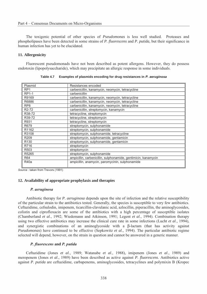
Part 4 – Consensus Documents on Micro-Organisms
338
The toxigenic potential of other species of Pseudomonas is less well studied. Proteases and phospholipases have been detected in some strains of P. fluorescens and P. putida, but their significance in human infection has yet to be elucidated.
11. Allergenicity
Fluorescent pseudomonads have not been described as potent allergens. However, they do possess endotoxin (lipopolysaccharide), which may precipitate an allergic response in some individuals.
Table 4.7 Examples of plasmids encoding for drug resistances in P. aeruginosa
Plasmid Resistances encoded
RP1 carbenicillin, kanamycin, neomycin, tetracycline
RP1-1 carbenicillin
R9169 carbenicillin, kanamycin, neomycin, tetracycline
R6886 carbenicillin, kanamycin, neomycin, tetracycline
RP8 carbenicillin, kanamycin, neomycin, tetracycline
R2-72 carbenicillin, streptomycin, kanamycin
R38-72 tetracycline, streptomycin
R39-72 tetracycline, streptomycin
R931 tetracycline, streptomycin
R679 streptomycin, sulphonamide
R1162 streptomycin, sulphonamide
R3108 streptomycin, sulphonamide, tetracycline
R209 streptomycin, sulphonamide, gentamicin
R130 streptomycin, sulphonamide, gentamicin
R716 streptomycin
R503 streptomycin
R5265 streptomycin, sulphonamide
R64 ampicillin, carbenicillin, sulphonamide, gentimicin, kanamycin
R40a ampicillin, anamycin, paromycinin, sulphonamide
Source : taken from Trevors (1991)
12. Availability of appropriate prophylaxis and therapies
P. aeruginosa
Antibiotic therapy for P. aeruginosa depends upon the site of infection and the relative susceptibility of the particular strain to the antibiotics tested. Generally, the species is susceptible to very few antibiotics. Ceftazidime, cefsulodin, imipenem, ticarcillin-clavulanic acid, azlocillin, piperacillin, the aminoglycosides, colistin and ciprofloxacin are some of the antibiotics with a high percentage of susceptible isolates (Chamberland et al., 1992; Wiedemann and Atkinson, 1991; Legent et al., 1994). Combination therapy using two effective antibiotics may increase the clinical cure rate in some infections (Lucht et al., 1994), and synergistic combinations of an aminoglycoside with a β-lactam (that has activity against Pseudomonas) have continued to be effective (Sepkowitz et al., 1994). The particular antibiotic regime selected will depend, however, on the strain in question and cannot be answered in a generic manner.
P. fluorescens and P. putida
Ceftazidime (Jones et al., 1989; Watanabe et al., 1988), imipenem (Jones et al., 1989) and meropenem (Jones et al., 1989) have been described as active against P. fluorescens. Antibiotics active against P. putida are ceftazidime, carbapenems, aminoglycosides, tetracyclines and polymixin B (Kropec
Section 2 – Pseudomonas
339
et al., 1994; Bergen, 1981; Papapetropoulou et al., 1994). Any possible treatment regime should be proposed for the strain in question and not based on generic information for the species.
P. chlororaphis, P. fragi, P. syringae, P. tolaasii
No antibiotic susceptibility data for these species were found in the literature searched.
V. Environmental and Agricultural Considerations
1. Natural Habitat and Geographic Distribution. Climatic Characteristics of Original Habitats
A. General considerations
Pseudomonas species have been isolated from a wide variety of habitats including soils, fresh or sea water, clinical specimens and laboratory reagents (including distilled water), food stuffs and wastes, flowers, fruit, vegetables, and diseased and healthy plants and animals. Many species appear to have a global distribution. For example, beneficial colonizers and Pseudomonas-incited plant diseases (such as P. syringae) are worldwide in distribution and involve representatives of most major groups of common plants.
Although pseudomonads are often considered to be ubiquitous, there are also many reports of niche specialisation. For instance, the number of epiphytic bacteria (such as P. syringae) present on the leaves of newly emerged plants is very low, indicating that the soil does not appear to serve as an important source of inoculum (Lindow, 1992) or habitat. As well, many phytopathogenic pseudomonads can only be isolated from the diseased host. For example, P. syringae isolates are generally only found in association with live plants or propagative material, and in these niches they appear as virtually homogeneous populations (Schroth et al., 1981). At present, the distribution of these pathogens in the absence of the host is unclear.
B. P. aeruginosa
P. aeruginosa is widely distributed in soil (Bradbury, 1986) and water (Palleroni, 1984). It is occasionally isolated from both healthy and diseased plants (Bradbury, 1986). Experiments conducted with lettuce and bean under varying conditions of temperature and humidity indicated that P. aeruginosa can colonise these plants under conditions of high temperature and humidity (27ºC, 80-95% humidity) (Green et al., 1974). The occurrence declined in lettuce and bean when the temperature and humidity were lowered (16ºC, 55-75% humidity).
Cho et al. (1975) studied the occurrence of P. aeruginosa on the foliage and in the soil of potted ornamental plants in order to determine their importance as a disseminating agent in hospital environments. They concluded that although potted plants are potential carriers for introduction of the species to hospital environments, there is no evidence that these plants constitute a primary source of bacteria for hospital infections. Results of a study to determine the prevalence of bacteria in passerines and woodpeckers suggest that Pseudomonas spp., including P. aeruginosa, are not uncommon in the gut flora of omnivorous and granivorous birds (Brittingham et al., 1988).
C. P. chlororaphis
P. aureofaciens (P. chlororaphis) was one of the most commonly occurring bacteria in soil, and on roots and leaves of both sugarbeet and spring wheat, during the growing season (De Leij et al., 1994). P.chlororaphis has also been isolated from water and from dead larvae of cockchafer, a large European beetle (Palleroni, 1984).
Part 4 – Consensus Documents on Micro-Organisms
340
D. P. fluorescens
P. fluorescens is commonly found on plant surfaces, as well as in decaying vegetation, soil and water (Bradbury, 1986). It can be isolated from soil, water, plants, animals, the hospital environment, and human clinical specimens. It is commonly associated with spoilage of foodstuffs such as fish and meat (Gilardi, 1991). The presence of P. fluorescens in the rhizosphere of plants has been widely reported. For example, Milus and Rothrock (1993) found P. fluorescens to be a very good coloniser of wheat roots, and Lambert et al. (1990) found P. fluorescens to be one of the most frequently occurring bacteria on root surfaces in young sugar beet plants in Belgium and Spain.
P. marginalis (P. fluorescens) is ubiquitous in soil and is often an internal resident of plant tissues (Schroth et al., 1992). Cuppels and Kelman (1980) detected P. marginalis in a Wisconsin river and lake, field soils, root zones of potato plants, washwater from a potato chip processing plant, and decaying carrot and cabbage heads. Strains were found in Wisconsin soils just after the spring thaw, and thus probably overwintered there.
E. P. fragi
P. fragi has been found associated with refrigerated meat and dairy products (Jay, 1992).
F. P. putida
P. putida is very common in soils and plant rhizospheres (Palleroni, 1984). Gilardi (1991) indicated the species can be isolated from soil, water, plants, animal sources, the hospital environment, and human clinical specimens. It can be isolated from soil and water after enrichment in mineral media with various carbon sources.
P. putida appears to have a broad global distribution. Sisinthy et al. (1989) isolated the species from soil samples collected in, and around, a lake in Antarctica. However, particular strains may have a more restricted distribution. Chanway and Holl (1993) studied strains obtained from spruce seedling rhizospheres at two different locations in British Columbia, Canada, and found two distinct strains based upon analysis of fatty acids. When the origin of the spruce seed was matched with that of the inoculated P. putida strain, a significant increase in the amount and rate of seedling emergence was detected compared to unmatched tests of seedling emergence, suggesting ecotype specificity of strains.
G. P. syringae
P. syringae occurs naturally among the microflora that inhabit the leaf surface of plants that are typically found in temperate and Mediterranean climates (Wilson and Lindow, 1994; Bradbury, 1986). P. syringae survives in association with the host plant and propagative material from the host plant. There is little evidence to suggest that these bacteria survive in soil. They may, however, survive in soil in association with residues of diseased plants, having some capacity to colonise root systems (both host and non-host plant). Stone or pome fruit pathogens, such as P. syringae, exist in lesions, cankers or tumours. Inoculum is therefore available for dissemination under favourable environmental conditions. Most of the P. syringae group appears to have the capacity to survive as epiphytes on protected parts of healthy leaves, in the buds of the host, and even on non-host plants.
H. P. tolaasii
P. tolaasii is a natural inhabitant of peat and lime used for casing material in the production of commercial mushrooms, and can be easily isolated from compost after pasteurisation (Howard et al.,1994). In the commercial production of mushrooms, high relative humidity and surface wetness encourage
Section 2 – Pseudomonas
341
the expression of symptoms of brown blotch caused by P. tolaasii (Howard et al., 1994). Symptoms of brown blotch occur more frequently on mushrooms that remain wet for a long time, and in places where they touch one another (Howard et al., 1994). Brown blotch, the mushroom disease caused by P. tolaasii,has been reported on all continents except Africa (Bradbury, 1986; Suyama and Fujii, 1993).
2. Significant Involvement in Environmental Processes, Including Biogeochemical Cycles and Potential for Production of Toxic Metabolites
Pseudomonads can have a significant involvement in a variety of environmental processes, including important biogeochemical cycles. For example, certain Pseudomonas species have the capacity for denitrification producing dinitrogen gas from nitrate. These species include P. aeruginosa, P. fluorescens(biotypes I and III) and P. chlororaphis (Palleroni, 1984).
Toxic metabolites of hazardous wastes
The microbial degradation of a hazardous waste may result in mineralisation of the parent waste, or in partial degradation of the parent waste to products which may be toxic. Some micro-organisms may not initially produce problematic metabolites. However, loss of a lower portion of a degradative pathway due to genetic instability may result in the generation of toxic metabolites. These toxic metabolites may result in death of the cell, thus limiting the metabolite’s production. The metabolite may also be released from the cell to soil or water and become rapidly inactivated or mineralised by other physical or biological processes. On the other hand, some metabolites may be released from the cell, remain stable in the environment, and have toxic effects equivalent to, or greater than, the parent hazardous waste. There is also a possibility that a micro-organism will not produce a metabolite of concern when presented with a single waste, but will produce toxic metabolites in the presence of a complex mixture of related compounds.
Many examples of hazardous waste metabolites have been detected in laboratory experiments, but no well-documented field studies on metabolite formation have been conducted. In many instances, the metabolites produced by one organism will be degraded further or mineralised by others in the immediate environment. For example, TCE epoxide and phosgene are likely degradation products from methanotrophic degradation of trichloroethylene and chloroform, respectively (Alvarez-Cohen and McCarty, 1991). Although these compounds are toxic in mammalian systems, both are also highly reactive and would likely react intracellularly and/or not persist in the environment once released from the cells. Examples of the potential for hazardous metabolites include the production and accumulation of formamide from cyanide as a result of cyanide degradation by P. fluorescens strain NCIMB 11764 (Kunz, et al., 1992). As well, Castro and Belser (1990) demonstrated that P. putida PpG-786 can dehalogenate 1,1,2-trichloroethane by two pathways under aerobic conditions. The dominant pathway is oxidative and leads to chloroacetic acid and glyoxylic acid. However, a competitive reductive pathway occurs simultaneously and yields vinyl chloride exclusively.
Complex mixtures can result in dead-end metabolite production, or failure to degrade one of the parent compounds, even though the individual wastes can be mineralised individually. Benzene, toluene, and p-xylene (BTX) are common contaminants of drinking water, and each individual BTX compound can be mineralised by naturally occurring organisms. However, a combination of the three cannot be mineralised naturally, and can result in accumulation of 3,6-dimethylcatechol from p-xylene and a lack of degradation of benzene (Lee et al., 1995).
Part 4 – Consensus Documents on Micro-Organisms
342
3. Pathogenicity – Host Range, Infectivity, Toxigenicity, Virulence, Vectors
A. General considerations
The fluorescent rRNA group I pseudomonads exhibit a range of pathogenicity characteristics. Some species have not been implicated in animal or plant disease. Other species may be opportunistic pathogens for weakened individuals. The fluorescent rRNA group I pseudomonads also include plant pathogens.
B. P. aeruginosa
Pathogenicity to animals
P. aeruginosa may be found as part of the normal bacterial flora of the intestines, mouth or skin of animals (e.g. cattle, dogs, horses, pigs). It has a broad host range among animals, which may also extend to plants. Under normal circumstances, colonisation is harmless and infection only occurs when local or general defence mechanisms are reduced. P. aeruginosa is usually associated with disease in individuals with low resistance to infection.
In susceptible hosts P. aeruginosa may cause infection at any site, particularly wounds and the respiratory tract. It can cause endocarditis, meningitis, pneumonia, otitis, vaginitis and conjunctivitis. Host defence mechanisms against P. aeruginosa are very low in mink and chinchilla, in which the bacterium can spread rapidly, causing fatal disease (Bergen, 1981).
P. aeruginosa has been associated with disease in pigs, sheep and horses (Hungerford, 1990), as well as cattle (Hamdy et al., 1974). Sheep inoculated epicutaneously with P. aeruginosa, and then wetted, can rapidly develop a bacterial exudative dermatitis (Hungerford, 1990). P. aeruginosa has been reported as the etiological agent in outbreaks of acute infectious disease in mink (Wang, 1987) and was the suspected etiological agent in a report of fatal bronchopneumonia and dermatitis in an Atlantic bottle-nosed dolphin (Diamond and Cadwell, 1979). It has also been reported to be associated with pathogenicity in ducks (Safwat et al., 1986), turkeys (Hafez et al., 1987), Japanese ptarmigan (Sato et al., 1986), and pheasant chicks (Honich, 1972) and to be the causal agent of a disease in broiler fowl in several countries.
P. aeruginosa was reported as one of the causative agents of infectious stomatitis or “mouthrot” in snakes (Draper et al., 1981), although it has been suggested that it is an opportunistic invader rather than an exogenous pathogen in snakes (Draper et al., 1981; Jacobson et al., 1981). Frogs (Rana pipiens) that were intraperitoneally injected with high doses (104-106 bacteria) of P. aeruginosa showed significant mortality under stressful conditions (Brodkin et al., 1992). P. aeruginosa has been associated with pathogenicity in Nile fish (Youssef et al., 1990) and catfish (Clarias batrachus) (Manohar et al., 1976) and as the etiological agent of fin rot in Rhamdia sapo (Angelini and Seigneur, 1988).
P. aeruginosa has been associated with pathogenicity in the tobacco hornworm (Manduca sexta)(Horohov and Dunn, 1984) and seven species of Lepidoptera, including the silkworms Pericallia ricini and Bombyx mori (Som et al., 1980). Experimental inoculation of honeybees, by dipping in a bacterial suspension of P. aeruginosa, resulted in a 70% death rate within 50 hours (Papadopoulou-Karabela et al., 1992). Dorn (1976) reported P. aeruginosa to be responsible for disease outbreaks in laboratory populations of the milkweed bug Oncopeltus fasciatus.
The abundant extracellular products of P. aeruginosa are thought to contribute to its adverse effects. These products include toxin A, alkaline protease, alkaline phosphatase, lipase, phospholipases and elastase. Toxin A is toxic to animals, with a mean lethal dose in mice of about 0.2 µg when injected intraperitoneally or 0.06 µg when injected intravenously (Nicas and Iglewski, 1986). Toxin A is produced
Section 2 – Pseudomonas
343
by about 90% of clinical isolates, and a chromosomal location has been established for the structural gene (Nicas and Iglewski, 1986). Most strains produce several extracellular proteases. For mice injected intravenously, the LD50 of the alkaline protease and the elastase is 375 and 300 µg respectively (Nicas and Iglewski, 1986). P. aeruginosa proteases are reported to be toxic to insects (Galleria mellonella) (Lysenko, 1974). P. aeruginosa also produces the haemolytic extracellular product phospholipase C, which causes hepatic necrosis and pulmonary edema when injected interperitoneally, and rhamnolipid, which has an LD50 of 5 mg when injected interperitoneally into mice (Nicas and Iglewski, 1986).
Pathogenicity to plants
P. aeruginosa has been described as an opportunistic invader of plants (Bradbury, 1986). It has been reported to cause blight disease in bean plants (El Said et al., 1982), and to have caused a lethal palm blight (Bradbury, 1986). Slow soft rot has been produced in plant tissue upon inoculation with strains of P. aeruginosa isolated from both animals and plants, and lesions and some necrosis have been found in tobacco leaves when inoculated with the bacterium (Bradbury, 1986). In a study involving 46 strains of P. aeruginosa isolated from human, plant and soil sources, the ratio of pathogenic to non-pathogenic strains for vegetables was 5:1 (Lebeda et al., 1984).
More recently, two strains of P. aeruginosa (a clinical isolate and a plant isolate) have been found to elicit severe soft rot symptoms in the leaves of inoculated Arabidopsis thaliana plants from certain ecotypes but not others (Rahme et al., 1995). These authors suggested that a strain that exhibited ecotype specificity would most likely be a true plant pathogen, in contrast to a strain that has no capacity to be a plant pathogen under natural settings but infects plants as a consequence of the artificial environment of a laboratory. The same two strains of P. aeruginosa were found to cause significant mortality in a mouse burn model. The authors identified genes encoding three virulence factors (toxA, plcS and gacA) that were required for the full expression of pathogenicity in both plants and animals.
C. P. chlororaphis
Pathogenicity to animals
A strain of P. chlororaphis has been reported to cause disease in salmon fry (Oncorhynchus rhodurus)and to kill trout, carp and eel, when inoculated (Egusa, 1992). This strain was judged to be pathogenic to fish (Hatai et al., 1975). P. chlororaphis has also been reported to inhibit egg hatch of the nematode, Criconemella xenoplax, at a concentration of 2x10⁸ cfu/ml (Westcott and Kluepfel, 1993). Shahata et al.(1988) reported that P. chlororaphis infected chickens.
Pathogenicity to plants
P. chlororaphis has been reported as the causal agent for a disease in straw mushrooms (Volvariella volvacea) in Puerto Rico, characterised by basal soft rot, internal water-soaking and discoloration (Hepperly and Ramos-Davila, 1986).
D. P. fluorescens
Pathogenicity to animals
P. fluorescens can infect a wide range of animals including horses (Sarasola et al., 1992), chickens (Lin et al., 1993), marine turtles (Glazebrook and Campbell, 1990), and many fish and invertebrate species. However, because it is unable to grow at elevated temperatures like that of the human body (Palleroni, 1992a), it is unlikely to be more than a rare opportunistic pathogen for warm-blooded animals.
Part 4 – Consensus Documents on Micro-Organisms
344
P. fluorescens is considered a secondary invader of damaged fish tissues, but may also be a primary pathogen (Roberts and Horne, 1978; Stoskopf, 1993). The species causes bacterial tail rot and can affect freshwater and saltwater fishes throughout the world (Stoskopf, 1993). P. fluorescens releases extracellular proteases upon invasion of the fish host (Li and Flemming, 1968), and morbidity can be quite high. Outbreaks of bacterial tail rot have been reported in goldfish, silver carp, bighead, tench, grass carp, black carp, golden shiner, rainbow trout, European eels, paradise fish, and other labyrinth fishes. Egusa (1992) reviewed Pseudomonas diseases in fish and indicated that, in the United States, the disease termed Pseudomonas septicemia, due to infection by bacteria related to P. fluorescens (AFS-FHS, 1975), is seen in comparatively large numbers in warm-water fish.
Adverse effects associated with P. fluorescens in fish species often appear to be linked to stress from transportation or cultivation of fish. For example, P. fluorescens has been associated with disease in the cultivation of rainbow trout, Oncorhynchus mykiss (Barros et al., 1986), Atlantic salmon, Salmo salar(Carson and Schmidtke, 1993), chinook salmon, Oncorhynchus tshawytscha (Newbound et al., 1993), sea bream, Evynnis japonica (Kusuda et al., 1974), bighead carp, Aristichthys nobilis, and silver carp, Hypophthalmichthys molitrix (Petrinec et al., 1985), catfish and carp (Gatti and Nigelli, 1984), tench (Ahne et al., 1982), and tilapia species (Okaeme, 1989; Miyashita, 1984; Miyazaki et al., 1984).
Barker et al. (1991) found that exposure of high numbers of P. fluorescens to egg surfaces of rainbow trout (Oncorhynchus mykiss) during the initial stages of incubation poses a threat to egg survival. Conversely, P. fluorescens was not pathogenic when injected into brown trout (Smith and Davey, 1993) or silver mullet fish (Mugil curema) (Alvarez and Conroy, 1987).
P. fluorescens has also been implicated in pathogenicity to some invertebrates. James and Lighthart (1992) determined an LC50 for the 1st, 2nd, 3rd and 4th instar larvae of a coleopteran insect (Hippodamia convergens) (4.8 x 10⁹, 2.8 x 10¹⁰, 3.9 x 10⁹, and 3.2 x10¹¹ CFU/ml, respectively) and concluded that P. fluorescens is a weak bacterial pathogen. P. fluorescens has also been reported to be associated with pathogenicity in the mosquitoes Culex quinquefasciatus, Anopheles stephensi, and Aedes aegypti (Murty etal., 1994) and in the field slug Deroeras reticulatum (Wilson et al., 1994). However, Genthner et al.(1993) studied the effects of P. fluorescens on eastern oysters (Crassostrea virginica) and found no signs of infectivity or pathogenicity.
Pathogenicity to plants
P. fluorescens is generally considered to be a saprophyte rather than a plant pathogen (Bradbury, 1986), although Ormrod and Jarvis (1994) considered it to be an opportunistic pathogen causing soft rot in plants. P. fluorescens biovar 2 (P. marginalis), however, is actively pectinolytic, causing soft rot of various plants, and is considered a plant pathogen (Tsuchiya et al., 1980; Hildebrand, 1989; Membre and Burlot, 1994; Brock et al., 1994). Bradbury (1986) recognised three pathovars in P. marginalis which cause soft rot in a wide range of vegetables and other plants.
A number of studies have reported adverse effects associated with P. fluorescens and plants (Gaudet et al., 1980; Anson, 1982; Hwang et al., 1989; Richardson, 1993; Ozaktan and Bora, 1994). Tranel et al.(1993) found that P. fluorescens strain D7 inhibited root growth of downy brome (Bromus tectorum) by production of a phytotoxin. Sellwood et al. (1981) confirmed pathogenicity experimentally for an atypical P. fluorescens biotype I on chicory plants and suggested that the group P. fluorescens does not solely comprise saprophytes. However, other studies have found no adverse effects on plants from inoculations with P. fluorescens (Arsenijevic, 1986; Arsenijevic and Balaz, 1986; Surico and Scala, 1992). At present, the epidemiology of pathogenic strains of P. fluorescens is not well understood (Hildebrand, 1994).
Section 2 – Pseudomonas
345
Liao (1991) has cautioned that, at present, it is not known for sure that P. fluorescens strains are non-pathogens and do not cause deleterious effects on plants. Liao indicated that the pel gene encoding production of pectate lysase (an enzyme which contributes ability to cause soft rot in plants) is well conserved in fluorescent pseudomonads, and may exist and remain repressed in certain strains or species which exhibit non-pectolytic phenotypes under laboratory conditions. Liao further indicated that saprophytic strains of P. fluorescens can be induced to become pathogenic and raised a concern about the safety of using the species as a biocontrol agent.
E. P. fragi
Pathogenicity to animals
No reports were found of P. fragi as an animal pathogen.
Pathogenicity to plants
No reports were found of P. fragi as an plant pathogen.
F. P. putida
Pathogenicity to animals
P. putida can infect a variety of animals including goats (Hungerford, 1990), koala (Ladds et al., 1990), turkey (Ononiwu, 1980) and fish (Kusuda and Toyoshima, 1976). However, because it is unable to grow at elevated temperatures like that of the human body (Palleroni, 1992a), it is unlikely to be more than a rare opportunistic pathogen for warm-blooded animals.
Kusuda and Toyoshima (1976) reported P. putida to be a pathogen to cultivated yellowtail fish. However, there have been no reports on the epizootiology, symptoms, or histological or pathological findings, and the disease has not been well-defined (Egusa, 1992). Austin and McIntosh (1991) considered P. putida to be one of a variety of gram-negative bacteria pathogens of potential concern to farmed and wild fish. P. putida has also been associated with pathogenicity in the snail, Biomphalaria glabrata(Cheng, 1986), the crayfish (Boemare and Vey, 1977), and the olive fly (Haniotakis and Avtizis, 1977).
Pathogenicity to plants
P. putida was included in the Guide to Plant Pathogenic Bacteria solely because its multiplication in the rhizosphere of paddy rice plants has been implicated in “suffocation disease”, which arises under conditions of poor drainage (Bradbury, 1986). Studies have reported that P. putida is not pathogenic to mushrooms (Ozakatan and Bora, 1994) or crucifer plants (Shaw and Kado, 1988).
Liao (1991) has cautioned that, at present, it is not known for sure that P. putida strains are non-pathogens and do not cause deleterious effects on plants. Liao indicated the pel gene encoding production of pectate lysase (an enzyme which contributes ability to cause soft rots in plants) is well conserved in fluorescent pseudomonads and may exist and remain repressed in certain strains or species which exhibit non-pectolytic phenotypes under laboratory conditions. Homologous sequences were found in strains of P. putida, and Liao raised a concern about the safety of using P. putida as a biocontrol agent.
Part 4 – Consensus Documents on Micro-Organisms
346
G. P. syringae
Pathogenicity to animals
No reports were found of P. syringae as an animal pathogen.
Pathogenicity to plants
P. syringae is principally an assemblage of foliar pathogens, although it occurs as both pathogenic and epiphytic (non-pathogenic) strains. The species has a broad range of potential plant hosts (Table 4.8). Pathogenic strains can exhibit both pathogenic (i.e. disease-causing) and epiphytic behaviours on susceptible hosts (Crosse, 1959). The initiation of infection results when a threshold level of bacteria is reached on the leaf surface; in the case of P. syringae pv. syringae this is reported to be 104 cfu gm-1 tissue (Hirano and Upper, 1983).
The association between rain and the onset of foliar blights caused by P. syringae is well recognised. Rain appears to stimulate the differential growth of pathogenic P. syringae isolates from the heterogeneous populations (pathogenic and non-pathogenic strains). Rain-triggered growth of P. syringae results in the establishment of large pathogenic populations required for disease development (Hirano and Upper, 1992).
There appears to be a distinctive set of symptoms associated with each causal agent. Pseudomonas syringae pv. savasatoni incites tumourous outgrowths on stems and leaves of oleander and olive under natural conditions. These symptoms have been found to be associated with the production of the auxin, indole acetic acid (IAA), in tissues infected with the bacterium. Furthermore, chlorosis, a common symptom when plants are infected by a number of pathogens belonging to the P. syringae group, is indicative of production of a toxin. For example, halo blight of beans caused by P. syringae pv. phaselicolais mediated by the toxin, phaseolotoxin. Other phytopathogenic pseudomonads producing toxins are illustrated in Table 4.9.
The Dictionary of Natural Products (Chapman and Hall, 1995) lists the following toxins produced by various strains of P. syringae: 1H-Indole-3-carboxaldehyde, octicidin (phytotoxin), phaseolotoxin (phytotoxin), N-Phosphosulfamylornithine (phytotoxin), syringomycin (phytotoxin), syringostatin A (phytotoxin), syringostatin B (phytotoxin), syringotoxin B (phytotoxin), tagetitoxin (phytotoxin), coronafacic acid (induces chlorosis in plants), halotoxin (phytotoxin), tabtoxin (phytotoxin). Coronatine (phytotoxin) is also produced by certain strains of P. syringae (Cuppels and Ainsworth, 1995). Gross (1985) determined that syringomycin production was stimulated by iron and suppressed by inorganic phosphate, that production occurred between 15 and 27°C, and that a slow growth rate of P. syringaefavours toxin production.
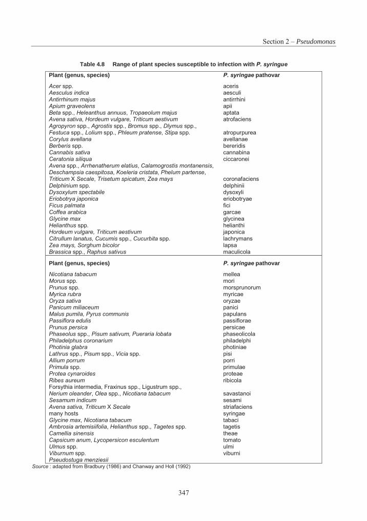
Section 2 – Pseudomonas
347
Table 4.8 Range of plant species susceptible to infection with P. syringue
Plant (genus, species) P. syringae pathovar
Acer spp. aceris Aesculus indica aesculi Antirrhinum majus antirrhini Apium graveolens apii Beta spp., Heleanthus annuus, Tropaeolum majus aptata Avena sativa, Hordeum vulgare, Triticum aestivum atrofaciens Agropyron spp., Agrostis spp., Bromus spp., Dlymus spp., Festuca spp., Lolium spp., Phleum pratense, Stipa spp. atropurpurea Corylus avellana avellanae Berberis spp. bereridis Cannabis sativa cannabina Ceratonia siliqua ciccaronei Avena spp., Arrhenatherum elatius, Calamogrostis montanensis,Deschampsia caespitosa, Koeleria cristata, Phelum partense,Triticum X Secale, Trisetum spicatum, Zea mays coronafaciens Delphinium spp. delphinii Dysoxylum spectabile dysoxyli Eriobotrya japonica eriobotryae Ficus palmata fici Coffea arabica garcae Glycine max glycinea Helianthus spp. helianthi Hordeum vulgare, Triticum aestivum japonica Citrullum lanatus, Cucumis spp., Cucurbita spp. lachrymans Zea mays, Sorghum bicolor lapsa Brassica spp., Raphus sativus maculicola
Plant (genus, species) P. syringae pathovar
Nicotiana tabacum mellea Morus spp. mori Prunus spp. morsprunorumMyrica rubra myricae Oryza sativa oryzae Panicum miliaceum panici Malus pumila, Pyrus communis papulans Passiflora edulis passiflorae Prunus persica persicae Phaseolus spp., Pisum sativum, Pueraria lobata phaseolicola Philadelphus coronarium philadelphi Photinia glabra photiniae Lathrus spp., Pisum spp., Vicia spp. pisi Allium porrum porri Primula spp. primulae Protea cynaroides proteae Ribes aureum ribicola Forsythia intermedia, Fraxinus spp., Ligustrum spp., Nerium oleander, Olea spp., Nicotiana tabacum savastanoi Sesamum indicum sesami Avena sativa, Triticum X Secale striafaciens many hosts syringae Glycine max, Nicotiana tabacum tabaci Ambrosia artemisiifolia, Helianthus spp., Tagetes spp. tagetis Camellia sinensis theae Capsicum anum, Lycopersicon esculentum tomato Ulmus spp. ulmi Viburnum spp. viburni Pseudostuga menziesii
Source : adapted from Bradbury (1986) and Chanway and Holl (1992)
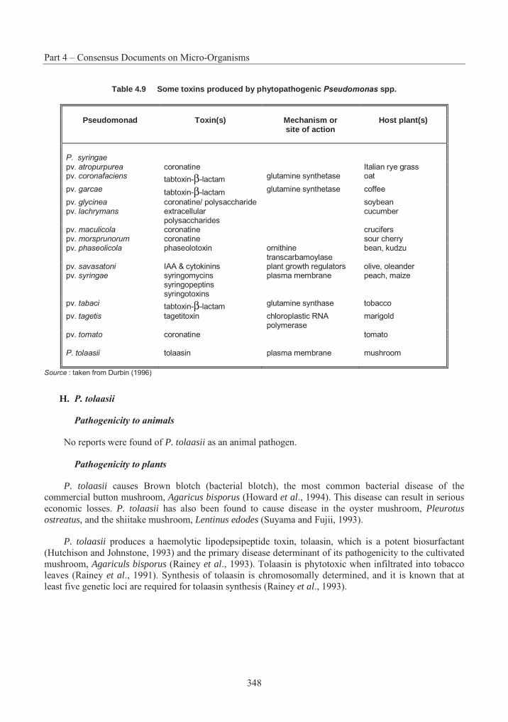
Part 4 – Consensus Documents on Micro-Organisms
348
Table 4.9 Some toxins produced by phytopathogenic Pseudomonas spp.
Pseudomonad Toxin(s) Mechanism or
site of action Host plant(s)
P. syringae
pv. atropurpurea coronatine Italian rye grass pv. coronafaciens tabtoxin-β-lactam glutamine synthetase oat
pv. garcae tabtoxin-β-lactam glutamine synthetase coffee
pv. glycinea coronatine/ polysaccharide soybean pv. lachrymans extracellular
polysaccharides cucumber
pv. maculicola coronatine crucifers pv. morsprunorum coronatine sour cherry pv. phaseolicola phaseolotoxin ornithine
transcarbamoylase bean, kudzu
pv. savasatoni IAA & cytokinins plant growth regulators olive, oleander pv. syringae syringomycins
syringopeptins syringotoxins
plasma membrane peach, maize
pv. tabaci tabtoxin-β-lactam glutamine synthase tobacco
pv. tagetis tagetitoxin chloroplastic RNA polymerase
marigold
pv. tomato coronatine tomato
P. tolaasii tolaasin plasma membrane mushroom
Source : taken from Durbin (1996)
H. P. tolaasii
Pathogenicity to animals
No reports were found of P. tolaasii as an animal pathogen.
Pathogenicity to plants
P. tolaasii causes Brown blotch (bacterial blotch), the most common bacterial disease of the commercial button mushroom, Agaricus bisporus (Howard et al., 1994). This disease can result in serious economic losses. P. tolaasii has also been found to cause disease in the oyster mushroom, Pleurotus ostreatus, and the shiitake mushroom, Lentinus edodes (Suyama and Fujii, 1993).
P. tolaasii produces a haemolytic lipodepsipeptide toxin, tolaasin, which is a potent biosurfactant (Hutchison and Johnstone, 1993) and the primary disease determinant of its pathogenicity to the cultivated mushroom, Agariculs bisporus (Rainey et al., 1993). Tolaasin is phytotoxic when infiltrated into tobacco leaves (Rainey et al., 1991). Synthesis of tolaasin is chromosomally determined, and it is known that at least five genetic loci are required for tolaasin synthesis (Rainey et al., 1993).
Section 2 – Pseudomonas
349
4. Interactions with and Effects on Other Organisms in the Environment
A. P. aeruginosa
Certain P. aeruginosa strains are antagonistic to plant pathogens such as damping-off fungi (Bradbury, 1986; Buysens et al., 1994). Duffy and Defago (1995) found that clinical and plant isolates of P. aeruginosa suppressed root diseases of cucumber, maize and wheat caused by soilborne fungi Gaeumannomyces graminis var. tritici, Phomopsis sclerotiodes, Pythium ultimum and Rhizoctonia solani.A soil isolate of P. aeruginosa suppressed foliar disease on wheat caused by Septoria tritici (Flaishman et al., 1990).
P. aeruginosa can have a synergistic effect on the survival of salmonellae, enabling them to survive more than 140 days in double-distilled water (Warburton et al., 1994). It has also been suggested that P. aeruginosa may act synergistically with pectolytic bacteria that colonise vegetables, such as P. marginalis(P. fluorescens) and Erwinia cartovora (Bradbury, 1986). A protective immunity against P. aeruginosainfection has been reported in mice vaccinated with heat-killed Lactobacillus casei (Miake et al., 1985).
P. aeruginosa is known to produce 1-phenazinecarboxamide (the amide of 1-phenazinecarboxylic acid), which is active against some phytopathogenic fungi and Candida albicans. A related compound, 1-phenazinol, which is active against gram-positive bacteria and fungi, and which shows some viral activity, is also produced by P. aeruginosa. 1-phenazinol has an LD50 of 500 mg/kg in mice dosed intraperitoneally. Pyoluteorin and its 3’-nitro derivative are produced by P. aeruginosa. Both compounds have antibacterial, antifungal and herbicidal properties. The LD50 of the pyoluteorin to mice is 125 mg/kg (Chapman and Hall, 1995). The antibiotic, 2-heptyl-4-hydroxyquinoline N-oxide, is a metabolite of P. aeruginosa and is a potent 5’- lipoxygenase inhibitor, with an LD50 of 40 mg/kg in mice dosed intraperitoneally (Chapman and Hall, 1995).
B. P. chlororaphis
P. chlororaphis has been widely investigated for its ability to enhance plant growth through suppression of deleterious root-colonising bacteria. Compounds known as siderophores are produced by P. chlororaphis. These compounds chelate iron, thereby depriving certain root-colonising plant pathogens of iron necessary for their growth (Smirnov et al., 1991).
Many studies have indicated that P. chlororaphis has the ability to suppress plant disease. For example, P. aureofaciens has been investigated as a biocontrol agent to suppress take-all, the wheat root fungal disease. The ability of P. aureofaciens to inhibit Gaeumannomyces graminis var. tritici, the causal agent of take-all, was demonstrated in vitro and in vivo (Harrison et al., 1993). It is believed that disease suppression is largely due to the production of phenazine antibiotics (Thomashow and Pierson, 1991).Carruthers et al. (1995) tested the ability of P. aureofaciens to suppress root rot of Asparagus officinaliscaused by Phytophthora megasperma var. sojae. P. aureofaciens significantly reduced the level of infection and disease severity. Other tests suggested that P. aureofaciens had a direct growth stimulatory effect on asparagus, independent of antibiotic production (Carruthers et al., 1995). Berg and Ballin (1994) found P. chlororaphis inhibited the growth of the phytopathogenic fungus Verticillium dahliae.
When Douglas fir seed was inoculated with P. aureofaciens, and grown in pasteurised soil, shoot biomass increased significantly when compared with non-inoculated controls (Chanway and Holl, 1992). P. aureofaciens has been found to inhibit mycelial growth of Rhizoctonia solani in dual culture between 15 and 30°C (Lee et al., 1990). Inoculation of rice seeds was found to control rice sheath blight in the early growth stages, and seedling blight caused by R. solani, Fusarium moniliforme and Pythium ultimum wassuppressed by seed treatment and soil incorporation of P. aureofaciens (Lee et al., 1990). In another
Part 4 – Consensus Documents on Micro-Organisms
350
experiment, the emergence of sweet corn seedlings from soil infested with Pythium ultimum was greatly enhanced by coating the seed with P. aureofaciens (Mathre et al., 1994). P. aureofaciens has also been evaluated for its ability to suppress Pythium ultimum damping off of cucumber seedlings (Sugimoto et al.,1990).
P. aureofaciens was antagonistic to Clavibacter michiganensis subsp. sepedonicus, the bacteria implicated in potato ring rot in greenhouse trials with potato seedlings (de la Cruz et al., 1992). P. aureofaciens significantly reduced populations of, and infection by, the ring rot bacteria (de la Cruz etal., 1992). Fukui et al. (1994) investigated the relationship between pericarp colonisation by Pythium ultimum in sugar beets and the growth of pseudomonads in the spermosphere. They found a positive correlation between the incidence of pericarp colonisation by Pythium ultimum and the length of the lag phase of the strain used to inoculate the seeds. England et al. (1993) investigated the nodulation of whitebean (Phaseolus vulgaris L.) by Rhizobium phaseoli in the presence of P. aureofaciens. No significant difference was found in the numbers of nodules produced in the presence of P. aureofaciens as a result of the symbiotic relationship between Rhizobium phaseoli and whitebean roots in vermiculite.
P. chlororaphis was observed to interfere with the growth of shiitake mushrooms in field experiments with shiitake cultivated logs (Raaska and Mattila-Sandholm, 1991). Siderophores were produced, however the addition of iron to in vitro cultures did not entirely neutralize the growth inhibition of mycelia by P.chlororaphis. It was concluded that although iron-binding plays an important role, it is not the only factor involved in the inhibition of shiitake by P. chlororaphis (Raaska and Mattila-Sandholm, 1991). A siderophore extracted from P. aureofaciens was found to inhibit uptake of ferric iron by maize and pea, and the synthesis of chlorophyll in these plants was reduced (Becker et al., 1985).
P. aureofaciens is reported to produce an antibiotic-like compound in iron-rich conditions that inhibits the growth of the plant fungal pathogen Aphanomyces euteiches (Carruthers et al., 1994). Mazzola et al.(1992) suggested that the production of phenazine antibiotics contributes to the ecological competence of P. aureofaciens, and that reduced survival of strains unable to produce the antibiotics is due to diminished ability to compete with the resident microflora. Thomashow et al. (1990) found that suppression of take-all is related directly to the presence of the antibiotic phenazine-1-carboxylic acid in the rhizosphere of wheat. In another experiment, 2-hydroxyphenazine-1-carboxylic acid and 2-hydroxyphenazine were also found to be responsible for take-all suppression in wheat (Pierson and Thomashow, 1992). Pyrrolnitrin [3-chloro-4-(3-chloro-2-nitrophenyl)-1H-pyrrole] is an antifungal compound produced by P. chlororaphis; its LD50 in mice dosed orally is 1 g/kg (Chapman and Hall, 1995). The antifungal compound, 1,3,6-trihydroxy-2,4-diacetophenone, has also been isolated from culture media (Harrison et al., 1993).
C. P. fluorescens
P. fluorescens has been recognised as beneficial to plant growth (Weller and Cook, 1986; Kloepper etal., 1988). It can enhance plant growth through production of siderophores, which efficiently complex environmental iron, making it unavailable to other components of the soil microflora. Increased plant yields achieved through the inoculation of plant roots have been mimicked by the application of the siderophore, pseudobactin, isolated from P. fluorescens. Antibiotic production by P. fluorescens has been recognised as an important factor in its ability to suppress phytopathogens. P. fluorescens has also been found to significantly promote nodulation, growth and nitrogen accumulation in faba beans (Vicia faba)(Omar and Abd-Alla, 1994). Heat-killed cells had no effect.
Certain strains of P. fluorescens can promote the formation of ice crystals in water at temperatures near 0°C (Lindow and Panopoulos, 1988; Lindow, 1992). Large populations of these ice+ bacteria on plant surfaces can cause frost injury. Only 0.01 to 40% of the total bacteria on plant surfaces are sufficient to cause frost injury. In the absence of these bacteria, water on plants can cool to -40°C.
Section 2 – Pseudomonas
351
Smith and Davey (1993) found that P. fluorescens strains were able to inhibit Aeromonas salmonicidathat was isolated from Atlantic salmon with furunculosis. Pre-smolts asymptomatically infected with A. salmonicida and bathed in a solution containing P. fluorescens strains were less likely to develop stress-induced furunculosis than non-treated fish. It was concluded that P. fluorescens inhibits A. salmonicida by competing for free iron, and that it protects against stress-induced furunculosis by inhibiting A. salmonicida on external locations. Kimura et al. (1990) found that a strain of P. fluorescens biovar I (46NW-04) isolated from the aquatic environment produced an antiviral substance that was effective against fish viruses.
D. P. fragi
Monitoring of microbial flora succession on minced lamb meat revealed that P. fragi was the dominant climax species (Drosinos and Board, 1995). Another study indicated that P. fragi dominated the flora on lamb carcasses at both 7 and 30°C (Prieto et al., 1992).
E. P. putida
P. putida is very common in soils and plant rhizospheres, where it seems to have a stimulating effect on plant growth (Palleroni, 1984). P. putida has been shown to suppress a variety of plant pathogens and to reduce the incidence of plant disease (Liao, 1989; Gamliel and Katan, 1993; Duijff et al., 1994; Freitas et al., 1991; Defago and Hass, 1990). This may be due in part to its inhibition of plant pathogenic micro-organisms by sequestering iron or producing metabolites with antibiotic properties. Formation of a siderophore complex by the plant may also be involved (Defago and Hass, 1990). Siderophore-mediated competition for iron was indicated as the mechanism of suppression of Fusarium wilt of carnation by P. putida (WCS358r) (Duijff et al., 1994) and suppression of phytopathogens to winter wheat (Freitas et al., 1991).
Al-Achi et al. (1991) found that, when introduced as pairs into irradiated, sterile soils, a P. fluorescensstrain prevented optimum colonisation by a P. putida strain. The addition of P. putida to sterile soil already populated by P. fluorescens impeded growth of P. putida in that soil. However, adding P. fluorescens to soil populated by P. putida did not prevent growth of P. fluorescens and caused a decrease in P. putida.These results suggested competition for similar niches in soils, and that P. fluorescens was the more competitive species studied.
F. P. syringae
Some strains of P. syringae have the ability to cause ice nuclei to form at temperatures just below 0°C, thus inducing freezing injury to susceptible plants and allowing disease development to occur (Lindow, 1983). Nutritional starvation for nitrogen, phorphorous, sulphur or iron at 32°C, followed by a shift to 14-18°C, led to the rapid induction (from non-detectable to 100% in 2 to 3 h) of type I ice nuclei (Nemecek-Marshall et al., 1993).
Replacement series experiments on bean leaves between P. syringae and epiphytic P. fluorescens, Pantoea agglomerans (Erwinia herbicola), Stenotrophomonas maltophilia (Xanthomonas maltophilia) and Methylobacterium organophilum have demonstrated that the epiphytes were all capable of higher levels of coexistence with P. syringae than was observed with another P. syringae strain. The level of coexistence with the epiphytes was inversely correlated with the ecological similarity of the strains and with a differential preference for amino acids, organic acids and carbohydrates (Wilson and Lindow, 1994).
The invasion and exclusion abilities of 29 strains of P. syringae were studied on leaves in 107 pairwise combinations in which each strain was inoculated on day 0, and the second (challenge) was
Part 4 – Consensus Documents on Micro-Organisms
352
inoculated on the same leaf on day 3 (Kinkel and Lindow, 1993). The presence of an established population often significantly reduced the growth of the second strain when quantified on day 6; successful invaders (challenge) were significantly less likely to exclude challenge populations than were non-successful invaders. Hirano and Upper (1993) determined that an introduced antibiotic-resistant strain of P. syringae spread but did not persist when applied to bean plants grown in the field; it was concluded that the introduced strain was less fit than the pool of indigenous species. Competition between indigenous soil bacteria and single cells of P. syringae pv. syringae engineered with bioluminescence genes from Vibrio harveyi can be monitored using charge-coupled enhanced microscopy (Silcock et al., 1992).
Defreitas et al. (1993) determined that P. syringae R25 inoculated on field peas (Pisum sativum) did not affect plant growth in plastic growth pouches but, in soil, did inhibit nitrogenase activity of nodules formed by indigenous rhizobia; P. syringae R25 inhibited the growth of field beans (Phaseolous vulgaris)in both plastic growth pouches and in soil. When peas were inoculated with both P. syringae R25 and Rhizobium leguminosarum, there was an increase in plant biomass in growth pouches but no effect was observed in soil; when beans were inoculated with both P. syringae R25 and Rhizobium phaseoli, there were severe deleterious effects on seedling emergence, plant biomass and nodulation in both growth pouches and soil.
Table 4.10 Phytopathogenic strains of P. syringae containing plasmids
Pathovar Reference
P. syringae pv. angulata Piwowarski and Shaw, 1982 P. syringae pv. atrpurpurea Sato et al., 1983 P. syringae pv. coronafaciens Piwowarski and Shaw, 1982 P. syringae pv. glycinea Curiale and Mills, 1983 P. syringae pv. lachrymans Coplin, 1989 P. syringae pv. papulans Burr et al., 1988 P. syringae pv. phaseolicola Quant and Mills, 1984 P. syringae pv. savastanoi Comai et al., 1982 P. syringae pv. striafaciens Beck-Von Bodmann and Shaw, 1987 P. syringae pv. syringae Gonzales et al., 1984 P. syringae pv. tabaci Obukowicz and Shaw, 1983; 1985 P. syringae pv. tomato Denny, 1988; Bender and Cooksey, 1986
G. P. tolaasii
P. tolaasii produces a haemolytic lipodepsipeptide toxin, tolaasin, which is a potent biosurfactant (Hutchison and Johnstone, 1993) and the primary disease determinant of its pathogenicity to the cultivated mushroom, Agariculs bisporus (Rainey et al., 1993). Tolaasin is also active against a range of basidiomycetes and gram-positive bacteria (Rainey et al., 1991).
The nematode Caenorhabditis elegans is reported to decrease the spread of P. tolaasii in mushroom growth chambers (Grewal, 1991). P. fluorescens biovar reactans was frequently isolated from the gut of C.elegans along with mushroom sporophores. All the isolates of P. fluorescens biovar reactans isolated from nematodes were antagonists to P. tolaasii. It was suggested that, as C. elegans selects P. fluorescens biovarreactans rather than P. tolaasii as a food, it probably spreads the antagonist in the mushroom crop and may contribute to the control of mushroom blotch (Grewal, 1991). P. fluorescens has also been described by other researchers as antagonistic to P. tolaasii (Khanna and Olivier, 1989; Munjal et al., 1989; Nair and Fahy, 1972). Nair and Fahy (1972) reported Enterobacter aerogenes to be antagonistic to P. tolaasii.
Section 2 – Pseudomonas
353
Thorn and Tsuneda (1992) report that 23 species of wood-decay basidiomycetes attacked or lysed P.tolaasii when tested. Attack took the form of increased hyphal branching within the bacterial colonies, often preceded by directional growth toward them.
5. Ability to form Survival Structures (e.g. Spores, Sclerotia)
Pseudomonads are asporogenous, that is, they do not form spores or other survival structures. Pseudomonads are, however, pleomorphic and represent a tremendously diverse group of strains able to tolerate extreme environmental conditions, including the extremes of temperature.
Bacteria that do not form survival structures like spores and cysts are suspected to have other survival strategies. A number of researchers have reported the existence of dwarf or ultramicrobacteria in nutrient-stressed environments (Rosak and Colwell, 1987). These cells have been described from seawater (Amy and Morita, 1983) and soil (Casida, 1977). Cells are able to develop to their full size, once exposed to an abundant supply of nutrients.
6. Routes of Dissemination, Physical or Biological
A. Physical
Pseudomonads may be disseminated by air or water currents. For example, Trevors et al. (1990) used soil-core microcosms to study the movement of a P. fluorescens isolate through soil planted with wheat and unplanted. In the absence of ground water flow, limited movement was detectable along the soil column planted with wheat, while no movement was detected in the unplanted soil. In contrast, movement of the strain through the column was dependent on the flow rate of the water and the number of times the columns were flushed through. Water flow also affected the distribution of the inoculant along the wheat roots. Bacterial cell size has been related to the movement of cells through a soil column, with smaller bacterial cells (< 1.0µm) moving fastest through the column (Gannon et al., 1991).
Rain may also be an important source of inoculum and means of dispersal for pseudomonads. Rain splash has been attributed to move pseudomonads colonising leaf surfaces down the plant canopy and into the soil (Hirano and Upper, 1992; Butterworth and McCartney, 1992; McCartney and Butterworth, 1992). Large drops of artificial rain were more effective in dispersing bacteria than smaller drops (Butterworth and McCartney, 1992). Humidity correlated positively with the consequent survival of pseudomonads dispersed by rain splash (McCartney and Butterworth, 1992). However, dispersal is short range (one or a few metres) (Constantidou et al., 1990). Pseudomonads (including P. syringae Ice+ strains) have been found to leave plant surfaces in an aerosol-stable state and enter the troposphere during dry, warm weather (Lindemann et al., 1982; Lindemann and Upper, 1985). They are then transported and washed downwards during rainfall (Constantidou et al., 1990).
B. Biological
Pseudomonads are motile bacteria characterised by the presence of at least one flagellum. While there is no convincing evidence that the bacteria are flagellated in soil (Stotzky et al., 1991), flagella appear to confer increased epiphytic fitness on P. syringae strains in association with moisture on leaf surfaces (Haefele and Lindow, 1987). The potential for certain fluorescent pseudomonads to colonise plant surfaces has been attributed to the presence of pili (Vesper, 1987; de Groot et al., 1994), surface charge properties (James et al., 1985), the production of agglutinin, a glycoprotein complex, released from root surfaces (Anderson, 1983), and the ability of certain saprophytic pseudomonads to adhere to the agglutinin of specific plant species (Glandorf et al., 1993; 1994).
Part 4 – Consensus Documents on Micro-Organisms
354
Earthworms moving through soil have been implicated in the dissemination of bacteria over short distances. As well, Johnson et al. (1993) have demonstrated the ability of honey bees to disseminate a biological control strain of P. fluorescens used against the fireblight pathogen, Erwinia amylovora, in apple and pear blossoms. Honey bees carrying approximately 104 to 106 cfu per bee effectively inoculated fruit tree blossoms with bacteria.
7. Containment and Decontamination
Containment plans have been proposed for microbial releases, although few of them have been used, and their efficacy is yet to be demonstrated. It is likely to be difficult to eliminate all the bacteria from a site of introduction. Many of the proposed chemical treatments have gross rather than localised effects; hence their application may have considerable impact on the natural flora, fauna and microflora at the site. Pseudomonads will colonise many laboratory and hospital disinfectants, and may exhibit broad spectrum resistance to a number of widely used antibiotics. Disinfectants based on quaternary ammonium compounds and chlorhexidine solutions have been found to be contaminated with pseudomonads. Disinfectant contaminants include P. aeruginosa, P. fluorescens, and P. cepacia (Bergen, 1981).
P. putida strains that degrade alkylbenzoates have been modified to carry a fusion of the P (lac) promoter to the gef gene, which encoded a killing protein (Molin et al., 1993; Ramos et al., 1994). Expression from P (lac) was controlled through a regulatory cascade, so that P (lac) was switched on or off by the absence or presence of alkylbenzoates respectively. Similar uncontained strains were also constructed and tested as a control. Contained and uncontained strains were genetically stable, and their survival and functionality in soil microcosms were as expected. Both contained and uncontained strains survived well in soils supplemented with alkylaromatics, whereas survival of the contained strain in soil microcosms without methylbenzoates was markedly reduced in contrast to the control strain, which survived in these soils in the absence of alkylbenzoates (Jensen et al., 1993; Ronchel et al., 1995).
8. Description of Detection and Monitoring Techniques, Including Specificity, Sensitivity and Reliability
A. Techniques mployed in the laboratory and/ or environment for detecting the presence of, and for monitoring, numbers of the organism
Information on detection and monitoring techniques is provided in this information element as well as in information element 2 and Table 4.11. Each of the well-described detection methods has limitations as well as advantages for enumeration and/or detection (Drahos, 1992). For example, under certain conditions an approach which provides reasonable sensitivity by culturing a micro-organism (e.g. the viable plate count) may give reliable data for culturable populations. Furthermore, many approaches are complementary; methods utilising nutritional, antibiotic and enzymatic markers rely on the ability of the target organism to express the marker genes during the selection or reculturing process. However, expression of these traits may not always be optimal, for instance under conditions of severe environmental stress. In these situations, a direct method of detection could be used.
Selective plating
Selective plating has been used widely in combination with selectable phenotypes based on antibiotic resistances (often spontaneous mutations) (Compeau et al., 1988; Fredrickson et al., 1989; Thompson etal., 1990) or introduced genes such as xylE (Winstanley et al., 1989; Morgan et al., 1989) lacZY (Cook et al., 1991; Drahos et al., 1988), lux (Shaw and Kado, 1986) and mer gene (Iwasaki et al., 1993; 1994). A number of these genes have been used for marking and tracking pseudomonads. It is important to ensure
Section 2 – Pseudomonas
355
that the marker is not found in the indigenous microflora of the environment to which the micro-organism will be introduced.
Pseudomonads appear to be highly culturable on laboratory media and may be isolated from environmental samples using viable plating (Drahos, 1992). Generally, 1 g of environmental sample is homogenised or shaken in 9 ml of an appropriate dilutent such as ¼ strength Ringer’s solution or physiological saline. The homogenate is serially diluted 1 in 10, 100 µl aliquots spread onto selective agar, and the plates incubated at 28°C. A number of selective media are available commercially, such as Pseudomonas selective agar (Oxoid) and Pseudomonas agar F (Difco). Both media have a low iron content, promoting the production of the iron-chelating, fluorescent siderophores. Selective agars are supplemented with antibiotics. A commercially prepared cocktail of cephalorodine, fucidin acid and cetrimide (Oxoid) is available which may be supplemented with ampicillin and the antifungal agent, cyclohexamide. Micro-organisms may be detected at or above a detection limit of 102 (i.e. one cell may be detected when a minimum of 100 are present per g of sample) (Trevors and van Elsas, 1989). Sensitivity may be increased by plating larger volumes or by using smaller dilutions, i.e. 1 in 2 instead of 1 in 10.
Most probable number
Most probable number (MPN) methods (Alexander, 1982) have been used to attain greater sensitivity. A serial dilution of the sample is made in an appropriate dilutent to an extinction point (Atlas, 1982). Three to ten replicates of each dilution are made and the pattern of positive and negative scores recorded (i.e.growth or no growth). Statistical tables are used to determine the MPN of micro-organisms present in the sample. MPNs like the viable plate count require growth and reproduction of the strains, and may be less accurate since an MPN is established with confidence limits (Jain et al., 1988).
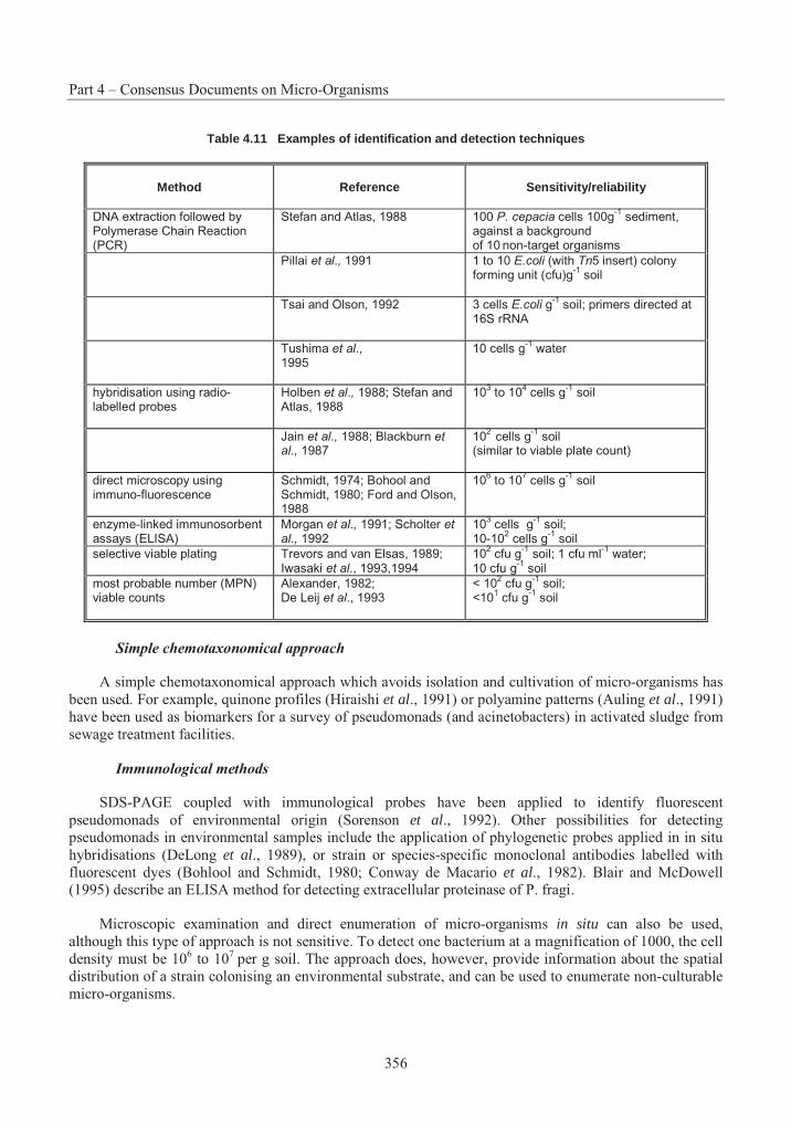
Part 4 – Consensus Documents on Micro-Organisms
356
Table 4.11 Examples of identification and detection techniques
Method Reference Sensitivity/reliability
DNA extraction followed by Polymerase Chain Reaction (PCR)
Stefan and Atlas, 1988 100 P. cepacia cells 100g-1
sediment, against a background of 10 non-target organisms
Pillai et al., 1991 1 to 10 E.coli (with Tn5 insert) colony forming unit (cfu)g
-1 soil
Tsai and Olson, 1992 3 cells E.coli g-1
soil; primers directed at 16S rRNA
Tushima et al.,1995
10 cells g-1
water
hybridisation using radio-labelled probes
Holben et al., 1988; Stefan and Atlas, 1988
103 to 10
4 cells g
-1 soil
Jain et al., 1988; Blackburn et al., 1987
102
cells g-1
soil (similar to viable plate count)
direct microscopy using immuno-fluorescence
Schmidt, 1974; Bohool and Schmidt, 1980; Ford and Olson, 1988
106 to 10
7 cells g
-1 soil
enzyme-linked immunosorbent assays (ELISA)
Morgan et al., 1991; Scholter et al., 1992
103 cells g
-1 soil;
10-102 cells g
-1 soil
selective viable plating Trevors and van Elsas, 1989; Iwasaki et al., 1993,1994
102 cfu g
-1 soil; 1 cfu ml
-1 water;
10 cfu g-1
soil
most probable number (MPN) viable counts
Alexander, 1982; De Leij et al., 1993
< 102 cfu g
-1 soil;
<101 cfu g
-1 soil
Simple chemotaxonomical approach
A simple chemotaxonomical approach which avoids isolation and cultivation of micro-organisms has been used. For example, quinone profiles (Hiraishi et al., 1991) or polyamine patterns (Auling et al., 1991) have been used as biomarkers for a survey of pseudomonads (and acinetobacters) in activated sludge from sewage treatment facilities.
Immunological methods
SDS-PAGE coupled with immunological probes have been applied to identify fluorescent pseudomonads of environmental origin (Sorenson et al., 1992). Other possibilities for detecting pseudomonads in environmental samples include the application of phylogenetic probes applied in in situ hybridisations (DeLong et al., 1989), or strain or species-specific monoclonal antibodies labelled with fluorescent dyes (Bohlool and Schmidt, 1980; Conway de Macario et al., 1982). Blair and McDowell (1995) describe an ELISA method for detecting extracellular proteinase of P. fragi.
Microscopic examination and direct enumeration of micro-organisms in situ can also be used, although this type of approach is not sensitive. To detect one bacterium at a magnification of 1000, the cell density must be 106 to 107 per g soil. The approach does, however, provide information about the spatial distribution of a strain colonising an environmental substrate, and can be used to enumerate non-culturable micro-organisms.
Section 2 – Pseudomonas
357
Ramos-Gonzalez et al. (1992) produced highly specific monoclonal antibodies against surface lipopolysaccharides (LPS) of P. putida 2440 and developed a semi-quantitative dot blot immunoassay for bacteria in liquid media. This allowed the authors to detect, in complex samples, as few as 100 cells per spot by using peroxidase-conjugated antibody against the antibody that recognised P. putida 2440. An intrinsic limitation of this technique is the turbidity of the samples, which may limit maximum assay volume. This assay is also of limited use for bacteria introduced into soils or sediments because of intrinsic fluorescent backgrounds. P. putida 2440 (pWWO) released in lake mesocosms have been successfully tracked with monoclonal antibodies (Brettar et al., 1994; Ramos-Gonzalez et al., 1992).
Nucleic acid probes and primers
Nucleic acid probes and/or PCR primers may be used for the detection of gene sequences in the environment. A number of sequence hybridisation techniques including Southern, slot-blot, dot-blot, and colony hybridisation have been used for environmental isolates. These approaches would be particularly applicable to strains with traits that are not widely distributed throughout the environment under study, and against which specific probes and primers may be designed. The sensitivity of the hybridisation approach is variable and for the most part strain-specific. Generally, radioactively labelled probes provide for more sensitivity than non-radioactive probes. Sensitivity can be enhanced using PCR. However, the increased efficiency of the amplified signal obtained by the PCR assay is countered by the inefficient extraction of nucleic acids from environmental samples (Bramwell et al., 1994). For example, soils contain positively charged cations which are sandwiched between layers of clay, and which are able to bind negatively charged nucleic acids, making their retrieval difficult. Caution is required in using PCR as a method for the enumeration of bacteria, as the extreme sensitivity of this procedure renders quantification by target dilution difficult (Drahos, 1992). Thiem et al. (1994) and Zhou and Tiedje (1995) point out the complexity of using molecular techniques for monitoring pseudomonads used for subsurface bioremediation.
Denaturing gradient gel electrophoresis (DGGE) of DNA is a suitable method for those species which are difficult to culture on growth media. This method has been used by Muyzer et al. (1993). Whole DNA is isolated. Using two primers, one with a GC-rich end, a fragment of 16s rDNA is amplified by PCR. This results in a mixture of DNA fragments, equal in size but different in sequence, corresponding to the various organisms in the sample. The mixture is fractionated by DGGE, resulting in one band for each organism type. The bands are sequenced, and based on the sequences, the rRNA-group can be determined.
Polymerase chain reaction (PCR) based sequence amplification
A technique that is finding increasing application for specific identification of micro-organisms is the technique referred to as REP-PCR (based on PCR amplification between repetitive sequences commonly found in bacteria). This technique relies on development of adequate databases, but is used with increasing frequency (De Bruijn, 1992). Other approaches are to follow the expressed phenotype attributed to the introduction of a marker gene (e.g. bioluminescent genes) (Prosser, 1994), and to use competitive PCR based on introduction of an internal standard during the PCR amplification (Leser, 1995).
Arbitrary PCR primers
Identification can be facilitated based on the analysis of DNA produced from total DNA, using PCR and arbitrary primers (Welsh and McClelland, 1990; Williams et al., 1990).
Specific PCR primers
P. aeruginosa can be identified using PCR amplification of the 16S-23S rDNA internal transcribed spacer region (Tyler et al., 1995).
Part 4 – Consensus Documents on Micro-Organisms
358
B. Specificity, sensitivity, reliability
The specificity of identification/monitoring methodologies will generally require some sort of experimental study to demonstrate that the method distinguishes the introduced inoculant from indigenous relatives. An approximate estimate of sensitivity for a number of methods is given in Table 8. However, sensitivity of detection is a function of the organism and of the habitat.
A problem with applying any method of detection is its dependence on extraction efficiencies. Problems are exemplified in soil. Traditionally, bacteria have been recovered from soils through the mechanical shaking of the soil in an appropriate dilutent. The ease of extracting cells or nucleic acids varies between soil types, with extraction efficiencies being higher in sand as opposed to clay-based soils. Strong chemical and physical interactions may occur between micro-organisms and the particulate matter of soil. These associations may be ionic, since bacteria are negatively charged and clay soil minerals contain positively charged cations. Dispersion of soil aggregates has been considered important, as entrapment of micro-organisms in soil aggregates is considered to be one of the most significant means by which micro-organisms are retained in soil (Hopkins et al., 1991). Attempts to disrupt these soil-microbe associations to extract bacteria have utilised homogenisation, chemical dispersants, cation exchange resins, and differential centrifugation (Faegri et al., 1977; Bakken, 1985; MacDonald, 1986; Herron and Wellington, 1990; Hopkins et al., 1991).
Soil is a highly heterogeneous substrate with a non-uniform spatial distribution of bacterial colonies (Wellington et al., 1990). Sampling strategies should consider the variability of the soil matrix under study; errors attributable to the difficulties of sampling heterogeneous substrates may be compensated for by taking composite samples (Atlas and Bartha, 1981).
Micro-organism themselves will also affect the efficiency of extraction of biological molecules such as DNA. For example, bacteria, even those quite closely related, vary in the conditions required for lysis. Hence methodologies aiming to extract the total DNA from soil will selectively recover DNA from isolates that lyse easily, making representative sampling of environmental substrates difficult.
Similar selective pressures apply to viable plating methodologies, since these methods favour the growth of bacteria that readily grow on agar plates under laboratory conditions. Furthermore, all media are selective to some extent, so that certain bacterial species will appear in different proportions, if at all, on different bacteriological agars. Sorheim et al. (1989) compared the populations recovered from soil on three different non-selective media. Bacterial populations exhibiting the same level of diversity were isolated on all media. Each of the media appeared to select for a different population of isolates, with 30% of the population appearing common to all three media. 20% of the isolates recovered from two of the media were distinct to that particular media, and 60% of isolates on the third media were unique to it.
The sensitivity of the viable plate count has been estimated to be 102 cfu/g soil (Trevors and van Elsas, 1989). However, this may be improved by combining methods to extract and concentrate the biomass from environmental material prior to plating. Detection limits as low as 10 streptomycete spores per 100 g sterile soil have been demonstrated (Herron and Wellington, 1990).
Pseudomonads are highly culturable on rich media. Their importance may therefore have been overestimated as a result of over-representation on isolation plates (Miller et al., 1990b; Sorheim et al.,1989). Nutritionally limiting isolation media and lower incubation temperatures with longer incubations may allow a greater diversity of bacterial isolates to be recovered from environmental substrates (Miller etal., 1990b).

Section 2 – Pseudomonas
359
Ottawa’ 92: The OECD Workshop on Methods for Monitoring Organisms in the Environment(OECD, 1994a) includes a review of the monitoring of micro-organisms (including P. aureofaciens) in the phyllosphere (Bailey et al., 1994) and a review of the different methods available. A companion document, Compendium of Methods for Monitoring Organisms in the Environment (OECD, 1994b), contains 39 methods for detecting or monitoring micro-organisms, including the following species of Pseudomonas: P. aureofaciens, P. fluorescens, P. putida, P. aeruginosa and P. syringae.
Part 4 – Consensus Documents on Micro-Organisms
360
REFERENCES
AFS-FHS (American Fisheries Society, Fish health Section). 1975. Suggested procedures for the detection and identification of certain infectious diseases. U.S. Fish and Wildlife Service, Washington, D.C., 99p.
Ahne, W., Popp, W. and Hoffman, R. 1982. Pseudomonas fluorescens as a pathogen for tench. Bulletin of the European Association of Fish Pathologists 2: 56-57.
Aksamit, T.R. 1993. Pseudomonas pneumonia and bacteremia in the immunocompromised patient. In: Fick, R.B. Jr. (ed.). Pseudomonas aeruginosa the opportunist. Pathogenesis and disease (pp. 177-188). Boca Raton: CRC Press.
Al-Achi, B.J., Platsouka, E. and Levy, S.B. 1991. Competitive colonisation between Pseudomonas species in sterile soils. Current Microbiology 23: 97-104.
Alaoukaty, A., Appanna, V.D. and Huang, J. 1992. Aluminum, chromium and manganese detoxification mechanisms in Pseudomonas syringae - An X-ray fluorescence study. Microbios 70(282): 13-22.
Alexander, M. 1982. Most probable number method for microbial populations. In: A.L. Page, R.H. Miller and Keeney, D.R. (eds). Methods of Soil Analysis, Part 2 (pp. 815-820). ASA and SSA, Madison, WI.
Alford, J.A. and Pierce, D.A. 1961. Lipolytic activity of microoganisms at low and intermediate temperatures. III. Activity of microbial lipases at temperatures below 0° C. Journal of Food Science. 26: 518-524.
Alvarez, J.D. and Conroy, D.A. 1987. The experimental induction of bacterial haemorrhagic septicaemia in silver mullet (Mugil curema Val., 1836) held in sea water. Bulletin of the European Association of Fish Pathologists 7: 8-10.
Alvarez-Cohen, L. and P.L. McCarty. 1991. Product toxicity and cometabolic competitive inhibition modelling of chloroform and trichloroethylene transformation by methanotrophic resting cells. Applied and Environmental Microbiology 57: 1031-1037.
Amy, P.S. and Morita, R.Y. 1983. Starvation-survival patterns of sixteen freshly isolated open-ocean bacteria. Applied and Environmental Microbiology 45: 1109-1115.
Anderson, A.J. 1983. Isolation from root and shoot surfaces of agglutinins that show specificity for saprophytic pseudomonads. Canadian Journal Botany 61: 3438-3443.
Anderson, M. and Davey, R. 1994. Pseudobacteraemia with Pseudomonas fluorescens. Medical Journal Aust. 160: 233-234.
Andreoni, V. and Bestetti, G. 1986. Comparative analysis of different Pseudomonas strains that degrade cinnamic acid. Applied and Environmental Microbiology 52: 930-934.
Section 2 – Pseudomonas
361
Angelini, N.M. and Seigneur, G.N. 1988. Fin rot in Rhamdia sapo. Isolation of the aetiological agents, Aeromonas hydophila and Pseudomonas aeruginosa, and experimental infection. Revista Argentina de Microbiologia 20(1): 37-48.
Angle, J.S., Levin, M.A., Gagliardi, J.V. and McIntosh, M.S. 1995. Validation of microcosms for examining the survival of Pseudomonas aureofaciens (lac ZY) in soil. Applied and Environmental Microbiology 61: 2835-2839.
Anson, A.E. 1982. A Pseudomonad producing orange soft rot disease in cacti. Phytopathologische Zeitschrift 103: 163-172.
Arsenijevic, M. 1986. Some weeds as test plants for the pathogenicity of plant pathogenic Pseudomonasspecies. II. Reaction of Datura stramonium L. and Chenopodium album L. Zastita Bilja 37: 225-229.
Arsenijevic, M. and Balaz, J. 1986. Wheat (Triticum aestivum ssp. vulgare (Vill.Host) MacKey) as a test plant for the pathogenicity of Pseudomonas syringae pv. syringae Van Hall. Zastita Bilja 37: 201-212.
Artenstein, A.W. and Cross, A.S. 1993. Local and disseminated diseases caused by Pseudomonas aeruginosa. In: Campa, M., et al. (eds). Pseudomonas aeruginosa as an opportunistic pathogen (pp. 224-244). Plenum Press, New York/London.
Assinder, S.J. and Williams, P.A. 1990. The TOL plasmids: determinants of the catabolism of toluene and xylenes. Advances in Microbial Physiology 31: 1-69.
Atlas, R.M. 1982. Enumeration and estimation of microbial biomass. In: Burns, R.G. and Slater, J.H. (eds). Experimental Microbial Ecology (pp. 84-102). Blackwell Scientific Publications, Oxford, UK.
Atlas, R.M. and Baratha, R. 1981. Determination of microbial numbers, biomass and activities. In: Microbial Ecology: Fundamentals and Applications (pp.81-132). London: Addison-Wesley, Publishing Company.
Auling, G. 1992. Polyamines, biomarkers for taxonomy and ecology of phytopathogenic and other bacteria. Belgian Journal of Botany. 125: 203-209.
Auling, G., Pilz, F., Busse, H.-J., Karrasch, S., Streichan, M. and Schon, G. 1991. Analysis of the polyphosphate-accumulating microflora in phosphorous-eliminating, anaerobic-aerobic activated sludge systems by using diaminopropane as a biomarker for rapid estimation of Acinetobacter spp. Applied and Environmental Microbiology 57: 3585-3592.
Austin, B., and McIntosh, D. 1991. New bacterial fish pathogens and their implications for fish farming. Reviews in Medical Microbiology 2: 230-236.
Babu, G.R.V., Chetty, C.S., Wolfram, J.H. and Chapatwala, K.D. 1994. Microbial metabolism of nitriles and cyanides. Journal of Environmental Science and Health - Part A - Environmental Science and Engineering 29: 1957-1971.
Baggi, G., Boga, M.M., Catelani, D., Galli, E. and Treccani, V. 1983. Styrene catabolism by a strain of Pseudomonas fluorescens. Systematic and Applied Microbiology 4: 141-147.
Part 4 – Consensus Documents on Micro-Organisms
362
Bailey, M.J., Thompson, I.P. and Lynch, J.M. 1994. Methods for monitoring microorganisms in the phyllosphere, pp. 44-53. In: Ottawa ‘92: The OECD Workshop on Methods for Monitoring Organisms in the Environment. OECD Environment Monograph No. 90. OECD, Paris.
Bakken, L.R. 1985. Separation and purification of bacteria from soil. Applied and Environmental Microbiology 49: 1482-1487.
Bale, M.J., Fry, J.C. and Day, M.J. 1988. Transfer and occurrence of large mercury resistance plasmids in river epilithon. Applied and Environmental Microbiology 54: 972-978.
Barker, G.A., Smith, S.N. and Bromage, N.R. 1991. Commensal bacteria and their possible relationship to the mortality of incubating salmonid eggs. Journal of Fish Diseases 14: 199-210.
Barrett, E.L., Solanes, R. E., Tang, J.S. and Palleroni, N.J. 1986. Pseudomonas fluorescens biovar V: its resolution into distinct component groups and the relationship of these groups to other P. fluorescensbiovars, to P. putida, and to psychrotrophic pseudomonads associated with food spoilage. Journal of General Microbiology 132: 2709-2721.
Barros, G.C., Vianni, M.C.E. and Nogueira, Y.A. 1986. Outbreak of disease in rainbow trout attributed to Pseudomonas fluorescens. Revista da Faculdade de Medicina Veterinaria e Zootecnia da Universidade de Sao Paulo 23: 167-173.
B.C.I. (Biotechnology Citation Index), Institute for Scientific Information, CD-ROM. 1991-1995. Philadelphia, U.S.A.
Beattie, G.A. and Lindow, S.E. 1994. Survival, growth and localization of epiphytic fitness mutants of Pseudomonas syringae on leaves. Applied and Environmental Microbiology 60(10): 3790-3798.
Beck-Von Bodman, S.B. and Shaw, P.D. 1987. Conservation of plasmids among plant-pathogenic Pseudomonas syringae isolates of diverse origins. Plasmid 17: 240-47.
Becker, J.O., Hedges, R.W. and Messens, E. 1985. Inhibitory effect of pseudobactin on the uptake of iron by higher plants. Applied and Environmental Microbiology 49(5): 1090-1093.
Bender, C. L. and Cooksey, D.A. 1986. Indigenous plasmids in P. syringae pv. tomato: conjugative transfer and role in copper resistance. Journal of Bacteriology 165: 534-541.
Berg, G. and Ballin, G. 1994. Bacterial antagonists to Verticillium dahliae kleb. Journal of Phytopathology 141(1): 99-110.
Bergen, T. 1981. Human- and animal-pathogenic members of the genus Pseudomonas. In: Starr, M.P., Stolp, H., Truper, H.G., Balows, A. and Schegel, H.G. (eds). The Prokaryotes - A Handbook on Habitats, Isolation and Identification of Bacteria. Springer-Verlag, Berlin.
Berger, T.G., Kaveh, S., Becker, D. and Hoffman, J. 1995. Cutaneous manifestations of Pseudomonasinfections in AIDS. Journal of the American Academy of Dermatology 32(1): 279-280.
Bestetti, G., Galli, E., Ruzzi, M. Baldicci, G., Zennato, E. and Frontali, L. 1984. Molecular characterization of a plasmid from Pseudomonas fluorescens involved in styrene degradation. Plasmid 12: 181-188.
Section 2 – Pseudomonas
363
Binnerup, S.J., Jensen, D.F., Thordal, C.H. and Sorensen, J. 1993. Detection of viable, but non-culturable Pseudomonas fluorescens DF 57 in soil using a microcolony epifluorescence technique. FEMS Microbiology Ecology 12: 97-105.
Bisbe, J., Gateil, J.M., Puig, J., Mallolas, J., Martinez, J.A., Jimenez de Anta, M.T. and Soriano, E. 1988. Pseudomonas aeruginosa bacteremia: Univariate and multivariate analysis of factors influencing the prognosis in 133 episodes. Review of Infectious Diseases 10: 629-635.
Blackburn, J.W., Jain, R.K. and Sayler, G.S. 1987. Molecular microbial ecology of a naphthalene degrading genotype in activated sludge. Environmental Scientific Technology 21: 884-890.
Blair, I.S. and McDowell, D.A. 1995. Detection of extracellular proteinase of Pseudomonas fragi by enzyme-linked immunosorbent-assay. Current Microbiology 31(3):180-185.
Bodey, G.P., Bolivar, R., Fainstein, V. and Jajeda, L. 1983. Infections caused by Pseudomonas aeruginosa.Review of Infectious Diseases 5: 279-313.
Boelens, J., Woestyne, M.V. and Verstraete, W. 1994. Ecological importance of motilty for the plant growth-promoting rhizoPseudomonas strain ANP15. Soil Biology and Biochemistry 26: 269-277.
Boemare, N. and Vey, A. 1977. Study on bacterial strains isolated from the crayfish Atlantoastacus pallipes affected by septicemia and hepato-intestinal diseases. Ann. Hydrobiol. 8: 153-162.
Bohool, B.B. and Schmidt, E.L. 1980. The immunofluorescence approach in microbial ecology. Advances in Microbial Ecology 4: 203-241.
Bottone, E.J. and Perez, A.A. 1993. Pseudomonas aeruginosa folliculitis acquired through use of a contaminated loofah sponge: an unrecognised potential public health problem. Journal of Clinical Microbiology 31: 480-483.
Bottone, E.J. and A.A. Perez. 1994. Loofah sponges as reservoirs and vehicles in the transmission of potentially pathogenic bacterial speciies to human skin. Journal of Clinical Microbiology 32: 469-472.
Bowers, J.H. and Parke, J.L. 1993. Colonisation of pea (Pisum sativum L.) taproots by Pseudomonas fluorescens: effect of soil temperature and bacterial motility. Soil Biology and Biochemistry 25: 1693-1701.
Boyle, A.W., Silvin, C.J., Hassett, H.P., Nakas, J.P. and Tanenbaum, S.W. 1993. Bacterial PCB degradation. In: Microorganisms to Combat Pollution (pp. 161-174). Kluwer Academic Publishers, Dordrecht, Netherlands.
Bradbury, J.F. 1986. Guide to Plant Pathogenic Bacteria. C.A.B. International Mycological Institute. Kew, Surrey, UK.
Bramwell, P.A., Barallon, R.V., Rogers, H.J. and Bailey, M.J. 1994. Extraction of DNA from the phylloplane. In: Akkermans, A.D.L., van Elsas, J.D. and de Bruijin, F.J. (eds). Molecular Microbial Ecology Manual. Kluwer Academic Publishers, in press.
Brettar, I., Ramos-Gonzalez, M.I., Ramos, J.L. and Hofle, M.G. 1994. Fate of Pseudomonas putida after release into lake water mesocosms: different survival mechanisms in response to environmental conditions. Microbial Ecology 27: 99-122.
Part 4 – Consensus Documents on Micro-Organisms
364
Brittingham, M.C., Temple, S.A. and Duncan, R.M. 1988. A survey of the prevalence of selected bacteria in wild birds. Journal of Wildlife Diseases 24(2): 299-307.
Brock, T.D., Madigan, M.T., Martinko, J.M. and Parker, J. 1994. Biology of Microorganisms (7th Edition). Prentice Hall, New Jersey.
Brodkin, M.A., Simon, M.P., DeSantis, A.M. and Boyer, K.J. 1992. Response of Rana pipiens to graded doses of the bacterium Pseudomonas aeruginosa. Journal of Herpetology 26(4): 490-495.
Bukanov, N., Ravi, V.N., Miller, D., Srivastava, K. and Berg, D.E. 1994. Pseudomonas aeruginosa corneal ulcer isolates distinguished using the arbitrarily primed PCR DNA fingerprinting method. Current Eye Research 13: 783-790.
Burr, T.J., Norelli, J.L, Katz, B., Wilcox, W.F. and Hoying, S.A. 1988. Streptomycin resistance of Pseudomonas syringae pv. papulans in apple orchards and its association with a conjugative plasmid. Phytopathology 78: 410-13.
Busse, J. and Auling, G. 1988. Polyamine pattern as a chemotaxonomic marker within the Proteobacteria.Systematic and Applied Microbiology 11: 1-8.
Busse, J., El-Banna, T. and Auling, G. 1989. Evaluation of different approaches for identification of xenobiotic degrading pseudomonads. Applied and Environmental Microbiology 55: 1578-1583.
Butterworth, J. and McCartney, H.A. 1991. The dispersal of bacteria from leaf surfaces by water splash. Journal of Applied Bacteriology 71(6): 484-496.
Butterworth, J. and McCartney, H.A. 1992. Effect of drop size on the removal of Bacillus subtilis from foliar surfaces by water splash. Microbial Releases 1: 177-185.
Buysens, S., Poppe, J. and Hofte, M. 1994. Role of siderophores in plant growth stimulation and antagonisms by Pseudomonas aeruginosa 7NSK2. In: Ryder, M.H., Stephens, P.M., and Bowen, G.D. (eds). Improving Plant Productivity with Rhizosphere Bacteria, pp. 139-141, CSIRO Division of Soils, Glen Osmond, S. Australia.
C.A.B. Abstracts CD-ROM, C.A.B. International. 1972-1995. Wallingford, Oxon, UK.
Cabral, J.P.S. 1995. Viability and respiration activity of Pseudomonas syringae cells starved in buffer. Canadian Journal of Microbiology 41(4-5): 372-377.
Carlson, C., Pierson L., Rosen J. and Ingraham J. 1983. Pseudomonal stutzeri and related species undergo natural transformation. Journal of Bacteriology 153: 93-99.
Carruthers, F.L., Conner, A.J. and Mahanty, H.K. 1994. Identification of a genetic locus in Pseudomonas aureofaciens involved in fungal inhibition. Applied and Environmental Microbiology 60(1): 71-77.
Carruthers, F.L., Shumthomas, T., Conner, A.J. and Mahanty, H.K. 1995. The significance of antibiotic production by Pseudomonas aureofaciens Pa147-2 for biological control of Phytophthora megasperma root rot of asparagus. Plant and Soil 170(2): 339-344.
Carson, J. and Schmidtke, L.M. 1993. Opportunistic infection by psychrotrophic bacteria of cold-comprised Atlantic salmon. Bulletin of the European Association of Fish Pathologists 13: 49-52.
Section 2 – Pseudomonas
365
Casida, L.E. 1977. Small cells in pure cultures of Agromyces ramosus and in natural soil. Canadian Journal of Microbiology 23: 214-216.
Castro, C.E. and Belser, N.O. 1990. Biodehalogenation: oxidative and reductive metabolism of 1,1,2-trichloroethane by Pseudomonas putida - biogeneration of vinyl chloride. Environmental Toxicology and Chemistry 9: 707-714.
Chakrabarty, A. 1995. Microbial degradation of hazardous chemicals: evolutionary considerations and bioremediative developments. In: Bioremediation: the Tokyo ‘94 Workshop (pp. 71-80). OECD, Paris.
Chamberland, S., L’Ecuyer, J., Lessard, C., Bernier, M., Provencher, P., Bergeron M.G. and the Canadian Study Group. 1992. Antibiotic susceptibility profiles of 941 gram-negative bacteria isolated from septicemic patients throughout Canada. Clinical Infectious Diseases 15: 615-628.
Chanway, C.P. and Holl, F.B. 1992. Influence of soil biota on Douglas-fir (Pseudotsuga menziesii)seedling growth: the role of rhizosphere bacteria. Canadian Journal of Botany 70: 1025-1031.
Chanway, C.P. and Holl, F.B. 1993. Ecotypic specificity of spruce emergence-stimulating Pseudomonas putida. Forest Science 39: 520-527.
Chapman and Hall, 1995. Dictionary of Natural Products on CD-ROM, Release 4:1. London, UK.
Chatterjee, D.K. and Chakrabarty, A.M. 1983. Genetic homology between independently isolated chlorobenzoate-degradative plasmids. Journal of Bacteriology 153: 532-534.
Chatterjee, A., Kwartz, J., Ridgway, A.E. and Storey, J.K. 1995. Disposable soft contact lens ulcers: a study of 43 cases seen at Manchester Royal Eye Hospital. Cornea 14(2): 138-141.
Cheng, T. 1986. Biological control studies of bacteria associated with moribund Biomphalaria glabratamollusca in the laboratory. Journal of Invertebrate Pathology 47: 219-224.
Cho, J.J., Schroth, M.N., Kominos, S.D. and Green, S.K. 1975. Ornamental plants as carriers ofPseudomonas aeruginosa. Phytopathology 65: 425-431.
Christensen, H., Boye, M., Poulsen, L.K. and Rasmussen, O.F. 1994. Analysis of fluorescent pseudomonads based on 23S ribosomal DNA sequences. Applied and Environmental Microbiology 60: 2196-2199.
Comai, L., Surico, G. and Kosuge, T. 1982. Relation of plasmid DNA to indolacetic acid production in different strains of Pseudomonas syringae pv. savasatoni. Journal of General Microbiology 128: 2157-63.
Compeau, G., Al-Achi, B.J., Plastouka, E. and Levy, S.B. 1988. The survival of rifampicin-resistant mutants of Pseudomonas putida and Pseudomonas fluorescens in soil systems. Applied and Environmental Microbiology 54: 2432-2438.
Constantinidou, H.A., Hirano, S.S., Baker, L.S. and Upper, C.D. 1990. Atmospheric dispersal of ice-nucleation bacteria: the role of rain. Phytopathology 80: 934-937.
Conway de Macario, E.A., Macario, J.L. and Kandler, O. 1982. Monoclonal antibodies for immunological analysis of methanogenic bacteria. Journal of Immunology 129: 1670-1674.
Part 4 – Consensus Documents on Micro-Organisms
366
Cook, R.J., Weller, D.M., Drahos, D.J., Kovacevich, P.A., Hemming, B.C., Barnes, G. and Peirson, E.I. 1991. Establishment, monitoring and termination of field tests with genetically altered bacteria applied to wheat for biological control of take-all. In: Mackenzie, D.R. (ed.), Biotechnology Field Results. Academic Press.
Cooksey, D.A. 1990. Genetics of bactericide resistance in plant pathogenic bacteria. Annual Review of Phytopathology 28: 201-219.
Coplin, D.L. 1989. Plasmids and their role in the evolution of plant pathogenic bacteria. Annual Review of Phytopathology 27: 187-212.
Crosse, J.E. 1959. Bacterial canker of stone-fruits. IV. Investigation of a method for measuring the inoculum potential of cherry trees. Annals of Applied Biology 47: 306-317.
Cuppels, D. and Ainsworth, T. 1995. Molecular and physiological characterization of Pseudomonas syringae pv. tomato and Pseudomonas syringae pv. maculicola strains that produce the phytotoxin coronatine. Applied and Environmental Microbiology 61(10): 3530-3536.
Cuppels, D.A. and Kelman, A. 1980. Isolation of pectolytic fluorescent pseudomonads from soil and potatoes. Phytopathology 70: 1110-1115.
Curiale, M.S. and Mills, D. 1983. Molecular relatedness among cryptic plasmids in Pseudomonas syringae pv. glycinea. Phytopathology 73: 1440-43.
Dandurand, L.M., Morra, M.J., Chaverra, M.H. and Orser, C.S. 1994. Survival of Pseudomonas spp. in air-dried mineral powders. Soil Biology and Biochemistry 26: 1423-1430.
De Bruijn, F.J. 1992. Use of repetitive (repetitive extragenic palindromic and enterobacterial repetitive intergeneric consensus) sequences and the polymerase chain reaction to fingerprint the genomes of Rhizobium meliloti isolates and other soil bacteria. Applied and Environmental Microbiology 58: 2180-2187.
De Groot, A., Heijnen, I., de Cock, H., Filloux, A. and Tommassen, J. 1994. Characterization of type IV pilus genes in plant growth-promoting Pseudomonas putida WCS358. Journal of Bacteriology 176: 642-650.
De la Cruz, A.R., Poplawsky, A.R. and Wiese, M.V. 1992. Biological suppression of potato ring rot by fluorescent pseudomonads. Applied and Environmental Microbiology 58(6): 1986-1991.
De Leij, F.A.A.M., Bailey, M.J., Lynch, J.M. and Whipps, J.M. 1993. A simple most probable number technique for the sensitive recovery of a genetically engineered Pseudomonas aureofaciens from soil. Letters in Applied Microbiology 16: 307-310.
De Leij, F.A.A.M., Sutton, E.J., Whipps, J.M. and Lynch, J.M. 1994. Effect of genetically modifiedPseudomonas aureofaciens on indigenous microbial populations of wheat. FEMS Microbiology Ecology 13: 249-258.
De Leij, F.A.A.M., Sutton, E.J., Whipps, J.M., Fenelon, J.S. and Lynch, J.M. 1995. Impact of field release of genetically modified Pseudomonas fluorescens on indigenous microbial populations of wheat. Applied and Environmental Microbiology 61: 3443-3453.
Section 2 – Pseudomonas
367
Defago, G. and Hass, D. 1990. Pseudomonads as antagonists of soilborne plant pathogens: modes of action and genetic analysis. In: Bollag, J.M. and Stotsky, G. (eds). Soil Biochemistry (pp. 249-291), Vol. 6. Marcel Dekker Inc, New York, N.Y.
Defreitas, J.R., Gupta, V.V.S.R. and Germida, J.J. 1993. Influence of Pseudomonas syringae R25 and P. putida R105 on the growth the N2 fixation (acetylene-reduction activity) of pea (Pisum sativum L) and field bean (Phaseolus vulgaris L). Biology and Fertility of Soils 16(3): 215-220.
DeLong, E.F., Wickham, G.S. and Pace, N.R. 1989. Phylogenetic stains: ribosomal RNA-based probes for the detection of single cells. Science 243: 1360-1363.
Denny, T.P. 1988. Phenotypic diversity in Pseudomonas syringae pv. tomato. Journal of General Microbiology 134: 1939-48.
Diamond, S.S. and Cadwell, G.A. 1979. Fatal bronchopneumonia and dermatitis caused by Pseudomonas aeruginosa in Atlantic Bottle-Nosed Dolphin. Journal of the American Veterinary Medical Association 175: 984-987.
Dick, J.D., Shull, V.B., Karp J.E. and Valentine, J. 1988. Bacterial and host factors affecting Pseudomonas aeruginosa colonization versus bacteremia in granulocytopenic patients. EJCCO 24: 547.
Dominik, R.H., Segebade, I.E. and Taenzer, V. 1995. Risk of microbial contamination of iodinated contrast media on multiple use of large-volume bottles. European Journal of Radiology 19(3): 198-205.
Dorn, A. 1976. Pseudomonas aeruginosa causes epidemic disease in the milkweed bug, Oncopeltus fasciatus Dalla (Insecta, Heteroptera). Experientia 32(5): 599-601.
Doyle, J.D., Short, K.A., Stotsky, G., King, R.J., Seidler, R.J. and Olsen, R.H. 1991. Ecologically significant effects of Pseudomonas putida PPO301 (pRO103), genetically engineered to degrade 2,4-dichlorophenoxyacetate, on microbial populations and processes in soil. Canadian Journal of Microbiology 37: 682-691.
Drahos, D.J. 1992. Methods for the detection, identification and enumeration of microbes. In: Andrews, J.H. and Hirano, S.S. (eds). Microbial Ecology of Leaves. Springer-Verlag, New York.
Drahos, D.J., Barry, G.F., Hemming, B.C., Brandt, E.J., Skipper, H.D., Kline, E.L., Klupfel, D.A., Hughes, T.A. and Gooden, D.T. 1988. Pre-release testing procedures: U.S. field test of a lacZY-engineered soil bacterium, pp. 181-191. In: Sussman, M., Collins, C.H., Skinner, F.A. and Stewart-Tull,
D.E. (eds). The Release of Genetically-Engineered Microorganisms. Academic Press, London.
Draper, C.S., Walker, R.D. and Lawler, H.E. 1981. Patterns of oral bacterial infection in captive snakes. Journal of the American Veterinary Medical Association 179: 1223-1226.
Dropulic, L.K., Leslie, J.M., Eldred, L.J., Zenilman, J. and Sears, C.L. 1995. Clinical manifestations and risk factors of Pseudomonas aeruginosa infection in patients with AIDS. Journal of Infectious Diseases 171(4): 930-937.
Drosinos, E.H. and Board, R.G. 1995. Microbial and physicochemical attributes of minced lamb: Sources of contamination with pseudomonads. Food Microbiology 12: 189-197.
Part 4 – Consensus Documents on Micro-Organisms
368
Duffy, B.K. and Defago, G. 1995. Fungal inhibition and suppression of root disease using clinical isolates of Pseudomonas aeruginosa. Phytopathology 85: 1188.
Duijff, B.J., Baker, P.A.H.M. and Schippers, B. 1994. Suppression of Fusarium wilt of carnation by Pseudomonas putida WCS358 at different levels of disease incidence and iron availability. Biocontrol Science and Technology 4: 279-288.
Durbin, R.D. 1992. Role of toxins for plant-pathogenic pseudomonads. In: Pseudomonas. Molecular Biology and Biotechnology (pp. 43-55). American Society for Microbiology, Washington, D.C.
Eaton, R.W. and Timmis, K.N. 1986. Characterization of a plasmid-specified pathway for catabolism of isopropylbenzene in Pseudomonas putida RE204. Journal of Bacteriology 168: 123-131.
Egusa, S. 1992. Infectious Diseases of Fish. A.A. Balkema Publishers, Bookfield, USA.
El Aalam, S., Pauss, A. and Lebeault, J. 1993. High efficiency styrene biodegradation in a biphasic organic/water continuous reactor. Applied Microbiology and Biotechnology 39: 696-699.
El Said, H.M., Gewaily, E.M., Salem, S.H. and Tohamy, M.R. 1982. Biological and chemical control of Pseudomonas aeruginosa, the causal organism of blight disease of bean plants in Egypt. Egyptian Journal of Microbiology 17: 65-80.
Emori, T.G. and Gaynes, R.P. 1993. An overview of nosocomial infections, including the role of the microbiology laboratory. Clinical Microbiology Reviews 6: 428-442.
England, L.S., Lee, H. and Trevors, J.T. 1993. Recombinant and wild-type Pseudomonas aureofaciensstrains in soil: survival, respiratory activity and effects on nodulation of whitebean Phaseolus vulgaris L. by Rhizobium species. Molecular Ecology 2: 303-313.
Ensley, B.D. 1994. Designing pathways for environmental purposes. Current Opinion in Biotechnology 5: 247-248.
Faegri, A., Torsvik, V.L. and Goksoyr, J. 1977. Bacterial and fungal activities in soil: Separation of bacteria and fungi by a rapid fractionation centrifugation technique. Soil Biology and Biochemistry 9: 105-112.
Fava, F., Gioia, D.D. and Marchetti, L. 1993. Characterization of a pigment produced by Pseudomonas fluorescens during 3-chlorobenzoate co-metabolism. Chemosphere 27: 825-835.
Festl, H.W., Ludwig, W. and Schleifer, K.H. 1986. A DNA hybridisation probe for the Pseudomonas fluorescens group. Applied and Environmental Microbiology 51: 1190-1194.
Fick, R.B., Jr. 1993. Pseudomonas aeruginosa - the microbial hyena and its role in disease: An introduction. In: Fica, R.B., Jr. (ed.). Pseudomonas aeruginosa the opportunist: Pathogenesis and disease (pp. 1-5). CRC Press, Boca Raton, Fla.
Fisher, A.A. 1994. Folliculitis from the use of a “loofah” cosmetic sponge [news]. Cutis 54: 12-13.
Fisher, M.C., Goldsmith, J.F. and Gilligan, P.H. 1985. Sneakers as a source of Pseudomonas aeruginosa in children with osteomyelitis following puncture wounds. Journal of Pediatrics 106: 607-609.
Section 2 – Pseudomonas
369
Flaishman, M., Eyal, Z., Voisard, C. and Haas, D. 1990. Suppression of Septoria tritici by phenazine or siderophore-deficient mutants of Pseudomonas. Current Microbiology 20: 121-124.
Ford, S.F. and Olsen, B. 1988. Methods for detecting genetically engineered microorganisms in the environment. Advances in Microbial Ecology 10: 45-79.
Foreman, N.K., Wang, W.C., Cullen, E.J., Stidham, G.L., Pearson, T.A. and Shenep, J.L. 1991. Endotoxic shock after transfusion of contaminated red blood cells in a child with sickle cell disease. Pediatric Infectious Disease Journal 10: 624-626.
Franzetti, F., Cernuschi, M., Esposito, R. and Moroni, M. 1992. Pseudomonas infections in patients with AIDS and AIDS-related complex. Journal of Internal Medicine 231(4): 437-443.
Fredrickson, J.K., Bentjen, S.A., Bolton, H. Jr. and Van voris, P. 1989. Fate of Tn5 mutants of growth-inhibiting Pseudomonas sp. in intact soil-core microcosms. Canadian Journal of Microbiology 35: 867-873.
Freitas, De, J.R. and Germida, J.J. 1990. Plant growth promoting rhizobacteria for winter wheat. Canadian Journal of Microbiology 36: 265-272.
Freitas, De, J.R., Germida, J.J. and De Freitas, J.R. 1991. Pseudomonas cepacia and Pseudomonas putidaas winter wheat inoculants for biocontrol of Rhizoctonia solani. Canadian Journal of Microbiology 37: 780-784.
Fujita, M., Ike, M., Hioki, J.I., Kataoka, K. and Takeo, M. 1995. Trichloroethylene degradation by genetically engineered bacteria carrying cloned phenol catabolic genes. Journal of Fermentation and Bioengineering 79: 100-106.
Fukui, R., Schroth, M.N., Hendson, M., Hancock, J.G. and Firestone, M.K. 1994. Growth patterns and metabolic activity of Pseudomonas in sugar beet spermospheres: relationship to pericarp colonization by Pythium ultimum. Phytopathology 84(11): 1331-1338.
Gallagher, P.G. and Watanakunakorn, C. 1990. Pseudomonas bacteremia in a community teaching hospital, 1980-1984. Review of Infectious Diseases 11(6): 846-852.
Galli, E., Barbiere, P. and Bestetti, G. 1992. Potential of pseudomonads in the degradation of methylbenzenes. In: Galli, E., Silver, S. and Witholt, B. (eds). Pseudomonas Molecular Biology and Biotechnology. American Society of Microbiology.
Gamliel, A. and Katan, J. 1992. Chemotaxis of fluorescent pseudomonads towards seed exudates and germinating seeds in solarized soil. Phytopathology 82: 328-332.
Gamliel, A. and Katan, J. 1993. Suppression of major and minor pathogens by fluorescent pseudomonads in solarized and nonsolarized soils. Phytopathology 83: 68-75.
Gannon, J.T., Manial, V.B. and Alexander, M. 1991. Relationship between cell surface properties and transport of bacteria through the soil. Applied and Environmental Microbiology 57: 190-193.
Gatti, R. and Nigrelli, D. 1984. Haemorrhagic septicaemia in catfish. Pathogenicity of the strains isolated and reproducibility of the disease. Obbiettivi e Documenti Veterinari 5: 61-63.
Part 4 – Consensus Documents on Micro-Organisms
370
Gaudet, D.A., Sands, D.C., Mathre, D.E. and Ditterline, R.L. 1980. The role of bacteria in the root and crown rot complex of irrigated sainfoin in Montana. Phytopathology 70: 161-167.
Genthner, F.J., Ross, S.S., Campbell, R.P. and Fournie, J.W. 1993. Fate and survival of microbial pest control agents in nontarget aquatic organisms. Diseases of Aquatic Organisms 16: 157-162.
Gilardi, G.L. 1991. Pseudomonas and related genera. In: Balows, A., Hausler, W.J., Herrmann, K.L., Isenberg, H.D. and Shadomy, H.D. (eds). Manual of Clinical Microbiology (fifth edition). American Society for Microbiology, Washington, D.C.
Gilligan, P.H. 1991. Microbiology of airway disease in patients with cystic fibrosis. Clinical Microbiology Reviews 4:35-51.
Glandorf, D.C.M., Peters, L.G.L., van der Sluis, I., Bakker, P.A.H.M. and Schippers, B. 1993. Crop specificity of rhizosphere pseudomonads and the involvement of root agglutinins. Soil Biology and Biochemistry 25: 981-989.
Glandorf, D.C.M., van der Sluis, I., Anderson, A.J., Bakker, P.A.H. and Schippers, B. 1994. Agglutination, adherence and root colonization by fluorescent pseudomonads. Applied and Environmental Microbiology 60: 1726-1733.
Glazebrook, J.S. and Campbell, R.S.F. 1990. A survey of the diseases of marine turtles in northern Australia. I. Farmed turtles. Diseases of Aquatic Organisms 9: 83-95.
Gonzalez, C.F., Layher, S.K., Vidaver, A.K. and Olsen, R.H. 1984. Transfer, mapping and cloning of Pseudomonas syringae pv. syringae plasmid pcg131 and assessment of its role in virulence. Phytopathology 74: 1245-50.
Gottlieb, T, Funnell, G. and Gosbell, I. 1991. Pseudomonas fluorescens pseudobacteraemia. Med. J. Aust. 155: 854-855.
Green, S.K., Schroth, M.N., Cho, J.J., Kominos, S.D. and Vitanza-Jack, V.B. 1974. Agricultural plants and soil as a reservoir for Pseudomonas aeruginosa. Applied Microbiology 28(6): 987-991.
Greer, G.G. 1989. Bacteria and meat quality. Canadian Institute of Food Science and Technology Journal 22(2): 116-117.
Grewal, P.S. 1991. Effects of Caenorhabditis elegans (Nematoda: Rhabditidae) on the spread of the bacterium Pseudomonas tolaasii in mushrooms (Agaricus bisporus). Annals of Applied Biology 118(1): 47-55.
Gross, D. 1985. Regulation of syringomycin synthesis in Pseudomonas syringae pv. syringae and defined conditions for its production. Journal of Applied Bacteriology 58: 167-174.
Gustafson, T.L., Band, L.D., Hutcheson, R.H. and Schaffer, W. 1983. Pseudomonas folliculitis: an outbreak and review. Review of Infectious Diseases 5:1-8.
Haefele, D.M. and Lindow, S.E. 1987. Flagella motility confers epiphytic fitness advantages upon Pseudomonas syringae. Applied and Environmental Microbiology 53: 2528-2533.
Hafez, H.M., Woernle, H. and Heil, G. 1987. Pseudomonas aeruginosa infections in turkey poults and treatment trials with apramycin. Berliner und Munchen Tierarztliche Wochenschrift 100(2): 48-51.
Section 2 – Pseudomonas
371
Hamdy, A.H.A., Farag, Y.A. and Micheal, F.F. 1974. Pseudomonas aeruginosa infection as a possible cause of infertility in Friesan bulls. Journal of the Egyptian Veterinary Medical Association 34: 612-622.
Handley, B.A. and Webster, A.J.F. 1993. Some factors affecting airborne survival of Pseudomonas fluorescens indoors. Journal of Applied Bacteriology 75: 35-42.
Haniotakis, G. and Avtizis, N. 1977. Mortality in Dacus oleae through infection with Pseudomonas putida.Ann. Zool. Ecol. Animal 9: 299-312.
Harrison, L.A., Letendre, L., Kovacevich, P., Pierson, E. and Weller, D. 1993. Purification of an antibiotic effective against Gaeumannomyces graminis var. tritici produced by a biocontrol agent, Pseudomonas aureofaciens. Soil Biology and Biochemistry 25(2): 215-221.
Hartel, P.G., Fuhrman, J.J., Johnson, W.F. and Lawrence, E.G. 1994. Survival of lac ZY-containing Pseudomonas putida strain under stressful abiotic soil conditions. Soil Science Society of America Journal 58: 770-776.
Hatai, K. and Willoughby, L.G. 1988. Saprolegnia parasitica from rainbow trout inhibited by the bacterium Pseudomonas fluorescens. Bulletin of the European Association of Fish Pathologists 8: 27-29.
Hatai, K., Egusa, S., Nakajima, M., and Chikahata, H. 1975. Pseudomonas chlororaphis as a fish pathogen. Bulletin of the Japanese Society of Scientific Fisheries 41(11): 1203.
Heijnen, C.E., Koh-A-Hin, C.H. and van Elsas, J.D. 1993. Root colonization by Pseudomonas fluorescensintroduced into soil amended with bentonite. Soil Biology and Biochemistry 25: 239-246.
Hepperly, P.R. and Ramos-Davila, E. 1986. Bacterial basal rot of straw mushrooms. Journal of Agriculture of the University of Puerto Rico 70: 219-221.
Herald, P.J. and Zottola, E.A. 1989. Effect of various agents upon the attachment of Pseudomonas fragi to stainless steel. Journal of Food Science 54(2): 461-464.
Herron, P.R. and Wellington, E.M.H. 1990. New method for extraction of streptomycete spores from soil and application to the study of lysogeny in sterile amended and non sterile soil. Applied and Environmental Microbiology 56: 1406-1412.
Hickey, W.J. and Focht, D.D. 1992. Degradation of mono-, di-, and trihalogenated benzoic acids by Pseudomonas aeruginosa JB2. Applied and Environmental Microbiology 56: 3842-3850.
Hildebrand, D.C., Palleroni, N.J., Hendson, M., Toth, J. and Johnson, J.L. 1994. Pseudomonas flavescenssp. nov., isolated from walnut blight cankers. International Journal of Systematic Bacteriology 44: 410-415.
Hildebrand, P.D. 1989. Surfactant-like characteristics and identity of bacteria associated with broccoli head rot in Atlantic Canada. Canadian Journal of Plant Pathology 11: 205-214.
Hildebrand, P.D. 1994. Head rot. In: Howard, R.J., Garland, J.A. and Seaman, W.L. (eds). Diseases and Pests of Vegetable Crops in Canada (pp. 94-95). The Canadian Phytopathological Society and Entomological Society of Canada. Ottawa, Canada.
Part 4 – Consensus Documents on Micro-Organisms
372
Hirano, S.S. and Upper, C.D. 1983. Ecology and epidemiology of foliar bacterial plant pathogens. Annual Review of Phytopathology 21:243-269.
Hirano, S.S. and Upper, C.D. 1990. Population biology and epidemiology of Pseudomonas syringae.Annual Review of Phytopathology 28: 155-177.
Hirano, S.S. and Upper, C.D. 1992. Population dynamics of Pseudomonas syringae in the phyllosphere. In: Galli, E., Silver, S. and Witholt, B. (eds). Pseudomonas. Molecular Biology and Biotechnology.American Society for Microbiology, Washington, DC.
Hirano, S.S. and Upper, C.D. 1993. Dynamics, spread, and persistence of a single genotype of Pseudomonas syringae relative to those of its conspecifics on populations of snap bean leaflets. Applied and Environmental Microbiology 59(4): 1082-1091.
Hiraishi, A., Morishima, Y. and Takeuchi, J.-I. 1991. Numerical analysis of lipoquinone patterns in monitoring bacterial community dynamics in wastewater treatment systems. Journal of General and Applied Microbiology 37: 57-70.
Holben, W.E., Jansson, J.K., Chelm, B.K. and Tiedje, J.M. 1988. DNA probe method for the detection of specific microorganisms in the soil bacterial community. Applied and Environmental Microbiology 54: 703-711.
Holder, I.A. 1993. Pseudomonas aeruginosa virulence-associated factors and their role in burn wound infections. In: Fica, R.B. (ed.). Pseudomonas aeruginosa the opportunist. Pathogenesis and disease(pp. 235-245). CRC Press: Boca Raton.
Holland, S.P., Pulido, J.S., Shires, T.K. and Costerton, J.W. 1993. Pseudomonas aeruginosa ocular infections. In: Fica, R.B. (ed.). Pseudomonas aeruginosa the opportunist. Pathogenesis and disease(pp. 159-176). CRC Press: Boca Raton.
Holloway, B.W. and Morgan, A.F. 1986. Genome organisation in Pseudomonas. Annual Review of Microbiology 40: 79-105.
Holloway, B.W., Dharmsthiti, S., Johnson, C., Kearney, A., Krishnapillai, V., Morgan, A.F., Ratnaningsih, E., Saffrey, R., Sinclair, M., Strom, D. and Zhang, C. 1990a. Chromosome organisation in Pseudomonas aeruginosa and Pseudomonas putida. In: Silver, S., Chakabarty, A.M., Iglewski, B. and Kaplan S. Pseudomonas: Biotransformation, Pathogenesis and Evolving Biotechnology.American Society for Microbiology, Washington, D.C.
Holloway, B.W., Dharmsthiti, S., Krishnapillai, V., Morgan, A., Obeyesekere, V., Ratnaningsih, E., Sinclair, M., Strom, C. and Zhang, C. 1990b. Patterns of gene linkages in Pseudomonas species. In: The Bacterial Chromosome (pp.97-105). American Society for Microbiology, Washington, D.C.
Holloway, B.W., Romling, U. and Tummler, B. 1994. Genomic mapping of Pseudomonas aeruginosa PAO. Microbiology 140: 2907-2929.
Honich, M. 1972. Outbreak of Pseudomonas aeruginosa infection among pheasant chicks. Magyar Allatorvosok Lapja 27(6): 329-335.
Hood, S.K. and Zottola, E.A. 1995. Biofilms in food-processing. Food Control 6(1): 9-18.
Section 2 – Pseudomonas
373
Hopkins, D.W., Macnaughton, S.J. and O’Donnell, A.G. 1991. A dispersion and differential centrifugation technique for representatively sampling microorganisms from soil. Soil Biology and Biochemistry 23: 227-232.
Horohov, D.W. and Dunn, P.E. 1984. Role of hemocytotoxins in the pathogenicity of Pseudomonas aeruginosa in larvae of the tobacco hornworm, Manduca sexta. Journal of Invertebrate Pathology 43: 297-298.
Howard, R.J., Garland, J.A. and Seaman, W.L. 1994. Diseases and Pests of Vegetable Crops in Canada: An Illustrated Compendium. The Canadian Phytopathological Society and The Entomological Society of Canada. Ottawa, Canada.
Hungerford, T.G. 1990. Diseases of Livestock (9th Edition). McGraw-Hill Book Co., Toronto.
Hutchison, M.L. and Johnstone, K. 1993. Evidence for the involvement of the surface active properties of the extracellular toxin tolaasin in the manifestation of brown blotch disease symptoms byPseudomonas tolaasii on Agaricus bisporus. Physiological and Molecular Plant Pathology 42(5): 373-384.
Hwang, S.F., Howard, R.J. and Goatcher, L. 1989. Bacteria associated with crown and root rot of sainfoin in southern Alberta. Canadian Plant Disease Survey 69: 5-8.
Iglewski, B.H., Rust, L. and Bever, R.A. 1990. Molecular analysis of Pseudomonas aeruginosa elastase. In: Silver, S., Chakabarty, A.M., Iglewski, B. and Kaplan, S. (eds). Pseudomonas: Biotransformation, Pathogenesis and Evolving Biotechnology. American Society for Microbiology, Washington, D.C.
Imayasu, M., Petroll, W.M., Jester, J.V., Patel, S.K., Ohashi, J. and Cavanagh, J.D. 1994. The relation between contact lens oxygen transmissibility and binding of Pseudomonas aeruginosa to the cornea after overnight wear. Ophthalmology 101: 371-388.
Ishihara, S., Takino, M., Okada, Y. and Mimura, K. 1995. Septic shock due to Pseudomonas aeruginosa in a previously healthy woman. Intensive Care Medicine 21(3): 226-228.
Iwasaki, K., Uchiyama, H. and Yagi, O. 1993. Survival and impact of genetically engineered Pseudomonas putida harboring mercury resistance gene in aquatic microcosms. Biosci. Biotech. Biochem. 57: 1264-1269.
Iwasaki, K., Uchiyama, H. and Yagi, O. 1994. Survival and impact of genetically engineered Pseudomonas putida harboring mercury resistance gene in soil microcosms. Biosci. Biotech. Biochem. 58: 1264-1269.
Jacobson, E., Gaskin, J.M., Page, D., Iverson, W.O. and Johnson, J.W. 1981. Illness associated with paramyxo-like virus infection in a zoologic collection of snakes. Journal of the American Veterinary Medical Association 179(11): 1227-1230.
Jacoby, G.A. 1975. Properties of R plasmids in Pseudomonas aeruginosa. In: Schlessinger, D. (ed.). Microbiology 1974. American Society for Microbiology, Washington, D.C.
Jacques, M.A., Kinkel, L.L. and Morris, C.E. 1995. Population sizes, immigration, and growth of epiphytic bacteria on leaves of different ages and positions of field-grown endive (Cichorium endivia var latifolia). Applied and Environmental Microbiology 61(3): 899-906.
Part 4 – Consensus Documents on Micro-Organisms
374
Jain, D.K., Lee, H. and Trevors, J.T. 1992. Effect of addition of Pseudomonas aeruginosa Ug2 inocula or biosurfactants on biodegradation of selected hydrocarbons in soil. Journal of Industrial Microbiology 10(2): 87-93.
Jain, N., Srivastava, S.K. and Srivastava, A.K. 1993. Optimization of culture parameters for degradation of black liquor by Pseudomonas putida. Fresenius Environmental Bulletin 2: 705-710.
Jain, R.K., Burlage, R.S. and Sayler, G.S. 1988. Methods for detecting recombinant DNA in the environment. CRC Critical Reviews in Biotechnology 8: 33-84.
James, D.W., Jr., Suslow, T.V. and Steinback, K.E. 1985. Relationship between rapid, firm adhesion and long-term colonization of roots by bacteria. Applied and Environmental Microbiology 50: 392-397.
James, R.R. and Lighthart, B. 1992. The effect of temperature, diet, and larval instar on the susceptibility of an aphid predator, Hippodamia convergens (Coleoptera: Coccinellidae), to the weak bacterial pathogen Pseudomonas fluorescens. Journal of Invertebrate Pathology 60: 215-218.
Janiyani, K.L., Wate, S.R. and Joshi, S.R. 1993. Morphological and biochemical characteristics of bacterial isolates degrading crude oil. Journal of Environmental Science and Health - Part A -Environmental Science and Engineering 28: 1185-1204.
Janse, J.D., Derks, J.H.J., Spit, B.E. and van der Tuin, W.R. 1992. Classification of fluorescent soft rot Pseudomonas bacteria, including P. marginalis strains, using whole cell fatty acid analysis. Systematic and Applied Microbiology 15: 538-555.
Jarvis, J.G. and Skipper, J. 1994. Pseudomonas osteochondritis complicating puncture wounds in children. J. Pediatr. Orthop. 14(6): 755-759.
Jarvis, W.R. and Martone, W.J. 1992. Predominant pathogens in hospital infections. Journal of Antimicrobial Chemotherapy 29(suppl. A): 19-24.
Jay, J.M. 1992. Modern Food Microbiology (4th Edition). Chapman and Hall, New York.
Jensen, L.B., Ramos, J.L., Kaneva, Z. and Molin, S. 1993. A substrate-dependent biological containment system for Pseudomonas putida based on the Escherichia coli gef gene. Applied and Environmental Microbiology 59: 3713-3717.
Johnson J.L. and Palleroni, N.J. 1989. Deoxyribonucleic acid similarities among Pseudomonas species. International Journal of Systematic Bacteriology 39: 230-235.
Johnson, K.B., Stockwell, V.O, Burgett, D.M., Sugar, D. and Loper, J.E. 1993. Dispersal of Erwinia amylovora and Pseudomonas fluorescens by honey bees from hives to apple and pear blossoms. Phytopathology 83: 478-484.
Jones, R.A., Broder, M.W. and Stotsky, G. 1991. Effects of genetically engineered microorganisms on nitrogen transformations and nitrogen-transforming microbial populations in soil. Applied and Environmental Microbiology 57: 3212-3219.
Jones, R.N., Aldridge, K.E., Allen, S.D., Barry, A.L., Fuchs, P.C., Gerlach, E.H. and Pfaller, M.A. 1989. Multicenter in vitro evaluation of Sm-7338, a new carbapenem. Antimicrob. Agents Chemother. 33: 562-565.
Section 2 – Pseudomonas
375
Kenna, M.A. 1994. Treatment of chronic suppurative otitis media. Otolaryngol. Clin. North Am. 27: 457-472.
Khalid, A.M. and Aleem, M.I.H. 1991. Application of microorganisms in coal cleaning process. In: Proceedings of an International Symposium on Biotechnology for Energy, Dec. 16-21, 1989 (pp.379-403). Faisalabad, Pakistan.
Khanna, M. and Stotzky, G. 1992. Transformation of Bacillus subtilis by DNA bound on montmorillonite and effect of DNase on the transforming ability of bound DNA. Applied and Environmental Microbiology 58: 1930-1939.
Khanna, P. and Olivier, J. M. 1989. Rapid screening of antagonists of blotch bacterium, Pseudomonas tolaasii. Indian Phytopathology 42(1): 139-141.
Kidambi, S.P., Ripp, S. and Miller, R.V. 1994. Evidence for phage-mediated gene transfer among Pseudomonas aeruginosa strains on the phylloplane. Applied and Environmental Microbiology 60: 496-500.
Kimura, T., Yoshimizu, M., Ezura, Y. and Kamei, Y. 1990. An antiviral agent (46NW-04A) produced by Pseudomonas sp. and its activity against fish viruses. Journal of Aquatic Animal Health 2: 12-20.
King, E.O., Ward, M.K. and Raney, D.E. 1954. Two simple media for the demonstration of pyocyanin and fluorescein. Journal of Laboratory and Clinical Medicine 44: 301-307.
King, J., Digrazia, P., Applegate, B., Burtage, R., Sanseverino, J., Dunbar, P., Larimer, F. and Sayler, G. 1990. Rapid, sensitive bioluminescent reporter technology for naphthalene exposure and biodegradation. Science 249: 778-780.
Kinkel, L.L. and Lindow, S.E. 1993. Invasion and exclusion among coexisting Pseudomonas syringaestrains on leaves. Applied and Environmental Microbiology 59(10): 3447-3454.
Kiyohara, H., Torigoe, S., Kaida, N., Asaki, T., Iida, T., Hayashi, H. and Takizawa, N. 1994. Cloning and characterization of a chromosomal gene cluster, pah, that encodes the upper pathway for phenanthrene and naphthalene utilization by Pseudomonas putida OUS82. Journal of Bacteriology 176: 2439-2443.
Kloepper, J.W., Hume, D.J., Scher, F.M., Singleton, C., Tipping, B., Laliberte, M., Frauley, K., Kutchaw, T., Simonson, C., Lifshitz, R., Zaleska, I. and Lee, L. 1988. Plant growth promoting rhizobacteria on canola (rapeseed). Plant Disease 72: 42-46.
Kluepfel, D.A. and Tonkyn, D.W. 1991a. Release of soil-borne genetically modified bacteria: biosafety implications from contained experiments. McKenzie, D.R. and Henry, S.C. (eds). Proceedings of the International Symposium on the Biosafety Results of Field Tests of Genetically Modified Plants and Microorganisms, Kiawah Island, South Carolina, November 27, 1990 (pp. 55-65). Bethesda, Maryland: Agricultural Research Institute.
Kluepfel, D.A., Kline, E.L., Hughes, T., Skipper, H., Gooden, D., Drahos, D.J., Barry, G.F., Hemming, B.C. and Brandt, E.J. 1991b. Field testing of a genetically engineered rhizosphere inhabiting pseudomonad: development of a model system. In: McKenzie, D.R. and Henry, S.C. (eds). Proceedings of the International Symposium on the Biosafety Results of Field Tests of Genetically Modified Plants and Microorganisms, Kiawah Island, South Carolina, November 27, 1990 (pp. 189-199). Bethesda, Maryland: Agricultural Research Institute.
Part 4 – Consensus Documents on Micro-Organisms
376
Kluepfel, D.A., Kline, E.L., Skipper, H.D., Hughes, T.A., Gooden, D.T., Drahos, D.J., Barry, G.F., Hemming, B.C. and Brandt, E.J. 1991c. The release and tracking of genetically engineered bacteria in the environment. Phytopathology 81(3) 348-352.
Knudsen, G.R. 1989. Model to predict aerial dispersal of bacteria during environmental release. Applied and Environmental Microbiology 55: 2641-2647.
Korfhagen, T.R., Sutton, L. and Jacoby, G.A. 1978. Classification and physical properties of Pseudomonasplasmids. In: Sclessinger, D. (ed.). Microbiology 1978, pp. 221-224. American Society for Microbiology, Washington, D.C.
Kropec, A., Lemmen, S., Wursthorn, M. and Daschner, F.D. 1994. Combination effect of meropenem with aminoglycosides and teicoplannin on Pseudomonas and enterococci. Infection 22(4): 306-308.
Kudoh, I., Wiener-Kronish, J.P., Hashimoto, S., Pittet, J.F. and Frank, D. 1994. Exoproduct secretions of Pseudomonas aeruginosa strains influence severity of alveolar epithelial injury. American Journal of Physiology 267: 551-556.
Kunz, D., Nagappan, O., Silva-Avalos, J. and DeLong, G. 1992. Utilization f-cyanide as a nitrogenous substrate by Pseudomonas fluorescens NCIMB11764: evidence for multiple pathways of metabolic conversion. Applied and Environmental Microbiology 58: 2022-2029.
Kusuda, R. and Toyoshima, T. 1976. Properties of pathogenic Pseudomonas isolated from cultivated yellowtail. Fish Pathology 11: 133-139.
Kusuda, R., Toyoshima, T. and Nishioka, J. 1974. Properties of pathogenic Pseudomonas isolated from cultivated Evynnis japonica. Fish Pathology 9: 71-78.
Ladds, P.W., Thomas, A.D., Speare, R. and Brown, A.S. 1990. Melioidosis in a koala. Australian Veterinary Journal 67: 304-305.
Lajoie, C.A., Layton, A.C. and Sayler, G.S. 1994. Cometabolic oxidation of polychlorinated biphenyls in soil with a surfactant-based field application vector. Applied and Environmental Microbiology 60: 2826-2833.
Lambert, B., Joos, H., Dierickx, S., Vantomme, R., Swings, J., Kersters, K. and van Montagu, M. 1990. The identification and plant interaction of a Phyllobacterium sp. a predominant rhizobacterium of young sugar beet plants. Applied and Environmental Microbiology 56: 1093-1102.
Lavery, L.A., Harkless, L.B., Felder-Johnson, K. and Mundine, S. 1994. Bacterial pathogens in infected puncture wounds in adults with diabetes. Journal of Foot and Ankle Surgery 33: 91-97.
Lebeda, A., Kudela, V. and Jedlickova, Z. 1984. Pathogenicity of Pseudomonas aeruginosa for plants and animals. Acta Phytopathologica Academiae Scientiarum Hungaricae 19: 271-284.
Lee, Y.H., Shim, G.Y., Lee, E.J. and Mew, T.W. 1990. Evaluation of biocontrol activity of fluorescent pseudomonads against some rice fungal diseases in vitro and greenhouse. Korean Journal of Plant Pathology 6(1): 73-80.
Lee, J-Y., Jung, K-H., Choi, S.H. and Kim, H-S. 1995. Combination of the tod and tol pathways in redesigning a metabolic route of Pseudomonas putida for the mineralization of a benzene, toluene, and p-xylene mixture. Applied and Environmental Microbiology 61: 2211-2217.
Section 2 – Pseudomonas
377
Legard, D.E., McQuilken, M.P., Whipps, J.M., Fenlon, J.S., Fermor, T.R., Thompson, I.P., Bailey, M.J. and Lynch, J.M. 1994. Studies of seasonal changes in the microbial populations on the phyllosphere of spring wheat as a prelude to the release of a genetically modified microorganism. Agriculture, Ecosystems and Environment 50(2): 87-101.
Legent, F., Bordure, P., Beauvillain, C. and Berche, P. 1994. Controlled prospective study of oral ciprofloxacin versus amyoxicillin/clavulanic acid in chronic suppurative otitis media in adults. Chemotherapy 40 (suppl 1): 16-23.
Lejeune, P. and Mergeay, M. 1980. R plasmid-mediated chromosome mobilization in Pseudomonas fluorescens G2. Arch. Physiol. Biochim. 80: 289-290.
Leser, T. 1995. Journal of Microbiological Methods 22: 249-262.
Lessie, T.G, Wood, M.S., Bryne, A. and Ferrante, A. 1990. Transposable gene-activating elements in Pseudomonas cepacia. In: Silver, S., Chakabarty, A.M., Iglewski, B. and Kaplan, S. (eds). Pseudomonas: Biotransformation, Pathogenesis and Evolving Biotechnology. (pp. 279-291). American Society for Microbiology, Washington, D.C.
Li, M.F. and Flemming, C. 1967. A proteolytic pseudomonad from skin lesions of rainbow trout (Salmo gairdneri). I. Characteristics of the pathogenic effects and the extracellular proteinase. Canadian Journal of Microbiology 13: 405-416.
Liao, C.H. 1989. Antagonism of Pseudomonas putida strain PP22 to phytopathogenic bacteria and its potential use as a biocontrol agent. Plant disease 73: 223-226.
Liao, C.H. 1991. Cloning of pectate lysase gene pel from Pseudomonas fluorescens and detection of sequences homologous to pel in Pseudomonas viridiflava and Pseudomonas putida. Journal of Bacteriology 173: 4386-4393.
Lilley, A.K., Fry, J.C., Day, M.J. and Bailey, M.J. 1994. In situ transfer of an exogenously isolated plasmid between Pseudomonas spp. in sugar beet rhizosphere. Microbiology 140: 27-33.
Lin, M.Y., Cheng, M.C., Huang, K.J. and Tsai, W.C. 1993. Classification, pathogenicity, and drug susceptibility of hemolytic gram-negative bacteria isolated from sick or dead chickens. Avian Diseases 37: 6-9.
Lindemann, J. and Upper, C.D. 1985. Aerial dispersal of epiphytic bacteria over bean plants. Applied and Environmental Microbiology 50: 1229-1232.
Lindemann, J., Constantinidou, H.A., Barchet, W.R. and Upper, C.D. 1982. Plants as sources of airborne bacteria including ice nucleation-active bacteria. Applied and Environmental Microbiology 44: 1059-1063.
Lindow, S.E. 1983. The role of bacterial ice nucleation in frost injury to plants. Annual Review of Phytopathology 21: 363-384.
Lindow, S.E. 1992. Ice- strains of Pseudomonas syringae introduced to control ice nucleation active strains on potato. In: Tjamos, E.S., Papavizas, G.C. and Cook, R.J. (eds). Biological Control of Plant Diseases. Plenum Press, New York.
Part 4 – Consensus Documents on Micro-Organisms
378
Lindow, S.E. and Panopoulos, N.J. 1988. Field tests of recombinant Ice- Pseudomonas syringae for biological frost control in potato. In: Sussman, M., Colins, C.H. and Skinner, F.A. (eds). Proceedings for the First International Conference on Release of Genetically Engineered Microorganisms. Academic Press, London.
Lindow, S.E., Knudsen, G.R., Seidler, R.J., Walter, M.V., Lambou, V.W., Amy, P.S., Schmedding, D., Prince, V. and Hern, S. 1988. Aerial dispersal and epiphytic survival of Pseudomonas syringaeduring a pretest for the release of genetically engineered strains into the environment. Applied and Enviornmental Microbiology 54(6): 1557-1563.
Lorenz, M.G. and Wackernagel, W. 1991. High frequency of natural genetic transformation of Pseudomonas stutzeri in soil extract supplemented with a carbon/energy and phosphorous source. Applied and Environmental Microbiology 57: 1246-1251.
Lorenz, M.G., Aardema, B.W. and Wackernagel, W. 1988. Highly efficient genetic transformation of Bacillus subtilis attached to sand grains. Journal of General Microbiology 134: 107-112.
Lucht, R.F., Fresard, A., Berthelot, P., Farizon, F., Aubert, Dorche, G. and Bousquet, G. 1994. Prolonged treatment of chronic Pseudomonas aeruginosa osteomyelitis with a combination of two effective antibiotics. Infection 22: 276-280.
Lynch, J.M. 1990. Longevity of bacteria: considerations in environmental release. Current Microbiology 20: 387-389.
Lysenko, O. 1974. Bacterial exoenzymes toxic for insects: proteinase and lecithinase. Journal of Hygiene, Epidemiology, Microbiology and Immunology 18(3): 347-352.
MacDonald, R.M. 1986. Sampling soil microfloras: dispersion of soil by ion exchange and extraction of specific microorganisms from suspension by elutriation. Soil Biology and Biochemistry 18: 399-406.
MacFarlane, L., Oppenheim, B.A. and Lorrigan, P. 1991. Septicaemia and septic arthritis due to Pseudomonas putida in a neutropenic patient. Journal of Infection 23(3): 346-347.
Macnaughton, S.J., Rose, D.A. and O’Donnell, A.G. 1992. Growth and survival of genetically engineered Pseudomonas putida in soils of varying texture. Journal of General Microbiology 138: 667-673.
Madhavan, T., Fisher, E.J., Cox, F. and Quinn, E.J. 1973. Pseudomonas putida and septic arthritis. Annals of Internal Medicine 78: 971-972.
Madsen, E.L. and Alexander, M. 1982. Transport of Rhizobium and Pseudomonas through soil. Soil Science Society of America Journal 46: 557-560.
Maharaj, R., Zielinski, N.A. and Chakrabarty, A.M. 1992. Environmental regulation of alginate gene expression in Pseudomonas aeruginosa. In: Pseudomonas. Molecular Biology and Biotechnology (pp. 105-115). American Society for Microbiology, Washington, D.C.
Mallolas, J.H., Gatell, J.M., Miro, J.M., Marco, F. and Soriano, E. 1990. Epidemiologic characteristics and factors influencing the outcome of Pseudomonas aeruginosa bacteremia. Reviews of Infectious Disease 12: 718-719.
Section 2 – Pseudomonas
379
Mancini, P., Fertels, S., Nave, D. and Gealt, M. 1987. Mobilization of plasmid pHSV106 from Escherichia coli HB101 in a laboratory-scale waste treatment facility. Applied and Environmental Microbiology 53: 665-671.
Manohar, L., Sheony, M. G., Reddy, T. K. K. and Chadramohan, K. 1976. A new bacterial fish pathogen causing skin disease in catfish Clarias batrachus Linn. (Pseudomonas aeruginosa). Current Research, University of Agricultural Sciences, Bangalore, India 5(5): 76-77.
Mathre, D. E., Callan, N. W., Johnston, R. H., Miller, J. B. and Schwend, A. 1994. Factors influencing the control of Pythium ultimum-induced seed decay by seed treatment with Pseudomonas aureofaciensAB254. Crop Protection 13(4): 301-307.
Mazzola, M., Cook, R.J., Thomashow, L.S., Weller, D.M. and Pierson, L.S. 1992. Contribution of phenazine antibiotic biosynthesis to the ecological competence of fluorescent pseudomonads in soil habitats. Applied and Environmental Microbiology 58(8): 2616-2624.
McCartney, H.A. and Butterworth, J. 1992. Effects of humidity on the dispersal of Pseudomonas syringae from leaves by water splash. Microbial Releases 1: 187-190.
McCubbin, M. and Fick, R.B. 1993. Pathogenesis of Pseudomonas lung disease in cystic fibrosis. In: Fica, R.B. (ed.) Pseudomonas aeruginosa the opportunist. Pathogenesis and disease (pp. 189-211). CRC Press: Boca Raton.
Membre, J.M. and Burlot, P.M. 1994. Effects of temperature, pH, and NaCl on growth and pectinolytic activity of Pseudomonas marginalis. Applied and Environmental Microbiology 60: 2017-2022.
Mergeay, M., Lejeune, P., Dadouk, A., Gerits, A.J. and Fabry, L. 1987. Shuttle transfer (or retrotransfer) of chromosomal markers mediated by plasmid pULB113. Mol. Gen. Genet. 209: 61-70.
Miake, S., Nomoto, K., Yokokura, T., Yoshikai, Y., Mutai, M., and Nomoto, K. 1985. Protective effect of Lactobacillus casei on Pseudomonas aeruginosa infection in mice. Infection and immunity 48(2): 480-485.
Miller, H.J., Liljeroth, E., Henken, G. and van Veen, J.A. 1990b. Fluctuations in the fluorescent pseudomonad and actinomycete populations of rhizosphere and rhizoplane during the growth of spring wheat. Canadian Journal of Microbiology 36: 254-258.
Miller, R.V., Kokjohn, T.A. and Sayler, G.S. 1990a. Environmental and molecular characterization of systems which affect genome alteration in Pseudomonas aeruginosa. In: Pseudomonas:transformations, pathogenesis and evolving biotechnology. American Society for Microbiology, Washington., D.C.
Milus, E.A. and Rothrock, C.S. 1993. Rhizosphere colonization of wheat by selected soil bacteria over diverse environments. Canadian Journal of Microbiology 39: 335-341.
Miyashita, T. 1984. Pseudomonas fluorescens and Edwardsiella tarda isolated from diseased tilapia. Fish Pathology 19: 45-50.
Miyazaki, T., Kubota, S.S. and Miyashita, T. 1984. A histopathological study of Pseudomonas fluorescensinfection in tilapia. Fish Pathology 19: 161-166.
Moayyedi, P., Lynch, D. and Axon, A. 1994. Pseudomonas and endoscopy. Endoscopy 26(6): 554-558.
Part 4 – Consensus Documents on Micro-Organisms
380
Molin, G. and Ternström, A. 1986. Phenotypically based taxonomy of psychrotrophic Pseudomonasisolated from spoiled meat, water and soil. International Journal of Systematic Bacteriology 36: 257-274.
Molin, S., Boe, L., Jensen, L.B., Kristensen, C.S., Givskov, M., Ramos, J.L. and Bej, A.K. 1993. Suicidal genetic elements and their use in biological containment of bacteria. Annual Review of Microbiology 47: 139-166.
Morgan, J.A.W., Winstanley, C., Pickup, R.W. and Sanders, J.R. 1991. The rapid immunocapture of Pseudomonas putida cells from lake water using bacterial flagella. Applied and Environmental Microbiology 57: 503-509.
Morgan, J.A.W., Winstanley, C., Pickup, R.W., Jones, J.G. and Saunders, J.R. 1989. Direct phenotypic and genotypic detection of a recombinant pseudomonad population in lake water. Applied and Environmental Microbiology 55: 2537-2544.
Morrison, W.D., Miller, R.V. and Sayler, G.S. 1979. Frequency of F116-mediated transduction of Pseudomonas aeruginosa in a fresh water environment. Applied and Environmental Microbiology 36: 724-730.
Moulton, P.J., Vivian, A., Hunter, P.J. and Taylor, J.D. 1993. Changes in cultivar-specificity toward pea can result from transfer of plasmid RP4 and other incompatibility group P1 replicons to Pseudomonas syringae pv. pisi. Journal of General Microbiology 139: 3149-3155.
Munjal, A., Khanna, P.K. and Garcha, H. S. 1989. In vitro chemical and biological control of bacterial blotch of Agaricus bisporus (Sing.) In: Lange, K. Grabbe, and Hilber, O. (eds). Proceedings of the Twelfth International Congress on the Science and Cultivation of Edible Fungi, Braunschweig, Germany, September 1987 (pp. 667-677). Mushroom Science XII, Part II. Braunschweig, Germany: International Society for Mushroom Science.
Murayama, A. and Saburo, T. 1970. Fragin, a new biologically active metabolite of Pseudomonas. Part III. Synthesis. Agricultural and Biological Chemistry 34: 122-130.
Murty, M.G., Srinivas, G. and Sekar, V. 1994. Production of a mosquitocidal exotoxin by a Pseudomonas fluorescens strain. Journal of Invertebrate Pathology 64: 68-70.
Muyzer, G., De Waal, E.C. and Uitterlinden, A.G. 1993. Profiling of complex microbial populations by denaturing gradient gel electrophoresis analysis of polymerase chain reaction-amplified genes coding for 16S rRNA. Applied and Environmental Microbiology 59: 695-700.
Nair, N.G. and Fahy, P.C. 1972. Bacteria antagonistic to Pseudomonas tolaasii and their control of brown blotch of the cultivated mushroom Agaricus bisporus. Journal of Applied Bacteriology 35(3): 439-442.
Nashif, S.A. and Nelson F.E. 1953. The lipase of Pseudomonas fragi. II. Factors affecting lipase production. Journal of Dairy Science 36: 471-480.
Nemecek-Marshall, M., Laduca, R. and Fall, R. 1993. High-level expression of ice nuclei in a Pseudomonas syringae strain is induced by nutrient limitation and low-temperature. Journal of Bacteriology 175(13): 4062-4070.
Section 2 – Pseudomonas
381
Nensey, Y.M., Malhotra, S.K., Schubert, T.T. and Zonca, M.S. 1990. Pseudomonas aeruginosa bacteremia associated with sphincter of Oddi manometry. American Journal of Gastroenterology 85(4): 478-479.
Neu, H.C. 1985. Ecology, clinical significance and antimicrobial susceptibility of Pseudomonas aeruginosa. In: Gilardi, G.L. (ed.). Nonfermentative gram-negative rods. Laboratory identification and clinical aspects (pp. 117-158). Marcel Dekker Inc., New York.
Newbound, G.C., Speare, D.J., Hammell, K.L., Kent, M.L., Ostland, V.E. and Traxler, G.S. 1993. Chehalis River disease: a unique gill disease of salmonids. Canadian Journal of Fisheries and Aquatic Sciences 50: 1092-1100.
Nicas, T.I. and Iglewski, B.H. 1986. Toxins and virulence factors of Pseudomonas aeruginosa. In: Sokatch, J.R. (ed.), The Biology of Pseudomonas (pp. 195-213), The bacteria, Volume X. Academic Press, Toronto.
Noble, W.C. 1993. Gram-negative bacterial skin infections. Semin. Dermatol. 12: 336-341.
O’Morchoe, S.B., Ogaunseitan, O., Sayler, G.S. and Miller, R.V. 1988. Conjugal transfer of R68.45 and FP5 between Pseudomonas aeruginosa strains in a freshwater environment. Applied and Environmental Microbiology 54: 1923-1929.
O’Sullivan, D.J. and O’Gara, F. 1992. Traits of fluorescent Pseudomonas spp. involved in suppression of plant root pathogens. Microbiological Reviews 56: 662-676.
Obukowicz, M. and Shaw, P.D. 1983. Tn3 labeling of a cryptic plasmid found in the plant pathogenic bacterium Pseudomonas tabaci and mobilization of RSF1010 by donation. Journal of Bacteriology 155: 438-42.
Obukowicz, M. and Shaw, P.D. 1985. Construction of Tn3-containing plasmids from plant-pathogenic pseudomonads and an examination of their biological properties. Applied and Environmental Microbiology 49: 468-73.
OECD 1986. Recombinant DNA Safety Considerations. OECD, Paris, 1986.
OECD 1994a. Ottawa ‘92: The OECD Workshop on Methods for Monitoring Organisms in the Environment, Environment Monograph No. 90. OECD, Paris, 1994.
OECD 1994b. Compendium of Methods for Monitoring Organisms in the Environment, Environment Monograph No. 91. OECD, Paris, 1994.
OECD 1995. Analysis of Information Elements Used in the Assessment of Certain Products of Modern Biotechnology, Environment Monograph No. 100. OECD, Paris, 1995.
OECD 1996. Industrial Products of Modern Biotechnology Intended for Release to the Environment, Environment Monograph No. 117. OECD, Paris, 1996.
Okaeme, A.N. 1989. Bacteria associated with mortality in tilapias, Heterobranchus bidorsalis, and Clarias lazera, in indoor hatcheries and outdoor ponds. Journal of Aquaculture in the Tropics 4: 143-146.
Omar, S.A. and Abd-Alla, M.H. 1994. Enhancement of faba bean nodulation, nitrogen fixation and growth by different microorganisms. Biologia Plantarum 36: 295-300.
Part 4 – Consensus Documents on Micro-Organisms
382
Ononiwu, J.C. 1980. Transmissable enteritis of turkeys (Bluecomb) in Nigeria. Bulletin of Animal Health and Protection in Africa 28: 352-354.
Ormrod, D.J. and Jarvis, W.R. 1994. Pseudomonas diseases. In: Howard, R.J., Garland, J.A. and Seaman, W.L. (eds). Diseases and Pests of Vegetable Crops in Canada (pp. 146-147). The Canadian Phytopathological Society and Entomological Society of Canada. Ottawa, Canada.
Oyaizu, H. and Komgata, K. 1983. Grouping of Pseudomonas species on the basis of cellular fatty acid composition and the quinone system with special reference to the existance of 3-hydroxy fatty acids. Jouurnal of General and Applied Microbiology 29: 17-40.
Ozaktan, H. and Bora, T. 1994. Studies on identification of bacterial microflora of mushroom in Turkey.Journal of Turkish Phytopathology 23: 73-78.
Palleroni, N.J. 1981. Introduction to the family Pseudomonadaceae. In: Starr, M.P., Stolp, H., Truper, H.G., Balows, A. and Schegel, H.G. (eds). The Prokaryotes - A Handbook on Habitats, Isolation and Identification of Bacteria (pp. 655-665), Chapter 59. Springer-Verlag, Berlin.
Palleroni, N.J. 1984. Pseudomonadaceae. In: Kreig, N.R. and Holt, J.G. (eds). Bergey’s Manual of Systematic Bacteriology (pp. 141-219), Vol. 1. The Williams and Wilkins Co., Baltimore.
Palleroni, N.J. 1992a. Human and animal pathogenic pseudomonads. In: Balows, A., Truper, H.G., Dworkin, M., Harder, W. and Schleifer, K.H. (eds). The Prokaryotes, Chapter 161 (pp. 3086-3103) (2nd Edition), Vol. III. Springer-Verlag, New York.
Palleroni, N.J. 1992b. Present situation of the taxonomy of the aerobic pseudomonads In: Galli, E., Silver, S. and Witholt, B. (eds). Pseudomonas. Molecular Biology and Biotechnology (pp. 105-115). American Society of Microbiology, Washington, D.C.
Palleroni, N.J. 1992c. Pseudomonas classification. Antonie v. Leeuwenhoek 64: 231-251.
Palleroni, N.J., Kunisawa, R., Contopoulou, R. and Doudoroff, M. 1973. Nucleic acid homologies in the genus Pseudomonas. International Journal of Systematic Bacteriology 23: 333-339.
Papadopoulou-Karabela, K., Iliadis, N., Liakos, V. and Bourdzy-Hatzopoulou, E. 1992. Experimental infection of honeybees by Pseudomonas aeruginosa. Apidologie 23(5): 393-397.
Papapetropoulou, M., Rodopoulou, G., Giannoulaki, E. and Stergiopoulos, P. 1994. Effect of temperature on antimicrobial susceptibilities of Pseudomonas species isolated from drinking water. Journal of Chemotherapy 6(6): 404-407.
Paul, J.H., Jeffrey, W.H. and Deflaun, M.E. 1987. Dynamics of extracellular DNA in the marine environment. Applied and Environmental Microbiology 53: 170-179.
Petrinec, Z., Naglic, T., Matasin Z. and Fijan, N. 1985. Pseudomonas fluorescens septicaemia in bighead (Aristichthys nobilis Rich) following handling. Veterinarski Arhiv. 55: 277-284.
Pierson, L.S. and Thomashow, L.S. 1992. Cloning and heterologous expression of the phenazine biosynthetic locus from Pseudomonas aureofaciens 30-84. Molecular Plant Microbe Interactions 5(4): 330-339.
Section 2 – Pseudomonas
383
Pillai, S.D., Josephson, K.L., Bailey, R.L., Gerba, C.P. and Pepper, I.L. 1991. Rapid method for processing soil samples for polymerase chain reaction amplification of specific gene sequences. Applied and Environmental Microbiology 57: 2283-2286.
Pitt, T.L. 1990. Pseudomonas. In: Wilson, G., Miles, A. and Parker, M.T. (eds). Topley and Wilson’s Principles of Bacteriology, Virology and Immunity (8th Edition). Edward Arnold, London.
Piwowarski, J.M. and Shaw, P.D. 1982. Characterization of plasmids from plant pathogenic pseudomonads. Plasmid 7: 85-94.
Plotkowski, M.C., Saliba, A.M., Pereira, S.H., Cervante, M.P. and Bajolet-Laudinat, O. 1994. Pseudomonas aeruginosa selective adherence to and entry into human epithelial cells. Infection and Immunity 62: 5456-5463.
Pollack, M. 1992. Pseudomonas. In: Gorbach, S.L., Bartlett, J.G. and Blackhew, N.R. (eds). Infectious Diseases (pp. 1502-1513). W.B. Saunders Co., Philadelphia/London.
Premalatha, A. and Rajakumar, G. S. 1994. Pentachlorophenol degradation by Pseudomonas aeruginosa.World Journal of Microbiology and Biotechnology 10: 334-337.
Prieto, M., Garciaarmesto, M.R., Garcialopez, M.L., Alonso, C. and Otero, A. 1992. Species of Pseudomonas obtained at 7° C and 30° C during aerobic storage of lamb carcasses. Journal of Applied Bacteriology 73(4): 317-323.
Prince, A. 1992. Adhesins and receptors of Pseudomonas aeruginosa associated with infection of the respiratory tract. Microbial Pathology 13: 251-260.
Prosser, J.I. 1994. Molecular marker systems for detection of genetically engineered microorganisms in the environment. Microbiology 140: 5-17.
Puckett, A., Davison, G., Entwistle, C.C. and Barbara, J.A.J. 1992. Post-transfusion septicaemia 1980-1989: importance of donor arm cleansing. Journal of Clinical Pathology 45: 155-157.
Pujar, B.G. and Ribbons, D.W. 1985. Phthalate metabolism in Pseudomonas fluorescens PHK: Purification and properties of 4,5-dihydroxyphthalate decarboxylase. Applied and Environmental Microbiology 49: 374-376.
Quant, R.L. and Mills, D. 1984. An integrative plasmid and multiple-sized plasmids of Pseudomonas syringae pv. phaseolicola have extensive homology. Molecular and General Genetics 193: 459-466.
Quinn, J.P. 1992. Intrinsic antibiotic resistance in Pseudomonas aeruginosa. In: Galli, E., Silver, S. and Witholt, B. (eds). Pseudomonas. Molecular Biology and Biotechnology. American Society for Microbiology, Washington, D.C.
Raaska, L. and Maffila-Sandholm, T. 1991. The antogonistic activity of Pseudomonas chlororaphis and Pseudomonas fluorescens against shiitake (Lentinula edodes) mycelia. Material und Organismen 26: 287-302.
Rahme, L.G., Stevens, E.J., Wolfort, S.F., Shoa, J., Tompkins, R.G. and Ausubel, F.M. 1995. Common virulence factors for bacterial pathogenicity in plants and animals. Science 268: 1899-1902.
Part 4 – Consensus Documents on Micro-Organisms
384
Rainey, P.B., Brodey, C.L. and Johnstone, K. 1991. Biological properties and spectrum of activity of tolaasin, a lipodepsipeptide toxin produced by the mushroom pathogen Pseudomonas tolaasii.Physiological and Molecular Plant Pathology 39(1): 57-70.
Rainey, P.B., Brodey, C.L. and Johnstone, K. 1993. Identification of a gene cluster encoding three high-molecular-weight proteins, which is required for synthesis of tolaasin by the mushroom pathogen Pseudomonas tolaasii. Molecular Microbiology 8(4): 643-652.
Ramos, J.L., Duque, E. and Ramos-Gonzalez, M.I. 1991. Survival in soils of an herbicide-resistant Pseudomonas putida strain bearing a recombinant TOL plasmid. Applied and Environmental Microbiology 57: 260-266.
Ramos, J.L., Diaz, E., Dowling, D., de Lorenzo, V., Molin, S., O’Gara, F., Ramos, C. and Timmis, K.N. 1994. Behavior of designed bacteria for biodegradation. Bio/Technology 12: 1349-1356.
Ramos-Gonzalez, M.I., Duque, E. and Ramos, J.L. 1991. Conjugational transfer of recombinant DNA in cultures and in soils: Host range of Pseudomonas putida TOL plasmids. Applied and Environmental Microbiology 57: 3020-3027.
Ramos-Gonzalez, M.I., Ruiz-Cabello, F., Brettar, I., Garrido, F. and Ramos, J.L. 1992. Tracking genetically engineered bacteria: monoclonal antibodies against surface determinants of the soil bacterium Pseudomonas putida 2440. Journal of Bacteriology 174: 2978-2985.
Ramos-Gonzalez, M.I., Ramos-Diaz, M.A., Ramos, J.L. 1994. Chromosomal gene capture mediated by the Pseudomonas putida TOL catabolic plasmid. Journal of Bacteriology 174: 2978-2985.
Ratnaningsih, E., Dharmsthiti, S., Krishnapillai, A., Morgan, M., Sinclair, M. and Holloway, B.W. 1990. A combined physical and genetic map of Pseudomonas aeruginosa PAO. Journal of General Microbiology 136: 2351-2357.
Rattray, E.A.S., Tyrrell, J.A., Prosser, J.I., Glover, L.A. and Killham, K. 1993. Effect of soil bulk density and temperature on wheat rhizosphere colonization by lux-marked Pseudomonas fluorescens.European Journal of Soil Biology 29: 73-82.
Richardson, P.N. 1993. Stipe necrosis of cultivated mushrooms (Agaricus bisporus) associated with a fluorescent pseudomonad. Plant Pathology 42: 927-929.
Richet, H., Escande, M.C., Marie, J.P., Zittoun, R. and Lagrange, P.H. 1989. Epidemic Pseudomonas aeruginosa serotype O16 bacteremia in hematology-oncology patients. Journal of Clinical Microbiology 27(9): 1992-1996.
Riley, M. 1989. Constancy and change in bacterial genomes. In: Poindexter, J.S. and Leadbeater, E.R. (eds). Bacteria in Nature (pp. 359-388).
Roberts, R.J. and Horne, M.T. 1978. Bacterial meningitis in farmed rainbow trout, Salmo gairdneriRichardson, affected with chronic pancreatic necrosis. Journal of Fish Diseases 1: 157-164.
Roilides, E., Butler, K.M., Husson, R.N., Mueller, B.U., Lewis, L.L. and Pizzo, P.A. 1992. Pseudomonasinfections in children with human immunodeficiency virus infection. Pediatric Infectious Disease Journal 11(7): 547-553.
Section 2 – Pseudomonas
385
Romanowski, G., Lorenz, M.G. and Wackernagel, W. 1991. Adsorption of plasmid DNA to mineral surfaces and protection against DNase I. Applied and Environmental Microbiology 57: 1057-1061.
Romling, U., Grothues, D., Bautsch, W. and Tummler, B. 1989. A physical genome map of Pseudomonas aeruginosa PAO. EMBO Journal 8: 4081-4089.
Romling, U., Fiedler, B., Bosshammer, J., Grothues, D., Greipel, J., von der Hardt, J. and Tummler, B. 1994. Epidemiology of chronic Pseudomonas aeruginosa infections in cystic fibrosis. Journal of Infectious Diseases 170: 1616-1621.
Romling, U., Wingender, J., Muller, H. and Tummler, B. 1994. A major Pseudomonas aeruginosa clone common to patients and aquatic habitats. Applied and Environmental Microbiology 60: 1734-1738.
Ronchel, M.C., Ramos, C., Jensen, L.B., Molin, S. and Ramos, J.L. 1995. Construction and behavior of biologically contained bacteria for environmental applications in bioremediation. Applied and Environmental Microbiology 61: 2990-2994.
Ros, S.P. 1989. Pseudomonas bacteremia in a previously healthy 2-year-old boy. Clinical Pediatrics (Phila.) 28(7): 336.
Rosak, D.B. and Colwell, R.R. 1987. Survival strategies of bacteria in the natural environment. Microbiological Reviews 51: 365-379.
Safwat, E.E.A., Awaad, M.H., Ammer, A.M. and El Kinawy, A.A. 1986. Studies on Pseudomonas aeruginosa, Proteus vulgaris and Salmonella typhimurium infection in ducklings. Egyptian Journal of Animal Protection 24: 287-294.
Sarsola, P., Taylor, D.J., Love, S. and McKellar, Q.A. 1992. Secondary bacterial infections following an outbreak of equine influenza. Veterinary Record 131: 441-442.
Sato, M., Nishiyama, K. and Shirata, A. 1983. Involvement of plasmid DNA in the productivity of coranatine by Pseudomonas syringae pv. atropurpurea. Annual Phytopathology Society, Japan 49: 522-528.
Sato, Y. et. al. 1986. Pseudomonas aeruginosa infection with granulomas in a Japanese ptarmigan. Journal of the Japan Veterinary Medical Association 39(8): 516-519.
Saye, D.J., Ogunseitan, O.A., Sayler, G.S. and Miller, R.V. 1987. Potential for transduction of plasmids in a natural freshwater environment: effect of plasmid donor concentration and a natural microbial community on transduction in Pseudomonas aeruginosa. Applied and Environmental Microbiology 53: 987-995.
Saye, D.J., Ogunseitan, O.A., Sayler, G.S. and Miller, R.V. 1990. Transduction of linked chromososmal genes between Pseudomonas aeruginosa strains during incubation in situ in a freshwater habitat. Applied and Environmental Microbiology 56: 140-145.
Sayre, P.S. and Miller, R.V. 1990. Bacterial mobile genetic elements: importance in assessing the environmental fate of genetically engineered sequences. Plasmid 26: 151-171.
Schleifer, K.H., Amann, R., Ludwig, W., Rothemund, C., Springer, N. and Dorn, S. 1992. Nucleic acid probes for the identification and in situ detection of pseudomonads. In: Galli, E., Silver, S. and
Part 4 – Consensus Documents on Micro-Organisms
386
Witholt, B. (eds). Pseudomonas. Molecular Biology and Biotechnology (pp. 105-115). American Society for Microbiology, Washington, D.C.
Schmidt, E.L. 1974. Quantitative autecological study of microorganisms in soil by immunofluorescence. Soil Science 118: 141-149.
Scholter, M., Bode, W., Hartmann, A. and Besse, F. 1992. Sensitive chemoluminescence-based immunological quantification of bacteria in soil extracts with monoclonal antibodies. Soil Biology and Biochemistry 24: 399-405.
Schroth, M.N., Hildebrand, D.C. and Starr, M.P. 1981. Phytopathogenic members of the genus Pseudomonas pp. 701-718 In: Starr, M.P., Stolp, H., Truper, H.G., Balows, A. and Schegel, H.G. (eds). The Prokaryotes - a Handbook on Habitats, Isolation and Identification of Bacteria. Springer-Verlag, Berlin.
Schroth, M.N., Hildebrand, D.C. and Panopoulos, N. 1992. Phytopathogenic Pseudomonads and related plant-associated Pseudomonads. In: Balows, A., Truper, H.G., Dworkin, M., Harder, W. and Schleifer, K.H. (eds). The Prokaryotes (pp. 3104-3131), Chapter 162, Vol. III (2nd Edition). Springer-Verlag Inc., New York.
Schuster, M.G. and Norris, A.H. 1994. Community-acquired Pseudomonas aeruginosa pneumonia in patients with HIV infection. AIDS 8(10): 1437-1441.
Scott, J., Boulton, F.E., Govan, J.R.W., Miles, R.S., McClelland, D.B.L. and Prowse, C.V. 1988. A fatal transfusion reaction associated with blood contaminated with Pseudomonas fluorescens. Vox Sang. 54: 201-204.
Sellwood, J.E., Ewart, J.M. and Buckler, E. 1981. Vascular blackening of chicory caused by a pectolytic isolate of Pseudomonas fluorescens. Plant Pathology 30: 179-180.
Sepkowitz, K.A., Brown, A.E. and Armstrong, D. 1994. Empirical therapy for febrile, neutropenic patients: Persistence of susceptibility of gram-negative bacilli to aminoglycoside antibiotics. Clinical Infectious Diseases 19(4): 810-811.
Shahata, M. A., El-Timawy, A. M. and Seddik, I. 1988. Occurrence of Pseudomonas infection in fowl in upper Egypt. Assiut Veterinary Medical Journal 20(39): 168-177.
Shaw, J.J. and Kado, C.I. 1986. Development of a Vibrio bioluminescence gene-set to monitor phytopathogenic bacteria during the on-going disease process in a non-disruptive manner. Bio/Technology 4: 560-564.
Shaw, J.J. and Kado, C.I. 1988. Whole plant wound inoculation for consistent reproduction of black rot of crucifers. Phytopathology 78: 981-986.
Short, K.A., Doyle, J.D., King, R.J., Seidler, R.J., Stotzky, G. and Olsen, R.G. 1991. Effects of 2,4-dichlorophenol, a metabolite of a genetically engineered bacterium, and 2,4-dichlorophenoxyacetate on some microorganism-mediated ecological processes in soil. Applied and Environmental Microbiology 57: 412-418.
Silcock, D.J., Waterhouse, R.N., Glover, L.A., Prosser, J.I. and Killham, K. 1992. Detection of a single genetically modified bacterial cell in soil by using charge coupled device-enhanced microscopy. Applied and Environmental Microbiology 58(8): 2444-2448.
Section 2 – Pseudomonas
387
Simor, A.E., Ricci, J., Lau, A., Bannatyne, R.M. and Ford-Jones, L. 1985. Pseudobacteremia due to Pseudomonas fluorescens. Pediatric Infectious Diseases 4: 508-512.
Sisinthy, S., Rao, N.S., Saisree, L., Vipula, S., Reddy, G.N.S. and Bhargava, P.M. 1989. Isolation and identification of Pseudomonas spp. from Schirmacher Oasis, Antarctica. Applied and Environmental Microbiology 55: 767-770.
Smirnov, V.V., Kiprianova, E.A., Boiko, O.I., Kolesova, E. A. and Garagulya, A.D. 1991. The effect of iron ions on antifungal activity of Pseudomonas. Mikrobiologicheskii Zhurnal 53(3): 80- 87.
Smith, P.R. and Davey, S. 1993. Evidence for the competitive exclusion of Aeromonas salmonicida from fish with stress-inducible furunculosis by a fluorescent pseudomonad. Journal of Fish Diseases 16: 521-524.
Som, N.C., Ghosh, B.B., Arora, G.S. and Majumdar, M.K. 1980. Isolation and identification of Pseudomonas aeruginosa pathogenic to insect larvae. Indian Journal of Experimental Biology 18(6): 590-593.
Sorenson, J., Skouv, J., Jorgensen, A. and Nybroe, O. 1992. Rapid identification of environmental isolates of Pseudomonas aeruginosa, P. fluorescens and P. putida by SDS-PAGE analysis of whole-cell protein patterns. FEMS Microbiology Ecology 101: 41-50.
Sorheim, R., Torsvik, V.L. and Goksoyr, J. 1989. Phenotypical divergences between populations of soil bacteria isolated on different media. Microbial Ecology 17: 181-192.
Spencer, R.C. 1994. Epidemiology of infection in ICUs. Intensive Care Medicine 20 (suppl 4): S2-6.
Stadt, S.J. and Saettler, A.W. 1981. Effect of host genotype on multiplication of Pseudomonas phaseolicola. Phytopathology 71: 1307-1310.
Stapleton, F., Dart, J.K., Seal, D.V. and Matheson, M. 1995. Epidemiology of Pseudomonas aeruginosakeratitis in contact lens wearers. Epidemiology and Infection 114: 395-402.
Steffan, R.J. and Atlas, R.M. 1988. DNA amplification to enhance detection of genetically engineered bacteria in environmental samples. Applied and Environmental Microbiology 55: 2185-2191.
Stenhouse, M.A. and Milner, L.V. 1992. A survey of cold-growing gram-negative organisms isolated from the skin of prospective blood donors. Transfus. Med. 2: 235-237.
Stephen, J. and Pietrowski, R.A. 1986. Bacterial Toxins. 2ndnd edition. American Society for Microbiology, Washington, D.C.
Stewart, G.J. 1990. The mechanism of natural transformation. In: Levy, S.B. and Miller, R.V. (eds). Gene Transfer in the Environment (pp. 139-164). McGraw-Hill Publishing Co, New York.
Stewart, G.J. and Carlson, C.A. 1986. The biology of natural transformation. Annual Review of Microbiology 40: 211-235.
Stone, L.S. and Zottola, E.A. 1985. Relationship between the growth phase of Pseudomonas fragi and its attachment to stainless steel. Journal of Food Science 50(4): 957-960.
Part 4 – Consensus Documents on Micro-Organisms
388
Stoskopf, M.K. 1993. Bacterial diseases of goldfish, koi, and carp. In: Stoskopf, M.K. Fish Medicine (pp. 473-475), Chapter 48. W.B. Saunders Co., Philadelphia, PA.
Stotzky, G., Zeph, L.R. and Devanas, M.A. 1991. Factors affecting the transfer of genetic information among microorganisms in soil. In: Ginzburg, L.R. (ed.). Assessing Ecological Risks in Biotechnology (pp. 95-112). Butterworth-Heineman.
Sturman, P.J., Jones, W.L. and Characklis, W.G. 1994. Interspecies competition in colonized porous pellets. Water Research 28(4): 831-839.
Stutz, E., Kahr, G. and Defago, G. 1989. Clays involved in suppression of tobacco black rot by a strain of Pseudomonas fluorescens. Soil Biology and Biochemistry 21: 361-366.
Sugimoto, E.E., Hoitink, H.A.J. and Tuovinen, O.H. 1990. Oligotrophic pseudomonads in the rhizosphere: suppressiveness to Pythium damping-off of cucumber seedlings (Cucumis sativus L.). Biology and Fertility of Soils 9(3): 231-34.
Suoniemi, A., Bjorklof, K., Haahtela, K. and Romantschuk, M. 1995. Pili of Pseudomonas syringaepathovar syringae enhance initiation of bacterial epiphytic colonization of bean. Microbiology UK 141(Feb, Part 2): 497-503.
Surico, G. and Scala, A. 1992. Carnation - Pseudomonas caryophylli interaction - Population dynamics of P. caryophylli and other bacteria in carnation leaves and cultured cells. Petria 2: 93-106.
Suyama, K. and Fujii, H. 1993. Bacterial disease occurred on cultivated mushroom in Japan. Journal of Agricultural Science 38(2): 35-50.
Tabor, M. and Gerety, R.J. 1984. Five cases of Pseudomonas sepsis transmitted by blood transfusions. Lancet i: 1403.
Taylor, M., Keane, C.T. and Falkiner, F.R. 1984. Pseudomonas putida in transfused blood. Lancet ii:107.
Thiem, S., Krumme, M., Smith, R. and Tiedje, J. 1994. Use of molecular techniques to evaluate the survival of a microorganism injected into an aquifer. Applied and Environmental Microbiology 60: 1059-1067.
Thomas, A.W., Slater, J.H. and Weightman, A.J. 1992. The dehalogenase gene deh1 from Pseudomonas putida PP3 is carried on an unusual mobile genetic element designated DEH. Journal of Bacteriology 174: 1932-1940.
Thomashow, L.S. and Pierson, L.S. 1991. Genetic aspects of phenazine antibiotic production by fluorescent pseudomonads that suppress take-all disease of wheat. In: Hennecke, H. and Verma, D.P.S. (eds). Proceedings of the 5th International Symposium on the molecular genetics of plant-microbe interactions, Interlaken, Switzerland, September, 1990 (pp. 443-449). Advances in molecular genetics of plant-microbe interactions. Vol. 1. Kluwer Academic Publishers, Netherlands.
Thomashow, L.S., Weller, D.M., Bonsall, R.F. and Pierson, L.S. 1990. Production of the antibiotic phenazine-1-carboxylic acid by fluorescent Pseudomonas species in the rhizosphere of wheat. Applied and Environmental Microbiology 56(4): 908-912.
Section 2 – Pseudomonas
389
Thompson, I.P., Cook, K.A., Letheridge, G. and Burns, R.G. 1990. Survival of two ecologically distinct bacteria (Flavobacterium and Arthrobacter) in unplanted and rhizosphere soil: Laboratory studies. Soil Biology and Biochemistry 22: 1029-1037.
Thompson, I.P., Bailey, M.J., Fenlon, J.S., Fermor, T.R., Lilley, A.K., Lynch, J.M., McCormack, P.J., McQuilken, M.P., Purdy, K.J., Rainey, P.B. and Whipps, J.M. 1993a. Quantitative and qualitative seasonal changes in the microbial community from the phyllosphere of sugarbeet (Beta vulgaris).Plant and Soil 150(2): 177-191.
Thompson, I.P., Bailey, M.J., Ellis, R.J. and Purdy, K.J. 1993b. Subgrouping of bacterial populations by cellular fatty acid composition. FEMS Microbiology Ecology 102: 75-84.
Thompson, S.V., Hanson, D.R., Flint, K.M. and Vandenberg, J.D. 1992. Dissemination of bacteria antagonistic to Erwinia amylovora by honey bees. Plant Disease 76: 1052-1056.
Thorn, R.G. and Tsuneda, A. 1992. Interactions between various wood-decay fungi and bacteria: antibiosis, attack, lysis, or inhibition. Reports of the Tottori Mycological Institute 30: 13-20.
Timmis, K.N. 1994. Environmental biotechnology. Current Opinion in Biotechnology 5: 247-248.
Top, E., Vanrolleghem, P., Mergeay, M. and Verstraete, W. 1992. Determination of the mechanism of retrotransfer by mechanistic modelling. Journal of Bacteriology 174: 5953-5960.
Tranel, P.J., Gealy, D.R. and Kennedy, A.C. 1993. Inhibition of downy brome (Bromus tectorum) root growth by a phytotoxin from Pseudomonas fluorescens strain D7. Weed Technology 7: 134-139.
Trevors, J.T. 1991. Development of a Microbial Profile on Selected Groups of Microorganisms to assist in evaluations under the Canadian Environmental Protection Act for the Genus: Pseudomonas.Environment Canada contract report KE144-0623901SS.
Trevors, J.T. and Berg, G. 1989. Conjugal RP4 transfer between pseudomonads in soil and recovery of RP4 plasmid DNA from soil. Systematic and Applied Microbiology 11: 223-227.
Trevors, J.T. and Starodub, M.E. 1987. R-Plasmid transfer in non-sterile agricultural soil. Systematic and Applied Microbiology 9: 312-315.
Trevors, J.T. and van Elsas, J.D. 1989. A review of selected methods in environmental microbial genetics. Microbiological Sciences 5: 132-136.
Trevors, J.T., van Elsas, J.D., Overbeek, L.S. and Starodub, M-E. 1990. Transport of a genetically engineered Pseudomonas fluorescens strain through a soil microcosm. Applied and Environmental Microbiology 56: 401-408.
Trueb, R.M., Gloor, M. and Wuthrich, B. 1994. Recurrent Pseudomonas folliculitis. Pediatric Dermatology 11: 35-38.
Tsai, Y-L. and Olson, B.H. 1992. Detection of low numbers of bacterial cells in soils and sediments by polymerase chain reaction. Applied and Environmental Microbiology 58: 754-757.
Tsuchiya, Y., Ohata, K.I. and Shirata, A. 1980. Pathogenicity of the causal bacteria of head rot of lettuce, Pseudomonas cichorii, P. marginalis and P. viridiflava, to various crop plants. Bulletin of the National Institute of Agricultural Sciences 34: 51-73.
Part 4 – Consensus Documents on Micro-Organisms
390
Tsuda, M., Minegishi, K.-I. and Iino, T. 1989. Toluene transposons Tn4651 and Tn4653 are class II transposons. Journal of Bacteriology 171: 1386-1393.
Tsuda, M. and Iino, T. 1990. Napthalene degrading genes on plasmid NAH7 are on a defective transposon. Molecular and General Genetics 22: 33-39.
Turkovskaya, O.V., Shub, G.M. and Panchenko, L.V. 1993. Microbial purification of nonionic surfactant sewage. pp. 171-183 In: Effluent Treatment and Waste Minimization Symposium. Yorkshire Branch of the Institution of Chemical Engineers and the Environmental Protection Subject Group, Leeds, UK, Mar. 31-Apr. 1, 1993. Symposium Series No. 132.
Tushima, S., Hasebe, A., Komoto, Y., Carter, J.P., Miyashita, K. and Pickup, R.W. 1995. Detection of genetically engineered microorganisms in paddy soil using a simple and rapid “nested” polymerase chain reaction method. Soil Biology and Biochemistry 27: 219-227.
Tyler, S.D., Strathdee, C.A., Rozee, K.R. and Johnson, W.M. 1995. Oligonucleotide primers designed to differentiate pathogenic pseudomonads on the basis of the sequencing of 16S-23S genes coding for internal transcribed spacers. Clinical Diagnostic Laboratory Immunology 2: 448-453.
van Dyke, M.I., Gulley, S.L., Lee, H. and Trevors, J.T. 1993. Evaluation of microbial surfactants for recovery of hydrophobic pollutants from soil. Journal of Industrial Microbiology 11: 163-170.
van Elsas, J.D., Dijkstra, A.F., Govaert, J.M. and van Veen, J.A. 1986. Survival of Pseudomonas fluorescens and Bacillus subtilis introduced into two soils of different texture in field microplots. FEMS Microbiology Ecology 38: 151-160.
van Elsas, J.D., Govaert, J.M. and van Veen, J.A. 1988. Bacterial conjugation between pseudomonads in the rhizosphere of wheat. FEMS Microbiology Ecology 53: 299-306.
van Elsas, J.D., Trevors, J.T. and van Overbeek, L.S. 1991. Influence of soil properties on the vertical movement of genetically marked Pseudomonas fluorescens through large soil microcosms. Biology and Fertility of Soils 10: 249-255.
van Elsas, J.D., Trevors, J.T., Jain, D., Wolters, A.C., Heijnen, C.E. and van Overbeek, L.S. 1992. Survival of, and root colonization by, alginate-encapsulated Pseudomonas fluorescens cells following introduction into soil. Biology and Fertility of Soils 14: 14-22.
Vancura, V. 1988. Inoculation of plants with Pseudomonas putida. In: Vancura, V. and Kunc, F. (eds).Interrelationships Between Microorganisms and Plants in Soil (pp. 185-190). Elsevier Science Publishers, Amsterdam, Netherlands.
Vandenbergh, P.A. and Kunka, B.S. 1988. Metabolism of volatile chlorinated aliphatic hydrocarbons by Pseudomonas fluorescens. Applied and Environmental Microbiology 54: 2578-2579.
Vandenhove, H., Merckx, R., Wilmots, H. and Vlassak, K. 1991. Survival of Pseudomonas fluorescensinocula of different physiological stages in soil. Soil Biology and Biochemistry 23: 1133-1142.
Vasil, M.L., Pritchard, A.E. and Ostroff, R.M. 1990. Molecular biology of exotoxin A and phospholipase C of Pseudomonas aeruginosa. In: Silver, S., Chakrabarty, A.M., Igelewski, B. and Kaplan, S. (eds). Pseudomonas: transformations, pathogenesis and evolving biotechnology. American Society for Microbiology, Washington, D.C.
Section 2 – Pseudomonas
391
Vesaluoma, M., Kalso, S., Jokipii, L., Warhurst, D., Ponka, A. and Tervo, T. 1995. Microbiological quality in Finnish public swimming pools and whirlpools with special reference to free living amoebae: a risk factor for contact lens wearers? British Journal of Ophthalmology 79: 178-181.
Vesper, S.J. 1987. Production of pili (fimbrae) by Pseudomonas fluorescens and correlation with attachment to corn roots. Applied and Environmental Microbiology 53: 1397-1405.
Walter, M., Porteous, A. and Seidler, R. 1987. Measuring genetic stability in bacteria of potential use in genetic engineering. Applied and Environmental Microbiology 53: 105-109.
Wang, S.Z. 1987. Diagnosis and control of a disease in mink caused by Pseudomonas aeruginosa. Chinese Journal of Veterinary Medicine 13(11): 16-17.
Warburton, D.W., Bowen, B. and Konkle, A. 1994. The survival and recovery of Pseudomonas aeruginosaand its effect upon salmonellae in water - Methodology to test bottled water in Canada. Canadian Journal of Microbiology 40(12): 987-992.
Warren, G.J. 1987. Bacterial ice nucleation: molecular biology and applications. Biotechnology and Genetic Engineering Review 5: 107-135.
Weissenfels, W., Beyer, M. and Klein, J. 1990. Bacterial degradation of naphthalene, phenathrene, fluorene and fluoranthene by pure strains. In: DECHEMA Biotechnology Conferences (pp. 867-870), Vol. 3, Part B. Frankfurt, Germany. May 30-31, 1989.
Weller, D.M. and Cook, R.J. 1986. Increased growth of wheat by seed treatments with fluorescent pseudomonads, implications of Pythium control. Canadian Journal of Plant Pathology 8: 328-334.
Wellington, E.M.H., Cresswell, N. and Saunders, V.A. 1990. Growth and survival of streptomycete inoculants and extent of plasmid transfer in sterile and non-sterile soil. Applied and Environmental Microbiology 56: 1413-1419.
Welsh, J. and McClelland. 1990. Fingerprinting genomems using PCR with arbitrary primers. Nucleic Acids Research 18: 7213-7218.
Wendtpotthoff, K., Backhaus, H. and Smalla, K. 1994. Monitoring the fate of genetically engineered bacteria sprayed on the phylloplane of bush beans and grass. FEMS Microbiology Ecology 15(3-4): 279-290.
Westcott, S.W. and Kluepfel, D.A. 1993. Inhibition of Criconemella xenoplax egg hatch by Pseudomonas aureofaciens. Phytopathology 83(11): 1245-1249.
White, C.P. and Dunn, H.W. 1978. Compatibility and sex specific phage plating characteristics of the TOL and NAH catabolic plasmids. Genetical Research 32: 207-213.
Wiedemann, B. and Atkinson, B.A. 1991. Susceptibility to antibiotics: Species incidence and trends. In: Lorian, V. (ed.). Antibiotics in Laboratory Medicine, pp. 962-1208. Williams and Wilkins, Baltimore, Maryland.
Williams, J.G.K., Kubelik, A.R., Livak, A.R., Rafalski, J.A. and Tingey, S.V. 1990. DAN polymorphisms amplified by arbitrary primers are useful as genetic markers. Nucleic Acids Research 18: 6531-6535.
Part 4 – Consensus Documents on Micro-Organisms
392
Williams, R.R., Dehdashti, B. and Lightner, D.V. 1992. Performance of an aquatic multispecies system in evaluating the effects of a model microbial pest control agent on nontarget organisms. Journal of Toxicology and Environmental Health 37: 247-264.
Wilson, M. and Lindow, S.E. 1993a. Effect of phenotypic plasticity on epiphytic survival and colonization by Pseudomonas syringae. Applied and Environmental Microbiology 59(2): 410-416.
Wilson, M. and Lindow, S.E. 1993b. Release of recombinant microorganisms. Annual Review of Microbiology 47: 913-944.
Wilson, M. and Lindow, S.E. 1994. Coexistence among epiphytic bacterial populations mediated through nutritional resource partitioning. Applied and Environmental Microbiology 60(12): 4468-4477.
Wilson, M.J., Glen, D.M., Hughes, L.A., Pierce, J.D. and Rodgers, P.B. 1994. Laboratory tests of the potential of entomopathogenic nematodes for the control of field slugs (Deroceras reticulatum).Journal of Invertebrate Pathology 64: 182-187.
Winstanley, C., Morgan, J.A.W., Pickup, R.W., Jones, J.G. and Saunders, J.R. 1989. Differential regulation of lambda pL and pR promotors by a cI repressor in a broad-host-range thermoregulated plasmid marker system. Applied and Environmental Microbiology 55: 771-777.
Winstanley, C., Seasman, M., Pickup, R.W., Carter, I.P., Morgan, J.A.W. and Saunders, J.R. 1993. A comparison of the survival of stable and unstable chromosomally located xylE marker cassettes as an indicator of cell division within populations of Pseudomonas putida released into lake water and soil. Microbial Releases 2: 97-107.
Wong, P.K., Lam, K.C. and Lo, C.M. 1993. Removal and recovery of Cu(II) from industrial effluent by immobilized cells of Pseudomonas putida II-11. Applied Microbiology and Biotechnology 39: 127-131.
Yabuuchi, E. and Devos, P. 1995a. Minutes of the meetings of the International Committee on Systematic Bacteriology Subcommittee on the Taxonomy of the Genus Pseudomonas and Related Organisms. Prague, Czech Republic, 4 July 1994. International Journal of Systematic Bacteriology 45: 875.
Yabuuchi, E and Devos, P. 1995b. Minutes of the meetings of the International Committee on Systematic Bacteriology Subcommittee on the Taxonomy of the Genus Pseudomonas and Related Organisms. Osaka, Japan, 17 and 20 September, 1990. International Journal of Systematic Bacteriology 45: 877-878.
Yang, P., De Vos, P. Kersters, K. and Swings, J. 1993. Polyamine patterns as chemotaxonomic markers for the genus Xanthomonas. International Journal of Systematic Bacteriology 43: 709-714.
Youssef, H., Mourad, M.I., Karim, S.A., Hassanein, K.M. and Sadek, A. 1990. Pathogenicity ofPseudomonas aeruginosa to Nile fish. Assiut Veterinary Medical Journal 24(47): 108-115.
Zhou, J. and Tiedje, J. 1995. Gene transfer from a bacterium injected into an aquifer to an indigenous bacterium. Molecular Ecology 4: 613-618.
Zilli, M., Converti, A., Lodi, A., del Borghi, M. and Ferraiolo, G. 1993. Phenol removal from waste gases with a biological filter by Pseudomonas putida. Biotechnology and Bioengineering 41: 693-699.

Section 2 – Pseudomonas
393
Zurrer, D.A., Cook, A. and Leisinger, T. 1987. Microbial desulfonation of substituted napthalene sulfonic acids and benzene sulfonic acids. Applied and Environmental Microbiology 53: 1459-1463.
394
SECTION 3
ACIDITHIOBACILLUS
I. General Introduction
This document presents information that is accepted in the literature about the known characteristics of bacteria in the genus Acidithiobacillus. Regulatory officials may find the technical information useful in evaluating properties of micro-organisms that have been derived for various environmental applications. Consequently, this document provides a wide range of information without prescribing when the information would or would not be relevant to a specific risk assessment. The document represents a snapshot of current information (end-2002) that may be potentially relevant to such assessments.
In considering information that should be presented on this taxonomic grouping, the Task Group on Micro-organisms has discussed the list of topics presented in the “Blue Book” (i.e. Recombinant DNA Safety Considerations (OECD, 1986)) and attempted to pare down that list to eliminate duplications as well as those topics whose meaning is unclear, and to rearrange the presentation of the topics covered to be more easily understood (the Task Group met in Vienna, 15-16 June, 2000). This document is a first draft of a proposed Consensus Document for environmental applications involving organisms from the genus Acidithiobacillus.
II. General Considerations
1. Subject of Document: Species Included and Taxonomic Considerations
The four species of Acidithiobacillus covered in this document were formerly placed in the genus Thiobacillus Beijerinck. In recent years several members of Thiobacillus were transferred to other genera while the remainder became part of three newly created genera, Acidithiobacillus, Halothiobacillus,Thermithiobacillus, and to the revised genus Thiobacillus sensu stricto (Kelly and Harrison, 1989; Kelly and Wood, 2000). The reassignment to these three newly designated genera was based on physiological characteristics and 16S rRNA gene sequence comparisons (Kelly and Wood, 2000). Acidithiobacilluscontains two species (A. ferrooxidans and A. thiooxidans) of the original genus Thiobacillus that have the potential to cause significant ecological damage. Two other species have been reassigned to this new genus, A. caldus and A. albertensis. All these species have been, or are likely to be, employed in various biotechnological applications in the environment.
2. Characteristics of the Organism: Identification and the Methods Used to Identify the Organism
A. Characterisation of the genus Acidithiobacillus
The genus was established by Kelly and Wood (2000), with A. thiooxidans (formerly Thiobacillus thiooxidans) as the type species. The four species included in the genus are Gram-negative, rod-shaped (0.4 x 2.0 µm), motile with one or two flagella, and possess the ability to use reduced sulphur compounds as electron donor for autotrophic growth, in common with various other unrelated “sulphur bacteria” (Kelly and Harrison, 1989; Kuenen et al., 1992). As with other Thiobacillus species now redistributed, members of this genus are distinguished morphologically from other colourless sulphur bacteria by
Section 3 - Acidithiobacillus
395
forming external rather than internal sulphur particles (Kuenen, 1989). They are strictly aerobic and obligately acidophilic (optimum pH < 4.0). Some species oxidise ferrous iron and hydrogen (Table 4.12) or use natural and synthetic metal sulphides to generate energy, while one species (A. ferrooxidans) can oxidise iron. The optimum temperature ranges from 30-35 OC for mesophilic species to 45 OC for moderately thermophilic species. All of the species contain ubiquinone Q-8, and the G+C content of the DNA is 52-64 mol %. Thiobacillus sensu stricto now contains only species belonging to the β-subclass of the Proteobacteria, but Acidithiobacillus, together with Halothiobacillus and Thermithiobacillus, have been assigned to the γ-subclass (Kelly and Wood, 2000). A full account of the genus is given in the section contributed by Kelly and Wood (2005) in the 2nd edition of Bergey’s Manual of Systematic Bacteriology.
B. Differentiation of Acidithiobacillus from related taxa
Members of the genus are distinguished by their obligate acidophilic nature (pH < 4.0) and possession of ubiquinone Q-8.
C. Characters used in classification
Phenotypic characters
Many of the phenotypic characters of Acidithiobacillus such as the rod-like shape, motility, Gram-negative reaction and utilisation of sulphur compounds are shared in common with species formerly placed in Thiobacillus. These characters are useful for broad recognition but no longer for critical identification.
Mol% G+C content
The determination of the mol % G+C content of the DNA of bacterial isolates has been used for a long time to determine whether strains could be related to each other. It is to some extent, a negative test. While widely differing G+C values can suggest that two isolates are not related, matching G+C values do not guarantee that they are the same. G+C values for the four species of Acidithiobacillus are, however, often sufficiently far apart to serve as useful species characteristics.
Ubiquinones and cellular fatty acid analysis
Lane et al. (1985) determined that there was a correlation between ubiquinone type and physiological behaviour. Katayama-Fujimura et al. (1982) used types of ubiquinones and the DNA base composition to differentiate 11 species of the former genus Thiobacillus. The association between species and ubiquinone type was constant except for T. perometabolis, one strain having 8 and the other 10-isoprene units. Species presently assigned to Acidithiobacillus all possessed eight units. The strain of A. ferrooxidans examined by the later authors was unique in that it had ubiquinones with 9 as well as 8 isoprene units.
Nucleotide structure
5S rRNA sequences were obtained for thirteen species of the original genus Thiobacillus (Lane et al., 1985, 1992) and these sequences were shown to be distinct for each species. Similarities within the sequences also enabled the species also to be assigned to the α, β or γ groups of the four groups of Proteobacteria, the last group including species of Acidithiobacillus.
DNA homologies
DNA hybridisation studies on A. ferrooxidans and A. thiooxidans, together with Thiobacillus thioparus and five bacteria formerly placed in Thiobacillus (Huber and Stetter, 1989, 1990) established the value of these tests because they showed that there was usually a high degree of homology (>70%) between strains of the same species.
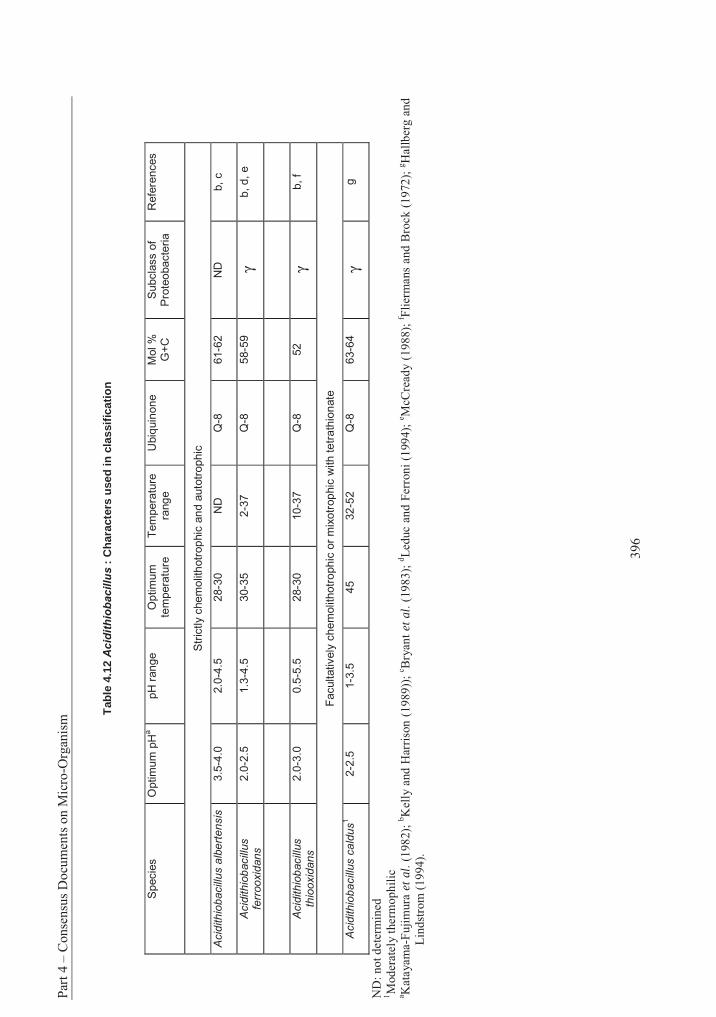
Part
4 –
Con
sens
us D
ocum
ents
on
Mic
ro-O
rgan
ism
396
Tab
le 4
.12
Acid
ith
iob
acillu
s :
Ch
arac
ters
use
d in
cla
ssif
icat
ion
Specie
s
Optim
um
pH
apH
ran
ge
Optim
um
te
mpera
ture
T
em
pera
ture
ra
ng
e
Ubiq
uin
one
Mol %
G
+C
S
ubcla
ss o
f P
rote
obacte
ria
Refe
rences
Str
ictly c
hem
olit
hotr
op
hic
an
d a
uto
trophic
Acid
ithio
bacill
us a
lbert
ensis
3.5
-4.0
2.0
-4.5
28-3
0
ND
Q
-8
61-6
2
ND
b,
c
Acid
ithio
bacill
us
ferr
ooxid
ans
2.0
-2.5
1.3
-4.5
30-3
5
2-3
7
Q-8
58-5
9
γb, d,
e
Acid
ithio
bacill
us
thio
oxid
ans
2.0
-3.0
0.5
-5.5
28-3
0
10-3
7
Q-8
52
γb, f
Facultatively
chem
olit
hotr
ophic
or
mix
otr
op
hic
with tetr
ath
ion
ate
Acid
ithio
ba
cill
us c
ald
us
1
2-2
.5
1-3
.5
45
32-5
2
Q-8
63-6
4
γg
ND
: not
det
erm
ined
1 M
oder
atel
y th
erm
ophi
lic
a K
atay
ama-
Fuj
imur
a et
al.
(198
2);
b Kel
ly a
nd H
arri
son
(198
9));
c Bry
ant
et a
l. (1
983)
; d L
educ
and
Fer
roni
(19
94);
e McC
read
y (1
988)
; f Fl
ierm
ans
and
Bro
ck (
1972
); g H
allb
erg
and
Lin
dstr
om (
1994
).
Section 3 - Acidithiobacillus
397
D. Comments on the species
Acidithiobacillus albertensis
Syn. Thiobacillus albertensis (Bryant et al., 1983; Kelly and Harrison, 1989; Kelly and Wood, 2000). This species is distinguished morphologically by a tuft of polar flagella and a glycocalyx extending outwards from the outer membrane of the bacterial cell envelope and which is used to attach itself to elemental sulphur (Bryant et al., 1983). These features together with the relatively high G+C content of the DNA, differentiate this species from the other three (Kelly and Wood, 2000). It has been tentatively assigned to Acidithiobacillus because a 16S rRNA sequence for the species is not yet available (Kelly and Wood, 2005).
Acidithiobacillus caldus
Syn. Thiobacillus caldus (Hallberg and Lindstrom, 1994; Kelly and Harrison, 1989; Kelly and Wood, 2000). This species is distinguished by extremely short rod-shaped cells, each with a single polar flagellum (Hallberg and Lindstrom, 1994), and by its moderately thermophilic nature. It cannot oxidise sulphidic ores, but it may be found associated with others involved in leaching. It is facultatively rather than obligately chemolithotrophic. A specific, fast and sensitive non-radioactive immuno-binding assay had been used for the detection and enumeration of this species (Amaro et al., 1994). Chemiluminescence or peroxidase-conjugated immunoglobulins are employed in a dot or slot blotting system. This method is very convenient for monitoring bioleaching micro-organisms in effluents from industrial bioleaching processes.
Acidithiobacillus ferrooxidans
Syn. Thiobacillus ferrooxidans (Temple and Colmer, 1951; Kelly and Harrison, 1989; Kelly and Wood, 2000). Morphologically this species appears to be distinguished by a single coiled flagellum in mature cells (Gonzalez and Cotoras, 1987). Also, this is the only species in the genus so far to be able to utilise iron as well as sulphur.
Serological and electrophoretic methods have been employed for the rapid detection of isolates of A. ferrooxidans and the differentiation of strains (Jerez et al., 1986). Different serotypes, characterised by specific lipopolysaccharide banding patterns in polyacrylamide gels, have been described (Koppe and Harms, 1994). A specific and very sensitive dot-immuno-binding assay for the detection and enumeration of A. ferrooxidans has been developed by Arredondo and Jerez (1989). Samples were spotted onto nitrocellulose membranes and first incubated with polyclonal antisera, derived from a rabbit inoculated with whole cells of A. ferrooxidans, and in 125I-labeled protein A or 125I-labelled goat anti-rabbit immunoglobulin G. The membranes were then dried, autoradiographed on Fuji Rx X-ray film and scanned at 550 nm. The antisera reacted with every strain of A. ferrooxidans tested but not with A. thiooxidans and H. neapolitanus and three species formerly placed in Thiobacillus.
A specific, fast and very sensitive immuno-electron microscopy method was also developed to identify A. ferrooxidans present with other iron oxidising bacteria in acidic mine waters (Coto et al., 1992). Polyclonal antisera, produced against whole cells of A. ferrooxidans, A. thiooxidans and Leptospirillum ferrooxidans gave highly specific reactions when cross-reacted with 23 strains of acidophilic bacteria using an immuno-fluorescence staining technique (Koppe and Harms, 1994). These methods have been criticised, however, because of the inability of the antisera to distinguish between dead and living cells (Khalid et al., 1993).
A systematic study of a large collection of strains ascribed to A. ferrooxidans revealed considerable diversity among them (Harrison, 1982). The members of seven DNA homology groups recognised by
Part 4 – Consensus Documents on Micro-Organism
398
Harrison (1982) were largely homologous with strains inside each group but to a lesser degree with strains in other groups. Although all strains grew between 25 and 30 OC, some were able to grow at 5 OC and others at 40 OC, and two genomic groups could be distinguished by different temperature optima. The strains in two other genomic groups (1 and 7) were apparently unable to use elemental sulphur, and their high mol% G+C values (53 and 65 respectively) were well outside the range normally accepted (see Table 4.12), giving rise to the suspicion that they might represent different species. Although strain m-1, comprising the seventh genomic group, was later shown to be able to oxidise elemental sulphur on prolonged incubation (Johnson, 1995b), it has been further distinguished from a more typical strain of A.ferrooxidans by its classification in the 5S rRNA sequence group III rather than in group II (Lane et al., 1985).
Acidithiobacillus thiooxidans
Syn. Thiobacillus concretivorus Parker Thiobacillus thiooxidans (Waksman and Joffe, 1922; Kelly and Harrison, 1989; Kelly and Wood, 2000). This species is motile by means of a single polar flagellum, and as with A. caldus, it cannot oxidise iron or pyrite, although it can grow on sulphur from pyrite in conjunction with Leptospirillum ferrooxidans (Kelly and Wood, 2000). In a study by Harrison (1982), four strains of A. thiooxidans were found to be largely homologous with a fifth strain but not with representatives of the seven homology groups of A. ferrooxidans. However, a sixth strain ascribed to A. thiooxidans showed no similarity to any of them. This indicates that there may be atypical representatives of A. thiooxidans as well as A. ferrooxidans.
3. Information on the Organisms’ Reproductive Cycle (Sexual/ Asexual)
Thiobacillus sensu lato, in which Acidithiobacillus was included, was found to reproduce by binary cell division (Sokolova and Karavaiko, 1968). No spores were observed.
4. Biological Features and Environmental Conditions Which Affect Survival, Reproduction, Growth, Multiplication or Dissemination
A. Growth requirements
Most strains are able to produce colonies on appropriate media solidified with agar or agarose. The use of elemental sulphur is avoided because of its insolubility. Formation of hydrogen sulphide is potentially toxic in moderate concentrations, and the most widely used sulphur compound is thiosulphate (Smith and Strohl, 1991).
Some strains, especially those of A. thiooxidans, grow poorly on agar media, possibly due to the toxicity of agar hydrolysis products (Kelly and Harrison, 1989). The problem is generally solved by the use of a minimal concentration of agar, screening for suitable brands of purified agars, use of agarose, and, in the case of A. thiooxidans, a combination of low agar concentration with pH 2.2-2.5 and ferrous sulfate at only about 20 mM (Johnson, 1995a). The growth of strains of A. ferrooxidans on solid media is also difficult. However, the double-layered plates such as FeTSBo and described by Johnson (1995a) allow also the growth of most strains.
Species of Acidithiobacillus, in common with other former species of Thiobacillus, are able to use carbon dioxide as the sole source of carbon for synthesis of cell material (Kuenen, 1975). Ribulose diphosphate carboxylase, the enzyme responsible for the fixation of carbon dioxide, appears to be located in polyhedral inclusions in the cell.
In contrast to species of Halothiobacillus which have been recorded as either halotolerant or having a strict NaCl requirement (Sievert et al., 2000), A. ferrooxidans has been recorded as unable to grow at salt
Section 3 - Acidithiobacillus
399
concentrations above 1% (Lazaroff, 1963; McCready, 1987; Razzell and Trussell, 1963) whereas specific information for other species of Acidithiobacillus for their tolerance to salt is not available.
Acidithiobacillus ferrooxidans has a remarkable physiology that allows it to thrive in an inorganic mining environment. Its minimum growth requirements can be satisfied by water, air, an oxidisable iron or sulphur source and trace minerals. The trace elements required are usually present as impurities in the water or ore (Rawlings and Woods, 1995). This statement is also true for A. thiooxidans, however elucidation of these abilities for other members of Acidithiobacillus is still not available. A. caldus was found to be relatively insensitive to a number of xanthate and dithiocarbamate-based flotation reagents but sensitive to a number of mercapto-benzthiazole-based reagents (Okibe and Johnson, 2002).
B. Oxidation of hydrogen
In contrast to strains of A. thiooxidans, H. neapolitanus, T. prosperus, Leptospirillum ferrooxidansand four species formerly placed in Thiobacillus, three strains of A. ferrooxidans were found to be facultative hydrogen oxidisers, being able to use molecular hydrogen as a sole source of energy (Drobner et al., 1990). The ability to oxidise hydrogen was repressed by ferrous iron or sulphur and occurred only in the presence of oxygen.
C. Nitrogen
In addition to their ability to fix carbon dioxide all strains of A. ferrooxidans examined so far are also able to fix atmospheric nitrogen (i.e. they are diazotrophic)(Rawlings and Kusano, 1994).
D. Aerobic/Anaerobic growth
Acidithiobacillus species are strict aerobes with the exception of A. ferrooxidans, which is a facultative aerobe. In the absence of oxygen, A. ferrooxidans is able to grow on reduced inorganic sulphur compounds using ferric iron as an alternative electron acceptor (Pronk et al., 1992; Sugio et al., 1985).
E. Resistance to metals
Resistance to metal ions is a function of those thiobacilli tested to date. Acidithiobacillus ferrooxidans is resistant to a variety of metal ions such as chromium (Baillet et al., 1998), copper, zinc, nickel, thorium and uranium (Leduc et al., 1997; Tuovinen et al., 1971) and mercury (Takeuchi et al., 1999, 2001; Sugio etal., 2001). According to Iwahori et al. (2000), the resistance of A. ferrooxidans to mercury is ferrous iron dependent.
F. Role of Rusticyanin
Rusticyanin is a blue copper protein present in the periplasmic space of A. ferrooxidans. Consisting of a single polypeptide chain with one copper atom as a cofactor (Hazra et al., 1992), rusticyanin reportedly serves as the initial electron acceptor upon oxidation of ferrous iron (Hazra et al., 1992; Hutchins et al.,1986). An acid-stable cytochrome c was found to catalyse the reduction of rusticyanin (Blake et al., 1988).
G. Survival
No information on factors influencing survival of Acidithiobacillus in the natural environment appears to be available. According to Kelly and Harrison (1989), A. ferrooxidans survives in culture on pyrite (FeS2) for very long periods when stored at 5-15 oC. Many strains have been successfully freeze-dried or have survived storage in liquid nitrogen or in glycerol suspension at -20 oC (Kelly and Harrison, 1989).
Part 4 – Consensus Documents on Micro-Organism
400
Hubert et al. (1994) have shown that survival rates of A. ferrooxidans decreased rapidly under laboratory conditions above and below the individual temperature ranges of psychrotrophic and mesophilic strains.
H. Adhesion
The ability to adhere to surfaces seems to be a peculiar feature of Acidithiobacillus. Myerson and Kline (1983) observed the physical adsorption of cells of A. ferrooxidans to the surface of different non-porous solid particles (glass, pyrite, sulphur). Selective adherence to iron containing minerals appears to occur naturally (Ohmura et al., 1993), and the ferrous ion, but not the ferric ion, inhibited such selective adhesion. A model of the biofilm structure has been proposed by Karamanev (1991). Intimate contact and adhesion are required for enzymatic attack by A. ferrooxidans on insoluble substrates such as sulphur, pyrite (FeS2), and chalcopyrite (CuFeS2), and this is brought about by either a proteinaceous surface appendage (Devasia et al., 1993) or by extra-cellular polymeric substances (specifically lipopolysaccharides) combined with iron (III) (Gehrke et al. 1998, 2001; Blais et al., 1994). Acidithiobacillus ferrooxidans does not randomly adsorb onto pyrite or other surfaces, but congregates selectively at sites where dislocations, grain boundaries and other non-uniformities in the crystal structure emerge to the surface (Andrews, 1988; Bagdigian and Myerson, 1986; Gehrke et al., 1998). It is possible that diffusion of sulphur atoms along dislocations in the substrate is an important part of the mechanism of microbial decomposition. This pattern of diffusion provides a great advantage to bacteria because sulphur oxidation has a much higher yield of free energy than iron oxidation. No corresponding advantage would be gained by adsorption onto pure pyrite sites because the diffusion through pyrite crystals is several orders of magnitude lower.
Growth of bacteria adhering to the mineral surface initiates the oxidation process in arsenopyrite bioleaching (Fernandez et al., 1995). Corrosion patterns appear, with the liberation of ferrous ions and formation of elemental sulphur. With the increase in number of the bacteria, the ferrous ions are oxidised to ferric ions with the ultimate production of ferric arsenate (Fernandez et al., 1995).
Adhesion structures, consisting of a filamentous matrix, have been observed in A. thiooxidans (Blais et al., 1994), linking the cells to the surface of sulphur particles. They were not observed however in T. thioparus, where the cells fixed directly onto the sulphur (Sokolova and Karavaiko, 1968). The latter process is the usual type of fixation observed in the species placed formerly in Thiobacillus.
Adhesion can be estimated using the technique of Dziurla et al. (1998), who developed an immuno-filtration assay (ELIFA) for this purpose. ELIFA is a modified ELISA using micro-titer plates with 0.2-µm pore-size filters in place at the bottom. Particles, either previously inoculated with bacteria or to be reacted with added bacteria are incubated in the wells and then successively filtered and washed by applying a vacuum to the bottom of the plate. The inoculated particles are retained by the filter. Polyclonal antiserum raised against a strain of A. ferrooxidans is added and the plates incubated at 35 °C for one hour. The polyclonal rabbit antibody used was shown to react with different A. ferrooxidans and A. thiooxidans but not with other bacterial genera. Following washing the bound antibody is detected with goat-anti-rabbit globulin conjugated to alkaline phosphatase and ρ-nitrophenylphosphate was used as the detection substrate.
I. pH and nutrition preference
Species of Acidithiobacillus are acidophilic as well as obligate or facultative chemolithotrophs according to the nutritional table presented by Kuenen et al. (1992). Species of Acidithiobacillus are abundant in acid mine drainage water where they oxidise and gain energy from the oxidation of metals such as iron. The optimum pH for A. ferrooxidans is between 2-3, but when the substrate is in large part pyritic, the pH can reach extremely low values, (less than 1). This is due to the availability of abundant
Section 3 - Acidithiobacillus
401
sulphur and the precipitation of ferric hydroxide when the solution reaches saturation (Morin, 1995). Acidithiobacillus caldus is the single mixotrophic species which can utilise sulphur or tetrathionate and yeast extract or glucose (Hallberg and Lindstrom, 1994).
Blais et al. (1993) have demonstrated that less acidophilic bacteria in sludge such asThermithiobacillus tepidarius, T. aquaesulis, T. denitrificans, T. thioparus and other species formerly placed in Thiobacillus, may initiate the acidification to the point where the acidophilic species can take over. Acidophilic bacteria decreased the pH of a sulphur-containing synthetic salts medium to the level of 1.4-1.6 in 10 days. Evangelou (1995) mentioned pH 3.5 as the upper limit below which Fe2+ is oxidised by A. ferrooxidans. For the less acidophilic bacterial species, the limit of acidification was more variable, between pH 2.2 and 6.9. This has important implications for the removal of heavy metals from sludge (presented later in this document).
J. Temperature relations
Acidithiobacillus contains one thermophilic species, A. caldus, but little is known about its actions. Acidithiobacillus ferrooxidans has been traditionally regarded as mesophilic. Recently, however, psychrotrophic strains have been isolated with a growth range on iron of 2-37 °C (Leduc and Ferroni, 1994). Some Canadian isolates have a greater cold tolerance than most strains with a temperature optimum of only 20 oC (McCready, 1988). The occurrence of broad temperature range for psychrotrophic isolates of A. ferrooxidans has been reported by Leduc et al. (1993) and by Berthelot et al. (1993). Temperature ranges of 2-35 °C and 4-21 °C, respectively, were observed. However, psychrophiles have not been isolated from cold tailing effluents where they would be expected (Berthelot et al., 1994). The temperature used for bioleaching in most studies is 35 oC. Although A. ferrooxidans was reported to grow most rapidly at 30 °C, it oxidised iron faster at 35 °C (Holmes, 1988). This has important implications for industrial bioleaching since the oxidation of sulfides is exothermic, and therefore cooling may be necessary to maintain a satisfactory industrial process (Morin, 1995). Industrial applications are further described in section 8.
K. Metabolic pathways: involvement of Acidithiobacillus in bioleaching
Bioleaching is a biochemical oxidation process, catalysed by a living organism, whereby an insoluble mineral is oxidised to a soluble form and recovered in a pure form. A large number of acidophilic bacteria capable of attacking mineral sulphides have been isolated from industrial leaching operations or from sites of natural leaching. Acidithiobacillus ferrooxidans and A. thiooxidans are prominent among the species isolated from leaching sites, although the redox potential and concentration of ferric iron will influence which species dominates (Rawlings et al., 1999).
Acidithiobacillus ferrooxidans
This bacterium derives its energy from oxidation-reduction reactions using insoluble sulphidic minerals as growth substrates, including pyrite (FeS2), chalcopyrite (CuFeS2), chalcocite (Cu2S) and sphalerite (ZnS). This, coupled with its resistance to high concentrations of normally toxic metal ions in solution, accounts for the ubiquity of this organism in leaching systems (Cripps, 1980). Combinations of A.ferrooxidans and either A. thiooxidans or Acidiphilium acidophilum and Leptospirillum ferrooxidans have been associated with degradation of pyrite and chalcopyrite. Metals can be released from sulphidic ores by direct or indirect leaching or by galvanic conversion (Hutchins et al., 1986).
Part 4 – Consensus Documents on Micro-Organism
402
Direct leaching
This involves oxidation of the substrate by the bacterium and may require physical attachment of the bacteria to particles of the mineral sulphide (see section 4.8). The process can be described in general by a simplified reaction:
MS + 2O2 MSO4 where M represents a bivalent metal [1] (Hutchins et al., 1986)
Several different metal sulphides can be acted on directly by cells of A. thiooxidans (Leduc and Ferroni, 1994). These include sulphides of copper, nickel, lead, iron, gallium, cobalt and zinc. Examples of direct leaching include the following:
CuS + 2O2 CuSO4 (Woods and Rawlings, 1989) [2] Covellite
2FeAsS + 7O2 + 2H2O 2FeAsO4 + 2H2SO4 (Brierley, 1995) [3] Arsenopyrite
Indirect leaching
This occurs through the production of an oxidative reagent or lixiviant that causes solubilisation to occur. The use of iron pyrites by A. thiooxidans as an energy source is a good example of how both direct and indirect leaching processes work together. The overall reaction describing pyrite oxidation is usually written as:
4FeS2 + 15O2 + 2H2O 4Fe2(SO4)3 + 2H2SO4 (Rawlings and Woods, 1995) [4]
However, the actual pathway for the oxidation of pyrite is not a simple one-step reaction, but a series of reactions, passing through a number of intermediates. Under natural conditions, the two main oxidising agents that act on the pyrite are oxygen and the ferric ion. The reaction can be catalyzed by A.ferrooxidans, which increases the rate of reaction by more than 106 (Singer and Stumm, 1970).
2FeS2 + 7O2 + 2H2O 2FeSO4 + 2H2SO4 [5] pyrite
4FeSO4 + O2 + 2H2SO4 2Fe2(SO4)3 + 2H2O [6] (bacteria)
Fe2(SO4)3 + FeS2 3FeSO4 + 2 S [7]
2S + 3O2 + 2H2O 2H2SO4 [8] (bacteria)
(Leduc and Ferroni, 1994; Monticello and Finnerty, 1985)
Reactions [5], [6] and [8] are part of the direct leaching mechanism, which may require the physical attachment of the bacteria to pyrite particles (section 4.8). Reaction [7] comprises the indirect mechanism that can take place independently of the bacteria. The role of the bacteria is to reoxidise the ferrous ions to ferric ions and sulphur to sulphuric acid (Monticello and Finnerty, 1985). A cyclical system develops in which ferrous ions released from pyrite are oxidised by the bacteria to ferric ion, which can then oxidise
Section 3 - Acidithiobacillus
403
pyrite again, generating more ferrous ions. The biomass specific oxygen consumption rate is dependent on the ratio of ferric to ferrous irons in the culture (Boon et al., 1999). These ferric ions are known as the lixiviant because they carry electrons from the mineral to the bacterium’s cell membrane (Leduc and Ferroni, 1994). The electrons are subsequently transported via an electron-transport chain to molecular oxygen in reaction [4]. Since iron is nearly always available in natural leaching environments, both the direct and indirect leaching mechanisms probably operate simultaneously in nature (Leduc and Ferroni, 1994; Monticello and Finnerty, 1985).
In the indirect reaction, bacterial activity is limited to the oxidation of pyrite (FeS2) and ferrous iron.Acidithiobacillus ferrooxidans does not directly interact with the metal in the minerals. The role of the bacterium is to continuously provide a powerful oxidation agent, ferric sulphate [Fe2(SO4)3] which is capable of dissolving a wide variety of metal sulphide minerals (Torma, 1991).
The following are examples of indirect leaching of minerals other than pyrites (Hutchins et al., 1986)
CuFeS2 + 2 Fe2(SO4)3- _ CuSO4 + 5FeSO4 + 2S0 [9]
Chalcopyrite
Cu2S + 2Fe2(SO4)3- 2CuSO4 + FeSO4 + S0 [10]
Chalcocite
CuS + 2 Fe2(SO4)3- _ CuSO4 + 2FeSO4 + S0 [11]
Covellite
Cu5FeS4 + 6Fe2(SO4)3- _ 5CuSO4 + 13FeSO4 + 4S0 [12]
Bornite
Ultimately, the indirect leaching mechanism depends on biological regeneration of ferric sulphate. Elemental sulphur (S0) generated by the reactions above can be converted to sulphuric acid by A.ferrooxidans:
2S0+ 3O2 + 2H2O 2H2SO4 [13]
The sulphuric acid maintains the pH at levels favourable to the bacteria and effectively leaches a variety of copper oxide minerals, giving copper sulphate as an end product (Hutchins et al., 1986).
Uranium leaching proceeds in a similar fashion, again without the bacterium metabolising the uranium ion:
UO2 + Fe2(SO4)3 + H2SO4 UO2(SO4)34- + 2FeSO4 + 4H4+ [14]
or:
UO3 + 3H2SO4 UO2(SO4)34- + H2O + 4H+ [15]
Acidithiobacillus ferrooxidans can be used to extract uranium from sulfidic ore bodies because the bacterium uses the iron and the sulfur in the ores, as energy sources (See reaction [5]). The oxidation of reduced sulfur results in the production of sulfuric acid, whereas the oxidation of reduced iron produces the oxidant Fe3+. Under such acidic conditions, the insoluble tetravalent uranium is oxidised by ferric ions to the soluble hexavalent state as follows (Berthelot et al., 1993):
Part 4 – Consensus Documents on Micro-Organism
404
UO2 + 2Fe3+ UO2 2+ + 2Fe2+ [16]
Thus, the soluble, oxidised uranium is released from the mineral into the leaching liquid and can be easily recovered. For other minerals where the sulphate formed from the mineral is insoluble, as is the case with lead, gold and silver, the metal may be concentrated by leaching away other soluble metal sulphates, leaving a concentrated metallic product (Hutchins et al., 1986). Gold ores are often recalcitrant. The gold may be encased in a pyrite/arsenopyrite matrix that has to be decomposed before the gold is accessible to cyanide extraction (Rawlings and Woods, 1995).
Galvanic conversion
This is a lesser-known process in bioleaching where physical contact between two dissimilar metal sulphide phases immersed in an electrolyte such as dilute sulphuric acid or ferric sulphate solution creates a galvanic cell. In a mixture of pyrite and chalcopyrite, the former acts as a cathode while the chalcopyrite behaves as an anode and undergoes rapid dissolution. Acidithiobacillus ferrooxidans may accelerate the reaction by continuously oxidising the film of elemental sulphur that would obstruct the diffusion of copper and iron salts.
Acidithiobacillus thiooxidans
This species differs from A. ferrooxidans in its inability to oxidise iron (Fe2+). Sulphur appears to provide the main source of energy for this bacterium (Sokolova and Karavaiko, 1968), and its ability to oxidise elemental sulphur allows it to take part in the indirect leaching of some minerals, particularly sulphides of cobalt, nickel and zinc (Hutchins et al., 1986; Norris, 1990). The sulphuric acid generated by A. thiooxidans through oxidation of sulphur (see reaction [8]), results in acid solubilisation of the metal. In mixed culture with iron-oxidising bacteria, A. thiooxidans oxidises the protective sulphur covering on the surface of minerals such as chalcopyrite. This allows bacteria such as Leptospirillum ferrooxidans, which cannot remove the sulphur, to attack the iron component beneath.
Acidithiobacillus thiooxidans can operate synergistically with A. ferrooxidans, or with Leptospirillum ferrooxidans which is able to use only ferrous iron. Either combination of bacteria can efficiently attack mineral sulphides and rapidly degrade a variety of ores (Paiment et al., 2001; Rawlings and Woods, 1995). Acidithiobacillus thiooxidans can augment the oxidation of pyrite in coal by A. ferrooxidans by oxidising elemental sulphur produced by the latter organism to sulphuric acid (see reaction [3]), which is then converted by A. ferrooxidans firstly to ferrous sulphate and then to ferric sulphate (Ford et al., 1977). Khalid and Naeveke (1991) have also observed that A. thiooxidans had the ability to solubilise heavy metals from carbonate-bearing complex sulphidic ore more efficiently than A. ferrooxidans. The reason may be that A. ferrooxidans alone could not produce sufficient acid to neutralise the carbonate contents and lower the pH, so that suitable conditions for growth of the bacterium could not be attained. A combination of both bacteria works efficiently since the sulphuric acid produced from sulphur by A. thiooxidans fulfils this condition.
I. Inhibition of bacterial oxidation and growth
A number of simple organic compounds have been found to inhibit bacterial oxidation of ferrous ion at low concentrations under laboratory conditions, including benzoate, sorbate and sodium dodecyl sulphate (SDS) (Onysko et al., 1984) and sodium tungsten (Sugio et al., 2001). The inhibitory effect of glucose, cellobiose, galacturonic acid and citric acid compared favourably with that of SDS (Frattini et al., 2000). Ferric and arsenite ions have a most detrimental effect on the growth of A. ferrooxidans and A.thiooxidans (Collinet and Morin, 1990). These compounds have been suggested for use in control of these species.
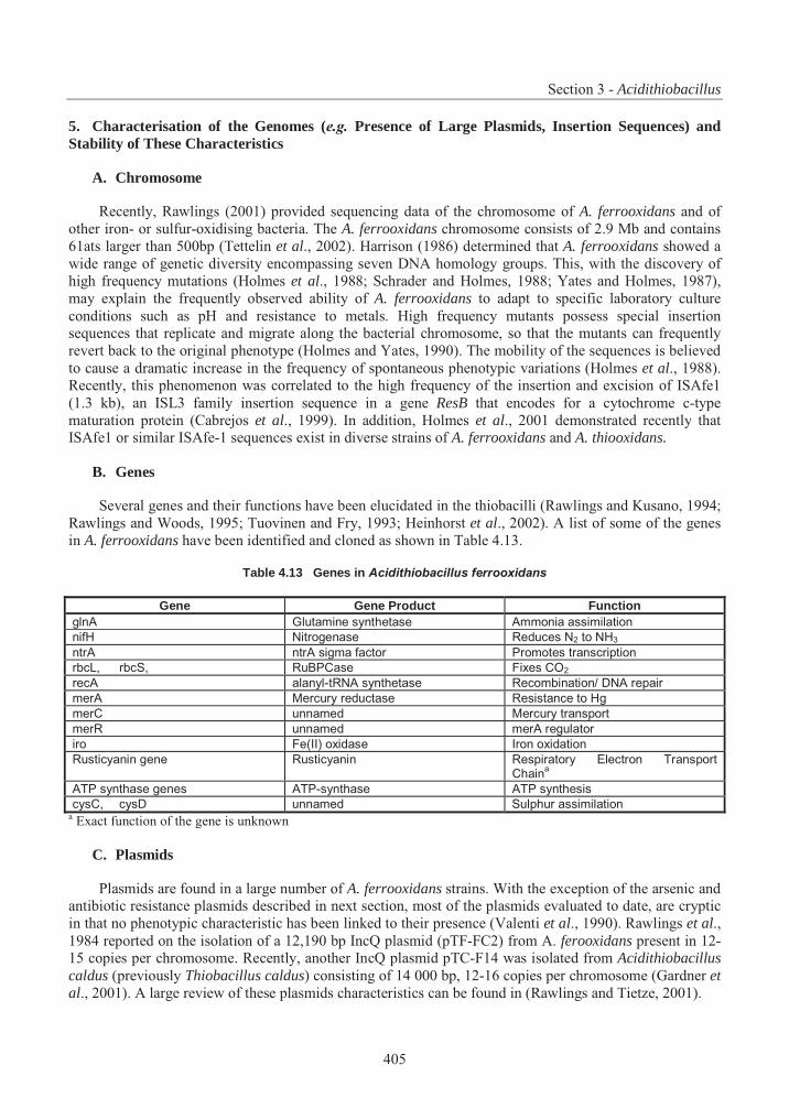
Section 3 - Acidithiobacillus
405
5. Characterisation of the Genomes (e.g. Presence of Large Plasmids, Insertion Sequences) and Stability of These Characteristics
A. Chromosome
Recently, Rawlings (2001) provided sequencing data of the chromosome of A. ferrooxidans and of other iron- or sulfur-oxidising bacteria. The A. ferrooxidans chromosome consists of 2.9 Mb and contains 61ats larger than 500bp (Tettelin et al., 2002). Harrison (1986) determined that A. ferrooxidans showed a wide range of genetic diversity encompassing seven DNA homology groups. This, with the discovery of high frequency mutations (Holmes et al., 1988; Schrader and Holmes, 1988; Yates and Holmes, 1987), may explain the frequently observed ability of A. ferrooxidans to adapt to specific laboratory culture conditions such as pH and resistance to metals. High frequency mutants possess special insertion sequences that replicate and migrate along the bacterial chromosome, so that the mutants can frequently revert back to the original phenotype (Holmes and Yates, 1990). The mobility of the sequences is believed to cause a dramatic increase in the frequency of spontaneous phenotypic variations (Holmes et al., 1988). Recently, this phenomenon was correlated to the high frequency of the insertion and excision of ISAfe1 (1.3 kb), an ISL3 family insertion sequence in a gene ResB that encodes for a cytochrome c-type maturation protein (Cabrejos et al., 1999). In addition, Holmes et al., 2001 demonstrated recently that ISAfe1 or similar ISAfe-1 sequences exist in diverse strains of A. ferrooxidans and A. thiooxidans.
B. Genes
Several genes and their functions have been elucidated in the thiobacilli (Rawlings and Kusano, 1994; Rawlings and Woods, 1995; Tuovinen and Fry, 1993; Heinhorst et al., 2002). A list of some of the genes in A. ferrooxidans have been identified and cloned as shown in Table 4.13.
Table 4.13 Genes in Acidithiobacillus ferrooxidans
Gene Gene Product Function glnA Glutamine synthetase Ammonia assimilation
nifH Nitrogenase Reduces N2 to NH3
ntrA ntrA sigma factor Promotes transcription
rbcL, rbcS, RuBPCase Fixes CO2
recA alanyl-tRNA synthetase Recombination/ DNA repair
merA Mercury reductase Resistance to Hg
merC unnamed Mercury transport
merR unnamed merA regulator
iro Fe(II) oxidase Iron oxidation
Rusticyanin gene Rusticyanin Respiratory Electron Transport Chain
a
ATP synthase genes ATP-synthase ATP synthesis
cysC, cysD unnamed Sulphur assimilation a Exact function of the gene is unknown
C. Plasmids
Plasmids are found in a large number of A. ferrooxidans strains. With the exception of the arsenic and antibiotic resistance plasmids described in next section, most of the plasmids evaluated to date, are cryptic in that no phenotypic characteristic has been linked to their presence (Valenti et al., 1990). Rawlings et al., 1984 reported on the isolation of a 12,190 bp IncQ plasmid (pTF-FC2) from A. ferooxidans present in 12-15 copies per chromosome. Recently, another IncQ plasmid pTC-F14 was isolated from Acidithiobacillus caldus (previously Thiobacillus caldus) consisting of 14 000 bp, 12-16 copies per chromosome (Gardner etal., 2001). A large review of these plasmids characteristics can be found in (Rawlings and Tietze, 2001).
Part 4 – Consensus Documents on Micro-Organism
406
Dominy et al. (1998) isolated and characterised a 19.8 kb plasmid from A. ferrooxidans ATCC33020. Fourteen complete open reading frames (ORFs) were identified, most of which were proteins involved in maintenance although three of the ORFs appeared to correspond to redox-active proteins and thus could constitute part or all of an electron transport chain.
None of the plasmids describe to date, have been correlated with metal resistance (Hutchins et al., 1986; Chisholm et al., 1998; Leduc and Ferroni, 1993), although circumstantial evidence suggests that genes for mercury and silver resistance may be located on an unnamed 19 x 106 daltons plasmid isolated from A. ferrooxidans (Visca et al., 1986). One isolate of A. ferrooxidans was found to possess mercuric reductase activity similar to that in heterotrophic bacteria containing the mer operon, but this could not be associated with a plasmid.
Valenti et al. (1990) discovered a 20-kb plasmid (pTFO) in eight out of twelve strains of A. ferrooxidans from Italy and Mexico, which was stably maintained for many bacterial generations. All of the strains resisted similar concentrations of metal ions in spite of overall differences in their plasmid pattern.
D. Genetic variation among leaching bacteria
Suzuki et al. (1989) demonstrated that naturally occurring strains of A. ferrooxidans vary widely in their ability to utilise iron and sulphur compounds, resistance to metal and mineral leaching activities. The abilities to adsorb onto solid surfaces and to oxidise sulphur with ferric ion are other variable properties. Natural variation and /or selection of mutants can be inferred from the ease with which new isolates are selected to enhance process efficiency in various industrial settings (see section 8).
E. Mutation
The occurrence of high-frequency mutation strains of A. ferrooxidans was previously mentioned (section 5.1). It was noted that high frequency mutants possess special insertion sequences that replicate and migrate along the bacterial chromosome, so that the mutants can frequently revert back to the original phenotype (Holmes and Yates, 1990). The mobility of the sequences is believed to cause a dramatic increase in the frequency of spontaneous phenotypic variations (Holmes et al., 1988; Holmes et al., 2001). In addition, Holmes and Debus (1991) ascribed the high mutation rate of A. ferrooxidans to repetitive DNA sequences in the chromosomes and plasmids whose mobility around the genome could cause mutations to arise.
The high frequency of spontaneous mutations overshadows mutations derived by chemical mutagenesis and makes the latter difficult to detect.
An important implication for environmental regulation is that the inherent instability of these ‘naturally’ engineered strains makes it difficult to predict ecological behaviour thus complicating assessment of risk.
6. Genetic Transfer Capability
Classical genetic engineering techniques for the development of new microbial strains include mutation, conjugation, transformation, transduction and electroporation. Very little work has been done in these areas with bioleaching micro-organisms (Holmes and Debus, 1991). However, Young (1993) has presented a good review of attempts at genetic engineering of these micro-organisms, particularly A. ferrooxidans.
Section 3 - Acidithiobacillus
407
A. Transduction
The high mutation rate of A. ferrooxidans was ascribed by Holmes and Debus (1991) to repetitive DNA sequences in the chromosomes and plasmids whose mobility around the genome could cause mutations to arise (see also sections 5.1 and 5.5). One of these DNA sequences has been shown to be an insertion sequence. However, bacteriophages for transduction have not been reported for Acidithiobacillus,although these authors cited a report of bacteriophage-like particles in acidophilic heterotrophs, which may prove valuable for developing transduction systems.
B. Conjugation
Transfer of plasmid DNA from heterotrophic bacteria to chemolithotrophic colourless sulphur bacteria by conjugation was first achieved experimentally by Kulpa et al. (1983). Halothiobacillus neapolitanus was used in the study because of its ability to grow at the near neutral pH required by the heterotrophic donor, Pseudomonas aeruginosa. It was discovered that plasmid RP1, which governed resistance to three common antibiotics was accepted, replicated and expressed in the chemolithotrophic bacterium. Transfer from Escherichia coli into A. thiooxidans and back of four broad-range IncP plasmids with antibiotic resistance markers has also been achieved (Jin et al., 1992), and two of the three antibiotic resistance markers have been expressed in A. thiooxidans.
However, although plasmids have been demonstrated in many strains of A. ferrooxidans, most of them are cryptic in that no identifiable phenotype has been linked to their presence (Leduc and Ferroni, 1994), and there are few cases where plasmid transfer has been successful. In spite of being cryptic in E.coli, however, the plasmid pTF-FC2 has been found to have a broad host-range of replication, and the discovery of such plasmids does suggest that a conjugation system exists in A. ferrooxidans (Rawlings and Woods, 1995; Rawlings and Tietze, 2001). This plasmid has been deemed particularly suitable for use as a cloning vector for genetic manipulation of A. ferrooxidans (Rawlings et al., 1986). Four plasmids (pTF35, pTF-FC2, pTF3320-1, pTF3302-2) from three different strains of A. ferrooxidans have been successfully cloned into the plasmid pBR325 and two into the related plasmid pBR322 (Rawlings and Woods, 1995). However, the desirable properties of increased uranium and arsenic resistance, present in the A. ferrooxidans parents, were not expressed by the transformed E. coli mutants.
Two arsenic-resistant plasmids, pSDRA1 and pSDRA21 have been constructed and introduced into A.ferrooxidans by conjugation with E. coli, using Solid 2:2 Medium as a mating medium (Peng et al., 1994b, 1994c). Arsenic resistance was demonstrated in the progeny. Unfortunately most attempts at returning functioning genes from E. coli to A. ferrooxidans have been unsuccessful. This means that, in principle, the statement by Holmes and Yates (1990) that genes can be extracted from A. ferrooxidans and be genetically modified, yet cannot be returned to A. ferrooxidans to create an improved organism is still largely true. However, this position may change as techniques improve for introducing foreign genetic information into A. ferrooxidans and other thiobacilli.
C. Transformation
Transformation does not appear to take place naturally in Acidithiobacillus and other genera of colourless sulphur bacteria, but transformation has been accomplished experimentally through electroporation. Transport of naked DNA into bacterial cells by application of a high voltage electrical discharge (electroporation) has been successful in two cases. Thiomonas intermedia was transformed by this method using the plasmid pRK415Km (Jin et al., 1994), conferring kanamycin resistance. The transformation efficiency ranged from 103 to 104 transformants µg-1 plasmid DNA under optimal conditions. Kusano et al. (1992) transformed A. ferrooxidans with natural plasmids and an artificially constructed one, but the efficiency was much less. The reason why only one strain out of the thirty tested
Part 4 – Consensus Documents on Micro-Organism
408
was amenable to transformation by electroporation is still uncertain. The plasmids in the transformed cells were stable for at least 110 generations.
7. Behaviour in Simulated Natural Environments Such as Microcosms
Microcosms have been used to investigate the role of A. thiooxidans and other thiobacilli in the degradation of concrete (Sand, 1987; Sand and Bock, 1988). Samples of concrete were inoculated in a simulation apparatus with bacteria originally isolated from concrete and incubated for nine months at an relative humidity of 95% and a temperature of 30 oC. Three compounds were tested as sources of energy: hydrogen sulphide, thiosulphate, and methyl mercaptan. Hydrogen sulphide at a concentration of 15 mg/m3
resulted in severe corrosion after nine months, and A. thiooxidans was the dominant species in the microflora. At the lesser concentration of 2 mg/m3 moderate corrosion resulted and the dominant species were Thiomonas intermedia, Starkeya novella and Halothiobacillus neapolitanus. Similar results were obtained with thiosulphate but methyl mercaptan at concentrations of 22 and 2 mg/m3 caused negligible corrosion and only heterotrophic bacteria and fungi thrived on the concrete blocks.
8. History of Use (Including Selection of Mutants and Examples of Environmental Applications of the Organism, and Information Derived from These):
A. Selection of industrially useful mutants
The rate of growth and mineral oxidation by a population of leaching bacteria can be improved simply by cultivating a population of bacteria in a continuous flow apparatus. If the flow rate through the apparatus is slowly increased, those bacteria that are capable of the most rapid growth will replace the others. Spontaneous mutants will be selected by their growth potential on the available substrate. The advantage of this method is that it does not require sophisticated procedures, but it may take a long time to improve the bacterial strain to any economically significant extent. This approach has nevertheless been used to improve the leaching rates of A. ferrooxidans to several fold over that of the original isolates. Vian et al. (1986) were able to progressively select mutants of A. ferrooxidans with high oxidative efficiency at low pH values (down to pH 1.5) and with high resistance to ferric ions, thus improving the leaching of metals from low-grade ore deposits and avoiding precipitation of oxidised iron compounds.
The improvement of biomining bacteria by mutation and selection has had a dramatic effect on the economics of biooxidation of gold-bearing arsenopyrite ores in particular (Rawlings and Woods, 1995). Natural selection in the laboratory and in pilot and full-scale plants over several years has produced A. ferrooxidans strains that are highly resistant to arsenic and are capable of rapid oxidation of gold-bearing arsenopyrite ores in a continuous industrial bioleaching process. Highly adapted strains decompose arsenopyrite ores to an extent that allows more than 95% gold recovery in 3 days compared to the more than 12 days required by the original isolates. Further improvement however, is likely to require the application of DNA recombinant technology to amplify genes or to enable the introduction of new genetic material. Similarly, resistance to nickel ions was enhanced in A. ferrooxidans by repeated culturing in a medium containing nickel and gradually increasing the nickel concentration (Kai et al., 1995) and the use of the modified strain, in turn, significantly increased the rate of industrial nickel extraction. No literature data was found on the selection of mutants of the three other species of Acidithiobacillus.
B. Main Industrial Uses
Historically there are nine main uses to which species of Acidithiobacillus have been applied. This is not meant to imply that their use should be restricted to these categories, but rather to illustrate the potential and diversity of organisms within this genus. The main uses of Acidithiobacillus are directly related to their bioleaching ability.
Section 3 - Acidithiobacillus
409
Removal of sulphides from industrial wastes
The toxicity, corrosive properties, and unpleasant odour dictate stringent control of release of sulphides into the environment. Reduced sulphur compounds can occur in industrial wastes of the oil and gas industries as a result of several processes. Whether directly as an end product of sulphate reduction (Buisman et al., 1989), or indirectly as a result of methanogenesis, the effluent from which the methane is generated may contain significant quantities of sulphate. The release of large amounts of sulphide into natural waters can result in oxygen depletion due to direct or biological oxidation (Kuenen, 1975) as well as corrosion of the concrete walls of reactors, sewer systems, and steel pipelines.
Acidithiobacillus ferrooxidans
Acidithiobacillus ferrooxidans has often been preferred for sour gas removal because the costs of neutralising sulphuric acid produced by the other bacteria can be avoided, since the sulphur is converted to ferric sulphate, and the bacterium is not inhibited by H2S (Jensen and Webb, 1995; Shiratori and Sonta, 1993). The Bio-SR Process (Satoh et al., 1988) comprises the following steps. Sour gas is introduced into an absorber containing ferric sulphate solution where it is oxidised to elemental sulphur and the ferric sulphate is reduced to ferrous sulphate:
H2S + Fe2(SO4)3 S0 + 2FeSO4 + H2SO4 [17]
Elemental sulphur is removed from the solution by a separator and the bacteria in the bioreactor then oxidise the ferrous sulphate in the solution back to ferric sulphate:
4FeSO4 + 2H2SO4 + O2 2Fe2(SO4)3 + 2H2O [18]
The oxidised solution is then recycled to the absorber to repeat the cycle. This procedure is environmentally sensible in that it is easy to operate, there are no waste products, no special chemicals are needed and the operating cost is low.
In earlier gas treatment processes (Onken et al., 1984; Sumitomo Jukai Envirotech, 1983) the hydrogen sulphide is first precipitated as CuS or FeS. These sulphides are then oxidised by the A. ferrooxidans to regenerate the precipitating agent. Cadenhead and Sublette (1990) have commented that the requirement of a low pH for these processes may induce corrosion, and these authors prefer Thiobacillus denitrificans with its higher pH tolerance as the microbiological conversion agent.
Acidithiobacillus thiooxidans
Berzaczy et al. (1990) have patented a microbiological conversion process for degradation of sulphur-containing pollutants such as H2S, CS2, COS, thioalcohols, thioethers and thiophenes in waste gas, especially from cellulose fibre manufacture. The gas makes contact with cells of the bacterium immobilised on packing material in a packed-bed reactor. The metabolic products (mainly H2SO4) draining from the reactor are neutralised by addition of lime and lime-water.
It has been suggested that A. thiooxidans could be used to convert hydrogen sulphide to sulphur or sulphate in industrial plants. This process has already been demonstrated experimentally, using a continuous column contacter (Lizama and Sankey, 1993). Acidithiobacillus thiooxidans was also shown experimentally to act as a bacterial deodorant in removing hydrogen sulphide and trimethylamine simultaneously from a mixture of these two compounds (Hirano et al., 1996). It has also been used as a deodoriser in a carrier-packed biological deodorisation reactor used in a sewage treatment plant (Shinabe et al., 1995). More than 99% of the hydrogen sulphide and 70-80% of the methanethiol were removed from
Part 4 – Consensus Documents on Micro-Organism
410
the raw gas in the early section of the packed bed. Lizama and Sankey (1993) pointed out that A. thiooxidans might have some advantages over T. denitrificans, since the energy requirement of A. thiooxidans for fixing carbon dioxide from the atmosphere is high. It must oxidise large quantities of sulphide for the production of relatively little biomass, and its tolerance to low pH makes it resistant to the sulphuric acid produced.
Removal of heavy metals from sludge and mine wastes
Extraction of heavy metals from sewage sludge
Application of sewage sludge to agricultural land is one of the most economical methods for final sludge disposal (Bruce and Davis, 1989), since it is a very good soil conditioner and sources of plant nutrients. However, the levels of toxic metals in sewage sludge make them unsuitable for agricultural land application because food plants for humans and animals take up these metals, causing them to accumulate in the food chain (Tyagi et al., 1993b).
Couillard and Mercier (1994) determined that bacterial leaching of metals from sludge using A. ferrooxidans was more economical than traditional methods of sludge management except in the case of a processing plant treating only 20,000 m 3 of wastewater per day. Furthermore, the use of biological leaching had less of an environmental impact and the product was acceptable for use on agricultural lands (Wong and Henry, 1984). Bacterial leaching was also more economical because acid consumption is reduced by more than 80% (Couillard and Mercier, 1991).
The presence of sulphur-oxidising microflora in sewage sludge is potentially useful for the removal of toxic metals found there (Blais et al., 1993). An acid medium is useful in inactivating many bacteria and viruses, though not all organisms are affected. The process is enhanced through the addition of elemental sulphur as an energy substrate, preferably in solid rather than powdered form since the sulphur is then easier to recover at the end of the operation (Ravishankar et al., 1994). Rapid decrease of sludge pH by a mixed culture through sulphur oxidation into sulphuric acid solubilised toxic metals to levels recommended for intensive use of residual sludge in agriculture (Tyagi et al., 1993b). The solubilised metals in the leachate could be separated from decontaminated sludge solids by centrifugation or filtration, precipitated by neutralising the leachate with lime and then safely disposed of. There exists the potential to recycle these metals for the metal industry. Decontaminated sludge solids must also be treated with lime to reduce acidity before application to agricultural land (Tyagi et al., 1993b).
Evidence seems to indicate that a combination of thiobacilli is more effective in the treatment of sewage sludge than the use of a single species. Acidithiobacillus ferrooxidans on its own, was not as capable of efficient solubilisation of metals as when compared to a mixture of indigenous sulphur-oxidising bacilli or with other Acidithiobacillus species (Couillard and Zhu, 1992; Tyagi and Couillard, 1987; Tyagi et al., 1993b). In Table 4.14, the efficiency of thiobacilli species to solubilise heavy metals is illustrated.
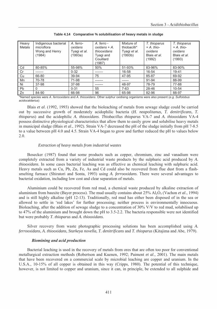
Section 3 - Acidithiobacillus
411
Table 4.14 Comparative % solubilisation of heavy metals in sludge
HeavyMetals
Indigenous bacterial microflora Wong and Henry (1984)
A. ferro- oxidans Tyagi et al.(1993a)
A. ferro - oxidans + A. thiooxidansTyagi and Couillard (1987)
Mixture of thiobacilli* Tyagi et al.(1993b)
T. thioparus+ A. thio- oxidansBlais et al.(1992)
T. thioparus+ A. thio- oxidansBlais et al.(1993)
Cd 80-85% 55-98% 50% 51-93% 83-96% 83-90%
Cr ------- 0-32 ------- 16-58 16-54 19-41
Cu 66-80 39-94 75 47-95 85-87 69-92
Mn 70-78 71-98 ------- ------ 91-94 88-99
Ni 37-98 37-98 ------- 48-97 78-79 77-88
Pb 0 0-31 55 7-63 28-46 10-54
Zn 84-90 66-98 96 65-98 82-96 88-97
*Named species were A. ferrooxidans and A. thiooxidans. Other sulphur oxidising organisms were also present (e.g. Sulfolobus acidocaldarius)
Blais et al. (1992, 1993) showed that the bioleaching of metals from sewage sludge could be carried out by successive growth of moderately acidophilic bacteria (H. neapolitanus, T. denitrificans, T. thioparus) and the acidophilic A. thiooxidans. Thiobacillus thioparus VA-7 and A. thiooxidans VA-4possess distinctive physiological characteristics that allow them to easily grow and solubilise heavy metals in municipal sludge (Blais et al., 1992). Strain VA-7 decreased the pH of the sludge initially from pH 7-8.5 to a value between pH 4.0 and 4.5. Strain VA-4 began to grow and further reduced the pH to values below 2.0.
Extraction of heavy metals from industrial wastes
Bosecker (1987) found that some products such as copper, chromium, zinc and vanadium were completely extracted from a variety of industrial waste products by the sulphuric acid produced by A.thiooxidans. In some cases bacterial leaching was as effective as chemical leaching with sulphuric acid. Heavy metals such as Cu, Pb, Zn, Fe, As and Cd could also be recovered from flue dust from a flash-smelting furnace (Shiratori and Sonta, 1993) using A. ferrooxidans. There were several advantages to bacterial oxidation, including low cost and clear separation of metals.
Aluminium could be recovered from red mud, a chemical waste produced by alkaline extraction of aluminium from bauxite (Bayer process). The mud usually contains about 25% Al2O3 (Vachon et al., 1994) and is still highly alkaline (pH 12-13). Traditionally, red mud has either been disposed of in the sea or allowed to settle in ‘red lakes’ for further processing; neither process is environmentally innocuous. Bioleaching, after the addition of sewage sludge to a concentration of 30% V/V to red mud, solubilised up to 47% of the aluminium and brought down the pH to 3.5-2.2. The bacteria responsible were not identified but were probably T. thioparus and A. thiooxidans.
Silver recovery from waste photographic processing solutions has been accomplished using A.ferrooxidans, A. thiooxidans, Starkeya novella, T. denitrificans and T. thioparus (Kitajima and Abe, 1979).
Biomining and acid production
Bacterial leaching is used in the recovery of metals from ores that are often too poor for conventional metallurgical extraction methods (Robertson and Kuenen, 1992; Paiment et al., 2001). The main metals that have been recovered on a commercial scale by microbial leaching are copper and uranium. In the U.S.A., 10-15% of all copper is obtained in this way (Cripps, 1980). The potential of this technique, however, is not limited to copper and uranium, since it can, in principle, be extended to all sulphide and
Part 4 – Consensus Documents on Micro-Organism
412
some oxide ores. Other metals that have been extracted using processes that involve bacteria include zinc, cobalt, lead, gold and molybdenum (Robertson and Kuenen, 1992).
Biological copper leaching is practised in many countries, including the U.S.A., Russia, Chile, Peru, Australia, Spain, Canada and Mexico. Typically, copper ore mined from open pits is segregated, higher-grade material being concentrated for smelting and the lower-grade ore subjected to leaching. The latter is piled to form a ‘dump’ up to 40 m high and several hectares wide. After the top is levelled, a leaching solution containing ferric sulphate and A. thiooxidans is flooded or sprayed onto the dump (Merson, 1992). Bacterial colonisation occurs mainly in the top metre. Leachates enriched with copper exit at the base of the dump and are conveyed to a central recovery facility. The copper in the solution is recovered by mixing it with iron scraps in large container units according to the following reaction:
CuSO4 + Fe0 FeSO4 + Cu0 [19]
The finely divided “cement copper” is periodically recovered and refined for sale, while the barren solution is recycled to the leachate dumps. A typical large dump may have an operating life of over ten years (Hutchins et al., 1986). Total copper recoveries of 80% were attained by the Chilean company Sociedad Minera Pudahuel (SMP) after leaching times ranging from 150 to 230 days, and about 90% recovery was attained after 7 to 11 months, more than double the quantities that would have been obtained without bacteria (Acevedo and Gentina, 1993).
Acidithiobacillus thiooxidans has also been used to obtain a high degree of copper extraction from covellite (Curutchet et al., 1995) by oxidising the layer of sulphur covering the sulphide surface and allowing sulphide oxidation by ferric ion. Both A. thiooxidans and A. ferrooxidans are effective in leaching covellite, although at different rates (Donati et al., 1996).
Acidithiobacillus ferrooxidans was used to extract uranium from low-grade ores, as for example in the Denison Mine project in Elliott Lake, Ontario, Canada (Brierley, 1990; McCready, 1988). The operation is similar to copper leaching in that it employs a bacterially assisted, flood-leaching process, but it is performed underground on mine waste rubble. About 12-13% of the uranium production is attributed to bioleaching at present, which could increase to 25% after refinement. After the uranium-bearing ore is leached, a uranium-bearing solution drains to lower portions of the mine and accumulates in sumps, after which it can be pumped to the surface for uranium recovery (Hutchins et al., 1986). The addition of elemental sulphur or sulphur slag as an external energy source enhanced the leaching process (Bhatti et al., 1991).
Acidithiobacillus ferrooxidans and A. thiooxidans were used together to extract cobalt from an ore containing 40.3% iron and 1.4% cobalt (Battaglia et al., 1994). It was found that for the system to operate at the highest efficiency, the acidity had to be maintained at a pH of between 1.1 and 2. Furthermore, the dissolution of pyrite was depressed when the concentration of ferric ions reached a level of 35 g/L.
According to Brierley (1995), an estimated one-third of the world’s total gold production is now from refractory deposits such as gold-arsenic concentrates. In these concentrates, gold and silver are finely disseminated in sulphide minerals, mainly arsenopyrite as well as pyrite, and partly antimonite (Karavaiko et al., 1986). Bio-oxidation, in which chemolithotrophic bacteria such as A. ferrooxidans have been used to decompose the ore, is a low-energy alternative to conventional methods which involve roasting and pressure leaching.
Lindstrom et al. (1992) and Morin (1995) have reviewed the process of microbial leaching for recovery of gold. Acidithiobacillus ferrooxidans was found to rapidly and selectively oxidise arsenopyrite and other sulphide minerals in the concentrates. Bio-oxidation increased gold recovery from low levels up
Section 3 - Acidithiobacillus
413
to 95-99% (Maturana et al., 1993; Morin, 1995) in comparison with traditional acid leaching. Compared to roasting, bio-oxidation with thiobacilli and their relatives could generally reduce capital costs by 12-20%, operating costs by 10% in some cases and construction time by 25% (Brierley, 1995). At the same time, the process was less polluting to the environment and had lower energy requirements since it operated at relatively low temperatures (Cripps, 1980). Highly adapted strains decompose arsenopyrite ores to an extent that allows more than 95% gold recovery in 3 days compared to the more than 12 days required by the original isolates.
Acidithiobacillus ferrooxidans accelerated the leaching of silver and other metals present in a mixed sulphide ore from Idaho (Ehrlich, 1986; 1988). Continuous leaching where iron in the solution was supplied to the reactor from a reservoir resulted in selective leaching of the silver.
Zinc has been effectively recovered from a zinc sulphide concentrate by continuous microbiological leaching with A. ferrooxidans using a two-stage reactor sequence (Sanmugasundaram et. al., 1986). Direct leaching by A. ferrooxidans together with A. thiooxidans has been found to be effective (Pistorio et al., 1994).
Sub-marginal mercury/antimony sulphidic ores were separated under experimental conditions into their components using a culture of A. ferrooxidans isolated from a coal field in the Moscow region (Lyalikova and Lyubavina, 1986).
Oil recovery and purification of oil shale
The position with oil reserves is similar to that of metallic ores outlined above: some 30,000 billion barrels of oil are present in shales, of which only about 2% are available because the recovery of the rest is uneconomical. Conventional extraction methods involve crushing and heating the shale to high temperatures to release the oil from the inorganic matrix. In this way, vast quantities of energy are consumed, only 75% of the organic material is liberated and large quantities of expended shale must be disposed of (Dalton, 1979). A biological process has been invented (Yen et al., 1976) to extract oil at ambient temperatures, giving good yields and avoiding the production of vast quantities of insoluble residue. Organisms mentioned in the patent included A. ferrooxidans, A. thiooxidans, H. neapolitanus, T. thioparus, other species of Thiobacillus since relocated, and various species of Desulfovibrio. Thesebacteria remove most of the organically bonded disulphides and polysulphides in the inorganic matrix of the shale oil, leaving an organic structure that can be used as fuel or can be converted into other materials such as petroleum or synthetic natural gas.
Bioleaching of pyrite from the Aleksinac oil shale in Yugoslavia was successfully carried out by using A. ferrooxidans as an alternative to chemical removal; chemical removal formerly led to undesirable changes in the oil substrate (Vrvic et al., 1988).
Desulphurisation of coal
To some extent coal desulphurisation is similar to the process above, in that in both cases sulphidic ores are oxidised; however both the aim and the end products are different. Coal, being of fossil origin, is not a homogeneous substance, containing a variable quantity of fixed carbon, hydrogen, oxygen, sulphur, nitrogen and trace minerals (Mannivannan et al., 1994). The aim of coal desulphurisation is to produce coal which is as free of sulphur and its derivatives as possible and it is necessary therefore to convert reduced sulphur compounds to soluble forms (Robertson and Kuenen, 1992).
Microbial desulphurisation by A. ferrooxidans, A. thiooxidans and Sulfolobus brierleyi can remove 90% or more of the inorganic sulphur from coal within a few days (Khalid and Aleem, 1991; Kilbane, 1989). The pyrite-oxidising capacity of A. ferrooxidans and related organisms has also been successfully
Part 4 – Consensus Documents on Micro-Organism
414
exploited in the desulphurisation of coal (Bos et al., 1988; Bos and Kuenen, 1990; Tuovinen and Fry, 1993) with the production of sulphuric acid instead of sulphur dioxide.
Sulphur is bound in inorganic and organic form in coal. Sulphur dioxide emissions arising by coal burning represent an important ecological problem, which can be solved by the conversion of the sulphur, compounds in the coal into different end products (Beck et al., 1988). These workers found that A. ferrooxidans was the most useful species to use, but other species such as A. acidophilum, (T. acidiphilus), Thiomonas perometabolis (T. perometabolis) and T. plumbophilus could also be used. Beyer et al. (1988) and Bos et al. (1988) found that it was possible to remove 90% of the pyrite from coal within 8 to 10 days, using a mesophilic pyrite-oxidising microbial system for which a plant design involving a cascade of Pachuca tank reactors was devised.
Mixed cultures of A. ferrooxidans and A. thiooxidans have also been used to remove sulphur from lignite, the lowest rank of coal intermediate between peat and anthracite (Raman et al., 1994).
Desulphurisation of rubber
Mixed cultures of A. ferrooxidans and A. thiooxidans satisfactorily removed sulphur inclusions in rubber materials that could be recycled from urban wastes (Torma and Raghaven, 1990). The two bacteria together were more efficient than the individual bacteria alone.
Detection of sulphur impurities in wine
A rapid and accurate sensor system was developed to determine free sulphite in wine (Nakamura et al., 1989), using immobilised cells of A. thiooxidans S3. The concentration of free sulphite could then be controlled so as to protect wine from oxidation processes and microbial spoilage. The same strain was used to detect sulphur dioxide in wine and various foodstuffs. The bacterium is converted into a microbial sensor by setting a piece of microbial membrane onto an O2 electrode soaked in 0.1 M citrate buffer and covered with a gas-permeable Teflon membrane (Kawamura et al., 1992; Kurosawa et al., 1990, 1994). A similar biosensor using strain JCM7814 was developed to detect concentrations of sulphur dioxide in wine up to 50 mg/l, with a limit of detection of 5 mg/l and a response time of 20 minutes (Nakamura et al., 1993).
Agricultural fertilisation
Since thiobacilli are involved in the sulphur cycle, the presence in the soil can be used to assess fertility. In Australia, for example, thiobacilli are scarce in sulphur-deficient areas (Kuenen et al., 1992).
Under warm climatic conditions rock phosphate pelleted with sulphur and seeded with thiobacilli has been shown to be a useful slow release source of phosphate and sulphate for soil fertilisation (Swaby, 1975). However, the addition of A. thiooxidans to a mixture of rock phosphate and sulphur granules, called ‘Biosuper’, has given variable results; in some cases the mixture increased the level of phosphorus in the soil and gave plant yields equivalent to those produced by the more expensive super-phosphate (Muchovej et al., 1989; Schofield et al., 1981), whereas in others, addition of A. thiooxidans did not improve performance of the fertiliser (Alvarez et al., 1981; Rajan, 1982). In pot experiments with guavas and ryegrass, the presence of A. thiooxidans in the soil enhanced the uptake of Fe, Zn and Mn (Azzazy et al., 1994; Schnug and Eckardt, 1981).
Acidithiobacillus thiooxidans is perhaps more useful in low-cost production of a supply of sulphuric acid for the dissolution of apatite in the production of phosphate fertilisers (Donati and Curutchet, 1995).
Section 3 - Acidithiobacillus
415
Soil Reclamation
Incorporating certain species of Thiobacillus with sulphur enrichment can reclaim alkali soils, since these soils are naturally poor in sulphur-oxidising bacteria. Thiobacillus thioparus has been inoculated with crushed sulphur into a calcareous solonetzic soil in virgin Alberta prairie land in order to promote acidification. This, combined with ripping the soil at a 60 cm depth and weekly irrigation, released several soluble salts, particularly those of sodium, calcium and magnesium (Bole, 1986). Acidithiobacillus thiooxidans was used to reclaim a saline alkaline soil by inoculating it together with elemental sulphur, thus lowering the pH and increasing the quantity of soluble salts (Bardiya et al., 1972).
III. Human Health Considerations
1. Diseases Caused and Mechanism of Pathogenicity, Including Invasiveness and Virulence
No species of Acidithiobacillus are known to be pathogenic based on the results of literature search conducted in various databases such as PubMed, Biosis, CAB Health and Current Contents.
2. Toxigenicity
There is no evidence to indicate that any species of Acidithiobacillus are toxigenic based on the results of literature search conducted in various databases such as PubMed, Biosis, CAB Health,Toxnet and Current Contents.
3. Allergenicity
No literature was found on the allergenicity of thiobacilli. However, the organisms are Gram negative and would therefore be expected to exhibit some of the characteristics associated with endotoxin. Nevertheless, no allergens of significance to humans have as yet been traced to this group of bacteria.
IV. Environmental and Agricultural Considerations
1. Natural Habitat and Geographic Distribution: Climatic Characteristics of Original Habitats
A. General overview
Although Acidithiobacillus is probably widely distributed, this distribution is usually related to the presence of sulphur as for example, coastal ecosystems such as salt marshes, sediments, mine dumps and sulphur-rich products of human industrial activity such as metal pipelines and concrete (Kelly and Harrison, 1989; Smith and Strohl, 1991).
As well, some of the products of human industrial activity such as metal pipelines and concrete have become a new ecological niche for some species. This has potentially destructive consequences (see below).
B. Correlation of natural incidence with usage and environmental impacts
While it can be assumed that Acidithiobacillus plays a role in the sulphur cycle, the extent of their involvement is largely unknown. Vitolins and Swaby (1969) showed that most thiobacilli in Australian soils rich in sulphur were autotrophic. Acidithiobacillus species’ natural habitats, usage and environmental impacts are presented in Table 4.15.
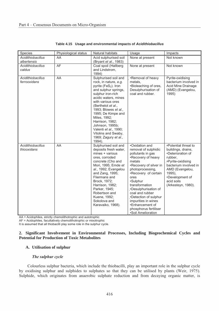
Part 4 – Consensus Documents on Micro-Organism
416
Table 4.15 Usage and environmental impacts of Acidithiobacillus
Species Physiological status Natural habitats Usage Impacts
Acidithiobacillus albertensis
AA Acid sulphurised soil (Bryant et al., 1983)
None at present Not known
Acidithiobacillus caldus
AF Coal spoil (Hallberg and Lindstrom, 1994)
None at present Not known
Acidithiobacillus ferrooxidans
AA Sulphurised soil and rock, in nature, e.g.pyrite (FeS2). Iron and sulphur springs, sulphur iron-rich acidic waters, mines with various ores (Berthelot et al., 1993; Blowes et al.,1995; De Kimpe and Miles, 1992;Harrison, 1982; Johnson, 1995b; Valenti et al., 1990; Vitolins and Swaby, 1969; Zagury et al., 1994).
•Removal of heavy metals, •Bioleaching of ores, Desulphurisation of coal and rubber.
Pyrite-oxidising bacterium involved in Acid Mine Drainage (AMD) (Evangelou, 1995)
Acidithiobacillus thiooxidans
AA Sulphurised soil and deposits fresh water, mines + various ores, corroded concrete (Cho and Mori, 1995; Emde et al., 1992; Evangelou and Zang, 1995; Fliermans and Brock, 1972; Harrison, 1982; Parker, 1945; Robertson and Kuene, 1992; Sokolova and Karavaiko, 1968).
•Oxidation and removal of sulphidic pollutants in gas •Recovery of heavy metals •Recovery of silver in photoprocessing, •Recovery of certain ores •Sulphur transformation •Desulphurisation of coal and rubber •Detection of sulphur impurities in wines •Enhancement of phosphorus fertiliser •Soil Amelioration
•Potential threat to buildings, drains, •Deterioration of rubber, •Pyrite-oxidising bacterium involved in AMD (Evangelou, 1995), •Development of acid soils (Arkesteyn, 1980).
AA = Acidophiles, strictly chemolithotrophic and autotrophic AF = Acidophiles, facultatively chemolithotrophic or mixotrophic It is assumed that all thiobacilli play some role in the sulphur cycle.
2. Significant Involvement in Environmental Processes, Including Biogeochemical Cycles and Potential for Production of Toxic Metabolites
A. Utilisation of sulphur
The sulphur cycle
Colourless sulphur bacteria, which include the thiobacilli, play an important role in the sulphur cycle by oxidising sulphur and sulphides to sulphates so that they can be utilised by plants (Weir, 1975). Sulphide, which originates from anaerobic sulphate reduction and from decaying organic matter, is
Section 3 - Acidithiobacillus
417
oxidised to sulphate under both aerobic and anaerobic conditions and by both chemical and biological means. Sulphate is assimilated by plants and micro-organisms and reduced to sulphides by other micro-organisms when these die.
In nature, a variety of reduced inorganic sulphur compounds occur as intermediates between sulphide and sulphate, which normally react very slowly with oxygen. Biological oxidation by the colourless sulphur bacteria plays an important role in the recycling of reduced sulphur compounds under aerobic conditions (Kuenen, 1975). The thiobacilli have received more attention than the other main groups of sulphur-oxidising micro-organisms. These comprise several genera of heterotrophic and facultative autotrophic bacteria and yeasts and are far more numerous than the thiobacilli. The thiobacilli, however, were deemed to be more efficient when conditions suited them (Vitolins and Swaby, 1969).
Role of thiobacilli in geologic sulphur deposits
Evidence shows that thiobacilli play a fundamental role in the development and weathering of sulphur deposits. In a review of sulphur deposits and waters with high sulphidic content in the former USSR, Sokolova and Karavaiko (1968) found that T. thioparus and A. thiooxidans were often associated. However, slight differences in distribution, according to the redox potential and the acidity of the environment, roughly correlating to the pH ranges cited in Table 4.12 were observed. Two examples show that these bacteria may play a role in the build-up and breakdown of sulphur deposits:
Formation of sulphur deposits
In the Shor-Su sulphur mines, A. thiooxidans occurred in the upper horizons of the deposit where an oxidative environment prevailed and the rocks were highly acidic (Sokolova and Karavaiko, 1968). In the aquifers throughout the lower horizon where the pH was neutral or weakly alkaline due to the proximity of limestone, T. thioparus was widespread and A. thiooxidans was absent. The presence of hydrogen sulphide prevented oxidation of the ore bed in the main deposit. Hydrogen sulphide was produced daily up to a rate of 0.2 mg/l by numerous sulphur-reducing bacteria in the groundwater and in the rocks, and ascended towards the surface waters where it was oxidised by T. thioparus to sulphur and water.
2H2S + O2 2H2O + 2S0 [20]
In several parts of the sulphur mines, sedimentation of molecular sulphur still continued by oxidation of the hydrogen sulphide of the underground waters, as shown by the presence of bacterial cells and small, freshly-deposited sulphur crystals retrieved on culture slides.
Degradation of sulphur deposits in the soil
In the early 1970s, millions of tonnes of elemental sulphur were extracted from sour natural gas and stored in blocks in Alberta, Canada (Maynard et al., 1986). Since 1979, large quantities of sulphur have been deposited in adjacent forest systems due to mechanical break-up and weathering of these blocks, causing considerable damage to the understory vegetation. Three sites at distances of 50, 250 and 750 m from a sulphur block were studied. The pH was 2.6, 3.7 and 4.4 respectively for each site. This increasing acidification with the declining proximity to the sulphur block, was attributed to A. thiooxidans, which was the main soil micro-organism responsible for elemental sulphur oxidation at all three sites. Thiobacillus thioparus was also present, but at a significantly lower population level at the first site (that with the lowest pH) than at the other two. The nutrient concentration of the soil as measured by recoverable calcium, magnesium and potassium also decreased sharply towards the sulphur block.
Part 4 – Consensus Documents on Micro-Organism
418
Reaction on pyrite (FeS2) in nature
Pyrite is the most prevalent form of iron disulphide and is usually associated with coalfields in the U.S. and elsewhere in the world. It is associated with many ores, including zinc, copper, uranium, gold and silver. Pyrite is formed in a reducing environment with a continuous supply of sulphates and iron in the presence of easily decomposable organic matter (Evangelou and Zhang, 1995).
Oxidation of pyrite deposits due to the combined action of A. ferrooxidans and A. thiooxidans in empoldered or flooded land has caused a pronounced acidification of the soil (Arkesteyn, 1980; Kuenen, 1975). When soils rich in pyrite are brought into agricultural production, “cat clay” is often formed where clay particles are cemented together by jarosite formed during the oxidation of pyrite, reducing agricultural production (Kuenen, 1975; Pronk and Johnson, 1992). Jarosite [KFe3(SO4)2(OH)6] is a basic ferric sulphate also found in deposits associated with pyrite (Ivarson, 1973). Acidithiobacillus ferrooxidans has been shown to play a part in the weathering of sulphide minerals with jarosite formation under humid conditions in metamorphic and igneous rocks in Ontario (De Kimpe and Miles, 1992). Other acidophilic species, such as T. prosperus, are able to perform the same reactions (Johnson, 1995b) but strains of A. ferrooxidans tend to grow more rapidly on ferrous iron than do other iron-oxidising acidophiles, thus causing them to dominate mixed populations (Pronk and Johnson, 1992).
Acid mine drainage (AMD)
Acid and metal pollution can be the result of the activities of thiobacilli in mine wastes (Tuovinen and Kelly, 1972). The natural oxidation of sulphide to sulphates, including sulphuric acid, as part of the sulphur cycle has become greatly enhanced by the world’s increasing demand for metals and fossil fuels. Acid mine drainage (AMD) result from land disturbances due to mining and ore processing, and has become an economic and environmental burden (Evangelou and Zhang, 1995). Acid mine drainage may be enriched with soluble iron, manganese, aluminium, sulphate and heavy metals, and the pH may be as low as 2. One of the effects of AMD is to kill established vegetation associated with mine sites and spoil tips. The typical situation found on a reclaimed spoil tip is a slow fall in pH over most of the site. Severe acid generation tends to neutralise any added lime or buffering systems in the soil quickly, and further accelerates the rate of oxidation of pyrite (Backes et al., 1986). The large quantities of sulphuric acid that are produced make the environment in which A. ferrooxidans grows, and to which it is well adapted, inhospitable to most other organisms (Rawlings and Woods, 1995; Léveillé et al., 2001; Leduc et al., 2002).
Mine spoils that were alkaline in nature (pH 9), with low sulphur content and a high concentration of chlorides tended to be free of A. ferrooxidans (Twardowska, 1986). The limiting pH value for growth of A. ferrooxidans in rock material and drainage was found to be about 7.2 (Twardowska, 1987).
B. Corrosion
Because many of the colourless sulphur bacteria produce sulphuric acid or ferric ion, they are often associated with the oxidative corrosion of concrete and pipes, and have been implicated in the corrosion of buildings and ancient monuments.
Corrosion of concrete by Acidithiobacillus thiooxidans (Thiobacillus concretivorus)
Bacterial involvement in concrete corrosion has been known since the pioneering work of Parker (1945). Acidithiobacillus thiooxidans has been found mainly responsible for the deterioration of concrete in sewage pipelines (Cho and Mori, 1995; Jozsa et al., 1995; Mori et al., 1991, 1992; Parker, 1945; Sand and Bock, 1988, 1991). The bacterium is able to use hydrogen sulphide released from the sewage and oxidise it to sulphuric acid which then attacks the concrete. It was found to coexist with an acid-resistant
Section 3 - Acidithiobacillus
419
fungus which could oxidise H2S to thiosulphate, and it is thought that the bacterium used the latter as an energy source, producing sulphuric acid that was responsible for the corrosion.
CaCO3 + H2SO4 CaSO4 + H2O + CO2 (Sand and Bock, 1988) [21]
The calcium loss results in a reduction in the stability of the structure.
The pH of fresh concrete sewer pipes is about 13, so that little microbial activity would normally occur in them. The pH would be lowered by carbonation of the concrete due to exposure to air, as well as by exposure to H2S and then by the activities of the bacterium itself. Other bacteria, including H.neapolitanus, T. intermedia and S. novella, have been found to accompany A. thiooxidans but played a minor role in corrosion (Sand and Bock, 1991).
Acidithiobacillus thiooxidans was shown experimentally to be able to degrade Sulphlex, a mixture of elemental sulphur and plasticisers used as a paving material and as a substitute for asphalt in road construction with a simultaneous production of sulphuric acid (Ferenbaugh et al., 1992). In concurrent studies, plants grown in soils amended with Sulphlex exhibited higher sulphur content and reduced growth consistent with poisoning. Indications are, therefore, that A. thiooxidans has the potential for adverse effects on sulphur-containing construction materials as well as on the local environment.
Corrosion of steel
Transportation of low-grade coal in railway carriages has been linked with accelerated corrosion of the steel framework (Brozel et al., 1995), involving scaling, pitting and cracking. Corrosion of 3CR12 steel coupons embedded in coal occurred under experimental conditions after the pH of the coal had been lowered to 2.5. The extent of the damage increased when the fungus Hormoconis resinae, another dominant member of the natural flora, was present in the substrate.
C. Deterioration of rubber
Thaysen et al. (1945) reported that the deterioration of rubber hoses was the result of the microbial oxidation of elemental sulphur present in the rubber by A. thiooxidans. This was confirmed by Raghaven et al. (1990) who also note similar effects on polyethylene.
3. Interactions with and Effects on Other Organisms in the Environment
Acidithiobacillus plays an important role in making sulphur available to plants. Acidithiobacillus thiooxidans may also make phosphorus, iron, zinc and manganese more available for plant growth. However, plants can be adversely affected by too high concentrations of sulphate and increased acidity in the soil as a result of activity due to A. thiooxidans. Very little is known about antagonists of these bacteria.
4. Routes of Dissemination: Biological or Physical
A. Biological
All species of Acidithiobacillus are motile (Kelly and Wood, 2000) so that they are able to disseminate within their immediate environment.
B. Physical
Water appears to be the major means of passive dissemination, and some dispersal must also be due to the spread of particles of soil or rock to which the bacteria have become attached.
Part 4 – Consensus Documents on Micro-Organism
420
V. Application of the Organism in Industry
1. Containment and Decontamination
A. Chemical Methods
The traditional method for controlling acidity in coal spoil and the deposition of pyrite in field drains and soils was to add high levels of lime in order to maintain the pH well above 4 to limit the activity of ferric ions and of A. ferrooxidans, so restricting the oxidation of pyrite to a process involving oxygen alone (Backes et al., 1986; Poissant, 1986; Trafford et al., 1973). Alkaline chemicals, such as limestone, sodium carbonate or sodium hydroxide, have been applied or pumped into active mines to neutralise acid soils. Limestone could hydrolyse most heavy metals, precipitating them as metal hydroxides (Evangelou and Zhang, 1995). This method depended on maintenance of the pH at a consistent value, and very often reacidification occurred as the lime was neutralised or washed out of the surface layers. Limestone is usually readily available but massive over liming of the site may result (Pulford et al, 1986) and its effectiveness is reduced because a coating of ferric hydroxide precipitates develops to shield it from further dissolution (Evangelou and Zhang, 1995). The use of soluble neutralising agents such as sodium hydroxide avoids this problem but can be costly and not very practical.
McCready (1987) successfully controlled A. ferrooxidans in pyritic shale in the laboratory by adding sodium chloride to reach a concentration of 1.5%. He has suggested that the incorporation of a salt layer would prevent AMD in pyritic sites. Another promising method of controlling pyrite oxidation is considered to be the application of phosphate, which can precipitate Fe3+ in an insoluble form as FePO4 or FePO4.2H2O (strengite). Coating of the pyrite with iron is prevented by leaching the pyrite with low but critical concentrations of H2O2 and a pH buffer, with or without KH2PO4. In the first case, iron phosphate precipitates as a coating on the pyrite surface; in the second, it precipitates as an iron oxide (Evangelou and Zhang, 1995). Treatments with phosphate, silicate, citrate and EDDDHA (ethylenediamine di-orthohydroxyphenylacetic acid) inhibited the release of acid and iron from pyritic mine waste (Pulford etal., 1986).
Growth of A. thiooxidans strain NB1-3 was also greatly reduced in the laboratory by 5 mM of NiSO4,the nickel binding to the cells and inhibiting the enzymes involved in sulphur oxidation (Maeda et al.,1996).
Inhibition of iron-oxidising bacteria may be achieved through the use of anionic surfactants (including common cleaning detergents), organic acids and food preservatives (Kleinman, 1989). Acid production may be reduced by 60 to 95%. However, wide use of surfactants is limited by the necessity for frequent treatments, since they are very soluble and motile, and they may also be adsorbed onto the surfaces of minerals without reaching the pyrite-bacterial interface (Evangelou and Zhang, 1995). The Witco product Microwet IITM incorporating various surfactants, when continuously applied to refuse leaving a coal mine satisfactorily controlled A. ferrooxidans and reduced acid production (Stancel, 1982). Sodium lauryl sulphate was shown to be inhibitory at a concentration of about 10-6 M in culture media and in mine spoils at higher concentration (Fox and Rastogi, 1983). It also has the additional benefit of being low in mammalian toxicity and quite biodegradable, so subsequent environmental problems are unlikely. For restoration of a good ground cover over reclaimed overburden, these authors proposed that a controlled release system be set up to promote a good ground cover, which, over a period of years, would eventually deprive the mine spoil of oxygen and water needed to generate acidity. 0.25% Sodium dodecyl sulphate at about 5,000 litre ha-1 resulted in dramatic reductions in acidity, sulphate, and dissolved-iron concentrations of discharge water for 3-6 months (Monticello and Finnerty, 1985).
Section 3 - Acidithiobacillus
421
Application of ProMac, a bactericide that can be applied both as a spray and in controlled-release monolithic pellets has been successful in controlling A. ferrooxidans by destroying its protective outer coating, making it susceptible to the acid it produces (Sanda, 1989). Controlled release bactericides including ProMac inhibited A. ferrooxidans and promoted regeneration of a mining site (Sobek, 1987; Sobek et al., 1990).
B. Biological Control
Inundation of pyritic material and acid sulphate soils or of disused mine shafts has been suggested (Backes et al., 1986); Evangelou and Zhang, 1995), since no significant growth of A. ferrooxidans has been demonstrated to occur in water-saturated environments (Kleinman and Crerar, 1979). A similar concept has been applied to the construction of wetlands to receive AMD in an anaerobic environment (Evangelou and Zhang, 1995). This environment encourages the activity of sulphate-reducing bacteria and so reduces acidity. Most of the hydrogen sulphide produced by these bacteria react with heavy metals to yield insoluble precipitates. Typical wetlands however, may not have sufficient permeability to take full advantage of this process.
Canada’s Natural Resources Department ‘Centre for Mineral and Energy Technology’ (CANMET) proposed to develop an anaerobic “sulphuretum” for mitigation of acidic mine drainage by envisioning construction of a system of drainage ditches to control the flow of effluent through a bed of straw (McCready, 1991). As aerobic degradation of the straw proceeds, the sugars released will be fermented to organic acids by acidophilic heterotrophs. The organic acids will then be utilised by anaerobic sulphate reducers to reduce the sulphate in the effluent to hydrogen sulphide. Hydrogen sulphide percolating through a water column will precipitate dissolved metal ions as metal sulphides. Microbially produced CO2
assists in buffering the system and provides a carbon source that may be combined with excess hydrogen by the methanogens to produce methane. In a laboratory study, the pH of the incoming liquid was 3.5 and after passage through the treatment zone it rose to pH 8.1. This process achieved a 65% reduction in the sulphate concentration and metals were not detected in the effluent.
Christison et al. (1985) reported that an unidentified zooflagellate was an effective predator, reducing the population of A. ferrooxidans from 108 cells/ml to 102 cells/ml within 18 hours at pH 2.3. Rotifers, as well as flagellated and ciliated protozoa were recorded as significant predators of A. ferrooxidans (McCready, 1987) but were incapable of eradicating them in liquid culture; moreover their large size makes it difficult for them to pursue their prey in interstitial spaces of mine tailings.
Padival et al., (1995) found that an unidentified strain of yeast introduced into continuously stirred tank reactors with H. neapolitanus or A. thiooxidans resulted in a 99% decrease in the population of the latter. The effect on these thiobacilli was enhanced by limitation of nitrogen. The results suggest that strategies based on the competitive displacement of thiobacilli to inhibit corrosion of concrete sewers may be feasible.
2. Description of Detection and Monitoring Techniques, Including Specificity, Sensitivity and Reliability
A. Thallous sulphide test
The ability to oxidise thiosulphates to sulphates with the production of elemental sulphur can be utilised to distinguish the sulphur-producing bacteria including Acidithiobacillus, from natural samples (Galizzi and Ferrari, 1976). Thallous sulphide paper moistened with pyridine is pressed onto agar plates with the colonies to be tested and then placed in dilute (0.12 N) nitric acid. The black thallous sulphide paper is bleached except in the presence of free sulphur, due to the presence of thallous polysulphides. If
Part 4 – Consensus Documents on Micro-Organism
422
sulphur is present, a brown spot is left at the site of the replicated colony. This test specifically enabled quantification of T. thioparus and A. thiooxidans in natural samples.
B. Molecular probes
There are some specific problems associated with the identification and quantitation of micro-organisms in biohydrometallurgical operations (Yates and Holmes, 1986). The numerous species of autotrophic and heterotrophic bacteria may be morphologically similar and are often difficult to purify since they grow poorly or not at all on solid media. Analysis may be frequently aggravated by the presence of small rock particles and by the production of ferric precipitates. Molecular probes using cloned DNA sequences in Southern Blots and Dot blots, could distinguish between A. ferrooxidans and other species (Acidiphilium acidophilum, Starkeya novella and T. thioparus) as well as recognising several strains of A. ferrooxidans. This technique could, moreover, detect as few as 105 bacterial cells of a given species. The reverse sample genome probing (RSGP) technique was used by Léveillé et al. (2001) to monitor the presence of Acidithiobacillus species in AMD environments. Another genomic tool such as fluorescent insitu hybridisation (FISH) was successfully used in the laboratory to detect strains of A. ferrooxidans in an ADM environment (Leduc, personal communication).
C. Polymerase-chain-reaction (PCR) and related methods
Recently PCR has been used in the detection and identification of Acidithiobacillus and other sulphur bacteria. Strains of A. ferrooxidans were differentiated by use of RAPD (random primer amplified polymorphic DNA) (Novo et al. 1996). Extending this observation, Selenska-Pobell et al. (1998) used genomic fingerprinting in the form of RAPD, rep-APD (repetitive primer amplified polymorphic DNA) and ARDREA (amplified ribosomal DNA restriction enzyme analysis) to distinguish four strains of A.ferrooxidans, and one strain each of A. thiooxidans, E. coli, Burkholderia cepacia, T. thioparus andThiomonas cuprinus. The procedures not only discriminated between the different species but also suggested that one of the strains of A. ferrooxidans was only distantly related to the three others. This variable sequence homology was attributed by the authors to the greater ability of the variant strain to accumulate uranium, although all strains were isolated for some ability to do this. Such variability suggests that more than one method should be used to identify or distinguish different strains or species of thiobacilli.
The differentiation of one strain from the others tested may not be surprising since the strains were isolated from different strata and Novo et al. (1996) suggested that strains of A. ferrooxidans isolated from different micro-environmental sources could give varying patterns on RAPD.
D. Isolation Media
Most sulphur bacteria can be isolated from natural habitats by the use of mineral media containing elemental sulphur or thiosulphate as an energy substrate. Use of media of different pH will assist differential selection of the neutrophilic and acidophilic species, whereas use of acid ferrous sulphate medium will frequently select for A. ferrooxidans (Kelly and Harrison, 1989).
A procedure has been described for the enrichment of facultatively autotrophic, mixotrophic bacteria, using a continuous flow chemostat provided with both organic and inorganic substrates (Gottschal and Kuenen, 1980). This provides a means of avoiding the predomination of heterotrophs in standard batch enrichment media containing supplements such as thiosulfate and glucose or acetate. In the latter, a mixture of obligatory chemolithotrophic thiobacilli and chemoorganotrophs normally develops. Harrison (1984) has described a general medium for cultivation of acidophilic bacteria comprising a basal mineral salts solution (MS) with the following (%w/v):
Section 3 - Acidithiobacillus
423
Media composition (per litre) (NH4)2SO4 0.200 KCl 0.010 K2HPO4 0.025 MgSO4.7H2O 0.025 Ca(NO3)2 0.001
The pH is adjusted to pH 2-4 with 1N H2SO4.
Johnson (1995a) has since reviewed various solid media formulations for acidophilic bacteria published in the literature. The use of agarose, a purified derivative of agar, was recommended to overcome the fastidiousness of thiobacilli in this and other media, as well as the use of a double-layered medium, the underlayer incorporating an acidophilic heterotroph. The latter significantly lowered the proportion of monosaccharides and resulted in a dramatic increase in plating efficiency of most strains of A. ferrooxidans.
The basic medium recommended comprised ferrous sulphate and tryptone soya broth and potassium tetrathionate, and enabled differentiation and identification of isolates of several iron-oxidising bacteria based on colony characteristics.
Acidithiobacillus albertensis
This species can be grown on the media suitable for A. thiooxidans (Kuenen et al., 1992) (see below). The pH should not be lower than pH 2.0.
Acidithiobacillus caldus
Hallberg and Lindstrom (1994) used tetrathionate as a main growth substrate adjusted to pH 2 and held at 32 OC or 52 OC according to the strain.
Acidithiobacillus ferrooxidans
This species does not readily form colonies on standard agar media because it is inhibited by some of the organic compounds found in unpurified agar (Holmes and Yates, 1990). The ability to form colonies on a solid medium is a necessary precondition for growth studies and for strain development. This problem has been overcome by substituting pure agarose for the routine agar medium or by growing the bacterium on membrane filters placed on the solid agar medium (Tuovinen and Kelly, 1973).
Growth of this species on solid media can be enhanced by the addition of small quantities of a surfactant such as Tween 80 (Garcia et al., 1992). Use of ferrous sulphate (FeSO4.7H2O) with other basal salts has also been successful in enhancing the growth of the bacterium (Kuenen et al., 1992; Harrison, 1984; Visca et al., 1989). One of the Thiobacillus solid media, TSM-1, developed by Visca et al., (1989), produced discrete and easily countable colonies and could be used for the isolation of single clones.
The ferrous sulphate medium is given by Kuenen et al. (1992) as follows:
Part 4 – Consensus Documents on Micro-Organism
424
Solution I (per litre) K2HPO4 0.5 g. (NH4)2SO4 0.5 g MgSO4.7H2O 0.5 g H2SO4 5 ml of a 15N solution
Solution II (per litre) FeSO4.7H2O 0.5 g H2SO4 150 ml of a 15N solution
Four parts of solution I are mixed with 1 part of solution II to give a medium containing 120 mMFe2+.Formation of iron precipitates can be avoided by lowering the pH through successive subcultures to a value of 1.3 with H2SO4.
Solid 2:2 medium for genetic manipulation of A. ferrooxidans
This medium was developed by Peng et al. (1994a) for the isolation of mutants. The medium, prepared in four parts, contains ferrous sulphate and sodium thiosulphate as energy sources for the growth of the bacterium at pH 4.6-4.8. Strains resistant to kanamycin and streptomycin could be obtained by incorporating increasing concentrations of these antibiotics in the medium and selecting out those colonies which developed. The medium encouraged the growth of a wide morphological range of colonies. It is also possible to introduce plasmids into bacterial cells using this medium and to develop strains resistant to heavy metals.
Acidithiobacillus thiooxidans
Kuenen et. al. (1992) list the isolation and growth media for this species as follows:
Media (per Litre) K2HPO4 3.5 g (NH4)2SO4 0.3 g MgSO4.7H2O 0.5 g FeSO4.7H2O 0.018 g CaCl 0.25 g Flowers of Sulphur 5.0 g Adjust to a pH of 4.5
Both A. ferrooxidans and A. thiooxidans may be cultivated in minimal salts (MS) containing 1% powdered sulphur. Sulphur melts at ~113 OC, so the MS-sulphur slurry is sterilised by heating at 105 OCfor one half-hour on two successive days (Harrison, 1984). These acidophilic thiobacilli have also been isolated on a mineral basal salts medium supplemented with ferrous sulphate and substituting 0.4% Gelrite, a bacterial polysaccharide, for agar (Khalid et al., 1993). Dark brown circular colonies have been observed to develop on this medium within 72-96 hours.
Section 3 - Acidithiobacillus
425
REFERENCES
Acevedo, F. and J.C. Gentina, 1993. Bioleaching of minerals - a valid alternative for developing countries. Journal of Biotechnology 31, 115-123.
Alvarez, E.V., R. Nunez-Escobar, L.J. Cajuste and C.R. Ferrera, 1981. Eficiencia agronomica de rocas fosforicas crudas y parcialmente aciduladas por metodos quimicos y biologicos. [Agronomic efficiency of crude rock phosphates and of rock phosphates partially acidulated by chemical and biological methods]. Agrociencia, Mexico 45, 25-52.
Andrews, G.F., 1988. The selective adsorption of thiobacilli to dislocation sites on pyrite surfaces. Biotechnology Bioengineering 31, 378-381.
Amaro, A.M., K.B. Hallberg, E.B. Lindstrom and C.A. Jerez, 1994. An immunological assay for detection and enumeration of thermophilic biomining micro-organisms. Applied Environmental Microbiology60 (9), 3470-3473.
Arkesteyn, G.J.M.W., 1980. Pyrite oxidation in acid sulphate soils: The role of micro-organisms. Plant and Soil 54, 119-134.
Arredondo, R. and C.A. Jerez, 1989. Specific dot-immunobinding assay for detection and enumeration of Thiobacillus ferrooxidans. Applied and Environmental Microbiology 55 (8), 2025-2029.
Azzazy, M.A., M.A. Maksoud and L.F. Haggag, 1994. Biological farming and sulfur application for improvement of Fe, Zn and Mn uptake by guava (Psidium guajava L.) seedlings grown on calcareous soil. Annals of Agricultural Science Cairo 39 (2), 731-738.
Backes, C.A., I.D. Pulford and H.J. Duncan, 1986. Studies on the oxidation of pyrite in colliery spoil. I. The oxidation pathway and inhibition of the ferrous-ferric oxidation. Reclamation and Revegetation Research 4 (4), 279-291.
Bagdigian, R. M. and A. S. Myerson, 1986. The adsorption of Thiobacillus ferrooxidans on coal surfaces. Biotechnology Bioengineering 28, 467-479.
Baillet, F, J.P. Magnin, A. Cheruy and P. Ozil, 1998. Chromium precipitation by the acidophilic bacterium Thiobacillus ferrooxidans. Biotechnology Letters 20 (1), 95-99.
Bardiya, M.C., N. Narula and S.R. Vyas, 1972. Reclamation of saline alkali soil by the application of sulfur and inoculation of Thiobacillus. I. Effect on physicochemical properties of soil. HaryanaAgricultural University Journal of Research 2 (3), 218-221.
Battaglia, F., D. Morin and P. Ollivier, 1994. Dissolution of cobaltiferous pyrite by Thiobacillus ferrooxidans and Thiobacillus thiooxidans - factors influencing bacterial leaching efficiency. Journal of Biotechnology 32 (1), 11-16.
Part 4 – Consensus Documents on Micro-Organism
426
Beck, D., H.J. Heinritz and M. Worbs, 1988. Microbial coal desulfurisation. Acta Biotechnologica 8 (1), 87-92.
Berthelot, D., L.G. Leduc and G.D. Ferroni, 1993. Temperature studies of iron-oxidising autotrophs and acidophilic heterotrophs isolated from uranium mines. Canadian Journal of Microbiology 39 (4), 384-388.
Berthelot, D., L.G. Leduc and G.D. Ferroni, 1994. The absence of psychrophilic Thiobacillus ferrooxidansand acidophilic heterotrophic bacteria in cold, tailings effluents from an uranium mine. Canadian Journal of Microbiology 40, 60-63.
Berzaczy, L., W. Etzenberger, L. Kloimstein, E. Niedermaayer, A. Schmidt and A. Windsperger, 1990. Procedure for Microbiological Transformation of Sulphur Containing Harmful Components in Exhaust Gases. United States Patent 4,968, 622.
Beyer, M., H.-J. Hone and J. Klein, 1988. A multi-stage bioreactor for continuous desulphurisation of coal by Thiobacillus ferrooxidans. pp. 514-516. In: BioHydroMetallurgy, (Norris, P.R and D.P. Kelly, Eds.). Proceedings of the International Symposium on Biohydrometallurgy, University of Warwick, July 12-16, 1987.
Bhatti, T.M., K.A. Malik and A.M. Khalid, 1991. Microbial leaching of low-grade sandstone uranium ores: column leaching studies. pp. 329-34 In: Biotechnology for Energy, (Malik, K.A., S.H.M. Naqvi and M.I.H. Aleem, Eds.) Nuclear Institute for Agriculture and Biology (NIAB) and National Institute for Biotechnology and Genetic Engineering (NIBGE), Faisalabad, Pakistan.
Blais, J.F., J.C. Auclair and R.D. Tyagi, 1992. Cooperation between two Thiobacillus strains for heavy-metal removal from municipal sludge. Canadian Journal of Microbiology 38, 181-187.
Blais, J.F., R.D. Tyagi and J.C. Auclair, 1993. Bioleaching of metals from sewage sludge: micro-organisms and growth kinetics. Water Research 27 (1), 101-110.
Blais, J.F., R.D. Tyagi, N. Meunier and J.C. Auclair, 1994. The production of extracellular appendages during bacterial colonisation of elemental sulphur. Process Biochemistry 29 (6), 475-482.
Blake, R.C. II, K.J. White and E.A Shute, 1988. Electron transfer from Fe (II) to rusticyanin is catalysed by an acid-stable cytochrome. pp. 103-110. In: BioHydroMetallurgy, (Norris, P.R and D.P. Kelly, Eds.). Proceedings of the International Symposium on Biohydrometallurgy, University of Warwick, July 12-16, 1987.
Blowes, D.W., T. Al, L. Lortie, W.D. Gould and J.L. Jambor, 1995. Microbiological, chemical, and mineralogical characterisation of the Kidd Creek mine tailings impoundment, Timmins Area, Ontario. Geomicrobiology Journal 13 (1), 13-31.
Bole, J.B., 1986. Amelioration of a calcareous soil by irrigation, deep ripping, and acidification with elemental sulfur. Canadian Journal of Soil Science 66, 347-356.
Boon, M., T. A. Meeder, C. Thöne, C. Ras and J. J. Heijnen, 1999. The ferrous iron oxidation kinetics of Thiobacillus ferrooxidans in continuous culture. Applied Microbiology and Biotechnology 51(6), 820-826.
Bos, P., T.F. Huber, K.C.A.M. Luyben and J.G. Kuenen, 1988. Feasibility of a Dutch process for microbial desulphurisation of coal. Resources, Conservation and Recycling 1, 279-291.
Section 3 - Acidithiobacillus
427
Bos, P. and J.G. Kuenen, 1990. Microbial treatment of coal. pp. 344-377. In: Microbial Mineral Recovery, (Ehrlich, H.L. and C.L. Brierley Eds.). McGraw-Hill, New York.
Bosecker, K., 1987. Microbial recycling of mineral waste products. Acta Biotechnologica 7 (6), 487-497.
Brierley, C.L., 1990. Biotechnology for the Extractive Metals Industries. Journal of Metallurgy 42 (10), 28-30.
Brierley, C.L., 1995. Bacterial Oxidation: Master key to unlock refractory gold ores? Engineering and Mining Journal 196 (5), 42-44.
Brozel, V.S., A. Hall, R. de Bruin, A. Bousher, M. Chandra and R. Edyvean, 1995. Effect of microbial flora in low-grade coal on the corrosion of 3CR12 steel. Biodeterioration and Biodegradation 9. pp. 494-499. Proceedings of the 9th Int. Biodeterioration and Biodegradation Symposium, Leeds, U.K., 5-10 September, 1993.
Bruce, A.M. and R.D. Davis, 1989. Sewage sludge disposal: current and future options. Water Science and Technology 21, 1113-1128.
Bryant, R.D., K.M. McGroarty, J.W. Costerton and E.J. Laishley, 1983. Isolation and characterisation of a new acidophilic Thiobacillus species (T. albertis). Canadian Journal of Microbiology 29, 1159-1170.
Buisman, C., R. Post, P. Ijspeert, G. Geraats and G. Lettinga, 1989. Biotechnological process for sulphide removal with sulphur reclamation. Acta Biotechnologia 9 (3), 255-267.
Cabrejos, M. E., H.L. Zhao, M. Guacucano, S. Bueno, G. Levican, E. Garcia, E. Jedlicki, and D. S. Holmes, 1999. IST1 insertional inactivation of the resB gene: implications for phenotypic switching in Thiobacillus ferrooxidans. FEMS Microbiol. Lett. 175, 223-229.
Cadenhead, P. and K.L. Sublette, 1990. Oxidation of hydrogen sulfide by thiobacilli. Biotechnology Bioengineering 35, 1150-1154.
Chisholm, I.A., L.G. Leduc and G.D. Ferroni, 1998. Metal resistance and plasmid DNA in Thiobacillus ferrooxidans. Antonie van Leeuwenhoek 73, 245-254.
Cho, K.S. and T. Mori, 1995. A newly isolated fungus participates in the corrosion of concrete sewer pipes. Water Science and Technology 31 (7), 263-271.
Christison, J., V.I. Lakshmanan, N.S. Belson and J. Cairns, 1985. Biological control of Thiobacillus ferrooxidans by predatory organisms. Final Report P-4483/G-2 (No. 41-10246), Ontario Research Foundation.
Collinet, M.-N. and D. Morin, 1990. Characterisation of arsenopyrite oxidising Thiobacillus. Tolerance to arsenite, arsenate, ferrous and ferric ion. Antonie van Leeuwenhoek 57, 237-244.
Coto, O., A.I. Fernandez, T. Leon and D. Rodriguez, 1992. Thiobacillus ferrooxidans detection using immunoelectron microscopy. Microbiologia Sem 8, 76-81.
Couillard, D. and G. Mercier, 1991. Optimum residence time (in CSTR and Airlift Reactor) for Bacterial Leaching of Metals from Anaerobic Sewage Sludge. Water Research 25, 211-218.
Part 4 – Consensus Documents on Micro-Organism
428
Couillard, D. and G. Mercier, 1994. An economic evaluation of biological removal of heavy metals from wastewater sludge. Water Environment Research 66 (1), 32-39.
Couillard, D. and S.C. Zhu, 1992. Bacterial leaching of heavy metals from sewage sludge for agricultural application. Water, Air and Soil Pollution 63 (1-2), 67-80.
Cripps, R.E., 1980. The recovery of metals by microbial leaching. Biotechnology Letter 2 (5), 225-230.
Curutchet, G., C. Pogliani and E. Donati, 1995. Indirect bioleaching of covelllite by Thiobacillus thiooxidans with an oxidant agent. Biotechnology Letter 17 (11), 1251-1256.
Dalton, H., 1979. Resources conservation by novel biological processes. II. The use of microbes in the petrochemical industry. Chemical Society Reviews 8 (2): 297-308.
De Kimpe, C. and N. Miles, 1992. Formation of swelling clay minerals by sulfide oxidation in some metamorphic rocks and related soils of Ontario, Canada. Canadian Journal of Soil Science 72 (3), 263-270.
Devasia, P., K.A. Natarajan, D.N. Sathyanarayana and G.R. Rao, 1993. Surface chemistry of Thiobacillus ferrooxidans relevant to adhesion on mineral surfaces. Applied Environmental Microbiology 59 (12), 4051-4055.
Dominy, C. N., N. J. Coram and D. E. Rawlings, 1998. Sequence analysis of plasmid pTF5, a 19.8-kb geographically widespread member of the Thiobacillus ferrooxidans pTF191-like plasmid family. Plasmid 40(1), 50-57.
Donati, E. and G. Curutchet, 1995. Solubilisacion de fosfato inorganico por accion de acido sulfurico biogenerado. [Dissolution of inorganic phosphate by biologically generated sulphuric acid]. Revista de la Facultad de Agronomia La Plata 71 (1), 87-92.
Donati, E., G. Curutchet, C. Pogliani and P. Tedesco, 1996. Bioleaching of covellite using pure and mixed cultures of Thiobacillus ferrooxidans and Thiobacillus thiooxidans. Process Biochemistry 31 (2), 129-134.
Drobner, E., H. Huber and K.O. Stetter, 1990. Thiobacillus ferrooxidans, a Facultative Hydrogen Oxidiser. Applied Environmental Microbiology 56 (9): 2922-2923.
Dziurla, M. A., W. Achouak, B.T. Lam, T. Heulin and J. Berthelin, 1998. Enzyme-linked immunofiltration assay to estimate attachment of thiobacilli to pyrite. Applied Environmental Microbiology 64(8): 2937-2942.
Ehrlich, H.L., 1986. Bacterial leaching of silver from a silver-containing mixed sulfide ore by a continuous process. pp. 77-88. In: Fundamental and Applied Biohydrometallurgy, (Lawrence, R.W., R.M.R. Branion and H.G. Ebner, Eds.). Elsevier, Amsterdam.
Ehrlich, H.L., 1988. Bioleaching of silver from a mixed sulfide ore in a stirred reactor. pp. 223-231. In:BioHydroMetallurgy (Norris, P.R and D.P. Kelly, Eds.). Proceedings of the International Symposium on Biohydrometallurgy, University of Warwick, July 12-16, 1987.
Emde, K.M.E., D.W. Smith and R. Facey, 1992. Initial investigation of microbially influenced corrosion (MIC) in a low temperature water distribution system. Water Research (Oxford) 26, (2), 169-175.
Section 3 - Acidithiobacillus
429
Evangelou, V.P., 1995. Potential microencapsulation of pyrite by artificial inducement of ferric phosphate coatings. Journal of Environmental Quality 24 (3), 535-542.
Evangelou, V.P. and Y.L. Zhang, 1995. A review - pyrite oxidation mechanisms and acid mine drainage prevention. Critical Reviews in Environmental Science and Technology 25 (2), 141-199.
Ferenbaugh, R.W., E.S. Gladney, L.F. Soholot, K.A. Lyall, M. K. Wallwork-Barber, and L.E. Hersman, 1992. Environmental interactions of Sulphlex pavement. Environmental Pollution 76, 141-145.
Fernandez, M.G.M., C. Mustin, P. Dedonato, O. Barres, P. Marion and J. Berthelin, 1995. Occurrences at mineral bacteria interface during oxidation of arsenopyrite by Thiobacillus ferrooxidans.Biotechnology Bioengineering 46, 13-21.
Fliermans, C.B. and T.D. Brock, 1972. Ecology of sulfur-oxidising bacteria in hot acid soils. Journal of Bacteriology 111 (2), 343-350.
Ford, W.H., H.K. Roffman and W.A. Beimborn, 1977. Bacterial removal of sulfur from coal. Combust 49 (2), 36-39.
Fox, L.A. and V. Rastogi, 1983. Developments in controlled release technology and its application in acid mine drainage. pp. 447-455. Proceedings of Symposium on Surface Mining Hydrology, Sedimentology and Reclamation, University of Kentucky, Lexington, KY, November 27-December 2, 1983.
Frattini, C.J., L.G. Leduc and G.D. Ferroni. 2000. Strain variability and the effects of organic compounds on the growth of the chemolithotrophic bacterium Thiobacillus ferrooxidans. Antonie van Leeuwenhoek 77, 57-64.
Galizzi, A. and E. Ferrari, 1976. Identification of thiobacilli by replica plating on thallous sulfide paper. Applied Environmental Microbiology 32 (3), 433-435.
Garcia, O., J.K. Mukai and C.B. Andrade, 1992. Growth of Thiobacillus ferrooxidans on solid medium: effects of some surface-active agents on colony formation. Journal of General Applied Microbiology38, 279-282.
Gardner, M. N., S. M. Deane, and D. E. Rawlings. 2001. Isolation of a new broad host-range plasmid, (pTC-F14) from the acidophilic bacterium Acidithiobacillus ferrooxidans and analysis of the plasmid replicon. J. Bacteriol. 183, 3303-3309)
Gehrke, T., R. Hallman, K. Kinzler and W. Sand, 2001. The EPS of Acidithiobacillus ferrooxidans - a model for structure - function relationships of attached bacteria and their physiology. Water Science and Technology 43(6), 159-167.
Gehrke, T., J. Telegdi, D. Thierry and W. Sand, 1998. Importance of extracellular polymeric substances from Thiobacillus ferrooxidans for bioleaching. Applied Environmental Microbiology 64(7), 2743-2747.
Gonzalez, C and D. Cotoras, 1987. Apendices de Thiobacillus ferrooxidans en cultivos sucesivos. [Appendages of Thiobacillus ferrooxidans in subcultures.] Revista Latinoamericano deMicrobiologia 29 (3), 235-238.
Part 4 – Consensus Documents on Micro-Organism
430
Gottschal, G.C. and J.G. Kuenen, 1980. Selective enrichment of facultatively chemolithotrophic thiobacilli and related organisms in continuous culture. FEMS Microbiology Letter 7, 241-247.
Hallberg, K.B. and E.B. Lindstrom, 1994. Characterisation of Thiobacillus caldus sp. nov., a moderately thermophilic acidophile. Microbiology 140 (12), 3451-3456.
Harrison, A.P., 1982. Genomic and physiological diversity amongst strains of Thiobacillus ferrooxidansand genomic comparison with Thiobacillus thiooxidans. Archives of Microbiology 131, 68-76.
Harrison, A.P., 1984. The acidophilic thiobacilli and other acidophilic bacteria that share their habitat. Annual Review of Microbiology 38, 265-292.
Harrison, A.P., 1986. The phylogeny of iron-oxidising bacteria. Biotechnology Bioengineering Symposium16, 311-317.
Hazra, T.K., M. Mukherjea and R.N. Mukherjea, 1992. Bioleaching of low-grade copper ores using Thiobacillus ferrooxidans. Applied Biochemistry and Biotechnology 34/35, 377-382.
Heinhorst, S., S.H. Baker, D.R. Johnson, P.S. Davies, G.C. Cannon and J.M. Shively, 2002. Two copies of form I RuBisCo genes in Acidithiobacillus ferrooxidans ATCC 23270. Current Microbiology 45, 115-117.
Hirano, T., H. Kurosawa, K. Nakamura and Y. Amano, 1996. Simultaneous removal of hydrogen sulfide and trimethylamine by a bacterial deodorant. Journal of Fermentation and Bioengineering 81 (4), 337-342.
Holmes, D.S., 1988. Biotechnology in the mining and metal processing industries - challenges and opportunities. Minerals and Metallurgical Processing, May, 49-56.
Holmes, D.S. and S.K. Debus, 1991. Biological opportunities for metal recovery. pp. 341-358. In:Biotechnology for Energy, (Malik, K.A., S.H.M. Naqvi and M.I.H. Aleem, Eds.). Nuclear Institute for Agriculture and Biology (NIAB) and National Institute for Biotechnology and Genetic Engineering (NIBGE), Faisalabad, Pakistan.
Holmes, D.S. and J.R. Yates, 1990. Basic principles of genetic manipulation of Thiobacillus ferrooxidans for biohydrometallurgical applications. pp. 29-54. In: Microbial Mineral Recovery, (Ehrlich, H.L. and C.L. Brierley, Eds.). McGraw-Hill, New York.
Holmes, D.S., J.R. Yates and J. Schrader, 1988. Mobile repeated DNA sequences in Thiobacillus ferrooxidans and their significance for biomining. pp. 153-160. In: BioHydroMetallurgy, (Norris, P.R. and D.P. Kelly, Eds.). Proceedings of the International Symposium on Biohydrometallurgy, University of Warwick, July 12-16, 1987.
Holmes, D.S., H.L. Zhao, G. Levican, J. Ratouchniak, V. Bonnefoy, P. Varela and E. Jedlicki, 2001. ISAfe1, an ISL3 Family Insertion Sequence from Acidithiobacillus ferrooxidans ATCC 19859. JBacteriol. 183 (14), 4323-4329.
Huber, H. and K.O. Stetter, 1989. Thiobacillus prosperus, new species, represents a new group of halotolerant metal-mobilizing bacteria isolated from a marine geothermal field. Archives of Microbiology 151 (6), 479-485.
Section 3 - Acidithiobacillus
431
Huber, H. and K.O. Stetter, 1990. Thiobacillus cuprinus sp. nov., a novel facultatively organotrophic metal-mobilizing bacterium. Applied Environmental Microbiology 56 (2), 315-322.
Hubert, W.A., Ferroni G.D. and L.G. Leduc, 1994. Temperature-dependent survival of isolates of Thiobacillus ferrooxidans. Current Microbiology 28 (3), 179-183.
Hutchins, S.R., M.S. Davidson, J.A. Brierley and C.L. Brierley, 1986. Micro-organisms in Reclamation of Metals. Annual Review of Microbiology 40, 311-386.
Ivarson, K.C., 1973. Microbiological formation of basic ferric sulfates. Canadian Journal of Soil Science53, 315-323.
Iwahori, K., F. Takeuchi, K. Kamimura and T. Sugio, 2000. Ferrous-dependent volatilisation of mercury by the plasma membrane of Thiobacillus ferrooxidans. Applied and Environmental Microbiology66(9), 3823-3827.
Jensen, A.B. and C. Webb, 1995. Treatment of H2S-containing gases: A review of microbiological alternatives. Enzyme and Microbial Technology 17, 2-10.
Jerez, C.A., I. Peirano, D. Chamorro and G. Campos, 1986. Immunological and electrophoretic differentiation of Thiobacillus ferrooxidans strains. pp. 443-456. In: Fundamental and Applied Biohydrometallurgy, (Lawrence, R.W., R.M.R. Branion and H.G. Ebner, Eds.). Elsevier, Amsterdam.
Jin, S., R.S. English and J.M. Shively, 1994. Transformation of Thiobacillus intermedius by electroporation. Abstracts of the General Meeting of the American Society for Microbiology 94, 223.
Jin, S.M., W.M. Yan and Z.N. Wang, 1992. Transfer of IncP plasmids to extremely acidophilic Thiobacillus thiooxidans. Applied Environmental Microbiology 58 (1), 429-430.
Johnson, D.B., 1995a. Selective solid media for isolating and enumerating acidophilic bacteria. Journal of Microbiological Methods 23 (2), 205-218.
Johnson, D.B., 1995b. Mineral cycling by micro-organisms: Iron bacteria. pp. 137-159. In: Microbial diversity and ecosystem function (Allsopp, D., R.R. Colwell and D.L. Hawksworth, Eds.). Proceedings of the IUB/IUMS workshop held at Egham, UK, 10-13 August, 1993, CABI.
Jozsa, P.G., T. Gehrke, F. Sattler, W. Sand, K. Zapel A. Bouscher, M Chandra and R. Edtvean, 1995. Microbiologically influenced deterioration of brickwork in sewage pipelines. Biodeterioration and Biodegradation 9. pp. 188-194. Proceedings of the 9th International Biodeterioration and Biodegradation Symposium, Leeds, U.K., 5-10 September, 1993.
Kai, T., M. Nishi and T. Takahashi, 1995. Adaptation of Thiobacillus ferrooxidans to nickel ion and bacterial oxidation of nickel sulfide. Biotechnology Letter 17 (2), 229-232.
Karamanev, D.G., 1991. Model of the biofilm structure of Thiobacillus ferrooxidans. Journal ofBiotechnology 20, 51-64.
Karavaiko, G.I., L.K. Chuchalin, T.A. Pivovarova, B.A. Yemel’yanov and A.G. Dorofeyev, 1986. Microbiological leaching of metals from arsenopyrite containing concentrates. pp. 115-126. In:
Part 4 – Consensus Documents on Micro-Organism
432
Fundamental and Applied Biohydrometallurgy (Lawrence, R.W., R.M.R. Branion and H.G. Ebner, Eds.). Elsevier, Amsterdam.
Katayama-Fujimura, Y., N. Tsuzaki and H. Kuraishi, 1982. Ubiquinone, fatty acid and DNA base composition determination as a guide to the taxonomy of the genus Thiobacillus. Journal of General Microbiology 128, 1599-1611.
Kawamura, Y., T. Satou, N. Kubo, H. Akano, H. Kurosawa, K. Nakamura, Y. Amano, Y. Ito and M. Fujimaki, 1992. [Measurement of sulphur dioxide in foods by biosensor method.] Journal of Japanese Society of Food Science and Technology [Nippon-Shokuhin-Kogyo-Gakkaishi] 39 (3), 233-238.
Kelly, D.P. and A.P. Harrison, 1989. Genus Thiobacillus Beijerinck. In: Bergey’s Manual of Systematic Bacteriology, Vol. 3, 1842-1858. (Staley, J.T., M.P. Bryant, N. Pfennig and J.G. Holt, Eds.), Springer-Verlag, New York.
Kelly, D.P. and A.P. Wood, 2000. Reclassification of some species of Thiobacillus to the newly designated genera Acidithiobacillus gen. nov., Halothiobacillus gen. nov., and Thermithiobacillus gen. nov. International Journal of Systematic and Evolutionary Microbiology 50, 511-516.
Kelly, D.P. and A.P. Wood, 2005. Genus Acidithiobacillus. In: Bergey’s Manual of Systematic Bacteriology Vol. 2, 2nd ed., in press. Springer-Verlag, New York
Khalid, A.M. and M.I.H. Aleem, 1991. Application of micro-organisms in coal cleaning processes. pp. 379-403. In: Biotechnology for Energy, (Malik, K.A., S.H.M. Naqvi and M.I.H. Aleem, Eds.). Nuclear Institute for Agriculture and Biology (NIAB) and National Institute for Biotechnology and Genetic Engineering (NIBGE), Faisalabad, Pakistan.
Khalid, A.M., T.M. Bhatti and M. Umar, 1993. An improved solid medium for isolation, enumeration and genetic investigations of autotrophic iron-oxidising and sulphur-oxidising bacteria. Applied Microbiology and Biotechnology 39 (2), 259-263.
Khalid, Z.M. and R. Naeveke, 1991. Mutualism between autotrophic and heterotrophic bacteria in leaching of low grade ores. pp. 313-321. In: Biotechnology for Energy (Malik, K.A., S.H.M. Naqvi and M.I.H. Aleem, Eds.). Nuclear Institute for Agriculture and Biology (NIAB) and National Institute for Biotechnology and Genetic Engineering (NIBGE), Faisalabad, Pakistan.
Kilbane, J.J., 1989. Desulfurisation of coal: the microbial solution. Trends in Biotechnology 7 (4), 97-102.
Kitajima, M. and A. Abe, 1979. Treating of waste photographic processing solution and silver recovery from there using chemosynthetic sulfur bacteria. United States Patent 4,155,810.
Kleinman, R.L.P., 1989. Acid mine drainage: U.S. bureau of mines researches and develops control methods for both coal and metal mines. Engineering and Mining Journal (July), 161.
Kleinman, R.L.P. and D.A. Crerar, 1979. Thiobacillus ferrooxidans and the formation of acidity in simulated coal mine environments. Geomicrobiology Journal 1, 373.
Koppe B. and H. Harms, 1994. Antigenic determinants and specificity of antisera against acidophilic bacteria. World Journal of Microbiology and Biotechnology 10 (2), 154-158.
Kuenen, J.G., 1975. Colourless Sulfur bacteria and their Role in the Sulfur Cycle. Plant and Soil 43, 49-76.
Section 3 - Acidithiobacillus
433
Kuenen, J.G., 1989. Colorless Sulfur Bacteria. In: Bergey’s Manual of Systematic Bacteriology, Vol. 3, p. 1834-1842. (Staley, J.T. M.P. Bryant, N. Pfennig and J.G. Holt, Eds), Springer-Verlag, New York.
Kuenen, J.G., L.A. Robertson and O.H. Tuovinen, 1992. The Genera Thiobacillus, Thiomicrospora and Thiosphaera. pp. 2638-2657 In: The Prokaryotes, (Balows, A., H.G. Trüper, M. Dworkin, W. Harder and K.-H. Scheifler, Eds.). 2nd edition, chapter 138, Vol. III, Springer-Verlag, New York.
Kulpa, C.F., M.T. Roskey, and M.T. Travis, 1983. Transfer of plasmid RP1 into chemolithotrophic Thiobacillus neapolitanus. Journal of Bacteriology 156, 434-436.
Kurosawa, H., D. Rosadi, K. Nakamura and Y. Amano, 1990. Determination of sulfur dioxide in wine and dried fruits using a microbial sensor. Journal of the Institute of Enology and Viticulture, Yamanashi University [Yamanashi Daigaku Hakko Kenkyusho Kenkyu Hokoku] 25, 15-19.
Kurosawa, H., T. Hirano, K. Nakamura and Y. Amano, 1994. Microbial sensor for selective determination of sulphide. Applied Microbiology and Biotechnology 41(5), 556-559.
Kusano, T., K. Sugawara, C. Inoue, T. Takeshima M. Numata and T. Shiratori, 1992. Electrotransformation of Thiobacillus ferrooxidans with plasmids containing a mer determinant as the selective marker by electroporation. Journal of Bacteriology 74, 6617-6623.
Lane, D.L., A.P. Harrison, D. Stahl, B. Pace, S.J. Giovannoni, G.J. Olsen and N.R. Pace, 1992. Evolutionary relationships among sulfur- and iron-oxidising eubacteria. Journal of Bacteriology174 (10), 269-278.
Lane, D.J., D.A. Stahl, G.J. Olsen, D.J. Heller and N.R. Pace, 1985. Phylogenetic analysis of the genera Thiobacillus and Thiomicrospora by 5S rRNA sequences. Journal of Bacteriology 163 (1), 75-81.
Lazaroff, N., 1963. Sulfate requirement of iron oxidation by Thiobacillus ferrooxidans. Journal of Bacteriology 85, 78-83.
Leduc, L.G. and G.D. Ferroni, 1993. The need for Thiobacillus ferrooxidans strain selection in applications in bioleaching. p. 25-42. Proceedings of the Tenth Annual Meeting of BIOMINET, Mississauga, Canada, Canada Centre for Mineral and Energy Technology, October 28, 1993.
Leduc, L.G. and G.D. Ferroni, 1994. The chemolithotrophic bacterium Thiobacillus ferrooxidans. FEMS Microbiology Letter 14, 103-120.
Leduc, L.G., G.D. Ferroni and J.T. Trevors, 1997. Resistance to heavy metals in different strains of Thiobacillus ferrooxidans. World Journal of Microbiology and Biotechnology 13, 453-455.
Leduc, L.G., J.T. Trevors, and G.D. Ferroni, 1993. Thermal characterisation of different isolates of Thiobacillus ferrooxidans. FEMS Microbiology Letter 97, 167-172.
Leduc, D., L.G. Leduc and G.D. Ferroni, 2002. Quantification of bacterial population indigenous to acidic drainage streams. Water, Air and Soil Pollution 135, 1-21.
Léveillé, S.A., L.G. Leduc, G.D. Ferroni, A.J. Telang and G. Voorduw, 2001. Monitoring of bacteria in acid mine environments by reverse sample genome probing. Canadian Journal of Microbiology 47, 431-442.
Part 4 – Consensus Documents on Micro-Organism
434
Lindstrom, E.B., E. Gunneriusson and O.H. Tuovinen, 1992. Bacterial oxidation of refractory sulfide ores for gold recovery. Critical Reviews in Biotechnology 12 (1/2), 133-155.
Lizama, H.M. and B.M. Sankey, 1993. Conversion of hydrogen sulphide by acidophilic bacteria. Applied Microbiology and Biotechnology 40 (2-3), 438-441.
Lyalikova, N.N. and L.L. Lyubavina, 1986. On the possibility of using a culture of Thiobacillus ferrooxidans to separate antimony and mercury sulfides during flotation. pp. 403-406. In:Fundamental and Applied Biohydrometallurgy, (Lawrence, R.W., R.M.R. Branion and H.G. Ebner, Eds.). Elsevier, Amsterdam.
Maeda, T., A. Negishi, Y. Nogami and T. Sugio, 1996. Nickel inhibition of the growth of a sulfur-oxidising bacterium isolated from corroded concrete. Bioscience Biotechnology and Biochemistry 60 (4), 626-629.
Mannivannan, T., S. Sandhya and R.A. Pandey, 1994. Microbial desulfurisation of coal by chemoautotrophic Thiobacillus ferrooxidans - an iron mine isolate. Journal of EnvironmentalScience and Health Part A-Environmental Science and Engineering 29 (10), 2045-2061.
Maturana, H., U. Lagos, V. Flores, M. Gaete, L. Cornejo and J.V. Wiertz, 1993. Integrated biological process for the treatment of a Chilean complex gold ore. FEMS Microbiology Letter 11 (1-3), 215-220.
Maynard, D.G., J.J. Germida and P.A. Addison, 1986. The effect of elemental sulfur on certain chemical and biological properties of surface organic horizons of a forest soil. Canadian Journal of Forestry Research 16, 1050-1054.
McCready, R.G.L., 1987. A review of the physical, chemical and biological measures to prevent acid mine drainage: an application to the pyritic Halifax shales. pp. 333-355. Proceedings of an Acid Mine Drainage Seminar/Workshop, Environment Canada, Halifax, NS, March 23-26, 1987.
McCready, R.G.L., 1988. Progress in the bacterial leaching of metals in Canada. pp. 177-195. In:BioHydroMetallurgy, (Norris, P.R. and D.P. Kelly, Eds.). Proceedings of the International Symposium on Biohydrometallurgy, University of Warwick, UK, July 12-16, 1987.
McCready, R.G.L., 1991. Biotechnological Processes in the Canadian Mining Industry. pp. 323-327. In:Biotechnology for Energy, (Malik, K.A., S.H.M. Naqvi and M.I.H. Aleem, Eds.). Nuclear Institute for Agriculture and Biology (NIAB) and National Institute for Biotechnology and Genetic Engineering (NIBGE), Faisalabad, Pakistan.
Merson, J., 1992. Mining with microbes. New Scientist 4, 17-19.
Monticello, D.J. and W.R. Finnerty, 1985. Microbial Desulfurisation of Fossil Fuels. Annual Review of Microbiology 39, 371-389.
Mori, T., M. Koga, Y. Hikosaka, T. Nonaka, F. Mishina, Y. Sakai and J. Koizumi, 1991. Microbial corrosion of concrete sewer pipes, H2S production from sediments and determination of corrosion rate. Water Science and Technology 23 (7-9), 1275-1282.
Mori, T., T. Nonaka, K. Tazaki, M. Koga, Y. Hikozaka and S. Noda, 1992. Interaction of nutrients, moisture and pH on microbial corrosion of concrete sewer pipes. Water Research 26, 29-37.
Section 3 - Acidithiobacillus
435
Morin, D., 1995. Bacterial leaching of refractory gold sulfide ores. pp. 25-62. In: Bioextraction and Biodeterioration of Metals, (Gaylarde, C.C. and H.A. Videla, Eds.) Cambridge University Press, Cambridge.
Muchovej, R.M.C., J.J. Muchovej and V.V.H. Alvarez, 1989. Temporal relations of phosphorus fractions in an oxisol amended with rock phosphate and Thiobacillus thiooxidans. Soil Science Society of American Journal 53 (4), 1096-1100.
Myerson, A.S. and P. Kline, 1983. Sorption of Thiobacillus ferrooxidans to particulate material. Biotechnology and Bioengineering 25, 1163-1168.
Nakamura, K., Y. Amano and O. Nakayama, 1989. Determination of free sulphite in wine using a microbial sensor. Applied Microbiology and Biotechnology 31 (4), 351-354.
Nakamura, K., K. Saegusa, H. Kurosawa H. and Y. Amano, 1993. Determination of free sulfur dioxide in wine by using a biosensor based on a glass electrode. Bioscience Biotechnology and Biochemistry 57 (3), 379-382.
Norris, P.R., 1990. Acidophilic bacteria and their activity in mineral sulphide oxidation. pp. 3-28. In:Microbial Mineral Recovery, (Ehrlich, H.L. and C.L. Brierley, Eds.). McGraw-Hill, New York.
Novo, M.T.M., A.P. de Souza, O. Garcia and L.M.M. Ottoboni, 1996. RAPD genomic fingerprinting differentiates Thiobacillus ferrooxidans strains. Systemic and Applied Microbiology 19, 91-95.
Ohmura, N., K. Kitamura and H. Saiki, 1993. Mechanism of microbial flotation using Thiobacillus ferrooxidans for pyrite suppression. Biotechnology and Bioengineering 41 (6), 671-676.
Okibe, N. and D. B. Johnson, 2002. Toxicity of flotation reagents to moderately thermophilic bioleaching micro-organisms. Biotechnology Letters 24: 2011-2016.
Onken, U., R. Buchholz and W. Schaefer, 1984. DE Patent No. 3,300,402.
Onysko, S.J., R.L.P. Kleinmann and P.M. Erickson, 1984. Ferrous iron oxidation by Thiobacillus ferrooxidans: Inhibition with benzoic acid, sorbic acid, and sodium lauryl sulfate. Applied Environmental Microbiology 48, 229-231.
Padival, N.A., J.S. Weiss, and R.G. Arnold, 1995. Control of Thiobacillus by means of microbial competition: Implications for corrosion of concrete sewers. Water Environmental Research 67 (2), 201-206.
Paiment, A., L.G. Leduc and G.D. Ferroni, 2001. The effect of the facultative chemolithotrophic bacterium Thiobacillus acidophilus on the leaching of low-grade Cu-Ni sulfide ore by Thiobacillus ferrooxidans. Geomicrobiology Journal 18, 157-165.
Parker, C.D., 1945. The corrosion of concrete I. The isolation of a species of bacterium associated with the corrosion of concrete exposed to atmospheres containing hydrogen sulphide. Australian Journal of Experimental Biology and Medical Sciences 23, 81-90.
Peng, J.-B., W.-M., Yan and X.-Z. Bao, 1994a. Solid medium for the genetic manipulation of Thiobacillus ferrooxidans. Journal of General Applied Microbiology 40, 243-253.
Part 4 – Consensus Documents on Micro-Organism
436
Peng, J.-B., W.-M., Yan and X.-Z. Bao, 1994b. Plasmid and transposon transfer to Thiobacillus ferrooxidans. Journal of Bacteriology 176, 2892-2897.
Peng, J.-B., W.-M., Yan and X.-Z. Bao, 1994c. Expression of heterogeneous arsenic resistance genes in the obligately autotrophic biomining bacterium Thiobacillus ferrooxidans. Applied Environmental Microbiology 60 (7), 2653-2656.
Pistorio M., Curutchet, G., E. Donati and P. Tedesco, 1994. Direct zinc sulphide bioleaching by Thiobacillus ferrooxidans and Thiobacillus thiooxidans. Biotechnology Letters 16 (4), 419-424.
Poissant, S.A., 1986. Geologically controlled alkalinity and the activity of Thiobacillus ferrooxidans within acid mine drainage. M.Sc. Thesis, Department of Geology, University of South Carolina, Columbia, SC.
Pronk, J.T. and D.B. Johnson, 1992. Oxidation and reduction of iron by acidophilic bacteria. Geomicrobiology Journal 10, 153-171.
Pronk, J.T., J.C. de Bruyn, P. Bos and J.G. Kuenen, 1992. Anaerobic growth of Thiobacillus ferrooxidans.Applied Environmental Microbiology 58 (7), 2227-2230.
Pulford, I.D., C.A. Backes and H.J. Duncan, 1986. Inhibition of pyrite oxidation in coal mine waste. pp. 59-67. In: Symposium on Acid Sulphate Soils, (Dost, H., Ed.). Dakar, Sénégal, January 1986. International Institute for Land Reclamation and Improvement (ILRI), Wageningen, Publication No. 44.
Raghaven, D., R. Guay and A.E. Torma, 1990. A Study of biodegradation of polyethylene and biodesulfurisartion of rubber. Applied Biochemistry and Biotechnology 24/25, 387-396.
Rajan, S.S.S., 1982. Availability to plants of phosphate from "biosupers" and partially acidulated phosphate rock Thiobacillus thiooxidans bacteria. New Zealand Journal of Agricultural Research 25 (3), 355-361.
Raman, V.K., R.A. Pandey, B.K. Handa and A.S. Bal, 1994. Microbial desulfurisation of lignite. Journal of Environmental Science and Health Part A-Environmental Science and Engineering 29 (1), 17-29.
Ravishankar, B.R., J.F. Blais, H. Benmoussa and R.D. Tyagi, 1994. Bioleaching of metals from sewage sludge: Elemental sulfur recovery. Journal of Environmental Engineering 120 (2), 462-470.
Rawlings, D.E. 2001. The molecular genetics of Thiobacillus ferrooxidans and other mesophilic, acidophilic, chemolithotrophic, iron- or sulfur-oxidising bacteria. Hydromettallurgy 59, 187-201.
Rawlings, D.E. and T. Kusano, 1994. Molecular genetics of Thiobacillus ferrooxidans.
Microbiological Reviews 58 (1), 39-55.
Rawlings, D. E., I. Pretorius, and D. R. Woods, 1984. Expression of a Thiobacillus ferrooxidans origin of replication in Escherichia coli. J. Bacteriol. 159, 737-738.
Rawlings, D.E., R. Sewcharan and D.R. Woods, 1986. Characterisation of a broad-host-range mobilizable Thiobacillus ferrooxidans plasmid and the construction of Thiobacillus cloning vectors. pp. 419-427. In: Fundamental and Applied Biohydrometallurgy, (Lawrence, R.W., R.M.R. Branion and H.G. Ebner, Eds.). Elsevier, Amsterdam.
Section 3 - Acidithiobacillus
437
Rawlings, D.E. and E. Tietze, 2001. Comparative Biology of IncQ and IncQ-Like Plasmids Microbiol. Mol. Biol. Rev. 65, 481-496
Rawlings, D. E., H. Tributsch and G. S. Hansford, 1999. Reasons why ″Leptospirillum″ -like species rather than Thiobacillus ferrooxidans are the dominant iron-oxidising bacteria in many commercial processes for the biooxidation of pyrite and related ores. Microbiology 145, 5-13.
Rawlings, D.E. and D.R. Woods, 1995. Development of improved biomining bacteria. pp. 63-84 In:Bioextraction and Biodeterioration of metals, (Gaylarde, C.C. and H.A. Videla, Eds.). Cambridge University Press, Cambridge.
Razzell, W.E. and P.C. Trussell, 1963. Isolation and properties of an iron-oxidising Thiobacillus. Journal of Bacteriology 85: 595-603.
Robertson, L.A. and J.G. Kuenen, 1992. The colorless sulfur bacteria. pp. 385-413. In: The Prokaryotes, (Balows, A., H.G. Trüper, M. Dworkin, W. Harder and K.-H. Scheifler, Eds.). 2nd edition, chapter 16, Vol. I, Springer-Verlag, New York.
Sand, W., 1987. Importance of hydrogen sulfide, thiosulfate, and mercaptan for growth of thiobacilli during simulation of concrete corrosion. Applied Environmental Microbiology 53 (7), 1645-1648
Sand, W. and E. Bock, 1988. Biogenic sulfuric acid attack in sewage systems. pp. 113-117. In:Biodeterioration 7, (Houghton, D.R., R.N. Smith and H.O.W. Eggins, Eds.). Seventh International Biodeterioration Symposium, Cambridge, England, UK, September 6-11, 1987.
Sand, W. and E. Bock, 1991. Biodeterioration of ceramic metals by biogenic acids. International Biodeterioration 27, 175-183.
Sanda, A.P., 1989. ProMac continues to prove itself. Coal 26 (9), 60-62.
Sanmugasundaram, V., R.M.R. Branion and D.W. Duncan, 1986. Two stage microbiological leaching, with and without recycle to the first stage, of a zinc sulphide concentrate by Thiobacillus ferrooxidans. pp. 263-275. In: Fundamental and Applied Biohydrometallurgy, (Lawrence, R.W., R.M.R. Branion and H.G. Ebner , Eds.). Elsevier, Amsterdam.
Satoh, H., J. Yoshizawa and S. Kametani, 1988. Bacteria help to desulfurise gas. Hydrocarbon Processing67 (5), 76D - 76F.
Schnug, E. and F.E.W. Eckardt, 1981. Einfluss von Thiobacillus thiooxidans auf die Spurenelementversorgung von Lolium multiflorum bei Schwefeldungung. [Effect of Thiobacillus thiooxidans on the trace element supply of Lolium multiflorum under sulphur fertilizing.] Mitteilungen-der-Deutschen-Bodenkundlichen-Gesellschaft 32, 491-498.
Schofield, P.E., P.E.H. Gregg and J.K. Syers, 1981. ″Biosuper″ as a phosphate fertiliser: a glasshouse evaluation. New Zealand Journal of Experimental Agriculture 9 (1), 63-67.
Schrader, J.A., and D.S. Holmes, 1988. Phenotypic switching of Thiobacillus ferrooxidans. Journal of Bacteriology 170: 3915-3923.
Selenska-Pobell, S., A. Otto and S. Kutschke, 1998. Identification and discrimination of thiobacilli using ARDREA, RAPD and rep-APD. Journal of Applied Microbiology 84, 1085-1091.
Part 4 – Consensus Documents on Micro-Organism
438
Shinabe, K., S. Oketani, T. Ochi and M. Matsumura, 1995. Characteristics of hydrogen sulfide removal by Thiobacillus thiooxidans KS1 isolated from a carrier-packed biological deodorisation system. Journal of Fermentation and Bioengineering 80 (6), 592-598.
Shiratori,, T. and H. Sonta, 1993. Application of iron-oxidising bacteria to hydrometallurgical flue dust treatment and H2S desulfurisation. FEMS Microbiology Letter 11, 165-174.
Sievert, S.M., T. Heidorn and J. Kuever, 2000. Halothiobacillus kellyi sp. nov., a mesophilic, obligately chemolithoautotrophic, sulfur-oxidising bacterium isolated from a shallow-water hydrothermal vent in the Aegean Sea and emended description of the genus Halothiobacillus. International Journal of Systematic and Evolutionary Microbiology 50, 1229-1237.
Singer, P.C. and W. Stumm, 1970. Acidic Mine drainage: the rate-determining step. Science 167, 1121-1123.
Smith, D.W. and W.R. Strohl, 1991. Sulfur-oxidising bacteria. pp. 121-146. In: Variations in Autotrophic Life, (Shively, J.M. and L.L. Barton, Eds.). Academic Press (Elsevier), Amsterdam.
Sobek, A.A., 1987. The use of surfactants to prevent AMD in coal refuse and base metal tailings. pp. 357-391. In: Proceedings of an Acid Mine Drainage Seminar/Workshop, Environment Canada, Halifax, NS, March 23-26, 1987.
Sobek, A.A., D.A. Benedetti and V. Rastogi, 1990. Successful reclamation using controlled release bactericides: two case studies. pp. 33-41. In: Proceedings of the 1990 Mining and Reclamation Conference and Exhibition, (Skousen, J. and J. Sencindiver, Eds.). April 23-26, 1990, Charleston, West Virginia, West Virginia University Publication Services, Morgantown, WV.
Sokolova, G.A. and G.I. Karavaiko, 1968. Physiology and geochemical activity of the thiobacilli. Translated from Russian by E. Rabinowitz (ed.), Israel Programme for Scientific Translations Ltd., Jerusalem. pp. 139-147 and 196-198.
Stancel, W.J., 1982. Prevention of acid drainage from Coal Refuse. pp. 119-122. In: Proceedings Symposium on Surface Mining Hydrology, Sedimentology and Reclamation, University of Kentucky, Lexington, KY, December 5-10, 1982.
Sugio, T., C. Domatsu, O. Munakata, T. Tano and K. Imai, 1985. Role of a ferric ion-reducing system in sulfur oxidation of Thiobacillus ferrooxidans. Applied Environmental Microbiology 49 (6), 1401-1406.
Sugio, T., K. Iwahori, F., Takeuchi, A. Negishi, T. Maeda and K. Kamimura, 2001. Cytochrome c oxidase purified from a mercury-resistant strain of Acidithiobacillus ferrooxidans volatilises mercury. Journal of Bioscience and Bioengineering 92(1), 44-49.
Sugio, T., H. Kuwano, A. Negishi, T. Maeda, F., Takeuchi and K. Kamimura, 2001. Mechanism of growth inhibition by tungsten in Acidithiobacillus ferrooxidans. Bioscience Biotechnology and Biochemistry65,(3), 555-562.
Sumitomo Jukakai Envirotech, 1983. Japanese Patent No. 58,152,488.
Suzuki, I., J.K. Oh, P.D. Tackaberry, H.M. Lizama and T.L. Takeuchi, 1989. Determination of activity parameters in Thiobacillus ferrooxidans strains as criteria for mineral leaching efficiency. Final
Section 3 - Acidithiobacillus
439
Report UP-M-589, Canada Centre for Mineral and Energy Technology; Energy, Mines and Resources Canada. 120 pp.
Swaby, R.J., 1975. Biosuper- biological superphosphate. pp. 213-220. In: Sulphur in Australasian Agriculture, (McLachlan, K.D., Ed.). Sydney University Press, Sydney, Australia.
Takeuchi, F., K., Iwahori, K. Kamimura and T. Sugio, 1999. Isolation and some properties of Thiobacillus ferrooxidans strains with differing levels of mercury resistance from natural environments. Journal of Bioscience and Bioengineering 88(4), 387-392.
Takeuchi, F., K., Iwahori, K. Kamimura and A. Negishi, 2001. Volatilisation of mercury under conditions from mercury-polluted soil by a mercury resistant Acidithiobacillus ferrooxidans SUG 2-2. Bioscience, Biotechnology and Biochemistry 65(9), 1981-1986.
Thaysen, A.C., H.J. Bunker and M.E. Adams, 1945. “Rubber acid” damage in fire hoses. Nature 155, 322-325.
Temple, K.L. and A.R. Colmer, 1951. The autotrophic oxidation of iron by a new bacterium, Thiobacillus ferrooxidans. Journal of Bacteriology 62, 605-611.
Tettelin, H., K.Geer, J. Vamathevan, F. Riggs, J. Maleck, M.levins, M. Ayodeji, S. Shatsman, G. Tsegaye, S. McGann, R. J. Dodson, R. Blake and C. Fraser, 2002. Complete genome sequence of Acidithiobacillus ferrooxidans Strain ATCC23270. Research Abstracts from the DOE Genome Contractor-Grantee Workshop IX January 27-31, 2002 Oakland, CA.
Torma, A.E., 1991. Microbiological metal extraction processes. pp. 263-312. In: Biotechnology for Energy, (Malik, K.A., S.H.M. Naqvi and M.I.H. Aleem, Eds.). Nuclear Institute for Agriculture and Biology (NIAB) and National Institute for Biotechnology and Genetic Engineering (NIBGE), Faisalabad, Pakistan.
Torma, A.E. and D. Raghaven, 1990. Biodesulfurisation of Rubber Materials. pp. 81-87. In : Proceedings of Bioprocess Engineering Symposium 4, Nov. 25-30, 1990, Dallas, Texas. Publication of the American Society of Mechanical Engineers.
Trafford, B.D., C. Bloomfield, W.I. Kelso and G. Pruden, 1973. Ochre formation in field drains in pyritic soils. Journal of Soil Science 24 (4), 454-460.
Tuovinen, O.H. and I.J. Fry, 1993. Bioleaching and mineral biotechnology. Current Opinion InBiotechnology 4 (3), 344-355.
Tuovinen, O.H. and D.P. Kelly, 1972. Biology of Thiobacillus ferrooxidans in relation to the microbiological leaching of sulphide ores. Zeitschrift für Allgemeine Mikrobiologie 12 (4), 311-346.
Tuovinen, O.H. and D.P. Kelly, 1973. Studies on the growth of Thiobacillus ferrooxidans. I. Use of membrane filters and ferrous iron agar to determine viable numbers, and comparison with 14CO2-fixation and iron oxidation as measures of growth. Archives of Microbiology 88, 285-298.
Tuovinen, O.H., S.I. Niemala and H.G. Gyllenberg, 1971. Tolerance of Thiobacillus ferrooxidans to some metals. Antonie van Leeuwenhoek 37, 489-496.
Twardowska, I., 1986. The Role of Thiobacillus ferrooxidans in pyrite oxidation in Colliery spoil tips. I. Model investigations. Acta Microbiologica Polonica 35 (3-4), 291-303.
Part 4 – Consensus Documents on Micro-Organism
440
Twardowska, I., 1987. The role of Thiobacillus ferrooxidans in pyrite oxidation in Colliery spoil tips. II. Investigation of samples taken from spoil tips. Acta Microbiologica Polonica 36 (1-2), 101-107.
Tyagi, R.D. and D. Couillard, 1987. Bacterial leaching of metals from digested sewage sludge. Process in Biochemistry 22 (4), 114-117.
Tyagi, R.D., J.F. Blais and J.C. Auclair, 1993a. Bacterial leaching of metals from sewage sludge by indigenous iron-oxidising bacteria. Environmental Pollution 82, 9-12.
Tyagi, R.D., J.F. Blais, B. Boulanger and J.C. Auclair, 1993b. Simultaneous municipal sludge digestion and metal leaching. Journal of Environmental Science and Health A28 (6), 1361-1379.
Vachon, P., R.D. Tyagi, J.C. Auclair and K.J. Wilkinson, 1994. Chemical and biological leaching of aluminum from red mud. Environmental Science and Technology 28 (1), 26-30.
Valenti, P., M. Polidoro, V. Buonfiglio, P. Visca and N. Orsi, 1990. Plasmid DNA profiles in Thiobacillus ferrooxidans. Journal of General and Applied Microbiology 36 (5), 351-356.
Vian, M., C. Creo, C. Dalmastri, A. Gionni, P. Palazzolo and G. Levi, 1986. Thiobacillus ferrooxidansselection in continuous culture. pp. 395-401. In: Fundamental and Applied Biohydrometallurgy, (Lawrence, R.W., R.M.R. Branion and H.G. Ebner, Eds.). Elsevier, Amsterdam.
Visca, P., E. Bianchi, M. Polidoro, V. Buonfiglio, P. Valenti and N. Orsi, 1989. A new solid medium for isolating and enumerating Thiobacillus ferrooxidans. Journal of General Applied Microbiology 35, 71-81.
Visca, P., P. Valenti and N. Orsi, 1986. Characterisation of plasmids in Thiobacillus ferrooxidans. pp. 419-427. In: Fundamental and Applied Biohydrometallurgy, (Lawrence, R.W., R.M.R. Branion and H.G. Ebner, Eds.). Elsevier, Amsterdam.
Vitolins, M.I. and R.J. Swaby, 1969. Activity of sulphur-oxidising micro-organisms in some Australian soils. Australian Journal of Soil Research 7, 171-183.
Vrvic, M.M., V. Dragutinovic, J. Vucucetic and D. Vitorovic, 1988. Elimination of pyrite from oil shale by Thiobacillus ferrooxidans: the effect of the substrate on the efficiency of the process. pp. 545-547. In: BioHydroMetallurgy, (Norris, P.R. and D.P. Kelly, Eds.). Proceedings of the International Symposium on Biohydrometallurgy, University of Warwick, July 12-16, 1987.
Waksman, S.A. and J.S. Joffe, 1922. Micro-organisms concerned in the oxidation of sulfur in the soil. II. Thiobacillus thiooxidans, a new sulfur-oxidising organism isolated from the soil. Journal of Bacteriology 7, 239-256.
Weir, R.G., 1975. The oxidation of elemental sulphur and sulphides in the soil. pp. 40-49 In: Sulphur in Australasian Agriculture, (McLachlan, K.D., Ed.). Sydney University Press.
Wong, L. and J.G. Henry, 1984. Decontaminating biological sludge for agricultural use. Water Science Technology 17, 575-586.
Woods, D. and D.E. Rawlings, 1989. Bacterial leaching and biomining. pp. 82-93. In: A Revolution in Biotechnology, (Marx, J.L., Ed.). Cambridge University Press, Cambridge.
Section 3 - Acidithiobacillus
441
Yates, J.R. and D.S. Holmes, 1986. Construction and use of molecular probes to identify and quantitate bioleaching micro-organisms. pp. 409-418 In: Fundamental and Applied Biohydrometallurgy, (Lawrence, R.W., R.M.R. Branion and H.G. Ebner, Eds.). Elsevier, Amsterdam.
Yates, J.R. and D.S. Holmes, 1987. Two families of repeated DNA sequences in Thiobacillus ferrooxidans. Journal of Bacteriology 169, 1861-1870.
Yen, T.F., M.D. Appleman and J.E. Findley, 1976. Method of converting oil shale into fuel. United States Patent 3,982,995.
Young, K.D., 1993. Genetic manipulation of coal-biotransforming micro-organisms. pp. 171-211. In:Microbial Transformation of Low Rank Coals, (Crawford, D.L., Eds.). CRC Press, Boca Raton, Florida.
Zagury, G.J., K. S. Narasiah and R. D. Tyagi, 1994. Adaptation of Indigenous Iron-Oxidising Bacteria for -Bioleaching of Heavy Metals in Contaminated Soils. Environmental Technology (Letter) 15 (6), 517-530.
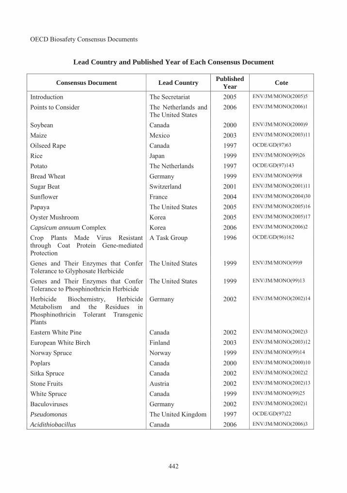
OECD Biosafety Consensus Documents
442
Lead Country and Published Year of Each Consensus Document
Consensus Document Lead Country Published
YearCote
Introduction The Secretariat 2005 ENV/JM/MONO(2005)5
Points to Consider The Netherlands and The United States
2006 ENV/JM/MONO(2006)1
Soybean Canada 2000 ENV/JM/MONO(2000)9
Maize Mexico 2003 ENV/JM/MONO(2003)11
Oilseed Rape Canada 1997 OCDE/GD(97)63
Rice Japan 1999 ENV/JM/MONO(99)26
Potato The Netherlands 1997 OCDE/GD(97)143
Bread Wheat Germany 1999 ENV/JM/MONO(99)8
Sugar Beat Switzerland 2001 ENV/JM/MONO(2001)11
Sunflower France 2004 ENV/JM/MONO(2004)30
Papaya The United States 2005 ENV/JM/MONO(2005)16
Oyster Mushroom Korea 2005 ENV/JM/MONO(2005)17
Capsicum annuum Complex Korea 2006 ENV/JM/MONO(2006)2
Crop Plants Made Virus Resistant through Coat Protein Gene-mediated Protection
A Task Group 1996 OCDE/GD(96)162
Genes and Their Enzymes that Confer Tolerance to Glyphosate Herbicide
The United States 1999 ENV/JM/MONO(99)9
Genes and Their Enzymes that Confer Tolerance to Phosphinothricin Herbicide
The United States 1999 ENV/JM/MONO(99)13
Herbicide Biochemistry, Herbicide Metabolism and the Residues in Phosphinothricin Tolerant Transgenic Plants
Germany 2002 ENV/JM/MONO(2002)14
Eastern White Pine Canada 2002 ENV/JM/MONO(2002)3
European White Birch Finland 2003 ENV/JM/MONO(2003)12
Norway Spruce Norway 1999 ENV/JM/MONO(99)14
Poplars Canada 2000 ENV/JM/MONO(2000)10
Sitka Spruce Canada 2002 ENV/JM/MONO(2002)2
Stone Fruits Austria 2002 ENV/JM/MONO(2002)13
White Spruce Canada 1999 ENV/JM/MONO(99)25
Baculoviruses Germany 2002 ENV/JM/MONO(2002)1
Pseudomonas The United Kingdom 1997 OCDE/GD(97)22
Acidithiobacillus Canada 2006 ENV/JM/MONO(2006)3

443
QUESTIONNAIRE TO RETURN TO THE OECD
The Consensus Documents have been produced with the intention that they will be updated regularly to reflect scientific and technical developments.
Users of Consensus Documents are invited to submit relevant new scientific and technical information, and to suggest additional related areas that might be considered in the future.
The questionnaire is already addressed (see reverse). Please mail or fax this page (or a copy) to the OECD, or send the requested information by E-mail:
OECD Environment Directorate Environment, Health and Safety Division
2, rue André-Pascal 75775 Paris Cedex 16, France
Fax: (33-1) 45 24 16 75 E-mail: [email protected]
===========================================================================
1. Did you find the information in this document useful to your work?
Yes No
2. What type of work do you do?
Regulatory Academic Industry Other (please specify)
3. Should changes or additions be considered when this document is updated?
4. Should other areas related to this subject be considered when the document is updated?
Name: ......................................................................................................................................................
Institution or company: ............................................................................................................................
Address:...................................................................................................................................................
City:............................................Postal code: ......................... Country: .................................................
Telephone:.................................Fax: ...................................... E-mail: ..................................................
Which Consensus Document are you commenting on?...........................................................................
444
FOLD ALONG DOTTED LINES AND SEAL...............................................................................................................................................................
PLACE
STAMP
HERE
OECD Environment Directorate Environment, Health and Safety Division 2, rue André Pascal 75775 Paris Cedex 16 France
......................................................................................................................................................................

OECD PUBLICATIONS, 2, rue André-Pascal, 75775 PARIS CEDEX 16
PRINTED IN FRANCE
(97 2006 05 1 P) ISBN 92-64-02258-9 – No. 55157 2006
Volum
e 2 S
AF
ET
Y A
SS
ES
SM
EN
T O
F T
RA
NS
GE
NIC
OR
GA
NIS
MS
OE
CD
Co
nse
nsu
s D
oc
um
en
ts
The full text of this book is available on line via these links:
http://www.sourceoecd.org/scienceIT/9264022589http://www.sourceoecd.org/agriculture/9264022589http://www.sourceoecd.org/environment/9264022589
Those with access to all OECD books on line should use this link:
http://www.sourceoecd.org/9264022589
SourceOECD is the OECD’s online library of books, periodicals and statistical databases. For more information about this award-winning service and free trials ask your librarian, or write to us at [email protected].
The goal of the OECD Biosafety Consensus Documents is to identify elements of scientific information used in the environmental safety and risk assessment of transgenic organisms which are common to OECD member countries. This is intended to encourage information sharing and prevent duplication of effort among countries.
This book offers ready access to those consensus documents which have been published thus far. As such, it should be of value to applicants for commercial uses of transgenic crops, regulators in national authorities as well as the wider scientific community.
More information on the OECD’s work related to the biosafety of transgenic organisms is found at BioTrack Online (www.oecd.org/biotrack).
Safety Assessment of Transgenic OrganismsOECD CONSENSUS DOCUMENTS
Volume 2
www.oecd.orgISBN 92-64-02258-9 97 2006 05 1 P-:HSTCQE=UWWZ]Y:
Safety Assessment of Transgenic Organisms OECD CONSENSUS DOCUMENTS
BIOSAFETY AGRICULTURE ENVIRONMENT
ENVIRONMENT AGRICULTURE BIOSAFETY
AGRICULTURE ENVIRONMENT BIOSAFETY
AGRICULTURE BIOSAFETY ENVIRONMENT AGRICULTURE BIOSAFETY ENVIRONMENT
ENVIRONMENT BIOSAFETY AGRICULTURE ENVIRONMENT BIOSAFETY AGRICULTURE
BIOSAFETY ENVIRONMENT AGRICULTURE BIOSAFETY ENVIRONMENT AGRICULTURE BIOSAFETY ENVIRONMENT AGRICULTURE
BIOSAFETY AGRICULTURE ENVIRONMENT BIOSAFETY AGRICULTURE ENVIRONMENT BIOSAFETY
ENVIRONMENT AGRICULTURE BIOSAFETY ENVIRONMENT AGRICULTURE BIOSAFETY ENVIRONMENT AGRICULTURE BIOSAFETY
AGRICULTURE ENVIRONMENT BIOSAFETY AGRICULTURE ENVIRONMENT BIOSAFETY AGRICULTURE
AGRICULTURE BIOSAFETY ENVIRONMENT AGRICULTURE BIOSAFETY ENVIRONMENT AGRICULTURE BIOSAFETY ENVIRONMENT
ENVIRONMENT BIOSAFETY AGRICULTURE ENVIRONMENT BIOSAFETY AGRICULTURE ENVIRONMENT
BIOSAFETY ENVIRONMENT AGRICULTURE BIOSAFETY ENVIRONMENT AGRICULTURE BIOSAFETY ENVIRONMENT AGRICULTURE
BIOSAFETY AGRICULTURE ENVIRONMENT BIOSAFETY AGRICULTURE ENVIRONMENT BIOSAFETY
ENVIRONMENT AGRICULTURE BIOSAFETY AGRICULTURE BIOSAFETY ENVIRONMENT AGRICULTURE BIOSAFETY ENVIRONMENT
AGRICULTURE ENVIRONMENT BIOSAFETY AGRICULTURE ENVIRONMENT BIOSAFETY AGRICULTURE
AGRICULTURE BIOSAFETY ENVIRONMENT AGRICULTURE BIOSAFETY ENVIRONMENT AGRICULTURE BIOSAFETY ENVIRONMENT
ENVIRONMENT BIOSAFETY AGRICULTURE ENVIRONMENT BIOSAFETY AGRICULTURE ENVIRONMENT
BIOSAFETY ENVIRONMENT AGRICULTURE BIOSAFETY ENVIRONMENT AGRICULTURE BIOSAFETY ENVIRONMENT AGRICULTURE
BIOSAFETY AGRICULTURE ENVIRONMENT BIOSAFETY AGRICULTURE ENVIRONMENT BIOSAFETY
ENVIRONMENT AGRICULTURE BIOSAFETY ENVIRONMENT AGRICULTURE BIOSAFETY ENVIRONMENT AGRICULTURE BIOSAFETY
AGRICULTURE ENVIRONMENT BIOSAFETY AGRICULTURE ENVIRONMENT BIOSAFETY AGRICULTURE
AGRICULTURE BIOSAFETY ENVIRONMENT AGRICULTURE BIOSAFETY ENVIRONMENT AGRICULTURE BIOSAFETY ENVIRONMENT
ENVIRONMENT BIOSAFETY AGRICULTURE ENVIRONMENT BIOSAFETY AGRICULTURE ENVIRONMENT
BIOSAFETY ENVIRONMENT AGRICULTURE BIOSAFETY ENVIRONMENT AGRICULTURE BIOSAFETY ENVIRONMENT
BIOSAFETY AGRICULTURE ENVIRONMENT BIOSAFETY AGRICULTURE
ENVIRONMENT AGRICULTURE BIOSAFETY ENVIRONMENT
AGRICULTURE ENVIRONMENT
Volume 2
Related Documents