UNIT 3 NON-ORGAN-DIRECTED TOXICITY Copyrighted Material Copyright © 2001 by The McGraw-Hill Companies Retrieved from: www.knovel.com

S3 ch08 chemical_carcinogenesis
Aug 14, 2015
Welcome message from author
This document is posted to help you gain knowledge. Please leave a comment to let me know what you think about it! Share it to your friends and learn new things together.
Transcript

UNIT 3
NON-ORGAN-DIRECTED TOXICITY
2996R_ch08_239-319 4/11/01 3:14 PM Page 239
Copy
right
ed M
ater
ial
Copyright © 2001 by The McGraw-Hill Companies Retrieved from: www.knovel.com

CHAPTER 8
CHEMICAL CARCINOGENESIS
Henry C. Pitot III and Yvonne P. Dragan
HISTORICAL FOUNDATION
DEFINITIONS
CARCINOGENESIS BY CHEMICALS
Organic Chemical CarcinogensInorganic Chemical CarcinogenesisFilm and Fiber CarcinogenesisHormonal CarcinogenesisChemical Carcinogenesis by Mixtures: Defined
and UndefinedChemical Carcinogenesis by Diet
MECHANISMS OF CHEMICAL CARCINOGENESIS
Metabolism of Chemical Carcinogens in Relation to Carcinogenesis
Free Radicals and the Metabolism of ChemicalCarcinogens
Chemical Structure and Chemical CarcinogenesisMutagenesis and CarcinogenesisMacromolecular Adducts Resulting from Reaction
with Ultimate Carcinogens
DNA REPAIR AND CHEMICAL CARCINOGENESIS
Persistence of DNA Adducts and DNA RepairMechanisms of DNA RepairDNA Repair, Cell Replication, and Chemical
Carcinogenesis
CHEMICAL CARCINOGENS AND THE NATURALHISTORY OF NEOPLASTIC DEVELOPMENT
The Pathogenesis of Neoplasia: BiologyInitiationPromotionProgression
Cell and Molecular Mechanisms of the Stages of CarcinogenesisInitiationMolecular Genetic Targets of DNA-Damaging
Carcinogenic AgentsPromotion
The Molecular Basis of the Reversibility of the Stageof Tumor Promotion
Cell Cycle RegulationProgression
The Bases for the Stages of Initiation, Promotion,and Progression
Genetic and Nongenetic Mechanisms of ChemicalCarcinogenesis in Relation to the Natural History of Cancer Development
CHEMICAL CARCINOGENESIS IN HUMANS
Epidemiologic and Animal Studies as Bases for theIdentification of Chemical Carcinogens in Humans
Lifestyle CarcinogenesisChemical Carcinogens Associated with OccupationsChemical Carcinogenesis Resulting from Medical
Therapy and Diagnosis
THE PREVENTION OF HUMAN CANCER INDUCEDBY CHEMICALS
IDENTIFICATION OF POTENTIAL CARCINOGENICAGENTS
Short-Term Tests—Mutagenesis AssaysGene Mutation Assays in VivoChromosomal AlterationsPrimary DNA Damage
Short-Term Tests—Transformation and Cell CultureChronic Bioassays for Carcinogenicity—Medium-
and Long-TermChronic 2-Year BioassayTissue-Specific BioassaysMedium-Term Bioassays
Multistage Models of Neoplastic DevelopmentTransgenic and Knockout Mice as Models of
Carcinogenesis
EVALUATION OF CARCINOGENIC POTENTIAL
The Problem of ExtrapolationThe Dose–Response ProblemThe Problem of the Potency of Carcinogenic Agents
RELATION (EXTRAPOLATION) OF BIOASSAY DATATO HUMAN RISK
STATISTICAL ESTIMATES OF HUMAN RISK FROMBIOASSAY DATA BY USING MATHEMATICALMODELS
REGULATION OF CARCINOGENIC RISK AT THEFEDERAL LEVEL
International Aspects of Environmental Regulation
RISK-BENEFIT CONSIDERATIONS IN THEREGULATION OF ACTUAL AND POTENTIALCARCINOGENIC ENVIRONMENTAL HAZARDS
241
2996R_ch08_239-319 4/13/01 11:20 AM Page 241
Copy
right
ed M
ater
ial
Copyright © 2001 by The McGraw-Hill Companies Retrieved from: www.knovel.com

242 UNIT 3 NON-ORGAN-DIRECTED TOXICITY
Cancer resulting from exposure to chemicals in the environment,though known for millennia, has taken on new importance in thiscentury. With the advent of advanced technology, new chemicalagents enter the environment, although at relatively low levels inmost cases, at a prodigious rate. It has been estimated (Korte andCoulston, 1994) that the number of organic chemicals that are con-tinually being brought into the environment (about 300 million tonsper year) may include more than 100,000 compounds. Chemicalcontamination of waste (Landrigan, 1983), the food chain (Foranet al., 1989), and the occupational environment (Anttila et al., 1993)is reportedly substantial. However, other researchers (Ames andGold, 1990) have noted numerous misconceptions about the rela-tionship of exposure to industrially based environmental chemicalsand the incidence of human cancer. Therefore, knowledge aboutthe mechanisms and natural history of cancer development as wellas the epidemiology of human cancer is critical to the control andprevention of human neoplastic disease.
HISTORICAL FOUNDATION
The historical foundations for the induction of carcinogenesis bychemicals dates back several thousand years to the description ofbreast cancer in the Edwin Smith papyrus (Shimkin, 1977). In 1700,Ramazzini described the first example of occupational cancer. Henoted the high incidence of breast cancer among nuns, which heattributed to their celibate life. A specific causal relationship be-tween exposure to environmental mixtures and the induction ofcancer was reported in 1775 by Percivall Pott, an eminent Englishphysician and surgeon. Pott described the occurrence of cancer ofthe scrotum in a number of patients with a history of employmentas chimney sweeps. With remarkable insight, Pott concluded thatthe occupation of those men was directly and causally related totheir malignant disease. In addition, Pott suggested that the soot towhich they were exposed was the causative agent of their condi-tion. While Pott’s publication soon led other observers to attributecancer in various sites to soot exposure, his work had little impacton British public health practice during the succeeding century(Lawley, 1994). Thus, more than a century later, Butlin (1892) re-ported the relative rarity of scrotal cancer among chimney sweepson the European continent compared with those in England. Thisdifference was attributed to the relatively low standards of hygienein Britain and the practice of exposing young “climbing boys” tothe combustion products of coal. However, the lesson from Pott’sfindings has been a long time in the learning. A hundred years af-ter the publication of Pott’s monograph, the high incidence of skincancer among certain German workers was traced to their expo-sure to coal tar, the chief constituent of the chimney sweeps’ soot(Miller, 1978). Even today—more than 200 years after Pott’s orig-inal scientific report on the association of soot and smoke prod-ucts with cancer—a large percentage of the world’s population isexposed to carcinogenic products that result from the combustionof tobacco and organic fuels.
During the nineteenth century, industrial chemicals, includingcutting oils and dyes, were implicated as causative factors in thedevelopment of skin and bladder cancer, respectively (Lawley,1994). Coal tar derivatives became the basis for the dye industryduring the middle of the nineteenth century in Europe. Amine-containing aromatics such as 2-naphthylamine and benzidine werediscovered and subsequently synthesized and used to yield a vari-ety of chemical species of pigments for coloring a variety of ma-terials. In 1895 Rehn reported the occurrence of bladder cancer in
workers in the aniline dye industry. This finding was rapidly sup-ported by other reports (Miller, 1978). Epidemiologic studies in-criminated a number of aromatic amines, such as naphthylaminesand benzidines, as the inciting agents (Hueper et al., 1938). Today2-naphthylamine is not used in the U.S. chemical industry and ex-posure to a variety of other aromatic amines is regulated by law.Thus, the reader may appreciate that the human being was the firstexperimental animal in which chemical carcinogenesis was stud-ied. Further on, both the development and the data derived fromstudies of chemical carcinogenesis in animals are considered.
DEFINITIONS
The term cancer describes a subset of lesions of the disease neo-plasia. Neoplasia or the constituent lesion, a neoplasm, is definedas a heritably altered, relatively autonomous growth of tissue (Pitot,1986a). The critical points of this definition are (1) the heritableaspects of neoplasia at the somatic or germ cell level and (2) therelative autonomy of neoplastic cells, reflecting their abnormal reg-ulation of genetic expression, which is inherent in the neoplasticcell or occurs in response to environmental stimuli. Neoplasms maybe either benign or malignant. The critical distinction betweenthese classes is related to the characteristic of successful metasta-tic growth of malignant but not benign neoplasms. Metastases aresecondary growths of cells from the primary neoplasm. Cancersare malignant neoplasms, whereas the term tumor describes space-occupying lesions that may or may not be neoplastic.
The nomenclature of neoplasia depends primarily on whetherthe neoplasm is benign or malignant and, in the latter case, whetherit is derived from epithelial or mesenchymal tissue. For most benignneoplasms, the tissue of origin is followed by the suffix -oma:fibroma, lipoma, adenoma, and so on. For malignant neoplasmsderived from tissues of mesenchymal origin, the term sarcoma isadded to the tissue descriptor: fibrosarcoma, osteosarcoma, li-posarcoma, and so on. Malignant neoplasms derived from tissuesof ectodermal or endodermal (epithelial) origin are termedcarcinomas with an antecedent tissue descriptor: epidermoid car-cinoma (skin), hepatocellular carcinoma, gastric adenocarcinoma,and so on.
In general a carcinogen is an agent that causes or induces neo-plasia. However, this definition is insufficient by current standards.The following definition may be more appropriate: “A carcinogenis an agent whose administration to previously untreated animalsleads to a statistically significant increased incidence of neoplasmsof one or more histogenetic types as compared with the incidencein appropriate untreated animals” (Pitot, 1986a).
This definition includes the induction of neoplasms that areusually not observed, the earlier induction of neoplasms that usu-ally are observed, and/or the induction of more neoplasms than areusually found. Although it is important to distinguish betweenagents that induce neoplasms through direct action on the cells thatbecome neoplastic and those which produce neoplasia through in-direct actions in the animal as a whole, this is not always possible.Some agents, such as immune suppressants, can increase the inci-dence of neoplasms in tissues that were previously exposed to car-cinogens through indirect effects on the host. When the action ofa chemical in causing an increase in neoplasms is known to be in-direct—that is, mediated by its effect on cells other than those un-dergoing carcinogenesis—that agent should not be designated asa carcinogen. Later in this chapter the stages and modifying fac-tors of the process of chemical carcinogenesis are considered, ne-
2996R_ch08_239-319 4/11/01 3:33 PM Page 242
Copy
right
ed M
ater
ial
Copyright © 2001 by The McGraw-Hill Companies Retrieved from: www.knovel.com
CHAPTER 8 CHEMICAL CARCINOGENESIS 243
cessitating a further refinement of the term carcinogen in relationto the action of specific chemicals in the carcinogenic process.
CARCINOGENESIS BY CHEMICALS
By the turn of this century, studies in humans showed that envi-ronmental and possibly internal chemical agents are causative fac-tors in the development of cancer (Shimkin, 1977; Lawley, 1994).However, a systematic study of the mechanisms of chemical car-cinogenesis was not possible without defined experimental sys-tems. In 1915, the Japanese pathologists Yamagawa and Ichikawa(1915) described the first production of skin tumors in animals bythe application of coal tar to the skin. These investigators repeat-edly applied crude coal tar to the ears of rabbits for a number ofmonths, finally producing both benign and later malignant epider-mal neoplasms. Later studies demonstrated that the skin of miceis also susceptible to the carcinogenic action of such organic tars.During the next 15 years, extensive attempts were made todetermine the nature of the material in the crude tars that causedmalignancy. In 1932 Kennaway and associates reported the production of carcinogenic tars by means of pyrolysis of organic
compounds consisting only of carbon and hydrogen (Kennaway,1955).
Organic Chemical Carcinogens
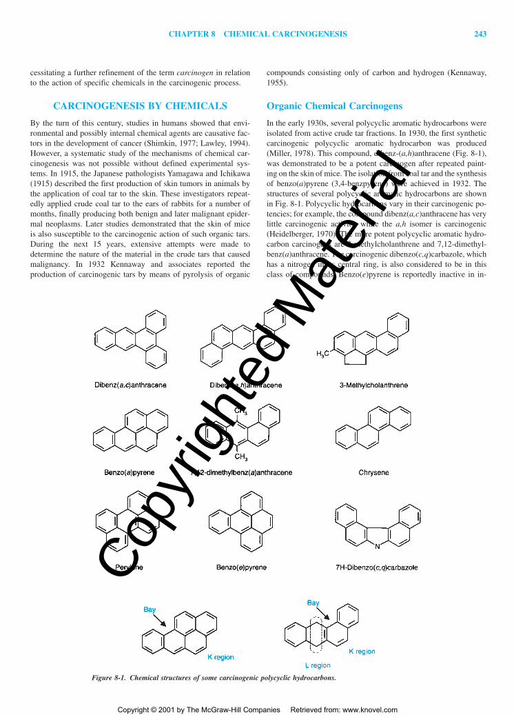
In the early 1930s, several polycyclic aromatic hydrocarbons wereisolated from active crude tar fractions. In 1930, the first syntheticcarcinogenic polycyclic aromatic hydrocarbon was produced(Miller, 1978). This compound, dibenz-(a,h)anthracene (Fig. 8-1),was demonstrated to be a potent carcinogen after repeated paint-ing on the skin of mice. The isolation from coal tar and the synthesisof benzo(a)pyrene (3,4-benzpyrene) were achieved in 1932. Thestructures of several polycyclic aromatic hydrocarbons are shownin Fig. 8-1. Polycyclic hydrocarbons vary in their carcinogenic po-tencies; for example, the compound dibenz(a,c)anthracene has verylittle carcinogenic activity, while the a,h isomer is carcinogenic(Heidelberger, 1970). The more potent polycyclic aromatic hydro-carbon carcinogens are 3-methylcholanthrene and 7,12-dimethyl-benz(a)anthracene. The carcinogenic dibenzo(c,q)carbazole, whichhas a nitrogen in its central ring, is also considered to be in thisclass of compounds. Benzo(e)pyrene is reportedly inactive in in-
Figure 8-1. Chemical structures of some carcinogenic polycyclic hydrocarbons.
2996R_ch08_239-319 4/11/01 3:33 PM Page 243
Copy
right
ed M
ater
ial
Copyright © 2001 by The McGraw-Hill Companies Retrieved from: www.knovel.com
244 UNIT 3 NON-ORGAN-DIRECTED TOXICITY
ducing skin cancer in mice but can “initiate” the carcinogenicprocess. Perylene is inactive as a chemical carcinogen, whereaschrysene may have slight carcinogenic activity.
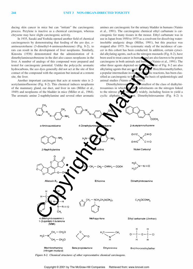
In 1935, Sasaki and Yoshida opened another field of chemicalcarcinogenesis by demonstrating that feeding of the azo dye, o-aminoazotoluene (3-dimethyl-4-aminoazobenzene) (Fig. 8-2), torats can result in the development of liver neoplasms. Similarly,Kinosita (1936) demonstrated that the administration of 4-dimethylaminoazobenzene in the diet also causes neoplasms in theliver. A number of analogs of this compound were prepared andtested for carcinogenic potential. Unlike the polycyclic aromatichydrocarbons, the azo dyes generally did not act at the site of firstcontact of the compound with the organism but instead at a remotesite, the liver.
Another important carcinogen that acts at remote sites is 2-acetylaminofluorene (Fig. 8-2). This chemical induces neoplasmsof the mammary gland, ear duct, and liver in rats (Miller et al.,1949) and neoplasms of the bladder in mice (Miller et al., 1964).The aromatic amine 2-naphthylamine and several other aromatic
amines are carcinogenic for the urinary bladder in humans (Vainioet al., 1991). The carcinogenic chemical ethyl carbamate is car-cinogenic for many tissues in the mouse. Ethyl carbamate was inuse in Japan from 1950 to 1975 as a cosolvent for dissolving water-insoluble analgesic drugs (Miller, 1991), but this practice wasstopped after 1975. No systematic study of the incidence of can-cer in this cohort has been conducted. In addition, certain cytoci-dal alkylating agents, such as the nitrogen mustards (Fig. 8-2), havebeen used to treat cancer in humans and are also known to be potentcarcinogens in both animals and humans (Vainio et al., 1991). Theother three agents depicted on the bottom line of Fig. 8-2 are alsoalkylating agents that are used industrially. Bis(chloromethyl)ether,a popular intermediate in organic synthetic reactions, has been clas-sified as carcinogenic to humans on the basis of epidemiologic andanimal studies (Vainio et al., 1991).
Dimethylnitrosamine is the smallest of the class of dialkylni-trosamines in which the alkyl substituents on the nitrogen linkedto the nitroso group may vary widely, including fusion to yield acyclic aliphatic substituent. Dimethylnitrosamine (Fig. 8-2) is
Figure 8-2. Chemical structures of other representative chemical carcinogens.
2996R_ch08_239-319 4/11/01 3:33 PM Page 244
Copy
right
ed M
ater
ial
Copyright © 2001 by The McGraw-Hill Companies Retrieved from: www.knovel.com

CHAPTER 8 CHEMICAL CARCINOGENESIS 245
highly carcinogenic for the liver and kidney in virtually all themammalian species tested (Schmähl and Habs, 1980). There is sub-stantial epidemiologic evidence for a role of nitroso compounds inthe induction of human cancer. The nitrosamine NNK (Fig. 8-2) isproduced in tobacco smoke from nicotine, a tobacco alkaloid(Hecht, 1985). This is an extremely potent carcinogen that mayplay a role in the induction of tobacco-related cancers in humans.Methapyrilene was developed as an antihistamine but is a potentcarcinogen in the rat (Mirsalis, 1987). Several investigators(Lijinsky, 1977; Magee and Swann, 1969; Mirvish et al., 1983)have shown that certain dietary components, especially in the pres-ence of high levels of nitrite, may give rise to low levels of ni-trosamines or nitrosamides and induce neoplasia of the gastroin-testinal tract in experimental animals. The action of bacterial florain the intestine may enhance the formation of these compounds.There is increasing evidence of an etiologic role for endogenouslyformed N-nitroso compounds in the development of certain humancancers (Bartsch et al., 1990).
Another important environmental and experimental hepato-carcinogenic agent is aflatoxin B1. This toxic substance is producedby certain strains of the mold Aspergillus flavus. Aflatoxin B1 isone of the most potent hepatocarcinogenic agents known and hasproduced neoplasms in rodents, fish, birds, and primates (Draganand Pitot, 1994). This agent is a potential contaminant of manyfarm products (for example, grain and peanuts) that are stored un-der warm and humid conditions for some time. Aflatoxin B1 andrelated compounds may cause some of the toxic hepatitis and he-patic neoplasia seen in various parts of Africa and the Far East(Wogan, 1992). Other products of molds and fungi are potentiallycarcinogenic in humans and animals (Schoental, 1985). A numberof plants, some of which are edible, also contain chemical car-cinogenic agents whose structures have been elucidated (Hirono,1993).
Ethionine is an antimetabolite of the amino acid methionine.Farber (1963) was the first to show definitively that administrationof ethionine in the diet for extended periods can result in the de-velopment of liver cancer in rats. This was the first example of di-rect interference with the metabolism of a normal metabolic con-stituent, resulting in the development of cancer.
We note here and discuss further later in the chapter that thedose of a chemical carcinogen is very important in relation to itseffects just as with any pharmacologic agent. Even though manychemical carcinogens exert their effects by mechanisms somewhatdifferent than many pharmacologic agents, the total administereddose, the rate at which it is given, and a number of other factorsin the organism itself each play significant roles in the ultimatecarcinogenic response.
Inorganic Chemical Carcinogenesis
In addition to organic compounds such as those illustrated in Figs.8-1 and 8-2, a number of inorganic elements and their compoundshave been shown to be carcinogenic in both animals and humans(Vainio and Wilbourn, 1993). Table 8-1 lists metals that are car-cinogenic in some form to humans (part A) and experimental an-imals (part B) (Sky-Peck, 1986). Many elements and their com-pounds have not been adequately tested for carcinogenicity inanimals, and at this time there is no evidence that such elementsexhibit effects in humans on the basis of epidemiologic studies. Bycontrast, compounds of cadmium, chromium, and nickel have in-duced malignant neoplasms in humans primarily in industrial and
refining situations (Table 8-1, part A) (Magos, 1991). In the caseof cadmium, the evidence for carcinogenicity in humans is some-what limited (Waalkes et al., 1992) because of the variety of con-founding factors that occur in situations of human exposure. How-ever, its carcinogenic effect in animals is well documented. Bycontrast, organonickel compounds, especially nickel carbonyl (Fig.8-2), are carcinogenic to humans in several tissues, as noted inTable 8-1. Exposures to several metals and their compounds, in-cluding lead (Verschaeve et al., 1979) and beryllium (Kuschner,1981), have been implicated as causes of cancer in humans, butthe data are not sufficient to demonstrate such an association un-equivocally. In contrast, arsenic and its derivatives present an in-teresting paradox (Landrigan, 1981) in that there is essentially noexperimental evidence to substantiate the carcinogenicity of thiselement and its compounds in lower animals, whereas the evidencefor its carcinogenicity in humans is quite clear (Sky-Peck, 1986).
Film and Fiber Carcinogenesis
A class of chemical carcinogens different from those described thusfar is the group of inert plastic and metal films or similar formsthat cause sarcomas at the implantation site in some rodents (Brandet al., 1975). The implantation site is usually subcutaneous. Ratsand mice are highly susceptible to this form of carcinogenesis, butguinea pigs appear to be resistant (Stinson, 1964). The carcino-genic properties of the implant are to a large extent dependent onits physical characteristics and surface area. Multiple perforationseach greater than a certain diameter (for example, 0.4 �m), pul-verization, or roughening of the surface of the implant (Ferguson,1977) markedly reduced the incidence of neoplasms. Plastic spongeimplants may also induce sarcomas subcutaneously, and in this in-stance the yield of tumors is dependent on the thickness of thesponge implant (Roe et al., 1967). The age of the animal at im-plantation also affects the time that elapses from implantation andtumor development (Paulini et al., 1975).
The chemical nature of the implant is not the critical factorin its ability to transform normal cells to neoplastic cells. Brandand associates (Johnson et al., 1970) studied this phenomenon in-tensively and demonstrated a variety of kinetic and morphologiccharacteristics of the process of “foreign-body tumorigenesis” inmice. These investigations have shown that DNA synthesis occursin the film-attached cell population throughout the preneoplasticphase and that preneoplastic cells may be identified well beforeneoplasms develop (Thomassen et al., 1978). Brand suggested thatsuch “preneoplastic” cells may be present in normal tissue beforeimplantation and that the implant appears to “create the conditions”required for carcinogenesis of these cells (Brand et al., 1975). Otherpossible mechanisms for this unique type of carcinogenesis are dis-cussed later in this chapter.
While the epidemiologic evidence that implants of prosthesesin humans, such as those used for the repair of hernias and jointreplacements, induce the formation of sarcomas is not substantial,there have been a number of isolated reports of neoplasms arisingin association with such foreign bodies (Sunderman, 1989). A studyin the rat of the carcinogenic potential of a number of materialsused in such prostheses demonstrated a small increase in sarcomasin animals with certain metal alloy implants that contained signif-icant amounts of cobalt, chromium, or nickel (Memoli et al., 1986).Of greater significance is the induction of malignant mesotheliomaand bronchogenic carcinoma in humans by exposure to asbestosfibers. In this case, the induction of the malignant mesotheliomas
2996R_ch08_239-319 4/11/01 3:33 PM Page 245
Copy
right
ed M
ater
ial
Copyright © 2001 by The McGraw-Hill Companies Retrieved from: www.knovel.com

Table 8-1Carcinogenicity of Metals
A. Metals Causally Associated with Human Cancer
METAL AND SOURCE MALIGNANCY
ArsenicCu refinery Pulmonary carcinomaAs pesticides Lymphoma, leukemiaChemical plants Dermal carcinomaDrinking water (oral) Hepatic angiosarcomaCigarette smoke
CadmiumCd refinery Pulmonary carcinoma
ChromiumCr refinery Pulmonary carcinomaChrome plating Gastrointestinal carcinomaChromate pigments
NickelNi refinery Pulmonary carcinoma
Nasolaryngeal carcinomaGastric and renal carcinomaSarcoma (?)
B. Carcinogenicity of Metals in Experimental Animals
METALS ANIMALS TUMOR SITE ROUTE
Beryllium Mice, rats, monkeys Osteosarcoma Bone IV, INHCarcinoma Lung
Cadmium Mice, rats, chickens Sarcoma Injection site IM, SC, ITSTeratoma Testes
Cobalt Rats, rabbits Sarcoma Injection site IM, SCChromium Mice, rats, rabbits Sarcoma Injection site IM, SC, IP,
Carcinoma Lung INHIron Hamsters, mice, Sarcoma Injection site IM, IP, SC
rats, rabbitsNickel Mice, rats, cats, Sarcoma Injection site IM, ITS, SC
hamsters, rabbitsGuinea pigs, rats Carcinoma Lung INH, IP, IR
Carcinoma KidneyLead Mice, rats Carcinoma Kidney IP, PO, SCTitanium Rats Sarcoma Injection site IMZinc Chickens, rats, Carcinoma Testes ITS
hamsters Teratoma Testes
KEY: IV, intravenous; INH, inhalation; IM, intramuscular; SC, subcutaneous; ITS, intratesticular; IP, intraperitoneal; IR, intrarenal;PO, per os.
SOURCE: From Sky-Peck (1986), with permission.
246 UNIT 3 NON-ORGAN-DIRECTED TOXICITY
appears to be dependent on the crystal structure rather than thecomposition of the asbestos both in experimental animals and inhumans (Craighead, 1982). In experimental animals, fibers longerthan 8 �m and with a diameter less than 1.5 �m induce mesothe-lioma fairly effectively. Similarly, certain types of asbestos, suchas the crocidolite form, are most strongly associated with the oc-currence of this neoplasm, whereas exposure to other forms, suchas chrysotile, may not be as important a cause of malignantmesothelioma. Thus, in both humans and animals, film and fibercarcinogenesis is largely independent of the chemical nature of theinciting agent.
Hormonal Carcinogenesis
Hormones consist of amines, steroids, and polypeptides. Beatson(1896) was the first to point out that hormones may be causally as-sociated with the development of specific neoplasms. He suggestedthat a relationship exists between breast cancer and the ovary, themajor site of production of female sex hormones.
Hormones play an important physiological role in maintainingthe “internal milieu” (Bernard, 1878, 1879). Some cancers mayresult from abnormal internal production of specific hormones.Alternatively, excessive production or the derangement of the
2996R_ch08_246 5/29/01 4:07 PM Page 246
Copy
right
ed M
ater
ial
Copyright © 2001 by The McGraw-Hill Companies Retrieved from: www.knovel.com
CHAPTER 8 CHEMICAL CARCINOGENESIS 247
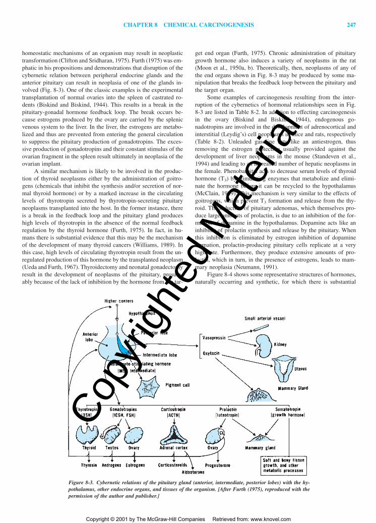
homeostatic mechanisms of an organism may result in neoplastictransformation (Clifton and Sridharan, 1975). Furth (1975) was em-phatic in his propositions and demonstrations that disruption of thecybernetic relation between peripheral endocrine glands and theanterior pituitary can result in neoplasia of one of the glands in-volved (Fig. 8-3). One of the classic examples is the experimentaltransplantation of normal ovaries into the spleen of castrated ro-dents (Biskind and Biskind, 1944). This results in a break in thepituitary-gonadal hormone feedback loop. The break occurs be-cause estrogens produced by the ovary are carried by the splenicvenous system to the liver. In the liver, the estrogens are metabo-lized and thus are prevented from entering the general circulationto suppress the pituitary production of gonadotropins. The exces-sive production of gonadotropins and their constant stimulus of theovarian fragment in the spleen result ultimately in neoplasia of theovarian implant.
A similar mechanism is likely to be involved in the produc-tion of thyroid neoplasms either by the administration of goitro-gens (chemicals that inhibit the synthesis and/or secretion of nor-mal thyroid hormone) or by a marked increase in the circulatinglevels of thyrotropin secreted by thyrotropin-secreting pituitaryneoplasms transplanted into the host. In the former instance, thereis a break in the feedback loop and the pituitary gland produceshigh levels of thyrotropin in the absence of the normal feedbackregulation by the thyroid hormone (Furth, 1975). In fact, in hu-mans there is substantial evidence that this may be the mechanismof the development of many thyroid cancers (Williams, 1989). Inthis case, high levels of circulating thyrotropin result from the un-regulated production of this hormone by the transplanted neoplasm(Ueda and Furth, 1967). Thyroidectomy and neonatal gonadectomyresult in the development of neoplasms of the pituitary, presum-ably because of the lack of inhibition by the hormone from the tar-
get end organ (Furth, 1975). Chronic administration of pituitarygrowth hormone also induces a variety of neoplasms in the rat(Moon et al., 1950a, b). Theoretically, then, neoplasms of any ofthe end organs shown in Fig. 8-3 may be produced by some ma-nipulation that breaks the feedback loop between the pituitary andthe target organ.
Some examples of carcinogenesis resulting from the inter-ruption of the cybernetics of hormonal relationships seen in Fig.8-3 are listed in Table 8-2. In addition to effecting carcinogenesisin the ovary (Biskind and Biskind, 1944), endogenous go-nadotropins are involved in the development of adrenocortical andinterstitial (Leydig’s) cell neoplasms in mice and rats, respectively(Table 8-2). Unleaded gasoline acts like an antiestrogen, thusremoving the estrogen protection usually provided against thedevelopment of liver neoplasms in the mouse (Standeven et al.,1994) and leading to an increased number of hepatic neoplasms inthe female. Phenobarbital acts to decrease serum levels of thyroidhormone (T3) by stimulating enzymes that metabolize and elimi-nate the hormone before it can be recycled to the hypothalamus(McClain, 1989). This mechanism is very similar to the effects ofgoitrogens, which prevent T3 formation and release from the thy-roid. The induction of pituitary adenomas, which themselves pro-duce large amounts of prolactin, is due to an inhibition of the for-mation of dopamine in the hypothalamus. Dopamine acts like aninhibitor of prolactin synthesis and release by the pituitary. Whenthis inhibition is eliminated by estrogen inhibition of dopamineformation, prolactin-producing pituitary cells replicate at a veryhigh rate. Furthermore, they produce extensive amounts of pro-lactin, which in turn, in the presence of estrogens, leads to mam-mary neoplasia (Neumann, 1991).
Figure 8-4 shows some representative structures of hormones,naturally occurring and synthetic, for which there is substantial
Figure 8-3. Cybernetic relations of the pituitary gland (anterior, intermediate, posterior lobes) with the hy-pothalamus, other endocrine organs, and tissues of the organism. [After Furth (1975), reproduced with thepermission of the author and publisher.]
2996R_ch08_239-319 4/11/01 3:33 PM Page 247
Copy
right
ed M
ater
ial
Copyright © 2001 by The McGraw-Hill Companies Retrieved from: www.knovel.com

248 UNIT 3 NON-ORGAN-DIRECTED TOXICITY
evidence of carcinogenicity in lower animals and/or humans. Inaddition to the structure of growth hormone, two other growthfactors expressed in adult tissues—transforming growth factor-�(TGF-�), which is expressed in the small intestine (Barnard et al.,1991), the major salivary glands (Wu et al., 1993), and other tissues(Lee et al., 1993), and insulin-like growth factor-II (IGF-II), whichis expressed in the forebrain, uterus, kidney, heart, skeletal mus-cle, and to a very small degree the liver (Murphy et al., 1987)—may be considered as chemical carcinogens. The carcinogenicaction of these two growth factor hormones in vivo has beendemonstrated by the use of transgenic mice, among which animalsoverexpressing TGF-� developed liver neoplasms in dramaticexcess of the number seen in controls (Lee et al., 1992), and animalsexpressing high levels of IGF-II developed excessive numbers ofhepatocellular carcinomas and lymphomas (Rogler et al., 1994).
As noted in Table 8-2, neoplasms of the pituitary and the pe-ripheral endocrine organs may be induced by the administration ofsteroid sex hormones. Although the kidney is not usually consid-ered a peripheral endocrine organ, its cells produce erythropoietin.Synthetic or natural estrogen administration can induce renal cor-tical carcinomas in male hamsters (Li and Li, 1990), and estradiolinduces Leydig cell tumors of the testes in mice (Huseby, 1980).However, a closely related structural analogue, 2-fluoroestradiol,which exhibits significant estrogenic potency, did not induce renalcarcinoma in the same sex and species (Liehr, 1983). Recently, thesynthetic antiestrogen tamoxifen was found to induce carcinomasof the liver in the rat as well (Williams et al., 1993). Evidence thatmale hormones by themselves are carcinogenic is not as strong asthe data for the carcinogenicity of female hormones. The naturalmale hormone testosterone does exhibit a weak ability to “trans-form” hamster embryo cells in culture into a neoplastic phenotype(Lasne et al., 1990). The evidence that synthetic androgens arecarcinogenic is somewhat greater, especially in humans. In addi-tion, elevated serum testosterone levels are associated with anincreased risk of hepatocellular carcinoma in humans (Yu andChen, 1993). A number of reports (Mays and Christopherson, 1984;
Chandra et al., 1984) have indicated a causative relationship be-tween the administration of synthetic androgens such asoxymetholone (Fig. 8-4B) for various clinical conditions and theappearance of hepatocellular neoplasms, predominantly benign.
In addition to apparently direct induction of neoplasia by hor-monal stimuli, hormones act in concert with known carcinogenicagents to induce neoplasia. One of the better studied examples ofthis phenomenon is the induction of mammary adenocarcinomasin rodents. Bittner (1956) demonstrated that three factors are es-sential for the production of mammary carcinoma in mice: geneticsusceptibility, hormonal influence, and a virus transmitted throughthe milk. The importance of the first two factors has been demon-strated repeatedly in a variety of species, including humans, but in-controvertible evidence for the participation of a virus in mammarycarcinogenesis has been obtained only in mice. In the rat, high lev-els of endogenous prolactin enhance the induction of mammarycarcinomas by dimethylbenz(a)anthracene (Ip et al., 1980).Chronic treatment with synthetic or natural estrogens alone mayinduce mammary carcinomas in rodents. Thus, mammary carcino-genesis in rodents is a complicated process that requires severalcomponents that may differ from species to species.
Both male and female sex steroid hormones have also beenshown to act in concert with known carcinogenic agents to increasethe incidence of neoplasia. Various synthetic estrogens adminis-tered chronically to animals that had been dosed with a known car-cinogen markedly enhanced the development of hepatocellular car-cinomas in the rat (Yager and Yager, 1980). Both testosterone andsynthetic androgens given with or after chemical carcinogens en-hance the induction of adenocarcinomas of the prostate and otheraccessory sex organs of the male (Hoover et al., 1990). A combi-
Table 8-2Interrupted Cybernetics of Hormonal Carcinogenesis in Rodents
HORMONAL INTERRUPTED
SPECIES/TISSUE INDUCING AGENT CARCINOGEN PATHWAY REFERENCE
Mouse/ovary Ovary transplant Gonadotropin Estrogen � hypo- Biskind andto spleen thalamus Biskind, 1944
Rat/thyroid Goitrogen or Thyrotropin T3 � hypothalamus Cf. Furth, 1975thyrotropin-secreting tumor
Mouse/adrenal Ovariectomy Gonadotropins Estrogen � hypo- Kawashima etcortex thalamus al., 1980
Female mouse/ Unleaded Androgens Estrogen synthesis Standeven etliver gasoline al., 1994
Rat/thyroid Phenobarbital Thyrotropin T3 � hypo- McClain, 1989thalamus
Rat/pituitary Estrogens ? Dopamine � Cf. Neumann,pituitary 1991
Rat/Leydig cells Antiantrogens Gonadotropins Androgens � Cf. Neumann,hypothalamus 1991
Rat/mammary Estrogens Prolactin Dopamine � pituitary Cf. Neumann,gland 1991
Figure 8-4 A. Structures of polypeptide hormones (hGH, human growthhormone; TGF-�, transforming growth factor alpha; IGF-II, insulingrowth factor-II). B. Structures of some naturally occurring (beta-estra-diol and testosterone) and synthetic steroid hormones and antihormones.
2996R_ch08_239-319 4/11/01 3:33 PM Page 248
Copy
right
ed M
ater
ial
Copyright © 2001 by The McGraw-Hill Companies Retrieved from: www.knovel.com

CHAPTER 8 CHEMICAL CARCINOGENESIS 249
2996R_ch08_239-319 4/13/01 11:20 AM Page 249
Copy
right
ed M
ater
ial
Copyright © 2001 by The McGraw-Hill Companies Retrieved from: www.knovel.com

250 UNIT 3 NON-ORGAN-DIRECTED TOXICITY
nation of testosterone and estradiol-17� after treatment withmethylnitrosourea also resulted in the development of adenocarci-nomas of the prostate (Bosland et al., 1991).
Chemical Carcinogenesis by Mixtures:Defined and Undefined
While most of this chapter is concerned with the carcinogenic ac-tion of specific chemicals, it is relatively unusual for an individualto be exposed to a single carcinogenic agent. Despite this, rela-tively few detailed studies on mixtures of carcinogenic chemicalshave been carried out experimentally. The most common environ-mental mixtures are those seen in tobacco smoke and other com-bustion products, including engine exhaust and air pollution(Mauderly, 1993). Interactions between the chemicals in mixturesmay be additive, synergistic, or inhibitory (Berger, 1995). In theexamples given above, however, the exact chemical nature of com-ponents in tobacco smoke or air pollution is not always known andtheir amounts determined. Thus, one may be forced to deal with amixture as if it were a single entity or, if the constituents are known,treat the effects of the mixture in an empiric way that usually isrelated to the most potent component in the mixture.
Studies on the carcinogenic action of defined mixtures ofchemicals are usually done with a knowledge of the carcinogeniceffect of the chemicals involved. Warshawsky and coworkers(1993) demonstrated that extremely low levels of benzo[a]pyrene,which produced no skin tumors on repeated application, resultedin a significant yield of neoplasms when applied in the presenceof five noncarcinogenic polycyclic aromatic hydrocarbons. In anearlier study, the administration of two noncarcinogenic aminoazodyes in the diet of the rat for a year resulted in the appearance ofa variety of neoplasms (Neish et al., 1967). More recently, theadministration of three to five N-nitrosamines resulted in either anadditive or synergistic carcinogenic effect of the combinations ofthe compounds when given at low dose rates (Berger et al., 1987;Lijinsky et al., 1983). In contrast, the administration of a mixtureof 40 chemical carcinogens to rats for 2 years at 50 percent of thedose normally used to induce neoplasms in 50 percent of the ani-mals resulted in significant tumor incidences only in the thyroidand liver (Takayama et al., 1989). In a more recent study, inges-tion of a mixture of 20 pesticides given at “acceptable daily intakelevels” was found to exert no effect on carcinogenesis in rat liver(Ito et al., 1998).
Thus the toxicologic study of complex mixtures not only inthe area of carcinogenesis but also as a more general problem intoxicology, is a critical field in human health, as evidenced by dis-ease resulting from a variety of chemical mixtures such as tobaccosmoke, diesel exhaust, solvent mixtures, petroleum distillates, andother components of outdoor air pollutants (Feron et al., 1998). Asnoted above, and as emphasized by others, exposures to chemicalsat low, non-toxic doses of the individual constituents may well beof no significant health concern (Cassee et al., 1998). One of themost important chemical mixtures associated with human neoplasiais diet.
Chemical Carcinogenesis by Diet There is substantial evidencein humans to indicate that many dietary components—includingexcessive caloric intake (Osler, 1987; Lutz and Schlatter, 1992),excessive alcohol intake (IARC, 1987), and a variety of chemicalcontaminants of the diet including aflatoxin B1 (Fig. 8-2) (Gorchev
and Jelinek, 1985; Lutz and Schlatter, 1992)—are carcinogenic.Other general and specific studies have supported these views(Jensen and Madsen, 1988; Habs and Schmähl, 1980; Miller et al.,1994), whereas others have been more controversial (Willett andMacMahon, 1984; Pariza, 1984). Evidence for the association ofdietary factors with cancer incidence in animals is more substan-tial and supports much of the evidence relating environmental fac-tors to increased cancer incidence in the human (Kritchevsky, 1988;Rogers et al., 1993). A number of the dietary factors implicated inthe nutritional etiology of specific human cancers may be seen inTable 8-3 (Trichopoulos, 1989). As noted in the table, the caloriccontent of diets as well as their individual chemical componentsare factors in cancer development. Although a relative lack of “an-tioxidant micronutrients” such as keratinoids, selenium, and the vi-tamins A, C, and E has been implicated as a factor in the incidenceof neoplastic development (Blot et al., 1993; Byers and Perry,1992), more studies are needed before the effectiveness of theseagents in cancer prevention in the human can be established. In ad-dition, food contaminants—either added, occurring endogenously,or as a result of the cooking process—may function as carcino-genic agents in the diet (McGregor, 1998).
Experimental evidence that the lack of available sources ofmethyl groups can actually induce liver cancer in rats is well doc-umented (Mikol et al., 1983; Ghoshal and Farber, 1984). This ob-servation may be closely related to the earlier studies by Farber(1963) on the induction of liver cancer in rats by the administra-tion of ethionine, which indirectly may cause a lack of availablemethyl groups in this tissue. Thus, it is apparent that carcinogen-esis induced by diet is an extremely complex effect of mixtures ofa variety of chemicals. Its importance in human cancer etiology isemphasized further later in this chapter.
MECHANISMS OF CHEMICALCARCINOGENESIS
Although the discovery that polycyclic hydrocarbons and otherchemical compounds can induce cancer in experimental animalsgave hope that the complete understanding of the nature of the gen-esis of neoplasia might be forthcoming, more than 60 years haveelapsed since those initial findings, and it appears that we are stilla long way from this goal. However, the realization that chemicalcarcinogens are altered within the living organism by metabolic re-actions has brought us much closer to achieving a working under-standing of the mechanisms of carcinogenesis.
Metabolism of Chemical Carcinogens in Relation to Carcinogenesis
When it became apparent from the studies of Yoshida and othersthat chemicals other than polycyclic hydrocarbons were carcino-genic by a variety of metabolic routes, the dilemma of under-standing the mechanisms of action of this variety of agents ap-peared almost insurmountable. It was noted that the excretorymetabolites of polycyclic hydrocarbons were hydroxylated deriv-atives, which usually had little or no carcinogenic activity. Simi-larly, hydroxylation of the rings of the aromatic amine carcinogenssuch as 2-acetylaminofluorene (AAF) and 4-dimethyl-amino-azobenzene often resulted in a complete loss of activity. The en-zymatic production of these more polar metabolites facilitated the
2996R_ch08_239-319 4/11/01 3:40 PM Page 250
Copy
right
ed M
ater
ial
Copyright © 2001 by The McGraw-Hill Companies Retrieved from: www.knovel.com

CHAPTER 8 CHEMICAL CARCINOGENESIS 251
further metabolism and excretion of the parent compound. The be-ginning of our present understanding of this dilemma was reportedby Elizabeth and James Miller, who first demonstrated that azodyes became covalently bound to proteins of the liver, but not toproteins of the resulting neoplasms (Miller and Miller, 1947). Theseinitial studies of the Millers led them to suggest that the bindingof carcinogens to proteins might lead to the loss or deletion of pro-teins critical for growth control.
As an extension of this work, Elizabeth Miller (1951) demon-strated the covalent binding of benzo(a)pyrene or some of itsmetabolites to proteins in the skin of mice treated with the hydro-carbon. Later Abell and Heidelberger (1962) described the samephenomenon with another carcinogenic polycyclic hydrocarbon,3-methylcholanthrene. These findings strongly suggested that acritical step in the induction of cancer by chemicals was the cova-lent interaction of some form of the chemical with macromole-cules. Since the parent compound was incapable of covalent bind-ing directly with macromolecules, the logical conclusion was thatthe interaction of the chemical with the macromolecule was theresult of the metabolic alteration of the parent compound.
Although a number of studies in the 1950s (cf. Weisburgerand Weisburger, 1958) demonstrated that ring-hydroxylation wasa major pathway in the metabolism of AAF, the Millers and Cramer(Miller et al., 1960) reported that hydroxylation of the nitrogen ofthe acetylamino group also occurred. They isolated N-hydroxy-AAF from the urine of AAF-treated rats and found this metaboliteto be more carcinogenic than the parent compound, AAF. Fur-thermore, N-hydroxy-AAF induced neoplasms not observed withthe parent compound, such as subcutaneous sarcomas at the site ofinjection. In animals, such as the guinea pig, that convert little ofthe AAF to its N-hydroxy derivative, cancer of the liver was notproduced by feeding the parent compound. These findings stronglysupported the suggestion that the parent compound might not be
the direct carcinogen; instead, certain metabolic derivatives wereactive in the induction of neoplasia. These studies paved the wayto further investigations of the activation of carcinogens by meansof their metabolism (Miller, 1970).
Figure 8-5 depicts a number of metabolic reactions involvedin the “activation” of chemicals to the ultimate carcinogenic forms.One may divide such metabolic functions into two general classes(Goldstein and Faletto, 1993). Those involved in phase I metabo-lism (Fig. 8-5) occur within the endoplasmic reticulum. These re-actions involve metabolism by cytochrome P-450 mixed-functionoxidases and their reductase as well as the mixed-function amineoxidase. Generally, these metabolic reactions induce biotransfor-mation by converting a substrate to a more polar compound throughthe introduction of molecular oxygen. Phase II metabolic reactions(Fig. 8-6) are biosynthetic reactions that involve conjugation andoccur primarily in the cytosol of the cell. A detailed considerationof xenobiotic metabolism pathways is beyond the scope of this text;the reader is referred to several pertinent reviews (Porter and Coon,1991; Guengerich, 1992) and Chap. 6 of this book.
As noted in Fig. 8-5, the N-hydroxylation of AAF can be fol-lowed by esterification of the N-hydroxyl group to yield a highlyreactive compound capable of nonenzymatic reaction with nucle-ophilic sites on proteins and nucleic acids. The demonstration ofthe metabolism of AAF to a highly reactive chemical led the Millersto propose that chemical carcinogens are or can be converted intoelectrophilic reactants (chemicals with electron-deficient sites).These electrophilic agents exert their carcinogenic effects by co-valent interaction with cellular macromolecules (Miller, 1978).Furthermore, the Millers proposed that chemical carcinogens re-quiring metabolism for their carcinogenic effect be termed pro-carcinogens, whereas their highly reactive metabolites were termedultimate carcinogens. Metabolites intermediate between the pro-carcinogens and ultimate carcinogens were called “proximate” car-
Table 8-3Nutritional Etiology of Human Cancer, by Site
POSITIVE OTHER
TOTAL FIBER,ENERGY VEGETABLES STARCH, �-CAROTENE
CANCER BALANCE LIPIDS AND FRUITS CEREALS PROTEINS ALCOHOL (VITAMIN A) VITAMIN C SALT
Esophagus � �� (�) (�) (�)Stomach �� (�) (�) (�) �Large bowel (�) � � (�) (�)Liver �Pancreas (�) (�)Gallbladder (�)Lung (�) ��BladderKidney (�)Breast* � (�) (�) (�) (�)Endometrium �Ovary (�) (�)Prostate (�) (�) (�)Cardiovascular �� �� (�) �� �
KEY: ��, �, (�), strong, moderate, and suggestive (but inadequate) evidence, respectively, for a positive (causal) relation; ��, �, (�), strong, moderate, and suggestive (but inadequate) evidence, respectively, for a negative (protective) relation.
*Height and, for postmenopausal women, obesity are breast cancer risk factors.SOURCE: Reproduced from Trichopoulos (1989), with permission of the author and publisher.
2996R_ch08_239-319 4/11/01 3:40 PM Page 251
Copy
right
ed M
ater
ial
Copyright © 2001 by The McGraw-Hill Companies Retrieved from: www.knovel.com

252 UNIT 3 NON-ORGAN-DIRECTED TOXICITY
cinogens. The “ultimate” form of the carcinogen, that is, the formthat actually interacts with cellular constituents and probablycauses the neoplastic transformation, is the final product shown inthe pathways provided in Fig. 8-5. In some instances the structureof the ultimate form of certain carcinogenic chemicals is still notknown, while in other cases there may be more than one ultimatecarcinogenic metabolite.
After the demonstration by the Millers of the critical signifi-cance of electrophilic metabolites in chemical carcinogenesis, theultimate forms of a number of compounds—specifically of the aro-matic amines such as benzidine, naphthylamine, and 4-amino-biphenyl—were described. However, the carcinogenic polycyclichydrocarbons still posed a problem. Pullman and Pullman (1955)had earlier proposed that the K region (Fig. 8-1) of polycyclic
hydrocarbons was important in predicting their carcinogenicity.Boyland (1950) proposed the formation of epoxide intermediatesin the metabolism of these chemicals. However, it was not until1970 that Jerina and associates detected the formation of such anintermediate in a biologic system (Jerina et al., 1970). Other in-vestigations showed that epoxides of polycyclic hydrocarbonscould react with nucleic acids and proteins in the absence of anymetabolizing system. Surprisingly, K-region epoxides of a numberof carcinogenic polycyclic hydrocarbons were weaker carcinogensthan the parent hydrocarbons. After this finding, scientific atten-tion shifted to other reactive metabolites of these molecules.Benzo(a)pyrene has been used as a model compound in studies ofcarcinogenic polycyclic hydrocarbons, and some of the metabolicreactions observed in vivo are provided in Figs. 8-5 and 8-6. In
Figure 8-5. Structures of representative chemical carcinogens and their metabolic derivatives, the proximate(Px) and ultimate (Ut) carcinogenic forms resulting from the action of phase 1 metabolism of procarcino-gens (Pr).
2996R_ch08_239-319 4/13/01 11:20 AM Page 252
Copy
right
ed M
ater
ial
Copyright © 2001 by The McGraw-Hill Companies Retrieved from: www.knovel.com

CHAPTER 8 CHEMICAL CARCINOGENESIS 253
1974, Sims and his associates proposed that a diol epoxide ofbenzo(a)pyrene was the ultimate form of this carcinogen (Sims etal., 1974). Subsequent studies by a number of investigators havedemonstrated that the structure of this ultimate form is (�)anti-benzo(a)pyrene-7,8-dihydrodiol-9,10-epoxide (Yang et al., 1976;also see reviews by: Conney, 1982; Harvey, 1981; Lowe andSilverman, 1984).
One of the ramifications of these findings is the importanceof oxidation of the carbons of the “bay region” of potentially car-cinogenic polycyclic hydrocarbons. Figure 8-1 indicated the bayregions of benz(a)anthracene and benzo(a)pyrene. Analogous bayregions may be identified in other polycyclic aromatic hydrocar-
bons (Fig. 8-1). The bay region is the sterically hindered regionformed by the angular benzo ring. Although the bay-region con-cept has not been tested with all known carcinogenic polycyclichydrocarbons, it appears to be generally applicable. Several authors(Levin et al., 1978; Conney, 1982) have proposed that epoxidationof the dihydro, angular benzo ring that forms part of a bay regionof a polycyclic hydrocarbon may form the ultimate carcinogenicform. In addition, Cavalieri and Rogan (1992) have proposed thatradical cations of polycyclic aromatic hydrocarbons formed by ox-idation of the parent compound via the cytochrome P-450 pathwayare also important intermediates in the formation of ultimate car-cinogenic metabolites of these chemicals. Thus, oxidation can re-
Figure 8-6. Structures of representative chemical carcinogens and their metabolic derivatives resulting fromthe action of phase II metabolism of procarcinogens.
2996R_ch08_239-319 4/11/01 3:40 PM Page 253
Copy
right
ed M
ater
ial
Copyright © 2001 by The McGraw-Hill Companies Retrieved from: www.knovel.com

254 UNIT 3 NON-ORGAN-DIRECTED TOXICITY
sult in the metabolic activation of a number of procarcinogens in-cluding the PAHs.
Although administration of polypeptide hormones and growthfactors can result in neoplasia, these compounds do not possess“ultimate” carcinogenic forms. There is, however, evidence thatsynthetic steroid hormones, especially estrogens, are metabolizedto more reactive intermediates. In the intensively studied estrogen-induced renal neoplasia in hamsters, Zhu and colleagues (1993)have developed substantial evidence that synthetic estrogens areconverted to catechol metabolites in significant amounts. These au-thors have proposed that such metabolites may act as ultimate car-cinogenic forms of the synthetic estrogens.
While conjugation with glutathione usually inactivates chem-ical carcinogens and permits rapid urinary excretion of the conju-gate due to water solubility, an exception has recently been de-scribed. Both haloalkanes and haloalkenes react with glutathionein a conjugation reaction catalyzed by glutathione S-transferase.Halogenated aliphatics may induce neoplasia in several organs,with the kidney as the predominant target site. The glutathione-dependent bioactivation of ethylene dibromide is provided as anexample (Fig. 8-6). The proximate carcinogen of ethylene dibro-mide, glutathione S-ethylbromide, spontaneously forms an episul-fonium ion as the ultimate carcinogenic form. This highly reactivechemical alkylates DNA at the N7 position of guanine (Koga et al.,1986). In addition to glutathione conjugates, cysteine S-conjugatesof several haloalkenes are nephrotoxic and mutagenic (Monks etal., 1990). The actual mechanism of the carcinogenic effect of thesetwo carbon compounds is not clear despite these observations(Monks et al., 1990).
Free Radicals and the Metabolism of Chemical Carcinogens
While phase I and II reactions (see above) catalyze the formationof electrophilic ultimate forms of chemical carcinogens, substan-tial evidence has accumulated demonstrating a role for free radi-cal reactions in the formation of the ultimate forms of chemicalcarcinogens (Sun, 1990; Clemens, 1991; Guyton and Kensler,1993). Free radicals are chemical elements or their compounds thatmay be positively or negatively charged or neutral but possess asingle unpaired electron. In living systems the principal initialsource of such free radicals is from the reduction of molecular oxy-gen by a variety of metabolic pathways including the phase I cy-tochrome P450 system, mitochondrial oxidation and reduction ofoxygen (Kowaltowski and Vercesi, 1999), and enzymes of perox-isomes that produce hydrogen peroxide as a metabolic product (vanden Bosch et al., 1992). Several of these forms of “active” oxygen(Fig. 8-7) are also generated during the process of inflammation(Cerutti and Trump, 1991). The superoxide radical may oxidize ni-tric oxide to the highly reactive peroxynitrite ion which is capableof initiating lipid peroxidation and free radical formation in thisspecies (Hogg and Kalyanaraman, 1999). Most free radicals formedin biological systems are extremely reactive, although a wide rangeof stabilities are known for a number of different free radicalspecies. While hydrogen peroxide is itself not a free radical, itbecomes a source of such on its interaction with transition metals,especially iron, resulting in the formation of the highly reactivehydroxyl free radical, HO· (Fig. 8-7). Although the biologicalreduction of molecular oxygen is the prime generative pathway forfree radical development, free radical intermediates are sometimesformed during the metabolism of chemical carcinogens(Guengerich, 1992), and the metabolic reactions of a number of
chemical carcinogens may proceed through free radical interme-diates (Floyd, 1981, 1990). Chemical carcinogens—including ni-trosamines (Bartsch et al., 1989), nitro compounds (Conaway etal., 1991), and diethylstilbestrol (Wang and Liehr, 1994)—maypossess ultimate forms that are free radicals. The formation of freeradicals also plays an important role in the carcinogenic effects ofionizing radiation (Biaglow, 1981).
Pathways other than those of the mixed-function oxidasesystem may also be involved in the bioactivation of chemicals.Marnett (1981) has described the co-oxygenation of polyunsatu-rated fatty acids, especially arachidonic acid, and polycyclicaromatic hydrocarbons with bioactivation of the hydrocarbon. Suchcooxygenation can occur during the synthesis of prostaglandins, aseries of autocoids important in normal homeostasis. Theprostaglandin H synthetase has two catalytic activities. In the firstreaction, the cyclooxygenase activity of prostaglandin H synthetasecatalyzes the oxidation of arachidonic acid to the endoperoxidaseprostaglandin G2 (Fig. 8-8). The associated peroxidase activity ofprostaglandin H synthase reduces the hydroperoxide prostaglandinG2 to the alcohol prostaglandin H2. Many tissues that have a lowexpression of monooxygenases contain prostaglandin H synthase.In these tissues, compounds can be activated to reactive forms byprostaglandin H synthase, since oxidation by the peroxidase activ-ity often yields a free radical product. The cooxidation of 2-amino-fluorene is an example (Fig. 8-8). In the case of benzo(a)pyrene7,8 diol, peroxidase-catalyzed transfer of the free radical from thehydroperoxide to the hydrocarbon results in formation of the ulti-mate carcinogenic form of benzo(a)pyrene, the 7,8 diol 9,10 epox-ide. This pathway of metabolic activation of carcinogens, while notubiquitous, is important in some extrahepatic tissues (Pruess-Schwartz et al., 1989). For example, Wise and colleagues (1984)demonstrated a marked metabolic activation of 2-naphthylaminevia the prostaglandin synthase in dog bladder without activation inthe liver. Mattammal and associates (1981) have suggested that anumber of renal and bladder carcinogens may be activated by thispathway. Several reviews on the role of prostaglandin synthetasein the metabolism of compounds, including their bioactivation inextrahepatic tissues, can be consulted for additional information(Eling et al., 1990; Smith et al., 1991).
In addition to the activation of chemical carcinogens, free rad-icals may directly react with DNA to produce a variety of struc-
Figure 8-7. Sequential and univalent reduction of molecular oxygen in-dicating various species produced. [Modified from Martínez-Cayuela(1995), with permission of authors and publishers.]
2996R_ch08_239-319 4/11/01 3:40 PM Page 254
Copy
right
ed M
ater
ial
Copyright © 2001 by The McGraw-Hill Companies Retrieved from: www.knovel.com

CHAPTER 8 CHEMICAL CARCINOGENESIS 255
tural changes in bases. Many of these structures are due to attackby hydroxyl free radicals directly with the DNA base. In a sensesuch structures are analogous to DNA adducts of carcinogens. Themore commonly found of these structures are 8-hydroxy(oxo)gua-nine and 5-hydroxy-6-hydrothymine. Because of the ubiquitous na-ture of oxygen free radicals, DNA of all living entities contains avariable number of such structures (Olinski et al., 1998). Undernormal circumstances, in mammalian cells there is a quite signif-icant formation of these oxidized DNA bases (see below).
Chemical Structure and ChemicalCarcinogenesis
Knowledge of the metabolic activation of chemicals has dramati-cally advanced our understanding of carcinogenic mechanisms un-derlying the extreme diversity of chemical structures involved incancer development. The relationship of chemical structure to car-
cinogenic activity plays a significant role in the potential identifi-cation and mechanism of potential chemical carcinogens. Com-puterized databases of carcinogenic and noncarcinogenic chemi-cals have been developed to relate structure to carcinogenic activityin a variety of carcinogens (Enslein et al., 1994; Rosenkranz andKlopman, 1994).
Using the results of rodent bioassays of more than 500 chem-icals, Ashby and Paton (1993) studied the influence of chemicalstructure on both the extent and the target tissue specificity of car-cinogenesis for these chemicals. From analysis of the presence ofpotential electrophilic sites (DNA-reactive), mutagenicity to Sal-monella, and level of carcinogenicity to rodents, these authors havedeveloped a list of chemical structures that possess a high corre-lation with the development of neoplasia in rodent tests (Ashby etal., 1989; Tennant and Ashby, 1991). These “structural alerts” sig-nify that a chemical having such structures should be examinedclosely for carcinogenic potential. These authors have developed acomposite model structure indicating the various “structural alerts”that appear to be associated with DNA reactivity or carcinogenic-ity (Fig. 8-9). The substantial database used to generate these struc-tural alerts indicates the utility of this information for the identifi-cation of potential carcinogens and their mechanisms of their actionin specific tissues. In addition, investigation of the metabolic acti-vation of such functional groups during the carcinogenic processshould provide insight into their role in the induction of cancer.
Mutagenesis and Carcinogenesis
Most chemical carcinogens must be metabolized within the cellbefore they exert their carcinogenic activity. In this respect, me-tabolism of some chemicals results in a bioactivation instead ofelimination. Thus, metabolic capabilities may underlie how a sub-stance that is not carcinogenic for one species may be carcinogenicfor another. This becomes important for carcinogen testing in wholeanimals for both hazard identification and risk assessment. Suchconsiderations impact directly on the choice of the most sensitivespecies or the species most similar to humans for these evaluations.
Studies on the induction of liver neoplasms by the food dyeN,N-dimethyl-4-aminoazobenzene (DAB) provided the first evi-dence that metabolites of carcinogens could bind to macromole-cules. This dye, known as butter yellow, was found to be cova-lently linked to proteins. Because DAB did not bind to purifiedprotein in vitro and yet could not be extracted from protein afterin vivo administration, it was deduced that DAB is metabolized invivo to a reactive form which covalently binds to cellular macro-molecules. The Millers (Miller and Miller, 1947) demonstrated thatthere was a high degree of correlation between extent of proteinbinding and carcinogenicity in different species. Because carcino-gens are reactive per se or are activated by metabolism to reactiveintermediates that bind to cellular components, including DNA,these electrophilic derivatives, which bound to a variety of nucle-ophilic (electron-dense) moieties in DNA, RNA, and protein, wereconsidered the carcinogenic form of the compounds of interest.Several lines of evidence indicate that DNA is the critical targetfor carcinogenesis. The first hint that DNA was the target for her-itable alterations due to carcinogen administration was from the in-creased incidence of cancer in genetically prone individuals withdefective ability to repair DNA damage (xeroderma pigmentosum;Friedberg, 1992). The second major piece of evidence that DNAwas the target of carcinogen action was the observation ofcarcinogen-induced mutations in specific target genes associatedwith neoplasia in a multitude of experimental systems. A compar-
Figure 8-8. The metabolic activation of benzo(a)pyrene 7,8 diol and N-hydroxy 2-acetylaminofluorene during the peroxidation of arachidonicacid.
2996R_ch08_239-319 4/11/01 3:40 PM Page 255
Copy
right
ed M
ater
ial
Copyright © 2001 by The McGraw-Hill Companies Retrieved from: www.knovel.com

256 UNIT 3 NON-ORGAN-DIRECTED TOXICITY
ison of DNA adduct formation with biologically effective doses ofcarcinogens with different potencies demonstrated that the level ofDNA damage was relatively similar. Since covalent adducts inDNA could be derived from carcinogenic compounds, the mecha-nism by which mutations arise and their relationship to carcino-genesis was the next area to be examined in the quest for an un-derstanding of cancer development.
The induction of mutations is due primarily to chemical orphysical alterations in the structure of DNA that result in inaccu-rate replication of that region of the genome. The process of mu-tagenesis consists of structural DNA alteration, cell proliferationthat fixes the DNA damage, and DNA repair that either directly re-pairs the alkylated base or bases or results in removal of larger seg-ments of the DNA. Electrophilic compounds can interact with thering nitrogens, exocyclic amino groups, carbonyl oxygens, and thephosphodiester backbone. The reaction of electrophiles with DNAresults in alkylation products that are covalent derivatives of thereactive chemical species with DNA. Direct-acting alkylationagents induce preferential binding to highly nucleophilic centerssuch as the N7 position of guanine. Less reactive species such asthe active form of diethylnitrosamine will also react with the nu-cleophilic oxygens in DNA. Carcinogenic agents that result in for-mation of bulky adducts often specifically react with sites in thepurine ring. For example, aromatic amines bind to the C8 positionof guanine, while the diol epoxide of polycyclic aromatic hydro-carbons binds to the N2 and N6 position of guanine. The positionof an adduct in DNA and its chemical and physical properties in
that context dictate the types of mutations induced (Essigmann andWood, 1993). This indicates that different adducts can induce a dis-tinct spectrum of mutations and additionally that any given adductcan result in a multitude of different DNA lesions. Observationson the need for metabolic activation of compounds to their ulti-mate reactive form were rapidly extended to a number of othercompounds, including 2-acetylaminofluorene. In tests of muta-genicity, it was demonstrated that whereas 2-acetylaminofluoreneitself is not mutagenic, its sulfate metabolite was highly mutagenicfor transforming DNA (Maher et al., 1968). These findings led tothe development of mutagenesis assays for the detection of chem-ical carcinogens from the premise that one could detect carcino-gens in highly mutable strains of bacteria given exogenous livermicrosomal preparations for in vitro metabolism of the test agent(see below). Cultured mammalian cells have also been developedfor evaluation of the mutagenic action of potential carcinogenicagents. Compounds are evaluated in the presence (Michalopouloset al., 1981) or absence (Li et al., 1991) of metabolic activationsystems such as irradiated hepatic feeder layers or hepatic micro-somes. The use of these in vitro screens of mutagenicity has per-mitted analysis of the mutational specificity of some carcinogens(Table 8-4). While the data shown in Table 8-4 were derived frombacterial mutagenesis studies, several other systems have also beenutilized in attempts to determine mutagenic specificity of variousagents (Essigmann and Wood, 1993).
Point mutations, frameshift mutations, chromosomal aberra-tions, aneuploidy, and polyploidization can be induced by chemi-
Figure 8-9. The substituents are as follows: (a) alkyl esters of either phosphonic or sulfonic acids; (b) aro-matic nitro groups; (c) aromatic azo groups, not per se, but by virtue of their possible reduction to an aro-matic amine; (d) aromatic ring, N-oxides; (e) aromatic mono and dialkylamino groups; (f) alkyl hydrazines;(g) alkyl aldehydes; (h) N-methylol derivatives; (i) monohaloalkenes; (j) a large family of N and S mustards(�-haloethyl); (k) N-chloramines (see below); (l) propiolactones and propiosultones; (m) aromatic andaliphatic aziridinyl derivatives; (n) both aromatic and aliphatic substituted primary alkyl halides; (o) deriva-tives of urethane (carbamates); (p) alkyl-N-nitrosamines; (q) aromatic amines, their N-hydroxy derivativesand the derived esters; (r) aliphatic and aromatic epoxides.
The N-chloramine substructure (k) has not yet been associated with carcinogenicity, but potent genotoxic ac-tivity has been reported for it (discussed in Ashby et al., 1989). Michael-reactive �,�-unsaturated esters, amides,or nitriles form a relatively new class of genotoxin (e.g., acrylamide). However, the structural requirements forgenotoxicity have yet to be established, and this structural unit is not shown in the figure. [Adapted from Ten-nant and Ashby (1991), with permission of the author and publisher.]
2996R_ch08_239-319 4/13/01 11:20 AM Page 256
Copy
right
ed M
ater
ial
Copyright © 2001 by The McGraw-Hill Companies Retrieved from: www.knovel.com

CHAPTER 8 CHEMICAL CARCINOGENESIS 257
cals with varying degrees of specificity that are, in part, dose–dependent. Mutagenesis can be the result of several different al-terations in the physical and chemical nature of DNA. While alky-lation of DNA with small alkyl groups or large bulky adducts canresult in mutation, other processes may also be involved. Confor-mation of the DNA has a major impact on the potential mutagenicactivity of a compound. This is best demonstrated by the relatedcompounds 2-acetylaminofluorene and 2-aminofluorene, whichboth form bulky DNA adducts at guanine residues in DNA. TheAAF adduct distorts the double helix, while the AF adduct remainsoutside the helix and does not distort it. The AAF adduct inducesframeshift mutations, whereas that of AF induces primarily trans-versions (Bichara and Fuchs, 1985). Planar agents that can inter-calate between the base pairs in DNA can effectively induceframeshift mutations by exacerbating slippage mispairing in repet-itive sequences. In addition, agents that lie within the major or mi-nor groove of DNA can perturb nucleosome formation and mayalter DNA replication. Some of these agents are potentialchemotherapeutic agents. Agents such as irradiation and topoiso-merase inhibitors that induce double-strand breaks can also en-hance mutagenesis (Eastman and Barry, 1992).
Several mechanisms of mutagenesis exist. The presence ofcertain alkylation products, such as the O6 alkyl deoxyguanosineand the O4 alkyl deoxythymidine, permits a degenerate base pair-ing able to base pair with the appropriate base as well as an inap-propriate base. This can be demonstrated in vitro and in vivo asthe induction of transition mutations after treatment with certainalkylating agents (Singer, 1986). Thus, methylating or ethylatingagents result in mutations as a result of base mispairing. The ac-tive metabolites of numerous compounds, such as PAHs and aro-matic amines, can form bulky DNA adducts that block DNA syn-thesis, resulting in a noncoding lesion. The synthetic machineryemploys bypass synthesis to avoid the lethal impact of these un-repaired lesions (Friedberg, 1994). In this condition, the mostprevalent base, frequently deoxyadenosine (Shearman and Loeb,1979), is inserted opposite the offending adducted nucleotide base.Thus, DNA binding and repair, induction of point mutations, andclastogenicity have proven useful as endpoints in the identificationof potential carcinogens as well as biomarkers of carcinogen ex-posure. The role of DNA repair in protection of the genome andin the induction of mutations is an essential component in the mu-tagenesis process (see below).
Not all chemical carcinogens require intracellular metabolismto become ultimate carcinogens. Examples of direct-acting muta-gens include alkylating agents such as �-propiolactone, nitrogenmustard, ethyleneimine, and bis(chloromethyl)ether (Fig. 8-3).Direct-acting carcinogens are typically carcinogenic at multiplesites and in all species examined. A number of the direct-acting
alkylating agents, including some used in chemotherapy, are car-cinogenic for humans (Vainio et al., 1991).
Macromolecular AdductsResulting from Reaction with Ultimate Carcinogens
One of the most intriguing problems in chemical carcinogenesis isthe chemical characterization of the covalent compounds derivedfrom reactions between the ultimate metabolite of a chemical car-cinogen and a macromolecule. The structures of several carcino-gens covalently bound to protein and nucleic acids are provided inFig. 8-10. As noted in the figure, the reaction of the ultimate formof N-methyl-4-aminoazobenzene with polypeptides involves ademethylation of methionine and reaction of the electrophilic po-sition ortho to the amino group of the azobenzene with the nucle-ophilic sulfur of methionine and subsequent loss of the methyl ofmethionine. The most nucleophilic site in DNA is the N7 positionof guanine, and many carcinogens form covalent adducts at thatsite. Adducts formed with DNA exhibit stereospecific configura-tions, as exemplified by the reaction of the epoxide of aflatoxin B1
with the N7 position of guanine. The ultimate carcinogenic formof AAF also reacts with guanine at two positions on the DNA base,as shown in the figure. In contrast, ethylene oxide directly alkylatesthe N7 position of guanine in DNA (Bolt et al., 1988). An interestingadduction occurs during the metabolism of 2-nitropropane, whichresults in the formation of 8-aminoguanine possibly from the spon-taneous reaction with the highly reactive intermediate (NH2
�)formed during the metabolism of the nitro group (Sodum et al.,1993). The formation of an additional ring structure in adenine andcytosine occurs with the ultimate form of vinyl chloride and struc-turally similar carcinogens (Bolt, 1988). For the detailed chemistryof the reactions involved in the formation of such adducts, severalreviews are suggested (Miller, 1970, 1978; Weisburger andWilliams, 1982; Hathway and Kolar, 1980; and Dipple et al., 1985).
Several carcinogens that adduct DNA by direct methylation,ethylation, or higher alkylations are of considerable experimentaland environmental significance. The sites on the individual DNAbases that are alkylated by ethylating and methylating chemicalsand the relative proportions of methylated bases present in DNAafter reaction with carcinogen-methylating agents are seen in Table 8-5 (Pegg, 1984). The predominant adduct seen with methy-lating agents such as methylmethane sulfonate is 7-methylguanine.In contrast, ethylation of DNA occurs predominantly in the phos-phate backbone. Pegg has argued that the principal carcinogenicadduct is the O6-alkylguanine. In contrast, Swenberg and associ-ates (1984) reported that O4-alkylthymine may be a more impor-tant adduct for carcinogenesis because this DNA adduct is retainedin the DNA for more extended periods than is the O6-alkylguanineadduct. The importance of the persistence of DNA adducts of ul-timate carcinogens are discussed below.
Another common structural change in DNA is the hydroxy-lation of DNA bases. Such changes have been found in all four ofthe bases making up DNA (Marnett and Burcham, 1993), but themost commonly analyzed are 5-hydroxymethylthymine (Srinivasanand Glauert, 1990) and 8-hydroxyguanine (Floyd, 1990). These hy-droxylated bases have been found in DNA of target organs in an-imals administered chemical carcinogens but are also present inthe DNA of organisms not subjected to any known carcinogenicagent (Marnett and Burcham, 1993). Estimates of a rate of en-
Table 8-4A Comparison of the Mutagenic Spectrum of Aflatoxin B1
(AFB1), Benzo[a]pyrene Diolepoxide (BPDE), and 2-Acetylaminofluorene (2-AAF)*
MUTATION AFB1 BPDE 2-AAF
GC to TA 0.94 0.76 0.88GC to AT 0.06 0.11 0.06GC to CG 0.00 0.13 0.06
SOURCE: Modified from Loechler (1989), with permission.
2996R_ch08_239-319 4/11/01 3:40 PM Page 257
Copy
right
ed M
ater
ial
Copyright © 2001 by The McGraw-Hill Companies Retrieved from: www.knovel.com

258 UNIT 3 NON-ORGAN-DIRECTED TOXICITY
dogenous depurination of DNA of 580 bases per hour per cell andDNA strand breaks at a rate of 2300/h per cell have been reported(Shapiro, 1981). These estimates are not incompatible with thepresence of oxidative DNA lesions at a level of 106 per cell in theyoung rat and almost twice this in the old rat (Ames et al., 1993).The source of such oxidative damage is presumably from free rad-ical reactions occurring endogenously in the cell that are capableof producing activated oxygen radicals (Floyd, 1990; Ames et al.,1993). Such oxidative reactions, occurring either as a result of anendogenous oxidative phenomenon or from the administration ofexogenous chemical and radiation carcinogens, presumably arerapidly repaired by mechanisms discussed below. Thus, endoge-nous mutations are kept to a minimum.
The best-studied endogenous modification of DNA is themethylation of deoxycytidine residues by the transfer of a methylgroup from S-adenosylmethionine by DNA methyltransferase(Holliday, 1989; Michalowsky and Jones, 1989). Such methylationresults in the heritable expression or repression of specific genesin eukaryotic cells. Genes that are actively transcribed are hy-pomethylated, whereas those which are hypermethylated tend tobe rarely transcribed. When such methylation occurs during de-velopment, the expression or repression of specific genes may be“imprinted” by DNA methylation at various stages during devel-opment (Barlow, 1993). Chemical carcinogens may inhibit DNAmethylation by several mechanisms, including the formation ofcovalent adducts, single-strand breaks in the DNA alteration in
Figure 8-10. Structures of some protein- and nucleic acid-bound forms of certain chemical carcinogens.
The macromolecular linkages are shown schematically. Esters of 2-acetylaminofluorene react predominantlywith the 8-position of guanine, whereas the epoxide of aflatoxin B1 reacts primarily with the N-7 position ofguanine. The ethano-adenine and ethano-cytosine adducts result from the reaction of DNA with halogenated ac-etaldehydes or ultimate forms of vinyl chloride and related structures. 7-(2-Hydroxyethyl)guanosine is a prod-uct of the reaction of ethylene oxide with DNA.
2996R_ch08_239-319 4/11/01 3:40 PM Page 258
Copy
right
ed M
ater
ial
Copyright © 2001 by The McGraw-Hill Companies Retrieved from: www.knovel.com

CHAPTER 8 CHEMICAL CARCINOGENESIS 259
methionine pools, and the direct inactivation of the enzyme, DNAS-adenosylmethionine methyltransferase, which is responsible formethylation (Riggs and Jones, 1983). Therefore, the inhibition ofDNA methylation by chemical carcinogens may represent a fur-ther potential mechanism for carcinogenesis induced by chemicals.Mikol and colleagues (1983) demonstrated the importance of thismechanism in hepatocarcinogenesis; half the animals receiving adiet devoid of methionine and choline for 18 months developed he-patocellular carcinomas and cholangiomas. The methyl-deficientdiet induces a drastic hypomethylation of hepatic nuclear DNA(Wilson et al., 1984), which may heritably alter the phenotype ofthe cell.
Finally, structural changes in DNA of largely unknown char-acter have been reported through the use of 32P-postlabeling(Reddy and Randerath, 1987). In this procedure (Fig. 8-11) DNAis digested to its constituent nucleotides by nucleases and each nu-cleotide is labeled by using �32P-labeled ATP and a bacterial ki-nase, an enzyme that transfers the terminal phosphate of ATP tothe available 5� hydroxyl of the 39 nucleotides to convert all of thenucleotides to a radioactive, biphosphorylated form. Nucleotidesof the normal DNA bases are removed by appropriate chromato-graphic procedures, leaving only those nucleotides that containstructural adducts. Although this technique has been used todemonstrate adduction of DNA by a variety of known chemicalcarcinogens, it is equally interesting that a number of adducts ofunknown structure have been discovered in living cells. Some ofthese structurally unknown DNA adducts, termed I-compounds (Liand Randerath, 1992), change with dietary modifications, drug ad-ministration (Randerath et al., 1992), and species and tissuedifferences (Li et al., 1990). Li and associates (1995) have pre-sented evidence that at least some of the I-compounds were relatedto peroxide derivatives of linoleic acid in particular one of the ma-jor adducts appears to be derived from 4-hydroxynonenal, a reac-tive intermediate lipid peroxidation product resulting from free rad-ical reaction with polyunsaturated fatty acids (Chung et al., 1996).I-compounds occur in human fetal tissues (Hansen et al., 1993),increase with age and caloric restriction, but decrease during he-
patocarcinogenesis (Randerath et al., 1991). The exact role, if any,of these DNA adducts of unknown structure in the process of car-cinogenesis remains a question.
Thus, the role of structural adducts of DNA in carcinogene-sis is not a simple one with adduct � mutation � carcinogenesis.Adducts of known carcinogens (Fig. 8-10) may play a significantrole in carcinogenesis induced by their procarcinogenic forms, butthe function of structurally undefined, endogenously producedadducts such as I-compounds in the carcinogenic process is not soclear. There is not substantial evidence that endogenously formedadducts lead to mutation. Whether a DNA adduct results in the for-mation is a consequence of its persistence through a period of cellproliferation, which in turn is partially a function of the process ofDNA repair.
DNA REPAIR AND CHEMICALCARCINOGENESIS
Persistence of DNA Adducts and DNA Repair
The extent to which DNA adducts occur after administration ofchemical carcinogens depends on the overall metabolism of thechemical agent as well as the chemical reactivity of the ultimatemetabolite. Once the adduct is formed, its continued presence inthe DNA of the cell depends primarily on the ability of the cellu-lar machinery to repair the structural alteration in the DNA.
It is from such considerations as well as the presumed criti-cal nature of the adduct in the carcinogenic process that a work-ing hypothesis on the relationship between mutagenesis and car-cinogenesis has evolved. It has been postulated that the extent ofDNA adduct formation and their persistence in the DNA shouldcorrelate with the biological effect of the agent (Neumann, 1983).In accordance with this hypothesis, several studies have correlatedthe persistence of DNA adducts during chemical carcinogenesiswith the high incidence of neoplasms in specific tissues (Table 8-6).Among the earliest of these studies was that of Goth and Rajewsky
Table 8-5Relative Proportions of Methylated Bases Present in DNA after Reaction with CarcinogenicAlkylating Agents
Percentage of Total Alkylation by
DIMETHYLNITROSAMINE
N-METHYL-N-NITROSOUREA DIETHYLNITROSAMINE
1,2-DIMETHYL-HYDRAZINE N-ETHYL-N-NITROSOUREA
1-Alkyladenine 0.7 0.33-Alkyladenine 8. 4.7-Alkyladenine 1.5 0.43-Alkylguanine 0.8 0.67-Alkylguanine 68. 12.O6-Alkylguanine 7.5 8.3-Alkylcytosine 0.5 0.2O2-Alkylcytosine 0.1 3.3-Alkylthymine 0.3 0.8O2-Alkylthymine 0.1 7.O4-Alkylthymine 0.1–0.7 1–4.Alkylphosphates 12. 53.
SOURCE: Adapted from Pegg (1984), with permission.
2996R_ch08_239-319 4/11/01 3:40 PM Page 259
Copy
right
ed M
ater
ial
Copyright © 2001 by The McGraw-Hill Companies Retrieved from: www.knovel.com

260 UNIT 3 NON-ORGAN-DIRECTED TOXICITY
(1974), who demonstrated the relative persistence of O6-ethylguanine in DNA of brain but not liver of animals adminis-tered ethylnitrosourea at 10 days of age. The rapid loss of the adductin liver DNA contrasted with the sevenfold slower loss in DNA ofthe brain. In this study, neoplastic lesions were observed in thebrain but not the liver later in life. Swenberg and his associates(1985) demonstrated an analogous situation in the liver, whereinadministration of dimethylhydrazine induced a high incidence ofneoplasms in hepatic vascular endothelium, but a very low inci-dence in the hepatocytes. Examination of the analogous adduct O6-methylguanine demonstrated its rapid removal from the DNAof hepatocytes but much slower removal from the DNA of vascu-lar endothelial cells. Similarly, Kadlubar and his associates (1981)demonstrated that more 2-naphthylamine adducts of guanine per-sisted in bladder epithelium (urothelium) than in liver after ad-ministration of the carcinogen to dogs. The bladder but not the liveris a target for this carcinogen in that species. When the suscepti-bility to carcinogenesis by diethylnitrosamine was investigated inthe same tissue in two different species, significant alkylation andthe development of neoplasia were observed in the hamster but notthe mouse lung. Thus, the difference in the persistence of DNAadducts plays an important role in target organ and species speci-ficity of select carcinogens.
Although the correlations noted in Table 8-6 support the work-ing hypothesis of the importance of specific adducts during thecarcinogenic process, the mere presence of DNA adducts is prob-ably not sufficient for the carcinogenic process to proceed; equallyor more important is the persistence of the adducts in the DNA ofviable cells. For example, Swenberg and associate (1984) havedemonstrated that the O4-ethylthymine adduct but not the O6-ethylguanine adduct is stable in liver parenchymal cells after thecontinuous exposure of rats to diethylnitrosamine. Furthermore,Müller and Rajewsky (1983) found that the O4-ethylthymine adductpersisted in all organs after the administration of ethylnitrosoureato neonatal or adult rats. By contrast, persistence of DNA adductsof the carcinogenic trans-4-aminostilbene does not correlate withtissue susceptibility. While the liver and kidney exhibited the great-est burden and persistence of the adduct and the ear duct glandsof Zymbal showed the lowest adduct concentration, it is the lattertissue that is most susceptible to carcinogenesis by this agent (Neumann, 1983). Such differences in susceptibility to carcino-genesis are undoubtedly the result of a number of factors, includ-
Figure 8-11. The basic features of 32P-postlabeling assay for carcinogen-adducted DNA.
The 32P-assay involves four steps: digestion of DNA, 32P-labeling of thedigestion products, removal of 32P-labeled nucleotides not containingadducts, and thin layer chromatography mapping of the [32P] nucleotideswith adducts. (Asterisks indicate the position of the 32P-label.) [Modifiedfrom Gupta et al. (1982), with permission.]
Table 8-6Organ and Species Specificity of Chemical Carcinogenesis in Relation to Persistence of Adducts in DNA
NEOPLASTIC
SPECIES CARCINOGEN TISSUE DNA ADDUCT (t1�2) DEVELOPMENT REFERENCE
Rat (neonates) ENU* Liver O6EtG (30 h) �† (Goth and Rajewsky,Rat (neonates) ENU Brain O6EtG (220 h) ��� 1974)Rat SDMH Liver, hepatocytes O6MeG (~1.6 days) � (Swenberg et al.,Rat SDMH Liver, nonparenchymal cells O6MeG (�20 days) ��� 1985)Dog 2-NA Liver N-(dG-8-yl)-2-NA (~2 days) 0 (Kadlubar et al.,Dog 2-NA Urothelium N-(dG-8-yl)-2-NA (�20 days) ��� 1981)Hamster DEN Lung O6EtG, (91 h) ��� (Becker and Shank,Rat DEN Lung O6EtG (undetectable) 0 1985)
*ENU, ethylnitrosourea; SDMH, symmetrical dimethylhydrazine; 2-NA, 2-naphthylamine; DEN, diethylnitrosamine; O6EtG, O6 ethylguanine; N-(dG-8-yl)-2-NA,N-(deoxyguanosin-8-yl)-2-naphthylamine.
†�, occasional neoplasm; ���, high incidence of neoplasms; 0, no increased incidence of neoplasia above untreated controls.
2996R_ch08_239-319 4/11/01 3:46 PM Page 260
Copy
right
ed M
ater
ial
Copyright © 2001 by The McGraw-Hill Companies Retrieved from: www.knovel.com

CHAPTER 8 CHEMICAL CARCINOGENESIS 261
ing replication of the target cells and repair of the carcinogen-DNAadduct (see below).
Despite exceptions to the working hypothesis, our knowledgeof the persistence of covalent adducts of DNA in tissues has beenutilized to quantitate the exposure of humans to carcinogenic chem-icals and relate the potential risk of neoplastic development to suchexposure. The occurrence of adducts of benzo(a)pyrene through-out the tissues of exposed animals at unexpectedly similar levels(Stowers and Anderson, 1985) further supports the rationale for theinvestigation of persistent adducts of DNA and protein as bio-markers of human exposure. Immunologic and highly sensitivechromatographic technologies have been used to demonstrate thepresence of adducts of several carcinogenic species (Perera et al.,1991; Shields and Harris, 1991). DNA adducts of carcinogenicPAHs have been demonstrated at relatively high levels in tissues,especially blood cells, of smokers and foundry workers comparedwith nonexposed individuals (Perera et al., 1991). Huh and cowork-ers (1989) have demonstrated an increased level of O4-ethylthymine in the DNA of liver from individuals with no knownexposure to ethylating agents, and a statistically significant in-creased level of ethylation of this base was noted in cancer patientscompared with controls. In a more recent study by Hsieh and Hsieh(1993), DNA adducts of aflatoxin B1 were demonstrated in sam-ples of human placenta and cord blood from patients in Taiwan,an area of high liver cancer incidence. In addition to detection ofspecific structural DNA adducts, the 32P-postlabeling assay has alsobeen exploited to determine the presence of DNA adducts in hu-man tissues (Beach and Gupta, 1992). As expected, a variety ofadducts are found in both normal individuals and those potentiallyexposed to specific carcinogenic agents. In addition to DNAadducts, specific carcinogens also covalently bind to serum pro-teins. For example, Bryant and colleagues (1987) showed a five-to sixfold greater level of hemoglobin adducts of 4-aminobiphenylin smokers than in nonsmokers. While this adduct has a finite life-time, chronic exposure to cigarette smoke maintains the dramaticincrease in the adduct level between these two groups, suggestinga potential use of such determinations in estimating exposure tocarcinogenic agents. Thus, the persistence of macromolecularadducts of the ultimate forms of chemical carcinogens may be veryimportant in the carcinogenic mechanism of such agents. However,as noted above, the presence and persistence of DNA adducts isonly one factor in the complex process of cancer development.
Mechanisms of DNA Repair
The persistence of DNA adducts is predominantly the result of thefailure of DNA repair. The types of structural alterations that mayoccur in the DNA molecule as a result of interaction with reactivechemical species or directly with radiation are considerable. Anumber of the more frequently seen structural changes in DNA areschematically represented in Fig. 8-12. The reaction of DNA withreactive chemical species produces adducts on bases, sugars, andthe phosphate backbone. In addition, bifunctional reactive chemi-cals may cause the cross-linking of DNA strands through reactionwith two opposing bases. Other structural changes, such as thepyrimidine dimer formation, are specific for ultraviolet radiation,while double-strand DNA breaks are most commonly seen withionizing radiation (see below). Most of the other lesions depictedin Fig. 8-12 may occur as a result of either chemical or radiationeffects on the DNA molecule. To cope with the many structurallydistinct types of DNA damage, a variety of mechanisms have
evolved to effectively repair each of the types of damage shown inFig. 8-12. It is estimated that over 100 genes are dedicated to DNArepair, emphasizing the essential nature of the genetic information.A summary of the types of DNA repair most commonly encoun-tered in mammalian systems is given in Table 8-7.
Two types of damage response pathways exist: repair path-ways and the tolerance mechanism (Friedberg, 1994). In repairmechanisms the DNA damage is removed, while tolerance mech-anisms circumvent the damage without fixing it. Tolerance mech-anisms are by definition error-prone. Certain repair mechanismsreverse the DNA damage, for example, removal of adducts frombases and insertion of bases into apurinic/apyrimidinic (AP) sites.An example of direct reversal is provided by the removal of smallalkyl groups from the O6 portion of guanine by alkyltransferases.Alkyltransferases directly transfer the alkyl (methyl or ethyl) groupfrom the DNA base guanine to a cysteine acceptor site in the alkyl-transferase protein (Pegg and Byers, 1992). In microorganisms, theintracellular concentration of the alkyltransferase protein is regu-lated by environmental factors, including the concentration of thealkylating agents. A similar adaptation may occur in certain mam-malian tissues in response to DNA-damaging agents and to treat-ments causing an increase in cell proliferation. In mammalian tis-sues, the level of the alkyltransferase protein is a major factor inthe resistance of some cancer cells to certain chemotherapeuticagents. At least for the alkyltransferase reaction, direct reversal ofthe premutational lesions restores normal base pairing specificity.
The excisional repair of DNA may involve either the removalof a single altered base having a relatively low-molecular-weightadduct, such as an ethyl or methyl group, and is termed base ex-cision repair, or the repair may involve a base with a very largebulky group adducted to it, termed nucleotide excision repair. Thelinkage of two bases seen in the dimerization of pyrimidines by ul-traviolet light is also repaired by the latter pathway. This nucleotideexcision pathway is represented diagrammatically in Fig. 8-13.
Nucleotide excision repair in multicellular organisms involvesa series of reactions noted in the figure. These include recognitionof the damage, unwinding of the DNA, 3� and 5� sequential dualincisions of the damaged strand, repair synthesis of the eliminatedpatch, and final ligation. Each of these steps as noted in the figureinvolves a number of different proteins. In Table 8-8 may be seena listing of the various proteins occurring in different fractions andtheir functions in the process of nucleotide excision repair (Petitand Sancar, 1999).
Other studies (cf. Sancar and Tang, 1993; Hanawalt, 1994)have also demonstrated that nucleotide excision repair in many in-stances occurs simultaneously with gene transcription. In fact,Hanawalt and his associates showed earlier (cf. Bohr et al., 1987)that nucleotide excision repair occurred preferentially in geneswhich were actively being transcribed. For the final resynthesis ofthe segment of excised DNA, both the proliferating cell nuclearantigen (PCNA) as well as at least two different DNA polymerases(� or �) are needed to complete the repair process together with aligase (Sancar, 1994).
Since animal cell DNA polymerases are not absolutely faith-ful in their replication of the template strand, there is the potentialfor a mutation to occur in the form of one or more mispaired basesduring the process outlined above. This possibility is greater in thecase of nucleotide excision repair as compared to simple base ex-cision since a much longer base sequence is removed and resyn-thesized during the nucleotide excision mechanism. The existenceand ultimate characterization of a number of the proteins involved
2996R_ch08_239-319 4/11/01 3:46 PM Page 261
Copy
right
ed M
ater
ial
Copyright © 2001 by The McGraw-Hill Companies Retrieved from: www.knovel.com
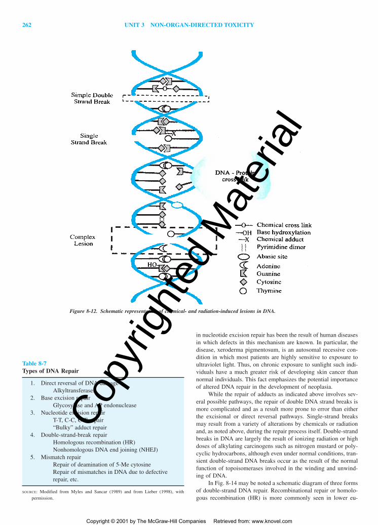
262 UNIT 3 NON-ORGAN-DIRECTED TOXICITY
in nucleotide excision repair has been the result of human diseasesin which defects in this mechanism are known. In particular, thedisease, xeroderma pigmentosum, is an autosomal recessive con-dition in which most patients are highly sensitive to exposure toultraviolet light. Thus, on chronic exposure to sunlight such indi-viduals have a much greater risk of developing skin cancer thannormal individuals. This fact emphasizes the potential importanceof altered DNA repair in the development of neoplasia.
While the repair of adducts as indicated above involves sev-eral possible pathways, the repair of double DNA strand breaks ismore complicated and as a result more prone to error than eitherthe excisional or direct reversal pathways. Single-strand breaksmay result from a variety of alterations by chemicals or radiationand, as noted above, during the repair process itself. Double-strandbreaks in DNA are largely the result of ionizing radiation or highdoses of alkylating carcinogens such as nitrogen mustard or poly-cyclic hydrocarbons, although even under normal conditions, tran-sient double-strand DNA breaks occur as the result of the normalfunction of topoisomerases involved in the winding and unwind-ing of DNA.
In Fig. 8-14 may be noted a schematic diagram of three formsof double-strand DNA repair. Recombinational repair or homolo-gous recombination (HR) is more commonly seen in lower eu-
Figure 8-12. Schematic representation of chemical- and radiation-induced lesions in DNA.
Table 8-7Types of DNA Repair
1. Direct reversal of DNA damageAlkyltransferases
2. Base excision repairGlycosylase and AP endonuclease
3. Nucleotide excision repairT-T, C-C, C-T repair“Bulky” adduct repair
4. Double-strand-break repairHomologous recombination (HR)Nonhomologous DNA end joining (NHEJ)
5. Mismatch repairRepair of deamination of 5-Me cytosineRepair of mismatches in DNA due to defectiverepair, etc.
SOURCE: Modified from Myles and Sancar (1989) and from Lieber (1998), withpermission.
2996R_ch08_239-319 4/11/01 3:46 PM Page 262
Copy
right
ed M
ater
ial
Copyright © 2001 by The McGraw-Hill Companies Retrieved from: www.knovel.com
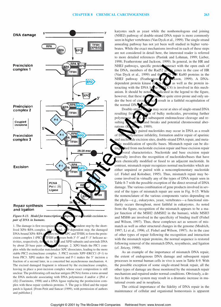
CHAPTER 8 CHEMICAL CARCINOGENESIS 263
karyotes such as yeast while the nonhomologous end joining(NHEJ) pathway of double-strand DNA repair is more commonlyseen in higher vertebrates (Van Dyck et al., 1999). The single-strandannealing pathway has not yet been well studied in higher verte-brates. While the exact mechanisms involved in each of these stepsare not considered in detail here, the interested reader is referredto more detailed references (Pastink and Lohman, 1999; Lieber,1998; Featherstone and Jackson, 1999). In general, in the HR andNHEJ pathways, specific proteins interact with the open ends ofthe DNA, members of the Rad52 group genes in the case of HR(Van Dyck et al., 1999) and the Ku70 and Ku80 proteins in theNHEJ pathway (Featherstone and Jackson, 1999). A DNA-dependent protein kinase (DNA-PKcs) as well as the protein in-teracting with the DNA ligase (XRCC4) is involved in this mech-anism. It should be noted as indicated in the legend to the figure,however, that these mechanisms are quite error-prone and only un-der the best of circumstances result in a faithful recapitulation ofthe normal DNA sequence.
Double-strand breaks may occur at sites of single-strand DNAresulting from adduction of bulky molecules, preventing furtherpolymerase action and subsequent endonuclease cleavage and re-sulting in double-strand breaks and potential chromosomal aber-rations (Kaufmann, 1989).
Incorrectly paired nucleotides may occur in DNA as a resultof DNA polymerase infidelity, formation and/or repair of apurinicand nucleotide excision sites, double-strand DNA repair, and meta-bolic modification of specific bases. Mismatch repair can be dis-tinguished from nucleotide excision repair and base excision repairby several characteristics. Nucleotide and base excision repairgenerally involves the recognition of nucleotides/bases that havebeen chemically modified or fused to an adjacent nucleotide. Incontrast, mismatch repair recognizes normal nucleotides which areeither unpaired or paired with a noncomplementary nucleotide (cf. Fishel and Kolodner, 1995). Thus, mismatch repair may be-come involved in virtually any of the types of DNA repair seen inTable 8-7 with the possible exception of the direct reversal of DNAdamage. The various combination of gene products involved in sev-eral of the types of mismatch repair are seen in Fig. 8-15. Whilethe nomenclature of the various components varies depending onthe phyla—e.g., eukaryotes, yeast, vertebrates—a functional sim-ilarity occurs throughout, most faithful in eukaryotes. As notedfrom the figure, recognition of the mismatch appears to be a ma-jor function of the MSH2 (hMSH2 in the human), while MSH3and MSH6 are involved in the specificity of binding itself (Fisheland Wilson, 1997). Thus, these complexes act as sensors of mis-match as well as other structural changes in the genome (Modrich,1997; Li et al., 1996; cf. Fishel and Wilson, 1997). As in the caseof other types of repair following the recognition and interactionwith the mismatch repair proteins, the normal sequence is restoredfollowing removal of the mismatch DNA, resynthesis, and ligation(cf. Jiricny, 1998).
As an example of the importance of mismatch DNA repair,the extent of endogenous DNA damage and subsequent repairprocesses in normal human cells in vivo is seen in Table 8-9. Withthe possible exception of some single-strand break repair, all theother types of damage are those monitored by the mismatch repairmechanism and repaired under normal conditions. Obviously, a de-fect in this repair system may result in a dramatic increase in mu-tational events and in neoplasia.
The critical importance of the fidelity of DNA repair in themaintenance of cellular and organismal homeostasis is apparent
TFIIH
TFIIH
XPA
RPA
RPA
TFIIH
RPA
TFIIH
TFIIH
TFIIH(XPB/D)
RPAXPA
RPA
RPA
XPA
XPA
XPC
XPC
XPC
Figure 8-13. Model for transcription-independent nucleotide excision re-pair of DNA in humans.
1. The damage is first recognized in an ATP-independent step by the short-lived XPA�RPA complex. In a second, ATP-dependent step, the damagedDNA-bound XPA�RPA complex recruits XPC and TFIIH, to form the prein-cision complex 1 (PIC1). TFIIH possesses both 3�-5� and 5�-3� helicase ac-tivities, respectively, through its XPB and XPD subunits and unwinds DNAby about 20 base pairs around the damage. 2. XPG binds the PIC1 com-plex while the molecular matchmaker XPC dissociates, leading to the morestable PIC2 excinuclease complex. 3. PIC2 recruits XPF�ERCC1 (F-1) toform PIC3. XPG makes the 3� incision and F-1 makes the 5� incision afraction of a second later, in a concerted but asynchronous mechanism. 4.The excised damaged fragment is released by the excinuclease complex,leaving in place a post-incision complex whose exact composition is stillunclear. The proliferating cell nuclear antigen (PCNA) forms a torus aroundthe DNA molecule associating with DNA polymerase � and/or � [Pol �(�)] (Tsurimoto, 1998) and a DNA ligase replacing the postincision com-plex with these repair synthesis proteins. 5. The gap is filled and the repairpatch is ligated. [From Petit and Sancar (1999), with permission of authorsand publisher.]
2996R_ch08_239-319 4/11/01 3:46 PM Page 263
Copy
right
ed M
ater
ial
Copyright © 2001 by The McGraw-Hill Companies Retrieved from: www.knovel.com
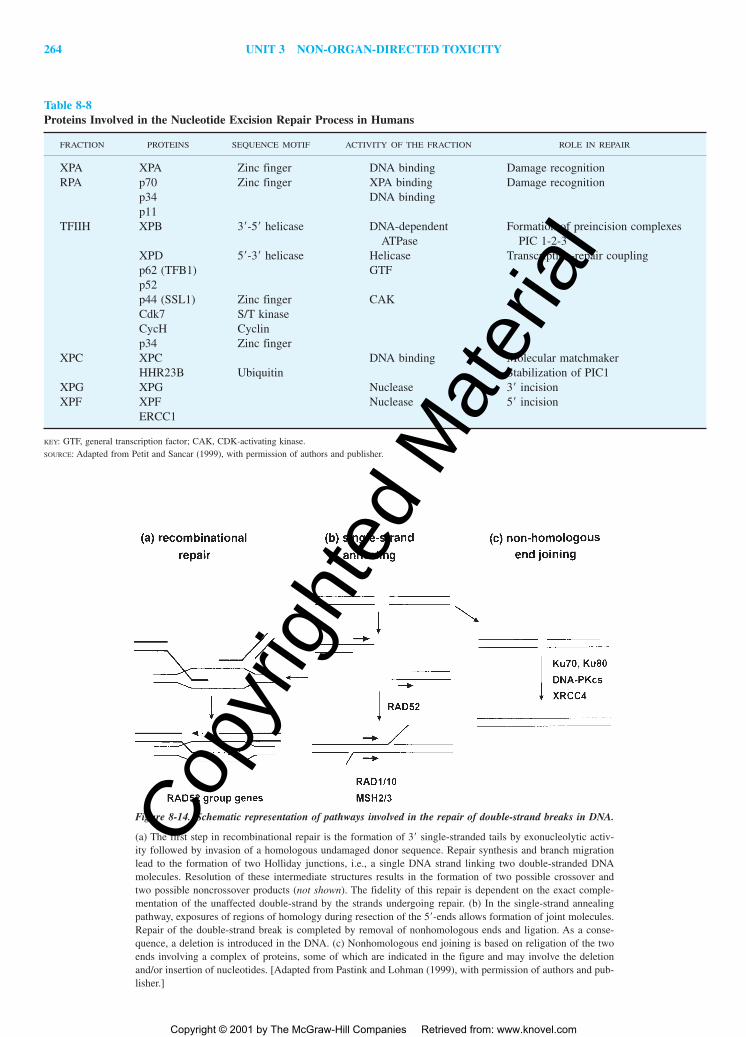
264 UNIT 3 NON-ORGAN-DIRECTED TOXICITY
Table 8-8Proteins Involved in the Nucleotide Excision Repair Process in Humans
FRACTION PROTEINS SEQUENCE MOTIF ACTIVITY OF THE FRACTION ROLE IN REPAIR
XPA XPA Zinc finger DNA binding Damage recognitionRPA p70 Zinc finger XPA binding Damage recognition
p34 DNA bindingp11
TFIIH XPB 3�-5� helicase DNA-dependent Formation of preincision complexesATPase PIC 1-2-3
XPD 5�-3� helicase Helicase Transcription-repair couplingp62 (TFB1) GTFp52p44 (SSL1) Zinc finger CAKCdk7 S/T kinaseCycH Cyclinp34 Zinc finger
XPC XPC DNA binding Molecular matchmakerHHR23B Ubiquitin Stabilization of PIC1
XPG XPG Nuclease 3� incisionXPF XPF Nuclease 5� incision
ERCC1
KEY: GTF, general transcription factor; CAK, CDK-activating kinase. SOURCE: Adapted from Petit and Sancar (1999), with permission of authors and publisher.
Figure 8-14. Schematic representation of pathways involved in the repair of double-strand breaks in DNA.
(a) The first step in recombinational repair is the formation of 3� single-stranded tails by exonucleolytic activ-ity followed by invasion of a homologous undamaged donor sequence. Repair synthesis and branch migrationlead to the formation of two Holliday junctions, i.e., a single DNA strand linking two double-stranded DNAmolecules. Resolution of these intermediate structures results in the formation of two possible crossover andtwo possible noncrossover products (not shown). The fidelity of this repair is dependent on the exact comple-mentation of the unaffected double-strand by the strands undergoing repair. (b) In the single-strand annealingpathway, exposures of regions of homology during resection of the 5�-ends allows formation of joint molecules.Repair of the double-strand break is completed by removal of nonhomologous ends and ligation. As a conse-quence, a deletion is introduced in the DNA. (c) Nonhomologous end joining is based on religation of the twoends involving a complex of proteins, some of which are indicated in the figure and may involve the deletionand/or insertion of nucleotides. [Adapted from Pastink and Lohman (1999), with permission of authors and pub-lisher.]
2996R_ch08_239-319 4/11/01 3:46 PM Page 264
Copy
right
ed M
ater
ial
Copyright © 2001 by The McGraw-Hill Companies Retrieved from: www.knovel.com

CHAPTER 8 CHEMICAL CARCINOGENESIS 265
from this brief discussion. Because induction of DNA repairprocesses occurs as a response to genetic damage, the inductioncan be used as an endpoint for its detection indirectly. In addition,an increase in repair mechanisms above constitutive levels can in-crease the magnitude of the genotoxic insult. The relative impor-tance of DNA repair in understanding mechanisms of chemical car-cinogenesis is most apparent when relating DNA damage and repairto DNA synthesis and cell replication. This is required to exceedthe intrinsic capacity of a cell to repair this damage.
DNA Repair, Cell Replication, andChemical Carcinogenesis
The persistence of DNA adducts in relation to the development ofneoplasia in specific tissues (Table 8-6) and differences in the re-pair of the adducts are critical factors in chemical carcinogenesis.The removal of methyl, ethyl, and similar small alkyl radicals fromindividual bases is to a great extent dependent on the presence ofalkyltransferases (see above). While in some tissues, such as liver,
Figure 8-15. Combinational specificities of heterocomplexes of gene products of mismatch repair genes.
(a) Base/base mispairs; (b) insertion/deletion mispairs; (c) 5� tailed DNA structures generated by single-strandDNA annealing following recombination, e.g. HR; and (d) Holliday junctions. [Adapted from Nakagawa et al.(1999), with permission of authors and publishers.]
Table 8-9Estimates of Endogenous DNA Damage and Repair Processes in Human Cells in Vivo
ESTIMATED OCCURRENCES MAXIMAL REPAIR RATE,OF DAMAGE PER HOUR BASE PAIRS PER HOUR
TYPE OF DAMAGE PER CELL* PER CELL*
Depurination 1,000 104 �Depyrimidination 55 104 �Cytosine deamination 15 104 �Single-stranded breaks 5,000 2 105
N7-methylguanine 3,500 Not reportedO6-methylguanine 130 104
Oxidation products 120 105
*Might be higher or lower by a factor of 2.SOURCE: Modified from data of the National Academy of Science (1989).
2996R_ch08_239-319 4/11/01 3:46 PM Page 265
Copy
right
ed M
ater
ial
Copyright © 2001 by The McGraw-Hill Companies Retrieved from: www.knovel.com

266 UNIT 3 NON-ORGAN-DIRECTED TOXICITY
it may be possible to increase the level of such enzymes in re-sponse to damage and hormonal or other influences, many tissuesdo not have an inducible repair mechanisms. Furthermore, someadducts are extremely difficult if not impossible for the cell to re-pair. An example of such a lesion, the 3-(deoxyguanosine)-N2-yl-acetylaminofluorene adduct first described by Kriek and his asso-ciates (Westra et al., 1976), is depicted in Fig. 8-10. This may inpart account for the relatively wide spectrum of neoplasms in-ducible by this chemical carcinogen.
Of equal importance is the continuous damage to DNA thatoccurs within cells as a result of ambient mutagens, radiation, andendogenous processes including oxidation, methylation, deamina-tion, and depurination. DNA damage induced by oxidative reac-tions (oxidative stress) is probably the source of most endogenousDNA damage. Ames et al. (1993) have estimated that the individ-ual reactive “hits” in DNA per cell per day is of the order of 105
in the rat and 104 in the human as a result of endogenous oxida-tive reaction. Such reactions can produce alkylation through per-oxidative reactions such as those described in Fig. 8-8 or hydrox-ylation of bases and single-strand breaks (Fig. 8-12). The endproduct of oxidative damage to DNA can also be interstrandcrosslinks and double-strand breaks (Demple and Harrison, 1994)with the potential for subsequent major genetic damage noted be-low. A more complete listing of the estimates of endogenous DNAdamage and repair processes in the human is seen in Table 8-9.The data of this table emphasize the considerable degree and sig-nificant variation in types of DNA damage and repair which oc-curs within each cell of the organism at a molecular level.
Experimental studies in mammalian cells have demonstratedthat active oxygen radicals may contribute to clastogenesis directly(Ochi and Kaneko, 1989) and indirectly through the production oflipid peroxides (Emerit et al., 1991). While methods for the repairof some types of oxidative damage including base hydroxylation(Bessho et al., 1993) and single-strand breaks (Satoh and Lindahl,1994), exist, such repair requires time and may be dependent onmany other intracellular factors. Because the formation of a muta-tion occurs during the synthesis of a new DNA strand by use ofthe damaged template, cell replication becomes an important fac-tor in the “fixation” of a mutation. The importance of the rate ofcell division and DNA synthesis in carcinogenesis has been em-phasized by several authors (Ames et al., 1993; Butterworth, 1991;Cohen and Ellwein, 1991). Thus, while many DNA repair mecha-nisms themselves may not be abnormal in neoplastic cells com-pared with their normal counterpart, a high rate of cell divisionwill tend to enhance both the spontaneous and induced level ofmutation through the chance inability of a cell to repair damageprior to DNA synthesis. An important pathway of DNA repair thatis genetically defective in a number of hereditary and spontaneousneoplasms in the human (Umar et al., 1994) is the mismatch repairmechanism that corrects spontaneous and post-replicative basealterations and thus is an important pathway for avoidance ofmutation in normal cells. Genetic defects in mismatch repairmechanisms lead to microsatellite DNA and instability with sub-sequent alteration in the stabilization of the genome itself (Mod-rich, 1994). Enhanced mitogenesis may also trigger more dramaticgenetic alterations including mitotic recombination, gene conver-sion, and nondisjunction. These genetic changes result in furtherprogressive genetic alterations with a high likelihood of resultingin cancer. The types of mutational events, the numbers of such mu-tations, and the cellular responses to them thus become important
factors in our understanding of the mechanisms of chemical car-cinogenesis.
CHEMICAL CARCINOGENS ANDTHE NATURAL HISTORY OF
NEOPLASTIC DEVELOPMENT
A number of chemicals can alter the structure of the genome and/orthe expression of genetic information with the subsequent appear-ance of cancer. However, cancer as a disease usually developsslowly with a long latent period between the first exposure to thechemical carcinogen and the ultimate development of malignantneoplasia. Thus, the process of carcinogenesis (the pathogenesis ofneoplasia) involves a variety of biological changes which, to a greatextent, reflect the structural and functional alterations in thegenome of the affected cell. However, when the biological changesoccurring during carcinogenesis are assessed at the molecular level,a better understanding of the mechanisms of cancer developmentmay be generated.
The Pathogenesis of Neoplasia: Biology
Although morphologic changes occurring during the early stagesof neoplasia were described during the early decades of this cen-tury, it was in the 1940s that a better understanding of the biolog-ical changes that occurred following carcinogen exposure was ob-tained. The first and best-studied model system was that of mouseskin carcinogenesis. The early investigations of Rous and Kidd(1941), Mottram (1944), and Berenblum and Shubik (1947) usedthe development of benign papillomas as an endpoint for studiesof epidermal carcinogenesis in mouse skin induced by polycyclichydrocarbons. These investigators coined the term “initiation” todesignate the initial alteration in individual cells within the tissueresulting from a single subcarcinogenic dose of the chemical car-cinogen. In these circumstances, papillomas were obtained onlywith subsequent chronic, multiple doses of a second agent that byitself was essentially carcinogenic. This latter “stage” was termedpromotion. Subsequent studies on a mammary adenocarcinomamodel in the mouse led to the proposal that processes subsequentto initiation comprised a stage termed progression (Foulds, 1954).Foulds’s description of this stage emphasized changes characteris-tic of malignant neoplasia and its evolution to higher degrees ofautonomy.
During the last two decades, these investigations have beenextended to a variety of tissue systems and to humans (Pitot, 1996).At present the pathogenesis of neoplasia is felt to consist of at leastthree operationally defined stages beginning with initiation fol-lowed by an intermediate stage of promotion, from which evolvesthe stage of progression. The third stage exhibits many of thecharacteristics described by Foulds. The biological characteristicsof the stages of initiation, promotion, and progression are listed inTable 8-10 (Pitot and Dragan, 1994; Boyd and Barrett, 1990;Harris, 1991). It is in the first and last stage of neoplasticdevelopment—initiation and progression—that structural changesin the genome (DNA) can be observed. The structural changes pre-viously discussed are most likely to be involved in the inductionof these stages. The intermediate stage of promotion does not ap-pear to involve direct structural changes in the genome of the cellbut rather depends on an altered expression of genes.
2996R_ch08_239-319 4/11/01 3:46 PM Page 266
Copy
right
ed M
ater
ial
Copyright © 2001 by The McGraw-Hill Companies Retrieved from: www.knovel.com

CHAPTER 8 CHEMICAL CARCINOGENESIS 267
Although it may be obvious to the reader, it is important toemphasize that the definition of neoplasia presented at the begin-ning of the chapter refers only to cells in the stage of progression.Initiated cells almost without exception cannot be identified, andtheir existence is presumed based on the natural history of neo-plastic development. Cells in the stage of promotion are termedpreneoplastic since their existence is entirely dependent on the con-tinued presence of the promoting agent in the environment. Pre-neoplasia must be distinguished from premalignant in that the lat-ter terminology also refers to cells in the stage of progression whichhave not yet expressed their full biologic malignant potential butdo have all the characteristics of the stage of progression.
Initiation Until recently the stage of initiation had been charac-terized and quantitated well after the process of carcinogenesis hadbegun. As with mutational events (see above), initiation requiresone or more rounds of cell division for the “fixation” of the process(Kakunaga, 1975; Columbano et al., 1981). The quantitative pa-rameters of initiation noted in Table 8-10—dose response and rel-ative potency—have been demonstrated in a variety of experi-mental systems (Pitot et al., 1987; Dragan et al., 1994); however,these parameters may be modulated by alteration of xenobiotic me-tabolism (Talalay et al., 1988) and by trophic hormones (Liao etal., 1993). The metabolism of initiating agents to nonreactive formsand the high efficiency of DNA repair of the tissue can alter theprocess of initiation.
One of the characteristics of the stage of initiation is its irre-versibility in the sense that the genotype/phenotype of the initiatedcell is established at the time of initiation, there is accumulatingevidence that not all initiated cells survive over the lifespan of theorganism or the period of an experiment. Their demise appears to
be due to the normal process of programmed cell death or apop-tosis (Wyllie, 1987).
Spontaneous preneoplastic lesions have been described in anumber of experimental systems (Maekawa and Mitsumori, 1990;Pretlow, 1994) as well as in the human (Dunham, 1972; Pretlow,1994; Pretlow et al., 1993). Thus, it would appear that the sponta-neous or fortuitous initiation of cells in a variety of tissues is a verycommon occurrence. If this is true, then the development of neo-plasia can be a function solely of the action of agents at the stagesof promotion and/or progression.
Promotion As in the stage of initiation, a variety of chemicalshave been shown to induce this stage. However, unlike chemicalsinducing the stage of initiation, there is no evidence that promot-ing agents or their metabolites directly interact with DNA or thatmetabolism is required at all for their effectiveness. In Fig. 8-16may be seen some representative structures of various promotingagents. Tetradecanoyl phorbol acetate (TPA) is a naturally occur-ring alicyclic chemical that is the active ingredient of croton oil, apromoting agent used for mouse skin tumor promotion. Saccharinis an effective promoting agent for the bladder, and phenobarbitalis an effective promoting agent for hepatocarcinogenesis.
2,3,7,8-Tetrachlorodibenzo-p-dioxin (TCDD) is probably themost effective promoting agent known for rat liver carcinogenesisbut is also effective in the lung and skin. Estradiol is shown as arepresentative of endogenous hormones that are effective promot-ing agents. Both androgens and estrogens, natural and synthetic,are effective promoting agents in their target end organ as well asin liver (Taper, 1978; Sumi et al., 1980; Kemp et al., 1989). Cholicacid enhances preneoplastic and neoplastic lesions in the rat colon(Magnuson et al., 1993), whereas 2,2,4-trimethylpentane and un-
Table 8-10Morphologic and Biologic Characteristics of the Stages of Initiation, Promotion, and Progression during Carcinogenesis
INITIATION PROMOTION PROGRESSION
Irreversible in viable cells Operationally reversible both at IrreversibleInitiated “stem cell” not morpho- the level of gene expression Morphologically discernible
logically identifiable and at the cell level alteration in cellulargenomic structure result-ing from karyotypicinstability
Efficiency sensitive to xenobiotic Promoted cell population existenceand other chemical factors dependent on continued adminis-
tration of the promoting agentSpontaneous (endogenous) occurrence Efficiency sensitive to aging and Growth of altered cells
of initiated cells dietary and hormonal factors sensitive to environ-Endogenous promoting agents may mental factors during
effect “spontaneous” promotion early phase of this stageRequires cell division for “fixation”Dose-response does not exhibit Dose-response exhibits measurable Benign or malignant neo-
a readily measurable threshold threshold and maximal effect plasms observed in thisstage
Relative potency of initiators depends Relative potency of promoters “Progressor” agents act toon quantitation of preneoplastic is measured by their effectiveness advance promoted cellslesions following defined period to cause an expansion of the into this stageof promotion cell progeny of the initiated
population
2996R_ch08_239-319 4/11/01 3:46 PM Page 267
Copy
right
ed M
ater
ial
Copyright © 2001 by The McGraw-Hill Companies Retrieved from: www.knovel.com

268 UNIT 3 NON-ORGAN-DIRECTED TOXICITY
leaded gasoline effectively promote renal tubular cell tumors in rats(Short et al., 1989). The final two structures noted—Wy-14,643and Nafenopin—are two members of the large class of carcino-genic peroxisome proliferators that induce the synthesis of perox-isomes in liver, are effective promoting agents, and on long-termadministration at high doses induce hepatic neoplasms (Reddy andLalwani, 1983). Many other agents including polypeptide hor-mones (see above), dietary factors including total calories, many
other halogenated hydrocarbons, and numerous other chemicalshave been found to enhance the development of preneoplastic andneoplastic lesions in one or more systems of carcinogenesis, in-cluding the human system.
The distinctive characteristic of promotion as contrasted withinitiation or progression is the reversible nature of this stage [Pitotand Dragan, 1995 (Table 8-10)]. Boutwell (1964) first demon-strated that by decreasing the frequency of application of the pro-
Figure 8-16. Structures of representative promoting agents.
2996R_ch08_239-319 4/11/01 3:46 PM Page 268
Copy
right
ed M
ater
ial
Copyright © 2001 by The McGraw-Hill Companies Retrieved from: www.knovel.com

CHAPTER 8 CHEMICAL CARCINOGENESIS 269
moting agent following initiation in mouse skin there was a loweryield of papillomas in comparison with that obtained by a morefrequent application of the promoting agent. Other investigators(Andrews, 1971; Burns et al., 1978) later demonstrated that papil-lomas developing during promotion in mouse epidermal carcino-genesis regress in large numbers both on removal of the promot-ing agent and during its continued application. The regression ofpreneoplastic lesions upon withdrawal of the promoting agents maybe due to apoptosis (Schulte-Herrmann et al., 1990). This proposedmechanism is supported by the demonstration that many promot-ing agents inhibit apoptosis in preneoplastic lesions (Schulte-Herrmann et al., 1993; Wright et al., 1994). Another potentialpathway of this operational reversibility is “redifferentiation” or re-modeling (Tatematsu et al., 1983). Thus, cells in the stage of pro-motion are dependent on continued administration of the promot-ing agent (Hanigan and Pitot, 1985) as implied by the early studiesof Furth (1959) on hormonally dependent neoplasia.
Another characteristic of the stage of promotion is its sus-ceptibility to modulation by physiologic factors. The stage of pro-motion may be modulated by the aging process (Van Duuren et al.,1975) and by dietary and hormonal factors (Sivak, 1979). Glauertand associates (1986) demonstrated that promotion of hepatocar-cinogenesis was less effective in rats fed a semisynthetic diet thanin those fed a crude, cereal-based diet. The promotion stage ofchemically induced rat mammary cancer is also modulated by di-etary factors (Cohen et al., 1991) and hormonal (Carter et al., 1988)factors. Many such modulating factors are themselves promotingagents. Several hormones can be carcinogenic. These hormones areeffective promoting agents and thus may serve as an exogenous orendogenous source for modulation of cell proliferation during car-cinogenesis (Pitot, 1991). Such physiologic agents may be onecomponent of endogenous promotion of initiated cells.
The dose–response relationships of promoting agents exhibitsigmoid-like curves with an observable threshold and maximaleffect. Such relationships are depicted in Fig. 8-17, in which the
dose–response curve for the binding of the phorbol TPA with itsreceptor is compared with a dose–response curve for the TPA pro-motion of dimethylbenzanthracene-initiated papillomas in mouseskin (Ashendel, 1985; Verma and Boutwell, 1980). The thresholdeffect of promoting agents may be considered a consequence ofthe reversible nature of their effects at the cellular level (see above).The maximal effect is due to a saturation of ligand binding in theformer case and to the promotion of all initiated cells in the latter(Fig. 8-17). Although one may not directly equate the variables inthe two processes, the similarity in the shape of the curves is strik-ing (Fig. 8-17). The relative potency of promoting agents may bedetermined as a function of their ability to induce the clonal growthof initiated cells. Thus, the net rate of growth of preneoplastic le-sions can be employed to determine relative potencies for pro-moting agents (Pitot et al., 1987).
The format of an experimental protocol for the demonstrationof initiation and promotion is provided in Fig. 8-18. As noted inthe figure, the endpoint of the study, which usually takes from 3to 6 months, depends on the tissue under investigation, the doseand nature of the initiating and promoting agents utilized, and fac-tors, such as diet and hormonal status, mentioned above. The end-point analyzed in such studies is properly a preneoplastic lesion(PNL) which develops clonally from initiated cells in the tissueunder study. These are altered hepatic foci in the rat or mouse liver(Pitot, 1990), epidermal papillomas for mouse skin (Wigley, 1983),hyperplasia of terminal end buds for rat mammary carcinomas(Purnell, 1980; Russo et al., 1983), and enzyme-altered foci in ratcolon carcinogenesis (Pretlow et al., 1993). Administration of thepromoting agent for the entire period of the experiment afterinitiation results in many preneoplastic lesions, whereas alterationof the format of administration of the promoting agent results inthe development of very few preneoplastic lesions. This reinforcesthe fact that the stage of promotion is operationally reversible(Boutwell, 1964) and indicates that a threshold dose effect levelmay exist.
Progression The transition from early progeny of initiated cellsto the biologically malignant cell population constitutes the majorpart of the natural history of neoplastic development. Foulds rec-ognized the importance of the development of neoplasia beyondthe appearance of any initial identifiable lesions (Foulds, 1965).
Figure 8-17. Composite showing the specific interaction of the receptorfor phorbol esters with its ligand determined as the inhibition of ra-dioactive TPA binding (closed circles).
The tumor response expressed as papillomas per mouse on mice initiatedwith dimethylbenzanthracene and promoted with various weekly doses ofTPA is noted in the open circles [composite graph from data of Ashendel(1985) and from Verma and Boutwell (1980) as published in Pitot (1986a).]
Figure 8-18. General experimental format demonstrating initiation (v)and promotion , for use with carcinogenesis studies in rodenttissues. PN, preneoplastic lesions.
2996R_ch08_239-319 4/11/01 3:46 PM Page 269
Copy
right
ed M
ater
ial
Copyright © 2001 by The McGraw-Hill Companies Retrieved from: www.knovel.com

270 UNIT 3 NON-ORGAN-DIRECTED TOXICITY
The characteristics of malignant progression that he observed—growth rate, invasiveness, metastatic frequency, hormonal respon-siveness, and morphologic characteristics—vary independently asthe disease develops. These characteristics have been ascribed tothe karyotypic instability during the irreversible progression stage(Pitot, 1993b). Environmental alterations can influence the stageof progression. For example, exposure to promoting agents can al-ter gene expression and induce cell proliferation. However, asgrowth of the neoplasm continues and karyotypic instabilityevolves, responses to environmental factors may be altered or lost(Noble, 1977; Welch and Tomasovic, 1985). Agents that act onlyto effect the transition of a cell from the stage of promotion to thatof progression may properly be termed progressor agents. Someexamples are listed in Table 8-11. Such agents presumably havethe characteristic of inducing chromosomal aberrations, may notnecessarily be capable of initiation, and in some cases may en-hance the clastogenesis associated with evolving karyotypic insta-
bility. As with the two stages of initiation and promotion, sponta-neous progression may also occur. In fact, spontaneous progres-sion would be highly fostered by increased cell replication (Ameset al., 1993).
The experimental demonstration of the stage of progressionis somewhat more complex than that of initiation and promotion.In Fig. 8-19 may be seen a general experimental format designedto demonstrate the effect of administration of a progressor agentafter a course of initiation and promotion with all of the appropri-ate controls. However, in this instance the endpoint that is quanti-tated is the number of neoplastic lesions (NL). In experimental sys-tems, the most effective development of neoplasia involves thecontinued administration of the promoting agent even after that ofthe progressor agent. This might be expected because cells earlyin the stage of progression respond to promoting agents, thus in-creasing the yield of neoplastic lesions in the experimental system(Table 8-10). As noted, a lower yield, usually still significant, may
Table 8-11Putative Progressor Agents in Carcinogenesis
INITIATING CLASTOGENIC CARCINOGENIC
AGENT ACTIVITY ACTIVITY ACTIVITY
Arsenic salts � � �Asbestos fibers ? � �Benzene � � �Benzoyl peroxide � � �Hydroxyurea � � �1,4-Bis[2-(3,5-dichloropyridyloxy)]-benzene � � �2,5,2�,5�-Tetrachlorobiphenyl � � �
SOURCE: Modified from Pitot and Dragan (1994), with permission.
Figure 8-19. General experimental format for demonstration of the stage of progression and the effect ofprogressor agents in experimental systems.
NL, neoplastic lesions; �, occasional or infrequent; 1�, few; 2�, some; 4�, many; III, administration of pro-gressor agent as single or several multiple doses; v, initiation; � promoting agent doses.
2996R_ch08_239-319 4/11/01 3:55 PM Page 270
Copy
right
ed M
ater
ial
Copyright © 2001 by The McGraw-Hill Companies Retrieved from: www.knovel.com
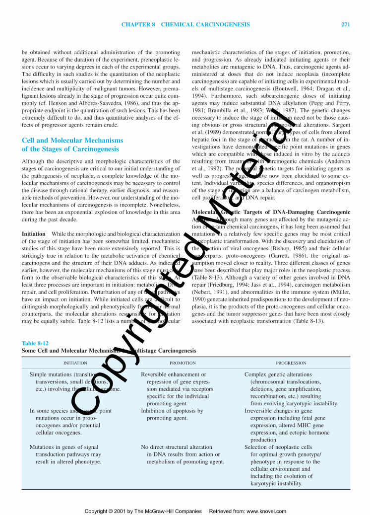
CHAPTER 8 CHEMICAL CARCINOGENESIS 271
be obtained without additional administration of the promotingagent. Because of the duration of the experiment, preneoplastic le-sions occur to varying degrees in each of the experimental groups.The difficulty in such studies is the quantitation of the neoplasticlesions which is usually carried out by determining the number andincidence and multiplicity of malignant tumors. However, prema-lignant lesions already in the stage of progression occur quite com-monly (cf. Henson and Albores-Saavedra, 1986), and thus the ap-propriate endpoint is the quantitation of such lesions. This has beenextremely difficult to do, and thus quantitative analyses of the ef-fects of progressor agents remain crude.
Cell and Molecular Mechanisms of the Stages of Carcinogenesis
Although the descriptive and morphologic characteristics of thestages of carcinogenesis are critical to our initial understanding ofthe pathogenesis of neoplasia, a complete knowledge of the mo-lecular mechanisms of carcinogenesis may be necessary to controlthe disease through rational therapy, earlier diagnosis, and reason-able methods of prevention. However, our understanding of the mo-lecular mechanisms of carcinogenesis is incomplete. Nonetheless,there has been an exponential explosion of knowledge in this areaduring the past decade.
Initiation While the morphologic and biological characterizationof the stage of initiation has been somewhat limited, mechanisticstudies of this stage have been more extensively reported. This isstrikingly true in relation to the metabolic activation of chemicalcarcinogens and the structure of their DNA adducts. As indicatedearlier, however, the molecular mechanisms of this stage must con-form to the observable biological characteristics of this stage. Atleast three processes are important in initiation: metabolism, DNArepair, and cell proliferation. Perturbation of any of these pathwayshave an impact on initiation. While initiated cells are difficult todistinguish morphologically and phenotypically from their normalcounterparts, the molecular alterations responsible for initiationmay be equally subtle. Table 8-12 lists a number of the molecular
mechanistic characteristics of the stages of initiation, promotion,and progression. As already indicated initiating agents or theirmetabolites are mutagenic to DNA. Thus, carcinogenic agents ad-ministered at doses that do not induce neoplasia (incompletecarcinogenesis) are capable of initiating cells in experimental mod-els of multistage carcinogenesis (Boutwell, 1964; Dragan et al.,1994). Furthermore, such subcarcinogenic doses of initiatingagents may induce substantial DNA alkylation (Pegg and Perry,1981; Brambilla et al., 1983; Ward, 1987). The genetic changesnecessary to induce the stage of initiation need not be those caus-ing obvious or gross structural chromosomal alterations. Sargentet al. (1989) demonstrated normal karyotypes of cells from alteredhepatic foci in the stage of promotion in the rat. A number of in-vestigations have demonstrated specific point mutations in geneswhich are compatible with those induced in vitro by the adductsresulting from treatment with carcinogenic chemicals (Andersonet al., 1992). The potential genetic targets for initiating agents aswell as progressor agents have now been elucidated to some ex-tent. Individual variability, species differences, and organotropismof the stage of initiation are a balance of carcinogen metabolism,cell proliferation, and DNA repair.
Molecular Genetic Targets of DNA-Damaging CarcinogenicAgents Although many genes are affected by the mutagenic ac-tion of certain chemical carcinogens, it has long been assumed thatmutations in a relatively few specific genes may be most criticalto neoplastic transformation. With the discovery and elucidation ofthe function of viral oncogenes (Bishop, 1985) and their cellularcounterparts, proto-oncogenes (Garrett, 1986), the original as-sumption moved closer to reality. Three different classes of geneshave been described that play major roles in the neoplastic process(Table 8-13). Although a variety of other genes involved in DNArepair (Friedburg, 1994; Jass et al., 1994), carcinogen metabolism(Nebert, 1991), and abnormalities in the immune system (Müller,1990) generate inherited predispositions to the development of neo-plasia, it is the products of the proto-oncogenes and cellular onco-genes and the tumor suppressor genes that have been most closelyassociated with neoplastic transformation (Table 8-13).
Table 8-12Some Cell and Molecular Mechanisms in Multistage Carcinogenesis
INITIATION PROMOTION PROGRESSION
Simple mutations (transitions, Reversible enhancement or Complex genetic alterationstransversions, small deletions, repression of gene expres- (chromosomal translocations,etc.) involving the cellular genome. sion mediated via receptors deletions, gene amplification,
specific for the individual recombination, etc.) resultingpromoting agent. from evolving karyotypic instability.
In some species and tissues, point Inhibition of apoptosis by Irreversible changes in genemutations occur in proto- promoting agent. expression including fetal geneoncogenes and/or potential expression, altered MHC genecellular oncogenes. expression, and ectopic hormone
production.Mutations in genes of signal No direct structural alteration Selection of neoplastic cells
transduction pathways may in DNA results from action or for optimal growth genotype/result in altered phenotype. metabolism of promoting agent. phenotype in response to the
cellular environment andincluding the evolution ofkaryotypic instability.
2996R_ch08_239-319 4/11/01 3:55 PM Page 271
Copy
right
ed M
ater
ial
Copyright © 2001 by The McGraw-Hill Companies Retrieved from: www.knovel.com

272 UNIT 3 NON-ORGAN-DIRECTED TOXICITY
Table 8-14 shows a listing of a number of functions of proto-oncogenes and cellular oncogenes and tumor suppressor genes,with specific examples and their localization within the cell whereknown. It is immediately obvious that the oncogenes are involvedprimarily in cellular growth, signal transduction, and nuclear tran-scription. Interestingly, similar functions are attributed to knowntumor suppressor genes, but in addition at least two of the latterare involved in regulation of the cell cycle. This table is not meantto be all inclusive, and the interested reader is referred to recentreviews and text for a more detailed discussion of these genes andtheir products (Hunter, 1991; Levine, 1993).
Mutations in proto-oncogenes can result in their activationwith subsequent neoplastic transformation similar to that observed
following the altered expression of cellular oncogenes. Activationof proto-oncogenes and cellular oncogenes can occur by variousmeans (Table 8-15). Scrutiny of these mechanisms suggests thatonly point mutations, small insertions and deletions, and possiblyaltered methylation status are potential events resulting in initia-tion. The other, more complex alterations in the genome listedwould be characteristic of the stage of progression, as will be dis-cussed below.
The activation of proto-oncogenes and cellular oncogenes byspecific base mutations, small deletions, and frameshift mutationsresults from DNA synthesis in the presence of DNA damage in-cluding the presence of adducts. Methods for determining such al-terations in specimens that consist of only a few hundred or a thou-
Table 8-14Functions of Representative Oncogenes and Tumor Suppressor Genes
FUNCTIONS OF GENE PRODUCT GENES CELL LOCALIZATION
A. OncogenesGrowth factors sis, fgf ExtracellularReceptor/protein tyrosine-kinase met, neu Extra cell/cell membraneProtein tyrosine kinase src, ret Cell membrane/cytoplasmicMembrane-associated G proteins ras, gip-2 Cell membrane/cytoplasmicCytoplasmic protein serine kinases raf, pim-1 CytoplasmicNuclear transcription factors myc, fos, jun NuclearUnknown, undetermined bcl-2, crk Mitochondrial, cytoplasmicB. Tumor suppressor genesGTPase-activation NF1 Cell membrane/cytoplasmicCell cycle–regulated nuclear
transcriptional repressor RB-1 NuclearCell cycle–regulated nuclear
transcription factor p53 NuclearZinc-finger transcription factor WT1 NuclearMismatch DNA repair hMLH1 Nuclear (?)Zinc-finger transcription factor (?) BRCA1 Unknown
SOURCES: Part A of the table was extracted from Hunter (1991). Information for part B was obtained from Levine (1993) as wellas Papadopoulos et al. (1994) and Bronner et al. (1994), while that for BRCA1 was obtained from Miki et al. (1994) andFutreal et al. (1994).
Table 8-13Characteristics of Proto-oncogenes, Cellular Oncogenes, and Tumor Suppressor Genes
PROTO-ONCOGENES CELLULAR ONCOGENES TUMOR SUPPRESSOR GENES
Dominant Dominant RecessiveBroad tissue specificity for Broad tissue specificity for Considerable tissue speci-
cancer development cancer development ficity for cancerdevelopment
Germline inheritance rarely Germline inheritance rarely Germline inheritance fre-involved in cancer involved in cancer quently involved indevelopment development cancer development
Analogous to certain viral No known analogs in onco- No known analogs in onco-oncogenes genic viruses genic viruses
Somatic mutations activate Somatic mutations activate Germline mutations mayduring all stages of neo- during all stages of initiate, but mutationplastic development neoplastic development to neoplasia occurs
only during the stageof progression
SOURCE: After Pitot (1993b), with permission.
2996R_ch08_239-319 4/11/01 3:55 PM Page 272
Copy
right
ed M
ater
ial
Copyright © 2001 by The McGraw-Hill Companies Retrieved from: www.knovel.com

CHAPTER 8 CHEMICAL CARCINOGENESIS 273
sand cells have been available only during the last decade (Mies,1994). The analysis of mutations in specific genes potentially in-volved in the neoplastic transformation is possible from very smallsamples by various molecular techniques.
The ras genes code for guanosine triphosphatases, whichfunction as molecular switches for signal transduction pathwaysinvolved in the control of growth, differentiation, and other cellu-lar functions (Hall, 1994). Table 8-16 lists a number of examplesin rodent tissues of specific mutation in two of the ras genes, theHa-ras proto-oncogene and the Ki-ras cellular oncogene. With theexception of mouse skin, the frequency of such mutations in pre-neoplastic lesions in experimental animals in the stage of promo-tion is about 20 to 60 percent. In instances of multistage carcino-genesis in mouse skin, the frequency increases to nearly 100percent (Bailleul et al., 1989). In general, the mutations noted arethose which theoretically could result from DNA-adducts formedby the particular carcinogen. Interestingly, spontaneously occur-ring neoplasms in mice also exhibit a significant incidence of pointmutations in the ras proto-oncogenes and cellular oncogenes(Rumsby et al., 1991; Candrian et al., 1991), but neoplasms in cor-responding tissues in other species do not necessarily exhibit acti-vating mutations in proto- or cellular oncogenes (Tokusashi et al.,1994; Kakiuchi et al., 1993; Schaeffer et al., 1990). In addition,mutated ras genes have been described in normal-appearing mouseskin after dimethylbenz(a)anthracene (DMBA) or urethane appli-cation (Nelson et al., 1992). By contrast, Cha and associates (1994)recently reported that a very high percentage of untreated rats con-tained detectable levels of Ha-ras mutations in normal mammarytissue. Thus, the mutations seen in neoplasms in untreated animalsmay result from the selective proliferation of cells containing pre-existing mutations.
Thus, while several classes of genes appear appropriate as tar-gets for DNA-damaging carcinogens, the actual role of proto- andcellular oncogene mutations in establishing carcinogenesis is notentirely clear. Among the earliest preneoplastic lesions studied(Table 8-15), less than one-third exhibit mutations in the ras genefamily, but it is quite possible that other proto- and cellular onco-genes may be targets. Evidence that tumor suppressor genes may
be targets for the initiation of early malignant development comelargely from studies of genetically inherited neoplasia. In these rarehereditary cancers, one of the alleles of a tumor suppressor genecontains a germline mutation in all cells of the organism (Paraskevaand Williams, 1992; Knudson, 1993).
Promotion Boutwell (1974) was the first to propose that pro-moting agents may induce their effects through their ability to al-ter gene expression. During the past decade, our understanding ofmechanisms involving the alteration of gene expression by envi-ronmental agents has increased exponentially (Morley and Thomas,1991; Rosenthal, 1994). The regulation of genetic information ismediated through recognition of the environmental effector, hor-mone, promoting agent, drug, etc., and its specific molecular in-teraction with either a surface or cytosolic receptor. Several typesof receptors exist in cells (Mayer, 1994; Pawson, 1993; Strader etal., 1994) (Fig. 8-20). Plasma membrane receptors may possess atyrosine protein kinase domain on their intracellular region, whileothers have multiple transmembrane domains with the intracellu-lar signal transduced through G proteins and cyclic nucleotides(Mayer, 1994). The other general type of receptor mechanism in-volves a cytosolic receptor that interacts with the ligand (usuallylipid-soluble) that has diffused through the plasma membrane. Theligand receptor complex then travels to the nucleus before inter-acting directly with specific DNA sequences known as responseelements.
In both instances is shown in a highly simplified manner thecascade effect of various protein kinases resulting in alterations intranscription as well as cell replication within the nucleus. Asshown in the figure, interaction of transmembrane receptors con-taining a tyrosine protein kinase domain involves initially theirdimerization induced by ligand interaction. This activates the pro-tein kinase domain of the receptor causing autophosphorylation.This in turn attracts a cytoplasmic complex, Grb2-Sos to the plasmamembrane (Aronheim et al., 1994). Sos is a member of a familyof regulatory proteins termed guanine-nucleotide exchange factors(GEFs) (Feig, 1994). Sos association with the G-protein, Ras, stim-ulates along with other protein interactions the exchange of GDP
Table 8-15Potential Mechanisms of Oncogene Activation
EVENT CONSEQUENCE EXAMPLES
Base mutation in coding sequences New gene product with altered activity v-onc genes, bladder carcinomaDeletion in noncoding sequences Altered regulation of normal Fibroblast transformation in vitro
gene productAltered promotion for RNA Increased transcription of mRNA Cell transformation in vitro,
polymerase (normal gene product) lymphoma in chickensInsertion or substitution with Altered regulation of gene product Canine venereal tumor, mouse
repetitive DNA elements (? normal) myeloma(“transposons”)
Chromosomal translocation Altered mRNA, new gene product Burkitt lymphoma in humans,(?), no altered regulation of gene mouse plasmacytomaexpression
Gene amplification Increased expression of normal Human colon carcinoma, humangene bladder carcinoma
Hypomethylation of c-onc gene Altered regulation of gene Human colon and lung cancerexpression (?), normal gene product
SOURCE: Adapted from Pitot (1986b), with permission.
2996R_ch08_239-319 4/11/01 3:55 PM Page 273
Copy
right
ed M
ater
ial
Copyright © 2001 by The McGraw-Hill Companies Retrieved from: www.knovel.com

274 UNIT 3 NON-ORGAN-DIRECTED TOXICITY
with GTP on the Ras � subunit. The GTP-Ras in turn interacts witha cytoplasmic serine-threonine protein kinase, raf, with subsequentactivation of its catalytic activity and initiation of a kinase cascadeultimately resulting in the phosphorylation and activation of tran-scription factors including Jun, Fos, Myc, CREB, and ultimatelyE2F and Rb, the tumor suppressor gene (Lewis et al., 1998;Janknecht et al., 1995; Roussel, 1998). A number of other tran-
scription factors are also activated by similar pathways involvingother pathways such as phospholipase C, phosphatidylinositol ki-nase, and protein kinase C (Vojtek and Der, 1998; Takuwa andTakuwa, 1996). The rate-limiting step in this process is themediation of the signal through the G-protein family. The G pro-teins are targeted to the plasma membrane through lipid moieties,both isoprenoid and fatty acyl, covalently linked to the carboxyl-terminal region of the protein (Yamane and Fung, 1993). In thisway the initiation of the signal by the ligand-receptor interactioncan be physically related to the rate-limiting G protein activationstep. The activation cycle of the G-protein family involves GTPbinding to the � subunit of the G protein, such binding being dra-matically stimulated by GEF proteins such as Sos. Activation alsoinvolves dissociation of the � from the � and subunits allowingthe � subunit to interact with and activate downstream membersof the pathway, a protein kinase, B-raf in the case of the growthfactor related pathway or with other membrane molecules such asadenyl cyclase in the case of multiple transmembrane domain re-ceptors (Fig. 8-20). The activated G protein has an extremely shorthalf-life because of the action of RGS (regulator of G-protein sig-naling) proteins which stimulate GTP hydrolysis to GDP with sub-sequent reassociation of the G protein in its inactive state (Koelle,1997).
The multiple transmembrane domain receptors (G protein–linked) are in direct association with G proteins and on activationof the receptor by interaction with a ligand may activate a kinasetermed a G-protein receptor kinase (GRK) or another effector suchas adenyl cyclase (Böhm et al., 1997; Rasenick et al., 1995). Inturn, adenyl cyclase produces cyclic AMP, which interacts with theregulatory component of protein kinase A, with a subsequent phos-phorylation cascade to the transcription apparatus. In addition tothe plasma membrane receptors, gene expression can be regulatedthrough the interaction of cytoplasmic receptors with their ligandsas previously discussed. Just as with membrane receptors, the path-ways of the cytoplasmic receptors involve multiple interactionswith proteins, phosphorylation, and ultimate alteration of tran-scription through factor interaction with DNA (Weigel, 1996; Prattand Toft, 1997). In all of these pathways, in addition to alterationof transcription and gene expression, enhancement or inhibition of
Figure 8-20. Diagram of principal mechanisms of intracellular signaltransduction initiated either within the cytosol or at the plasma mem-brane. [Modified from Mayer (1994), with permission.]
Table 8-16Mutational Activation of ras Oncogenes during the Stages of Initiation and Promotion
SPECIES/TISSUE CARCINOGEN LESION GENE/MUTATION* FREQUENCY† REFERENCE
Rat/colon Azoxymethane Aberrant crypt K-ras/G�A/12 5/16 Shivapurkar et al., 1994foci
Mouse/liver Diethylnitros- G6Pase� foci Ha-ras/C�A/61 12/127 Bauer-Hofmann et al., 1992amine A�G
Mouse/lung Urethane Small adenomas Ki-ras/A�G/61 32/100 Nuzum et al., 1990A�T
Rat/mammary N-methyl-N- Initiated cell H-ras/G�A/12 17% Zhang et al., 1991gland nitrosourea clones
Hamster/ N-nitroso-bis Papillary K-ras/G�A/12 12/26 Cerny et al., 1992pancreas 2-oxopropyl)- hyperplasia
amineMouse/skin DMBA/TPA Papilloma Ha-ras/A�T/61 12/14 Quintanilla et al., 1986
*The numbers in this column refer to the codon position in the cDNA (mRNA) of the gene product.†The numerator indicates the number of animals exhibiting the mutation; the denominator refers to the total number of animals studied.
2996R_ch08_239-319 4/11/01 3:55 PM Page 274
Copy
right
ed M
ater
ial
Copyright © 2001 by The McGraw-Hill Companies Retrieved from: www.knovel.com
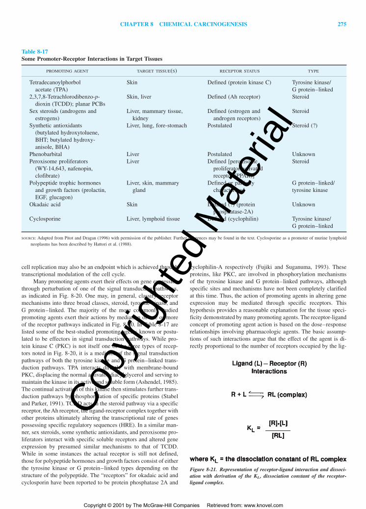
CHAPTER 8 CHEMICAL CARCINOGENESIS 275
cell replication may also be an endpoint which is achieved throughtranscriptional modulation of the cell cycle.
Many promoting agents exert their effects on gene expressionthrough perturbation of one of the signal transduction pathways,as indicated in Fig. 8-20. One may, in general, classify receptormechanisms into three broad classes, steroid, tyrosine kinase, andG protein–linked. The majority of the more commonly studiedpromoting agents exert their actions by mediation of one or moreof the receptor pathways indicated in Fig. 8-20. In Table 8-17 arelisted some of the best-studied promoting agents known or postu-lated to be effectors in signal transduction pathways. While pro-tein kinase C (PKC) is not itself one of the three types of recep-tors noted in Fig. 8-20, it is a mediator of the signal transductionpathways of both the tyrosine kinase and G protein–linked trans-duction pathways. TPA interacts directly with membrane-boundPKC, displacing the normal activator diacylglycerol and serving tomaintain the kinase in its active and soluble form (Ashendel, 1985).The continual activation of this kinase then stimulates further trans-duction pathways by phosphorylation of specific proteins (Stabeland Parker, 1991). TCDD acts in the steroid pathway via a specificreceptor, the Ah receptor, the ligand-receptor complex together withother proteins ultimately altering the transcriptional rate of genespossessing specific regulatory sequences (HRE). In a similar man-ner, sex steroids, some synthetic antioxidants, and peroxisome pro-liferators interact with specific soluble receptors and altered geneexpression by presumed similar mechanisms to that of TCDD.While in some instances the actual receptor is still not defined,those for polypeptide hormones and growth factors consist of eitherthe tyrosine kinase or G protein–linked types depending on thestructure of the polypeptide. The “receptors” for okadaic acid andcyclosporin have been reported to be protein phosphatase 2A and
cyclophilin-A respectively (Fujiki and Suganuma, 1993). Theseproteins, like PKC, are involved in phosphorylation mechanismsof the tyrosine kinase and G protein–linked pathways, althoughspecific sites and mechanisms have not been completely clarifiedat this time. Thus, the action of promoting agents in altering geneexpression may be mediated through specific receptors. Thishypothesis provides a reasonable explanation for the tissue speci-ficity demonstrated by many promoting agents. The receptor-ligandconcept of promoting agent action is based on the dose–responserelationships involving pharmacologic agents. The basic assump-tions of such interactions argue that the effect of the agent is di-rectly proportional to the number of receptors occupied by the lig-
Table 8-17Some Promoter-Receptor Interactions in Target Tissues
PROMOTING AGENT TARGET TISSUE(S) RECEPTOR STATUS TYPE
Tetradecanoylphorbol Skin Defined (protein kinase C) Tyrosine kinase/acetate (TPA) G protein–linked
2,3,7,8-Tetrachlorodibenzo-p- Skin, liver Defined (Ah receptor) Steroiddioxin (TCDD); planar PCBs
Sex steroids (androgens and Liver, mammary tissue, Defined (estrogen and Steroidestrogens) kidney androgen receptors)
Synthetic antioxidants Liver, lung, fore-stomach Postulated Steroid (?)(butylated hydroxytoluene,BHT; butylated hydroxy-anisole, BHA)
Phenobarbital Liver Postulated UnknownPeroxisome proliferators Liver Defined [peroxisome Steroid
(WY-14,643, nafenopin, proliferator-activatedclofibrate) receptor (PPAR)]
Polypeptide trophic hormones Liver, skin, mammary Defined or partially G protein–linked/and growth factors (prolactin, gland characterized tyrosine kinaseEGF, glucagon)
Okadaic acid Skin Defined (?) (protein Unknownphosphatase-2A)
Cyclosporine Liver, lymphoid tissue Defined (cyclophilin) Tyrosine kinase/G protein–linked
SOURCE: Adapted from Pitot and Dragan (1996) with permission of the publisher. Further references may be found in the text. Cyclosporine as a promoter of murine lymphoidneoplasms has been described by Hattori et al. (1988).
Figure 8-21. Representation of receptor-ligand interaction and dissoci-ation with derivation of the KL, dissociation constant of the receptor-ligand complex.
2996R_ch08_239-319 4/11/01 3:55 PM Page 275
Copy
right
ed M
ater
ial
Copyright © 2001 by The McGraw-Hill Companies Retrieved from: www.knovel.com
276 UNIT 3 NON-ORGAN-DIRECTED TOXICITY
and. The intrinsic activity of the chemical and the signal trans-duction pathways available in the tissue are important factors inthe type and degree of response observed.
The Molecular Basis of the Reversibilityof the Stage of Tumor Promotion
The theoretical and practical aspects of ligand-receptor interactionshave been previously reviewed (cf. Pitot, 1995). Herein we willconsider such relationships as they are concerned with the actionof tumor promoters. The basic assumption of the ligand-receptorinteraction is that the effect of the agent is directly proportional tothe number of receptors occupied by that chemical ligand and thata maximum response of the target is obtained only when all re-ceptors are occupied. As seen in Fig. 8-21, a simple bimolecularinteraction between the ligand and receptor can be utilized todetermine a dissociation constant, KL of the receptor ligand com-plex, as noted in the figure. While a variety of mathematical rela-tionships may be derived from this simple equation (cf. Ruffolo,1992), herein we consider only the dose–response relationship. Thedose–response of the receptor-ligand interaction takes the shape ofa sigmoidal curve identical to that seen with the inhibition of TPAbinding depicted in Fig. 8-17. The figure denotes a threshold re-sponse at very low doses and a maximal effect above a specificdose. Theoretically, the linear conversion of the sigmoidal curvessuch as seen in Fig. 8-17 may indicate effects at even lower dosesthan those usually studied, but such depends on the associationconstant of the ligand-receptor complex and the subsequent fate ofthe complex (Aldridge, 1986). Withdrawal of the ligand reverts thesystem to its original state. Thus, the regulation of genetic expres-sion that occurs by the ligand-receptor mechanism predicts athreshold and reversible effect unlike that of genotoxic carcino-genic agents, in which an irreversible non-threshold response is as-sumed on theoretical grounds and can be demonstrated in a vari-ety of instances (Druckrey, 1967; Zeise et al., 1987). Furthermore,at very low doses of some carcinogenic agents, an apparent rever-sal or “protective” effect of the agent can actually be demonstrated.This phenomenon has been termed hormesis (Teeguarden et al.,1998). Regardless of whether this latter effect can be more gener-alized, it is apparent that both the measured dose response and thereceptor mechanisms of tumor promotion imply a no-effect orthreshold level for the action of these agents during carcinogene-sis. Thus, the stage of tumor promotion, unlike that of initiationand progression, does not involve mutational or structural eventsin the genome but rather is concerned with the reversible alterationof the expression of genetic information.
The selective induction of proliferation of initiated cell pop-ulations was first intimated by the work of Solt and Farber (1976),who used 2-acetylaminofluorene administration as a “selectionagent” for the enhancement of proliferation of altered hepatic fociin a modified initiation-promotion protocol. A similar “selection”of certain initiated clones by TPA promotion has also been postu-lated as occurring during multistage carcinogenesis in mouse skin(cf. DiGiovanni, 1992). Later studies demonstrated that 2-acetylaminofluorene was acting as a promoting agent in this pro-tocol (Saeter et al., 1988). Farber and his colleagues (Roomi et al.,1985) espoused the concept that the lowered xenobiotic metabo-lism of preneoplastic cell populations gave such cells a competi-tive advantage in toxic environments such as those provided by thechronic administration of carcinogens. Schulte-Hermann and his
associates (1981) have demonstrated that several hepatic promot-ing agents including phenobarbital, certain steroids, and peroxi-some proliferators selectively enhanced the proliferation of cellswithin preneoplastic lesions in rat liver. A similar effect was re-ported by Klaunig in preneoplastic and neoplastic hepatic lesionsin mice responding to promotion by phenobarbital (Klaunig, 1993).The response of preneoplastic hepatocytes in the rat to partial he-patectomy is also greater than that of normal hepatocytes (Laconiet al., 1994). Preneoplastic hepatocytes in culture exhibit an in-herent higher level of replicative DNA synthesis than normal he-patocytes (Xu et al., 1988). Thus, the characteristic of promotingagents at the cell and molecular level to increase cell proliferationof preneoplastic cell populations selectively more than that of theirnormal counterparts may be the result of altered mechanisms ofcell cycle control within the preneoplastic cell.
Cell Cycle Regulation Although the exact mechanism(s) bywhich promoting agents selectively enhance cell replication in pre-neoplastic cells is unknown, our understanding of the interactionof ligand-receptor signaling with the cell cycle and its regulationhas dramatically increased in recent years. Figure 8-22 diagramsan integration of the cell cycle and apoptosis with the signal trans-duction pathways (Fig. 8-20). Phosphorylation of the mitogen-activated protein kinase (MAPK) via the signal transduction path-way activates this kinase (Fig. 8-22), which then activates varioustranscription factors, some of which are noted above in the figureas proto-oncogene products, c-myc, c-jun, and c-fos (Seger andKrebs, 1995). Rb, the retinoblastoma tumor suppressor gene, ismade throughout the cell cycle. It becomes highly phosphorylatedat the beginning of DNA synthesis (G,1S). This releases a tran-scription factor, E2F, which is complexed with the highly phos-phorylated but not the hypophosphorylated Rb protein. E2F thenis available to stimulate the transcription of a variety of genesneeded for the transition from G1 and the initiation of DNA syn-thesis. As we noted above, ligand-receptor interactions can resultin the activation of E2F and thus the transcription of genes neededfor continuation of the cell cycle. This continuation involves a va-riety of protein kinases and proteins, known as cyclins, which arelisted in the figure. Another tumor suppressor gene, the p53 gene,also plays a role as a transcription factor, preventing continuanceof the cell cycle on the occasion of DNA damage (Wu and Levine,1994). Such a pause allows the cells to repair such damage or, ifthe damage is excessive, to undergo apoptosis (Fig. 8-22). If thep53 gene is mutated or absent, such a pause does not occur, andthe cell cycle continues replicating despite the presence of damageresulting in mutations and clastogenesis (Lane, 1992; Sander et al.,1993; Dulic et al., 1994). Obviously, the missing mechanistic linkis a clear understanding of the selective enhancement of the cellcycle in preneoplastic cells by promoting agents. A variety of pos-sibilities exist, including increased concentrations of receptors orany one or more of the components of the signal transduction path-way, as well as mutations in transcription factors, cyclins, cdks, orother components of the cell cycle. As yet, however, definitive stud-ies to pinpoint such mechanisms have not been performed.
Progression The stage of progression usually develops from cellsin the stage of promotion but may develop directly from normalcells, usually as a result of the administration of relatively high,usually cytotoxic doses of complete carcinogenic agents capableof inducing both initiation and progression. In addition, the incor-poration into the genome of genetic information such as oncogenic
2996R_ch08_239-319 4/11/01 3:55 PM Page 276
Copy
right
ed M
ater
ial
Copyright © 2001 by The McGraw-Hill Companies Retrieved from: www.knovel.com

CHAPTER 8 CHEMICAL CARCINOGENESIS 277
viruses, the stable transfection of genetic material, or from spon-taneous chromosomal alterations may induce the stage of progres-sion. As noted in Tables 8-10 through 8-12, the major hallmark ofthe stage of progression is evolving karyotypic instability. It is thismolecular characteristic of cells in the stage of progression that po-tentially leads to multiple “stages” or changes in malignant cellswhich were first described by Foulds (1954) as “independent char-acteristics.” Foulds noted that cells in the stage of progression mightevolve in such a way that the characteristics of invasion, metasta-tic growth, anaplasia, as well as the rate of growth and responsesto hormonal influences changed toward higher and higher degreesof malignancy. Such “independent characteristics” may all be un-derstood as resulting from karyotypic changes that are constantlyevolving in cells during the stage of progression. Included in these“characteristics” may be such things as fetal gene expression, the
expression of the major histocompatibility complex (MHC) classI and II surface proteins, and the ectopic production of hormonesby cells derived from non-hormone-producing tissues as well asseveral other characteristics of neoplastic cells (Hanahan andWeinberg, 2000). Thus, in some tissues it may be possible to de-scribe multiple “stages” that reflect the evolving karyotypic insta-bility of neoplasms such as in the evolution of colonic (Fearon andVogelstein, 1992) and other neoplasms (Nowell, 1990). Simulta-neous with these changes may be the occurrence of mutated proto-and cellular oncogenes (Liu et al., 1988) and tumor suppressorgenes (Yokota and Sugimura, 1993). However, since karyotypicinstability is unlikely to lead directly to point mutations in onco-genes and tumor suppressor genes, it is more likely that their ap-pearance reflects the selection of cells better suited to the growthenvironment of the neoplasm.
The critical molecular characteristic of the stage of progres-sion is karyotypic instability. As pointed out by Harris (1991), thegenetic instability of this stage is primarily a reflection of the kary-otypic changes seen rather than point mutations or gene amplifi-cation. Mechanisms that can lead to karyotypic instability are nu-merous and include disruption of the mitotic apparatus, alterationin telomere function (Blackburn, 1994), DNA hypomethylation, re-combination, gene amplification, and gene transposition (cf. Chengand Loeb, 1993). The recent demonstration of the role of alter-ations in mismatch repair genes (see above) in some forms of can-cer suggests a potential for both karyotypic and genetic instability.Many neoplasms exhibit one or more of these events, which verylikely play a role in the evolution of the carcinogenic process.Numerical and structural genetic changes can occur in populationsof cells without adequate repair such as in those with mutant p53genes (see above). Histologically distinct neoplasms may exhibitdifferent pathways during their evolution throughout the stage ofprogression (Heim et al., 1988; Fearon and Vogelstein, 1992).
The Bases for the Stages of Initiation,Promotion, and Progression
In Fig. 8-23 may be seen an artist’s conception of the natural historyof neoplastic development, beginning with a single initiated celland resulting in metastatic neoplastic lesions. The bases for the celland molecular biology of the stages of neoplastic development maybe considered in a relatively simple format, as follows:
Initiation results from a simple mutation in one or more cellulargenes controlling key regulatory pathways of the cell.
Promotion results from the selective functional enhancement of sig-nal transduction pathways induced in the initiated cell and itsprogeny by the continuous exposure to the promoting agent.
Progression results from continuing evolution of a basically un-stable karyotype.
These seminal characteristics of the three stages of carcinogenesisreadily distinguish one from the other and also form a basis for themolecular action of chemicals acting at each of these stages.
Based on a knowledge of the seminal characteristics of eachof the stages of carcinogenesis, it is reasonable to classify chemi-cal agents in regard to their primary action during one or more ofthe stages of carcinogenesis. Table 8-18 presents such a classifi-cation. Agents that are capable of initiation and thus are true in-complete carcinogens are unusual, if they exist at all. Although the“pure” initiating activity of certain chemicals in specific tissues has
Figure 8-22. Diagram of the cell cycle and its associated cycle to apop-tosis or terminal differentiation with potential to return to the active cy-cle under the influence of growth factors and related components.
Signal transduction may regulate the cell cycle through kinase activationinvolving the E2F-Rb interaction or other kinases and related molecules in-volved in the cell cycle.
2996R_ch08_239-319 4/11/01 3:55 PM Page 277
Copy
right
ed M
ater
ial
Copyright © 2001 by The McGraw-Hill Companies Retrieved from: www.knovel.com

278 UNIT 3 NON-ORGAN-DIRECTED TOXICITY
been reported (cf. DiGiovanni, 1992), in most instances, at higherdoses or in different tissues, such agents can be shown to be car-cinogenic, usually acting as complete carcinogens. On the otherhand, in distinguishing experimentally between the stages of initi-ation and promotion, low single doses of complete carcinogensmay act to initiate cells, but since they cannot sustain the stage ofpromotion, they may act de facto as pure initiating agents or in-complete carcinogens. The list of promoting agents and putativepromoting agents is, like that of complete carcinogens, growingsteadily. Progressor agents in the strict sense of inducing thecharacteristics noted in Tables 8-10 and 8-12 have not been wellcharacterized. It should be noted, however, that in order to desig-nate a chemical as a complete carcinogen, its ability to induce eachof the stages of carcinogenesis is a prerequisite by definition.
Genetic and Nongenetic Mechanisms of Chemical Carcinogenesis in Relationto the Natural History of CancerDevelopmentSome agents, specifically initiating and progressor agents, possessas a primary aspect of their carcinogenic mechanism the ability toalter the structure of DNA and/or chromosomes. Such “genotoxic”effects have been linked directly to the induction of neoplasia.However, when administered chronically to animals, a number ofchemicals induce the development of neoplasia, but there is no ev-idence of their direct “genotoxic” action on target cells. Consider-ing the effects of chemicals on the development of neoplasia via amultistage process, one may quickly classify such agents as pro-
Figure 8-23. The natural history of neoplasia, beginning with the initiated cell after application of an initi-ating agent (carcinogen), followed by the potentially reversible stage of promotion to a visible tumor, withsubsequent progression of this tumor to malignancy.
The relation to karyotype is presented as a generalization on the lower arrows. The reader should again be cau-tioned that not all neoplastic cells undergo this entire natural history. It is theoretically possible, although thishas not yet been definitively shown, that some neoplasms, such as those induced in animals by radiation or highdoses of chemical carcinogens, may enter this sequence in the stage of progression, exhibiting aneuploidy, andthus bypass the early euploid cell stages.
Table 8-18Classification of Chemical Carcinogens in Relation to Their Action on One or More Stagesof Carcinogenesis
Initiating agent (incomplete carcinogen): a chemical capable only of initiating cellsPromoting agent: a chemical capable of causing the expansion of initiated cell clonesProgressor agent: a chemical capable of converting an initiated cell or a cell in the stage of
promotion to a potentially malignant cellComplete carcinogen: a chemical possessing the capability of inducing cancer from normal
cells, usually possessing properties of initiating, promoting, and progressor agents
2996R_ch08_239-319 4/11/01 3:55 PM Page 278
Copy
right
ed M
ater
ial
Copyright © 2001 by The McGraw-Hill Companies Retrieved from: www.knovel.com

CHAPTER 8 CHEMICAL CARCINOGENESIS 279
moting agents acting to expand clones of spontaneously initiatedcells. The consequent selective enhancement of cell replication insuch initiated cell clones sets the stage for the spontaneous transi-tion of an occasional cell into the stage of progression as discussedabove. However, this explanation of “nongenotoxic” carcinogene-sis may be oversimplified. Table 8-19 lists a representative sampleof chemicals that are nonmutagenic as assessed by induction ofmutations in bacteria or mammalian cells, but which, on chronicadministration, are carcinogenic in experimental systems. As indi-cated in the table, a number of these chemicals have been shownto be promoting agents, but some are not. Several of those that arenot promoting agents may be classified as putative progressoragents as evidenced by their effectiveness as clastogens in exper-imental systems (see above). A number of other chemicals (Ten-nant, 1993) have not been tested for their action at specific stagesof carcinogenesis and thus cannot neatly be placed into the classi-fication of Table 8-19.
In addition to the several potential progressor agents such asbenzene noted in Table 8-19, other nongenotoxic mechanisms havebeen proposed to account for carcinogenesis by some of thesechemicals (Grasso and Hinton, 1991). One such class are the so-called peroxisome proliferators, so named because on administra-tion they induce an increase in the number and proteins of perox-isomes, primarily in the liver. These compounds now make up arelatively large list of chemicals (Reddy and Lalwani, 1983), whichare generally nongenotoxic (Stott, 1988); but many are hepatocar-cinogenic and are promoting agents for hepatocarcinogenesis inthe rat (Cattley and Popp, 1989). Because of the peroxidative func-tion of many of the enzymes in peroxisomes, Reddy and others(Reddy and Rao, 1989) have proposed that the carcinogenic actionof these chemicals may be mediated by an increased oxidative po-tential for DNA damage in cells treated with such agents. Thedemonstration of such increased oxidative damage to DNA in liversof peroxisome proliferator–treated rats has been variable (Kasai et
al., 1989; Hegi et al., 1990). It should also be remembered thatmany of the peroxisome proliferators exert their effects through aspecific receptor (Issemann and Green, 1990; Motojima, 1993).
Another series of chemical carcinogens that induce renal cellneoplasms in rodents have been found also to induce a dramaticincrease in the accumulation of urinary proteins in renal tubularcells. This is observed only in the male rat in which there is anaccumulation of the male-specific urinary protein, �2u-globulin,which is correlated with the production of renal neoplasms in malebut not female rats. This is found with d-limonene (Dietrich andSwenberg, 1991) and unleaded gasoline (Short et al., 1989). Sev-eral halogenated hydrocarbons induce �2u-globulin and renal neo-plasms in male rats (Konishi and Hiasa, 1994). Compounds thatinduce �2u-globulin dramatically increase cell proliferation in thekidney as a result of the chronic accumulation of the protein withsubsequent cell degeneration. However, this �2u-globulinnephropathy is not itself carcinogenic (Dominick et al., 1991).
Agents that are not mutagenic or genotoxic may induce directtoxicity with sustained tissue damage and subsequent cell prolif-eration. Both direct DNA toxicity and increased cell proliferationmay lead to clastogenesis (Scott et al., 1991) or damage geneticDNA indirectly through oxidative mechanisms. Finally, the cellproliferation resulting from toxicity may selectively induce en-hanced replication of an already damaged genome in the initiatedcell population. While cell toxicity does not directly induce car-cinogenesis, it is capable of enhancing the process. Because manyagents that are tested at chronic doses induce at least a mild de-gree of toxicity, it has been argued that the format of the testingsystem leads to the induction of neoplasia. Thus, neoplastic de-velopment observed with test compound administration may occuras a result of the toxicity and cell proliferation associated withchronic high doses utilized rather than from a direct carcinogeniceffect of the agent (Ames and Gold, 1990). Thus, nongenotoxic ornonmutagenic mechanisms of carcinogenesis involve mechanisms
Table 8-19Some Nonmutagenic Chemical Carcinogens
COMPOUND SPECIES/TARGET ORGAN PROMOTING ACTION
Benzene Rat, mouse/Zymbal gland �Butylated hydroxyanisole Rat, hamster/forestomach �Chlorobenzilate Rat/liver �Chloroform Rat, mouse/liver �Clofibrate Rat/liver �Dieldrin Mouse/liver �Diethylhexyl Phthalate Rat/liver �p,p�-Dichlorodiphenyldichloroethylene Rat/liver �1,4 Dioxane Mouse, rat/liver, NT*
Nasal turbinateFurfural Mouse/liver �Lindane Mouse/liver �Methapyrilene Rat/liver �Polychlorinated biphenyls Rat, mouse/liver �Reserpine Mouse/mammary tissue NTSaccharin Rat/bladder �2,3,7,8-Tetrachlorodibenzo-p-dioxin Rat/liver, lung �Trichloroethylene Mouse/liver �
*NT � not tested.SOURCE: Modified from Lijinsky (1990) and Tennant (1993), with permission.
2996R_ch08_239-319 4/11/01 3:55 PM Page 279
Copy
right
ed M
ater
ial
Copyright © 2001 by The McGraw-Hill Companies Retrieved from: www.knovel.com
280 UNIT 3 NON-ORGAN-DIRECTED TOXICITY
as yet uncharacterized. Several types of compounds appear to haveprimarily a promoting type of effect, including agents that induceP-450s, other mitogenic agents, cytotoxic agents, and many thatact through receptor-mediated processes.
CHEMICAL CARCINOGENESIS IN HUMANS
There is substantial evidence that chemical agents can cause can-cer in the human. Ramazzini reported in 1700 that breast canceroccurred in a very high incidence in celibate nuns (Wright, 1940).Ramazzini proposed that the development of this neoplasm in thisoccupational group was the result of their lifestyle, a thesis com-patible with present knowledge that endogenous hormone expo-sure plays a causal role in breast cancer development (Hendersonet al., 1982). The initial evidence of an exogenous chemical causeof cancer in the human was related by Hill, who described the as-sociation of the use of tobacco snuff with the occurrence of nasalpolyps (Hill, 1761). As discussed earlier in this chapter, Pottdemonstrated the causal relationship of chimney soot to scrotal can-cer in young individuals employed as chimney sweeps. During thelast 150 years a number of specific chemicals or chemical mix-tures, industrial processes, and lifestyles have been causally relatedto the increased incidences of a variety of human cancers. The pro-portion of human cancer caused by a variety of environmentalagents is provided in Fig. 8-24 (Doll and Peto, 1981). While thischart is more than two decades old, substantial evidence has ac-crued in support of these proportional differences. How these pro-portions were determined and the specific chemicals associatedwith those segments related to chemical carcinogenesis are the sub-ject of this section.
Epidemiologic and Animal Studies asBases for the Identification of ChemicalCarcinogens in Humans
Epidemiology has been defined as the study of the distribution anddeterminants of disease (Stewart and Sarfaty, 1978). Epidemiologic
methodologies develop their findings from observation rather thancontrolled experimentation. Animal studies of carcinogenesis pro-vide data from controlled experiments in vivo and in vitro. Becausehuman beings cannot and should not be treated as experimental an-imals, epidemiologic observations may take a number of forms(Rogan and Brown, 1979; Pitot, 1986a), including the following:
1. Episodic observations. Observations of isolated cases of can-cer in relation to a specific environmental factor(s) haveyielded information in the past as to cause-and-effect rela-tionships. However, deductions from these types of observa-tions must be carefully evaluated in properly designed studies.
2. Retrospective studies. Retrospective studies, which are inves-tigations of the histories and habits of groups of individualswho have developed a disease, have been frequent sources ofepidemiologic data. An important factor in such investigationsis the use of case controls, i.e., individuals not exposed to thevariable under study. In many instances, the suitable designa-tion of such controls is the critical component in the study.This type of study is usually the first step in attempting to iden-tify factors that may be causative in the development of hu-man cancer.
3. Prospective studies. Prospective investigations involve analy-ses of the continuing and future development of cancers in in-dividuals with specific social habits, occupational exposures,and so on. Such investigations require large populations, longfollow-up periods (usually 10 to 30 years), with a large per-centage of both controls and test groups continuing for the du-ration of the study. Many such investigations are presently un-der way in the United States and throughout the world.
Epidemiologic studies may be concerned with a single factoror with multiple factors potentially causative of specific humancancers. However, it is rarely possible to identify a single chemi-cal as the sole causative factor in the development of a specifictype of human cancer because of the numerous other environmen-tal variables to which the human population or cohort (group un-der study) is exposed. In addition, environmental factors in the cau-sation of human cancer—including chemical exposure, infection
Figure 8-24. Proportions of cancer deaths attributed to various environmental factors [After Doll and Peto(1981), with permission.]
2996R_ch08_239-319 4/11/01 3:55 PM Page 280
Copy
right
ed M
ater
ial
Copyright © 2001 by The McGraw-Hill Companies Retrieved from: www.knovel.com

CHAPTER 8 CHEMICAL CARCINOGENESIS 281
with various parasites, ultraviolet and ionizing radiation, and indi-vidual genetic background—may be additive, synergistic, or an-tagonistic in relation to one another. As a confounding factor, anagent may act at the same or different stages in carcinogenesis.
Epidemiologic studies can identify only factors that are dif-ferent between two populations and that are sufficiently importantin the etiology of the condition under study to play a determiningrole under the conditions of exposure. Furthermore, on the basisof epidemiologic studies alone, it is usually very difficult to de-termine whether a specific chemical is or is not carcinogenic to hu-mans. The reasons for this difficulty are the extended periods be-tween first exposure and clinical occurrence of the neoplasm, thehigh background incidence for many cancers in the general popu-lation, the relatively imprecise knowledge of the nature of the ex-posure in most instances, and other confounding variables. Thus,many negative epidemiologic studies must be considered as in-conclusive for indicating the risk factor of relatively weak car-cinogens or low doses of carcinogens for the induction of neo-plastic disease in the human population (Pitot, 1986a). In view ofthe fact that epidemiologic studies in themselves are many timesinsufficient to establish the carcinogenicity of an agent for humans,laboratory studies with laboratory animals in vivo and cells in vitrohave been employed to complement or in some cases supplant epi-demiologic observations where they exist.
Utilizing both epidemiologic and experimental animal data,several agencies throughout the world have proposed classificationof agents based on the evidence for their carcinogenicity in hu-mans. The first scheme that was generally recognized was devisedby the International Agency for Research on Cancer (IARC). Inthe IARC scheme (Table 8-20), the evaluation is based on demon-stration of the carcinogenicity of the agent. The terminology thatis utilized is as follows (Pitot, 1986a):
1. Sufficient evidence of carcinogenicity, which indicates thatthere is a causal relationship between the agent(s) and humancancer.
2. Limited evidence of carcinogenicity, which indicates that acausal interpretation is credible but that alternative explana-tions, such as chance, bias, or confounding variables, couldnot be completely excluded.
3. Inadequate evidence, which indicates that one of three condi-tions prevailed: (a) there were few pertinent data; (b) the avail-
able studies, while showing evidence of association, did notexclude chance, bias, or confounding variables; (c) studieswere available that did not show evidence of carcinogenicity.
For an agent to be classified as carcinogenic to humans (group1), there must be substantial epidemiologic evidence to support theclaim. While epidemiologic studies attempt to examine the humanevidence with exposures at biologically relevant doses, such stud-ies are often limited by their expense and the prolonged durationnecessary for the detection of clinically relevant malignancy. In ad-dition, numerous confounding variables exist, and the studies usu-ally occur after exposure to a compound (retrospective). In the caseof chemicals administered for therapeutic purposes as well as insome industrial exposures, the studies may be well controlled withrespect to exposure conditions, but the number of individuals andduration of the exposure are generally limited. Thus, in many in-vestigations with animals, primarily rodents, short-term tests (seebelow) are used to provide weight to the argument for the poten-tial risk from exposure to the compound. In this instance, the stip-ulation should be that the endpoint is a qualitative one concernedprimarily with hazard identification.
Several other classification schemes exist including that of theEnvironmental Protection Agency (EPA), the Chemical Manufac-turers Association, and the European Community (EC). Theseclassifications agree with respect to classification of compoundsthat are known human carcinogens but place different emphasis onthe results of animal and genotoxicity studies. This is particularlytrue for single sex or strain-specific effects such as induction of�2-microglobulin in the rat kidney, peroxisome proliferation in therodent liver, and thyroid neoplasia in the rodent. Thus, in assessingpotential human cancer risk from compound exposure, severalfactors are considered to provide a greater or lesser concern as to a potential for induction of human cancer from exposure (Table 8-21). Many of these factors are based on the pharmacoki-netic and pathologic response similarities of humans and the sur-rogate test species.
In spite of the limitations of these classifications, an agentcannot be proven to be carcinogenic for the human unless sub-stantial epidemiologic evidence supporting such a claim is avail-able. Despite this restriction, a number of chemical agents,processes, and lifestyles have been shown to be carcinogenic in hu-mans according to the IARC classification.
Table 8-20IARC Classification of the Evaluation of Carcinogenicity for Human Beings
GROUP EVIDENCE EXAMPLES
1. Agent is carcinogenic Sufficient (human) Arsenic, aflatoxin, benzene,estrogens, vinyl chloride
2A. Agent is probably Limited (human) Benz[a]anthracene, DEN,carcinogenic Sufficient (animal) PCBs, styrene oxide
2B. Agent is possibly Limited (human) TCDD, styrene, urethanecarcinogenic or
Inadequate (human)Sufficient (animal)
3. Agent is not classifiable 5-Azacytidine, diazepamas to carcinogenicity
4. Agent is probably not Inadequate (human) Caprolactamcarcinogenic Inadequate (animal)
2996R_ch08_239-319 4/11/01 3:55 PM Page 281
Copy
right
ed M
ater
ial
Copyright © 2001 by The McGraw-Hill Companies Retrieved from: www.knovel.com

282 UNIT 3 NON-ORGAN-DIRECTED TOXICITY
Lifestyle Carcinogenesis
Historians have noted that the cancer incidence in the human pop-ulation appears to go up with the “advancement of civilization.”Thus, the more affluent populations tend to have higher general in-cidences of cancer. A major factor is related to the style of life thatthe more affluent individual can choose, such lifestyles not alwaysbeing the most healthy. Chemical factors involved in the develop-ment of cancer from lifestyle practices may be related to complexchemical mixtures or, in some instances, related to specific exter-nal or internal environmental chemicals. Table 8-22 lists chemicalcarcinogenic agents associated with lifestyle. Of the agents listed,three—alcoholic beverages, aflatoxins, and dietary intake—are re-lated to the nutritional status of the individual. While aflatoxin hasbeen shown to be a complete carcinogen in experimental animals,the carcinogenic effect of alcoholic beverages and dietary intake isnot readily apparent. As noted from the table, elevated risks of sev-eral neoplasms in the human result from excessive intake of alco-holic beverages. Since all types of alcoholic beverages are impli-cated in the variety of studies supporting these claims, ethanol itselfand its metabolites appear to be the common components of thesebeverages that have been implicated as the effective moieties (Blot,1992). Ethanol is metabolized directly to acetaldehyde, which hasbeen shown to be mutagenic (Garro and Lieber, 1990). There is,however, no evidence that ethanol or alcoholic beverages are com-plete carcinogens in any system. However, chronic ethanol ad-ministration after initiation in rat liver may act to enhance car-cinogenesis. In several other organs, ethanol, when givensimultaneously with a carcinogenic agent, acts as a cocarcinogen(Seitz and Simanowski, 1988). In support of the promoting actionof ethanol in the human, cancer of the oral cavity and pharynx ismarkedly increased when the individual smokes tobacco as well asabuses alcoholic beverages (Blot, 1992). Furthermore, individualswho are infected with the hepatitis B virus and drink alcoholic bev-erages excessively are prone to the more rapid appearance of he-patic neoplasms (Ohnishi et al., 1982).
Aflatoxins, especially aflatoxin B1, which are produced bysome strains of a ubiquitous mold Aspergillus flavus, are potenthepatocarcinogens in the rodent (Eaton and Groopman, 1994). Epi-demiologic studies have demonstrated that in some geographic ar-eas where there has been extensive contamination of foodstuffs byA. flavus and its product, aflatoxin, these areas exhibit a high in-cidence of human liver cancer (Wogan, 1992). Other dietary con-taminants—produced directly by organisms such as molds, sub-
stances naturally occurring in plants such as the pyrrolizidine alky-loids, and products of the metabolism of dietary components bycontaminating molds, which include carcinogenic nitroso com-pounds (Shixin et al., 1979)—have been demonstrated as carcino-genic in experimental systems. In most of these examples epi-demiologic studies are not sufficient to identify them as knownhuman carcinogens (class I in Table 8-20). A number of otherknown carcinogenic contaminants reportedly also occur in the av-erage diet, and Lutz and Schlatter (1993) have argued that, fromrisk estimates, one could account for much of the “dietary induced”cancer in the human. Among the contaminants listed are ethanoland a group of carcinogenic heterocyclic amines that are productsof the cooking of food; however, Gold and associates (1994) haveargued that exposures to any or all of these contaminants in theusual western diet do not pose any significant risk of cancer de-velopment to humans.
According to Doll and Peto (1981), the causal relationship be-tween dietary factors and human cancer is quite substantial. To jus-tify this statement, they proposed several mechanisms whereby di-etary factors could be causally associated with the induction ofhuman neoplasia, as shown in Table 8-23. We have discussed thefirst point in relation to aflatoxin and other dietary contaminantsthat are known carcinogenic agents. With regard to the secondpoint, the exogenous production of heterocyclic amines duringcooking leads to their presence in the ingested food (Bogen, 1994).Another potential mechanism is the endogenous formation of car-cinogenic agents from dietary constituents as a result of endoge-nous processes. A principal pathway for the formation of suchchemicals is the nitrosation of secondary amines either naturallypresent in the diet or formed during ingestion, digestion, or me-tabolism. The source of the nitroso group is from nitrite, most ofwhich is produced endogenously in the human (Leaf et al., 1989).The products of the endogenous nitrosation include nitrosoprolineas well as minute amounts of other carcinogens (Bartsch et al.,1990). In addition, a wide variety of other chemicals, most of whichmay be taken in the diet, are potentially nitrosatable, thus afford-ing the presence of potential carcinogenic nitrosamines (Shephardet al., 1987). While endogenous exposure to such nitroso com-pounds appears to be fairly ubiquitous, their relevance to the de-velopment of human neoplasia is still questionable (Bartsch andOhshima, 1991). The third point in Table 8-23 involves primarilydietary factors that inhibit the development of cancer. In some in-stances, as with ethanol (see above), an agent’s cocarcinogenic ef-fect when it is given together with a carcinogenic agent is to en-
Table 8-21Factors Relating to Concern for Potential Human Cancer Induction in Evaluation of Cancer Data
GREAT CONCERN LESS CONCERN
Human epidemiologic evidence Negative human epidemiologic dataGenotoxic Not genotoxicMultiple species Single species effectTumor site concordance between species Tumors are species-specificMultiple tumor sites No human equivalent to rodent tumor siteTumors not associated with toxicity Toxicity associated tumorsSimilar metabolism in species Inconsistent metabolism between speciesInduction by multiple routes of exposure Exposure route not relevant for humanStructural alert
2996R_ch08_239-319 4/11/01 3:55 PM Page 282
Copy
right
ed M
ater
ial
Copyright © 2001 by The McGraw-Hill Companies Retrieved from: www.knovel.com

CHAPTER 8 CHEMICAL CARCINOGENESIS 283
hance the production of neoplasia by a mechanism that enhancesthe activation of the carcinogenic agent. By contrast, many dietarycomponents have been shown to inhibit the carcinogenic processat the stage of promotion, such as retinoids, while antioxidants suchas vitamin E, �-carotene, selenium, etc., may serve to inhibit themetabolic activation of chemical carcinogens by the cytochromeP-450 system or the oxidative changes in DNA that may facilitatespontaneous development of the stage of progression (see above).
The most common mechanism of diet-associated carcinogen-esis in the human is through the action of major dietary constituents(fat, carbohydrate, and protein) as promoting agents. Considerableexperimental evidence has developed to demonstrate that carbo-hydrate and lipid are effective promoting agents in the develop-ment of several tissue types of neoplasms in different species(Freedman et al., 1990; Wynder et al., 1983).
There is substantial epidemiologic evidence that overnutritionresulting in overweight may increase the incidence of a variety ofhuman cancers (Doll and Peto, 1981). The fact that overnutritionin experimental animals is carcinogenic has been described in nu-merous publications over several decades (Kritchevsky et al., 1986;Boutwell, 1992). This is most evident when one compares the spon-taneous cancer incidence in animals fed a calorically restricted dietwith those fed ad libitum. The cancer incidence may differ by four-to sixfold in the two groups. Relatively high levels of dietary fatare associated with increased death rates from cancer of theprostate, colon, and breast in humans (Statland, 1992). However,Willett and associates have argued that, at least for mammary can-cer, the fat content of the diet itself does not appear to be the de-finitive causal agent. Others have argued that the increase in mam-
mary cancer may be related to an increased production of estro-genic and hypophyseal hormones, especially prolactin, as a resultof the dietary composition (Henderson et al., 1982).
Endogenous hormone production is also probably related tothe phenomenon of the enhanced risk of breast cancer in patientswho wait until the fourth decade or more to have their first child.This finding is quite similar to the original observations ofRamazzini on the high incidence of breast cancer in celibate nuns.Doll and Peto (1981) pointed out that cancers of the endometrium,ovary, and breast are significantly less common in women whohave borne children early than in women who have had no chil-dren. In addition to late first full-term pregnancy, early menarcheand late menopause appear to increase the risk of breast cancer inhumans (Pike et al., 1983).
Perhaps the most common exogenous cause of human canceris tobacco smoking and other forms of tobacco abuse. On the ba-sis of their epidemiologic data, Doll and Peto (1981) estimated that85 to 90 percent of annual lung cancer cases in the United Statesare a direct result of tobacco use. If we add to this statistic thenumbers of cancers of the bladder, gastrointestinal tract, and up-per respiratory passages that can be attributed to tobacco smoking,one may conclude that about 30 percent of all cancer deaths in theUnited States result from this habit. There are substantial data todemonstrate that the complete cessation of tobacco smoking re-sults in a decreasing risk of lung cancer with increasing time aftersmoking cessation (Reif, 1981). Zatonski and associates (1990)noted that interruption of the smoking habit leads to a significantdecrease in laryngeal cancer even when the habit is resumed afterthe cessation. This finding is very comparable to the intermittent
Table 8-22Carcinogenic Factors Associated with Lifestyle
CHEMICAL(S), PHYSIOLOGIC EVIDENCE FOR
CONDITION, OR NATURAL PROCESS ASSOCIATED NEOPLASM(S) CARCINOGENICITY
Alcoholic beverages Esophagus, liver, Sufficientoropharynx, and larynx
Aflatoxins Liver SufficientBetel chewing Mouth SufficientDietary intake (fat, protein, Breast, colon, endo- Sufficient
calories) metrium, gallbladderReproductive history1. Late age at 1st pregnancy Breast Sufficient2. Zero or low parity Ovary SufficientTobacco smoking Mouth, pharynx, larynx, Sufficient
lung, esophagus, bladder
SOURCE: Adapted from Pitot (1986a) and Vainio et al. (1991), with permission.
Table 8-23Possible Mechanisms of Dietary Carcinogenesis
1. Ingestion of complete carcinogens or initiating Aflatoxinor progressor agents
2. Exogenous or endogenous production of Heterocyclic aminescarcinogens from dietary constituents N-nitrosation
3. Alteration of transport, metabolic activation, Ethanol, vitamins A, Eor inactivation of carcinogens
4. Serve as promoting agents to act on Calories, dietary fatspontaneously initiated cells
2996R_ch08_239-319 4/11/01 3:55 PM Page 283
Copy
right
ed M
ater
ial
Copyright © 2001 by The McGraw-Hill Companies Retrieved from: www.knovel.com

284 UNIT 3 NON-ORGAN-DIRECTED TOXICITY
format for promoting agent administration described by Boutwell(1964), which results in little or no tumor promotion even thoughthe final total dose of the promoting agent remains the same. Thus,the stage of tumor promotion occupies the most time and poses thegreatest risk in the development of cancer in smokers.
The chewing of tobacco leads to cancer of the mouth. Thismay also be seen in the Far East, where betel nuts are chewed withor without tobacco in the form of a quid (a packet of betel, to-bacco, and other materials). Extracts of the quid have been shownto be carcinogenic in several species (Bhide et al., 1979).
The least is known about the mechanisms of cancer inductionby lifestyle factors, but cancers resulting from lifestyle account fortwo-thirds or more of the chemical induction of this human dis-ease (Fig. 8-24). The stage involved in lifestyle-induced humancancer is primarily that of promotion.
Chemical Carcinogens Associated with Occupations
We have already discussed at least two examples of occupationsassociated with the development of specific cancers: the report ofRamazzini on the incidence of breast cancer in celibate nuns andthat of Pott on the observation of scrotal cancer in men who hadbeen employed as chimney sweeps during their childhood. Afterthese observations, a number of reports of the association of spe-cific cancers with the mining, smelting, dyeing, and lubricationprocesses and industries were published. It was not until after 1970that the IARC began intensive studies to establish the carcinogenicrisk of chemicals and chemical processes in industry. Table 8-24lists a number of chemical processes for which there is an estab-lished (sufficient) amount of data to implicate these agents as car-cinogenic to humans. The same table lists a number of chemicals,some of which are designated as having limited evidence of car-cinogenicity in humans (IARC), and for some of which there is es-tablished carcinogenic activity only in animals.
The association of occupational exposure to asbestos with thesubsequent development of bronchogenic carcinoma and malignantmesothelioma has been well established. The development of bron-chogenic carcinoma is seen much more commonly following as-bestos exposure than is malignant mesothelioma in those with ahistory of cigarette smoking. In fact, Muscat and Wynder (1991)found no association between cigarette smoking and mesotheliomaincidence in studying patients with the latter disease. Furthermore,Sandén and coworkers (1992), in a study of nearly 4000 shipyardworkers exposed to asbestos, found no increased risk of bron-chogenic carcinoma 7 to 15 years after exposure to asbestos hadceased. These authors argue that asbestos exposure may act as apromoting agent in relation to the development of bronchogeniccarcinoma. In contrast, they argue that the continued risk ofmesothelioma years after asbestos exposure indicates its completecarcinogenic action in the development of the latter neoplasm.Other fibrous materials such as fiberglass have been shown to becarcinogenic in the rodent (Stanton et al., 1977), but evidence inthe human is not sufficient (Merchant, 1990). Although the mech-anism of asbestos induction of cancer is unknown, the type andsize of asbestos fibers are significant factors for the carcinogenic-ity of this material, indicating that a mechanism similar to “plas-tic film” carcinogenesis is operative (Stanton et al., 1981). In cellculture of Syrian hamster embryo cells, asbestos fibers induce bothkaryotypic abnormalities and neoplastic transformation (Oshimuraet al., 1984). Wood dust carcinogenesis may also be related mech-
anistically to asbestos carcinogenesis. The induction of cancer bymetallic compounds and arsenic has been discussed in this chap-ter. Aromatic amines used in the chemical and dye industries wereknown to induce cancer in the human well before the disease wasreproduced in experimental animals. In the last century, up to 100percent of individuals involved in the purification of 2-naphthy-lamine for use in the dye industry developed bladder cancer (Con-nolly and White, 1969). Prolonged exposure to benzene has beenimplicated by several epidemiologic studies in the induction ofacute myelogenous leukemia in humans, usually exposed in an oc-cupational setting (Snyder and Kalf, 1994). However, Wallace(1989) has emphasized the ubiquitous nature of benzene exposureto the population in general. Just as with 2-naphthylamine for thedemonstration of its animal carcinogenicity in 1938 (Hueper et al.,1938), the induction of leukemia in lower animals by benzene hasnot yet been reported, although some solid tumors of other organshave been described (Snyder and Kalf, 1994).
There is considerable controversy about several of the agentson the list of suspected human carcinogens to which there is ex-posure in the workplace. Formaldehyde is a ubiquitous chemicalintermediate and is also utilized by several fields in the health sci-ences. The IARC has labeled the evidence of the carcinogenic po-tential of formaldehyde in the human as inadequate. However, atrelatively high doses, formaldehyde gas is carcinogenic to rodents,but only in the presence of extensive cytotoxicity and cell prolif-eration (Starr and Gibson, 1985).
Among the most controversial suspected human carcinogenicagents are the phenoxyacetic acids and their contaminating halo-genated dioxins. A review by Bond and Rossbacher (1993) indi-cated no clear evidence of human carcinogenicity of phenoxy her-bicides, but a more recent investigation by Hardell and coworkers(1994) that extended earlier investigations argues strongly for acausative relationship with lymphoma. The controversy extendsfarther because these chemicals are contaminated with polyhalo-genated dioxins, specifically TCDD. As noted earlier, this chemi-cal is one of the most potent promoting agents for neoplasia inrodent liver and has an extended half-life of more than 7 years inthe human (Pirkle et al., 1989). Fingerhut and colleagues (1991)concluded from a large study of industrial workers that dioxin had induced a small number of soft tissue sarcomas in this cohort.This finding has been disputed by a number of other investigators(Johnson, 1992). The U.S. government has undertaken an extensivestudy of TCDD as a risk factor in human cancer development.
A less controversial subject is the association of angiosarcomaof the liver with exposure to monomeric vinyl chloride, the basicchemical used in the production of a number of plastics. Althoughthe incidence of this neoplasm even in workers with a history ofexposure to vinyl chloride is relatively low (Cooper, 1981), sug-gesting that the potency of vinyl chloride as a carcinogen is rela-tively low, the rarity of hepatic angiosarcoma in the generalpopulation gives strong support to a causal relationship betweenexposure to this organic halogen and the induction of hepaticangiosarcoma.
In regard to the general causative relationship between expo-sure to chemicals in the workplace and the development of humancancer, Doll and Peto (1981) have presented compelling argumentsthat only about 4 percent of all cancer deaths in the United Statescould be attributed to occupational circumstances. With strict gov-ernment regulation of actual and potential industrial health hazardsduring the last two decades, it is likely that this figure will decreaseto even lower levels in the future.
2996R_ch08_239-319 4/11/01 3:55 PM Page 284
Copy
right
ed M
ater
ial
Copyright © 2001 by The McGraw-Hill Companies Retrieved from: www.knovel.com

CHAPTER 8 CHEMICAL CARCINOGENESIS 285
Chemical Carcinogenesis Resultingfrom Medical Therapy and Diagnosis
In the modern practice of medicine, the original dictum of Hip-pocrates that a physician should do no harm to a patient haschanged as a result of a consideration of the potential benefit tothe patient of a specific procedure or therapy in relation to the riskof the procedure or therapy. In general, the risk of intervention tothe patient has been unknown or unsuspected, but at a later datethe toxic consequences of the therapy have become apparent. Adramatic example of this has been seen with the administration ofthe synthetic estrogenic compound diethylstilbestrol to pregnant
women in order to avert a threatened abortion. While this therapywas originally thought to be beneficial, its risk did not become ob-vious until many years later. A small percentage of the female off-spring of mothers treated with diethylstilbestrol during pregnancydeveloped clear cell carcinomas of the vagina, usually within a fewyears after puberty (Herbst, 1981) (Table 8-25). The use of oralcontraceptives that contain synthetic steroidal estrogens as the pre-dominant or sole component results in the development of livercell adenomas (Goldfarb, 1976; Barrows et al., 1988). Regressionof a number of adenomas occurred upon withdrawal of the oralcontraceptive, suggesting that the effect of these agents is reversible(Steinbrecher et al., 1981). In addition, prolonged use of oral con-
Table 8-24Exposures to Chemical Carcinogens in the Workplace
INDUSTRIES AND TRADES WITH
PROVED EXCESS CANCERS
AGENT AND EXPOSURE PRIMARY AFFECTED SITE
EstablishedPara-aminodiphenyl Chemical manufacturing Urinary bladderAsbestos Construction, asbestos mining and Pleura, peritoneum, bronchus
milling, production of frictionproducts and cement
Arsenic Copper mining and smelting Skin, bronchus, liverAlkylating agents (mechloro-ethamine Chemical manufacturing Bronchus
hydrochloride and bis[chloromethyl]ether)Benzene Chemical and rubber manufacturing, Bone marrow
petroleum refiningBenzidine, beta-naphthylamine, Dye and textile production Urinary bladder
and derived dyesChromium and chromates Tanning, pigment making Nasal sinus, bronchusIsopropyl alcohol manufacture Chemical manufacturing Cancer of paranasal sinusesNickel Nickel refining Nasal sinus, bronchusPolynuclear aromatic hydrocarbons (from Steel making, roofing, chimney Skin, scrotum, bronchus
coke, coal tar, shale, mineral oils, and cleaningcreosote)
Vinyl chloride monomer Chemical manufacturing LiverWood dust Cabinetmaking, carpentry Nasal sinus
AGENT INDUSTRIES AND TRADES SUSPECTED HUMAN SITES
SuspectedAcrylonitrile Chemical and plastics Lung, colon, prostateBeryllium Beryllium processing, aircraft Bronchus
manufacturing, electronics,secondary smelting
Cadmium Smelting, battery making, welding BronchusEthylene oxide Hospitals, production of hospital Bone marrow
suppliesFormaldehyde Plastic, textile, and chemical Nasal sinus, bronchus
production; health careSynthetic mineral fibers (e.g., fibrous glass) Manufacturing, insulation BronchusPhenoxyacetic acid Farming, herbicide application Soft tissue sarcomaPolychlorinated biphenyls Electrical-equipment production and Liver
maintenanceOrganochlorine pesticides (e.g., chlordane, Pesticide manufacture and application, Bone marrow
dieldrin) agricultureSilica Casting, mining, refracting Bronchus
SOURCE: Modified from Cullen et al. (1990), with permission.
2996R_ch08_239-319 4/11/01 3:55 PM Page 285
Copy
right
ed M
ater
ial
Copyright © 2001 by The McGraw-Hill Companies Retrieved from: www.knovel.com
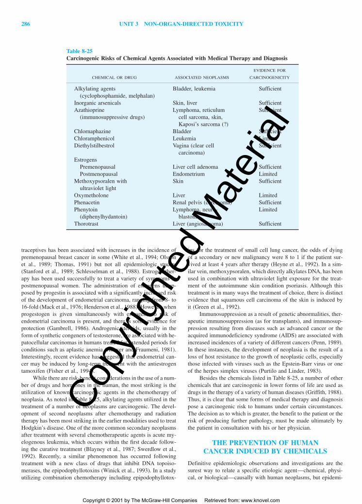
286 UNIT 3 NON-ORGAN-DIRECTED TOXICITY
traceptives has been associated with increases in the incidence ofpremenopausal breast cancer in some (White et al., 1994; Olssonet al., 1989; Thomas, 1991) but not all epidemiologic studies(Stanford et al., 1989; Schlesselman et al., 1988). Estrogen ther-apy has been used successfully to treat a variety of symptoms inpostmenopausal women. The administration of estrogens unop-posed by progestin is associated with a significantly increased riskof the development of endometrial carcinoma, ranging from 8- to16-fold (Mack et al., 1976; Henderson et al., 1988). However, whenprogestogen is given simultaneously with estrogen, no risk ofendometrial carcinoma is present, and there is some evidence forprotection (Gambrell, 1986). Androgenic steroids, usually in theform of synthetic congeners of testosterone, are associated with he-patocellular carcinomas in humans treated for extended periods forconditions such as aplastic anemia (Hoover and Fraumeni, 1981).Interestingly, recent evidence has suggested that endometrial can-cer may be induced by long-term treatment with the antiestrogentamoxifen (Fisher et al., 1994).
While there are risk-benefit considerations in the use of a num-ber of drugs and hormones in the human, the most striking is theutilization of known carcinogenic agents in the chemotherapy ofneoplasia. As noted in Table 8-25, alkylating agents utilized in thetreatment of a number of neoplasms are carcinogenic. The devel-opment of second neoplasms after chemotherapy and radiationtherapy has been most striking in the earlier modalities used to treatHodgkin’s disease. One of the more common secondary neoplasmsafter treatment with several chemotherapeutic agents is acute my-elogenous leukemia, which occurs within the first decade follow-ing the curative treatment (Blayney et al., 1987; Swerdlow et al.,1992). Recently, a similar phenomenon has occurred followingtreatment with a new class of drugs that inhibit DNA topoiso-merases, the epipodophyllotoxins (Winick et al., 1993). In a studyutilizing combination chemotherapy including epipodophyllotox-
ins for the treatment of small cell lung cancer, the odds of dyingof a secondary or new malignancy were 8 to 1 if the patient sur-vived at least 4 years after therapy (Heyne et al., 1992). In a sim-ilar vein, methoxypsoralen, which directly alkylates DNA, has beenused in combination with ultraviolet light exposure for the treat-ment of the autoimmune skin condition psoriasis. Although thistreatment is in many ways the treatment of choice, there is distinctevidence that squamous cell carcinoma of the skin is induced byit (Green et al., 1992).
Immunosuppression as a result of genetic abnormalities, ther-apeutic immunosuppression (as for transplants), and immunosup-pression resulting from diseases such as advanced cancer or theacquired immunodeficiency syndrome (AIDS) are associated withincreased incidences of a variety of different cancers (Penn, 1989).In these instances, the development of neoplasia is the result of aloss of host resistance to the growth of neoplastic cells, especiallythose infected with viruses such as the Epstein-Barr virus or oneof the herpes simplex viruses (Purtilo and Linder, 1983).
Besides the chemicals listed in Table 8-25, a number of otherchemicals that are carcinogenic in lower forms of life are used asdrugs in the therapy of a variety of human diseases (Griffith, 1988).Thus, it is clear that some forms of medical therapy and diagnosispose a carcinogenic risk to humans under certain circumstances.The decision as to which is greater, the benefit to the patient or therisk of producing further pathology, must be made ultimately bythe patient in consultation with his or her physician.
THE PREVENTION OF HUMANCANCER INDUCED BY CHEMICALS
Definitive epidemiologic observations and investigations are thesurest way to relate a specific etiologic agent—chemical, physi-cal, or biological—causally with human neoplasms, but epidemi-
Table 8-25Carcinogenic Risks of Chemical Agents Associated with Medical Therapy and Diagnosis
EVIDENCE FOR
CHEMICAL OR DRUG ASSOCIATED NEOPLASMS CARCINOGENICITY
Alkylating agents Bladder, leukemia Sufficient(cyclophosphamide, melphalan)
Inorganic arsenicals Skin, liver SufficientAzathioprine Lymphoma, reticulum Sufficient
(immunosuppressive drugs) cell sarcoma, skin,Kaposi’s sarcoma (?)
Chlornaphazine Bladder SufficientChloramphenicol Leukemia LimitedDiethylstilbestrol Vagina (clear cell Sufficient
carcinoma)Estrogens
Premenopausal Liver cell adenoma SufficientPostmenopausal Endometrium Limited
Methoxypsoralen with Skin Sufficientultraviolet light
Oxymetholone Liver LimitedPhenacetin Renal pelvis (carcinoma) SufficientPhenytoin Lymphoma, neuro- Limited
(diphenylhydantoin) blastomaThorotrast Liver (angiosarcoma) Sufficient
2996R_ch08_239-319 4/11/01 3:55 PM Page 286
Copy
right
ed M
ater
ial
Copyright © 2001 by The McGraw-Hill Companies Retrieved from: www.knovel.com

CHAPTER 8 CHEMICAL CARCINOGENESIS 287
ologic studies are still relatively insensitive for identifyingcausative factors in human cancer. Such studies can identify onlyfactors that are different between two populations and that are suf-ficiently important to play a determining role under the conditionsof exposure. Furthermore, on the basis of epidemiologic studies, itis extremely difficult to determine whether a specific chemical isor is not carcinogenic to humans because of the extended lag pe-riod between exposure and clinical occurrence of a neoplasm, thehigh background incidence of many cancers in the general popu-lation, the relatively imprecise knowledge of the nature of the ex-posure in most instances, and a number of other confounding vari-ables. Only under exceptional circumstances such as the inductionof rare and infrequent neoplasms—e.g., vinyl chloride and an-giosarcoma (Dannaher et al., 1981)—is it possible to identify anagent as carcinogenic solely on the basis of epidemiologic studieswhen the incidence of cancer induced by that agent is less than 50percent more than the occurrence of the resulting cancer in the gen-eral human population. Therefore, a “negative” result of an epi-demiologic investigation must be considered as inconclusive fordetermining whether a relatively weak carcinogenic agent has arole in the etiology of human neoplasia. How, then, is it possibleto identify actual and potential carcinogenic agents in our envi-ronment by methods other than epidemiologic studies? This ques-tion has been answered in part by relating the results of additionalstudies, usually carried out with experimental animals, to the prob-lem of the etiology of human cancer and the risks of environmen-tal agents to populations and/or specific individuals. It is from suchstudies that government agencies make decisions that ultimatelyregulate the production and use of, and accordingly the exposuresof populations to, agents determined to be actually or potentiallycarcinogenic for the human.
The ultimate goal of such epidemiologic and basic studies isthe prevention of human cancer. There is today sufficient scientificknowledge to allow the prevention of more than 60 percent of hu-man cancers. The failure to achieve such a goal is largely the re-sult of personal and societal decisions well beyond the realm ofscience. However, since the prevention of disease is by far the mosteffective and inexpensive mode of health care, it is appropriate thatthere be a constant and sustained effort to utilize the ever-expandingknowledge of neoplasia to accomplish its control through cancerprevention.
Cancer prevention in humans may in general be grouped intotwo approaches: active and passive. Table 8-26 depicts an outlineof various methods of cancer prevention with an indication of thestage of carcinogenesis toward which the preventive measure is di-rected. The passive prevention of cancer involves the cessation ofsmoking, dietary restrictions, and modification of other personalhabits such as those of a sexual nature. Active prevention of can-cer development is usually accomplished by the administration ofan agent to prevent infection by carcinogenic viruses and other organisms or by the intake of chemicals, nutrients, or other factorsthat may modify or prevent the action of carcinogenic agents. The-oretically, passive cancer prevention or the alteration of one’s “car-cinogenic” habits can be the most effective and unintrusive methodof cancer prevention. However, for many individuals, passive pre-vention requires external persuasion, such as governmental regu-lation or peer pressure, to force an alteration of their habits. Obviously, in many instances such methods are doomed to fail-ure. Active cancer prevention, which many consider a form of pre-ventive “therapy,” is likely to be the most effective method in thisarea.
Individuals with hereditary conditions involving alterations inspecific oncogenes or tumor suppressor genes constitute a rela-tively small part of the population. However, genes that may mod-ify the susceptibility of an individual to the development of cer-tain types of neoplasms probably represent significant factors inthe development of an important fraction of human cancers (Spitzand Bondy, 1993). In reviewing Table 8-26, one can see that mostmethods of cancer prevention are linked to action at the stage ofpromotion. Because this is the reversible stage of neoplastic de-velopment, such a finding is not surprising. However, since we stilldo not know all or even most of the causes of human cancer, thecontinued identification of agents, especially chemicals, that mightinduce human cancer is important. While the results of epidemio-logic studies, when exhibiting sufficient evidence for a causal re-lationship, may be considered the “gold standard,” such detailedstudies, even where feasible, for all the potentially carcinogenicagents existing and entering into our environment would be im-possible. Therefore, during the last half century, as knowledge ofthe mechanisms of carcinogenesis has increased, a significant ef-fort backed by a number of governmental agencies throughout theworld was directed toward the development of methods for theidentification of potentially carcinogenic agents in the environmentby a variety of different systems from bacteria to whole animals.This chapter deals with the identification, characterization, and ul-timate estimation of human risk from chemical, biological, andphysical agents.
IDENTIFICATION OF POTENTIALCARCINOGENIC AGENTS
A major factor in determining the carcinogenic potential of an agentis its identification as being carcinogenic. While this statement ap-pears obvious and even redundant, identification of a carcinogenis necessary but not sufficient for determining carcinogenic poten-
Table 8-26Modes of the Prevention of Cancer
MODE STAGE
PassiveSmoking cessation Pr, PgDietary restriction PrModeration of alcohol intake PrModification of sexual and I, Pr
reproductive habitsAvoidance of excessive I, Pr
ultraviolet exposureActive
Dietary modification and supplements PrVaccination against oncogenic viruses I, PrApplication of ultraviolet blocking agents I, Pr
in appropriate situationsSelective screening for certain I, Pr
preneoplastic lesionsDetermination of genetic background I, Pr
in relation to neoplastic diseaseAdministration of antihormones Pr
KEY: I, initiation; Pr, promotion; Pg, progression.SOURCE: After Pitot (1993), with permission.
2996R_ch08_239-319 4/11/01 3:55 PM Page 287
Copy
right
ed M
ater
ial
Copyright © 2001 by The McGraw-Hill Companies Retrieved from: www.knovel.com
288 UNIT 3 NON-ORGAN-DIRECTED TOXICITY
tial. Still, identification is the starting point, and for this reason ithas received the most attention. Generally speaking, the varioustests that have been applied to identifying agents with carcinogenicpotential may be classified into several general areas. These areseen in Table 8-27. As noted in the table, the time involved in theassay has been arbitrarily separated into short, medium, and long.Short-term assays usually involve days to a few weeks for devel-opment of an endpoint; medium-term assays require weeks to somemonths but much less than a year. Long-term bioassays usually in-volve 11�2 to 2 years of treatment of animals with a test agent. Eachof these general categories consists of specific methods. Each ofthese general categories is considered below in somewhat greaterdetail.
Short-Term Tests—Mutagenesis Assays
A variety of short-term tests, almost all of which are involved indirect or indirect assays of mutagenicity, both in vivo and in vitro,have now been developed and are used to aid in the identificationof potential carcinogens. However, virtually all of these methodsare of limited use in directly establishing the estimation of the riskthat such chemicals pose for the human population. As noted ear-lier, a ubiquitous characteristic of neoplastic cells is the presenceof a variety of different types of mutations. The fact that many butnot all carcinogenic agents are mutagenic or may be metabolizedto mutagenic forms further establishes the importance of mutationsin the development of the neoplastic process. It is on this basis thatshort-term tests for mutagenicity were developed to identify po-tential carcinogenic agents on the basis of their capacity for in-ducing mutations in DNA in cells in vitro or in vivo.
Table 8-28 lists many of the more commonly used short-termtests for mutagenicity and thus carcinogenic potential. The mostwidely utilized of these mutagenicity assays was originally devel-oped in Salmonella typhimurium by Bruce Ames and associates(Ames et al., 1975). In this assay, bacterial cells that are deficientin DNA repair and lack the ability to grow in the absence of his-tidine are treated with several dose levels of the test compound,after which reversion to the histidine-positive phenotype is ascer-tained. Because bacteria differ in their metabolic capabilities com-pared with mammals, a drug-metabolizing system is added to theseassays. Specifically, the 9000 g supernatant (S9) that results fromcentrifuging a liver homogenate prepared from a rat treated withan inducer of multiple P-450s, such as Aroclor 1254, is used incombination with an NADPH regenerating system. The method forperforming the Salmonella assay (the Ames assay) is depicted inFig. 8-25. Several different lines of Salmonella have been gener-
ated to permit the detection of point mutations (TA100, TA1535)and frameshift mutations (TA98, TA1537, TA1538), and the assayis continuously being refined. Typically, five dose levels of the testcompound are used in addition to the solvent control. Activation-dependent and activation-independent positive control mutagenicsubstances are tested concurrently. Certain types of carcinogens arenot detected by these bacterial mutagenicity assays, including hor-monal carcinogens, metals, agents that have a multiple-target-organmode of action, and agents with a nongenotoxic mode of action.This bacterial reverse mutation system, when performed in the pres-ence of a mammalian S9 activation system, is, however, a very sen-sitive screen for the detection of many mutagenic agents.
In addition to the bacterial mutational assay, several in vitromammalian cell mutation assays exist, including the mouse lym-phoma L5178Y (MOLY) assay and the Chinese hamster ovary(CHO) assay. These mammalian mutagenicity assays use either thehypoxanthine-guanine phosphoribosyltransferase (HGPRT) or thethymidine kinase (TK) gene as the endpoint. The basis for theseassays is seen in Fig. 8-26. They are similar to the Ames assay inthat the phenotypic expression of a mutation in a single-copy geneis compared in treated and untreated cells. These assays are fre-quently performed in the presence of an exogenous metabolizingsource such as an epithelial cell layer that has been irradiated. Themammalian mutation test systems are forward mutation assays inwhich the heterozygous state of a gene is used as a tool to detectgenetic damage that might result in the loss of a phenotype, e.g.,growth in the presence of a toxic compound. In CHO cells, the X-linked HPGRT locus is used as the target gene for analysis. Thisenzyme is important in purine salvage and allows the incorpora-tion of toxic purine analogues such as 6-thioguanine and 8-azaguanine into DNA, resulting in inhibition of cell growthand/or cell death. Alternatively, a mutation in this gene that resultsin phenotype loss may permit colony formation in the presence oftoxic analogs. Assays based on the forward mutation of TK aresimilar in that colony formation in the presence of a DNA-dam-aging agent is scored in the presence of a pyrimidine analog. Be-cause these short-term tests are based on the premise that car-cinogens damage DNA, their concordance with the chronicbioassay in vivo (see below) is only between 30 and 80 percent.In addition, the results of tests are coincident with each other andtend to detect the same types of carcinogens without providing thebattery approach that has been suggested. Among the short-termmutagenicity tests that use mutation as the endpoint, the Ames as-say has been the best studied and has been applied to the greatestnumber of compounds.
Gene Mutation Assays in Vivo Until relatively recently, a mea-surement of mutational effects in vivo was rather difficult to per-form. One of the more popular assays utilized in this area was thedominant lethal assay, in which male mice are exposed to a po-tential genotoxic stimulus, mated with untreated female mice, andthe percentage of pregnancies or number of implants is determined(Lockhart et al., 1992). While the method is relatively easy to per-form, relatively few carcinogenic agents have been studied by thismethod. Similarly, the production of sperm abnormalities in miceby the administration of chemical agents in vivo has not found gen-eral use as a short-term mutagenic assay (Wyrobek and Bruce,1975).
In recent years, with a variety of genetic tools available, ge-netically engineered cells and animals have been developed thathave found use in short-term mutagenesis assays. The four exam-
Table 8-27General Methods for Identification of Potential Carcinogens
METHODS TIME FRAME
Short termMutagenesis assays Several weeksTransformation in cell culture 1–3 months
Medium termQualitative and quantitative 2–8 months
analysis of preneoplasiaLong term
Chronic bioassay in animals 18–24 months
2996R_ch08_239-319 4/11/01 3:55 PM Page 288
Copy
right
ed M
ater
ial
Copyright © 2001 by The McGraw-Hill Companies Retrieved from: www.knovel.com

CHAPTER 8 CHEMICAL CARCINOGENESIS 289
ples given in Table 8-28 are those most commonly used for muta-tional analysis in vivo. The first three involve genetically engi-neered animals containing transgenes within which are componentsof the lac operon of Escherichia coli, a set of coordinately regu-lated genes involved in lactose metabolism. A schematic represen-tation of the lac operon is seen in Fig. 8-27. Some details of thefunction of the lac operon are given in the figure legend. Basically,the lacI and lacZ genes are the ones utilized in the mutational as-says. As noted in the figure, mutations in the lacI gene will alterthe regulation of expression of the lacZ gene, which codes for �-galactosidase activity. Thus, the transgene contains either one orthe other of the operons. Mutations in the bacterial transgene aredetermined by the methods seen in Fig. 8-28. In this technique,DNA is extracted from the tissue of interest, and because of thenature of the transgene, construct may be packaged into a bacter-ial virus, lambda, which then infects the bacteria, E. coli, on a lawn
of bacterial growth on a dish as noted in the figure. By selectingappropriate bacterial strains and media, one can isolate mutantphage and analyze the sequence of the lacI or lacZ gene as ap-propriate. Thus, one may obtain both the number of mutations perunit DNA from the mouse or, more importantly, the actual sequencechanges induced by the mutagenic action of the original agent. TherpsL transgene works by a similar mechanism but by a differentmetabolic pathway (Gondo et al., 1996).
Since several of the transgenic animals are commerciallypatented, this assay may entail some expense, but is relatively ver-satile for an in vivo assay for mutagenic identification, and its abil-ity to detect nonmutagenic carcinogens is doubtful. Of interest isthe fact that with at least one carcinogenic agent, ethylnitrosourea,the relative sensitivities of mutations induced in the lacI transgeneand an endogenous gene, hprt, were essentially identical (Skopeket al., 1995). Species differences occur with different carcinogens
Table 8-28Short-Term Tests for Mutagenicity
TEST ENDPOINT REFERENCE
Gene mutation assays in vitroProkaryote mutagenesis in vitro Back or forward mutations Maron & Ames, 1983
(Ames test, etc.) in specific bacterial strainsMouse lymphoma thymidine Mutations in TK Majeska & Matheson, 1990
kinase (TK)Chinese hamster ovary (CHO) and Mutations in HGPRT Li et al., 1987
V79 hypoxanthine guanine phos-phoribosyltransferase (HGPRT)
Gene mutation assays in vivoDominant lethal assay Death of fertilized egg in Bateman, 1973
mammalian implanted species Lockhart et al., 1992Sperm abnormality induction Microscopically abnormal sperm Wyrobek & Bruce, 1975
Mutation induction in transgenes in vivoLacZ� mouse Mutations in LacZ� gene Myhr, 1991LacI mouse Mutations in LacI gene cf. Mirsalis et al., 1994LacI rat Mutations in LacI gene de Boer et al., 1996rpsL mouse Mutations in rpsL gene Gondo et al., 1996
Chromosomal alterations in vivoHeritable translocation test (mice) Translocations induced in Generoso et al., 1980
germ cellsRat bone marrow clastogenesis Chromosomal aberrations in Ito et al., 1994
in vivo bone marrow cells in vivoMicronucleus test Appearance of micronuclei Tinwell and Ashby, 1994
in bone marrow cells in vivo Heddle et al., 1983Chromosomal alterations in vitro
Mitotic recombination, mitotic Conversion of heterozygous Wintersberger & Klein, 1988crossing over, or mitotic gene alleles to homozygous stateconversion in yeast
Induced chromosomal aberrations Visible alterations in Galloway et al., 1985in cell lines karyotype
Sister chromatid exchange Visible exchange of differ- Latt, 1981entially labeled sister chromatids Murphy et al., 1992
Primary DNA damageDNA repair in vivo or in vitro Unscheduled DNA synthesis Furihata & Matsushima, 1987
and/or DNA strand breaksRodent liver: unscheduled DNA Unscheduled DNA synthesis Kennelly, 1995
synthesis induction in rodent liver cells in vivo Steinmetz et al., 1988and/or in vitro
2996R_ch08_239-319 4/11/01 3:55 PM Page 289
Copy
right
ed M
ater
ial
Copyright © 2001 by The McGraw-Hill Companies Retrieved from: www.knovel.com
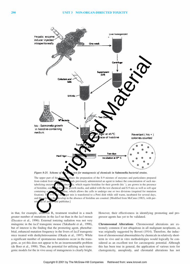
290 UNIT 3 NON-ORGAN-DIRECTED TOXICITY
However, their effectiveness in identifying promoting and pro-gressor agents has yet to be validated.
Chromosomal Alterations Chromosomal alterations are ex-tremely common if not ubiquitous in all malignant neoplasms, aswas originally suggested by Boveri (1914). Therefore, the induc-tion of chromosomal abnormalities by chemicals in relatively short-term in vivo and in vitro methodologies would logically be con-sidered as an excellent test for carcinogenic potential. Althoughthis has been true in general, the application of various tests forclastogenicity, aneuploidy, and chromatid alterations has not
in that, for example, aflatoxin B1 treatment resulted in a muchgreater number of mutations in the lacI rat than in the lacI mouse(Dycaico et al., 1996). External ionizing radiation was not verymutagenic in the lacZ transgenic mouse (Takahashi et al., 1998),but of interest is the finding that the promoting agent, phenobar-bital, enhanced mutation frequency in the livers of lacZ transgenicmice treated with diethylnitrosamine (Okada et al., 1997). Whilea significant number of spontaneous mutations occur in the trans-gene, as yet this does not appear to be an insurmountable problem(de Boer et al., 1998). Thus, the potential for utilizing such trans-genic models for the in vivo assay of mutagenesis is clearly bright.
Figure 8-25. Scheme of the Ames test for mutagenesis of chemicals in Salmonella bacterial strains.
The upper part of the figure outlines the preparation of the S-9 mixture of enzymes and particulates preparedfrom rodent liver taken from animals previously administered an agent to induce the concentration of such me-tabolizing enzymes. The Salmonella, which require histidine for their growth (his�), are grown in the presenceof histidine, separated from the growth media, and added with the test chemical and S-9 mix as well as soft agarcontaining a trace of histidine, which allows the cells to undergo one or two divisions (required for mutationfixation). The 5-9 and soft agar mix is transferred to a Petri dish while still warm, incubated for several days,and the colonies that develop in the absence of histidine are counted. [Modified from McCann (1983), with per-mission of author and publisher.]
2996R_ch08_239-319 4/11/01 4:02 PM Page 290
Copy
right
ed M
ater
ial
Copyright © 2001 by The McGraw-Hill Companies Retrieved from: www.knovel.com

CHAPTER 8 CHEMICAL CARCINOGENESIS 291
Figure 8-26. Outline of chemically induced mutation in mouse cell lines with thymidine kinase (TK) or hy-poxanthine-guanine phosphoribosyltransferase (HGPRT) as the target gene. [Reproduced from Pitot andDragan (1996), with permission of authors and publisher.]
formed the basis for determining potential carcinogenicity of chem-icals. In part, the technology involved is more complicated and ex-pensive than most of the gene mutation assays, and the molecularbasis for at least one of the more common tests, that of sister chro-matid exchange, is not fully understood. Theoretically, short-termassays for the induction of clastogenicity and related abnormali-ties would allow the rapid identification of potential progressoragents.
Analysis of chromosomal alterations in vivo was studied ingerm cells two decades ago by Generoso et al. (1980) in mice. Thisprocedure, as carried out by these workers, involves the adminis-tration of an agent to male mice shortly before breeding and sub-sequent examination of male offspring for sterility and/or chro-mosomal abnormalities in both germ and somatic cells. The test issomewhat complex, and thus far only a few very potent mutagenicagents have been found positive in this test. A more commonlyemployed short-term test for clastogenesis is the micronucleus test,which measures induced clastogenesis in rodent bone marrow invivo by morphologic evaluation of micronuclei containing chro-mosome fragments in cell preparations from bone marrow (Heddleet al., 1983). However, this assay also has an occasional false pos-itive, such as vitamin C (Tinwell and Ashby, 1994). With the LEC
rat, which exhibits a defect in copper metabolism leading to hep-atitis and hepatomas, an increased frequency of chromosome aber-rations was seen in the bone marrow after administration of direct-acting alkylating agents that did not need metabolic activation (Itoet al., 1994). However, carcinogenic agents requiring metabolic ac-tivation, especially in the liver, induced a lesser amount of chro-mosomal abnormalities in the bone marrow of these rats than innormal rats.
Studies in vitro of chromosomal alterations have been carriedout both in yeast and in cultured mammalian cells. In the former,various genetic end-points are studied, the abnormalities seen be-ing the result of chromosomal alterations (Wintersberger and Klein,1988). In mammalian cell lines, most of the systems used the samelines as for the gene mutation assays, e.g., Galloway et al. (1985).Relatively few analyses of induced chromosomal alterations havebeen carried out in normal diploid cells in culture. This test is usedmuch more extensively than most of the other short-term tests in-volving chromosomal alterations (cf. Ishidate et al., 1988). Asmight be expected, some discrepancies have arisen between themutagenic and clastogenic effects of chemicals by these two dif-ferent systems (cf. Ashby, 1988). Furthermore, chromosomal al-terations in these cell lines are sensitive to oxidants (Gille et al.,
2996R_ch08_239-319 4/11/01 4:02 PM Page 291
Copy
right
ed M
ater
ial
Copyright © 2001 by The McGraw-Hill Companies Retrieved from: www.knovel.com

292 UNIT 3 NON-ORGAN-DIRECTED TOXICITY
Figure 8-27. Schematic representation of the lac operon in E. coli.
A. The lacI gene codes for protein that forms a homotetramer that binds tothe lacO operator sequence. Binding of the repressor to lacO prevents tran-scription of lacZ. B. Transcription of lacZ occurs in the presence of the in-ducing agent, isopropyl-�-thiogalactoside (IPTG). Mutations of the lacImay result in partial or complete inactivation of the lac repressor, the lacItetrameric protein. Furthermore, mutations in the lacZ gene may preventinteraction with the repressor or may be nonfunctional, resulting in no pro-duction of the structural gene, lacZ. [Reproduced from Provost et al. (1993),with permission of authors and publisher.]
1993; Shamberger et al., 1973; Kirkland et al., 1989), and prefer-ential targets of chemicals in these aneuploid cell lines are chro-mosomes bearing amplified genes, already indicative of the kary-otypic instability of the cell lines being used (Ottagio et al., 1993).
Another short-term test involving changes in chromosomalstructure by mechanisms not entirely understood is the techniqueof “sister chromatid exchange” (SCE). During metaphase, sisterchromatids, each of which is a complete copy of the chromosome,are bound together by mechanisms that involve specific proteins(Nasmyth, 1999). SCE reflects an interchange between DNA mol-ecules within different chromatids at homologous loci within areplicating chromosome (Latt, 1981). The detection of SCEs re-quires methods of differentially labeling sister chromatids. Theusual technique is to allow a cell to incorporate a label, usually ahalogenated pyrimidine such as bromodeoxyuridine (BrdU), forone replication cycle and then it undergoes a second replicationcycle in which the presence of the labeled precursor is actually op-tional. The procedure has been used in vivo as well as in vitro(DuFrain et al., 1984). In an extensive examination and compari-son of the SCE method with cytogenetic changes, the two meth-ods were about 70 percent congruent, again indicating that clasto-genesis and SCEs are not identical phenomena (Gebhart, 1981).
Primary DNA Damage The measurement of DNA damage andrepair induced by exogenous chemicals, both in vivo and in vitro,has been a relatively common technology used in short-term testsfor potential carcinogenicity. The most generally utilized technol-ogy involves the analysis of non-replicative DNA synthesis withappropriately labeled precursor nucleotides (cf. Harbach et al.,1991). More sophisticated techniques involve the measurement ofDNA strand breakage by eluting DNA fragments from columns to
Figure 8-28. Sequence of steps utilized in the determination of the mutagenicity of chemicals in transgenicrodent mutagenicity assays in vivo.
The details of the test are briefly discussed in the text or the reader is referred to the original article. [From Re-cio (1995), reproduced with permission of author and publisher.]
2996R_ch08_239-319 4/11/01 4:02 PM Page 292
Copy
right
ed M
ater
ial
Copyright © 2001 by The McGraw-Hill Companies Retrieved from: www.knovel.com
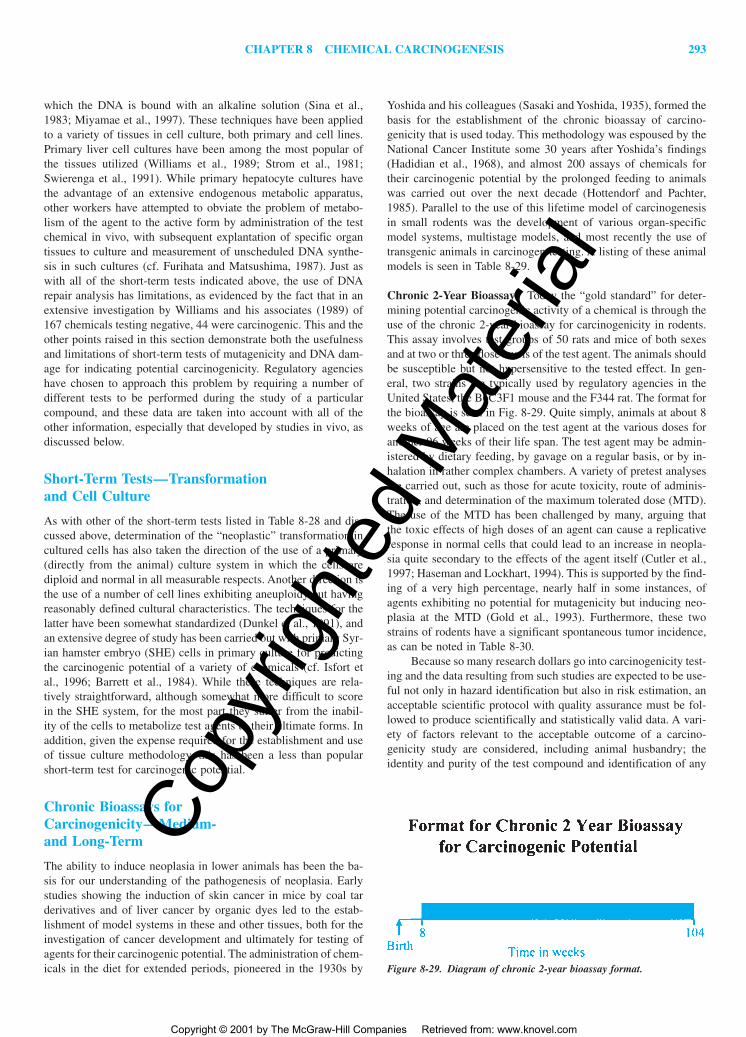
CHAPTER 8 CHEMICAL CARCINOGENESIS 293
which the DNA is bound with an alkaline solution (Sina et al.,1983; Miyamae et al., 1997). These techniques have been appliedto a variety of tissues in cell culture, both primary and cell lines.Primary liver cell cultures have been among the most popular ofthe tissues utilized (Williams et al., 1989; Strom et al., 1981;Swierenga et al., 1991). While primary hepatocyte cultures havethe advantage of an extensive endogenous metabolic apparatus,other workers have attempted to obviate the problem of metabo-lism of the agent to the active form by administration of the testchemical in vivo, with subsequent explantation of specific organtissues to culture and measurement of unscheduled DNA synthe-sis in such cultures (cf. Furihata and Matsushima, 1987). Just aswith all of the short-term tests indicated above, the use of DNArepair analysis has limitations, as evidenced by the fact that in anextensive investigation by Williams and his associates (1989) of167 chemicals testing negative, 44 were carcinogenic. This and theother points raised in this section demonstrate both the usefulnessand limitations of short-term tests of mutagenicity and DNA dam-age for indicating potential carcinogenicity. Regulatory agencieshave chosen to approach this problem by requiring a number ofdifferent tests to be performed during the study of a particularcompound, and these data are taken into account with all of theother information, especially that developed by studies in vivo, asdiscussed below.
Short-Term Tests—Transformation and Cell Culture
As with other of the short-term tests listed in Table 8-28 and dis-cussed above, determination of the “neoplastic” transformation incultured cells has also taken the direction of the use of a primary(directly from the animal) culture system in which the cells arediploid and normal in all measurable respects. Another direction isthe use of a number of cell lines exhibiting aneuploidy but havingreasonably defined cultural characteristics. The techniques for thelatter have been somewhat standardized (Dunkel et al., 1991), andan extensive degree of study has been carried out with primary Syr-ian hamster embryo (SHE) cells in primary culture for predictingthe carcinogenic potential of a variety of chemicals (cf. Isfort etal., 1996; Barrett et al., 1984). While these techniques are rela-tively straightforward, although somewhat more difficult to scorein the SHE system, for the most part they suffer from the inabil-ity of the cells to metabolize test agents to their ultimate forms. Inaddition, given the expense required for the establishment and useof tissue culture methodology, this has been a less than popularshort-term test for carcinogenic potential.
Chronic Bioassays forCarcinogenicity—Medium- and Long-Term
The ability to induce neoplasia in lower animals has been the ba-sis for our understanding of the pathogenesis of neoplasia. Earlystudies showing the induction of skin cancer in mice by coal tarderivatives and of liver cancer by organic dyes led to the estab-lishment of model systems in these and other tissues, both for theinvestigation of cancer development and ultimately for testing ofagents for their carcinogenic potential. The administration of chem-icals in the diet for extended periods, pioneered in the 1930s by
Yoshida and his colleagues (Sasaki and Yoshida, 1935), formed thebasis for the establishment of the chronic bioassay of carcino-genicity that is used today. This methodology was espoused by theNational Cancer Institute some 30 years after Yoshida’s findings(Hadidian et al., 1968), and almost 200 assays of chemicals fortheir carcinogenic potential by the prolonged feeding to animalswas carried out over the next decade (Hottendorf and Pachter,1985). Parallel to the use of this lifetime model of carcinogenesisin small rodents was the development of various organ-specificmodel systems, multistage models, and most recently the use oftransgenic animals in carcinogen testing. A listing of these animalmodels is seen in Table 8-29.
Chronic 2-Year Bioassay Today the “gold standard” for deter-mining potential carcinogenic activity of a chemical is through theuse of the chronic 2-year bioassay for carcinogenicity in rodents.This assay involves test groups of 50 rats and mice of both sexesand at two or three dose levels of the test agent. The animals shouldbe susceptible but not hypersensitive to the tested effect. In gen-eral, two strains are typically used by regulatory agencies in theUnited States, the B6C3F1 mouse and the F344 rat. The format forthe bioassay is seen in Fig. 8-29. Quite simply, animals at about 8weeks of age are placed on the test agent at the various doses foranother 96 weeks of their life span. The test agent may be admin-istered by dietary feeding, by gavage on a regular basis, or by in-halation in rather complex chambers. A variety of pretest analysesare carried out, such as those for acute toxicity, route of adminis-tration, and determination of the maximum tolerated dose (MTD).The use of the MTD has been challenged by many, arguing thatthe toxic effects of high doses of an agent can cause a replicativeresponse in normal cells that could lead to an increase in neopla-sia quite secondary to the effects of the agent itself (Cutler et al.,1997; Haseman and Lockhart, 1994). This is supported by the find-ing of a very high percentage, nearly half in some instances, ofagents exhibiting no potential for mutagenicity but inducing neo-plasia at the MTD (Gold et al., 1993). Furthermore, these twostrains of rodents have a significant spontaneous tumor incidence,as can be noted in Table 8-30.
Because so many research dollars go into carcinogenicity test-ing and the data resulting from such studies are expected to be use-ful not only in hazard identification but also in risk estimation, anacceptable scientific protocol with quality assurance must be fol-lowed to produce scientifically and statistically valid data. A vari-ety of factors relevant to the acceptable outcome of a carcino-genicity study are considered, including animal husbandry; theidentity and purity of the test compound and identification of any
Figure 8-29. Diagram of chronic 2-year bioassay format.
2996R_ch08_239-319 4/13/01 11:20 AM Page 293
Copy
right
ed M
ater
ial
Copyright © 2001 by The McGraw-Hill Companies Retrieved from: www.knovel.com

294 UNIT 3 NON-ORGAN-DIRECTED TOXICITY
in the refinement of the 2-year chronic bioassay is now becomingmore appreciated (Keenan et al., 1996; Allaben et al., 1996).
The statistical analysis of results obtained in chronic bioassayshas also been difficult when the analysis results in relatively fewneoplasms in test animals. As can be seen from Table 8-31, a rel-atively high percentage of animals must bear tumors before a sta-tistically significant result can be obtained in the face of signifi-cant development of spontaneous lesions. Since the latterphenomenon is clearly a problem in these animals (Table 8-30),borderline results become a very difficult problem for regulatoryagencies in determining whether or not a compound actually is car-cinogenic in the assay or not. An exception to this is when a veryunusual histogenetic type of neoplasm not seen spontaneously isfound in the test animals at a significant, even very low level (Chuet al., 1981; Basu et al., 1996). The enumeration of all neoplasmsversus those in specific tissues also can raise difficulties in inter-pretation of the bioassay. Despite these criticisms and problems,the chronic 2-year bioassay continues to be the major basis for reg-ulatory action in this country and in many countries throughout theworld.
Tissue-Specific Bioassays During the performance of long-termbioassays, it became obvious that certain tissues in specific speciesexhibited neoplasms more frequently than others when a test agentwas administered. From these observations, several tissue-specificbioassays were developed with the objective of a reasonablysensitive assay carried out in a shorter time than the usual chronic2-year bioassay. The best known of tissue-specific assays is thatutilizing the mouse liver. In a recent analysis of chronic bioassayscarried out by the National Toxicology Program, Crump et al. re-ported that 108 of 390 studies indicated a positive carcinogenic re-sponse to the test chemical. In 81 of these studies, female mice ex-hibited significant increases in the incidence of hepatic neoplasms.
Table 8-29Animal Models of Neoplastic Development
ENDPOINT REFERENCES
Chronic 2-year bioassay Tumors in all organs Sontag, 1977Tissue specific bioassays
Liver, mouse Hepatomas Carmichael et al., 1997Lung, mouse Pulmonary adenomas Shimkin and Stoner, 1975Brain, rat Gliomas Kroh, 1995Mammary gland, rat/mouse Adenomas and carcinomas Dunnick et al., 1995
Medium-term bioassaysIto model Hepatic adenomas and carcinomas Ito et al., 1989Newborn mouse Neoplasms in liver, lung, Fujii, 1991
lymphoid organsMultistage models of neoplastic development
Bladder, rat Papillomas/carcinomas Hicks, 1980Colon, rat Aberrant crypt polyp Sutherland and Bird, 1994Epidermis, mouse Papillomas DiGiovanni, 1992Liver, rat Altered hepatic foci Pitot et al., 1996
Transgenic miceKnockout of p53 tumor Tumors in heterozygous animals Donehower, 1996
suppressor gene ( p53def) having normal phenotypev-Ha-ras with zetaglobin Induced transgene expression in Spalding et al., 1993
promoter; tandem insertion skin leads to papillomaon chromosome 11 (TG.AC) development
contaminants; the homogeneity, stability, and physical propertiesof the test compound under various storage conditions; and the sol-ubility, stability, and availability of the test compound in the sol-vent. In addition, the formulation should be either that which is tobe administered to humans or that which permits bioavailability inthe test organism. The environment of the rodent is also important,and care should be taken to control for sources of variability in theanimals, their diet, and their housing. While the usual comparisonin animal studies is the concurrent control, for a number of situa-tions historical controls may be more appropriate (Haseman et al.,1997).
The underlying basis for risk extrapolation from animals tohuman is that the animal is a good model for human cancer de-velopment. In fact, 2-year bioassay models have been used to de-tect the compounds listed by IARC (Vainio et al., 1991) as knownhuman carcinogens. Also, most known human chemical carcino-gens have a carcinogenic potential in animals that supports the re-sults of epidemiologic studies (Vainio et al., 1985). Exceptionsinclude ethanol and arsenic. In addition, it has now become evi-dent that some neoplastic responses to chemicals in animals areunique to the rodent and species as well as the sex involved. Theseinclude such responses as thyroid neoplasia (McClain, 1989), theinduction of �2u-globulin (Swenberg et al., 1985) resulting in re-nal neoplasms in male rats, and peroxisome proliferation (Ashbyet al., 1994) associated with the induction of hepatic neoplasia inrats. In addition, a significant problem that has arisen in the con-tinued use of the chronic bioassay is the requirement for ad libi-tum feeding. This results in animals, especially in rats, of extremeweight by the end of the 2 years; many will have died sponta-neously prior to the end of the test. Such complications are nowbeing remedied by the use of dietary restriction in the chronic bioas-say for the 2-year period. This phenomenon reduces spontaneouscancer incidence and extends lifespan in rodents, and its usefulness
2996R_ch08_239-319 4/11/01 4:02 PM Page 294
Copy
right
ed M
ater
ial
Copyright © 2001 by The McGraw-Hill Companies Retrieved from: www.knovel.com

CHAPTER 8 CHEMICAL CARCINOGENESIS 295
As noted in Table 8-30, there is a high incidence of spontaneoushepatoma development in mice, more so in the male. This has ledto controversy in the interpretation of the significance of the de-velopment of mouse hepatomas, especially if they are the only sta-tistically significant increased neoplastic response in the test ani-mals. As a result of this controversy, the interpretation of thesignificance of the induction of mouse hepatic lesions has beencalled into question (cf. Dragan et al., 1998; Moch et al., 1996). Afurther complication of this assay is the fact that in at least onestudy the majority of the chemicals testing positive exhibited noevidence of an ability to induce DNA damage or mutation(Carmichael et al., 1997).
Another tissue-specific bioassay that was developed byShimkin and his associates more than two decades ago (Shimkinand Stoner, 1975) is the development of pulmonary adenomas andcarcinomas, primarily in strain A mice. The assay was shown toeffectively identify a number of relatively potent carcinogenicagents, including a few inorganic carcinogens (Stoner et al., 1976).However, the assay has not been generally accepted as a majorcomponent for the determination of carcinogenicity of chemicals,
but it has found usefulness in the determination of the molecularmechanisms of pulmonary carcinogenesis in this strain of animals(You et al., 1989; Nuzum et al., 1990). In addition, as noted fromTable 8-29, induction of gliomas in the rat brain and of mammaryneoplasms in both the rat and the mouse may exhibit potential fortissue-specific bioassays. There have also been attempts to utilizelower vertebrates in the development of tissue-specific bioassays,such as the rainbow trout embryo (Hendricks et al., 1980).
Medium-Term Bioassays While tissue-specific bioassays weredirected in part at decreasing the time required for the analysis ofcarcinogenic potential in vivo, at least two assays have been specif-ically designated as having reduced the time for the developmentof an endpoint. The one most intensively used today, primarily inJapan, is the model developed by Dr. Nobuyuki Ito and his col-leagues (Ogiso et al., 1990; Shirai, 1997). A diagram of the for-mat for this assay is seen in Fig. 8-30. The entire assay takes only8 weeks, and the endpoint is nodules and focal lesions in the liverof rats that stain for glutathione S-transferase pi (GST-P). The ini-tial “programming” of the liver by administration of a necrogenic
Table 8-31Percentage of Animals with Tumors (Rx) Administered a Test Agent Required to ObtainStatistical Significance when Compared with Control Animals with Tumors (Co)
Numbers of Animals
TUMORS IN CONTROL CONTROL WITH TEST AGENT TUMORS IN RX
0 50 50 10100 50 6500 50 4
10 50 50 26100 50 22500 50 20
20 50 50 38100 50 34500 50 32
30 50 50 50100 50 46500 50 44
SOURCE: Adapted from Sontag (1977), with permission.
PERCENT WITH PERCENT WITH
Table 8-30Spontaneous Tumor Incidence (Combined Benign and Malignant) in Selected Sites of theTwo Species, B6C3F1 Mice and F344 Rats, Used in the NCI/NTP Bioassay
B6C3F1 Mice F344 Rats
SITE MALE FEMALE MALE FEMALE
Liveradenoma 10.3 4.0 3.4 3.0carcinoma 21.3 4.1 0.8 0.2
Pituitary 0.7 8.3 24.7 47.5Adrenal 3.8 1.0 19.4 8.0Thyroid 1.3 2.1 10.7 9.3Hematopoietic 12.7 27.2 30.1 18.9Mammary gland 0 1.9 2.5 26.1Lung 17.1 7.5 2.4 1.2
SOURCE: Adapted from Pitot and Dragan (1996), with permission of publisher.
2996R_ch08_239-319 4/11/01 4:02 PM Page 295
Copy
right
ed M
ater
ial
Copyright © 2001 by The McGraw-Hill Companies Retrieved from: www.knovel.com

296 UNIT 3 NON-ORGAN-DIRECTED TOXICITY
Figure 8-30. The medium-term liver bioassay protocol for identification of potentially carcinogenic agents.
DEN, diethylnitrosamine; GST-P, glutathione S-transferase–�. [Reproduced from Shirai (1997), with permis-sion of author and publisher.]
dose of diethylnitrosamine poses some problems in that this doseby itself is carcinogenic, but only after a year or more. Further-more, this high dose is also clastogenic to rat hepatocytes in vivo(Sargent et al., 1989). However, these authors and their colleagueshave demonstrated a significant degree of correlation between long-and medium-term results, indicating the usefulness of this assay asa potential surrogate for the chronic bioassay (Ogiso et al., 1990).More recently these authors have used a slightly modified proto-col in which five potent carcinogenic agents are administered fora 4-week period, followed by administration of the test chemicalfor a subsequent 24- to 32-week period (Ito et al., 1996). Unlikethe assay depicted in Fig. 8-29, this more complicated proceduremay allow the detection of promoting and progressor agents as wellas complete carcinogens in a variety of different tissues. However,outside of Japan these assay procedures have not been generallyutilized.
The newborn mouse model of chemical carcinogenesis wasinitially described by Shubik and his colleagues (Pietra et al., 1959)and later used extensively in studies of mouse hepatocarcinogene-sis by Vesselinovitch and his colleagues (1978). More recently, Fu-jii (1991) has utilized this procedure in the determination of thecarcinogenic potential of 45 different chemicals with quite rea-sonable results. The endpoint of neoplasms in a variety of differ-ent tissues, including lung, liver, lymphoid and hematopoietic tis-sues, is determined within a 1-year period. The assay is relativelyinexpensive, utilizing small amounts of the test materials. As yet,however, this assay has not found general usefulness in the deter-mination of carcinogenic potential by regulatory agencies.
Multistage Models of Neoplastic Development
As we have previously noted, the original studies on multistagemodels of carcinogenesis were developed with the epidermis of themouse. It was not until some 40 years after those initial experimentsthat there was some attempt at standardization of the multistagemodel of carcinogenesis in mouse skin for the analysis of the car-
cinogenic potential of specific chemicals (Pereira, 1982). The for-mat for such assays was essentially that depicted in Fig. 8-18. Fewrefinements in the procedure were added with the exception of theuse of a genetically susceptible strain of mice, the SENCAR strain,which is now utilized in such tests (Slaga, 1986). This system mayalso be extended to the potential analysis of progressor agents (Hen-nings et al., 1993; Warren et al., 1993).
Considerably later than the initial reports of the mouse skinsystem, Hicks et al. (1975) demonstrated the cocarcinogenic orpromoting action of several agents in the development of bladdercancer in the rat. Subsequently, other promoting agents have beendemonstrated with this or a related assay, some of which appear tobe relatively unique to this tissue for both anatomical and chemicalreasons (Cohen and Lawson, 1995; Ito and Fukushima, 1989). Atabout the same time as the initial report of the multistage bladdermodel of carcinogenesis, Peraino and associates (1977) reported amultistage model of carcinogenesis in the rat liver. This finding hasled to the development of a number of models of multistage car-cinogenesis in the rat liver. Solt and Farber (1976) reported a modelsomewhat analogous to that of Ito and his colleagues, but with anaim directed primarily at studying mechanisms of hepatocarcino-genesis rather than utilizing it as an assay system for potentialcarcinogens. Shortly thereafter, Pitot et al. (1978) developed amodel wherein initiation was performed with a nonnecrogenic doseof the initiating agent, subsequently followed by chronic adminis-tration of a promoting agent. The format of these two assay sys-tems are noted in Fig. 8-31. The endpoint of these systems is thequantitative analysis of altered hepatic foci measured by one ofseveral enzymatic markers, the most sensitive being the expressionof GSTP (Hendrich et al., 1987). Several studies have investigatedthe potential for such analyses in the detection of chemical car-cinogens (Pereira and Stoner, 1985; Williams, 1989; Oesterle andDeml, 1990). A similar format has been used to study the preneo-plastic aberrant crypt foci in the colon of animals administered po-tential carcinogens (Ghia et al., 1996). However, as yet all such as-says utilizing preneoplastic endpoints have not found generalusefulness in the identification of potential carcinogenic agents. It
2996R_ch08_239-319 4/11/01 4:02 PM Page 296
Copy
right
ed M
ater
ial
Copyright © 2001 by The McGraw-Hill Companies Retrieved from: www.knovel.com

CHAPTER 8 CHEMICAL CARCINOGENESIS 297
Figure 8-31. Formats of short-term models of multistage hepatocar-cinogenesis in the rat.
A. The Solt-Farber model, in which animals are administered a necrogenicdose of diethylnitrosamine followed 2 weeks later by the administration of0.02% acetylaminofluorene (shaded bar) with a 70 percent partial hepat-ectomy performed after 1 week of AAF feeding and sacrifice 1 week fol-lowing the surgery. B. The Pitot et al. (1978) model, in which a nonnecro-genic dose (10 mg/kg) of DEN is administered 24 h after a partialhepatectomy (PH) and animals are fed a normal diet for 8 weeks, at whichtime they are placed on a diet containing 0.05 percent phenobarbital for asubsequent 24 weeks and then sacrificed. The endpoint of both models isthe quantitation of altered hepatic foci.
is possible that in the future such assays may be useful in distin-guishing between agents exerting their carcinogenic effect prima-rily at one or another of the stages of carcinogenesis.
Transgenic and Knockout Mice as Models of Carcinogenesis
With the advent of the development of transgenic animals as wellas gene targeting in mice, recent efforts have been directed towardsthe development of animal models with specific genetic alterationsthat make them more susceptible to carcinogenesis by externalagents. As noted from Table 8-29, the most popular of these aremice exhibiting one defective allele of the p53 tumor suppressorgene and a transgenic mouse line (TG·AC) carrying a v-Ha-rasoncogene fused to a zeta globin promoter. p53-deficient mice de-velop a high frequency of a variety of spontaneous neoplasms. Theincidence of such tumors varies but, in general, all of the ho-mozygous p53-defective mice develop neoplasms by 10 months ofage, while the heterozygous mice have a 50 percent incidence by18 months, with over 90 percent incidence by 2 years of age (Done-hower, 1996). However, the heterozygous animals did not show anaccelerated carcinogenesis of the liver, even when hepatocarcino-gens were administered (Dass et al., 1999). In addition to this modelsystem, which mimics the Li-Fraumeni syndrome in humans, alarge number of other gene-targeted mutations have been devel-oped in mice but have not yet been utilized as model systems foridentifying potential carcinogenic agents (Rosenberg, 1997).
The TG·AC transgenic mouse is one of a large number of po-tential transgenic mice and rats that might be considered for thestudy of the development of neoplasia in response to test agents.However, in most cases the expression of the transgene is targetedto a specific tissue, and thus one deals with a tissue-specific de-velopment of neoplasia (cf. Goldsworthy et al., 1994). The TG·ACtransgenic mouse is very effective in the identification of potentialpromoting agents for the skin. Administration of the well-knownskin-promoting agent, TPA, could induce the development of pa-pillomas after only three to ten applications (Spalding et al., 1993).These investigators also studied several other potential promoting
and progressor agents, all of which exhibited a short latency pe-riod and high incidence of papilloma induction. Thus, it is appar-ent that each of these genetic models of carcinogenesis has a roleto play in the identification of potential carcinogenic agents. It willrequire considerable effort to validate each of the models with re-spect to tissue-specific carcinogenesis by complete carcinogens orby promoting and/or progressor agents (cf. Tennant, 1998).
EVALUATION OF CARCINOGENICPOTENTIAL
The multiple in vivo and in vitro tests described thus far presentthe experimentalist or the regulator with an extensive amount ofdata from which to draw conclusions about the carcinogenic po-tential of the test agent. In addition, epidemiologic studies provideperhaps the most definitive means of estimating the carcinogenicpotential to humans from exposure to a specific agent. While suchstudies, if definitively positive, are the best evidence for carcino-genic potential of an agent to humans, the evidence is usually ob-tained after an exposure has occurred in a population. In general,epidemiological studies can only detect differences between pop-ulations when there is approximately a twofold increase above thebackground incidence of neoplasia in the control population. Sincemany more agents than those classified as group 1 by IARC ex-hibit carcinogenic potential, the in vitro and in vivo tests describedearlier have been used as surrogates in attempting to determine car-cinogenic potential and risk to the human population. The resultsof such tests clearly offer qualitative information with respect tothe identification of agents exhibiting some potential hazard withrespect to one or more aspects of the process of carcinogenesis aswe know it today. Major difficulties remain in attempting to ex-trapolate in a scientific and meaningful way information obtainedfrom in vitro and in vivo tests to an estimation of the potential riskof such agents to the human population as inducers of disease, es-pecially neoplasia. As might be expected, a number of problemsare involved in the scientific and practical application of informa-tion developed from short- and long-term tests to the estimation ofhuman risk.
The Problem of Extrapolation
Since bacterial mutagenicity (Fig. 8-25) is the most widely and ex-tensively utilized test for estimating the qualitative carcinogenicpotential of an agent, a number of investigations have been directedtoward determining the relationship of bacterial mutagenesis andcarcinogenesis of the same chemical, usually in rodents. An earlygraphical relationship of such a series of tests is seen in Fig. 8-32.Obviously, considerable more efforts have been carried out sincethe publication of this in 1976 (Sugimura et al., 1976). However,the figure does place in rather definitive terms compounds that ex-hibit either carcinogenic and/or mutagenic activities. The indicateddescription of complete carcinogen, promoting agent, initiatingagent, etc., is an exercise allowing the further classification of suchagents in multistage carcinogenesis. In a far more extensive study,Tennant et al. (1987) related the results of bacterial mutagenicityto carcinogenic potential as determined in the chronic 2-year bioas-say and found that the short-term assay detected only about halfof the carcinogens as mutagens. These studies and a slightly laterone by Ashby (1989) pointed again to the importance of using morethan a single short-term assay in attempting to relate DNA struc-tural alterations to potential carcinogenicity. Although the predic-tion of carcinogenic potential by the bacterial mutagenicity tests
2996R_ch08_239-319 4/11/01 4:02 PM Page 297
Copy
right
ed M
ater
ial
Copyright © 2001 by The McGraw-Hill Companies Retrieved from: www.knovel.com

298 UNIT 3 NON-ORGAN-DIRECTED TOXICITY
Figure 8-32. Graphic representation of mutagens and nonmutagens in relation to their known carcinogenicpotential in animal tests.
The labeling of the quadrants using the classification of Table 8-18 is a further potential extrapolation of thesedata. [After Sugimura et al. (1976), with permission of authors and publishers.]
together with other short-term tests is in the neighborhood of 60to 70 percent, it is somewhat surprising that interspecies extrapo-lation of carcinogenesis in rats and mice is not much greater thanthis. In an analysis by Gold et al. (1989), chronic bioassays for car-cinogenic potential in either mice or rats were only about 70 to 75percent predictive of carcinogenicity in the other species. In a morerecent study (Fung et al., 1993) of 379 chemicals tested by the Na-tional Cancer Institute/National Toxicology Program for carcino-genic potential, only slightly more than 50 percent of the chemi-cals tested exhibited carcinogenicity in at least one organ of onesex of one species. Less than half of these exhibited carcinogenicpotential in both species tested, a situation most likely to be in-dicative of a carcinogenic hazard to humans.
Several other issues are also relevant to cross-species extrap-olation, including differences in metabolism of chemical agents be-tween the species. While metabolic schemes are qualitatively sim-ilar across species, significant quantitative differences, especiallyin metabolic rate, partly owing to elimination kinetics, are the rule.Exposure estimation is frequently based on the daily dose admin-istered or on plasma concentration used as a surrogate for con-centrations in the tissue. Using plasma concentrations for extrap-olation across species assumes that each species responds in thesame manner to any given dose of an agent. In the final analysis,it may be that the best basis for cross-species comparison is serumconcentration expressed as milligrams per kilogram body weight,since this better predicts tissue concentration-response effects af-ter chronic administration (Allen et al., 1988; Monro, 1992). Thus,it is clear that the problem of extrapolation of both short- and long-term tests to carcinogenic potential in the human is much less thanperfect, suggesting an important need for reevaluation and reinter-pretation of the tests currently in use. Needless to say, a programto develop better extrapolative endpoints should be a major priority.
The Dose–Response Problem
Another important component in the analysis of assays for car-cinogenic potential, both in vivo and in vitro, is that of the dose–response to a particular test agent. The effectiveness of the induc-tion of neoplasia by a chemical agent is dependent on the dose ofthat agent administered to the test animal. We have already notedthe importance of dose–response curves for the stages of initiationand promotion. The differences in the shapes of the curves for thesetwo processes are of considerable significance in assessing car-cinogenic potential and risk.
Other factors may also influence a dose–response curve, suchas the toxicity of the agent, the bioavailability of the agent, and themetabolic or pharmacokinetic characteristics of the agent withinthe living organism. A classic dose – response of a completecarcinogen and some of its ramifications are seen in Fig. 8-33(Druckrey et al., 1963). In this figure, curve 1 shows the relation-ship between the daily dose administered and the median total doseof animals developing carcinoma. Thus, the left ordinate indicatesthe sum of all doses administered up to a 50 percent tumor inci-dence, thus relating the total dose to the tumor incidence. In thisway the straight line relationship, if extrapolated, would proceedthrough the origin. Curve 2 relates the daily dose of carcinogen tothe median induction time of the appearance of the first neoplasm.While extrapolation of curve 1 through the origin indicates thatthere is no dose at which some incidence of neoplasms is not ap-parent, it should be noted that if the daily dose is less than 0.1mg/kg, no experimental data points are available. Furthermore, ex-trapolation of curve 2 to this low-dose region indicates that at doseslower than 0.1 mg/kg, the rats used in this experiment and whoselifetime is approximately 1000 days will not live sufficiently longfor carcinomas to appear. In the assay depicted in Fig. 8-33, the
2996R_ch08_298 5/29/01 4:08 PM Page 298
Copy
right
ed M
ater
ial
Copyright © 2001 by The McGraw-Hill Companies Retrieved from: www.knovel.com
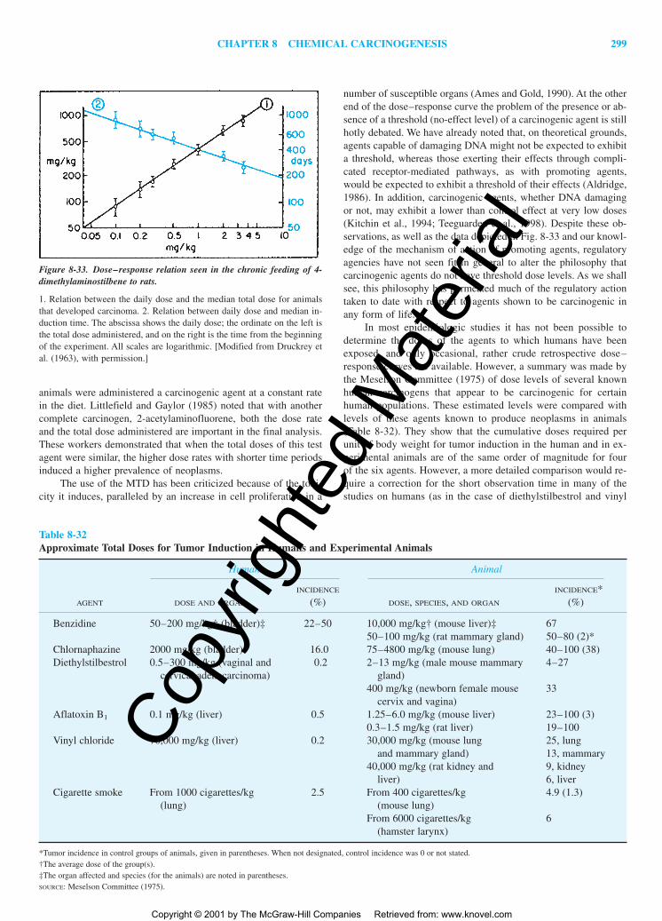
CHAPTER 8 CHEMICAL CARCINOGENESIS 299
animals were administered a carcinogenic agent at a constant ratein the diet. Littlefield and Gaylor (1985) noted that with anothercomplete carcinogen, 2-acetylaminofluorene, both the dose rateand the total dose administered are important in the final analysis.These workers demonstrated that when the total doses of this testagent were similar, the higher dose rates with shorter time periodsinduced a higher prevalence of neoplasms.
The use of the MTD has been criticized because of the toxi-city it induces, paralleled by an increase in cell proliferation in a
number of susceptible organs (Ames and Gold, 1990). At the otherend of the dose–response curve the problem of the presence or ab-sence of a threshold (no-effect level) of a carcinogenic agent is stillhotly debated. We have already noted that, on theoretical grounds,agents capable of damaging DNA might not be expected to exhibita threshold, whereas those exerting their effects through compli-cated receptor-mediated pathways, as with promoting agents,would be expected to exhibit a threshold of their effects (Aldridge,1986). In addition, carcinogenic agents, whether DNA damagingor not, may exhibit a lower than control effect at very low doses(Kitchin et al., 1994; Teeguarden et al., 1998). Despite these ob-servations, as well as the data depicted in Fig. 8-33 and our knowl-edge of the mechanism of action of promoting agents, regulatoryagencies have not seen fit in general to alter the philosophy thatcarcinogenic agents do not have threshold dose levels. As we shallsee, this philosophy has permeated much of the regulatory actiontaken to date with respect to agents shown to be carcinogenic inany form of life.
In most epidemiologic studies it has not been possible todetermine the doses of the agents to which humans have beenexposed, and only occasional, rather crude retrospective dose–response curves are available. However, a summary was made bythe Meselson Committee (1975) of dose levels of several knownhuman carcinogens that appear to be carcinogenic for certainhuman populations. These estimated levels were compared withlevels of these agents known to produce neoplasms in animals(Table 8-32). They show that the cumulative doses required perunit of body weight for tumor induction in the human and in ex-perimental animals are of the same order of magnitude for fourof the six agents. However, a more detailed comparison would re-quire a correction for the short observation time in many of thestudies on humans (as in the case of diethylstilbestrol and vinyl
Figure 8-33. Dose–response relation seen in the chronic feeding of 4-dimethylaminostilbene to rats.
1. Relation between the daily dose and the median total dose for animalsthat developed carcinoma. 2. Relation between daily dose and median in-duction time. The abscissa shows the daily dose; the ordinate on the left isthe total dose administered, and on the right is the time from the beginningof the experiment. All scales are logarithmic. [Modified from Druckrey etal. (1963), with permission.]
Table 8-32Approximate Total Doses for Tumor Induction in Humans and Experimental Animals
Human Animal
INCIDENCE INCIDENCE*AGENT DOSE AND ORGAN (%) DOSE, SPECIES, AND ORGAN (%)
Benzidine 50–200 mg/kg† (bladder)‡ 22–50 10,000 mg/kg† (mouse liver)‡ 6750–100 mg/kg (rat mammary gland) 50–80 (2)*
Chlornaphazine 2000 mg/kg (bladder) 16.0 75–4800 mg/kg (mouse lung) 40–100 (38)Diethylstilbestrol 0.5–300 mg/kg (vaginal and 0.2 2–13 mg/kg (male mouse mammary 4–27
cervical adenocarcinoma) gland)400 mg/kg (newborn female mouse 33
cervix and vagina)Aflatoxin B1 0.1 mg/kg (liver) 0.5 1.25–6.0 mg/kg (mouse liver) 23–100 (3)
0.3–1.5 mg/kg (rat liver) 19–100Vinyl chloride 70,000 mg/kg (liver) 0.2 30,000 mg/kg (mouse lung 25, lung
and mammary gland) 13, mammary40,000 mg/kg (rat kidney and 9, kidney
liver) 6, liverCigarette smoke From 1000 cigarettes/kg 2.5 From 400 cigarettes/kg 4.9 (1.3)
(lung) (mouse lung)From 6000 cigarettes/kg 6
(hamster larynx)
*Tumor incidence in control groups of animals, given in parentheses. When not designated, control incidence was 0 or not stated.†The average dose of the group(s).‡The organ affected and species (for the animals) are noted in parentheses.SOURCE: Meselson Committee (1975).
2996R_ch08_239-319 4/11/01 4:02 PM Page 299
Copy
right
ed M
ater
ial
Copyright © 2001 by The McGraw-Hill Companies Retrieved from: www.knovel.com

300U
NIT
3N
ON
-OR
GA
N-D
IRE
CT
ED
TO
XIC
ITY
Table 8-33Some Methods for the Measurement of the Potency of Carcinogens
RELATIONSHIP DESCRIPTION REFERENCE
AbsoluteCarcinogenic (Iball) index � The percentage number of tumor-bearing animals was Iball, 1939
% of tumors (animals with) calculated from the total number of animals used in the Average latent period in days particular assay or from the total number of animals
surviving at the time the first tumor became manifest
ln2(Hueper, 1963).
K �D1�2
D1�2 is the total animal dose which gives a 50% incidence Meselson and Russell, 1977of cancer after a 2-year exposure. K is defined as potency.
R � �ln (1 � p) � � � �d R � potency where p is the probability of developing Crouch and Wilson, 1979cancer at dose d, and � and � are derived constants.
TD50 � log(2)�b The TD50 (carcinogenic potency) of a chemical is defined Peto et al., 1984as the dose rate (mg/kg body weight/day, b) which, if administered chronically for a standard period, would halve the probability of an animal remaining without any neoplasia (Bernstein, 1985).
RelativeRelative potency � The relative potency of a given agent is defined as the ratio Glass et al., 1991
Dose of a reference compound needed to produce a of the dose of that chemical required to induce carcino-specific effect in a particular bioassay (reference dose) genesis in a particular bioassay, relative to the dose of
Dose of a test compound needed to produce the another (reference) agent required to produce the samesame magnitude of the same effect in the outcome in the same type of bioassay.
same bioassay (test dose)MultistageInitiation Index � #AHF/liver/mmole/kg body weight Index based on administration of a single dose of initiating Pitot et al., 1987
agent in mmole/kg body weight.Promotion Index � Vf�Vc � mmol�1 � weeks�1 Vf is the total volume fraction (%) occupied by AHF in the Pitot et al., 1987
livers of rats treated with the test agent, and Vc is the total volume of AHF in control animals, which have only been initiated. The dose rate of administration of the promoting agent is expressed as millimoles/week.
2996R_ch08_239-319 4/11/01 4:02 PM Page 300
Copy
right
ed M
ater
ial
Copyright © 2001 by The McGraw-Hill Companies Retrieved from: www.knovel.com

CHAPTER 8 CHEMICAL CARCINOGENESIS 301
chloride), since many cancers in humans do not appear for 20 to30 years after exposure. In addition, both vinyl chloride and di-ethylstilbestrol cause a very rare neoplasm in humans that is notusually seen in experimental animals. Thus, the effective doses ofseveral agents known to be carcinogenic for humans and rodentsare not markedly dissimilar in the two species. If this conclusioncan be extended to other chemical carcinogens in the human en-vironment, then both the qualitative and quantitative extrapola-tions of such findings in the animal to the human situation havesome degree of validity.
The Problem of the Potency ofCarcinogenic Agents
It should be apparent to the student by now that not all carcino-genic agents are equally effective in inducing neoplasia, i.e., theyexhibit differing carcinogenic potencies. The potency of an agentto induce neoplasia has been simply defined as the slope of thedose–response curve for induction of neoplasms (Choy, 1996).However, such a definition has generally not been the basis for es-timates of carcinogenic potency based on data from chronic bioas-says with continuous administration of the agent. In Table 8-33may be seen a listing of some methods for the measurement of thepotency of carcinogens, beginning with the early study by Iball(1939) resulting in the Iball Index, which was used for a numberof years thereafter. The relationship of Meselson and Russell (1977)may also be derived from the results of bacterial mutagenesis as-says. The potency relationship developed by Crouch and Wilson(1979) is dependent on a linear, no-threshold extrapolation of theanimal bioassay result, giving the slope as � in the equation seenin the table (Barr, 1985). The TD50 has been extensively used, andvalues were recently compiled by Gold and Zeiger (1997) for alarge number of chemicals. The range of carcinogenic potenciesdeveloped from such a relationship may be seen in Fig. 8-34. Ten-nant and his associates (Dybing et al., 1997) have modified theTD50 potency relationship, using a different fraction of animals thatdevelop neoplasms. The T25 is defined by these workers as thechronic dose rate in mg/kg body weight/day that will give 25 per-cent of the animals neoplasms at a specific tissue site, after cor-rection for spontaneous incidence, within the standard lifetime ofthe test species. As expected, since the relationship is basically thesame as that noted in Table 8-33 except for only half the percent-age, the T25 values are usually roughly one-half those of the TD50
values. Pepelko (1991) has pointed out one of the difficulties ofthese absolute potency measurements in that differences insolubility, bioavailability, and some other pharmacokinetic param-eters do cause considerable variability in some of the potency val-ues reported.
While the four relationships noted in the table under “ab-solute” do analyze carcinogenic potency of a chemical from thedata on the bioassay of that chemical alone, Glass and associ-ates (1991) did propose a relative potency relationship that hassome degree of flexibility and may have some application in riskassessment different from the absolute analyses. Pitot et al.(1987) have attempted to determine indices relating the stagesof initiation and promotion to the potency of the agents induc-ing such stages. In the case of the initiation index, which is rel-atively straightforward, the values obtained are absolute. In thecase of the promoting index, the value is always given in rela-tion to the nontreated control, which does develop focal lesions
from endogenous promotion of spontaneously initiated hepato-cytes. However, these measures of initiating and promoting po-tencies have been applied only to multistage hepatocarcinogen-esis in the rat. However, it is quite feasible to extend suchanalyses to multistage carcinogenesis in a number of other solidorgans where the immediate results of initiation can be quanti-tated and the relative growth of lesions from the initiated cellpopulation can be determined with some degree of accuracy(Pitot et al., 1987).
RELATION (EXTRAPOLATION) OFBIOASSAY DATA TO HUMAN RISK
Campbell (1980) suggested the thesis that the risk (R) of someagent or event can be estimated as a function of the product of the
Figure 8-34. Range of carcinogenic potency as determined by the TD50
potency relationship of Peto et al. (1984).
[Adapted from Gold et al. (1998), with permission of authors and pub-lisher.]
2996R_ch08_239-319 4/11/01 4:03 PM Page 301
Copy
right
ed M
ater
ial
Copyright © 2001 by The McGraw-Hill Companies Retrieved from: www.knovel.com
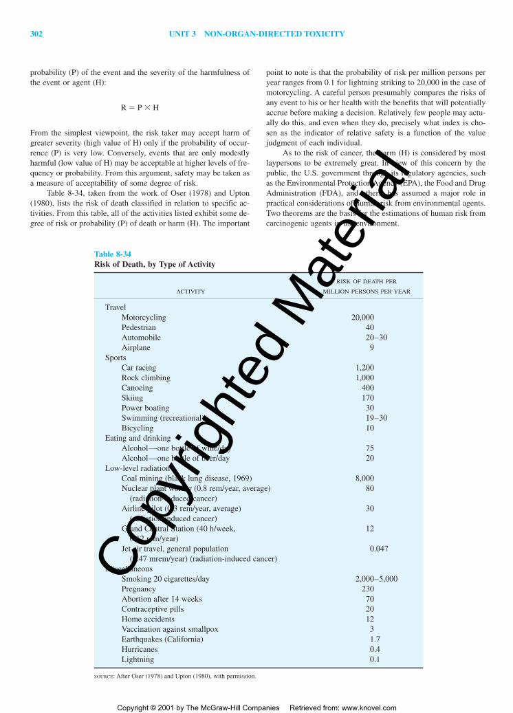
probability (P) of the event and the severity of the harmfulness ofthe event or agent (H):
R � P � H
From the simplest viewpoint, the risk taker may accept harm ofgreater severity (high value of H) only if the probability of occur-rence (P) is very low. Conversely, events that are only modestlyharmful (low value of H) may be acceptable at higher levels of fre-quency or probability. From this argument, safety may be taken asa measure of acceptability of some degree of risk.
Table 8-34, taken from the work of Oser (1978) and Upton(1980), lists the risk of death classified in relation to specific ac-tivities. From this table, all of the activities listed exhibit some de-gree of risk or probability (P) of death or harm (H). The important
point to note is that the probability of risk per million persons peryear ranges from 0.1 for lightning striking to 20,000 in the case ofmotorcycling. A careful person presumably compares the risks ofany event to his or her health with the benefits that will potentiallyaccrue before making a decision. Relatively few people may actu-ally do this, and even when they do, precisely what index is cho-sen as the indicator of relative safety is a function of the valuejudgment of each individual.
As to the risk of cancer, the harm (H) is considered by mostlaypersons to be extremely great. In view of this concern by thepublic, the U.S. government through its regulatory agencies, suchas the Environmental Protection Agency (EPA), the Food and DrugAdministration (FDA), and others, has assumed a major role inpractical considerations of human risk from environmental agents.Two theorems are the basis for the estimations of human risk fromcarcinogenic agents in the environment.
Table 8-34Risk of Death, by Type of Activity
RISK OF DEATH PER
ACTIVITY MILLION PERSONS PER YEAR
TravelMotorcycling 20,000Pedestrian 40Automobile 20–30Airplane 9
SportsCar racing 1,200Rock climbing 1,000Canoeing 400Skiing 170Power boating 30Swimming (recreational) 19–30Bicycling 10
Eating and drinkingAlcohol—one bottle of wine/day 75Alcohol—one bottle of beer/day 20
Low-level radiationCoal mining (black lung disease, 1969) 8,000Nuclear plant worker (0.8 rem/year, average) 80
(radiation-induced cancer)Airline pilot (0.3 rem/year, average) 30
(radiation-induced cancer)Grand Central Station (40 h/week, 12
0.12 rem/year)Jet air travel, general population 0.047
(0.47 mrem/year) (radiation-induced cancer)Miscellaneous
Smoking 20 cigarettes/day 2,000–5,000Pregnancy 230Abortion after 14 weeks 70Contraceptive pills 20Home accidents 12Vaccination against smallpox 3Earthquakes (California) 1.7Hurricanes 0.4Lightning 0.1
SOURCE: After Oser (1978) and Upton (1980), with permission.
302 UNIT 3 NON-ORGAN-DIRECTED TOXICITY
2996R_ch08_239-319 4/13/01 11:20 AM Page 302
Copy
right
ed M
ater
ial
Copyright © 2001 by The McGraw-Hill Companies Retrieved from: www.knovel.com

CHAPTER 8 CHEMICAL CARCINOGENESIS 303
1. A threshold (no-effect) level for a carcinogenic agent cannotbe determined with any degree of accuracy.
2. All carcinogenic agents produce their effects in an irreversiblemanner, so that the actions of small amounts of carcinogenicagents in our environment are additive—producing a “car-cinogenic burden” for the average individual during his or herlifetime.
These bases may be considered as default assumptions that are uti-lized if there is not sufficient evidence to alter these assumptions.Recent guidelines by the EPA have indicated that at least one reg-ulatory agency is beginning to consider and even include in theirfinal disposition of the regulation of a chemical data that may al-ter these default assumptions (Page et al., 1997). Since we have al-ready noted the presence of thresholds of promoting agents as wellas their reversibility, such data may become useful in considera-tion of regulation of chemicals in the future. However, the goldstandard chronic 2-year bioassay that is utilized as the mainstay inregulation of both industrial and pharmaceutical chemicals doesnot distinguish between initiating, promoting, and progressoragents; it will require substantial additional studies to give causeto alter the default assumptions. As noted above, scientific risk es-timation should be carried out with the full knowledge of the ac-tion of the carcinogenic agent as a complete carcinogen, or as hav-ing a major action at one or more of the stages of carcinogenesis.
The extrapolation of bioassay data to human risk estimationis one of the most difficult problems that has faced society and willface us for years to come as numerous new chemicals enter the en-vironment. In attempting to predict the behavior of a chemical inthe human from data obtained from bioassays, a number of factorsshould be considered in extrapolation of bioassay data to humanrisk (Kraybill, 1978). These include:
� Reproducibility of experimental data� Tumor incidence in experimental animals on a dose–depend-
ent basis� Relative approximation of experimental dose to that of human
exposure� Acceptable design and statistical evaluation of bioassay� Consensus on interpretation of histopathologic changes� Availability of biochemical, metabolic, and pharmacokinetic
data to be considered in final decision making
Not included in these factors proposed more than two decades agois a knowledge of the action of the agent as an initiating, promot-ing, or progressor agent. Unfortunately, not all of these factors are
taken into account when regulatory decisions are made at the gov-ernmental level concerning specific compounds in our environ-ment. Newer requirements for more extensive studies of com-pounds that would satisfy these factors are a goal to be achievedbut as yet not attained.
Another consideration in determination of human risk iswhether or not the estimation is qualitative or quantitative. Quali-tative risk estimation is much easier to develop based on qualita-tive analyses of the variety of bioassay procedures utilized. IARCas well as regulatory agencies throughout the world take very se-riously the qualitative finding of induction of neoplasia in one ortwo species of animals as a qualitative indication of risk of car-cinogenicity to the human. However, quantitative risk analysis ismuch more difficult. In fact, a number of epidemiologists have re-fused to make such quantitative relationships on the basis of ani-mal data and would only use data in the human to carry out suchestimates. Still, as we have seen from the utilization of various“safe” doses of carcinogenic agents and a variety of other factors,quantitative risk assessment has been and is being applied to hu-man risk situations of specific chemicals and mixtures. Paramountin such considerations are the use of mathematical models in which,making a variety of assumptions, one may develop quantitative riskestimates for the human. We will consider some of these modelsbelow.
STATISTICAL ESTIMATES OFHUMAN RISK FROM BIOASSAY
DATA BY USING MATHEMATICALMODELS
The statistical analyses of whole-animal bioassay data have em-ployed over the years a number of mathematical models in an at-tempt to relate experimental data to the human situation, especiallyfor the purposes of quantitating human risk inso far as is possible.As Gaylor and Shapiro (1979) have pointed out, “There is no choicebut to extrapolate.” This means, in essence, that because of the in-sensitivity of epidemiologic studies and the number and quantityof actual and potential carcinogens in our environment, one mustmake every attempt possible to relate data from bioassay studiesto the human condition, especially the potential risk to the public.Most of these mathematical models have as a basic tenet the as-sumption that carcinogenic agents lack a threshold, act irreversibly,and have effects that are additive. Equations for some of the morecommonly used models are given in Table 8-35. None of these
Table 8-35Mathematical Models Used in the Extrapolation of the Risk of Carcinogenic Agents to the Human
EQUATION FOR THE PROBABILITY (P) OF TUMOR
INDUCTION AT DOSE d
One-hit (linear) model P(d) � 1 � e(��d)
Multihit (k-hit) model P(d) � 1 � �k�1
i�0(�d)ie��di!
Multistage model P(d) � 1 � exp[�(�1 �1d) . . . (�k �kd)]Extreme value model P(d) � 1 � exp[�exp(� �log10d)]Log-probit model P(d) � � (� �log10d)
2996R_ch08_239-319 4/13/01 11:20 AM Page 303
Copy
right
ed M
ater
ial
Copyright © 2001 by The McGraw-Hill Companies Retrieved from: www.knovel.com

304 UNIT 3 NON-ORGAN-DIRECTED TOXICITY
models can prove or disprove the existence of a threshold of re-sponse, and none can be completely verified on the basis of bio-logical argument; however, the models have been useful in dataevaluation and are presently being used by some federal agenciesin extrapolating experimental data to the human risk situation. Oneof the most commonly used techniques is the log-probit model. Inthe earlier use of this model, the procedure was to regard everyagent as carcinogenic. On this assumption, one must determinesome “safe” dosage level at which the risk calculated would notexceed some very small level such as 1 in 100 million or 10�8.
The linear multistage model, first proposed by Armitage andDoll (1954), incorporates the idea of multiple steps into a statisti-cal approach for risk analysis. This multistage model (Fig. 8-35)incorporates one aspect of the pathogenesis of neoplastic develop-ment, that of multiple stages, but cell cycle–dependent processes,the dynamics of cell kinetics, birth rate, and death rate are not con-sidered. Furthermore, the transition from one stage to the next isconsidered irreversible. Despite these deficiencies, the linearizedmultistage model is one of the most commonly utilized models atthe present time. At a low dose the multistage model is used to fitthe observed tumor incidence data to a polynomial of the dose asnoted in Table 8-35. The linear multistage model is not appropri-ate for estimating low-dose carcinogenic potency for many chem-icals. In most cases, the dose response of high doses of testing dif-fers substantially from the considerably lower doses for exposure.Pharmacokinetic and pharmacodynamic models provide informa-
tion that can help bridge the gap between the high dose and lowdose scenarios (Anderson, 1989). A second problem is associatedwith extrapolation of lifetime exposure of animals to the MTD ofa compound to the less than lifetime exposure common for hu-mans. This problem has been addressed by the EPA through theuse of the Weibull model (Hanes and Wedel, 1985), which assumesthat risk is greater when encountered at a younger age, and, onceexposure occurs, risk continues to accrue despite the cessation ofexposure. However, observations in humans and experimental an-imals have demonstrated that in many cases risk decreases afterexposure ceases, as would be true if the agent were a promotingagent.
More recently, biomathematical modeling of cancer risk as-sessment has been used in an attempt to relate such models moreclosely to the biological characteristics of the pathogenesis of neo-plasia. The best known of these biologically based models is thatdescribed originally by Moolgavkar, Venzon, and Knudson, termedthe MVK model (Moolgavkar, 1986). This model, which is de-picted in Fig. 8-35, reproduces quite well the multistage charac-teristics of neoplastic development with �1, the rate at which nor-mal cells are converted to “intermediate” cells (initiated cells), and�2, the rate at which intermediate cells are converted to neoplas-tic (N) cells. These rates model the rates of initiation and progres-sion in multistage carcinogenesis, while the stage of promotion rep-resents the expansion of the intermediate cell population, which isa function of �2, the rate of division of “intermediate cells,” and�2, the rate of differentiation and/or death of intermediate cells.Other factors in the model that are also true in biology are the rateof replication and cell death of normal or stem cells. While thismodel originally was developed to explain certain epidemiologiccharacteristics of breast cancer incidence and mortality in humans(Moolgavkar, 1986), it has found potential application in a varietyof multistage models including that of rat liver (Luebeck et al.,1991). Application of the model to risk assessment problems hasnot found wide use, but this may change in the next few years (An-derson et al., 1992). In addition, integration of biological data, in-cluding pharmacokinetic and pharmacodynamic parameters,should aid in the development of a more biologically based riskassessment model.
REGULATION OF CARCINOGENICRISK AT THE FEDERAL LEVEL
At least four federal agencies have as their primary responsibilitythe regulation of risk. These agencies include the Consumer Prod-ucts Safety Commission (CPSC), the EPA, the FDA, and the Oc-cupational Safety and Health Administration (OSHA). At least twotypes of regulations affect risk analysis; these include regulationssimilar to the Clean Water Act, which imposes technology-basedstandards that are dictated by the best available technology, andthe Clean Air Act, which imposes health- or risk-based standardsto protect human health by providing an ample margin of safety.A number of laws have been passed that control exposure to car-cinogens in food, drugs, and the environment (Table 8-36). Per-haps the most controversial is the Food, Drug, and Cosmetic Actof 1938, including the 1958 amendment known as the Delaneyamendment. The Delaney amendment was passed to curtail anypossible use of additives in food and drugs that had been demon-strated to induce cancer in humans or animals. This law ignoresthe presence of endogenous or endogenously produced compounds
Figure 8-35. The Armitage and Doll (upper) and MKV (lower) modelsof multistage carcinogenesis.
In the former, the number of stages is unspecified (Tk), and the transitionbetween them is irreversible. In the MKV model, the fates of stem cells(S) and intermediate (I) cells are death (D) or proliferation. Rarely, I cellsundergo (M2 to malignancy (M). The rates of replication (�2) and apopto-sis (�2) for I cells are indicated, and similar rates for S cells are implied.�1 and �2 are the rates of the first genetic event (initiation) and the sec-ond genetic event (progression). [Adapted from Pitot and Dragan (1996),with permission of publishers.]
2996R_ch08_239-319 4/11/01 4:03 PM Page 304
Copy
right
ed M
ater
ial
Copyright © 2001 by The McGraw-Hill Companies Retrieved from: www.knovel.com

CHAPTER 8 CHEMICAL CARCINOGENESIS 305
that have carcinogenic action. For example, nitrites are effectivebacteriocidal agents when used at low levels as food additives, butnitrites are produced extensively in vivo during normal metabolismof nitrogenous compounds, especially when nitrates are present inthe diet (Rogers, 1982). High doses of nitrites given with second-ary amines result in the formation of nitrosamines, which are car-cinogenic in rodents (Rogers, 1982). Thus, a number of difficul-ties are encountered when food and additives are regulated withstrict adherence to the Delaney amendment.
Besides science, a major driving force in legislative actions con-cerning the regulation of carcinogenic or potentially carcinogenicchemicals in the environment is the benefit obtained from such reg-ulation. The saccharin-cyclamate debates were an interesting exam-ple of this (Kraybill, 1976). Saccharin is carcinogenic at a very highdose in rat uroepithelium (Anderson et al., 1988). After considerabledebate, the U.S. Congress passed a law permitting the use of this“carcinogenic” compound as an artificial sweetener because of itslow cost and benefit to a variety of individuals, especially diabetics.Recently, the courts rejected the use of two food colorings in drugsand cosmetics on the basis of an interpretation of the Delaney amend-ment as prohibitive of the use of additives even when only minimalrisk can be demonstrated. The EPA faces a difficult situation in theregulation of pesticides when it attempts to balance the requirementof the Federal Insecticide, Fungicide, and Rodenticide Act (FIFRA),which requires a balance of risk and benefit in the application ofpesticides to raw agricultural products, and the zero tolerance forcarcinogens in processed foodstuffs mandated by the Delaneyamendment. OSHA is responsible for regulating workers’ exposureto potential toxins, including carcinogens. The statutes require thatfeasibility be considered in concert with lack of effect on workers’health. In the case of Industrial Union Department v American Pe-troleum Institute, the Supreme Court found that the allowable levelsof a compound (i.e., benzene) could be established only if a signif-icant risk from exposure could be demonstrated and that this riskcould be lessened by a change in practice. In the final analysis, a
significant proportion of risk to the average citizen is based on theperception of risk.
International Aspects of EnvironmentalRegulation
Other countries have both preceded and followed legal actions inthe United States in regulating noxious and carcinogenic agentsthat can and do occur in the human environment. The United King-dom passed a Clean Air Act some three decades ago, well beforesuch legislation appeared in the United States (cf. Hall, 1976). Thissame nation passed legislation regulating pollution in natural wa-ters within the country at about the same time as similar legisla-tion was enacted in the United States. The European Common Mar-ket has also advanced several programs in the area of environmentalpollution, especially as related to air and water environments. Moregenerally, they have established an environmental program thatconcerns itself with the impact of factors involving alterations inthe environment, waste disposal, and educational programs (cf.Johnson, 1976). Other countries throughout the world have recog-nized the importance of controlling potentially damaging agentsand have acted accordingly.
RISK-BENEFIT CONSIDERATIONSIN THE REGULATION OF ACTUALAND POTENTIAL CARCINOGENIC
ENVIRONMENTAL HAZARDS
We have briefly reviewed the methods for determining the actualand potential carcinogenic agents in our environment, methods forthe estimation of risk to the human population of such agents, andthe governmental approach to the regulation of such agents in ourenvironment. An equally important consideration includes some-what undefined concepts such as benefit-risk analysis, cost-
Table 8-36Selected Federal Laws for Regulation of Toxic and Carcinogenic Agents
NAME OF ACT AND YEAR PASSED AND AMENDED AREA OF CONCERN
Food Drug and Cosmetic Act (FDC): 1906, Food, drugs, cosmetics, food1938, amended 1958 (Delaney), 1960, 1962, additives, color additives, new1968, 1976, 1980, 1984, 1986, 1987, drugs, animal feed additives,1990, 1992 medical devices.
Federal Insecticide, Fungicide, and PesticidesRodenticide Act (FIFRA): 1948, amended1972, 1975, 1976
Clean Air Act: 1970, amended 1974, 1977, Air pollutants1978, 1980, 1981, 1982, 1983, 1990
Clean Water Act: 1972, amended 1977–1983, Water pollutants1987, 1988, 1990, 1992; originally the FederalWater Control Act
Occupational Safety and Health Act (OSHA): Workplace exposure to1970, amended 1974, 1978, 1979, 1982, 1990, toxicants1992
Toxic Substances Control Act (TOSCA): 1976, Hazardous chemicals notamended 1981, 1983, 1984, 1986, 1988, 1990, covered elsewhere, including1992 premarket review
SOURCE: Adapted from Office of Science and Technology Policy (1986).
2996R_ch08_239-319 4/11/01 4:03 PM Page 305
Copy
right
ed M
ater
ial
Copyright © 2001 by The McGraw-Hill Companies Retrieved from: www.knovel.com

306 UNIT 3 NON-ORGAN-DIRECTED TOXICITY
effectiveness, and risk-cost analysis in the regulation of hazardousagents in our environment. These concepts are concerned with suchtraditional regulatory terms as “safe,” “lowest feasible,” and “bestpracticable technology.”
Some of the regulatory legislation leaves no latitude for con-siderations of benefit versus risk. This is the case with the Delaneyamendment, a simplistic legal statement that can create major prob-lems when the regulatory agencies are faced with obeying the law.Problems also arose with respect to nitrite, since the benefits of re-moving nitrites as preservatives in packaged meats were balancedagainst the risk of bacterial contamination, especially by Clostrid-ium botulinum, in nonpreserved packaged products. Federal regu-latory agencies decided that these data were insufficient to ban ni-trites under the Delaney clause, and thus nitrite continues to bewidely used as a preservative, although at lower levels.
Attempts have been made to quantitate and characterize risksversus benefits. One way is to consider risks to the environmentand to health as opposed to risks to society and to general aspectsof health. It is evident that reduction in risk from direct exposureto an environmental factor will, at some level of additional cost ofcontrol, create new risks to society in terms of increased costs ofproducts, availability of services, personal freedoms, employment,and so on. This relation is shown in Fig. 8-36. In controlling risksto the environment and to health there is a point beyond which the
benefits to society and the individual begin to decrease because ofthe cost, both financial and otherwise, incurred in reducing risk to-ward actual zero. As was implied previously, there are very rareinstances in which actual zero risk is obtained in any circumstance.The points for best practicable technology (BPT) and for best avail-able technology (BAT) are seen on the risk curve. Clearly BPT inrisk reduction is less costly than BAT.
More extensive risk-benefit analyses have been published, suchas those of Moll and Tihansky (1977), in which dollar values havebeen estimated for each life that could potentially be saved by elim-inating a specific agent from the environment. They also point outthat the risks of specific agents in industrial situations may be fargreater than those to society as a whole. An example is asbestos,which, though clearly hazardous to some industrial workers, causeslittle or no hazard at the levels of exposure of the population in gen-eral. In this respect, Samuels (1979) has pointed to the potential fal-lacy in many benefit-risk determinations unless one takes into con-sideration the concept of necessary risk, especially as related tooccupational and industrial hazards. This concept stresses the im-portance of making every effort to eliminate hazardous agents inour environment that are important to society by replacing themwith equally useful but less hazardous or nonhazardous components.If this cannot be done and a necessary risk is present, this consid-eration must be balanced against the benefits.
Figure 8-36. Risk-cost-benefit relation emphasizing the impact of cost control on all risks and demonstrat-ing the loss of benefit beyond a certain cost of risk reduction.
BPT, best practicable technology; BAT, best available technology. [Modified from Blair and Hoerger (1979),with permission.]
REFERENCES
Abell CW, Heidelberger C: Interaction of carcinogenic hydrocarbons withtissues: VIII. Binding of tritium-labeled hydrocarbons to the solubleproteins of mouse skin. Cancer Res 22:931–946, 1962.
Aldridge WN: The biological basis and measurement of thresholds. AnnuRev Pharmacol Toxicol 26:39–58, 1986.
Allaben WT, Turturro A, Leakey JEA, et al: FDA points-to-consider documents: The need for dietary control for the reduction of ex-perimental variability within animal assays and the use of dietary restriction to achieve dietary control. Toxicol Pathol 24:776–781,1996.
2996R_ch08_239-319 4/13/01 11:20 AM Page 306
Copy
right
ed M
ater
ial
Copyright © 2001 by The McGraw-Hill Companies Retrieved from: www.knovel.com

CHAPTER 8 CHEMICAL CARCINOGENESIS 307
Allen B, Crump K, Shipp A: Correlation between carcinogenic potency ofchemicals in animals and humans. Risk Anal 8:531–561, 1988.
Ames BN, Gold LS: Chemical carcinogenesis: Too many rodent carcino-gens. Proc Natl Acad Sci USA 87:7772–7776, 1990.
Ames BN, Gold LS: Misconceptions on pollution and the causes of can-cer. Angew Chem Int Ed Engl 29:1197–1208, 1990.
Ames BN, McCann J, Yamasaki E: Methods for detecting carcinogens andmutagens with the Salmonella/mammalian-microsome mutagenicitytest. Mutat Res 31:347–364, 1975.
Ames BN, Shigenaga MK, Gold LS: DNA lesions, inducible DNA repair,and cell division: Three key factors in mutagenesis and carcinogene-sis. Environ Health Perspect 93:35–44, 1993.
Anderson ME: Tissue dosimetry, physiologically based pharmacokineticmodeling, and cancer risk assessment. Cell Biol Toxicol, 5:405–415,1989.
Anderson ME, Krishnan K, Conolly RB, McClellan RO: Mechanistictoxicology research and biologically based modeling: partners forimproving quantitative risk assessments. Chem Indust Inst Toxicol12:1–7, 1992.
Anderson MW, Reynolds SH, You M, Maronpot RM: Role of proto-oncogene activation in carcinogenesis. Environ Health Perspect98:13–24, 1992.
Anderson R, Lefever F, Maurer J: Comparison of the responses of malerats to dietary sodium saccharin exposure initiated during nursing withresponses to exposure initiated to weaning. Food Che Toxicol 26:899–907, 1988.
Andrews EJ: Evidence of the nonimmune regression of chemically inducedpapillomas in mouse skin. J Natl Cancer Inst 47:653–665, 1971.
Anttila A, Sallmén M, Hemminki K: Carcinogenic chemicals in the occu-pational environment. Pharmacol Toxicol 72:69–76, 1993.
Armitage P, Doll R: The age distribution of cancer and a multi-stage the-ory of carcinogenesis. Br J Cancer 8:1–12, 1954.
Aronheim A, Engelberg D, Li N, et al: Membrane targeting of the nu-cleotide exchange factor Sos is sufficient for activating the Ras sig-naling pathway. Cell 78:949–961, 1994.
Ashby J: An opinion on the significance of the 19 non-clastogenic gene-mutagens reported by Tennant et al (1987). Mutagenesis 3:463–465,1988.
Ashby J: Origins of current uncertainties in carcinogen/mutagen screening.Environ Mol Mutagen 16:51–59, 1989.
Ashby J, Brady A, Elcombe CR, et al: Mechanistically-based human haz-ard assessment of peroxisome proliferator-induced hepatocarcinogen-esis Human Exp Toxicol 13:S1–S117, 1994.
Ashby J, Paton D: The influence of chemical structure on the extent andsites of carcinogenesis for 522 rodent carcinogens and 55 different hu-man carcinogen exposures. Mutat Res 286:3–74, 1993.
Ashby J, Tennant RW, Zeiger E, Stasiewicz S: Classification according tochemical structure, mutagenicity to Salmonella and level of carcino-genicity of a further 42 chemicals tested for carcinogenicity by theU.S. National Toxicology Program. Mutat Res 223:73–103, 1989.
Ashendel CL: The phorbol ester receptor: A phospholipid-regulated pro-tein kinase. Biochim Biophys Acta 822:219–242, 1985.
Bailleul B, Brown K, Ramsden M, et al: Chemical induction of oncogenemutations and growth factor activity in mouse skin carcinogenesis.Environ Health Perspect, 81:23–27, 1989.
Barlow DP: Methylation and imprinting: From host defense to gene regu-lation? Science 260:309–310, 1993.
Barnard JA, Polk WH, Moses HL, Coffey RJ: Production of transforminggrowth factor-� by normal rat small intestine. Am J Physiol,261:C994–C1000, 1991.
Barr JT: The calculation and use of carcinogenic potency: A review. RegulToxicol Pharmacol 5:432–459, 1985.
Barrett JC, Hesterberg TW, Thomassen DG: Use of cell transformation sys-tems for carcinogenicity testing and mechanistic studies of carcino-genesis. Pharmacol Rev 36:53S–70S, 1984.
Barrows GH, Mays ET, Christopherson WM: Steroid related neoplasia inhuman liver, in Miller RW et al (eds): Unusual Occurrences as Clues
to Cancer Etiology. Tokyo: Japan Science Society Press/Taylor &Francis, 1988, pp 47–59.
Bartsch H, Hietanen E, Malaveille C.: Carcinogenic nitrosamines: Free rad-ical aspects of their action. Free Radic Biol Med 7:637–644, 1989.
Bartsch H, Ohshima H: Endogenous N-nitroso compounds: How relevantare they to human cancer? in Rhoads JE, Fortner J (eds): General Mo-tors Cancer Research Foundation. Philadelphia: Lippincott, 1989, pp304–317.
Bartsch H, Ohshima H, Shuker DEG, et al: Exposure of humans to en-dogenous N-nitroso compounds: Implications in cancer etiology. Mu-tat Res 238:255–267, 1990.
Basu AP, Gaylor DW, Chen JJ: Estimating the probability of occurrence oftumor for a rare cancer with zero occurrence in a sample Regul Tox-icol Pharmacol 23:139–144, 1996.
Bateman AJ: The dominant lethal assay in the mouse, in Agents and Ac-tions. Vol 3, No 2. Basel: Birkhäuser Verlag, 1973, pp 73–76.
Bauer-Hofmann R, Klimek F, Buchmann A, et al: Role of mutations atcodon 61 of the c-Ha-ras gene during diethylnitrosamine-induced he-patocarcinogenesis in C3H/He mice. Mol Carcinog 6:60–67, 1992.
Beach AC, Gupta RC: Human biomonitoring and the 32P-postlabeling as-say. Carcinogenesis 13:1053–1074, 1992.
Beatson GT: On the treatment of inoperable cases of carcinoma of themamma: Suggestions for a new method of treatment, with illustrativecases. Lancet 2:104–107, 1896.
Becker RA, Shank RC: Kinetics of formation and persistence of ethylgua-nines in DNA of rats and hamsters treated with diethylnitrosamine.Cancer Res 45:2076–2084, 1985.
Berenblum I, Shubik P: A new quantitative approach to the study of stagesof chemical carcinogenesis in the mouse’s skin. Br J Cancer 1:383,1947.
Berger MR: Synergism and antagonism between chemical carcinogens inArcos JC, Argus MF, Woo YT (eds): Chemical Induction of Cancer.Basel: Birkhäuser, 1995, pp 23–49.
Berger MR, Schmähl D, Zerban H: Combination experiments with verylow doses of three genotoxic N-nitrosamines with similar organotropiccarcinogenicity in rats. Carcinogenesis 8:1635–1643, 1987.
Bernard C: Leçons sur les phénomènes de la vie. Paris: Baillière, 1878,1879.
Bessho T, Roy R, Yamamoto K, et al: Repair of 8-hydroxyguanine in DNAby mammalian N-methylpurine-DNA glycosylase. Proc Natl Acad Sci,USA 90:8901–8904, 1993.
Bhide SV, Shivapurkar NM, Gothoskar SV, Ranadive KJ: Carcinogenicityof betal quid ingredients: Feeding mice with aqueous extract and thepolyphenol fraction of betel nut. Br J Cancer 40:922–926, 1979.
Biaglow JE: The effects of ionizing radiation on mammalian cells. J ChemEduc, 58:144–156, 1981.
Bichara M, Fuchs RPP: DNA binding and mutation spectra of the car-cinogen N-2-aminofluorene in Escherichia coli: A correlation betweenthe conformation of the premutagenic lesion and the mutation speci-ficity. J Mol Biol 183:341–351, 1985.
Bishop JM: Viral oncogenes. Cell 42:23–38, 1985.Biskind MS, Biskind GR: Development of tumors in the rat ovary after
transplantation into the spleen. Proc Soc Exp Biol Med 55:176–179,1944.
Bittner JJ: Mammary cancer in C3H mice of different sublines and theirhybrids. J Natl Cancer Inst 16:1263–1286, 1956.
Blackburn EH: Telomeres: No end in sight. Cell 77:621–623, 1994.Blair EH, Hoerger FD: Risk/benefit analysis as viewed by the chemical in-
dustry. Ann NY Acad Sci 329:253–262, 1979.Blayney DW, Longo DL, Young RC, et al: Decreasing risk of leukemia
with prolonged follow-up after chemotherapy and radiotherapy forHodgkin’s disease. N Engl J Med 316:710–714, 1987.
Blot WJ: Alcohol and cancer. Cancer Res 52:2119s–2123s, 1992.Blot WJ, Li JY, Taylor PR, et al: Nutrition intervention trials in Linxian,
China: Supplementation with specific vitamin/mineral combinations,cancer incidence, and disease-specific mortality in the general popu-lation. J Natl Cancer Inst 85:1483–1492, 1993.
2996R_ch08_239-319 4/11/01 4:03 PM Page 307
Copy
right
ed M
ater
ial
Copyright © 2001 by The McGraw-Hill Companies Retrieved from: www.knovel.com
308 UNIT 3 NON-ORGAN-DIRECTED TOXICITY
Bogen KT: Cancer potencies of heterocyclic amines found in cooked foods.Food Chem Toxicol 32:505–515, 1994.
Böhm SK, Grady EF, Bunnett NW: Regulatory mechanisms that modulatesignalling by G-protein-coupled receptors. Biochem J 322:1–18, 1997.
Bohr VA, Phillips DH, Hanawalt PC: Heterogeneous DNA damage and re-pair in the mammalian genome. Cancer Res 47:6426–6436, 1987.
Bolt HM: Roles of etheno-DNA adducts in tumorigenicity of olefins. CRCCrit Rev Toxicol 18:299–309, 1988.
Bolt HM, Peter H, Föst U: Analysis of macromolecular ethylene oxideadducts. Int Arch Occup Environ Health 60:141–144, 1988.
Bond GG, Rossbacher R: A review of potential human carcinogenicity ofthe chlorophenoxy herbicides MCPA, MCPP, and 2,4-DP. Br J IndustMed 50:340–348, 1993.
Bosland MC, Dreef-Van Der Meulen HC, Sukumar S, et al: Multistageprostate carcinogenesis: The role of hormones. Int Symp PrincessTakamatsu Cancer Res Fund 22:109–123, 1991.
Boutwell RK: Caloric intake, dietary fat level, and experimental carcino-genesis, in Jacobs MN (ed): Exercise, Calories, Fat, and Cancer. NewYork: Plenum Press, 1992.
Boutwell RK: Function and mechanism of promoters of carcinogenesis.CRC Crit Rev Toxicol, 2:419–443, 1974.
Boutwell RK: Some biological aspects of skin carcinogenesis. Progr ExpTumor Res 4:207–250, 1964.
Boveri T: Zur Frage der Enstebung maligner Tumorgen. Jena: Fischer, 1914.Boyd JA, Barrett JC: Genetic and cellular basis of multistep carcinogene-
sis. Pharmacol Ther 46:469–486, 1990.Boyland E: The biological significance of metabolism of polycyclic com-
pounds. Biochem Soc Symp 5:40–54, 1950.Brambilla G, Carlo P, Finollo R, et al: Viscometric detection of liver DNA
fragmentation in rats treated with minimal doses of chemical car-cinogens. Cancer Re 43:202–209, 1983.
Brand KG, Buoen LC, Johnson KH, Brand I: Etiological factors, stages,and the role of the foreign body in foreign body tumorigenesis: A re-view. Cancer Res 35:279–286, 1975.
Bronner CE, Baker SM, Morrison PT, et al: Mutation in the DNA mis-match repair gene homologue hMLH 1 is associated with hereditarynon-polyposis colon cancer. Nature 368:258–261, 1994.
Bryant MS, Skipper PL, Tannenbaum SR, MacLure M: Hemoglobinadducts of 4-aminobiphenyl in smokers and nonsmokers. Cancer Res47:602–608, 1987.
Burns FJ, Vanderlaan M, Snyder E, Albert RE: Induction and progressionkinetics of mouse skin papillomas, in Slaga TJ, Sivak A, Boutwell RK(eds): Carcinogenesis. Vol 2. Mechanism of Tumor Promotion and Co-carcinogenesis. New York: Raven Press, 1978, pp 91–96.
Butlin HJ: Three lectures on cancer of the scrotum in chimney-sweeps andothers: I. Secondary cancer without primary cancer: II. Why foreignsweeps do not suffer from scrotal cancer. III. Tar and paraffin cancer.Br Med J 1:1341–1346, 1892; 2:1–6, 66–71, 1892.
Butterworth BE: Chemically induced cell proliferation as a predictive as-say for potential carcinogenicity, in Chemically Induced Cell Prolif-eration: Implications for Risk Assessment. New York: Wiley-Liss,1991, pp 457–467.
Byers T, Perry G: Dietary carotenes, vitamin C, and vitamin E as protec-tive antioxidants in human cancers. Annu Rev Nutr 12:139–159, 1992.
Campbell TC: Chemical carcinogens and human risk assessment. Fed Proc39:2467–2484, 1980.
Candrian U, You M, Goodrow T, et al: Activation of protooncogenes inspontaneously occurring non-liver tumors from C57BL/6 � C3H F1
mice. Cancer Res 51:1148–1153, 1991.Carmichael NG, Enzmann H, Pate I, Waechter F: The significance of mouse
liver tumor formation for carcinogenic risk assessment: Results andconclusions from a survey of ten years of testing by the agrochemi-cal industry. Environ Health Perspect 105:1196–1203, 1997.
Carter JH, Carter HW, Meade J: Adrenal regulation of mammary tumori-genesis in female Sprague-Dawley rats: Incidence, latency, and yieldof mammary tumors. Cancer Res 48:3801–3807, 1988.
Cassee FR, Groten JP, van Bladeren PJ, Feron VJ: Toxicological evalua-tion and risk assessment of chemical mixtures. Crit Rev Toxicol 28:73–101, 1998.
Cattley RC, Popp JA: Differences between the promoting activities of theperoxisome proliferator WY-14,643 and phenobarbital in rat liver.Cancer Res 49:3246–3251, 1989.
Cavalieri EL, Rogan EG: The approach to understanding aromatic hydro-carbon carcinogenesis: The central role of radical cations in metabolicactivation. Pharmacol Ther 55:183–199, 1992.
Cerny WL, Mangold KA, Scarpelli DG: K-ras mutation is an early eventin pancreatic duct carcinogenesis in the Syrian golden hamster. Can-cer Res 52:4507–4513, 1992.
Cerutti PA, Trump BF: Inflammation and oxidative stress in carcinogene-sis. Cancer Cells 3:1–6, 1991.
Cha RS, Thilly WG, Zarbl H: N-Nitroso-N-methylurea-induced rat mam-mary tumors arise from cells with preexisting oncogenic Hras1 genemutations. Proc Natl Acad Sci USA, 91:3749–3753, 1994.
Chandra RS, Kapur SP, Kelleher J, et al: Benign hepatocellular tumors inthe young. Arch Pathol Lab Med 108:168–171, 1984.
Cheng KC, Loeb LA: Genomic instability and tumor progression: Mecha-nistic considerations. Adv Cancer Res 60:121–156, 1993.
Choy WN: Principles of genetic toxicology, in Fan AM, Chang LW (eds):Toxicology and Risk Assessment: Principles, Methods, and Applica-tions. New York: Marcel Dekker, 1996, pp 25–36.
Chu KC, Cueto C Jr, Ward JM: Factors in the evaluation of 200 NationalCancer Institute carcinogen bioassays. J Toxicol Environ Health8:251–280, 1981.
Chung FL, Chen HJC, Nath RG: Lipid peroxidation as a potential en-dogenous source for the formation of exocyclic DNA adducts. Car-cinogenesis 17:2105–2111, 1996.
Clemens MR: Free radicals in chemical carcinogenesis. Klin Wochenschr69:1123–1134, 1991.
Clifton KH, Sridharan BN: Endocrine factors and tumor growth, in BeckerFF (ed): Cancer—A Comprehensive Treatise, Vol 3. New York: PlenumPress, 1975, pp 249–285.
Cohen LA, Kendall ME, Zang E, et al: Modulation of N-nitrosomethylurea-induced mammary tumor promotion by dietary fiber and fat. J NatlCancer Inst 83:496–501, 1991.
Cohen SM, Ellwein LB: Genetic errors, cell proliferation, and carcinogen-esis. Cancer Res 51:6493–6505, 1991.
Cohen SM, Lawson TA: Rodent bladder tumors do not always predict forhumans. Cancer Lett 93:9–16, 1995.
Columbano A, Rajalakshmi S, Sarma DSR: Requirement of cell prolifera-tion for the initiation of liver carcinogenesis as assayed by three dif-ferent procedures. Cancer Res 41:2079–2083, 1981.
Conaway CC, Nie G, Hussain NS, Fiala ES: Comparison of oxidative dam-age to rat liver DNA and RNA by primary nitroalkanes, secondary ni-troalkanes, cyclopentanone oxime, and related compounds. CancerRes 51:3143–3147, 1991.
Conney AH: Induction of microsomal enzymes by foreign chemicals andcarcinogenesis by polycyclic aromatic hydrocarbons: GHA ClowesMemorial Lecture. Cancer Res 42:4875–4917, 1982.
Connolly JG, White EP: Malignant cells in the urine of men exposed tobeta-naphthylamine. Can Med Assoc J 100:879–882, 1969.
Cooper WC: Epidemiologic study of vinyl chloride workers: Mortalitythrough December 31, 1972. Environ Health Perspect 41:101–106,1981.
Craighead JE: Asbestos-associated diseases. Arch Pathol Lab Med106:542–597, 1982.
Crouch E, Wilson R: Interspecies comparison of carcinogenic potency. J Toxicol Environ Health 5:1095–1118, 1979.
Cullen MR, Cherniack MG, Rosenstock L: Occupational medicine. N EnglJ Med 322:675–682, 1990.
Cutler NR, Sramek JJ, Greenblatt DJ, et al: Defining the maximum toler-ated dose: Investigator, academic, industry and regulatory perspec-tives. J Clin Pharmacol 37:767–783, 1997.
2996R_ch08_239-319 4/11/01 4:03 PM Page 308
Copy
right
ed M
ater
ial
Copyright © 2001 by The McGraw-Hill Companies Retrieved from: www.knovel.com

CHAPTER 8 CHEMICAL CARCINOGENESIS 309
Dannaher CL, Tamburro CH, Yam LT: Occupational carcinogenesis: TheLouisville experience with vinyl chloride-associated hepatic an-giosarcoma. Am J Med 70:279–287, 1981.
Dass, SB, Bucci TJ Heflich RH, Casciano DA: Evaluation of the transgenicp53��� mouse for detecting genotoxic liver carcinogens in a short-term bioassay. Cancer Lett 143:81–85, 1999.
de Boer JG, Erfle H, Holcroft J, et al: Spontaneous mutants recovered fromliver and germ cell tissue of low copy number lacI transgenic rats.Mutat Res 352:73–78, 1996.
de Boer JG, Provost S, Gorelick N, et al: Spontaneous mutation in lacItransgenic mice: A comparison of tissues. Mutagenesis 13:109–114,1998.
Demple B, Harrison L: Repair of oxidative damage to DNA: Enzymologyand biology. Annu Rev Biochem 63:915–948, 1994.
Dietrich DR, Swenberg JA: The presence of �2u-globulin is necessary ford-Limonene promotion of male rat kidney tumors. Cancer Res51:3512–3521, 1991.
DiGiovanni J: Multistage carcinogenesis in mouse skin. Pharmacol Ther54:63–128, 1992.
Dipple A, Michejda CJ, Weisburger EK: Metabolism of chemical carcino-gens. Pharm Ther 27:265–296, 1985.
Doll R, Peto R: The Causes of Cancer. New York: Oxford University Press,1981.
Dominick MA, Robertson DG, Bleavins MR, et al: �2u-Globulinnephropathy without nephrocarcinogenesis in male Wistar rats ad-ministered 1-(aminomethyl)cyclohexaneacetic acid. Toxicol ApplPharmacol 111:375–387, 1991.
Donehower LA: The p53-deficient mouse: A model for basic and appliedcancer studies. Semin Cancer Biol 7:269–278, 1996.
Dragan Y, Klaunig J, Maronpot R, Goldsworthy T: Mechanisms of sus-ceptibility to mouse liver carcinogenesis. Toxicol Sci 41:3–7, 1998.
Dragan YP, Hully JR, Nakamura J, et al: Biochemical events during initi-ation of rat hepatocarcinogenesis. Carcinogenesis 15:1451–1458,1994.
Dragan YP, Pitot HC: Aflatoxin carcinogenesis in the context of the mul-tistage nature of cancer, in The Toxicology of Aflatoxins: HumanHealth, Veterinary, and Agricultural Significance. New York: Acade-mic Press, 1994, pp 179–206.
Druckrey H: Quantitative aspects in chemical carcinogenesis, in TruhantR, (ed): Potential Carcinogenic Hazards from Drugs. Evaluation ofRisks. UICC Monograph Series. Vol 7. Berlin: Springer-Verlag, 1967,pp 60–77.
Druckrey H, Schmähl D, Dischler W: Dosis-Wirkungs-Beziehungen bei derKrebserzeugung durch 4-Dimethylamino-stilben bei Ratten. Z Krebs-forsch 65:272–288, 1963.
DuFrain RJ, McFee AF, Linkous S, et al: In vivo SCE analysis using bro-modeoxyuridine, iododeoxyuridine, and chlorodeoxyuridine. MutatRes 139:57–60, 1984.
Dulic V, Kaufmann WK, Wilson SJ, et al: p53-Dependent inhibition of cyclin-dependent kinase activities in human fibroblasts during radia-tion-induced G1 arrest. Cell 76:1013–1023, 1994.
Dunham LJ: Cancer in man at site of prior benign lesion of skin or mu-cous membrane: A review. Cancer Res 32:1359–1374, 1972.
Dunkel VC, Rogers C, Swierenga SHH, et al: Recommended protocolsbased on a survey of current practice in genotoxicity testing labora-tories: III. Cell transformation in C3H/10T1/2 mouse embryo cell,BALB/c 3T3 mouse fibroblast and Syrian hamster embryo cell cul-tures. Mutat Res 246:285–300, 1991.
Dunnick JK, Elwell MR, Huff J, Barrett JC: Chemically induced mammarygland cancer in the National Toxicology Program’s carcinogenesisbioassay. Carcinogenesis 16:173–179, 1995.
Dybing E, Sanner T, Roelfzema H, et al: T25: A simplified carcinogenicpotency index: Description of the system and study of correlations be-tween carcinogenic potency and species/site specificity and muta-genicity. Pharmacol Toxicol 80:272–279, 1997.
Dycaico MJ, Stuart GR, Tobal GM, et al: Species-specific differences in
hepatic mutant frequency and mutational spectrum among lambda/lacItransgenic rats and mice following exposure to aflatoxin B1. Car-cinogenesis 17:2347–2356, 1996.
Eastman A, Barry MA: The origins of DNA breaks: A consequence of DNAdamage, DNA repair, or apoptosis? Cancer Invest 10:229–240, 1992.
Eaton DL, Groopman JD, eds: The Toxicology of Aflatoxins —HumanHealth, Veterinary, and Agricultural Significance. San Diego, CA:Academic Press, 1994.
Eling TE, Thompson DC, Foureman GL, et al: Prostaglandin H synthaseand xenobiotic oxidation. Annu Rev Pharmacol Toxicol 30:1–45,1990.
Emerit I, Khan SH, Esterbauer H: Hydroxynonenal, a component of clas-togenic factors? Free Radic Biol Med 10:371–377, 1991.
Enslein K, Gombar VK, Blake B: Use of SAR in computer-assistedprediction of carcinogenicity and mutagenicity of chemicals by theTOPKAT program. Mutat Res 305:47–61, 1994.
Essigmann JM, Wood ML: The relationship between the chemical struc-tures and mutagenic specificities of the DNA lesions formed by chem-ical and physical mutagens. Toxicol Lett 67:29–39, 1993.
Farber E: Ethionine carcinogenesis. Adv Cancer Res 7:383–474, 1963.Fearon ER, Vogelstein B: A genetic model for colorectal tumorigenesis.
Cell 61:759–761, 1992.Featherstone C, Jackson SP: Ku, a DNA repair protein with multiple cel-
lular functions? Mutat Res 434:3–15, 1999.Feig LA: Guanine-nucleotide exchange factors: A family of positive regu-
lators of Ras and related GTPases. Curr Opin Cell Biol 6:204–211,1994.
Ferguson DJ: Cellular attachment to implanted foreign bodies in relationto tumorigenesis. Cancer Res 37:4367–4371, 1977.
Feron VJ, Cassee FR, Groten JP: Toxicology of chemical mixtures: inter-national perspective. Environ Health Perspect 106:1281–1289, 1998.
Fingerhut MA, Halperin WE, Marlow DA, et al: Cancer mortality in work-ers exposed to 2,3,7,8-tetrachlorodibenzo-p-dioxin. N Engl J Med324:212–218, 1991.
Fischer B, Constantino JP, Redmond CK, et al: Endometrial cancer in ta-moxifen-treated breast cancer patients: Findings from the NationalSurgical Adjuvant Breast and Bowel Project (NSABP) B-14. J NatlCancer Inst 86:527–537, 1994.
Fishel R, Kolodner RD: Identification of mismatch repair genes and theirrole in the development of cancer. Curr Opin Genet Dev 5:382–395,1995.
Fishel R, Wilson T: MutS homologs in mammalian cells. Curr Opin GenetDev 7:105–113, 1997.
Floyd RA: Free-radical events in chemical and biochemical reactions in-volving carcinogenic arylamines. Radiat Res 86:243–263, 1981.
Floyd RA: Role of oxygen free radicals in carcinogenesis and brain is-chemia. FASEB J 4:2587–2597, 1990.
Foran JA, Cox M, Croxton D: Sport fish consumption advisories and pro-jected cancer risks in the Great Lakes basin. Am J Public Health79:322–325, 1989.
Foulds L: Multiple etiologic factors in neoplastic development. Cancer Res25:1339–1347, 1965.
Foulds L: The experimental study of tumor progression: A review. CancerRes 14:327–339, 1954.
Freedman LS, Clifford C, Messina M: Analysis of dietary fat, calories, bodyweight, and the development of mammary tumors in rats and mice: Areview. Cancer Res 50:5710–5719, 1990.
Friedberg EC: DNA repair: Looking back and peering forward. Bioessays16:645–649, 1994.
Friedberg EC: Xeroderma pigmentosum, Cockayne’s syndrome, helicases,and DNA repair: What’s the relationship? Cell 71:887–889, 1992.
Fry RJM, Ley RD, Grube D, Staffeldt E: Studies on the multistage natureof radiation carcinogenesis, in Hecker E, Fusenig NE, Kunz W, et al(eds): Carcinogenesis—A Comprehensive Survey. Vol 7. Cocarcino-genesis and Biological Effects of Tumor Promoters. New York: RavenPress, 1982, pp 155–165.
2996R_ch08_239-319 4/11/01 4:03 PM Page 309
Copy
right
ed M
ater
ial
Copyright © 2001 by The McGraw-Hill Companies Retrieved from: www.knovel.com
310 UNIT 3 NON-ORGAN-DIRECTED TOXICITY
Fujii K: Evaluation of the newborn mouse model for chemical tumorigen-esis. Carcinogenesis 12:1409–1415, 1991.
Fujiki H, Suganuma M: Tumor promotion by inhibitors of protein phos-phatases 1 and 2A: The okadaic acid class of compounds. Adv Can-cer Res 61:143–194, 1993.
Fung VA, Huff J, Weisburger EK, Hoel DG: Predictive strategies for se-lecting 379 NCI/NTP chemicals evaluated for carcinogenic potential:Scientific and public health impact. Fundam Appl Toxicol 20:413–436, 1993.
Furihata C, Matsushima T: Use of in vivo/in vitro unscheduled DNA syn-thesis for identification of organ-specific carcinogens. CRC Crit RevToxicol 17:245–277, 1987.
Furth J: A meeting of ways in cancer research: Thoughts on the evolutionand nature of neoplasms. Cancer Res 19:241–256, 1959.
Furth J: Hormones as etiological agents in neoplasia, in Becker FF (ed):Cancer—A Comprehensive Treatise. Vol 1. New York: Plenum Press,1975, pp 75–120.
Futreal PA, Liu Q, Shattuck-Eidens D, et al: BRCA1 mutations in primarybreast and ovarian carcinomas. Science 266:120–122, 1994.
Galloway SM, Bloom AD, Resnick M, et al: Development of a standardprotocol for in vitro cytogenesis testing with Chinese hamster ovarycells: Comparison of results for 22 compounds in two laboratories.Environ Mutagen 7:1–51, 1985.
Gambrell RD Jr: Cancer and the use of estrogens. Int J Fertil 31:112–122,1986.
Garrett CT: Oncogenes. Clin Chim Acta 156:1–40, 1986.Garro AJ, Lieber CS: Alcohol and cancer. Annu Rev Pharmacol Toxicol
30:219–249, 1990.Gaylor DW, Shapiro RE: Extrapolation and risk estimation for carcino-
genesis. Adv Med Toxicol 1:65–87, 1979.Gebhart E: Sister chromatid exchange (SCE) and structural chromosome
aberration in mutagenicity testing. Hum Genet 58:235–254, 1981.Generoso WM, Bishop JB, Gosslee DG, et al: Heritable translocation test
in mice. Mutat Res 76:191–215, 1980.Ghia M, Mattioli F, Mereto E: A possible medium-term assay for detect-
ing the effects of liver and colon carcinogens in rats. Cancer Lett105:71–75, 1996.
Ghoshal AK, Farber E: The induction of liver cancer by dietary deficiencyof choline and methionine without added carcinogens. Carcinogene-sis 5:1367–1370, 1984.
Gille JJP, van Berkel CGM, Joenje H: Mechanism of hyperoxia-inducedchromosomal breakage in Chinese hamster cells. Environ Mol Muta-gen 22:264–270, 1993.
Glass LR, Easterly CE, Jones TD, Walsh PJ: Ranking of carcinogenic po-tency using a relative potency approach. Arch Environ Contam Toxi-col 21:169–176, 1991.
Glauert HP, Schwarz M, Pitot HC: The phenotypic stability of altered he-patic foci: Effect of the short-term withdrawal of phenobarbital andof the long-term feeding of purified diets after the withdrawal of phe-nobarbital. Carcinogenesis 7:117–121, 1986.
Gold LS, Bernstein L, Magaw R, Slone TH: Interspecies extrapolation incarcinogenesis: Prediction between rats and mice Environ Health Per-spect 81:211–219, 1989.
Gold LS, Manley NB, Slone TH, Garfinkel, GB, et al: The fifth plot of thecarcinogenic potency database: Results of animal bioassays publishedin the general literature through 1988 and by the National ToxicologyProgram through 1989. Environ Health Perspect 100:65–135, 1993.
Gold LS, Slone TH, Ames BN: What do animal cancer tests tell us abouthuman cancer risk? Overview of analyses of the carcinogenic potencydatabase. Drug Metab Rev 30:359–404, 1998.
Gold LS, Slone TH, Manley NB, Ames BN: Heterocyclic amines formedby cooking food: Comparison of bioassay results with other chemi-cals in the Carcinogenic Potency Database. Cancer Lett 83:21–29,1994.
Gold LS, Zeiger E (eds): Handbook of Carcinogenic Potency and Geno-toxicity Databases. Boca Raton, FL: CRC Press, 1997.
Goldfarb S: Sex hormones and hepatic neoplasia. Cancer Res 36:2584–2588, 1976.
Goldstein JA, Faletto MB: Advances in mechanisms of activation and de-activation of environmental chemicals. Environ Health Perspect100:169–176, 1993.
Goldsworthy TL, Recio L, Brown K, et al: Transgenic animals in toxicol-ogy. Fundam Appl Toxicol 22:8–19, 1994.
Gondo Y, Shioyama Y, Nakao K, Katsuki M: A novel positive detectionsystem of in vivo mutations in rpsL (strA) transgenic mice. Mutat Res360:1–14, 1996.
Gorchev HG, Jelinek CF: A review of the dietary intakes of chemical con-taminants. Bull WHO 63:945–962, 1985.
Goth R, Rajewsky MF: Persistence of O6-ethylguanine in rat-brain DNA:Correlation with nervous system-specific carcinogenesis by ethylni-trosourea. Proc Natl Acad Sci USA 71:639–643, 1974.
Grasso P, Hinton RH: Evidence for and possible mechanisms of nongeno-toxic carcinogenesis in rodent liver. Mutat Res 248:271–290,1991.
Green C, Diffey BL, Hawk JLM: Ultraviolet radiation in the treatment ofskin disease. Phys Med Biol 37:1–20, 1992.
Griffith RW: Carcinogenic potential of marketed drugs. J Clin Res DrugDev 2:141–144, 1988.
Guengerich FP: Metabolic activation of carcinogens. Pharm Ther 54:17–61, 1992.
Gupta RC, Reddy MV, Randerath K: 32P-postlabeling analysis of non-radioactive aromatic carcinogen-DNA adducts. Carcinogenesis3:1081–1092, 1982.
Guyton KZ, Kensler TW: Oxidative mechanisms in carcinogenesis. Br MedBull 49:523–544, 1993.
Habs M, Schmähl D: Diet and cancer. J Cancer Res Clin Oncol 96:1–10,1980.
Hadidian Z, Fredrickson TN, Weisburger EK, et al: Tests for chemical car-cinogens. Report on the activity of derivatives of aromatic amines, ni-trosamines, quinolines, nitroalkanes, amides, epoxides, aziridines, andpurine antimetabolites. J Natl Cancer Inst 41:985–1036, 1968.
Hall A: A biochemical function for ras at last. Science 264:1413–1414,1994.
Hall TW: Environmental regulation: An international view. I. Britain. ChemSoc Rev 5:431–440, 1976.
Hanahan D, Weinberg RA: The hallmarks of cancer. Cell 100:57–70, 2000.Hanawalt PC: Transcription-coupled repair and human disease. Science
266:1957–1958, 1994.Hanes B, Wedel T: A selected review of risk models: One hit, multihit, mul-
tistage, probit, Weibull, and pharmacokinetic. J Am Coll Toxicol4:271–278, 1985.
Hanigan MH, Pitot HC: Growth of carcinogen-altered rat hepatocytes inthe liver of syngeneic recipients promoted with phenobarbital. CancerRes 45:6063–6070, 1985.
Hansen C, Asmussen I, Autrup H: Detection of carcinogen-DNA adductsin human fetal tissues by the 32P-postlabeling procedure. EnvironHealth Perspect 99:229–231, 1993.
Harbach PR, Rostami HJ, Aaron CS, et al: Evaluation of four methods forscoring cytoplasmic grains in the in vitro unscheduled DNA synthe-sis (UDS) assay. Mutat Res 252:139–148, 1991.
Hardell L, Eriksson M, Degerman A: Exposure to phenoxyacetic acids,chlorophenols, or organic solvents in relation to histopathology, stage,and anatomical localization of non-Hodgkin’s lymphoma. Cancer Res54:2386–2389, 1994.
Harris CC: Chemical and physical carcinogenesis: Advances and perspec-tives for the 1990s. Cancer Res 51:5023s–5044s, 1991.
Harvey RG: Activated metabolites of carcinogenic hydrocarbons. Acc ChemRes 14:218–226, 1981.
Haseman JK, Boorman GA, Huff J: Value of historical control data andother issues related to the evaluation of long-term rodent carcino-genicity studies. Toxicol Pathol 25:524–527, 1997.
Haseman JK, Lockhart A: The relationship between use of the maximum
2996R_ch08_239-319 4/11/01 4:03 PM Page 310
Copy
right
ed M
ater
ial
Copyright © 2001 by The McGraw-Hill Companies Retrieved from: www.knovel.com

CHAPTER 8 CHEMICAL CARCINOGENESIS 311
tolerated dose and study sensitivity for detecting rodent carcino-genicity. Fundam Appl Toxicol 22:382–391, 1994.
Hathway DE, Kolar GF: Mechanisms of reaction between ultimate chem-ical carcinogens and nucleic acid. Chem Soc Rev 9:241–253, 1980.
Hattori A, Kunz HW, Gill TJ III, et al: Diversity of the promoting actionof cyclosporine on the induction of murine lymphoid tumors. Car-cinogenesis 9:1091–1094, 1988.
Hecht SS: Chemical carcinogenesis: An overview. Clin Physiol Biochem3:89–97, 1985.
Heddle JA, Hite M, Kirkhart B, et al: The induction of micronuclei as ameasure of genotoxicity. A report of the U.S. Environmental Protec-tion Agency Gene-Tox Program. Mutat Res 123:61–118, 1983.
Hegi ME, Ulrich D, Sagelsdorff P, Richter C, Lutz WK: No measurableincrease in thymidine glycol or 8-hydroxydeoxyguanosine in liverDNA of rats treated with nafenopin or choline-devoid low-methion-ine diet. Mutat Res 238:325–329, 1990.
Heidelberger C: Chemical carcinogenesis, chemotherapy: Cancer’s contin-uing core challenges. GHA Clowes Memorial Lecture. Cancer Res30:1549–1569, 1970.
Heim S, Mandahl N, Mitelman F: Genetic convergence and divergence intumor progression. Cancer Res 48:5911–5916, 1988.
Henderson BE, Ross R, Bernstein L: Estrogens as a cause of human can-cer: The Richard and Hinda Rosenthal Foundation Award Lecture.Cancer Res 48:246–253, 1988.
Henderson BE, Ross RK, Pike MC, Casagrande JT: Endogenous hormonesas a major factor in human cancer. Cancer Res 42:3232–3239, 1982.
Hendrich S, Campbell HA, Pitot HC: Quantitative stereological evaluationof four histochemical markers of altered foci in multistage hepato-carcinogenesis in the rat. Carcinogenesis 8:1245–1250, 1987.
Hendricks JD, Wales JH, Sinnhuber RO, et al: Rainbow trout (Salmo gaird-neri) embryos: A sensitive animal model for experimental carcino-genesis. Fed Proc 39:3222–3229, 1980.
Hennings H, Glick AB, Greenhalgh DA, et al: Critical aspects of initiation,promotion, and progression in multistage epidermal carcinogenesis.Proc Soc Exp Biol Med 202:1–18, 1993.
Henson DE, Albores-Saavedra J: The Pathology of Incipient Neoplasia.Philadelphia: Saunders, 1986.
Herbst AL: Clear cell adenocarcinoma and the current status of DES-exposed females. Cancer 48:484–488, 1981.
Heyne KH, Lippman SM, Lee JJ, et al: The incidence of second primarytumors in long-term survivors of small-cell lung cancer. J Clin Oncol10:1519–1524, 1992.
Hicks RM: Multistage carcinogenesis in the urinary bladder. Br Med Bull36:39–46, 1980.
Hicks RM, Wakefield J St J, Chowaniec J: Evaluation of a new model todetect bladder carcinogens or co-carcinogens, results obtained withsaccharin, cyclamate and cyclophosphamide. Chem Biol Interact11:225–233, 1975.
Hill J: Cautions Against the Immoderate Use of Snuff, 2d ed. London, 1761.Hirono I: Edible plants containing naturally occurring carcinogens in Japan.
Jpn J Cancer Res 84:997–1006, 1993.Hogg N, Kalyanaraman B: Nitric oxide and lipid peroxidation. Biochim
Biophys Acta 1411:378–384, 1999.Holliday R: A different kind of inheritance. Sci Am 260:60–73, 1989.Hoover DM, Best KL, McKenney BK, et al: Experimental induction of
neoplasia in the accessory sex organs of male Lobund-Wistar rats.Cancer Res 50:142–146, 1990.
Hoover R, Fraumeni JF Jr: Drug-induced cancer. Cancer 47:1071–1080,1981.
Hottendorf GH, Pachter IJ: Review and evaluation of the NCI/NTP car-cinogenesis bioassays. Toxicol Pathol 13:141–146, 1985.
Hsieh LL, Hsieh T: Detection of aflatoxin B1-DNA adducts in human pla-centa and cord blood. Cancer Res 53:1278–1280, 1993.
Hueper WC, Wiley FH, Wolfe HD: Experimental production of bladder tu-mors in dogs by administration of beta-naphthylamine. J Ind Hyg Tox-icol 20:46–84, 1938.
Huh NH, Satoh MS, Shiga J, et al: Immunoanalytical detection of O4-ethylthymine in liver DNA of individuals with or without malignanttumors. Cancer Res 49:93–97, 1989.
Hunter T: Cooperation between oncogenes. Cell 64:249–270, 1991.Huseby RA: Demonstration of a direct carcinogenic effect of estradiol on
Leydig cells of the mouse. Cancer Res 40:1006–1013, 1980.IARC Monographs on the Evaluation of Carcinogenic Risks to Humans:
Alcohol Drinking. International Agency for Research on Cancer,Lyons, France, 44:101–105, 1987.
Iball J: The relative potency of carcinogenic compounds. Am J Cancer35:188–190, 1939.
Ip C, Yip P, Bernardis LL: Role of prolactin in the promotion of di-methylbenz[a]anthracene-induced mammary tumors by dietary fat.Cancer Res 40:374–378, 1980.
Isfort RJ, Kerckaert GA, LeBoeuf RA: Comparison of the standard and re-duced pH Syrian hamster embryo (SHE) cell in vitro transformationassays in predicting the carcinogenic potential of chemicals. MutatRes 356:11–63, 1996.
Ishidate M Jr, Harnois MC, Sofuni T: A comparative analysis of data onthe clastogenicity of 951 chemical substances tested in mammaliancell cultures. Mutat Res 195:151–213, 1988.
Issemann I, Green S: Activation of a member of the steroid hormone re-ceptor superfamily by peroxisome proliferators. Nature 347:645–650,1990.
Ito Y, Fujie K, Matsuda S, et al: Long-Evans A and C rat strains suscepti-ble to clastogenic effects of chemicals in the bone marrow cells. JpnJ Cancer Res 85:26–31, 1994.
Ito N, Fukushima S: Promotion of urinary bladder carcinogenesis in ex-perimental animals. Exp Pathol 36:1–15, 1989.
Ito N, Hasegawa R, Imaida K, et al: Effects of ingestion of 20 pesticidesin combination at acceptable daily intake levels on rat liver carcino-genesis. Food Chem Toxicol 33:159–163, 1995.
Ito N, Hasegawa R, Imaida K, et al: Medium-term liver and multi-organcarcinogenesis bioassays for carcinogens and chemopreventive agents.Exp Toxic Pathol 48:113–119, 1996.
Ito N, Imaida K, Hasegawa R, Tsuda H: Rapid bioassay methods for car-cinogens and modifiers of hepatocarcinogenesis. CRC Crit Rev Toxi-col 19:385–415, 1989.
Ito N, Imaida K, Hirose M, Shirai T: Medium-term bioassays for carcino-genicity of chemical mixtures. Environ Health Perspect 106:1331–1336, 1998.
Janknecht R, Cahill MA, Nordheim A: Signal integration at the c-fos pro-moter. Carcinogenesis 16:443–450, 1995.
Jass JR, Stewart SM, Stewart J, Lane MR: Hereditary non-polyposis col-orectal cancer—Morphologies, genes and mutations. Mutat Res310:125–133, 1994.
Jensen H, Madsen JL: Diet and cancer. Acta Med Scand 223:293–304,1988.
Jerina DM, Daly JW, Witkop B, et al: 1,2-Naphthalene oxide as an inter-mediate in the microsomal hydroxylation of naphthalene. Biochem-istry 9:147–156, 1970.
Jiricny J: Replication errors: cha(lle)nging the genome. EMBO J 17:6427–6436, 1998.
Johnson ES: Human exposure to 2,3,7,8-TCDD and risk of cancer. CritRev Toxicol 21:451–462, 1992.
Johnson KH, Buoen LC, Brand I, Brand KG: Polymer tumorigenesis:Clonal determination of histopathological characteristics during earlypreneoplasia, relationships to karyotype, mouse strain, and sex. J NatlCancer Inst 44:785–793, 1970.
Johnson SP: Environmental regulation: An international view. II. Europeaneconomic community. Chem Soc Rev 5:441–451, 1976.
Kadlubar FF, Anson JF, Dooley KL, Beland FA: Formation of urothelialand hepatic DNA adducts from the carcinogen 2-naphthylamine. Car-cinogenesis 2:467–470, 1981.
Kakiuchi H, Ushijima T, Ochiai M, et al: Rare frequency of activation ofthe Ki-ras gene in rat colon tumors induced by heterocyclic amines:
2996R_ch08_239-319 4/11/01 4:03 PM Page 311
Copy
right
ed M
ater
ial
Copyright © 2001 by The McGraw-Hill Companies Retrieved from: www.knovel.com
312 UNIT 3 NON-ORGAN-DIRECTED TOXICITY
Possible alternative mechanisms of human colon carcinogenesis. MolCarcinogen 8:44–48, 1993.
Kakunaga T: The role of cell division in the malignant transformation ofmouse cells treated with 3-methylcholanthrene. Cancer Res 35:1637–1642, 1975.
Kasai H, Okada Y, Nishimura S, et al: Formation of 8-hydroxy-deoxyguanosine in liver DNA of rats following long-term exposure toa peroxisome proliferator. Cancer Res 49:2603–2605, 1989.
Kaufmann WK: Pathways of human cell post-replication repair. Carcino-genesis 10:1–11, 1989.
Kawashima S, Wakabayashi K, Nishizuka Y: Low incidence of nodular hy-perplasia of the adrenal cortex after ovariectomy in neonatally estro-genized mice than in the controls. Proc Japan Acad 56:350–354, 1980.
Keenan KP, Laroque P, Ballam G, et al: The effects of diet, ad libitum over-feeding, and moderate dietary restriction on the rodent bioassay: Theuncontrolled variable in safety assessment. Toxicol Pathol 24:757–768, 1996.
Kemp CJ, Leary CN, Drinkwater NR: Promotion of murine hepatocar-cinogenesis by testosterone is androgen receptor-dependent but notcell autonomous. Proc Natl Acad Sci USA 86:7505–7509, 1989.
Kennaway E: The identification of a carcinogenic compound in coal-tar.Br Med J 2:749–752, 1955.
Kennelly JC: Design and interpretation of rat liver UDS assays. Mutagen-esis 10:215–221, 1995.
Kinosita R: Researches on the cancerogenesis of the various chemical sub-stances. Gann 30:423–426, 1936.
Kirkland DJ, Marshall RR, McEnaney S, et al: Aroclor-1254-induced rat-liver S9 causes chromosomal aberrations in CHO cells but not humanlymphocytes: A role of active oxygen? Mutat Res 214:115–122, 1989.
Kitchin KT, Brown JL, Setzer R: Dose-response relationship in multistagecarcinogenesis: promoters. Environ Health Perspect Suppl 1:255–264,1994.
Klaunig JE: Selection induction of DNA synthesis in mouse preneoplasticand neoplastic hepatic lesions after exposure to phenobarbital. Envi-ron Health Perspect 101:235–240, 1993.
Knudson AG: Antioncogenes and human cancer. Proc Natl Acad Sci USA90:10914–10921, 1993.
Koelle MR: A new family of G-protein regulators—the RGS proteins. CurrOpin Cell Biol 9:143–147, 1997.
Koga N, Inskeep PB, Harris TM, Guengerich FP: S-[2-N7-Guanyl)-ethyl]glutathione, the major DNA adduct formed from 1,2-dibromoethane.Biochemistry 25:2192–2198, 1986.
Konishi N, Hiasa Y: Renal carcinogenesis, in Waalkes MP, Ward JM (eds):Carcinogenesis New York: Raven Press, 1994, pp 123–159.
Korte F, Coulston F: Some consideration of the impact of energy and chem-icals on the environment. Regul Toxicol Pharmacol 19:219–227, 1994.
Kowaltowski AJ, Vercesi AE: Mitochondrial damage induced by conditionsof oxidative stress. Free Radic Biol Med 26:463–471, 1999.
Kraybill H: Food chemicals and food additives, in Newberne P (ed): TraceSubstances and Health: A Handbook. Part I. New York: Marcel-Dekker, 1976, pp 245–318.
Kraybill HF: Proper perspectives in extrapolation of experimental carcino-genesis data to humans. Food Technol 32:62–64, 1978.
Kritchevsky D: Dietary effects in experimental carcinogenesis: Animalmodels, in Beynen AC, West CE (eds): Use of Animal Models for Re-search in Human Nutrition Comparative Animal Nutrition. Vol 6.Basel: Karger, 1988, pp 174–185.
Kritchevsky D, Weber MM, Buck CL, Klurfeld DM: Calories, fat and can-cer. Lipids 21:272–274, 1986.
Kroh H: Chemical neuroncogenesis of the central nervous system. J Neu-ropathol Exp Neurol Suppl 54:48S–49S, 1995.
Kuschner M: The carcinogenicity of beryllium. Environ Health Persp40:101–105, 1981.
Laconi E, Vasudevan S, Rao PM, et al: An earlier proliferative response ofhepatocytes in -glutamyl transferase positive foci to partial hepatec-tomy. Cancer Lett 81:229–235, 1994.
Landrigan PJ: Arsenic—State of the art. Am J Ind Med 2:5–14 1981.
Landrigan PJ: Epidemiologic approaches to persons with exposures towaste chemicals. Environ Health Perspect 48:93–97, 1983.
Lane DP: p53, guardian of the genome. Cancer 358:15–16, 1992.Lasne C, Lu YP, Orfila L, et al: Study of various transforming effects of
the anabolic agents trenbolone and testosterone on Syrian hamster em-bryo cells. Carcinogenesis 11:541–547, 1990.
Latt SA: Sister chromatid exchange formation. Annu Rev Genet 15:11–55,1981.
Lawley, PD: Historical origins of current concepts of carcinogenesis. AdvCancer Res 65:17–111, 1994.
Leaf CD, Wishnok JS, Tannenbaum SR: Mechanisms of endogenous ni-trosation. Cancer Surv 8:323–334, 1989.
Lee DC, Luetteke NC, Qiu TH, et al: Transforming growth factor-alpha.Its expression, regulation, and role in transformation, in Tsang RC,Lemons JA, Balistren WF (eds): Growth Factors in Perinatal Devel-opment. New York: Raven Press, 1993, pp 21–38.
Lee GH, Merlino G, Fasuto N: Development of liver tumors in transform-ing growth factor � transgenic mice. Cancer Res 52:5162–5170,1992.
Levin W, Thakker DR, Wood AW, et al: Evidence that benzo[a]anthracene3,4-diol-1,2-epoxide is an ultimate carcinogen on mouse skin. CancerRes 38:1705–1710, 1978.
Levine AJ: The tumor suppressor genes. Annu Rev Biochem, 62:623–651,1993.
Lewis TS, Shapiro PS, Ahn NG: Signal transduction through MAP kinasecascades. Adv Cancer Res 74:49–139, 1998.
Li AP, Aaron CS, Auletta AE, et al: An evaluation of the roles of mam-malian cell mutation assays in the testing of chemical genotoxicity.Regul Toxicol Pharmacol 14:24–40, 1991.
Li AP, Carver JH, Choy WN, et al: A guide for the performance of the Chi-nese hamster ovary cell/hypoxanthine guanine phosphoribosyl trans-ferase gene mutation assay. Mutat Res 189:135–141, 1987.
Li D, Randerath K: Modulation of DNA modification (I-compound) levelsin rat liver and kidney by dietary carbohydrate, protein, fat, vitamin,and mineral content. Mutat Res 275:47–56, 1992.
Li D, Wang M, Liehr JG, Randerath K: DNA adducts induced by lipids andlipid peroxidation products: Possible relationships to I-compounds. Mu-tat Res 344:117–126, 1995.
Li D, Xu D, Randerath K: Species and tissue specificities of I-compoundsas contrasted with carcinogen adducts in liver, kidney and skin DNAof Sprague-Dawley rats, ICR mice and Syrian hamsters. Carcinogen-esis 11:2227–2232, 1990.
Li GM, Wang H, Romano LJ: Human MutS� specifically binds to DNAcontaining aminofluorene and acetylaminofluorene adducts. J BiolChem 271:24084–24088, 1996.
Li JJ, Li SA: Estrogen carcinogenesis in hamster tissues: A critical review.Endocr Rev 11:524–531, 1990.
Liao D, Porsch-Hällström I, Gustafsson JA, Blanck A: Sex differences atthe initiation stage of rat liver carcinogenesis—Influence of growthhormone. Carcinogenesis 14:2045–2049, 1993.
Lieber MR: Pathological and physiological double-strand breaks. Am JPathol 153:1323–1332, 1998.
Liehr JG: 2-Fluoroestradiol. Separation of estrogenicity from carcino-genicity. Mol Pharmacol 23:278–281, 1983.
Lijinsky W: Nitrosamines and nitrosamides in the etiology of gastroin-testinal cancer. Cancer 40:2446–2449, 1977.
Lijinsky W: Non-genotoxic environmental carcinogens. J Environ SciHealth C8(1):45–87, 1990.
Lijinsky W, Reuber MD, Riggs C: Carcinogenesis by combinations ofN-nitroso compounds in rats. Food Chem Toxicol 21:601–605, 1983.
Littlefield NA, Gaylor DW: Influence of total dose and dose rate in car-cinogenicity studies. J Toxicol Environ Health 15:545–560, 1985.
Liu E, Dollbaum C, Scott G, et al: Molecular lesions involved in the pro-gression of a human breast cancer. Oncogene 3:323–327, 1988.
Lockhart AMC, Piegorsch WW, Bishop JB: Assessing overdispersion anddose-response in the male dominant lethal assay. Mutat Res 272:35–58, 1992.
2996R_ch08_239-319 4/11/01 4:03 PM Page 312
Copy
right
ed M
ater
ial
Copyright © 2001 by The McGraw-Hill Companies Retrieved from: www.knovel.com
CHAPTER 8 CHEMICAL CARCINOGENESIS 313
Loechler EL: Adduct-induced base-shifts: A mechanism by which theadducts of bulky carcinogens might induce mutations. Biopolymers28:909–927, 1989.
Lowe JP, Silverman BD: Predicting carcinogenicity of polycyclic aromatichydrocarbons. Chem Res 17:332–338, 1984.
Luebeck E, Moolgavkar S, Buchman A, Schwarz M: Effects of polychlo-rinated biphenyls in rat liver: Quantitative analysis of enzyme-alteredfoci. Toxicol Appl Pharmacol 111:469–484, 1991.
Lutz WK, Schlatter J: Chemical carcinogens and overnutrition in diet-related cancer. Carcinogenesis 13:2211–2216, 1992.
Lutz WK, Schlatter J: The relative importance of mutagens and carcino-gens in the diet. Pharmacol Toxicol 72:s104–s107, 1993.
Mack TM, Pike MC, Henderson BE, et al: Estrogens and endometrial cancer in a retirement community. N Engl J Med 294:1262–1267,1976.
Maekawa A, Mitsumori K: Spontaneous occurrence and chemical induc-tion of neurogenic tumors in rats influence of host factors and speci-ficity of chemical structure. Crit Rev Toxicol 20:287–310, 1990.
Magee PN, Swann PF: Nitroso compounds. Br Med Bull 25:240–244, 1969.Magnuson BA, Carr I, Bird RP: Ability of aberrant crypt foci characteris-
tics to predict colonic tumor incidence in rats fed cholic acid. CancerRes 53:4499–4504, 1993.
Magos L: Epidemiological and experimental aspects of metal carcinogen-esis: Physicochemical properties, kinetics, and the active species. En-viron Health Perspect 95:157–189, 1991.
Maher VM, Miller EC, Miller JA, Szybalski W: Mutations and decreasesin density of transforming DNA produced by derivatives of the car-cinogens 2-acetylaminofluorene and N-methyl-4-aminoazobenzene.Mol Pharmacol 4:411–426, 1968.
Majeska JB, Matheson DW: Development of an optimal S9 activation mix-ture for the L5178 TK� mouse lymphoma mutation assay. EnvironMol Mutagen 16:311–319, 1990.
Marnett LJ: Polycyclic aromatic hydrocarbon oxidation duringprostaglandin biosynthesis. Life Sci 29:531–546, 1981.
Marnett LJ, Burcham PC: Endogenous DNA adducts: Potential and para-dox. Chem Res Toxicol 6:771–785, 1993.
Maron DM, Ames BN: Revised methods for the Salmonella mutagenicitytest. Mutat Res 113:173–215, 1983.
Martínez-Cayuela M: Oxygen free radicals and human disease. Biochimie77:147–161, 1995.
Mattammal MB, Zenser TV, Davis BB: Prostaglandin hydroperoxidase-mediated 2-amino-4-(5-nitro-furyl)[14C]thiazole metabolism and nu-cleic acid binding. Cancer Res 41:4961–4966, 1981.
Mauderly JL: Toxicological approaches to complex mixtures. EnvironHealth Perspect 101:155–165, 1993.
Mayer EA: Signal transduction and intercellular communication, in WalshJH, Dockray GJ (eds): Gut Peptides: Biochemistry and Physiology.New York: Raven Press, 1994, pp 33–73.
Mays ET, Christopherson W: Hepatic tumors induced by sex steroids. SeminLiver Dis 4:147–157, 1984.
McCann J: In vitro testing for cancer-causing chemicals. Hosp Pract 73–85, 1983.
McClain RM: The significance of hepatic microsomal enzyme inductionand altered thyroid function in rats: Implications for thyroid gland neo-plasia. Toxicol Pathol 17:294–306, 1989.
McGregor D: Diets, food components and human cancer. Biotherapy11:189–200, 1998.
Memoli VA, Urban RM, Alroy J, Galante JO: Malignant neoplasms asso-ciated with orthopedic implant materials in rats. J Orthop Res 4:346–355, 1986.
Merchant JA: Human epidemiology: A review of fiber type and character-istics in the development of malignant and nonmalignant disease.Environ Health Perspect 88:287–293, 1990.
Meselson M, Russell K: Carcinogenic and mutagenic potency, in Hiatt HH,Watson JD, Weinsten JA (eds): Origins of Human Cancer. Book C.Cold Spring Harbor Conferences on Cell Proliferation. Cold SpringHarbor, NY: Cold Spring Harbor Laboratory, 1977.
Meselson MS, Chairman: Pest Control: An Assessment of Present and Al-ternative Technologies. Vol 1. Contemporary Pest Control Practicesand Prospects: The Report of the Executive Committee. Washington,DC: National Academy of Sciences, 1975.
Michalopoulos, G, Strom SC, Kligerman AD, et al: Mutagenesis inducedby procarcinogens at the hypoxanthine-guanine phosphoribosyl trans-ferase locus of human fibroblasts cocultured with rat hepatocytes.Cancer Res 41:1873–1878, 1981.
Michalowsky LA, Jones PA: DNA methylation and differentiation. Envi-ron Health Perspect 80:189–197, 1989.
Mies C: Molecular biological analysis of paraffin-embedded tissues. HumPathol 25:555–560, 1994.
Miki Y, Swensen J, Shattuck-Eidens D, et al: A strong candidate for thebreast and ovarian cancer susceptibility gene BRCA1. Science 266:66–71, 1994.
Mikol YB, Hoover KL, Creasia D, Poirier L: Hepatocarcinogenesis in ratsfed methyl-deficient, amino acid-defined diets. Carcinogenesis4:1619–1629, 1983.
Miller AB, Berrino F, Hill M, et al: Diet in the aetiology of cancer: a re-view. Eur J Cancer 30A:207–220, 1994.
Miller EC: Some current perspectives on chemical carcinogenesis in hu-mans and experimental animals: Presidential address. Cancer Res38:1479–1496, 1978.
Miller EC: Studies on the formation of protein-bound derivatives of 3,4-benzopyrene in the epidermal fraction of mouse skin. Cancer Res11:100–108, 1951.
Miller EC, Miller J, Enomoto M: The comparative carcinogenicities of2-acetylaminofluorene and its N-hydroxy metabolite in mice, ham-sters, and guinea pigs. Cancer Res 24:2018–2026, 1964.
Miller EC, Miller JA: The presence and significance of bound aminoazodyes in the livers of rats fed p-dimethylaminoazobenzene. Cancer Res7:468–480, 1947.
Miller EC, Miller JA, Sandin RB, Brown RK: The carcinogenic activitiesof certain analogues of 2-acetylaminofluorene in the rat. Cancer Res9:504–509, 1949.
Miller JA: Carcinogenesis by chemicals: An overview-GHA. ClowesMemorial Lecture. Cancer Res 30:559–576, 1970.
Miller JA: The need for epidemiological studies of the medical exposuresof Japanese patients to the carcinogen ethyl carbamate (urethane) from1950 to 1975. Jpn J Cancer Res 82:1323–1324, 1991.
Miller JA, Cramer JW, Miller EC: The N- and ring-hydroxylation of 2-acetylaminofluorene during carcinogenesis in the rat. Cancer Res20:950–962, 1960.
Mirsalis JC: Genotoxicity, toxicity, and carcinogenicity of the antihistaminemethapyrilene. Mutat Res 185:309–317, 1987.
Mirsalis JC, Monforte JA, Winegar RA: Transgenic animal models formeasuring mutations in vivo. Crit Rev Toxicol 24:255–280, 1994.
Mirvish SS, Salmasi S, Cohen SM, et al: Liver and forestomach tumorsand other forestomach lesions in rats treated with morpholine andsodium nitrite, with and without sodium ascorbate. J Natl Cancer Inst,71:81–85, 1983.
Miyamae Y, Iwasaki, K, Kinae, N, et al: Detection of DNA lesions inducedby chemical mutagens using the single-cell gel electrophoresis(Comet) assay. 2. Relationship between DNA migration and alkalinecondition. Mutat Res 393:107–113, 1997.
Moch RW, Dua PN, Hines FA: Problems in consideration of rodent hepa-tocarcinogenesis for regulatory purposes. Toxicol Pathol 24:138–146,1996.
Modrich P: Mismatch repair, genetic stability, and cancer. Science266:1959–1960, 1994.
Modrich P: Strand-specific mismatch repair in mammalian cells. J BiolChem 272:24727–24730, 1997.
Moll KD, Tihansky DP: Risk-benefit analysis for industrial and socialneeds. Am Ind Hyg Assoc J 38:153–161, 1977.
Monks TJ, Anders MW, Dekant W, et al: Contemporary issues in toxicol-ogy. Glutathione conjugate mediated toxicities. Toxicol Appl Phar-macol 106:1–19, 1990.
2996R_ch08_239-319 4/11/01 4:03 PM Page 313
Copy
right
ed M
ater
ial
Copyright © 2001 by The McGraw-Hill Companies Retrieved from: www.knovel.com
314 UNIT 3 NON-ORGAN-DIRECTED TOXICITY
Monro A: What is an appropriate measure of exposure when testing drugsfor carcinogenicity in rodents. Toxicol Appl Pharmacol 112:171–181,1992.
Moolgavkar SH: Carcinogenesis modeling: from molecular biology to epi-demiology. Annu Rev Public Health 7:151–169, 1986.
Moon HD, Simpson ME, Li CH Evans HM: Neoplasms in rats treated withpituitary growth hormone. I. Pulmonary and lymphatic tissues. Can-cer Res 10:297, 1950a.
Moon HD, Simpson ME, Li CH, Evans HM: Neoplasms in rats treated withpituitary growth hormone. III. Reproductive organs. Cancer Res10:549, 1950b.
Morley SJ, Thomas G: Intracellular messengers and the control of proteinsynthesis. Pharmacol Ther 50:291–319, 1991.
Motojima K: Peroxisome proliferator-activated receptor (PPAR): Structure,mechanisms of activation and diverse functions. Cell Struct Funct18:267–277, 1993.
Mottram JC: A developing factor in experimental blastogenesis. J PatholBacteriol 56:181–187, 1944.
Müller H: Recessively inherited deficiencies predisposing to cancer. Anti-cancer Res 10:513–518, 1990.
Müller R, Rajewsky MF: Enzymatic removal of O6-ethylguanine versusstability of O4-ethylthymine in the DNA of rat tissues exposed to thecarcinogen ethylnitrosourea: Possible interference of guanine-O6 alky-lation with 5-cytosine methylation in the DNA of replicating targetcells. Z Naturforsch 38:1023–1029, 1983.
Murphy LJ, Bell GI, Friesen HG: Tissue distribution of insulin-like growthfactor I and II messenger ribonucleic acid in the adult rat.Endocrinology 120:1279–1282, 1987.
Murphy SA, Tice RR, Smith MG, Margolin BH: Contributions to the de-sign and statistical analysis of in vivo SCE experiments. Mutat Res271:39–48, 1992.
Muscat JE, Wynder EL: Cigarette smoking, asbestos exposure, and malig-nant mesothelioma. Cancer Res 51:2263–2267, 1991.
Myhr BC: Validation studies with Muta Mouse: A transgenic mouse modelfor detecting mutations in vivo. Environ Mol Mutag 18:308–315, 1991.
Myles GM, Sancar A: DNA repair. Chem Res Toxicol 2:197–226, 1989.Nakagawa T, Datta A, Kolodner RD: Multiple functions of MutS- and
MutL-related heterocomplexes. Proc Natl Acad Sci USA 96:14186–14188, 1999.
Nasmyth K: Separating sister chromatids Trends Biochem Sci 24:98–104,1999.
National Academy of Science: Biological significance of DNA adducts andprotein adducts, in Drinking Water and Health. Vol 9. Washington DC:National Academy Press, 1989, pp 6–37.
Nebert DW: Role of genetics and drug metabolism in human cancer risk.Mutat Res 247:267–281, 1991.
Neish WJP, Parry EW, Ghadially FN: Tumour induction in the rat by a mix-ture of two non-carcinogenic aminoazo dyes. Oncology 21:229–240,1967.
Nelson MA, Futscher BW, Kinsella T, et al: Detection of mutant Ha-rasgenes in chemically initiated mouse skin epidermis before the devel-opment of benign tumors. Proc Natl Acad Sci USA 89:6398–6402,1992.
Neumann F: Early indicators for carcinogenesis in sex-hormone-sensitiveorgans. Mutat Res 248:341–356, 1991.
Neumann HG: Role of extent and persistence of DNA modifications inchemical carcinogenesis by aromatic amines. Recent Results CancerRes 84:77–89, 1983.
Noble RL: Hormonal control of growth and progression in tumors of Nbrats and a theory of action. Cancer Res 37:82–94, 1977.
Nowell PC: Cytogenetics of tumor progression. Cancer 65:2172–2177,1990.
Nuzum EO, Malkinson AM, Beer DG: Specific Ki-ras codon 61 mutationsmay determine the development of urethan-induced mouse lung ade-nomas or adenocarcinomas. Mol Carcinog 3:287–295, 1990.
Ochi T, Kaneko M: Active oxygen contributes to the major part of chro-mosomal aberrations in V79 Chinese hamster cells exposed to
N-hydroxy-2-naphthylamine. Free Radic Res Commun 5:351–358,1989.
Oesterle D, Deml E: Detection of chemical carcinogens by means of the“rat liver foci bioassay.” Exp Pathol 39:197–206, 1990.
Office of Science and Technology Policy: Chemical carcinogens: A reviewof the science and its associated principles. U.S. Interagency StaffGroup on Carcinogens. Environ Health Perspect 67:201–282, 1986.
Ogiso T, Tatematsu M, Tamano S, et al: Correlation between medium-termliver bioassay system data and results of long-term testing in rats. Car-cinogenesis 11:561–566, 1990.
Ohnishi K, Iida S, Iwama S, et al: The effect of chronic habitual alcoholintake on the development of liver cirrhosis and hepatocellular carci-noma: Relation to hepatitis B surface antigen carriers. Cancer 49:672–677, 1982.
Okada N, Honda A, Kawabata M, Yajima N: Sodium phenobarbital-enhanced mutation frequency in the liver DNA of lacZ transgenic micetreated with diethylnitrosamine. Mutagenesis 12:179–184, 1997.
Olinski R, Jaruga P, Zastawny TH: Oxidative DNA base modifications asfactors in carcinogenesis. Acta Biochim Polonica 45:561–572, 1998.
Olsson H, Möller TR, Ranstam J: Early oral contraceptive use and breastcancer among premenopausal women: Final report from a study insouthern Sweden. J Natl Cancer Inst 81:1000–1004, 1989.
Oser BL Benefit/risk: Whose? What? How much? Food Technol 32:55–58,1978.
Oshimura M, Hesterberg TW, Tsutsui T, Barrett JC: Correlation of asbestos-induced cytogenetic effects with cell transformation of Syrian ham-ster embryo cells in culture. Cancer Res 44:5017–5022, 1984.
Osler M: Obesity and cancer. Dan Med Bull 34:267–274, 1987.Ottagio L, Bonatti S, Cavalieri Z, Abbondandolo A: Chromosomes bear-
ing amplified genes are a preferential target of chemicals inducingchromosome breakage and aneuploidy. Mutat Res 301:149–155, 1993.
Page NP, Singh DV, Farland W, et al: Implementation of EPA revised can-cer assessment guidelines: Incorporation of mechanistic and pharma-cokinetic data. Fundam Appl Toxicol 37:16–36, 1997.
Papadopoulos N, Nicolaides N, Wei Y, et al: Mutation of a mutL homologin hereditary colon cancer. Science 263:1559–1560, 1994.
Paraskeva C, Williams AC: Promotability and tissue specificity of heredi-tary cancer genes: Do hereditary cancer patients have a reduced re-quirement for tumor promotion because all their somatic cells are het-erozygous at the predisposing locus? Mol Carcinog 5:4–8, 1992.
Pariza MW: A perspective on diet, nutrition, and cancer. JAMA 251:1455–1458, 1984.
Pastink A, Lohman PHM: Repair and consequences of double-strand breaksin DNA. Mutat Res 428:141–156, 1999.
Paulini K, Beneke G, Körner B, Enders R: The relationship between thelatent period and animal age in the development of foreign body sar-comas. Beitr Pathol 154:161–169, 1975.
Pawson T: Signal transduction–A conserved pathway from the membraneto the nucleus. Dev Genet 14:333–338, 1993.
Pegg AE: Methylation of the O6 position of guanine in DNA is the mostlikely initiating event in carcinogenesis by methylating agents. Can-cer Invest 2:223–231, 1984.
Pegg AE, Byers TL: Repair of DNA containing O6-alkylguanine. FASEBJ 6:2302–2310, 1992.
Pegg AE, Perry W: Alkylation of nucleic acids and metabolism of smalldoses of dimethylnitrosamine in the rat. Cancer Res 41:3128–3132,1981.
Penn I: Why do immunosuppressed patients develop cancer? Crit RevOncog 1:27–52, 1989.
Pepelko WE: Effect of exposure route on potency of carcinogens. Reg Tox-icol Pharmacol 13:3–17, 1991.
Peraino C, Fry RJM, Staffeldt E: Effects of varying the onset and durationof exposure to phenobarbital on its enhancement of 2-acetylamino-fluorene-induced hepatic tumorigenesis. Cancer Res 37:3623–3627,1977.
Pereira MA: Mouse skin bioassay for chemical carcinogens. J Am Coll Tox-icol 1:47–74, 1982.
2996R_ch08_239-319 4/11/01 4:03 PM Page 314
Copy
right
ed M
ater
ial
Copyright © 2001 by The McGraw-Hill Companies Retrieved from: www.knovel.com

CHAPTER 8 CHEMICAL CARCINOGENESIS 315
Pereira MA, Stoner GD: Comparison of rat liver foci assay and strain Amouse lung tumor assay to detect carcinogens: a review. Fundam ApplToxicol 5:688–699, 1985.
Perera F, Mayer J, Santella RM, et al: Biologic markers in risk assessmentfor environmental carcinogens. Environ Health Perspect 90:247–254,1991.
Petit C, Sancar A: Nucleotide excision repair: From E. coli to man.Biochimie 81:15–25, 1999.
Peto R, Pike MC, Bernstein L, et al: The TD50: A proposed general con-vention for the numerical description of the carcinogenic potency ofchemicals in chronic-exposure animal experiments. Environ HealthPerspect 58:1–8, 1984.
Pietra G, Spencer K, Shubik P: Response of newly born mice to a chemi-cal carcinogen. Nature 183:1689, 1959.
Pike MC, Krailo MD, Henderson BE, et al: “Hormonal” risk factors, “breasttissue age” and the age-incidence of breast cancer. Nature 303:767–770, 1983.
Pirkle JL, Wolfe WH, Patterson DG: Estimates of the half-life of 2,3,7,8-tetrachlorodibenzo-p-dioxin in Vietnam veterans of operation ranchhand. J Toxicol Environ Health 27:165–171, 1989.
Pitot HC: Altered hepatic foci: Their role in murine hepatocarcinogenesis.Annu Rev Pharmacol Toxicol 30:465–500, 1990.
Pitot HC: Endogenous carcinogenesis: the role of tumor promotion. ProcSoc Exp Biol Med 198:661–666, 1991.
Pitot HC: Fundamentals of Oncology, 3d ed. New York: Marcel Dekker,1986a.
Pitot HC: Multistage carcinogenesis—Genetic and epigenetic mechanismsin relation to cancer prevention. Cancer Detect Prev 17:567–573,1993.
Pitot HC: Oncogenes and human neoplasia. Clin Lab Med 6:167–179,1986b.
Pitot HC: Stages in neoplastic development, in Schottenfeld D, FraumeniJF (eds): Cancer Epidemiology and Prevention, 2d ed. Oxford,England: Oxford University Press, 1996, pp 65–79.
Pitot HC: The molecular biology of carcinogenesis. Cancer 72:962–970,1993b.
Pitot HC: The role of receptors in multistage carcinogenesis. Mutat Res333:3–14, 1995.
Pitot HC, Barsness L, Goldsworthy T, Kitagawa T: Biochemical charac-terization of stages of hepatocarcinogenesis after a single dose ofdiethylnitrosamine. Nature 271:456–458, 1978.
Pitot HC, Dragan YP: Chemical carcinogenesis, in Klaasen CD (ed):Casarett and Doull’s Toxicology—The Basic Science of Poisons, 5thed. New York: McGraw-Hill, 1996, pp 201–267.
Pitot HC, Dragan YP: Chemical induction of hepatic neoplasia, in AriasIM, Boyer JL, Fausto N, et al (eds): The Liver: Biology and Pathobi-ology, 3d ed. New York: Raven Press, 1994.
Pitot HC, Dragan YP: The instability of tumor promotion in relation to hu-man cancer risk. In McClain M, Slaga TJ, LeBoeuf R, Pitot HC (eds):Growth Factors and Tumor Promotion: Implications for Risk Assess-ment, Progress in Clinical and Biological Research. Vol 391. NewYork: Wiley, 1995, pp 21–38.
Pitot HC, Dragan YP, Teeguarden J, et al: Quantitation of multistage car-cinogenesis in rat liver. Toxicol Pathol 24:119–128, 1996.
Pitot HC: The dynamics of carcinogenesis: Implications for human risk.CIIT Act 13:1–6, 1993a.
Pitot HC, Goldsworthy TL, Moran S, et al: A method to quantitate the rel-ative initiating and promoting potencies of hepatocarcinogenic agentsin their dose-response relationship to altered hepatic foci. Carcino-genesis 8:1491–1499, 1987.
Porter TD, Coon MJ: Cytochrome P-450: Multiplicity of isoforms, sub-strates, and catalytic and regulatory mechanisms. J Biol Chem266:13469–13472, 1991.
Pratt WB, Toft DO: Steroid receptor interactions with heat shock proteinand immunophilin chaperones. Endocr Rev 18:306–360, 1997.
Pretlow TP: Alterations associated with early neoplasia in the colon, inPretlow TG, Pretlow TP (eds): Biochemical and Molecular Aspects of
Selected Cancers. Vol 2. San Diego, CA: Academic Press, 1994, pp93–114.
Pretlow TP, O’Riordan MA, Spancake KM, Pretlow TG: Two types of pu-tative preneoplastic lesions identified by hexosaminidase activity inwhole-mounts of colons from F344 rats treated with carcinogen. AmJ Pathol 142:1695–1700, 1993.
Provost GS, Kretz PL, Hamner RT, et al: Transgenic systems for in vivomutation analysis. Mutat Res 288:133–149, 1993.
Pruess-Schwartz D, Nimesheim A, Marnett LJ: Peroxyl radical- and cytochrome P-450-dependent metabolic activation of (�)-7,8-dihydroxy-7,8-dihydrobenzo(a)pyrene in mouse skin in vitro and invivo. Cancer Res 49:1732–1737, 1989.
Pullman A, Pullman B: Electronic structure and carcinogenic activity ofaromatic molecules. New developments. Adv Cancer Res 3:117–169,1955.
Purnell DM: The relationship of terminal duct hyperplasia to mammarycarcinoma in 7,12-dimethylbenzo(�)anthracene-treated LEW/Mairats. Am J Pathol 98:311–324, 1980.
Purtilo DT, Linder J: Oncological consequences of impaired immune sur-veillance against ubiquitous viruses. J Clin Immunol 3:197–206, 1983.
Quintanilla M, Brown K, Ramsden M, Balmain A: Carcinogen-specific mu-tation and amplification of Ha-ras during mouse skin carcinogenesis.Nature 322:78–80, 1986.
Randerath E, Hart RW, Turturro A, et al: Effects of aging caloric restric-tion on I-compounds in liver, kidney and white blood cell DNA ofmale Brown-Norway rats. Mech Ageing Dev 58:279–296, 1991.
Randerath K, van Golen KL, Dragan YP, Pitot HC: Effects of phenobarbi-tal on I-compounds in liver DNA as a function of age in male rats fedtwo different diets. Carcinogenesis 13:125–130, 1992
Rasenick MM, Caron MG, Dolphin AC, et al: Receptor-G protein-effectorcoupling: Coding and regulation of the signal transduction process, inCuello AC, Collier B (eds): Pharmacological Sciences: Perspectivesfor Research and Therapy in the Late 1990s. Basel: Birkhäuser Ver-lag, 1995, pp 91–102.
Recio L: Transgenic animal models and their application in mechanisticallybased toxicology research. Chem Ind Inst Toxicol 15:1–7, 1995.
Reddy JK, Lalwani ND: Carcinogenesis by hepatic peroxisome prolifera-tors: Evaluation of the risk of hypolipidemic drugs and industrial plas-ticizers to humans. CRC Crit Rev Toxicol 12:1–58, 1983.
Reddy JK, Rao MS: Oxidative DNA damage caused by persistent peroxi-some proliferation: Its role in hepatocarcinogenesis. Mutat Res214:63–68, 1989.
Reddy MV, Randerath K: 32P-Postlabeling assay for carcinogen-DNAadducts: Nuclease P1-mediated enhancement of its sensitivity and ap-plications. Environ Health Perspect 76:41–47, 1987.
Reif AE: Effect of cigarette smoking on susceptibility to lung cancer. On-cology 38:76–85, 1981.
Riggs AD, Jones PA: 5-Methylcytosine, gene regulation, and cancer. AdvCancer Res 40:1–30, 1983.
Roe FJC, Dukes CE, Mitchley BCV: Sarcomas at the site of implantationof a polyvinyl plastic sponge: Incidence reduced by use of thin im-plants. Biochem Pharmacol 16:647–650, 1967.
Rogan WJ, Brown SM: Some fundamental aspects of epidemiology: Aguide for laboratory scientists. Fed Proc 38:1875–1879, 1979.
Rogers A: Nitrosamines, in Newberne P (ed): Trace Substances and Health:A Handbook. Part 2. New York: Marcel Dekker,1982, pp 47–80.
Rogers AE, Zeisel SH, Groopman J: Diet and carcinogenesis. Carcino-genesis 14:2205–2217, 1993.
Rogler CE, Yang D, Rossetti L, et al: Altered body composition and in-creased frequency of diverse malignancies in insulin-like growthfactor-II transgenic mice. J Biol Chem 269:13779–13784, 1994.
Roomi MW, Ho R K, Sarma DSR, Farber E: A common biochemical pat-tern in preneoplastic hepatocyte nodules generated in four differentmodels in the rat. Cancer Res 45:564–571, 1985.
Rosenberg MP, Gene knockout and transgenic technologies in risk assess-ment: The next generation. Mol Carcinog 20:262–274, 1997.
Rosenkranz HS, Klopman G: Structural implications of the ICPEMC
2996R_ch08_239-319 4/11/01 4:03 PM Page 315
Copy
right
ed M
ater
ial
Copyright © 2001 by The McGraw-Hill Companies Retrieved from: www.knovel.com

316 UNIT 3 NON-ORGAN-DIRECTED TOXICITY
method for quantifying genotoxicity data. Mutat Res 305:99–116,1994.
Rosenthal N: Molecular medicine. Regulation of gene expression. N EnglJ Med 331:931–933, 1994.
Rous P, Kidd JG: Conditional neoplasms and sub-threshold neoplasticstates: A study of the tar tumors of rabbits. J Exp Med 73:369–390,1941.
Roussel MF: Key effectors of signal transduction and G1 progression. AdvCancer Res 74:1–24, 1998.
Ruffolo RR Jr: Fundamentals of receptor theory: Basics for shock research.Circ Shock 37:176–184, 1992.
Rumsby PC, Barrass NC, Phillimore HE, Evans JG: Analysis of the Ha-ras oncogene in C3H/He mouse liver tumors derived spontaneouslyor induced with diethylnitrosamine or phenobarbitone. Carcinogene-sis 12:2331–2336, 1991.
Russo J, Tait L, Russo IH: Susceptibility of the mammary gland to car-cinogenesis: III. The cell of origin of rat mammary carcinoma. Am JPathol 113:50–66, 1983.
Saeter G, Schwarze PE, Nesland JM, Seglen PO: 2-Acetylaminofluorenepromotion of liver carcinogenesis by a non-cytotoxic mechanism. Car-cinogenesis 9:581–587, 1988.
Samuels SW: The fallacies of risk/benefit analysis. Ann NY Acad Sci329:267–273, 1979.
Sancar A: Mechanisms of DNA excision repair. Science 266:1954–1956,1994.
Sancar A., Tang MS: Nucleotide excision repair. Photochem Photobiol57:905–921, 1993.
Sandén A, Järvholm B, Larsson S, Thiringer G: The risk of lung cancerand mesothelioma after cessation of asbestos exposure: A prospectivecohort study of shipyard workers. Eur Respir J 5:281–285, 1992.
Sander CA, Yano T, Clark HM, et al: p53 Mutation is associated with pro-gression in follicular lymphomas. Blood 82:1994–2004, 1993.
Sargent L, Xu Yh, Sattler GL, et al: Ploidy and karyotype of hepatocytesisolated from enzyme-altered foci in two different protocols of multi-stage hepatocarcinogenesis in the rat. Carcinogenesis 10:387–391,1989.
Sasaki T, Yoshida T: Experimentelle Erzeugung des Lebercarcinoms durchFütterung mit o-Amidoazotoluol. Virchows Arch Abt A Pathol Anat295:175–200, 1935.
Satoh MS, Lindahl T: Enzymatic repair of oxidative DNA damage. Can-cer Res 54:1899s–1901s, 1994.
Schaeffer BK, Zurlo J, Longnecker DS: Activation of c-Ki-ras not de-tectable in adenomas or adenocarcinomas arising in rat pancreas. MolCarcinog 3:165–170, 1990.
Schlesselman JJ, Stadel BV, Murray P, Shenghan L: Breast cancer in rela-tion to early use of oral contraceptives. JAMA 259:1828–1833, 1988.
Schmähl D, Habs M: Carcinogenicity of N-nitroso compounds. Species androute differences in regard to organotropism. Oncology 37:237–242,1980.
Schoental R: Trichothecenes, zearalenone, and other carcinogenic metabo-lites of Fusarium and related microfungi. Adv Cancer Res 45:217–274, 1985.
Schulte-Hermann R, Bursch W, Kraupp-Grasl B, et al: Cell proliferationand apoptosis in normal liver and preneoplastic foci. Environ HealthPerspect 101:87–90, 1993.
Schulte-Hermann R, Ohde G, Schuppler J, Timmermann-Trosiener I: En-hanced proliferation of putative preneoplastic cells in rat liver fol-lowing treatment with the tumor promoters phenobarbital, hexa-chlorocyclohexane, steroid compounds, and nafenopin. Cancer Res41:2556–2562, 1981.
Schulte-Hermann R. Timmermann-Trosiener I. Barthel G. Bursch W: DNAsynthesis, apoptosis, and phenotypic expression as determinants ofgrowth of altered foci in rat liver during phenobarbital promotion. Can-cer Res 50:5127–5135, 1990.
Scott D, Galloway SM, Marshall RR, et al: Genotoxicity under extremeculture conditions. Mutat Res 257:147–204, 1991.
Seger R, Krebs EG: The MAPK signaling cascade. FASEB J 9:726–735,1995.
Seitz HK, Simanowski UA: Alcohol and carcinogenesis. Annu Rev Nutr8:99–119, 1988.
Shamberger RJ, Baughman FF, Kalchert SL, et al: Carcinogen-inducedchromosome breakage decreased by antioxidants. Proc Natl Acad SciUSA 70:1461–1463, 1973.
Shapiro R: Damage to DNA caused by hydrolysis, in Seeberg E, KleppeK (eds): Chromosome Damage and Repair. New York: Plenum Press,1981, pp 3–18.
Shearman CW, Loeb LA: Effects of dupurination on the fidelity of DNAsynthesis. J Mol Biol 128:197–218, 1979.
Shephard SE, Schlatter Ch, Lutz WK: Assessment of the risk of formationof carcinogenic N-nitroso compounds from dietary precursors in thestomach. Fundam Chem Toxicol 25:91–108, 1987.
Shields PG, Harris CC: Molecular epidemiology and the genetics of envi-ronmental cancer. JAMA 266:681–687, 1991.
Shimkin MB: Contrary to Nature. Washington, DC: U.S. Department ofHealth, Education, and Welfare, Public Health Service, National In-stitutes of Health, 1977.
Shimkin MB, Stoner GD: Lung tumors in mice: Application to carcino-genesis bioassay. Adv Cancer Res 21:1–58, 1975.
Shirai T: A medium-term rat liver bioassay as a rapid in vivo test for car-cinogenic potential: A historical review of model development andsummary of results from 291 tests. Toxicol Pathol 25:453–460,1997.
Shivapurkar N, Tang Z, Ferreira A, et al: Sequential analysis of K-ras mu-tations in aberrant crypt foci and colonic tumors induced byazoxymethane in Fischer-344 rats on high-risk diet. Carcinogenesis15:775–778, 1994.
Shixin L, Mingxin L, Chuan J, et al: An N-nitroso compound, N-3-methylbutyl-N-1-methylacetonylnitrosamine, in cornbread inoculatedwith fungi. Sci Sin 22:601, 1979.
Short BG, Steinhagen WH, Swenberg JA: Promoting effects of unleadedgasoline and 2,2,4-trimethylpentane on the development of atypicalcell foci and renal tubular cell tumors in rats exposed to N-ethyl-N-hydroxyethylnitrosamine. Cancer Res 49:6369–6378, 1989.
Sims P, Grover PL, Swaisland A, et al: Metabolic activation ofbenzo[a]pyrene proceeds by a diol-epoxide. Nature 252:326–328,1974.
Sina JF, Bean CL, Dysart GR, et al: Evaluation of the alkaline elution/rathepatocyte assay as a predictor of carcinogenic/mutagenic potential.Mutat Res 113:357–391, 1983.
Singer B: O-Alkyl pyrimidines in mutagenesis and carcinogenesis: Occur-rence and significance. Cancer Res 46:4879–4885, 1986.
Sivak A: Cocarcinogenesis. Biochim Biophys Acta 560:67–89, 1979.Skopek TR, Kort KL, Marino DR: Relative sensitivity of the endogenous
hprt gene and lacI transgene in ENU-treated Big Blue B6C3F1 mice.Environ Mol Mutagen 26:9–15, 1995.
Sky-Peck HH: Trace metals and neoplasia. Clin Physiol Biochem 4:99–111, 1986.
Slaga TJ: SENCAR mouse skin tumorigenesis model versus other strainsand stocks of mice. Environ Health Perspect 68:27–32, 1986.
Smith BJ, Curtis JF, Eling TE: Bioactivation of xenobiotics by prostaglandinH synthase. Chem Biol Interact 79:245–264, 1991.
Snyder R, Kalf GF: A perspective on benzene leukemogenesis. Crit RevToxicol 24:177–209, 1994.
Sodum RS, Nie G, Fiala ES: 8-Aminoguanine: A base modificationproduced in rat liver nucleic acids by the hepatocarcinogen 2-nitropropane. Chem Res Toxicol 6:269–276, 1993.
Solt D, Farber E: New principle for the analysis of chemical carcinogene-sis. Nature 263:701–703, 1976.
Sontag JM: Aspects in carcinogen bioassay, in Hiatt H, Watson J, WinstenJ (eds): Origins of Human Cancer. Cold Spring Harbor, NY: ColdSpring Harbor Laboratory, 1977.
Spalding JW, Momma J, Elwell MR, Tennant RW: Chemically induced skin
2996R_ch08_239-319 4/11/01 4:03 PM Page 316
Copy
right
ed M
ater
ial
Copyright © 2001 by The McGraw-Hill Companies Retrieved from: www.knovel.com

CHAPTER 8 CHEMICAL CARCINOGENESIS 317
carcinogenesis in a transgenic mouse line (TG·AC) carrying a v-Ha-ras gene. Carcinogenesis 14:1335–1341, 1993.
Spitz MR, Bondy ML: Genetic susceptibility to cancer. Cancer 72:991–995, 1993.
Srinivasan S, Glauert HP: Formation of 5-hydroxymethyl-2�-deoxyuridinein hepatic DNA of rats treated with -irradiation, diethylnitrosamine,2-acetylaminofluorene or the peroxisome proliferator ciprofibrate.Carcinogenesis 11:2021–2024, 1990.
Stabel S, Parker PJ: Protein kinase C. Pharm Ther 51:71–95, 1991.Standeven AM, Wolf DC, Goldsworthy TL: Investigation of antiestro-
genicity as a mechanism of female mouse liver tumor induction byunleaded gasoline. Chem Ind Inst Toxicol 14:1–5, 1994.
Stanford JL, Brinton LA, Hoover RN: Oral contraceptives and breast can-cer: Results from an expanded case-control study. Br J Cancer 60:375–381, 1989.
Stanton MF, Layard M, Tegeris A, et al: Carcinogenicity of fibrous glass:Pleural response in the rat in relation to fiber dimension. J Natl Can-cer Inst 58:387–603, 1977.
Stanton MF, Layard M, Tegeris A, et al: Relation of particle dimension tocarcinogenicity in amphibole asbestoses and other fibrous minerals. JNatl Cancer Inst 67:965–975, 1981.
Starr TB, Gibson JE: The mechanistic toxicology of formaldehyde and itsimplications for quantitative risk estimation. Annu Rev Pharmacol Tox-icol 25:745–767 1985.
Statland BE: Nutrition and cancer. Clin Chem 38:1587–1594, 1992.Steinbrecher UP, Lisbona R, Huang SN, Mishkin S: Complete regression
of hepatocellular adenoma after withdrawal of oral contraceptives. DigDis Sci 26:1045–1050, 1981.
Steinmetz KL, Green CE, Bakke JP, et al: Induction of unscheduled DNAsynthesis in primary cultures of rat, mouse, hamster, monkey, and hu-man hepatocytes. Mutat Res 206:91–102, 1988.
Stewart BW, Sarfaty GA: Environmental chemical carcinogenesis. Med JAust 1:92–95, 1978.
Stinson NE: The tissue reaction induced in rats and guinea-pigs by poly-methylmethacrylate (acrylic) and stainless steel (18/8/Mo). Br J ExpPathol 45:21–29, 1964.
Stoner GD, Shimkin MB, Troxell MC, et al: Test for carcinogenicity ofmetallic compounds by the pulmonary tumor response in strain Amice. Cancer Res 36:1744–1747, 1976.
Stott WT: Chemically induced proliferation of peroxisomes: Implicationsfor risk assessment. Regul Toxicol Pharmacol 8:125–159, 1988.
Stowers SJ, Anderson MW: Formation and persistence of benzo(a)py-rene metabolite-DNA adducts. Environ Health Perspect 62:31–39,1985.
Strader CD, Fong TM, Tota MR, et al: Structure and function of G protein-coupled receptors. Annu Rev Biochem 63:101–132, 1994.
Strom S, Kligerman AD, Michalopoulos G: Comparisons of the effects ofchemical carcinogens in mixed cultures of rat hepatocytes and humanfibroblasts. Carcinogenesis 2:709–715, 1981.
Sugimura T, Sato S, Nagao M, et al: Overlapping of carcinogens and mu-tagens, in Magee PN et al (eds): Fundamentals in Cancer Prevention.Baltimore: University Park Press, 1976, pp 191–215.
Sumi C, Yokoro K, Kajitani T, Ito A: Synergism of diethylstilbestrol andother carcinogens in concurrent development of hepatic, mammary,and pituitary tumors in castrated male rats. J Natl Cancer Inst 65:169–175, 1980.
Sun Y: Free radicals, antioxidant enzymes, and carcinogenesis. Free RadicBiol Med 8:583–599, 1990.
Sunderman FW Jr: Carcinogenicity of metal alloys in orthopedic prosthe-ses: Clinical and experimental studies. Fund Appl Toxicol 13:205–216,1989.
Sutherland LAM, Bird RP: The effect of chenodeoxycholic acid on the de-velopment of aberrant crypt foci in the rat colon. Cancer Lett 76:101–107, 1994.
Swenberg JA, Dyroff MC, Bedell MA, et al: O4-Ethyldeoxythymidine, butnot O6-ethyldeoxyguanosine, accumulates in hepatocyte DNA of rats
exposed continuously to diethylnitrosamine. Proc Natl Acad Sci USA81:1692–1695, 1984.
Swenberg JA, Richardson FC, Boucheron JA, Dyroff MC: Relationshipsbetween DNA adduct formation and carcinogenesis. Environ HealthPerspect 62:177–183, 1985.
Swerdlow AJ, Douglas AJ, Hudson G, et al: Risk of second primary can-cers after Hodgkin’s disease by type of treatment: Analysis of 2846patients in the British National Lymphoma Investigation. Br Med J304:1137–1143, 1992.
Swierenga SHH, Bradlaw JA, Brillinger RL, et al: Recommended proto-cols based on a survey of current practice in genotoxicity testing lab-oratories: I. Unscheduled DNA synthesis assay in rat hepatocyte cul-tures. Mutat Res 246:235–253, 1991.
Takahashi S, Kubota Y, Sato H: Mutant frequencies in lacZ transgenic micefollowing the internal irradiation from 89Sr or the external -ray irra-diation. J Radiat Res 39:53–60, 1998.
Takayama S, Hasegawa H, Ohgaki H: Combination effects of forty car-cinogens administered at low doses to male rats. Jpn J Cancer Res80:732–736, 1989.
Takuwa N, Takuwa Y: Signal transduction of cell-cycle regulation: Itstemporo-spacial architecture. Jpn J Physiol 46:431–449, 1996.
Talalay P, De Long MJ, Prochaska HJ: Identification of a common chem-ical signal regulating the induction of enzymes that protect againstchemical carcinogenesis. Proc Natl Acad Sci USA 85:8261–8265,1988.
Taper HS: The effect of estradiol-17-phenylpropionate and estradiol ben-zoate on N-nitrosomorpholine-induced liver carcinogenesis in ovariec-tomized female rats. Cancer 42:462–467, 1978.
Tatematsu M, Nagamine Y, Farber E: Redifferentiation as a basis for re-modeling of carcinogen-induced hepatocyte nodules to normal ap-pearing liver. Cancer Res 43:5049–5058, 1983.
Teeguarden JG, Dragan YP, Pitot HC Implications of hormesis on the bioas-say and hazard assessment of chemical carcinogens. Hum Exp Toxi-col 17:254–258, 1998.
Tennant RW: Evaluation and validation issues in the development of trans-genic mouse carcinogenicity bioassays. Environ Health Perspect106:473–476, 1998.
Tennant RW: A perspective on nonmutagenic mechanisms in carcinogen-esis. Environ Health Perspect Suppl 101:231–236, 1993.
Tennant RW, Ashby J: Classification according to chemical structure, mu-tagenicity to Salmonella and level of carcinogenicity of a further 39chemicals tested for carcinogenicity by the U.S. National ToxicologyProgram. Mutat Res 257:209–227, 1991.
Tennant RW, Spalding JW, Stasiewicz S, et al: Comparative evaluation ofgenetic toxicity patterns of carcinogens and noncarcinogens: Strate-gies for predictive use of short-term assays. Environ Health Perspect75:87–95, 1987.
Thomas DB: Oral contraceptives and breast cancer: Review of the epi-demiologic literature. Contraception 43:597–642, 1991.
Thomassen MJ, Buoen LC, Brand I, Brand KG: Foreign-body tumorigen-esis in mice: DNA synthesis in surface-attached cells during preneo-plasia. J Natl Cancer Inst 61:359–363, 1978.
Tinwell H, Ashby J: Comparative activity of human carcinogens and NTProdent carcinogens in the mouse bone marrow micronucleus assay: Anintegrative approach to genetic toxicity data assessment. EnvironHealth Perspect 102:758–762, 1994.
Tokusashi Y Fukuda I, Ogawa, K: Absence of p53 mutations and variousfrequencies of Ki-ras exon 1 mutations in rat hepatic tumors inducedby different carcinogens. Mol Carcinogen 10:45–51, 1994.
Trichopoulos D: Epidemiology of diet and cancer, in Rhoads JE, Fortner J(eds): Accomplishments in Cancer Research. Philadelphia: Lippincott,1989, pp 318–324.
Tsurimoto T: PCNA, a multifunctional ring on DNA. Biochim Biophys Acta1443:23–39, 1998.
Ueda G, Furth J: Sacromatoid transformation of transplanted thyroid car-cinoma. Arch Pathol 83:3, 1967.
2996R_ch08_239-319 4/11/01 4:03 PM Page 317
Copy
right
ed M
ater
ial
Copyright © 2001 by The McGraw-Hill Companies Retrieved from: www.knovel.com

318 UNIT 3 NON-ORGAN-DIRECTED TOXICITY
Umar A, Boyer JC, Thomas DC, et al: Defective mismatch repair in ex-tracts of colorectal and endometrial cancer cell lines exhibiting mi-crosatellite instability. J Biol Chem 269:14367–14370, 1994.
Upton AC: Radiation injury: Past, present and future, in Hill RB, TerzianJA (eds): Topics in Environmental Pathology: Elements of a Curricu-lum for Students of Medicine. Assoc. Univ. Res. Education Pathol.,Bethesda, MD, 1980.
Vainio H, Coleman M, Wilbourn J: Carcinogenicity evaluations and ongo-ing studies: The IARC databases. Environ Health Perspect 96:5–9,1991.
Vainio H, Hemminki K, Wilbourn J: Data on the carcinogenicity of chem-icals in the IARC Monographs programme. Carcinogenesis 6:1653–1665, 1985.
Vainio H, Wilbourn J: Cancer etiology: Agents causally associated with hu-man cancer. Pharmacol Toxicol 72:4–11, 1993.
van den Bosch H, Schutgens RBH, Wanders RJA, Tager JM: Biochemistryof peroxisomes. Annu Rev Biochem 61:157–197, 1992.
Van Duuren BL, Sivak A, Katz C, et al: The effect of aging and intervalbetween primary and secondary treatment in two-stage carcinogene-sis on mouse skin. Cancer Res 35:502–505, 1975.
Van Dyck E, Stasiak AZ, Stasiak A, West SC: Binding of double-strandbreaks in DNA by human Rad52 protein. Nature 398:728–731, 1999.
Verma AK, Boutwell RK: Effects of dose and duration of treatment withthe tumor-promoting agent, 12-O-tetradecanoylphorbol-13-acetate onmouse skin carcinogenesis. Carcinogenesis 1:271–276, 1980.
Verschaeve L, Driesen M, Kirsch-Volders M, et al: Chromosome distribu-tion studies after inorganic lead exposure. Hum Genet 49:147–1581979.
Vesselinovitch SD, Mihailovich N, Rao KVN: Morphology and metastaticnature of induced hepatic nodular lesions in C57BL � C3H F1 mice.Cancer Res 38:2003–2010, 1978.
Vojtek AB, Der CJ: Increasing complexity of the Ras signaling pathway. JBiol Chem 273:19925–19928, 1998.
Waalkes MP, Coogan TP, Barter RA: Toxicological principles of metal car-cinogenesis with special emphasis on cadmium. Crit Rev Toxicol22:175–201, 1992.
Wallace LA: The exposure of the general population to benzene. Cell BiolToxicol 5:297–314, 1989.
Wang MY, Liehr JG: Identification of fatty acid hydroperoxide cofactorsin the cytochrome P450-mediated oxidation of estrogens to quinonemetabolites. J Biol Chem 269:284–291, 1994.
Ward EJ: Persistent and heritable structural damage induced in hete-rochromatic DNA from rat liver by N-nitrosodimethylamine. Bio-chemistry 26:1709–1717, 1987.
Warren BS, Naylor MF, Winberg LD, et al: Induction and inhibition of tu-mor progression. Proc Soc Exp Biol Med 202:9–15, 1993.
Warshawsky D, Barkley W, Bingham E: Factors affecting carcinogenic po-tential of mixtures. Fund Appl Toxicol 20:376–382, 1993.
Weigel NL: Steroid hormone receptors and their regulation by phosphory-lation. Biochem J 319:657–667, 1996.
Weisburger EK, Weisburger JH: Chemistry, carcinogenicity and metabo-lism of 2-fluorenamine and related compounds. Adv Cancer Res5:331–431, 1958.
Weisburger JH, Williams GH: Metabolism of chemical carcinogens, inBecker FF (ed): Cancer: A Comprehensive Treatise. Vol 1. New York:Plenum Press, 1982, pp 241–333.
Welch DR, Tomasovic SP: Implications of tumor progression on clinicaloncology. Clin Exp Metast 3:151–188, 1985.
Westra JG, Kriek E, Hittenhausen H: Identification of the persistently boundform of the carcinogen N-acetyl-2-aminofluorene to rat liver DNA invivo. Chem Biol Interact 15:149–164, 1976.
White E, Malone KE, Weiss NS, Daling JR: Breast cancer among youngU.S. women in relation to oral contraceptive use. J Natl Cancer Inst86:505–514, 1994.
Wigley CB: Experimental approaches to the analysis of precancer. CancerSurv 2:495–515, 1983.
Willett WC, MacMahon B: Diet and cancer—An overview. N Engl J Med310:633–638, 697–701, 1984.
Williams ED: TSH and thyroid cancer, in Pfeiffer EF, Reaven GM (eds):Hormone and Metabolic Research. Suppl Series. Vol 23. New York:Theime, 1989, pp 72–75 .
Williams GM: The significance of chemically induced hepatocellular al-tered foci in rat liver and application to carcinogen detection. ToxicolPathol 17:663–674, 1989.
Williams GM, Iatropoulos MJ, Djordjevic MV, Kaltenberg OP: The tri-phenylethylene drug tamoxifen is a strong liver carcinogen in the rat.Carcinogenesis 14:315–317, 1993.
Williams GM, Mori H, McQueen CA: Structure-activity relationships inthe rat hepatocyte DNA-repair test for 300 chemicals. Mutat Res221:263–286, 1989.
Wilson MJ, Shivapurkar N, Poirier LA: Hypomethylation of hepatic nu-clear DNA in rats fed with a carcinogenic methyl-deficient diet.Biochem J 218:987–990, 1984.
Winick NJ, McKenna RW, Shuster JJ, et al: Secondary acute myeloidleukemia in children with acute lymphoblastic leukemia treated withetoposide. J Clin Oncol 11:209–217, 1993.
Wintersberger U, Klein F: Yeast-mating-type switching: A model systemfor the study of genome rearrangements induced by carcinogens. AnnNY Acad Sci 534:513–520, 1988.
Wise RW, Zenser TV, Kadlubar FF, Davis BB: Metabolic activation of car-cinogenic aromatic amines by dog bladder and kidney prostaglandinH synthase. Cancer Res 44:1893–1897, 1984.
Wogan GN: Aflatoxins as risk factors for hepatocellular carcinoma in hu-mans. Cancer Res 52:2114s–2118s, 1992.
Wright SC, Zhong J, Larrick J: Inhibition of apoptosis as a mechanism oftumor promotion. FASEB J 8:654–660, 1994.
Wright WC: De Morbis Artificum by Bernardino Ramazzini. The Latin Textof 1713. Chicago: University of Chicago Press, 1940, p 191.
Wu HH, Kawamata H, Wang DD, Oyasu R: Immunohistochemical local-ization of transforming growth factor � in the major salivary glandsof male and female rats. Histochem J 25:613–618, 1993.
Wu X, Levine AJ: p53 and E2F-1 cooperate to mediate apoptosis. ProcNatl Acad Sci USA 91:3602–3606, 1994.
Wyllie AH: Apoptosis: Cell death in tissue regulation. J Pathol 153:313–316, 1987.
Wynder EL, Weisburger JH, Horn C: On the importance and relevance oftumour promotion systems in the development of nutritionally linkedcancers. Cancer Surv 2:557–576, 1983.
Wyrobek AJ, Bruce WR: Chemical induction of sperm abnormalities inmice. Proc Natl Acad Sci USA 72:4425–4429, 1975.
Xu YH, Sattler GL, Pitot HC: A method for the comparative study ofreplicative DNA synthesis in GGT-positive and GGT-negative hepa-tocytes in primary culture isolated from carcinogen-treated rats. InVitro Cell Dev Biol 24:995–1000, 1998.
Yager JD, Yager R: Oral contraceptive steroids as promoters of hepatocar-cinogenesis in female Sprague-Dawley rats. Cancer Res 40:3680–3685, 1980.
Yamagawa K, Ichikawa K: Experimentelle Studie über die Pathogenese derEpithelialgeschwülste. Mitteilungen Med Fakultät Kaiserl Univ Tokyo15:295–344, 1915.
Yamane HK, Fung BKK: Covalent modifications of G-proteins. Annu RevPharmacol Toxicol 32:201–241, 1993.
Yang SK, McCourt DW, Roller PP, Gelboin HV: Enzymatic conversion ofbenzo[a]pyrene leading predominantly to the diol-epoxide r-7,t-8-dihydroxy-t-9,10-oxy-7,8,9,10-tetrahydrobenzo[a]pyrene through asingle enantiomer of r-7, t-8-dihydroxy-7,8-dihydrobenzo[a]pyrene.Proc Natl Acad Sci USA 73:2594–2598, 1976.
Yokota J, Sugimura T: Multiple steps in carcinogenesis involving alter-ations of multiple tumor suppressor genes. FASEB J 7:920–925,1993.
You M, Candrian U, Maronpot RR, et al: Activation of the Ki-ras pro-tooncogene in spontaneously occurring and chemically induced lung
2996R_ch08_239-319 4/11/01 4:03 PM Page 318
Copy
right
ed M
ater
ial
Copyright © 2001 by The McGraw-Hill Companies Retrieved from: www.knovel.com

CHAPTER 8 CHEMICAL CARCINOGENESIS 319
tumors of the strain A mouse. Proc Natl Acad Sci USA 86:3070–3074,1989.
Yu MW, Chen CJ: Elevated serum testosterone levels and risk of hepato-cellular carcinoma. Cancer Res 53:790–794, 1993.
Zatonski W, Becher H, Lissowska J: Smoking cessation: Intermediate non-smoking periods and reduction of laryngeal cancer risk. J Natl Can-cer Inst 82:1427–1428, 1990.
Zeise L, Wilson R, Crouch E: Dose response relationships for carcinogens:A review. Environ Health Perspect 73:259–308, 1987.
Zhang R, Haag JD, Gould MN: Quantitating the frequency of initiation andcH-ras mutation in in situ N-methyl-N-nitrosourea-exposed rat mam-mary gland. Cell Growth Diff 2:1–6, 1991.
Zhu BT, Roy D, Liehr JG: The carcinogenic activity of ethinyl estrogensis determined by both their hormonal characteristics and their con-version to catechol metabolites. Endocrinology 132:577–583,1993.
2996R_ch08_239-319 4/11/01 4:03 PM Page 319
Copy
right
ed M
ater
ial
Copyright © 2001 by The McGraw-Hill Companies Retrieved from: www.knovel.com
Related Documents