ABSTRACTS Indian J. Hematol. Blood Transfus 25(4):130–199 50th Golden Jubilee Conference of Indian Society of Hematology & Transfusion Medicine ISHTM 2009, November 19-22, 2009, New Delhi, India Location Jawaharlal Auditorium, AIIMS New Delhi, India Organizers Departments of Hematology, All India Institute of Medical Science, New Delhi, India O 1 Natural & acquired coagulation inhibitors and activated protein C resistance in recurrent pregnancy losses Lalita Jyotsna P. 1 · Sharma S. 1 · Trivedi S.S. 2 1 Department of Pathology 2 Department of Obstetrics and Gynaecology, Lady Hardinge Medical College, New Delhi, India e-mail: [email protected] Background Recurrent pregnancy loss is a heterogeneous condition with several etiologic factors. Thrombophilias, both acquired & inherited have been investigated in the etiopathogenesis of recurrent pregnancy loss. Aim To study natural & acquired coagulation inhibitors and Activated Protein C Resistance in recurrent pregnancy losses occurring in second and third trimesters. Methodology Thirty pregnant women (Group A) with two or more recurrent unexplained foetal loses were evaluated for Activated Protein C Resistance (APCR), Protein C deficiency, Protein S deficiency, Anti-thrombin III deficiency and anti-phospholipid antibodies (APLA). Thirty age-matched controls were taken (Group B) comprising of pregnant women with at least one live issue. Result Protein C (mean: 69.95±18.33%) & Protein S (mean: 74.43±19.8%) levels were reduced in Group A as compared to Group B (81.33±9.9% and 83.05±7.44% respectively) and the difference was statistically significant (p-0.005 and p-0.032 respectively). The mean value of anti-thrombin III was slightly reduced in Group A (90.40±15.39%) compared to Group B (96.90±8.79%). APCR was observed in 16.6% cases and 3.3% controls. However, the difference was not statistically significant. APLA was observed in 20% cases and none of the controls. Out of these, Lupus Anti-coagulant (LA) was positive in 16.6% cases and anti-cardiolipin antibodies (aCL) in 10% cases. Combines defects were seen in seven patients. Out of these, four patients had combined Protein C and Protein S deficiencies, one case each had combined APLA and Protein C deficiency, Protein C deficiency, APLA and APCR respectively. Conclusion There is a significant risk of recurrent pregnancy losses in pregnant women with thrombophilias. Therefore, screening for thrombophilias may be justified in pregnant women with unexplained recurrent foetal wastage especially in second and third trimester. O 2 Markers for detection of early onset DIC in obstetric sepsis Tejwani N. · Nangia A. · Saha U. · Puri M. Lady Hardinge Medical College and Associated Smt. Sucheta Kriplani Hospital and Kalawati Saran Childrens Hospital, New Delhi, India e-mail: [email protected] Background Sepsis is the host inflammatory response to infection and is associated with high mortality and morbidity. The systemic activation of intravascular coagulation leading to DIC plays a central role in development of the sepsis and its complications. The routinely used tests- PT, aPTT and D-Dimer levels, diagnose DIC at a late stage. Thus, there is a need to identify the markers which can detect the subtle changes of developing coagulopathy. The markers of anticoagulant pathway (Protein C and ATIII) and fibrinolytic markers (t-PA and PAI-1) can be evaluated for predicting early onset of DIC.

Welcome message from author
This document is posted to help you gain knowledge. Please leave a comment to let me know what you think about it! Share it to your friends and learn new things together.
Transcript
130 Indian J. Hematol. Blood Transfus 25(4):130–199
ABSTRACTS
Indian J. Hematol. Blood Transfus 25(4):130–199
50th Golden Jubilee Conference of Indian Society of Hematology & Transfusion MedicineISHTM 2009, November 19-22, 2009, New Delhi, India
Location Jawaharlal Auditorium, AIIMSNew Delhi, India
OrganizersDepartments of Hematology, All India Institute of Medical Science, New Delhi, India
O 1
Natural & acquired coagulation inhibitors and activated protein C resistance in recurrent pregnancy losses
Lalita Jyotsna P.1 · Sharma S.1 · Trivedi S.S.2
1Department of Pathology2Department of Obstetrics and Gynaecology, Lady Hardinge Medical College, New Delhi, Indiae-mail: [email protected]
Background Recurrent pregnancy loss is a heterogeneous condition with several etiologic factors. Thrombophilias, both acquired & inherited have been investigated in the etiopathogenesis of recurrent pregnancy loss.Aim To study natural & acquired coagulation inhibitors and Activated Protein C Resistance in recurrent pregnancy losses occurring in second and third trimesters.Methodology Thirty pregnant women (Group A) with two or more recurrent unexplained foetal loses were evaluated for Activated Protein C Resistance (APCR), Protein C defi ciency, Protein S defi ciency, Anti-thrombin III defi ciency and anti-phospholipid antibodies (APLA). Thirty age-matched controls were taken (Group B) comprising of pregnant women with at least one live issue.Result Protein C (mean: 69.95±18.33%) & Protein S (mean: 74.43±19.8%) levels were reduced in Group A as compared to Group B (81.33±9.9% and 83.05±7.44% respectively) and the difference was statistically signifi cant (p-0.005 and p-0.032 respectively). The mean value
of anti-thrombin III was slightly reduced in Group A (90.40±15.39%) compared to Group B (96.90±8.79%). APCR was observed in 16.6% cases and 3.3% controls. However, the difference was not statistically signifi cant. APLA was observed in 20% cases and none of the controls. Out of these, Lupus Anti-coagulant (LA) was positive in 16.6% cases and anti-cardiolipin antibodies (aCL) in 10% cases. Combines defects were seen in seven patients. Out of these, four patients had combined Protein C and Protein S defi ciencies, one case each had combined APLA and Protein C defi ciency, Protein C defi ciency, APLA and APCR respectively.Conclusion There is a signifi cant risk of recurrent pregnancy losses in pregnant women with thrombophilias. Therefore, screening for thrombophilias may be justifi ed in pregnant women with unexplained recurrent foetal wastage especially in second and third trimester.
O 2
Markers for detection of early onset DIC in obstetric sepsis
Tejwani N. · Nangia A. · Saha U. · Puri M.Lady Hardinge Medical College and Associated Smt. Sucheta Kriplani Hospital and Kalawati Saran Childrens Hospital, New Delhi, Indiae-mail: [email protected]
Background Sepsis is the host infl ammatory response to infection and is associated with high mortality and morbidity. The systemic activation of intravascular coagulation leading to DIC plays a central role in development of the sepsis and its complications. The routinely used tests- PT, aPTT and D-Dimer levels, diagnose DIC at a late stage. Thus, there is a need to identify the markers which can detect the subtle changes of developing coagulopathy. The markers of anticoagulant pathway (Protein C and ATIII) and fi brinolytic markers (t-PA and PAI-1) can be evaluated for predicting early onset of DIC.
Indian J. Hematol. Blood Transfus 25(4):130–199 131
Aim 1. To study the mortality and morbidity in septic obstetric patients in relation to above mentioned markers.2 To study sequential changes in these parameters in Obstetric septic patients.Methodology 60 patients with obstetric sepsis (as per ACCP and SCCM criteria) were included in the study. The levels of Protein C, ATIII, t-PA and PAI-1 were measured in citrated plasma collected on Day 0, 1, 4 and 7. All Patients were followed till the end of hospital stay.Results At the end of the study 52 out of 60 patients survived and were discharged from hospital. Remaining 8 patients (13.3%) expired during the study due to sepsis and its complications. The most common cause was Puerperal sepsis (78%). Duration of hospital stay varied from 8 to 27 days. Consistently low levels of AT III and Protein C were found in patients with poor outcome. The levels of protein C correlated well with hospital stay.Mean t-PA and PAI-1levels were higher in non survivors in comparison to survivors (p = <0.001) on day 0, 1, 4 and 7. The levels were directly proportional to length of hospital stay.Conclusions Both markers of anticoagulant pathway and fi brinolysis are highly sensitive in diagnosing the early hemostatic derangements, signifying developing DIC in the course of the disease. Their levels on Day 0 are as sensitive as those on Day 1, 4 and 7 in predicting prognosis and clinical outcome, hence, assessment of sequential changes through repetitive tests is not needed.
O 3
Hematopoietic stem cell transplant in aplastic anemia: Single institution experience
Niranjan Rathod · M. Mahapatra · P. Mishra · T. SethDepartment of Hematology, All India Institute of Hematology, New Delhi, Indiae-mail: [email protected]
Background Hematopoietic Stem Cell Transplant (HSCT) is the most effective therapy for Aplastic Anemia (AA). Use of cyclosporine as Graft Versus Host Disease (GVHD) prophylaxis has improved outcome over period of years. Age, severity of AA is most important predictors of survival in AA. However, In India due to signifi cant time delay between diagnoses to HSCT, alloimmunisation is very important factor in outcome.Aim To evaluate short and long term outcome of HSCT in patients with severe alpastic anemia in Indian settings.Methodology We reviewed medical records of 30 HSCT performed over last 5 years, at our institution
retrospectively with respects to different variables and analyzed systematically. Fludarabine/ATG/Cyclophosphamide conditioning regimen was used in all. G-GSF was given to all from D+1. Antibacterial and antifungal prophylaxis was administered along with conditioning, and at the onset of fever, systemic antibiotics were started. Antifungal agents were added if fever persisted for 3 days.Results We present our experience of 30 transplants performed in 28 patients with severe aplastic anemia over period of last 5 years. All these transplants performed in non-HEPA fi ltered single room. Out of these, 20 were males and 10 females. Median age of patients was 25.5 years (9-37). In 28 out of 30 patients PBSC was used as source for transplant as compared to 2 patients with BM as source. Median Time from diagnosis to transplant - 10.5 months (1-65) with median number of blood transfusions- 30 (3-106). Fever occurred in all patients requiring initiation of antibiotics. Antifungal were used in 14 (46.6%). Median Day of Neutrophil Engraftment - 10 (8-17) and that of platelet engraftment– 15 (10-33). There were 3 secondary graft failures, 1 of which was successfully transplanted again from same donor. 8 patients and 9 patients developed acute and chronic GVHD respectively. 7 out 9 of chronic GVHD patients had acute GVHD proceeding to it.The 30-day mortality was 1(3.3%), and 100-day mortality was 3 (10%). After day 100, there were seven fatalities (26.5%) due to chronic GVHD-3, graft rejection-1, infections like disseminated tuberculosis-1 and aspergillosis-1, platelet refractoriness leading to IC bleed-1. In these patients who died, median time from diagnosis to transplant was higher at 14 months (3-60) compared to 8 months (1-65), and median number of Blood transfusions was also higher at 85 (50-92) compared to 23(3-106).Conclusions Time to transplant from diagnosis and number of blood transfusions are signifi cantly associated with poor outcome. Acute GVHD is most important risk factor for chronic GVHD. Uncontrolled Infections and GVHD are two most important risk factors determine poor outcome.
O 4
Clinical outcome of hepatitis-associated aplastic anemia in adults treated with immunomodulatory therapy
Sachin Suresh Jadhav · Rajasekar Thirugnanam · Biju George · Aby Abraham · Auro Viswabandya · Vikram Mathews · Mammen Chandy · Alok Srivastava.Department of Hematology, Christian Medical College, Vellore, Tamil Nadu, Indiae-mail: [email protected]
132 Indian J. Hematol. Blood Transfus 25(4):130–199
Background Hepatitis-associated aplastic anemia (HAAA) is a variant of aplastic anemia in which aplasia follows an acute hepatitis. There is limited data from India as to the clinical outcome of these patients following treatment with immunomodulatory therapy. Objective To study the clinical outcome of patients with hepatitis-associated aplastic anemia who were treated between January 2000 and January 2006.Methodology The clinical outcome of all patients with hepatitis associated aplastic anemia as defi ned by the International Agranulocytosis and Aplastic Anemia Study (IAAAS)(patient who had either sought medical attention for jaundice, or who had a documented aspartate transaminase or alanine aminotransaminase level more than 150% of normal within 90 days prior to their fi rst documented hemoglobin level of less than 100 g/L) was analyzed. Results Out of 211 patients treated for aplastic anemia with immunomodulatory therapy at our center during this period, 24 had elevated liver enzymes when they had presented to us. Of these only 8 (3.8%) patients had (HAAA) as defi ned by IAAAS. All 8 patients were males with a mean age of 48.8 years (range 29-63 years). These patients had been symptoms related to aplastic anemia for 15-120 days. None of the patients had presented with clinical features of hepatitis and all had negative serology for hepatitis B surface antigen and hepatitis C virus. Severity of the aplasia was non-severe in 4 (50%), severe in 2 (25%) and very severe in 2 (25%). The mean SGOT levels were 127.5 U/l (range 35-294 U/l) and mean SGPT was 307.7 U/l (range 108-710 U/l). The bilirubin levels were normal in these patients with a mean of 0.89 mg% (range 0.5-1.4 mg%). Six (75%) patients were treated with anti-lymphocyte globulin (ALG) and cyclosporine, while two (25%) patients received only cyclosporine. The mean follow-up is of 35 months (range 10-67 months). Among the 6 patients who received ALG and cyclosporine 5 (83.33%) had a partial response (PR) and 1 (16.67%) had a complete response (CR) at a follow-up of 42.5 months. Of the two patients who were treated with cyclosporine as a fi rst line agent one had a PR and was transfusion independent at 67 months of follow-up. The other did not have any response to cyclosporine after 7 months of treatment and subsequently developed a PR following ALG with cyclosporine. The overall response with immunomodulatory therapy was 100%, with PR in 7 (87.5%) and CR in 1 (12.5%). Conclusion HAAA treated with immunomodulatory therapy is associated with a good prognosis.
O 5
Outcome of aplastic anemia treated with antithymocyte globulin therapy and cyclosporine: A single center study from India
Tuphan Kanti Dolai · Manoranjan Mahapatra · Rajat Kumar · Pravas Mishra · Tulika SethHematology Department, All India Institute of Medical Sciences, New Delhi, Indiae-mail: [email protected]
Objective The aim of the study was to confi rm the effi cacy of treatment with horse anti-thymocyte globulin (h-ATG) in patients of idiopathic aplastic anaemia (AA). Methodology 03 of AA were studied. Only patients followed for at least 3 months and 6 months in order to assess the response to the treatment were included in the analysis. The diagnosis of AA was established as per described criteria. All the patients were given equine ATG-ATGAM. ATGAM was used in the dose of 40mg/kg/day (total dose 160mg/kg) for 4 consecutive days. Those who could not afford, ATGAM was used in lower doses also i.e. 20mg/kg/day (total dose 80mg/kg) for 4 consecutive days, and 15mg/kg/day (total dose 75mg/kg) for 5 consecutive days. All remission had to be confi rmed by at least 2 blood counts at least 4 week apart. A patient was considered in relapse if he or she had transfusions with red blood cells or platelets after having been independent from transfusions for at least 3 months.Results Out of 103 patients 6 patients were died before 3 months and in consequence were excluded from the fi nal analysis, which was made in 97 patients. There were 66 male and 31 female with a median age of 27 years (range, 2.5-75 years). Thirty four patients were received 160mg/kg, 13 patients in 80mg/kg and 50 patients in 75mg/kg. The median interval from diagnosis to treatment was 120 days (range, 30-2,160 days). At 3 months 6 patients had CR, 33 patients PR and 58 had no response. The overall response rate of 42.20% was achieved. At 6 months 10 patients had CR, 43 patients PR and 44 had no response. The overall response rate of 54.63% was achieved. Fifteen patients relapsed at a median interval of 360 days from treatment (range, 225-1,050 days). The actuarial survival at 4 years those who responded was 89.58% versus 61.29% those who not responded. The 3 months response rate was 44.11%, 30.76%, 40% and 6 months response rate was 61.76%, 53.84%, 52% with using total dose 160mg/kg, 80mg/kg, 75mg/kg respectively. There was no signifi cant difference in response rate but the cost of 40mg/kg for 4 days was double compare with total dose 80mg/kg and 75mg/kg. Conclusion So, from our study low dose ATG may be one of the treatment options in the resource poor developing countries like India.
Indian J. Hematol. Blood Transfus 25(4):130–199 133
O 6
Automated red cell morphology using research population data on red cells by VCS on beckman coulter LH 750
David Inbakumar · Janakiram Kamal · Govindan Karthikeyan · Josphine Sukesh Chandran · AdhakrishananChristian Medical College and Hospital, Vellore, Indiae-mail: [email protected]
Introduction The Coulter LH 750 system uses VCS technology which measures volume, conductivity and light scatter on each cell in a hydrodynamically focused stream. This system also includes an investigation screen that gives statistical information (Mean and SD) of VCS about the red cell VCS during reticulocyte analysis. The irregular shape of mature red cells and Reticulocytes produce unpredictable light scatter information when subjected to a laser beam at angle 0 deg to 90 deg. This was used as basis to study the mature red cells in the non – retic area and to correlate with the presence of Poikilocytes in smears and which shows increase in Conductivity mean, SD and scatter mean, SD of red cells in Non – Retic area.
Detection of ovalocytesMethods The total of 232 EDTA samples processed in the reticulocyte mode. 159 samples were having normal blood picture (No poikilocytosis) and 73 were having poikilocytes on smear review. We compared the Non-retic Scatter Mean and SD of samples with no poikilocytes to the samples having Ovalocytes.Result For the detection of Ovalocytes the best parameters were the Mean reticulocyte Mean Scatter (nretscme) and RDW when the presence of Ovalocytes were not so high (Ovalocytes +) (see Table 1). When the presence of ovalocytes was very high (++++) also MCHC and the Table 1
Ovalocytes +
ROC AUC Sens Spec Cut-off
nretscme 0.819 86.11 64.62 >56
RDW 0.8425 88.89 63.59 >17.1
Table 2
Ovalocytes ++++
ROC AUC Sens Spec Cut-off
nretscme 0.8877 100 81.94 >62
RDW 0.9681 100 88.55 >23.1
nretscsd 0.9824 100 96.48 >18
MCHC 0.8265 100 62.56 >33.2
Reticulocyte Scatter SD (nretscsd) were useful for its detection. (see Table 2).
Detection of target cellsMethods The total of 232 EDTA samples processed on the reticulocyte mode. 159 samples were having normal blood picture (No poikilocytosis) and 23 were having Target cells on smear review. We compared the Non-retic Conductivity Mean and SD of samples with no poikilocytes to the samples having Target cells.Results Table 3
Table 3Target cells 1+
ROC AUC Sens Spec Cut-off
nret_cme 0.7684 82.6 55.8 >72
nret_csd 0.8091 82.61 75.48 >25.24
Table 4Target cells ++++
ROC AUC Sens Spec Cut-off
nret_cme 0.9957 100 99.57 >92
nret_csd 0.9913 100 99.13 >33.66
Detection of dacryocytes and schistocytesMethods The total of 131 EDTA samples were processed in the reticulocyte mode. 100 samples were having normal blood picture (No poikilocytosis) and 31 were having Dacryocytes/schistocytes on smear review. We compared the Non-retic Scatter Mean and SD of samples with no poikilocytes to the samples having Dacryocytes/schistocytes.Results Non-retic Mean Scatter and SD of the sample having Dacryocytes/schistocytes was higher with typical scatterplot pattern than the sample with no poikilocytes. Non - reticulocyte Scatter Mean (nretscme) and Non- reticulocyte scatter SD (nretscsd) was also higher in samples with Dacryocytes/schistocytes compared to samples with ovalocytes which also gives higher scatter compared to
Table 5Dacryocytes/schistocytes Vs Normal Red cells
ROC AUC
Sensitivity Specifi city Cut-off p-Value
Nretscme 0.982 96.77 93 >63 0.0001
Nretscsd 0.908 90.3 85 >16 0.0001
Table 6
Dacryocytes/schistocytes Vs Ovalocytes
ROC AUC
Sensitivity Specifi city Cut-off p-Value
Nretscme 0.818 87.1 75 >67 0.0001
Nretscsd 0.833 87.1 86.1 >18 0.0001
134 Indian J. Hematol. Blood Transfus 25(4):130–199
samples with no poikilocytes(previous study). Using a higher cut-off it was possible to discriminate sa mples that contained Dacryocytes/schistocytes from that with ovalocytes with fair degree of specifi city and sensitivity.Conclusion Our study has shown that it is possible to identify various poikilocytes like Ovalocytes, Target cells, Dacryocytes/schistocytes with high degree of specifi city and sensitivity by VCS Red cell RPD (positional parameters) on the LH 750. It can help to generate fl ags which will help in the detection of cases with these red cell abnormalities.
O 7
Label free characterization of hematopoietic stem cells by nanotechnology
Rajib De · Hirak K Patra · Utpal Chaudhuri · Prantar Chakrabarti · Sidhhartha Shankar Roy · Anjan Kr. Dasgupta Institute of Haematology & Transfusion Medicine(IHTM), Medical College, Kolkata, Indiae-mail: [email protected]
Background Morphologically hematopoietic stem cells (HSC) resemble activated lymphocytes. The HSC’s are negative for lineage specifi c markers (Lin-). CD34, CD133, ABCG2 or Sca1 are mainly employed as effi cient markers labeled with fl uophores for detection of HSCs. But neither of such markers is a true indicator of pure HSC, as low level of engraftment and hematopoietic capacity has been demonstrated in human CD34- cells also.Aim To characterize HSCs more effi ciently by an alternative method (nanotechnology) other than immunophenotyping.Methodology near Infra red Fluorescence (NIRF) technique is used and is based on giant Stokes shift in the NIR region, and as the NIRF peak is highly sensitive to the surface adsorbed aqueous interface, no fl uorochrome dyes are required for detection. The NIRF is calibrated using a “supervised learning” technique in which NIRF signal is tested at different stages of purifi cation and labeling.Results & conclusions Stem cell identifi cation by NIRF technique has been seen to be a viable one. It showed signifi cant correlation with the available method of stem cell identifi cation i.e by immunophenotyping. Unlike fl uorophore-based staining, that interferes with the stem cell function, the nano-labeling by iron oxide or by AuNp’s can resolve the issue as a result of their non-toxic nature and high degree of maneuverability. The NIRF platform is ready. The validation of the method is in the preliminary stage. In this report we propose a
label free imaging technique based on Near Infra red Fluorescence [NIRF] window (Patent fi led at US IPO) that can be employed to sort HSCs without any requirement of fl uorescent labelling.
O 8
Utility of 72 hours PHA stimulated peripheral blood mononuclear cell culture for demonstration of Philadelphia chromosome in chronic myelogenous leukemia (CML) patients
Sachdeva M.U.S.1 · Varma N.1, Rana K.S.1 · Anand M.S.1 · Malhotra P.2 · Varma S.2 1Department of Hematology, 2Department of Internal Medicine, Postgraduate Institute of Medical Education & Research, Chandigarh, Indiae-mail: [email protected]
Background In the context of myeloproliferative neoplasms (MPNs), Philadelphia (Ph) chromosome demonstration in bone marrow aspirate by conventional cytogenetics is confi rmatory of CML and is recommended for therapeutic monitoring as well. Unstimulated 24 hours and 48 hours peripheral blood mononuclear cell (PBMC) cultures similarly provide the desired information. However, these aforementioned samples often have the limitation of suboptimal quantity/quality of satisfactory metaphases in bone marrow samples.Aim Evaluation of the utility of 72 hours PHA stimulated PBMC culture for Ph chromosome demonstration in CML patients. Methodology Patients included were referred to department of Hematology, PGIMER, for either diagnostic evaluation of MPN or were CML patients on Signal Transduction Inhibitor (STI) for routine follow up. Standard protocol was followed to prepare Giemsa stained slides for analysis of metaphase plates from bone marrow and PBMCs cultured for 24 & 48 hours. Slides from an additional tube of PHA stimulated PBMC cultures (incubated for 72 hours) were also prepared. Clinical, hematological and molecular parameters were also taken into consideration. Results Out of 400 CML patients evaluated for cytogenetic analysis between August 2007 to April 2009, informative metaphases from bone marrow or unstimulated and 72 hours (PHA-stimulated) PBMC cultures were available for comparison in 50 (12.5%) patients. These included 7 newly diagnosed cases and 43 CML cases on STI. PHA stimulated 72 hours preparations of all 50 cases had good metaphases with Ph chromosome positivity ranging from 0 to 75%. 72 hours PBMC metaphases provided concordant results in
Indian J. Hematol. Blood Transfus 25(4):130–199 135
86% cases, when compared to the other metaphase source materials.Conclusion PHA stimulated 72 hour PBMC culture always yields good metaphases as compared to low yield in unstimulated samples. T lymphocytes appear to be a part of CML clone more frequently than reported in literature as PHA stimulated PBMC culture was found to be a dependable material for conventional cytogenetic analysis in 86% of CML patients.
O 9
A highly informative multiplex STR-PCR for post BMT chimerism analysis and prenatal diagnosis
Sankari Devi G. · Raj Kumar S.V · Sathya, Eunice S. Edison · B. Poonkuzhali · Shaji R.V · Auro Viswabandhya· Vikram Mathews · Alok Srivastava · Mammen ChandyDepartment of Haematology, Christian Medical College, Vellore, India
Monitoring of chimerism status after allogeneic bone marrow transplantation is important for early diagnosis of graft failure or disease relapse. Analysis of short tandem repeats (STRs) is the most suitable method for monitoring the post transplant chimerism status. As a first step in this diagnostic test an informative STR for individual patient-donor pair has to be identified. STR analysis is also important for testing maternal contamination in cases of prenatal diagnosis. Earlier approaches to analyse these markers included uniplex polymerase chain reactions (PCRs) for each locus followed by polyacrylamide gel electrophoresis or capillary electrophoresis of fluorescent labeled PCR products. Amplification of individual PCR is time consuming and expensive. Here we describe a multiplex PCR- capillary electrophoresis method to identify informative markers in patient-donor pairs. We previously reported that five STR tetra-nucleotide markers VWA, THO 1, F13, Fes and ACTBP2 were informative in 95% of the patient-donor pairs. We labeled forward primers with one of the three fluorescent dyes, HEX, FAM or NED. We combined these primers and standardized PCR conditions to co-amplify all these loci. Genomic DNA samples were extracted by standard methods and both patient and donor DNA samples were amplified by multiplex PCRs. Amplification of the PCR products was checked and samples were subjected to capillary electrophoresis in an ABI 310 Genetic Analyzer using POP4 polymer (Applied Biosystems, USA). Data were analyzed using the Genotyper 2.5 software. Using this modified approach, we were able to
identify informative markers in all the 90 patient-donor pairs analyzed. This method was also used to exclude maternal contamination in 44 out of 45 cases of prenatal diagnosis of thalassaemia. The new multiplex STR-PCR approach is a simple, high throughput and cheaper method which can be used for chimerism analysis in patients undergoing bone marrow transplantation and in testing maternal contamination in prenatal diagnosis.
O 10
Alternate fl uorophores in fl ow cytometry immunophenotyping
Venkat Kalambur1 · Raghavendar M S1 · Anand Sivaraman1 · Sridhar Ramanathan1 · Murthy PSR1 · Snehitha1 · Anil Handoo2 1ReaMetrix India Pvt. Ltd., Bangalore, India 2B L Kapur Memorial Hospitale-mail: [email protected]
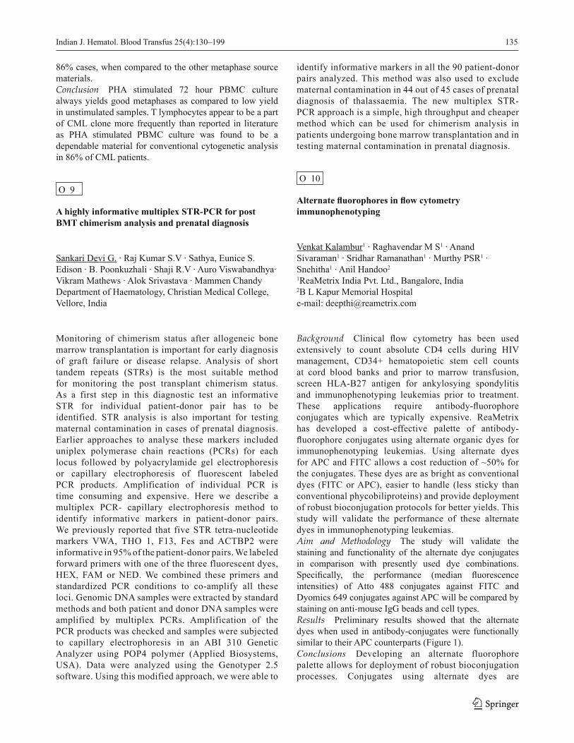
Background Clinical fl ow cytometry has been used extensively to count absolute CD4 cells during HIV management, CD34+ hematopoietic stem cell counts at cord blood banks and prior to marrow transfusion, screen HLA-B27 antigen for ankylosying spondylitis and immunophenotyping leukemias prior to treatment. These applications require antibody-fl uorophore conjugates which are typically expensive. ReaMetrix has developed a cost-effective palette of antibody-fl uorophore conjugates using alternate organic dyes for immunophenotyping leukemias. Using alternate dyes for APC and FITC allows a cost reduction of ~50% for the conjugates. These dyes are as bright as conventional dyes (FITC or APC), easier to handle (less sticky than conventional phycobiliproteins) and provide deployment of robust bioconjugation protocols for better yields. This study will validate the performance of these alternate dyes in immunophenotyping leukemias.Aim and Methodology The study will validate the staining and functionality of the alternate dye conjugates in comparison with presently used dye combinations. Specifi cally, the performance (median fl uorescence intensities) of Atto 488 conjugates against FITC and Dyomics 649 conjugates against APC will be compared by staining on anti-mouse IgG beads and cell types. Results Preliminary results showed that the alternate dyes when used in antibody-conjugates were functionally similar to their APC counterparts (Figure 1).Conclusions Developing an alternate fluorophore palette allows for deployment of robust bioconjugation processes. Conjugates using alternate dyes are
136 Indian J. Hematol. Blood Transfus 25(4):130–199
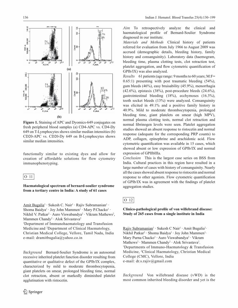
Figure 1. Staining of APC and Dyomics-649 conjugates on fresh peripheral blood samples (a) CD4-APC vs. CD4-Dy 649 on T-Lymphocytes shows similar median intensities (b) CD20-APC vs. CD20-Dy 649 on B-Lymphocytes shows similar median intensities.
(b)
(a)
functionally similar to existing dyes and allow for creation of affordable solutions for flow cytometry immunophenotyping.
O 11
Haematological spectrum of bernard-soulier syndrome from a tertiary centre in India: A study of 61 cases
Amit Bugalia1 · Sukesh C. Nair1 · Rajiv Subramanian1 · Shoma Baidya1 · Joy John Mammen1 · Mary P.Chacko1 · Nikhil V. Patkar2 · Auro Viswabandya2 · Vikram Mathews2, Mammen Chandy2 · Alok Srivastava2 1Department of Immunohaematology and Transfusion Medicine and 2Department of Clinical Haematology, Christian Medical College, Vellore, Tamil Nadu, Indiae-mail: [email protected]
Background Bernard-Soulier Syndrome is an autosomal recessive inherited platelet function disorder resulting from quantitative or qualitative defect of the GPIb/IX complex, characterized by mild to moderate thrombocytopenia, giant platelets on smear, prolonged bleeding time, normal clot retraction, absent or markedly diminished platelet agglutination with ristocetin.
Aim To retrospectively analyze the clinical and haematological profi le of Bernard-Soulier Syndrome diagnosed in our institute.Materials and Methods Clinical history of patients referred for evaluation from July 1966 to August 2009 was accrued (demographic details, bleeding history, family history and consanguinity). Laboratory data (haemogram, bleeding time, plasma clotting tests, clot retraction test, platelet aggregation, and fl ow cytometric quantifi cation of GPIb/IX) was also analyzed.Results 61 patients (age range: 9 months to 60 years; M:F= 0.65:1) presenting with post traumatic bleeding (54%), gum bleeds (46%), easy bruisability (45.9%), menorrhagia (42.6%), epistaxis (38%), post-procedure bleeds (24.6%), gastrointestinal bleeding (18%), ecchymoses (16.3%), tooth socket bleeds (13%) were analyzed. Consanguinity was elicited in 49.1% and a positive family history in 40.9%. Mild to moderate thrombocytopenia, prolonged bleeding time, giant platelets on smear (high MPV), normal plasma clotting tests, normal clot retraction and normal fi brinogen levels were seen. Platelet aggregation studies showed an absent response to ristocetin and normal response (adequate for the corresponding PRP counts) to ADP, collagen, epinephrine and arachidonic acid. Flow cytometric quantifi cation was available in 15 cases, which showed absent or low expression of GPIb/IX and normal expression of GPIIbIIIa.Conclusion This is the largest case series on BSS from India. Cultural practices in this region have resulted in a large number of cases with history of consanguinity. Nearly all the cases showed absent response to ristocetin and normal response to other agonists. Flow cytometric quantifi cation of GPIb/IX was in agreement with the fi ndings of platelet aggregation studies.
O 12
Clinico-pathological profi le of von willebrand disease: Study of 265 cases from a single institute in India
Rajiv Subramanian1 · Sukesh C Nair1 · Amit Bugalia1· Nikhil Patkar2 · Shoma Baidya1 · Joy John Mammen1· Mary Purna Chacko1 · Auro Viswabandya2 · Vikram Mathews2 · Mammen Chandy2 · Alok Srivastava2. 1Departments of Immuno-Haematology & Transfusion Medicine, 2Clinical Haematology, Christian Medical College (CMC), Vellore, Indiae-mail: [email protected]
Background Von willebrand disease (vWD) is the most common inherited bleeding disorder and yet is the
Indian J. Hematol. Blood Transfus 25(4):130–199 137
most under diagnosed disorder in developing countries. This is the largest case series of vWD from India.Objectives To study the clinico-pathological profile of patients with vWD.Materials and methods The coagulation data of all patients who have been evaluated in the coagulation lab of CMC from 2001 to 2009 was reviewed from the records. The clinical profi le and coagulation test Results of patients with vWD was analysed in detail.Results From 2001 to 2009, 2471 patients were evaluated in the coagulation lab for various bleeding manifestations. Amongst patients with a specifi c coagulation defect, the most common diagnosis was Haemophilia (34.6%), followed by vWD (10.7%). Amongst 953 patients with low factor VIII levels, further evaluation showed evidence of vWD in 265 cases (27.8%). vWD was equally prevalent in males and females (M:F::1:1.05). Most common symptoms were mucosal bleeds (87%)- Epistaxis in 39% and Gum bleed in 64%, followed by easy bruisability (39%) and menorrhagia (16%). Type 1, 2 and 3 vWD was seen respectively in 34%, 15% and 51% of patients. In Type 2 vWD, further sub-classifi cation was suggested as: Type 2A in 11, Type 2B in 16, Type ?2A/2M in 11 and ?Malmo/Newyork in 1 patient. There was history of consanguinity in 39% and a positive family history of bleeding in 32% patients. The median value of FVIII:C, vWF:Ag, vWF:CBA, vWAg:Rco in Type 3 vWD was 3.4%, 0u/dl, 6% and 8% respectively. The same for Type 1 vWD was 41%, 32u/dl, 41% and 33% respectively. The values however, varied greatly between the individual sub-types of type 2 vWD. Parents of 53 patients were also evaluated. They were found to have mild vWD in 67% (35/52 parents). Conclusion Since vWD is a common yet highly under diagnosed bleeding disorder, a high index of suspicion and careful evaluation should be done before making a final diagnosis of vWD. Specifically, all cases with a mild decrease in Factor VIII level should be further evaluated for possible vWD.
O 13
Calibrated automated thrombography(CAT) in congenital bleeding disorders
Shenbagapriya · Baidya S. · M.L. Kavitha · A. Viswabandya, V. Mathews · A. Srivastava · S.C. Nair Departments of Clinical Pathology and Haematology Christian Medical College, Vellore, Indiae-mail: [email protected]
Background Thrombin is the central to the coagulation process but there is currently no routine laboratory test that can quantitatively measure the thrombin forming capacity of a plasma, classical clotting test such as activated partial thromboplastin time and prothrombin time assess only the initiation of clot formation and do not reflect the entire thrombin generation. Estimation of an individuals thrombin generation potential may correlate more closely with a hypercoagulable or hypocoagulable phenotype when compared to traditional coagulation test. We investigated the relation between clotting factor concentration and parameters of thrombogram in patients with congenital bleeding disorders.
Results Subjects(N = number)
Factor levels(Median / range)
ETP[nM thrombin/min](Median / range)
Peak[nM thrombin](Median / range)
Normal (n = 20) - 2290(1553.5/2540) 532.7(315.7/508.25)
FVIII
Mild HA (n = 8) 10(5.4/15.4) 1784(986/2051) 286(73.13/360.28)
Mod.HA (n = 18) 2(1/3.7) 1319(586/2014) 109.6(41/259)
SHA(n = 70) 0.2(0.2/0.9) 825.25(273.6/1919) 38.56(16.24/155.07)
FIX
Mild HB( n = 3) 9.3(7.8/11.2) 2269(2088/2777) 385.32(283.5/387.9)
Mod.HB( n = 3) 3.4(1.6/4.2) 1687.5(586/2077) 140.1(58.4/256.8)
SHB (n = 16) 0.6(0.1/0.9) 883(113/1987) 67.8(34.2/231.5)
vWD
Type 1(n = 5) - 1720.7(828/1943) * 257.2(53.9/416)**
Type 3 (n = 15) - 1315.5(860/1850) * 136.7(70.3/322.2)**
RBD
FV( n = 3) 0.8(0.2/4.3) 875.8(75/1695) 216.7(7.9/425)*
FV&FVIII (n = 3) 10.1(9/12):9.6(9/11)
1996.7(1533/2540) 343.8(315.6/508.3)
FVII (n = 4) 10.7(0.2/28) 2085(1077/2713.5) 476.69(315.37/567.2)
FX ( n = 5) 5.1(0.2/23.1) 1461(1071.5/2077)**
119.8 (112.1/344.8)**
** - p value – < 0.01;* - p value – < 0.05
Methods CAT was measured in all patients as per Hemker et al,.2003, fl uorescence was detected using Fluoroskan Ascent and calculations are made with thrombinoscope software.Conclusion We observed that with decrease in clotting factor concentrations, The thrombogram parameters were deranged with signifi cant difference in peak thrombin in all haemophiliacs, vWD and severe FV and FX defi ciency. Our results suggest that Thrombin generation test can represent a helpful guide in the evaluation of coagulation capacity of patients with congenital bleeding disorders.
138 Indian J. Hematol. Blood Transfus 25(4):130–199
O 14
Thromboelatography(TEG) in Congenital Bleeding Disorders
Shenbagapriya · Baidya S. · M.L. Kavitha · A. Viswabandya · V. Mathews · A. Srivastava · S.C. NairDepartments of Clinical Pathology and Haematology Christian Medical College, Vellore, Indiae-mail: [email protected]
Background Haemostasis is the consequence of balanced interactions of cellular and molecular components responsible for the maintenance of an intact circulation.A wide range of congenital bleeding disorders has been described originating from an inadequate function of the platelets as seen in Glansmann thrombasthenia(GT) and Bernard Soulier syndrome(BSS) or caused by defi ciency of procoagulant proteins such as fi brinogen or coagulation factors FII, FV, FVII, FVIII, FIX, FX, FXI, FXIII. The traditional methods used for diagnosis of these disorders and laboratory monitoring of treatment involves the measurement of clotting factor levels in plasma in the presence of supra physiological levels of phospholipids. Global whole blood coagulation assays provide a novel method to study the process of coagulation in an environment in which platelet interaction with enzymatic factors are preserved. We studied the various TEG patterns using whole blood that was activated with minimal amount of tissue factor, TEG produces a continuous profi le of the overall rheological changes occurring during coagulation which is more nearer to physiology. Materials In all subjects continuous whole blood clot formation profi les were recorded by the TEG 5000 series, citrated whole blood samples were allowed to rest for 30 minutes as per the methods recommended by Sorenson et al., 2003.
Statistical analysis SPSS version 11.0Samples from persons with haemophilia showed signifi cant difference for all TEG parameters and vWD showed a signifi cant difference only for R time. Among RBD, R time and k were signifi cant with p value <0.05.
Platelet disorders BSS(n = 4) does not show any signifi cant difference in TEG parameters compared to healthy individuals except for G[p value - <0.05]. Out of 21 GT cases analyzed all showed a signifi cance difference for k, angle , MA and G [p value - <0.01].Conclusion Thromboelastography seems to be a promising tool for diagnosis and characterization of phenotypic
Results Infl uence of TEG parameters on congenital bleeding disorders
Subjects(N = number)
Factor levels(Median /
range)
R time[min](Median / range)
K[min](Median/
range)
Angle[deg](Median /
range)
Normal (n = 20)
- 6.7(5.1/8.3) 2.8(2/4) 53.2(43.9/53.2)
FVIII
Mild HA(n = 8)
10(5.4/15.4) 12.4(7.3/15.3) 4.7(2.2/8.6) 38.2(18.4/58.3)
Mod.HA(n = 18)
2(1/3.7) 16(12.4/50.5) 4.5(2.8/9) 38.5(11.4/55)
SHA(n = 70)
0.2(0.2/0.9) 46.7(13/117) 20(2.8/47) 11.2(1/54)
FIX
Mild HB(n = 3)
9.3(7.8/11.2) 13.7(10.8/14.1) 4.1(2.3/4.5) 45.8(43.2/61.4)
Mod.HB(n = 3)
3.4(1.6/4.2) 11.3(10.6/31) 3.9(2.2/8.1) 44(16.3/44.7)
SHB (n = 16)
0.6(0.1/0.9) 42.9(14.6/01) 18.6(3.1/50.9) 11.3(1/52.6)
vWD
Type 1(n = 5)
- 9.3(7/20.4)* 3.4(1.9/4.6) 47.7(41.6/63.9)
Type 3 (n = 15)
- 11.1(6.2/16.3)* 3.4(1.3/6.1) 48.3(29.5/71.7)
RBD
FV(n = 3)
0.8(0.2/4.3) 14.7(10.6/21)* 4.5(2.6/5.3)* 43.3(37.1/54.3)
FV&FVIII(n= 3)
10.1(9/12);9.6(9/11)
19.9(15.5/38.9)* 6.5(3.7/12.9)* 30.2(16.4/47.7)
FVII (n = 4)
10.7(0.2/28) 9.9(7/13.2) 2.5(2.2/3) 51.4(46.2/58.8)
FX (n = 5)
5.1(0.2,23.1) 15(10.3,33.6)* 4.8(2.5,9.8)* 37.1(22.3,53.4)
Platelet Disorders
K[min](Median/
range)
Angle[deg](Median / range)
MA[mm](Median/
range)
G[dynes/cm](Median/ range)
Normal (n = 20)
2.8(2/4) 53.2(43.9/53.2) 56.7(53/65) 6.5(5.6/9.3)
GT (n = 21)
7.8(2/15.3)** 34.5(8.5/64.2)** 24.1(11/36)** 1.6(0.7/2.8)**
BSS (n = 4)
2.5(1.6/3.8) 49.2(43.6/67.6) 69.2(65.2/75.1) 11.2(9.4/15)*
** p value - <0.01 , * - p value - <0.05
variance amongst patients with various bleeding disorders based on laboratory data.
O 15
Acquired platelet dysfunction with eosinophilia in south India
Sukumaran D. · Kamalaselvi S. ·Soma Baidya · Mary
Indian J. Hematol. Blood Transfus 25(4):130–199 139
Purna Chako · Vikram Mathew · Auro Viswabandya · Alok Srivastava · Sukesh C. Nair.Department of Transfusion Médicine and Immuno Haematology,Christian Medical College, Vellore, Indiae-mail: [email protected]
Acquired platelet dysfunction with eosinophilia is an acquired bleeding disorder of unknown aetiology associated with platelet dysfunction and eosinophilia. Thirty eight children aged 11 months to 10 years diagnosed to have acquired platelet dysfunction with eosinophilia (APDE) were studied. They are mostly from the state of Kerala-76.4%. The male to female ratio was 2:1.These children have no family history of bleeding and no history of recent drug intake that causes platelet dysfunction. All of the children had easy bruisibility and recurrent spontaneous echymotic patches on the body and face. The number of platelets in all these children was within the normal range but the platelet morphology showed hypo granular and pale stained platelets in 76.40% of them. Eosinophilia was detected in all of the children (AEC-990-4500-Normal range-200-600). Prolonged bleeding time was detected in 65.8 % of these patients. Platelet aggregometry showed abnormal patterns similar to storage pool defi ciency or Glanzman Thrombasthenia variant. The platelet aggregation patterns are shown below the Table.
Aggregation Reaponses
Ristocetin ADP Epinephrin Collagen A.Acid ATP Release
with Thrombin
Absent 42.1% 86.9% 71.1% 79.0% 46.6%
Disaggregation 57.9% 28.9% 21.0%
Subnormal 13.1%
Normal 100% 54.4%
These changes in platelet functions and morphology may be due to acquired storage pool defi ciency of the platelet. In four patients serum showed total IgE levels higher than 2000 IU/ml (832->2000). There was no correlation between the number of eosinophils and serum total IgE and the severity of bleeding symptoms. The majority of children with APDE did not receive any treatment except those who had severe bleeding symptoms which required platelet concentrate to stop bleeding. Among the 38 patients, we re-evaluated 11 patients. They received treatment for eosinophilia, (Hetrazan 100mg three times daily for 21 days). The patients became asymptomatic and the aggregometry studies were normal with normal eosinophil count.
O 16
Diagnosis of PNH by fl owcytometry
Maya R. Gupta · Manisha Madkaikar · Suchitra Swaminathan · Sangeetha Samadharman · Sonali Sarawade* · Farah Jijina * · Kanjaksha GhoshNational Institute of Immunohaematology, Parel Mumbai, Indiae-mail: [email protected].
Background Evaluation of expression of GPI associated proteins (CD55/CD59) and GPI anchor itself (FLAER) by fl owcytometry are the most sensitive and specifi c methods currently used for diagnosis PNH. Aim To compare 3 different techniques (i) Stain-lyse-wash (ii) Lyse-wash-stain-wash (iii) Stain-no lyse-no wash technique for optimum detection of CD55 and CD59 defi cient PNH cells. 2. To compare FLAER with expression of CD55/CD59 for detection of PNH clone size.Material & method BD FACS lyse and BD Pharm lyse solution were used for lysing steps in stain-lyse-wash and lyse-wash-stain-wash protocols for CD55 and CD59 staining on PB leukocytes. FLAER staining was optimized using lyse-wash-stain-wash technique and compared with CD55/CD59 expression Results in PNH PB.Results Stain-lyse-wash technique showed adequate detection of PNH clone on granulocytes, however it required much higher amount of antibodies due to the presence of red cells and platelets during staining step. With this technique it was diffi cult to differentiate Type III cells from Type II. With lyse-wash-stain-wash technique using BD FACS solution and stain-no lyse-no wash method, the clone was completely missed in known PNH patients. Lyse-wash-stain-wash technique using BD Pharm lyse solution and wash buffer (PBS+BSA+EDTA) showed optimum separation of Type I, II, & III cells and it correlated well with expression of FLAER on granulocytes & monocytes.Conclusion Thus lyse-wash-stain-wash technique using BD Pharm lyse solution and wash buffer (PBS+BSA+EDTA) was found to be the best for CD55/CD59 staining on PB leukocytes. FLAER as single agent was found to be equally sensitive and specifi c for the diagnosis of PNH.
O 17
Hereditary spherocytosis versus auto immune
140 Indian J. Hematol. Blood Transfus 25(4):130–199
haemolytic anaemia: An automated approach using the beckman coulter LH 755
Neeraj Arora · David Inbakumar · Janakiram Kamal · Joy Mammen · Sukesh Chandran RadhakrishananChristian Medical College and Hospital, Vellore, Indiae-mail: [email protected]
Background While processing the blood samples on LH 755 in the Reticulocyte mode using the VCS technology the reticulocyte indicies show volumetric research parameters like Mean Sphered Cell Volume (MSCV) and Mean Reticulocyte Volume (MRV). During this processing a compound derived from Methylene Blue fi rst precipitates the Ribosomal RNA of the Reticulocyte, then the red cells are sphered in acidic hypoosmolar conditions. MSCV is the mean volume of whole RBC population where as the MRV is the average volume of all Reticulocyte. It has been observed that the difference between MCV and MSCV is higher in the cases of HS and it is well established screening tool. It has also been observed that the volume of the HS Reticulocyte is reduced in comparison with AIHA or normal Reticulocyte.Aim Evaluation of Reticulocyte Volumetric research parameter like MSCV and MRV during Reticulocyte analysis on LH 755 in diagnosis of Hereditary spherocytes.Materials and Methods EDTA samples from 57 cases of HS and 29 cases of AIHA were processed on LH 755 in both the differential and the Reticulocyte mode. The data generated was analysed and compared with 46 normal healthy donors. Results Using the algorithm (MCV – MSCV > 10 and MRV – MSCV < 25) sensitivity of 84.2% and specifi city of 100% was observed in Hereditary spherocytosis. Using the algorithm of (MCV – MSCV > 10 and MRV – MSCV > 25) sensitivity of 70.6% and 100% specifi city was observed in AIHA.Conclusion With the Reticulocyte analysis on LH 755 we now have an additional tool for diagnosis of Hereditary Spherocytosis.
O 18
Molecular characterization and infl uence of αα/-α3.7 deletion on hematological and clinical heterogeneity of sickle beta 0 thalassemia in western Orissa, India
Preetinanda Manaswini Dash · Dilip Kumar Patel · Siris Patel · Ranjeet Singh MashonVeer Surendra Sai Medical College, Burla, Orissa, Indiae-mail: [email protected]
Background Heterogeneity in clinical manifestation of
Sickle Beta Thalassemia (Sβ-Thal) may occur from nature of beta-thal mutation. In Western Orissa, India the Sβ0-Thal patients with similar β thal mutation (IVS1-5G→C) are presenting with variable clinical severity. Aim To determine the β globin cluster haplotype, co-inheritance of alpha thalassemia and Xmn-I polymorphism; and to assess the impact of these genetic determinants on the phenotypic variability in Sβ0-Thal subjects.Methodology The study was conducted at Sickle cell Clinic, V.S.S. Medical College, Burla, Orissa. Detailed clinical and hematological parameters were studied in 45 Sβ0-Thal [βS / βTh IVS1-5(G→C)] patients with informed consent. PCR analysis (RFLP, MULTIPLEX) were done to characterize the β globin cluster haplotype (Hinc-II €, HindIII γG and γA,Hinc-II Ψβ, and 3’Ψβ , Ava-IIβ, Hinf-I 5’β), co-inheritance of alpha thalassemia (3.7 and 4.2kb deletions) and Xmn-I polymorphism. Clinical severity was evaluated on the basis of pain rate (VOC/pt/Yr), frequency of hospitalizations, blood transfusion (BT/pt/Yr) and spleen size (ultrasonography). The clinical severity was correlated with hematological indices (Hb, MCV, HbA2, HbF, HbS), β globin cluster haplotype, alpha thalassemia and Xmn-I polymorphism. Statistical analyses were done using t-test and correlation analysis.Result IVS1-5(G→C) mutation was strongly linked with single βS Asian haplotype (++-+++-) & multiple βTh haplotype (+-----+, ------+, -------). The overall frequency of single alpha chain deletion was 30% in the Sβ0- Thal subjects (αα/-α3.7 in 28% cases & αα/-α4.2 in a lone case) and Xmn-I polymorphism was seen in all the cases (+/+ in 11% and +/- in 89%).Discussion Patients with αα/-α3.7 deletion were clinically less symptomatic (decrease in pain and transfusion rate) with signifi cant decrease in HbS (p<0.05) and an increase in HbF value (p<0.05) regardless of beta globin cluster haplotype and Xmn-I polymorphism, whereas frequency of hospitalization and spleen size were similar.
O 19
Screening, awareness, genetic counseling of thalassemia syndromes in Punjab
Sheila Das · Shavinder Singh · R.P. Britt · John Cherian · A. SoodChristian Medical College, Ludhiana, Indiae-mail: [email protected]
Background Thalassemias and other Hemoglobinopathies pose major health problem in Asian countries, causing immeasurable, emotional, psychological and economic
Indian J. Hematol. Blood Transfus 25(4):130–199 141
burden on the affected. (Wheatherall and Clegg 2001; Fucharoen and Winichagoon, 2007). The prevalence of βThalassemia in the general population is 3-4% while in some communities it is in the range 5-15%! Management of children with β Thalassemia Major cost the family Rs 1–1.5 lakh per year per child.Aim Christian Medical College Ludhiana , was chosen by Indian Council of Medical Research to be part of Jai Vigyan Mission multi-centric project in Punjab: a)To determine high risk groups. b) To create awarness for Hemoglobinopathies amongst the common man, medical fraternity and Health Planners.Methodology The period of study was from 2000–2005. The target population comprised: a) 5000 Male/Female students in premarital age b)5000 pregnant ladies in antenatal clinics. 33 Camps were held in 16 colleges 2 km –300 km from CMC , Ldh. For ANC cases 4 ANC clinics were included and ANC camps were held 1km–150 km from CMC, Ldh.Laboratory parameters comprised NESTROFT, RBC indices , ZPP for Iron status , Hb electrophoresis & HPLC–v. Follow up of carriers amongst College students and ANC cases was regularly done to create awarness.Result & Conclusion High risk groups for βThalassemia carriers amongst college students and ANC women were Aroras ( 9- 10%), Khatris & Jains (5%) each , and Ramgariah Sikhs (2.2 – 5.5%). Carriers Hb D Pb (1.2 – 1.6%) followed by Hb D Iran trait & Hb Q India trait (0.26%) each.
O 20
Effi cacy of dapsone in management of patients with chronic immune thrombocytopaenic purpura
Bhavna Dhingra · Rahul Naithani · M. Mahapatra · H. Pati · P. Mishra · Renu SaxenaDepartment of Haematology, All India Institute of Medical Sciences, New Delhi, Indiae- mail: [email protected]
Background Idiopathic thrombocytopaenic purpura is an immune mediated disorder with enhanced platelet destruction in the reticulo-endothelial system. For chronic cases, various treatments are tried. Some patients tend to initially respond but eventually relapse. These medications are expensive and have side effects. Dapsone is an easily available, cheap and less toxic drug with a single daily oral dose. Aim To evaluate the response to dapsone in patients with chronic ITP.Methods Fifty patients with chronic ITP initiated on dapsone, in the hematology clinic were analyzed
retrospectively. Six patients were excluded as they received steroids concomitantly. Patients with other etiologies for thrombocytopenia were excluded. Statistical analysis was done using the Stata 9.0 software.Results Total 44 patients were eligible for analysis. 26 (59.1%) were males and 18 (40.9%) females. All patients had received steroids earlier and 23 patients had received various other forms of therapy, before starting dapsone. Overall, 12 patients achieved a CR (27.3%) and R was seen in 9 (20.4%) patients. The observed median time to response was 3.5 months (range 1-6). Twenty three patients had received more than one form of therapy before dapsone and no statistically signifi cant difference was seen in patients who had received two or more therapies as compared to those who received only steroids (P = 0.62). Those who attained a CR had received dapsone at a mean dose of 1.9mg/kg/day (range 1-2.2), while those in the NR group received a mean dose of 2.05 mg/kg/day (range 1- 2.4), and the difference did not show any statistical signifi cance. No serious side effects, necessitating drug- withdrawal were observed in any of the patients.Conclusions Dapsone is effective in patients with chronic ITP. Prospective randomized controlled trials comparing dapsone with other forms of therapy, and larger sample size are warranted to corroborate these results.
O 21
To study the effi cacy and safety of low dose rituximab in chronic immune thrombocytopenic purpura (ITP)
Kapoor R · Mahapatra M · Pati HP · Mishra P. Dept. of Hematology, All India Institute of Medical Sciences, New Delhi, Indiae-mail: [email protected]
Background Rituximab is a new agent used in patients with refractory chronic ITP. Rituximab has been conventionally administered in dose of 375 mg/m2 every seven days for four weeks.We ran a prospective clinical trial using lower dose rituximab in patients with chronic ITP who had not responded to at least one line of therapy & with platelet count below 30,000/mm3. Study has been approved by institute ethics committee. Patients & Methods Rituximab was given at the fi xed dose of 100 mg administered as an intravenous infusion weekly (on day 1 of weeks 1, 2, 3 and 4) to patients above 15 years, of chronic ITP who had not responded to at least one line of therapy. All cases of secondary ITP and pregnant patients were excluded from the study. A complete response (CR) was defi ned as a platelet count >100,000/mm3 and discontinuation of the steroid therapy. Partial response (PR)
142 Indian J. Hematol. Blood Transfus 25(4):130–199
was defi ned as a platelet counts between 50 and 100,000/mm3 and discontinuation of the steroid therapy. Results 15 patients have so far been given low dose rituximab as per protocol. Ten patients have completed at least 03 months of follow-up and are available for evaluation. Out of these 10 patients, 02 patients are post splenectomy relapse. Overall low dose rituximab was tolerated well in all patients with only infusion related adverse events noted. CR was achieved in 4/10 (40%) while 02 patients achieved PR giving an overall response of 6/10 (60%). Median time for response in these patients was 75 days (range 30 to 240 days). Median time of follow-up in 04 patients who did not respond was 94 days (range 91 to 133 days).Conclusions In patients of chronic ITP, lower dose rituximab is safe and effective and seems to show similar activity to standard dose. However longer follow-up is required to assess the durability of response to low dose of rituximab.
O 22
Response to intravenous administration of anti-d (rh0-d) immune globulin to thrombocytopenia associated with dengue hemorrhagic fever
Shailesh R Singi · P Srikanth · Rajesh P · D. Jhansi Vani Department of Hematology, Hemato-Pathology and Microbiology, CARE Hospitals, Banjara Hills, Hyderabad, Indiae-mail: [email protected]
Background Dengue hemorrhagic fever (DHF) is characterized by severe thrombocytopenia and increased vascular permeability. An immune mechanism of thrombocytopenia due to increased platelet destruction appears to be operative in patients with DHF. Anti-D (Rh0 D IgG) immune globulin is highly effective in producing Fc receptor blockade and in raising the platelet count in non-dengue and Dengue forms of ITP.Case Series We report here response to Anti-D (Rh0 D IgG) immune globulin in 64 cases of Dengue infection with Thrombocytopenia and active bleeding. Diagnosis of dengue infection was made either by screening with a Dengue Duo IgM and IgG Rapid Strip Test and / or with an IgM and IgG capture enzyme-linked immunosorbent assay (ELISA). Thrombocytopenia was confi rmed when a manual platelet count was 100,000/mm3, and severe thrombocytopenia was defi ned for this series when the platelet count was 50,000/mm3. Results In our case series of 64 cases from June 2007 to July 2009 with DHF, 43 were males (24 less than 12 years and 19 more than 12 years) and 21 were females (11
less than 12 years and 9 more than 12 years). Anti-D was administered at a dose of 250 IU/kg (50 μg/kg) over 5 to 10 min as intravenous rapid infusion with written consent, confi rmation of Rh positive type of blood group in those whose platelet count was less than 20,000/ mm3 or ≤50,000/mm3 with active bleeding in form of epistaxis, hematuria, mennorrhagia, hemoptysis or malaena etc. Out of 64 patients 45 had received some form of platelet transfusions in last 48 to 72 hours with no signifi cant increase in platelet count. 28 patients received steroids and 17 were on steroids at the time of Anti-D administration. Platelet count was then done at every 24 hours interval for next 5 days. The mean time to increase platelet counts by 20,000/mm3 from baseline was 24 hours after anti-D infusion for all the patients. The mean platelet count was 28,000/ mm3 before Anti D infusion and 44,000/ mm3 after 24 hours after Anti-D infusion. The mean Platelet count at 48, 72, 96 and 120 hours after anti-D infusion was 56,000/ mm3 , 69,000/ mm3, 85,000/ mm3 and 160,000/ mm3 respectively. The mean drop in Hemoglobin was 1.9 gm%. Commonest adverse event observed in 38 patients out of 64 was hemoglobinuria which was managed with intravenous fl uids. Mild chills, rigors and fever were observed in 45 patients. No other major adverse event was noted in all the patients.Conclusion The use of platelet concentrate has been abused despite data from many countries with indication that there is no role for a prophylactic platelet transfusion in DHF. Thrombocytopenia makes management of patients diffi cult at presentation when clinicians cannot predict which patients may progress to severe thrombocytopenia with hemorrhagic symptoms. All the patients in our case series responded to Anti-D therapy after 24 to 48 hours with satisfactory platelet increment. Anti-D (Rh0-D) is particularly interesting to clinicians because of its relatively low cost when compared with other treatment modalities which have very limited role.
O 23
Experience of allogenic hematopietic stem cell transplant stem cell transplant in non-HEPA fi ltered single rooms
Niranjan Rathod · M Mahapatra · P Mishra · T Seth Department of Hematology, All India Institute of Hematology, New Delhi, Indiae-mail: [email protected].
Background Hematopoietic Stem Cell Transplant (HSCT) is conventionally performed in HEPA fi ltered room as patient has to pass through critical period of 1-2 weeks of severe neutropenia. This reduces potential bacterial and
Indian J. Hematol. Blood Transfus 25(4):130–199 143
fungal infections which would have signifi cant impact on early transplant related mortality.Aim To evaluate short and long term outcome of HSCT performed in various hematological patients, in non-HEPA fi ltered single roomsMethodology We reviewed medical records of 66 HSCT performed in non-HEPA fi ltered single rooms, over last 5 years, at our institution retrospectively with respects to different variables and analyzed systematically. G-CSF was given to all from D+1. Antibacterial and antifungal prophylaxis was administered along with conditioning, and at the onset of fever, systemic antibiotics were started. Antifungal agents were added if fever persisted for 3 days.Results We present our single centre experience of 66 allogenic stem cell transplants performed over period of last 5 years. All these transplants were performed in non-HEPA fi ltered single room. Source of stem cells were PBSC-56, BM-9, Combined-1. The indications were SAA-30, CML-10, AML-8, ALL-5, Biphenotypic AL-1, Thalassemia-9, and MDS-3. The median age was 24 years (range 2.2-46) with 16 females and 55 males as participants. Median time for neutrophil engraftment was 10 days (range 8–17). Fever occurred in 59 (89%) for a median of 5 days (range 1–38), Systemic antibiotics were used in 88% and antifungal in 52% cases. The 30-day mortality was 3(4.7%), and 100-day mortality was 5 (7.8%). After day 100, there were seventeen fatalities (26.5%) due to chronic GVHD-5, relapse-2, graft rejection-2, infections like disseminated tuberculosis-1 and aspergillosis-3, VOD-2, platelet refractoriness leading to IC bleed-2. Conclusion Our experience suggests that allogeneic HSCT can be safely performed in non-HEPA fi lter rooms in India.
O 24
50 allogeneic stem cell transplants at Narayana Hrudayalaya
Sharat DamodarNarayan Hrudayalaya, Bangalore, Indiae-mail: [email protected]
The bone marrow transplant (BMT) unit at Narayana Hrudayalaya was started in November 2004. The unit consists of 3 HEPA fi ltered, positive pressure rooms. This unit is used for both autologous and allogeneic BMT. Since its inception there have been more than 50 allogeneic Stem cell transplants till date. We present data on the fi rst 50 allogeneic transplants. There are 26 pediatric and 24 adult patients who underwent allogeneic BMT. The pediatric age group ranged from 3-15 years, with 14 Males and 12 females. 13 patients (50%) had thalassemia
major, 3 patients had aplastic anemia, 4 patients had fanconi’s anemia 4 patients had acute myeloid leukemia, and 1 patient each with chronic myeloid leukemia and relapsed acute lymphatic leukemia. Bone marrow was the stem cell source for all thalassemia transplants. Busulfan with cyclophosphamide +/- ATG was the commonest conditioning used. Cyclosporin with methotrexate was used as graft versus host disease (GVHD) prophylaxis. Neutrophil engraftment occurred between days 9 to 18. Acute GVHD developed in 14 patients with chronic GVHD in 6 of them. Day 100 overall survival was 76% with the longest follow up of 60 months (6- 60 months).The adult age group ranged from 17-57 years, with 13 Males and 11 Females. 7 patients (30%) had acute myeloid leukemia, 5 patients had chronic myeloid leukemia- accelerated phase, 4 patients had aplastic anemia, 4 patients had sickle cell disease, 2 patients with thalassemia and 1 patient each with myelodysplastic syndrome and relapsed acute lymphatic leukemia. Peripheral blood was the stem cell source for all patients except thalassemia. Busulfan with cyclophosphamide +/- ATG was the commonest conditioning used. Cyclosporin with methotrexate was used as graft versus host disease (GVHD) prophylaxis. Neutrophil engraftment occurred between days 9 to 15. Acute GVHD developed in 12 patients with chronic GVHD in 6 of them. Day 100 overall survival was 83% with the longest follow up of 52 months (6- 52 months).
O 25
Costing of allogeneic bone marrow transplant procedure in patients admitted at a public tertiary care hospital, New Delhi, India
Shalini Kumar1 · Shakti Kumar Gupta1 · Sidhartha Satpathy1 · Manoranjan Mahapatra2
1Department of Hospital Administration; 2Hematology AIIMS, New Delhi, Indiae-mail: [email protected]
Background and Methods The department of Haematology at AIIMS started its allogeneic HSCT program in 2004 using 6-Antigen HLA-matched sibling donors. A study was required to better estimate the cost of the transplant. The objective of the study was to identify various direct and indirect costs associated with the transplant and to suggest rational package charges, using traditional and allocation costing methods. 13 HSCTs were observed during July 2008-March 2009 of which 10 were males and 3 Females. Median age was 31 years (range 5-40 years). Average length of stay was 48 days. The average length of stay of 13 sibling donors was 4 days. 1 BMT and 12 PBSCTs
144 Indian J. Hematol. Blood Transfus 25(4):130–199
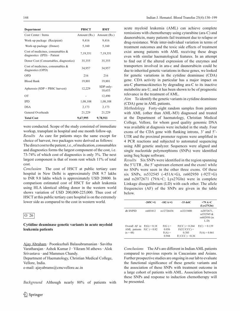
Department PBSCT BMT
Cost Center / Items Amount (Rs.) Amount (Rs.)
Work-up package (Recipient) 9,416 9,416
Work-up package (Donor) 5,160 5,160
Cost of medicines, consumables & diagnostics (IPD) – Patient 7,19,351 7,19,351
Donor Cost (Consumables, diagnostics) 35,355 35,355
Cost of medicines, consumables & diagnostics (OPD) 34,957 34,957
OPD 216 216
Blood Bank 19,801 19,801
Apheresis (SDP + PBSC harvest) 12,229 SDP only-10,655
OT - 12,490
IPD 1,08,108 1,08,108
DSA 2,173 2,173
General Overheads 21,229 21,229
Total Cost 9,67,995 9,78,911
were conducted. Scope of the study consisted of immediate workup, transplant in hospital and one month follow-up. Results As care for patients stays the same except for choice of harvest, two packages were derived as follows: The direct cost to the patient, i.e., of medication, consumables and diagnostics forms the largest component of the cost, i.e. 73-74% of which cost of diagnostics is only 3%. The next largest component is that of room rent which 11% of total cost.Conclusion The cost of HSCT at public tertiary care hospital in New Delhi is approximately INR 9.7 lakhs to INR 9.8 lakhs which is approximately USD 20800. In comparison estimated cost of HSCT for adult leukemia using HLA identical sibling donor in the western world shows variation of USD 200,000-225,000. Thus cost of HSCT at this public tertiary care hospital is on the extremely lower side as compared to the cost in western world.
O 26
Cytidine deaminase genetic variants in acute myeloid leukemia patients
Ajay Abraham · Poonkuzhali Balasubramanian · Savitha Varatharajan · Ashok Kumar J · Vikram M athews · Alok Srivastava · and Mammen Chandy.Department of Haematology, Christian Medical College, Vellore, India.e-mail: [email protected]
Background Although nearly 80% of patients with
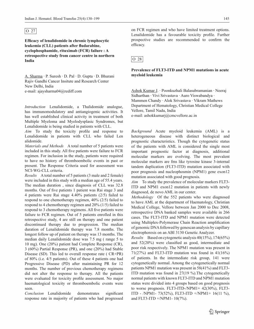
acute myeloid leukemia (AML) can achieve complete remissions with chemotherapy using cytarabine (ara-C) and daunorubicin, many patients fail treatment due to relapse or drug-resistance. Wide inter-individual variation in terms of treatment outcomes and the toxic side effects of treatment exist among patients with AML receiving these drugs even with similar haematological features. In an attempt to fi nd out if the altered expression of the enzymes and transporters involved in ara-c and daunorubicin could be due to inherited genetic variations in these genes, we looked for genetic variations in the cytidine deaminase (CDA) gene. CDA activity in particular has a major impact on ara-C pharmacokinetics by degrading ara-C to its inactive metabolite ara-U, and it has been shown to be of prognostic relevance in the treatment of AML.Aim To identify the genetic variants in cytidine deaminase (CDA) gene in AML patients. Methodology Forty-eight random samples from patients with AML (other than AML-M3) diagnosed and treated at the Department of haematology, Christian Medical College, Vellore, for whom good quality genomic DNA was available at diagnosis were included in the study. Four exons of the CDA gene with fl anking introns, 3’ and 5’-UTR and the proximal promoter regions were amplifi ed in 4 PCR reactions and subjected to automated sequencing using ABI genetic analyzer. Sequences were aligned and single nucleotide polymorphisms (SNPs) were identifi ed using Seq Scape software.Results Six SNPs were identifi ed in the region spanning the 5’UTR , the 5’ upstream element and the exon1 while no SNPs were seen in the other three exons. Of these six SNPs, rs532545 (-451A>G), rs602950 (-92T>G) and rs2072671 (79A>C; Lys27Gln) were in complete Linkage disequilibrium (LD) with each other. The allele frequencies (AF) of the SNPs are given in the table below.
-205C>G -182 A>G -33 delC +79 A>C(Lys27Gln)
db SNPID rs603412 rs12726436 rs3215400 rs2072671, rs532545 &rs602950 (in
L.D)
Overall AF in AML patients (n = 48)
F(G) = 0.18F(C ) = 0.82
F(G ) = 0.056
F(A) = 0.944
F(CC ) = 0.264F(CC/CCC) =
0.385F( CCC ) =0.36
F(C) = 0.139
F(A) = 0.861
Conclusions The AFs are different in Indian AML patients compared to previous reports in Caucasians and Asians. Further prospective studies are ongoing in our lab to evaluate the functional signifi cance of these genetic variants and the association of these SNPs with treatment outcome in a large cohort of patients with AML. Association between these SNPs and response to induction chemotherapy will be presented.
Indian J. Hematol. Blood Transfus 25(4):130–199 145
O 27
Effi cacy of lenalidomide in chronic lymphocytic leukemia (CLL) patients after fl udarabine, cyclophosphamide, rituximab (FCR) failure : A retrospective study from cancer centre in northern India
A. Sharma · P. Suresh · D. Pal · D. Gupta · D. Bhurani Rajiv Gandhi Cancer Insitute and Research CenterNew Delhi, Indiae-mail: [email protected]
Introduction Lenalidomide, a Thalidomide analogue, has immunomodulatory and antiangiogenic activities. It has well established clinical activity in treatment of both Multiple Myeloma and Myelodysplasic Syndromes, but Lenalidomide is being studied in patients with CLL.Aim To study the toxicity profi le and response to Lenalidomide in patients with CLL who failed Len alidomide.Materials and Methods A total number of 5 patients were included in this study. All fi ve patients were failure to FCR regimen. For inclusion in the study, patients were required to have no history of thromboembolic events in past or present. The Response Criteria used for assessment was NCI-WG-CLL criteria.Results A total number of 5 patients (3 male and 2 female) were included in this study with a median age of 55.4 years. The median duration , since diagnosis of CLL was 32.8 months. Out of fi ve patients 1 patient was Rai stage 3 and 4 patients were Rai stage 4.40% patients (2/5) failed to respond to one chemotherapy regimen, 40% (2/5) failed to respond to 4 chemotherapy regimen and 20% (1/5) failed to respond to 3 chemotherapy regimens. All fi ve patients were failure to FCR regimen. Out of 5 patients enrolled in this retrospective study, 4 are still on therapy and one patient discontinued therapy due to progression. The median duration of Lenalidomide therapy was 7.8 months. The longest follow up of patient on therapy was 13 months. The median daily Lenalidomide dose was 7.5 mg ( range 5 to 10 mg). One (20%) patient had Complete Response (CR), 3 (60%) Partial Response (PR), and one (20%) had Stable Disease (SD). This led to overall response rate ( CR+PR) of 80% (i.e. 4/5 patients). Out of these 4 patients one had Progressive Disease (PD) after maintaining PR for 12 months. The number of previous chemotherapy regimens did not alter the response to therapy. All the patients were evaluated for toxicity profi le assessment. No major haematological toxicity or thromboembolic events were seen.Conclusion Lenalidomide demonstrates signifi cant response rate in majority of patients who had progressed
on FCR regimen and who have limited treatment options. Lenalidomide has a favourable toxicity profi le. Further prospective studies are recommended to confi rm the effi cacy.
O 28
Prevalence of FLT3-ITD and NPM1 mutations in acute myeloid leukemia
Ashok Kumar J. · Poonkuzhali Balasubramanian · Neeraj Sidharthan · Vivi Srivastava · Auro Viswabandya · Mammen Chandy · Alok Srivastava · Vikram MathewsDepartment of Hematology, Christian Medical College Vellore, Tamil Nadu, Indiae-mail: [email protected]
Background Acute myeloid leukemia (AML) is a heterogeneous disease with distinct biological and prognostic characteristics. Though the cytogenetic status of the patients with AML is considered the single most important prognostic factor at diagnosis, additional molecular markers are evolving. The most prevalent molecular markers are fms like tyrosine kinase 3-internal tandem duplication (FLT3-ITD) mutation associated with poor prognosis and nucleophosmin (NPM1) gene exon12 mutation associated with good prognosis.Aim To study the prevalence of molecular markers FLT3-ITD and NPM1 exon12 mutation in patients with newly diagnosed, de novo AML in our center.Methodology Of the 552 patients who were diagnosed to have AML at the department of Haematology, Christian Medical College, Vellore between Jan 2000 to Dec 2008, retrospective DNA banked samples were available in 266 cases. The FLT3-ITD and NPM1 mutation were detected using Multiplex-Polymerase Chain Reaction amplifi cation of genomic DNA followed by genescan analysis by capillary electrophoresis on an ABI 3130 Genetic Analyzer.Results Based on cytogenetic analysis 40(15%), 174(65%) and 52(20%) were classifi ed as good, intermediate and poor risk respectively. The NPM1 mutation was present in 71(27%) and FLT3-ITD mutation was found in 41(16%) of patients. In the intermediate risk group, 141 were cytogenetically normal. Among the cytogenetically normal patients NPM1 mutation was present in 58(41%) and FLT3-ITD mutation was found in 27(19 %).The cytogenetically normal patients with known FLT3-ITD and NPM1 mutation status were divided into 4 groups based on good prognosis to worse prognosis. FLT3-ITD-/NPM1+ 42(30%), FLT3-ITD - /NPM1- 73(52%), FLT3-ITD +/NPM1+ 16(11 %), and FLT3-ITD +/NPM1- 10(7%).
146 Indian J. Hematol. Blood Transfus 25(4):130–199
Conclusion The above result suggest that in newly diagnosed AML, cytogenetic risk group and molecular marker FLT3 ITD and NPM1 have a similar prevalence to that previously reported in the western literature.
O 29
Lenalidomide in low risk myelodysplastic syndrome: Single centre experience
Nitin Gupta · Rajan Kapoor · Shyam Rathi · Tulika Seth · Pravas Mishra, Manoranjan Mahapatra, Hara Prasad Pati Department of Hematology, All India Instittute of Medical Sciences, New Delhi, Indiae-mail: [email protected]
Background Low risk myelodysplastic syndrome (MDS) is characterized by transfusion dependent anemia and lower propensity to progression to acute myeloid leukemia. Lenalidomide has been approved for treatment of low risk MDS.Objective Prospective study of lenalidomide in low risk MDSMethods Inclusion criteria: IPSS low risk de novo MDS, with transfusion dependent anemia, base line platelet count >50000/cmm, absolute neutrophil count (ANC)>1000/cmm. Lenalidomide starting dose -10mg/day with modifi cation for cytopenias. Study period - 4 months and to be continued in patients who responded. Monitoring included hemogram with ANC weekly, liver and renal function tests every 2 weeks.Results After ethical approval and patient’s consent twelve patients studied, out of which 10 (Male: female: 4:6) have completed 4 months of therapy ( Lenalidomide 10 mg/day -8 & 5mg/day - 2 patients). Mean age was 43.2years (27-72 years). WHO types: RA-1, RARS-1, RCMD-3, MDSU-1, RAEB1-2, RAEB2-1, ?5q-syndrome-1. Cytogenetics data available in 7 patients (normal- 5, 5q del-1, 46xy, del 2,del 18 -1). PNH - all negative, EPO level markedly increased (>2000 mIU/ml - 9, 500 mIU/ml - 1). Base line mean hemoglobin 5.09gm% (3-6.7). Early myelosuppression was the predominant drug toxicity, occurred in 90% of patients, however grade 3/4 neutropenia & thrombocytopenia occurred in 50% & 60% patients respectively. Five patients became transfusion independent while 5 did not show any improvement, in them further therapy was stopped. Of the fi ve responding patients, three are still on treatment & transfusion independent (mean hemoglobin rise - 5.3gm%, median follow up 12 months). Delayed cytopenia developed in two responding patients after 6 and 12 months of therapy. Renal and liver dysfunction was seen in none.
Conclusion Lenalidomide is effective in low risk MDS with 30% response rate. Predominant toxicity is cytopenias necessitating frequent monitoring, dose modifi cation & supportive treatment. Lower dose of 5mg/day appears to be effective and better tolerated.
O 30
Genetic variant in ABCG2 is associated with occurrence of complete cytogenetic response to imatinib in patients with chronic myeloid leukemia
Preetha Markose1 · Poonkuzhali Balasubramanian1 · Vikram Mathews1 · Vivi M Srivastava2 · Biju George1 · Auro Viswabandhya1 · Alok Srivastava1 · Mammen Chandy1.Department of Hematology, Christian Medical College, Vellore, Indiae-mail: [email protected]
Background The tyrosine kinase inhibitor Imatinib Mesylate as a single agent has revolutionized the treatment of chronic myeloid leukemia. Despite the excellent effi cacy of the drug, a proportion of patients develop resistance to imatinib probably due to development of mutations in the tyrosine kinase domain or due to sub-optimal exposure to the drug resulting from pharmacogenetic variations. Aim The aim of the present study was to evaluate the role of common genetic variants in the infl ux transporter for imatinib hOCT1 (SLC22A1), and the effl ux transporters ABCG2 (BCRP) and ABCB1 (MDR1) on attainment of complete cytogenetic response (CCyR) in patients with CML treated with Imatinib. Methodology In total, 12 SNPs in these three genes were screened in genomic DNA samples obtained from 55 patients with CML, who received imatinib as a fi rst-line therapy. All patients for whom FISH result was available at or around 12 months after the start of imatinib therapy were included in the study. We used PCR amplifi cation of the regions of interest followed by direct sequencing and polymorphisms were identifi ed using SEQSCAPE software. All patients achieved complete haematological response. CCyR was defi ned as 100% Philadelphia chromosome negativity by FISH at or before 12 months after the start of imatinib. Results The incidence of these 12 polymorphisms in patients in CCyR vs. those who did not achieve CCyR was compared using Fischer’s exact or Chi-square test. The frequency of AA or AC genotype in ABCG2 exon 5 (rs2231142) associated with decreased ABCG2 transcript expression was signifi cantly lower in patients who achieved
Indian J. Hematol. Blood Transfus 25(4):130–199 147
CCyR (1/25; 4%) compared to those who did not (9/30; 30%: p = 0.009). None of the MDR1 and hOCT1 SNPs that were previously reported to be associated with treatment outcome with imatinib were signifi cant in this cohort of patients. This may be due to difference in allele frequencies of these SNPs between populations. Conclusion In conclusion, genotyping of rs2231142 polymorphism could be useful to identify the best responders to imatinib among CML patients.
O 31
p15(INK4B) gene methylation in patients with myelodysplastic syndromes-AIIMS experience
Rekha Chaubey · Sudha Sazawal · Manoranjan Mahapatra· Rashid Mir · Renu SaxenaDepartment of Hematology, All India Institute of Medical Sciences, New Delhi, Indiae-mail: [email protected]
Background Myelodysplastic syndromes (MDSs) are clonal hematologic disorders that frequently represent an intermediate disease stage before progression to acute myeloid leukemia (AML). Although several cytogenetic abnormalities have been associated with this heterogenous disorder little is known about what genetic changes are responsible for the disease. Deregulated epigenetic mechanisms are likely involved in the pathogenesis of MDS. Abberent promoter methylation is frequently observed in MDS patients, and is recognized as a critical event in the disease’s pathogenesis and progression. p15(INK4B) is a tumor suppressor gene and the hypermethylation of its promoter, has been frequently found in MDS. Methylation status of p15gene in MDS patients has rarely been reported from India. In view of this we aimed to investigate the methylation status of p15gene in MDS patients using a Methylation- specifi c polymerase chain reaction.Aim To investigate the frequency of p15 gene methylation in MDS patients.Methodology Total genomic DNA was extracted from bone marrow/peripheral blood leucocytes of MDS patients. Bisulphite treatment of DNA was performed. Bisulphite modifi ed DNA was amplifi ed by methylation specifi c PCR.Results A total of 30 patients (median age 40 yrs, range 14-75yrs; M: F 3:2; median TLC-4.15X10⁹/l, range 0.95-116 x109/l Median platelet count- 102 x109/l, range 5-274 x109/l, Median hemoglobin -6.7 g/dl , range 2.9- 16.1 g/dl, were studied. Karyotypes of only twenty four patients were available. Aberrent methylation of p15 was
detected in 12/30 (40%) patients. The frequency was found to be highest in patients with refractory cytopenia with multilineage dysplasia (RCMD) and in patients with abnormal karyotype.Conclusion p15(INK4B) gene methylation is a frequent event in patients with Myelodysplastic Syndromes which might be contributing to the pathogenesis of the MDS.
O 32
To study effi cacy and toxicity of lenalidomide, cyclophosphamide and prednisolone combination in multiple myeloma
Shyam Rathi · M. Mahapatra · P. Mishra · N. Gupta · R. Kapoor · H. PatiDepartment of Hematology, All India Institute of MedicalSciences, New Delhi, Indiae-mail: [email protected]
Background The combination of lenalidomide and dexamethasone has been shown to be highly effective in newly diagnosed and relapse multiple myeloma patients. In combination high dose dexamethasone shown to has higher toxicity and low overall survival rate. Addition of a third agent to this combination may increase the response rate at the cost of toxicity. In a phase II trial of lenalidomide cyclophosphamide and dexamethasone shown to has higher response rate but toxicity was also high. We report the results from combining low dose lenalidome, cyclophosphamide and prednisolone in newly diagnosed and relapsed multiple myeloma patients.Methods Trial was initiated in August 2008, the treatment protocol consisted of lenalidomide given orally at a dose of 10 mg daily on days 1-21 of a 28 day cycle. Prednisolone given at a dose of 2mg/kg (max 100 mg) daily for 4 days of each cycle. Cyclophosphamide at a dose of 300 mg (fi xed dose) was given on days 1, 8, and 15 of each cycle. After achievement of maximum response, two more cycles were given. Patients also received an aspirin once daily as thromboprophylaxis. Response was defi ned as per modifi ed EBMT criteria. The study was approved by institutional ethics committee.Results Total nine patients were enroll. Seven patients had completed the study and available for evaluation. Five patients were newly diagnosed. Two patients were relapsed on thalidomide. (Median time of treatment with thalidome was 2 years). Median age of the patient was 59 years. Five are male and 2 female. In newly diagnose response was very good partial response (VGPR) in 2, partial response (PR) in 2 and minimal response in (MR) 1 patients. Median
148 Indian J. Hematol. Blood Transfus 25(4):130–199
time for response was 5 cycles (range is 4 to 7 cycle). Out of two relapse patients, one had progression of disease after 2 cycles and other had progression of disease after 3 cycles. Hematological toxicity grade 3 in one patient. Main Non hematological toxicity was fatigue.Conclusion The combination of lenalidomide, cyclophosphamide and dexamethasone has excellent activity in the setting of newly diagnosed myeloma patients. In relapsed patients on thalidomide, may not be effective. This may be due to low dose of lenlidomide in our study. This fi nding needs to be confi rmed in larger number of patients with longer duration of follow up.
O 33
Clinical profi le and outcome of hodgkin lymphoma in children
Vineeta Gupta1 · Akash Kumar1 · T.B. Singh1 · Mohan Kumar2 · Baldev Bhatia1
1Department of Pediatrics; 2Pathology, Institute of Medical Sciences, Banaras Hindu University, Varanasi, Indiae-mail: [email protected]
Background Several studies have established the effi cacy of chemotherapy alone protocols in pediatric Hodgkin lymphoma. The use of systemic therapy also avoids the long term complications of radiation therapy. The aim was to study the patient profi le and treatment outcome in childhood Hodgkin lymphoma treated with chemotherapy alone protocol.Method Between Aug 2003 to July 2008, 36 patients (34 boys and 2 girls) were diagnosed with Hodgkin lymphoma (HL). Diagnosis was established by lymph node biopsy and staged by USG/CT of thorax and abdomen and bone marrow examination. 32 patients who completed treatment, received 6–8 cycles of either COPP regimen (15) or alternating COPP/ABVD (17). Two patients with features of spinal cord compression received additional radiotherapy. One patient with coexistent HIV infection refused treatment and three did not complete the treatment. The follow-up ranged from 12 to 60 months. Results Mean age of the patients was 7.2 years (range 4-10). Duration of symptoms was 3 months to 2 years. 20 patients had received anti-tubercular treatment before diagnosis of HL. Five patients had mediastinal widening of which 2 had features of superior mediastinal syndrome. One patient each had pleural effusion and ascites. 2 patients had involvement of vertebral body with paravertebral mass and features of compressive myelopathy. None had
bone marrow involvement. The patients were staged as follows: Stage II (3), Stage III (29) and Stage IV (4). 27 patients (75%) had B symptoms. Mixed cellularity was the commonest histologic subtype. All the 32 patients who completed treatment achieved complete remission. Two patients on COPP regimen relapsed in follow up. One had systemic relapse while the other had intracranial relapse. Both received ABVD regimen and achieved complete remission.Conclusion Majority of the patients had advanced disease with systemic symptoms. The remission rate was high with chemotherapy alone protocols. Few patients required additional radiotherapy.
O 34
CDX2: A novel target in human leukemias
Vijay Pal S. Rawat1,2· Christian Buske1,2
1Clinical Cooperative Group “Leukemia”, Helmholtz Zentrum Muenchen, Munich, Germany and the Department of Medicine III, University of Munich- Grosshadern, Munich, Germany2The Institute of Experimental Tumor Research, CCCU and University Hospital Ulme-mail: [email protected]
HOX genes are homeobox genes which code for transcription factors that are highly expressed selectively in CD34+ hematopoietic stem cells but not in their progeny. Interestingly, gene expression profi ling studies have reported aberrant expression of multiple HOX genes in leukemia of different origins especially in AML patients with normal karyotype (AML-NK). It was largely unknown, how aberrant expression of Hox genes is initiated in malignant clones especially in AML-NK. Using retroviral gene transfer murine bone marrow transplantation, we have shown that ectopic expression of the homeobox gene Cdx2 in murine bone marrow progenitors induces aggressive AML in vivo (PNAS, 2004). Furthermore ectopic expression of Cdx2 upregulates leukemogenic Hox genes and stem cells regulatory transcription factors and confers self renewal properties to murine progenitors. Although CDX2 is not expressed in normal HSC and hematopoietic cells, surprisingly ectopic expression of CDX2 was detectable in 90% of patients with with different genetic alterations. Gene expression results revealed that the level of CDX2 expression correlates with HOX gene expression in human AML patients and supported a potential role of CDX2 in the development of human AML with aberrant Hox gene expression AML (Blood 2008 and JCI 2007).We could
Indian J. Hematol. Blood Transfus 25(4):130–199 149
also demonstrate that CDX2 is expressed aberrantly in the majority of cases with B-lineage ALL and T-ALL. High expression of CDX2 correlated signifi cantly with the ALL subtype pro-B ALL, cALL, Ph(+) ALL and early T-ALL. High expression of CDX2 was associated with inferior overall survival and showed up as a novel and independent risk factor for ALL in a bivariate analysis (Leukemia 2009 and Blood 2009). Therefore we believed that CDX2 might be a potential therapeutic target in leukemic disease. Our recent data link the oncogenic capacity of the Cdx2 to the MAPK signaling, opening the possibility to counteract homeobox-associated leukemogenesis by kinase inhibitors.
O 35
Assessment of angiogenesis in acute leukemias
Saroha V. · Singh T. · Khurana N. · Gupta N.1 · Dubey A.P.2
1Department of Pathology, Medicine; 2Pediatrics, MAMC, New Delhi, Indiae-mail: [email protected]
Background Angiogenesis is the formation of new blood vessels. Increased angiogenesis has been observed in various hematological malignancies. In last 5 years, role of angiogenesis in acute leukemias has become more evident as anti angiogenic drugs have been discovered which target the establishment and growth of tumor vessels and show a synergistic activity when used in combination with chemotherapy. Aim To evaluate angiogenesis in acute leukemias pre therapy and post therapy and correlate the degree of neovascularization with the FAB subtype, TLC and marrow blast %.Methodology Bone marrow trephine biopsy specimens from 46 patients of acute leukemia and 46 controls were taken. Angiogenesis was assessed by evaluating mean vessel density (MVD) on sections stained immunohistochemically with CD34 by defi ning HOT SPOTS at diagnosis and following remission. Statistical analysis was carried out using Mann-Whitney U test and Pearson's coeffi cient of correlation.Results MVD was found to be signifi cantly higher in patients of acute leukemia as compared to controls (P<.001) and decreased after remission. Cases in partial remission showed MVD intermediate between those in complete remission and leukemia patients suggesting bone marrow vascularity may be an indirect indicator of remission. A weak positive correlation was seen between MVD and marrow blast percentage while no correlation was observed between the vessel density and TLC or the FAB subtypes.
CD34 was also found to stain varying number of blasts, the % age positivity of which decreased with maturation.Conclusion It is suggested that bone marrow biopsy should be carried out pre therapy and post therapy in acute leukemia cases to evaluate mean vessel density. Possibility of using angiogenic inhibitors (in addition to chemotherapy) may prove to be a novel therapeutic strategy in management of acute leukemias.
O 36
Prenatal diagnosis for leukocyte adhesion defi ciency-I for the fi rst time in India
Madkaikar · Gupta Maya Manisha · Samadharman Sangeetha · Swaminathan Suchitra · Rao Meghana and Ghosh Kanjaksha National Institute of Immunohaematology, 13th fl oor KEM Hospital, Parel, Mumbai, Indiae-mail: [email protected]
Introduction Adhesion molecules play a major role in the recruitment of neutrophils to the site of infl ammation. Leukocyte Adhesion Defi ciency-I (LAD-I) is characterized by recurrent bacterial infections, neutrophilia and delayed separation of umbilical cord. It results from mutations in the gene for the 2 subunit (CD18), which is located on the long arm of chromosome 21. The patients with severe LAD-I have absent or markedly reduced expression of CD18, CD11a, CD11b and CD11c. Hence diagnosis can be offered by studying the expression of these antigens on the peripheral blood leukocytes. There are very few centers available for diagnosis of this disorder and no facilities available for the antenatal diagnosis for the affected families in India.Aims To establish the normal ranges for CD18 and CD11 in cord blood leukocytes at 16-21 weeks of gestation and based on these values offer the antenatal diagnosis for families at risk of inheriting severe LAD-I.Material and methods Normal ranges for expression of CD18, and CD11 were established on fetal blood samples at 16-21 weeks of gestation obtained from second trimester aborted fetuses after informed consent. Antenatal diagnosis was done in two females at 18 weeks of gestation and had previously given birth to a child with LAD-I. The expression of CD18 and CD11 was studied using multiparametric fl owcytometry. The maternal contamination was ruled out by VNTR analysis.Results The expression of CD18 and CD11c was normal in both the cases. The cord blood samples from both the cases were also examined after delivery and showed normal
150 Indian J. Hematol. Blood Transfus 25(4):130–199
expression of these antigens. Both the children are normal after one year of follow up. Thus this report shows that fl owcytometry offers a reliable and rapid technique of antenatal diagnosis of LAD-I especially since facilities for molecular diagnosis are not available.
O 37
Hematological manifestations of parvovirus B19 infection: Institutional experience
Meeta Singh · Parul Gupta · Shramana Mandal · T. Singh · A.P. DubeyDepartment of Pathology, Maulana Azad Medical College and associated Lok Nayak Hospital, New Delhi, Indiae-mail: [email protected]
Background The characteristic bone marrow manifestation of Parvovirus B19 infection is presence of Giant Proerythroblast, which is pathognomonic of this infection. Serology as a diagnostic tool is not much reliable in immunocompromised patients. Aim To study the hematological spectrum in cases of Parvovirus B19 infection.Materials and method Over past 9 years 54 cases of parvovirus B19 infection were diagnosed based on presence of Giant Proerythroblast in the marrow. Serology was carried out only in doubtful cases. Follow-up was available for only 19 cases out of which 13 showed complete recovery. Result Out of 54 cases, Immunodefi ciency (26 cases) was the most common predisposing factor comprised predominantly of hematological malignancies on treatment (ALL -13, CLL- 2 and Burkitt’s lymphoma -1) followed by HIV positive cases (10). Other predisposing conditions were Hemolytic anemias (11; comprised of 4 Hereditary spherocytosis, 3 Sickle cell anaemias, 2 thalassemia and 2 autoimmune hemolytic anemia), nutritional defi ciency (7), congenital dyserythropoetic anemia (3), protein energy malnutrition (3), tuberculosis (2), Gaucher’s disease (1) with no data available in one.The manifestations included PRCA in 17 cases, red cell hypoplasia in 14, hypoplastic marrow in 9, aplastic anemia in 7, red cell hypoplasia with thrombocytopenia in 3, isolated thrombocytopenia with dysmegakaryopoesis in 2 (ITP), red cell hypoplasia with leucopenia in 2 with dysmyelopoesis in one of them.Associated marrow fi ndings included Gelatinous Marrow transformation in 21, increased marrow lymphocytes in 13, plasmacytosis in 4, megakaryocytic hyperplasia in 2.Conclusion Most common predisposing factor for B19 infection is immunodefi ciency, and giant proerythroblast
should be actively searched for in cases of red cell aplasia/ hypoplasia with gelatinous marrow transformation.
O 38
Morphological appraisal of lysosomal storage disorders
Dinesh Pradhan · Neelam Varma · Reena Das · Jasmina Ahluwalia · Man Updesh Singh Sachdeva · Ram Kumar MarwahaDepartments of Hematology & Pediatrics, Post Graduate Institute of Medical Education & Research, Sector - 12, Chandigarh, India e-mail: [email protected]
Background Lysosomal storage disorders (LSD) are a heterogeneous group of entities with varying prognosis and outcome requiring sub-classifi cation.Aim To subclassify paediatric storage disorders into gaucher and non-gaucher category based on the morphology in bone marrow aspiration smears and trephine biopsy sections.Methodology Paediatric (<12 years age) cases of lysosomal storage disorders diagnosed by bone marrow aspiration and trephine biopsy examination, in the last 12 years period, were retrieved from the records of Hematology department. The archival material and the relevant clinico-hematological information were reviewed.Results Till December 2008, out of the 55 cases diagnosed as LSD during the study period, 31 were diagnosed as non-gaucher and the remaining 24 cases as gaucher disease, the ratio being 1.29:1. Anemia and thrombocytopenia were more common in gaucher disease (91.67% and 62.5%)) as compared to non-gaucher group (74.19% and 19.35%). Neurological symptoms and signs were more frequently observed in non-gaucher cases (45.16%) as compared to gaucher group (29.17%).Conclusion Lysosomal storage disorders can be classifi ed into gaucher and non-gaucher types, based on the characteristic morphology of the storage cells in Giemsa stained bone marrow aspirate material and on hematoxylin and eosin stained trephine biopsy sections. This approach would be fairly adequate for therapeutic and prognostic purposes in resource constrained settings, where enzyme studies and mutation analysis may not be easily available.
O 39
(CACCC)5 polymorphism in tmprss6 gene is associated with increased risk of iron defi ciency anemia
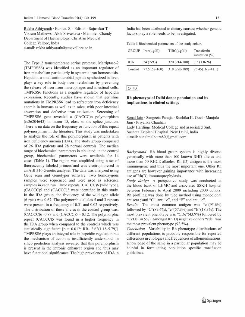
Indian J. Hematol. Blood Transfus 25(4):130–199 151
Rekha Athiyarath · Eunice. S. · Edison · Rajasekar T. · Vikram Mathews · Alok Srivastava · Mammen Chandy Department of Haematology, Christian Medical College,Vellore, India e-mail: [email protected]
The Type 2 transmembrane serine protease, Matriptase-2 (TMPRSS6) was identifi ed as an important regulator of iron metabolism particularly in systemic iron homeostasis. Hepcidin, a small antimicrobial peptide synthesized in liver, plays a key role in body iron metabolism by preventing the release of iron from macrophages and intestinal cells. TMPRSS6 functions as a negative regulator of hepcidin expression. Recently, studies have shown that germline mutations in TMPRSS6 lead to refractory iron defi ciency anemia in humans as well as in mice, with poor intestinal absorption and defective iron utilization. Screening of TMPRSS6 gene revealed a (CACCC)n polymorphism (rs36204643) in intron 15, close to the splice junction. There is no data on the frequency or function of this repeat polymorphism in the literature. This study was undertaken to analyze the role of this polymorphism in patients with iron defi ciency anemia (IDA). The study group comprised of 26 IDA patients and 28 normal controls. The median range of biochemical parameters is tabulated; in the control group, biochemical parameters were available for 14 cases (Table 1). The region was amplifi ed using a set of fl uorescently labeled primers and was electrophoresed in an ABI 310 Genetic analyzer. The data was analyzed using Gene scan and Genotyper software. Two homozygous samples were sequenced and were used as reference samples in each run. Three repeats (CACCC)6 [wild type], (CACCC)5 and (CACCC)3 were identifi ed in this study. In the IDA group, the frequency of the wild type allele (6 rpts) was 0.67. The polymorphic alleles 5 and 3 repeats were present in a frequency of 0.31 and 0.02 respectively. The distribution of these alleles in the control group was: (CACCC)6 -0.88 and (CACCC)5 – 0.12. The polymorphic repeat (CACCC)5 was found in a higher frequency in the IDA group when compared to the controls which was statistically signifi cant [p = 0.012; RR- 2.62(1.18-5.79)]. TMPRSS6 plays an integral role in hepcidin regulation but the mechanism of action is insuffi ciently understood. In silico prediction analysis revealed that this polymorphism is present in the intronic enhancer region and thus may have functional signifi cance. The high prevalence of IDA in
India has been attributed to dietary causes; whether genetic factors play a role needs to be investigated.
Table 1 Biochemical parameters of the study cohort
GROUP Iron(μg/dl) TIBC(μg/dl) Transferrinsaturation (%)
IDA 24 (7-93) 320 (214-380) 7.5 (1.8-26)
Control 77.5 (52-160) 318 (270-389) 25.45(16.2-41.1)
O 40
Rh phenotype of Delhi donor population and its implications in clinical settings
Sonal Jain · Sangeeta Pahuja · Ruchika K. Goel · Manjula Jain · Priyanka ChauhanLady Hardinge Medical College and associated Smt. Sucheta Kriplani Hospital, New Delhi, Indiae-mail: [email protected]
Background Rh blood group system is highly diverse genetically with more than 100 known RHD alleles and more than 50 RHCE allaeles. Rh (D) antigen is the most immunogenic and thus the most important one. Other Rh antigens are however gaining importance with increasing use of Rh(D) immunoprophylaxis.Study design A prospective study was conducted at the blood bank of LHMC and associated SSKH hospital between February to April 2009 including 2000 donors. Rh profi ling was done by tube method using monoclonal antisera ; anti “C”, anti “c”, anti “E” and anti “e”.Results The most common antigen was “e”(95.6%) followed by “C”(89.6%), “c”(57.3%) and “E”(18.3%). The most prevalent phenotype was “CDe”(43.9%) followed by “CcDe(34.5%). Amongst Rh(D) negative donors “cde” was the most prevalent phenotype (92.5%).Conclusion Variability in Rh phenotype distributions of different populations is probably responsible for reported differences in etiologies and frequencies of alloimunisations. Knowledge of the same in a particular population may be helpful in formulating population specifi c transfusion guidelines.
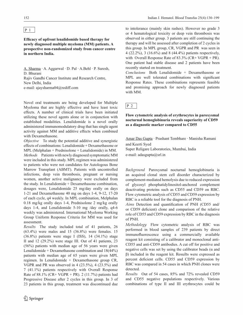
152 Indian J. Hematol. Blood Transfus 25(4):130–199
P 1
Effi cacy of upfront lenalidomide based therapy for newly diagnosed multiple myeloma (MM) patients. A prospective non-randomized study from cancer centre in northern India.
A. Sharma · A. Aggarwal · D. Pal · A.Behl · P. Suresh, D. BhuraniRajiv Gandhi Cancer Institute and Research Centre, New Delhi, Indiae-mail: [email protected]
Novel oral treatments are being developed for Multiple Myeloma that are highly effective and have least toxic effects. A number of clinical trials have been initiated utilizing these novel agents alone or in conjunction with established modalities. Lenalidomide is a novel orally administered immunomodulatory drug that has single agent activity against MM and additive effects when combined with Dexamethasone.Objective To study the potential additive and synergistic effects of combinations: Lenalidomide + Dexamethasone or MPL (Melphalan + Prednisolone + Lenalidomide) in MM.Methods Patients with newly diagnosed symptomatic MM were included in this study. MPL regimen was administered to patients who were not candidates for Autologous Bone Marrow Transplant (ABMT). Patients with uncontrolled infections, deep vein thrombosis, pregnant or nursing women, another active malignancy were excluded from the study. In Lenalidomide + Dexamethasone combination, dosages were, Lenalidomide 25 mg/day orally on days 1-21 and Dexamethasone 40 mg on days 1-4, 9-12, 17-20 of each cycle, q4 weekly. In MPL combination, Melphalan 0.18 mg/kg orally days 1-4, Prednisolone 2 mg/kg orally days 1-4, and Lenalidomide 5-10 mg /day orally, q4-6 weekly was administered. International Myeloma Working Group Uniform Response Criteria for MM was used for assessment.Results The study included total of 41 patients, 26 (63.4%) were males and 15 (36.8%) were females. 15 (36.8%) patients were stage I (ISS), 14 (34.1%) stage II and 12 (29.2%) were stage III. Out of 41 patients, 23 (56%) patients with median age of 56 years were given Lenalidomide + Dexamethasone combination and 18(44%) patients with median age of 65 years were given MPL regimen. In Lenalidomide + Dexamethasone group CR, VGPR and PR was observed in 4 (23.5%), 4 (23.5%) and 7 (41.1%) patients respectively with Overall Response Rate of 88.1% (CR+ VGPR + PR). 2 (11.7%) patients had Progressive Disease after 2 cycles in this group. In 3 of 23 patients in this group, treatment was discontinued due
to intolerance (mainly skin rashes). However no grade 3 or 4 hematological toxicity or deep vein thrombosis was observed in either group. 3 patients are still continuing the therapy and will be assessed after completion of 2 cycles in this group. In MPL group, CR, VGPR and PR was seen in 4 (22.2%), 3 (16.6%) and 8 (44.4%) patients respectively, with Overall Response Rate of 83.3% (CR+ VGPR + PR). One patient had stable disease and 2 patients have been recently started on treatment.Conclusions Both Lenalidomide + Dexamethasone or MPL are well tolerated combinations with signifi cant Response Rates. These combinations represents feasible and promising approach for newly diagnosed patients with MM.
P 2
Flow cytometric analysis of erythrocytes in paroxysmal nocturnal hemoglobinuria reveals superiority of CD59 as a diagnostic marker compared to CD55
Amar Das Gupta · Prashant Tembhare · Manisha Ramani and Keerti SyedSuper Religare Laboratories, Mumbai, Indiae-mail: [email protected]
Background Paroxysmal nocturnal hemoglobinuria is an acquired clonal stem cell disorder characterized by complement-mediated hemolysis due to reduced expression of glycosyl phosphatidylinositol-anchored complement deactivating proteins such as CD55 and CD59 on RBC. Flow cytometric analysis of CD55 and CD59 expression by RBC is a reliable tool for the diagnosis of PNH. Aims Detection and quantifi cation of PNH (CD55 and/or CD59 defi cient) clone and comparison of the relative role of CD55 and CD59 expression by RBC in the diagnosis of PNH.Methodology Flow cytometric analysis of RBC was performed in blood samples of 239 patients by direct immunofl uorescence using a commercially available reagent kit consisting of a calibrator and monoclonal anti-CD55 and anti-CD59 antibodies. A cut off for positive and negative cells was set by using the calibrator beads (α and β) included in the reagent kit. Results were expressed as percent defi cient cells. CD55 and CD59 expression by RBC was compared in 54 cases in which PNH clones were detected.Results Out of 54 cases, 85% and 72% revealed CD59 and CD55 negative populations respectively. Various combinations of type II and III erythrocytes could be
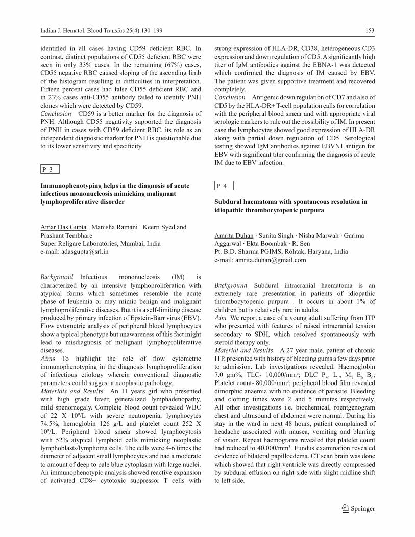
Indian J. Hematol. Blood Transfus 25(4):130–199 153
identifi ed in all cases having CD59 defi cient RBC. In contrast, distinct populations of CD55 defi cient RBC were seen in only 33% cases. In the remaining (67%) cases, CD55 negative RBC caused sloping of the ascending limb of the histogram resulting in diffi culties in interpretation. Fifteen percent cases had false CD55 defi cient RBC and in 23% cases anti-CD55 antibody failed to identify PNH clones which were detected by CD59. Conclusion CD59 is a better marker for the diagnosis of PNH. Although CD55 negativity supported the diagnosis of PNH in cases with CD59 defi cient RBC, its role as an independent diagnostic marker for PNH is questionable due to its lower sensitivity and specifi city.
P 3
Immunophenotyping helps in the diagnosis of acute infectious mononucleosis mimicking malignant lymphoproliferative disorder
Amar Das Gupta · Manisha Ramani · Keerti Syed and Prashant TembhareSuper Religare Laboratories, Mumbai, Indiae-mail: [email protected]
Background Infectious mononucleosis (IM) is characterized by an intensive lymphoproliferation with atypical forms which sometimes resemble the acute phase of leukemia or may mimic benign and malignant lymphoproliferative diseases. But it is a self-limiting disease produced by primary infection of Epstein-Barr virus (EBV). Flow cytometric analysis of peripheral blood lymphocytes show a typical phenotype but unawareness of this fact might lead to misdiagnosis of malignant lymphoproliferative diseases. Aims To highlight the role of fl ow cytometric immunophenotyping in the diagnosis lymphoproliferation of infectious etiology wherein conventional diagnostic parameters could suggest a neoplastic pathology. Materials and Results An 11 years girl who presented with high grade fever, generalized lymphadenopathy, mild spenomegaly. Complete blood count revealed WBC of 22 X 109/L with severe neutropenia, lymphocytes 74.5%, hemoglobin 126 g/L and platelet count 252 X 109/L. Peripheral blood smear showed lymphocytosis with 52% atypical lymphoid cells mimicking neoplastic lymphoblasts/lymphoma cells. The cells were 4-6 times the diameter of adjacent small lymphocytes and had a moderate to amount of deep to pale blue cytoplasm with large nuclei. An immunophenotypic analysis showed reactive expansion of activated CD8+ cytotoxic suppressor T cells with
strong expression of HLA-DR, CD38, heterogeneous CD3 expression and down regulation of CD5. A signifi cantly high titer of IgM antibodies against the EBNA-1 was detected which confi rmed the diagnosis of IM caused by EBV. The patient was given supportive treatment and recovered completely. Conclusion Antigenic down regulation of CD7 and also of CD5 by the HLA-DR+ T-cell population calls for correlation with the peripheral blood smear and with appropriate viral serologic markers to rule out the possibility of IM. In present case the lymphocytes showed good expression of HLA-DR along with partial down regulation of CD5. Serological testing showed IgM antibodies against EBVN1 antigen for EBV with signifi cant titer confi rming the diagnosis of acute IM due to EBV infection.
P 4
Subdural haematoma with spontaneous resolution in idiopathic thrombocytopenic purpura
Amrita Duhan · Sunita Singh · Nisha Marwah · Garima Aggarwal · Ekta Boombak · R. Sen Pt. B.D. Sharma PGIMS, Rohtak, Haryana, Indiae-mail: [email protected]
Background Subdural intracranial haematoma is an extremely rare presentation in patients of idiopathic thrombocytopenic purpura . It occurs in about 1% of children but is relatively rare in adults. Aim We report a case of a young adult suffering from ITP who presented with features of raised intracranial tension secondary to SDH, which resolved spontaneously with steroid therapy only. Material and Results A 27 year male, patient of chronic ITP, presented with history of bleeding gums a few days prior to admission. Lab investigations revealed: Haemoglobin 7.0 gm%; TLC- 10,000/mm3; DLC P80 L17 M3 E0 B0; Platelet count- 80,000/mm3; peripheral blood fi lm revealed dimorphic anaemia with no evidence of parasite. Bleeding and clotting times were 2 and 5 minutes respectively. All other investigations i.e. biochemical, roentgenogram chest and ultrasound of abdomen were normal. During his stay in the ward in next 48 hours, patient complained of headache associated with nausea, vomiting and blurring of vision. Repeat haemograms revealed that platelet count had reduced to 40,000/mm3. Fundus examination revealed evidence of bilateral papilloedema. CT scan brain was done which showed that right ventricle was directly compressed by subdural effusion on right side with slight midline shift to left side.
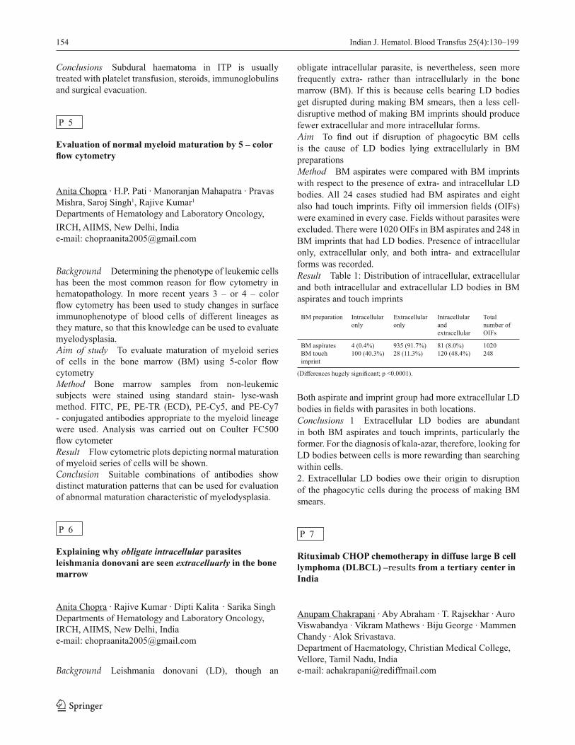
154 Indian J. Hematol. Blood Transfus 25(4):130–199
Conclusions Subdural haematoma in ITP is usually treated with platelet transfusion, steroids, immunoglobulins and surgical evacuation.
P 5
Evaluation of normal myeloid maturation by 5 – color fl ow cytometry
Anita Chopra · H.P. Pati · Manoranjan Mahapatra · Pravas Mishra, Saroj Singh1, Rajive Kumar1 Departments of Hematology and Laboratory Oncology,IRCH, AIIMS, New Delhi, Indiae-mail: [email protected]
Background Determining the phenotype of leukemic cells has been the most common reason for fl ow cytometry in hematopathology. In more recent years 3 – or 4 – color fl ow cytometry has been used to study changes in surface immunophenotype of blood cells of different lineages as they mature, so that this knowledge can be used to evaluate myelodysplasia. Aim of study To evaluate maturation of myeloid series of cells in the bone marrow (BM) using 5-color fl ow cytometry Method Bone marrow samples from non-leukemic subjects were stained using standard stain- lyse-wash method. FITC, PE, PE-TR (ECD), PE-Cy5, and PE-Cy7 - conjugated antibodies appropriate to the myeloid lineage were used. Analysis was carried out on Coulter FC500 fl ow cytometer Result Flow cytometric plots depicting normal maturation of myeloid series of cells will be shown. Conclusion Suitable combinations of antibodies show distinct maturation patterns that can be used for evaluation of abnormal maturation characteristic of myelodysplasia.
P 6
Explaining why obligate intracellular parasites leishmania donovani are seen extracelluarly in the bone marrow
Anita Chopra · Rajive Kumar · Dipti Kalita · Sarika SinghDepartments of Hematology and Laboratory Oncology, IRCH, AIIMS, New Delhi, Indiae-mail: [email protected]
Background Leishmania donovani (LD), though an
obligate intracellular parasite, is nevertheless, seen more frequently extra- rather than intracellularly in the bone marrow (BM). If this is because cells bearing LD bodies get disrupted during making BM smears, then a less cell-disruptive method of making BM imprints should produce fewer extracellular and more intracellular forms. Aim To fi nd out if disruption of phagocytic BM cells is the cause of LD bodies lying extracellularly in BM preparations Method BM aspirates were compared with BM imprints with respect to the presence of extra- and intracellular LD bodies. All 24 cases studied had BM aspirates and eight also had touch imprints. Fifty oil immersion fi elds (OIFs) were examined in every case. Fields without parasites were excluded. There were 1020 OIFs in BM aspirates and 248 in BM imprints that had LD bodies. Presence of intracellular only, extracellular only, and both intra- and extracellular forms was recorded. Result Table 1: Distribution of intracellular, extracellular and both intracellular and extracellular LD bodies in BM aspirates and touch imprints
BM preparation Intracellular only
Extracellular only
Intracellular and extracellular
Total number of OIFs
BM aspirates BM touch imprint
4 (0.4%) 100 (40.3%)
935 (91.7%) 28 (11.3%)
81 (8.0%) 120 (48.4%)
1020 248
(Differences hugely signifi cant; p <0.0001).
Both aspirate and imprint group had more extracellular LD bodies in fi elds with parasites in both locations. Conclusions 1 Extracellular LD bodies are abundant in both BM aspirates and touch imprints, particularly the former. For the diagnosis of kala-azar, therefore, looking for LD bodies between cells is more rewarding than searching within cells. 2. Extracellular LD bodies owe their origin to disruption of the phagocytic cells during the process of making BM smears.
P 7
Rituximab CHOP chemotherapy in diffuse large B cell lymphoma (DLBCL) –results from a tertiary center in India
Anupam Chakrapani · Aby Abraham · T. Rajsekhar · Auro Viswabandya · Vikram Mathews · Biju George · Mammen Chandy · Alok Srivastava. Department of Haematology, Christian Medical College, Vellore, Tamil Nadu, Indiae-mail: [email protected]
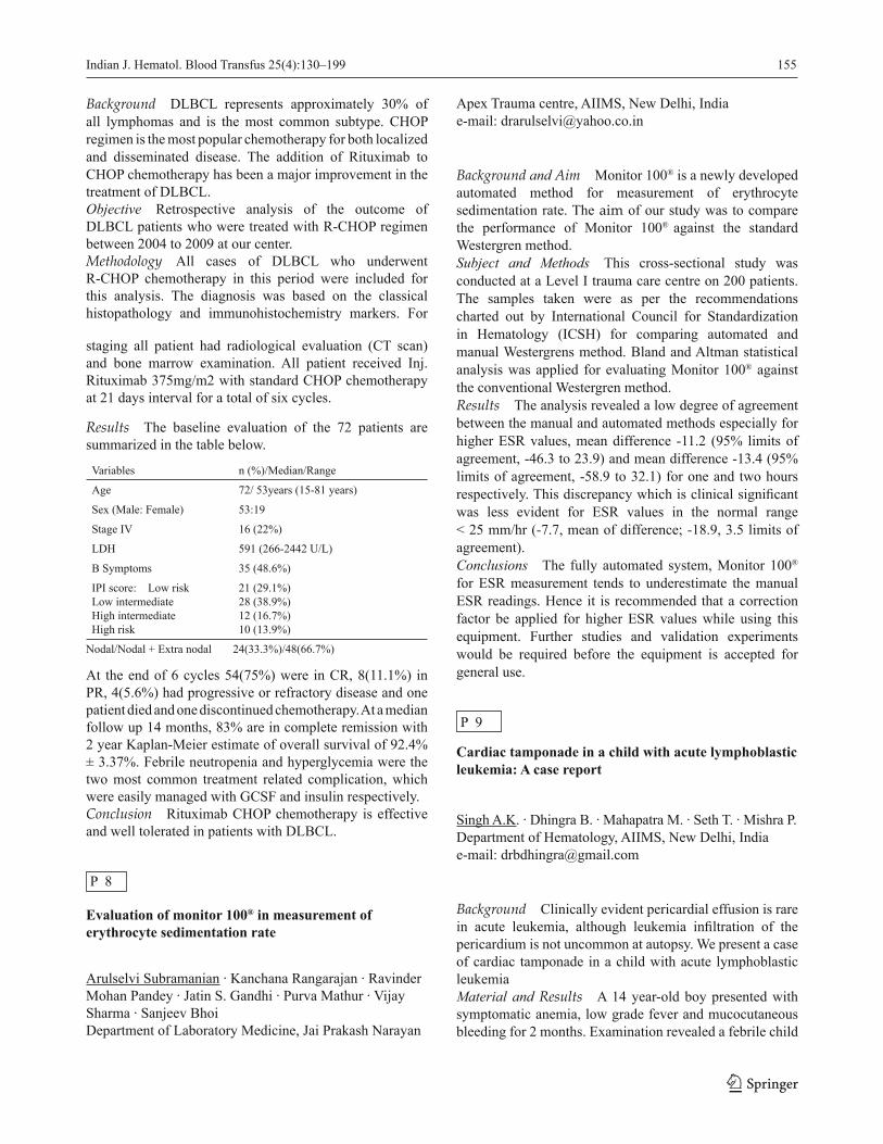
Indian J. Hematol. Blood Transfus 25(4):130–199 155
Background DLBCL represents approximately 30% of all lymphomas and is the most common subtype. CHOP regimen is the most popular chemotherapy for both localized and disseminated disease. The addition of Rituximab to CHOP chemotherapy has been a major improvement in the treatment of DLBCL.Objective Retrospective analysis of the outcome of DLBCL patients who were treated with R-CHOP regimen between 2004 to 2009 at our center.Methodology All cases of DLBCL who underwent R-CHOP chemotherapy in this period were included for this analysis. The diagnosis was based on the classical histopathology and immunohistochemistry markers. For
staging all patient had radiological evaluation (CT scan) and bone marrow examination. All patient received Inj. Rituximab 375mg/m2 with standard CHOP chemotherapy at 21 days interval for a total of six cycles.
Results The baseline evaluation of the 72 patients are summarized in the table below.
Variables n (%)/Median/Range
Age 72/ 53years (15-81 years)
Sex (Male: Female) 53:19
Stage IV 16 (22%)
LDH 591 (266-2442 U/L)
B Symptoms 35 (48.6%)
IPI score: Low riskLow intermediateHigh intermediateHigh risk
21 (29.1%)28 (38.9%)12 (16.7%)10 (13.9%)
Nodal/Nodal + Extra nodal 24(33.3%)/48(66.7%)
At the end of 6 cycles 54(75%) were in CR, 8(11.1%) in PR, 4(5.6%) had progressive or refractory disease and one patient died and one discontinued chemotherapy. At a median follow up 14 months, 83% are in complete remission with 2 year Kaplan-Meier estimate of overall survival of 92.4% ± 3.37%. Febrile neutropenia and hyperglycemia were the two most common treatment related complication, which were easily managed with GCSF and insulin respectively.Conclusion Rituximab CHOP chemotherapy is effective and well tolerated in patients with DLBCL.
P 8
Evaluation of monitor 100® in measurement of erythrocyte sedimentation rate
Arulselvi Subramanian · Kanchana Rangarajan · Ravinder Mohan Pandey · Jatin S. Gandhi · Purva Mathur · Vijay Sharma · Sanjeev Bhoi Department of Laboratory Medicine, Jai Prakash Narayan
Apex Trauma centre, AIIMS, New Delhi, Indiae-mail: [email protected]
Background and Aim Monitor 100® is a newly developed automated method for measurement of erythrocyte sedimentation rate. The aim of our study was to compare the performance of Monitor 100® against the standard Westergren method. Subject and Methods This cross-sectional study was conducted at a Level I trauma care centre on 200 patients. The samples taken were as per the recommendations charted out by International Council for Standardization in Hematology (ICSH) for comparing automated and manual Westergrens method. Bland and Altman statistical analysis was applied for evaluating Monitor 100® against the conventional Westergren method. Results The analysis revealed a low degree of agreement between the manual and automated methods especially for higher ESR values, mean difference -11.2 (95% limits of agreement, -46.3 to 23.9) and mean difference -13.4 (95% limits of agreement, -58.9 to 32.1) for one and two hours respectively. This discrepancy which is clinical signifi cant was less evident for ESR values in the normal range < 25 mm/hr (-7.7, mean of difference; -18.9, 3.5 limits of agreement). Conclusions The fully automated system, Monitor 100®
for ESR measurement tends to underestimate the manual ESR readings. Hence it is recommended that a correction factor be applied for higher ESR values while using this equipment. Further studies and validation experiments would be required before the equipment is accepted for general use.
P 9
Cardiac tamponade in a child with acute lymphoblastic leukemia: A case report
Singh A.K. · Dhingra B. · Mahapatra M. · Seth T. · Mishra P.Department of Hematology, AIIMS, New Delhi, Indiae-mail: [email protected]
Background Clinically evident pericardial effusion is rare in acute leukemia, although leukemia infi ltration of the pericardium is not uncommon at autopsy. We present a case of cardiac tamponade in a child with acute lymphoblastic leukemia Material and Results A 14 year-old boy presented with symptomatic anemia, low grade fever and mucocutaneous bleeding for 2 months. Examination revealed a febrile child
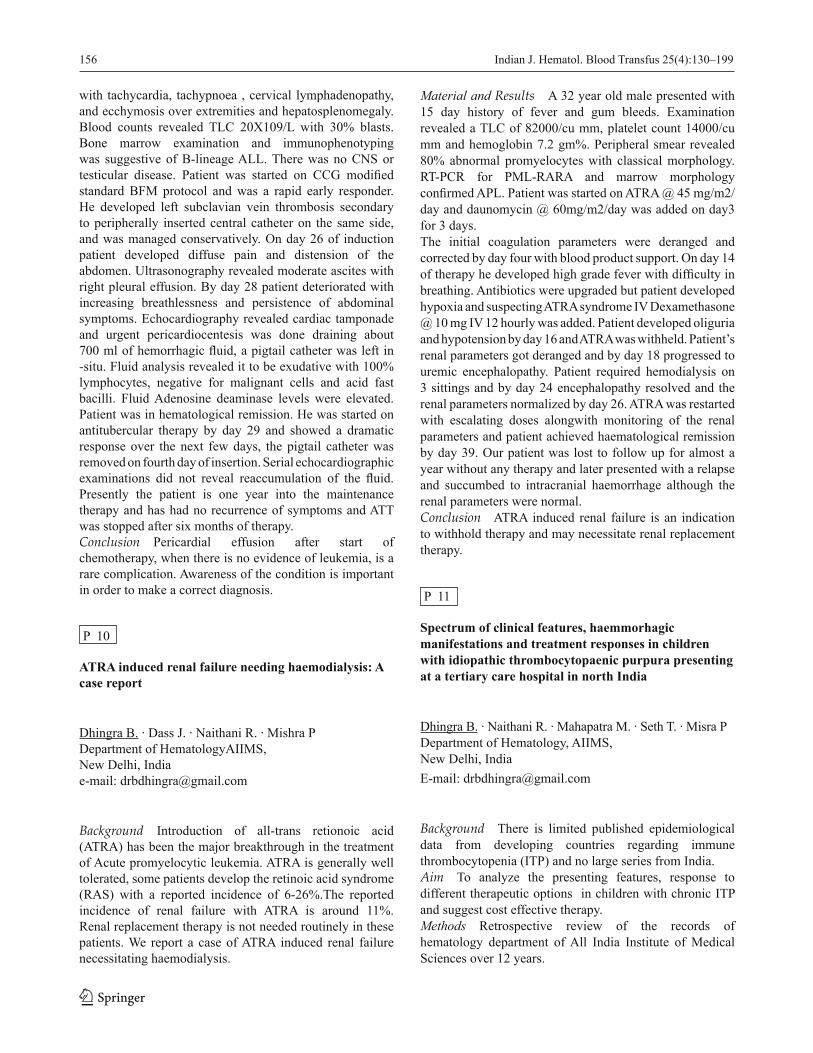
156 Indian J. Hematol. Blood Transfus 25(4):130–199
with tachycardia, tachypnoea , cervical lymphadenopathy, and ecchymosis over extremities and hepatosplenomegaly. Blood counts revealed TLC 20X109/L with 30% blasts. Bone marrow examination and immunophenotyping was suggestive of B-lineage ALL. There was no CNS or testicular disease. Patient was started on CCG modifi ed standard BFM protocol and was a rapid early responder. He developed left subclavian vein thrombosis secondary to peripherally inserted central catheter on the same side, and was managed conservatively. On day 26 of induction patient developed diffuse pain and distension of the abdomen. Ultrasonography revealed moderate ascites with right pleural effusion. By day 28 patient deteriorated with increasing breathlessness and persistence of abdominal symptoms. Echocardiography revealed cardiac tamponade and urgent pericardiocentesis was done draining about 700 ml of hemorrhagic fl uid, a pigtail catheter was left in -situ. Fluid analysis revealed it to be exudative with 100% lymphocytes, negative for malignant cells and acid fast bacilli. Fluid Adenosine deaminase levels were elevated. Patient was in hematological remission. He was started on antitubercular therapy by day 29 and showed a dramatic response over the next few days, the pigtail catheter was removed on fourth day of insertion. Serial echocardiographic examinations did not reveal reaccumulation of the fl uid. Presently the patient is one year into the maintenance therapy and has had no recurrence of symptoms and ATT was stopped after six months of therapy.Conclusion Pericardial effusion after start of chemotherapy, when there is no evidence of leukemia, is a rare complication. Awareness of the condition is important in order to make a correct diagnosis.
P 10
ATRA induced renal failure needing haemodialysis: A case report
Dhingra B. · Dass J. · Naithani R. · Mishra P Department of HematologyAIIMS,New Delhi, Indiae-mail: [email protected]
Background Introduction of all-trans retionoic acid (ATRA) has been the major breakthrough in the treatment of Acute promyelocytic leukemia. ATRA is generally well tolerated, some patients develop the retinoic acid syndrome (RAS) with a reported incidence of 6-26%.The reported incidence of renal failure with ATRA is around 11%. Renal replacement therapy is not needed routinely in these patients. We report a case of ATRA induced renal failure necessitating haemodialysis.
Material and Results A 32 year old male presented with 15 day history of fever and gum bleeds. Examination revealed a TLC of 82000/cu mm, platelet count 14000/cu mm and hemoglobin 7.2 gm%. Peripheral smear revealed 80% abnormal promyelocytes with classical morphology. RT-PCR for PML-RARA and marrow morphology confi rmed APL. Patient was started on ATRA @ 45 mg/m2/day and daunomycin @ 60mg/m2/day was added on day3 for 3 days.The initial coagulation parameters were deranged and corrected by day four with blood product support. On day 14 of therapy he developed high grade fever with diffi culty in breathing. Antibiotics were upgraded but patient developed hypoxia and suspecting ATRA syndrome IV Dexamethasone @ 10 mg IV 12 hourly was added. Patient developed oliguria and hypotension by day 16 and ATRA was withheld. Patient’s renal parameters got deranged and by day 18 progressed to uremic encephalopathy. Patient required hemodialysis on 3 sittings and by day 24 encephalopathy resolved and the renal parameters normalized by day 26. ATRA was restarted with escalating doses alongwith monitoring of the renal parameters and patient achieved haematological remission by day 39. Our patient was lost to follow up for almost a year without any therapy and later presented with a relapse and succumbed to intracranial haemorrhage although the renal parameters were normal. Conclusion ATRA induced renal failure is an indication to withhold therapy and may necessitate renal replacement therapy.
P 11
Spectrum of clinical features, haemmorhagic manifestations and treatment responses in children with idiopathic thrombocytopaenic purpura presenting at a tertiary care hospital in north India
Dhingra B. · Naithani R. · Mahapatra M. · Seth T. · Misra P Department of Hematology, AIIMS, New Delhi, IndiaE-mail: [email protected]
Background There is limited published epidemiological data from developing countries regarding immune thrombocytopenia (ITP) and no large series from India. Aim To analyze the presenting features, response to different therapeutic options in children with chronic ITP and suggest cost effective therapy. Methods Retrospective review of the records of hematology department of All India Institute of Medical Sciences over 12 years.
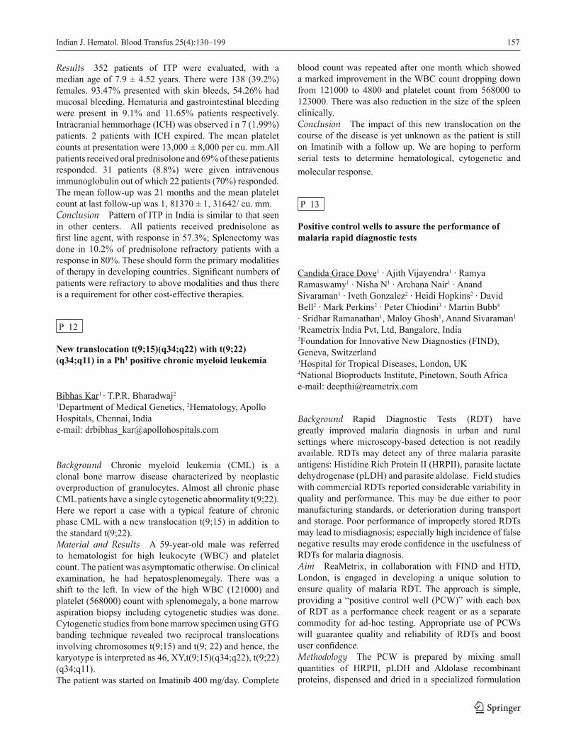
Indian J. Hematol. Blood Transfus 25(4):130–199 157
blood count was repeated after one month which showed a marked improvement in the WBC count dropping down from 121000 to 4800 and platelet count from 568000 to 123000. There was also reduction in the size of the spleen clinically. Conclusion The impact of this new translocation on the course of the disease is yet unknown as the patient is still on Imatinib with a follow up. We are hoping to perform serial tests to determine hematological, cytogenetic and molecular response.
P 13
Positive control wells to assure the performance of malaria rapid diagnostic tests
Candida Grace Dove1 · Ajith Vijayendra1 · Ramya Ramaswamy1 · Nisha N1 · Archana Nair1 · Anand Sivaraman1 · Iveth Gonzalez2 · Heidi Hopkins2 · David Bell2 · Mark Perkins2 · Peter Chiodini3 · Martin Bubb4 · Sridhar Ramanathan1, Maloy Ghosh1, Anand Sivaraman1
1Reametrix India Pvt, Ltd, Bangalore, India2Foundation for Innovative New Diagnostics (FIND), Geneva, Switzerland3Hospital for Tropical Diseases, London, UK4National Bioproducts Institute, Pinetown, South Africae-mail: [email protected]
Background Rapid Diagnostic Tests (RDT) have greatly improved malaria diagnosis in urban and rural settings where microscopy-based detection is not readily available. RDTs may detect any of three malaria parasite antigens: Histidine Rich Protein II (HRPII), parasite lactate dehydrogenase (pLDH) and parasite aldolase. Field studies with commercial RDTs reported considerable variability in quality and performance. This may be due either to poor manufacturing standards, or deterioration during transport and storage. Poor performance of improperly stored RDTs may lead to misdiagnosis; especially high incidence of false negative results may erode confi dence in the usefulness of RDTs for malaria diagnosis.Aim ReaMetrix, in collaboration with FIND and HTD, London, is engaged in developing a unique solution to ensure quality of malaria RDT. The approach is simple, providing a “positive control well (PCW)” with each box of RDT as a performance check reagent or as a separate commodity for ad-hoc testing. Appropriate use of PCWs will guarantee quality and reliability of RDTs and boost user confi dence.Methodology The PCW is prepared by mixing small quantities of HRPII, pLDH and Aldolase recombinant proteins, dispensed and dried in a specialized formulation
Results 352 patients of ITP were evaluated, with a median age of 7.9 ± 4.52 years. There were 138 (39.2%) females. 93.47% presented with skin bleeds, 54.26% had mucosal bleeding. Hematuria and gastrointestinal bleeding were present in 9.1% and 11.65% patients respectively. Intracranial hemmorhage (ICH) was observed i n 7 (1.99%) patients. 2 patients with ICH expired. The mean platelet counts at presentation were 13,000 ± 8,000 per cu. mm.All patients received oral prednisolone and 69% of these patients responded. 31 patients (8.8%) were given intravenous immunoglobulin out of which 22 patients (70%) responded. The mean follow-up was 21 months and the mean platelet count at last follow-up was 1, 81370 ± 1, 31642/ cu. mm.Conclusion Pattern of ITP in India is similar to that seen in other centers. All patients received prednisolone as fi rst line agent, with response in 57.3%; Splenectomy was done in 10.2% of prednisolone refractory patients with a response in 80%. These should form the primary modalities of therapy in developing countries. Signifi cant numbers of patients were refractory to above modalities and thus there is a requirement for other cost-effective therapies.
P 12
New translocation t(9;15)(q34;q22) with t(9;22)(q34;q11) in a Ph1 positive chronic myeloid leukemia
Bibhas Kar1 · T.P.R. Bharadwaj2 1Department of Medical Genetics, 2Hematology, Apollo Hospitals, Chennai, India e-mail: [email protected]
Background Chronic myeloid leukemia (CML) is a clonal bone marrow disease characterized by neoplastic overproduction of granulocytes. Almost all chronic phase CML patients have a single cytogenetic abnormality t(9;22). Here we report a case with a typical feature of chronic phase CML with a new translocation t(9;15) in addition to the standard t(9;22). Material and Results A 59-year-old male was referred to hematologist for high leukocyte (WBC) and platelet count. The patient was asymptomatic otherwise. On clinical examination, he had hepatosplenomegaly. There was a shift to the left. In view of the high WBC (121000) and platelet (568000) count with splenomegaly, a bone marrow aspiration biopsy including cytogenetic studies was done. Cytogenetic studies from bone marrow specimen using GTG banding technique revealed two reciprocal translocations involving chromosomes t(9;15) and t(9; 22) and hence, the karyotype is interpreted as 46, XY,t(9;15)(q34;q22), t(9;22)(q34;q11). The patient was started on Imatinib 400 mg/day. Complete
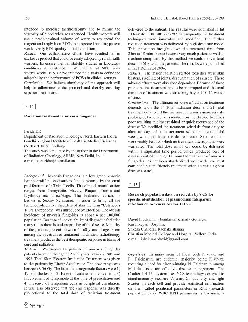
158 Indian J. Hematol. Blood Transfus 25(4):130–199
intended to increase thermostability and to mimic the viscosity of blood when resuspended. Health workers will use a predetermined volume of water to resuspend the reagent and apply it on RDTs. An expected banding pattern would verify RDT quality in fi eld condition.Results Our collaborative efforts have resulted in an exclusive product that could be easily adopted by rural health workers. Extensive thermal stability studies in laboratory conditions demonstrated PCW stability at 60˚C over several weeks. FIND have initiated fi eld trials to defi ne the adaptability and performance of PCWs in clinical settings. Conclusion We believe simplicity of the approach will help in adherence to the protocol and thereby ensuring superior health care.
P 14
Radiation treatment in mycosis fungoides
Parida DKDepartment of Radiation Oncology, North Eastern Indira Gandhi Regional Institute of Health & Medical Sciences (NEIGRIHMS), ShillongThe study was conducted by the author in the Department of Radiation Oncology, AIIMS, New Delhi, Indiae-mail: [email protected]
Background Mycosis Fungoides is a low grade, chronic lymphoproliferative disorder of the skin caused by abnormal proliferation of CD4+ T-cells. The clinical manifestation ranges from Premycotic, Macule, Plaques, Tumos and Erythrodermic phase/stage. The leukemic variant is known as Sezary Syndrome. In order to bring all the lymphoproliferative disorders of skin the term “Cutaneous T-Cell Lymphoma” was introduced by Eldelson. The overall incidence of mycosis fungoides is about 4 per 100,000 population. Because of unavailability of diagnostic facilities many times there is underreporting of the disease. Majority of the patients present between 40-60 years of age. From among the spectrum of treatment modalities, radiotherapy treatment produces the best therapeutic response in terms of cure and palliation.Material We treated 14 patients of mycosis fungoides patients between the age of 27-82 years between 1985 and 1998. Total Skin Electron Irradiation Treatment was given to the patients by Linear Accelerator. The dose range was between 8-36 Gy. The important prognostic factors were 1) Type of the lesions 2) Extent of cutaneous involvement, 3) Involvement of lymphnode at the time of presentation and 4) Presence of lymphoma cells in peripheral circulation. It was also observed that the end response was directly proportional to the total dose of radiation treatment
delivered to the patient. The results were published in Int J Dermatol 2001:40; 295-297. Subsequently the treatment techniques were innovated and modifi ed. The further radiation treatment was delivered by high dose rate mode. This innovation brought down the treatment time from 2 hrs to 15 mins, hence became very much patient as well as machine compliant. By this method we could deliver total dose of 36Gy to all the patients. The results were published in Int J Dermatol 2004. Results The major radiation related toxicities were skin blisters, swelling of joints, desquamation of skin etc. These adverse effects were also dose dependant. Because of these problems the treatment has to be interrupted and the total duration of treatment was stretching beyond 10-12 weeks of time. Conclusions The ultimate response of radiation treatment depends upon the 1) Total radiation dose and 2) Total treatment duration. If the treatment duration is unnecessarily prolonged, the effect of radiation on the disease becomes poor resulting in either residual or quick recurrence of the disease.We modifi ed the treatment schedule from daily to alternate day radiation treatment schedule beyond third week, which produced the desired result. Skin reactions were visibly less for which no treatment interruptions were warranted. The total dose of 36 Gy could be delivered within a stipulated time period which produced best of disease control. Though till now the treatment of mycosis fungoides has not been standardized worldwide, we must consider a patient friendly treatment schedule resulting best disease control.
P 15
Research population data on red cells by VCS for specifi c identifi cation of plasmodium falciparum infection on beckman coulter LH 750
David Inbakumar · Janakiram Kamal · Govindan Karthikeyan · JosphineSukesh Chandran RadhakrishananChristian Medical College and Hospital, Vellore, India e-mail: [email protected]
Objectives In many areas of India both Pl.Vivax and Pl. Falciparum are endemic, majority being Pl.Vivax, requiring a need for discriminating Pl. Falciparum among Malaria cases for effective disease management. The Coulter LH 750 system uses VCS technology designed to simultaneously measure Volume, Conductivity and light Scatter on each cell and provide statistical information on them called positional parameters or RPD (research population data). WBC RPD parameters is becoming a
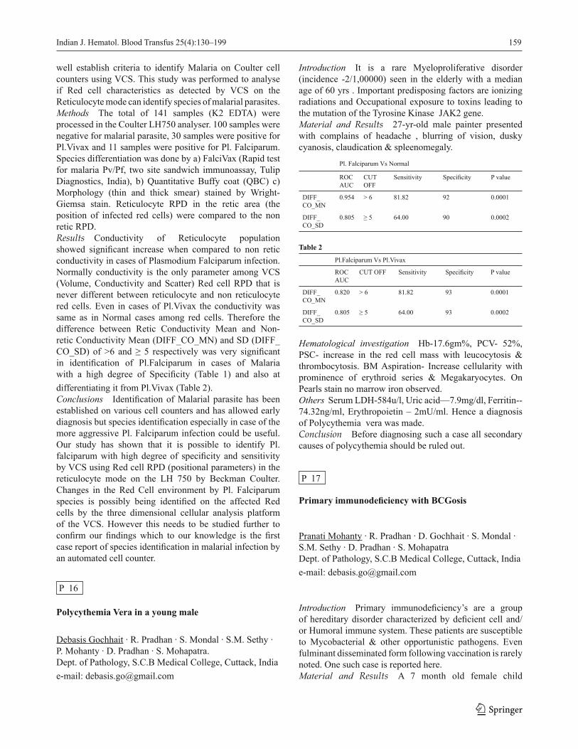
Indian J. Hematol. Blood Transfus 25(4):130–199 159
well establish criteria to identify Malaria on Coulter cell counters using VCS. This study was performed to analyse if Red cell characteristics as detected by VCS on the Reticulocyte mode can identify species of malarial parasites.Methods The total of 141 samples (K2 EDTA) were processed in the Coulter LH750 analyser. 100 samples were negative for malarial parasite, 30 samples were positive for Pl.Vivax and 11 samples were positive for Pl. Falciparum. Species differentiation was done by a) FalciVax (Rapid test for malaria Pv/Pf, two site sandwich immunoassay, Tulip Diagnostics, India), b) Quantitative Buffy coat (QBC) c) Morphology (thin and thick smear) stained by Wright-Giemsa stain. Reticulocyte RPD in the retic area (the position of infected red cells) were compared to the non retic RPD.Results Conductivity of Reticulocyte population showed signifi cant increase when compared to non retic conductivity in cases of Plasmodium Falciparum infection. Normally conductivity is the only parameter among VCS (Volume, Conductivity and Scatter) Red cell RPD that is never different between reticulocyte and non reticulocyte red cells. Even in cases of Pl.Vivax the conductivity was same as in Normal cases among red cells. Therefore the difference between Retic Conductivity Mean and Non-retic Conductivity Mean (DIFF_CO_MN) and SD (DIFF_CO_SD) of >6 and ≥ 5 respectively was very signifi cant in identifi cation of Pl.Falciparum in cases of Malaria with a high degree of Specifi city (Table 1) and also at differentiating it from Pl.Vivax (Table 2).Conclusions Identifi cation of Malarial parasite has been established on various cell counters and has allowed early diagnosis but species identifi cation especially in case of the more aggressive Pl. Falciparum infection could be useful. Our study has shown that it is possible to identify Pl. falciparum with high degree of specifi city and sensitivity by VCS using Red cell RPD (positional parameters) in the reticulocyte mode on the LH 750 by Beckman Coulter. Changes in the Red Cell environment by Pl. Falciparum species is possibly being identifi ed on the affected Red cells by the three dimensional cellular analysis platform of the VCS. However this needs to be studied further to confi rm our fi ndings which to our knowledge is the fi rst case report of species identifi cation in malarial infection by an automated cell counter.
P 16
Polycythemia Vera in a young male
Debasis Gochhait · R. Pradhan · S. Mondal · S.M. Sethy · P. Mohanty · D. Pradhan · S. Mohapatra. Dept. of Pathology, S.C.B Medical College, Cuttack, Indiae-mail: [email protected]
Introduction It is a rare Myeloproliferative disorder (incidence -2/1,00000) seen in the elderly with a median age of 60 yrs . Important predisposing factors are ionizing radiations and Occupational exposure to toxins leading to the mutation of the Tyrosine Kinase JAK2 gene.Material and Results 27-yr-old male painter presented with complains of headache , blurring of vision, dusky cyanosis, claudication & spleenomegaly.
Pl. Falciparum Vs Normal
ROC AUC
CUT OFF
Sensitivity Specifi city P value
DIFF_CO_MN
0.954 > 6 81.82 92 0.0001
DIFF_CO_SD
0.805 ≥ 5 64.00 90 0.0002
Table 2 Pl.Falciparum Vs Pl.Vivax
ROC AUC
CUT OFF Sensitivity Specifi city P value
DIFF_CO_MN
0.820 > 6 81.82 93 0.0001
DIFF_CO_SD
0.805 ≥ 5 64.00 93 0.0002
Hematological investigation Hb-17.6gm%, PCV- 52%, PSC- increase in the red cell mass with leucocytosis & thrombocytosis. BM Aspiration- Increase cellularity with prominence of erythroid series & Megakaryocytes. On Pearls stain no marrow iron observed. Others Serum LDH-584u/l, Uric acid—7.9mg/dl, Ferritin-- 74.32ng/ml, Erythropoietin – 2mU/ml. Hence a diagnosis of Polycythemia vera was made.Conclusion Before diagnosing such a case all secondary causes of polycythemia should be ruled out.
P 17
Primary immunodefi ciency with BCGosis
Pranati Mohanty · R. Pradhan · D. Gochhait · S. Mondal · S.M. Sethy · D. Pradhan · S. MohapatraDept. of Pathology, S.C.B Medical College, Cuttack, Indiae-mail: [email protected]
Introduction Primary immunodefi ciency’s are a group of hereditary disorder characterized by defi cient cell and/or Humoral immune system. These patients are susceptible to Mycobacterial & other opportunistic pathogens. Even fulminant disseminated form following vaccination is rarely noted. One such case is reported here.Material and Results A 7 month old female child
160 Indian J. Hematol. Blood Transfus 25(4):130–199
presented with generalized lymphadenopathy & hepatospleenomegaly with low grade fever for last 2 months . Past history was nothing suggestive except BCG vaccination in the 1st month. No abnormality was noted in the PS & Hemogram. Cytosmear of lymph node showed good no. of macrophages packed with AFB. Lymph node biopsy revealed granuloma’s with AFB packed Macrophages. Culture from LN aspirate in LJ medium confi rmed M Bovis. Patient was HIV negative, & non responsive to ATT even after 3 months. Repeat aspirate showed similar picture. The baby was investigated for primary immune-defi ciency when family history suggested one of her female sibling death of BCGosis. Absolute T cell count showed reduced no of CD3 & CD4 cells. With this a diagnosis of primary immune-defi ciency was made & the baby is on follow-up. Conclusion Dissemination following BCG vaccination warrants prompt investigation to diagnose primary immune defi ciency disorders in HIV negative individuals.
P 18
Neurofi bromatosis associated pediatric MDS evolving into JMML
Rajshree Pradhan · D. Gochhait · S. Mondal · S.M. Sethy · P. Mohanty · D. Pradhan · S. MohapatraDept. of Pathology, S.C.B Medical College, Cuttack, Indiae-mail: [email protected]
Introduction Pediatric MDS is an extremely rare entity. Its association secondary to familial neurofi bromatosis in pediatric population is extremely rare. It may evolve in to acute leukemia or JMML.Material and Results A six-yr-old male child with defi nite history of familial neurofi bromatosis presented with transfusion dependant anemia. PS & BM revealed features of MDS and was diagnosed as RCMD. karyogram was normal & the child was followed up . After 2 yrs PS showed monocytosis ( >1×10⁹/lt) and blasts (including Promonocytes) <20% of leucocytes, HBF -6.5 gm%, and immature granulocytes in the PS. Hence a fi nal diagnosis of pediatric MDS evolving in to JMML was made.Conclusion While following a case of pediatric MDS even with a normal karyogram possibility of JMML should be kept in mind.
P 19
Clinicohaematological, fl owcytometric, cytogenetics
study in acute biphenotypic leukemia: Short series of fi ve cases
Devika Gupta · Tathagata Chatterjee · S. Bhattacharya · D.K. Mishra · Reena Bharadwaj · Velu Nair · S. Das · Sanjeev Chowdhury · Mandeep KaurArmy Hospital Research & Referral , New Delhi, Indiae-mail: [email protected]
Introduction Leukemias are characterized by an idiopathic proliferation of a progenitor cell that is committed to a single cell lineage. The diagnosis of acute leukemias (AL) relies both on clinical features and an array of multidisciplinary approaches including morphology, immunophenotyping, cytogenetic and molecular investigations. A minority of acute leukemias have features characteristic of both the myeloid and lymphoid lineages and for this reason are designated mixed-lineage, hybrid or biphenotypic acute leukemias (BAL).Material and Method Five cases diagnosed as acute leukemia showing biphenotypic morphology were studied for clinical presentation. Heamatological parameters, bone marrow examination and fl owcytometery for immunophenotyping was carried out. Five cases of leukemia with dual lineage differentiation are being described here.Results Peripheral smear & bone marrow morphological study revealed two types of blasts.Immunophenotyping done by fl ow cytometry showed expression of both myeloid and lymphoid cell markers with a score of >2 in each of the lineages.Conclusion Biphenotypic acute leukemia is an uncommon type of leukemia which probably arises in a multipotent progenitor cell with the capability of differentiating along both myeloid and lymphoid lineages.The Criteria for the diagnosis of BAL is based on scoring system adopted by European Group Of Immunological Classifi cation of Leukemia (EGIL).The scoring system distinguishes BAL from leukemias with aberrant expression.
P 20
“Clinicohematological correlation and trends of haematological malignancies: An Experience at a major tertiary care centre of Himalayan Region”
Harish Chandra · Smita Chandra · Neena Chauhan · Geeta Negi · Nadia Shirazi · Sanjiv Kumar VermaDepartment of Pathology Himalayan Institute of Medical Sciences Swami Ram Nagar, Jolly Grant Dehradun, Uttarakhand, Indiae-mail: [email protected]
Indian J. Hematol. Blood Transfus 25(4):130–199 161
Background Haematological malignancies form an integral part of the total burden of tumours in any area. The study of trends and clinical correlation of these malignancies form a basic platform for better diagnosis and formulation of steps for early detection and appropriate treatment. Aim The present study analysed the epidemiological trends and clinicohematologically correlated these cases which were diagnosed in the haematology laboratory of Himalayan Institute of Medical Sciences Dehradun, from 1 January 2006 till 31 August 2009.Methods A detailed clinical history and examination for every patient was followed by routine haematological investigations. Bone marrow examination (aspiration/ trephine) and cytochemistry was done for every case. Selected cases were also subjected to fl ow cytometry.Results Total of 360 haematological malignancies were diagnosed with a male to female ratio of 1.64:1 and mean age of 40.86 years. Acute myeloid leukaemia was the commonest malignancy (19.44% of total malignancies) followed by chronic myeloid leukaemia (15.27 %). Bilineal leukaemia was diagnosed in 14 cases and 13 cases were without morphological typing and were also not subjected to immunophenotyping. One rare case of plasma cell leukaemia along with a case of chronic lymphocytic leukaemia in a female with infi ltrating breast carcinoma on therapy was also diagnosed. Weakness, body aches and fever were the commonest symptoms and anaemia the commonest sign.Conclusion There is an increase in incidence of the haematological malignancies in the Himalayan region of Uttarakhand which may be due to better diagnostic facilities and awareness in the remote areas. Males are affected more and acute myeloid leukaemia is the commonest malignancy. Few rare malignancies were also observed along with certain notable clinical presentations. Morphological features along with cytochemistry still remain the main diagnostic criteria and immunophenotyping or cytogenetics is not possible in majority of cases due to poor fi nancial condition of the patients.
P 21
Morphology vs fl ow cytometry in the diagnosis of acute leukemia
Himabindu Nemani · Chetan Manohar · Sushma Belurkar KMC, Manipal, Indiae-mail: [email protected]
Background Morphology and cytochemical staining has been the gold standard in the diagnosis of acute leukemia
for many decades. However, in recent years the availability of fl ow cytometers along with extensive panel of antibody reagents for characterizing blood cell lineage question the usefulness of continuing routine use of the cytochemical staining as gold standard for the diagnosis of acute leukemia. Aim 1. Correlation of morphologic diagnosis with fl ow cytometric analysis results in acute leukemia 2. To evaluate the usefulness of fl ow cytometry in acute leukemia Methodology A retrospective study of 2-years from August 2007 to August 2009 was done at Kasturba Medical Hospital, Manipal, to correlate the diagnosis of acute leukemia based on morphology with fl ow cytometry. During this period, 200 patients with age ranging from 2 to 89-years were diagnosed as acute leukemia based on morphology. Out of these 200 patients, fl ow cytometry was done for 47 patients. Bone marrow aspirates and fl ow results were evaluated for these 47 patients (34 ALL and 13 AML). Result Flow cytometry and cytochemical stains correlated well. Defi nitive diagnoses were feasible using cytochemical staining and morphology results alone in 42 out of 47 patients (89.36 %). In 5 patients with unclear morphology, fl ow cytometry proved to be a useful tool in identifying the cell lineage.Conclusion Results from this study indicate that fl ow cytometry study has become a powerful tool for diagnosis and management of acute leukemia. In about 90% of our cases, morphology provided defi nitive diagnosis but failed in about 10%. Flow cytometry became essential in these 10% cases for a defi nitive diagnosis. Flow cytometry also helped in identifying biphenotypic and leukemia with aberrant expressions. Thus fl ow cytometry has become an invaluable tool in diagnosis, classifi cation and monitoring of patients with acute leukemia.
P 22
Identifi cation of leishmania donovani haemoparasite on LH 750
Janakiram Kamal · David Inbakumar · Govindan Karthikeyan · Josphine · Sukesh Chandran Radhakrishanan1 Christian Medical College and Hospital, Vellore, Indiae-mail: [email protected]
Background Leishmaniasis is a vector borne parasitic disease caused by the haemofl agellate Leishmania. The diagnosis of Leishmaniasis is reliably made by
162 Indian J. Hematol. Blood Transfus 25(4):130–199
demonstration of the parasite on Bone marrow slides stained with Romanowsky dyes or on histopathology and by isolation either in culture or by animal inoculation of biopsy samples or tissue aspirate. Though the amastigote forms of the parasite are revealed inside the circulating monocytes and neutrophils, it is often diffi cult to locate because of small numbers and further compounded by cytopenia. It is described that VCS technology can be used for detection of malarial parasite and other haemoparasites including Leishmania. Detection of Malaria on various cell counters has been well established.Methods 32-year-old female, a resident of an area endemic for Leishmaniasis in India was admitted in our hospital on 2nd Aug 2008. She was hospitalized with complaints of fever, weight loss and abdominal discomfort of 3 months duration. On clinical evaluation she was found to have massive splenomegaly. Haemogram done are shown in Table 1. Peripheral Blood showed Monocytosis with many activated forms and very few monocytes with intracytoplasmic LD bodies. Buffy coat smears revealed monocytes with varying numbers – ranging from one to many- of intracellular parasite.The parasite was observed in a few neutrophils also. No extracellular forms were detected. Bone marrow evaluation done on 4th Aug 2008 showed a hypercellular aspirate with numerous intracellular and extracellular forms of the parasite Leishmania. The CBC profi le and the WBC positional parameters as analysed by Coulter LH 755 received on dates 30th July 2008 (out patient), 4th and 5th Aug 2008 are shown in Table 2.
Hb (gm%
TC/ mm3
DC %(Hemogram) PLC/mm3
P L M E B
Day1 5.6 1700 48.1 32.5 18.3 0.7 0.4 95000
Day2 4.4 1300 44 34 19.7 0.9 0.9 55000
Day3 4.2 1900 33.8 42.8 21.3 0.8 0.5 52000
Lymphocyte Monocyte
Mean SD Mean SD
V D1 93 16.89 171 26.88
D2 86 17.16 169 28.85
D3 77 25.07 169 27.12
C D1 118 14.32 129 5.44
D2 118 13.79 127 5.50
D3 123 21.19 128 5.4
S D1 70 18.53 91 11.96
D2 71 20.63 90 12.34
D3 69 22.49 90 12.09
Results There was an abnormal population to the left of lymphocytes in the WBC histogram even beyond the 30fL threshold. Mean of the monocyte volume and SD and Mean lymphocyte volume were consistently elevated in all the samples and later samples revealed elevated lymphocyte volume SD as has been described in Malarial Infection. The
mean monocyte conductivity was elevated or on the higher side of normal range in all samples. The mean monocyte scatter was towards the lower normal level.Conclusion Though the amastigote forms of the parasite are revealed inside the circulating monocytes and neutrophils, it is often diffi cult to locate because of small numbers. This case reveals that VCS technology can be used for detection of haemoparasites like Leishmania in addition to malarial parasites. To our knowledge this is the fi rst case report of automated detection of Leishmania on a cell counter.
P 23
Rare bleeding disorders: The PGIMER experience
J. Ahluwalia1 · R. Das1 · M.U.S. Sachdeva1 · A. Trehan2
· D. Bansal2 · S. Sharda2 · S. Naseem2 · P. Malhotra3, RKMarwaha2 · S. Varma3 · N. Varma1 Departments of Hematology1, Pediatrics2 & Internal Medicine3 The Postgraduate Institute of Medical Education and Research, Chandigarh, Indiae-mail: [email protected]
Background The major bulk of tests in a coagulation laboratory comprises screen tests for bleeding disorders, monitoring for oral anticoagulants and work up for thrombotic disorders. The Hemophilias and von Willebrand (vWD) disease are the common bleeding disorders seen. There is paucity on data on rare bleeding disorders in the North Indian Population. Aim To study the spectrum of rare bleeding disorders Methods Clinical and laboratory test records of patients referred for work up of bleeding disorder were screened. Cases of Hemophilia A, B and vWD and thrombocytopenia were excluded. History of type of bleeds and the fi nal laboratory diagnosis was recorded. Results In a period of 7 years (2002-2009), 802 patients were referred for work up of a bleeding diathesis. HA and HB were the commonest bleeding disorders encountered. Seventy fi ve patients (9.4%) had disorders other than these. The mean age was 17.48 years (range 1 month to 61 years). Majority (59) were males. Platelet function defects were seen in 42 and factor defi ciency in 27 cases Factor defi ciency was congenital in 22 and acquired in 5 cases. Platelet function defects were seen in 42 cases. The frequency of factor defi ciencies were as follows: Factor V (2), VII(2), FX( 10), hypofi brinogenemia/afi brinogenemia (3) Factor V &VIII (1). 18 patients had Glanzmanns thrombasthenia and 28 had a variety of platelet defects. Results were inconclusive in 2 and no abnormality was seen in 6 cases.
Indian J. Hematol. Blood Transfus 25(4):130–199 163
Conclusions The rare bleeding disorders are indeed rare even in a busy tertiary care centre. This may partly be attributed to the low prevalence of consanguinity in this part of the country. The presentation of the rare bleeding disorders is akin to that in the commoner factor defi ciencies. Acquired bleeding disorders are exceedingly rare.
P 24
Chediak-higashi syndrome
Mona Bargotya1 · Jyoti Shukla1 · Ashok Kumar2 · Narendra Bagri2 · Sandip Kumar1 · Pranay Tanwar1 · Shashikant Patne1 · 1Department of Pathology, 2Department of Pediatrics, Institute of Medical Sciences, Banaras Hindu University, Varanasi, Indiae-mail: [email protected]
Backgorund Chediak- Higashi syndrome is an extremely rare autosomal recessive disorder characterized by recurrent pyogenic infection,partial oculocutaneous albinism and abnormally large granules in leucocytes and other granule containing cells.Aim Herein we are presenting a case of Chediak- Higashi syndrome .Material and Result A 1½-year old boy born of non consanguineous marriage presented to us with complaints of high fever, distended abdomen, diarrhoea and rapid respiration for last 10 days. His general blood picture and bone marrow examination revealed characteristic fi ndings of abnormally large granules in most of the leucocytes pointing towards a diagnosis of Chediak-Higashi syndrome. (lab no-1268/09). In this report we describe the clinical details, radiological, haematological fi ndings including coagulation/ platelet profi le suggesting the diagnosis as accelerated phase of Chediak- Higashi syndrome. Patients bone marrow sample is under evaluation for electron microscopy. Treatment options will be discussed during presentation. This case is being presented due to its extreme rarity.
P 25
Utility of reticulocyte research population data for the detection of schistocytes and dacryocytes on beckman coulter LH 750
Govindan Karthikeyan · David Inbakumar, Janakiram Kamal · Joshpine · Sukesh Chandran Radhakrishanan
Christian Medical C ollege and Hospital, Vellore, Indiae-mail: [email protected]
Objectives The Coulter LH 750 system uses VCS technology which measures volume, conductivity and light scatter on each cell in a hydrodynamically focused stream. This system also includes an investigation screen that gives statistical information (Mean and SD) of VCS about the main WBC population and also describes about the red cell VCS during reticulocyte analysis. The irregular shape of mature red cells and Reticulocytes produce unpredictable light scatter information when subjected to a laser beam at angle 0 deg to 90 deg. Extending our previous study on detection of Target Cells, Ovalocytes and Elliptocytes based on RPD, we reviewed the Dacryocytes/schistocytes based on these parameters. This was used as basis to study the mature red cells in the non – retic area and to correlate with the presence of Dacryocytes/schistocytes in smears. This population shows increase in scatter mean and SD of red cells in Non – Retic area along with a characteristic scatterplot pattern.Methods The total of 131 samples (K2 EDTA) were processed in the reticulocyte mode on the Coulter LH750 analyser. 100 of these samples were having normal blood picture (No poikilocytosis) and 31 samples were having Dacryocytes/schistocytes on smear review. We compared the Non-retic Scatter Mean and SD of samples with no poikilocytes to the samples having Dacryocytes/schistocytes.Results Non-retic Mean Scatter and SD of the sample having Dacryocytes/schistocytes was higher with typical scatterplot pattern than the sample with no poikilocytes. Non - reticulocyte Scatter Mean (nretscme) and Non- reticulocyte scatter SD (nretscsd) was also higher in samples with Dacryocytes/schistocytes compared to samples with ovalocytes which also gives higher scatter compared to samples with no poikilocytes(previous study). Using a higher cut-off it was possible to discriminate samples that contained Dacryocytes/schistocytes from that with ovalocytes with fair degree of specifi city and sensitivity. Conclusions It is possible today with the instruments of the LH700 series from Beckman Coulter to create rules not only with the classical parameters MCHC, RDW, etc. but also with the reticulocyte Research Population Data and produce fl ags that may be will increase the detection of cases withthese red cell abnormalities. This report adds on to our previous reports on other poikilocytes (Codocytes, Elliptocytes and Ovalocytes) detection on automated cell counter LH 750.
P 26
Acute myeloid leukemia in a case of mycosis fungoides on therapy; a case report and brief review of literature
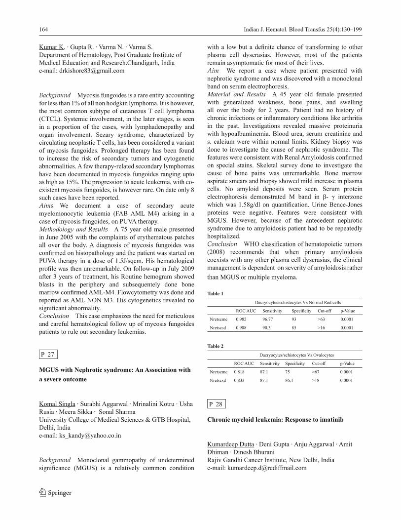
164 Indian J. Hematol. Blood Transfus 25(4):130–199
Kumar K. · Gupta R. · Varma N. · Varma S.Department of Hematology, Post Graduate Institute of Medical Education and Research.Chandigarh, Indiae-mail: [email protected]
Background Mycosis fungoides is a rare entity accounting for less than 1% of all non hodgkin lymphoma. It is however, the most common subtype of cutaneous T cell lymphoma (CTCL). Systemic involvement, in the later stages, is seen in a proportion of the cases, with lymphadenopathy and organ involvement. Sezary syndrome, characterized by circulating neoplastic T cells, has been considered a variant of mycosis fungoides. Prolonged therapy has been found to increase the risk of secondary tumors and cytogenetic abnormalities. A few therapy-related secondary lymphomas have been documented in mycosis fungoides ranging upto as high as 15%. The progression to acute leukemia, with co-existent mycosis fungoides, is however rare. On date only 8 such cases have been reported. Aims We document a case of secondary acute myelomonocytic leukemia (FAB AML M4) arising in a case of mycosis fungoides, on PUVA therapy. Methodology and Results A 75 year old male presented in June 2005 with the complaints of erythematous patches all over the body. A diagnosis of mycosis fungoides was confi rmed on histopathology and the patient was started on PUVA therapy in a dose of 1.5J/sqcm. His hematological profi le was then unremarkable. On follow-up in July 2009 after 3 years of treatment, his Routine hemogram showed blasts in the periphery and subsequentely done bone marrow confi rmed AML-M4. Flowcytometry was done and reported as AML NON M3. His cytogenetics revealed no signifi cant abnormality. Conclusion This case emphasizes the need for meticulous and careful hematological follow up of mycosis fungoides patients to rule out secondary leukemias.
P 27
MGUS with Nephrotic syndrome: An Association with a severe outcome
Komal Singla · Surabhi Aggarwal · Mrinalini Kotru · Usha Rusia · Meera Sikka · Sonal SharmaUniversity College of Medical Sciences & GTB Hospital, Delhi, Indiae-mail: [email protected]
Background Monoclonal gammopathy of undetermined signifi cance (MGUS) is a relatively common condition
with a low but a defi nite chance of transforming to other plasma cell dyscrasias. However, most of the patients remain asymptomatic for most of their lives. Aim We report a case where patient presented with nephrotic syndrome and was discovered with a monoclonal band on serum electrophoresis.Material and Results A 45 year old female presented with generalized weakness, bone pains, and swelling all over the body for 2 years. Patient had no history of chronic infections or infl ammatory conditions like arthritis in the past. Investigations revealed massive proteinuria with hypoalbuminemia. Blood urea, serum creatinine and s. calcium were within normal limits. Kidney biopsy was done to investigate the cause of nephrotic syndrome. The features were consistent with Renal Amyloidosis confi rmed on special stains. Skeletal survey done to investigate the cause of bone pains was unremarkable. Bone marrow aspirate smears and biopsy showed mild increase in plasma cells. No amyloid deposits were seen. Serum protein electrophoresis demonstrated M band in β- γ interzone which was 1.58g/dl on quantifi cation. Urine Bence-Jones proteins were negative. Features were consistent with MGUS. However, because of the antecedent nephrotic syndrome due to amyloidosis patient had to be repeatedly hospitalized. Conclusion WHO classifi cation of hematopoietic tumors (2008) recommends that when primary amyloidosis coexists with any other plasma cell dyscrasias, the clinical management is dependent on severity of amyloidosis rather than MGUS or multiple myeloma.
Table 1 Dacryocytes/schistocytes Vs Normal Red cells
ROC AUC Sensitivity Specifi city Cut-off p-Value
Nretscme 0.982 96.77 93 >63 0.0001
Nretscsd 0.908 90.3 85 >16 0.0001
Table 2 Dacryocytes/schistocytes Vs Ovalocytes
ROC AUC Sensitivity Specifi city Cut-off p-Value
Nretscme 0.818 87.1 75 >67 0.0001
Nretscsd 0.833 87.1 86.1 >18 0.0001
P 28
Chronic myeloid leukemia: Response to imatinib
Kumardeep Dutta · Deni Gupta · Anju Aggarwal · Amit Dhiman · Dinesh BhuraniRajiv Gandhi Cancer Institute, New Delhi, India e-mail: [email protected]
Indian J. Hematol. Blood Transfus 25(4):130–199 165
Background Imatinib is the initial treatment of choice for the majority of patients with chronic myeloid leukemia (CML). Conventional cytogenetics has been main investigational method for response evaluation in majority of guidelines. Design and Methods We analyzed the clinical outcome of 48 CML patients who underwent bone marrow examination and cytogenetic test at one year of completion of therapy.Results Out of total 227 patients on fi rst line Imatinib in our institute, 77 patients were lost to follow up, 4 patients progressed before completion of 1 year of Imatinib. Out of 47 patients who had bone marrow & conventional cytogenetic assessment done at 1 year, 37 patients (80%) achieved complete cytogenetic response (CCR), another 7 patients (15%) were in major cytogenetic response and 3 patients had minor cytogenetic response. After median follow up of 18 months, 2 patients (5%) out of 37 patients who were in CCR progressed to blast crisis. 1 out of 7 patients with major cytogenetic response progressed, while others were on Imatinib. Out of 3 patients with minimal response at 1 year, 1 patient achieved CCR with Dasatinib, one went into CCR with higher dose of Imatinib & one progressed. Conclusion Achievement of complete cytogenetic response at one year is major mile stone in treatment of CML with Imatinib. This under scores the effi cacy of Imatinib as fi rst line therapy for patients with CML.
P 29
Burkitt’s lymphoma with bilateral breast involvement: A rare presentation
Singh Lavleen · Shukla Bhaskar · Chopra Anita · Sharma M.C. Pati H.P.Departments of Pathology and Hematology, All India Institute of Medical Sciences, New Delhi, Indiae-mail: [email protected]
Background Burkitts lymphoma is an aggressive lymphoma with liver, kidney, bone marrow, brain and gastro-intestinal tract as the common sites of extra- nodal involvement. Bilateral breast involvement is a very rare mode of presentation with only a few case reports in the world literature.Case report We report here a case of a 32 year female who presented with bilateral breast lumps. Systemic examination revealed pallor. There was no lymphadenopathy or organomegaly. CT scan abdomen showed bilateral ovarian masses with peritoneal deposits. Peripheral smear examination revealed leukoerythroblastic picture. Bone marrow aspirate showed replacement by monomorphic immature lymphoid
cells with vacuolated basophilic cytoplasm. Mitotic fi gures and binucleate cells were also seen. Trucut biopsy of breast showed a malignant small round cell tumor with numerous starry sky macrophages and atypical mitotic fi gures. The tumor cells showed immunopositivity for CD 20, CD 45, MIC-2. MIB-1 labelling index was almost 100%. Based on the above clinical and investigational fi ndings we proffered a diagnosis of Burkitt’s lymphoma with bilateral breast involvement.Conclusion The present case is being presented for its rarity. The importance of a close co-operation between the clinician, the hematologist and the pathologist in arriving at a correct diagnosis of this case cannot be overemphasized.
P 30
Three cases of acute lymphoblastic leukemia with Ph+ in a tertiary care hospital
Mandeep Kaur · T. Chatterjee · S. Bhattacharya · D.K. Mishra · Reena Bharadwaj · Velu Nair · S. Das · Sanjeev Chowdhury · Devika Gupta · Megha Singhal Army Hospital Research & Referral, New Delhi, Indiae-mail:[email protected]
Background Leukemias are characterized by an idiopathic proliferation of a progenitor cell that is committed to a single cell lineage. The diagnosis of acute leukemias (AL) relies both on clinical features and an array of multidisciplinary approaches including morphology, immunophenotyping, cytogenetic and molecular investigations. About 25% of adult and 2-4% of childhood leukemias show positivity for Ph+. But there are no unique morphological or cytochemical features that distinguish these types from all other types of ALL.Material and Method Three cases diagnosed as acute leukemia showing BCR-ABL1 association were studied for clinical presentation. Heamatological parameters, bone marrow examination and fl owcytometery for immunophenotyping was carried out. Three cases of ALL with BCR-ABL1 association differentiation are being described here.Results Out of the three cases cases studied two were adult onset ALL with BCR-ABL1 association and one case which was diagnosed morphologically on peripheral smear & bone marrow as AML and Immunophenotyping done by fl ow cytometry showed expression of both myeloid and lymphoid cell markers with a score of >2 in each of the lineages and was also Ph+.
166 Indian J. Hematol. Blood Transfus 25(4):130–199
Conclusion ALL with BCR-ABL1 association has the worst prognosis among patients with ALL. Treatment with imitanab in addition to high dose chemotherapy has been reported to improve early event free survival
P 31
3q abnormalities in myeloid neoplasia
Md. Rahman K. · Monika Gupta · Vikram Mathews · Auro Viswabandya · Biju George · Mammen Chandy · Alok Srivastava · Vivi M SrivastavaDepartment of Haematology, Christian Medical College, Vellore, Indiae-mail: [email protected]
Background Abnormalities of the long(q) arm of chromosome 3, bands 3q21q26 are seen in myeloid neoplasia and are associated with dysplasia, abnormal megakaryocytopoiesis, inappropriate expression of the EVI1gene, an oncogenic transcription factor, and a poor outcome. Platelet counts are reported to be normal or elevated in the t(3;3) and inversion(3). Monosomy 7 often occurs as a secondary aberration. Patients and methods All patients with 3q abnormalities and myeloid neoplasia seen between January 2003 and December 2008 were included in the study. Blood, bone marrow and karyotype fi ndings were correlated. Results-There were 36 patients with 3q abnormalities - 26 had AML (of a total of 1180 patients with AML); 6 had MDS, 3 had CML and 1, an unclassifi ed MPD. The median age was 43.5 years (range: 1- 73 ). There were 32 adults; 29 patients were males. The cytogenetic abnormalities noted were: t(3;3)(q21;q26) in 14, inversion (3)(q21q26) in 8, t(3;5) (q21;q35) in 6, deletion (3)(q21) in 7 and t(3;21)(q21;q22) in one. Monosomy 7 was seen in 10 patients (28%), 9 of whom had a complex karyotype which included monosomy 5 or deletion 5q. The t(3;3) and inversion (3) accounted for 13 of 16 patients with normal or increased platelet counts. However, these abnormalities were also present in 9 of 20 patients with thrombocytopaenia. Megakaryocytic dysplasia was present in 27 of 28 (96 %) assessable cases and trilineage dysplasia in 14 (50%).Conclusion 3q abnormalities were found in less than 2% of patients with AML, similar to the reported occurrence. Most patients with normal or elevated platelet counts had the t(3;3) or inversion(3) but this was not consistent. Megakaryocyte dysplasia was present in almost all patients; about half of them had trilineage dysplasia in addition. There appears to be no previous data from India.
P 32
Thrombophilia related molecular markers in young patient with CAD
Mingma Sherpa# · Satendra Sharma# · Rajnish Avasthi* · Usha Rusia#
Department of Pathology# and Medicine*. UCMS & GTB Hospital, Delhi-110095, Indiae-mail: [email protected]
Background There has been an alarming rise in the incidence of CAD in India especially involving the young age groups <45 years. In recent past, various studies focused on haemostatic aspects of CAD in young patients have been carried out but could not determine the signifi cance of thrombophilic molecular marker in combinationAims To study the association of thrombophilia related molecular markers in young patients with CAD Methods Thirty diagnosed patients with CAD of either sex under 40 years were included. Thirty healthy age and sex matched control subjects without evidence of CAD formed the control group. Detailed history and clinical examination fi ndings were recorded. In addition to routine investigations lipid profi le, screening coagulation test, fi brinogen levels and PCR based molecular analysis for FVL, MTHFR, TNFR-2 and Prothrombin gene mutation was carried out.Results The mean age (± SD) was 36.86 ± 3.90 years in the patient group. Smoking was the most prevalent risk factor. FVL, MTHFR and TNFR-2 gene mutation were seen in 9(30%) patients; among these mutations in combination was seen in 3(10%) patients. FVL was seen in 4(13.3%), MTHFR gene mutation in 3(10%) & TNFR-2 gene mutation in 5(16.6%) Prothrombin gene mutation was not seen in any of the subjects. There was no signifi cant difference in lipid profi le, fi brinogen levels and CRP among the patients with mutation and patients without mutation. There was a signifi cant difference in the CRP>6/CRP<6 among the patients with TNFR2 mutation.Conclusion Almost one third of the cases were positive for the various mutations in the study and the presence of at least one or the other risk factor among these patients adds on to the risk of future thrombosis. There is a need to demonstrate or document these mutations in a larger group further based upon ethnicity and geographic distribution.
Indian J. Hematol. Blood Transfus 25(4):130–199 167
P 33
The t(6;9)(p23;q34) in myeloid neoplasms: A retrospective study of 16 cases
Monika Gupta · Nancy A. · Vikram Mathews · Auro Viswabandya · Biju George · Mammen Chandy · Alok Srivastava · Vivi M SrivastavaDeapartment of Haematology, Christian Medical College, Vellore, India e-mail: [email protected]
Background Cytogenetic fi ndings are an important determinant of outcome in acute myeloid leukaemia (AML). It is now recognised that the t(6;9),although rare, defi nes a subset of AML with poor prognosis. This translocation has been reported to be associated with marrow basophilia, dysplasia, distinct immunophenotypic features, a high incidence of FLT 3-ITD mutations and the formation of an oncogenic DEK/NUP14 fusion gene.Patients and methods All patients with the t(6;9) seen in the Department of Haematology, Christian Medical College, Vellore between January 2004 and March 2009 were included in this retrospective study. The blood, bone marrow and immunophenotype fi ndings of these patients were correlated. Result There were 16 patients with the t(6;9) (p23;q34) (four of which were post treatment samples). The median age was 34.5 years (range: 7-62 years with 14 adults and 12 males). Thirteen had AML (out of a total of 1090 during this period), two, myelodysplastic syndrome and one, chronic myeloid leukaemia in myeloid blast crisis (BC). Trilineage dysplasia was present in 10 (83%), all of which were among the 12 pre-treatment samples assessed. Marrow basophilia was seen in only two patients, one of whom had CML in myeloid BC. HLA-DR was positive in all 12 patients assessed, CD13 and CD33 in 10 and CD34 in seven. Five cases had one other abnormality apart from t(6;9)(p23;q34). There were no complex karyotypes. Discussion The t(6;9)(p23;q34) was seen in 1.2% of all AML patients. This is comparable with the reported occurrence of 1%. Marrow basophilia was not found in the majority of cases; however, the association with dysplasia and positivity for CD13, CD33, CD34 and HLA-DR is similar to what has been reported. This is one of the largest series from a single centre.
P 34
Evaluation of computerized on-line donor processing system on error, deferral rate and donor satisfaction in esfahan, 2005
Aghahoseini M. · Shaemi A.· Azarbaijani K. · Akbari N. Iranian Blood Transfusion Organization-Research Center, Esfahan, IRANe-mail: [email protected]
Background The use of information technology and electronic registers generates new perspectives for the studies of transfusion practices. Few reports evaluated such technology in transfusion-management system.Aim To evaluate the effectiveness of the system, we have traced all blood components via the network system. This took place with the computerized donor processing system running in parallel with the present manual operations in Esfahan. Methodology This Analytic Descriptive study carried out using census method in Esfahan Blood Transfusion Organization, 2005. Error incidence due to loss of inventory and Behavioral deferral rate in all blood donors at Khajoo center that uses computerized on-line donor processing compared with Shariati center with data entering after blood donation. Donor satisfaction in 100 donors at khajoo center compared with the same as at Shariati center using a Questionnaire. Data analyzed using exact fi sher and қ2 ant T test with SPSS 11.5.Result deferral rate (0.2% vs. 1%) and error incidence (0.00 vs. 0.62 percent) in Khajoo center are lower (CI: 95%, P<0.05). Donor satisfaction scores due to blood donation procedures showed it was lower than Shariati center signifi cantly (t=2.5). Donor satisfaction scores for employees did not make any difference (t=1.38). Conclusion Computerized on-line donor processing system is effective for minimizing error incidence and increasing deferral rate. It contributes to safe and effi cient blood but several computerized donor session procedures may cause dissatisfaction and disturbance to private donor counseling.
P 35
Reporting of actual and near-miss events for transfusion medicine: improving transfusion safety in isfahan blood transfusion organizatin in 2006-2007
168 Indian J. Hematol. Blood Transfus 25(4):130–199
Hariri M.M.· Agha Hoseini M. · Akbari N. · Yavari F. Isfahan Blood Transfusion Organization, Iran e-mail: [email protected]
Background Errors are common in blood banks but fortunately, Mortality and Morbidity are rare. If we focus in on them, it presents the system strengths and weaknesses before they occur then we may improve blood safety. This study was conducted to assess no-fault error reports and the corrective procedures.Aim This study was conducted to assess no-fault error reports and the corrective procedures.Methodology In this prospective study, 201 reports from EBTO and 4 Hospital (Community) being sent over 12 months in 2008 were assessed. When the errors Occur, Employees were fi lled in the questionnaires then analyzed.Result The reports were 201(184 cases in EBTO and 17 cases in the hospitals). 99% (198 cases) was near-miss Events and actual events were in 2 cases. 75.5% were human errors that 55% (101 cases) related to computer registries in EBTO and 41% (7 cases) to sampling.Conclusion Some near-miss events were unplanned and another planned such as using the algorhythm (1 to 5). During present study, IBTO software was upgraded but it is necessary to create a unique classifi ed event reporting system with potential standard causes.
P 36
Evaluation of correlation in blood donors’ rejection and trend of viral markers from 2004 To 2007, Isfahan, Iran
Nahid Akbari1 · Mohamad Fazilati · Mohamad Mehdi Hariri · Ziba EbrahimianIranian Blood Transfusion Organization-Research Center, Esfahan, Irane-mail: [email protected]
Background Recently, Blood trolled therefore, it is necessary to evaluate prevalence and correlation with viral markers trends.Aim This study presents, Do recent donor selection procedures lead to decreasing of viral markers?Methodology All of eligible blood donors from 2004 to 2007 referred to Isfahan Blood Transfusion Organization included to the study. Data related to deferral rate due to potential TTDs and viral markers prevalence and correlation were accounted using IBTO software.
Result Blood donors populations from 2004 to 2007 were 84275, 95879, 100290 individuals and overall rejection frequencies were 23%, 32%, 29% respectively. Rejection frequencies due to potential TTDs were 4.1%, 5.4%, 5.2% and viral markers prevalence was 0.16%, 0.13%, 0.12% respectively.Conclusion Overall deferral rate increased and deferral rate in blood donors due to potential TTDs correlated reversely to decreasing of viral markers prevalence. It may be due to stringent rules of IBTO.
P 37
Pure red cell aplasia in a chronic myelocytic leukemia patient on imatinib mesylate treatment
Jyoti Togra · Ruchi Gupta · Neelam Varma · Subhash VarmaDepartments of Hematology & Internal Medicine, Post Graduate Institute of Medical Education, Sector - 12, Chandigarh, Indiae-mail: [email protected]
Background Imatinib produces various types of side effects and toxicities in chronic myelocytic leukemia (CML) patients. Suppression of erythroid lineage is a rare complication caused by imatinib. Case report A 65-years, male was diagnosed with CML in September, 2007 with high total leucocyte count (TLC) and preserved hemoglobin as well as platelet count. The patient was started on hydroxyurea and the counts started improving with the hemoglobin of 12.2 gm%, TLC of 8.8×10⁹/l and platelet count of 2.62×10⁹/l in November, 2007. Routine follow-up hemogram in February 2008 revealed high total leucocyte count of 38.9×10⁹/l and preserved hemoglobin and platelet count. So the patient was shifted to imatinib mesylate therapy in March, 2008. The follow up hemogram revealed progressive pancytopenia over a span of 5 months with a hemoglobin of 4.4gm%, TLC of 1.6×10⁹/l and platelet count of 23×109/l. Bone marrow examined at the same time revealed hypocellular marrow spaces with marked reduction in all the hematopoietic elements which was thought to be imatinib induced severe aplasia. The patient was then kept off imatinib since August 2008 and given hematinics. Follow up hemograms showed progressive improvement in the leucocyte and platelet counts however low hemoglobin persisted, ranging between 4.4gm% to 6.3gm% up to July 2009. Bone marrow examination performed in July 2009 showed markedly suppressed erythropoiesis with a myeloid to erythroid ratio of >100:1 and well preserved leucocytes and megakaryocytes.
Indian J. Hematol. Blood Transfus 25(4):130–199 169
Conclusion Marked erythroid suppression observed in the index patient seems to be related to protracted effect of imatinib on the erythroid progenitor cells. To our knowledge, this is only the second case of imatinib related marked erythroid lineage suppression in CML; the other being reported in 2007.
P 38
Effect of storage on fl owcytometric detection of GPI anchored proteins (CD 55 and CD59) on normal erythrocytes & granulocytes - a comparative study
Parveen Bose · Neelam Varma · Man Updesh Singh Sachdeva · Jasmina Ahluwalia · Reena Das ·Department of Hematology, Post Graduate Institute of Medical Education & Research, Sector -12, Chandigarh, India e-mail: [email protected]
Background Defi ciency of GPI anchored proteins (e.g. CD 55 & CD 59) in Paroxymal Nocturnal Hemoglobinuria (PNH) renders the erythrocytes extremely sensitive to complement mediated lysis. Flowcytometric (FCM) evaluation of CD 55 & CD 59 expression on erythrocytes & neutrophils is the preferred technique for quick diagnosis of PNH. Recent blood transfusions adversely affect the results of FCM analysis using erythrocytes, whereas granulocytes remain unaffected. Specifi c tests for PNH are mostly done by prior appointment, using fresh samples.Aim To study the effect of storage at 4-80C, up to 120 hrs, on the CD 55 & CD 59 expression on erythrocytes & neutophils.Material & Method 3.0 ml of EDTA and 2.0 ml of CPD anticoagulated blood samples were taken from 5 healthy individuals. Standard protocols were followed for labelling of erythrocytes & neutrophills with monoclonal anti-CD 55 and CD 59 antibodies, using fresh sample (at 0 hrs), and samples stored at 4-80C (at 24, 48, 72, 96 & 120 hrs). The data was analysed on FACSCanto (BD, USA) after acquiring 10,000 events in each tube. The results were expressed as qualitative and semi-quantitative parameter. Result Present study showed no signifi cant change in expression of CD 55 & CD 59 on erythrocytes & neutrophils processed at 24, 48, 72, 96 & 120 hrs after storage at 4-80C in EDTA or CPD anticoagulated blood, as compared to fresh samples.Conclusion Flowcytometric analysis of expression of CD 55 & CD59 on erythrocytes and neutrophils for diagnosis of PNH can be performed on stored anticoagulated blood samples, as no loss of expression was noted in blood
samples stored for at least six days. This would defi nitely save precious time for diagnosing or excluding PNH, in our patients who are referred from far off places and often investigated as outpatients. This study needs to be extended to include more number of normal controls as well as PNH patients.
P 39
Correlation of high Hb A2 levels with macrocytic anemia
Neha Kapoor · Sangeeta Rawat · Rashmi Thakur · Aakanksha Saini · Sheefa Khan · Pranav Dorwal · Mrinalini Priyadarshini · Poonam Singh Dept of Hematology, Indraprastha Apollo Hospitals, New Delhi, Indiae-mail: [email protected]
Background Haemoglobin (Hb) A2 determination is a useful diagnostic tool in case of heterozygous β-thalassaemia and also in other abnormal hemoglobinopathies. The levels are modifi ed in a number of congenital and acquired conditions. There have been few studies in the literature, which have documented increased levels in cases with vitamin B12 and folate defi ciencies as well, creating a diagnostic dilemma with thalassemia trait. We report here, fi ve cases of macrocytic anemia which were detected to have raised Hb A2 levels.Aim The purpose of this report is to establish an association between raised Hb A2 levels and macrocytic anemia.Methodology Peripheral blood smears of cases with raised Hb A2 levels were examined and the picture was suggestive of macrocytic anemia. These were further evaluated for their Red Blood Cell indices ( MCV, MCH, MCHC ) which were found to be increased. The method employed for Hb A2 estimation was based on Cation - Exchange High Performance Liquid Chromatography (CE-HPLC) which is shown to be rapid, sensitive, specifi c and reproducible in the diagnosis of haemoglobinopathies and can accurately identify and quantitate abnormal haemoglobins. Folate levels were not estimated.Results Five cases with high HbA2 levels were detected with a peripheral blood picture indicative of macrocytic anemia. The RBC indices were also raised.Conclusion Though the hemoglobin A2 levels are routinely normal in macrocytic anemia, however, an increase in the level may be found in some cases. This can, at times lead to a diagnostic confusion with thalassemia trait. The precise explanation for this increase remains obscure, although, they were advised to get their Vitamin B12 and folic acid
170 Indian J. Hematol. Blood Transfus 25(4):130–199
levels checked. A follow up of their Hb A2 levels post therapy, was also suggested.
P 40
Castleman disease: A report of two cases
Rathod N · Dhingra B. · Rathi S. · Mishra P. · Mahapatra M. · Seth TDepartment of Hematology, AIIMS, New Delhi, Indiae-mail: [email protected]
Background Castleman’s disease ( angiofollicular lymph node hyperplasia), is a lymphoproliferative disorder. It has association with HIV and HHV-8and is associated with a number of malignancies. Disease presents in two forms: Unicentric with local symptoms and the multicentric form with systemicfeatures.Case report A 65 year old male presented with four months history of fatiguability, low grade fever and swellings in the neck. On evaluation he had anaemia with generalized lymphadenopathy and hepatosplenomegaly. Peripheral smear revealed microcytic hypochromic anaemia with marked rouleaux formation and lympho-plasmacytoid cells. Serum LDH was high and direct coombs test was positive. Lymph node biopsy revealed effacement of nodal architecture with paracortical expansion by plasma cells and lymphocytes. Bone marrow had 22% plasma cells and immunophenotyping was positive for CD 2, 4, 5, 8, 45, 38. Beta 2 Microglobulin- 13399 ug/l (510- 1470) S. Immunoglobulin- IgG- 9565 mg/dl (700-1600). PET CT was suggestive of generalized lymphadenopathy and splenomegaly. Final diagnosis of multicentric plasma cell rich castleman’s disease was made.Patient received steroids followed by 6 cycles of CHOP regime. He continued to be in remission for 8 months and then and was lost to followup.Case two: A 24 year old man presented with three year history of gradually progressive swellings in the neck, fever and night sweats. Examination revealed bilateral multiple cervical lymphadenopathy and rest of the examination was unremarkable. Bone marrow had 5% plasma cells . CT scan revealed multiple cervical lymphadenopathy and lymph node biopsy was suggestive of hyaline vascular variant of castleman disease. Patient has received 2 cycles of CHOP and his lymphadenopathy has completely regressed, he continues to be in followup.Conclusion There are few case reports of this disease from India. Treatment options for disease variants are different.
P 41
Acute myeloid leukemia manifested as intracranial haemorrhage
Nisha Marwah · Sunita Singh · Shilpa Garg · Sanjay Verma · Rajesh Rajpoot · Rajeev SenDepartment of Pathology, Pt. B.D Sharma PGIMS, Rohtak, Indiae-mail: [email protected]
Abstract AML most often presents with non specifi c symptoms like anaemia, leukopenia/leukostasis or thrombocytopenia. Intracerebral haemorrhage is a well known potential complication of leukemia. Intracerebral haemorrhage as a primary presentation has rarely been reported. Here we present a case where intracerebral haemorrhage brings AML into picture. A 25 year old male was admitted in medicine ward with history of sudden loss of consciousness since one day. There was no history of trauma, bleeding diathesis, seizures or any drug intake. On examination pulse rate was 100/minute ,blood pressure 124/88 mm of Hg and respiratory rate 26/minute. Cardiovascular, respiratory and abdominal system examination was NAD. CNS examination revealed a comatose patient moving only half of body on painful stimuli. There was no sign of neck rigidity. A provisional diagnosis of acute cerebrovascular accident was made. CT scan revealed intracerebral bleed in left parietal region. Patient was put on decongestive therapy and other routine ancillary investigations were sent. Complete hemogram showed Hb of 5gm/dl, platelet count 30,000/cmm and TLC 16,000/cmm with 95% immature cells with a predominant population of promyelocytes of hypogranular variant. Sudan staining and chloracetate reaction were highly positive. Patient was diagnosed as a case of AML-M3, hypogranular variant. PT and PTTK were normal. Blood sugar, urea, creatinine, chest x ray and ECG did not reveal any abnormality.A fi nal diagnosis of AML with intracerebral hemorrhage was made. However, despite the best possible efforts patient died within 36 hours of admission.
P 42
Bone marrow aplasia in two MDS patients treated with lenalidomide
Nitin Gupta · Prashant Sharma · Shyam Rathi · Hara Prasad Pati · Manoranjan Mahapatra.
Indian J. Hematol. Blood Transfus 25(4):130–199 171
Department of Hematology, All India Instittute of Medical Sciences, New Delhi, Indiae-mail: [email protected]
Introduction Lenalidomide is an immunomodulatory drug used in myelodysplastic syndrome, multiple myeloma, chronic lymphocytic leukemia etc. Its predominant toxicity is myelosuppression.Objective To report prolonged myelosuppression and bone marrow aplasia in two cases of myelodysplastic syndrome treated with lenalidomide. Case 1: was a 40-year-old female, transfusion dependent, cellular bone marrow suggestive of MDS-RARS, cytogenetics 46XX, IPSS- low risk, requiring 3 units packed red cells per month, with a baseline Hb 6.3 gm/dl, TLC 4.9x10⁹/l, platelet 176 x109/l, serum EPO >2000mIU/ml, PNH-negative. She received lenalidomide 10mg/day. After 3 months she developed grade 4 thrombocytopenia and neutropenia. Lenalidomide was stopped and G-CSF was given. However, her cytopenias did not improve even after stopping the drug. A bone marrow biopsy demonstrated marrow aplasia (cellularity <5%). Her latest hemogram after 10 weeks of drug with drawl has shown normal platelet and leucocyte counts. Case 2: A 38-year-old woman with transfusion-dependent anemia, cellular bone marrow suggestive of MDS-RCMD, normal cytogenetics, IPSS- low risk, PNH negative, was treated with lenalidomide 10mg/day. Her base line hemoglobin, ANC, platelets were 6.5gm/dl, 1.2 x10⁹/l and 52x109/l respectively. She showed some response after 10 weeks of therapy - hemoglobin increased and stayed around 8-10gm% without requiring blood transfusions. After 6 months of therapy she developed grade 4 thrombocytopenia and neutropenia, prompting lenalidomide withdrawal. Pancytopenia persisted even after 8 weeks of withdrawing lenalidomide. A bone marrow biopsy done to look for disease progression revealed marrow hypoplasia with a cellularity of 10-15%.Conclusion Although myelosuppression is the commonest toxicity described with the use of lenalidomide in MDS, these cases are the fi rst report of marrow aplasia with this drug in MDS and only the second report of marrow aplasia following lenalidomide therapy in any disorder.
P 43
Prospective evaluation of supersaturated calcium phosphate oral rinse for prevention of mucositis in patients undergoing hematopoietic stem cell transplantation (HSCT)
Nitya · Lincy Issac · Shiby · Binu · Jipsy · D. Gupta · K.D. Dutta · A. Sharma · D. BhuraniRajiv Gandhi Cancer Institute, New Delhi, Indiae-mail: [email protected]
Background Mucositis has been found a major dose-limiting toxicity, in conditioning regime being used for patient undergoing autologus or allogenic HSCT. We have evaluated the role of supersaturated calcium phosphate oral rinse in these situations in accordance with some preliminary data suggesting its effi cacy.Methods 12 patients were included in the study irrespective of autologus or allogenic status (9 patients had autologus HSCT, 3 patients underwent allogenic transplantation receiving myeloablative therapy). Out of 12 patients, 7 patients were given calcium phosphate as oral rinse 4 times a day along with chlorhexidine and 5 patients were given chlorhexidine only. For painful mucositis syrup diclofenac was added in all patients. Patents were monitored daily for mucositis and severity was scored according to National Cancer Institute toxicity scales.Results Out of the 7 patients, who received supersaturated calcium phosphate oral rinse, 1 patient had Grade 1 mucositis, 5 patients had Grade 2-3 mucositis and only 1 patient had Grade IV mucositis (which persisted for 19 days post transplantation). On the other hand, out of 5 patients who were given chlorhexidine, 1 patient had Grade 1 mucostis, 2 patients had grade 2-3 mucostis and 2 patients are under follow up.Conclusion Though we have a data of limited number of patients, supersaturated calcium phosphate seems not to make any major impact in prevention of mucositis in patients undergoing HSCT. To establish its role in preventing mucositis in transplant setting, it requires validation in a larger group of patients.
P 44
Treatment of myelodysplastic syndrome transforming into acute myelogenous leukemia with azacytidine---- a retrospective study from cancer centre in northern india
P. Suresh · A. Sharma · D. BhuraniRajiv Gandhi Cancer Institute , Sector - 5 , Rohini, New Delhi, Indiae-mail: [email protected]
Background Patients more than 55 years of age with Acute Myelogenous Leukemia (AML) are less likely to achieve
172 Indian J. Hematol. Blood Transfus 25(4):130–199
complete remission and more likely to experience toxicity with conventional induction chemotherapy than younger patients. Azacytidine is the fi rst drug in a new class of compounds classifi ed as DNA hypomethylating agents, to receive FDA approval for the treatment of myelodysplastic syndromes (MDS). At higher doses azacytidine has activity in AML as well. Aim To assess the response to azacytidine in patients with MDS transforming to AML who are unable to tolerate standard chemotherapy. Secondary aim was to assess hematological improvement with transfusion independence and the toxicity profi le of azacytidine.Materials and methods Five patients were retrospectively identifi ed of MDS transforming to AML who had been treated with azacytidine. All patients had multiple co-morbidities and hence were high risk for chemotherapy complications. Azacytidine 75 mg/m2/day was administered subcutaneously for 7 days every 4 weeks which was defi ned as one cycle and therapy was continued for as long as response was maintained. Response was assessed by NCI defi nition of response in AML or by International Working Group (IWG) criteria for hematological improvement in MDS. Results A total of 5 patients were included in this study with a median age of 60 years. 3 were male and 2 female. All of them were MDS transforming into AML. The median duration of therapy with azacytidine was 6 months and the median time to response 3 months (2-5months). Complete Response (CR) was achieved in one patient (20%) and Partial Response (PR) in 3 patients (60%) and one patient had progressive disease after 3 cycles. This led to an Overall Response Rate (ORR) of 80% (CR + PR). The median duration of response (n = 4) was 6 months (range 3-14). Hematological improvement was noted in all responders. Toxicity was largely manageable in outpatient setting and ECOG performance status in responders remained 1 or less throughout therapy.Conclusion Azacytidine appears to be active in treatment of older patients with MDS transforming to AML who are poor candidates for standard induction chemotherapy. This therapy appears tolerable in poor risk patients and can be managed largely in an out patient setting. Further prospective studies are required to confi rm the effi cacy and survival advantage with this agent.
P 45
Frequency of red cell alloantibodies in multitransfused β-thalassemia patients in a tertiary care centre
Puttaiah Parimala · Karuna Ramesh Kumar · Shanthala Devi A.M · Vanamala Alwar · Sitalakshmi SSt. Johns Medical College Hospital, Bangalore, India e-mail: [email protected]
Background β Thalassemia major is an autosomal recessive blood disorder due to defect in β chain synthesis. The mean age of diagnosis is around 6-12 months. The main stay of treatment in developing countries is regular life long blood transfusions and iron chelation. These patients receive multiple transfusions from different donors which increases progressively and predisposes to alloimmunization to red cells causing diffi culty in cross match compatibility. As our hospital cares for more than 100 Thalassemics, we undertook this study to know the frequency of antibodies to red cells. Objectives To study the prevalence of alloimmunization among β Thalassemia patients and the frequency of transfusionsMaterials and Methods During a period of 4 years between May 2005 to August 2009 120 patients on regular transfusions were studied. The patients who had repeated blood transfusions (once in 15 days), rapidly dropping Haemoglobin ( Hb <7g/dl in 2 weeks) and major cross match incompatibility problems were included in the study. Cross match and alloantibodies were tested by using microtube gel method.Results 3 of 120 patients (2.5% M-1 F-2) were detected to have alloantibodies. Mean age of these patients was 10.2 years. All three patients had major cross match incompatibility and were indirect coombs test positive. In one patient antibody screening was done and found to have anti-Xga alloantibody. All three patients required a short course of steroids in addition to transfusions.Conclusions Blood transfusion is main stay of treatment in children in developing countries where bone marrow transplant is farfetched due to cost factor. Alloimmunization to the red cells is one of the rare major complications. Red cell antibody screening in these patients followed by transfusion of blood free of the corresponding antigen will help these patients.
P 46
Age, occupation and sex specifi c incidence of transfusion transmissible infections in blood donors in Haryana (north India)
Pawan Singh · S.P. Kataria1 · Paramjit K. Sehgal · Manju Daiya · Rama Sikka2 · Sunita Singh1 1Deptt. of Blood Transfusion, Pathology; 2Microbiology, Pt. B.D. Sharma Post Graduate Institute of Medical Sciences, Rohtak, Haryana, Indiae-mail: [email protected]
Background Safe blood transfusion requires a safe donor. It is important to analyze the various blood
Indian J. Hematol. Blood Transfus 25(4):130–199 173
donor characteristics in order to manage blood donor programmes.Aim Study was conducted to analyse the prevalence of different transfusion transmissible infections (TTIs) in different age groups, occupation and sex-wise to identify the safest blood donors group.Methodology and Results Total 26353 blood donors during the year 2007-8 were screened for HIV I and II, HBsAg, HCV, syphilis and malaria parasite.As per age groups, seropositivity was lowest in age group of <20 years (1.5%) followed by age group 21-30 years (3.24%), which was statistically signifi cant (p-value <0.05). While considering occupation, total seropositivity in students was lowest (2.3%) as compared to other occupation such as farmers, businessmen and drivers as well as donors from other occupation like servicemen, labourers etc. The overall seropositivity in females was less (2.1%) than in the males (3.2%). Conclusion Schools and colleges should have special motivational programmes for these type of donors to educate and motivate for voluntary, altruistic blood donations to meet increasing demand through repeat donations from the safer donor group considering age, occupation and sex. There is need to sought full cooperation of women in donating blood, when they are equally capable to donate blood.
P 47
Probing rate of YMDD motif mutant in lamivudine treatment of iranian patients with chronic hepatitis bvirus infection
Freshte Ghandehari1 · Abbas Ali Pourazar2 · Mehrnaz Shanehsaz Zadeh3 · Nahid Tajedin4 1 & 4 Faculty Member, Dept of Microbiology & Genetics, Islamic Azad University, Fellavarjan, Isfahan, Iran2Professor of Immunology, Isfahan University of Medical Sciences, Isfahan, Iran3Medical Biotechnologist, Park Pathology lab., Isfahan, Iran
Background Lamivudine is an analog nucleoside used for treatment of chronic hepatitis B virus infection. Studies showed that prolonged therapy could induce lamivudine resistance HBV variants (YMDD motif). In this study, the rate of YMDD motif mutants is determined in lamivudine treated chronic hepatitis B patients in Iran.Patients and Methods A total of 33 serum specimens of patients with chronic hepatitis who had been treated with lamivudine were included in the study. Serum samples of
patients were tested by PCR fl ash and RFLP as well as tested for HBe Ag, HBs Ag and liver enzymes (ALT and AST).Results Out of the 33 patients enrolled in this study, 82% were male and 18% female respectively. Mean ALT levels were between 20 – 100 Iu/1. HBe Ag was positive in 76% of the patients while HBs was positive only in 61% of the patient. Furthermore, in 28 patients liver biopsy grade was between 2-17 having the stage of 1 to 6. Moreover, HBs Ag negative and HBe Ag positive were observed in 30% of the patients.Conclusion During therapy it was found that patients with lamivudin incidence YMDD mutation were approximately 14%. The ALT levels were also reduced in these patients. This study revealed that there was a signifi cant difference between HBe Ag, grade and YMDD mutation whereas no signifi cant different was observed between HBs Ag and HBV DNA PCR. Conclusively, it was found that signifi cant difference exists between YMDD mutation and lamivudine therapy.
P 48
Pure red cell aplasia rare cause of anemia: Our experience
Pratibha Dhiman · Anita Tahlan · Anshu Palta · Sandeep Chauhan1. Department of Pathology & 1Medicine, Govt. Medical College, Chandigarh, India e-mail: [email protected]
Background Pure red cell aplasia (PRCA) describes a condition in which RBC precursors in bone marrow are nearly absent, while megakaryocytes and WBC precursors are adequate. Pure red cell aplasia exists in several forms, and the commonest is an acute self-limited condition. The chronic form is associated with underlying disorders such as thymomas, lymphoma and autoimmune diseases.Aim To identify various aetiologies of red cell aplasia.Methodology We conducted a retrospective analysis of ten cases of PRCA diagnosed in the last four years [2004-2008]. All the patients were investigated using complete hemogram and bone marrow examination. CT chest and autoimmune work up was also done in related cases.The diagnosis was made on bone marrow examination showing reduced erythroblasts with normal granulocytic and megakaryocytic series.Result Out of total ten cases, four were male and six were female with an age range of 9-63 years. Four out of ten cases were diagnosed as primary pure red cell aplasia as no cause could be identifi ed. In remaining cases, the
174 Indian J. Hematol. Blood Transfus 25(4):130–199
underlying aetiology was found including two cases showing morphological changes of Parvovirus infection. Out of these two cases, one was a known case of thalassemia.Two cases were diagnosed as red cell aplasia secondary to autoimmune diseases. One case had an anterior mediastinal mass suggestive of thymoma on CT. One case was on treatment for seizures on phenytoin for seven months. The cases secondary to Parvovirus infection had recovered, case with thymoma lost follow up while other were transfusion dependent till the last follow up.Conclusion The aetiology of pure red cell aplasia is diverse and thorough clinico-pathological work up is important to rule out underlying secondary causes.
P 49
Changing trends in clinico-morphological profi le of CML
Anshu Palta · Anita Tahlan · Pratibha Dhiman · Sandeep Chauhan*Department of Pathology & Medicine*, Govt. Medical College, Chandigarh, Indiae-mail: [email protected]
Background Chronic myeloid leukemia is a clonal stem cell disorder. It most frequently presents in chronic phase with peak incidence in 53 years & male:female ratio of 3:2.Most of the patients are either asymptomatic or presents with fatigue and abdominal fullness due to spleenomegaly.Aim To study changing trends of clinico-morphological profi le of CML cases.Methodology Cases of CML diagnosed in last 4years [2004-2008] were included. The age, symptoms at presentation & phase of disease were analysed in detail.Results On retrospective analysis, a total of 44 cases of CML were detected. Out of these maximum [32%] cases were found in age group of 21-30 years & minimum number of cases [9%] were in 31-40 years age group.Male:female ratio was 1:1.3. Twelve cases [27%] presented with low grade fever of 5-7 months duration while 8 cases[18%] complaint of abdominal fullness. Five cases [11%] were detected to have with bleeding and 4cases [9%] had bony pain. Four cases[9%] had thrombotic events & one[2%] case was diagnosed in pregnant female. Rest of the cases were either asymptomatic [9%] or presented with fatigue. Three cases [6.8%] had no spleenomegaly which was an unusual fi nding and 14 cases presented with mild spleenomegaly.Twenty fi ve[ 57%] cases were diagnosed as chronic phase, seven[16%] cases in accelerated phase and twelve[27%] cases in blast crisis. Thrombocytopenia in CML–chronic
phase was found in three cases which was again an unusual fi nding. BCR- ABL/Ph studies were available in 35 cases and was contributory. In rest of the cases diagnosis was supported by marrow fi nding and low LAP score.Conclusion Our study highlights a few unusual features of CML: younger age, variable symptoms and absence of spleenomegaly.
P 50
Excess of plasma cells with pseudogaucher cells in tuberculosis: An unusual fi nding
Anshu Palta · Pratibha Dhiman · Ram Singh1. 1Department of Pathology & MedicineGovt. Medical College, Chandigarh, Indiae-mail: [email protected]
A 50-year-old female presented with fever and bony pain since last fi ve years. The routine hemogram showed anemia with a hemoglobin of 5.3 g/dl, high TLC of 14,000/μl, DLC P83 L11 M2 E2 and platelet count was 2.4 Χ 105/ μl. Both blood & urine cultures were negative. ELISA for HIV was negative. Skeletal survey showed lytic lesion in skull. On CT scan a focal cystic lesion with fi brotic change was reported in lung. Serum electrophoresis for M band showed a polyclonal band in γ region. Urine for Bence jones proteins was negative. Bone marrow aspiration & biopsy was done in view of lytic lesions with persistent anemia.The bone marrow aspirate showed 25% plasma cells along with many binucleate forms. Many scattered large cells with small, irregular nuclei and abundant amount of foamy granular cytoplasm. Biopsy also showed collection of these cells which were negative for Perl’s stain conforming to morphology of pseudogaucher cells. Also there was an excess of lymphocytes & areas of fatty degeneration along with fi brosis. Although stain for AFB was negative and the count of plasma cells was in range of criteria of multiple myeloma ,in view of presence of polyclonal M band on serum and urine negative for BJP, a possibility of chronic granulomatous infl ammation was suggested. The patient responded to ATT, became afebrile with followup hemoglobin 9.2g/dl and reticulocyte count of 4%.The more commonly reported pseudogaucher cells seen in cases of chronic lymphocytic leukemia, myelodysplasia, Hodgkin’s lymphoma & multiple myeloma. Present case is of tuberculosis with excess of plasma cells with pseudogaucher cells is a rare fi nding.Keywords Tuberculosis, pseudogaucher cell.
Indian J. Hematol. Blood Transfus 25(4):130–199 175
P 51
Anemia in critical illness- a descriptive study in a tertiary care centre ICU
Preeti T. · Kotwal J. · Bharadwaj R. · Mani N.S.Dept of Pathology, Armed Forces Medical College, Pune, Indiae-mail: [email protected]
Background The anemia of critic al illness is a distinct clinical entity with multifactorial etiology. This anemia resembles the Anemia of Chronic Disease in pathophysiology and lab characteristics. The prevalence of anemia of critically ill in Indoasian countries and its effects on morbidity and mortality is poorly defi ned.Aim of study To prospectively defi ne the prevalence, rate of progression, morphology, possible etiological factors and effect on overall survival of the anemia of critically ill. Methodology A total of 104 critically ill patients were followed up for a period of 30 days or until hospital discharge. Patients with ICU stay of <24 hrs, transfused or with active bleeding were excluded from the study. The frequency of Phlebotomies, volume of blood lost, infl ammatory markers and hematological profi le was checked for each patient. BM aspirate study was undertaken wherever indicated.Results Average duration of stay in ICU was 7.5 days. The mean number of phlebotomies per day was 3 with an average blood loss of 19.85 ml. Mean Hb at admission was 11.3 Gms% with 62.5% patients being anemic at admission, number rising to 79% at day 3 and 84.6% at day 7. The rate of fall of Hb in anemic and non anemic patients between D0-D3 and D3-D7 was (0.41 Gms/day vs 0.31 Gms/day) and (0.15 Gm/day vs 0.13 Gm/day) respectively. The rate of fall plateued after 7th day in both categories. The type of anemia on PBS was normocytic normochromic (80%) followed by dimorphic picture. Retic response was low (0.86%) in these patients with low serum iron, low TIBC and high to normal Serum Ferritin. Bone marrow study showed picture of anemia of critically in 72%. and increased bone marrow iron stores indicating a functional iron deficiency. The overall mortality rate in our study was 19%. When correlated with APACHE score there was no significant difference in the mortality rate of two groups (p value -0.95). Conclusion This prospective, observational study validates the common occurrence of anemia in critically ill patients which remains multifactorial in origin, with phlebotomy losses being one of the major contributory factors. This anemia does not signifi cantly increase the mortality but adds to the morbidity in these patients.
P 52
Role of bone marrow biopsy in diagnosing granulo-matous diseases in patients with unexplained fever
Prerna Arora · R. Gupta · Parul Gupta · Deepali Varma, Nita Khurana · Tejinder Singh1 · Naresh Gupta2 · G R. Sethi3
1Department of Pathology, 2Medicine and 3Pediatrics, Maulana Azad Medical College and Associated Hospitals, New Delhi, Indiae-mail: [email protected]
Background Bone marrow (BMA) aspiration and biopsy (BMB) are used commonly in clinical practice to diagnose systemic infections in patients with unexplained fever especially in persons with acquired immunodefi ciency syndrome (AIDS) and cytopenias. Aim To evaluate the role of trephine biopsy in diagnosing granulomatous lesions in cases of unexplained fever along with an analysis of hematological alterations in these patients. Materials and Methods Over a period of 10 years, 365 patients with unexplained fever underwent bone marrow aspiration at our hospital. Out of these 150 patients underwent both bone marrow aspiration and trephine biopsy as a part of diagnostic work-up. These cases were reviewed for their clinical data and hematological fi ndings. BMB was looked for granulomas and correlated with clinicopathological features. Special stains like AFB, PAS, silver methamine, mucicarmine were carried out wherever necessary. Results Out of 150 BMB’s granulomas were found in 96 cases (64%). A wide age range (17 months- 65 years) was noted with male predominance. Fever was the commonest presenting complaint, followed by abdominal distention 45% and weight loss 41%. Pallor was found in 92% of cases followed by hepatomegaly and/or splenomegaly (45%). 6 patients were diagnosed as HIV positive with CD 4 count varying from 110-160/cumm. Clinical differentials were disseminated tuberculosis, hematological malignancy, autoimmune disorder, kala-azar and HIV infection .Anemia was seen in 95% of cases. BMA revealed increased histiocytes in 36% of cases and hemophagocytosis was found in 13% of them.BMA revealed ill defi ned granulomas in 3 cases, while BMB was diagnostic in 64% of cases revealing granulomas. Caesation was found in 16% of cases. In our study tuberculosis was the commonest etiology.Other etiologies included Hodgkins and non Hodgkins lymphoma,sarcoidosis,fungal infections (Histoplasmosis 6 cases, Cryptococcus 2 cases), Pure red cell aplasia, fi lariasis and leishmaniasis. Etiology could not be ascertained in 22% of the cases.
176 Indian J. Hematol. Blood Transfus 25(4):130–199
Conclussion Bone marrow biopsy is extremely useful in diagnosis of focal granulomatous diseases of marrow and should be routinely performed in such cases.
P 53
Aberrant heterosis of G-6-PD defi ciency and sickle cell disorders: Need to limit family size in carrier couples in India
R.S. BalgirDepartment of Biochemistry, Regional Medical Research Centre for Tribals (ICMR), Nagpur Road, Jabalpur, Madhya Pradesh, Indiae-mail: [email protected]
Background The sickle cell disease and glucose-6-phosphate dehydrogenase (G-6-PD) enzyme defi ciency are important genetic and public health challenges in India. Effect of compound heterosis of these disorders is still not fully understood and need exploration. Aims & Objectives To study the interaction of sickle cell disease and G-6-PD defi ciency in relation to reproductive outcome among some Dhelki Kharia tribal families of Orissa. Methodology A random genetic study of screening for hemoglobinopathies and G-6-PD defi ciency among Dhelki Kharia tribal community in Sundargarh district of Orissa was carried out for intervention. Out of 81 Dhelki Kharia families screened, six families with double heterozygosity for genetic anomalies were encountered. About 2-3 ml. intravenous blood samples were collected in EDTA after taking informed consent in the presence of doctor and community leaders and analyzed for hematological investigations. Analysis was carried out following the routine standard procedures. Results There were 12 children (about 52%) out of 23 who were either suffering from sickle cell trait or disease in concurrence with G-6-PD defi ciency in hemizygous/ heterozygous/homozygous condition in Dhelki Kharia tribal community of Orissa. There were on an average 3.83 number of surviving (range 2-6) children per mother in families of G-6-PD defi ciency and sickle cell disorders. The average number of children (3.83) born (range 2-6 children) per mother to carrier/affected mother was much higher than the average for India (2.73).Conclusions One of the implications of aberrant heterosis is its adverse affects on individual physiology and routine
activities. To limit the family size in carrier couples to avoid aberrant heterosis in offsprings is suggested.
P 54
Is HbA2 level the only criterion for diagnosing β-thalassemia trait?
Rachna · Nita K. · T. Singh · N. Gupta1 · A.P. Dubey2 . Department of Pathology, 1Medicine and 2PaediatricsMaulana Azad Medical College and Lok Nayak Hospital, New delhi, Indiae-mail: [email protected]
Background An accurate diagnosis of β-thalassemia carriers is important for epidemiological studies as well as for management and prevention of the major hemoglobin disorders. The increase in HbA2 level is the most signifi cant parameter in the identifi cation of β-thalassaemia carriers. Aim The Aim of our study was to detect β-thalassemia carriers with special emphasis on the need for accuracy in quantifi cation and interpretation of the HbA2 data especially in cases with “borderline” HbA2 values.Methodology A total of 100 cases were screened for β-thalassemia trait. Clinical history, complete haematological workup of the patients including RBC count, MCV, MCH, peripheral smear examination, reticulocyte count, HPLC and serum ferritin levels, wherever necessary were carried out.Results Out of 100 cases screened, 35 cases were diagnosed as β-thalassemia trait with HbA2 levels >4%. In 4 cases, HbA2 levels were “borderline” between 3.5 and 3.9%. In 2 of these cases, one with Hb A2 level 3.5% and other with HbA2 3.8%, RBC count was found to be normal for the respective haemoglobin level and the red cell indices were in the normal range. In other two cases with HbA2 levels 3.5% and 3.9% respectively, peripheral smear examination as well as red cell indices were suggestive of microcytic hypochromic anemia but RBC count was found to be increased for the respective Hb level, possible diagnosis of β-thalassemia trait was suggested.Conclusion In some cases of β-thalassemia trait the level of HbA2 is not typically elevated and some diffi culties may arise in making the diagnosis. For these reasons the results must be interpreted together with other haematological and biochemical evidence to make the diagnosis and if necessary, Hb A2 levels may be repeated after iron therapy in “borderline” cases.
Indian J. Hematol. Blood Transfus 25(4):130–199 177
P 55
A method for reticulocyte enumeration to improve precision and specifi city
Ragavendar M.S. · Monalisa Chatterji · Venkat Kalambur · Anand Sivaraman. ReaMetrix India Pvt. Ltd., #50-B, II phase, Peenya Industrial Area, Peenya, Bangalore, Indiae-mail: [email protected]
Background Reticulocytes (Retics) are transitional cells from erythroblasts to mature erythrocytes. The Retic count provides an estimate of the erythropoietic activity of the bone marrow. It enables diagnosis of underlying pathophysiology of anemia, following response to therapy for bone marrow transplants, Erythropoietin therapy, iron defi ciency therapy, impact of chemotherapy. Fluorescence based fl ow cytometry method for Retic enumeration allow large number of cells to be assessed to provide % retics however, is affected negatively by interference from other cells like platelet and Leukocytes. Aim is to develop a cost effective single platform reticulocyte enumeration method with enhanced precision and reduced interferents, providing both % retics and absolute retic count. Methodology A Reagent formulation containing a pan-RBC marker, nucleic acid dye and fl uorescent beads has been developed. The assay design allows specifi c gating of RBCs, separating Retics from mature erythocytes based on nucleic acid staining and pre-counted fl uorescent beads allow absolute number of RBC and Retics to be counted Results & Conclusion The assay has been developed as a dried down unitized tubes with easy workfl ow and room temperature storage using ReaMetrix’s pioneering drying technology. The assay has been tested on normal samples, initial data on 30 samples show that a pan-RBC marker based staining of RBCs improves specifi city and precounted beads provide enhanced precision. The assay is currently being validated on clinical samples.
P 56
T-cell-Prolymphocytic leukaemia
Sant Prakash Kataria · Rajni Yadav · Gajender Singh · Sanjay Kumar · Rajeev Sen · Sheena Khetarpal Department of Pathology, Pt. B.D. Sharma PGIMS, Rohtak, Haryana, Indiae-mail:[email protected]
Background T-cell-prolymphocytic leukaemia is a very rare leukaemia primarily affecting adults over the age of 30, representing 2% of all small lymphocytic leukaemias in adults.Prolymphocytic leukaemia was described originally by Galton and his colleagues in 1974. Case report A 55 year male presented with complaints of lump abdomen and multiple swellings in axillary and cervical region since 2 months. General physical examination showed pallor, cervical and axillary lymphadenopathy and splenomegaly, 8 cm below costal margin. Haemotological investigations revealed Haemoglobin 8.0 gm%, T LC 35000/cumm, with a differential count of Neutrophils- 20%, Eosinophils-3%, Monocytes-4%, Lymphocytes-10%, Atypical lymphoid cells- 63%, Platelet count- 80,000/cumm. Peripheral blood fi lm showed normocytic normochromic anaemia, with presence of atypical lymphoid cells containing round to oval nuclei with irregular nuclear outline, prominent nucleolus and abundant light basophilic cytoplasm. Possibility of lymphoproliferative disorder was suggested and bone marrow aspiration advised which showed hypercellular marrow with replacement of normal trilineage series of cells by atypical lymphoid cells (approx.60%) having round to oval nuclei, irregular nuclear membrane and prominent centrally placed nucleolus. Megakaryocytes were reduced in number. Based on the marrow fi ndings- a likely possibility of prolymphocytic leukaemia was made. Immunophenotypic studies showed positivity for CD2, CD3 and CD7, confi rming the diagnosis of T-cell-Prolymphocytic leukaemia. Differential diagnosis includes CLL, Richter syndrome and acute leukaemia. Patient was given combination chemotherapy, responded poorly and was lost to follow up.
P 57
Mycoplasma pneumonia presenting as autoimmune haemolytic anaemia
Rajeshwari S.H. · P.R. Malur · Arati D · Lovlesh Nigam .K.L.E’s Dr. P.K. Hospital & Medical Research Center Belgaum, Indiae-mail: [email protected]
Mycoplasma pneumoniae, a pleomorphic organism is a respiratory pathogen that causes community acquired pneumonia and grouped under atypical pneumonia. It is known for its extrapulmonary complications and AIHA due to cold agglutinins is one amongst them. Presenting a 55-years-old female with life threatening haemolytic anaemia complicating M. pneumonia. Haematological investigation revealed haemoglobin of 3.3 gm%, reticulocyte
178 Indian J. Hematol. Blood Transfus 25(4):130–199
count of 20% , total biluribin 2.8 ,direct 0.8 . Peripheral smear showed red cell autoagglutination , haemolysis and toxic neutrophilia . Coombs test (DAT & IAT) was positive. Cold agglutination test at 4º C - strongly positive with high titres (1:256 ). Mycoplasma antibody IgM – Positive. This case is presented for its rare occurrence.
P 58
Infl uence of sickle cell gene on Plasmodium falciparum MSP-1 and 2 alleles in symptomatic malaria
Ranjeet Singh Mashon · Dilip Kumar Patel · Siris Patel · Preetinanda Manaswini Dash*Veer Surendra Sai Medical College, Burla, Orissa, Indiae-mail: [email protected]
Background Malaria exerts selective pressure on various human hemoglobin variant and as a corollary, these Hb variants affect the prevalence to distinct Plasmodium falciparum genotypes (MSP1 and 2). Many of the studies on asymptomatic malaria infections have supported this theory. While a lone study carried out in symptomatic malaria has reported a limited influence of sickle gene. Aim Assess the infl uence of sickle cell gene on the prevalence and multiplicity of Plasmodium falciparum infection (MSP-1 and 2 allele) in symptomatic malaria cases.Methodology The study was conducted at V.S.S. Medical College Hospital, Orissa, India and approved by the Institutional Ethical Committee. Sixty adult symptomatic malaria cases were recruited in three groups Hb AA (n = 25), AS (n = 18) and SS (n = 17) from July 2006 to August 2007. Genotyping of peripheral blood P. falciparum parasites for MSP-1 (K1, MAD20 and R033 allele) and MSP-2 (3D7 and FC27) gene was done by Nested PCR. Multiplicity of infection (MOI) and Multiclonal infection was calculated from the band pattern obtained. ANOVA and Х2 trend test were used as required.Results MOI was signifi cantly different for MSP-1 allele (ANOVA p = 0.04), while it was comparable for MSP-2 allele in the three groups. K1 and MAD20 allele showed a signifi cant decreasing prevalence, Х2 trend 7.25 and 6.17; and p-value 0.007 and 0.01 respectively in HbAA, AS and SS cases, while no signifi cant trend was observed for R033, 3D7 and FC27 alleles. Polyclonal infections were present signifi cantly high for MSP1 isolates in HbAA (88%) type as compared to AS (40%) and SS (37.5) variants (Х2 trend =10.4; p=0.001), while no such trend was observed for MSP2 where multiclonal isolates were AA (50%), AS (18.2%) and SS (40% ).
Conclusion The sickle cell gene infl uences the prevalence of selective alleles and multiplicity of P.falciparum genotypes in symptomatic malaria.
P 59
Paraneoplastic and neoplastic megakaryocyte/platelet disorders three case reports
Rashmi Kushwaha · Ashutosh KumarCSMMU, Lucknow, Indiae-mail: [email protected]
Background In this poster we are discussing three rare case reports of -(1) Essential thrombocythemia- incidence of 0.6-2.5 per 1 lakh persons per year. (2) Myelodysplastic syndrome with isolated deletion (5q)(3) Acute megakaryoblastic leukaemia - an uncommon disease comprising less than 5% cases of AML.Case history (1) A 32 year female patient presented with history of pain in right hypogastrium, weakness, digital ischaemia and gangrene. Her peripheral blood examination showed normocytic normochromic RBC’s, raised WBC count (46200cells/cu.mm.). Platelet count was 15.2 lakh cells/cu.mm. Bone marrow smears were hypercellular. Erythropoesis was normoblastic. Granulocytic precursors were increased but showed normal maturation without dysplasia. Megakaryocytes showed hyperplasia. Many large aggregates of megakaryocytes were present showing active platelet synthesis. Micromegakaryocytes were also seen in large number.diagnosis of essential thrombocythemia was made.Case history (2) A 52 year male patient presented with history of progressive weakness.Peripheral blood smear examination showed macrocytic blood picture with thrombocythemia. His bone marrow aspiration showed hypercellular bone marrow with erythroid hypoplasia. Megakaryocytes were increased in number and were small in size with hypolobated nuclei. There was no dysplasia in erythroid and myeloid lineages. A presumptive Diagnosis of Myelodysplastic syndrome with suspicion of isolated deletion (5q) was made. Later on cytogenetics was done which showed deletion of 5q.Case history (3) A 12 year boy presented with history of progressive weakness. His bone marrow aspiration smears were hypercellular with 30% blasts and most of them were megakaryoblasts. These cells were of large size with indented nucleus and cytoplasmic blebs. Diagnosis of Acute megakaryoblastic leukaemia was made. Later on these cells were found to be positive for CD41 and CD61.
Indian J. Hematol. Blood Transfus 25(4):130–199 179
Discussion Essential thrombocythemia occurs in 50 to 60 years of age. However it can be seen in third decade also. Diagnosis requires a sustained platelet count of more than 450X109/L. Polycythemia vera, primary myelofi brosis, CML, MDS or other myeloid neoplasms should be ruled out.Myelodysplastic syndrome with isolated deletion (5q) ocuur often in women with a median age of 67 years. The sole cytogenetic abnormality here is deletion of chromosome 5.Acute megakaryoblastic leukaemia comprises <5% cases of AML. It can occur both in children and adults. Here at least 50% blasts are of megakaryocyte lineage.
P 60
Pure intrasinusoidal bone marrow infi ltration of non Hodgkin’s lymphoma: a report of fi ve cases from North India
Reena Das · Jasmina Ahluwalia · Ashim Das1 · Man Updesh Sachdeva · Pankaj Malhotra2 · Amanjit Bal1 · Sanjay Jain2 · Neelam Varma · Subhash Varma2 Departments of Haematology, 1Histopathology and 2Internal Medicine, Postgraduate Institute of Medical Education and Research, Chandigarh, India e-mail: [email protected]
Background Bone marrow involvement in non Hodgkin’s lymphoma (NHL) is of prognostic importance for the appropriate treatment to be offered to the patient. Intrasinusoidal pattern of bone marrow infi ltration in NHL is poorly identifi ed on hematoxylin and eosin (H&E) stained trephine biopsies and is being recognized as a distinct pattern of infi ltration in the last few years. Objective To analyse the clinical, hematological and histopathological spectrum of cases of NHL showing a pure intrasinusoidal bone marrow infi ltration. Method Five patients included in the study underwent a detailed clinical examination, automated blood cell counts and peripheral smear examination, relevant radiological and biochemical investigations. Pyrexia of unknown origin, varying degrees of cytopenias and presence of atypical lymphoid cells/ blasts in the peripheral circulation were the indications of further evaluation. Bone marrow aspiration stained with geimsa, H&E stained trephine biopsies and immunophenotyping by fl ow cytometry using antibodies from Becton Deckinson and evaluated on Facs Caliber (BD). Immunohistochemistry using appropriate antibodies (Dako) was done on trephine sections. Result Using the fl ow cytometry and immunohistochemical profi le, three of the fi ve cases were categorized as T cell hepatosplenic NHL and two as B cell NHL (one each of splenic marginal zone lymphoma and intravascular
large B cell lymphoma). All the cases, except for one case with intravascular large B cell lymphoma, showed moderate to massive splenomegaly. None of the cases had lymphadenopathy. Immunohistochemistry with B and T cell markers showed the predominant and distinct intrasinusoidal pattern of infi ltration which was diffi cult to discern on H&E stained section. Conclusion This brief analysis of fi ve cases highlights the fi nding that pure intrasinusoidal pattern of infi ltration of extranodal NHL is diffi cult to diagnose and can be seen in a variety of lymphomas.
P 61
Prevalence of RAS and FLT3-ITD gene mutations in patients with Myelodysplastic Syndromes in India: AIIMS experience
Rekha Chaubey · Sudha Sazawal · Manoranjan Mahapatra · Rashid Mir · Renu Saxena Department of Hematology, All India Institute of Medical Sciences, New Delhi, India e-mail: [email protected]
Background Chromosomal abnormalities and molecular detection has potential importance for diagnosis and prognosis of MDS, although the mechanisms underlying the development of MDS and their progressive evolution to AML are still largely unknown. Since, no studies have been reported from India on the prevalence of N-RAS, K-RAS point mutation in codon 12 and FLT3-ITD mutations in patients with MDS, we undertook this study. Method RNA was extracted from bone marrow/peripheral blood and reversely transcribed to cDNA. Using RT-PCR the patients were screened for length mutations in FLT3 gene. DNA extraction was performed using standard protocol and PCR-RFLP as well as nested PCR-RFLP was used for the detection of point mutation in codon 12 of N-RAS and K-RAS. Results A total of 53 patients (median age 39 yrs, range 9-78yrs; M: F 2:1; median TLC-3.9X10⁹/l, range 0.8-116 x109/l Median platelet count- 87 x109/l, range 1-349 x109/l, Median hemoglobin -6.8 g/dl , range 2.7- 16.1 g/dl, were studied. One out of 53 patients (2%) was found positive for N-RAS and four patients were positive for K-RAS (8%) mutation. FLT3-ITD mutation was studied in 47 patients; all the patients were found negative. The mean observation of all the patients was 30 months and the median overall survival was 28 months. Nine patients died during follow up. The
180 Indian J. Hematol. Blood Transfus 25(4):130–199
presence of N-RAS codon 12 mutation was associated with the poor survival. FLT3-ITD mutation was not observed in any of our cases, which is in contrast to 3% reported from the West. Conclusion Thus, it appears that the RAS and FLT3 mutations are uncommon in MDS patients in India.
P 62
A clinico-hematological study of pancytopenia in paediatric age group.
Chauhan R. · Chauhan D.S. · Agarwal S. · Chandra J.*.Department of Pathology and Department of Paediatrics*, Lady Hardinge Medical College and Kalawati Saran’s Children Hospital*, New Delhi, Indiae-mail: [email protected]
Background Pancytopenia is a common clinical problem, particularly in paediatric age group. It is a triad of haematological fi ndings; anemia, leucopenia and thrombocytopenia.Aim The study was conducted to ascertain the various causes of pancytopenia in the paediatric age group in patients belonging to North and Eastern parts of India and to correlate the haematological parameters with clinical features.Methodology The study group comprised of 100 patients in the age group 0-18 years of either sex having pancytopenia on peripheral smear. A complete blood count, peripheral blood smear examination, reticulocyte count and bone marrow examination was done in each case.Result The age group varied from 3 months to 18 years with maximum number of patients in the age group of 3-6 years (26%). The male to female ratio was 1.5:1. Amongst the various causes of pancytopenia, megaloblastic anemia was the commonest (32%) followed by aplastic anemia (20%), leishmaniasis (16%), leukemia (14%), enteric fever (5%), and other infections comprising of malaria, tuberculosis, hepatitis and septicaemia (8%). Fever was observed in 87%, followed by mucosal bleeding (54%), loss of weight (38%) and skin pigmentation (17%). Pallor was seen in all cases. Other physical fi ndings were hepatosplenomegaly (58%), splenomegaly (56%), petechiae (44%), icterus (10%), lympahadenopathy (9%) and sternal tenderness (4%). Conclusion The study focus on identifying easily treatable causes such as megaloblastic anemia and infections presenting with pancytopenia as they look ominous but respond rapidly to effective therapy.
P 63
Evaluation of bone marrow fi ndings in Human Immundefi ciency Virus infected individuals: A North Indian tertiary care center study
Gupta R. · Sachdeva M.U.S. · Ahluwalia J. · Das R. · Varma N.Department of Hematology, Post Graduate Institute of Medical Education and Research. Chandigarh, India
Background Human immunodefi ciency virus (HIV) has diverse clinical and pathological manifestations. Hematological system is one of the major organs affected by the infection manifesting as anemia, bi-cytopenia or pancytopenia. These changes can be attributed to the infection per se, retroviral therapy or secondary infections/neoplasms acquired during the immunocompromised state. A bone marrow examination may be essential to document the exact etiology of the cytopenias.Aims To analyze the bone marrow fi ndings in HIV infected individualsMaterial and methods A retrospective analysis of indicated bone marrow examinations accompanied with peripheral blood fi ndings of HIV infected individuals was carried out. These patients were referred to the Department of Hematology, PGIMER, Chandigarh over a time period of last 5 years (June 2005-2009). Ziehl Neelsen and periodic acid Schiff stains were also performed on all the bone marrow trephine biopsies apart from Geimsa stained smears, H&E and reticulin stains on trephine biopsies.Results A total of 32 cases were examined. The clinical indications for the marrow examinations included pyrexia of unknown origin (PUO) (17/32), pancytopenia (8/32), lymphoma staging in (5/32), 1 case each of acute leukemia and isolated thrombocytopenia. Anemia and thrombocytopenia were noted in 30/32 cases and 20/32 cases, respectively. Bone marrow was hypercellular in 15/32 cases, hypocellular in 2 cases and rest 15 were normocellular. Out of the 20 cases of thrombocytopemia, 7 showed reduction in megakaryopoieisis. Plasmacytosis was seen in majority of cases. The trephine biopsy showed granulomatous infl ammation in 7 cases, with AFB positivity seen in 3/7 cases. Other signifi cant fi ndings included 2 patients of cryptococcosis and a single case each of visceral leishmaniasis and histoplasmosis. Bone marrow infi ltration was seen in 2/5 cases of lymphoma.Conclusion Bone marrow trephine biopsy remains indispensable for evaluation of HIV infected individuals with PUO or pancytopenia, revealing granulomatous pathology in substantial number of cases. In addition, bone marrow culture should be performed in all cases, because special stains on trephine sections might not be suffi cient to identify micro-organisms.
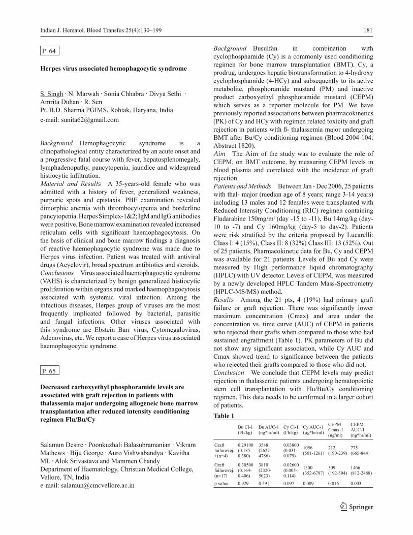
Indian J. Hematol. Blood Transfus 25(4):130–199 181
P 64
Herpes virus associated hemophagocytic syndrome
S. Singh · N. Marwah · Sonia Chhabra · Divya Sethi · Amrita Duhan · R. SenPt. B.D. Sharma PGIMS, Rohtak, Haryana, Indiae-mail: [email protected]
Background Hemophagocytic syndrome is a clinopathological entity characterized by an acute onset and a progressive fatal course with fever, hepatosplenomegaly, lymphadenopathy, pancytopenia, jaundice and widespread histiocytic infi ltration. Material and Results A 35-years-old female who was admitted with a history of fever, generalized weakness, purpuric spots and epistaxis. PBF examination revealed dimorphic anemia with thrombocytopenia and borderline pancytopenia. Herpes Simplex-1&2; IgM and IgG antibodies were positive. Bone marrow examination revealed increased reticulum cells with signifi cant haemophagocytosis. On the basis of clinical and bone marrow fi ndings a diagnosis of reactive haemophagocytic syndrome was made due to Herpes virus infection. Patient was treated with antiviral drugs (Acyclovir), broad spectrum antibiotics and steroids.Conclusions Virus associated haemophagocytic syndrome (VAHS) is characterized by benign generalized histiocytic proliferation within organs and marked haemophagocytosis associated with systemic viral infection. Among the infectious diseases, Herpes group of viruses are the most frequently implicated followed by bacterial, parasitic and fungal infections. Other viruses associated with this syndrome are Ebstein Barr virus, Cytomegalovirus, Adenovirus, etc. We report a case of Herpes virus associated haemophagocytic syndrome.
P 65
Decreased carboxyethyl phosphoramide levels are associated with graft rejection in patients with thalassemia major undergoing allogeneic bone marrow transplantation after reduced intensity conditioning regimen Flu/Bu/Cy
Salamun Desire · Poonkuzhali Balasubramanian · Vikram Mathews · Biju George · Auro Vishwabandya · Kavitha ML · Alok Srivastava and Mammen ChandyDepartment of Haematology, Christian Medical College, Vellore, TN, India e-mail: [email protected]
Background Busulfan in combination with cyclophosphamide (Cy) is a commonly used conditioning regimen for bone marrow transplantation (BMT). Cy, a prodrug, undergoes hepatic biotransformation to 4-hydroxy cyclophosphamide (4-HCy) and subsequently to its active metabolite, phosphoramide mustard (PM) and inactive product carboxyethyl phosphoramide mustard (CEPM) which serves as a reporter molecule for PM. We have previously reported associations between pharmacokinetics (PK) of Cy and HCy with regimen related toxicity and graft rejection in patients with ß- thalassemia major undergoing BMT after Bu/Cy conditioning regimen (Blood 2004 104: Abstract 1820).Aim The Aim of the study was to evaluate the role of CEPM, on BMT outcome, by measuring CEPM levels in blood plasma and correlated with the incidence of graft rejection.Patients and Methods Between Jan - Dec 2006, 25 patients with thal- major (median age of 8 years; range 3-14 years) including 13 males and 12 females were transplanted with Reduced Intensity Conditioning (RIC) regimen containing Fludarabine 150mg/m2 (day -15 to -11), Bu 14mg/kg (day-10 to -7) and Cy 160mg/kg (day-5 to day-2). Patients were risk stratifi ed by the criteria proposed by Lucarelli: Class I: 4 (15%), Class II: 8 (32%) Class III: 13 (52%). Out of 25 patients, Pharmacokinetic data for Bu, Cy and CEPM was available for 21 patients. Levels of Bu and Cy were measured by High performance liquid chromatography (HPLC) with UV detector. Levels of CEPM, was measured by a newly developed HPLC Tandem Mass-Spectrometry (HPLC-MS/MS) method. Results Among the 21 pts, 4 (19%) had primary graft failure or graft rejection. There was signifi cantly lower maximum concentration (Cmax) and area under the concentration vs. time curve (AUC) of CEPM in patients who rejected their grafts when compared to those who had sustained engraftment (Table 1). PK parameters of Bu did not show any signifi cant association, while Cy AUC and Cmax showed trend to signifi cance between the patients who rejected their grafts compared to those who did not. Conclusion We conclude that CEPM levels may predict rejection in thalassemic patients undergoing hematopoietic stem cell transplantation with Flu/Bu/Cy conditioning regimen. This data needs to be confi rmed in a larger cohort of patients. Table 1
Bu Cl-1(l/h/kg)
Bu AUC-1(ng*hr/ml)
Cy Cl-1(l/h/kg)
Cy AUC-1(μg*hr/ml)
CEPMCmax-1(ng/ml)
CEPMAUC-1(ng*hr/ml)
Graft failure/rej.+(n=4)
0.29100(0.185-0.380)
3548(2627-4786)
0.03800(0.031-0.079)
1056(501-1261)
212(190-239)
775(665-844)
Graft failure/rej. (n=17)
0.30500(0.164-0.406)
3810(2320-5023)
0.02600(0.005-0.114)
1500(352-6797)
309(192-504)
1466(812-2488)
p value 0.929 0.591 0.097 0.089 0.016 0.003
182 Indian J. Hematol. Blood Transfus 25(4):130–199
P 66
Drug induced Hemophagocytosis – Case report
Sajeeb Mondal · S.M. Sethy · Pranati Mohanty · Dileswari Pradhan · Sitaram MohapatraS.C.B. Medical College, Cuttack, Orissa, India
Objectives/Methods Hemophagocytic histocytosis can occur due to variety of causes, like familial infection related and drug induced. In our case a patient developed Hemophagocytic histocytosis due to intake of antipsychotic drug (Gold induced) – quite unique according to literature. Results 27 years / Male patient presented with fever for 15 days and Malaise. No hepatosplenomegaly was present. Hematological Examination:Anaemia present [8.5 gm x Hemoglobin] Leukopenia was present [2.2 x 10⁹ /L] Thrombocytopenia [120 x 109/L] was also present. Bone Marrow: Hypocellular granulopoiesis, Erythropoiesis and Megakaryopoiesis all were depressed. There was signifi cant increase in Bone marrow macrophages. Macrophages were vacuolated with ingested erythrocytes, platelets and neutrophils. The diagnosis was given as drug induced Hemophagocytois. Conclusion Though Drug Induced Hemophagocytosis is not rare but gold induced hemophagocytosis is defi nitely rare.
P 67
Utility of reticulocyte production index in the classifi cation of anemias
Sharada Rai · Ramandeep Singh · Astha Gupta · Ramadas Naik · Muktha R. PaiKasturba Medical College, Mangalore, Indiae-mail: [email protected]
Background The reticulocyte count and the reticulocyte production index (RPI) are the simplest practical tests available to assess the bone marrow erythropoietic activity and can be used to differentiate hypoproliferative from hyperproliferative anemiasAim To classify anemias using reticulocyte count and calculated RPI.Methodology EDTA anticoagulated blood samples from 100 anemic patients were analysed with automated analyser Sysmex XT 1800i . Leishman stained peripheral smears as well as New Methylene Blue stained reticulocyte preparations were evaluated to classify
anemias according to RPI. RPI of 2 was taken as cut off and patients were divided into two groups, effective bone marrow response (RPI >2) and ineffective bone marrow response (RPI <2).Result and Conclusion Ninety six percent cases showed ineffective bone marrow response with RPI < 2 and only 4% cases showed effective bone marrow response. The reticulocyte production index was useful in the diagnosis of especially hypoproliferative diseases like aplastic anaemia, myelofi brosis and marrow infi ltrations.
P 68
Post stem cell transplant hemophagocytic Lymphohistiocytosis
Saumya Ranjan Mallick · Prasenjit Das · Gaurav Prakash · Anita Chopra · Lalit Kumar · H.P. Pati · Siddhartha Datta Gupta.Departments of Pathology, AIIMS, New Delhi, Indiae-mail: [email protected]
Background Hemophagocytic lymphohistiocytosis (HLH) is an uncommon lethal condition, characterized by proliferation of histiocytes that actively engulf other hematopoietic cells causing cytopenia. The disease may be inherited or acquired due to a variety of infections, collagen-vascular diseases, malignancies, and very rarely associated with autologus hematopoietic stem cell transplantation (HSCT). Unawareness, delay in diagnosis and improper treatment may leads to death.Case report This 38-year female, a known case of multiple myeloma was taken for autologus HSCT after 4 cycles of VAD. Subsequently (day 5), she developed fever with mucositis and pneumonia. On day 11, she had an attack of tonic-clonic seizure due to subarachnoid haemorrhage. Laboratory investigation showed: WBC – 1500/cumm, absolute neutrophil count – 500/cumm, platelet – 8000/cumm & Hb – 8.7 gm%. There was rapid deterioration in her condition and she died on day 13th post-transplant. Microscopic examination from lung nodule shows features of invasive mucormycosis. Bone marrow aspirate and lymph node biopsy showed prominence of foamy histiocytes with hemophagocytosis.Conclusion HLH in this case may be secondary to marrow transplant or the complication to invasive fungal infection. Association of HPS with bone marrow transplant is very rarely described. The role of HPS in this case along with fungal pneumonia cannot be excluded.
Indian J. Hematol. Blood Transfus 25(4):130–199 183
P 70
Myelodysplastic/myeloproliferative neoplasm – unclassifi able
Shanaz Khodaiji · Ekta Bhutada · Sachin Almel · Asha KapadiaP.D. Hinduja National Hospital and Medical Research Centre, Mumbai, Indiae-mail: [email protected]
Background In 2001, the WHO classifi cation of hemato-poeitic malignancies defi ned a rare group of myeloid neoplasms which are characterized by the simultaneous presence of both myelodysplastic and myeloproliferative features. These Myelodysplastic/Myeloproliferative Neoplasm – Unclassifi able (MDS/MPN-U often go undiagnosed due to lack of awareness and paucity of molecular studies. Case report We report a case of MDS/MPN-U in a 70 year old male patient in whom a CBC performed preoperatively showed a high Total White Cell Count (23.51 X 10⁹ /dL) and P latelet Count (1480 X 109 /dL.) along with 7% Blasts and a fair number of dysmyelopoetic neutrophils. Bone marrow aspirate yielded a dry tap because of extensive fi brosis. Bone marrow biopsy showed prominent (grade 3) fi brosis. Megakaryocyte showed dysplastic features and were seen in clumps. No splenomegaly or lymphadenopathy present. Cytogenetic analysis did not show any abnormality. Molecular studies such as BCR/ABL, FIP1L1-PDGFRa Fusion assay and JAK2 mutation analysis were negative. Based on the morphological, cytogenetic and molecular fi ndings a diagnosis of MDS/MPN-U was made and he was put on hydroxyurea. MDS/MPN-U is a clinicopathological diagnosis based on a multiparametric approach requiring integration of peripheral blood and bone marrow morphology with clinical fi ndings and absence of molecular defects commonly associated with these disordersConclusion This case is reported because it is a relatively new entity which has been described by the WHO committee on classifi cation in 2008. Hence it has not been extensively reported so far. This case reiterates the fact that an integrated multidisciplinary approach is necessary for the classifi cation of myeloid neoplasms. (WHO)
P 71
Evaluation of different platelet counting methodologies vis-a-vis the international reference method by fl ow cytometry
P 69
Reactive hemophagocytic lymphohistiocytosis in a young girl with systemic onset juvenile idiopathic arthritis
Seema Sharma · Shalu SinhaDepartments of Pathology, Sanjay Gandhi Postgraduate Institue of Medical Science, Lucknow, Indiae-mail: [email protected]
Introduction Reactive hemophagocytic Lymp-hohistiocytosis/Macrophage activation syndrome (RHLH/MAS) is a serious and potential life threatening disorder caused by excessive activation and proliferation of T cells and macrophages. It occurs in the course of systemic infl ammatory disorders like primary systemic type juvenile Rheumatoid Arthritis ,systemic onset juvenile idiopathic arthritis (SJIA) Systemic Lupus Erythematosus and Adult Onset Still’s Disease. Macrophage activation syndrome associated with SJIA is extremely rare. Case report A 20 year old girl presented with fever, erythematous skin rash, and oral ulcers since fi fteen days. She gave history of fever off and on with symmetrical joint pains involving small and large joints and skin rash for past fi ve years. On physical examination she had cervical lymphadenopathy, hepatomegaly and splenomegaly, erythematous skin rash over neck, back and bilateral leg and left facial palsy. Investigations revealed bicytopenia i.e. anemia and thrombocytopenia. During her stay in hospital she developed severe pancytopenia.. Her liver function tests were deranged. She had positive D-dimer, hypofi brinogenemia, hyperferritinemia and hypertriglyceridemia. Her immunological investigations were negative. With these fi ndings a diagnosis of systemic onset juvenile idiopathic arthritis with macrophage activation syndrome was suspected and bone marrow examination and cervical lymph node aspiration was done. Bone marrow smears showed proliferation of small sized lymphoid cells and macrophages with active hemophagocytosis. Myeloid and erythroid cells were reduced.. Ziehl Neelson stain for acid fast bacilli was negative. She was treated with iv methylprednisolone, cyclosporine A, hydrocortisone along with supportive therapy. Her hemoglobin, TLC and platelet count became normal in about 15 days.Conclusion RHLH /MAS is a potentially life-threatening complication of rheumatic diseases, so it is important to have a high threshold of suspicion, make an early diagnosis, and start prompt treatment for sucess.
184 Indian J. Hematol. Blood Transfus 25(4):130–199
S. Khodaiji · Monisha Sethi · Sarika Patil · Arpita Mehta · Sandhya BastianP.D. Hinduja National Hospital and Medical Research Centre, Mumbai, Indiae-mail: [email protected]
Introduction An accurate platelet count is of utmost importance for better management. of oncology patients in whom transfusion decisions have to be made, and in patients with dengue, malaria, chikungunya, leptospirosis and those with low platelet counts needing emergency surgery. The manual count by phase contrast microscopy has now been replaced (recommended by ICSH and the ISLH) by the immuno-platelet method called the international method (IRM) as the reference method for enumeration of platelets. The Aim of our study was to compare the conventional impedance counts and newly introduced optical platelet counts obtained on the hematology analyzer with the IRM. Since manual counting is laborious time consuming and subjective, requiring skilled technologists to perform, the optical count which is quicker to perform than the immuno-platelet count by fl ow cytometry, can be reported if statistical correlation is seen Material and Method Samples were collected in K2EDTA vacutainers. Anticoagulated samples with clots, or fi brin strands were rejected. A total of 61 samples, 31 normal and 30 with low or normal platelet counts not correlating with smear examination were included in the study. Platelet counting was performed by manual phase contrast method, impedance counts on Sysmex XE 2100 and Beckman Coulter LH750, optical count on the Sysmex XE2100 and by fl ow cytometry on FACS Canto II using CD61 antibody. The platelet / RBC ratio method was used for counting platelets Results Using the regression analysis and co-effi cient of correlation ® obtained between the various methods it was seen that the impedance and optical methods of platelet counting are reliable when compared with the IRMWe recommend that if an automated impedance platelet count does not correlate with the smear, the optical count, should be reported after correlating with smear as it is quicker than the manual and IRM methods and equally accurate.
P 72
Pediatric patients with bicytopenia /pancytopenia: A retrospective review of etiologies and clinco-hematological profi le in 571 cases at a tertiary center
Naseem S.1 · Varma N.2 · Marwaha R.K.1
1Hemato-oncology Unit, Advanced Pediatric Centre, 2Department of Hematology
Postgraduate Institute of Medical Education and Research, Chandigarh, India
Background The etiology of bicytopenia/pancytopenia varies widely in children, ranging from transient marrow viral suppression to marrow infi ltration by fatal malignancy. Depending on the etiology, the clinical presentation can be with fever, pallor, infection, or serious illness. Knowing the exact etiology is therefore important for specifi c treatment and prognostication. Aim We planned this study with the objective to evaluate the spectrum of bicytopenia and pancytopenia in children by conducting a retrospective analysis over a two year period.Methodolgy Records from January 2007 to December 2008, of children less than 12 years of age, registered at Advanced Pediatric Center and referred for bone marrow examination to Department of Hematology, PGIMER were retrieved and analyzed. Detailed history, clinical examination and hematological parameters at presentation were recorded.Results During the study period a total of 990 children were referred for bone marrow examination for different indications. Of these, 571(57.7%) had either pancytopenia (n = 175, 17.7%) or bicytopenia (n = 396, 40%). Patients with pancytopenia had a lower incidence of underlying malignancy (n = 37, 21.1%) as compared to patients with bicytopenia (n = 241, 60.9%). The most common cause of pancytopenia was found to be aplastic anemia in 26.9% (n = 26) cases, whereas the most common cause of bicytopenia was acute leukemia in 58.6% (n = 232) cases.The clinical features in both the groups were predominantly fever, pallor and hepatomegaly in more than half of the cases. However, a higher incidence of bleeding manifestations was seen in patients with pancytopenia (26.6% vs 12.1%); and of spenomegaly (60.5% vs 37.4%) and lymphadenoapthy (41.8% vs 15.1%) in patients with bicytopenia.Reduction in platelet count and hemoglobin was the most common form of bicytopenia, seen in 317 cases (80.1%); followed by reduction in hemoglobin and total leucocyte count in 60 (15.2%) cases. Circulating blasts were seen in 28 out of 175 (16%) cases of pancytopenia and 224 out of 396 (56.6%) cases of bicytopenia.Conclusion This study analysed the clinico-hematological and etiologic profi le of bicytopenia and pancytopenia in children. Findings noted were- (i) most common etiology was acute leukemia in bicytopenic children; and aplastic anemia in pancytopenic children; (ii) incidence of splenomegaly, lymphadenopathy and circulating blasts was higher in bicytopenic children and bleeding in pancytopenic children.
Indian J. Hematol. Blood Transfus 25(4):130–199 185
P 73
JAK2 mutation in non CML myeloproliferative disorders
Sheefa Haq · Sangita Rawat · Rashmi Thakur · Aakanksha Saini · Pranav Dorwal · Neha Kapoor · Mrinalini Priyadarshini · Poonam Singh.Department of Hematology, Indraprastha Apollo Hospitals, New Delhi, Indiae-mail: [email protected]
Background Acquired somatic mutations of JAK2 at chromosome 9p24, have shown to play pivotal role in the pathogenesis of many cases of BCR-ABL1 negative myeloproliferative neoplasms. Most common mutation, JAK2V617F, results in active cytoplasmic JAK2 that activates signal transducer and activator of transcription (STAT), mitogen activated protein kinase (MAPK) and phosphotidylinositol 3-kinase (PI3K) signaling pathway to promote transformation and proliferation of haematopoietic progenitors. WHO 2008 states that JAK2V617F mutation is found in nearly all patients with polycythaemia vera and in nearly half of those of primary myelofi brosis and essential thrombocythaemia. It is recommended that the diagnostic algorithms should include information regarding JAK2 mutation analyses. Trials are going on for targeted therapy against JAK2 mutation .
Impression No. of cases
JAK2 BCR-ABL1
Essential thrombocythaemia 2 Positive
Polycythaemia Vera 2 Positive
Chronic myeloproliferative Disorder NOS /Atypical CML
1 Positive Negative
Aim of study To detect the presence of JAK2 mutation in non CML myeloproliferative disorders .Methodology This is an on going study from March 2009 .Cases of non CML myeloproliferative disorders are being taken up for JAK2 mutation analyses. mRNA transcripts are estimated using Real-Time Polymerase Chain Reaction .Results In a short span of 4.25 months, seven cases were diagnosed as non CML myeloproliferative disorders. JAK2 muatation analysis was carried out in fi ve of these patients which were all found to be positive. The remaining 2 cases were lost to follow up. The table showing distribution of JAK2 positive cases.Conclusion The incidence of JAK2 mutation in non CML myeloproliferative disorders in our study is similar to that seen in international literature. Targeted therapy may soon
be available against JAK2 mutation . Hence JAK2 mutation analysis holds promise for patients with this disorder.
P 74
Bone marrow cryptococcosis: A case report
D. Mohapatra · P. Paul · M. Kaur · N. Kakkar · A. Mani, S. Das .Christian Medical College, Ludhiana, Indiae-mail: [email protected]
Cryptococcus Neoformans presents as opportunistic infection of immunocompromised hosts, such as Leukemia, Lymphoma, Hodgkins Disease, Sarcoidosis, SLE and Acquired Immunodefi ciency Syndrome. [AIDS] Most of the infected cases develop meningoencephalitis. Acute disseminated cryptococcal infection occurs in the lung, liver,skin and lymphnodes Dissemination to bone marrow is extremely unusual.We hereby report a a case of disseminated Cryptococcus in Bone Marrow visualized in a 32 year old male, a known case of Pulmonary Tuberculosis. He presented with fever, cough with expectoration and dyspnoea for 2-3 months. On examination the patient had generalized lymphadenopathy, pustular skin leisions and hepatosplenomegaly. Chest X-ray was suggestive of Military tuberculosis with bilateral pleural effusion.ELISA by Western Blot technique for HIV antigen was positive.The peripheral blood fi lm showed anemia ,lymphopenia and thrombocytopenia. Bone Marow aspiration was a dry tap On careful scrutiny of trephine biopsy revealed a Normocellular Marrow showing normal hematopoiesis and occasional scattered capsulated spherical organisms, few with unequal budding which stained positive for PAS and silver methanamine. At that time Fine needle aspiration of lymph node ,and sputum culture received were teeming with Cryptococcus. Sputum culture in addition showed Pneumocystitis Carini infection.
P 75
Atypical hematologic profi le in vivax malaria
Manmeet Kaur · Ruth P. Mark · Sheila Das · Ruchita Tyagi Christian Medical College, Ludhiana, Indiae-mail: [email protected]
186 Indian J. Hematol. Blood Transfus 25(4):130–199
Plasmodium vivax was known to be a benign form of malaria, but in recent years change in behavioral pattern resulting in more deranged hematological parameters have been observed.Aim To analyse the hematological profi le in patients with plasmodium vivax malaria.Methodology In Christian medical college and hospital, Ludhiana 62 cases of malaria were diagnosed over a period of three years. The clinical and laboratory fi ndings of the patients were noted from the case fi les in the Medical records department.Result and Conclusion Out of the 62 cases of malaria diagnosed Plasmodium falciparum comprised 4 cases (6.4%), P.vivax 49 cases (79.1%) and mixed infection. 9 cases (14.5%. ) Of these hematological profi le of 49 cases of Plasmodium vivax are presented. Youngest patient was 3 years and the oldest 65-years. All patients presented with Fever , others symptoms recorded were weakness ,nausea and vomiting, abdominal pain, head ache and altered sensorium .Splenomegaly and hepatomegaly were noted in 46.9% cases and 10% patients presented with pallor. Forty out of forty nine patients had atypical hematological profi le in the form of anemia(2%), thrombocytopenia (26.5%), bicytopenia(36.5%) and pancytopenia(16.3%). Further details will be discussed.
P 76
Hematologic profi le of dengue fever
P. Paul · S. Das · D. Mohapatra · M. Makkar · S. WaliaChristian Medical College, Ludhiana, Indiae-mail: [email protected]
Dengue fever is caused by Flavivirus and transmitted to humans by Aedes aegypti mosquito. Epidemics of dengue in Punjab have occurred in 1996, 1999, 2003, 2006 and 2008. Aim To analyze the hematological parameters of sero-positive dengue cases admitted in our hospital. Methodology Hematologic features of 319 sero positive cases with Dengue Fever diagnosed during the epidemics 2006 and 2008 were analyzed. Their case fi les were requisitioned from the Medical Records Department for study.Result & Conclusion Of these, Dengue Fever (DF) comprised 197 cases (61.7%), Dengue Hemorrhagic Fever (DHF) 102 cases (31.9% ) and Dengue Shock Syndrome (DSS) 17 cases (5.3%). The youngest patient was a
5-month old boy and the oldest a 76-year old male. Male-female ratio was 2.6:1. Common clinical features were fever and abdominal pain.due to .cholecystitis .while shock and hypotension were seen in DSS patients. Bleeding manifestations were observed in DHF and DSS cases .Mild to moderate anemia was a common feature.while the hematocrit was elevated predominantly in DSS cases. Mild to moderate leucopenia was observed in 26.4% and 24.8% cases of DHF and DF respectively, while moderate leucocytosis was seen in most cases of DSS. Severe thrombocytopenia was observed in all cases of DSS and 67% cases of DHF, whereas moderate thrombocytopenia was seen in 60.4% cases of DF. Coagulation parameters, especially Activated partial thromboplastin time (APTT) were deranged in most patients. Mortality of DSS group was higher than DHF group, while a good outcome was seen in DF patients.
P 77
Signifi cance of careful peripheral smear examination in a case of ichthyosis
Smita Singh · Sunita Sharma · Shashi Bansal · Deepti · Bincey Varghese* · Ramchander*.Departments of Pathology & Dermatology*, Lady Hardinge Medical College, New Delhi, Indiae-mail: [email protected]
Dorfman Chanarin Syndrome is a rare autosomal recessive inherited lipid storage disease with congenital ichthyotic erythroderma due to an acylglycerol recycling defects. A two and half years old female presented with congenital ichthyosis and delayed milestones. She had brown scaly lesions all over the body with presence of collodion membrane and hepatomegaly on abdominal examination. Lipid profi le and Liver function tests were with in normal limits. Peripheral blood examination showed multiple persistent vacuoles in neutrophils, monocytes and eosinophils which stained positively with sudan black confi rming their lipid nature. The younger sibling also had similar dermatological and hematological fi ndings.Neutral lipid storage should be considered in differential diagnosis of Collodion babies with congenital ichthyosis. We recommend that every case of ichthyosis should have a peripheral blood smear evaluation with special attention to the morphology of the leucocytes.
Indian J. Hematol. Blood Transfus 25(4):130–199 187
P 78
Prevalence of the activating JAK2 V617F mutation in patients with splanchnic vein thrombosis: AIIMS experience
S. Rathi · S. Sazawal · S. Chhikara · T. Seth · A. Saraya · R. SaxenaAIIMS, New Delhi, Indiae-mail: [email protected]
Background Splanchnic venous thrombosis (SVT) includes thrombosis of hepatic, portal and mesenteric venous system. SVT may be presenting or late complications of myeloproliferative neoplasms (MPNs). The recently identifi ed JAK2 V617F somatic mutation that occurs in MPD patients is a risk factor for portal, hepatic and mesenteric venous thrombosis, independently of the presence of overt MPDs. Screening of JAK2 mutation may be useful in identifying patients who should be carefully observed for the subsequent development of overt MPDs. In view of this, we studied JAK2 V617F mutation in patients with splanchnic vein thrombosis. Materials and Methods A total of 50 patients with splanchnic vein thrombosis and without cirrhosis of liver were screened by Allele Specifi c PCR technique for JAK2 mutation. These patients presented to the Hematology and Gastroenterology Department, All India Institute of Medical Sciences, New Delhi, India. The distribution of cases was as follows; thirty had portal vein thrombosis (PVT) out of which four had both portal and mesenteric vein thrombosis, twenty had Budd-Chiari syndrome (BCS).Results The JAK2 V617F mutation was identifi ed in 10 of 50 patients (20%); fi ve patients among 30 with PVT (17%) and 4 among the 20 patients with BCS (20%). Average white blood cell and platelet count was higher in JAK2 positive (WBC 9.1x 10/L, Plt 262x 10/L) compared to JAK2 negative patients (WBC 4.9x 10/L, Plt 142x 10/L). Patients with JAK mutation had higher median age than those without the mutation (40 yrs vs. 30 yrs) There was a female predominance in BCS compared to PVT (65% vs. 28%)Conclusions This study shows presence of JAK2 mutation in splanchnic vein thrombosis and its association with higher leukocyte count and platelet count. This observation needs to be confi rmed in a larger number of patients. Hence screening for the JAK2 V617F mutation may be useful to recognize patients who should be carefully observed for the subsequent development of overt MPD.
P 79
Aplastic anemia associated with antituberculous therapy: A case report
Sumiti Gupta · Harpreet Singh · Sunita Singh · Nisha Marwah · Puja Gupta · Rajeev Sen Pt. B.D.Sharma PGIMS, Rohtak, Indiae-mail: [email protected]
Aplastic anemia is mostly acquired type which occurs especially in young age. The commonest agents causing aplastic anemia are drugs, viruses, organic compounds and radiation etc. Antituberculous drugs causing aplastic anemia have been rarely described in the literature. We are presenting a case of aplastic anemia occurring in association with antituberculous therapy. The hemogram profi le done as a routine investigation revealed pancytopenia. Bone marrow aspiration was performed which revealed hypocellular marrow fragments with increased fat, suppression of all the three blood cell lineage with relative increase in plasma cells and reticulum cells. Subsequently bone biopsy confi rmed the aspiration fi ndings. ATT was stopped as she had almost completed six months of therapy. The hematological profi le started showing improvement within two weeks of discontinuation of ATT.
P 80
AML with multilineage dysplasia – a diagnostic challenge
Sushama Belurkar · Chethan Manohar · Annamma kurienKMC, Manipal, Indiae-mail: [email protected]
Background In the WHO classifi cation, refractory anemia with excess blasts in transformation (RAEB-t) is no longer considered a distinct clinical entity and is instead included within the broader category “AML with multilineage dysplasia” as one of the following:
AML following a MDS/MPD. AML without antecedent MDS.
AML with multilineage dysplasia is characterized by 20% or more blasts in the blood or bone marrow and dysplasia in two or more myeloid cell lines, generally including megakaryocytes. This study refl ects the diffi culties
188 Indian J. Hematol. Blood Transfus 25(4):130–199
encountered in establishing diagnosis in this condition and differentiating it from MDS and other AMLs.Aims 1) To study the incidence of AML with multilineage dysplasia as compared with other AMLs.2) To evaluate the diagnostic diffi culties encountered in these cases.Methodology A retrospective study of 3 and ½ years from January 2006 to June 2009 was undertaken in Kasturba Medical college, Manipal. The bone marrow aspirates and biopsies done during this period were evaluated to study the incidence of AML with multilineage dysplasia .The age and sex incidence, clinical fi ndings, hematological parameters, peripheral smear and bone marrow picture were analysed in these patients.Results Total 3232 bone marrow aspirates were received of which 110 cases of AML were diagnosed out of which 10 were diagnosed as AML with mulitlineage dysplasia.The total incidence of AML was 3.43% whereas of AML with multilineage dysplasis was 0.3%. AML with multilineage dysplasia formed 9% of all the AMLs.The age group of these patients ranged from 32 to 70 yrs.Male to female ratio was almost equal.Most of these patients presented with chronic anaemia. Bone marrow was advised either due to panctyopenia or leukoerythroblastic picture on peripheral smear.Bone marrow showed blasts >20% and trilineage dyspoeisis.Conclusion AML with multilineage dysplasia is a rare type of AML seen commonly in elderly age group. Careful examination of bone marrow with an accurate blast count is a must for diagnosis.Morphology alone can lead to misdiagnosis due to its close resemblance to AML M6/M2 and non neoplastic conditions like megaloblastic anaemia.Flowcytometry and immunophenotyping are the more advanced tools available for a confi rmatory diagnosis.
P 81
Donor red cell phenotyping of clinically important blood group systems
Swati Kulkarni · K. Vasantha · Kanjaksha GhoshNational Institute of Immunohaematology, 13th fl oor, NMS Building, KEM Hhospital Campus, Parel, Mumbai, Indiae-mail: [email protected]
Background Donors and patients requiring blood transfusions are tested for ABO and Rh blood group systems in the transfusion medicine laboratory. In addition to these there are many other blood group systems, of clinical signifi cance i.e their antibodies are capable of causing transfusion reaction and/or hemolytic disease of newborn. It
is important and helpful to know the frequency distribution of these antigens in a population, to select compatible antigen negative units for patients who have produced single or multiple antibodies against these antigens.Aim To determine frequency distribution of common phenotypes of ABO, Rh, Kell, Kidd, Duffy, MNSs blood group systems on the donor population of a Mumbai based Blood bank (KEM blood bank).Methodology Donor blood samples were tested for ABO, Rh, Kell, Kidd, Duffy, MNSs blood group antigens using the respective blood group antisera as per manufacturer’s instructions.Results A total of 648 donor samples were investigated for ABO, Rh, Kell, Duffy and 448 samples for Kidd, MNSs blood group systems. The frequency of ‘k’ antigen of the Kell blood group is 98.3%. About 81% donor population were Fya positive and 67% Jka positive. The phenotype ‘ss’ of the MNSs blood group system is the most common (83%). Rh phenotypes like R2R2, Ror, r’r, r’r’ and R1Rz have been found to be present in less than 1.5% in this population.Conclusion Fya, Jka, ‘k’, ‘s’ antigens are the most commonly occurring antigens in this population. Patients who might have produced antibodies against any one or more of these antigens will require respective antigen negative units for transfusion. If blood is selected only by cross matching, knowing the frequency of a particular antigen in the population helps in calculating how many donors will have to be screened to get antigen negative units. Further, if regular repeat donors are phenotyped for clinically signifi cant blood group antigens, selection of blood for patients with multiple antibodies will become easier.
P 82
Familial hemophagocytic lymphohistiocytosis
Swati Sharma · Lakshmi Rao · Sushama Belurkar · Chethan Manohar · Annamma Kurien · Geeta VKMC, Manipal, Indiae-mail: [email protected]
Background Hemophagocytic lymphohistiocytosis (HLH) is a life threatening condition characterized by uncontrolled hyperinfl ammation on the basis of various inherited or acquired immune defi ciencies. Cardinal symptoms are prolonged fever, hepatosplenomegaly and cytopenias. Biochemical markers include elevated triglyceride and ferritin, high levels of the alpha-chain of the soluble interleukin-2 receptor and low fi brinogen. Familial
Indian J. Hematol. Blood Transfus 25(4):130–199 189
hemophagocytic lymphohistiocytosis (FHL) occurs due to homozygous loss of function defect in human perforin gene mapped in chromosome 10q21-22. Both acquired and genetic forms are triggered by infections, mostly viruses or other stimuli. Impaired function of natural killer (NK) cells and cytotoxic T-cells (CTL) is a characteristic of all forms of HLH. Case report We present a case of a 14 year old female with the complaints of fever for 3 months and weight loss. There was history of death of her elder sister with similar complaints. Also there is history of fi rst degree consanguinous marriage of the parents. On examination Pallor +, bilateral axillary lymph nodes 1x1 cms. Per abdomen Hepatomegaly 6 cms below the right costal margin, fi rm and non tender. Splenomegaly 6 cms below the left costal margin, fi rm and non tender. Lab reports confi rmed pancytopenia, Serum ferritin >2000 ng/ml (27-300), Triglycerides 406 mg/dl (40-140). Lymph node biopsy showed reactive lymphadenitis. Bone marrow aspirate and biopsy revealed lymphocytosis and macrophages showing platelet phagocytosis. Splenectomy was done and histopathological examination showed hemophagocytosis with extra medullary hematopoiesis Patient was started on chemotherapy but her condition deteriorated and she died of ARDS and neutropenic sepsis. On the basis of family history, clinical features, lab fi ndings and biopsy reports the fi nal diagnosis of Familial Hemophagocytic Lymphohistiocytosis was established. Conclusion HLH is an uncommon but life-threatening disease. Awareness of its clinical symptoms and diagnostic criteria is important to start prompt life-saving therapy. The immediate aim is suppression of the increased infl ammatory response by immunosuppressive, immunomodulatory agents and cytotoxic drugs. Genetic cases can only be cured with stem cell transplantation.
P 83
Frequency of ABO and rhesus blood groups in blood donors in Uttar Pradesh
Tulika Chandra Department of Pathology, CSMMU, Lucknow, UP, Indiae-mail: [email protected]
Aim To document the frequency of ABO & Rhesus blood groups in the blood donors in Uttar Pradesh.Methods ABO & Rh grouping was done on a total of 23,320 blood donors in State Blood Bank of C.S.M. Medical University Lucknow Uttar Pradesh over a period of one year from January 2007 to December 2007. The frequencies of
ABO & Rhesus blood groups in all ages, religions, and both sexes have been analyzed.Results Out of 23,320 blood donors, 21863 (93.75%) were replacement and 1457 (6.24%) voluntary. Blood group B positive (34.84%) was most prevalent among both types of the donors. The percentage of male donors (99.83%) was higher than female donors (0.16%) in Uttar Pradesh. Total Rh positivity of blood donors was 95.30%.Conclusion This study revealed the percentage of different blood groups of U.P population in different age groups, religions and sexes. The donor population is predominantly rhesus positive. The predominant blood group in blood donors was ‘B’ positive followed by ‘O’ positive.
P 84
Fanconi anemia: A clinical and genetic study of Indian patients presenting with hematological manifestations
T. Seth · S. Mohanty1 · K. Nath1 · R. Saini1 · B. Dhingra · M. Mahapatra · P. Misra · H.P. Pati. Dept of Hematology and 1Stem Cell Facility, All India Institute of Medical Sciences, New Delhi, India.e-mail: [email protected]
Fanconi anemia is an inherited bone marrow failure syndrome associated with variable age of onset of pancytopenia and phenotypic defects. In vitro enhancement of chromosome breakage by clastogens is reliable technique to identify FA homozygotes. Molecular diagnostics has further improved the specifi city of FA diagnosis. Genetic variability has been reported in different ethnic groups. Few reports are available on incidence and complementation group of Indian patients. Methods Consecutive patients of aplastic anemia, presenting to our 7% of all cases (728) of Aplastic anemia (Jan 2002-Dec 2007), 57 patients presented with positive chromosomal fragility test. Data of all aplastic anemia patients reviewed after informed consent, complementation gene studies of 22 consecutive Fanconi anemia patients presenting with hematological problems to our clinic at AIIMS was performed. All patients had been identifi ed after positive chromosomal breakage studies. A detailed proforma with evaluation of all reported phenotypic characteristics, endocrine workup and samples for complementation genes were duly performed.Results Median age 15 + 2 years, Male:Female 4:1, patients presenting without phenotype 43% and 3/57 developed leukemia. A wide spectrum of phenotypic defects were seen- 20/22 had more than 2 SD of growth retardation, 20/22 had radial ray defects, 4/22 had kidney abnormality, 19/22 had eye and mouth features and endocrine problems were documented in 3/22. Complementation group A and C were analysed and data will be presented.
190 Indian J. Hematol. Blood Transfus 25(4):130–199
Conclusions Univariate analysis has revealed signifi cantly earlier onset of bone marrow failure and poorer survival for complementation group C compared with group A. Genetic identifi cation may guide choice of therapy and aid families and physicians in deciding the best option for treatment.
P 85
Acute megakaryocytic leukemia presenting as lytic lesion bone
Veena Gupta · Sunita Singh · Meenu Gill · Sanchita Ghosh · Nisha Marwah · Rajeev SenPt. B.D. Sharma PGIMS, Rohtak, Indiae-mail: [email protected]
Background Acute myeloid leukemia is diverse in its morphology and clinical presentation. Acute megakaryocytyic leukemia is a rare subtype of acute myeloid leukemia which is more common in children. Clinical presentation of acute megakaryocytic leukemia is not substantially different from that of other subtypes of acute myeloid leukemia.Rarely it can present with symptoms related to bone destruction.The diagnosis rests on the demonstration of bone marrow infi ltration with megakaryoblasts and is substantiated by a positive platelet peroxidase reaction,reactivity with platelet specifi c antibodies on the leukemia cellsThe prognosis is poor. Case report We describe a case of 54 years female who presented with complaints of pain in neck, generalized weakness for 1 month and intermittent bleeding from nose and mouth for 7 days.Complete hemogram revealed TLC 9000/cmm with prescence of 8-10% atypical cells and thrombocytopenia. MRI of cervical spine revealed pathological collapse of C6 vertebra. Bone scan revealed multiple lytic lesions in skull, maxilla, 5th rib,C5 & C6 vertebrae, multiple thoracic and lumbar vertebrae. Serum protein electrophoresis revealed no M band. Bone marrow aspiration revealed almost total replacement by undifferentiated blasts which were found to be Sudan negative, CD13, CD19, CD20, CD33 and CD34 negative whereas CD41 and CD61 were found to be positive. A fi nal diagnosis of AML-M7 was made. However patient died within 2 days of starting treatment.
P 86
A clinicohaematologic analysis of 34 cases of myelodysplastic syndrome in and around Varanasi
Vijai Tilak · D.D. Sookmane · Gopeshwar Narayan · Vineeta Gupta · Madhukar Rai · Jyoti ShuklaInstitute of Medical Sciences, BHU, Indiae-mail: [email protected]
Background MDS is a clonal disorder of pluripotential stem cells of the bone marrow.Aim was to obtain epidemiological data of MDS.Methodology The study was carried over a period of 54 months on 34 cases of MDS.WHO criteria was used to diagnose MDS.Clinical details,peripheral blood fi ndings,bone marrow aspirate,bone marrow biopsy & cytogenetic features were reviewed.Results There were 21 males and 13 females in the age group from 3½ years to 65 years.There were 17 children (children ≤18 years).There were 11, 13, 8 & 2 cases of RA, RAEB, RCMD & MDS unclassifi able respectively. All were denonovo MDS. All patients had pallor. Hepatosplenomegaly was seen in 11 cases. Panytopenia & bicytopenia were seen in 27 & 7 cases respectively.Bone marrow was normo, hyper & hypocellular in 16, 15 & 3 cases respectively. Bone marrow biopsy (BMB) was available in 6 cases. It was helpful in the diagnosis of hypocellular MDS in 3 cases.On follow up 2 cases developed AML & 2 cases of RCMD changed to RAEB-1.Conclusion Despite increased awareness,MDS is still a rare entity. MDS is increasingly being recognized in children & young adults.
P 87
Langerhans’ cell histiocytosis in children
Neha Thakur · Vijai Tilak* · Mohan Kumar* · Baldev Bhatia · Vineeta GuptaDepartment of Pediatrics & Pathology*, Institute of Medical Sciences Banaras Hindu University, Varanasi, Indiae-mail: [email protected]
Background Childhood histiocytoses constitute a diverse group of disorders with variable clinical expression. Three classes are recognized based on histopathologic fi ndings of which class I or Langerhans cell histiocytosis (LCH) is the best recognized entity. We present the clinical profi le and the treatment outcome of children with histiocytosis.Method This study is a retrospective analysis of 12 cases of LCH diagnosed over a period of 5 years. The diagnosis was based on light morphologic fi ndings supported by supplemental stain for S-100 protein/CDIa. Electron
Indian J. Hematol. Blood Transfus 25(4):130–199 191
microscopy was not available. Patients were grouped as: localized (group1) and disseminated disease which included multifocal bone disease (group A), multisystem disease without organ dysfunction (group B) and multisystem disease with organ dysfunction (group C). Children with localized disease were treated with oral prednisolone while those with disseminated disease were given I/V Vinblastine every 3 weeks in addition to oral prednisolone.Results The age of the patients ranged from 5 months to 12 years. Male to female ratio was 4:1. Bony involvement was seen in 7/12 (58.3%), hepatosplenomegaly in 4/12 (33.3%) and skin involvement in 2/12 (16.7%). Skull was the commonest site for bone lesion followed by vertebrae and long bones of the upper limb. Patients were classifi ed as: group 1 (1), group A (6), group B (2) and group C (3). 2 patients with vertebral involvement were misdiagnosed as Pott’s spine initially and received antitubercular treatment. None of them had diabetes insipidus at presentation and one developed it later. Patients in group 1, A and B had complete remission on treatment. However, one patient in group A and both in B relapsed. Patients in group C, both infants refused treatment and third patient had progressive disease.Conclusion Although the numbers are small in different groups, patients in group B and C had worse outcome.
P 88
Quality control in hematology cell counters using patient samples
Ravindra Kumar Agrawal · Chethan ManoharKasturba Medical College, Manipal, Indiae-mail: [email protected]
Background Most labs use automated cell counter, but automation without quality control can be misleading and therefore not recommended.Aim To use patient samples for internal quality control to predict trend and foresee calibration errors in automated cell counters. Methodology From February to August 2009, fi ve samples were run on four instruments at Kasturba Hospital, Manipal, Two Coulter LH 750 and two Sysmex KX-21 each day. Eight parameters (Hb, WBC, RBC, Platelet counts, MCV, HCT, MCH and MCHC) were statically analyzed with the help of MS excel sheet. All results were compared with a pre-determined coeffi cient of variation (CV) i.e. Hb 2%, RBC 3 %, WBC 3.5 %, PLT 7%, and MCV, HCT, MCH & MCHC 3%. Commercial controls were run on one LH 750 and peer group comparison was done as part of Interlaboratory
Quality Assurance Program, Beckman Coulter Inc, Miami, Florida 33116. Result With the help of this quality control we were able to keep a very low CV between the instruments (Hb <1 %, WBC <2 % and PLT <3 % and others <1.5%. At the beginning of the study outlier values were 5.3 % which comes down to a minimum of 0.4% and average of 1.7% after using this program. We were able to know the calibration, calibration factors and service requirement of the instruments by analyzing the data.Conclusion This method gives good quality control at low price with trend analysis. This method is better because it checks reproducibility not the repeatability since here we are comparing different instruments. Calculations are very easy with the help of MS excel sheet. The same template can be used each time and the Excel sheet automatically highlights the outlier values which can be analyzed.
P 89
Enhanced bone marrow angiogenesis in primary and secondary myelofi brosis: an early event or a fi nal common pathway?
Prashant Sharma · Ruchika Gupta · Alok Sharma · ony Jacob · Pravas Chandra Mishra · Amit Kumar Dinda · Hara Prasad PatiDepartments of Hematology (PS, PCM, HPP), Pathology (RG, AS, AKD) and Anatomy (TJ), All India Institute of Medical Sciences, Ansari Nagar, New Delhi, Indiae-mail: [email protected]
Background This study compared immunohistochemical markers for angiogenesis in primary myelofi brosis (PMF) and secondary myelofi brosis from various causes.Materials and methods Bone marrow biopsies from 21 patients with PMF, 19 patients with CML-MF, 4 patients with PV-MF and 20 cases of secondary myelofi brosis from miscellaneous causes were stained using anti-CD34 (clone Q-bend, Neomarkers™) immunostaining. Microvessel density (MVD) and microvessel surface area (MSA) were calculated using Image ProPlus™ software.Observations MVD and MSA were markedly increased in BM biopsies from all patients with marrow fi brosis (MF) compared to controls. Differences between groups were statistically signifi cant. Highest mean MVD was seen in PMF, followed by secondary MF, and then by MPN-associated MF. The highest MSA values were seen in PMF, refl ecting the relatively large and abnormally ecstatic blood vessels in this condition. In contrast to MVD, cases with post MPN-MF had the higher mean MSA than secondary MF cases.
192 Indian J. Hematol. Blood Transfus 25(4):130–199
MVD and MSA in all 3 groups had extensive overlap with each other and also with control bone marrows without fi brosis. The overlap with normal was greatest in the case of secondary MF which also had the most inter-case variability.Discussion Enhanced angiogenesis is believed to be an early and integral part of the epiphenomena associated with PMF and other myeloproliferative neoplasms. This also forms the rationale for use of anti-angiogeneic therapies like Imid’s in PMF. Our fi ndings suggest that the increased angiogenesis may actually be a vital but secondary phenomenon that is common to all forms of marrow fi brosis, and does not necessarily originate from the clonal hematopoiesis of PMF or other myeloproliferative neoplasms.Conclusions While angiogenesis is unlikely to be of use in the histopathological distinction of PMF from secondary myelofi brosis, its enhancement in myriad secondary fi brosing disorders provides interesting insights into the pathogenesis of marrow fi brosis.
P 90
Coexisting anti C and anti D antibodies in three Rh negative pregnancies – a clinical and diagnostic challenge
Sonal Jain · Sangeeta Pahuja · Ruchika K. Goel · Manjula Jain · Smiley Vasudeva Lady Hardinge Medical College and associated Smt. Sucheta Kriplani Hospital, Indiae-mail: [email protected]
Allosensitization of pregnant women towards fetal antigens which may sometimes result in fatal hemolytic disease of newborn is an area of major advances in the recent past. It is very important to screen pregnant females for irregular antibodies of which anti ‘D’ in Rh negative mothers is the most important. In Rh negative mothers with Rh positive husbands, a positive antiglobulin test is taken to imply presence of anti D particularly in countries with limited resources like India. We are reporting here 3 cases with dual antibodies (anti C and anti D) and its differentiation from anti G which mimics anti C + D, with an Aim to emphasize the fact that non ‘D’ Rh antigens also need our attention since they may coexist with anti ‘D’ and may have treatment implications particularly with increasing use of anti ‘D’ immunoprophylaxis.
P 91
Oxalosis in a patient with renal failure: An unusual cause of erythropoietin resistance
Divya Mittal · Naveen KakkarDepartment of Pathology, CMC & Hospital, Ludhiana, Indiae-mail: [email protected]
Background Erythropoietin (EPO) has revolutionalized the treatment options for anemia in patients with renal failure. EPO resistance due to anti-EPO antibodies has, however emerged as an important challenge for the clinicians. Case report We present an unusual case of erythropoietin resistance in a 30 year old woman with chronic renal failure who was on regular hemodialysis. Examination showed moderate pallor and mild splenomegaly. She has been on regular erythropoietin injections but her hemoglobin was not improving in the last 2 months. Complete blood count showed hemoglobin of 5.2 g/dl, total leucocyte count of 3,800/mm3, platelet count of 1,68,000/mm3 with leucoerythroblastic picture in peripheral blood smear. Bone marrow aspiration was done in view of anemia refractory to erythropoietin therapy, leucoerythroblastic blood picture and associated splenomegaly. The aspirate was a dry tap with poorly cellular touch smears. The trephine biopsy showed replacement of most of the marrow space by r adially arranged aggregates of grey-yellow crystals with surrounding areas of fi brosis and ill defi ned epitheliod cell collections. The crystals were birefringent when viewed under polarized light. Very scanty residual hematopoietic tissue was seen in focal areas. A diagnosis of systemic primary oxalosis with end stage renal disease and extensive bone marrow replacement by calcium oxalate crystals was made. Oxalosis (systemic deposition of calcium oxalate crystals) can be primary or secondary. Although primary oxalosis commonly presents in childood, there are many reports in the literature of the diagnosis being made in adulthood especially in patients maintained on hemodialysis as was the case with our patient.
P 92
Angiogenesis and proliferation index in adults with acute leukaemia
J. Prabhavati · Debdatta Basu · T.K. DuttaJIPMER, Puduchery, Indiae-mail: [email protected]
Background Angiogenesis and proliferation are essential biologic correlates of malignancy. The prognostic implication
Indian J. Hematol. Blood Transfus 25(4):130–199 193
of angiogenesis in hematolymphoid malignancies is under study.Objectives To assess angiogenesis and proliferation index in acute leukemiaMaterials and Methods There were 58 adults with acute leukemia which included 28 with AML and 30 cases of ALL. Seven cases of ALL went into remission after one cycle of induction. Bone marrow aspiration and biopsy was done in all cases. Age and sex matched controls with histologically normal marrows were also included. Acute leukemia was classifi ed according to the WHO 2008 classifi cation. CD34, factor VIII rAg was used to outline endothelial cells. Microvessel density (MVD) was expressed as the mean of the microvessel counts obtained in four hotspots in the biopsies. Proliferation index was calculated as the percentage of marrow cells staining for Ki-67 and PCNA. The MVD and PI of leukemic marrows were compared with the controls, and the relation of these parameters among the various immunophenotypes of leukemia was studied. The change in the MVD and PI were assessed following remission. Results The MVD values in the AML, ALL and controls were 28, 45 and 14 respectively. The PI values in these groups were 95, 97 and 53. There was a signifi cant increase in MVD and PI when compared to controls (P<0.0001). ALL cases showed a signifi cantly higher MVD when compared to AML (P = 0.0102). There was no relation of MVD and PI to immunophenotypes of acute leukemia. Following remission in ALL cases there was a signifi cant decrease in MVD value (17.75) when compared with that at diagnosis (42) (P = 0.0156) . There was no signifi cant change in the value of PI.Conclusion Angiogenesis and proliferation index are higher in acute leukemia when compared to controls. Angiogenesis is signifi cantly higher in patients with ALL than in AML, with a signifi cant reduction in angiogenesis after achievement of remission in cases with ALL.
P 93
Results of autologus transplants for lymphomas from Tata Memorial Centre, Mumbai, India
Bhausaheb Bagal · Nandish Kumar · Randeep Singh · Amit Joshi · Sadhna Kannan · Navin Khattry BMT Unit, Dept. of Medical Oncology, Tata Memorial Centre, Mumbai, Indiae-mail: [email protected]
Background Autologous stem cell transplantation is the standard of care for patients of relapsed and refractory
Non-Hodgkin’s (NHL) and Hodgkin’s lymphoma (HL). We report the results of transplants in lymphomas from our center and role of possible prognostic factors.Material and Methods All 59 consecutive patients who underwent transplant for HL (64%) and NHL (36%) from August 1994- September 2009 were included in this retrospective study. Fifty-four percent of patients received BEAM (Carmustine, etoposide, ara-c and melphalan), 35% LACE (lomustine, ara-c, cyclophosphamide and etoposide), 6% ICE (ifosfamide, carboplatin and etoposide) and 1.6% high dose Melphalan (HD-Mel) conditioning regimens. Eighty- one percent of patients received peripheral blood stem cells (PBSC), 7% bone marrow (BM) and 12% both PBSC and BM. Prognostic factors evaluated for progression free survival (PFS) were serum albumin level and body mass index (BMI) at the time of transplant, stage at diagnosis and source of stem cells, while for over all survival (OS), status of disease at transplant was also included.Results The median time to transplant was 2.25 years from the time of diagnosis. The median age at transplant was 26 years. At the time of transplant, thirty seven percent were in complete remission (CR), 48% in partial remission (PR) and 15% had refractory disease (RD). The median serum albumin and BMI at the time of transplant were 4 g/dl and 22.2 kg/m2 respectively. The best disease response rate was 90% (CR+PR) in patients evaluable for response. The cumulative probability of OS and progression free survival (PFS) at 5 years were 40% and 34% respectively for the whole group. Multivariate analysis using cox regression identifi ed serum albumin greater than 4 g/dl and those receiving PBSC grafts as independently associated with improved OS and PFS.Conclusion These data provide the fi rst published report of outcomes of autologous transplants in lymphomas from India. Our data suggests that serum albumin level at the time of transplant and stem cell source are important prognostic factors for PFS and OS.
P 94
Comparison of toxicity of BEAM vs. LACE regimen in patients of lymphoma
Amit Joshi · Bhausaheb Bagal · Nandish Kumar · Randeep Singh · Navin KhattryBMT Unit, Dept. of Medical Oncology, Tata Memorial Centre, Mumbai, Indiae-mail: ID: [email protected]
Background BEAM (carmustine, etoposide, ara-c and melphalan) is commonly used high dose regimen
194 Indian J. Hematol. Blood Transfus 25(4):130–199
for Hodgkin’s disease (HD) and non-Hodgkin’s (NHL) lymphomas. LACE (lomustine, etoposide, ara-c and cyclophosphamide) is also well-tolerated and effective regimen. We retrospectively compared the two regimens for early toxicity.Material and Methods Patients of HD and NHL with primary refractory or relapsed disease from August 1994- September 2009 were included.BEAM (carmustine-300 mg/m2 x 1 day(d), etoposide 200mg/m2 x 4 d, ara-c-200 mg/m2 12 hourly x 4 d and melphalan-140 mg/m2 x 1d) was used between August 1994-March 2007. April 2007 onwards, all patients received LACE (lomustine-200 mg/m2 x 1d, etoposide 1000mg/m2 x1d, ara-c 2000 mg/m2 x 2d and cyclophosphamide 1800mg/m2 for 3d).Oral mucositis (OM) and diarrhea were graded according to WHO scale. Neutrophil engraftment (NE) and platelet engraftment (PE) were defi ned according to standard defi nition. Veno-occlusive disease (VOD) of the liver was recorded according to Seattle criteria.Results Thirty-two patients received BEAM and 21 received LACE with comparable median age (28y vs 27y). Thirty-four patients had HD (BEAM-22, LACE-12) while 19 had NHL (BEAM-10, LACE-9). Six patients in BEAM had chemorefractory relapse compared to 2 in LACE .The body mass index and serum albumin level at the time of transplant were comparable. The mononuclear count (MNC) of the graft was comparable, though all patients in the LACE group received PBSC grafts (100% vs 72%; P = 0.029). Grade 3 and 4 Oral mucositis (OM) was more commonly seen in BEAM (56% vs. 5%; P = 0.00035) and more patients in BEAM received parenteral nutrition (72% vs. 38%; P = 0.015). Eight patients in LA CE did not develop any OM (P = 0.017). The maximum grade and duration of diarrhea were comparable. No patient in LACE developed veno-oculsive (VOD) compared to 4 in BEAM. Sepsis leading to death occurred more frequently in BEAM (16% vs. 7%; P = 0.462). The median days to neutrophil engraftment (11 vs. 9.5; P = 0.0001) and platelet engraftment (18 vs. 12; P = 0.0001) were shorter in LACE. One patient in BEAM group developed pulmonary toxicity attributable to chemotherapy. The complete response rates at day 100 were comparable (BEAM-59%, LACE-58%).Conclusion LACE is better tolerated than BEAM in lymphomas. The follow up in the LACE group is short to compare survival rates.
P 95
Low-dose GCSF prophylaxis is as effective as standard-dose in pediatric cancer patients receiving myelosuppressive chemotherapy A prospective rando-mized open labeled parallel group phase III study
A. Dongre · A. Paradkar · P. Agarwal· S. Gulia · N. Jivangi A. Akhade · P.A. Kurkure · S.K. Pai · R. Havaldar · S.D. Banavali · B. Arora. Department of Medical Oncology, Tata Memorial Hospital, Parel, Mumbai, Indiae-mail: [email protected]
Purpose G-CSF administered prophylactically after chemotherapy reduces the duration and severity of neutropenia. The dose and duration required to gain maximum clinical and economic benefi t has not been fully investigated. This randomized study was designed to assess whether a lower dose of G-CSF is as effective as a standard dose of 5 microg/kg daily. Patients and Methods Patients who received standard-dose chemotherapy regimens expected to cause neutropenia received G-CSF (Figrastim) that started the day after chemotherapy for 14 days or until the absolute neutrophil count (ANC) recovered to greater than 5 x 10(9)/L. The fi lgrastim dose was randomly allocated to be 2.5 or 5 microg/kg daily in the fi rst cycle of chemotherapy. The study was designed to accrue 172 assessable patients to provide a power of 80% to detect a difference in duration of neutropenia of 1 day. 172 patients were randomized to treatment and 167 patients completed the planned therapy. Results Both standard (n-83)- and low-dose Filrastim(n-89) resulted in a similar mean duration of grade IV neutropenia (ANC > or = 0.5 x 10(9)/L: 4.05 vs. 4.65 days, P = 0.2). Other frequencies and durations of grade 4 leukocytopenia and neutropenia were similar in the two groups. There was no signifi cant difference in blood product support, antibiotic usage or documented infection. The total cost of G-CSF (cost/drug x duration of administration) was signifi cantly lower in patients who received low dose fi lgrastim. Conclusion A low dose of Filgrastim is as safe, effective as standard dose and pharmaco-economically benefi cial in children receiving myelosuppressive chemotherapy.
P 96
Excellent effi cacy and tolerability of imatinib mesylate in pediatric chronic myeloid leukemia in a large cohort: Results from a tertiary care referral center in India
A. Akhade · N. Ghadiyalpatil · B. Arora · N. Jivangi · S. Gulia · A Dongre · P.A. Kurkure · A. Chougle · S. Polampalli · K. Prabhash · H. Menon · S. Gujral1 · M. subramanian1 · P. Amre2 · SD Banavali1Department of Medical Oncology, Hemato Pathology and 2cancer Cytogenetics Laboratory Tata Memorial Hospital,
Indian J. Hematol. Blood Transfus 25(4):130–199 195
Parel, Mumbai, Indiae-mail: [email protected]
Background Chronic myeloid leukemia (CML) is a rare disease in children and there is limited data of safety and effi cacy of imatinib mesylate (IM) in this age group. Methods We analyzed the outcomes of 48 consecutive children (September 1998 to December 2008) in chronic phase (CP) or accelerated phase (AP) CML not eligible for Allo-SCT and were treated with IM [Glivec® (Novartis), through patient assistance programme GIPAP or Veenat® (NATCO), generic brand for GIPAP ineligible patients] within 12 months of diagnosis. The dose of IM was 260 mg/m2 (maximum 400 mg) per day. Results The median age at the time of diagnosis was 12 years (range 3-18 years). Of 48 patients, (34 males and 14 females) 46 were in CP and 2 in AP. Forty-three patients (89.5 %) achieved complete cytogenetic response (CCR) at median time of 10 months (range 3-31 months). Five patients (10 %) had hematological response but did not have CCR, of which 2 progressed to AP and 1 had hematological relapse. One patient had secondary IM resistance and had progressive disease even on dose escalation. Two patients in AP at diagnosis achieved CCR at 5 and 7 months and continue to be in CCR. Thirty-one out of 35 patients on Glivec and 12 out of 13 patients on Veenat achieved CCR. At a median follow up 29 months, the event free survival and overall survival was 74.1% and 100 % respectively. IM was well tolerated with grade III and IV neutropenia and thrombocytopenia seen in 2 and 7 patients respectively. Signifi cant non hematological toxicities were uncommon except for hypopigmentation which was seen in more than half the cohort.Conclusion Results from this largest single center study indicate that outcome of children with CML receiving IM is similar to adults. This data will be especially useful for fi nancially challenged patients in developing countries where Allo-SCT is still not an affordable option while generic brand of IM seems to be feasible alternative.
P 97
Posterior reversible encephalopathy syndrome (PRES) in pediatric cancer patients
S. Gulia · B. Arora · N. Jivangi · A Dongre · A. Akhade · V. Sodhi · P.A. Kurkure · N. Ghadiyalpatil · S. Medhi1 · S.D. Banavali 1Department of Medical Oncology and Radiology; Tata Memorial Hospital, Parel, Mumbai, Indiae-mail: [email protected]
Aim To study the clinical profi le, predisposing factors, imaging features and clinical outcome of posterior reversible encephalopathy in children receiving treatment for malignancy.Methods During a 3 ½ year period between October 2004 to April 2008, 13 patients who had clinical and radiological features consistent with PRES were recognized. A retrospective analysis of the clinical data (obtained from the medical records) and the images retrieved form PACS was done. Results Amongst all the pediatric malignancies treated in our hospital, all 13 cases of this syndrome were observed in patients with Leukemia. Of these 13 patients, 9 had Acute Lymphoblastic Leukemia (ALL), 3 had Acute Myeloid Leukemia (AML) and 1 patient was suffering rom chronic myeloid leukemia (CML). The nine ALL patients were receiving Induction chemotherapy or had just finished it when they developed Pres. The drugs included in the treatment regimen were Cytarabine, L-asparginase , methotrexate and steroids.All the three patients with AML was post high dose Cytarabine. The patient with CML was post bone marrow transplant case on Cyclosporine for last 120 days. The mean age of presentation was 9.1 years. The most common presenting symptom was convulsion (9/13), followed by altered sensorium (8/13). Classical visual symptoms were present in 2 patients. At presentation blood pressure was elevated in eleven patients. Screening CT scan was performed in 9 patients, the results of which were abnormal in 6. MRI was preformed in 11 patients, all of these studies showing abnormalities on FLAIR and T2W sequences. Diffusion was normal except for focal restriction in small areas as compared to the FLAIR images in two patients. Only one patient showed lepto-meningeal contrast enhancement. One patient who didn’t undergo MRI had classical pattern on CT scan. Follow up MRI was obtained in 7 patients which revealed no residual abnormality. Eleven patients had normal neurological assessment on clinical examination. Two patients expired in follow up.Conclusion Our series reaffi rms the fact that MRI can easily recognize this transient cause of neurological symptoms in the given clinical setting and prompt diagnosis & treatment of this entity can lead to complete recovery in most cases. In our study, Induction chemotherapy, hypertension, high dose Cytarabine and Cyclosporine were the triggers for PRES and fi nds basic FLAIR and T2W images useful for diagnosis. There was no residual neurological defi cit in any of our patient.
P 98
Results of autologous transplants in pediatric patients (<18 years) from Tata Memorial Centre, Mumbai
196 Indian J. Hematol. Blood Transfus 25(4):130–199
Randeep Singh · Amit Joshi · Bhausaheb Bagal · Sadhna Kannan · Navin KhattryBMT Unit, Dept. of Medical Oncology, Tata Memorial Centre, Mumbai, Indiae-mail: [email protected]
Background Autologous transplant is standard of care in few pediatric malignancies either as a consolidation in upfront management such as in neuroblastoma or in a relapsed situation as in lymphomas. Material and Methods We retrospectively analyzed the data for all 22 pediatric patients who underwent autologous transplant from January 1994 to January 2009.Disesase wise distribution of patients included; 7 (31%) neuroblastoma (NB), 7 (31%) Hodgkin’s disease (HD), 3 (14 %) acute myeloid leukemia (AML), 3 (14%) primitive neuroectodermal tumor (PNET), 1 (5%) acute lymphoblastic leukemia (ALL) and 1(5%) rhabdomyosarcoma (RMS). Conditioning regimens used were LACE (lomustine, etoposide, ara-c and cyclophosphamide) or BEAM (carmustine, etoposide, ara-c and melphalan) for HD; Etoposide, carboplatin, melphalan (ECM) with or without total body irradiation or busulfan-melphalan (Bu-Mel) for NB; Bu-Mel for PNET , high dose melphalan for AML, BuCy(2) + etoposide for ALL and melphalan-carboplatin for RMS. Peripheral blood stem cells (PBSC) were used in 14 (64%), bone marrow in 6(27%) and both in 2 (9 %) patients. Results Median age at transplant was 9.47 year. Median time to transplant from diagnosis was 11 months. Median total nucleated (TNC) cell dose was 6.36 x 10 8 Cells/kg. Median time to white blood cell and platelet engraftment were day +13 and day+18 respectively. Incidence of Grade III and IV mucositis was 59%. Neutropenic sepsis occurred in 68 % of patients. Transplant related mortality (TRM) was 13%. Nine (40%) patients have progressed; Six (85 %) of NB, 1(14%) HD, 1(33%) AML and 1 RMS. Cumulative probability of overall survival (OS) and progression-free survival (PFS) at 5 year were 29% and 28.7% respectively.Conclusion This is the fi rst published report of pediatric autologous transplant from India as far as we are aware that suggests that patients of NB have a poor prognosis even after transplant in our setting.
P 99
Plasma cell dyscrasias in young - Report of two cases
Shivanjali Raghuvanshi · Stuti Gupta · Vineeta gupta·1 N.K. singh · Usha · Jyoti Shukla Deptt. of Pathology, 1Paediatrics and Medicine, Institute of Medical Sciences, Banaras Hindu University, Varanasi Indiae-mail: [email protected]
Plasma cell dyscr sias are uncommon in young persons. Multiple Myeloma is the commonest type of Plasma cell dyscrasia which occurs in elderly with a peak incidence between 60-70 years of age. Only 1-2 % cases occur in patients younger than 40 years. It is extremely rare in second decade of life and has more indolent course. Here in we are reporting 2 cases of Multiple Myeloma- one an eight year old boy and the other a 13 year old girl who were seen by us over a period of ten years. Both had severe anaemia , fever, and bleeding manifestations. One had osteolytic lesion in skull whereas the other had no extramedullary involvement. Both had bone marrow plasmacytosis with monoclonal immunoglobulin spike on serum electrophoresis. The clinical details, therapeutic options and prognosis will be discussed at the time of presentation.
P 100
Unusual pleomorphism and multinuclearity in acute primary plasma cell leukaemia: A case report
Shashikant C.U. Patne1 · Mahendra Kumar1 · Kunal Bahrani2 · Ravi Tandon2 · K.K.Gupta2 · Jyoti Shuk1Departments of Pathology; 2Medicine, Institute of Medical Sciences, Banaras Hindu University, Varanasi, Indiae-mail: [email protected]
Plasma cell leukaemia is a rare and aggressive variant of multiple myeloma. The incidence of primary plasma cell leukaemia is less than 1 case / million. Here we are reporting a case of primary plasma cell leukaemia in a 55 year old female who presented with fever, weakness and bone pains for last 2 months. Laboratory investigations revealed Hb- 85 gm/L, TLC- 78×109 cells/L, platelet count- 80×109/L. General blood picture showed 60% plasma -lymphocytoid cells, some were very large multinucleated giant cells resembling non functional megakaryocytes in morphology and 30% cells morphologically resembled plasmablast/ myeloblast. Amongst other cells few neutrophils, myelocyte, metamyelocyte were seen. Bone marrow aspiration also revealed similar cells. A differential diagnosis between acute plasma cell leukaemia and AML-M7 was kept. Patient was investigated for serum protein, serum electrophoresis, urine electrophoresis and immunofi xation which confi rmed myeloma peak of IgG type. Bence-Jones protein in urine was also positive. Flow cytometric analysis of peripheral blood cells confi rmed abnormal cells as plasma cells ruling out AML-M7. Blood sample also submitted for electron microscopy. Based on these fi ndings the case was diagnosed as acute primary plasma cell leukaemia. Treatment was
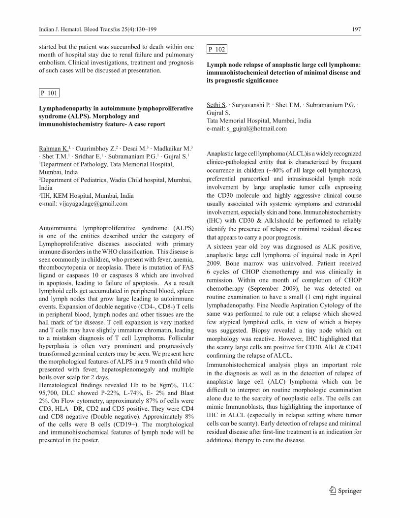
Indian J. Hematol. Blood Transfus 25(4):130–199 197
started but the patient was succumbed to death within one month of hospital stay due to renal failure and pulmonary embolism. Clinical investigations, treatment and prognosis of such cases will be discussed at presentation.
P 101
Lymphadenopathy in autoimmune lymphoproliferative syndrome (ALPS). Morphology and immunohistochemistry feature- A case report
Rahman K.1 · Cuurimbhoy Z.2 · Desai M.3 · Madkaikar M.3
· Shet T.M.1 · Sridhar E.1 · Subramaniam P.G.1 · Gujral S.1
1Department of Pathology, Tata Memorial Hospital, Mumbai, India2Department of Pediatrics, Wadia Child hospital, Mumbai, India3IIH, KEM Hospital, Mumbai, Indiae-mail: [email protected]
Autoimmunne lymphoproliferative syndrome (ALPS) is one of the entities described under the category of Lymphoproliferative diseases associated with primary immune disorders in the WHO classifi cation. This disease is seen commonly in children, who present with fever, anemia, thrombocytopenia or neoplasia. There is mutation of FAS ligand or caspases 10 or caspases 8 which are involved in apoptosis, leading to failure of apoptosis. As a result lymphoid cells get accumulated in peripheral blood, spleen and lymph nodes that grow large leading to autoimmune events. Expansion of double negative (CD4-, CD8-) T cells in peripheral blood, lymph nodes and other tissues are the hall mark of the disease. T cell expansion is very marked and T cells may have slightly immature chromatin, leading to a mistaken diagnosis of T cell Lymphoma. Follicular hyperplasia is often very prominent and progressively transformed germinal centers may be seen. We present here the morphological features of ALPS in a 9 month child who presented with fever, hepatosplenomegaly and multiple boils over scalp for 2 days.Hematological fi ndings revealed Hb to be 8gm%, TLC 95,700, DLC showed P-22%, L-74%, E- 2% and Blast 2%. On Flow cytometry, approximately 87% of cells were CD3, HLA –DR, CD2 and CD5 positive. They were CD4 and CD8 negative (Double negative). Approximately 8% of the cells were B cells (CD19+). The morphological and immunohistochemical features of lymph node will be presented in the poster.
P 102
Lymph node relapse of anaplastic large cell lymphoma: immunohistochemical detection of minimal disease and its prognostic signifi cance
Sethi S. · Suryavanshi P. · Shet T.M. · Subramanium P.G. · Gujral S.Tata Memorial Hospital, Mumbai, Indiae-mail: [email protected]
Anaplastic large cell lymphoma (ALCL)is a widely recognized clinico-pathological entity that is characterized by frequent occurrence in children (~40% of all large cell lymphomas), preferential paracortical and intrasinusoidal lymph node involvement by large anaplastic tumor cells expressing the CD30 molecule and highly aggressive clinical course usually associated with systemic symptoms and extranodal involvement, especially skin and bone. Immunohistochemistry (IHC) with CD30 & Alk1should be performed to reliably identify the presence of relapse or minimal residual disease that appears to carry a poor prognosis.A sixteen year old boy was diagnosed as ALK positive, anaplastic large cell lymphoma of inguinal node in April 2009. Bone marrow was uninvolved. Patient received 6 cycles of CHOP chemotherapy and was clinically in remission. Within one month of completion of CHOP chemotherapy (September 2009), he was detected on routine examination to have a small (1 cm) right inguinal lymphadenopathy. Fine Needle Aspiration Cytology of the same was performed to rule out a relapse which showed few atypical lymphoid cells, in view of which a biopsy was suggested. Biopsy revealed a tiny node which on morphology was reactive. However, IHC highlighted that the scanty large cells are positive for CD30, Alk1 & CD43 confi rming the relapse of ALCL.Immunohistochemical analysis plays an important role in the diagnosis as well as in the detection of relapse of anaplastic large cell (ALC) lymphoma which can be diffi cult to interpret on routine morphologic examination alone due to the scarcity of neoplastic cells. The cells can mimic Immunoblasts, thus highlighting the importance of IHC in ALCL (especially in relapse setting where tumor cells can be scanty). Early detection of relapse and minimal residual disease after fi rst-line treatment is an indication for additional therapy to cure the disease.
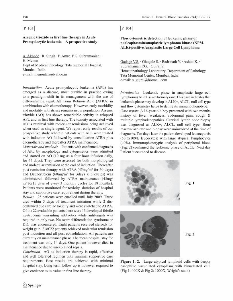
198 Indian J. Hematol. Blood Transfus 25(4):130–199
P 103
Arsenic trioxide as fi rst line therapy in Acute Promyelocytic leukemia – A prospective study
A. Akhade · R. Singh · P. Amre. P.G. Subramanian · H. Menon Dept of Medical Oncology, Tata memorial Hospital, Mumbai, Indiae-mail: [email protected]
Introduction Acute promyelocytic leukemia (APL) has emerged as a disease, most curable in practice owing to a paradigm shift in its management with the use of differentiating agent, All Trans Retinoic Acid (ATRA) in combination with chemotherapy. However, early morbidity and mortality with its use remains in our population. Arsenic trioxide (AO) has shown remarkable activity in relapsed APL and in fi rst line therapy. The toxicity associated with AO is minimal with molecular remissions being achieved when used as single agent. We report early results of our prospective study wherein patients with APL were treated with induction AO followed by consolidation ATRA plus chemotherapy and thereafter ATRA maintenance. Materials and methods Patients with confi rmed diagnosis of APL by morphology and cytogenetics were admitted and started on AO (10 mg as a four hour infusion daily, for 45 days). They were assessed for both morphological and molecular remission at the end of induction. Thereafter post remission therapy with ATRA (45mg/m2 for 60 days) and Daunorubicin (60mg/m2 for 3days x 3 cycles) was administered followed by ATRA maintenance (45mg/m2 for15 days of every 3 monthly cycles for 18 months). Patients were monitored for toxicity, duration of hospital stay and supportive care requirement during therapy. Results 25 patients were enrolled until July 2009. Three died within 5 days of treatment initiation while 2 dis-continued due cardiac toxicity and were switched to ATRA. Of the 22 evaluable patients there were 13 developed febrile neutropenia warranting antibiotics while antifungals was required in only two. No overt differentiation syndrome or DIC was encountered. Eight patients received steroids for weight gain. 21of 22 patients achieved molecular remission post induction and all post consolidation. All patients are currently on maintenance phase. The mean hospital stay for treatment was only 14 days. One patient however died in maintenance due to unexplained sepsis. Conclusion AO as induction therapy is rapid, effective and well tolerated regimen with minimal supportive care requirements. Best results are achieved with minimal hospital stay. Long term follow up is however required to give credence to its value in fi rst line therapy.
P 104
Flow cytometric detection of leukemic phase of nucleophosmin/anaplastic lymphoma kinase (NPM-ALK)-positive Anaplastic Large Cell Lymphoma
Gadage V.S. · Ghogale S. · Badrinath Y. · Ashok K. · Subramanian P.G. · Gujral S.Hematopathology Laboratory, Department of Pathology, Tata Memorial Center, Mumbai, Indiae-mail: [email protected]
Introduction Leukemic phase in anaplastic large cell lymphoma (ALCL) is extremely rare. This case indicates that leukemic phase may develop in ALK+, ALCL, null cell type and fl ow cytometry helps to defi ne its immunophenotype. Case report A 16-year-old boy presented with two months history of fever, weakness, abdominal pain, cough & multiple lymphadenopathies. Cervical lymph node biopsy was diagnosed as ALK+, ALCL, null cell type. Bone marrow aspirate and biopsy were uninvolved at the time of diagnosis. Ten days later the patient developed leucocytosis (50.5x10⁹/L leucocytes) with large atypical lymphocytes (40%). Immunophenotypic analysis of peripheral blood (Fig. 2) confi rmed the leukemic phase of ALCL. Next day Patient succumbed to disease.
Figure 1, 2. Large atypical lymphoid cells with deeply basophilic vacuolated cytoplasm with binucleated cell. (Fig 1: 400X & Fig 2: 1000X, Wright’s stain)
Fig. 1
Fig. 2
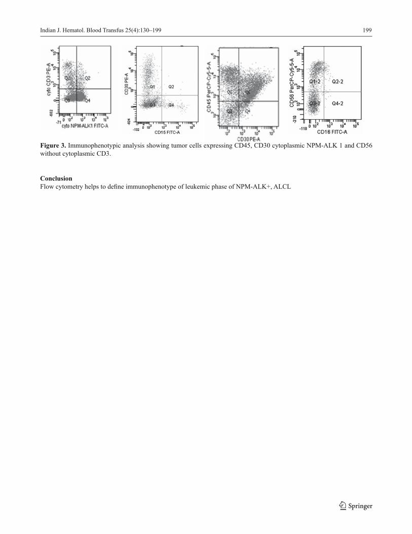
Indian J. Hematol. Blood Transfus 25(4):130–199 199
Figure 3. Immunophenotypic analysis showing tumor cells expressing CD45, CD30 cytoplasmic NPM-ALK 1 and CD56 without cytoplasmic CD3.
Conclusion Flow cytometry helps to defi ne immunophenotype of leukemic phase of NPM-ALK+, ALCL
Related Documents