S100A expression in normal corneal-limbal epithelial cells and ocular surface squamous cell carcinoma tissue Jing Li, 1,2,3 Andri K. Riau, 2 Melina Setiawan, 2 Jodhbir S. Mehta, 2,3,4 Seng-Ei Ti, 4 Louis Tong, 2,4,5 Donald T.H. Tan, 2,3,4 Roger W. Beuerman 2,3,6 1 Department of Ophthalmology, Xinhua Hospital, Shanghai Jiao Tong University, Shanghai, China; 2 Singapore Eye Research Institute, Singapore; 3 Department of Ophthalmology, Yong Loo Lin School of Medicine, National University of Singapore, Singapore; 4 Singapore National Eye Centre, Singapore; 5 Department of Clinical Sciences, Duke-NUS Graduate Medical School, Singapore; 6 SRP Neuroscience and Behavioral Disorders, Duke-NUS Graduate Medical School, Singapore Purpose: To study the expression and cellular distribution of multiple S100A genes and proteins in normal corneal-limbal epithelium and ocular surface squamous cell carcinoma (SCC) tissue. Methods: Normal corneal-limbal tissue was obtained from the Lions Eye Bank, Tampa, FL. Ocular surface SCC tissues were excised from patients undergoing surgery at Singapore National Eye Centre. S100A mRNA expression was measured by quantitative PCR. S100 protein distribution was determined by immunofluorescent staining analysis. Results: Twelve S100 mRNAs were identified in human corneal and limbal epithelial cells. S100A2, A6, A8, A9, A10, and A11 mRNA was expressed at high level, while S100A1, A3, A4, A5, A6, A7, and A12 mRNA expression was low. The intracellular localization of S100A2, A6, A8, A9, A10 and A11 protein was determined in normal corneal-limbal and SCC tissues. S100A2 and S100A10 proteins were enriched in basal limbal epithelial cells of the normal tissue. S100A8 and S100A9 were found only at the surface of peripheral corneal and limbal epithelium. S100A6 was uniformly found at the plasma membrane of corneal and limbal epithelial cells. S100A11 was found at the supralayer limbal epithelial cells adjacent to the conjunctiva. SCC tissue showed typical pathological changes with expression of cytokeartin (CK) 14 and CK4 in the epithelial cells. All SCC epithelial cells were positive of S100A2, S100A10, S100A6 and S100A11 staining. Intracellular staining of S100A8 and S100A9 was found in several layers of SCC epithelium. Expression of S100A2 and S100A10 decreased dramatically in cultured limbal epithelial cells with increased passaging, which was accompanied by a small increase of S100A9 mRNA, with no changes of S100A8 gene expression. Serum and growth hormone depletion of the culture serum caused a small reduction of S100A2 and S100A10 gene expression, which was accompanied by a small increase of S100A9 mRNA while no changes of S100A8 expression was measured. Conclusions: Normal corneal and limbal epithelial cells express a broad spectrum of S100 genes and proteins. Ocular surface SCC express high levels of S100A2, S100A10, S100A8 and S100A9 proteins. The expression of S100A2 and S100A10 is associated with limbal epithelial cell proliferation and differentiation. S100 proteins are a group of small acidic proteins of 10– 12 kDa [1]. With more than 20 proteins identified, they form the largest family of calcium binding proteins. Each S100 protein has two calcium-binding EF-hand motifs: a modified S100-specific EF hand at the NH2-terminus and a classical one at the COOH-terminus. The two EF-hand motifs are connected by a central hinge sequence. Upon calcium binding, the hinge region undergoes large reorientation and exposes the binding interface for its target proteins such as annexins, cytoskeleton proteins, p53, and pattern recognition receptors [2-5]. Through binding with different proteins, S100 proteins are involved in the regulation of many important cellular activities such as calcium homeostasis, cytoskeleton organization, stress response, cell motility, cell proliferation and differentiation. Most noticeably, abnormal expression of many S100 proteins, such as S100A2, S100A4, S100A6, Correspondence to: Jing Li, Ph.D., 11 Third Hospital Avenue, 06-19 SNEC Building, Singapore 168751; Phone: 65-63224547; FAX: 65-63224599; email: [email protected] S100A8, S100A9, S100A10, and S100A11 is found in numerous cancers [6]. Several studies have reported the expression of S100 proteins in the ocular tissue. For example, abnormal S100A2 and S100A4 expression was found in human keratoconus tissue [7,8]. S100A4 and S100B proteins were found in activated stromal myofibroblast after corneal debridement, likely involved in stromal cell proliferation and wound healing [9,10]. A recent study reported the role of neutrophil secreted S100A8 and A9 proteins in mouse models of corneal neovascularization [11]. Upregulation of multiple other S100 mRNA expression such as S100A4, S100A6, and S100A13 was reported in the study [11]. However, the cellular source for these gene products was unclear. We have previously reported increased expression of S100A6,S100A8, and S100A9 mRNA and protein in pterygial tissue compared to normal conjunctiva [12]. Increased concentration of S100A8 and S100A9 was also detected in pterygium patient tear samples compared to healthy controls [13]. In another Molecular Vision 2011; 17:2263-2271 <http://www.molvis.org/molvis/v17/a246> Received 23 May 2011 | Accepted 17 August 2011 | Published 20 August 2011 © 2011 Molecular Vision 2263

Welcome message from author
This document is posted to help you gain knowledge. Please leave a comment to let me know what you think about it! Share it to your friends and learn new things together.
Transcript

S100A expression in normal corneal-limbal epithelial cells andocular surface squamous cell carcinoma tissue
Jing Li,1,2,3 Andri K. Riau,2 Melina Setiawan,2 Jodhbir S. Mehta,2,3,4 Seng-Ei Ti,4 Louis Tong,2,4,5
Donald T.H. Tan,2,3,4 Roger W. Beuerman2,3,6
1Department of Ophthalmology, Xinhua Hospital, Shanghai Jiao Tong University, Shanghai, China; 2Singapore Eye ResearchInstitute, Singapore; 3Department of Ophthalmology, Yong Loo Lin School of Medicine, National University of Singapore,Singapore; 4Singapore National Eye Centre, Singapore; 5Department of Clinical Sciences, Duke-NUS Graduate Medical School,Singapore; 6SRP Neuroscience and Behavioral Disorders, Duke-NUS Graduate Medical School, Singapore
Purpose: To study the expression and cellular distribution of multiple S100A genes and proteins in normal corneal-limbalepithelium and ocular surface squamous cell carcinoma (SCC) tissue.Methods: Normal corneal-limbal tissue was obtained from the Lions Eye Bank, Tampa, FL. Ocular surface SCC tissueswere excised from patients undergoing surgery at Singapore National Eye Centre. S100A mRNA expression was measuredby quantitative PCR. S100 protein distribution was determined by immunofluorescent staining analysis.Results: Twelve S100 mRNAs were identified in human corneal and limbal epithelial cells. S100A2, A6, A8, A9, A10,and A11 mRNA was expressed at high level, while S100A1, A3, A4, A5, A6, A7, and A12 mRNA expression was low.The intracellular localization of S100A2, A6, A8, A9, A10 and A11 protein was determined in normal corneal-limbal andSCC tissues. S100A2 and S100A10 proteins were enriched in basal limbal epithelial cells of the normal tissue. S100A8and S100A9 were found only at the surface of peripheral corneal and limbal epithelium. S100A6 was uniformly found atthe plasma membrane of corneal and limbal epithelial cells. S100A11 was found at the supralayer limbal epithelial cellsadjacent to the conjunctiva. SCC tissue showed typical pathological changes with expression of cytokeartin (CK) 14 andCK4 in the epithelial cells. All SCC epithelial cells were positive of S100A2, S100A10, S100A6 and S100A11 staining.Intracellular staining of S100A8 and S100A9 was found in several layers of SCC epithelium. Expression of S100A2 andS100A10 decreased dramatically in cultured limbal epithelial cells with increased passaging, which was accompanied bya small increase of S100A9 mRNA, with no changes of S100A8 gene expression. Serum and growth hormone depletionof the culture serum caused a small reduction of S100A2 and S100A10 gene expression, which was accompanied by asmall increase of S100A9 mRNA while no changes of S100A8 expression was measured.Conclusions: Normal corneal and limbal epithelial cells express a broad spectrum of S100 genes and proteins. Ocularsurface SCC express high levels of S100A2, S100A10, S100A8 and S100A9 proteins. The expression of S100A2 andS100A10 is associated with limbal epithelial cell proliferation and differentiation.
S100 proteins are a group of small acidic proteins of 10–12 kDa [1]. With more than 20 proteins identified, they formthe largest family of calcium binding proteins. Each S100protein has two calcium-binding EF-hand motifs: a modifiedS100-specific EF hand at the NH2-terminus and a classical oneat the COOH-terminus. The two EF-hand motifs areconnected by a central hinge sequence. Upon calcium binding,the hinge region undergoes large reorientation and exposesthe binding interface for its target proteins such as annexins,cytoskeleton proteins, p53, and pattern recognition receptors[2-5]. Through binding with different proteins, S100 proteinsare involved in the regulation of many important cellularactivities such as calcium homeostasis, cytoskeletonorganization, stress response, cell motility, cell proliferationand differentiation. Most noticeably, abnormal expression ofmany S100 proteins, such as S100A2, S100A4, S100A6,
Correspondence to: Jing Li, Ph.D., 11 Third Hospital Avenue, 06-19SNEC Building, Singapore 168751; Phone: 65-63224547; FAX:65-63224599; email: [email protected]
S100A8, S100A9, S100A10, and S100A11 is found innumerous cancers [6].
Several studies have reported the expression of S100proteins in the ocular tissue. For example, abnormal S100A2and S100A4 expression was found in human keratoconustissue [7,8]. S100A4 and S100B proteins were found inactivated stromal myofibroblast after corneal debridement,likely involved in stromal cell proliferation and woundhealing [9,10]. A recent study reported the role of neutrophilsecreted S100A8 and A9 proteins in mouse models of cornealneovascularization [11]. Upregulation of multiple otherS100 mRNA expression such as S100A4, S100A6, andS100A13 was reported in the study [11]. However, the cellularsource for these gene products was unclear. We havepreviously reported increased expression of S100A6,S100A8,and S100A9 mRNA and protein in pterygial tissue comparedto normal conjunctiva [12]. Increased concentration ofS100A8 and S100A9 was also detected in pterygium patienttear samples compared to healthy controls [13]. In another
Molecular Vision 2011; 17:2263-2271 <http://www.molvis.org/molvis/v17/a246>Received 23 May 2011 | Accepted 17 August 2011 | Published 20 August 2011
© 2011 Molecular Vision
2263

study, we reported increased S100A4, S100A8, S100A9, andS100A11 proteins in tear samples obtained from dry eyepatients [14]. Collectively, these studies suggest theinvolvement of multiple S100A proteins in inflammatory andproliferative conditions of the ocular surface. However, thescope of S100A gene and protein expression in human cornealcells remains unknown.
Ocular surface squamous cell carcinoma (SCC) is one ofthe major cause for ocular morbidity and mortality. It isfeatured by dysregulated proliferation and differentiation ofcorneal and conjunctival epithelial cells [15]. A benign formof proliferative disorder of the ocular surface is the corneal/conjunctival intraepithelial neoplasm (CIN) [16]. Despite theextensive report on S100 proteins in various cancers, theinvolvement of S100 proteins in these conditions is unknown.Here we report the differential expression and cellulardistribution of multiple S100A genes and proteins in normalcorneal-limbal and ocular surface SCC epithelial cells. Wefurther demonstrate the association between limbal epithelialcell differentiation and the expression of S100A2 andS100A10 genes. Our results suggest that selective S100proteins are involved in corneal epithelial cell proliferationand differentiation under both normal and pathologicalconditions.
METHODSHuman corneal and limbal epithelial cell isolation andculture: Cadaver corneal-limbal tissues were obtained fromthe Lions Eye Bank, Tampa, Florida. The corneal epithelialcells were collected by scraping the corneal surface using asterile surgical blade. The blade was rinsed with 1 ml of Trizolsolution immediately and RNA was extracted. The peripheral/limbal region 2–3 mm inside the thin circle of pigmentationwas avoided during the scraping. After the scraping, theremaining limbal rim was excised, washed with antibioticsand subjected to dispase followed by trypsin digestion.Detailed protocols for the isolation and culture of humanlimbal epithelial cells have been published previously by ourgroup [17,18]. Isolated limbal epithelial cells were cultured inSHEM medium which contained equal volumes of DMEMand F12, 2 ng/ml of recombinant human epidermal growthfactor (EGF), 1 μg/ml bovine insulin, 0.1 μg/ml cholera toxin,0.5 μg/ml hydrocortisone, and 10% fetal bovine serum (FBS)in the presence of mitomycin-C inactivated 3T3 fibroblasts.Limbal cells were passaged when more than 70% of theculture dish area was covered by colonies and the majority ofthe colonies had about 100–200 cells. For serum and growthfactor depletion experiment, the limbal epithelial cells werecultured in the above medium without EGF, insulin, and FBSfor 24 h.
TABLE 1. PRIMERS USED FOR S100A QPCR.
Gene name Primer sequences (5′-3′) Amplicon size (bp) Annealing temperatureS100A1 F: CTTGGCCATCTGTCCAGAAC
R: CAATGTGGCTGTCTGCTCAA 362 56 °CS100A2 F: GCCAAGAGGGCGACAAGTT
R: AGGAAAACAGCATACTCCTGGA 175 60 °CS100A3 F: GCGGTAGCTGCCATCGTGTG
R: GCAGTCCTTGAAGTACTCGT 258 56 °CS100A4 F: GATGAGCAACTTGGACAGCAA
R: CTGGGCTGCTTATCTGGGAAG 123 60 °CS100A5 F: CTGCACACTGTGATGGAGAC
R: GAAGTCGTTGTAGGCCATGC 270 56 °CS100A6 F: AAGCTGCAGGATGCTGAAAT
R: CCCTTGAGGGCTTCATTGTA 131 56 °CS100A7 F: TGCTGACGATGATGAAGGAG
R: ATGTCTCCCAGCAAGGACAG 131 56 °CS100A8 F: ATGCCGTCTACAGGGATGAC
R: ACGCCCATCTTTATCACCAG 160 56 °CS100A9 F: GTGCGAAAAGATCTGCAAAA
R: TCAGCTGCTTGTCTGCATTT 103 56 °CS100A10 F: ATGCCATCTCAAATGGAACA
R: CTACTTCTTTCCCTTCTGCT 294 56 °CS100A11 F: ATGGCAAAAATCTCCAGCCC
R: TCATCATGCGGTCAAGGACA 193 56 °CS100A12 F: CCTCTCTAAGGGTGAGCTGA
R: CTGGGTTTTGGTGAGGGAAA 271 56 °CACTB F: ATCATGTTTGAGACCTTCAACA
R: CATCTCTTGCTCGAAGTCCA 318 56 °CGAPDH F: CCATGTTCGTCATGGGTGTGAACGA
R: GCCAGTAGAGGCAGGGATGATGTTC 254 56 °C
Molecular Vision 2011; 17:2263-2271 <http://www.molvis.org/molvis/v17/a246> © 2011 Molecular Vision
2264

Reverse transcription (RT) and quantitative PCR (qPCR)analysis: PureLink (Invitrogen, Singapore) was used for totalRNA extraction. RNA concentration was evaluated byNanodrop spectrophotometer. RNA (250 ng) was reversetranscribed to cDNA using RTIII (Invitrogen, Singapore).cDNA (0.5 μl) was used in SYBR-green based qPCRanalysis using Roche LightCycler 480 and Roche PCRmaster mix (Roche , Singapore). Primer sequences forindividual S100A mRNAs are listed in Table 1. A single peakof qPCR product with the melting temperature between 80 and90 °C was observed for each reaction. Glyceraldehyde 3-phosphate dehydrogenase (GAPDH) and β-actin (ACTB) wereused as internal controls for the calculation of relative cyclethreshold (ΔCp; ΔCp=Cp [target] – Cp [ACTB] or Cp[GAPDH]). Each cDNA sample was analyzed in triplicates.Fold changes was calculated as 2- ΔΔCp relative to S100A4 incorneal cells.SCC patient information: Ocular surface SCC tissues wereobtained from 2 patients who underwent surgical excision.The first case was a 60-year-old Chinese male with recurrentcorneal/conjunctival intraepithelial neoplasia for 5 years thatdeveloped into SCC. The second case was another 60-year-old Chinese male with a left eye SCC who originally presentedwith sclerokeratitis for 8 months with corneal scarring. Theappearance of a large nodule nasal to the area of sclerathinning appeared on the 8th month and a diagnosis of SCCwas made following excision biopsy. No signs of tumorrecurrence were noticed 5 months after the excision surgery.
Informed written consent was obtained from eachparticipant. This study was performed in accordance with the
tenets of the Declaration of Helsinki and the study protocolwas approved by SingHealth Institutional Review Board.Histological and immunofluorescent staining analysis:Histological analysis of SCC biopsy and immunofluorescentstaining of S100 proteins of SCC and normal corneal andlimbal tissues were performed as previously described [12].The slides were fixed with freshly made 4%paraformaldehyde (PFA), blocked by 4% BSA with 0.1%Triton X-100 and incubated with the following antibodiesovernight at 4 °C: mouse monoclonal anti-S100A2 (clone SH-L1; Sigma, Singapore) antibody at 1:1000 dilution; mousemonoclonal anti-S100A6 antibody (6B5; Abnova, Taipei,Taiwan) at 1:300 dilution; mouse monoclonal anti-S100A8antibody (8–5C2; Acris, Herford, Germany) at 1:150 dilution;mouse monoclonal anti-S100A9 antibody (1C10; Abnova) at1:150 dilution; rabbit polyclonal anti-S100A10 (Abcam,Cambridge, MA) at 1:100 dilution; mouse monoclonal anti-S100A11 antibody (2F4; Abnova) at 1:150 dilution; mousemonoclonal anti-CK3/12 antibody (2Q1040 Abcam) at 1:50dilution; mouse monoclonal anti-CK4 antibody (6B10 Acris)at 1:50 dilution; and goat polyclonal anti-CK14 antibody(Santa Cruz Biotechnology, Santa Cruz, CA) at 1:50 dilution.This was followed by incubation with an Alexa Fluor 488-conjugated secondary antibody for 1 h and the slides weremounted with DAPI-containing UltraCruz Mounting Medium(Santa Cruz Biotechnology). The slides were examined undera Zeiss Axioplan 2 fluorescence microscope (Zeiss,Gottingen, Germany) and digital images of representativeareas were taken.
Figure 1. Relative abundance of 12 S100A mRNA in human corneal and primary cultured limbal epithelial cells. The data represents averagedvalue from 6 different preparations of limbal and 3 corneal epithelial cells. For each cDNA samples, qPCR was performed in triplicates. ΔCpwas calculated against β-actin (ACTB). The ΔCp of S100A4 in corneal cells was taken as 100% and used as reference for the calculation ofthe relative abundance of other S100A mRNA. Error bars stand for the standard deviation of the averaged results. *Represents statisticallysignificant difference (p<0.05 by unpaired t-test) between corneal and limbal epithelial cells for the same mRNA.
Molecular Vision 2011; 17:2263-2271 <http://www.molvis.org/molvis/v17/a246> © 2011 Molecular Vision
2265

Western blot analysis of cytokeratin 3 protein: Culturedhuman primary limbal epithelial cells were washed with 2 mMEDTA followed by PBS to remove 3T3 feeder cells and lysedwith RIPA buffer [18]. Total protein (20 µg) was loaded onSDS–PAGE and transferred to nitrocellulose paper. The blotwas probed by an anti-cytokeratin 3 antibody (AE5; SantaCruz Biotechnology, Santa Cruz, CA) and a major band of64 kDa (cytokeratin 3) was detected.
RESULTSExpression of S100 genes in human corneal and limbalepithelial cells: Expression of S100A1, A2, A3, A4, A5, A6,A7, A8, A9, A10, A11 and A12 mRNA was measured byqPCR. The levels of individual mRNA expression in corneal(3 samples) and limbal epithelial cells (6 samples) werecalibrated using ACTB as an internal control and expressedrelative to S100A4 in corneal cells (Figure 1). Similar resultswere obtained using GAPDH as an internal control (Data notshown). Between corneal and limbal epithelial cells, S100A7and S100A9 mRNA was more abundant in corneal epithelialcells while S100A11 mRNA was more abundant in culturedlimbal epithelial cells (p<0.05, unpaired t-test).
Histological analysis and cytokeratin (CK) expression inocular surface SCC epithelial cells: H&E staining revealedthickening of the squamous epithelium of the SCC tissue, aswell as local disruption of the basement membrane andepithelial cell invasion of the stroma (Figure 2A,B). Theinvaded epithelial cells in the stroma showed an enlargednucleus. Keratin pearls formed by necrotic epithelial cells inthe stroma were observed. Massive leukocytes infiltration,stromal fibrosis and irregular collagen fiber deposition in thestroma tissue were also observed.
Immunofluorescent staining analysis showed CK14expression in all SCC epithelial cells (Figure 2C). CK4staining was also positive in epithelial cells except for thebasal cells (Figure 2D). CK3/12 staining was very weak in theSCC epithelium (Figure 2E).
Immunofluorescent analysis of S100 proteins in normalcorneal-limbal and SCC epithelia: Based on the mRNAabundance, the localization of S100A2, S100A6, S100A8,S100A9, S100A10, and S100A11 proteins was analyzed innormal corneal-limbal and SCC tissue.
Figure 2. Morphological features and cytokeratin protein expression in SCC tissue. A and B: H&E staining of SCC tissue. A: Notice thethickening of the epithelium as demarcated by the white dotted line, the breach of the basement membrane and massive leukocytes infiltration(white arrows). B: The epithelial cell invasion of the stroma (marked by the dotted white lines). The keratin pearls formed by necrotic epithelialcells (white arrow). The invading cells showed typical metaplastic appearance with enlarged nucleus and cell volume. The original pictureswere taken at 100× magnification. C and D: Immunofluorescent staining of cytokeratin proteins (green) in SCC tissue. C: CK14 antibodystained the full thickness of the epithelium. The signal was more intense along the basal cell layer. D: CK4 staining of the SCC epitheliumexcept for the basal layer. E: Weak CK3/12 staining of the SCC epithelium. The nucleus was counter-stained blue with DAPI. The originalpictures were taken at 200× magnification.
Molecular Vision 2011; 17:2263-2271 <http://www.molvis.org/molvis/v17/a246> © 2011 Molecular Vision
2266

S100A2 staining was not observed in normal cornealepithelium (Figure 3A). In contrast, a strong S100A2 stainingwas consistently observed at the plasma membrane of basallimbal epithelial cells and the staining decreasedprogressively toward the wing layers (Figure 3B). Sporadicstaining was also observed at the superficial layers of thelimbal epithelium. The SCC epithelium showed
overwhelmingly strong diffusive S100A2 staining in all cells(Figure 3C).
S100A6 staining was observed at the plasma membraneof normal corneal, limbal and SCC epithelial cells (Figure 3D-F). No obvious difference in staining intensity betweennormal corneal-limbal and SCC epithelial cells was observed.
Figure 3. Immunofluorescent staining of S100A2, A6, A8, A10, and A11 proteins (in green) in normal human corneal, limbal and SCCepithelia. The nucleus was counter-stained blue with DAPI. Except for the pictures on S100A10 staining, which were taken at 400×, all otherswere taken at 200× magnification originally.
Molecular Vision 2011; 17:2263-2271 <http://www.molvis.org/molvis/v17/a246> © 2011 Molecular Vision
2267
S100A8 and A9 showed the same staining pattern in thesame tissue tested, therefore only the images of S100A8staining are presented. In normal corneal and limbal tissue,positive staining of both proteins was observed only at thesurface of the peripheral corneal and limbal epithelium(Figure 3G,H). However, strong intracellular staining of bothproteins was observed in top layers of SCC epithelium. Thestaining intensity progressively reduced toward the stromaand was negative in the basal epithelial cells (Figure 3I).
S100A10 staining was absent in normal cornealepithelium (Figure 3J). Strong S100A10 staining existedpredominately in the nucleus of limbal basal epithelial cells,although some diffusive cytoplasmic staining was alsoobserved (Figure 3K). In addition, S100A10 positive cellsprogressively decreased toward the upper layers of the limbus.In SCC epithelium, the staining was uniformly concentratedin the nucleus of all cells (Figure 3L).
S100A11 staining was observed at the plasma membraneof superficial layer cells of limbal epithelium adjacent toconjunctiva, but not in the cornea (Figure 3M,N). In SCCepithelia, uniform plasma membrane staining of S100A11was observed in all cells (Figure 3O).
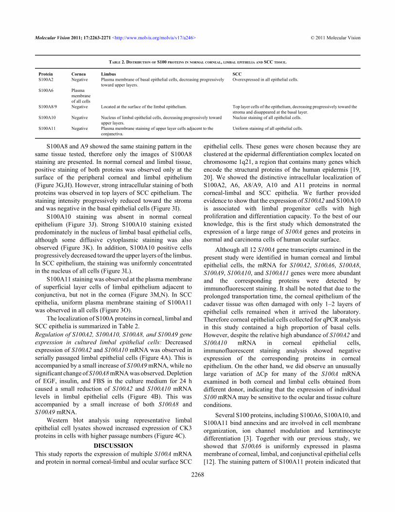
The localization of S100A proteins in corneal, limbal andSCC epithelia is summarized in Table 2.Regulation of S100A2, S100A10, S100A8, and S100A9 geneexpression in cultured limbal epithelial cells: Decreasedexpression of S100A2 and S100A10 mRNA was observed inserially passaged limbal epithelial cells (Figure 4A). This isaccompanied by a small increase of S100A9 mRNA, while nosignificant change of S100A8 mRNA was observed. Depletionof EGF, insulin, and FBS in the culture medium for 24 hcaused a small reduction of S100A2 and S100A10 mRNAlevels in limbal epithelial cells (Figure 4B). This wasaccompanied by a small increase of both S100A8 andS100A9 mRNA.
Western blot analysis using representative limbalepithelial cell lysates showed increased expression of CK3proteins in cells with higher passage numbers (Figure 4C).
DISCUSSIONThis study reports the expression of multiple S100A mRNAand protein in normal corneal-limbal and ocular surface SCC
epithelial cells. These genes were chosen because they areclustered at the epidermal differentiation complex located onchromosome 1q21, a region that contains many genes whichencode the structural proteins of the human epidermis [19,20]. We showed the distinctive intracellular localization ofS100A2, A6, A8/A9, A10 and A11 proteins in normalcorneal-limbal and SCC epithelia. We further providedevidence to show that the expression of S100A2 and S100A10is associated with limbal progenitor cells with highproliferation and differentiation capacity. To the best of ourknowledge, this is the first study which demonstrated theexpression of a large range of S100A genes and proteins innormal and carcinoma cells of human ocular surface.
Although all 12 S100A gene transcripts examined in thepresent study were identified in human corneal and limbalepithelial cells, the mRNA for S100A2, S100A6, S100A8,S100A9, S100A10, and S100A11 genes were more abundantand the corresponding proteins were detected byimmunofluorescent staining. It shall be noted that due to theprolonged transportation time, the corneal epithelium of thecadaver tissue was often damaged with only 1–2 layers ofepithelial cells remained when it arrived the laboratory.Therefore corneal epithelial cells collected for qPCR analysisin this study contained a high proportion of basal cells.However, despite the relative high abundance of S100A2 andS100A10 mRNA in corneal epithelial cells,immunofluorescent staining analysis showed negativeexpression of the corresponding proteins in cornealepithelium. On the other hand, we did observe an unusuallylarge variation of ΔCp for many of the S100A mRNAexamined in both corneal and limbal cells obtained fromdifferent donor, indicating that the expression of individualS100 mRNA may be sensitive to the ocular and tissue cultureconditions.
Several S100 proteins, including S100A6, S100A10, andS100A11 bind annexins and are involved in cell membraneorganization, ion channel modulation and keratinocytedifferentiation [3]. Together with our previous study, weshowed that S100A6 is uniformly expressed in plasmamembrane of corneal, limbal, and conjunctival epithelial cells[12]. The staining pattern of S100A11 protein indicated that
TABLE 2. DISTRIBUTION OF S100 PROTEINS IN NORMAL CORNEAL, LIMBAL EPITHELIA AND SCC TISSUE.
Protein Cornea Limbus SCCS100A2 Negative Plasma membrane of basal epithelial cells, decreasing progressively
toward upper layers.Overexpressed in all epithelial cells.
S100A6 Plasmamembraneof all cells
S100A8/9 Negative Located at the surface of the limbal epithelium. Top layer cells of the epithelium, decreasing progressively toward thestroma and disappeared at the basal layer.
S100A10 Negative Nucleus of limbal epithelial cells, decreasing progressively towardupper layers.
Nuclear staining of all epithelial cells.
S100A11 Negative Plasma membrane staining of upper layer cells adjacent to theconjunctiva.
Uniform staining of all epithelial cells.
Molecular Vision 2011; 17:2263-2271 <http://www.molvis.org/molvis/v17/a246> © 2011 Molecular Vision
2268
it is likely associated with CK4-positive conjunctivalepithelial cells. This is evidenced by the positive staining ofS100A11 in conjunctival epithelial cells as previouslyreported [12]. In this study, positive S100A11 staining wasfound only in part of the supralayer limbal epithelial cellsadjacent to the conjunctiva. In SCC epithelium, S100A11positive cells also coincided with CK4 positive cells, whilethe basal cells of SCC epithelium were negative of bothproteins. We speculate that the distinct expression of annexin-binding S100 proteins contributes to the differentcharacteristics of conjunctival and corneal epithelia.
Two S100A proteins were found specifically enriched inthe limbal epithelial cells of the normal tissue: S100A2 and
S100A10. The cellular localization of these proteins is inagreement with previous findings in other cell types [21-25].Interestingly, both proteins were found overexpressed in SCCepithelial cells. To test whether S100A2 and S100A10expression was associated with the proliferation anddifferentiation capacity of the limbal progenitor cells, weexamined the expression of these two genes in seriallypassaged limbal epithelial cells and in cells cultured underserum and growth hormone depleted conditions. Limbalepithelial cells co-cultured with mitomycin-C inactivated 3T3cells form colonies and are able to maintain a relatively highproliferation potential [17,26]. However, with each passagethe cultured cells gradually lose the proliferation capacity and
Figure 4. S100A2, S100A10, S100A8,and S100A9 mRNA expression incultured limbal epithelial cells. A: Foldchanges of the above mRNA in P1, P3,and P5 limbal epithelial cells. mRNAlevel in P1 limbal cells was set as 100%.B: Fold changes of the above mRNA inlimbal epithelial cells cultured in serum-and growth hormone-depleted medium.mRNA level in cells grown in fullmedium was set as 100%. C: westernblot analysis of CK3 protein in P1, P3,and P5 limbal epithelial cells.
Molecular Vision 2011; 17:2263-2271 <http://www.molvis.org/molvis/v17/a246> © 2011 Molecular Vision
2269

undergo differentiation as indicated by the increasingexpression of CK3/12 protein. Significant progressivereduction of S100A2 and S100A10 mRNA was observed inlimbal cells with increasing passaging. Serum and growthfactor depletion also induced a small reduction of both mRNAexpressions. Collectively the data suggested that theexpression of S100A2 and S100A10 is positively associatedwith the intrinsic proliferation and differentiation capacity oflimbal epithelial cells.
S100A8 and A9, also known as calgranulins or myeloidrelated proteins (MRPs), are mainly expressed in granulocytesand epithelial cells. Secreted S100A8 and S100A9 proteinsform heterodimer and are important members of damage-associated molecular pattern proteins (DAMPs) [4,27]. Ourresults suggested that both proteins were secreted in normalcorneal and limbal tissue, similar to what was reported innormal skin epidermis [25]. However, both proteins wereoverexpressed in SCC epithelial cells and found intra- andextracellularly. This is likely related to the inflammatoryconditions associated with SCC as it was also reported inenvironmentally stressed and inflammatory epidermis as wellas in cancer cells [25,28,29]. The changes of S100A8 andS100A9 gene expression in limbal epithelial cells under serumand growth factor depleted culture conditions may representa nutrition-depletion triggered stress response.
Ocular surface SCC is a major cause for ocular morbidityand mortality. Ocular SCC can sometimes develop from CIN,a benign proliferative condition which is more frequently seenthan SCC [16]. Finding markers that indicate the progressionof CIN and the malignancy of SCC will help clinicians tochoose an appropriate therapeutic modality from early on.While abnormal expression of many S100A proteins werefound in cancer cells, changes of S100A2 and S100A10expression were reported in various SCC of the oral cavity,esophagus, the larynx, kidney, and the thyroid [30-36]. In thisregard, these two proteins may also be candidates for markersassociated with the severity of ocular surface SCC.
In summary, this study showed that distinct S100Aproteins are involved in the structural and biologic activitiesof normal corneal and limbal epithelial cells and are associatedwith ocular surface squamous cell carcinoma. The resultspresented here warrant further investigation to understand theroles of specific S100A proteins in the regulation of ocularsurface epithelial cell proliferation, differentiation and tumordevelopment.
ACKNOWLEDGMENTSThis work was supported by grants from National MedicalResearch Council (NMRC) of Singapore (NMRC/1046/2006)and Singhealth Research Foundation (SHF/FG416P/2009) toDr. J. Li.
REFERENCES1. Santamaria-Kisiel L, Rintala-Dempsey AC, Shaw GS.
Calcium-dependent and -independent interactions of the S100
protein family. Biochem J 2006; 396:201-14. [PMID:16683912]
2. van Dieck J, Fernandez-Fernandez MR, Veprintsev DB, FershtAR. Modulation of the oligomerization state of p53 bydifferential binding of proteins of the S100 family to p53monomers and tetramers. J Biol Chem 2009; 284:13804-11.[PMID: 19297317]
3. Miwa N, Uebi T, Kawamura S. S100-annexin complexes–biology of conditional association. FEBS J 2008;275:4945-55. [PMID: 18795952]
4. Ehrchen JM, Sunderkotter C, Foell D, Vogl T, Roth J. Theendogenous Toll-like receptor 4 agonist S100A8/S100A9(calprotectin) as innate amplifier of infection, autoimmunity,and cancer. J Leukoc Biol 2009; 86:557-66. [PMID:19451397]
5. Leclerc E, Fritz G, Vetter SW, Heizmann CW. Binding of S100proteins to RAGE: An update. Biochim Biophys Acta 2009;1793:993-1007. [PMID: 19121341]
6. Salama I, Malone PS, Mihaimeed F, Jones JL. A review of theS100 proteins in cancer. Eur J Surg Oncol 2008; 34:357-64.[PMID: 17566693]
7. Nielsen K, Heegaard S, Vorum H, Birkenkamp-Demtroder K,Ehlers N, Orntoft TF. Altered expression of CLC, DSG3,EMP3, S100A2, and SLPI in corneal epithelium fromkeratoconus patients. Cornea 2005; 24:661-8. [PMID:16015083]
8. Nielsen K, Vorum H, Fagerholm P, Birkenkamp-Demtroder K,Honore B, Ehlers N, Orntoft TF. Proteome profiling ofcorneal epithelium and identification of marker proteins forkeratoconus, a pilot study. Exp Eye Res 2006; 82:201-9.[PMID: 16083875]
9. Ryan DG, Taliana L, Sun L, Wei ZG, Masur SK, Lavker RM.Involvement of S100A4 in stromal fibroblasts of theregenerating cornea. Invest Ophthalmol Vis Sci 2003;44:4255-62. [PMID: 14507869]
10. Yew DT, Lam TK, Tsang D, Au YK, Li WW, Tso MO. Changesof cytochemical markers in the conjunctival and cornealepithelium after corneal debridement. Cell Mol Neurobiol2000; 20:465-82. [PMID: 10901267]
11. Li C, Zhang F, Wang Y. S100A proteins in the pathogenesis ofexperimental corneal neovascularization. Mol Vis 2010;16:2225-35. [PMID: 21139687]
12. Riau AK, Wong TT, Beuerman RW, Tong L. Calcium-bindingS100 protein expression in pterygium. Mol Vis 2009;15:335-42. [PMID: 19223989]
13. Zhou L, Beuerman RW, Ang LP, Chan CM, Li SF, Chew FT,Tan DT. Elevation of human alpha-defensins and S100calcium-binding proteins A8 and A9 in tear fluid of patientswith pterygium. Invest Ophthalmol Vis Sci 2009;50:2077-86. [PMID: 19168894]
14. Zhou L, Beuerman RW, Chan CM, Zhao SZ, Li XR, Yang H,Tong L, Liu S, Stern ME, Tan D. Identification of tear fluidbiomarkers in dry eye syndrome using iTRAQ quantitativeproteomics. J Proteome Res 2009; 8:4889-905. [PMID:19705875]
15. Mehta M, Fay A. Squamous cell carcinoma of the eyelid andconjunctiva. Int Ophthalmol Clin 2009; 49:111-21. [PMID:19125070]
Molecular Vision 2011; 17:2263-2271 <http://www.molvis.org/molvis/v17/a246> © 2011 Molecular Vision
2270
16. Hamam R, Bhat P, Foster CS. Conjunctival/cornealintraepithelial neoplasia. Int Ophthalmol Clin 2009;49:63-70. [PMID: 19125065]
17. Liu S, Li J, Wang C, Tan D, Beuerman R. Human limbalprogenitor cell characteristics are maintained in tissue culture.Ann Acad Med Singapore 2006; 35:80-6. [PMID: 16565759]
18. Li J, Shen J, Beuerman RW. Expression of toll-like receptorsin human limbal and conjunctival epithelial cells. Mol Vis2007; 13:813-22. [PMID: 17615542]
19. Mischke D, Korge BP, Marenholz I, Volz A, Ziegler A. Genesencoding structural proteins of epidermal cornification andS100 calcium-binding proteins form a gene complex(“epidermal differentiation complex”) on humanchromosome 1q21. J Invest Dermatol 1996; 106:989-92.[PMID: 8618063]
20. Eckert RL, Broome AM, Ruse M, Robinson N, Ryan D, Lee K.S100 proteins in the epidermis. J Invest Dermatol 2004;123:23-33. [PMID: 15191538]
21. Böni R, Burg G, Doguoglu A, Ilg EC, Schafer BW, Muller B,Heizmann CW. Immunohistochemical localization of theCa2+ binding S100 proteins in normal human skin andmelanocytic lesions. Br J Dermatol 1997; 137:39-43. [PMID:9274623]
22. Zhang L, Yu W, He T, Yu J, Caffrey RE, Dalmasso EA, Fu S,Pham T, Mei J, Ho JJ, Zhang W, Lopez P, Ho DD.Contribution of human alpha-defensin 1, 2, and 3 to the anti-HIV-1 activity of CD8 antiviral factor. Science 2002;298:995-1000. [PMID: 12351674]
23. Li Y, Gudjonsson JE, Woods TL, Zhang T, Johnston A, StollSW, Elder JT. Transgenic expression of S100A2 in hairlessmouse skin enhances Cxcl13 mRNA in response to solar-simulated radiation. Arch Dermatol Res 2009; 301:205-17.[PMID: 18773213]
24. Zobiack N, Gerke V, Rescher U. Complex formation andsubmembranous localization of annexin 2 and S100A10 inlive HepG2 cells. FEBS Lett 2001; 500:137-40. [PMID:11445072]
25. Broome AM, Ryan D, Eckert RL. S100 protein subcellularlocalization during epidermal differentiation and psoriasis. JHistochem Cytochem 2003; 51:675-85. [PMID: 12704215]
26. Li W, Hayashida Y, He H, Kuo CL, Tseng SC. The fate of limbalepithelial progenitor cells during explant culture on intactamniotic membrane. Invest Ophthalmol Vis Sci 2007;48:605-13. [PMID: 17251456]
27. Foell D, Wittkowski H, Vogl T, Roth J. S100 proteins expressedin phagocytes: a novel group of damage-associated molecularpattern molecules. J Leukoc Biol 2007; 81:28-37. [PMID:16943388]
28. Marionnet C, Bernerd F, Dumas A, Verrecchia F, Mollier K,Compan D, Bernard B, Lahfa M, Leclaire J, Medaisko C,Mehul B, Seite S, Mauviel A, Dubertret L. Modulation ofgene expression induced in human epidermis byenvironmental stress in vivo. J Invest Dermatol 2003;121:1447-58. [PMID: 14675196]
29. Gebhardt C, Nemeth J, Angel P, Hess J. S100A8 and S100A9in inflammation and cancer. Biochem Pharmacol 2006;72:1622-31. [PMID: 16846592]
30. Almadori G, Bussu F, Galli J, Rigante M, Lauriola L, MichettiF, Maggiano N, Schafer BW, Heizmann CW, Ranelletti FO,Paludetti G. Diminished expression of S100A2, a putativetumour suppressor, is an independent predictive factor of necknode relapse in laryngeal squamous cell carcinoma. JOtolaryngol Head Neck Surg 2009; 38:16-22. [PMID:19344608]
31. Tsai WC, Tsai ST, Jin YT, Wu LW. Cyclooxygenase-2 isinvolved in S100A2-mediated tumor suppression insquamous cell carcinoma. Mol Cancer Res 2006; 4:539-47.[PMID: 16908593]
32. Suzuki F, Oridate N, Homma A, Nakamaru Y, Nagahashi T,Yagi K, Yamaguchi S, Furuta Y, Fukuda S. S100A2expression as a predictive marker for late cervical metastasisin stage I and II invasive squamous cell carcinoma of the oralcavity. Oncol Rep 2005; 14:1493-8. [PMID: 16273244]
33. Imazawa M, Hibi K, Fujitake S, Kodera Y, Ito K, Akiyama S,Nakao A. S100A2 overexpression is frequently observed inesophageal squamous cell carcinoma. Anticancer Res 2005;25:1247-50. [PMID: 15865073]
34. Nagy N, Brenner C, Markadieu N, Chaboteaux C, Camby I,Schafer BW, Pochet R, Heizmann CW, Salmon I, Kiss R,Decaestecker C. S100A2, a putative tumor suppressor gene,regulates in vitro squamous cell carcinoma migration. LabInvest 2001; 81:599-612. [PMID: 11304580]
35. Ito Y, Arai K, Nozawa R, Yoshida H, Higashiyama T,Takamura Y, Miya A, Kobayashi K, Kuma K, Miyauchi A.S100A10 expression in thyroid neoplasms originating fromthe follicular epithelium: contribution to the aggressivecharacteristic of anaplastic carcinoma. Anticancer Res 2007;27:2679-83. [PMID: 17695432]
36. Domoto T, Miyama Y, Suzuki H, Teratani T, Arai K, SugiyamaT, Takayama T, Mugiya S, Ozono S, Nozawa R. Evaluationof S100A10, annexin II and B-FABP expression as markersfor renal cell carcinoma. Cancer Sci 2007; 98:77-82. [PMID:17083565]
Molecular Vision 2011; 17:2263-2271 <http://www.molvis.org/molvis/v17/a246> © 2011 Molecular Vision
Articles are provided courtesy of Emory University and the Zhongshan Ophthalmic Center, Sun Yat-sen University, P.R. China.The print version of this article was created on 17 August 2011. This reflects all typographical corrections and errata to thearticle through that date. Details of any changes may be found in the online version of the article.
2271
http://www.ncbi.nlm.nih.gov/entrez/query.fcgi?cmd=Retrieve&db=PubMed&dopt=abstract&list_uids=8618063
http://www.ncbi.nlm.nih.gov/entrez/query.fcgi?cmd=Retrieve&db=PubMed&dopt=abstract&list_uids=8618063
http://www.ncbi.nlm.nih.gov/entrez/query.fcgi?cmd=Retrieve&db=PubMed&dopt=abstract&list_uids=9274623
Related Documents