S-Protected Thiolated Chitosan for Oral Delivery of Hydrophilic Macromolecules: Evaluation of Permeation Enhancing and Efflux Pump Inhibitory Properties Sarah Dü nnhaupt, † Jan Barthelmes, † Deni Rahmat, † Katharina Leithner, † Clemens C. Thurner, ‡ Heike Friedl, ‡ and Andreas Bernkop-Schnü rch* ,† † Department of Pharmaceutical Technology, Institute of Pharmacy, Leopold-Franzenz-University of Innsbruck, Innrain 80/82, 6020 Innsbruck, Austria ‡ ThioMatrix GmbH, Research Center Innsbruck, Trientlgasse 65, 6020 Innsbruck, Austria ABSTRACT: The objective of this study was the investigation of permeation enhancing and P-glycoprotein (P-gp) inhibition effects of a novel thiolated chitosan, the so-named S-protected thiolated chitosan. Mediated by a carbodiimide, increasing amounts of thioglycolic acid (TGA) were covalently bound to chitosan (CS) in the first step of modification. In the second step, these thiol groups of thiolated chitosan were protected by disulfide bond formation with the thiolated aromatic residue 6- mercaptonicotinamide (6-MNA). Mucoadhesive properties of all conjugates were evaluated in vitro on porcine intestinal mucosa based on tensile strength investigations. Permeation enhancing effects were evaluated ex vivo using rat intestinal mucosa and in vitro via Caco-2 cells using the hydrophilic macromolecule FD 4 as the model drug. Caco-2 cells were further used to show P-gp inhibition effects by using Rho-123 as P-gp substrate. Apparent permeability coefficients (P app ) were calculated and compared to values obtained from each buffer control. Three different thiolated chitosans were generated in the first step of modification, which displayed increasing amounts of covalently attached free thiol groups on the polymer backbone. In the second modification step, more than 50% of these free thiol groups were covalently linked with 6-MNA. Within 3 h of permeation studies on excised rat intestine, P app values of all S-protected chitosans were at least 1.3-fold higher compared to those of corresponding thiomers and more than twice as high as that of unmodified chitosan. Additional permeation studies on Caco-2 cells confirmed these results. Because of the chemical modification and higher amount of reactive thiol groups, all S-protected thiolated chitosans exhibit at least 1.4-fold pronounced P-gp inhibition effects in contrast to their corresponding thiomers. These features approve S-protected thiolated chitosan as a promising excipient for various drug delivery systems providing improved permeation enhancing and efflux inhibition effects. KEYWORDS: thiomers, S-protected thiomers, disulfide bonds, permeation enhancement, P-gp inhibition, oral drug delivery 1. INTRODUCTION Oral administration is the most popular route for drug administration since dosing is convenient and noninvasive, and many drugs are well absorbed by the gastrointestinal tract. However, the gastrointestinal mucosa represents various diffusion and enzymatic barriers to the systemic availability of orally ingested, pharmacologically active molecules. 1 Pharma- ceutical strategies to overcome these barriers include the use of enzyme 2 and efflux inhibitors, 3 permeation enhancers, 4 and multifunctional polymers. 5 In the case of multifunctional polymers, these effects, however, can only take place if a tight contact of the polymer with the mucosa is provided for the whole period of drug release and absorption. Apart from enzyme or efflux inhibitory and permeation enhancing properties, multifunctional polymers should offer additional mucoadhesive features. Among this group of multifunctional polymers, thiolated polymers, designated thiomers, exhibit all of the aforementioned properties. One of these thiomers, which exhibits a cationic charge, biocompatibility, and biodegrad- ability, is the thiolated chitosan. So far, various different thiolated chitosan derivatives have been synthesized and showed improved mucoadhesive, permeation enhancing, and efflux and enzyme inhibition effects. 6−8 However, besides all these advantages, a drawback of thiomers having been developed so far is their instability toward oxidation and pH- dependent reactivity. The reactive form of thiomers is the thiolate anion, whereas the pK a of alkyl thiols is in the range of 8−10, meaning thiomers will be most reactive in a pH range slightly above the physiological pH. 9 Consequently, they do not reach their full potential in cases where the pH is lower, such as in the stomach or the proximal small intestine. As opposed to Received: November 23, 2011 Revised: March 2, 2012 Accepted: April 10, 2012 Published: April 10, 2012 Article pubs.acs.org/molecularpharmaceutics © 2012 American Chemical Society 1331 dx.doi.org/10.1021/mp200598j | Mol. Pharmaceutics 2012, 9, 1331−1341

Welcome message from author
This document is posted to help you gain knowledge. Please leave a comment to let me know what you think about it! Share it to your friends and learn new things together.
Transcript
S-Protected Thiolated Chitosan for Oral Delivery of HydrophilicMacromolecules: Evaluation of Permeation Enhancing and EffluxPump Inhibitory PropertiesSarah Dunnhaupt,† Jan Barthelmes,† Deni Rahmat,† Katharina Leithner,† Clemens C. Thurner,‡
Heike Friedl,‡ and Andreas Bernkop-Schnurch*,†
†Department of Pharmaceutical Technology, Institute of Pharmacy, Leopold-Franzenz-University of Innsbruck, Innrain 80/82, 6020Innsbruck, Austria‡ThioMatrix GmbH, Research Center Innsbruck, Trientlgasse 65, 6020 Innsbruck, Austria
ABSTRACT: The objective of this study was the investigationof permeation enhancing and P-glycoprotein (P-gp) inhibitioneffects of a novel thiolated chitosan, the so-named S-protectedthiolated chitosan. Mediated by a carbodiimide, increasingamounts of thioglycolic acid (TGA) were covalently bound tochitosan (CS) in the first step of modification. In the secondstep, these thiol groups of thiolated chitosan were protected bydisulfide bond formation with the thiolated aromatic residue 6-mercaptonicotinamide (6-MNA). Mucoadhesive properties ofall conjugates were evaluated in vitro on porcine intestinalmucosa based on tensile strength investigations. Permeation enhancing effects were evaluated ex vivo using rat intestinal mucosaand in vitro via Caco-2 cells using the hydrophilic macromolecule FD4 as the model drug. Caco-2 cells were further used to showP-gp inhibition effects by using Rho-123 as P-gp substrate. Apparent permeability coefficients (Papp) were calculated andcompared to values obtained from each buffer control. Three different thiolated chitosans were generated in the first step ofmodification, which displayed increasing amounts of covalently attached free thiol groups on the polymer backbone. In thesecond modification step, more than 50% of these free thiol groups were covalently linked with 6-MNA. Within 3 h ofpermeation studies on excised rat intestine, Papp values of all S-protected chitosans were at least 1.3-fold higher compared to thoseof corresponding thiomers and more than twice as high as that of unmodified chitosan. Additional permeation studies on Caco-2cells confirmed these results. Because of the chemical modification and higher amount of reactive thiol groups, all S-protectedthiolated chitosans exhibit at least 1.4-fold pronounced P-gp inhibition effects in contrast to their corresponding thiomers. Thesefeatures approve S-protected thiolated chitosan as a promising excipient for various drug delivery systems providing improvedpermeation enhancing and efflux inhibition effects.
KEYWORDS: thiomers, S-protected thiomers, disulfide bonds, permeation enhancement, P-gp inhibition, oral drug delivery
1. INTRODUCTIONOral administration is the most popular route for drugadministration since dosing is convenient and noninvasive,and many drugs are well absorbed by the gastrointestinal tract.However, the gastrointestinal mucosa represents variousdiffusion and enzymatic barriers to the systemic availability oforally ingested, pharmacologically active molecules.1 Pharma-ceutical strategies to overcome these barriers include the use ofenzyme2 and efflux inhibitors,3 permeation enhancers,4 andmultifunctional polymers.5 In the case of multifunctionalpolymers, these effects, however, can only take place if a tightcontact of the polymer with the mucosa is provided for thewhole period of drug release and absorption. Apart fromenzyme or efflux inhibitory and permeation enhancingproperties, multifunctional polymers should offer additionalmucoadhesive features. Among this group of multifunctionalpolymers, thiolated polymers, designated thiomers, exhibit all ofthe aforementioned properties. One of these thiomers, which
exhibits a cationic charge, biocompatibility, and biodegrad-ability, is the thiolated chitosan. So far, various differentthiolated chitosan derivatives have been synthesized andshowed improved mucoadhesive, permeation enhancing, andefflux and enzyme inhibition effects.6−8 However, besides allthese advantages, a drawback of thiomers having beendeveloped so far is their instability toward oxidation and pH-dependent reactivity. The reactive form of thiomers is thethiolate anion, whereas the pKa of alkyl thiols is in the range of8−10, meaning thiomers will be most reactive in a pH rangeslightly above the physiological pH.9 Consequently, they do notreach their full potential in cases where the pH is lower, such asin the stomach or the proximal small intestine. As opposed to
Received: November 23, 2011Revised: March 2, 2012Accepted: April 10, 2012Published: April 10, 2012
Article
pubs.acs.org/molecularpharmaceutics
© 2012 American Chemical Society 1331 dx.doi.org/10.1021/mp200598j | Mol. Pharmaceutics 2012, 9, 1331−1341
this, thiomers show comparatively low stability in solutions, asthey are subject of thiol oxidation at pH ≥ 6.5 unless sealedunder inert conditions.10 In body regions such as the distalsmall intestine, colon, and rectum where the pH is higher, theirpotential use as an absorption enhancer is thereby limited.11
The design and development of thiomers reacting in a non-pH-dependent manner and being stable in these environmentswould therefore be highly advantageous, opening the door fornumerous additional applications. According to this, it was theaim of the present study to generate a thiolated chitosan, whosethiol groups are protected by already formed disulfide bonds,which offer the advantage of not being subject to oxidation.The hypothesis to achieve this goal is based on covalentchromatography, where, for instance, peptides and proteins arevery efficiently linked to thiol bearing resins, when they arepreactivated via pyridyl substructures.12,13 Pyridyl disulfidesreact very rapidly and quantitatively with sulfhydryl groups overa broad pH range to form disulfide bonds. During this reaction,a disulfide exchange occurs between the molecule’s −SH groupand the pyridyl thiol group, i.e., the thiomer forms new mixeddisulfides with the pyridyl thiol moiety as the leaving group.Because of the toxic nature of the pyridyl thiol leaving group,utilizing nicotinamide (vitamin B3) instead of the pyridyl groupcan exclude such toxic effects. It was therefore the aim of thisstudy to synthesize and characterize a thiol protected chitosanby the use of thiolated nicotinamide regarding its permeationenhancing and P-gp inhibitory effects in vitro. Within thisstudy, chitosan was modified by immobilization of free thiolgroups by thioglycolic acid to the polymer backbone withvarious degrees of thiolation. Subsequently, these immobilizedthiol groups were protected through disulfide bond formationby the use of the thiolated aromatic residue 6-mercaptonico-tinamide. The newly synthesized chitosans were investigated interms of disulfide bond formation, viscosity changes, cytotox-icity, mucoadhesive properties, and P-gp inhibitory as well aspermeation enhancing effects. The presence of improvedmucoadhesive features in combination with efflux inhibitoryand permeation enhancement effects could render S-protectedthiolated chitosan a promising excipient for various drugdelivery systems.
2. EXPERIMENTAL SECTION
2.1. Materials. Low viscous chitosan with an averagemolecular weight of 150 kDa and a deacetylation degree of 85%was obtained from Sigma Aldrich, Austria. Thioglycolic acid,dimethyl sulfoxide (DMSO), 5,5′-dithiobis(2-nitrobenzoic acid)(Ellman’s Reagent), sodium borohydride (NaBH4), resazurinpowder, hydrogen peroxide (H2O2), dialysis tubes (Mw cutoff
12 kDa), thiourea, 6-chloronicotinamide, glutathione inreduced form (GSH), 1-ethyl-3-(3-dimethylaminopropyl)carbodiimide hydrochloride (EDAC), and rhodamine-123were also purchased from Sigma Aldrich, Austria. Fluorescei-nisothiocyanate-dextran (FD4, 4400 Da) was supplied by TdBConsultancy AB (Uppsala, Sweden). Caco-2 cells were kindlydonated by Professor Pfaller, Institute of Physiology, MedicalUniversity of Innsbruck. For the cell culture medium, MinimumEssential Medium (MEM) with Earle′s salts, L-glutamine, andphenol red as well as penicillin/streptomycin solution (100 Upenicillin, 0.1 mg/L streptomycin) and fetal calf serum (FCS)were purchased from PAA, Austria. Colorless MEM andphosphate buffered saline (PBS) were obtained from SigmaAldrich, Austria. ThinCert cell culture inserts with polycar-bonate membrane, 12 mm diameter, 0.4 μm pore size, and 24-well plates were obtained from BioGreiner, Austria. All otherchemicals were of analytical grade.
2.2. Synthesis and Characterization of the Conju-gates. 2.2.1. Synthesis and Characterization of Chitosan-Thioglycolic Acid (CS-TGA). Thiolated chitosan can besynthesized by derivatization of its primary amino groupswith coupling reagents bearing thiol functions. The firstmodification in this study was achieved via the covalentattachment of thioglycolic acid (TGA) to chitosan as describedpreviously.6 Briefly, 3 g of chitosan was dissolved in acetic acid0.05% (v/v). TGA was chemically treated with EDAC in a finalconcentration of 100 mM in order to activate the carboxylicacid moieties. Thereafter, activated TGA in different amountsas listed in Table 1 was added to each chitosan solution, and thereaction mixture was incubated for 3 h at room temperatureunder vigorous stirring. Unbound compounds were isolated byexhaustive dialysis five times at acid pH conditions. Eachthiomer was lyophilized and kept at 4 °C for further use.
2.2.2. Quantification of Conjugated Thiol Groups andDisulfide Bonds. The degree of modification was determinedby quantifying the amount of thiol groups on the thiolatedconjugates. The amount of free thiol groups fixed on chitosanwas determined photometrically with Ellman’s reagent.9 Inbrief, 0.5 mg of each conjugate was hydrated in 250 μL ofdistilled water. After a short period of hydration, 250 μL of 0.5M phosphate buffer and 500 μL of Ellman’s reagent (3 mg ofDTNB dissolved in 10 mL of 0.5 M phosphate buffer) wereadded. Samples were incubated at 37 °C in a water bath andprotected from light for 6 h followed by centrifugation at13,400g for 5 min (Minispin Eppendorf, Austria). Subse-quently, 100 μL of each sample was transferred into amicrotitration plate, and absorbance was measured at awavelength of 450 nm with a microplate reader (Tecan Austria
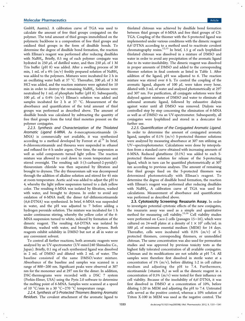
Table 1. Reaction Conditions for the Synthesis of Thiolated and S-Protected Chitosans in Order to Evaluate the Influence ofthe Weight Ratio of Polymer to Added Ligand on the Amount of Conjugated S-Protecting Ligand, Free Thiol Groups, and TotalAmount of Thiol Groups on the Thiolated and S-Protected Thiolated Chitosans (Means ± SD)
polymeraddedligand
amount of added ligand(mg)
amount of conjugated S-protecting ligand(μmol/g)
amount of free thiol groups(μmol/g)
total amount of thiol groups(μmol/g)
CS-TGA-340 TGA 500 340 ± 10 350 ± 15CS-TGA-660 TGA 1000 660 ± 21 680 ± 40CS-TGA-980 TGA 1500 980 ± 23 1010 ± 50TGA-MNA-340
6,6′-DTNA
200 170 ± 90 155 ± 75 550 ± 105
TGA-MNA-660
6,6′-DTNA
400 400 ± 150 245 ± 80 1200 ± 160
TGA-MNA-980
6,6′-DTNA
600 690 ± 100 310 ± 85 1850 ± 185
Molecular Pharmaceutics Article
dx.doi.org/10.1021/mp200598j | Mol. Pharmaceutics 2012, 9, 1331−13411332
GmbH, Austria). A calibration curve of TGA was used tocalculate the amount of free thiol groups conjugated on thepolymer. The total amount of thiol groups immobilized on thepolymeric backbone is a composition of free thiol groups andoxidized thiol groups in the form of disulfide bonds. Todetermine the degree of disulfide bond formation, the reactionwith Ellman’s reagent was performed after reducing disulfideswith NaBH4. Briefly, 0.5 mg of each polymer conjugate washydrated in 250 μL of distilled water, and then 250 μL of 1 MTris buffer (pH 8) was added. After a swelling process of 30min, 1 mL of a 4% (m/v) freshly prepared solution of NaBH4was added to the polymers. Mixtures were incubated for 2 h inan oscillating water bath at 37 °C. Thereafter, 200 μL of 5 MHCl was added, and the reaction mixtures were agitated for 10min in order to destroy the remaining NaBH4. Solutions wereneutralized by 1 mL of phosphate buffer (pH 8). Subsequently,100 μL of a 0.4% (m/v) Ellman’s solution was added andsamples incubated for 2 h at 37 °C. Measurement of theabsorbance and quantification of the total amount of thiolgroups was performed as described above. The amount ofdisulfide bonds was calculated by subtracting the quantity offree thiol groups from the total thiol moieties present on thepolymer conjugates.2.2.3. Synthesis and Characterization of the Thiolated
Aromatic Ligand 6-MNA. As 6-mercaptonicotinamide (6-MNA) is commercially not available, it was synthesizedaccording to a method developed by Forrest et al.14 In brief,6-chloronicotinamide and thiourea were suspended in ethanoland refluxed for 6 h under argon. Over time, the suspension aswell as solid components turned light yellow. The reactionmixture was allowed to cool down to room temperature andstirred overnight. The resulting salt S-(5-carbamyl-2-pyridyl)-thiouronium chloride was then separated by filtration andbrought to dryness. The dry thiouronium salt was decomposedthrough the addition of alkaline solution and stirred for 45 minat room temperature. Acetic acid was added to adjust the pH to4, whereby the light yellow suspension turned to a dark yellowcolor. The resulting 6-MNA was isolated by filtration, washedwith water, and brought to dryness. In order to avoid anyoxidation of 6-MNA itself, the dimer 6,6′-dithionicotinamide(6,6-DTNA) was synthesized. In brief, 6-MNA was suspendedin water, and the pH was adjusted to 7 before adding ahydrogen peroxide solution. The mixture was incubated for 1 hunder continuous stirring, whereby the yellow color of the 6-MNA suspension turned to white, induced by formation of thedimeric reagent. The resulting 6,6-DTNA was isolated byfiltration, washed with water, and brought to dryness. Bothreagents exhibit solubility in DMSO but not at all in water orany convenient solvent.To control all further reactions, both aromatic reagents were
analyzed by an UV-spectrometer (UV-mini1240 Shimadzu Co.,Japan). Briefly, 0.1 mg of each synthesized ligand was dissolvedin 2 mL of DMSO and diluted with 2 mL of water. Thebaseline consisted of the same DMSO/water mixture.Absorbance of the baseline and samples was scanned in arange of 800−200 nm. Significant peaks were observed at 307nm for the monomer and at 297 nm for the dimer. In addition,DSC-thermograms were recorded with a DSC 7 system(Perkin-Elmer, USA) using the Pyris 2.0 software to determinethe melting point of 6-MNA. Samples were scanned at a speedof 10 °C/min in a 30 °C−270 °C temperature range.2.2.4. Synthesis of S-Protected Thiomers Bearing Aromatic
Residues. The covalent attachment of the aromatic ligand to
thiolated chitosan was achieved by disulfide bond formationbetween thiol groups of 6-MNA and free thiol groups of CS-TGA. Coupling of the thiomer with the S-protected ligand wasimplemented under various conditions with the dimeric reagent6,6′-DTNA according to a method used to reactivate covalentchromatography resins.12,13 In brief, 1.5 g of each lyophilizedthiolated chitosan was dissolved in a mixture of DMSO andwater in order to avoid any precipitation of the aromatic liganddue to its water-insolubility. The dimeric reagent was dissolvedin proper amounts of DMSO and added to the correspondingthiomer solution in final contents as listed in Table 1. Afteraddition of the ligand, pH was adjusted to 6. The reactionmixture was stirred over 6 h. To control the coupling of thearomatic ligand, aliquots of 100 μL were taken every hour,diluted with 3 mL of water and analyzed photometrically at 297and 307 nm. For purification, all conjugate solutions were firstdialyzed against mixtures of DMSO and water to eliminate allunbound aromatic ligand, followed by exhaustive dialysisagainst water until all DMSO was removed. Dialysis wascontrolled step by step concerning the elimination of 6-MNAas well as of DMSO via an UV-spectrometer. Subsequently, allconjugates were lyophilized and stored in a desiccator forfurther use.
2.2.5. Quantification of the Conjugated Aromatic Ligand.In order to determine the amount of conjugated aromaticligand, samples of 0.1% (m/v) S-protected thiomer solutionswere analyzed by measuring absorbance at 307 nm utilizing anUV−spectrophotometer. Calculations were done by interpola-tion from a standard curve obtained with increasing amounts of6-MNA. Reduced glutathione (0.1%) was added to each S-protected thiomer solution for release of the S-protectingligand, which in turn can be quantified photometrically at 307nm according to previous analyses. The amount of remainingfree thiol groups fixed on the S-protected thiomers wasdetermined photometrically with Ellman’s reagent. Todetermine the degree of disulfide bond formation, the reactionwith Ellman’s reagent was performed after reducing disulfideswith NaBH4. A calibration curve of TGA was used forcalculations. Measurement of absorbance and quantificationwas performed as described above.
2.3. Cytotoxicity Screening: Resazurin Assay. In orderto investigate potential cytotoxic effects of the new conjugates,the resazurin assay was used as a simple and quantitativemethod for measuring cell viability.15,16 Cell viability studieswere performed on Caco-2 cells (passages 15−16), which werecultured on 24-well plates at a density of 1 × 10⁵ cells/mL in500 μL of minimum essential medium (MEM) for 14 days.Thereafter, cells were incubated with 0.5% (m/v) of S-protected thiomers, corresponding thiomers, or unmodifiedchitosan. The same concentration was also used for permeationstudies and was approved by previous toxicity tests as thehighest fully tolerated concentration of all available conjugates.Chitosan and its modifications are not soluble at pH 7.4. Allsamples were therefore first dissolved in sterile water at aconcentration of 1% (m/v), before diluting 1:2 in cell culturemedium and adjusting the pH to 7.4. Furthermore,nicotinamide (vitamin B3) as well as the dimeric reagent in aconcentration of 0.5% (m/v) were tested for their influence oncell viability. Because of the insolubility of 6,6′-DTNA, it wasfirst dissolved in DMSO at a concentration of 10%, beforediluting 1:20 in MEM and adjusting the pH to 7.4. Untreatedcells served as the positive control, whereas a 10% solution ofTriton X-100 in MEM was used as the negative control. The
Molecular Pharmaceutics Article
dx.doi.org/10.1021/mp200598j | Mol. Pharmaceutics 2012, 9, 1331−13411333
pH value of all samples was kept constant at 7.4. After 3 and 24h of incubation at 37 °C and 5% CO2, cells were washed withphosphate buffered saline (PBS) and the medium replaced by500 μL of resazurin solution. Cells were incubated for 3 h alongwith the resazurin solution at 37 °C. Thereafter, 100 μL weretaken from the supernatant of each well and transferred to a 96-well plate, and the resulting fluorescence (λex = 544 nm and λem= 590 nm) was measured by a microplate reader. Cell viabilitywas calculated as percentages of the positive control as follows:
= ×
cell viability [%]average abosrbance value of each triplicate
positive control100
2.4. Rheological Investigations. A plate−plate combina-tion rheometer (RotoVisco RT20, Haake GmbH, Karlsruhe,Germany) was used to determine the viscoelastic features ofeach compound. In brief, 10 mg of each polymer sample wasfirst hydrated in 1 mL of demineralized water, followingdilution with 1 mL of PBS (pH 7.4) to obtain a 0.5% (m/v)solution. Immediately after suspending in buffer and anequilibration period of 3 or 24 h in an incubator at 37 °C,500 μL of each polymer solution was transferred to the plate−plate viscometer. For all samples, the apparent viscosity (η) wasmeasured as described previously.10 The shear stress was set ata range of 0.5−500 Pa, the gap between two plates was 0.5 mm,and the temperature was maintained at 37 °C.2.5. Ex Vivo Evaluation of Permeation Enhancement
Properties across Freshly Excised Rat Intestine. For exvivo permeation studies, the small intestine (jejunum, ileum) ofnonfasting male Sprague−Dawley rats weighing between 200−300 g was immediately removed after sacrificing the rats. Theexcised intestine was cut into strips of 1.5 cm, washed free ofluminal contents, and mounted in Ussing-type chambers with apermeation area of 0.64 cm2. Aliquots (1 mL) of freshlyprepared medium containing 138 mM NaCl, 1 mM MgSO4, 5mM KCl, 10 mM glucose, and 2 mM CaCl2 buffered with 10mM HEPES pH 6.8 were added to the acceptor and donorcompartments. Ussing chambers were then placed in a waterbath with a temperature of 37 °C. After 30 min of equilibration,media of the donor compartment were substituted by 0.5% (m/v) unmodified chitosan, 0.5% (m/v) of each thiomer (CS-TGA-340, CS-TGA-660, and CS-TGA-980), and 0.5% (m/v)of each corresponding S-protected thiomer (TGA-MNA-340,TGA-MNA-660, TGA-MNA-980) mixed with FD4 in a finalconcentration of 0.1% (v/v). FD4 was also used as the controlin a final concentration of 0.1% (v/v). Over a 3 h incubationperiod, 100 μL aliquots were withdrawn from the acceptorchamber at 30 min intervals and replaced by the same volumeof medium preheated at 37 °C. The amounts of permeatedmarker were analyzed using fluorescence measurements (λex =485 nm and λem = 535 nm) by a microplate reader. Cumulativecorrections were made for previously removed samples.Apparent permeability coefficients (Papp, cm/s) for FD4 werecalculated by the following equation:
=× ×
PQ
A c tapp
where Q is the total amount permeated within 3 h (μg), A thediffusion area of the Ussing-type chambers (0.64 cm2) ortranswells (1.13 cm2), c the initial concentration in the donorchamber (μg/cm3), and t the time of the experiment (10,800
s). Transport enhancement ratios (ER) were calculated fromPapp values by the following equation:
= P PER (sample)/ (control)app app
2.5.1. Determination of the Transepithelial ElectricalResistance (TEER). Epithelial Voltohmmeter (EVOM, WorldPrecision Instruments, Germany) connected to a pair of side-by-side electrodes was used to monitor the TEER of the freshlyexcised rat intestinal mucosa. Measurements were done at thebeginning, during the study (60 and 120 min) to observe theeffect of transport enhancer, and at the end of each experimentto ensure that tissue integrity and viability had not beenadversely affected by the experimental conditions. Themeasured TEER previous to each experiment was between370−600 Ω cm2 and set as 100%. All other values werecalculated according to this value.
2.6. In Vitro Evaluation of Permeation Enhancementand P-gp Inhibition Properties across the Caco-2Monolayer. Caco-2 cells were maintained in the mediadescribed above at 95% humidity and 37 °C in an atmosphereof 5% CO2. All permeation and efflux experiments wereconducted during passages 15−17. Cells were plated directlyafter splitting in a density of 1 × 10⁵ cells/mL onto themembrane inserts of 12-well plates. Cells were allowed to growand differentiate for 21 days, during this time the media werechanged every 48 h. Permeation studies were carried out in atranswell monolayer system, displaying a volume of 1 mL ofeach chamber and a permeation area of 1.13 cm2. After 30 minof preincubation, the media of the donor compartment weresubstituted by 0.5% (m/v) of each polymer sample as describedabove. All formulations were mixed with FD4 in a finalconcentration of 0.1% (m/v) before placing the samples inevery donor compartment for absorptive transport. FD4 inincubation medium was also used as the control in a finalconcentration of 0.1% (m/v). Aliquots of 100 μL were takenfrom the acceptor compartment every 30 min for a 3 h durationand the volume substituted by incubation medium pre-equilibrated at 37 °C. The amount of permeated FD4 wasdetermined as mentioned above. Cumulative corrections weremade for previously removed samples.Furthermore, inhibition of efflux of the newly synthesized
conjugates was tested with the known P-gp substraterhodamine-123 (Rho-123) in the absorptive direction (A →B).17 Prior studies to show expression of P-gp in the monolayerwas performed by placing wells in the incubator (37 °C) andrefrigerator (4 °C), respectively. An increased permeation ofRho-123 at 4 °C indicates the expression of P-gp. Hence, allpassage numbers could be used to evaluate the P-gp inhibitoryproperties. Studies were performed in the presence of 0.5% (m/v) unmodified, thiolated, and S-protected thiolated chitosanmixed with a 0.01% (m/v) Rho-123 solution. Hence, allformulations contained 0.001% Rho-123. Rho-123 in theincubation medium was also used as the control in a finalconcentration of 0.001% (m/v). Over a period of 3 h, 100 μLaliquots were withdrawn from the acceptor chamber at 30 minintervals and replaced by the same volume of mediumpreheated at 37 °C. Transport of Rho-123 was investigated inthe absence and presence of each polymer. Amounts of Rho-123 were analyzed using fluorescence measurements (λex = 488nm and λem = 525 nm). Papp values for Rho-123 were calculatedas described above for FD4. Enhancement ratios werecalculated from the ratio between Papp of tested compoundsover Papp of the buffer control at 37 °C.
Molecular Pharmaceutics Article
dx.doi.org/10.1021/mp200598j | Mol. Pharmaceutics 2012, 9, 1331−13411334
2.6.1. Effect of conjugates on TEER measurements. Theintegrity of the monolayer was measured by determining theTEER with Epithelial Voltohmmeter. TEER was measuredprior to each experiment, during transport studies, and after thecompletion of the experiment. Subsequently, cells were washedthoroughly with PBS to ensure complete removal of allsamples. Thereafter, both chambers were replenished with freshmedium and incubated at 37 °C. TEER values were measuredafter 24 and 48 h to ensure that integrity and viability had notbeen adversely affected by the experimental conditions. Themeasured TEER previous to each experiment was between800−1000 Ω cm2 and set as 100%. All other values werecalculated according to this value.2.7. In Vitro Evaluation of Mucoadhesive Properties
via Tensile Studies. In order to evaluate the adhesive featuresof the novel conjugates, mucoadhesion was tested using tensilestrength investigations on porcine intestinal mucosa. Therefore,all lyophilized polymer conjugates were compressed by a singlepunch excentric press (Paul Weber, Remshalden-Grunbach,Germany) into 30 mg, 5.0 mm diameter flat-faced tablets. Thecompaction pressure of 12 kN was kept constant during thepreparation of all tablets. Following this, tensiometer studieswith all test discs were carried out on native porcine intestinalmucosa. All tablets were attached to a stainless steel flat disk (8mm in diameter, 0.3 g of weight in the system), which was hungby a nylon thread from a laboratory stand. Porcine mucosa wasfixed to a glass tissue mount using a cyanoacrylate adhesive.The tissue mount and fixed mucosa were placed in a beaker,and 100 mM phosphate buffer at pH 6.8 (37 °C) was addedsufficiently to immerse the mount and tissue. The beaker wasplaced on a balance and then carefully raised by a mobileplatform until the mucus came in contact with the tablet. Thecontact was determined when the nylon thread holding thetablet became bent. After 30 min of incubation, the mucosa waspulled down from the tablet at a rate of 0.1 mm/s. Data pointswere collected every second by a personal computer (SartaCollect software; Sartorius AG, Austria) linked to the balance.Data were transferred to Excel, and the area under the force/distance curve, i.e., the total work of adhesion (TWA) as well asthe maximum detachment force (MDF) were calculated by useof the trapezoidal rule.2.8. Statistical Data Analysis. GraphPad Prism 5 was used
to calculate the mean values of all samples. Unpaired Student’s ttest was performed to test the significance of the differencebetween the mean value of the control and the respectivesample. When comparisons of more than two mean values were
made, the data were analyzed with one way ANOVA. The levelof p ≤ 0.05 was set for significant, p ≤ 0.01 for very significant,and p ≤ 0.001 for highly significant.
3. RESULTS AND DISCUSSION
3.1. Characterization of the Thiolated Chitosans.Chitosan was modified by covalent attachment of thioglycolicacid due to the formation of amide bonds between the primaryamino groups of the polymer and carboxylic acid groups ofTGA. Mediated by the carbodiimide EDAC and increasingamounts of TGA added to the polymer, three different types ofthiolated chitosan with higher degrees of conjugated thiolgroups could be obtained. As illustrated in Table 1, the amountof free thiol groups attached to the polymer was determined tobe 340 (CS-TGA-340), 660 (CS-TGA-660), and 980 (CS-TGA-980) μmol thiol groups per gram polymer. A controlprepared in the same way as the chitosan−TGA conjugates butwith the carbodiimide omitted during the coupling reactiondisplayed only a negligible amount of the remaining traces ofTGA. All lyophilized thiomers were white, of fibrous structure,and water-soluble.
3.2. Characterization of the Thiolated AromaticLigands. The monomer 6-mercaptonicotinamide was synthe-sized according to a method developed by Forrest et al.14 Theresulting product could be obtained as a fine yellow powder.The dimer 6,6′-dithionicotinamide was generated due to theaddition of H2O2 to the monomer 6-MNA and could beattained as a fine white powder. UV-analyses in the case of 6,6′-DTNA showed a significant peak at 297 nm and a minor one at253 nm. 6-MNA, however, displayed a well pronounced peak at307 nm. In addition, DSC-thermograms were recorded tocharacterize the synthesized aromatic ligands to ensure that thedesired products were received. The monomer 6-MNA showeda melting point at around 264 °C, which is in good correlationwith literature values (266−268 °C).14
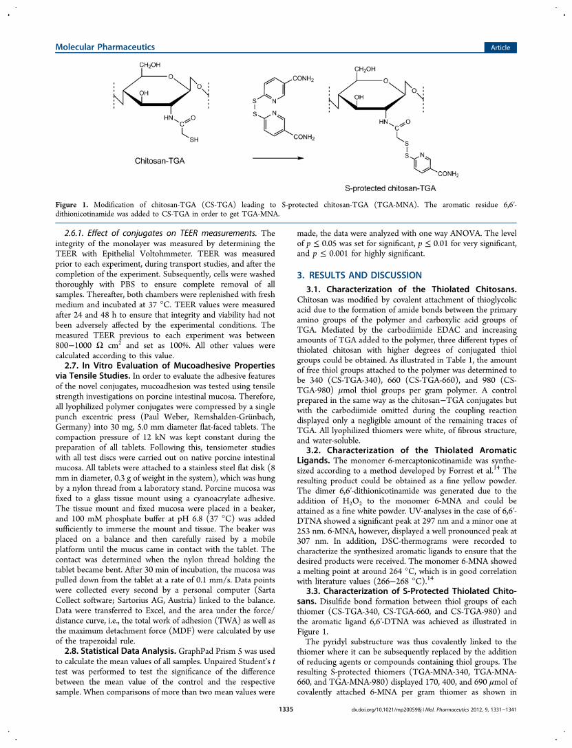
3.3. Characterization of S-Protected Thiolated Chito-sans. Disulfide bond formation between thiol groups of eachthiomer (CS-TGA-340, CS-TGA-660, and CS-TGA-980) andthe aromatic ligand 6,6′-DTNA was achieved as illustrated inFigure 1.The pyridyl substructure was thus covalently linked to the
thiomer where it can be subsequently replaced by the additionof reducing agents or compounds containing thiol groups. Theresulting S-protected thiomers (TGA-MNA-340, TGA-MNA-660, and TGA-MNA-980) displayed 170, 400, and 690 μmol ofcovalently attached 6-MNA per gram thiomer as shown in
Figure 1. Modification of chitosan-TGA (CS-TGA) leading to S-protected chitosan-TGA (TGA-MNA). The aromatic residue 6,6′-dithionicotinamide was added to CS-TGA in order to get TGA-MNA.
Molecular Pharmaceutics Article
dx.doi.org/10.1021/mp200598j | Mol. Pharmaceutics 2012, 9, 1331−13411335
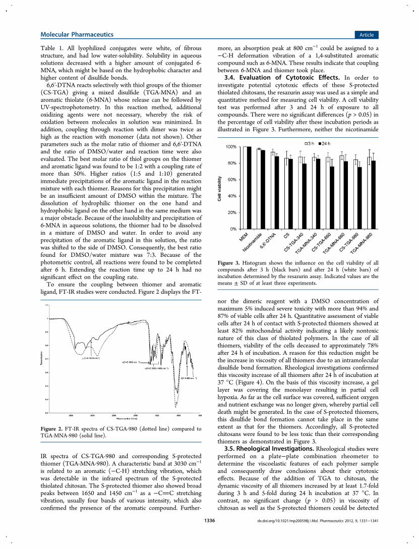
Table 1. All lyophilized conjugates were white, of fibrousstructure, and had low water-solubility. Solubility in aqueoussolutions decreased with a higher amount of conjugated 6-MNA, which might be based on the hydrophobic character andhigher content of disulfide bonds.6,6′-DTNA reacts selectively with thiol groups of the thiomer
(CS-TGA) giving a mixed disulfide (TGA-MNA) and anaromatic thiolate (6-MNA) whose release can be followed byUV-spectrophotometry. In this reaction method, additionaloxidizing agents were not necessary, whereby the risk ofoxidation between molecules in solution was minimized. Inaddition, coupling through reaction with dimer was twice ashigh as the reaction with monomer (data not shown). Otherparameters such as the molar ratio of thiomer and 6,6′-DTNAand the ratio of DMSO/water and reaction time were alsoevaluated. The best molar ratio of thiol groups on the thiomerand aromatic ligand was found to be 1:2 with a coupling rate ofmore than 50%. Higher ratios (1:5 and 1:10) generatedimmediate precipitations of the aromatic ligand in the reactionmixture with each thiomer. Reasons for this precipitation mightbe an insufficient amount of DMSO within the mixture. Thedissolution of hydrophilic thiomer on the one hand andhydrophobic ligand on the other hand in the same medium wasa major obstacle. Because of the insolubility and precipitation of6-MNA in aqueous solutions, the thiomer had to be dissolvedin a mixture of DMSO and water. In order to avoid anyprecipitation of the aromatic ligand in this solution, the ratiowas shifted to the side of DMSO. Consequently, the best ratiofound for DMSO/water mixture was 7:3. Because of thephotometric control, all reactions were found to be completedafter 6 h. Extending the reaction time up to 24 h had nosignificant effect on the coupling rate.To ensure the coupling between thiomer and aromatic
ligand, FT-IR studies were conducted. Figure 2 displays the FT-
IR spectra of CS-TGA-980 and corresponding S-protectedthiomer (TGA-MNA-980). A characteristic band at 3030 cm−1
is related to an aromatic (−C-H) stretching vibration, whichwas detectable in the infrared spectrum of the S-protectedthiolated chitosan. The S-protected thiomer also showed broadpeaks between 1650 and 1450 cm−1 as a −CC stretchingvibration, usually four bands of various intensity, which alsoconfirmed the presence of the aromatic compound. Further-
more, an absorption peak at 800 cm−1 could be assigned to a−C-H deformation vibration of a 1,4-substituted aromaticcompound such as 6-MNA. These results indicate that couplingbetween 6-MNA and thiomer took place.
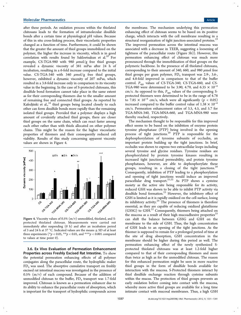
3.4. Evaluation of Cytotoxic Effects. In order toinvestigate potential cytotoxic effects of these S-protectedthiolated chitosans, the resazurin assay was used as a simple andquantitative method for measuring cell viability. A cell viabilitytest was performed after 3 and 24 h of exposure to allcompounds. There were no significant differences (p > 0.05) inthe percentage of cell viability after these incubation periods asillustrated in Figure 3. Furthermore, neither the nicotinamide
nor the dimeric reagent with a DMSO concentration ofmaximum 5% induced severe toxicity with more than 94% and87% of viable cells after 24 h. Quantitative assessment of viablecells after 24 h of contact with S-protected thiomers showed atleast 82% mitochondrial activity indicating a likely nontoxicnature of this class of thiolated polymers. In the case of allthiomers, viability of the cells deceased to approximately 78%after 24 h of incubation. A reason for this reduction might bethe increase in viscosity of all thiomers due to an intramoleculardisulfide bond formation. Rheological investigations confirmedthis viscosity increase of all thiomers after 24 h of incubation at37 °C (Figure 4). On the basis of this viscosity increase, a gellayer was covering the monolayer resulting in partial cellhypoxia. As far as the cell surface was covered, sufficient oxygenand nutrient exchange was no longer given, whereby partial celldeath might be generated. In the case of S-protected thiomers,this disulfide bond formation cannot take place in the sameextent as that for the thiomers. Accordingly, all S-protectedchitosans were found to be less toxic than their correspondingthiomers as demonstrated in Figure 3.
3.5. Rheological Investigations. Rheological studies wereperformed on a plate−plate combination rheometer todetermine the viscoelastic features of each polymer sampleand consequently draw conclusions about their cytotoxiceffects. Because of the addition of TGA to chitosan, thedynamic viscosity of all thiomers increased by at least 1.7-foldduring 3 h and 5-fold during 24 h incubation at 37 °C. Incontrast, no significant change (p > 0.05) in viscosity ofchitosan as well as the S-protected thiomers could be detected
Figure 2. FT-IR spectra of CS-TGA-980 (dotted line) compared toTGA-MNA-980 (solid line).
Figure 3. Histogram shows the influence on the cell viability of allcompounds after 3 h (black bars) and after 24 h (white bars) ofincubation determined by the resazurin assay. Indicated values are themeans ± SD of at least three experiments.
Molecular Pharmaceutics Article
dx.doi.org/10.1021/mp200598j | Mol. Pharmaceutics 2012, 9, 1331−13411336
after these periods. An oxidation process within the thiolatedchitosans leads to the formation of intramolecular disulfidebonds after a certain time at physiological pH values. Becauseof this in situ cross-linking process, their viscoelastic propertieschanged as a function of time. Furthermore, it could be shownthat the greater the amount of thiol groups immobilized on thepolymer, the higher the increase in viscosity, which is in goodcorrelation with results found by Sakloetsakun et al.10 Forexample, CS-TGA-980 with 980 μmol/g free thiol groupsrevealed a dynamic viscosity of 381 mPas after 24 h ofincubation, resulting in a 6-fold increase compared to the initialvalue. CS-TGA-340 with 340 μmol/g free thiol groups,however, exhibited a dynamic viscosity of 207 mPas, whichresulted in a 3.8-fold increase only in comparison to its viscosityvalue in the beginning. In the case of S-protected chitosans, thisdisulfide bond formation cannot take place in the same extentas for their corresponding thiomers due to the smaller amountof remaining free and connected thiol groups. As reported byKafedjiiski et al.,18 thiol groups being located closely to eachother can form disulfide bonds more rapidly than the remainingisolated thiol groups. Provided that a polymer displays a highamount of covalently attached thiol groups, there are closerthiol groups on the same chain, which can react faster amongeach other rather than with thiols allocated on other polymerchains. This might be the reason for the higher viscoelasticproperties of thiomers and their consequently reduced cellviability. Results of this study concerning apparent viscosityvalues are shown in Figure 4.
3.6. Ex Vivo Evaluation of Permeation EnhancementProperties across Freshly Excised Rat Intestine. To showthe potential permeation enhancing effects of all polymerconjugates along the paracellular route, the hydrophilic makerFD4 was used. The absorptive cumulative transport on freshlyexcised rat intestinal mucosa was investigated in the presence of0.5% (m/v) of each compound. Because of the addition ofunmodified chitosan to the buffer, FD4 transport was 1.7-foldimproved. Chitosan is known as a permeation enhancer due toits ability to enhance the paracellular route of absorption, whichis important for the transport of hydrophilic compounds across
the membrane. The mechanism underlying this permeationenhancing effect of chitosan seems to be based on its positivecharge, which interacts with the cell membrane resulting in astructural reorganization of tight junction-associated proteins.19
The improved permeation across the intestinal mucosa wasassociated with a decrease in TEER, suggesting a loosening oftightness of the paracellular route (Figure 5b)). However, thispermeation enhancing effect of chitosan was much morepronounced through the immobilization of thiol groups on thepolymeric backbone. In the presence of all thiolated chitosans,corresponding to their amount of 340, 660, and 980 μmol freethiol groups per gram polymer, FD4 transport was 2.9-, 3.6-,and 4.8-fold improved in comparison to that of the buffercontrol. Papp values of CS-TGA-340, CS-TGA-660, and CS-TGA-980 were determined to be 3.90, 4.79, and 6.35 × 10⁻⁶
cm/s. As opposed to this, Papp values of the corresponding S-protected thiomers were determined to be in the range of 4.78to 7.85 × 10⁻⁶ cm/s, which were all significantly (p < 0.05)increased compared to the buffer control value of 1.38 × 10⁻⁶
cm/s. Permeation enhancement ratios of 3.5, 4.5, and 5.7 forTGA-MNA-340, TGA-MNA-660, and TGA-MNA-980 werethereby reached, respectively.The mechanism thought to be responsible for this improved
effect seems to be based on the inhibition of enzyme proteintyrosine phosphatase (PTP) being involved in the openingprocess of tight junctions.20 PTP is responsible for thedephosphorylation of tyrosine subunits of occludin, animportant protein building up the tight junctions. In brief,occludin was shown to express two extracellular loops includingseveral tyrosine and glycine residues. Tyrosine residues arephosphorylated by protein tyrosine kinases resulting inincreased tight junctional permeability, and protein tyrosinephosphatases, however, are able to dephosphorylate thesegroups, resulting in a closing of the tight junctions.20
Consequently, inhibition of PTP leading to a phosphorylationand opening of tight junctions would induce an improvedparacellular drug transport.21,22 As PTP shows a cysteinemoiety as the active site being responsible for its activity,reduced GSH was shown to be able to inhibit PTP activity viadisulfide bond formation.23 However, the inhibitory effect ofGSH is limited as it is rapidly oxidized on the cell surface, losingits inhibitory activity.24 The presence of thiomers is thereforeessential, as they are capable of reducing oxidized glutathione(GSSG) to GSH.22 Consequently, thiomers being attached onthe mucosa as a result of their high mucoadhesive properties25
can shift the balance between GSSG and GSH on themembrane to the side of GSH. Thus, the high concentrationof GSH leads to an opening of the tight junctions. As thethiomer is supposed to remain for a prolonged period of time atthe site of drug absorption, GSH concentration in themembrane should be higher during this period as well. Thepermeation enhancing effect of the newly synthesized S-protected thiolated chitosans was at least 1.2-fold highercompared to that of their corresponding thiomers and morethan twice as high as for the unmodified chitosan. The reasonfor this enhanced permeation might be seen in more reactivethiol groups in the form of disulfide bonds available forinteraction with the mucosa. S-Protected thiomers interact bythiol disulfide exchange reaction through cysteine subunitswithin the mucus. The protection of thiol groups prevents anearly oxidation before coming into contact with the mucosa,whereby more active thiol groups are available for a long timeintimate contact with mucosal membranes. Thus, a high GSH
Figure 4. Viscosity values of 0.5% (m/v) unmodified, thiolated, and S-protected thiolated chitosan. Measurements were carried outimmediately after suspending (0 h) and after an incubation periodof 3 and 24 h at 37 °C. Indicated values are the means ± SD of at leastthree experiments (*p < 0.05, **p < 0.01, and ***p < 0.001 comparedto values at time point 0).
Molecular Pharmaceutics Article
dx.doi.org/10.1021/mp200598j | Mol. Pharmaceutics 2012, 9, 1331−13411337
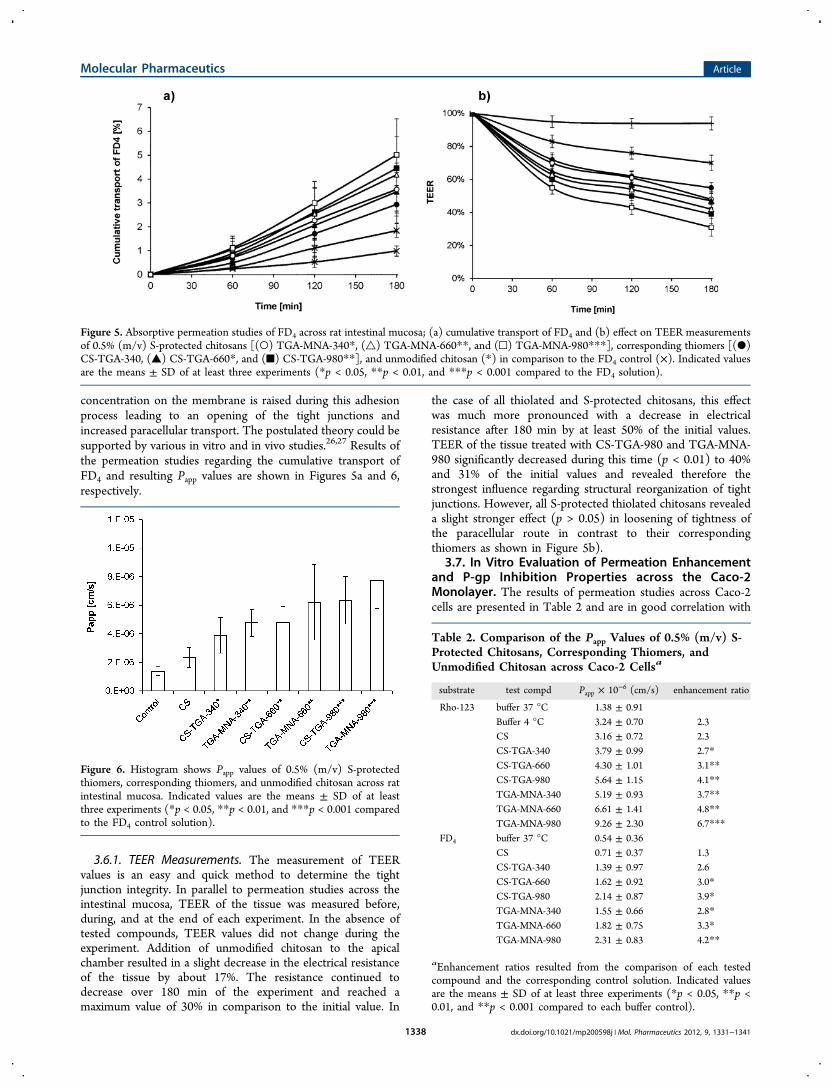
concentration on the membrane is raised during this adhesionprocess leading to an opening of the tight junctions andincreased paracellular transport. The postulated theory could besupported by various in vitro and in vivo studies.26,27 Results ofthe permeation studies regarding the cumulative transport ofFD4 and resulting Papp values are shown in Figures 5a and 6,respectively.
3.6.1. TEER Measurements. The measurement of TEERvalues is an easy and quick method to determine the tightjunction integrity. In parallel to permeation studies across theintestinal mucosa, TEER of the tissue was measured before,during, and at the end of each experiment. In the absence oftested compounds, TEER values did not change during theexperiment. Addition of unmodified chitosan to the apicalchamber resulted in a slight decrease in the electrical resistanceof the tissue by about 17%. The resistance continued todecrease over 180 min of the experiment and reached amaximum value of 30% in comparison to the initial value. In
the case of all thiolated and S-protected chitosans, this effectwas much more pronounced with a decrease in electricalresistance after 180 min by at least 50% of the initial values.TEER of the tissue treated with CS-TGA-980 and TGA-MNA-980 significantly decreased during this time (p < 0.01) to 40%and 31% of the initial values and revealed therefore thestrongest influence regarding structural reorganization of tightjunctions. However, all S-protected thiolated chitosans revealeda slight stronger effect (p > 0.05) in loosening of tightness ofthe paracellular route in contrast to their correspondingthiomers as shown in Figure 5b).
3.7. In Vitro Evaluation of Permeation Enhancementand P-gp Inhibition Properties across the Caco-2Monolayer. The results of permeation studies across Caco-2cells are presented in Table 2 and are in good correlation with
Figure 5. Absorptive permeation studies of FD4 across rat intestinal mucosa; (a) cumulative transport of FD4 and (b) effect on TEER measurementsof 0.5% (m/v) S-protected chitosans [(○) TGA-MNA-340*, (△) TGA-MNA-660**, and (□) TGA-MNA-980***], corresponding thiomers [(●)CS-TGA-340, (▲) CS-TGA-660*, and (■) CS-TGA-980**], and unmodified chitosan (*) in comparison to the FD4 control (×). Indicated valuesare the means ± SD of at least three experiments (*p < 0.05, **p < 0.01, and ***p < 0.001 compared to the FD4 solution).
Figure 6. Histogram shows Papp values of 0.5% (m/v) S-protectedthiomers, corresponding thiomers, and unmodified chitosan across ratintestinal mucosa. Indicated values are the means ± SD of at leastthree experiments (*p < 0.05, **p < 0.01, and ***p < 0.001 comparedto the FD4 control solution).
Table 2. Comparison of the Papp Values of 0.5% (m/v) S-Protected Chitosans, Corresponding Thiomers, andUnmodified Chitosan across Caco-2 Cellsa
substrate test compd Papp × 10−6 (cm/s) enhancement ratio
Rho-123 buffer 37 °C 1.38 ± 0.91Buffer 4 °C 3.24 ± 0.70 2.3CS 3.16 ± 0.72 2.3CS-TGA-340 3.79 ± 0.99 2.7*CS-TGA-660 4.30 ± 1.01 3.1**CS-TGA-980 5.64 ± 1.15 4.1**TGA-MNA-340 5.19 ± 0.93 3.7**TGA-MNA-660 6.61 ± 1.41 4.8**TGA-MNA-980 9.26 ± 2.30 6.7***
FD4 buffer 37 °C 0.54 ± 0.36CS 0.71 ± 0.37 1.3CS-TGA-340 1.39 ± 0.97 2.6CS-TGA-660 1.62 ± 0.92 3.0*CS-TGA-980 2.14 ± 0.87 3.9*TGA-MNA-340 1.55 ± 0.66 2.8*TGA-MNA-660 1.82 ± 0.75 3.3*TGA-MNA-980 2.31 ± 0.83 4.2**
aEnhancement ratios resulted from the comparison of each testedcompound and the corresponding control solution. Indicated valuesare the means ± SD of at least three experiments (*p < 0.05, **p <0.01, and **p < 0.001 compared to each buffer control).
Molecular Pharmaceutics Article
dx.doi.org/10.1021/mp200598j | Mol. Pharmaceutics 2012, 9, 1331−13411338
results ascertained by permeation studies across the intestinalmucosa. The absorptive transport of the hydrophilic macro-molecule FD4 was also investigated in the presence of 0.5% (m/v) of each compound. Unmodified chitosan showed a Papp valueof 0.71 × 10⁻⁶ cm/s, which was not significantly different (p >0.05) from the coefficient of the control (0.51 × 10⁻⁶ cm/s).Papp values of the thiolated chitosans CS-TGA-340, CS-TGA-660, and CS-TGA-980 were determined to be 1.39, 1.62, and2.14 × 10−6 cm/s. According to that, the transport enhance-ment ratios in comparison to the buffer control were calculatedto be 2.6-, 3-, and 3.9-fold increased for CS-TGA-340, CS-TGA-660, and CS-TGA-980, respectively. S-Protected thiomersdisplayed slightly higher permeation enhancing propertiescompared to those of their corresponding thiomers. CalculatedPapp values for TGA-MNA-340, TGA-MNA-660, and TGA-MNA-980 were in the range of 1.55 to 2.31 × 10⁻⁶ cm/s,resulting in a 2.8-, 3.3-, and 4.2-fold increase in permeationenhancement compared to that of the buffer control.For additional efflux inhibition studies across Caco-2 cells,
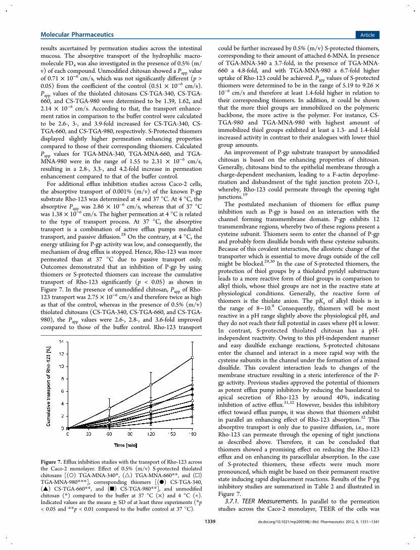
the absorptive transport of 0.001% (m/v) of the known P-gpsubstrate Rho-123 was determined at 4 and 37 °C. At 4 °C, theabsorptive Papp was 2.86 × 10⁻⁶ cm/s, whereas that of 37 °Cwas 1.38 × 10⁻⁶ cm/s. The higher permeation at 4 °C is relatedto the type of transport process. At 37 °C, the absorptivetransport is a combination of active efflux pumps mediatedtransport, and passive diffusion.28 On the contrary, at 4 °C, theenergy utilizing for P-gp activity was low, and consequently, themechanism of drug efflux is stopped. Hence, Rho-123 was morepermeated than at 37 °C due to passive transport only.Outcomes demonstrated that an inhibition of P-gp by usingthiomers or S-protected thiomers can increase the cumulativetransport of Rho-123 significantly (p < 0.05) as shown inFigure 7. In the presence of unmodified chitosan, Papp of Rho-123 transport was 2.75 × 10⁻⁶ cm/s and therefore twice as highas that of the control, whereas in the presence of 0.5% (m/v)thiolated chitosans (CS-TGA-340, CS-TGA-660, and CS-TGA-980), the Papp values were 2.6-, 2.8-, and 3.6-fold improvedcompared to those of the buffer control. Rho-123 transport
could be further increased by 0.5% (m/v) S-protected thiomers,corresponding to their amount of attached 6-MNA. In presenceof TGA-MNA-340 a 3.7-fold, in the presence of TGA-MNA-660 a 4.8-fold, and with TGA-MNA-980 a 6.7-fold higheruptake of Rho-123 could be achieved. Papp values of S-protectedthiomers were determined to be in the range of 5.19 to 9.26 ×10⁻⁶ cm/s and therefore at least 1.4-fold higher in relation totheir corresponding thiomers. In addition, it could be shownthat the more thiol groups are immobilized on the polymericbackbone, the more active is the polymer. For instance, CS-TGA-980 and TGA-MNA-980 with highest amount ofimmobilized thiol groups exhibited at least a 1.3- and 1.4-foldincreased activity in contrast to their analogues with lower thiolgroup amounts.An improvement of P-gp substrate transport by unmodified
chitosan is based on the enhancing properties of chitosan.Generally, chitosans bind to the epithelial membrane through acharge-dependent mechanism, leading to a F-actin depoylme-rization and disbandment of the tight junction protein ZO-1,whereby, Rho-123 could permeate through the opening tightjunctions.19
The postulated mechanism of thiomers for efflux pumpinhibition such as P-gp is based on an interaction with thechannel forming transmembrane domain. P-gp exhibits 12transmembrane regions, whereby two of these regions present acysteine subunit. Thiomers seem to enter the channel of P-gpand probably form disulfide bonds with these cysteine subunits.Because of this covalent interaction, the allosteric change of thetransporter which is essential to move drugs outside of the cellmight be blocked.29,30 In the case of S-protected thiomers, theprotection of thiol groups by a thiolated pyridyl substructureleads to a more reactive form of thiol groups in comparison toalkyl thiols, whose thiol groups are not in the reactive state atphysiological conditions. Generally, the reactive form ofthiomers is the thiolate anion. The pKa of alkyl thiols is inthe range of 8−10.9 Consequently, thiomers will be mostreactive in a pH range slightly above the physiological pH, andthey do not reach their full potential in cases where pH is lower.In contrast, S-protected thiolated chitosan has a pH-independent reactivity. Owing to this pH-independent mannerand easy disulfide exchange reactions, S-protected chitosansenter the channel and interact in a more rapid way with thecysteine subunits in the channel under the formation of a mixeddisulfide. This covalent interaction leads to changes of themembrane structure resulting in a steric interference of the P-gp activity. Previous studies approved the potential of thiomersas potent efflux pump inhibitors by reducing the basolateral toapical secretion of Rho-123 by around 40%, indicatinginhibition of active efflux.31,32 However, besides this inhibitoryeffect toward efflux pumps, it was shown that thiomers exhibitin parallel an enhancing effect of Rho-123 absorption.32 Thisabsorptive transport is only due to passive diffusion, i.e., moreRho-123 can permeate through the opening of tight junctionsas described above. Therefore, it can be concluded thatthiomers showed a promising effect on reducing the Rho-123efflux and on enhancing its paracellular absorption. In the caseof S-protected thiomers, these effects were much morepronounced, which might be based on their permanent reactivestate inducing rapid displacement reactions. Results of the P-pginhibitory studies are summarized in Table 2 and illustrated inFigure 7.
3.7.1. TEER Measurements. In parallel to the permeationstudies across the Caco-2 monolayer, TEER of the cells was
Figure 7. Efflux inhibition studies with the transport of Rho-123 acrossthe Caco-2 monolayer. Effect of 0.5% (m/v) S-protected thiolatedchitosans [(○) TGA-MNA-340*, (△) TGA-MNA-660**, and (□)TGA-MNA-980***], corresponding thiomers [(●) CS-TGA-340,(▲) CS-TGA-660**, and (■) CS-TGA-980**], and unmodifiedchitosan (*) compared to the buffer at 37 °C (×) and 4 °C (+).Indicated values are the means ± SD of at least three experiments (*p< 0.05 and **p < 0.01 compared to the buffer control at 37 °C).
Molecular Pharmaceutics Article
dx.doi.org/10.1021/mp200598j | Mol. Pharmaceutics 2012, 9, 1331−13411339
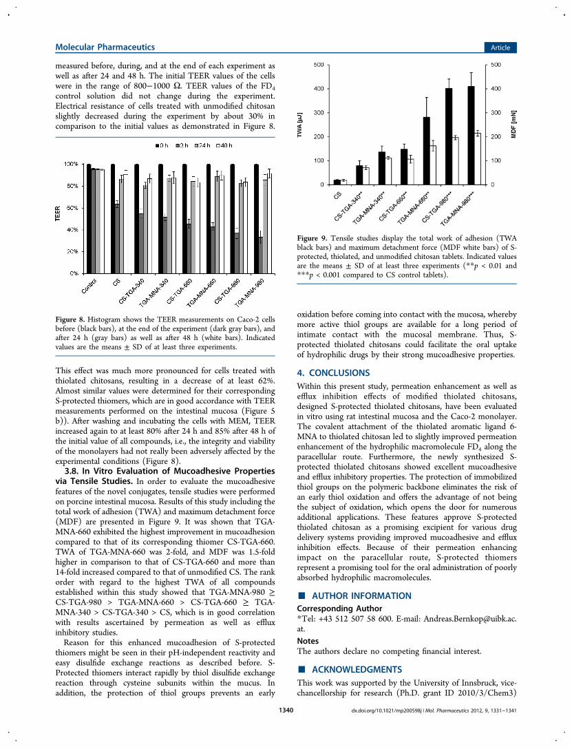
measured before, during, and at the end of each experiment aswell as after 24 and 48 h. The initial TEER values of the cellswere in the range of 800−1000 Ω. TEER values of the FD4control solution did not change during the experiment.Electrical resistance of cells treated with unmodified chitosanslightly decreased during the experiment by about 30% incomparison to the initial values as demonstrated in Figure 8.
This effect was much more pronounced for cells treated withthiolated chitosans, resulting in a decrease of at least 62%.Almost similar values were determined for their correspondingS-protected thiomers, which are in good accordance with TEERmeasurements performed on the intestinal mucosa (Figure 5b)). After washing and incubating the cells with MEM, TEERincreased again to at least 80% after 24 h and 85% after 48 h ofthe initial value of all compounds, i.e., the integrity and viabilityof the monolayers had not really been adversely affected by theexperimental conditions (Figure 8).3.8. In Vitro Evaluation of Mucoadhesive Properties
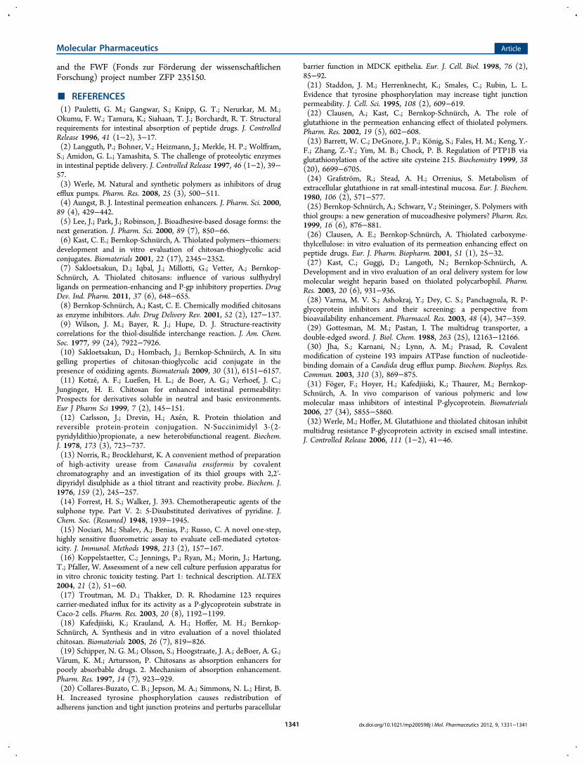
via Tensile Studies. In order to evaluate the mucoadhesivefeatures of the novel conjugates, tensile studies were performedon porcine intestinal mucosa. Results of this study including thetotal work of adhesion (TWA) and maximum detachment force(MDF) are presented in Figure 9. It was shown that TGA-MNA-660 exhibited the highest improvement in mucoadhesioncompared to that of its corresponding thiomer CS-TGA-660.TWA of TGA-MNA-660 was 2-fold, and MDF was 1.5-foldhigher in comparison to that of CS-TGA-660 and more than14-fold increased compared to that of unmodified CS. The rankorder with regard to the highest TWA of all compoundsestablished within this study showed that TGA-MNA-980 ≥CS-TGA-980 > TGA-MNA-660 > CS-TGA-660 ≥ TGA-MNA-340 > CS-TGA-340 > CS, which is in good correlationwith results ascertained by permeation as well as effluxinhibitory studies.Reason for this enhanced mucoadhesion of S-protected
thiomers might be seen in their pH-independent reactivity andeasy disulfide exchange reactions as described before. S-Protected thiomers interact rapidly by thiol disulfide exchangereaction through cysteine subunits within the mucus. Inaddition, the protection of thiol groups prevents an early
oxidation before coming into contact with the mucosa, wherebymore active thiol groups are available for a long period ofintimate contact with the mucosal membrane. Thus, S-protected thiolated chitosans could facilitate the oral uptakeof hydrophilic drugs by their strong mucoadhesive properties.
4. CONCLUSIONSWithin this present study, permeation enhancement as well asefflux inhibition effects of modified thiolated chitosans,designed S-protected thiolated chitosans, have been evaluatedin vitro using rat intestinal mucosa and the Caco-2 monolayer.The covalent attachment of the thiolated aromatic ligand 6-MNA to thiolated chitosan led to slightly improved permeationenhancement of the hydrophilic macromolecule FD4 along theparacellular route. Furthermore, the newly synthesized S-protected thiolated chitosans showed excellent mucoadhesiveand efflux inhibitory properties. The protection of immobilizedthiol groups on the polymeric backbone eliminates the risk ofan early thiol oxidation and offers the advantage of not beingthe subject of oxidation, which opens the door for numerousadditional applications. These features approve S-protectedthiolated chitosan as a promising excipient for various drugdelivery systems providing improved mucoadhesive and effluxinhibition effects. Because of their permeation enhancingimpact on the paracellular route, S-protected thiomersrepresent a promising tool for the oral administration of poorlyabsorbed hydrophilic macromolecules.
■ AUTHOR INFORMATIONCorresponding Author*Tel: +43 512 507 58 600. E-mail: [email protected] authors declare no competing financial interest.
■ ACKNOWLEDGMENTSThis work was supported by the University of Innsbruck, vice-chancellorship for research (Ph.D. grant ID 2010/3/Chem3)
Figure 8. Histogram shows the TEER measurements on Caco-2 cellsbefore (black bars), at the end of the experiment (dark gray bars), andafter 24 h (gray bars) as well as after 48 h (white bars). Indicatedvalues are the means ± SD of at least three experiments.
Figure 9. Tensile studies display the total work of adhesion (TWAblack bars) and maximum detachment force (MDF white bars) of S-protected, thiolated, and unmodified chitosan tablets. Indicated valuesare the means ± SD of at least three experiments (**p < 0.01 and***p < 0.001 compared to CS control tablets).
Molecular Pharmaceutics Article
dx.doi.org/10.1021/mp200598j | Mol. Pharmaceutics 2012, 9, 1331−13411340
and the FWF (Fonds zur Forderung der wissenschaftlichenForschung) project number ZFP 235150.
■ REFERENCES(1) Pauletti, G. M.; Gangwar, S.; Knipp, G. T.; Nerurkar, M. M.;Okumu, F. W.; Tamura, K.; Siahaan, T. J.; Borchardt, R. T. Structuralrequirements for intestinal absorption of peptide drugs. J. ControlledRelease 1996, 41 (1−2), 3−17.(2) Langguth, P.; Bohner, V.; Heizmann, J.; Merkle, H. P.; Wolffram,S.; Amidon, G. L.; Yamashita, S. The challenge of proteolytic enzymesin intestinal peptide delivery. J. Controlled Release 1997, 46 (1−2), 39−57.(3) Werle, M. Natural and synthetic polymers as inhibitors of drugefflux pumps. Pharm. Res. 2008, 25 (3), 500−511.(4) Aungst, B. J. Intestinal permeation enhancers. J. Pharm. Sci. 2000,89 (4), 429−442.(5) Lee, J.; Park, J.; Robinson, J. Bioadhesive-based dosage forms: thenext generation. J. Pharm. Sci. 2000, 89 (7), 850−66.(6) Kast, C. E.; Bernkop-Schnurch, A. Thiolated polymers−thiomers:development and in vitro evaluation of chitosan-thioglycolic acidconjugates. Biomaterials 2001, 22 (17), 2345−2352.(7) Sakloetsakun, D.; Iqbal, J.; Millotti, G.; Vetter, A.; Bernkop-Schnurch, A. Thiolated chitosans: influence of various sulfhydrylligands on permeation-enhancing and P-gp inhibitory properties. DrugDev. Ind. Pharm. 2011, 37 (6), 648−655.(8) Bernkop-Schnurch, A.; Kast, C. E. Chemically modified chitosansas enzyme inhibitors. Adv. Drug Delivery Rev. 2001, 52 (2), 127−137.(9) Wilson, J. M.; Bayer, R. J.; Hupe, D. J. Structure-reactivitycorrelations for the thiol-disulfide interchange reaction. J. Am. Chem.Soc. 1977, 99 (24), 7922−7926.(10) Sakloetsakun, D.; Hombach, J.; Bernkop-Schnu rch, A. In situgelling properties of chitosan-thioglycolic acid conjugate in thepresence of oxidizing agents. Biomaterials 2009, 30 (31), 6151−6157.(11) Kotze, A. F.; Lueßen, H. L.; de Boer, A. G.; Verhoef, J. C.;Junginger, H. E. Chitosan for enhanced intestinal permeability:Prospects for derivatives soluble in neutral and basic environments.Eur J Pharm Sci 1999, 7 (2), 145−151.(12) Carlsson, J.; Drevin, H.; Axen, R. Protein thiolation andreversible protein-protein conjugation. N-Succinimidyl 3-(2-pyridyldithio)propionate, a new heterobifunctional reagent. Biochem.J. 1978, 173 (3), 723−737.(13) Norris, R.; Brocklehurst, K. A convenient method of preparationof high-activity urease from Canavalia ensiformis by covalentchromatography and an investigation of its thiol groups with 2,2′-dipyridyl disulphide as a thiol titrant and reactivity probe. Biochem. J.1976, 159 (2), 245−257.(14) Forrest, H. S.; Walker, J. 393. Chemotherapeutic agents of thesulphone type. Part V. 2: 5-Disubstituted derivatives of pyridine. J.Chem. Soc. (Resumed) 1948, 1939−1945.(15) Nociari, M.; Shalev, A.; Benias, P.; Russo, C. A novel one-step,highly sensitive fluorometric assay to evaluate cell-mediated cytotox-icity. J. Immunol. Methods 1998, 213 (2), 157−167.(16) Koppelstaetter, C.; Jennings, P.; Ryan, M.; Morin, J.; Hartung,T.; Pfaller, W. Assessment of a new cell culture perfusion apparatus forin vitro chronic toxicity testing. Part 1: technical description. ALTEX2004, 21 (2), 51−60.(17) Troutman, M. D.; Thakker, D. R. Rhodamine 123 requirescarrier-mediated influx for its activity as a P-glycoprotein substrate inCaco-2 cells. Pharm. Res. 2003, 20 (8), 1192−1199.(18) Kafedjiiski, K.; Krauland, A. H.; Hoffer, M. H.; Bernkop-Schnurch, A. Synthesis and in vitro evaluation of a novel thiolatedchitosan. Biomaterials 2005, 26 (7), 819−826.(19) Schipper, N. G. M.; Olsson, S.; Hoogstraate, J. A.; deBoer, A. G.;Varum, K. M.; Artursson, P. Chitosans as absorption enhancers forpoorly absorbable drugs. 2. Mechanism of absorption enhancement.Pharm. Res. 1997, 14 (7), 923−929.(20) Collares-Buzato, C. B.; Jepson, M. A.; Simmons, N. L.; Hirst, B.H. Increased tyrosine phosphorylation causes redistribution ofadherens junction and tight junction proteins and perturbs paracellular
barrier function in MDCK epithelia. Eur. J. Cell. Biol. 1998, 76 (2),85−92.(21) Staddon, J. M.; Herrenknecht, K.; Smales, C.; Rubin, L. L.Evidence that tyrosine phosphorylation may increase tight junctionpermeability. J. Cell. Sci. 1995, 108 (2), 609−619.(22) Clausen, A.; Kast, C.; Bernkop-Schnurch, A. The role ofglutathione in the permeation enhancing effect of thiolated polymers.Pharm. Res. 2002, 19 (5), 602−608.(23) Barrett, W. C.; DeGnore, J. P.; Konig, S.; Fales, H. M.; Keng, Y.-F.; Zhang, Z.-Y.; Yim, M. B.; Chock, P. B. Regulation of PTP1B viaglutathionylation of the active site cysteine 215. Biochemistry 1999, 38(20), 6699−6705.(24) Grafstrom, R.; Stead, A. H.; Orrenius, S. Metabolism ofextracellular glutathione in rat small-intestinal mucosa. Eur. J. Biochem.1980, 106 (2), 571−577.(25) Bernkop-Schnu rch, A.; Schwarz, V.; Steininger, S. Polymers withthiol groups: a new generation of mucoadhesive polymers? Pharm. Res.1999, 16 (6), 876−881.(26) Clausen, A. E.; Bernkop-Schnu rch, A. Thiolated carboxyme-thylcellulose: in vitro evaluation of its permeation enhancing effect onpeptide drugs. Eur. J. Pharm. Biopharm. 2001, 51 (1), 25−32.(27) Kast, C.; Guggi, D.; Langoth, N.; Bernkop-Schnurch, A.Development and in vivo evaluation of an oral delivery system for lowmolecular weight heparin based on thiolated polycarbophil. Pharm.Res. 2003, 20 (6), 931−936.(28) Varma, M. V. S.; Ashokraj, Y.; Dey, C. S.; Panchagnula, R. P-glycoprotein inhibitors and their screening: a perspective frombioavailability enhancement. Pharmacol. Res. 2003, 48 (4), 347−359.(29) Gottesman, M. M.; Pastan, I. The multidrug transporter, adouble-edged sword. J. Biol. Chem. 1988, 263 (25), 12163−12166.(30) Jha, S.; Karnani, N.; Lynn, A. M.; Prasad, R. Covalentmodification of cysteine 193 impairs ATPase function of nucleotide-binding domain of a Candida drug efflux pump. Biochem. Biophys. Res.Commun. 2003, 310 (3), 869−875.(31) Foger, F.; Hoyer, H.; Kafedjiiski, K.; Thaurer, M.; Bernkop-Schnurch, A. In vivo comparison of various polymeric and lowmolecular mass inhibitors of intestinal P-glycoprotein. Biomaterials2006, 27 (34), 5855−5860.(32) Werle, M.; Hoffer, M. Glutathione and thiolated chitosan inhibitmultidrug resistance P-glycoprotein activity in excised small intestine.J. Controlled Release 2006, 111 (1−2), 41−46.
Molecular Pharmaceutics Article
dx.doi.org/10.1021/mp200598j | Mol. Pharmaceutics 2012, 9, 1331−13411341
Related Documents