Welcome message from author
This document is posted to help you gain knowledge. Please leave a comment to let me know what you think about it! Share it to your friends and learn new things together.
Transcript


CRITICAL CARE
4 Respiratory Therapy Vol. 4 No. 1 February-March 2009
Editorial
Table of Contents
DEPARTMENTS
4 Editorial: Leasing Options
11 News
15 Companies
17 Clinical Update
18 Spotlight on Capnography/Aerosol Delivery
18 Facility Review
19 Ventilation Roundtable
ARTICLES
24 The History of Humidification
26 Oxygenation and Kangaroo Care
32 Beliefs About Asthma
39 TB Therapy
45 Alveolar Volume
52 Training for Medical Devices
54 Omalizumab for Asthma
Vol. 4 No. 1February-March 2009
TTThe Journaee JournJJThe Journuhe JourTThe Je JourJThe JournurnThe JoJThe Joo rnTheTh rhee Joooourrn l of Pulmo of Pulmf ulmf PulmoPulmmomof PPPuf Pulmof Pulf PPPuof Pu oooonary Technnary Tea ynary TTnary Tnary Technary Tecry Technary Techy TTry Tecnary Technn ryy TT ccary Techrary TTry Tnararnary Tery TTy T cchyyyy iqueiquequeqiqueqquuueeqqqqqqThe JournTh JThe Journahe JournaThe Journahe JJJournrnhh Je Je Journarnahee Jee Joooooouurnue Jourrr ahe J u l of Pul of Pulmooof Pulmf P lf P lff P lPPPulmoPof Pulml f P lmolof Puof Pulof Pulmfof Pulmof uu mmmmooooof nary TechnTTechnnary Tnary TechnTT hnary Technhy Techny Techchnhnary Tecnary Tenary Techaaary Technaarry Technryyyy TecheeTechnnnary TT iiiqueiquequequequeququeueuuee
No Capital Budget? A Primer on Leasing
John Campbell
To those reading this who may be intimidated by various financing options, welcome to the club. Very few people understand all the intricacies of equipment financing. Hopefully, the information to follow will help you understand how knowing even the most basic aspects of financial options can help you get what you need.
What is leasing?Leasing is not rocket science. There are really just a few terms and ideas that you need to know to make this work for you. For starters, let’s take the term “leasing” out of the equation and use the term “anything but a cash deal.” How many times have you wanted or needed new equipment but were faced with no available capital dollars? You probably just wrote it down on your To Do list as something to follow up on for the next budget. After reading this, it should be clear that “no cash” should never mean “no new equipment” for you.
By the way, all hospitals use borrowed money of one sort or another. Your hospital may claim that you don’t lease, but you rent. Or you don’t rent, but you do lease. All you need to understand is that in one way or another, all hospitals use borrowed money.
How does a lease work?You decide you want more equipment but you have no cash. Very simply, a hospital can go to the bank and borrow enough to buy that equipment and make payments each month. [Or maybe, these days, you can’t go to the bank—Ed] In a lease, a hospital goes to a leasing company which buys the equipment from the manufacturer and the hospital makes payments to the leasing company instead of the bank.
So why doesn’t a hospital just borrow the money and buy versus using the leasing company? Good question!
Why would/should a hospital lease equipment?Equipment Management. No technology obsolescence or life expectancy problem and nothing to dispose of at the end of the term. At the end of the contract you can either give the equipment back to the leasing company or continue to rent. This eliminates the burden and risk of outdated equipment in the hospital inventory.
Reasons for leasing:Operating leases and rentals are paid out of the hospitals operating budget and thus •do not show up on balance sheets. This means it doesn’t become a liability to the hospital’s overall financial status.Locks in a price and an interest rate right now, not when the budget comes in.•No need to use cash or lines of credit to finance deals. This helps preserve the •hospital’s working capital.Convenience. Less paperwork than for loans. One of the misconceptions about •leasing is a perceived complexity of paperwork.Tax considerations are often applicable. Depreciation vs operating costs.•Leasing usually costs less per month than paying cash or acquiring debt.•Lower monthly payments than for a loan. Like an auto lease, you get more •equipment for the same monthly payment.
Continued on page 31…

WE MADE A LISTOF THE
SHORTCOMINGSOF ALL
HIGH FLOWDEVICES.
(AND ELIMINATED THEM)

Published six times each year byGoldstein and Associates, Inc.10940 Wilshire Blvd., Suite 600Los Angeles, CA 90024 USATel: 310-443-4109Fax: 310-443-4110E-mail: [email protected]: www.respiratorytherapy.ca Video website: www.rttv.ca
PublisherSteve Goldstein
EditorLes Plesko
Senior EditorCarol Brass
Assistant EditorLaszlo Sandor
Design and Production Managementhttp://accugraphics.net
Circulation, Coverage, Advertising Rates: Complete details regarding circulation, coverage, advertising rates, space sizes, and similar information are available to prospective advertisers. Closing date is 45 days preceding date of issue.
Change of Address notices should be sent promptly to Circulation Department. Provide old mailing label as well as new address. Allow two months for change.
Editorial Contributions will be handled with reasonable care. However, publishers assume no responsibility for the safety of artwork, photographs or manuscripts. All submissions may be emailed to [email protected]. Every precaution is taken to ensure accuracy, but the publish ers cannot accept responsi bility for the correctness or accuracy of information supplied herein or for any opinion expressed. Editorial closing date is the first day of the month preceding month of issue.
©2009 by Goldstein & Associates, Inc. All rights reserved. Reproduction in whole or in part without written permission is strictly prohibited.
Mohammed Al Ahmari, BSRT, MSc., RRTPrince Sultan Military College
of Health SciencesAl-Khobar, Saudi Arabia
Muhammad Aslam, MDClinical Fellow in Newborn Medicine
Harvard Neonatal-Perinatal FellowshipProgram
Children’s Hospital BostonInstructor in Pediatrics,
Harvard Medical School, Boston, MA
Larry Conway, RRT North Mississippi Medical Center
Tupelo, MS
Ed Coombs, MA, RRTSr. Marketing Manager—Ventilation
Draeger Medical Telford, PA
Antonio Esquinas, MD, PhD, FCCPIntensive Care Unit
Hospital Morales MeseguerMurcia, Spain
Dr. Javier FernandezDirector of Clinical Affairs & Education
Respiratory Division Latin AmericaMiami, FL
Gerardo N. Ferrero, PTClinical Specialist, Latin America
Buenos Aires, Argentina
Charles J. Gutierrez, PhD, RRT, FAARCAssistant Chief, Neurorespiratory Care Program—Spinal Cord Injury Center
James A. Haley Veterans HospitalTampa, FL
Surinder K. Jindal, MDPostgraduate Institute of Medical
Education & ResearchChandigarh, India
Rebecca A. MabryGeneral Sleep Manager
Viasys Healthcare, Yorba Linda, CA
Paul Mathews, PhD, RRT, FCCM, FCCP, FAARC
Associate Professor, Respiratory CareUniversity of Kansas Medical Center
Kansas City, KS
Hossein Razavi, MD, FCCPPulmonary, Critical Care &
Sleep MedicineSt. Helena, CA
Daniel D. Rowley, BS, RRT-NPS, RPFTSurgical/Trauma/Burn ICU
University of Virginia Medical CenterCharlottesville, VA
J. Kyle Schwab, MDMedical Director
Louisiana Sleep FoundationBaton Rouge, LA
Editorial Advisory Board
If disaster strikes, is your oxygen supplyprepared to move as quickly as your people?Do your alternate care site plans includeoxygen and an effective distribution system?Does your all-hazards plan include up to96 hours of oxygen for mass casualty andpandemic scenarios?
As North America’s leading producer ofmedical gases, Praxair Healthcare Servicescan supply not only oxygen and relatedproducts, but also consulting services tohelp you ensure the security of your supply.
Call us at 1-800-PRAXAIR today.
You assumed oxygen was part of your disaster plan.
© Copyright 2008 Praxair Technology, Inc. All rights reserved.


1111 Lakeside Drive, Gurnee, IL 60031-4099 USA 1.866.549.6446 www.ohiomedical.com
a l w a y s t h e r e f o r l i f e
Is your suction safe?Ours was designed with your patients in mind.
Patented Push-to-SetTM – Built-in feature that eliminatesthe need for you to occlude patient tubing to set accuratevacuum levels Quick-To-Max – Only 2 turns from 0 to full wall vacuum Quietest on the market Stat Set-up – Intermittent mode starts inthe “ON” cycle – No Waiting

NHF ™
Fisher & Paykel Healthcare, 15365 Barranca Parkway, Irvine, CA 92618 USA
Contact Fisher & Paykel Healthcare
to experience OptiflowTM today
Fisher & Paykel Healthcare, 15365 Barranca Parkway, Irvine, CA 92618 USA. Tel: 800 446 3908 or +1 949 453 4000 Fax:+1 949 453 4001
Comfortable, accurate oxygen delivery
It’s that simple
(800) 446 3908
A World of Products for Better BreathingSM
Available from B&B Medical Technologies and Finer Specialty Care Distributors Worldwide Contact Us Today: 1.800.242.8778 www.BandB-Medical.com
B&B Medical Technologies3800 Watt Avenue, Suite 275Sacramento, CA 95821Tel + 1.916.331.5221Fax + 1.916.331.0161www.BandB-Medical.com
HopeTM . E T TapeTM . E T Tape-IITM . StabilTubeTM . Bite Proof Bite BlockTM . Universal Bite BlockTM . Bite Proof Bite BlockTM TrachG
uardTM
. TrachStayTM
. Hybrid Baby Tape
TM . H
ybrid Pediatric TapeTM
. NeO
2 SafeTM
. Sat Point Plus Oxim
eter TM . Sat Point Pedi O
ximeter TM
. The Test LungTM
Precision Resistor KitTM . LockTiteTM . ClickStopTM 80/20 HeliOx Regulator . HopeTM Adult and Pediatric Nebulizer Kits
Trac
hGua
rdTM
. T
rach
Sta
yTM .
Hyb
rid B
aby
Tape
TM . S
tabi
lTub
eTM . W
rapS
afeTM
. S
at P
oint
Plu
s O
xim
eter
TM . S
at P
oint
Ped
i Oxi
met
erTM
. T
he T
est L
ungTM


12 Respiratory Therapy Vol. 4 No. 1 February-March 2009
INHALATION IMPROVEMENTCombination therapy with budesonide/formoterol inhalation improves lung function, quality of life, and sleep scores in patients with moderate to severe COPD, according to two trials. In the SHINE study, 1,704 patients aged 40+ years with moderate to severe COPD were randomized, twice a day, to 1 of 6 treatments: 1). Two inhalations of budesonide/formoterol metered-dose inhaler (160/4.5 mcg); 2. Two inhalations of combined b/f metered-dose inhaler (80/4.5 mcg); 3. Two inhalations of budesonide (160 mcg) metered-dose inhaler plus 2 inhalations of formoterol dry powder inhaler (4.5 mcg); 4. Two inhalations of budesonide metered-dose inhaler (160 mcg); 5. Two inhalations of formoterol dry powder inhaler (4.5 mcg); or 6. Placebo. The higher-dose combination therapy met the highest standard of superior efficacy of either component alone, according to researchers a UCLA, and the lower dose combination therapy met the requirement of greater efficacy than budesonide alone. Both doses of the combined budesonide/formoterol metered-dose inhaler demonstrated a significantly greater improvement from baseline in predose forced expiratory volume in 1 second and 1-hour postdose FEV1 compared with budesonide alone. B/f 160/4.5 mcg demonstrated a significant improvement from baseline for predose FEV1 compared with formoterol. Improvements from baseline in morning and evening PEF were significantly greater for both doses of combined b/f metered-dose inhaler compared with formoterol alone, budesonide alone, and placebo. Both doses of combined metered-dose inhaler significantly improved the sum of BCSS, sleep score, awakening-free nights, and rescue medication use compared with placebo. The 12-month SUN results, involving 1,964 patients with moderate to severe COPD, were similar. Patients were randomized to receive 1 of 4 different twice-daily treatments: 2 inhalations of combined b/f metered-dose inhaler (160/4.5 mcg and 80/4.5 mcg), 2 inhalations of formoterol (4.5 mcg) dry powder inhaler, or placebo. The higher-dose combination therapy demonstrated significantly greater improvements from baseline. Both combination doses demonstrated significantly greater improvements from baseline in morning and evening PEF compared with formoterol and placebo. Exacerbation rates were reduced by 25% to 30% with both combination doses compared to formoterol, and by approximately 40% compared with placebo. Combined b/f had significantly greater improvements in BCSS (high dose), sleep score, awakening-free nights (lower dose), and rescue medication use, compared with formoterol. Median time to 15% improvement in FEV1 was 6.8 minutes with the higher dose of combined b/f, 4.9 minutes with the lower dose, and 9 minutes with formoterol alone. The percentage of patients achieving a 15% or greater improvement in 15 minutes were 72.7% with the higher dose, 70.2% with the lower, 19.8% with budesonide alone, 60.5% with formoterol alone, and in 13.6% of patients receiving placebo. Both doses of b/f were well tolerated in both
News� February-March 2009
trials. Funding for the study was from AstraZeneca. The study appeared in Chest. The above information is from an article by Em Brown, Copyright © 2008 P\S\L Consulting Group Inc. All rights reserved.
DERMATOMYOSITISJuvenile dermatomyositis (JDM) is a rare chronic idiopathic inflammatory disorder primarily affecting the striated muscle and the skin. Pulmonary involvement is a common complication and cause of morbidity and mortality, but few data are available concerning pulmonary function impairment in childhood. The aim of a prospective study was to assess pulmonary function impairment in JDM. Sixteen patients (9 girls) with diagnosis of JDM (age 3–16.2 yrs) performed pulmonary function tests (PFT); 14 were receiving treatment; all had muscular testing; 13 had weakness as major symptom. Respiratory symptoms: dry cough in 2/16; reduced exercise tolerance: 3/16. Chest x-ray: normal in 13/13. Echocardiogram and ECG: normal in all. 9/16 had positive ANA-Ab; antiJO1 Ab negative in all. Results: 7/15 pt (46%): restrictive syndrome measured on TLC and VC; 9/16 (56%): reduced FRC; 9/13 pt: reduced RV. DLCO: impaired in 2 with restrictive pattern. No patient had bronchial obstruction. We didn’t find a relation between PFT and steroid treatment/impregnation, nor with relapses, nor with muscle enzymes nor with ANA/FAN. Muscular testing didn’t always correlate with respiratory impairment. The authors concluded that lung involvement is frequent in JDM and is better detected by PFT than by chest x-ray or symptoms. The most frequent respiratory pattern is restrictive syndrome secondary to respiratory muscular deficit and evidenced by a reduction of lung volumes with normal DLCO and chest x-ray. The authors couldn’t detect any risk factor predicting a major lung involvement, but concluded that the longer the follow-up, the more frequent a functional impairment is present. This item is from a poster presentation at the 15th Paediatric Rheumatology European Society (PreS) Congress, London, Pediatric Rheumatology 2008, 6(Suppl 1), © 2008 Fabi et al; licensee BioMed Central Ltd, M. Fabi1, Respiratory Involvement in Juvenile Dermatomyositis, M. Le Bourgeois, C. Bodemer, V. Beguin, A.M. Prieur, P. Quartier, J. de Blic.
AUTUMN SONATAChildren born four months before the peak of cold and flu season have a greater risk of developing childhood asthma than children born at any other time of year, according to new research from Vanderbilt University Medical Center. The study involved an analysis of the birth and medical records of more than 95,000 children and their mothers. Researchers found that the timing of when a child is born in relationship to the annual cold and flu season alters the risk for developing asthma. Autumn-born babies, who are about 4 months of age when the winter virus season peaks, have a nearly 30% increased risk of developing asthma compared with births during other times of the year, and this risk was similar to or greater than other well-established risk factors for asthma. This evidence suggests that avoiding early respiratory infections during infancy may have long-term as well as short-term benefits. Over the course of the study, if the peak of RSV occurred in December, the highest rates of asthma was in August-born babies. If the peak was in February, the highest rates of asthma were seen in October-born babies. Researchers said there’s a genetic susceptibility common to both bronchiolitis and the development of asthma, and that an environmental exposure such as a winter viral infection causes asthma.

Respiratory Therapy Vol. 4 No. 1 February-March 2009 13
A TALE OF TWO SISTERSResearchers at Cincinnati Children’s Hospital have discovered a familial genetic mutation that causes an inherited form of PAP. The research team studied the cases of two sisters, ages 6 and 8, whose PAP progressed slowly prior to diagnosis. For two years, the younger sister had suffered from labored breathing and had below average height and weight of unexplained origin. The 8-year-old sister had been considered healthy until results for several novel disease biomarkers prompted doctors to examine her. She also was found to be below average height and weight for her age. She also had blood and breathing test results consistent with PAP. Researchers found a mutation of the CSF2RA gene in both sisters. The mutation disrupted the signaling activity of protein called GM-CSF, a cytokine that facilitates cellular communication and is critical for the body to control the right amount of surfactant production. Both parents are healthy and do not have PAP, but each carried different forms of the gene mutation that caused PAP in both children. The sisters’ gene mutations were detected on maternal and paternal X chromosomes. The researchers suggested that an inhaled GM-CSF aerosol could boost the activity of the GM-CSF receptor to correct surfactant levels, and also said a bone marrow transplant and gene therapy might be alternatives. The younger sister underwent whole-lung lavage treatment, and this improved her condition.
GENE GENIUSESIn a related study, Cincinnati researchers identified a gene critical to lung maturation in newborns, and the production of surfactant. The study demonstrated the Foxm1 gene’s central importance to lung maturation and surfactant production in mice. Previous research has shown that Fox genes (a group of transcription factors that control the transfer of genetic information to regulate proteins within cells) are important for the embryonic development of lungs and other organs. To study the role of Foxm1 during embryonic lung development, investigators generated transgenic mice, which are engineered to allow genetic manipulation, and deleted Foxm1 in developing lung epithelium. Deletion did not impact the initial budding and branching or the growth of the lung, but did inhibit anatomic and biochemical maturation of the lung, where air sacs did not fully form and surfactant production was compromised. The research team is working to find pharmacological compounds that can activate Foxm1.
DUCK!SARS may have originated in bats, but the actual animal source is not known. In an effort to understand how SARS-CoV may have jumped from bats to humans, a team of investigators from Vanderbilt University Medical Center and the University of North Carolina at Chapel Hill generated a synthetic SARS-like bat coronavirus. The virus, the largest replicating synthetic organism ever made, is infectious in cultured cells and mice. The findings identify pathways by which a bat coronavirus may have adapted to infect humans. The studies also provide a model approach for rapid identification, analysis and public health responses to future natural or intentional virus epidemics. The researchers noted that new human epidemics would continue to originate in animals, but that the trans-species movement and adaptation of viruses from animals to humans remain poorly understood. At the time of the SARS epidemic, investigators became convinced that bats were the likely source, but bat coronaviruses had never been successfully grown in culture or animals. The research team decided to use synthetic biology to
recover a non-cultivatable virus, using SARS-like bat coronavirus sequences to establish a consensus genome sequence, then used commercial DNA synthesis and reverse genetics to build the consensus viral genome and several variations. A small region of SARS-CoV was sufficient to allow the bat virus to move from zero growth to very efficient growth in cells. The studies suggest that a very simple recombination event may have been enough to allow a coronavirus to move from one species to another. The researchers noted that they took extreme safety precautions during their research.
MAKING RABBITSResearchers at the Christèle Gras-Le Guen (Hôpital Mère Enfant CHU Nantes, France), doing research with rabbits, confirmed an association between antenatal infection and post-natal BPD. Pregnant rabbits infected 3-4 days before the end of gestation and were treated by antibiotics (ceftriaxone) six hours after inoculation. The presence of BPD was assessed by histological and morphometric methods in pups between 0 and 15 days of life. The researchers were able to reproduce the alveolar growth arrest typically observed in human BPD. They also showed that post-natal growth in these newborns was altered as soon as five days after birth. The investigators noted that the relationship between antenatal infection and the impaired post-natal growth still needs to be investigated.
CALLING IN SICKPeople with sleep apnea have an increased risk of needing to take long-term sick leave or give up working completely, according to a study at the University of Bergen, in Norway. Researchers studied more than 7,000 subjects aged 40-45.
VVVVV A RA RA RA RA R T MT MT MT MT M - M o n i t o r- M o n i t o r- M o n i t o r- M o n i t o r- M o n i t o r
•
•
•
•
•
14 Respiratory Therapy Vol. 4 No. 1 February-March 2009
Participants completed a questionnaire to identify symptoms of obstructive sleep apnea. At the same time, researchers recorded the frequency of episodes of fatigue and sleepiness at work or during free time. Finally, they obtained data about long-term sick leave and permanent health-related inability to work. More than six percent of subjects were considered to be affected by obstructive sleep apnea syndrome, with a disproportionately high number of men and people with a low educational level, and the authors concluded that these patients had almost double the risk of needing to take long-term sick leave. Patients with sleep apnea were also found to have double the risk of needing to retire from work for health reasons. Daytime sleepiness contributed the most to the need for sick leave and ill-health retirement.
COUGH IT UPOne in five adult women in the United States still smokes, even though smoking takes an average of 14.5 years off their lives, according to ACOG. About 438,000 men and women in the USA die prematurely as a result of smoking. ACOG says more women die from lung cancer than from any other cancer, and that the number of yearly lung cancer deaths of women in the USA has increased six-fold since the mid 1900s. Women smokers have twice the risk of developing coronary heart disease, and run ten times the risk for developing COPD. Women smokers also have a harder time conceiving, and run a greater risk of delivering a preemie, and/or a baby with lung problems. The CDC claims that there might be 300,000 children under 18 months who get respiratory infections because of exposure to secondhand smoke. A quarter of high school girls and a tenth of middle school girls smoke regularly, and poor girls are more likely to take up smoking. Reported in Medical News Today by Christine Nordqvist, copyright Medical News Today.
THIS YEAR’S MODELIt would seem that every couple years a “new” mode of ventilation is introduced, according to Paul Garbarini, with Hamilton Medical. In Hamilton Medical’s newsletter, he writes: “Despite this, during delivery of a breath, there are only two possible parameters that can be controlled. Either pressure is the constant variable (independent variable) and flow/volume changes (dependent variable) or flow is constant (independent variable) and pressure varies (dependent variable). So the first thing we need to know when approaching any “new” mode is whether pressure or flow is what’s being controlled during the inspiratory phase. What differentiates modes are other control variables such as what cycles breaths on and off, what control algorithms are employed, etc. The clinician is handicapped in that each new mode presents a new set of variables, rules, etc that must be comprehended to safely apply the mode. This, all despite the fact that no one mode has been demonstrated to improve outcomes. A recent AARC webcast titled How to Select the Best Mode of Ventilation took a different approach to assessing the plethora of available modes. The Webcast presented a novel goal oriented approach to evaluating modes of ventilation. The presenter, Robert Chatburn, is recognized as the authority on classification of ventilator modes. A scoring system was developed that scored modes of ventilation based on clinical goals to achieve safety (do no harm), comfort (optimizing patient vs vent work of breathing) and liberation (weaning). The maximum achievable score was 13 points, (though
my interpretation of the criteria is that as some of the criteria were mutually exclusive, a score of 13 was not possible). Chatburn classified 22 modes of ventilation and narrowed the list down to 9 mode types. 6 of the 9 modes scored no higher than 2 points, 2 modes scored 3 points (SmartCare and Automode) and 1 mode achieved a score of 6 points; double that of any other mode. That mode was ASV/Adaptive Support Ventilation on Hamilton Medical ventilators. The AARC webcast is available for viewing at no cost to AARC members ($15 for CRCE credit) at aarc.org/education/webcast/archives/2008/best_mode_ventilation.asp or by accessing the AARC Web site and going to Education, then the Webcasts links.” From Hamilton Medical, Is there a Best Mode of Ventilation? by Paul Garbarini, MS, RRT.
TARGET DATESDiscovery Laboratories, Inc announced that the FDA has accepted for review its Complete Response for Surfaxin (lucinactant) for the prevention of RDS in premature infants. The FDA has designated the Complete Response as a Class 2 resubmission and has established April 17, 2009 as its target action date under the Prescription Drug User Fee Act to complete its review and potentially grant marketing approval for Surfaxin. The Complete Response addressed all of the remaining requirements contained in the May 2008 Approvable Letter that must be satisfied to gain US marketing approval for Surfaxin. Discovery Labs provided the FDA specific data, information and minor clarifying analyses and believes that its Complete Response supports the approval of Surfaxin. The May Approvable Letter did not require any additional clinical trials. Prior to receiving the Approvable Letter, Discovery Labs made notable progress towards gaining FDA approval of Surfaxin, including agreeing with the FDA on the content of the Surfaxin package insert and successfully concluding a pre-approval inspection of Discovery Labs’ manufacturing operations. Surfaxin represents the first peptide-containing, synthetic surfactant potentially available for addressing RDS. Contact discoverylabs.com.
IT ONLY GETS WORSELong-term use of low-dose macrolide antibiotic therapy may reduce the frequency of exacerbations in patients with moderate to severe COPD by as much as 35% percent, according to a London-based study. The researchers followed 109 patients with moderate to severe COPD for a year, after randomly assigning them to receive either a placebo or a twice daily 250 mg dose of erythromycin. The patients recorded their exacerbations and hospitalizations and they were assessed using spirometry, sputum testing and blood testing. The researchers found that patients randomized to receive erythromycin have fewer exacerbations, and that 60% of the exacerbations that occurred were within the placebo group. Researchers, however, noted that the threat of growing antibiotic resistance resulting from widespread prophylactic use of erythromycin was a concern.
BAD NEWS DOWN UNDERSix million Australians suffer from COPD, asthma, and other respiratory diseases, and 90% of patients are misusing their inhalers, according to the Australian Lung Foundation. The study noted that most Australians who used an inhaler weren’t doing it properly because they’d never been shown how to do it right,

Respiratory Therapy Vol. 4 No. 1 February-March 2009 15
or succumbed to poor technique over time. It was advised that inhaler users should ask their GP or pharmacist how to do it correctly. Mistakes in use included not shaking a puffer between each dose, not holding a Turbuhaler or Accuhaler at the right angle when loading a dose, and putting multiple doses into a spacer, rather than inhaling each individual dose one at a time.
COMPANIESY NOT?Siemens Healthcare has received FDA 510(k) market clearance for the Ysio, a new generation digital radiography system with a wireless detector (wi-D) for maximum positioning flexibility. Offering one digital radiography (DR) solution for virtually all clinical demands of the growing digital radiography market, the Ysio can be customized to suit the patient’s needs—such as one or two detectors, with or without a patient table, and with fully automated or synchronized movements. Due to its versatility, the Ysio serves radiography needs no matter what the imaging volume, protocols, or patient profiles, and its performance features geared toward short examination times make it an ideal system for increasing daily patient throughput. Ysio is available in a variety of configurations based on customers imaging needs: as a wall stand with an integrated detector, a wall stand and table system with a wireless detector, or even as a mixed detector solution for high throughput and flexibility. Ysio serves as an integrated command center where users can control their workflow from registration to image data management with features like more than 500 automated system positions; power-assisted movements; a unique table design, together with the wi-D; and a color touch screen panel for convenient system utilization. With more than 500 different preset examination positions, Ysio can save preparation time and effort. Simply pressing a button on the wireless remote will automatically move the X-ray tube into position. The system can be configured to bypass room fixtures during its automated travel to the imaging position. Thus, Ysio is also a tailored solution in terms of space planning. Power-assisted servo movements help to further reduce the strain of heavy workloads while the collimation can be adjusted from anywhere in the room via remote control. As a charity promotion, Siemens has donated its Ysio digital radiography system to the Children’s Health Fund. CHF enabled hospitals to bid online for the system, with all benefits going to CHF. Founded in 1987 by Paul Simon, and pediatrician Dr Irwin Redlener, CHF is committed to providing healthcare to the nation’s most medically under-served children and their families. The auction for the Ysio system enabled authorized bidders from US hospitals to submit bids anonymously online, with the highest bid displayed on the auction’s website, ysioauction.com. The starting bid for the Ysio was $99,999. (The system’s list price is $450,000.) Contact siemenshealthcare.com.
HOMEWARD BOUNDLinde North America has acquired Respiratory Support Services, a company that delivers post-acute respiratory care in the US. Linde North America is a member of the Linde Group, a gases and engineering company that provides high acuity clinical services in Europe and South America. Respiratory Support Services, in Livingston, TN, provides respiratory care serices and equipment in a variety of settings, including sub-acute ventilator care, skilled nursing facilities and patient homes. Under the terms, the company will operate as Linde RSS LLC. The acquisition enables Linde to introduce to the US market its
REMEO program for the care of ventilated patients. REMEO, Latin for “I return home,” is meant to bridge the gap between the hospital ICU and the patient’s home for ventilated patients. The Linde Group has more than 50,000 employees in 100 countries, and had $18.7 billion in sales in 2007. Contact linde.com.
GOING HOMEVapotherm has announced that industry veteran Nick Macmillan has joined the company as Manager of its Home Care Segment. Macmillan will be responsible for developing and managing the company’s expansion of respiratory therapy products that address chronic and sleep disorders. Macmillan has been in the healthcare management and respiratory industry for more than 25 years, having most recently served as National Clinical Director at Rotech Healthcare Inc. Macmillan was the Global Sleep Product Director of Sunrise/DeVilbiss where he oversaw sleep product strategies and successfully executed several product launches. He has held several state and national appointments and elected positions including the President of the Indiana Society for Respiratory Care and Chairperson for the Home Care Section of the American Association for Respiratory Care. In 2003, Nick was inducted as a Fellow of the American Association for Respiratory Care. Contact vtherm.com.
DON’T CRY FOR ME, EVITADraeger Medical, Inc announced release of Version 7.0 of Software for its Evita XL ventilator. The new Evita XL 7.0 Software can help to address some everyday challenges faced by respiratory therapists. Clinicians can find the appropriate expiration time and easily apply a constant I:E ratio as well as realize better recruitment maneuvers via standard QuickSet and PressureLink features. A new measured value called “f trigg” indicates the frequency of all triggered breaths to help with the patient weaning process. The more breaths triggered by the patient, the more active the patient is. The software offers easier ventilator screen information access and visibility, with stronger more contrasting colors and larger numbers, a big yellow standby indicator, as well as an options overview page to quickly determine what options a particular ventilator has installed. Users can apply standard O2 therapy with an ICU ventilator, using one device, a ventilator, for both O2 therapy and ventilation (no O2 flowmeter needed). Evita offers online help text (Cause/Remedy) in case of alarm messages, and easier recognition when a ventilator is not ventilating a patient and is in standby mode. With Evita XL 7.0 software, clinicians can provide a direct backup for O2 therapy for quicker reaction time and therefore enhanced patient safety for patients when ventilation is needed again. Contact draeger.com.
NO STEROIDSData presented at the American College of Allergy, Asthma & Immunology (ACAAI) shows that Xolair (Omalizumab) for Subcutaneous Use significantly reduced asthma attacks in children aged six through 11 with moderate or severe persistent allergic asthma inadequately controlled with inhaled corticosteroids. The study further defines the safety profile of Xolair in this patient population. The Phase III study showed that children treated with Xolair demonstrated a 31% reduction in clinically significant asthma exacerbations compared to children treated with placebo at 24 weeks. After a year of treatment, children treated with Xolair suffered 43% fewer clinically significant asthma exacerbations than those receiving placebo. Xolair is a biologic treatment currently approved for patients 12 years of age and above with moderate-to-severe
16 Respiratory Therapy Vol. 4 No. 1 February-March 2009
persistent asthma who have a positive skin test or in vitro reactivity to a perennial aeroallergen and whose symptoms are inadequately controlled with inhaled corticosteroids. It is the only approved therapy which blocks IgE (immunoglobulin E), a major component of allergic asthma. Genentech, co-marketer of Xolair, plans to submit these data to the US Food and Drug Administration (FDA) seeking to expand the current labeled indication for Xolair. The pivotal Phase III double-blind, randomized placebo-controlled study evaluated children aged six through 11 with moderate-to-severe allergic asthma uncontrolled despite inhaled corticosteroid (ICS) therapy. For eight weeks, ICS doses were optimized and baseline measures established in all study participants; 628 children still symptomatic after reaching optimized ICS dosing were randomized to receive add-on Xolair therapy or placebo. The study comprised a 24-week fixed-dose ICS phase, followed by a 28-week phase in which ICS doses could be reduced, and a 16-week safety follow-up period. Xolair (Omalizumab) for Subcutaneous Use is a humanized monoclonal antibody for moderate-to-severe allergic asthma and the only approved therapy which blocks IgE (immunoglobulin E), a major component of allergic asthma. Contact xolair.com.
SOUND THE ALARMVortran Medical Technology, Inc announced FDA approval of the new VAR-Monitor. It is designed specifically to monitor any non-cycling condition of the VAR (Vortran Automatic Resuscitator) and meets one of the alarm requirements specified by the AARC’s Guidelines for Acquisition of Ventilators to Meet Demands for Pandemic Flu and Mass Casualty Incidents. The VAR-Monitor works with all the current VAR models and is easy to set up and use. It operates with a standard 9 VDC battery and is financially practical for all your existing VAR users to stockpile. The cost of stockpiling full feature mechanical ventilators with alarm features is very expensive and financially impractical. That is why state and local government agencies and healthcare facilities have augmented their ventilation cache with the VAR and E-Vent Case for their emergency preparedness. The advantages of the VAR for disaster preparedness and the pandemic flu are clear because of its cost, size, ease of set up and use. Contact (800) 434-4034, vortran.com.
GO TO BEDHill-Rom has received a Gold 2008 Medical Device Excellence Award for the TotalCare Bariatric Plus therapy system. The system is an integrated bed system that addresses microclimate management, pulmonary management, and immobility in medical-surgical and intensive-care unit settings while serving as bed, chair, and transporter. While the product specifically addresses the needs of bariatric patients and their caregivers, it is done using the same look and feel of Hill-Rom’s TotalCare Therapy System. Entries for the award are evaluated on the basis of their design and engineering features. Key to the innovative quality of the bed are features that enhance microclimate management, including the ability to wick heat and moisture underneath the patient to the head of the mattress and then expel them into the atmosphere. Elements of the surface adjust to individual patients to optimize interface pressure. Hill Rom’s new bariatric adjustable bed for overweight patients provides progressive mobility with flexible wound and pulmonary therapies in a safe, efficient, and dignified manner from med-surg to the ICU. Additional bariatric hospital bed features include: optional low air loss pressure redistribution surface; turn assist, seat and foot deflate, and optional pulmonary therapies; cradle transition, which minimizes sliding as patients move
their bariatric hospital bed into the FullChair position; and 30-degree head of bed angle, an industry first. The bed provides for alarm and trending for patient care protocols. The bed has an accessory outlet for use with other bariatric equipment, and its single-post patient helper trapeze allows patients to reposition themselves, by themselves. Hill-Rom’s SpO2RT system combines features that allow caregivers unrestrained access to patients, including 30 degree head of bed alarm and trending, a turn-assist feature to facilitate easier patient handling, Opti-Rest Comfort Modality, which provides a wave-like motion with alternating cushion pressures, FullChair patient positioning mechanism, Therapy-on-Demand modules, and FullChair Egress Position Mechanism. The retractable FlexAfoot mechnisms allows for a retracted footboard to reduce the need for additional foot support devices or to aid the caregiver in tight spaces. Contact hill-rom.com.
PRESENTINGVapotherm President and CEO Robert Storey, offered a presentation at the recent Piper Jaffray annual healthcare conference in Manhattan. The conference brought together public and private companies in the medical and healthcare sectors to present and discuss trends, advances, challenges and opportunities in the industry. Storey’s presentation included an overview of Vapotherm, its strategic plan, and details of its advances in high flow respiratory therapies including its recent 510(k) clearance from the Food and Drug Administration (FDA) for Precision Flow, the first high flow humidification system to integrate gas blending, flow control and humidification technology into one device. Additional information on the conference can be found at piperjaffray.com. For more info on Vapotherm, Inc, visit vtherm.com.
FEATUREDThe Vapotherm 2000i, a high flow oxygen nasal cannula device was recently featured at the AARC Symposium, “Use of High Flow Oxygen Delivery System in a Critically Ill Patient with Dementia.” It focused on the use of the high flow oxygen delivery system may enhance quality of life by reducing symptoms of hypoxemia in patients who are unable to tolerate conventional noninvasive methods of delivering high oxygen concentration. In other Vapotherm news, the company has created an investigator-initiated research funding opportunity for investigators to submit proposals for funds needed to conduct small clinical trials. The program is open ot all respiratory practitioners and will award five grants of up to $5,000 each. The deadline is July, 2009. For more contact Thomas Miller, [email protected]. Vapotherm’s Ann Hannam and the company’s marketing team received second place honors at the AARC Exhibit Hall Best of Shows competition for its Precision Flow high flow nasal cannula therapy. Vapotherm’s education center offers the new, accredited course, High Flow Therapy: Mechanisms of Action. Other courses are also offered. Contact vtherm.com.
CLINICAL UPDATE STROKESRespiratory infections are common in acute stroke. Researchers wanted to know if patients who are Nil by Mouth (NBM) and tube-fed have higher risk of developing infections due to aspiration of bacteria-laden saliva or reflux than oral-fed stroke patients. A prospective cohort of 330 ischemic stroke survivors were followed for 30 days; 115 infections were treated with

Respiratory Therapy Vol. 4 No. 1 February-March 2009 17
antibiotics, including 51 respiratory infections. The incidence of infection in NBM tube-fed stroke patients was 69%, with 30 respiratory infections occurring in 74 patients who received enteral feeding after stroke. Tube feeding during admission was a significant risk for respiratory infection. Researchers also saw a significant time-to-event effect with 73% respiratory infections in tube-fed survivors diagnosed 2-4 days after stroke, and 76% of infections in all tube-fed survivors occurring by day 7. NBM tube-fed survivors were unlikely to have aspirated anything other than saliva/secretions or reflux, yet experienced significantly higher rates of respiratory infections than survivors fed orally. From Neuroepidemiology. 2008 Nov 27;32(2):107-113. High Incidence of Respiratory Infections in ‘Nil by Mouth’ Tube-Fed Acute Ischemic Stroke Patients. Langdon, Lee, Binns; School of Public Health, Curtin University of Technology, Perth, W.A., Australia, © 2008 S. Karger AG, Basel.
JOB HAZARDResearchers examined the association of COPD mortality with years of work in diesel exposed jobs held by railroad workers. To examine the possible confounding effects of smoking, multiple imputation was used to model smoking history. A Cox proportional hazards model was used to estimate an incidence rate ratio, adjusted for age, calendar year, and length of follow-up after leaving work. Workers in jobs with diesel exhaust exposure had an increased risk of COPD mortality relative to those in unexposed jobs. Workers hired after the introduction of diesel locomotives had a 2.5% increase in COPD mortality risk for each additional year of work in a diesel exposed job. This risk was only slightly attenuated after adjustment for imputed smoking history. These results support an association between occupational exposure to diesel exhaust and COPD mortality. From Occup Environ Med 2008 Nov 27, Chronic Obstructive Pulmonary Disease Mortality in Railroad Workers; Hart, Laden, Eisen, Smith, Garshick, Harvard School of Public Health, © BMJ Journals.
SARS TESTA new ELISA-based IgG + IgM antibody detection test for severe acute respiratory syndrome (SARS) has been developed by using a cocktail of four recombinant polypeptides as antigen. These recombinant fragments were designed as parts of two different structural proteins from SARS-CoV. One recombinant polypeptide, S251-683, was designed as part of the spike glycoprotein, and the other three polypeptides involved almost the whole nucleocapsid protein, avoiding the last 25 C-terminal amino acids. Immunization with a cocktail of these four polypeptides yielded a specific polyclonal antibody able to recognize SARS-CoV infected cells by immunofluorescence assay. It was also used to set up an ELISA-based IgG + IgM antibody detection test, which was evaluated using sera from 100 healthy negative controls and 20 SARS patients and showed 99% specificity and 90% sensitivity. Separate immunoreactivity assays with each recombinant polypeptide demonstrated that a combination of N and S protein fragments was more suitable to develop a serological assay for SARS-CoV. From Clin Vaccine Immunol 2008 Nov 26, SARS-Associated Coronavirus Diagnostic kit: development of an ELISA-based antibody detection test with a cocktail of nucleocapsid and spike SARS-CoV proteins, Giménez, Rojas, Rojas, Mendoza, Camacho, Laboratorios Vircell, SL, Granada, Spain, © 2008, American Society for Microbiology.
DIAPHRAGMATIC HERNIAResearchers sought to describe the interaction of spontaneous
breaths, manual ventilation, and tidal volumes during stabilization of infants with congenital diaphragmatic hernia in the delivery room. Researchers studied infants with CDH receiving respiratory support at birth. Airway pressure, flow, and volume were measured, and each breath or inflation was analyzed. Each V(T) was classified as a manual inflation, a spontaneous breath, or a spontaneous breath coinciding with manual inflation on the basis of the timing of the pressure and flow waves. Twelve infants had 2,957 breaths suitable for analysis, with spontaneous breathing in 11 infants (92%). The mean proportion of manual inflations was 41%, spontaneous breaths 43%, and spontaneous but coinciding with manual inflation 16%. V(T) was significantly different for spontaneous breaths (3.8±1.9 mL/kg), spontaneous breaths coinciding with manual inflation (4.7±2.5 mL/kg), and manual inflations alone (2.6±1.6 mL/kg). The researchers concluded that most infants with CDH breathed spontaneously, and manual ventilation was mostly asynchronous. They observed large differences in tidal volumes between spontaneous breaths, manual inflations, or where these coincided, with manual inflations having the lowest V(T). Monitoring the respiratory pattern of these infants could improve respiratory support. From J Pediatr. 2008 Nov 25, Ventilation and Spontaneous Breathing at Birth of Infants with Congenital Diaphragmatic Hernia; Te Pas, Kamlin, Dawson, O’Donnell, Sokol, Stewart, Morley, Davis, Division of Neonatology, Department of Pediatrics, Leiden University Medical Center, Leiden, the Netherlands, Oxford Journals, Oxford University Press, © 2008 Society of Pediatric Psychology.
SPOTLIGHT ON CAPNOGRAPHYSPEAKS VOLUMESIn addition to its other comprehensive monitors, the AVEA ventilator now boasts volumetric capnography. This exciting feature adds several new monitored parameters including: VCO2, VtCO2, alveolar, anatomic and physiologic dead spaces, Vd/Vt ratio, alveolar minute ventilation, oxygen index and PaO2/FIO2 ratio. Waveforms and loops include the capnogram and single-breath exhaled CO2. Based on the goldstandard of CO2 monitoring, the Capnostat 5 sensor, this feature in the AVEA is both intuitive to use and easy to correlate trends in monitored values. Volumetric Capnography is yet another tool AVEA gives the clinician to improve patient outcomes and provide efficient care. Contact cardinalhealth.com.
FLEXIBLERespironics’ flexible CO2 monitoring solutions are available in products from most major monitoring and ventilator companies and include a unique “plug & play” design that enables switching easily between mainstream and sidestream CO2 monitoring modalities. The industry leading Capnostat 5 mainstream CO2 sensor can be used to optimize ventilator performance on intubated patients while the LoFlo sidestream CO2 sensor can be used for non-intubated applications or short-term monitoring of intubated patients. Plug & play design provides the flexibility to easily and cost-effectively mix and match sensor types to fulfill all CO2 monitoring requirements. The small, lightweight mainstream Capnostat 5 CO2 sensor in conjunction with easy-to-use on-airway adapters are designed to optimize ventilator performance and to enable hassle-free, uninterrupted monitoring of all intubated patients from neonates to adults. Use of the Capnostat 5 CO2 sensor with our proprietary flow technology allows the addition of bedside spirometry and volumetric

18 Respiratory Therapy Vol. 4 No. 1 February-March 2009
capnography monitoring, which offers additional insight into the patients respiratory status. The LoFlo Sensor with 50 ml/min sampling rate is available as a lightweight package that allows easy movement between monitoring systems or use during transport for reliable CO2 monitoring of adult, pediatric and neonatal non-intubated patients. LoFlo’s broad range of sampling accessories incorporate an advanced filtering system and external sample cell that provides up to 120 hours of protection against occlusions caused by moisture or secretions. Capnostat and LoFlo are trademarks of Respironics, Inc and its affiliates. Contact respironics.com.
SPOTLIGHT ON AEROSOL DELIVERYNEBULIZERSAerogen is a specialty medical device company dedicated to improving patients lives through the use of its nebulizer range for pulmonary drug delivery to both acute care and home care patients. The Aeroneb Pro nebulizer offers caregivers the opportunity for improving drug delivery efficiency while reducing the drug and personnel costs associated with respiratory care in the hospital setting. Incorporating Aerogen’s OnQ micropump technology, the Aeroneb Pro nebulizer adds no pressure or volume to ventilator circuits and minimizes drug waste by nebulizing virtually all medication. The Aeroneb Pro produces a fine particle, low velocity aerosol optimised for deep lung deposition. Being autoclavable, it enables multi-patient use with infants through adults. The Aeroneb Solo Single Patient Use Nebulizer is designed for use with ventilated patients from infants through adults, the Aeroneb Solo features all the advantages of the Aeroneb Pro but with the increased convenience of being a single patient use device. The Aeroneb Solo represents a new dimension in acute care nebulization and is the first single patient use, high efficiency nebulizer available to care givers. It provides the additional functionality of continuous nebulization when powered by the Aeroneb Pro-X controller. No other nebulizer offers such flexibility, and, when coupled with the high efficiency that our customers have become accustomed to from the Aeroneb Pro, the Aeroneb Solo nebulizer creates a new standard of care for nebulization of mechanically ventilated patients.
EXECUTIVE PROFILE – FACILITY REVIEW
PraxairInformation provided by Paul Garvey, Marketing Manager, North America.
Describe your products for the hospital setting.Praxair Healthcare Services is the largest supplier of medical gases in cylinder and bulk form in North and South America. We provide cylinder gases, related equipment, services and on-site gas management. Praxair serves customers from more than 350 packaged gases locations and 47 cryogenic air separation plants.
Please tell us about your MedGas-Live system.Praxair’s MedGas-Live system is a comprehensive medical
gas management solution for hospitals, making compliance management more efficient. In detail, MedGas-Live is a revolutionary technical solution for the advanced CAD-integrated, browser-accessed, management of medical gas systems for hospitals. It consists of a (i) series of proprietary technologies for the inventory of all the components of a hospital’s medical gas system, and (ii) a cross reference to predetermined standards-based performance criteria integrated with hospital maintenance programs both of which are missing from most off-the-shelf software solutions sold to hospitals today. And, MedGas-Live is accessed under license from a standard web-browser using the Active Visual System operated and maintained by Advanced Technologies Group. It’s easy to implement because the hospital has no software to install or maintain.
What does all this mean to the hospital? It means the inventory and assessment of all the medical gases system components is conducted by Praxair and entered using a proprietary Praxair system, designed to assure accurate quality checks – on the front end. The data is turned into meaningful information by cross-referencing and integrating with hospital CAD drawings; where its easily accessed. This dynamic system provides a greater assurance of accuracy unlike static blueprints used in the past. MedGas-Live includes additional proprietary spreadsheet technology to assist the hospital in developing plans for shut-downs, system maintenance and permit access to manufacturer’s operating and maintenance manuals.
MedGas-Live comes with the Active Visual System, a technology platform developed by our partner, Advanced Technologies Group, Inc (ATG) for the management of “Life Safety and Environment of Care” standards the hospital is required to maintain for Joint Commission compliance. So, if hospitals are already ATG customers using their AVS platform, MedGas-Live is an enhancement. And, if they aren’t already a customer when they implement MedGas-Live, they will receive the AVS platform which enables them to add features like eSOC (electronic Statement of Conditions) and Life Safety management components later, as they grow with the technology.
What education and training do you offer for hospital staff at all levels? In addition to the instruction literature that comes with our products and services we also provide on line training for safe cylinder handling, which includes CME credits.
Discuss end-user input by hospital staff and administrators. Our MedGas-Live product has benefited from the results of focus group meetings we’ve had with hospital engineering staff and with respiratory therapists. What they said they’d love to see was medical gas management software which allows us to integrate medical gas with their regular maintenance software for the hospital. Praxair has a proprietary system for analysis and recording of the components related to medical gas systems, and MedGas-Live ties that to CAD drawings which are cross-referenced for the hospital’s system.
Please tell us about major hospitals are currently using your products. Among the first hospitals to use MedGas-Live is Montgomery General Hospital in Olny, MD, as well as Edwards Hospital Naperville, IL, Marymount Medical Center, London, KY, Arkansas

Respiratory Therapy Vol. 4 No. 1 February-March 2009 19
Children’s Hospital, Little Rock, AR and Washington Adventist Hospital, Takoma Park, MD.
VENTILATION ROUNDTABLEWe asked ventilation products manufacturers to answer the following questions. Here are the questions we posed: • What ventilation products do you currently offer? • What recent advances in technology have you introduced over
the past year? • How has your company pursued R&D efforts to continue
improving this technology? • Discuss your training and customer support programs for
technical or clinical issues. • Where do you believe ventilation technology will be five years
from now? Responses may have been edited for clarity and length. It is Respiratory Therapy’s policy not to print trademarks or registration marks; company names will not be printed in all caps unless they are initials.
Cardinal HealthPRODUCTS: Cardinal Health provides the most comprehensive portfolio of ventilation products for neonatal through adult patients, in critical care, sub-acute, emergency, transport, and home care applications. Our product lines include the critical care neonatal through adult AVEA with integrated Bicore technology and heliox delivery, the pediatric through adult VELA with excellent noninvasive leak compensation and six hour transport capability. We also offer high frequency oscillatory ventilators for neonatal through adult patients, the SensorMedics 3100A and 3100B. Our transport and homecare product portfolio includes the complete LTV product line that is suitable for use in transport by air, ambulance, through the hospital, home use, or just to the park.
ADVANCES: In addition to its other comprehensive monitors, the AVEA ventilator now boasts volumetric capnography. This exciting feature adds several new monitored parameters including: VCO2, VtCO2, alveolar, anatomic and physiologic dead spaces, Vd/Vt ratio, alveolar minute ventilation, oxygen index and PaO2/Fio2 ratio. Waveforms and loops include the capnogram and single-breath exhaled CO2. Based on the goldstandard of CO2 monitoring, the Capnostat 5 sensor, this feature in the AVEA is both intuitive to use and easy to correlate trends in monitored values. Volumetric Capnography is yet another tool AVEA gives the clinician to improve patient outcomes and provide efficient care.
R&D: Cardinal Health and Viasys are continuing to invest significant resources into R&D and have several new options and ventilation products in development. We have recently completed a multi-center clinical trial for a new feature for the AVEA ventilator that will automatically adjust the Fio2 setting based on input from an integrated pulse oximeter.
TRAINING & CUSTOMER SUPPORT: Beyond its product breadth, Cardinal Healthcare includes a comprehensive Customer Care organization totaling over 340 trained personnel providing customer support, product support, and field service to customers worldwide. Cardinal is committed to customer
education. We offer comprehensive training on all of our products and regional advanced courses for some of our conventional and high frequency oscillation ventilators. We also support third-party workshops, and make these available to our customers as they become available. All courses are listed on our website (viasyshealthcare.com). Our technical and clinical support personnel are all Registered Respiratory Therapists and are on-call 24 hours a day. By dialing a toll free number anytime of the day or night, our customers are placed in contact with our staff to help troubleshoot devices as well as clinical problems.
THE FUTURE: We are investing R&D resources in next generation designs that will continue to demonstrate our leadership in innovation for this industry. Ventilation products are migrating towards smaller more portable devices that have battery driven compressors and modular designs that enable more flexibility. Additionally, as devices become smarter, there will be a significant increase in cross-platform communication and data sharing. Devices will be able to present large amounts of useful information in a clear efficient manner.
Draeger Responses by Ed Coombs, MA, RRT, Sr. Marketing Manager—Respiratory Care.
PRODUCTS: Draeger offers a wide variety of mechanical ventilators that are designed to meet a specific need for its customers. Our ventilator portfolio includes the Evita XL for intensive care of all patients, the Babylog 8000+ for neonatal specific ventilation, the Oxylog 3000 for emergency/transport needs, and our newest release is the Carina which can provide both invasive and non-invasive ventilation in the emergency, acute, or subacute care units.
ADVANCES: Draeger introduced the SmartCare option for the EvitaXL ventilator. SmartCare is a knowledge-based ventilation system developed to improve the efficiency and effectiveness of the weaning protocols. SmartCare automates the weaning process, based on the user’s input, and uses continuously measured parameters and patient respiratory profiles. As the level of ventilator support is adjusted automatically, the patient’s response and ability to adapt to each change in support is evaluated. Automating your weaning protocol can lead to reductions in the cost of care and improved resource utilization. Additionally, in August 2008 Draeger released its latest version of operating software for the Evita XL which provides new features that have been requested by clinicians to assist at the bedside.
R&D: Draeger is constantly investing in R&D efforts with a goal of improving patient outcomes and facilitating efficiencies for health care professionals. The development of the lung protection package option for the Evita XL which provides two methods of lung recruitment is an example. Now caregivers have the option of using a slow volume inflation curve or an incremental/decremental EIP/PEEP procedure to safely and effectively recruit the lung. Through customer feedback, Draeger has provided a customizable interface that can match the monitoring needs of the most critical patients as well as those requiring less diagnostic bedside care.
TRAINING & CUSTOMER SUPPORT: All customers want to know that a manufacturer supports its products through
20 Respiratory Therapy Vol. 4 No. 1 February-March 2009
clinical support, biomedical support, and exceptional customer service. As a corporate partner of the AARC, Draeger actively participates in promoting the respiratory care profession. Draeger has a team of product specialists and clinical applications specialists that provide product training on a wide array of its ventilation products such as the Evita Ventilator series, Babylog 8000 ventilator, Oxylog transport ventilator, Carina, and other various ventilator accessories. Draeger Medical also maintains a relationship with “Intensive Care On-Line Network” (ICON) which can provide consultation services 24 hours a day, 7 days a week.
THE FUTURE: Ventilation systems will continue to incorporate closed-loop feedback systems that are focused on minimizing the length of stay for patients requiring mechanical ventilation. Technologies or protocols that reduce the incidence of ventilator-induced lung injury, ventilator associated pneumonia, and associated complications from mechanical ventilation will continue to be developed. Additionally, efforts to reduce the chances of operator error will be undertaken to minimize the possibility of sentinel events. This includes integrating ventilator technology with information systems and centralized monitoring stations. Draeger sees this as a tremendous opportunity to work with the respiratory care and medical communities to increase awareness of current trends in mechanical ventilation and the needs of our customers.
eVent Medical, Inc.Information provided by Michael Browning, VP of Sales for the US and Canada.
PRODUCTS: eVent Medical was founded in 2001 with the sole purpose of providing high-performance, cost-effective mechanical ventilators, enabling clinicians to provide world-class respiratory care and treatments. Corporate headquarters are located in San Clemente, CA, with offices in Ireland (manufacturing) and Switzerland (engineering). eVent’s parent company, Kobayashi Pharmaceuticals, is based in Japan. The company places an emphasis on innovative approaches to research, product design, manufacturing, and distribution while providing unparalleled value to the worldwide respiratory care community. Our featured product, the Inspiration LS ventilator, has the capability of ventilating neonatal, pediatric and adult patients. eVent offers one of the most comprehensive platforms on this ventilator, including—as standard options—Heliox, battery power, built-in compressor, volume targeted modes, APRV, noninvasive ventilation and auto-weaning modes. With proven reliability, the Inspiration LS offers the only 5-year parts warranty along with one of the lowest costs of ownership in the industry.
ADVANCES: eVent has released the first ventilator to provide NCPAP with a rate built into the ventilator as a standard option for the neonatal population. This will allow the clinician to utilize the ventilator as a noninvasive CPAP device in conjunction with conventional nasal prongs. We have also developed a simple user interface that allows for easy transition to conventional ventilation should the patient require more invasive modes of ventilation. Inspiration ventilators allow Heliox gas delivery that is simply delivered by an 80/20 tank connected to the DISS air inlet port. Heliox can be used with both pediatric and adult patients, and studies have shown that the Inspiration utilizes less
Heliox gas consumption due to the very low bleed loss.
R&D: Our parent company supports ongoing R&D efforts for the Inspiration LS, as well as future product development. For example, we have added several new functions to the Inspiration LS based on respiratory clinician feedback and delivered in a short period of time due to our complete focus on our specialty—ventilation.
TRAINING & CUSTOMER SUPPORT: eVent Medical continues to enhance our web site to provide clinicians with training and educational programs to review at their leisure. Our ICU and ITU programs will enable users to train their staff in both clinical and technical issues that may arise in hospital departments. These programs will grow as the need is created for more education that may be limited in the hospitals due to lack of in-house educators.
THE FUTURE: Communication is the most necessary item to be developed into the future of ventilation. The Inspiration LS has the ability to communicate via wireless or Ethernet, parameters and settings that previously could only be viewed at the bedside. The world is moving to an internet and intranet society, so the ventilator industry will need to have the solutions for this type of growth in the future. Imagine being able to see your patient on a hand held device from anywhere in the world. That will be the future of ventilation.
Smiths MedicalPRODUCTS: Smiths Medical offers a diverse line of easy to use pneumatic transport ventilators that are fully MRI compatible. These ventilators incorporate advanced pneumatics which offer full ventilator support, but also allows relaxed spontaneously breathing for patients not needing full support. The alarm module is specifically designed for noisy environments like the MRI suite or aeromedical transport.
ADVANCES: Smiths Medical has continued to improve its pneumatic technology with smaller and lighter ventilators with the complexity of features needed to ventilate a large variety of patients. The Pneupac VR1 with Air Mix version incorporates all the features and functionality of the Pneupac PAC series of ventilators in smaller and lighter package. Weighing under a pound the VR1 series can be held in the clinicians’ hand and used as a manual resuscitator or a ventilator.
R&D: Smiths Medical continues its research and development efforts in transport and non-invasive ventilation. Smiths Medical is committed to providing the customer simple to use devices with wide array of features and functionality. Pneupac’s VR1 ventilator was developed using customer guided specifications and recommendations. Smiths Medical current R&D ventilation projects are based on strong customer input.
TRAINING & CUSTOMER SUPPORT: Smiths Medical provides a variety of training opportunities for Pneupac ventilator program. Each ventilator comes with a training DVD that explains each ventilator’s specific features, operation, and maintenance. The Smiths Medical website has these same videos which can be viewed over the internet on the customer standard internet browser. Smiths Medical also provides additional printed materials that highlight user application and device features.

Respiratory Therapy Vol. 4 No. 1 February-March 2009 21
A CRCE program on ventilation is available at no cost to the customer. Service and technical support is available via phone or email.
THE FUTURE: Positive pressure ventilation has fundamentally changed very little over the last 30 years, but technology has given the clinician more information about their patients. New modes and new airway interfaces have enhanced patient comfort and the computer firmware has given better information to the clinicians. Non-invasive ventilation will continue to improve, and new patient interfaces will improve and make intubation one of many options for the patient ventilation. Chest wall non-invasive ventilation will be the technology of the future making non-invasive ventilation practical without uncomfortable mask and nasal interfaces.
Hamilton MedicalPRODUCTS: Hamilton Medical currently offers three ventilators in the US market: Raphael XTC, Galileo Gold, and the most clinically advanced ventilation platform in the industry, the Hamilton G5. Each of these instruments offer many advanced ventilation modalities, including invasive and noninvasive ventilation; however, the unique and distinguishing characteristic of Hamilton Ventilators is closed-loop, adaptive support ventilation (ASV). While Hamilton Medical ventilators are technologically advanced, they are easy to operate, and have proven to be among the most reliable instruments in the industry with ease of maintenance and very low operating costs. For this reason, Hamilton consistently earns the highest customer satisfaction ratings.
ADVANCES: Adaptive Support Ventilation, while new to the US market, has been in clinical use around the world for over nine years. ASV is proving to be the “best mode” of ventilation according to a growing number of respiratory clinicians around the country and, in many hospitals, ASV has become the default mode of ventilation. Raphael XTC offers a compact, biphasic design that helps patients breathe more freely in all modes and phases. Raphael XTC is easy to use, meets constrained budget requirements, and is an exceptional value for a full range of clinical requirements: invasive ventilation, automated ventilation with ASV, and NIV. Galileo Gold is an extremely reliable and versatile instrument with extensive and configurable monitoring. Galileo is a full featured ICU Ventilator that offers superior performance at a competitive price. Galileo features 26 monitored parameters, loops, waveforms, and trends providing you the data you want and need. Intelligent features like ASV and the P/V Tool help you determine appropriate ventilator settings, based on the patient’s respiratory mechanics. The intuitive user interface helps ease set up and monitoring, and requires minimal training. The Hamilton G5 incorporates an award winning design, with unrivaled performance and a graphical display that provides the clinician immediate recognition of the patient’s ventilation status and a clear understanding of lung mechanics. The new Ventilation Cockpit is designed to improve safety through intuitive operation and monitoring. The proven closed-loop ventilation automatically applies lung protective strategies, reduces the risk of operator error, and promotes early weaning. Recent introduction of 2ml VT, Heliox, and ETCO2 makes the G5 your best choice for a ventilation platform that more than meets the needs of your entire patient population, from the NICU to every adult ICU.
R&D: Hamilton Medical has a dedicated R&D team lead by Dr Marc Wysocki. Our research and development priorities are focused on several areas: The need to reduce complexity of mechanical ventilation as reflected in our current and future closed loop ventilation technology. The need to provide intelligent tools that allow the clinician to reliably and easily apply evidence based medicine goals such as screening for liberation from mechanical ventilation, implementation of protective ventilatory patterns for ALI/ARDS patients and transition to spontaneous breathing trials as soon as possible.
TRAINING & CUSTOMER SUPPORT: Our Clinical Specialist team averages 10+ years experience and provides pre-and post sales support. Training methods include CRCE workshops, simulation software, competencies and live internet based training.
THE FUTURE: More intelligent closed loop systems and advanced displays will transform mechanical ventilation from a task orientation to an orientation focused on outcomes and physiologic monitoring.
PhilipsPRODUCTS: Our ventilation product portfolio is subdivided into ventilator devices, patient interfaces and ventilation monitoring devices. We offer the Respironics Esprit Critical Care Ventilator, the Respironics BiPAP Vision and BiPAP Focus noninvasive ventilators, and the PLV Continuum II portable ventilator. We have a complete line of noninvasive ventilation patient interfaces, including the PerforMax full-face mask, the PerformaTrak series, the Image3 series, the Contour Deluxe nasal mask, and the Respironics Total face mask. In the area of ventilatory monitoring, our NICO2 respiratory profile monitor is the gold standard in volumetric capnography. We also offer the Tidal Wave handheld capnograph.
ADVANCES: In 2008, we added the digital Auto-Trak algorithm to the Respironics Esprit critical care ventilator. With the AutoTrak algorithm, the Respironics Esprit provides noninvasive ventilation comparable to the Respironics BiPAP Vision ventilator. It automatically adjusts triggering and cycling thresholds and responds to dynamic leaks and changing patient breathing patterns. This upgrade makes it easy for our customers to switch from invasive to noninvasive ventilation with the confidence that they are providing optimal NIV to their patients. Our interface technology also took another leap this year with the release of the CapStrap headgear for our PerformaTrak mask series. This innovative design allows quick removal of the mask for eating, drinking or nursing care. Applying the noninvasive interface is now as easy as slipping on a baseball cap.
R&D: Respironics brand promise has been “Envisioning tomorrow, Improving today.” Our R&D objectives not only include developing new products, but also improving the products we currently have in the market. Our improvements in our ventilators and interfaces are testaments to this promise. Over the past year, our acquisition by Philips has given us enormous opportunities to combine our ventilator expertise with Philip’s monitoring expertise. This partnership has greatly expanded our ability to address the problems of our customers and offer innovative solutions for the present and future.
TRAINING & CUSTOMER SUPPORT: Our division, the Hospital

22 Respiratory Therapy Vol. 4 No. 1 February-March 2009
Respiratory Care Division of Philips Healthcare, offers the most CEU based programs in the market today. Clinicians obtain up to 19 hours of continuing education credit from programs given in the hospital and on-line. We also employ, in the US alone, 19 Clinical Specialists who concentrate solely on the education and training of our customers. Customers received a quarterly newsletter, InterVentions, which spotlights the great work being done in Respiratory Therapy Departments around the country. Our Technical Service Support is continually rated in the top percentile by MDB. Respi-Link, our remote diagnostic service, allows Biomedical Technicians to receive ventilator software updates and service advice over the internet. It was also given an award by the software company that supports it for its innovative application.
THE FUTURE: We expect to see great improvements in integrated monitoring and decision support technology. Our acquisition by Philips positions us perfectly as ventilator technology becomes more miniature, modular and integrated into patient monitoring systems.
Cardinal HealthPRODUCTS: Cardinal Health, Alternate Care (Formerly, Pulmonetic Systems, a Division of Viasys Healthcare) currently offers the LTV Series of Ventilators. Each of the LTV Series appeals to a different patient group or customer segment. LTV 1200 is outstandingly popular in the Hospital Preparedness and Pandemic Influenza Preparedness efforts. It has a home in many hospitals, emergency rooms, transport systems, and homecare referral environments. It is employed in rotary and fixed wing air transports because of its rugged construction, clinical performance, and small form factor. LTV 1150 has been on the market for a little over a year and is being well received. It incorporates the preferred features of LTV 1200 users, and then uses the same settings to transfer the patient to home care or long term care. The LTV 1150 is intended for homecare and institutional environments where high pressure oxygen blending is not required. We have many more applications of the LTV Series in the home, hospital, and transport environments. Be sure to visit us for more information at: viasyshealthcare.com/ltv
ADVANCES: The LTV 1200 employs a new user selectable feature called Oxygen Conserve. It is useful when oxygen is a scarce resource. For example, air transport, where the weight of additional cylinders is a significant concern. The feature allows for the cessation of bias flow. At higher FIO2 settings, this can result in a significant savings of oxygen. Oxygen Conserve is also valuable in mass casualty scenarios, where oxygen supplies are finite and require smart conservation. Oxygen Conserve in combination with the LTV Oxygen Cylinder Duration Calculator, offer the clinician better tools for trip planning and decision making during transport or mass casualty response efforts.
R&D: We will be launching the MRI Conditional LTV 1200 in the very near future. It is supplied with a rolling stand and accessories that are non-magnetic and tested in the MRI environment. This will expand the utility and application of the LTV even further. Expect to see many new and exciting products from Cardinal Health Alternate Care.
TRAINING & CUSTOMER SUPPORT: Cardinal Health/LTV Series of ventilators offers a wide variety of training and educational
programs. Several of the programs provide continuing education units (CEUs) for nurses, respiratory therapists and other medical professionals. Our experienced clinical instructors can tailor the course content to meet the specific needs of the requesting organization. Each program includes a pre-and post-test, reference materials and a comprehensive program evaluation. Also, we have technical training and certification classes available throughout the year.
THE FUTURE: We are engaged in efforts to make ventilation more portable and to enhance the freedom and mobility of our customers that have breathing challenges. We have been a leader in high performance, miniature ventilators and will soon set new standards. After all, care should not be defined by the place it is provided; but by the requirements of the patient wherever they are.
Newport MedicalInformation provided by Cyndy Miller, Clinical Education, Newport Medical Instruments.
PRODUCTS & ADVANCES: For over 27 years Newport Medical has focused on ventilators and the patients they serve. We offer a family of ventilators, with monitors, compressors and ventilation accessories to support them. For critical and subacute care we offer our Newport e500 and Newport e360 Ventilators with built in graphical and pulmonary mechanics monitoring. Both of these ventilators offer full range breath type selections and settings customization while being easy to learn and use. The e500 and e360 ventilators feature Newport Medical’s unique form of Automatic Leak Compensation which is inter-coupled with the Flow Trigger in order to maximize trigger reliability without user intervention, in spite of changing system leaks. For post acute, emergency intervention and home care, we offer our portable ventilator, the Newport HT50. Newport’s patented dual-micro-piston gas generating system sets the HT50‘s capabilities apart from all other portable ventilators. In emergency-disaster situations, the HT50 is unsurpassed in providing portable power efficiency and convenience. HT50’s internal battery will provide power for up to 10 hours at standard ventilator settings. HT50’s VersaVolt power system allows the ventilator to be powered by a very wide range of AC or DC voltages and Hz (including 400 Hz aircraft power systems). The internal battery is simultaneously charged whenever you are connected to any external power source. Any external battery (12-30 volt) will work with the ventilator. Because HT50’s low power consumption, any battery that you use to power it will last longer than it would when powering another portable ventilator. Besides power efficiency, our gas generating HT50 offers superior ventilation flexibility because it is a servo-controlled, variable flow system that easily adapts to variable ventilation modalities. Newport provides standard accessories for all of our products plus breathing circuits, masks for noninvasive ventilation, humidifiers, a heated exhalation filter system and test lungs.
TRAINING & CUSTOMER SUPPORT: Newport offers comprehensive initial and ongoing training for all of our products both on-site and at Newport corporate offices. Training classes are taught by fully trained clinicians (for clinical training) and experienced technicians (for technical service training). The clinical training classes provide continuing education contact hours. Clinical and Technical support is available from

Respiratory Therapy Vol. 4 No. 1 February-March 2009 23
Onsite training for clinicians is done during installation and follow-up thereafter. Our clinical lecture series has 21 approved CRCEs from the American Association for Respiratory Care which in part is given in conjunction with product training. On a global basis, Maquet hosts an annual international symposium on the latest topics pertaining to mechanical ventilation. More information can be found at our website, maquet.com/symposium. We have a technical support center that operates 24x7 for our customers. The toll free phone number is 1-888-MAQUET3. Calls are handled by our technical support specialists who will assist customers in technical troubleshooting and clinical support referrals are handled by the clinical applications specialists, all of whom are registered respiratory therapists. Our field staff and support staff value the strong relationship that develops between the manufacturer and the end-user. When dealing with life support equipment it is essential to have a mutual partnership that allows for customer feedback, assures clinical support, and to have products that continuously meet the needs and demands of clinicians.
experienced, extensively trained respiratory therapists and biomedical technicians 24/7 with our telephone helpline, at no charge. The Newport Medical website (ventilators.com) offers customers access to Q&A discussions, product, clinical and technical bulletins and reference materials. Newport’s Customer Support team approach guarantees that trained clinical and technical staff is always available to assist customers with their immediate needs. The Team meets regularly to ensure that any open issues are fully resolved to the customer’s satisfaction and that customer suggestions are passed along to appropriate product managers for consideration.
THE FUTURE: Newport Medical’s ventilator technology development will continue to be based on our corporate mission of providing clinically effective ventilators at a cost affordable to every society. We believe that our R&D will continue to benefit from growth and competition in the computer industry. We will continually strive to achieve the right balance of clinical benefits and cost so that we can offer safe, reliable, high quality products to all of our customers around the world.
MAQUET, Inc.PRODUCTS: Maquet manufactures, sells, and supports the SERVO ventilator product line. The SERVO-i is used in neonatal, pediatric, and adult critical care units, while the SERVO-s is designed for chronic patient care areas.
NEW TECHNOLOGY: Maquet has been on the forefront of technology through its commitment to research and development. The SERVO-i ventilator uses an “open architecture” system design which allows customers to upgrade their units to the latest in technology. Since the initial introduction of the SERVO-i, Maquet has released new clinical features every year. With input from clinicians, Maquet has introduced features such as the Open Lung Tool with dynamic compliance for lung recruitment, noninvasive ventilation, nasal CPAP, and a proximal airway sensor option. Maquet is committed to continuing to release new features to help clinicians better care for their patients. Maquet has developed NAVA—Neurally Adjusted Ventilatory Assist, which is new technology based on neurally driven ventilation, in contrast to pneumatically driven ventilation which has existed up to this point.
R&D: Research and development have played a pivotal role in Maquet’s core philosophy. Approximately 10% of revenue is reinvested in R&D efforts. The SERVO product line has introduced many changes to the mechanical ventilation field such as pressure support, PRVC, volume support, Automode, and the Open Lung Tool. Maquet will continue its efforts with the clinician in mind bringing new technologies to the field.
COSTS: The cost of maintaining equipment is a major concern for any department. The SERVO-i platform is software based and has very few mechanical parts that require replacement over time. Maquet has also introduced its ultrasonic O2 sensor that can be used as an alternative to the conventional O2 cell. This new technology will be cost effective over time in that it does not have a galvanic cell that expires and requires replacement.
TRAINING AND CUSTOMER SUPPORT: Proper training and customer support are of paramount importance to Maquet. Our clinical applications staff has grown nationwide in the US.

24 Respiratory Therapy Vol. 4 No. 1 February-March 2009
SummaryThe history of the development of humidifiers as a necessary accompaniment to mechanical ventilation is a fascinating one. In New Zealand in the 1960s, Fisher and Paykel, an established importer of a variety of household appliances, launched into the design and manufacture of humidifiers following the requirements envisaged at that time for optimal use in the intensive care setting. This was a completely new venture for the company and led to the establishment of a separate Fisher and Paykel Health Care company for medical equipment manufacture, which has continued successfully to the present day.
The recent history of the Australasian manufacturing industry is not a happy one, and once-thriving companies with connections to anesthesia have failed for a variety of reasons.1 Much more cheerful has been the success of two antipodean businesses, both of which have contributed significantly to anesthesia and intensive care.
Woolf Fisher and Maurice Paykel both descended from Jewish refugees fleeing from Czarist anti-Semitic persecution and had made New Zealand their final destination—perhaps because it was as far away from Russia as possible.
The families were to be linked by romance—Woolf courted and later married Maurice’s sister Joyce—and then commercially in 1934 when they founded a company to import refrigerators, radios and washing machines.2
Fisher and Paykel complemented each other perfectly: Woolf the born salesman, extrovert and networker, Maruice meticulous and precise, with a streak of frugality. These talents saw them through the economic abyss of the Great Depression, during which no country was harder hit than New Zealand.
Steadily, the business grew and new products were added to their range—vacuum cleaners, record players, irons and toasters; until by the outbreak of World War II, Fisher and Paykel were the largest importers of household appliances in New Zealand. On one voyage, 75% of the cargo on the SS Mariposa from the US was consigned to Fisher and Paykel Ltd.
Against the OddsR. Holland
Articles
R. Holland, MB, BS, FANZCA, FCAHK, is Chairman, History of Anaesthesia Special Interest Group, Australian Society of Anaesthetists. This article is reprinted with the kind permission of Anaesthesia and Intensive Care, Volume 36, Supplement 1, July 2008, Anaesth Intensive Care 2008; 36(Suppl1):3-6.
For a company so dependent on the importation of foreign goods, severe restrictions imposed by a nearly bankrupt government could have meant the end. No other threat could have been of such daunting magnitude, and Fisher and Paykel’s survival was almost miraculous. Yet in one respect the seeds of future greatness were germinating, because the ban on imported products forced Woolf and Maurice to consider either local manufacture or assembly, or both.
The outbreak of hostilities in 1939 emphasized the need for national self-sufficiency and later, the arrival of large numbers of American troops created another opportunity: the demand for ice-cream making machinery!
At the war’s end, Fisher and Paykel were well placed to take advantage of the pent-up domestic demand. A contract with Nash-Kelvinator to manufacture its refrigerators under license was followed by a similar deal with Bendix for washing machines. The company also ventured timidly into manufacturing its own wholly-local clothes dryers. From such a modest beginning, the capability which would enable the company to enter the healthcare business started to grow.
In the 1960s, intensive care in Australia and New Zealand was dominated by the imposing, albeting controversial, personality of Matthew Spence,3 whose expertise in mechanical ventilation was unexcelled. His early appreciation of the need to humidified inspired gases was frustrated by the deficiencies of commercially available humidifiers, based as they were, for the most part, on modified pressure-cooker designs.
Spence was therefore very receptive to the device based on a new principle—the brainchild of Alf Melville, at that time
Typical example of the “modified pressure cooker” type humidifier, which was less than satisfactory.
The “Jam-Jar.”

Respiratory Therapy Vol. 4 No. 1 February-March 2009 25
working for the New Zealand Government’s Department of Scientific and Industrial Research.3
Actually, Melville had already been introduced to the arcane world of high-tech medicine and surgery via Brian Barratt Boyes’ Cardiothoracic Unit at Green lane Hospital, Auckland, so he was not intimidated by Spence’s request that he turn his attention to the problems of humidifiers, especially that of “rain-out,” was a major curse of all existing devices at that time.
Melville’s elegant solution, a heating element in the inspiratory limb of the breathing circuit, plus a feedback control of temperature and moisture content—actually met the need for a fail-safe heating system as well. His experimental prototype—the famous “Jam Jar”— now resides at the Fisher and Paykel head office in Auckland.
It was Woolf Fisher’s networking which created the link between Fisher and Paykel and the Department of Scientific and Industrial Research, since Woolf was a friend of Jack Brooke, Director of Industrial Development in the Department. He also knew the Chairman of the Auckland Hospital Board, so that the company was already taking an interest in medical devices.
It was into that fertile soil that Alf Melville’s throwaway line fell when he mentioned the device hew as developing with Matt Spence. Within a fairly short time a pre-production version was ready, but this was only the beginning of complex process of refinement which tried the patience of Woolf Fisher, who threatened the more patient Maurice Paykel with the following outburst: “Any more money for this humidifier and it had better be yours.”
Maurice had realized that the development of a medical device on which lives of very sick patients would depend was a lot more complicated than debugging an automatic washing machine. But eventually the humidifier which came to dominate the market was ready, allowing Spence and Melville to submit their paper to Anesthesiology, describing this ground-breaking apparatus.
It is not clear just when the concept of continuing research and development was adopted to keep the humidifier ahead of the competition, but more and more user-friendly improvement such
as the single-use (“disposable”) chamber were incorporated into the original design.
The company’s sales personnel now had access to the specialist community in hospitals, namely the anesthetists and intensivists, and were encouraged to seek other opportunities for the company in the healthcare field. The emerging subspecialty of sleep apnea attracted their attention, with the realization that humidification of the positive pressure devices used to treat this condition would be a considerable advance.
By now Fisher and Paykel had a large staff with expertise in large-scale plastics manufacture and sterile packaging, ensuring the company’s ability to compete in the huge North American market. Within a relatively short time, 80% of Fisher and Paykel’s output of medical devices was exported, mostly to the USA. With an annual turnover of $190 million, the time had come for healthcare to be hived off, so a new entity entitled Fisher and Paykel Health Care was floated as a public company, listed on both the Australian and New Zealand Stock Exchanges.
By then both Woolf Fisher and Maurice Paykel had gone, but the latter’s descendants still play important roles in the business their ancestor had established 70 years ago.
References1 Holland R. Decline and fall—a tragedy in three acts. Anaesth
Intensive Care 2007;35 (Suppl 1):11-16.2 Trubuhovich RV, Judson JA. Intensive Care in New Zealand:
A History of the New Zealand Region of ANZICS. RV Trubuhovich and JA Hudson, Department of Critical Care Medicine, Auckland Hospital, Auckland, New Zealand 2001.
3 Spence M, Melville AW. A new humidifier. Anaethesiology 1972; 36:89-93.
A new principle for humidifiers. The MR850a—an early production model. Dave O’Hare with the first prototype humidifier.
26 Respiratory Therapy Vol. 4 No. 1 February-March 2009
Cerebral Oxygenation Responses During Kangaroo Care in Low Birth Weight InfantsEsmot Ara Begum, Motoki Bonno, Noriko Ohtani, Shigeko Yamashita, Shigeki Tanaka, Hatsumi Yamamoto, Masatoshi Kawai, Yoshihiro Komada
This is an Open Access article distributed under the terms of the Creative Commons Attribution License.
AbstractBackground: Kangaroo care (KC) has been widely using to improve the care of low birth weight infants. However, very little is known about cerebral hemodynamics responses in low birth weight infants during KC intervention. The objective of this study was to elucidate the response of cerebral hemodynamics during KC in low birth weight infants.
Methods: Near infrared spectroscopy measured regional cerebral oxygenation (rSO2), heart rate (HR), respiration rate (RR) measured by electrocardiogram, and percentages of oxygen saturation (SpO2) measured by pulse oxymetry was monitored in 16 preterm infants (< 1600 g) in three sessions: before, during, and after KC. Using power spectral analysis, total power (TP), low-frequency (LF, 0.02–0.20 Hz) and high-frequency (HF, 0.20–0.50 Hz) bands, the ratio of LF/HF were calculated and normalized as %LF or %HF = LF or HF/TP × 100 (%).
Results: Significant differences were not observed in the mean rSO2, HR, and SpO2 throughout sessions; however, the TP of these parameters was significantly decreased during KC and increased after KC (p < 0.001). The %LF of LrSO2 and RrSO2 was decreased during KC (p < 0.05) with decreased %HF in RrSO2 (p < 0.05). The %LF of HR was significantly increased during KC while %HF was decreased (p < 0.05). Mean and TP of RR was increased during KC (p < 0.01 respectively) with the increase of quiet sleep state (p < 0.05) and decreased after KC (p < 0.01). The %LF of RR was increased after KC (p < 0.05) with decreased
%HF (p < 0.05); however, significant changes were not observed during KC.
Conclusion: KC intervention appears to have influence on cerebral hemodynamics as well as cardiorespiratory parameters. The results of rSO2 and HR might be associated with quiet sleep states. The results of this study may indicate the contribution of KC intervention to the activation of central nervous system and brain function. Further study is needed to determine the underlying physiology responsible for these differences.
IntroductionRecently, skin-to-skin care, called kangaroo care (KC), has been widely practiced for preterm and low birth weight (LBW) infants in the neonatal intensive care unit (NICU). During kangaroo care, the mother holds a naked infant in a vertical position against the breasts so that the infant can achieve skin-to-skin contact. KC was first introduced in Bogotá, Columbia by Dr. Edgar Rey and Hector Martinez in 1978 as a way of compensation for the overcrowding of incubators in hospitals caring for preterm infants.1 According to their report on KC, they have found improved outcomes in survival rates and health status. Now, KC is practiced not only in developing countries, but also in developed countries.
Many studies have been performed to evaluate the psychological and physiological responses during KC in preterm infants.2-4
Positive psychological effects on mothers and mother—infant bonding are well recognized;5 however, the physiological effects of KC are still inconclusive. Previously, it has been reported that KC improves thermal regulation,6-8 respiratory pattern and oxygen saturation,9-10 reduces apnea and bradycardia,9,11
accelerates weight gain,12 increases vagal tone responses,13 reduces activity level, and enhances the duration of quiet sleep14,15 in preterm infants. On the other hand, increases in body temperature have been found to be associated with an increased frequency of apnea and bradycardia16 and an increased oxygen requirement during KC was found in intubated infants.17 Most of these studies were performed on cardiorespiratory parameters rather than cerebral hemodynamics. Preterm infants are highly susceptible to develop various cerebral lesions like intraventricular hemorrhage or periventricular leucomalacia following cerebral hypoperfusion because of their immature
Authors Begum, Bonno, Tanaka and Yamamoto are with the Clinical Research Institute and Department of Pediatrics, National Hospital Organization; Ohtani and Yamashita are with the Department of Nursing, National Hospital Organization, Miechuo Medical Center, Mie; Kawai is with the Department of Developmental Clinical Psychological Institute for Education, Mukogawa Women’s University, Hyogo; Komada is with the Department of Pediatrics and Developmental Science, Mie University Graduate School of Medicine, Mie, Japan. The authors wish to thank the staff of the Department of Nursing for their assistance in the data collection in the NICU. Reprinted from BioMed Central, BMC Pediatrics, © 2008 Begum et al; licensee BioMed Central Ltd.
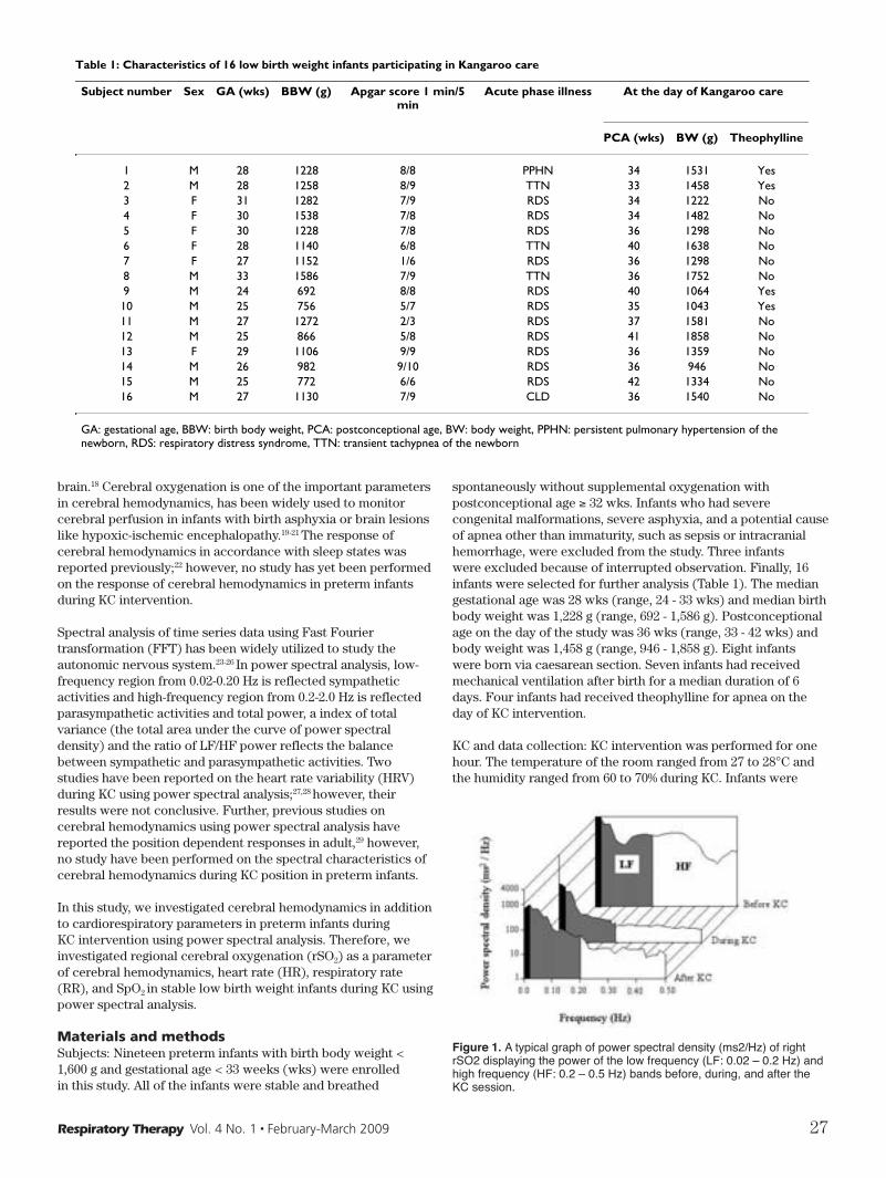
Respiratory Therapy Vol. 4 No. 1 February-March 2009 27
brain.18 Cerebral oxygenation is one of the important parameters in cerebral hemodynamics, has been widely used to monitor cerebral perfusion in infants with birth asphyxia or brain lesions like hypoxic-ischemic encephalopathy.19-21 The response of cerebral hemodynamics in accordance with sleep states was reported previously;22 however, no study has yet been performed on the response of cerebral hemodynamics in preterm infants during KC intervention.
Spectral analysis of time series data using Fast Fourier transformation (FFT) has been widely utilized to study the autonomic nervous system.23-26 In power spectral analysis, low-frequency region from 0.02-0.20 Hz is reflected sympathetic activities and high-frequency region from 0.2-2.0 Hz is reflected parasympathetic activities and total power, a index of total variance (the total area under the curve of power spectral density) and the ratio of LF/HF power reflects the balance between sympathetic and parasympathetic activities. Two studies have been reported on the heart rate variability (HRV) during KC using power spectral analysis;27,28 however, their results were not conclusive. Further, previous studies on cerebral hemodynamics using power spectral analysis have reported the position dependent responses in adult,29 however, no study have been performed on the spectral characteristics of cerebral hemodynamics during KC position in preterm infants.
In this study, we investigated cerebral hemodynamics in addition to cardiorespiratory parameters in preterm infants during KC intervention using power spectral analysis. Therefore, we investigated regional cerebral oxygenation (rSO2) as a parameter of cerebral hemodynamics, heart rate (HR), respiratory rate (RR), and SpO2 in stable low birth weight infants during KC using power spectral analysis.
Materials and methodsSubjects: Nineteen preterm infants with birth body weight < 1,600 g and gestational age < 33 weeks (wks) were enrolled in this study. All of the infants were stable and breathed
spontaneously without supplemental oxygenation with postconceptional age ≥ 32 wks. Infants who had severe congenital malformations, severe asphyxia, and a potential cause of apnea other than immaturity, such as sepsis or intracranial hemorrhage, were excluded from the study. Three infants were excluded because of interrupted observation. Finally, 16 infants were selected for further analysis (Table 1). The median gestational age was 28 wks (range, 24 - 33 wks) and median birth body weight was 1,228 g (range, 692 - 1,586 g). Postconceptional age on the day of the study was 36 wks (range, 33 - 42 wks) and body weight was 1,458 g (range, 946 - 1,858 g). Eight infants were born via caesarean section. Seven infants had received mechanical ventilation after birth for a median duration of 6 days. Four infants had received theophylline for apnea on the day of KC intervention.
KC and data collection: KC intervention was performed for one hour. The temperature of the room ranged from 27 to 28°C and the humidity ranged from 60 to 70% during KC. Infants were
Table 1: Characteristics of 16 low birth weight infants participating in Kangaroo care
Subject number Sex GA (wks) BBW (g) Apgar score 1 min/5 min
Acute phase illness At the day of Kangaroo care
PCA (wks) BW (g) Theophylline
1 M 28 1228 8/8 PPHN 34 1531 Yes2 M 28 1258 8/9 TTN 33 1458 Yes3 F 31 1282 7/9 RDS 34 1222 No4 F 30 1538 7/8 RDS 34 1482 No5 F 30 1228 7/8 RDS 36 1298 No6 F 28 1140 6/8 TTN 40 1638 No7 F 27 1152 1/6 RDS 36 1298 No8 M 33 1586 7/9 TTN 36 1752 No9 M 24 692 8/8 RDS 40 1064 Yes10 M 25 756 5/7 RDS 35 1043 Yes11 M 27 1272 2/3 RDS 37 1581 No12 M 25 866 5/8 RDS 41 1858 No13 F 29 1106 9/9 RDS 36 1359 No14 M 26 982 9/10 RDS 36 946 No15 M 25 772 6/6 RDS 42 1334 No16 M 27 1130 7/9 CLD 36 1540 No
GA: gestational age, BBW: birth body weight, PCA: postconceptional age, BW: body weight, PPHN: persistent pulmonary hypertension of the newborn, RDS: respiratory distress syndrome, TTN: transient tachypnea of the newborn
Figure 1. A typical graph of power spectral density (ms2/Hz) of right rSO2 displaying the power of the low frequency (LF: 0.02 – 0.2 Hz) and high frequency (HF: 0.2 – 0.5 Hz) bands before, during, and after the KC session.

28 Respiratory Therapy Vol. 4 No. 1 February-March 2009
observed carefully and monitored in three conditions: 30 minutes in the incubators (before KC), 1 hour of KC intervention (during KC), and 30 minutes in the incubator again after KC intervention. Mothers were seated on a reclining chair at a 60° angle, wearing a front opening blouse. The infants were placed naked except for a diaper directly onto the skin between the breasts and covered with a light blanket. Infants were fed 1 hour before KC. All infants were continuously monitored with electrocardiogram to determine HR, RR and with pulse oxymetry for percentages of oxygen saturation (SpO2). Regional cerebral oxygenation was measured with a near infrared spectroscopy, NIRS (INVOS 4100, Somanetics, Troy, MI), with the two probes positioned on the bilateral frontoparietal areas. Physiological data were recorded at 10 second intervals (averaged over 10 second period) through a Wave Archiving System (WAS-J: Agilent Technologies, Inc.) and
further analyzed. Data from the first 30 minutes during KC were excluded from the analysis to minimize the effects of changes due to the adaptation process. The data for rSO2 were recorded from both the left and right hemispheres independently (LrSO2
and RrSO2, respectively).
Behavioral states of infants were recorded throughout the observation period using the Brazelton Neonatal Behavior Assessment Scale.30 Behavioral states of infant were observed by nurses trained on the observational scale. Five different behavioral states were observed: 1) Quiet sleep; 2) Active sleep; 3) Drowsiness; 4) Alert inactivity; and 5) Active awake. Behavioral states were judged before KC, during KC, at the end of KC, and 30 minutes after KC. Body temperature was measured at the beginning of data recording before the KC session and after the end of KC. Informed consents were obtained from the parents and the study was approved by the ethical committee of the institute.
Power spetral analysis: Power spectral analysis was performed on rSO2, HR, RR, and SpO2 in three sessions as previously reported.28 The power spectral density was calculated and divided into two frequency bands in each session as shown in Figure 1. The regional power of low-frequency (LF, 0.02 – 0.2 Hz) and high-frequency (HF, 0.20 – 0.5 Hz) bands, and the ratio of LF/HF were calculated. The powers of LF and HF were normalized using the formulas % LF = LF/TP × 100 (%) and % HF = HF/TP × 100 (%).31 Total power (TP) was obtained by integrating the power spectrum from frequency 0.02 to 0.50 Hz.
Statistical analysis: Data were analyzed with SPSS 13.0 for Windows (SPSS Inc., Chicago, IL). A repeated measures ANOVA was performed to analyze the differences of parameters among three sessions. Conventional statistics: mean, standard deviation, coefficient of variance (CV), range, normal distribution, and median were performed for all parameters. Data are expressed as median (range) or mean ± SD.
ResultsDescriptive statistics of HR, RR, SpO2, LrSO2 and RrSO2 are shown in Table 2. In mean HR, SpO2, LrSO2, and RrSO2, there were no significant differences among the three sessions; however, RR was significantly increased during KC and decreased after KC (p < 0.05, respectively). Body temperature was increased by 0.3°C during KC (p < 0.01). The CV of HR was
Figure 2. Individual values of total power (TP) of power spectral density before, during, and after KC, displaying visually qualitative changes during KC. A repeated measures ANOVA was performed to determine the statistical differences among the three sessions: before, during, and after KC. * p < 0.01, ** p < 0.001, before vs during KC or during vs after KC.
Table 2: Central tendencies and coefficient of variances of physiological variables before, during and after KC intervention
Variables Measures Before KC During KC After KC
Heart rate (bpm) Mean 149.4 ± 6.3 150.0 ± 10.0 150.4 ± 8.7CV 6.8 ± 3.2 4.6 ± 1.3* 7.5 ± 4.7 †
Respiratory rate (bpm) Mean 39.4 ± 8.9 44.0 ± 5.1** 39.7 ± 7.9 † †CV 24.1 ± 5.4 22 ± 6.7 19.7 ± 7.6
SpO2(%) Mean 98.0 ± 1.3 97.6 ± 2.5 97.5 ± 1.7CV 2.3 ± 1.4 1.5 ± 0.9 2.9 ± 2.4
Left-rSO2(%) Mean 46.8 ± 5.6 47.3 ± 6.1 47.5 ± 7.3CV 6.6 ± 4.1 5.8 ± 3.3 4.1 ± 1.6
Right-rSO2(%) Mean 48.6 ± 6.9 49.1 ± 9.4 47.8 ± 6.9CV 7.2 ± 4.6 7.2 ± 5.5 5.6 ± 2.3
Body temperature, (°C) Mean 37.0 ± 0.2*** 37.3 ± 0.3Median (range) 37.1 (36.6 – 37.4) 37.3 (36.8 – 37.4)
Data were expressed as mean ± SD or median (range). * p < 0.05, ** p < 0.01, *** p < 0.001, before versus during KC and † p < 0.05, † † p < 0.01, during versus after KC. CV: coefficient of variance, rSO2: regional cerebral oxygenation.

Respiratory Therapy Vol. 4 No. 1 February-March 2009 29
decreased by 2.2% during KC (p < 0.05), while it was increased again after KC by 2.9% (p < 0.05). There were no significant differences in the CVs of RR, SpO2, LrSO2, and RrSO2.
Changes in TP during the sessions were shown in Figure 2. The TP of HR was significantly decreased during KC and increased after KC (before: 635 ± 280, during: 268 ± 134, after: 618 ± 240 ms2/Hz; before vs during and during vs after, p < 0.01). The TP of SpO2 had a similar tendency to that of HR. On the other hand, the TP of RR was significantly increased during KC and decreased after KC (before: 458 ± 186, during: 713 ± 175, after: 461 ± 231 ms2/Hz; before vs during and during vs after, p < 0.01). In regional cerebral oxygenation, the TP of either LrSO2 or RrSO2
was significantly decreased during KC and increased again after KC (LrSO2: before: 121 ± 91, during: 54 ± 41, after: 90 ± 56 ms2/Hz; RrSO2: before: 123 ± 69, during: 55 ± 30, after: 98 ± 46 ms2/Hz; before vs during and during vs after, p < 0.001, respectively in each parameter).
The percentages of LF, HF, and the ratio of LF/HF are shown in Figure 3A and 3B. For HR, the %LF was significantly increased during KC (before: 41.4 ± 11.5, during: 48.3 ± 9.8%, p < 0.05), while %HF was decreased (before: 45.9 ± 13.9, during: 33.5 ± 15.3%). In contrast, the %LF of rSO2 was significantly decreased during KC for both LrSO2 (before: 63.5 ± 11.2, during: 49.2 ± 9.2, after: 63.8 ± 10.2%; before vs during and during vs after, p < 0.01) and RrSO2 (before: 59.1 ± 11.5 during: 49.9 ± 6.9, after: 57.0
± 13.6%; before vs during, p < 0.05). The %HF was decreased during KC for RrSO2 (before: 28.1 ± 13.8, during: 22.6 ± 10.9, after: 26.2 ± 19.2%; before vs during, p < 0.01), although there was no significant change in this parameter for LrSO2 during the sessions. For RR and SpO2, significant changes were not observed in %LF or HF during KC, although the %LF of RR after KC was significantly increased while the %HF was decreased (p < 0.05, respectively) with an increased LF/HF ratio (p < 0.01). There were no significant differences in the ratio of HR, SpO2, LrSO2, and RrSO2 during the sessions (Figure 3B).
At the day of KC, four infants had received theophylline. Infants who received theophylline showed the similar tendency to those without theophylline during KC and after KC.
Behavioral analysis: The behavioral states during the observation period are shown in Figure 4. The percentage of infants with quiet sleep states remarkably increased during KC (61.5%) compared to those before KC (15.4%). This percentage increased further to 76.9% at the end of KC and decreased to 38.5% at 30 minutes after KC. In contrast, the percentage of infants in active sleep was 53.8% before KC, decreased to 23.1% in the middle of KC, and increased to 54.0% at 30 minutes after KC.
DiscussionIn this study, regional cerebral oxygenation using NIRS was measured during KC in addition to cardiorespiratory parameters in stable low birth weight infants and analyzed by using power spectral analysis. By conventional analysis, the CV of HR was decreased, and mean RR and body temperature were increased during KC as previously reported;7,9,14 however, significant changes were not found in mean rSO2. By power spectral analysis, TP was decreased in rSO2; HR, and SpO2 during KC, whereas the TP of RR was increased. Further, the LF of rSO2
was found to be decreased during KC while the LF of HR was increased with decreased HF. This study has shown that the spectral characteristics of cerebral oxygenation are significantly different between during KC and before KC or after KC as well as HR, RR and SpO2; however, there were no significance differences in their mean values. The power spectral analysis was found as a more effective analytic approach for revealing the physiological responses compared to the conventional analysis.
Figure 3. A: Power spectral density in low – frequency (LF: close circle) and high – frequency (HF: open circle) bands before, during, and after KC. LF and HF are expressed as normalized values (%LF = LF/total power × 100 and %HF = HF/total power × 100). B. The ratio of LF/HF before, during, and after KC. Data is presented as means ± SEM. A repeated measures ANOVA was performed to determine the statistical differences among the three sessions: before, during, and after KC. * p < 0.05, ** p < 0.01, before vs during KC. † p < 0.05, † † p < 0.01, during vs after KC.
Figure 4. Behavioral states of infants observed before KC, in the middle of KC, at the end of KC, and 30 minutes after KC. The quiet sleep state was remarkably increased in the middle of KC and at the end of KC. A chi square test was performed to determine the signifi cant differences in the four behavioral states.* p < 0.05, ** p < 0.01, before versus middle of KC or before versus end of KC.
30 Respiratory Therapy Vol. 4 No. 1 February-March 2009
In spectral analysis of heart rate variability (HRV), low-frequencies (0.02 - 0.20 Hz) reflect sympathetic activities such as the baroreceptor reflex, high-frequencies (0.2 - 2.0 Hz) reflect parasympathetic activities such as vagal activity, and the ratio of LF/HF power reflects the balance between sympathetic and parasympathetic activities.23-26,32 Several studies have been published on autonomic function of HRV in preterm infants and only two studies had been previously reported on HRV during KC intervention using power spectral analysis up to now. One of them showed a decrease of LF and HF during KC,27 while the other showed an increase of LF during KC.28 In our study, the LF of HR was increased during KC and the LF of rSO2 was decreased. In general, the increase in sympathetic activities and decrease in cerebral oxygen delivery with the head in an up-tilting position are assumed to be due to gravity causing pooling of blood and thereby activating baroreceptors as previously reported,29,33 and the decrease of LF of rSO2 during KC with increased LF of HR described in this study might be supported previous studies. These results could be understood as activation of the central nervous system and brain function during KC position. Besides these, the increase in body temperature and respiration observed in our study may have an effect on the LF of HR as previously reported on HRV.28,34 Further, TP is the index of total variance in power spectral analysis and changes in TP during KC indicate the changes in total variance. In our study, TP of rSO2, HR, or SpO2 have been shown to decrease during KC. A possible explanation for these decreases during KC might be associated with reduced activity and increased quiet sleep state during KC as previously reported.35,36 These results could be interpreted as physiological stability elicited by KC intervention. Although the dominance of HF has been reported in HR during the quiet sleep state,37,38 this was not observed during KC in a previous study28 or in the current study. The position of the head of infants during KC might be responsible for the discrepancy between the left and right rSO2.
To our knowledge, the present study was the first study to analyze the response of rSO2 due to KC in LBW preterm infants through spectral characteristics. Therefore, it was difficult to compare the results of this study with other similar studies and apply to the clinical settings. Despite the sampling limitation, this reference data provide new understanding into the response of cerebral hemodynamics in preterm infants and should have significant implications to generalize the rSO2 responses with different modalities in LBW infants. Further studies are necessary to determine the clinical relevance of the present findings.
ConclusionThe results of this study revealed that changes of cerebral hemodynamics associated with KC position in preterm infants as well as cardiorespiratory parameters. These changes were especially apparent by power spectral analysis. Furthermore, the results of this study indicate that KC may contribute to the activation of central nervous system and brain function. Further study is needed to determine the underlying physiology responsible for these differences.
References1 Whitelaw A, Sleath K: Myth of the marsupial mother: home
care of very low birth weight babies in Bogota, Colombia. Lancet 1985, 1:1206-1208.
2 Anderson GC: Current knowledge about skin-to-skin (kangaroo) care for preterm infants. J Perinatol 1991, 11:216-
226.3 Ludington-Hoe SM, Swinth JY: Developmental aspects of
kangaroo care. J Obstet Gynecol Neonatal Nurs 1996, 25:691-703.
4 Dodd VL: Implications of kangaroo care for growth and development in preterm infants. J Obstet Gynecol Neonatal Nurs 2005, 34:218-232.
5 Tessier R, Cristo M, Velez S, Giron M, de Calume ZF, Ruiz-Palaez JG, et al.: Kangaroo mother care and the bonding hypothesis. Pediatrics 1998, 102:e17.
6 Mondlane RPdGAMP, Ebrahim GJ: Skin-to-Skin Contact as a Method of Body Warmth for Infants of Low Birth Weight. J Trop Pediatr 1989, 35:321-326.
7 Bauer K, Uhrig C, Sperling P, Pasel K, Wieland C, Versmold HT: Body temperatures and oxygen consumption during skin-to-skin (kangaroo) care in stable preterm infants weighing less than 1500 grams. J Pediatr 1997, 130:240-244.
8 Ludington-Hoe SM, Nguyen N, Swinth JY, Satyshur RD: Kangaroo care compared to incubators in maintaining body warmth in preterm infants. Biol Res Nurs 2000, 2:60-73.
9 Ludington-Hoe SM, Anderson GC, Simpson S, Hollingsead A, Argote LA, Rey H: Birth-related fatigue in 34–36-week preterm neonates: rapid recovery with very early kangaroo (skin-to-skin) care. J Obstet Gynecol Neonatal Nurs 1999, 28:94-103.
10 Fohe K, Kropf S, Avenarius S: Skin-to-skin contact improves gas exchange in premature infants. J Perinatol 2000, 20:311-315.
11 Acolet D, Sleath K, Whitelaw A: Oxygenation, heart rate and temperature in very low birthweight infants during skin-to-skin contact with their mothers. Acta Paediatr Scand 1989, 78:189-193.
12 Cattaneo ADR, Bergman N, Charpak N: Kangaroo mother care in low-income countries. International network in Kangaroo Mother Care. J Trop Pediatr 1998, 44:279-282.
13 Feldman R, Eidelman AI: Skin-to-skin contact (Kangaroo Care) accelerates autonomic and neurobehavioural maturation in preterm infants. Dev Med Child Neurol 2003, 45:274-281.
14 Messmer PR, Rodriguez S, Adams J, Wells-Gentry J, Washburn K, Zabaleta I, Abreu S: Effect of kangaroo care on sleep time for neonates. Pediatr Nurs 1997, 23:408-414.
15 Ludington SM: Energy conservation during skin-to-skin contact between premature infants and their mothers. Heart Lung 1990, 19:445-451.
16 Bohnhorst B, Heyne T, Peter CS, Poets CF: Skin-to-skin (kangaroo) care, respiratory control, and thermoregulation. J Pediatr 2001, 138:193-197.
17 Smith SL: Physiologic Stability of Intubated VLBW Infants During Skin-to-Skin Care and Incubator Care. Advances in Neonatal care 2001, 1:28-40.
18 Volpe JJ: Hypoxic-ischemic encephalopathy. In Neurology of the newborn Edited by: Volpe JJ. Philadelphia, Saunders; 2001:296-330.
19 Toet MC, Lemmers PM, van Schelven LJ, van Bel F: Cerebral oxygenation and electrical activity after birth asphyxia: their relation to outcome. Pediatrics 2006, 117:333-339.
20 Meek JH, Elwell CE, McCormick DC, Edwards AD, Townsend JP, Stewart AL, Wyatt JS: Abnormal cerebral haemodynamics in perinatally asphyxiated neonates related to outcome. Arch Dis Child Fetal Neonatal Ed 1999, 81:F110-115.
21 van Bel F, Dorrepaal CA, Benders MJ, Zeeuwe PE, Bor M van de, Berger HM: Changes in cerebral hemodynamics and oxygenation in the first 24 hours after birth asphyxia.

Respiratory Therapy Vol. 4 No. 1 February-March 2009 31
Pediatrics 1993, 92:365-372.22 Munger DM, Bucher HU, Duc G: Sleep state changes
associated with cerebral blood volume changes in healthy term newborn infants. Early Hum Dev 1998, 52:27-42.
23 Patural H, Pichot V, Jaziri F, Teyssier G, Gaspoz JM, Roche F, Barthelemy JC: Autonomic cardiac control of very preterm newborns: A prolonged dysfunction. Early Hum Dev 2008.
24 De Rogalski Landrot I, Roche F, Pichot V, Teyssier G, Gaspoz JM, Barthelemy JC, Patural H: Autonomic nervous system activity in premature and full-term infants from theoretical term to 7 years. Auton Neurosci 2007, 136:105-109.
25 Nakamura T, Horio H, Miyashita S, Chiba Y, Sato S: Identification of development and autonomic nerve activity from heart rate variability in preterm infants. Biosystems 2005, 79:117-124.
26 Andriessen P, Oetomo SB, Peters C, Vermeulen B, Wijn PF, Blanco CE: Baroreceptor reflex sensitivity in human neonates: the effect of postmenstrual age. J Physiol 2005, 568:333-341.
27 McCain GC, Ludington-Hoe SM, Swinth JY, Hadeed AJ: Heart rate variability responses of a preterm infant to kangaroo care. J Obstet Gynecol Neonatal Nurs 2005, 34:689-694.
28 Smith SL: Heart period variability of intubated very-low-birthweight infants during incubator care and maternal holding. Am J Crit Care 2003, 12:54-64.
29 Tachtsidis I, Elwell CE, Lee CW, Leung TS, Smith M, Delpy DT: Spectral characteristics of spontaneous oscillations in cerebral haemodynamics are posture dependent. Adv Exp Med Biol 2003, 540:31-36.
30 Brazelton TB: Neonatal Behavioral Assessment Scale 2nd edition. London: Heinemann; 1984.
31 Andriessen P, Koolen AM, Berendsen RC, Wijn PF, ten Broeke ED, Oei SG, Blanco CE: Cardiovascular fluctuations and transfer function analysis in stable preterm infants. Pediatr Res 2003, 53:89-97.
32 Smith SL, Doig AK, Dudley WN: Characteristics of heart period variability in intubated very low birth weight infants with respiratory disease. Biol Neonate 2004, 86:269-274.
33 Schord L, Walter J: Effect of head-up body tilt position on autonomic function and cerebral oxygenation in preterm infants. Biology of the Neonate 2002, 81:255-259.
34 Davidson S, Reina N, Shefi O, Hai-Tov U, Akselrod S: Spectral analysis of heart rate fluctuations and optimum thermal management for low birth weight infants. Med Biol Eng Comput 1997, 35:619-625.
35 Ludington-Hoe SM, Thompson C, Swinth J, Hadeed AJ, Anderson GC: Kangaroo care: research results, and practice implications and guidelines. Neonatal Netw 1994, 13:19-27.
36 Porges SW, McCabe PM, Yongue BG: Respiratory-heart rate interaction and psycho-physiological implications for pathophysiology and behavior. Cacioppor JT, Petty R, editors. Perspectives in Cardiovascular Psychophysiology. New York Guilford Press; 1982.
37 Porges SW, Doussard-Roosevelt JA, Stifter CA, McClenny BD, Riniolo TC: Sleep state and vagal regulation of heart period patterns in the human newborn: an extension of the polyvagal theory. Psychophysiology 1999, 36:14-21.
38 Villa MP, Calcagnini G, Pagani J, Paggi B, Massa F, Ronchetti R: Effects of sleep stage and age on short-term heart rate variability during sleep in healthy infants and children. Chest 2000, 117:460-466.
What are some terms I need to know?Capital Budget – This is what the hospital uses to pay for new construction, remodeling, beds, ventilators, or any “hard” asset.
Operating Budget – This is what is used to pay for wages, utilities and any disposable products like IV fluids or light bulbs.
By the way, these budgets are in separate accounts and cannot be moved from one to the other. If the capital budget is used up, you can’t take money from the operating budget to buy your equipment.
What are the different types of leases that I need to know about?Capital Lease – Other names used might be Rent-to-Own or Dollar Buy-Out. This is like buying a house. The hospital pays a number of monthly payments with each payment going toward the eventual payoff of the equipment. Funding comes out of the capital budget.
Operating Lease – This is like leasing a car. The hospital only pays for the use of the equipment for a set period of time, like a rental. At the end of the term you have a return, purchase or extension option. The return option paves the way for a new lease on brand new equipment. Funding for an operating lease comes out of the operating budget.
Rental Rewards – If your hospital is renting to fill basic long-term equipment needs, this gives you an opportunity to acquire brand new equipment. Through each payment made, you earn credits and ultimately would own the equipment if you continue renting. Funding comes from the operating budget.
OK, this is an oversimplification but it will get you started. There are many options and variations for the lease, depending on how it is set up. Suffice it to say, you probably will not be interested any further than the three choices above.
How do I use leasing with my equipment supplier?Find out if they have a leasing program within their company. Just about every company that sells capital equipment over $5,000 has a relationship with a source to help you through this and they will then put you in touch with these people. Some leasing companies can usually have the paperwork done in hours and the form is short, as few as two pages. There is no obligation to do the lease. After all is laid out for you, all they would need from you would be a P.O. stating the terms. Simple and fast.
Remember, leasing is just another tool for you to get what you want and need now rather than later. For additional information, visit MedOne Capital.
John Campbell is with MedOne Capital. This article was provided by Hamilton Medical, from its newsletter.
Editorial…continued from page 4

32 Respiratory Therapy Vol. 4 No. 1 February-March 2009
Asthma Beliefs Among Mothers and Children from Different Ethnic Origins Living in AmsterdamQ.M. van Dellen, W.M.C. van Aalderen, P.J.E. Bindels, F.G. Öry, J. Bruil, K. Stronks, and the PEACE study group
AbstractBackground: Doctors and patients hold varying beliefs concerning illness and treatment. Patients’ and families’ explanatory models (EMs) vary according to personality and sociocultural factors. In a multi-ethnic society, it is becoming increasingly significant that doctors understand the different beliefs of their patients in order to improve patient/doctor communication as well as patient adherence to treatment.
Methods: Twelve focus groups were formed, consisting of 40 children diagnosed with asthma, as well as 28 mothers of these children. These groups included mothers and children of different ethnicities who were living in Amsterdam, the Netherlands. In order to understand the beliefs that both mothers and children hold regarding asthma and its treatment, the explanatory models were analysed and compared.
Results: Study findings show that mothers and children, regardless of ethnicity and age, have their own EMs. Overall, there is a great deal of uncertainty related to the causes, consequences, problems, and symptoms of asthma and its treatment. It also seems that many concerns and feelings of discomfort are the result of lack of knowledge. For instance, the fact that asthma is not seen as a chronic disease requiring daily intake of an inhaled corticosteroid, but rather as an acute phenomenon triggered by various factors, may be very relevant
for clinical practice. This particular belief might suggest an explanation for non-adherent behavior.
Conclusion: A thorough understanding of the mothers’ and children’s beliefs regarding the illness and its treatment is an important aspect in the management of asthma. Gaining an understanding of these beliefs will provide a foundation for a solid clinician-patient/family partnership in asthma care. Although ethnic differences were observed, the similarities between the mothers’ and children’s beliefs in this multi-ethnic population were striking. In particular, a common belief is that asthma is considered an acute rather than a chronic condition. In addition, there is a lack of knowledge about the course and the self-management of asthma. Health care providers should be aware of these commonly held beliefs, and this information could be shared in educational programs.
BackgroundAsthma is the most prevalent chronic disease among children in Western countries. Despite the availability of effective treatments aimed at controlling asthma symptoms, up to 50% of children with asthma continue to have frequent and bothersome symptoms. US and UK studies among adults and children with asthma suggest that factors such as individual beliefs, attitudes, behavior, inadequate patient-clinician communication and a foreign ethnic origin may all contribute to poor adherence to treatment with inhaled corticosteroids (ICSs), which in turn results in poor asthma control.
As a result of migration, Western European countries are becoming more ethnically diverse. In the Netherlands approximately 10% of the population is of non-Western origin. A logical consequence of this trend is that the number of non-Western children is also growing. In the Netherlands’ larger cities, these non-Western children constitute more than 50% of the population. (see after Introduction for more information) Although there is limited literature on asthma prevalence among the Netherlands’ largest ethnic minority groups, the number of ethnic children diagnosed with asthma seems to be similar to that among ethnic Dutch children. However, there is evidence that children from ethnic minorities have less control over their asthma.
Authors van Dellen, and van Aalderen are with the Department of Paediatric Pulmonology, Emma Children’s Hospital; Bindels is with the Department of General Practice, Academic Medical Centre, University of Amsterdam; Öry and Bruil are with the TNO Quality of Life, Division Prevention and Health, Leiden; Stronks is with the Department of Social Medicine, Academic Medical Centre, University of Amsterdam. The authors would like to thank and acknowledge the help of S. Detmar, N. Hdoudou, H. Tisoudali, S. Orikli, N. Lale, N. Asthiani and I. van Boxel for facilitating the focus group interviews and Pacemaker in Global Health, Amsterdam, for its contribution in general to the study. Reprinted from BioMed Central, BMC Public Health, © 2008 van Dellen et al, licensee BioMed Central Ltd. This is an open access article distributed under the terms of the Creative Commons Attribution License. This article has been slightly edited for Respiratory Therapy. For references, please visit BioMed Central and type the full name of the article.

Respiratory Therapy Vol. 4 No. 1 February-March 2009 33
According to the theoretical model of the medical anthropologist Kleinman, healthcare providers and patients have different beliefs about the patients’ illness and treatment. Patients’ and families’ explanatory models (EMs) vary according to personality and sociocultural factors. Studies among ethnic minority populations in the US suggest that cultural factors have an impact on the manner in which patients and families from these groups explain asthma. Therefore, it is becoming increasingly recognized that doctors must understand the different beliefs of their patients in order to improve communication as well as patient adherence to treatment.
In Europe, there are few studies that describe the differences in beliefs about asthma and its treatment among mothers and their asthmatic children from different ethnic origins. This study explored and compared EMs in etiology of the condition, onset of symptoms, course of illness and treatment of the mothers and children from Moroccan, Turkish, Surinamese and Dutch backgrounds. The purpose of this exploration and comparison was to gain more insight into the groups’ beliefs about causes, consequences, symptoms and the self-management of asthma. To elicit the EMs of Moroccan, Turkish, Surinamese and ethnic Dutch mothers and children, a qualitative study was conducted using focus group interviews. Attention was given to the similarities and differences between mothers and children and also among the different ethnicities. The results of this study provide data that can be used in the development of a questionnaire and information aids. These various tools can then be implemented by the clinician when focusing on asthma care and self-management when dealing with the largest ethnic groups in the Netherlands.
The Moroccan, Surinamese and Turkish populations are three
of the largest ethnic minority groups in the Netherlands. Of the country’s approximately 16 million residents, 315,821 are Moroccan, 329,430 Surinamese and 358,846 Turkish. First-generation Turks and Moroccans are mainly economic migrants with a low level of education. They came to the Netherlands mainly between 1960 and 1980. The Surinamese group is from the former Dutch colony of Surinam, and consists largely of people of Creole (45%) and Hindustani ethnic backgrounds (45%). Creole people have a mixed background (African and European) and Hindustani people originate from India. The migration to the Netherlands peaked in 1975 when Surinam became independent. With increasing migration, the socioeconomic position of the Surinamese migrant population became more diverse. Of the Turks and Moroccans aged 35 to 54, 74% and 84% respectively have only primary education and there is a higher percentage of illiteracy among Moroccans than among Turks. Of the Surinamese aged 35 to 54, 31% have primary education only. A relatively high percentage of these migrants are unemployed. As a consequence, household incomes and socioeconomic status of the three migrant groups are lower than that of the ethnic Dutch population. However, the Surinamese have a higher socioeconomic status than Turkish and Moroccan migrants. Generally, the Surinamese speak fluent Dutch, mainly because of their colonial past, whereas most Turks and Moroccans have difficulty in speaking Dutch. Furthermore, all first-generation migrants remain mainly oriented towards their own ethnic group, but the Surinamese have relatively more contact with the ethnic Dutch than Moroccan and Turkish migrants. This may be explained in part by their familiarity with the Dutch language and culture.
MethodsChildren and mothers only were recruited by one physician
Table 1. Demographics and characteristics of participating children and adolescents
Children/adolescents Dutch Moroccan Turkish Surinamese Total
Total number of participating young children (7-12years)
5 10 6 5 26
Gender
boys 2 4 4 3 13
girls 3 6 2 2 13
Medication
inhaled corticosteroids 5 9 5 5 24
bronchodilators 4 9 6 5 24
Asthma control¶
well controlled 4 4 3 3 14
not well controlled 1 6 3 2 12
Total number of participating
adolescents (13-17years) 3 4 3 4 14
Gender
boys 3 4 3 4 14
girls 0 0 0 0 0
Medication
inhalation corticosteroids 2 4 3 4 13
bronchodilators 3 4 3 4 14
Asthma control¶
well controlled 1 2 1 2 6
not well controlled 2 2 2 2 8
¶ information from results multicentre study

34 Respiratory Therapy Vol. 4 No. 1 February-March 2009
(first author QvD) who had previously seen all these children and mothers in a home setting as part of a multicentre study. During these home visits, detailed information was collected including the child’s age, gender, ethnicity, level of education, use of asthma medication and level of asthma control. Moreover, information about the parental level of education and socioeconomic status was also collected. A detailed description of this study has been previously published. Briefly, the inclusion criteria for children in this multicentre study were: 1) having asthma diagnosed by a paediatrician, 2) attending an outpatient clinic of one of the six participating hospitals, 3) being aged between 7 and 17 years, 4) being of Moroccan, Turkish, Surinamese or ethnic Dutch descent. Children were classified as belonging to an ethnic group other than Dutch if the child itself, or at least one of its parents, was born outside of the Netherlands. An additional criterion for the focus group interviews was that the children were prescribed inhaled corticosteroid (ICSs) in the year prior to the focus group sessions. An attempt was made to gain a well-balanced representation of the children and mothers of the multicentre study (purposive sampling) for participation in the focus group sessions. This involved ascertaining the level of the mothers’ education, the socioeconomic status of the family, as well as the age, gender and level of asthma control in the children. After obtaining the adolescents’ and mothers’ verbal agreement to participate in the study, a letter with further information was sent, followed by a reminder phone call two days before the sessions took place.
After the mothers had been subdivided according to ethnicity (ethnic Dutch, Turkish, Surinamese, Moroccan), and the children on the basis of ethnicity and age (6 to 12 year olds (young children) and 13 to 17 year olds (adolescents)), 4 focus group
sessions were established among young children (total of 26 children), 4 sessions among adolescents (total of 14 children), and 4 sessions among mothers (total of 28 women). Each session took about two hours and was chaired by two experienced individuals, the moderator and the observer, of the same ethnic background as the participants. Consequently the principal researcher, also first author (QvD), was not present at al sessions. Corresponding ethnic backgrounds enabled the study participants to speak in their native languages and therefore culturally sensitive information was conveyed accurately and sensitively. Each session began with an explanation of the purpose of the focus groups. The moderator followed a semi-structured interview procedure and used a checklist containing questions/topics to be discussed. The observer, who did not actively participate in the interviews, took field notes. The topic guide was based on Kleinman’s Explanatory Models. Interviews based on this model focus on five issues: etiology of the condition, onset of symptoms, pathophysiology, course of illness and treatment. Examples of questions that were posed to start the discussion include the following: What is asthma?, How do you treat asthma?, Will you or your child be bothered by asthma in the future?, When does your child take his medication?, How do you or does your child recognize an asthma exacerbation?, What do you or does your child do in case of an exacerbation? In an attempt to promote an informative discussion, open-ended questions were asked as much as possible. However, due to the fact that both the mothers and children sometimes found it difficult to engage in conversation, it became necessary to stimulate and guide them using yes or no questions and examples, such as, In case of an exacerbation, do you start with salbutamol?, Do you know that the best way to treat asthma is to use your ICSs daily?, Do you think a good relationship with your doctor will have a positive influence on the course of asthma?
Table 2. Demographics and characteristics of participating mothers
Mothers Dutch Moroccan Turkish Surinamese Total
Total number of participants 4 8 10 6 28
Generation¶
fi rst n/a 8 10 4 22
second 0 0 2 2
Education¶
yes 4 4 2 6 16
no or primary education only 0 4 8 0 12
Occupation¶
skilled labour 4 2 1 3 10
manual labour 0 4 7 3 14
homemaker (no more information) 0 2 2 0 4
Children of participating mothers 4 10 10 8 32
Gender
boys 2 4 8 6 20
girls 2 6 2 2 12
Age
6-12 years 4 8 7 7 26
13-17 years 0 2 3 1 6
Medication
inhalation corticosteroids 4 9 9 8 30
bronchodilators 3 10 10 8 31
Asthma control¶
well controlled 3 3 5 4 15
not well controlled 1 7 5 4 17
¶ information from results multicentre study

Respiratory Therapy Vol. 4 No. 1 February-March 2009 35
Furthermore, at the end of each discussion a summary was made by the moderator. Participants were then asked if this summary was accurate and complete, and when necessary, participants provided additional information.
The participants’ ideas about etiology of the condition, onset of symptoms, course of illness and treatment in accordance with the theoretical model of Kleinman were selected from each session and categorized. The category of pathophysiology was explored superficially and therefore omitted from the analysis. Once the coding and categorizing was completed the researchers compared their work and discussed any disagreements in order to form a consensus.
ResultsTable 1 and 2 show demographics and characteristics of the respectively participating children and mothers. Initially 43 mothers were invited to participate, and 28 (65%) ultimately attended. Forty young children aged 6 to 12 were invited, and 26 attended (65%). Twenty-four older children aged 13 to 17 were invited and 14 attended (58%). Most of those who declined stated they were too ill or lived too far away and did not have any transport. Almost 50% of the participating young children had well controlled asthma. (Table 1) Two of the younger children had recently stopped using their ICSs and two others had stopped using their bronchodilators. There were 14 adolescents participating in the study and all of them were boys. Out of those 14 boys, 43% had well controlled asthma. (Table 1) The 28 mothers involved in the focus groups had in total 32 children. (Table 2) The educational level of most of the mothers was low: 43% had never attended school or had primary education only. The characteristics of the children of these participating mothers were similar to these of the participants of the focus groups among young children and adolescents.
None of the study participants, mothers nor children, actually knew what caused asthma. However, most of the participants had some kind of explanatory model explaining their asthma, ranging from their genes to a broad spectrum of triggers which cause asthma symptoms. It was discovered that most children, regardless of age or ethnicity, were taught about the etiology of asthma by their GPs or pediatricians and not by their mothers or fathers. However, in spite of the ideas the children expressed about the possible etiology of asthma, the majority of the children stated they were uncertain about the true etiology:
“I don’t know why I have asthma?” (the majority of the children), and “Why do I have asthma and my friend doesn’t?” (a Dutch boy, 8 years old).
In addition, the majority of the mothers, regardless of ethnicity, were uncertain about the cause of asthma. One of the most striking similarities among the mothers was that they seemed to know that asthma has a genetic component: “It runs in the family” (the majority of the mothers). Moreover, a remarkable difference was seen in the immigrant Moroccan and Turkish mothers. The most commonly reported EM for the etiology of asthma held by these mothers was the rainy, damp Dutch climate: “When we are in Morocco, my child never has asthma” (a Moroccan mother). “We live in a very run-down house and because of the damp climate, the house is also very damp. This is the reason my daughter suffers from asthma” (a Turkish mother). These types of comments, most frequently stated by the non-Dutch mothers, are examples that suggest that many of
these mothers believed that the onset of asthma was related to a particular trigger. In addition to stating that the cold weather triggered asthma, the mothers mentioned dust, pets, cigarette smoke, playing sports and becoming fatigued as other possible triggers. Related to this, a common notion among all mothers and children was the idea of asthma as an acute rather than a chronic illness: “Fortunately it is not present every day. Sometimes, very suddenly, it is there. It depends on what I do”(a Turkish boy, 16 years old). In other words, the idea of no symptoms, no asthma was frequently heard among all mothers and children: “During the summer my child doesn’t have asthma” (a Turkish mother). “My child only has asthma in her father’s house” (a divorced Dutch mother). “I only have asthma when I’m at school. It is very dusty at school says my mum” (a Turkish boy, 8 years).
All the mothers and children, regardless of ethnicity and age, had individual expectations about the course of the illness. These expectations were mainly focused on the consequences of the asthma in both the near and distant future. The majority of the Surinamese mothers had the idea that, although their children would be affected by asthma their entire lives, they could live with it and manage the disease: “If you know how to deal with your asthma, you can live with it” (a Surinamese mother suffering from asthma herself). “If my child takes his daily medication, he can live a normal life and will have no problems in the future” (another Surinamese mother). However, all ethnic Dutch mothers had strong concerns about their children’s future, the level of education they could obtain and their careers. They mentioned that their children miss more school than their peers due to illness, and this could influence their marks. An opinion shared by both mothers and children of non-Dutch origin, and which is somewhat similar to the opinion expressed by the Dutch mothers, is that asthma can be particularly bothersome during the night and because of this the children are often very sleepy in the classroom and have trouble following class lessons. Another similarity that was highlighted during most child and mother sessions, regardless of ethnicity and age, was that children with asthma get tired more quickly and experience more physical constraints than children without asthma: “I can’t do everything my friends can do. Like swimming or gym in school. Most of the times I stand aside” (a Moroccan girl, 11 years old). “It is not that I can’t do as much as my friends can do, I just get tired earlier and have to stop the activity” (a Dutch boy, 15 years old).
Because of these physical restrictions, young children are afraid of being bullied and excluded by their peers: “None of the boys in our street ever ask me to join a football game, because they know I have asthma and so I can’t run fast” (a Dutch boy, 11 years old). On the other hand, there were some younger and older children, regardless of ethnicity, who told us that it could also be to an asthmatic child’s advantage to tell friends and classmates about having asthma, because then they could show you consideration: “I can’t go to my friend’s house to play, because they have a cat and I’m allergic to cats. Since my friend knows, we always play at my home” (a Turkish girl, 12 years old). The Moroccan and Turkish adolescent boys did not share this view. These boys did not see the benefit of telling their friends they are suffering from asthma: “As long as no one can see I have asthma, why should I tell them” (a Moroccan boy, 14 years old). It remained unclear why exactly the Moroccan and Turkish boys felt this way.
36 Respiratory Therapy Vol. 4 No. 1 February-March 2009
A remarkable similarity in all mothers was the concern about the duration of the disease:
“When will my child get better?” (the majority of the mothers). Nevertheless, most Turkish and Surinamese mothers were hopeful for the future. The majority of the participants, including mothers and children, shared the belief that asthma was less severe than in the past. Therefore, they were under the impression that the asthma would continue to improve. Some children were even convinced that they could outgrow their asthma. However, contrary to this hopeful idea was the pessimistic view held by the majority of the mothers and children that asthma would always remain a problem in their lives: “Once I heard, I forgot who told me, that asthma is a chronic disease, which means, I have heard this too, that it will always bother me”(a Surinamese boy, 15 years old). “I think I will have asthma my entire life, because I was born with it” (a Turkish boy, 16 years old).
When discussing the onset of symptoms of asthma, the mothers mentioned a very broad spectrum of triggers. One of the similarities between the majority of the children and mothers, regardless of age and ethnicity, was that they had clear ideas about which triggers provoke asthmatic symptoms. Some mothers mentioned triggers such as dust, grass, milk, peanuts, trees and animals, which can all be tested for in an allergy test. Other mothers mentioned triggers such as fatigue, crying, laughing, playing (sports), perfume, unpleasant smells, fog, rain, humidity, cold weather, cigarette smoke, cold, or spicy foods. All participants were quite sure of the provoking triggers and the following onset of symptoms: “It is something you learn over the years” (a Dutch boy, 14 years). “I recognize the symptoms very early, I’ve learned it over the years. It looks like my child is drowning” (a Moroccan mother). “The moment my daughter tells me, puffing and blowing, that she is feeling tired, I know her asthma is bothering her” (a Dutch mother). “It always starts in the evening, when he is playing around with his younger brother. I always think he will drop dead. He looks so pale!” (a Turkish mother), “Thanks to my maternal instinct, I recognise the symptoms immediately. Sometimes my daughter gets stuck halfway up the stairs. I can see in her eyes that she is having a hard time.” (a Surinamese mother).
An interesting difference between the adolescents boys and younger children was that the adolescents expressed experiencing a feeling of sadness when they feel the onset of symptoms, for instance during a football game or at a school party: “I feel like a dope when it starts on the dance floor” (a Turkish boy, 15 years old). “For me I feel like a weakling when I need to stop before the game is over. For me the game is over” (a Moroccan boy, 14 years old).
Asthma causes problems in the daily lives of affected children and their families. Treating this disease is a necessity, and the children and their families must find ways to manage the asthma. This section of the paper explains how children and mothers manage the asthma. In all sessions, regardless of ethnicity and age, we saw that children and mothers knew that by avoiding triggers, they could reduce the risk of an asthma attack. But even with this understanding that asthma triggers should be avoided, in actual practice it appeared to be sometimes difficult to do so. In particular, the avoidance of some triggers was considered to be beyond the control of the individual: “Sometimes I can’t avoid the trigger. I can’t help it if it’s misty” (a Moroccan boy, 14
years old). “In our culture, it is bad manners to ask a guest not to smoke in the house” (Moroccan and Turkish mothers). “My child needs anti-allergic bed covers, but my insurance company will not reimburse the costs, so I can’t afford it” (a Moroccan mother). “I don’t want to stay home when my friends are going to the school party. But I can’t avoid it when they smoke” (a Dutch boy, 15 years old). “Sometimes I feel confused. The doctors and my mum tell me it is good and healthy to do some sports. But, on the other hand, when I start running I’m short of breath” (a Surinamese girl, 12 years old).
In addition to being aware of the importance of avoiding triggers, a clear similarity among all mothers, regardless of ethnicity, was the idea that their children should live healthy lives in order to suffer fewer side effects from asthma. During all focus group interviews, both mothers and children mentioned that clean houses, no pets, healthy food, no smoking and plenty of exercise are factors which will help reduce asthma symptoms.
In all sessions, regardless of age and ethnicity, mothers reported that two forms of management include the daily ICSs and remembering to take salbutamol with you when leaving the house. When we asked more specifically about the proper use of ICSs as maintenance therapy, all mothers, with the exception of the Surinamese, expressed reservations in some form or another. Their experience with the efficacy of ICSs as maintenance therapy was very good. The Surinamese mothers even mentioned that they supervised the use of medication, in order to make sure that their children inhaled the right medication at the right time. Most Moroccan mothers voiced that they performed a self-assessment, and also their assessment was more emotionally then rationally supported: “If my daughter does not show any signs of asthma, I can’t give her uuhh, her orange medication. I think it’s very sad for children to take daily medication when they are feeling OK” (a Moroccan mother). The Dutch mothers shared an overall negative attitude towards ICSs. Most Dutch mothers reported performing a detailed self-assessment of the state of their childrens’ asthma before deciding whether or not to administer their ICSs. They mentioned this is something they have learned over the years. Most of the time these decisions conflicted with their doctors’ instructions. Some of the children were taken off ICSs by their mothers when symptoms were absent, and other mothers terminated therapy when symptoms did not abate despite adherence: “Often I do not give my child her Seretide, because I fail to see the point of it. Treatment based on maternal instinct is still the best” (a Dutch mother). This statement was based on the mother’s maternal instinct and feeling rather than knowledge. Another reason behind their decisions to sometimes not give the medication was that the Dutch mothers also worry about long-term side effects of ICSs. These mothers thought it would be harmful to give them for a long period. The Turkish mothers were also under the impression that the use of ICSs will have some side effects. The most frequently mentioned side effects, which were also mentioned as the major factor preventing mothers from giving children their ICSs, were weight gain, growth retardation and addiction.
The only mothers who mentioned using alternative medication with their children were the Turkish and Dutch mothers. When the Turkish mothers administered alternative medication to their children, they stopped the regular medication. The Dutch mothers, however, mentioned using a combination of alternative and regular medication. Examples of the alternative medication
Respiratory Therapy Vol. 4 No. 1 February-March 2009 37
used includes over –the-counter drugs and homemade herbal (cough) mixtures. We also observed reservations towards medication among adolescents. The majority of the adolescents admitted they listen to their bodies when deciding whether to take their ICSs, although their mothers remind them daily: “It’s rather annoying my mother asking me daily if I’ve taken my Flixotide. What’s that got to do with her? I don’t want to get mixed up in her problems either. I’ll take it when I feel symptoms” (a Turkish boy, 15 years old). “I manage my asthma myself. I’m old enough and wise enough to do so” (a Moroccan boy, 15 years old). In contrast, the majority of the younger children, regardless of ethnicity, were not reluctant to take their ICSs. However, almost all the children mentioned forgetting to take their ICSs on a daily basis. The majority of the children expressed that a reason for taking their ICSs daily is because their parents tell them to, not because they are aware of the effects of ICSs. While most children are reminded by their parents, others put their medication in places where it will catch their eye, for instance on the TV remote control or on the computer.
Another important element in the management of asthma, according to all the mothers and children, is their relationship with their doctors. All mothers and children expressed that they had a fairly good relationship with their paediatricians, but less so with their general practitioners (GPs). The most frequently heard reasons for being satisfied with the care were the doctor taking enough time for the visit, listening to the complaint and taking it seriously, and being easily accessible and friendly. Many Moroccan and Turkish mothers said that they experienced a linguistic barrier and found it difficult to make themselves clear. One of the most frequently heard complaints among children as well as mothers was that their doctor could be very impersonal and cold: “He only looks at his computer, instead of looking at my son. As if the computer is his patient” (a Moroccan mother).
DiscussionTo our knowledge, this study provides some of the first published data in Europe focusing on the beliefs of mothers and children from different ethnic backgrounds related to asthma and its therapy. More specifically, we tried to understand and explain their Explanatory Models regarding asthma. These proved to be broadly similar across ethnic groups, although there were some variabilities in beliefs regarding asthma and its treatment by ethnicity and also by age group. For instance, when focusing on the etiology, we found that Moroccan and Turkish mothers in our study were more likely than ethnic Dutch and Surinamese mothers to mention that the Dutch climate caused their children’s asthma. Moreover, a consequence of the damp Dutch climate is that most of the inexpensive, run-down houses are also very damp. A recent study showed a consistent association between reported moulds and dampness in the living room or the child’s bedroom and an increased risk for severe airway hyper responsiveness. Since most of the immigrant Moroccan and Turkish people living in Amsterdam live in these run-down, damp houses, it is not surprising they expressed this to be the cause of asthma in their children. In relation to this, many of these mothers indicated that as long their children live in the Netherlands, their children will always suffer from asthma. Other non-Dutch mothers share the belief that it is impossible for their children to one day live without asthma as the triggers are always present.
There were few differences between the ethnic and age groups
with regard to the beliefs towards the onset of symptoms. Our study results showed that all mothers were extremely aware of the triggers which cause asthma symptoms in their children, as well as signs of an asthma exacerbation. Two factors appeared to be especially important for good asthma management: (1) parental support, and (2) putting the asthma medication in a highly visible place which will help the children remember to take their medication. Apart from good management, however, a common belief in all focus groups was that additional adequate care by paediatricians and GPs is important for asthma control. Clinicians must be prepared to work in an ongoing partnership with patients and parents to ensure that they are offered a clear rationale as to why an ICSs is necessary and to address the concerns about potential adverse effects. This approach should be based on a detailed examination of patients’ perspectives on asthma and its treatment. In addition, clinicians must treat patients in an open, non-judgemental manner. By improving their understanding of the different beliefs of their patients, clinicians can communicate more effectively and increase patient adherence to treatment.
All mothers had doubts about how asthma could interfere with the social and economic future of their children. The Dutch mothers were particularly concerned about this. These mothers, who had the highest socioeconomic status of all the mothers, were worried about the educational possibilities and future careers available for their children who suffered from asthma. In addition, most of the younger children expressed a fear of being excluded by their peers because of their asthma. Conducting long term follow up research with these children may provide interesting results regarding whether having asthma at a young age has a detrimental effect on the children as they get older. Given these worries, it is interesting to note that instead of following the doctors’ advice to use ICSs daily, the ethnic Dutch mothers expressed that they make a detailed self-assessment of the state of their children’s asthma before deciding whether or not to administer their ICSs.
The topic that highlighted the most significant ethnic differences was treatment. Similar to other studies, we can conclude that the mothers decided to discontinue medication for reasons such as fear of unknown side effects and disbelief in the need for daily therapy. Horne and Weinman also suggested that patients will be more likely to adhere to preventive medication if their beliefs in its necessity outweigh their concerns about potential adverse effects. Moreover, we know that in addition to misconceptions, fear of unpleasant side effects or addiction and disbelief, other factors such as unmet expectations, failure by healthcare workers to address health beliefs and patient preferences, and inadequate patient-clinician communication have also been reported to be associated with non-adherence, especially amongst children from ethnic minority groups. The Turkish and Moroccan mothers in this study who had difficulties with the Dutch language stated that their relationship with their doctors was fairly good, but they experienced a linguistic barrier and found it difficult to make themselves clear. Therefore, the patient-clinician communication may have been hampered. This in turn could have led to non-adherence to treatment or caused mothers to feel the need to manage the asthma care of their children on their own.
The idea of no symptoms, no asthma is a health belief which is seen world wide. We sensed that the majority of our participants, regardless of ethnicity and age, shared this health
38 Respiratory Therapy Vol. 4 No. 1 February-March 2009
belief. This brings us to the conclusion that concerning the chronicity of asthma, the participants’ EMs differed from the biomedical model of asthma. Our participants also see asthma as a situation that appears and disappears, rather than as a chronic inflammatory condition which needs daily ICSs. This no symptoms, no asthma belief could also be an explanation for non-adherence to daily ICSs. Moreover, in accordance with previous studies, this belief and the related behavior (not taking ICSs) was seen more often in adolescents than in young children. From a developmental perspective, adolescence can be a tumultuous transitional period because of the desire for autonomy and denial of the disease. Not surprisingly, the adolescents in this study also mentioned they could manage their asthma by themselves. In addition, the adolescents stated that they found it rather annoying and patronizing when their mothers continually reminded them to take their medication. It is interesting to consider whether these interferences of the mothers have a contrary effect and perhaps even become a reason for non-adherence in adolescents. The younger children however, expressed that their reason for taking their ICSs daily is that their parents told them to do so. It is known that asthma management in young children is highly dependent on parents. Young children are unable to plan their own treatment (eg realizing that they will need a bronchodilator before playing outside) and cannot have the primary responsibility for avoiding triggers (for example keeping their room dust-free).
The strength of the focus group interviews in this study is that they build on the direct experiences of participants and allow for open-ended generation of ideas. It is a hypothesis generating technique. Rather than testing a predetermined hypothesis, findings are gradually developed from the collected data and are continuously evolving and being tested against the existing data. However, due to the fact that both the non-Dutch mothers and all children sometimes found it difficult to engage in conversation, it became necessary to stimulate and guide them using yes or no questions and examples. If we had not used this method, however, we would have known less about these groups. Furthermore, as is the case for most qualitative methods, the number of participants in our study was small. Moreover, the adolescent groups consisted of boys only. Despite these limitations, the study yielded interesting results as possible differences between ethnic groups in beliefs towards asthma and asthma treatment. These could be considered as hypotheses which should be further tested in quantitative studies.
ConclusionsConsistent with earlier studies of patients’ beliefs on asthma and practices in other populations, this study also showed that mothers and children have their own EMs of asthma and practices. Although ethnic differences were observed, the similarities between the mothers’ and children’s beliefs in this multi-ethnic population were striking. In particular, the fact that asthma is not seen as a chronic disease which requires daily use of an ICS, but as an acute phenomenon triggered by various factors, may be extremely relevant for clinical practice. It might suggest an explanation for some non-adherent behavior. Beliefs about asthma and its treatment as seen in this study can be addressed during patient-clinician interactions. More specifically, a good understanding of the mothers’ and children’s beliefs about the illness and treatment is an important aspect in the management of asthma and will be a basis for good clinician-patient/family partnership in asthma care. In addition, in our multicultural society, health care providers should keep an open
mind regarding consultations, especially with ethnic minority patients. It is also important for health care providers to reflect on the way differences in cultural values may play an important role in intercultural medical communication.

Respiratory Therapy Vol. 4 No. 1 February-March 2009 39
Timing of Therapy for Latent Tuberculosis Infection Among Immigrants Presenting to a US Public Health Clinic: A Retrospective StudyKathleen R. Page, Yukari C. Manabe, Akintoye Adelakun, Lynn Federline, Wendy Cronin, James D. Campbell, Susan E. Dorman
This is an Open Access article distributed under the terms of the Creative Commons Attribution License.
Background: In the US, more than half of incident tuberculosis (TB) cases occur in immigrants. Current guidelines recommend screening and treatment for latent TB infection (LTBI) within 5 years of arrival to the U.S. This study evaluates the timing of LTBI therapy among immigrants presenting for care to a public health TB clinic.
Methods: Retrospective chart review of patients prescribed LTBI treatment based on medical records from Prince Georges County Health Department.
Results: 1,882 immigrants received LTBI therapy at Prince Georges County Health Department between 1999 and 2004. 417 of these patients were diagnosed with LTBI through contact investigations and were excluded from the analysis. Among the remaining 1465 individuals, median time from arrival to the US until initiation of LTBI therapy was 5 months (range 0–42.4 years). 16% of all immigrants initiated therapy more than 5 years after arrival to the U.S. A logistic regression model using risks identified on univariate analysis revealed that referral for therapy by non-immigration proceedings was the strongest predictor of initiation of therapy more than 5 years after arrival to the US. Other factors associated with > 5 year US residence prior to initiation of LTBI therapy included female gender (adjusted odds ratio (AOR) 1.8, 95% CI 1.2–2.6), age ≥ 35 (AOR = 4.1, 95% 2.5–6.6), and originating from Latin American and the Caribbean (AOR = 1.9, 95% CI 1.3–3.0).
Conclusion: Foreign-born individuals who are not referred for LTBI therapy through immigration proceedings are less likely to receive LTBI therapy within 5 years of arrival to the US. These data highlight the need to explore other mechanisms for timely LTBI screening beyond services provided by immigration.
BackgroundTuberculosis (TB) case rates in the United States have declined since 1993, but the decline among foreign-born persons has been less substantial than that among individuals born in the US.1 Each year since 2002, more than half of incident TB cases in the US occurred among foreign-born individuals.1 In order to accelerate the decline of TB in the US, the Institute of Medicine has emphasized the need to identify and treat individuals with latent TB infection (LTBI), including the need for screening of immigrants from countries with high TB rates.2 A key component of the US. TB elimination strategy is the identification and treatment of foreign-born individuals at high risk for TB.3,4 Current US immigration laws require screening persons older than 14 years old for active TB with a chest radiograph during the application for immigrant visas prior to entry to the U.S. Individuals with abnormal radiographs suggestive of TB have three sputum smears evaluated for the presence of acid fast bacilli. Immigrants with abnormal radiographs but no evidence of active TB are able to travel to the U.S. but are required to report to a local health department for further evaluation within 30 days of arrival to the US. Many of these patients are found to be candidates for LTBI therapy.5 Screening for LTBI with the tuberculin skin test is also required for immigrants already residing in the US who apply for adjustment of immigrant status to permanent residents (Code of Federal Regulations, Title 42).6,7
Therefore, LTBI screening among foreign-born populations targets individuals at high risk of TB applying for entry visas to the U.S. or immigrants already residing in the US applying for permanent residency. This strategy facilitates prompt recognition of TB disease in some classes of documented immigrants at risk for TB,5,8-10 but does not address the needs of people from TB endemic areas who enter the US without documentation or who overstay tourist visas and do not apply for status change.3,10
Screening and treatment for latent infection are recommended for all foreign-born persons who have resided in the U.S. for up to 5 years,3,11 since the highest rates of active disease occur in
Authors Page, Manabe and Dorman are with the Department of Medicine, Johns Hopkins University School of Medicine, Baltimore; Adelakun and Federline are with the Prince Georges County Tuberculosis Control Program, Cheverly, MD; Cronin is with the Maryland State Department of Health and Mental Hygiene, Baltimore; Campbell is with the Department of Pediatrics, University of Maryland School of Medicine, Baltimore, MD. The authors wish to thank Mark Sherwood for expert assistance with computer database management, Patricia Bey, MD, and Rene Washington, MD, for assistance with chart abstraction, Ernestine Rice and Vida Kwanza for assistance with medical records Reprinted from BioMed Central, BMC Public Health, © 2008 Page et al; licensee BioMed Central Ltd.
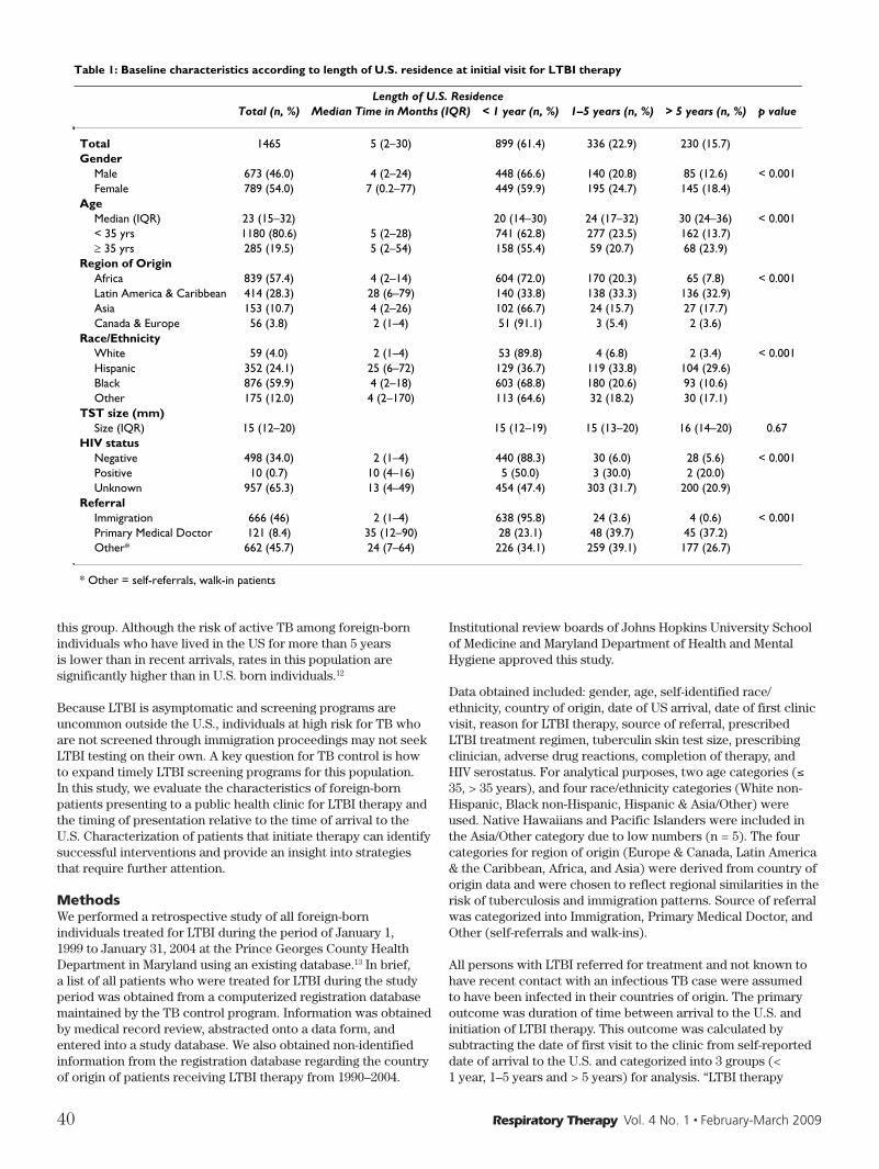
40 Respiratory Therapy Vol. 4 No. 1 February-March 2009
this group. Although the risk of active TB among foreign-born individuals who have lived in the US for more than 5 years is lower than in recent arrivals, rates in this population are significantly higher than in U.S. born individuals.12
Because LTBI is asymptomatic and screening programs are uncommon outside the U.S., individuals at high risk for TB who are not screened through immigration proceedings may not seek LTBI testing on their own. A key question for TB control is how to expand timely LTBI screening programs for this population. In this study, we evaluate the characteristics of foreign-born patients presenting to a public health clinic for LTBI therapy and the timing of presentation relative to the time of arrival to the U.S. Characterization of patients that initiate therapy can identify successful interventions and provide an insight into strategies that require further attention.
MethodsWe performed a retrospective study of all foreign-born individuals treated for LTBI during the period of January 1, 1999 to January 31, 2004 at the Prince Georges County Health Department in Maryland using an existing database.13 In brief, a list of all patients who were treated for LTBI during the study period was obtained from a computerized registration database maintained by the TB control program. Information was obtained by medical record review, abstracted onto a data form, and entered into a study database. We also obtained non-identified information from the registration database regarding the country of origin of patients receiving LTBI therapy from 1990–2004.
Institutional review boards of Johns Hopkins University School of Medicine and Maryland Department of Health and Mental Hygiene approved this study.
Data obtained included: gender, age, self-identified race/ethnicity, country of origin, date of US arrival, date of first clinic visit, reason for LTBI therapy, source of referral, prescribed LTBI treatment regimen, tuberculin skin test size, prescribing clinician, adverse drug reactions, completion of therapy, and HIV serostatus. For analytical purposes, two age categories (≤ 35, > 35 years), and four race/ethnicity categories (White non-Hispanic, Black non-Hispanic, Hispanic & Asia/Other) were used. Native Hawaiians and Pacific Islanders were included in the Asia/Other category due to low numbers (n = 5). The four categories for region of origin (Europe & Canada, Latin America & the Caribbean, Africa, and Asia) were derived from country of origin data and were chosen to reflect regional similarities in the risk of tuberculosis and immigration patterns. Source of referral was categorized into Immigration, Primary Medical Doctor, and Other (self-referrals and walk-ins).
All persons with LTBI referred for treatment and not known to have recent contact with an infectious TB case were assumed to have been infected in their countries of origin. The primary outcome was duration of time between arrival to the U.S. and initiation of LTBI therapy. This outcome was calculated by subtracting the date of first visit to the clinic from self-reported date of arrival to the U.S. and categorized into 3 groups (< 1 year, 1–5 years and > 5 years) for analysis. “LTBI therapy
Table 1: Baseline characteristics according to length of U.S. residence at initial visit for LTBI therapy
Length of U.S. ResidenceTotal (n, %) Median Time in Months (IQR) < 1 year (n, %) 1–5 years (n, %) > 5 years (n, %) p value
Total 1465 5 (2–30) 899 (61.4) 336 (22.9) 230 (15.7)Gender
Male 673 (46.0) 4 (2–24) 448 (66.6) 140 (20.8) 85 (12.6) < 0.001Female 789 (54.0) 7 (0.2–77) 449 (59.9) 195 (24.7) 145 (18.4)
AgeMedian (IQR) 23 (15–32) 20 (14–30) 24 (17–32) 30 (24–36) < 0.001< 35 yrs 1180 (80.6) 5 (2–28) 741 (62.8) 277 (23.5) 162 (13.7) 35 yrs 285 (19.5) 5 (2–54) 158 (55.4) 59 (20.7) 68 (23.9)
Region of OriginAfrica 839 (57.4) 4 (2–14) 604 (72.0) 170 (20.3) 65 (7.8) < 0.001Latin America & Caribbean 414 (28.3) 28 (6–79) 140 (33.8) 138 (33.3) 136 (32.9)Asia 153 (10.7) 4 (2–26) 102 (66.7) 24 (15.7) 27 (17.7)Canada & Europe 56 (3.8) 2 (1–4) 51 (91.1) 3 (5.4) 2 (3.6)
Race/EthnicityWhite 59 (4.0) 2 (1–4) 53 (89.8) 4 (6.8) 2 (3.4) < 0.001Hispanic 352 (24.1) 25 (6–72) 129 (36.7) 119 (33.8) 104 (29.6)Black 876 (59.9) 4 (2–18) 603 (68.8) 180 (20.6) 93 (10.6)Other 175 (12.0) 4 (2–170) 113 (64.6) 32 (18.2) 30 (17.1)
TST size (mm)Size (IQR) 15 (12–20) 15 (12–19) 15 (13–20) 16 (14–20) 0.67
HIV statusNegative 498 (34.0) 2 (1–4) 440 (88.3) 30 (6.0) 28 (5.6) < 0.001Positive 10 (0.7) 10 (4–16) 5 (50.0) 3 (30.0) 2 (20.0)Unknown 957 (65.3) 13 (4–49) 454 (47.4) 303 (31.7) 200 (20.9)
ReferralImmigration 666 (46) 2 (1–4) 638 (95.8) 24 (3.6) 4 (0.6) < 0.001Primary Medical Doctor 121 (8.4) 35 (12–90) 28 (23.1) 48 (39.7) 45 (37.2)Other* 662 (45.7) 24 (7–64) 226 (34.1) 259 (39.1) 177 (26.7)
* Other = self-referrals, walk-in patients
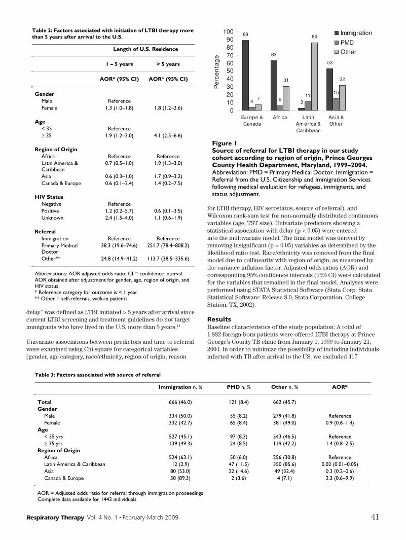
Respiratory Therapy Vol. 4 No. 1 February-March 2009 41
delay” was defined as LTBI initiated > 5 years after arrival since current LTBI screening and treatment guidelines do not target immigrants who have lived in the U.S. more than 5 years.11
Univariate associations between predictors and time to referral were examined using Chi square for categorical variables (gender, age category, race/ethnicity, region of origin, reason
for LTBI therapy, HIV serostatus, source of referral), and Wilcoxon rank-sum test for non-normally distributed continuous variables (age, TST size). Univariate predictors showing a statistical association with delay (p < 0.05) were entered into the multivariate model. The final model was derived by removing insignificant (p > 0.05) variables as determined by the likelihood ratio test. Race/ethnicity was removed from the final model due to collinearity with region of origin, as measured by the variance inflation factor. Adjusted odds ratios (AOR) and corresponding 95% confidence intervals (95% CI) were calculated for the variables that remained in the final model. Analyses were performed using STATA Statistical Software (Stata Corp: Stata Statistical Software: Release 8.0, Stata Corporation, College Station, TX, 2002).
ResultsBaseline characteristics of the study population: A total of 1,882 foreign-born patients were offered LTBI therapy at Prince George’s County TB clinic from January 1, 1999 to January 21, 2004. In order to minimize the possibility of including individuals infected with TB after arrival to the US, we excluded 417
Table 2: Factors associated with initiation of LTBI therapy more than 5 years after arrival to the U.S.
Length of U.S. Residence
1 – 5 years > 5 years
AOR* (95% CI) AOR* (95% CI)
GenderMale ReferenceFemale 1.3 (1.0–1.8) 1.8 (1.2–2.6)
Age< 35 Reference 35 1.9 (1.2–3.0) 4.1 (2.5–6.6)
Region of OriginAfrica Reference ReferenceLatin America & Caribbean
0.7 (0.5–1.0) 1.9 (1.3–3.0)
Asia 0.6 (0.3–1.0) 1.7 (0.9–3.2)Canada & Europe 0.6 (0.1–2.4) 1.4 (0.2–7.5)
HIV StatusNegative ReferencePositive 1.2 (0.2–5.7) 0.6 (0.1–3.5)Unknown 2.4 (1.5–4.0) 1.1 (0.6–1.9)
ReferralImmigration Reference ReferencePrimary Medical Doctor
38.3 (19.6–74.6) 251.7 (78.4–808.2)
Other** 24.8 (14.9–41.2) 113.7 (38.5–335.6)
Abbreviations: AOR adjusted odds ratio, CI = confidence interval AOR obtained after adjustment for gender, age, region of origin, and HIV status* Reference category for outcome is < 1 year** Other = self-referrals, walk-in patients
Source of referral for LTBI therapy in our study cohort according to region of origin, Prince Georges County Health Department, Maryland, 1999–2004Figure 1Source of referral for LTBI therapy in our study cohort according to region of origin, Prince Georges County Health Department, Maryland, 1999–2004. Abbreviation: PMD = Primary Medical Doctor. Immigration = Referral from the U.S. Citizenship and Immigration Services following medical evaluation for refugees, immigrants, and status adjustment.
89
63
3
53
4 611
157
31
86
32
0102030405060708090
100
Europe &Canada
Afr ica LatinAm er ica &Car ibbean
As ia &Other
Per
cen
tag
e
Immigration
PMD
Other
Table 3: Factors associated with source of referral
Immigration n, % PMD n, % Other n, % AOR*
Total 666 (46.0) 121 (8.4) 662 (45.7)Gender
Male 334 (50.0) 55 (8.2) 279 (41.8) ReferenceFemale 332 (42.7) 65 (8.4) 381 (49.0) 0.9 (0.6–1.4)
Age< 35 yrs 527 (45.1) 97 (8.3) 543 (46.5) Reference 35 yrs 139 (49.3) 24 (8.5) 119 (42.2) 1.4 (0.8–2.5)
Region of OriginAfrica 524 (63.1) 50 (6.0) 256 (30.8) ReferenceLatin America & Caribbean 12 (2.9) 47 (11.5) 350 (85.6) 0.02 (0.01–0.05)Asia 80 (53.0) 22 (14.6) 49 (32.4) 0.3 (0.2–0.6)Canada & Europe 50 (89.3) 2 (3.6) 4 (7.1) 2.3 (0.6–9.9)
AOR = Adjusted odds ratio for referral through immigration proceedingsComplete data available for 1443 individuals

42 Respiratory Therapy Vol. 4 No. 1 February-March 2009
individuals who were diagnosed with LTBI during a contact investigation because we could not rule out the possibility of recently acquired infection. In addition, 3 patients had incomplete data and were excluded from the final multivariate model which included 1463 patients. Baseline characteristics of the study population are shown in Table 1. The cohort was relatively young (median age 23 yrs, interquartile range or IQR = 15–32) with a slight female predominance (54%). The vast majority of patients (n = 1406, 96%) originated from one of 3 regions: Africa (n = 839, 57%), Latin America & the Caribbean (n = 414, 28%), and Asia (n = 153, 11%). Relatively few patients were born in Europe & Canada (n = 53, 4%). Racial and ethnic distribution mimicked the distributions of the region of origin. Among patients tested for HIV (n = 508), few were HIV seropositive (n = 10, 2%). Of the patients with unknown HIV status (n = 957), one fifth (n = 180, 19%) refused HIV testing, and the remainder (n = 777, 81%) had no documentation of HIV counseling or testing. A greater proportion of patients from Latin America & the Caribbean (92%) or Asia (66%) had an unknown HIV serostatus compared to patients from Africa (54%) or Europe & Canada (30%, p = 0.001). However, refusal of HIV testing was not associated with region of origin (Chi square, p = 0.96).
Time to initiation of LTBI therapy after arrival to the US: Table 1 shows the period of residence in the US in years at initiation of LTBI therapy according to baseline patient characteristics. The median time from arrival to the U.S. until LTBI therapy was 5 months (IQR range 2–30 months). In accordance to guidelines targeting recent arrivals to the U.S., the majority of patients received LTBI therapy within 5 years of arrival to the US (n = 1235, 84%); 16% of the patients had been in the U.S. for over 5 years prior to receiving LTBI therapy (n = 230).
Univariate analysis identified the following risk factors for initiation of LTBI therapy more than five years after arrival to the US: age, gender, region of origin, race/ethnicity, HIV status, and source of referral (Table 1). In multivariate analysis adjusting for gender, age, region of origin, HIV serostatus, and source of referral we identified source of referral as the major predictor for delays in LTBI therapy (Table 2). Patients who were not
referred for LTBI therapy through immigration proceedings had a very high risk of receiving LTBI therapy more than 5 years after arrival to the U.S. More than one third of patients referred for LTBI therapy by their primary medical doctor (PMD) received therapy 5 years after arrival to the U.S. (AOR = 251.7, 95% CI 78.4–808.2), and over one fourth of patients who self-referred or walked-in to the TB clinic received LTBI therapy 5 years after arrival (AOR = 113.7, 95% CI 38.5–335.6). Other risk factors for delayed LTBI therapy included female gender (AOR = 1.80, 95% C.I. 1.24–2.60), age > 35 years old (AOR = 4.11, 95% C.I. 2.54–6.64), and originating from Latin America and the Caribbean (AOR = 1.95, 95% C.I. 1.28–2.95)
Given the strong association between timing of LTBI therapy and source of referral, we stratified source of referral by region of origin, age, and gender. Figure 1 shows that more than half of the patients from Africa (63%), Asia (53%), and Europe and Canada (89%) were referred through immigration proceeding related to applications for refugees, immigrants, and status adjustment, compared to only 3% of patients from Latin America and the Caribbean. Eleven percent of patients from Latin America & the Caribbean were referred by their primary medical doctor (PMD), but over 80% of patients from this region self-referred or walked-in to the clinic. Univariate analysis showed that fewer women than men (43% vs. 50%, Chi square p = 0.016) were referred to LTBI therapy by immigration services, but there was no significant association between age and source of referral (Chi square p = 0.41). Multivariate analysis adjusting for age, gender, and region of origin showed that region of origin was the only factor independently associated with referral by immigration proceedings (Table 3).
Regional distribution and source of referral for patients treated for LTBI at Prince George’s County Health Department: In order to determine whether the regional differences in time to initiation of LTBI therapy after arrival to the US could be due to a cohort effect, we obtained non-identified information regarding region of origin from the computerized registration database for all foreign-born individuals treated for LTBI from 1990–2004. Figure 2 shows the proportion of immigrants from Asia, Africa, Latin America & the Caribbean, and Europe & Canada who
Regional Distribution of Foreign-born Individuals Receiving LTBI Therapy at Prince Georges County Health Department, Mar-yland, 1990–2004Figure 2Regional Distribution of Foreign-born Individuals Receiving LTBI Therapy at Prince Georges County Health Department, Maryland, 1990–2004.
1990
1990
1990
1990
1998
1998
1998
1998
2003
2003
2003
2003
0
0.1
0.2
0.3
0.4
0.5
0.6
0.7
0.8
0.9
1
Europe &Canada
Africa Latin America &Caribbean
Asia & Other
Pro
po
rtio
n o
f p
atie
nts
Respiratory Therapy Vol. 4 No. 1 February-March 2009 43
received LTBI therapy at Prince Georges County since 1990. These data suggest that immigration to Prince Georges County from all regions began more than 5 years prior to our dates of inclusion in our database (1998–2004).
DiscussionIn this retrospective study of 1,465 foreign-born individuals receiving LTBI therapy at a public TB clinic, we found that referral through immigration proceedings was the strongest predictor of timely initiation of LTBI therapy. In fact, 99.4% of patients referred by immigration proceedings received LTBI therapy within 5 years of arrival to the U.S. as recommended by current guidelines.11 These findings are not surprising given the close link between TB control programs and immigration proceedings. However, a significant proportion of foreign-born individuals in the US never seeks entry visas and may not have access to immigration-based LTBI screening programs. It is estimated that over 25% of immigrants currently living in the US are undocumented and are not in a position to apply for permanent residency status.14
In our study, over 50% of patients were not referred through immigration proceedings. These patients were at high risk for receiving LTBI therapy more than 5 years after arrival to the US. This finding is concerning because guidelines published in 2000 by the Centers of Disease Control and Prevention (CDC) and the American Thoracic society recommend testing and treating only foreign-born persons who have been in the U.S. for 5 years or less.11 However, immigrants have a high risk of TB even after living in the US for more than 5 years and would likely benefit from LTBI therapy. In 2004, almost one quarter (24%) of all cases of TB in the U.S. occurred among immigrants who had arrived to the U.S. more than 5 years previously.12 Over 50% of TB cases in Mexican-born individuals occurs after prolonged (> 5 years) duration of residence in the U.S.15 Furthermore, DNA fingerprinting analysis of M. tuberculosis isolated from TB cases in foreign-born patients shows nonclustering, suggesting that most cases are due to reactivation of infection acquired in their country of origin.16,17 These data suggest that timely diagnosis of LTBI has the potential to prevent most cases of active TB disease among immigrants who comply with therapy.18 Adherence to LTBI therapy is almost twice as high among individuals from Latin America, the Caribbean and Asia compared to US-born individuals.13
Other factors associated with delays in LTBI therapy included older age, female gender, and originating from Latin America and the Caribbean. Delays in adults older than 35 years might reflect previous guidelines which did not recommend LTBI therapy for patients older than 35 years, discouraging LTBI testing in this age group.19 However, this finding is subject to bias or cohort effect since younger immigrants are less likely to have been in the U.S. for > 5 years. Our dataset did not allow us to discern why females may be at higher risk for delays in LTBI therapy, though gender inequalities have been documented for other preventive services. Although fewer women were referred by immigration proceedings, gender was not independently associated with referral mode. Almost one third of patients from Latin America and the Caribbean received LTBI therapy more than 5 years after arrival to the US These findings are concerning given that in 2005 69% of all TB cases among foreign-born people in the U.S. occurred among individuals from Latin America and the Caribbean.20 As in other immigrant groups, timing of LTBI therapy in this population was mostly influenced by referral
mode since only 3% of patients from Latin America and the Caribbean were referred through immigration services. However, our data did not allow us to explore other independent risk factors associated with delays in LTBI therapy in this group.
Because our data were limited to patients who presented to the clinic for LTBI therapy, we could not evaluate the characteristics of immigrants who never access LTBI screening or therapy. Nonetheless, our findings provide a basis for future studies exploring other strategies for timely LTBI screening and therapy in this crucial population for TB control. Over 50% of the patients in our cohort were self-referred or referred by their PMD, highlighting the importance of developing LTBI programs beyond immigration service proceedings. Self-referrals and walk-ins were an important method of entry to the clinic, suggesting the increased awareness of public TB services at the community level may facilitate access to LTBI screening and therapy. Research is needed to identify factors associated with self-referral, such as personal acquaintances (word-of-mouth), educational programs, work-related requirements, or informational brochures. Raising awareness of LTBI screening among the medical community may also enhance timely referral for therapy in high risk populations. However, delayed referrals by primary medical providers may reflect poor access to general medical care in immigrant populations. Our data suggests that expansion of LTBI screening and treatment to include foreign-born individuals who have lived in the US for more than 5 years may be one way to enhance TB control programs.
There are several limitations to our study. In estimating delays in initiation of LTBI therapy, we assumed that most LTBI infection in immigrants was acquired in the country of origin. However, epidemiologic clustering of active cases among immigrants from the same region may lead to ongoing exposure to TB after arrival in the U.S. We removed individuals identified through contact investigations to minimize the chance of including patients infected with TB after arrival to the US. Furthermore, despite the relatively large size of our patient population, our study was limited to one county TB control program in Maryland. The generalizability of our results to the total U.S. immigrant population depends on local immigration patterns and health care delivery strategies for foreign-born individuals.
ConclusionThe CDC is considering several strategies to address the high rate of TB among foreign born individuals in the US, such as revising medical screening for applicants for US immigration, and working with international organizations to reduce TB in the countries of origin.1 These strategies should decrease the importation of new TB cases from other countries, but do not address the risk of TB in foreign-born individuals who already reside in the U.S., especially among undocumented immigrants who are not routinely screened at entry to the country. Our study shows that immigrants who are not referred by immigration proceedings have a high risk of presenting for LTBI therapy more than 5 years after arrival to the US. These data highlight the need for strategies beyond immigration-linked programs to identify and overcome missed opportunities for LTBI therapy among high risk foreign-born individuals, including those who have lived the US for > 5 years.
References1 CDC: Trends in Tuberculosis Incidence- United States, 2006.
MMWR 2007, 56(11):245-250.

44 Respiratory Therapy Vol. 4 No. 1 February-March 2009
2 Institute of Medicine. Ending Neglect: The Elimination of Tuberculosis in the United States. National Academy Press 2000.
3 CDC: Recommendations for Prevention and Control of Tuberculosis Among Foreign-Born Persons. MMWR 1998, 47(no RR-16):2-29.
4 Jasmer RM, Nahid P, Hopewell PC: Clinical practice. Latent tuberculosis infection. N Engl J Med 2002, 347(23):1860-1866.
5 DeRiemer K, Chin DP, Schecter GF, Reingold AL: Tuberculosis among immigrants and refugees. Arch Intern Med 1998, 158(7):753-760.
6 Code of Federal Regulations. Title 42- Public Health. [http://www.access.gpo.gov/nara/cfr/waisidx_03/42cfr34_03.html]
7 Technical Instructions for Civil Surgeons. [http:// www.cdc.gov/Ncidod/dq/technica.htm]. CDC
8 McKenna MT, McCray E, Onorato I: The epidemiology of tuberculosis among foreign-born persons in the United States, 1986 to 1993. N Engl J Med 1995, 332(16):1071-1076.
9 Asch S, Leake B, Anderson R, Gelberg L: Why do symptomatic patients delay obtaining care for tuberculosis? Am J Respir Crit Care Med 1998, 157(4 Pt 1):1244-1248.
10 Sciortino S, Mohle-Boetani J, Royce SE, Will D, Chin DP: B notifications and the detection of tuberculosis among foreign-born recent arrivals in California. Int J Tuberc Lung Dis 1999, 3(9):778-785.
11 Targeted tuberculin testing and treatment of latent tuberculosis infection. This official statement of the American Thoracic Society was adopted by the ATS Board of Directors, July 1999. This is a Joint Statement of the American Thoracic Society (ATS) and the Centers for Disease Control and Prevention (CDC). This statement was endorsed by the Council of the Infectious Diseases Society of America. (IDSA), September 1999, and the sections of this statement. Am J Respir Crit Care Med 2000, 161(4 Pt 2):S221-47.
12 Cain KP, Haley CA, Armstrong LR, Garman KN, Wells CD, Iademarco MF, Castro KG, Laserson KF: Tuberculosis among Foreign-born Persons in the United States: Achieving Tuberculosis Elimination. Am J Respir Crit Care Med 2007, 175(1):75-79.
13 Page KR, Sifakis F, Montes de Oca R, Cronin WA, Doherty MC, Federline L, Bur S, Walsh T, Karney W, Milman J, Baruch N, Adelakun A, Dorman SE: Improved adherence and less toxicity with rifampin vs isoniazid for treatment of latent tuberculosis: a retrospective study. Arch Intern Med 2006, 166(17):1863-1870.
14 Undocumented Immigrants, Facts and Figures. [http://www.urban.org/publications/1000587.html]. Urban Institute
15 Talbot EA, Moore M, McCray E, Binkin NJ: Tuberculosis among foreign-born persons in the United States, 1993-1998. Jama 2000, 284(22):2894-2900.
16 Geng E, Kreiswirth B, Driver C, Li J, Burzynski J, DellaLatta P, LaPaz A, Schluger NW: Changes in the transmission of tuberculosis in New York City from 1990 to 1999. N Engl J Med 2002, 346(19):1453-1458.
17 Cronin WA, Golub JE, Lathan MJ, Mukasa LN, Hooper N, Razeq JH, Baruch NG, Mulcahy D, Benjamin WH, Magder LS, Strickland GT, Bishai WR: Molecular epidemiology of tuberculosis in a low- to moderate-incidence state: are contact investigations enough? Emerg Infect Dis 2002, 8(11):1271-1279.
18 Sterling TR, Bethel J, Goldberg S, Weinfurter P, Yun L, Horsburgh CR: The scope and impact of treatment of latent tuberculosis infection in the United States and Canada. Am J Respir Crit Care Med 2006, 173(8):927-931.
19 ATS, ALA, CDC: Preventive therapy of tuberculosis infection. Am J Respir Crit Care Med 1974, 110:371-373.
20 CDC: Reported Tuberculosis in the United States, 2005. Atlanta, GA: US Department of Health and Human Services. 2006.

Respiratory Therapy Vol. 4 No. 1 February-March 2009 45
Assessment of the Alveolar Volume When Sampling Exhaled Gas at Different Expired Volumes in the Single Breath Diffusion TestRenato Prediletto, Edo Fornai, Giosuè Catapano, Cristina Carli
AbstractBackground: alveolar volume measured according to the American Thoracic Society-European Respiratory Society (ATS-ERS) guidelines during the single breath diffusion test can be underestimated when there is maldistribution of ventilation. Therefore, the alveolar volume calculated by taking into account the ATS-ERS guidelines was compared to the alveolar volume measured from sequentiallly collected samples of the expired volume in two groups of individuals: COPD patients and healthy individuals. The aim of this study was to investigate the effects of the maldistribution of ventilation on the real estimate of alveolar volume and to evaluate some indicators suggestive of the presence of maldistribution of ventilation.
Methods: thirty healthy individuals and fifty patients with moderate-severe COPD were studied. The alveolar volume was measured either according to the ATS-ERS guidelines or considering the whole expired volume subdivided into five quintiles. An index reflecting the non-uniformity of the distribution of ventilation was then derived (DeltaVA/VE).
Results: significant differences were found when comparing the two measurements and the alveolar volume by quintiles appeared to have increased progressively towards residual volume in healthy individuals and much more in COPD patients. Therefore, DeltaVA/VE resulted in an abnormal increase in COPD.
Conclusions: the results of our study suggest that the alveolar volume during the single breath diffusion test should be measured through the collection of a sample of expired volume which could be more representative of the overall gas composition, especially in the presence of uneven distribution of
ventilation. Further studies aimed at clarifying the final effects of this way of calculating the alveolar volume on the measure of DLCO are needed. DeltaVA/VE is an index that can help assess the severity of inhomogeneity in COPD patients.
BackgroundThere is evidence that the gases inspired into the alveolar regions are not well mixed and that the alveolar units fill and empty sequentially.1-4 Importantly, in the presence of prevalent lung diseases such as COPD, this process may be exaggerated because of the increased time constant of lung units.5,6 As a matter of fact, when airways are narrowed by inflammatory cells and mucus, the distribution of the inspired gas in the alveoli becomes progressively impaired. This may result in marked differences in gas composition within the lungs, as well as in inhomogeneous patterns of filling and emptying.7,8 The different profile of emptying and filling of the lung units causes discrepancy in their gas tracer composition, thus creating regional differences within lung units for tracer inert insoluble gases such as helium or methane.9-10 Indeed, if there is unevenness in the distribution of a single inspiration in the dilution of the tracer gas and incomplete equilibration within respiratory units, as in the presence of airflow obstruction, the pattern of reappearance of that gas in a single expiration to residual volume will be consequently influenced.11,12 This may account for the extreme difficulty in obtaining a sample that is representative of the overall gas composition using the most common laboratory tests.13 It is well recognized that the single breath diffusion test14-16 requires a measurement of the alveolar volume. According to recent ATS-ERS guidelines, such volume is based on the sampling of 750-1000 ml of expired volume after washout of the dead spaces when the tracer gases, such as helium or methane, are promptly inhaled and expected to be diluted in well ventilated units during the manoeuvre of inspiratory vital capacity in the course of the test.17,18 Whenever severe airflow occurs, the gas mixes less effectively and, therefore, sampling in the initial expired concentration of the gas tracer is not always correct as it is likely to overestimate the real value of alveolar gas concentrations and consequently underestimate that of the alveolar volume. This discrepancy could lead to substantial differences when calculating DLCO in COPD patients.17,19
The authors are with the Institute of Clinical Physiology, National Research Council of Italy, Pisa, Italy. The authors are grateful to Mrs Manuella Walker for the English language revision of the manuscript, Mr. Luca Serasini for his valuable help in the graphic editing of the manuscript, Mr Claudio Michelassi for his assistance in the statistical revision and Prof. Carlo Giuntini for his valuable comments and suggestions. Reprinted from BioMed Central, BMC Pulmonary Medicine, © 2007 Prediletto et al, licensee BioMed Central Ltd. This is an open access article distributed under the terms of the Creative Commons Attribution License.

46 Respiratory Therapy Vol. 4 No. 1 February-March 2009
We hypothesize that the alveolar volume measured according to the ATS-ERS method is very different from that calculated considering subsequent phases of the expired volume in those
areas where the gas composition is different owing to the fact that the slow-emptying units predominate. In the course of the single breath diffusion test, with the aid of rapid response analyzers it is now possible to follow exhalation to the residual volume after breathhold and to measure in selected points of the exhalation process the instantaneous expired inert gas fractions which could enter into the calculations of the alveolar volume. In this way, we compared the measurements of the standard alveolar volume obtained following the ATSERS recommendations (VAst) to those derived by considering the whole expirate of the same single breath diffusion test, minus the dead spaces, divided into five quintiles and considering the related expired inert gas fractions (VAq) in each quintile. This procedure allowed us to evaluate whether there existed any large discrepancy between the two measurements of the alveolar volume in those cases like COPD, where the process of sequential emptying of different alveolar regions may be excessive. This comparison was made both in healthy individuals and in COPD patients.
MethodsThe study included 30 healthy individuals and 50 patients affected by COPD. The healthy subjects had no history of smoking, nor respiratory symptoms consistent with the diagnosis of COPD nor other pulmonary disease. The healthy subjects were receiving no respiratory medication nor any other medication which could affect the respiratory function. The patients with COPD fulfilled the diagnostic criteria of the Global Initiative for Chronic Obstructive Lung Disease guidelines.20 All patients with COPD were smokers and were recruited from the pulmonary disease unit of our Institution. At the time of the study all patients with COPD were stable. Before the testing session, all subjects were asked to withhold taking inhaled short-acting ß-agonist and/or anticholinergic agents. Patients had no other cardiopulmonary disease, and had experienced no upper respiratory tract infection during the previous 4 weeks. Patients with COPD were classified according to the results of spirometry
Table 1. Demographic and functional characteristics of the study population
“healthy” individuals
(n=30)
COPD patients (n=50)
Signifi cance
Age years 65±6 68±6 ns
Weight Kg 71±10.48 75±12.53 ns
Height cm 162±9.58 168±6.44 p<.0038
BMI Kg/m2 27±4 27±6 ns
Pack-years n 0 43.4±17.5 NA*
Males % 47 92 NA*
Hemoglobin g/dl 13.97±1.07 14.55±1.07 p<.02
FVC %pred 120±13 84±17 p<.0001
FEV1 %pred 116±15 45±12 p<.0001
FEV1/VC %pred 97± 7 53±13 p<.0001
VC %pred 119±13 87±16 p<.0001
RV %pred 102±22 145±37 p<.0001
TLC %pred 106±11 106±14 ns
RV/TLC %pred 95±15 131±22 p<.0001
DLCO %pred 95±17 64±22 p<.0001
VA %pred 93±10 80±13 p<.0001
DL/VA %pred 97±14 71±26 p<.0001
Demographic and functional characteristics of the study population. Data are expressed as mean ± standard deviation. Signifi cant differences appear at the 0.05 level. Abbreviations and references for the predicted formulas of each variable can be seen in the text. * Not applicable
Table 2. Coeffi cients of repeatability (defi ned as 2.77 x SD) for VAst (standard alveolar volume obtained following the ERS-ATS recommendations) and VAq (alveolar volume measured according to the method of subdividing the whole expirate into quintiles) between two consecutive tests for healthy individuals and patients with COPD using the method of Bland-Altman (24) and comparison of the repeatibilities between groups (healthy individuals and COPD patients).
healthy individuals(n=30)
COPD patients(n=50)
°p< *p< **p<
VAst ± 144 ml .073 ± 165 ml .011 .753
VAq (fi rst) ± 78 ml .481 ± 58 ml .198 .999
VAq (2nd) ± 71 ml .745 ± 86 ml .022 .843
VAq (3rd) ± 80 ml .825 ± 129 ml .161 .992
VAq(4th) ± 95 ml .356 ± 78 ml .256 .149
VAq (5th) ± 80 ml .740 ± 94 ml .507 .787
Coeffi cients of repeatability (defi ned as 2.77 x SD) for VAst (standard alveolar volume obtained following the ERS-ATS recommendations) and VAq (alveolar volume measured according to the method of subdividing the whole expirate into quintiles) for healthy individuals and patients with COPD using the Bland-Altman method (24), in order to examine the variation between two consecutive tests. The level of statistical signifi cance in terms of probability (p) has been reported. It is evident that the variation in the calculated alveolar volume between two tests appeared statistically signifi cant for the VAst of COPD patients. In the healthy individuals it resulted approximately close to the level of signifi cance. On the contrary, for the alveolar volumes calculated by the quintile method, the variation between two consecutive tests was only signifi cant in the 2nd quintile of the COPD patients. In addition, no differences were observed in the comparison in repeatibilities between groups at each VAq (and VAst). level of signifi cance related to the comparison between coeffi cients of repeatibilities within two measurements for healthy individuals (°p<) and COPD patients (*p<); level of signifi cance of the repeatibilities by the analysis of variance between groups (healthy individuals and COPD patients) (** p<)

Respiratory Therapy Vol. 4 No. 1 February-March 2009 47
as having a moderate to severe obstruction. Informed written consent was obtained for the study protocol according to the policy of our Internal Review Board. Spirometry was performed as stated by the ATS-ERS recommendations.21 Airflow limitation was characterized by FEV1 and FEV1/VC respectively below 70% and 88% of the normal value predicted after inhalation of a bronchodilator. Bronchodilator reversibility was defined as an increase of 12% of the baseline value and 200mL respectively for either FEV1 or FVC above the prebronchodilator baseline, 30 minutes after inhalation of 400µg of salbutamol.22 All values were expressed as percentage of reference values.23 In order to assess the pulmonary function, we measured static lung volumes which were expressed as percentage of reference values.23 The single breath diffusing capacity for carbon monoxide was determined using a fully computerized spirometric system (Comprehensive Pulmonary Laboratories Collins Medical Ferraris, USA-England) which was the same instrument employed for spirometry and lung volumes. The test was performed at sea level. The system was equipped with rapid infrared analyzer devices for carbon monoxide (CO) and methane (CH4). This gas analyser system provided continuous tracings of CO and tracer gas concentration during the test. DLCO was measured according to the ATS-ERS guidelines.16-18 Each subject performed the single breath CO test in sitting position. The patients who were current smokers stopped smoking at least 24 hours before the tests. The interval between tests was only 4 minutes for healthy individuals and over 10 minutes for severe COPD patients, according to the ATS-ERS guidelines. The measured DLCO was adjusted for
hemoglobin concentration. The alveolar volume was measured by dilution in the lung of 0.3% of CH4 present in the inspiratory bolus of the breath test, in the course of inspiratory vital capacity. The remainder of the test gas mixture included 0.3% of CO, 21% of oxygen and balance nitrogen.
In accordance with the purpose of this study the effective alveolar volume was calculated by two different methods. The first method directly measures tracer gas reduction during breathholding time according to the ATS-ERS guidelines and was defined VAst (standard alveolar volume) obtained following the ERS-ATS recommendations according to the following formula:
VAst=[ FICH4/FACH4(750mL)] x [ Vinsp – (Vd instrumental+VD anat)]
where FICH4=methane concentration in the inspired gas, and FACH4 (750 mL)=methane concentration in the alveolar sample collected for 750 mL after having discarded the instrumental and anatomical dead spaces; Vinsp=inhaled volume; VD=instrumental dead space and VD anat=anatomical dead space. Sampling of the tracer gas CH4 was executed at the mouth level of the patient in real time and in correspondence of the measurement of the inspiratory and mean expiratory volumes. The second method, which allowed us to derive the alveolar volume subdividing the whole expirate into quintiles (VAq), uses measurements made during the same manoeuver. The difference is that the total expired space is divided into
Figure 1
Figure 2 Figure 3

48 Respiratory Therapy Vol. 4 No. 1 February-March 2009
quintiles. In these quintiles the mean concentrations of CH4 were promptly read and retained for the calculations during exhalation from TLC to RV, according to the following formula:
VAq=[(FICH4/FACH4(quintile)) x (Vinsp)] where:
FACH4 (quintile)=the methane concentration in the expired volume of that quintile; Vinsp=inhaled volume.
Two examples of the different ways of deriving the alveolar volume from the single breath CO manoeuver have been reported in Figure 1. From the analysis of VAq (alveolar volume measured according to the method of subdividing the whole expirate into quintiles) in our sample we derived a parameter, defined as DeltaVA/VE, which represents the changes in percentage of the alveolar volume for each litre of expired volume exhaled. This parameter was compared between healthy individuals and COPD patients.
Statistical analysis: Group data are expressed as mean and standard deviation. The groups and functional parameters in Table 1 were compared by the unpaired Student t-test. The mean values of VAst (standard alveolar volume obtained following the ERS-ATS recommendations) and VAq (alveolar volume measured according to the method of subdividing the whole expirate into quintiles) of two consecutive tests were compared by the paired Student t-test to assess their statistical differences. Differences in mean VAst and VAq between two consecutive tests and their respective coefficients of repeatibility (defined as 2.77 x SD) were derived from healthy subjects and patients
with COPD using the Bland Altman method.24 Fisher’s variance test was used to compare the repeatibilities between groups at each VAq and VAst and the level of significance was reported in Table 2. Frequency distribution of changes in percentage of DeltaVA/VE for each litre of expired volume exhaled was employed in our two samples and was graphed as box-whisker plot diagram where the horizontal lines in the box represent the 50th percentile median, the borders of the box the 25th and 75th percentiles of distribution and the whiskers the 10th and 90th percentiles.
ResultsSubjects’ characteristicsDemographic characteristics and parameters of lung function in healthy subjects and COPD patients have been reported in Table 1. Our COPD patients were different only in terms of height when compared to healthy individuals who had a history of smoking; males were more present in the COPD group when compared to healthy individuals (92% vs 47% respectively and only 8% of 50 COPD patients were females, whereas the control group was close to half female, indicating a disproportion between males and females in this cohort of patients and control group which surely indicates a gender difference in terms of smoking habit. Functional parameters showed signs of severe airflow obstruction and of hyperinflation in COPD patients. DLCO single breath was significantly reduced in COPD patients, as well as its ratio to alveolar volume. With regard to patient performance of the single breath test, 3 healthy individuals and 7 COPD patients were discarded because their inspiratory time during the manoeuver was over 4 seconds, while 9 healthy subjects and 34 COPD patients were discarded because their breath-hold time was too long. The dead space washout volumes (including instrumental, filtering and anatomic dead space) resulted 563±134 ml (range 150-750 ml) and 447±134 (range 340-750 ml) respectively in normals and in patients with COPD. The sample collection volumes were 561±195 ml (range 240-920 ml) and 490±140 ml (range 240-880 ml) respectively in healthy individuals and in patients with COPD.
Calculated alveolar volume, choice of quintiles and repeatibility between tests : The ideal approach would be to compute automatically, point by point, as a continuum, the FICH4/FECH4
Figure 4
Figure 5

Respiratory Therapy Vol. 4 No. 1 February-March 2009 49
ratio during the whole expiration in the course of the single-breath CO test. In order to evaluate whether the sampling of five quintiles was sufficient, we subdivided the whole expired volume into a series of four, five, six and ten exact portions. The alveolar volume calculated using four portions was significantly different when compared to that obtained using five, six or ten portions in both healthy individuals and in COPD patients. At the same time, no differences were observed when the calculated alveolar volumes, obtained subdividing the whole expirate into six portions, were compared to five or ten exact portions both in healthy individuals and in COPD patients. As a result of this prior analysis, the method of sampling five quintiles appeared sufficiently good, more precise than the use of four portions, not different from the use of six or ten portions. Therefore, the method was considered suitable for the purpose of our study and was ultimately chosen and compared to the ERS-ATS method of measuring the alveolar volume. The repeatability of VAst (standard alveolar volume obtained following the ERS-ATS recommendations) and of VAq (alveolar volume measured according to the method of subdividing the whole expirate into quintiles) was tested both in healthy individuals and in patients with COPD and has been reported in Table 2.
It is evident that variation in the calculated alveolar volume between two tests appeared statistically significant for the VAst of COPD patients. In the healthy individuals it resulted approximately close to the level of significance. On the contrary, for the alveolar volume calculated by the quintile method, the variation was only statistically significant for the 2nd quintile of the COPD patients. No other significant variations were observed in the healthy individuals nor in the other quintiles of the COPD patients. In addition, the healthy individuals and COPD patients were not different in repeatability (Table 2). The results of the comparison between the two ways of measuring the alveolar volume have been reported in Figures 2 and 3, for healthy individuals and COPD patients respectively. In the healthy individuals the values of the alveolar volume were not significantly different between the two methods of assessment in the first 20 and 40% of exhaled volume. Significant differences were instead detected in the following portions of expired volume when exhaled to residual volume. In the patients with airflow obstruction there were remarkable differences between the two methods of measuring the alveolar volume at the beginning of exhalation and were present throughout the emptying phase to residual volume. The alveolar volume measured in the last quintile appeared significantly greater than that measured in the previous quintiles, thus suggesting an effect related to the sequential emptying of lung units. As reported in Figure 4, it is evident that the alveolar volume does not exhibit any remarkable changes when related to the expired volume in healthy individuals at variance with those of the COPD patients. This result suggests a progressive increase of alveolar volume along with the process of emptying of the lung units in patients with disease. The frequency distribution of DeltaVA/VE (changes in percentage of the alveolar volume for each litre of expired volume exhaled) at different lung volumes was compared between healthy individuals and COPD patients in the box-plot graph of Figure 5. Ninety percent of COPD patients showed a progressive increase of the alveolar volume along with exhalation which was approximately more than 20% with respect to normals. Thus, it appears that the changes of alveolar volume during exhalation are quite relevant in COPD patients and that uneven emptying is necessary for these results, but not sufficient; uneven dilution ratios are also necessary.
DiscussionThe main findings of this study are as follows: 1. the measure of the alveolar volume is different depending on the point where sampling for evaluation of the alveolar concentration of inert tracer gas methane is done in the course of exhalation of the single breath diffusion test; 2. the alveolar volume measured by the quintile method shows a progressive increase from total lung capacity to residual volume and appears significantly different when compared with that measured according to the ATS-ERS guidelines; 3. its size increases much more in COPD patients than in healthy individuals from the beginning to the end of the exhalation; 4. changes of the mean alveolar volume per litre of the expired volume exhaled, expressed as DeltaVA/VE, are significantly and remarkably greater in COPD patients with severe airflow obstruction than in healthy individuals; 5. DeltaVA/VE represents a parameter that is influenced by the effect of non-uniform distribution of convective ventilation as well as by the increased time-constants of the emptying of lung units in diseased lungs. It is well documented that the single breath diffusion test may markedly increase or slightly change upon the effect of variation in lung volume.14,15,25-29
Although this study did not provide any conclusive information on how the final DLCO can be affected by the changes of lung volume as well as by size and precise estimate of the alveolar volume, in our COPD patients it seems to increase on the average by 1mmol/min/mmHg for a total increase of 2.5 litres of alveolar volume, as reported in figure 4. A different type of behaviour was observed in the healthy individuals who showed values of DLCO that decreased by 1.5 mmol/min/mmhg when compared to the slight changes of the alveolar volumes.
The purpose of our study was to indicate the weakness of the ATS-ERS method to measure the alveolar volume, which collects the alveolar inert gas concentration at the beginning of exhalation, especially in diseased lungs. In fact at this exact point (on the average a volume of 750 ml) the concentration of the tracer gas is not representative of the mean alveolar gas concentration and thus of the real alveolar volume, since it represents only the behaviour of the faster lung units.11,17 This feature is remarkable in patients with severe airflow obstruction and is less evident in healthy individuals.30 We could argue that in subjects like our healthy individuals, who are non smokers, and whose lungs have a near-normal distribution of ventilation, sampling of the alveolar inert gas CH4 at the beginning of exhalation does not appear much influenced by the effect of differences in the physiological regional dilution during inhalation of the test gas.7,31 Instead, in the case of our patients affected by airflow obstruction and with signs of hyperinflation of their lungs, extreme nonuniformity of ventilation may predominate.
Literature well documents the extent to which the distribution of ventilation becomes progressively more inhomogeneous at high lung volume32 or under the effect of modifications in tidal volumes, flows, posture and in the presence of asymmetrical geometry of lung units as well as in normal lungs.31,33 In this study, no striking differences were detected when all healthy individuals and COPD patients were reclassified according to their significantly higher or lower expired flows and consequently tested to search any relations with the calculated alveolar volumes. This analysis showed only a slight increase of VAst (standard alveolar volume obtained following the ATS-ERS recommendations) when it was associated with a higher flow,

50 Respiratory Therapy Vol. 4 No. 1 February-March 2009
not exhibited by the calculated VAq (alveolar volume measured according to the method of subdividing the whole expirate into quintiles). This result indicates that VAst (standard alveolar volume obtained following the ATS-ERS recommendations) is more sensitive to the different profile of emptying of alveoli when compared to the method of quintiles, which always takes into account its standardization for the whole expired volume of the subject (which is 20% of the whole expirate).
The exaggerated asymmetry of lung units caused by the obstructive airway diseases may ultimately be responsible for the inequality of gas concentration within alveolar gas and, therefore, for the very inhomogeneous dilution of the concentration of the tracer gas methane in the course of the single breath CO inhalation test. In 1978 Ferris et al34 compared the single breath helium dilution alveolar volume and a 7-min rebreathing helium alveolar volume in normals and in COPD patients, and found that the alveolar volume measured according to the two different methods did not underestimate DLCO in the population of normals except for those patients with severe airflow obstruction. In conclusion, the authors suggested that the single breath method may be adequate for normals, but not for COPD, since the variations and fluctuations in the alveolar gas concentrations during exhalation are responsible for an incorrect estimate of the alveolar volume. This led the authors to conclude that more accurate methods for the alveolar volume were required.17,35 Some explanations for the remarkable differences in the values of the alveolar volume when measured at the different intervals of exhalation are well reported in the paper by Yuh T Huang et al.36 These authors provided strong evidence in the evaluation of the volume dependent distribution of DLCO in normals at rest and during exercise; they showed that the intrabreath DLCO during single exhalation in the healthy subjects was non linear and could be described by a polynomial model. The two explanations for such behaviour seem to be in accordance with the results found in our study: one explanation entails the sequential emptying profile of the lung (i.e. the “ first in-last out phenomenon”) which accounts for the difference in the contribution of alveolar gas concentration. Indeed, it is well known that the gas sampled at higher lung volume generally reflects the contribution from the lower part of the lung and of the faster units, which empty earlier and probably have a different regional concentration of tracer gas. The other explanation may reside in the time-constants of the lung units, which are increased in diseased lungs. As a matter of fact, by looking at the methane profile during exhalation of a COPD patient (Figure 1, right side) its steeper decreasing slope from TLC to RV is evident. This increase may primarily suggest that the recovery of the concentration of the inhaled inert gas is progressively diminishing towards residual volume, which may result in an increased time constant of lung units. The heterogeneous profile of inert gas methane during exhalation, amplified in COPD, is ultimately responsible for the differences in the calculations of the alveolar volume and it allowed us to derive the index named DeltaVA/VE (changes in percentage of the alveolar volume for each litre of expired volume exhaled), which seems to reflect the effect of the phenomena described above. In fact, this index DeltaVA/VE, as direct expression of the non-uniform distribution of ventilation, resulted significantly higher—in quantitative terms—in COPD than in healthy individuals (Figure 5). As a matter of fact, we propose this index as a very rapid, non invasive and simple tool that can be obtained routinely in patients to help the pulmonologists evaluate the effect of inhomogeneity of ventilation in the course of the single
breath-hold test. Finally, the changes of the alveolar volume from TLC to RV measured by quintiles were 0.110 litres in the healthy individuals, 1.332 litres in COPD patients (Figures 2 and 3). Instead, when we use the ATS-ERS method to compare the alveolar volume with that derived from the average of the alveolar volume measured in each quintile we find similarity in healthy individuals (VAst 5.10±1.33 litres vs VAq 5.15±1.34 litres, p=ns) but significant differences, as expected, in COPD patients (VAst 4.98±1.04 vs VAq 5.36±1.57 litres, p<0.0001). It follows that the true mean alveolar volume should be that derived from the average of each alveolar volume exhaled in each quintile (Figures 2-3).
ConclusionsIn summary, our study provides additional information on the real estimate of the alveolar volume when different sampling points are used in the course of the single breath diffusion test for the assessment of diffusing capacity. A model was developed which subdivided into 5 parts the total volume of air exhaled after the breath-hold manoeuver was developed. The instantaneous concentration of tracer gas methane was considered in each quintile; the calculation of the alveolar volume was consequently derived and compared with that derived from the traditional method according to the ATS-ERS recommendations. A significant difference was found between these two ways of measuring the alveolar volume, and the results showed significant differences in COPD patients. The conclusion drawn is that sampling at the beginning of exhalation of the single breath-test is not representative of the real mean alveolar gas concentration, especially when an important ventilation/perfusion mismatch is present. A non-uniform distribution of ventilation, coupled with an exaggerated time constant of emptying of lung units, seems ultimately to be the mechanism responsible for the differences in the size of the alveolar volume when measured differently in the course of expiration. An index reflecting this process was identified which appeared useful to assess the degree of non-uniformity of the ventilation distribution. These analyses provide a basis for further study in order to test the effects on DLCO of this way of measuring the alveolar volume from the sampling of the instantaneous tracer gas concentrations at different intervals of exhalation, but also to observe the behaviour of the diffusivity of carbon monoxide at different intervals of exhalation, as a direct consequence of the complex emptying process of lung units in different diseased states.
References1 Tsoukias NM, Dabdub D, Wilson AF, George SC: Effect
of alveolar volume and sequential filling on the diffusing capacity of the lungs: II. Experiment. Respir Physiol 2000; 120:251-271.
2 Tsoukias NM, Wilson AF, George SC. Effect of alveolar volume and sequential filling on the diffusing capacity of the lungs: I. Theory. Respir Physiol 2000; 120:231-249.
3 Meyer M, Hook C, Rieke H, Piiper J: Gas mixing in dog studied by single-breath washout of He and SF6. J Appl Physiol 1983; 55:1795-1802.
4 Cotton DJ, Newth CJ, Portner PM, Nadel JA: Measurement of single-breath CO diffusing capacity by continuous rapid CO analysis in man. J Appl Physiol 1979; 46:1149-1156.
5 Otis AB, McKerrow CM, Bartlett RA, Mead J, Mcilroy MC, Selver-Stone NJ, Radford EP J Mechanical factors in the distribution of pulmonary ventilation. J Appl Physiol 1956; 8: 427-443.

Respiratory Therapy Vol. 4 No. 1 February-March 2009 51
6 Hogg JC, Macklem PT, Thurlbeck WM. Site and nature of airway obstruction in chronic obstructive lung disease. New Engl J Med 1968; 278: 1355-1360.
7 Engel A, Fahri L. Airway closure, closing volume and changes in inert gas mixing. In: Macklem PT and Permutt S, eds. The Lung in the transition between health and disease. New York, NY: Marcel Dekker, 1979; 153-182.
8 Paiva M, Engel LA. The anatomical basis for the sloping N2 plateau. Respir Physiol 1981; 44:325-337.
9 Buist AS, Ross BB. Quantitative analysis of the alveolar plateau in the diagnosis of early airway obstruction. Amer Rev Resp Dis 1973; 108:1078-1087.
10 Cotton DJ, Prabhu MB, Mink JT, Graham BL: Effect of ventilation inhomogeneity on “intrabreath” measurements of diffusing capacity in normal subjects. J Appl Physiol 1993; 75:927-932.
11 Giannella NA, Paoletti P, Fornai E, Giuntini C. Estimates of mean alveolar gas in patients with chronic airways obstruction. Eur Respir J 1989; 2: 451-460.
12 Beaver WL, Lamarra N, Wasserman K. Breath-by-breath measurement of true alveolar gas exchange. J Appl Physiol.: Respirat Environ Exrcise Physiol 1981; 51:1662-1675.
13 Lewis SM: Emptying patterns of the lung studied by multiple-breath N2 washout. J Appl Physiol 1978; 44: 424-430.
14 Ogilvie CM, Forster RE, Blakemore WS, Morton JW: A standardized breath holding technique for the clinical measurement of the diffusing capacity of the lung for carbon monoxide. J Clin Invest 1957; 36:1-17.
15 Ogilvie C. Editorial. The single-breath carbon monoxide transfer test 25 years on: a reappraisal. Thorax 1983; 38:1-9.
16 Cotes JE, Chinn DJ, Quanjer PhH, Roca J, Yernault JC. Standardization of the measurement of transfer factor (diffusing capacity) Report Working Party Standardization of Lung Function Tests, European Community for Steel and Coal. Official Statement of the European Respiratory Society. Eur Respir J 1993; 6, Suppl. 16: 41-52.
17 N. MacIntyre, R. O. Crapo, G. Viegi, D. C. Johnson, C. P. M. van der Grinten, V. Brusasco, F. Burgos, R. Casaburi, A. Coates, P. Enright, P. Gustafsson, J. Hankinson, R. Jensen, R. McKay, M. R. Miller, D. Navajas, O. F. Pedersen, R. Pellegrino, and J. Wanger Standardisation of the single-breath determination of carbon monoxide uptake in the lung. Eur Respir J 2005; 26: 720-735.
18 American Thoracic Society. Single-breath carbon monoxide diffusing capacity (Transfer factor): recommendations for a standard technique; 1995 update. Am J Respir Crit Care Med 1995;152:2185-2198.
19 Chinn DJ, Askew L, Rowley L , Cotes JE: Measurement technique influences the response of transfer factor (Tlco) to salbutamol in patients with airflow obstruction. Eur Respir J 1988; 1: 15-21.
20 Pauwels RA, Buist AS, Calverley PM, Jenkins CR, Hurd SS: GOLD Scientific Committee: Global strategy for the diagnosis, management, and prevention of chronic obstructive pulmonary disease. NHLBI/WHO Global Initiative for Chronic Obstructive Lung Disease (GOLD) Workshop summary. Am J Respir Crit Care Med 2001; 163:1256-1276.
21 Miller MR, Hankinson J, Brusasco V, Burgos F, Casaburi R, Coates A, Crapo R, Enright P, van der Grinten CPM, Gustafsson P, Jensen R, Johnson DC, MacIntyre N, McKay R, Navajas D, Pedersen OF, Pellegrino R, Viegi G, and Ranger J. Standardisation of spirometry. Eur Respir J 2005; 26: 319-338.
22 Dales RE, Spitzer WO, Tousignant P, Schechter M, Suissa S: Clinical interpretation of airway response to a
bronchodilator. Epidemiologic considerations. Am Rev Respir Dis 1988; 138:317-20.
23 Quanjer PH, Tammeling GJ, Cotes JE, Pedersen OF, Peslin R, Yernault JC: Lung volumes and forced ventilatory flows. Report Working Party, Standardization of Lung Function Tests, European Community for Steal and Coal. Official Statement of the European Respiratory Society. Eur Respir J 1993; 16: 15-40.
24 Bland JM, Altman DG: Statistical methods for assessing agreement between two methods of clinical measurement. Lancet 1986:1: 307-310.
25 Hughes JMB Pride NB. In defence of the carbon monoxide transfer coefficient KCO (TL/VA). Eur Respir J 2001; 17:168-174.
26 Hughes JMB. The single breath transfer factor (TL,CO) and the transfer coefficient (KCO): a window onto th pulmonary microcirculation. Clin Physiol & Func Im 2003; 23:63-71.
27 Jones RS, Meade F. A theoretical and experimental analysis of anomalies in the estimation of pulmonary diffusing capacity by the single breath method. Q J Exp Physiol 1961; 46:131-143.
28 Newth CJ, Cotton DJ, Nadel JA. Pulmonary diffusing capacity measured at multiple intervals during a single exhalation in man. J Appl Physiol 1977; 43:617-625.
29 Graham BL, Mink JT, Cotton DJ. Effect of breath-hold time on DLCO(SB) in patients with airway obstruction. J Appl Physiol 1985; 58:1319-1325.
30 Gelb AF, Schein M, Kuei J, Tashkin DP, Müller NL, Hogg JC, Epstein JD, Zamel N: Limited contribution of emphysema in advanced chronic obstructive pulmonary disease. Am Rev Respir Dis 1993; 147:1157-1161.
31 Engel LA: Gas mixing within the acinus of the lung. J Appl Physiol 1983; 54: 609-618.
32 Crawford AB, Cotton DJ, Paiva M et al. Effect of lung volume on ventilation distribution. J Appl Physiol 1989; 66: 2502-2510.
33 Grönkvist M, Bergsten E, Gustafsson PM. Effects of body posture and tidal volume on inter-and intraregional ventilation distribution in healthy men. J Appl Physiol 2001; 92: 634-642.
34 Ferris BG. Epidemiology Standardization Project . Am Rev Respir Dis 1978; 118:62-72.
35 Mitchell MM, Renzetti AD. Application of the single-breath method of total lung capacity measurement to the calculation of the carbon monoxide diffusing capacity. Am Rev Respir Dis 1968; 154:405-416.
36 Huang YT, O’Brien SR, MacIntyre NR. Intrabreath diffusing capacity of the lung in healthy individuals at rest and during exercise. Chest 2002; 122:177-185.
52 Respiratory Therapy Vol. 4 No. 1 February-March 2009
Which Way Is Up? Human Factors Training: The Interaction Between Clinician and Medical DeviceDavid Costa
There exists a struggle between medical professionals and the medical devices utilized in patient care. Ask yourself a few questions.
• How many types of medical devices are in your ICU? • Are all devices standardized? • How is information displayed in the ICU? • Do you always look in the same place for the same
information?• Does it really matter?
The healthcare professional is under appreciated. I am proud to be associated with such a knowledgeable and caring group of people. They are often stuck with an antiquated system of technology integration that creates confusion, increases their workload and increases the probability for error. Do patient safety initiatives place inappropriate blame instead of realizing that it is the systems that hold much of this responsibility?
What are some simple steps that will improve the human-device interface? There are easy methods that cost virtually nothing but improve workflow and reduce hassle. What should any medical device company consider in their equipment design and training methods to serve their clients (the healthcare community), and most importantly, the patient that we all serve?
In preparation for an upcoming lecture, I came across an interesting perspective on the definition of safety:
Safety: 1. The condition of being safe; freedom from danger, risk, or injury; 2. A device designed to prevent accidents, as a lock on a firearm preventing accidental firing.
Safety is a relative term. The elimination of all risk in the ICU is nearly impossible. But let me ask you a question. What are you doing, what tools are you using to assist you in your goal of improving patient safety? Hiring additional staff? Using computerized prescription systems? Utilizing educational tools that focus on the prevention and monitoring of errors? Nothing?
Aviation Level Patient Safety is simple. It brings techniques, procedures and systems that have already been proven
successful in commercial aviation to the healthcare professional. The parallels between aviation safety and patient safety are stunning.
The basic Sperry gyro was developed in 1929, tested by Jimmy Doolittle and the Guggenheim Foundation. This was a revolutionary advance, for the first time an aircraft could be flown in the clouds without falling out of them. This one instrument was a mainstay on the flight deck for 50 years.
In the old days of flying, all I needed to do as a pilot was to scan the flight deck instruments properly in order to remove any doubt or uncertainty about my flight path. Pilots used a term “1000 crosschecks,” along with the need for “recurrent training.” This term was coined probably in the 1940s, half a century ago. Situational awareness is vital to safe operations. It is easily lost and many times a solid stream of data presented or understood incorrectly makes the problem worse, instead of better.
Pilots like me have a lot in common with healthcare professionals like you. The elimination of mistakes and the achievement of perfection are ambitions. Do we really comprehend the amount of work required to accomplish these goals with museum-like equipment and antiquated systems? In fact, these two elements serve not only as a hindrance to us doing our jobs to the best of our ability, but they also contribute to error.
There is one big difference between our work environments. In aviation, we are discussing things like synthetic vision. We now can see through clouds, fog and darkness in order to maintain our situational awareness. We can see into the future with trending predictions that are amazingly accurate. If we miss a step in our protocol, we have both human and automated support. As a captain, I have learned that I need not be the expert in everything. I have the responsibility for conducting safe flight operations but I rely upon trust in my team and my instrumentation to help me. In your environment, the ICU, you still work, literally, with a wall full of equipment and information, much of in not integrated, and an antiquated method of working as a team of experts. Yet your job is much tougher than mine. Why don’t you have the ability to take advantage of the same safety breakthroughs?
Medical device companies must accept much more responsibility David Costa is CEO, Renegade Concepts, Inc and Aviation Level Patient Safety. (All copyrights reserved, Renegade Concepts, Inc., 2008)

Respiratory Therapy Vol. 4 No. 1 February-March 2009 53
regarding ICU situational awareness. Did you ask for such complexity in these devices? How was your initial training? Some sales rep wearing a suit who ran through some rote explanation regarding the operation of that device? What kind of recurrent training do you now receive? What kind of simulations did you run through before a critical event occurred in the bed next to that device? Is this your fault?
When medical devices are designed or implemented in clinical practice, we must all be a part of the solution. In my twenty-plus years of executive management in the medical device industry, I have had the pleasure of working with some of the most knowledgeable and caring people. No surprise here, most are clinicians. They came to the medical device world to make a difference. Despite this level of caring, there is still a missing link. Most medical devices simply lack modern safety features can cause or contribute to as much harm as a loaded gun.
So does Aviation Level Patient Safety suggest that only the latest technology is suitable for use in the modern ICU? What impact does this technology have on the rapid escalation of healthcare costs?
Existing technology can be safer and safer practice can improve the financial bottom line of any healthcare facility. It’s that simple. What if there was a way to improve ICU situational awareness right now, with the devices that you have today? What if medical device companies, healthcare organizations and even individual departments within the hospital could dramatically improve training methods by utilizing proven methods from ultra-safe industries like aviation? What if this could all be accomplished with little or no additional cost to the medical device or the facility?
Clinicians are human; we don’t want to change that, any more than I want automation to replace me in flying the aircraft. Humans are integral contributors to the error chain and they are integral to breaking the error chain. The challenge is to address errors of system operation by supporting human performance. This is the goal of Human Factors Training.
When clinicians interact with medical devices, those devices must provide information in such a way that the clinical team can make informed decisions rather than assumptions or guesses. There has been a great deal of research and testing in an effort to determine root cause-analysis of error and to find the various causes of faulty decision-making. This information must be incorporated into medical device training and standardization. Medical devices must incorporate this information into new technology to prevent errors before they occur. Dumb things will still happen, in both aviation and medicine, but the proper device education, protocols and tools will take honest mistakes out of the equation.
In aviation, we have what are called “safety standowns.” These events focus on safety improvement. A bombardier, at a recent event, made the following assertion: “Personal error must be slowly untangled in a private battle within each individual.” Error does not have to happen as often as it does. We can all do a better job of managing error.
Despite all the emphasis on safety in aviation, did you know that the FAA reports that 78% of all corporate aviation accidents are caused by human error?
Most medical device training is woefully inadequate. Terminologies are not standardized. Modes and displays are not standard and are often ambiguous. Training is oriented toward operation of the equipment, rather than knowledge-based safety and awareness training. In my opinion, part of the problem is an over-reliance on modern technology and a general relaxation in situational awareness because of two factors: one, complacency and, two, over burdened staff. We are seeing great strides in medical instrumentation but many clinicians have expressed their concern to me that the “street sense” is going away. This is apparent in aviation as well, and we address that in our training scenarios.
Here are just a few situational awareness tips:
• Plan ahead. Have everything you need nearby to accommodate the most frequent or most hazardous elements of operation.
• Slow down. We all agree that your workload is high. Do you think that this will ease or continue to get worse? Rushing through something will not make you more efficient. Know the content of the operator’s manual. In twenty-plus years of working with errors involving almost every type device in the ICU, the number one cause of problems with equipment was due to lack of operator knowledge about the device itself. The Joint Commission agrees.
• Use established procedures, flows and checklists. What are the critical events that could cause errors? What are your immediate actions? Does the entire team use the same set of actions? What is being done to improve that? Is it working?
• Recurrent training. Do you have a systematic method of ensuring qualification on each device? Do you have a standardized scan of information? Do you use differences training, scenario-based training or simulation?
Aviation Level Patient Safety offers free information related to error reduction, situational awareness and other safety improvement tools to anyone in healthcare or the medical device industry. Just go to www.AviationLevelpatientSafety.com.
Clay Lacy, a well-respected aviation expert said it best: “If I were outfitting a corporate jet today, I’d invest in all the safety equipment and flight deck technology available.” However, while flight deck technology promises to be an even more powerful weapon against error, every flight continues to be influenced by pilot experience, judgment and skill. Safety is the very foundation of aviation. As humans, we remain the weakest link in aviation safety systems, but we can all work toward more effective safety cultures by avoiding unauthorized procedures and shortcuts and resisting the complacency that comes with overdependence on technology.
Safety initiatives need not be costly or complicated. Safety need not be totally dependent on the investment in new medical devices. In this economy, large capital purchases will be limited. Medical device companies have an investment in the installed base of instrumentation already in your facility. There are many ways to improve human performance with the medical instrumentation that you have today. Be sure to document how your initiatives actually produce a favorable outcome on the facility’s financial health. Use this as ammunition to continue to make new technology investments where they make sense. You, the healthcare professionals, are the people who care for those we love. You deserve better. We in the medical device industry have an obligation to help.
54 Respiratory Therapy Vol. 4 No. 1 February-March 2009
Severe Asthma and the Omalizumab OptionChristopher W. T. Miller, Narayanaswamy Krishnaswamy, Chambless Johnston, Guha Krishnaswamy
AbstractAtopic diseases and asthma are increasing at a remarkable rate on a global scale. It is no well recognized that asthma is a chronic inflammatory disease of the airways. The inflammatory process in many patients is driven by immunoglobulin E (IgE)-dependent process. Mast cell activation and release of mediators, in response to allergen and IgE, results in a cascade response, culminating in B lymphocyte, T lymphocyte, eosinophil, fibroblast, smooth muscle cell and endothelial activation. This complex cellular interaction, release of cytokines, chemokines and growth factors and inflammatory remodeling of the airways leads to chronic asthma. A subset of patients develops severe airway disease which can be extremely morbid and even fatal. While many treatments are available for asthma, it is still a chronic and incurable disease, characterized by exacerbation, hospitalizations and associated adverse effects of medications. Omalizumab is a new option for chronic asthma that acts by binding to and inhibiting the effects of IgE, thereby interfering with one aspect of the asthma cascade reviewed earlier. This is a humanized monoclonal antibody against IgE that has been shown to have many beneficial effects in asthma. Use of omalizumab may be influenced by the cost of the medication and some reported adverse effects including the rare possibility of anaphylaxis. When used in selected cases and carefully, omalizumab provides a very important tool in disease management. It has been shown to have additional effects in urticaria, angioedema, latex allergy and food allergy, but the data is limited and the indications far from clear. In addition to decreasing exacerbations, it has a steroid sparing role and hence may decrease adverse effects in some patients on high-dose glucocorticoids. Studies have shown improvement in quality of life measures in asthma following the administration of omalizumab, but the effects on pulmonary function are surprisingly small, suggesting a disconnect between pulmonary
function, exacerbations and quality of life. Evolving post-marketing studies and ongoing studies in children under the age of 12 years will improve our understanding of this medication and the positioning of omalizumab in the chronic management of asthma. As currently practiced and as suggested by the new asthma guidelines, this biological agent is indicated in moderate or severe persistent allergic asthma (steps 5 and 6).
IntroductionAsthma is a chronic inflammatory airway disease characterized by infiltration of the mucosa by inflammatory cells, mucus hypersecretion, subbasement membrane fibrosis, smooth muscle hypertrophy, epithelial loss and alterations of angiogenesis. The result of these changes is airway obstruction, a cumulative effect of airway inflammation and remodeling changes. Several inflammatory events occur in asthma, resulting in the observed pathophysiological and clinical effects. Th2-type T cells secreting a distinctive set of cytokines [such as interleukin (IL)-4, IL-5 and IL-13] play a pivotal role in asthma. We and others have showed that IL-5 transcripts are detectable in the lungs of patients with asthma, dominantly derived from the T cell fraction. IL-5 is pivotal to eosinophil activation and survival. The basic driving process in allergic asthma is the class switching to and secretion of IgE by B lymphocytes, in response to cytokines such as IL-4. This defines the atopic phenotype. IgE to environmental allergens (referred to as specific antibody) binds to the respective allergen with high affinity. The allergen interacts with IgE on the surface of human mast cells, and mediates signaling via the high affinity IgE receptor (FcεR1). FcεR1 aggregation is followed by mast cell activation and degranulation. Mast cells release a plethora of mediators which can, in turn, regulate eosinophil activation, Th2 skewing and B cell class switching to IgE. Mast cells can also be activated by IgE-independent mechanisms such as bacterial infection, toll-like receptors, IL-1 and by contact with either T cells or fibroblasts. IgE-mediated inflammatory responses may be responsible for a variety of atopic disorders, including rhinitis, asthma, eczema, food allergy, otitis media, anaphylaxis and asthma. Mast cells (1) can interact with B cells (2) which can both interact with Th2 type T cells (3), an interaction mediated by cell surface cognate molecules and resulting gin IgE class switching in B cells and cytokine expression. This can lead to endothelial activation (4), allowing the emigration of activated eosinophils (5) into
Miller and Johnston are with the Department of Medicine; Krishnaswamy is with the Division of Allergy and Clinical Immunology, Quillen College of Medicine, Johnson City, TN. Reprinted from BioMed Central, Clinical and Molecular Allergy, © 2008 Miller et al, licensee BioMed Central Ltd. This is an open access article distributed under the terms of the Creative Commons Attribution License. This article has been edited for our readers. For references and more info, go to BioMed Central on the web and type in the full name of the article.
Respiratory Therapy Vol. 4 No. 1 February-March 2009 55
airway tissue. Eosinophil-derived products (including major basic protein, cytokines, chemokines, and leukotrienes) can influence airway remodeling by inducing changes in airway cells (6) such as epithelium, fibroblasts and smooth muscles. Airway inflammation and airway remodeling together result in airway obstruction, which manifests clinically as dyspnea and wheezing.
The dominant mechanism of mast cell activation remains by IgE-antigen- FcεR1 interactions. Hence, inhibition of this pathway is likely to modulate an early phase of allergic inflammation. This concept led to the development of an anti-IgE antibody (omalizumab) as a treatment option for atopic diseases. The following secretions will review the structure and clinical use of omalizumab.
The ProblemOn the one hand, we are seeing an alarming increase in asthma incidence, severity and mortality. On the other, in spite of major advances in therapies, new delivery devices and improved potency of medications (inhaled steroids, long acting beta2-adrenergic agonists/LABA, combination medications, leukotriene antagonists), asthma is still a chronic and incurable disease. The major morbidity of asthma is associated with frequent exacerbations, emergency room visits, hospitalizations, and complications associated with some of the therapies. Moreover, a significant number of cases are poorly controlled despite combination therapy with high doses of inhaled corticosteroids and long-acting β-agonists, leukotriene antagonists and allergy immunotherapy. The GOAL study demonstrated that 38-53% of patients using “optimal therapy” continued to have poorly controlled disease. This suggests a need for alternative strategies and agents. Such patients have additional comorbid problems such as esophaegeal reflux, chronic sinusitis or severe sensitivity to indoor inhalant allergens or pollutants, but continue to be symptomatic even when these factors are evaluated and treated. Moreover, the risk for dying in patients with severe asthma is fairly high, with a 6-fold increased risk for dying 3 years after hospital discharge. The new guidelines suggest that such patients with moderate or severe persistent asthma may be candidates for the use of omalizumab.
Omalizumab: historical and molecular aspects: The clinical applicability of anti-IgE products was products was conceptualized over 15 years ago, with a number of subsequent trials demonstrating their safety in several allergic conditions. The idea for use in humans stemmed from the development of a murine monoclonal antibody termed MAE11, shown to block the interaction of IgE with basophils or mast cells without leading to cross-linking or degranulation. Multiple attempts were made to humanize this antibody and it was version 25 of these experiments which rendered a product with a profile similar to that of MAE11. This recombinant humanized monoclonal antibody was termed rhuMAb-E25, alternative names including IgE 025, omalizumab, and Xolair (which is how it is currently being marketed by Genentech/Novartis, South San Francisco, CA, and Tanox, Inc, Houston, TX). It consists of 95% human IgG1 and 5% murine IgG, a framework which reduced the potential for immunogenicity. Omalizumab targets the Cε3 epitope on the fragment of IgE which binds the α chain of the high-affinity trimeric IgE receptor, thus blocking the binding of IgE with its receptor.
Omalizumab pharmacokinetics: Omalizumab is usually administered via the subcutaneous route, after which it will have
a bioavailability of 62% and reach its peak serum concentration within seven to eight days. Omalizumab has a long half-life (19 to 22 days), in part due to the slow removal by the hepatic reticuloendothelial system conferred by the IgG1 portion. Availability of IgE and the receptor to which it is bound (low- or high-affinity) dictate the clinical effects of anti-IgE therapy, explaining a potential delay of weeks before any appreciable activity is noted. The efficacy of omalizumab does not appear to differ when given subcutaneously or intraveneously, with both routes causing a dramatic decrease in IgE levels. In spite of this, there may be more to explaining the mechanisms of omalizumab, as very few IgE molecules on a mast cell or basophil are required to cause sensitization. With regards to other potential forms of administering the product, the study by Fahy et al utilizing aerosolized E25 in allergic patients failed to demonstrate a significant decrease in serum IgE levels or any appreciable advantage over placebo in ameliorating early phase response, despite reporting the presence of anti-IgE in blood. In addition, it appears this route is more immunogenic, as IgG and IgA antibodies to E25 were detected in one patient.
Molecular structure and interactions with IgE: Omalizumab acts by selectively binding free IgE molecules, consequently disabling their interaction with the heavy chain FcεR1. With IgE-mediated antigen cross-linking being inhibited, there will be no activation of mast cells or basophils. This mechanism is made possible by the spatial arrangement of the Fc of IgE, composed of two heavy chains with binding sites for FcεR1, FcεRII (CD23), and anti-IgE symmetrically positioned along a central rotational axis through its two CH3 domains. These three binding sites happen to be situated in an overlapping manner, hence occupation of one of these sites precludes any of the other two from additional interactions due to conformational changes. Also, when there is binding of FcεR1 to one CH3 domain, this inhibits anti-IgE from interacting with its antigenic site on the other CH3 domain. Interestingly, there may be attachment of anti-IgE to both CH3 domains, probably because this interaction does not lead to a significant change in conformation.
The affinity of anti-IgE for IgE is about 10-10M, which is comparable to that between FcεR1 and IgE. Omalizumab leads to a reversible decrease in the unbound serum IgE of 84-99% within approximately 1 hour, with low levels lasting 4-6 weeks after a single subcutaneous injection. This is extremely significant, as expression of FcεR1 is directly influenced by IgE levels, with the use of omalizumab down-regulating this receptor on the surface of basophils, dendritic cells (types 1 and 2), and skin mast cells (at a slower rate). This effect on dendritic cells will consequently disturb normal IgE-facilitated presentation of antigens to T cells. The use of omalizumab will cause a progressive dissociation of IgE from basophils extending over 3-4 weeks. As FcεR1 becomes free from IgE on the surface of mast cells and basophils, the receptors will suffer engulfment and degradation, with a similar process occurring to the newly formed FcεR1 molecules which never interact with IgE. These events results in desensitization of both mast cells and basophils, leading to a much higher antigenic requirement to trigger an atopic reaction.
As mentioned, omalizumab is nonimmunogenic thanks to a specific property inherent to the product, which is a lack of interaction with IgE molecules already complexed with FcεR1 or FcεRII, avoiding histamine release and potential anaphylactic reactions. Anti-IgE does however interact with mIgE (membrane-bound IgE) on the surface of B cells which express this

56 Respiratory Therapy Vol. 4 No. 1 February-March 2009
molecule, with subsequent cell lysis or inhibition.
As the IgE molecules become free from FcεR1, they will be found by anti-IgE molecules circulating close by, forming immune complexes which will provide additional protection by attaching to new incoming allergens and impede their binding to other FcεR1 molecules. The omalizumab-IgE immune complexes have not been shown to cause tissue damage or to fix complement, mainly due to their small size (the molecular weight is less than 1,000 kDa) and to the fact that there is very little accumulation of the complexes (the main route of excretion is urinary), even in the face of very high levels of IgE. There may be local accumulation of immune complexes in the extravascular space (such as the mucosal epithelial lining), as anti-IgE can cross capillaries but is not able to return once complexed with IgE, and may therefore contribute to local protection against allergens.31 The level of total IgE is actually elevated during use of omalizumab (as this assay includes the IgE-omalizumab complexes, which are cleared at a slower rate than IgE), while in reality free IgE is decreased.
Molecular and clinical effects: Use of omalizumab has been evaluated in several allergic conditions, including allergic rhinitis, asthma, Churg-Strauss syndrome, atopic eczema, urticaria, angioedema, latex allergy, and concurrently with allergy immunotherapy to try to blunt reactions. While approved by the Food and Drug Administration (FDA) for asthma, the other indications need to be studied further.
One of the main focus points of recent trials has been the use of omalizumab in asthma. Researchers have analyzed both direct and indirect effects of the biological agent. Direct effects include decreased free level of IgE and decreased FcεR1 expression on mast cells and basophils. Indirect effects are probably mediated by effects of mast cell activation and IgE regulation. These include decreased tissue levels of eosinophils, mast cells, T cells and B cells. Omalizumab has been postulated to have a mast cell “stabilizing” effect.
By way of its inhibition of several key mediators (eg proinflammatory cytokines, growth factor, nitric oxide), omalizumab significantly affects both early and late phases of asthma, diminishing the frequency of exacerbations and the need for inhaled steroids. The late phase response is dependent on the influx and activation of granulocytes, and omalizumab acts by decreasing the number of eosinophils in sputum, blood, and nasal mucosa, achieved through inhibition of IL-5 secretion by mast cells and basophils and by directly inducing apoptosis of eosinophils. Another beneficial effect is the decrease in airway responsiveness to adenosine 5'-monophosphate (a marker of airway inflammation in allergic asthma) seen with omalizumab use. It needs to be understood that the early and late phase responses are experimental constructs and efficacy in these responses may not necessarily represent improvement in clinical asthma, which is best determined by improvements in quality of life measures, lung function, peak expiratory flow rates, and hospitalization or medication use. Nevertheless, the early and late phase responses provide one measure of studying drug efficacy in airway inflammatory disease.
The clinical effects on asthma include improved asthma scores, decreased exacerbations, decreased steroid use, improved peak flows, decreased hospitalizations, and improved asthma control. Several studies have demonstrated that administration
of omalizumab is associated with a decreased incidence of exacerbations in asthma. Milgrom and colleagues evaluated high- and low-dose omalizumab (rhuMAbE25) in a placebo-controlled study of patients with asthma requiring inhaled or oral corticosteroids. After a 4-week run-in period, 317 patients were randomly assigned to receive wither placebo or high-dose (5.8 µg/Kg body weight per nanogram of IgE) or low-dose omalizumab (2.5 µg/Kg per nanogram of IgE) administered intravenously. For the first 12 weeks of the study, subjects were allowed to continue their regular doses of corticosteroids, while in the subsequent 8 weeks, the doses of corticosteroids were tapered, in an attempt to discontinue therapy. The investigators demonstrated lower asthma symptom scores in the treatment groups. More subjects in the anti-IgE group were able to lower doses or completely come off corticosteroids.54 The INNOVATE study was a double-blind, multicenter, parallel-group study of patients with asthma in which patients were randomized to receive omalizumab or placebo for 28 weeks. At the end of the 28 weeks, patients receiving omalizumab had a 26% reduction in clinically significant exacerbations, 50% reduction in numbers of severe exacerbations and a 44% reduction in emergency room visits compared to placebo. The omalizumab-treated patients also experienced less hospitalizations, improved asthma scores, and greater improvements in peak expiratory flows and pulmonary functions. Such effects on exacerbations were also observed in several other studies of severe or moderate-severe persistent asthma. In a meta-analysis, Holgate demonstrated that omalizumab administration in severe asthma halved the rate of exacerbations and improved quality of life parameters.
Effects of omalizumab in non-asthma conditions: The use of omalizumab has been successful in patients with both perennial and seasonal rhinitis, in addition to a demonstrable benefit in patients with both allergic rhinitis and asthma. The documented advantages include an improvement in overall quality of life, a decrease in the use of rescue antihistamine therapy, and fewer nasal symptoms. The one caveat with regards to seasonal rhinitis is that the efficacy of omalizumab has only been shown to occur with doses capable of suppressing IgE levels to <25 ng/ml.
A trial conducted with another monoclonal anti-IgE (TNX-901) reported an increase in the threshold for peanut sensitivity, signifying a potentially protective effect against severe adverse reactions deriving from unintentional ingestion. A phase II trial with similar characteristics was initiated with omalizumab but put to a halt owing to reports of serious reactions to peanut flour in some patients before they received omalizumab. Irrespective of this setback, further studies are planned for the near future.
Omalizumab has been used with a positive outcome in several small studies of urticaria. In the skin, omalizumab has a predominant influence on the late phase response, mainly due to the more rapid uncoupling of IgE from its low-affinity CD23 receptors (directly involved in the late phase) when compared to the high-affinity FcεR1. Many studies have highlighted the positive effect of omalizumab on atopic dermatitis, especially in patients with moderately elevated IgE levels, but at least one study has documented failure of this treatment (the patients had very high levels of IgE). Despite this, IgE levels did not factor in the efficacy of omalizumab in patients with chronic urticaria, with a positive outcome observed with levels ranging from low to very high. In addition, other studies have demonstrated a positive effect in alleviating symptoms in patients with latex allergy and in the treatment of cold-induced urticaria.
Respiratory Therapy Vol. 4 No. 1 February-March 2009 57
Omalizumab: adverse events and safety: Three events are probably important and need to be discussed. Local reactions occur fairly frequently in patients receiving omalizumab injections. These usually manifest as bruising, warmth, erythema, swelling, urticaria-like eruption. The local reactions are sometimes severe (in up to 12% injections). Anaphylaxis can occur in about 0.1% of injections. As reviewed by the American Academy Of Allergy, Asthma and Immunology and the American College of Allergy, Asthma and Immunology Joint Task Force Report, 35 patients had 41 episodes of anaphylaxis associated with omalizumab, corresponding to an anaphylaxis-reporting rate of 0.09% of patients. Of these 36 events for which time of reactions was known, 22 (61%) of the reactions occurred in the first 2 hours after injection, usually after one of the first three doses. The Task Force concluded that an observation time of 2 hours for the first three injections and 30 minutes after that would have captured 75% of the reactions. That still leaves 25% of reactions that could pose a risk but the combination of anaphylaxis education, provision of an epinephrine autoinjector and close monitoring should be effective in averting or treating anaphylaxis, should that occur. Finally, initial studies showed a small increase in the numbers of malignancies in the treated individuals. Out of 4,127 omalizumab-treated patients, 20 cases of malignancy appeared. These included breast, prostate, melanoma, and parotid tumors. One case of lymphoma was recorded. Initial data suggested that these malignancies occurred in 0.5% of omalizumab-treated patients compared to 0.2% of controls. A later review of that data by an independent review group and comparisons with the SEER database of cancer incidence suggested that the incidence with omalizumab was no different from that of the general population. Currently under way is the EXCEL trial, a long-term prospective study which addresses the specific issue of the risk of cancer associated with omalizumab therapy.
Interestingly, no cases of serum sickness of anti-omalizumab monoclonal antibodies have been recorded to date, although Dreyfuss and Randolph described one case of anaphylactoid reaction to omalizumab which evolved later into a serum sickness-like syndrome after pretreatment with a nonsteroidal agent. This required discontinuation of the medication. Omalizumab is pregnancy category B and is excreted in milk.
Insurance and reimbursement issues: The use of omalizumab was first approved in the United States in 2003 and in Europe in 2005. In the US, apart from certain particularities which may vary according to region, most issues (ie reimbursement, preauthorization, continued coverage) have been standardized. Insurance companies may differ with regards to copayment and deductible specifications. In order for a prescription of omalizumab to be filled, a participating specialty pharmacy will require a “statement of medical necessity” (SMN). This document will include the diagnosis of allergic asthma, a list of current medications, the patient’s serum IgE level, and the positive results of the allergy testing performed. The pharmacy will then proceed to contact the patient’s insurance company and determine if coverage is provided. In the event of a claim denial, Genentech has set up a Single Point of Contact (SPOC) program which offers services such as benefit investigations and appeals assistance free of charge, apart from assisting patients in obtaining reimbursement for their products. If the patient does not have insurance or has been rejected for insurance and meets certain financial criteria, this program may provide medications free or at a reduced rate. Even when approval for omalizumab
use is given, it is usually only for a period of six months. Most insurance companies will then require a “recertification of continued use” to establish if the medication is truly providing any benefit. Documentation must be provided by the physicians that there has been an improvement in symptoms or a reduced frequency of exacerbations, usually at six and twelve months after omalizumab has been started. The physician may also opt to document improvement via pulmonary function testing. Of note, the efficacy of omalizumab is determined after a minimum treatment period of twelve weeks, due to the delay in onset (as specified above).
Indications for omalizumab: Some criteria for approval of use that have become virtually universal across insurance plans include: (1) a diagnosis of moderate to severe persistent asthma, (2) age ≥ 12 years, (3) serum IgE levels between 30 and 700 IU/ml, and (4) a positive skin test or blood test (such as radioallergosorbent test—RAST) for at least one perennial aeroallergen. The recent asthma guidelines recommend the use of omalizumab for moderate to severe persistent asthma (steps 5 and 6) specifically.
Additional criteria, such as need for pre-bronchodilator FEV1 within the past six months, negative current smoking status, use of high-dose inhaled steroids for at least six weeks (usually in association with a long-acting bronchodilator and leukotriene modifier), and presence of active symptoms (ie daily use of bronchodilators /constant need for rescue therapy, asthma-related hospitalization within the past twelve months) have been variably used. It is important to note that omalizumab will not be approved for treatment of asthma exacerbations (acute bronchospasm or status asthmaticus). Allergic rhinitis is not considered an indication for omalizumab use and will not usually be covered by insurance carriers. However, when used in patients with asthma and concurrent rhinitis, omalizumab is likely to be effective in controlling upper airway symptoms. Omalizumab is currently approved for use in adults and children over 12 years of age; studies are ongoing for children under the age of 12.
The acceptable codes for reimbursement (according to the Ninth Revision of the International Classification of Disease – ICD-9) are 493.00, 493.1, 493.9. the current procedural terminology (CPT) code for omalizumab use is 90772 and can be billed for each given injection. The CPT codes for percutaneous skin testing are 95024, 95028, 95004. In addition to the CPT code, there is a J code applicable for omalizumab, which is J2357, utilized when the medication is given in the office setting. The HCPC code for therapeutic, prophylactic, or diagnostic injections of omalizumab is 90772. HCPC codes describing chemotherapy injections are not to be used.
Omalizumab dosing and duration of therapy: Omalizumab dose is calculated based on the patient’s baseline serum IgE and the patient’s body weight. The decision on the dose and frequency of administration can be made using standardized tables. The dosage of omalizumab chosen is calculated to result in neutralization of free IgE to levels < 5% at baseline (0.016 mg/kg of omalizumab per IU/mL per 4 weeks). The total IgE may actually increase secondary to formation of omalizumab-IgE complexes and hence the IgE levels should not be measured after treatment initiation. The patient needs to understand that this is a long-term therapy, administered subcutaneously every 2 or 4 weeks depending on body weight and baseline IgE

58 Respiratory Therapy Vol. 4 No. 1 February-March 2009
level.83 Each vial of omalizumab contains 150 mg of the drug. After reconstitution with 1.4 mL of sterile water for injection, the vial will contain 150 mg of omalizumab in 1.2 mL of liquid. Swirling is necessary to accomplish complete solution of the medication, which is then administered subcutaneously. Two caveats about administration exist, namely that the total dose cannot exceed 375 mg and the dose/single injection should not exceed 150 mg, resulting in the need sometimes for multiple injections. Observation for anaphylactic events is required as discussed under the adverse events section. The cost per vial of omalizumab is $470, and depending on dosage, the cost/year may vary between $6,000 and $36,000. A recent study by Wu et al assessed the cost-effectiveness of omalizumab in the treatment of asthmatic patients. They found that the costs amount to $821,000 per quality-adjusted life year (QALY) and $120 for each symptom-free day gained. They go onto conclude that omalizumab therapy is only cost-effective in seriously ill asthmatics failing to achieve adequate control with other treatment modalities.
ConclusionsThere are several unmet needs in asthma. Severe asthma is a difficult disease to control and is associated with significant morbidity and mortality. Omalizumab, a humanized monoclonal antibody, has been shown to be effective in difficult-to-control asthma. Further studies are required to determine which patients may most benefit from omalizumab.

Critical Care Ventilation
Delivering your highest standard of care
This exciting feature adds several new monitored parameters including:
VCO2, VtCO2, alveolar, anatomic and physiologic dead spaces, Vd/Vt ratio,
alveolar minute ventilation, oxygen index and PaO2/FiO2 ratio. Waveforms
and loops include the capnogram and single-breath exhaled CO2. This
feature in the AVEA is both intuitive to use and easy to correlate trends in
monitored values. Volumetric Capnography is yet another tool AVEA gives
the clinician to improve patient outcomes and provide efficient care.
cardinalhealth.com
Cardinal HealthCritical Care Ventilation22745 Savi Ranch ParkwayYorba Linda, CA 92887800-231-2466
© 2008 Cardinal Health, Inc. All rights reserved.
The AVEA® ventilator now boasts volumetric capnography
Utilizing the goldstandard for CO2monitoring, the Capnostat5® sensor

Now you won’t have to do backflipsto meet regulatory compliance.Why? Because the cobas b 221 blood gas analyzer:� Is the only analyzer with FDA 510(k) clearance for pleural fluid pH testing� Provides innovative and reliable IT solutions for remote control, patient data management
and QC reporting� Features an extensive, labor-saving AutoQC� module with automatic lot-to-lot comparisons� Offers eQAP, online CEU programs and remote troubleshooting capability
To find out more, contact your Roche Diagnostics representative or call 800-428-5076.
COBAS, LIFE NEEDS ANSWERS, AUTOQC and EQAP are trademarks of Roche.©2006 Roche Diagnostics. All rights reserved. 574-31643-0606
http://us.labsystems.roche.com
✓✓
✓✓
Related Documents