Research Article Root Canal Anatomy of Maxillary First Premolar by Microscopic Computed Tomography in a Chinese Adolescent Subpopulation Xiaojing Liu , 1,2,3 Meili Gao, 4,5 Jianping Ruan, 1 and Qun Lu 2 1 Clinical Research Center of Shaanxi Province for Dental and Maxillofacial Diseases, Department of Preventive Dentistry, College of Stomatology, Xi’an Jiaotong University, XiWu Road 98, Xi’an 710004, Shaanxi Province, China 2 State Key Laboratory of Military Stomatology & National Clinical Research Center for Oral Diseases & Shaanxi Key Laboratory of Stomatology, Department of Operative Dentistry and Endodontics, School of Stomatology, Air Force Medical University, West Changle Road 145, Xi’an 710032, China 3 Department of Stomatology, Yulin First Hospital, Yuxi Avenue 93, Yulin 719000, Shaanxi Province, China 4 Key Laboratory of Shaanxi Province for Craniofacial Precision Medicine Research, Colleague of Stomatology, Xi’an Jiaotong University, Xi’an 710004, Shaanxi Province, China 5 Department of Biological Science and Engineering, e Key Laboratory of Biomedical Information Engineering of Ministry of Education, School of Life Science and Technology, Xi’an Jiaotong University, Xi’an 710049, China Correspondence should be addressed to Qun Lu; [email protected] Received 5 July 2019; Revised 27 August 2019; Accepted 14 September 2019; Published 16 November 2019 Academic Editor: Friedrich P. Paulsen Copyright © 2019 Xiaojing Liu et al. is is an open access article distributed under the Creative Commons Attribution License, which permits unrestricted use, distribution, and reproduction in any medium, provided the original work is properly cited. Objectives. To investigate the root morphology and root canal anatomy of maxillary first premolar using microscopic computed to- mography (micro-CT). Methods. 324 maxillary first premolars were collected and scanned. e root and canal diameter, canal wall thickness, root taper, and cross-sectional shapes were determined in the single root with 1 canal (SR1C), single root with 2 canals (SR2C), and 2 roots with 2 canals (2R2C) by micro-CT. Results. e results showed that single-rooted maxillary premolars were more common than other types. e incidence of SR1C, SR2C, and 2R2C reached 25%, 26.39%, and 26.39%, respectively. Root and canal diameters and canal wall thickness were decreased from coronal third to apical foramen. e three parameters and canal taper showed increases from buccal and palatal (BP) to mesiodistal (MD) aspects. e root canal tapers were smallest of the middle third level. e findings showed the different variations in 2R2C teeth. e root canal cross-sectional morphology in maxillary first premolars is complicated, including round, oval, long oval, flat canal, and irregular canal shapes. e distribution varied in different aspects. Conclusion. Root canal morphology showed a wide variation and complicated structure. e single-rooted teeth were more common in the Chinese adolescent population, and the majority of maxillary first premolars have two canals. 1. Introduction Understanding and mastering of the appearance and internal structure of human teeth are essential for endodontists and researchers. Maxillary first premolars have unique anatomical features owing to its variation in root numbers and canal configuration. e anatomical structure of maxillary first premolars is complex, including bifurcated roots, great var- iations of root and canal morphology, and multiple canals [1–3]. ese complex structures increase the difficulty of root canal treatment and postcore restoration, incompletion of root canal cleaning, and root canal lateral penetration or even root fracture [4]. us, a thorough understanding of the anatomical characteristics of the root canal system in the maxillary first premolar is essential for improving the success rate of root canal therapy and postcore restoration and re- ducing complications. In recent years, microscopic computed tomography (micro-CT) has been used to study tooth morphology be- cause of the ultrahigh resolution and high-precision three- Hindawi BioMed Research International Volume 2019, Article ID 4327046, 9 pages https://doi.org/10.1155/2019/4327046

Welcome message from author
This document is posted to help you gain knowledge. Please leave a comment to let me know what you think about it! Share it to your friends and learn new things together.
Transcript
Research ArticleRoot Canal Anatomy of Maxillary First Premolar by MicroscopicComputed Tomography in a Chinese Adolescent Subpopulation
Xiaojing Liu ,1,2,3 Meili Gao,4,5 Jianping Ruan,1 and Qun Lu 2
1Clinical Research Center of Shaanxi Province for Dental and Maxillofacial Diseases, Department of Preventive Dentistry,College of Stomatology, Xi’an Jiaotong University, XiWu Road 98, Xi’an 710004, Shaanxi Province, China2State Key Laboratory of Military Stomatology & National Clinical Research Center for Oral Diseases& Shaanxi Key Laboratory of Stomatology, Department of Operative Dentistry and Endodontics, School of Stomatology,Air Force Medical University, West Changle Road 145, Xi’an 710032, China3Department of Stomatology, Yulin First Hospital, Yuxi Avenue 93, Yulin 719000, Shaanxi Province, China4Key Laboratory of Shaanxi Province for Craniofacial Precision Medicine Research, Colleague of Stomatology,Xi’an Jiaotong University, Xi’an 710004, Shaanxi Province, China5Department of Biological Science and Engineering,6e Key Laboratory of Biomedical Information Engineering of Ministry of Education, School of Life Science and Technology,Xi’an Jiaotong University, Xi’an 710049, China
Correspondence should be addressed to Qun Lu; [email protected]
Received 5 July 2019; Revised 27 August 2019; Accepted 14 September 2019; Published 16 November 2019
Academic Editor: Friedrich P. Paulsen
Copyright © 2019 Xiaojing Liu et al. -is is an open access article distributed under the Creative Commons Attribution License,which permits unrestricted use, distribution, and reproduction in any medium, provided the original work is properly cited.
Objectives. To investigate the root morphology and root canal anatomy of maxillary first premolar using microscopic computed to-mography (micro-CT). Methods. 324 maxillary first premolars were collected and scanned. -e root and canal diameter, canal wallthickness, root taper, and cross-sectional shapes were determined in the single root with 1 canal (SR1C), single root with 2 canals (SR2C),and 2 roots with 2 canals (2R2C) by micro-CT. Results. -e results showed that single-rooted maxillary premolars were more commonthan other types.-e incidence of SR1C, SR2C, and 2R2C reached 25%, 26.39%, and 26.39%, respectively. Root and canal diameters andcanal wall thickness were decreased from coronal third to apical foramen. -e three parameters and canal taper showed increases frombuccal and palatal (BP) tomesiodistal (MD) aspects.-e root canal tapers were smallest of themiddle third level.-e findings showed thedifferent variations in 2R2C teeth.-e root canal cross-sectionalmorphology inmaxillary first premolars is complicated, including round,oval, long oval, flat canal, and irregular canal shapes. -e distribution varied in different aspects. Conclusion. Root canal morphologyshowed a wide variation and complicated structure. -e single-rooted teeth were more common in the Chinese adolescent population,and the majority of maxillary first premolars have two canals.
1. Introduction
Understanding and mastering of the appearance and internalstructure of human teeth are essential for endodontists andresearchers. Maxillary first premolars have unique anatomicalfeatures owing to its variation in root numbers and canalconfiguration. -e anatomical structure of maxillary firstpremolars is complex, including bifurcated roots, great var-iations of root and canal morphology, and multiple canals[1–3]. -ese complex structures increase the difficulty of root
canal treatment and postcore restoration, incompletion ofroot canal cleaning, and root canal lateral penetration or evenroot fracture [4]. -us, a thorough understanding of theanatomical characteristics of the root canal system in themaxillary first premolar is essential for improving the successrate of root canal therapy and postcore restoration and re-ducing complications.
In recent years, microscopic computed tomography(micro-CT) has been used to study tooth morphology be-cause of the ultrahigh resolution and high-precision three-
HindawiBioMed Research InternationalVolume 2019, Article ID 4327046, 9 pageshttps://doi.org/10.1155/2019/4327046
dimensional images. Micro-CT is a noninvasive assessmentthat can vividly and intuitively reproduce three-dimensionalimages of teeth and root canals. It accurately measures toothand is widely used to generate a series of cross-sectionalimages of a tooth [5–7]. Based on the micro-CT technology,it has already been reported that pulp cavities [8], three-dimensional images of root canal morphology [9], cross-sectional root canal shape (CSRCS) [10], buccal root withfurcation groove [11], and root surface area [12] were ob-served and investigated in maxillary first premolars.
Teeth often indicate numerous external and internalchanges with aging. -e changes in root canal morphologyover the course of a lifetime are a challenge to the clinician asthey increase the difficulty of treatment. In adolescence, theroot canal and its cavity are wide. -e root canals becomenarrower with aging [13, 14]. Currently, the root canals byclearing technique and the percentage of each type ofmaxillary first premolar were reported [15]. -e CBCTmethod was used to investigate condylar position and jointspaces [16] and buccal bone thickness [17] of maxillary firstpremolar in adolescent population. However, the detailedinformation of root canal anatomy of maxillary first pre-molar using micro-CT remained largely unknown in theChinese adolescent population.
-e aim of this work was to investigate the root canalanatomy of maxillary first premolars by using micro-CT in aChinese adolescent subpopulation and to supply the furtherdata of maxillary first premolar and to provide the rea-sonable suggestion for postpreparation in clinical treatment.
2. Materials and Methods
2.1. Samples. Samples used in this study were 324 maxillaryfirst premolars collected from the adolescent population(15–25 years) which were removed due to orthodontics. -eteeth were stored at the Orthodontic Department of Sto-matology Hospital of Fourth Military Medical University inXi’an, a northwestern province of China. Teeth selected inthis study had met the following criteria: (1) the teeth arecomplete and have no fracture; (2) mature teeth with fullydeveloped root; and (3) no root canal fillings, posts, orrestoration. -ese teeth were cleaned in 3% hydrogenperoxide. After the removal of periodontal tissue and cal-culus, they were dried at room temperature. -is researchwas approved by the Ethics Committee of Fourth MilitaryMedical University. -is study was conducted in accordancewith the Helsinki Declaration.
2.2.Micro-CTScanningandAnalysis. All teeth were scannedusing a micro-CTscanner (Siemens Inveon MM Gantry CT,Germany) with an isotropic resolution of 14.97 μm andexposure time 500ms at 80 kV and 500 μA. -e Mimics10.01 (Materialise, Leuven, Belgium) was used for the 3-dimensional analysis.
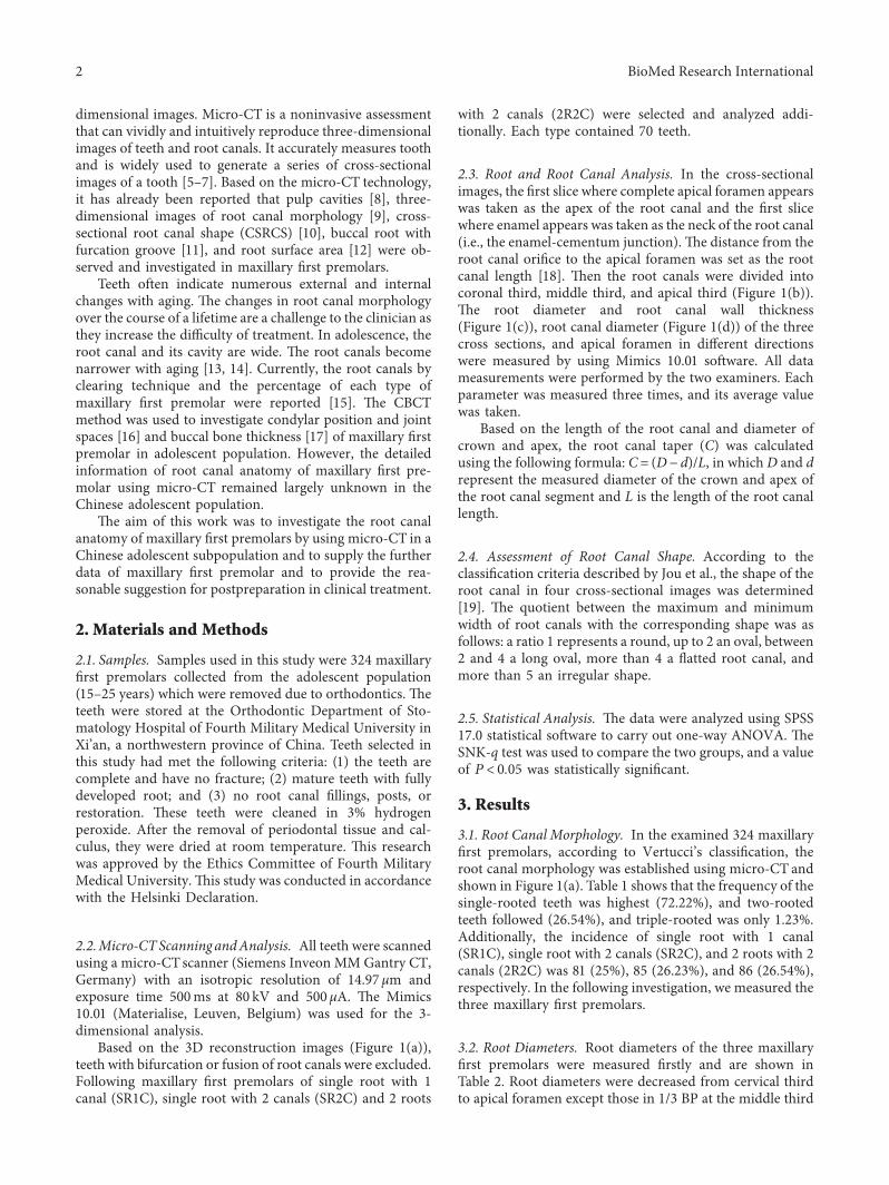
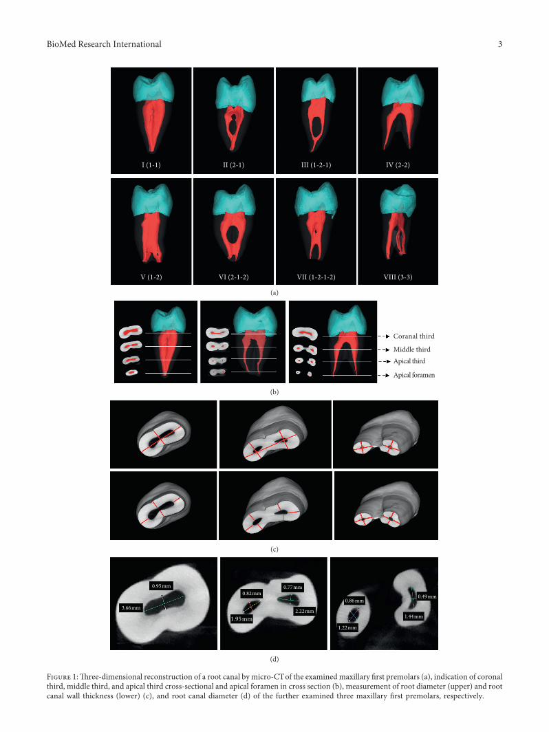
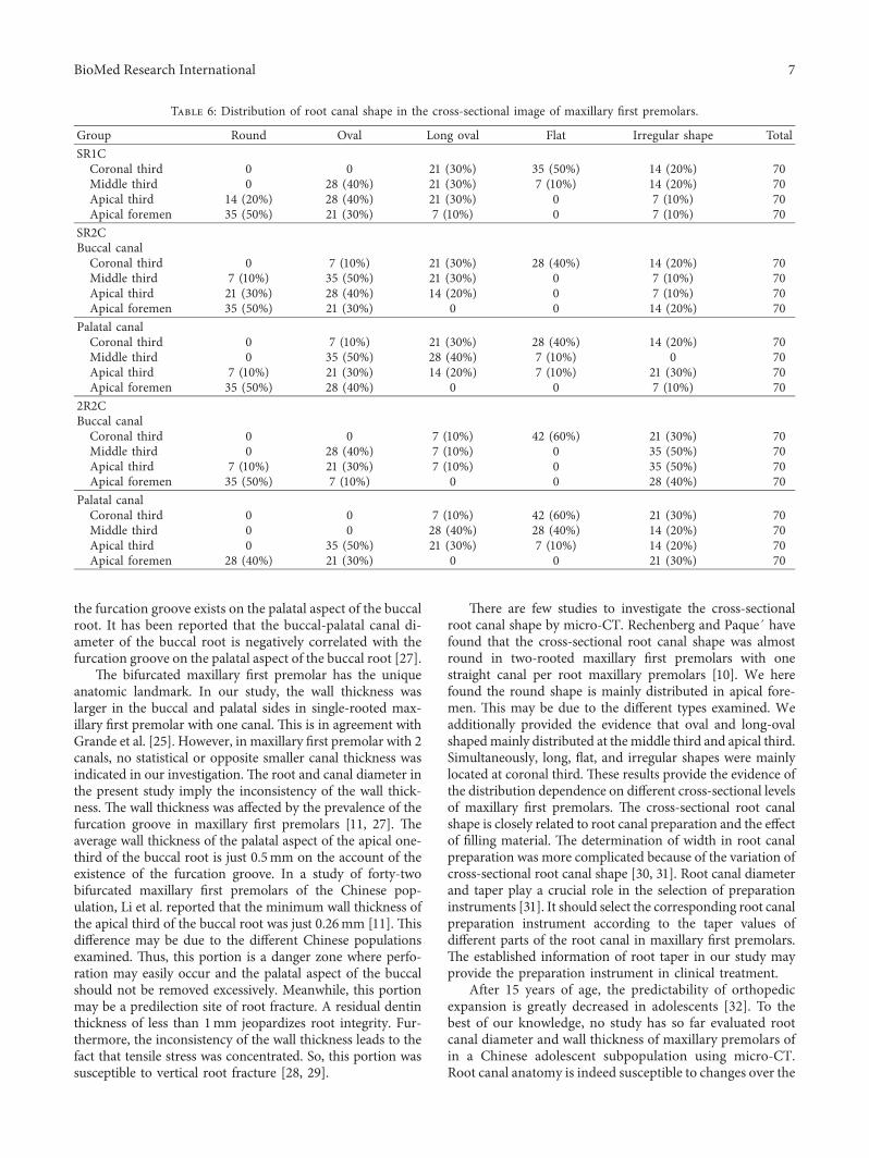
Based on the 3D reconstruction images (Figure 1(a)),teeth with bifurcation or fusion of root canals were excluded.Following maxillary first premolars of single root with 1canal (SR1C), single root with 2 canals (SR2C) and 2 roots
with 2 canals (2R2C) were selected and analyzed addi-tionally. Each type contained 70 teeth.
2.3. Root and Root Canal Analysis. In the cross-sectionalimages, the first slice where complete apical foramen appearswas taken as the apex of the root canal and the first slicewhere enamel appears was taken as the neck of the root canal(i.e., the enamel-cementum junction). -e distance from theroot canal orifice to the apical foramen was set as the rootcanal length [18]. -en the root canals were divided intocoronal third, middle third, and apical third (Figure 1(b)).-e root diameter and root canal wall thickness(Figure 1(c)), root canal diameter (Figure 1(d)) of the threecross sections, and apical foramen in different directionswere measured by using Mimics 10.01 software. All datameasurements were performed by the two examiners. Eachparameter was measured three times, and its average valuewas taken.
Based on the length of the root canal and diameter ofcrown and apex, the root canal taper (C) was calculatedusing the following formula: C� (D − d)/L, in which D and drepresent the measured diameter of the crown and apex ofthe root canal segment and L is the length of the root canallength.
2.4. Assessment of Root Canal Shape. According to theclassification criteria described by Jou et al., the shape of theroot canal in four cross-sectional images was determined[19]. -e quotient between the maximum and minimumwidth of root canals with the corresponding shape was asfollows: a ratio 1 represents a round, up to 2 an oval, between2 and 4 a long oval, more than 4 a flatted root canal, andmore than 5 an irregular shape.
2.5. Statistical Analysis. -e data were analyzed using SPSS17.0 statistical software to carry out one-way ANOVA. -eSNK-q test was used to compare the two groups, and a valueof P< 0.05 was statistically significant.
3. Results
3.1. Root Canal Morphology. In the examined 324 maxillaryfirst premolars, according to Vertucci’s classification, theroot canal morphology was established using micro-CT andshown in Figure 1(a). Table 1 shows that the frequency of thesingle-rooted teeth was highest (72.22%), and two-rootedteeth followed (26.54%), and triple-rooted was only 1.23%.Additionally, the incidence of single root with 1 canal(SR1C), single root with 2 canals (SR2C), and 2 roots with 2canals (2R2C) was 81 (25%), 85 (26.23%), and 86 (26.54%),respectively. In the following investigation, we measured thethree maxillary first premolars.
3.2. Root Diameters. Root diameters of the three maxillaryfirst premolars were measured firstly and are shown inTable 2. Root diameters were decreased from cervical thirdto apical foramen except those in 1/3 BP at the middle third
2 BioMed Research International
I (1-1) II (2-1) III (1-2-1) IV (2-2)
V (1-2) VI (2-1-2) VII (1-2-1-2) VIII (3-3)
(a)
Coranal third
Middle thirdApical third
Apical foramen
(b)
(c)
0.49mm0.82mm
1.95mm1.22mm
0.86mm
0.77mm0.95mm
3.66mm1.44mm
2.22mm
(d)
Figure 1:-ree-dimensional reconstruction of a root canal by micro-CTof the examinedmaxillary first premolars (a), indication of coronalthird, middle third, and apical third cross-sectional and apical foramen in cross section (b), measurement of root diameter (upper) and rootcanal wall thickness (lower) (c), and root canal diameter (d) of the further examined three maxillary first premolars, respectively.
BioMed Research International 3
level of single root with 1 canal and 2 roots with 2 canals.Similarly, the root diameters indicated increases from thebuccal and palatal diameter (BP) to mesiodistal diameter(MD) except those in apical third and apical foramen ofbuccal roots of single root with 2 canals.
3.3.RootCanalWall6ickness. -e root canal wall thicknessat different levels is shown in Table 3. Generally, the wallthickness was decreased from the CEJ to the apex.-e buccaland palatal walls of the single rooted with 1 canal maxillaryfirst premolar were significantly thicker (P< 0.05) than thedistal and mesial walls at each portion. No significant dif-ference in wall thickness was observed at different aspects inthese single root with 2 canals teeth.-e wall thickness of thepalatal wall of the buccal root was markedly less than themesio- and distal-aspects among these 2 roots with 2 canalsteeth (P< 0.05). -e average wall thickness of the palatalaspect of the apical one-third of the buccal root is just0.5mm. -is result corresponds to the result reported byPaola; he found that the average wall thickness is on averageless than 1mm (discussion). -e palatal wall of the palatal
root was significantly thicker than the distal and mesial wallsat the level of the cervical third and middle third, and thedifference was statistically significant (P< 0.05).
3.4. Root Canal Diameters. As shown in Table 4, the rootcanal diameters of maxillary first premolars were increasingfrom the apical foramen to cervical third. -e root canaldiameter of single root with 1 canal, single root with 2 canals,and 2 roots with 2 canals in buccal-palatal direction wassignificantly greater than that in mesial-distal direction(P< 0.05) except that in the apical foramen. However, thesignificant smaller diameter was observed (P< 0.05) at apicalthird in the buccal-palatal direction of the buccal canal of 2roots with 2 canals teeth.
3.5. RootCanal Taper. -e root canal tapers of the examinedteeth (Table 5) were the smallest of the middle third. Ad-ditionally, the root canal tapers were significantly larger(P< 0.05) at the buccal and palatal aspects than those in thecorresponding mesiodistal aspect except those at the apical
Table 1: Frequency of root canal morphology of maxillary first premolars according to Vertucci’s [20] classification.
Roots No. (%)Root canal types
I II III IV V VI VII VIII(1-1) (2-1) (1-2-1) (2-2) (1-2) (2-1-2) (1-2-1-2) (3-3)
One root 234 (72.22%) 81 27 9 85 18 9 5 0Two roots 86 (26.54%) 0 0 0 86 0 0 0 0-ree roots 4 (1.23%) 0 0 0 0 0 0 0 4Total 324 81 (25%) 27 (8.33%) 9 (2.78%) 171 (52.78%) 18 (5.56%) 9 (2.78%) 5 (1.54%) 4 (1.23%)
Table 2: Root diameters in different levels of the three maxillary first premolars (mean± SD, mm, n� 70).
Groups Cervical third Middle third Apical third Apical foramenSR1CBP 8.02± 2.73 6.78± 3.04 4.98± 2.89 2.23± 0.451/3 BP 2.67± 0.91 2.26± 1.01 1.66± 0.96 0.74± 0.15MD 4.14± 0.13 2.61± 1.11 2.13± 0.73 1.46± 0.671/3 MD 1.47± 0.04 0.87± 0.37 0.71± 0.24 0.49± 0.22
SR2CBP 8.91± 2.68 8.20± 2.71 7.30± 1.55 2.10± 0.551/3 BP 2.97± 0.89 6.73± 0.90 2.43± 0.31 0.70± 0.18MD (buccal) 5.83± 1.89 4.17± 1.93 3.28± 0.87 1.86± 0.131/3 MD (buccal) 1.94± 0.63 1.39± 0.64 1.09± 0.29 0.62± 0.04MD (palatal) 5.67± 2.33 4.01± 0.83 2.75± 0.35 1.85± 0.331/3 MD (palatal) 1.89± 0.78 1.34± 0.27 0.90± 0.11 0.61± 0.11
2R2CBuccal rootsBP 8.84± 3.00 8.24± 3.12 2.61± 0.54 1.30± 0.341/3 BP 2.95± 1.00 3.74± 1.04 0.87± 0.18 0.44± 0.11MD 4.62± 1.01 3.74± 1.13 2.90± 0.59 1.33± 0.311/3 MD 1.54± 0.34 1.24± 0.37 0.97± 0.19 0.44± 0.10
Palatal rootsBP 8.84± 3.35 8.24± 3.41 2.93± 0.71 1.80± 0.211/3 BP 2.95± 1.12 3.74± 1.13 0.98± 0.23 0.60± 0.07MD 4.44± 1.11 3.22± 0.57 2.68± 0.48 1.46± 0.321/3 MD 1.48± 0.37 1.41± 0.19 0.90± 0.16 0.49± 0.11
SR1C, single root with 1 canal; SR2C, single root with 2 canals; 2R2C, 2 roots with 2 canals; BP, buccal and palatal diameters; MD, mesiodistal diameter.
4 BioMed Research International
third of the buccal canal of single root with 2 canals andpalatal canal of 2 roots with 2 canals. On the contrary, theroot taper at the apical third of the buccal and palatal aspectwas significantly smaller (P< 0.05) than that in themesiodistal aspect.
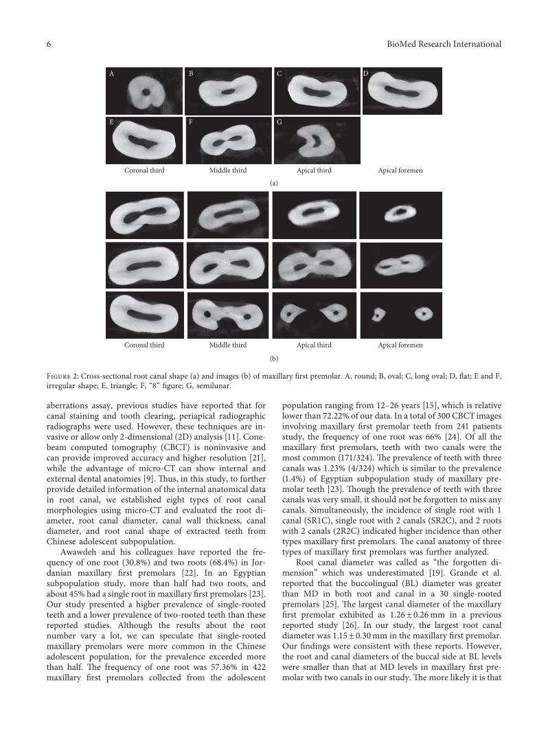
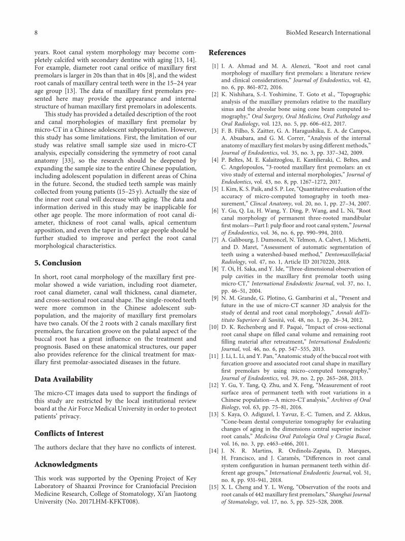
3.6. Root Canal Cross-Sectional Shape. -e root canal cross-sectional morphology in maxillary first premolars is com-plicated, including round canal, oval canal, long oval canal,flat canal, and irregular canal (Figure 2, Table 6). Triangle, “8”figure and semilunar shapes were found in irregular canals(Figure 2). At the cervical third level, long oval, flat, andirregular shapes (90%–100%) were found in the three types ofmaxillary first premolars. At the middle third level, oval andlong oval shapes (70%–90%) were observed in single-rootedmaxillary first premolars. In 2-rooted maxillary first pre-molars, oval and irregular shapes (90%) were found at themiddle third of the buccal canal, and long, flat, and irregularshapes (100%) were observed at the middle third of the palatalcanal. Round and oval shapes (60–80%) were found at apicalthird and apical foramen levels of in single-rooted maxillaryfirst premolars. Additionally, for the buccal canal, oval andirregular shapes (80%) at apical third level and round andirregular shapes (90%) were observed in 2 roots with 2 canalsmaxillary first premolars. For palatal canal, besides irregularshape in both levels, oval and flat shapes at apical third level
and round and oval shapes at apical foramen were observed inthe examined 2-rooted maxillary first premolars.
4. Discussion
Root canal morphology of the maxillary first premolarshowed a wide variation, and the complexity of the rootcanal system brings great challenges to root canal treatment[1–4]. It is very important for the clinician to be familiarizedwith the canal morphology. For root canal anatomy
Table 3: Root canal wall thickness in different levels of maxillaryfirst premolar (mean± SD, mm, n� 70).
Groups Cervicalthird Middle third Apical third Apical
foramenSR1CB 2.32± 0.12∗ ,# 2.19± 0.23∗ ,# 1.76± 0.46∗,# 0.89± 0.25P 2.43± 0.44∗ ,# 2.36± 0.38∗ ,# 2.01± 0.13∗ ,# 0.73± 0.19M 1.52± 0.35 1.00± 0.12 0.87± 0.32 0.66± 0.15D 1.84± 0.45 1.12± 0.24 0.90± 0.35 0.52± 0.16
SR2CBuccal canalB 2.33± 0.32 1.91± 0.34 1.37± 0.50 0.58± 0.12M 2.08± 0.12 1.55± 0.16 1.33± 0.67 0.57± 0.10D 1.99± 0.33 1.68± 0.61 1.34± 0.43 0.65± 0.24
Palatal canalP 2.53± 0.30 1.86± 0.36 1.33± 0.62 0.37± 0.08M 1.83± 0.38 1.67± 0.61 1.27± 0.33 0.63± 0.11D 1.90± 0.17 1.77± 0.23 1.17± 0.26 0.32± 0.09
2R2CBuccal canalB 2.21± 0.34 1.60± 0.60 1.08± 0.29 0.47± 0.09P — — 0.5± 0.13∗,# 0.3± 0.07∗ ,#M 1.75± 0.40 1.32± 0.53 1.07± 0.35 0.49± 0.10D 1.75± 0.53 1.46± 0.33 1.07± 0.33 0.49± 0.12
Palatal canalB — — 1.09± 0.31 0.64± 0.12P 2.52± 0.33∗ ,# 1.83± 0.31∗ ,# 1.10± 0.30 0.52± 0.15M 1.49± 0.19 1.07± 0.35 1.07± 0.22 0.52± 0.21D 1.80± 0.54 1.15± 0.28 1.07± 0.18 0.49± 0.08
∗represents vs. correspondingM, P< 0.05;# represents vs. corresponding D,P< 0.05. SR1C, single root with 1 canal; SR2C, single root with 2 canals;2R2C, 2 roots with 2 canals; B, buccal aspect; P, palatal aspect; M, mesio-aspect; D, distal-aspect.
Table 4: Root canal diameter in different levels of maxillary firstpremolar (mean± SD, mm, n� 70).
Groups Cervicalthird
Middlethird
Apicalthird
Apicalforamen
SR1CBP 3.34± 0.85a 2.16± 0.61a 1.57± 0.32a 0.59± 0.18aMD 0.75± 0.23 0.42± 0.13 0.42± 0.10 0.3± 0.11
SR2CBuccal canal
BP — 1.38± 0.21a 0.87± 0.25a 0.62± 0.17aMD 0.98± 0.30 1.01± 0.31 0.59± 0.09 0.44± 0.09
Palatal canalBP — 1.09± 0.21a 0.57± 0.14a 0.21± 0.05MD 0.82± 0.31 0.66± 0.11 0.35± 0.06 0.21± 0.02
2R2CBuccal canal
BP — 1.66± 0.23a 0.38± 0.14a 0.12± 0.06aMD 1.12± 0.30 0.96± 0.28 0.77± 0.13 0.50± 0.09
Palatal canalBP — 1.25± 0.21a 0.75± 0.11a 0.65± 0.10aMD 1.15± 0.30 0.93± 0.23 0.55± 0.09 0.33± 0.06
a represents vs. the corresponding MD, P< 0.05. SR1C, single root with 1canal; SR2C, single root with 2 canals; 2R2C, 2 roots with 2 canals; BP,buccal and palatal diameters; MD, mesiodistal diameter.
Table 5: Root canal taper in different levels of maxillary firstpremolar (mean± SD, mm, n� 70).
Groups Coronal third Middle third Apical thirdSR1C
BP 0.30± 0.082a 0.16± 0.042a 0.25± 0.126aMD 0.12± 0.033 0.07± 0.035 0.12± 0.041
SR2CBuccal canal
BP — 0.20± 0.091a 0.09± 0.027MD 0.15± 0.026 0.14± 0.081 0.09± 0.039
Palatal canalBP — 0.16± 0.044a 0.10± 0.051aMD 0.12± 0.039 0.05± 0.027 0.03± 0.018
2R2CBuccal canal
BP — 0.17± 0.043a 0.11± 0.022aMD 0.14± 0.041 0.07± 0.027 0.06± 0.018
Palatal canalBP — 0.15± 0.031a 0.03± 0.016aMD 0.12± 0.032 0.10± 0.042 0.06± 0.013
a represents vs. the corresponding MD, P< 0.05. SR1C, single root with 1canal; SR2C, single root with 2 canals; 2R2C, 2 roots with 2 canals; BP,buccal and palatal aspects; MD, mesiodistal aspect.
BioMed Research International 5
aberrations assay, previous studies have reported that forcanal staining and tooth clearing, periapical radiographicradiographs were used. However, these techniques are in-vasive or allow only 2-dimensional (2D) analysis [11]. Cone-beam computed tomography (CBCT) is noninvasive andcan provide improved accuracy and higher resolution [21],while the advantage of micro-CT can show internal andexternal dental anatomies [9]. -us, in this study, to furtherprovide detailed information of the internal anatomical datain root canal, we established eight types of root canalmorphologies using micro-CT and evaluated the root di-ameter, root canal diameter, canal wall thickness, canaldiameter, and root canal shape of extracted teeth fromChinese adolescent subpopulation.
Awawdeh and his colleagues have reported the fre-quency of one root (30.8%) and two roots (68.4%) in Jor-danian maxillary first premolars [22]. In an Egyptiansubpopulation study, more than half had two roots, andabout 45% had a single root in maxillary first premolars [23].Our study presented a higher prevalence of single-rootedteeth and a lower prevalence of two-rooted teeth than thesereported studies. Although the results about the rootnumber vary a lot, we can speculate that single-rootedmaxillary premolars were more common in the Chineseadolescent population, for the prevalence exceeded morethan half. -e frequency of one root was 57.36% in 422maxillary first premolars collected from the adolescent
population ranging from 12–26 years [15], which is relativelower than 72.22% of our data. In a total of 300 CBCT imagesinvolving maxillary first premolar teeth from 241 patientsstudy, the frequency of one root was 66% [24]. Of all themaxillary first premolars, teeth with two canals were themost common (171/324). -e prevalence of teeth with threecanals was 1.23% (4/324) which is similar to the prevalence(1.4%) of Egyptian subpopulation study of maxillary pre-molar teeth [23]. -ough the prevalence of teeth with threecanals was very small, it should not be forgotten to miss anycanals. Simultaneously, the incidence of single root with 1canal (SR1C), single root with 2 canals (SR2C), and 2 rootswith 2 canals (2R2C) indicated higher incidence than othertypes maxillary first premolars. -e canal anatomy of threetypes of maxillary first premolars was further analyzed.
Root canal diameter was called as “the forgotten di-mension” which was underestimated [19]. Grande et al.reported that the buccolingual (BL) diameter was greaterthan MD in both root and canal in a 30 single-rootedpremolars [25]. -e largest canal diameter of the maxillaryfirst premolar exhibited as 1.26± 0.26mm in a previousreported study [26]. In our study, the largest root canaldiameter was 1.15± 0.30mm in the maxillary first premolar.Our findings were consistent with these reports. However,the root and canal diameters of the buccal side at BL levelswere smaller than that at MD levels in maxillary first pre-molar with two canals in our study. -e more likely it is that
A
E
DC
F G
Coronal third Middle third Apical third Apical foremen
B
(a)
Coronal third Middle third Apical third Apical foremen
(b)
Figure 2: Cross-sectional root canal shape (a) and images (b) of maxillary first premolar. A, round; B, oval; C, long oval; D, flat; E and F,irregular shape; E, triangle; F, “8” figure; G, semilunar.
6 BioMed Research International
the furcation groove exists on the palatal aspect of the buccalroot. It has been reported that the buccal-palatal canal di-ameter of the buccal root is negatively correlated with thefurcation groove on the palatal aspect of the buccal root [27].
-e bifurcated maxillary first premolar has the uniqueanatomic landmark. In our study, the wall thickness waslarger in the buccal and palatal sides in single-rooted max-illary first premolar with one canal. -is is in agreement withGrande et al. [25]. However, in maxillary first premolar with 2canals, no statistical or opposite smaller canal thickness wasindicated in our investigation. -e root and canal diameter inthe present study imply the inconsistency of the wall thick-ness. -e wall thickness was affected by the prevalence of thefurcation groove in maxillary first premolars [11, 27]. -eaverage wall thickness of the palatal aspect of the apical one-third of the buccal root is just 0.5mm on the account of theexistence of the furcation groove. In a study of forty-twobifurcated maxillary first premolars of the Chinese pop-ulation, Li et al. reported that the minimum wall thickness ofthe apical third of the buccal root was just 0.26mm [11]. -isdifference may be due to the different Chinese populationsexamined. -us, this portion is a danger zone where perfo-ration may easily occur and the palatal aspect of the buccalshould not be removed excessively. Meanwhile, this portionmay be a predilection site of root fracture. A residual dentinthickness of less than 1mm jeopardizes root integrity. Fur-thermore, the inconsistency of the wall thickness leads to thefact that tensile stress was concentrated. So, this portion wassusceptible to vertical root fracture [28, 29].
-ere are few studies to investigate the cross-sectionalroot canal shape by micro-CT. Rechenberg and Paque´ havefound that the cross-sectional root canal shape was almostround in two-rooted maxillary first premolars with onestraight canal per root maxillary premolars [10]. We herefound the round shape is mainly distributed in apical fore-men. -is may be due to the different types examined. Weadditionally provided the evidence that oval and long-ovalshapedmainly distributed at themiddle third and apical third.Simultaneously, long, flat, and irregular shapes were mainlylocated at coronal third. -ese results provide the evidence ofthe distribution dependence on different cross-sectional levelsof maxillary first premolars. -e cross-sectional root canalshape is closely related to root canal preparation and the effectof filling material. -e determination of width in root canalpreparation was more complicated because of the variation ofcross-sectional root canal shape [30, 31]. Root canal diameterand taper play a crucial role in the selection of preparationinstruments [31]. It should select the corresponding root canalpreparation instrument according to the taper values ofdifferent parts of the root canal in maxillary first premolars.-e established information of root taper in our study mayprovide the preparation instrument in clinical treatment.
After 15 years of age, the predictability of orthopedicexpansion is greatly decreased in adolescents [32]. To thebest of our knowledge, no study has so far evaluated rootcanal diameter and wall thickness of maxillary premolars ofin a Chinese adolescent subpopulation using micro-CT.Root canal anatomy is indeed susceptible to changes over the
Table 6: Distribution of root canal shape in the cross-sectional image of maxillary first premolars.
Group Round Oval Long oval Flat Irregular shape TotalSR1CCoronal third 0 0 21 (30%) 35 (50%) 14 (20%) 70Middle third 0 28 (40%) 21 (30%) 7 (10%) 14 (20%) 70Apical third 14 (20%) 28 (40%) 21 (30%) 0 7 (10%) 70Apical foremen 35 (50%) 21 (30%) 7 (10%) 0 7 (10%) 70
SR2CBuccal canalCoronal third 0 7 (10%) 21 (30%) 28 (40%) 14 (20%) 70Middle third 7 (10%) 35 (50%) 21 (30%) 0 7 (10%) 70Apical third 21 (30%) 28 (40%) 14 (20%) 0 7 (10%) 70Apical foremen 35 (50%) 21 (30%) 0 0 14 (20%) 70
Palatal canalCoronal third 0 7 (10%) 21 (30%) 28 (40%) 14 (20%) 70Middle third 0 35 (50%) 28 (40%) 7 (10%) 0 70Apical third 7 (10%) 21 (30%) 14 (20%) 7 (10%) 21 (30%) 70Apical foremen 35 (50%) 28 (40%) 0 0 7 (10%) 70
2R2CBuccal canalCoronal third 0 0 7 (10%) 42 (60%) 21 (30%) 70Middle third 0 28 (40%) 7 (10%) 0 35 (50%) 70Apical third 7 (10%) 21 (30%) 7 (10%) 0 35 (50%) 70Apical foremen 35 (50%) 7 (10%) 0 0 28 (40%) 70
Palatal canalCoronal third 0 0 7 (10%) 42 (60%) 21 (30%) 70Middle third 0 0 28 (40%) 28 (40%) 14 (20%) 70Apical third 0 35 (50%) 21 (30%) 7 (10%) 14 (20%) 70Apical foremen 28 (40%) 21 (30%) 0 0 21 (30%) 70
BioMed Research International 7
years. Root canal system morphology may become com-pletely calcifed with secondary dentine with aging [13, 14].For example, diameter root canal orifice of maxillary firstpremolars is larger in 20s than that in 40s [8], and the widestroot canals of maxillary central teeth were in the 15–24 yearage group [13]. -e data of maxillary first premolars pre-sented here may provide the appearance and internalstructure of human maxillary first premolars in adolescents.
-is study has provided a detailed description of the rootand canal morphologies of maxillary first premolar bymicro-CT in a Chinese adolescent subpopulation. However,this study has some limitations. First, the limitation of ourstudy was relative small sample size used in micro-CTanalysis, especially considering the symmetry of root canalanatomy [33], so the research should be deepened byexpanding the sample size to the entire Chinese population,including adolescent population in different areas of Chinain the future. Second, the studied teeth sample was mainlycollected from young patients (15–25 y). Actually the size ofthe inner root canal will decrease with aging. -e data andinformation derived in this study may be inapplicable forother age people. -e more information of root canal di-ameter, thickness of root canal walls, apical cementumapposition, and even the taper in other age people should befurther studied to improve and perfect the root canalmorphological characteristics.
5. Conclusion
In short, root canal morphology of the maxillary first pre-molar showed a wide variation, including root diameter,root canal diameter, canal wall thickness, canal diameter,and cross-sectional root canal shape. -e single-rooted teethwere more common in the Chinese adolescent sub-population, and the majority of maxillary first premolarshave two canals. Of the 2 roots with 2 canals maxillary firstpremolars, the furcation groove on the palatal aspect of thebuccal root has a great influence on the treatment andprognosis. Based on these anatomical structures, our paperalso provides reference for the clinical treatment for max-illary first premolar-associated diseases in the future.
Data Availability
-e micro-CT images data used to support the findings ofthis study are restricted by the local institutional reviewboard at the Air Force Medical University in order to protectpatients’ privacy.
Conflicts of Interest
-e authors declare that they have no conflicts of interest.
Acknowledgments
-is work was supported by the Opening Project of KeyLaboratory of Shaanxi Province for Craniofacial PrecisionMedicine Research, College of Stomatology, Xi’an JiaotongUniversity (No. 2017LHM-KFKT008).
References
[1] I. A. Ahmad and M. A. Alenezi, “Root and root canalmorphology of maxillary first premolars: a literature reviewand clinical considerations,” Journal of Endodontics, vol. 42,no. 6, pp. 861–872, 2016.
[2] K. Nishihara, S.-I. Yoshimine, T. Goto et al., “Topographicanalysis of the maxillary premolars relative to the maxillarysinus and the alveolar bone using cone beam computed to-mography,” Oral Surgery, Oral Medicine, Oral Pathology andOral Radiology, vol. 123, no. 5, pp. 606–612, 2017.
[3] F. B. Filho, S. Zaitter, G. A. Haragushiku, E. A. de Campos,A. Abuabara, and G. M. Correr, “Analysis of the internalanatomy of maxillary first molars by using different methods,”Journal of Endodontics, vol. 35, no. 3, pp. 337–342, 2009.
[4] P. Beltes, M. E. Kalaitzoglou, E. Kantilieraki, C. Beltes, andC. Angelopoulos, “3-rooted maxillary first premolars: an exvivo study of external and internal morphologies,” Journal ofEndodontics, vol. 43, no. 8, pp. 1267–1272, 2017.
[5] I. Kim, K. S. Paik, and S. P. Lee, “Quantitative evaluation of theaccuracy of micro-computed tomography in tooth mea-surement,” Clincal Anatomy, vol. 20, no. 1, pp. 27–34, 2007.
[6] Y. Gu, Q. Lu, H. Wang, Y. Ding, P. Wang, and L. Ni, “Rootcanal morphology of permanent three-rooted mandibularfirst molars—Part I: pulp floor and root canal system,” Journalof Endodontics, vol. 36, no. 6, pp. 990–994, 2010.
[7] A. Galibourg, J. Dumoncel, N. Telmon, A. Calvet, J. Michetti,and D. Maret, “Assessment of automatic segmentation ofteeth using a watershed-based method,” DentomaxillofacialRadiology, vol. 47, no. 1, Article ID 20170220, 2018.
[8] T. Oi, H. Saka, and Y. Ide, “-ree-dimensional observation ofpulp cavities in the maxillary first premolar tooth usingmicro-CT,” International Endodontic Journal, vol. 37, no. 1,pp. 46–51, 2004.
[9] N. M. Grande, G. Plotino, G. Gambarini et al., “Present andfuture in the use of micro-CT scanner 3D analysis for thestudy of dental and root canal morphology,” Annali dell’Is-tituto Superiore di Sanita, vol. 48, no. 1, pp. 26–34, 2012.
[10] D. K. Rechenberg and F. Paque, “Impact of cross-sectionalroot canal shape on filled canal volume and remaining rootfilling material after retreatment,” International EndodonticJournal, vol. 46, no. 6, pp. 547–555, 2013.
[11] J. Li, L. Li, and Y. Pan, “Anatomic study of the buccal root withfurcation groove and associated root canal shape in maxillaryfirst premolars by using micro–computed tomography,”Journal of Endodontics, vol. 39, no. 2, pp. 265–268, 2013.
[12] Y. Gu, Y. Tang, Q. Zhu, and X. Feng, “Measurement of rootsurface area of permanent teeth with root variations in aChinese population—A micro-CT analysis,” Archives of OralBiology, vol. 63, pp. 75–81, 2016.
[13] S. Kaya, O. Adiguzel, I. Yavuz, E.-C. Tumen, and Z. Akkus,“Cone-beam dental computerize tomography for evaluatingchanges of aging in the dimensions central superior incisorroot canals,” Medicina Oral Patologıa Oral y Cirugia Bucal,vol. 16, no. 3, pp. e463–e466, 2011.
[14] J. N. R. Martins, R. Ordinola-Zapata, D. Marques,H. Francisco, and J. Carames, “Differences in root canalsystem configuration in human permanent teeth within dif-ferent age groups,” International Endodontic Journal, vol. 51,no. 8, pp. 931–941, 2018.
[15] X. L. Cheng and Y. L. Weng, “Observation of the roots androot canals of 442maxillary first premolars,” Shanghai Journalof Stomatology, vol. 17, no. 5, pp. 525–528, 2008.
8 BioMed Research International
[16] M. S. Alhammadi, M. S. Fayed, and A. Labib, “-ree-di-mensional assessment of condylar position and joint spacesafter maxillary first premolar extraction in skeletal class IImalocclusion,” Orthodontics & Craniofacial Research, vol. 20,no. 2, pp. 71–78, 2017.
[17] O. Adiguzel, C. Aktuna Belgin, S. Falakaloglu, S. Cangul, andZ. Akkus, “Maxillary cortical bone thickness in a South-Eastern Anatolian population: a cone-beam computed to-mography study,” Medical Science Monitor, vol. 23,pp. 5812–5817, 2017.
[18] S. M. Al-Shahrani, D. Al-Sudani, M. Almalik, G. Gambarini,and F. A. Alrumaihi, “Microcomputed tomographic analysisof the furcation grooves of maxillary first premolars,” Annalidi Stomatologia, vol. 4, pp. 142–148, 2013.
[19] Y. T. Jou, B. Karabucak, J. Levin, and D. Liu, “Endodonticworking width: current concepts and techniques,” DentalClinics of North America, vol. 48, no. 1, pp. 323–335, 2004.
[20] F. J. Vertucci, “Root canal anatomy of the human permanentteeth,” Oral Surgery, Oral Medicine, Oral Pathology, and OralRadiology, vol. 58, no. 5, pp. 589–599, 1984.
[21] D. Wu, G. Zhang, R. Liang et al., “Root and canal morphologyof maxillary second molars by cone-beam computed to-mography in a native Chinese population,” Journal of In-ternational Medical Research, vol. 45, no. 2, pp. 830–842, 2017.
[22] L. Awawdeh, H. Abdullah, and A. Al-Qudah, “Root form andcanal morphology of Jordanian maxillary first premolars,”Journal of Endodontics, vol. 34, no. 8, pp. 956–961, 2008.
[23] S. E. D. M. Saber, M. H. M. Ahmed, M. Obeid, andH. M. A. Ahmed, “Root and canal morphology of maxillarypremolar teeth in an Egyptian subpopulation using twoclassification systems: a cone beam computed tomographystudy,” International Endodontic Journal, vol. 52, no. 3,pp. 267–278, 2019.
[24] Y. Y. Tian, B. Guo, R. Zhang et al., “Root and canal mor-phology of maxillary first premolars in a Chinese sub-population evaluated using cone-beam computedtomography,” International Endodontic Journal, vol. 45,no. 11, pp. 996–1003, 2012.
[25] N. M. Grande, G. Plotino, R. Pecci, R. Bedini, C. H. Pameijer,and F. Somma, “Micro–computerized tomographic analysisof radicular and canal morphology of premolars with longoval canals,” Oral Surgery, Oral Medicine, Oral Pathology,Oral Radiology, and Endodontology, vol. 106, no. 3, pp. e70–e76, 2008.
[26] J. Krapez and A. Fidler, “Location and dimensions of accesscavity in permanent incisors, canines, and premolars,” Journalof Conservative Dentistry JCD, vol. 16, no. 5, pp. 404–407,2013.
[27] A. Tmase, A. Katz, and R. Pilo, “Furcation groove of buccalroot of maxillary first premolars—A morphometric study,”Journal of Endodontics, vol. 26, no. 6, pp. 359–363, 2000.
[28] A. Versluis, H. H. Messer, and M. R. Pintado, “Changes incompaction stress distributions in roots resulting from canalpreparation,” International Endodontic Journal, vol. 39,no. 12, pp. 931–939, 2006.
[29] P. A. Lammertyn, S. B. Rodrigo, M. Brunotto, and M. Crosa,“Furcation groove of maxillary first premolar, thickness, anddentin structures,” Journal of Endodontics, vol. 35, no. 6,pp. 814–817, 2009.
[30] A. Al-Bulushi, M. Levinkind, M. Flanagan, Y.-L. Ng, andK. Gulabivala, “Effect of canal preparation and residual rootfilling material on root impedance,” International EndodonticJournal, vol. 41, no. 10, pp. 892–904, 2008.
[31] M. E. de Lima Machado, L. A. Bichels Sapia, S. Cai,G. H. R. Martins, and C. K. Nabeshima, “Comparison of tworotary systems in root canal preparation regarding disinfec-tion,” Journal of Endodontics, vol. 36, no. 7, pp. 1238–1240,2010.
[32] L. Lin, H. W. Ahn, S. J. Kim, S. C. Moon, S. H. Kim, andG. Nelson, “Tooth-borne vs bone-borne rapid maxillary ex-panders in late adolescence,” 6e Angle Orthodontist, vol. 85,no. 2, pp. 253–262, 2015.
[33] Y. H. Li, S. J. Bao, X. W. Yang, X. M. Tian, B. Wei, andY. L. Zheng, “Symmetry of root anatomy and root canalmorphology in maxillary premolars analyzed using cone-beam computed tomography,” Archives of Oral Biology,vol. 94, pp. 84–92, 2018.
BioMed Research International 9

Hindawiwww.hindawi.com
International Journal of
Volume 2018
Zoology
Hindawiwww.hindawi.com Volume 2018
Anatomy Research International
PeptidesInternational Journal of
Hindawiwww.hindawi.com Volume 2018
Hindawiwww.hindawi.com Volume 2018
Journal of Parasitology Research
GenomicsInternational Journal of
Hindawiwww.hindawi.com Volume 2018
Hindawi Publishing Corporation http://www.hindawi.com Volume 2013Hindawiwww.hindawi.com
The Scientific World Journal
Volume 2018
Hindawiwww.hindawi.com Volume 2018
BioinformaticsAdvances in
Marine BiologyJournal of
Hindawiwww.hindawi.com Volume 2018
Hindawiwww.hindawi.com Volume 2018
Neuroscience Journal
Hindawiwww.hindawi.com Volume 2018
BioMed Research International
Cell BiologyInternational Journal of
Hindawiwww.hindawi.com Volume 2018
Hindawiwww.hindawi.com Volume 2018
Biochemistry Research International
ArchaeaHindawiwww.hindawi.com Volume 2018
Hindawiwww.hindawi.com Volume 2018
Genetics Research International
Hindawiwww.hindawi.com Volume 2018
Advances in
Virolog y Stem Cells International
Hindawiwww.hindawi.com Volume 2018
Hindawiwww.hindawi.com Volume 2018
Enzyme Research
Hindawiwww.hindawi.com Volume 2018
International Journal of
MicrobiologyHindawiwww.hindawi.com
Nucleic AcidsJournal of
Volume 2018
Submit your manuscripts atwww.hindawi.com
Related Documents