MOLECULAR AND CELLULAR BIOLOGY, 0270-7306/01/$04.0010 DOI: 10.1128/MCB.21.5.1499–1508.2001 Mar. 2001, p. 1499–1508 Vol. 21, No. 5 Copyright © 2001, American Society for Microbiology. All Rights Reserved. Roles of the Mitotic Inhibitors Wee1 and Mik1 in the G 2 DNA Damage and Replication Checkpoints NICHOLAS RHIND AND PAUL RUSSELL* Departments of Molecular Biology and Cell Biology, The Scripps Research Institute, La Jolla, California 92037 Received 4 October 2000/Returned for modification 21 November 2000/Accepted 30 November 2000 The G 2 DNA damage and DNA replication checkpoints in many organisms act through the inhibitory phosphorylation of Cdc2 on tyrosine-15. This phosphorylation is catalyzed by the Wee1/Mik1 family of kinases. However, the in vivo role of these kinases in checkpoint regulation has been unclear. We show that, in the fission yeast Schizosaccharomyces pombe, Mik1 is a target of both checkpoints and that the regulation of Mik1 is, on its own, sufficient to delay mitosis in response to the checkpoints. Mik1 appears to have two roles in the DNA damage checkpoint; one in the establishment of the checkpoint and another in its maintenance. In contrast, Wee1 does not appear to be involved in the establishment of either checkpoint. Checkpoints are mechanisms that allow cells to deal with DNA damage or other insults (10, 18). A major role of check- points is to delay cell cycle transitions, in order to allow time for the damage to be repaired. It is therefore important to understand how checkpoints regulate the basic cell cycle ma- chinery. In the fission yeast Schizosaccharomyces pombe and in mammalian cells, the DNA damage and DNA replication check- points arrest cells in G 2 through inhibitory phosphorylation of Cdc2 on tyrosine-15 (5, 19, 35, 38). Cdc2, in association with its regulatory subunit cyclin B, is the kinase that determines the timing of the G 2 -M transition. While Cdc2 is controlled in many ways, the dephosphorylation of tyrosine-15 is the rate- limiting step for Cdc2 activation at mitosis and therefore reg- ulates the timing of the G 2 -M transition (17, 20). This phos- phorylation is catalyzed by members of the Wee1/Mik1/Myt1 family of tyrosine kinases (Wee1 and Myt1 in vertebrates and Wee1 and Mik1 in S. pombe) and removed by Cdc25 phospha- tases (8). Thus, to regulate Cdc2 by tyrosine-15 phosphoryla- tion, the checkpoints must increase its phosphorylation by one or more of the Wee1/Mik1/Myt1 kinases or decrease its de- phosphorylation by Cdc25. In fission yeast, the DNA damage and DNA replication checkpoints act through related signal transduction pathways that culminate in the serine/threonine kinase Chk1, the effec- tor of the DNA damage checkpoint, or the serine/threonine kinase Cds1, the effector of the DNA replication checkpoint (reviewed in references 10, 36, and 37). In S. pombe and ver- tebrate cells, Cdc25 has been shown to be a target of these checkpoint pathways (4, 15, 21, 33, 38). Both Chk1 and Cds1 phosphorylate Cdc25 in vitro, and this phosphorylation inhibits its phosphatase activity (4, 14, 15, 21, 33, 40, 44). In addition, checkpoint-dependent phosphorylation of Cdc25 inhibits its nuclear import, presumably sequestering it away from Cdc2 (9, 22, 24, 43, 45). However, recent studies have shown that the regulation of Cdc25’s subcellular localization is not required to enforce a checkpoint-dependent cell cycle delay (25). In S. pombe, Cdc2 is also dephosphorylated by the tyrosine phos- phatase Pyp3 (28). Pyp3 plays a minor role in normal mitotic control, and unlike Cdc25, loss of its activity does not arrest the cell cycle. Therefore, Pyp3 cannot be a sufficient checkpoint target. It has been less certain to what extent the regulation of the Wee1/Mik1/Myt1 kinases contributes to the function of the checkpoints. There have been several reports correlating check- point activation with changes in Wee1 abundance and phos- phorylation. The degradation of exogenous Wee1 appears to be inhibited by the DNA replication checkpoint in Xenopus egg extracts (27). In S. pombe cell lysates, exogenous Wee1 is bound to and phosphorylated by Cds1 in a checkpoint-depen- dent manner (6). Wee1 has been reported to be phosphory- lated in vivo in response to UV radiation, and it is also phos- phorylated by Chk1 in vitro (32). However, none of these studies provide evidence that Wee1 regulation is important for checkpoint function in vivo. Mik1 has also been proposed to be a target of the check- points. While Mik1 is not required for either checkpoint, cells deficient for both wee1 and cdc25 still arrest before mitosis in response to a replication block induced by hydroxyurea (HU), suggesting that regulation of Mik1 or Pyp3 is sufficient to enforce the replication checkpoint (11, 26). That Mik1 may play a role in this circumstance is suggested by the accumula- tion of Mik1 during a replication arrest. In response to the DNA replication checkpoint, mik1 1 mRNA accumulates to high levels and Mik1 protein appears to be stabilized, leading to a dramatic increase in steady-state protein levels (1, 6, 7). Prolonged activation of the DNA damage checkpoint causes an increase in the steady-state level of Mik1 to a lesser extent, without affecting the level of mik1 1 mRNA (1, 7). The increase in Mik1 abundance in response to prolonged exposure to DNA damage may explain how Mik1 acts to enforce the extended maintenance of a DNA damage checkpoint (1). Despite these correlations between Mik1 protein abundance and checkpoint activation, it is unknown if Mik1 regulation is involved in establishing a G 2 delay in response to either checkpoint. These studies on checkpoint regulation of Wee1 and Mik1 * Corresponding author. Mailing address: Departments of Molecu- lar Biology and Cell Biology, The Scripps Research Institute, La Jolla, CA 92037. Phone: (858) 784-8273. Fax: (858) 784-2265. E-mail: [email protected]. 1499 on June 15, 2015 by guest http://mcb.asm.org/ Downloaded from

Welcome message from author
This document is posted to help you gain knowledge. Please leave a comment to let me know what you think about it! Share it to your friends and learn new things together.
Transcript
MOLECULAR AND CELLULAR BIOLOGY,0270-7306/01/$04.0010 DOI: 10.1128/MCB.21.5.1499–1508.2001
Mar. 2001, p. 1499–1508 Vol. 21, No. 5
Copyright © 2001, American Society for Microbiology. All Rights Reserved.
Roles of the Mitotic Inhibitors Wee1 and Mik1 in the G2 DNADamage and Replication Checkpoints
NICHOLAS RHIND AND PAUL RUSSELL*
Departments of Molecular Biology and Cell Biology, The Scripps Research Institute,La Jolla, California 92037
Received 4 October 2000/Returned for modification 21 November 2000/Accepted 30 November 2000
The G2 DNA damage and DNA replication checkpoints in many organisms act through the inhibitoryphosphorylation of Cdc2 on tyrosine-15. This phosphorylation is catalyzed by the Wee1/Mik1 family of kinases.However, the in vivo role of these kinases in checkpoint regulation has been unclear. We show that, in thefission yeast Schizosaccharomyces pombe, Mik1 is a target of both checkpoints and that the regulation of Mik1is, on its own, sufficient to delay mitosis in response to the checkpoints. Mik1 appears to have two roles in theDNA damage checkpoint; one in the establishment of the checkpoint and another in its maintenance. Incontrast, Wee1 does not appear to be involved in the establishment of either checkpoint.
Checkpoints are mechanisms that allow cells to deal withDNA damage or other insults (10, 18). A major role of check-points is to delay cell cycle transitions, in order to allow timefor the damage to be repaired. It is therefore important tounderstand how checkpoints regulate the basic cell cycle ma-chinery. In the fission yeast Schizosaccharomyces pombe and inmammalian cells, the DNA damage and DNA replication check-points arrest cells in G2 through inhibitory phosphorylation ofCdc2 on tyrosine-15 (5, 19, 35, 38). Cdc2, in association with itsregulatory subunit cyclin B, is the kinase that determines thetiming of the G2-M transition. While Cdc2 is controlled inmany ways, the dephosphorylation of tyrosine-15 is the rate-limiting step for Cdc2 activation at mitosis and therefore reg-ulates the timing of the G2-M transition (17, 20). This phos-phorylation is catalyzed by members of the Wee1/Mik1/Myt1family of tyrosine kinases (Wee1 and Myt1 in vertebrates andWee1 and Mik1 in S. pombe) and removed by Cdc25 phospha-tases (8). Thus, to regulate Cdc2 by tyrosine-15 phosphoryla-tion, the checkpoints must increase its phosphorylation by oneor more of the Wee1/Mik1/Myt1 kinases or decrease its de-phosphorylation by Cdc25.
In fission yeast, the DNA damage and DNA replicationcheckpoints act through related signal transduction pathwaysthat culminate in the serine/threonine kinase Chk1, the effec-tor of the DNA damage checkpoint, or the serine/threoninekinase Cds1, the effector of the DNA replication checkpoint(reviewed in references 10, 36, and 37). In S. pombe and ver-tebrate cells, Cdc25 has been shown to be a target of thesecheckpoint pathways (4, 15, 21, 33, 38). Both Chk1 and Cds1phosphorylate Cdc25 in vitro, and this phosphorylation inhibitsits phosphatase activity (4, 14, 15, 21, 33, 40, 44). In addition,checkpoint-dependent phosphorylation of Cdc25 inhibits itsnuclear import, presumably sequestering it away from Cdc2 (9,22, 24, 43, 45). However, recent studies have shown that theregulation of Cdc25’s subcellular localization is not required
to enforce a checkpoint-dependent cell cycle delay (25). InS. pombe, Cdc2 is also dephosphorylated by the tyrosine phos-phatase Pyp3 (28). Pyp3 plays a minor role in normal mitoticcontrol, and unlike Cdc25, loss of its activity does not arrest thecell cycle. Therefore, Pyp3 cannot be a sufficient checkpointtarget.
It has been less certain to what extent the regulation of theWee1/Mik1/Myt1 kinases contributes to the function of thecheckpoints. There have been several reports correlating check-point activation with changes in Wee1 abundance and phos-phorylation. The degradation of exogenous Wee1 appears tobe inhibited by the DNA replication checkpoint in Xenopusegg extracts (27). In S. pombe cell lysates, exogenous Wee1 isbound to and phosphorylated by Cds1 in a checkpoint-depen-dent manner (6). Wee1 has been reported to be phosphory-lated in vivo in response to UV radiation, and it is also phos-phorylated by Chk1 in vitro (32). However, none of thesestudies provide evidence that Wee1 regulation is important forcheckpoint function in vivo.
Mik1 has also been proposed to be a target of the check-points. While Mik1 is not required for either checkpoint, cellsdeficient for both wee1 and cdc25 still arrest before mitosis inresponse to a replication block induced by hydroxyurea (HU),suggesting that regulation of Mik1 or Pyp3 is sufficient toenforce the replication checkpoint (11, 26). That Mik1 mayplay a role in this circumstance is suggested by the accumula-tion of Mik1 during a replication arrest. In response to theDNA replication checkpoint, mik11 mRNA accumulates tohigh levels and Mik1 protein appears to be stabilized, leadingto a dramatic increase in steady-state protein levels (1, 6, 7).Prolonged activation of the DNA damage checkpoint causesan increase in the steady-state level of Mik1 to a lesser extent,without affecting the level of mik11 mRNA (1, 7). The increasein Mik1 abundance in response to prolonged exposure to DNAdamage may explain how Mik1 acts to enforce the extendedmaintenance of a DNA damage checkpoint (1). Despite thesecorrelations between Mik1 protein abundance and checkpointactivation, it is unknown if Mik1 regulation is involved inestablishing a G2 delay in response to either checkpoint.
These studies on checkpoint regulation of Wee1 and Mik1
* Corresponding author. Mailing address: Departments of Molecu-lar Biology and Cell Biology, The Scripps Research Institute, La Jolla,CA 92037. Phone: (858) 784-8273. Fax: (858) 784-2265. E-mail:[email protected].
1499
on June 15, 2015 by guesthttp://m
cb.asm.org/
Dow
nloaded from
have led to models in which both kinases are proposed to beimportant targets of the checkpoints. We designed an exper-imental system to directly test these hypotheses. By usingS. pombe strains in which Cdc25 is replaced with a phosphatasethat is not regulated by the checkpoints, we created a situationin which any checkpoint regulation of Cdc2 phosphorylationmust act through Wee1 or Mik1. This experimental systemallowed us to examine the checkpoint regulation of Wee1 andMik1 in vivo.
MATERIALS AND METHODS
General methods for studying fission yeast were followed as described previ-ously (29). The following strains were used: PR109 (h2 leu1-32 ura4-D18),PR754 (h2 leu1-32 ura4-D18 wee1-50 mik1::ura41), NR1826 (h2 leu1-32 ura4-D18 rad3::ura41), NB2117 (h2 leu1-32 ura4-D18 cds1::ura41), AL2521 (h2
leu1-32 ura4-D18 chk11:9Myc), NR2648 (h2 leu1-32 ura4-D18 rad3::ura41
chk11:9Myc), NR2613 (h2 leu1-32 ura4-D18 nmt1:pyp31 cdc25::ura41), NR2640(h2 leu1-32 ura4-D18 nmt1:pyp31 cdc25::ura41 wee1-3x), NR2630 (h2 leu1-32ura4-D18 nmt1:pyp31 cdc25::ura41 wee1-50ts), NR2657 (h1 leu1-32 ura4-? nmt1:pyp31 cdc25::ura41 mik1-s14ts), NR2634 (h2 leu1-32 ura4-D181 nmt1:pyp31
cdc25::ura41 mik1::ura41), NR2644 (h2 leu1-32 ura4-? nmt1:pyp31 cdc25::ura41
wee1-50 mik1-s14ts), NR2646 (h2 leu1-32 ura4-D18 nmt1:pyp31 cdc25::ura41
chk11:9Myc), NR2650 (h2 leul-32 ura4-D18 nmt1:pyp31 cdc25::ura41 mik11:13Myc), KS1362 (h1 leu1-32 ura4-? wee1-50ts cdc25-22ts), and PR1928 (h2
leu1-32 ura4-? wee1-50ts cdc25-22ts mik1::ura4). The designation “ura4-?” indi-cates that the ura4 allele may be ura4-D18 or ura4-294. Unless otherwise stated,strains were grown in Edinburgh minimal medium 2, supplemented with leucine,uracil, adenine, and histidine, with or without 5 mg of thiamine per ml at 32°C.For the experiments presented in Fig. 3, strains were grown in YES, a yeastextract-based medium with the same supplements. Strains were grown in theindicated media for at least 48 h before each experiment.
Synchronous cultures were prepared by centrifugal elutriation with a BeckmanJE-5.0 elutriation rotor, a technique that selects the smallest cells from anasynchronous population. In S. pombe, replication occurs immediately aftermitosis and concurrently with cytokinesis; thus elutriation produces a populationof early G2 cells. The time from elutriation to septation varies reproduciblyamong various strains, with that of the nmt1:pyp31 cdc25::ura41 strains beingparticularly short. Because all of the experiments were internally controlled, thisvariation does not affect the interpretation of the results. The number of cellshaving passed mitosis (N) was determined as N 5 (S 1 D)/(T 2 D), where S isthe number of septated cells, D is the number of divided cells, and T is the totalnumber of cells. This equation corrects for the fact that once a cell has dividedit is counted as two cells. Cells were photographed and measured using a Quantixdigital camera (Photometrics) and IP Lab software (Signal Analytics Corpora-tion). Cells were irradiated with gamma radiation from a cesium-137 source at3.3 Gy min21 for 30 min, or at 1 Gy min21 for the continuous exposure used inFig. 1. For HU experiments, cells were grown for 60 to 90 min in 10 mM HUbefore elutriation. This protocol ensures that the elutriated cells are arrested inS phase. HU was then washed out of half the culture, which replicated and wenton to divide with kinetics similar to that of untreated cultures.
The wee1-50 allele was used instead of wee1D because wee1D cells diploidize ata high frequency. wee1-50 strains were maintained at 25°C and shifted to 32°C forat least 12 h before any experiment conducted at 32°C. To establish that 32°C isa restrictive temperature for wee1-50, we compared the length of wee1-50 cells atvarious temperatures. wee1-50 cells at 32°C are indistinguishable from wee1Dcells (Table 1). To determine if wee1-50 might have some residual activity at 32°Cthat would be unmeasurable at normal expression levels, we overexpressedwee1-50 approximately 30- to 60-fold from the adh1 promoter (our unpublisheddata). The normalized activity of wee1-50 at 32°C is less than 3%, assuming30-fold overexpression (Table 1). In addition, all experiments with wee1-50strains were repeated at 35°C with comparable results. Since wee1 mutant strainsreplicate later in the cell cycle than wild-type strains, we confirmed by flowcytometry that the synchronized cultures had completed replication before beingirradiated (our unpublished data). The temperature-sensitive (ts) mik1-s14 allelewas isolated on the basis of its synthetic lethality with wee1-50. It is tightly linkedto mik1 and rescued by mik11 genomic sequences (our unpublished data). Thelowest restrictive temperature for mik1-s14, as assayed by viability of wee1Dmik1-s14 cells, is 33°C. For the HU experiments involving mik1-s14, cells weregrown and elutriated at 25°C and then cultured at 25°C for 60 min to allow the
cells that had been washed out of HU to replicate. The cultures were then shiftedto 35°C to inactivate Mik1.
The nmt1 promoter was inserted in place of the pyp3 promoter by one-stepreplacement. pFA6a-kanMX-P3nmt1 was amplified with the following primers(Integrated DNA Technologies, Inc): 59-GAATGTGAACGTGAACTAGATTACGACTACAACTAGAAACTAGCGCTATGTGGGGGCCGTACAATGATGATTTATTAAACGAATTCGAGCTCGTTTAAAC-39 and 59-ATAATCATGTACGCTTTTTCTTTTATTGTGATCAAGGGTGTTAGAACACCATTTTCTGTAGATACTTCTTTAAAAGACATGATTTAACAAAGCGACTATA-39.The amplified DNA was transformed into PR109, and integrants were selectedas described previously (2).
Protein affinity purifications, Western blotting, and kinase assays were per-formed as previously described (1, 6, 35).
RESULTS
Constitutive overexpression of pyp31 rescues cdc25D. Wesought to create a strain of S. pombe in which the dephosphor-ylation of Cdc2 tyrosine-15 was not regulated by the check-points. To that end, we integrated the thiamine-repressiblenmt1 promoter upstream of the pyp31 genomic open readingframe. When grown in the absence of thiamine, nmt1:pyp31
cells overexpress Pyp3 to a level sufficient to rescue the lethal-ity of cdc25D (Fig. 1A). nmt1:pyp31 cdc25D cells are healthyand grow with a generation time of 3.0 h, similar to the wildtype (data not shown). They divide at about 16 mm, comparedwith 13 mm for the wild type, suggesting that, at this level ofexpression, Pyp3 is not quite as active as endogenous Cdc25(Fig. 1A). To demonstrate that the nmt1:pyp31 cdc25D cellsare still responsive to variations in Wee1 activity, we usedwee1-3x, an allele that has three copies of wee11 integrated atits genomic locus (39). These cells divide at about 21 mm, thesame length as otherwise-wild-type wee1-3x cells (39). Thus, innmt1:pyp31 cdc25D cells, in which Pyp3 replaces Cdc25, thecells undergo mitosis with close-to-wild-type timing and retaina normal response to changes in Wee1 activity.
For our purposes it was important that the overexpressedPyp3 not be regulated by either DNA damage or DNA repli-cation checkpoints. To test whether the checkpoints regulatePyp3, we adapted an assay that has been used to demonstratethe regulation of Cdc25 by the checkpoints (35). This assayuses a strain carrying ts alleles of wee1 and mik1 that is syn-chronized in early G2 by centrifugal elutriation. When thissynchronous population of wee1-ts mik1-ts cells is shifted to35°C, the two kinases are inactivated and Cdc2 can no longerbe phosphorylated. The extent of tyrosine phosphorylation isthen dependent only on the rate of dephosphorylation, which
TABLE 1. Comparison of activities of length of S. pombestrains at various temperatures
Strain Temp(°C)
Size at septation(mm)a
NormalizedWee1 activity
PR109 wild type 32 13.1 6 1.1 1.0PR47 wee1D 32 8.1 6 0.9 0PR1080 wee1-50 25 10.5 6 1.6 0.5PR1080 wee1-50 30 8.0 6 1.0 0PR1080 wee1-50 32 8.2 6 1.2 0PR1080 wee1-50 35 8.0 6 1.6 0PR36 wee1-50 adh:wee1-50 25 Arrested in G2 NAb
PR36 wee1-50 adh:wee1-50 30 19.9 6 2.6 0.08PR36 wee1-50 adh:wee1-50 32 12.4 6 1.5 0.03PR36 wee1-50 adh:wee1-50 35 8.8 6 1.0 0.005
a Values are means 6 standard deviations.b NA, not applicable.
1500 RHIND AND RUSSELL MOL. CELL. BIOL.
on June 15, 2015 by guesthttp://m
cb.asm.org/
Dow
nloaded from

is predominantely catalyzed by Cdc25. Because dephosphory-lation of Cdc2 is the rate-limiting step for entry into mitosisfrom G2, the rate at which Cdc2 is dephosphorylated can beinferred from the rate at which cells enter mitosis (17, 35). Ittakes wee1-ts mik1D cells about 40 min to dephosphorylateCdc2 when shifted to 35°C, and the cells septate about 20 minlater (Fig. 1B) (35). If either checkpoint is activated before theshift (by gamma radiation or by pretreatment of the cells withHU so that the elutriated cells are arrested in S phase), thedephosphorylation is delayed 40 to 60 min. These results dem-onstrate, as previously shown, that the dephosphorylation ofCdc2 by Cdc25 is inhibited by both checkpoints (Fig. 1B) (35,38). When the experiments were repeated in an nmt1:pyp31
cdc25D wee1-ts mik1-ts strain, no delay in mitosis was observed,demonstrating that Pyp3 is not regulated by either checkpoint(Fig. 1C).
These results show that nmt1:pyp31 cdc25D cells are unableto regulate the rate of Cdc2 dephosphorylation in response toeither checkpoint. Because both checkpoints are dependent ontyrosine-15 phosphorylation of Cdc2 (35, 38), any checkpointregulation of mitosis in the nmt1:pyp31 cdc25D cells must bedue to regulation of the rate of Cdc2 phosphorylation by Wee1or Mik1. We used this situation to test if Wee1 or Mik1 isregulated by either checkpoint.
Regulation of Wee1 and Mik1 by the DNA damage check-point. To determine whether Wee1 and Mik1 are regulated inresponse to activation of the checkpoint, we examined theability of nmt1:pyp31 cdc25D cells to delay mitosis in responseto DNA damage. A synchronous population of nmt1:pyp31
cdc25D cells was exposed to 100 Gy of gamma radiation in G2
and was monitored through the first mitosis. Wild-type cellsdelayed mitosis about 60 min in response to such treatment(Fig. 2A). nmt1:pyp31 cdc25D cells also showed a delay in G2,albeit a much reduced one, of about 20 min (Fig. 2B). A delayof this length was reproducibly seen in four similar experi-ments, and all other cell cycle kinetic results presented arerepresentative of at least three similar experiments. This dem-onstrates that in the absence of Cdc25 regulation, cells are ableto delay mitosis in response to DNA damage but not to the fullextent seen in wild-type cells. We conclude that the regulationof either Wee1 or Mik1 must play a role in delaying mitosis inresponse to DNA damage.
We tested Wee1 and Mik1 individually by repeating theabove experiment in wee12 or mik12 backgrounds. The partialdelay seen in nmt1:pyp31 cdc25D cells is also seen in nmt1:pyp31 cdc25D wee1-ts cells at the restrictive temperature (Fig.2C). In nmt1:pyp31 cdc25D wee1-ts cells, neither Cdc25 norWee1 can be regulated, and thus Mik1 regulation must be
FIG. 1. Constitutive overexpression of Pyp3 rescues cdc25D. (A) Wild-type (PR109), nmt1:pyp31 cdc25D (NR2613), and nmt1:pyp31 cdc25Dwee1-3x (NR2640) cells were grown in synthetic media lacking thiamine to induce high levels of expression of Pyp3 from the nmt1 promoter. Theinset is the average length at septation for at least 50 cells, plus or minus the standard deviation. (B and C) wee1-50ts mik1D (PR754) and nmt1:pyp31 cdc25D wee1-50ts mik1-s14ts (NR2644) cells were grown at 25°C, elutriated to collect a synchronous population of cells, and shifted to 35°C.The irradiated cultures were continuously irradiated with gamma radiation at 1 Gy/min from 30 min before the shift. The HU-blocked cultureswere grown in 10 mM HU for 60 min before elutriation, so that the elutriated cells would be blocked in S phase. Half of the culture was thenreleased from the HU block and allowed to complete replication before the shift to 35°C. Cell cycle progression was monitored microscopically.For clarity, the data from the HU-released cultures is not shown, but those cultures divided with kinetics similar to that of the untreated cultures.The cell cycle kinetic data shown here and in the other figures are representative of at least three similar experiments.
VOL. 21, 2001 Wee1 AND Mik1 IN G2 CHECKPOINTS 1501
on June 15, 2015 by guesthttp://m
cb.asm.org/
Dow
nloaded from

responsible for the delay observed. nmt1:pyp31 cdc25D mik1Dcells, in which neither Cdc25 nor Mik1 can be regulated, areunable to delay mitosis in response to DNA damage (Fig. 2D).Thus, Wee1 is not significantly up-regulated in response toDNA damage, and Cdc25 and Mik1 are the only major targetscontrolling Cdc2 phosphorylation in response to the check-point.
The level of Mik1 protein has been reported to increasein response to DNA damage. We therefore tried to correlatethe accumulation of Mik1 with the delay seen in nmt1:pyp31
cdc25D cells. Mik1 accumulated in response to prolonged check-point activation such as that evoked by continuous exposure tothe DNA-damaging drug bleomycin or high doses of gammaradiation (250 Gy) (1, 7). However, Mik1 did not accumulatein response to the dose of gamma radiation (100 Gy) used inthis study (Fig. 2E), presumably because this dose triggers only
a relatively short delay which provides insufficient time forMik1 to significantly accumulate (Fig. 2B). Thus, Mik1 activitymust be regulated at some other level to induce the delay seenin nmt1:pyp31 cdc25D cells.
As a control, to show that overexpression of Pyp3 does notinterfere with activation of the DNA damage signal transduc-tion pathway, we examined the phosphorylation of Chk1. Thisphosphorylation results in a decrease in the mobility of Chk1on sodium dodecyl sulfate-polyacrylamide gel electrophoresisand correlates with the activation of the DNA damage check-point (42). Chk1 is phosphorylated normally in response toDNA damage in the nmt1:pyp31 cdc25D background, demon-strating that the checkpoint signal transduction pathway isintact (Fig. 2F).
As an alternate test of the function of Mik1 in the DNAdamage checkpoint, we examined the checkpoint responses of
FIG. 2. The phosphorylation of Cdc2 by Mik1, but not Wee1, is up-regulated by the DNA damage checkpoint. (A through D) Wild-type(PR109), nmt1:pyp31 cdc25D (NR2613), nmt1:pyp31 cdc25D wee1-50ts (NR2630), and nmt1:pyp31 cdc25D mik1D (NR2634) cells were elutriatedand irradiated with 100 Gy of gamma radiation from approximately 2 h before the midpoint of septation, as indicated by the bracket (irradiated),or mock irradiated (unirradiated). Cell cycle progression was monitored microscopically. (E) nmt1:pyp31 cdc25D mik11:13Myc (NR2650) cellswere grown to logarithmic phase and irradiated with 100 Gy of gamma radiation or incubated for 2 h with 10 mM HU. At indicated times, sampleswere taken and cleared whole-cell extracts were prepared. The extracts were analyzed by Western blotting with monoclonal anti-Myc antibodies(9E10; Covance). The slightly greater signal in the asynchronous sample compared with that in the irradiated samples is due to the small percentageof S-phase cells in the asynchronous sample, which produce higher levels of Mik1. (F) chk11:9Myc (BF2521), rad3D chk11:9Myc (NR2648), andnmt1:pyp31 cdc25D chk11:9Myc (NR2646) cells were grown to logarithmic phase, irradiated with 100 Gy of gamma radiation, and analyzed asdescribed for panel E.
1502 RHIND AND RUSSELL MOL. CELL. BIOL.
on June 15, 2015 by guesthttp://m
cb.asm.org/
Dow
nloaded from

cells lacking both Wee1 and Cdc25 functions. Cells mutatedfor both wee1 and cdc25 are viable but have severely compro-mised mitotic control and are thus difficult to synchronize (12,34). However, by using the double-ts strain wee1-50ts cdc25-22ts, we were able to maintain and synchronize the cells at apermissive temperature and then to inactivate both Wee1 andCdc25 by shifting the cells to a restrictive temperature. wee1-50ts and cdc25-22ts are both strong-ts alleles that are inacti-vated quickly and behave as null alleles at 35°C (12, 30, 39). IfMik1 is up-regulated by the DNA damage, wee1-50ts cdc25-22ts cells should show a DNA damage-induced delay of mito-sis.
wee1-50ts cdc25-22ts cells were elutriated at 25°C, shifted to35°C to inactivate Wee1 and Cdc25, and then treated withgamma radiation, UV radiation, or bleomycin, a gamma raymimetic. In response to either radiation treatment the cellsexhibited approximately a 20-min delay of mitosis, similar tothe delay seen in the nmt1:pyp31 cdc25D background (Fig. 2Aand 3A). In response to bleomycin, which causes persistentDNA damage, the cells delayed mitosis for the duration of theexperiment. The delay seen in this strain is due to Mik1, sincewee1-50ts cdc25-22ts mik1D cells fail to mitosis to any of theDNA-damaging agents (Fig. 3B). While the brief Cdc25-inde-pendent, Mik1-dependent delay induced by UV radiation inwee1-50ts cdc25-22ts cells is detectable in synchronized cul-
tures, it is not obvious in asynchronous cultures (Fig. 3C). Tomore easily measure this delay in asynchronous cultures, weused bleomycin. As with the synchronous cultures, the Cdc25-independent delay is completely dependent on Mik1 (Fig. 3C).
The results obtained with the wee1-50ts cdc25-22ts mik1Dstrain allow another informative comparison. Because wee1-50ts mik1D cells cannot phosphorylate Cdc2 at 35°C, the rate atwhich they enter mitosis is dependent on the rate at whichCdc25 dephosphorylates Cdc2, that is, on the in vivo activity ofCdc25. Inactivation of Cdc25 by the DNA damage checkpointdelays the entry of wee1-50ts mik1D cells shifted to 35°C byabout 40 min (35). We compared this amount of delay withthat caused by inactivating Cdc25 with the cdc25-22ts allele.wee1-50ts mik1D cells were elutriated, irradiated with 100 Gyof gamma radiation or mock irradiated, and shifted to 35°C.The mitotic entry of these cells was graphed with that of wee1-50ts cdc25-22ts mik1D cells shifted to 35°C at the same amountof time after elutriation (Fig. 3D). The wee1-50ts cdc25-22tsmik1D cells and the irradiated wee1-50ts mik1D cells displaythe same kinetics of mitotic entry, demonstrating that, to a firstapproximation, the DNA damage checkpoint inhibits Cdc25 tothe same extent as a strong-ts allele.
Regulation of Wee1 and Mik1 by the DNA replication check-point. Next, we tested the DNA replication checkpoint re-sponse in nmt1:pyp31 cdc25D cells. We treated the asynchro-
FIG. 3. Mik1 is regulated by the DNA damage checkpoint. (A and B) wee1-50ts cdc25-22ts (KS1362), wee1-50ts cdc25-22ts mik1D (PR1928),and wee1-50ts mik1D (PR754) cells were elutriated and shifted to 35°C at 20 min. Cells were irradiated with 100 Gy of gamma radiation from 30to 60 min, irradiated with 50 J/m2 at 60 min, or treated with 5 mU of bleomycin from 60 min. (C) Wild-type (PR109), wee1-50ts cdc25-22ts(KS1362), and wee1-50ts cdc25-22ts mik1D (PR1928) cells were grown at 25°C, shifted to 35°C for 40 min to inactivate wee1-50ts and cdc25-22ts,and irradiated with 100 J/m2 or treated with 2.5 U/of bleomycin per ml. Septation was monitored microscopically and normalized to the zero timepoint. The septation index of wee1-50ts cdc25-22ts mik1D cultures rises as the cells enter mitotic catastrophe (26). (D) wee1-50ts cdc25-22ts mik1D(PR1928) and wee1-50ts mik1D (PR754) cells were elutriated, irradiated with 100 Gy of gamma radiation from 0 to 30 min, and shifted to 35°Cat 30 min.
VOL. 21, 2001 Wee1 AND Mik1 IN G2 CHECKPOINTS 1503
on June 15, 2015 by guesthttp://m
cb.asm.org/
Dow
nloaded from

nous starting culture with HU for 90 min before elutriation. Inthis way, we could obtain a synchronous population arrested inS phase. HU was removed from half of the culture. This HU-released culture completes replication and goes on to dividewith kinetics similar to that of an untreated culture. In wild-type cells, activation of the DNA replication checkpoint arrestscells for the duration of the experiment (Fig. 4A). Similarly, innmt1:pyp31 cdc25D cells, the DNA replication checkpoint isable to arrest the majority of cells (Fig. 4B). Thus, either Wee1or Mik1 must be up-regulated by the DNA replication check-point. To determine which one is regulated, we repeated theexperiment for strains in which Wee1 or Mik1 is inactivated.HU inhibits mitosis in nmt1:pyp31 cdc25D wee1-ts cells at therestrictive temperature, suggesting that Wee1 is not signifi-cantly up-regulated in response to the DNA replication check-point (Fig. 4C). In contrast, nmt1:pyp31 cdc25D mik1-ts cells atthe restrictive temperature fail to arrest (Fig. 4D). Therefore,
Mik1 is up-regulated in response to the DNA replication check-point, and such up-regulation is sufficient to arrest most cells.
As a control, to show that overexpression of Pyp3 does notinterfere with activation of the DNA replication checkpointsignal transduction pathway, we examined the activation ofCds1. As the most downstream event in the DNA replicationcheckpoint, the activation of Cds1 demonstrates that the check-point signal transduction pathway is intact (6, 23). As assayedby its ability to bind to and phosphorylate the amino terminusof Wee1 (6), Cds1 is activated normally in response to HU innmt1:pyp31 cdc25D strains (Fig. 4E).
In the DNA replication checkpoint experiments, we used ats allele of mik1 instead of a deletion. This was done becausenmt1:pyp31 cdc25D mik1D cells divide soon after being ar-rested in S phase with HU. They divide sooner than normal,about 1 h after the previous division compared with 3 h foruntreated cells, and they divide at a smaller size than normal,
FIG. 4. Phosphorylation of Cdc2 by Mik1, but not Wee1, is up-regulated by the DNA replication checkpoint. (A through D) Wild-type (PR109),nmt1:pyp31 cdc25D (NR2613), nmt1:pyp31 cdc25D wee1-50ts (NR2630), and nmt1:pyp31 cdc25D mik1-s14ts (NR2657) cells were grown for 60 to90 minutes in 10 mM HU and elutriated to produce a synchronous culture arrested in S phase. Half of the culture was left in HU (HU blocked),while HU was washed out of the other half (HU released), which quickly went through DNA replication and then divided with kinetics similar tothat of untreated cultures. Cell cycle progression was monitored microscopically. (E) Wild-type (PR109), cds1D (NB2117), rad3D (NR1826), nmt1:pyp31 cdc25D (NR2613), nmt1:pyp31 cdc25D wee1-50ts (NR2630), and nmt1:pyp31 cdc25D mik1D (NR2634) cells were grown to logarithmic phaseand incubated for 4 h with 10 mM HU. Cds1 activity was assayed by its ability to bind and phosphorylate the amino terminus of Wee1 (6).
1504 RHIND AND RUSSELL MOL. CELL. BIOL.
on June 15, 2015 by guesthttp://m
cb.asm.org/
Dow
nloaded from

11.1 6 1.7 mm (mean 6 standard deviation) compared with17.4 6 1.5 mm for untreated cells (data not shown). Check-point-defective rad3D cells behave similarly when treated withHU, dividing at 9.3 6 0.9 mm when arrested in HU, comparedwith 13.2 6 1.2 mm normally (data not shown). These resultsare consistent with the conclusion that nmt1:pyp31 cdc25Dmik1D cells lack all the targets of the DNA replication check-point. Possible reasons for this accelerated division of check-point-defective cells in HU are discussed below.
In these experiments we used nmt:pyp3 to suppress the cellcycle arrest phenotype of cdc25D. Another plausible strategy isto use human T-cell PTPase to replace Cdc25. PTPase rescuescdc25D but has no sequence similarity to Cdc25 (16). Thus, itis unlikely to be regulated by the checkpoints in S. pombe. Weperformed the synchronous-checkpoint experiments illustratedin Fig. 2 and 4 in an nmt1:PTPase background and obtainedcomparable results (our unpublished data). However, for tech-nical reasons we had to use a cdc25-ts allele instead of cdc25D,and we were unable to do the controls shown in Fig. 1. Wetherefore used nmt1:pyp3 for the experiments described in thispaper.
DISCUSSION
We constructed strains of S. pombe in which a checkpointdelay of mitosis in response to DNA damage or a DNA rep-lication block can act only through Wee1 or Mik1. We did thisby replacing Cdc25, the phosphatase that normally dephos-phorylates Cdc2, with overexpressed Pyp3, another Cdc2 phos-phatase that is not regulated by either checkpoint. Since the G2
checkpoints in S. pombe act through the tyrosine-15 phosphor-ylation of Cdc2 (35, 38), this situation leaves Wee1 and Mik1as the remaining possible targets of the checkpoints and allowsus to assay the regulation of Wee1 and Mik1 in the absence ofconfounding regulation of Cdc25.
Experiments with these strains show that Mik1 is regulatedby both checkpoints. Mik1 regulation in response to the DNAreplication checkpoint is sufficient to arrest most cells in G2,independent of checkpoint regulation of Cdc25 (Fig. 4C). Thisregulation may be due to the large increase in Mik1 proteinlevels in response to the replication checkpoint (6, 7), althoughthe specific activity of Mik1 may also be regulated. Cdc25regulation is also able to arrest cells independent of Mik1activity (26). The fact that some cells leak through the arrest inthe nmt1:pyp31 cdc25D background, while mik1D cells arresttightly in HU, suggests that Cdc25 is a somewhat more impor-tant target. However, both Mik1 and Cdc25 appear to be majortargets of the DNA replication checkpoint in S. pombe.
Mik1 regulation plays a less important role in the DNAdamage checkpoint. nmt1:pyp31 cdc25D cells and wee1-50tscdc25-22ts cells have an attenuated DNA damage checkpoint,delaying mitosis by less than 20 min in response to damage thatarrests wild-type cells for 60 min (Fig. 2A and B and 3A). Thisdelay is dependent upon Mik1 (Fig. 2D and 3B). Mik1 has alsobeen shown to be required for prolonged arrest in response tocontinuous DNA damage (1). However, the effect described inthat work is different from the one described here. In thatwork, mik1D cells still arrested but were unable to maintain aprolonged arrest in the presence of continuous damage. Here,we show that in response to a short pulse of DNA damage,
mik1D cells that are also cdc25D are unable to establish acheckpoint (Fig. 2D and 3B). Conversely, cells that lack Cdc25but retain Mik1 can establish a checkpoint but cannot maintainit for as long as wild-type cells (Fig. 2A and B and 3A).
These two roles for Mik1, one in establishment and theother in maintenance of the checkpoint, may be due to differ-ent modes of Mik1 regulation. The requirement of Mik1 inmaintaining a prolonged DNA damage checkpoint correlateswith increased abundance of Mik1 (1). In contrast, the shortduration of checkpoint used in this work is insufficient to causeMik1 accumulation (Fig. 2E). These results suggest that Mik1activity is up-regulated immediately by a mechanism indepen-dent of protein level and that this up-regulation is able to causea brief delay of mitosis. If the damage persists, Mik1 proteinlevels increase, and this increase may be important for main-tenance of a prolonged arrest. Therefore, there may be twomodes of regulation of Mik1 in response to the DNA damagecheckpoint: one that increases Mik1 activity immediately toestablish the checkpoint and another that increases Mik1abundance over time to maintain the checkpoint. Both modesof regulation are dependent on Chk1, as demonstrated by thefact that the checkpoint is abolished in chk1D cells. It is plau-sible that Chk1 acts in both cases through the direct phosphor-ylation of Mik1. Alternatively, Chk1 may regulate Mik1 indi-rectly.
In contrast to Mik1, Wee1 does not appear to be requiredfor the establishment of either checkpoint. The presence ofWee1, in the absence of Mik1, is not sufficient to effect amitotic delay in response to either DNA damage or replicationinhibition (Fig. 2D, 3B, and 4D). Were Wee1 up-regulatedeven threefold, it would be readily apparent, as demonstratedby the effect of adding two copies of wee11 to the nmt1:pyp31
cdc25D background (Fig. 1A). The different effects of the wee1and mik1 mutations on checkpoint control in the nmt1:pyp31
cdc25D background contrast sharply with the fact that during anormal cell cycle, cells are quite sensitive to mutations in wee1but not sensitive to mutations in mik1 (13, 26). Thus it appearsthat Wee1 is the more important kinase under normal growthconditions, while Mik1 is the more important kinase in check-point situations. Previous work has shown that Wee1 binds toand is phosphorylated by Cds1 in response to activation of thereplication checkpoint (6). These results lead to the hypothesisthat Wee1 is an important target of the replication checkpoint.The experiments presented here, undertaken in large part totest that hypothesis, do not support an important role for Wee1regulation in establishment of the replication checkpoint.However, while Wee1 does not appear to be regulated byeither checkpoint, it should be noted that these experimentswere designed to assay specifically the establishment of thecheckpoints. In addition to checkpoint establishment, themaintenance of and adaptation to checkpoints are also regu-lated (1, 41). It is possible that Wee1 could be regulated atsome other point in the checkpoint cycle.
A recently published study, using approaches similar tosome of the ones described here, has reached a different con-clusion regarding the role of Wee1 in the DNA damage check-point (34). Raleigh and O’Connell conclude that Wee1 is reg-ulated by the DNA damage checkpoint. They present two setsof experiments examining cell cycle kinetics in response toDNA damage that support their conclusions. First, they dem-
VOL. 21, 2001 Wee1 AND Mik1 IN G2 CHECKPOINTS 1505
on June 15, 2015 by guesthttp://m
cb.asm.org/
Dow
nloaded from
onstrate that cells lacking Cdc25 but viable due to either theexpression of human T-cell PTPase or the presence of a sup-pressing mutation in cdc2 can delay mitosis in response toDNA damage. These results are similar to those presentedhere and elsewhere and support the conclusion that there is atarget other than Cdc25 (Fig. 2B) (1). For these experiments,Raleigh and O’Connell used synchronous cultures and wereable to detect a brief Cdc25-independent delay, similar to theone seen in Fig. 2. They then base their conclusion that Wee1is the other target on the fact that strains lacking functionalWee1 and Cdc25 display no DNA damage-induced delay ofmitosis. However, they use only asynchronous cultures forthese experiments and are therefore unable to detect theCdc25- and Wee1-independent delay seen in Fig. 2 and 3.Previous experiments with asynchronous cultures have alsofailed to detect the attenuated Mik1-dependent DNA damagecheckpoint delay in cdc252 strains. In fact, when we originallyinvestigated the role of Cdc25 in the DNA damage checkpoint,we used asynchronous cultures and were unable to detect anyCdc25-independent checkpoint delay (15). The attenuatedMik1-dependent delay is apparent only when the experimentsare done with synchronous cultures (Fig. 2B and 3A) (1). Thus,the asynchronous experiments presented by Raleigh andO’Connell and reproduced in Fig. 3C, would not be expectedto detect the Cdc25-independent delay that they had identifiedin their synchronous experiments. Moreover, a prolonged check-point delay that can be seen in asynchronous cultures is in-duced by bleomycin in cells lacking functional Cdc25 andWee1 (Fig. 3A and C), and this arrest is entirely dependent onMik1 (Fig. 3B and C).
Our conclusion that the checkpoint up-regulates Mik1 hasthe positive attribute of providing a straightforward explana-tion for why wee1 mutations suppress mutational inactivationof Cdc25 and yet wee1 mutants are fully checkpoint proficient(3, 12, 39). If the damage checkpoint regulated only Wee1 andCdc25, as proposed by Raleigh and O’Connell, then wee1 mu-tations should suppress cell cycle arrest caused by negativeregulation of Cdc25 by Chk1. On the other hand, if the damagecheckpoint up-regulates Mik1, wee1 mutants should undergocheckpoint arrest in response to DNA damage, as in fact theydo.
In the course of the DNA replication checkpoint experi-ments, we discovered that nmt1:pyp31 cdc25D mik1D cells,when grown in HU, greatly advance the timing of mitosis.nmt1:pyp31 cdc25D mik1D cells placed in HU during G2 willgrow to about 16 mm, the normal size for mitosis, divide, andthen attempt to replicate. Because of HU, the cells will arrestin early S phase. Up to this point, nmt1:pyp31 cdc25D mik1Dcells behave in the same way as wild-type cells. However, in-stead of delaying mitosis as wild-type cells would, nmt1:pyp31
cdc25D mik1D cells divide again. In fact, they divide soonerthan normal, and consequently, they divide at a smaller thannormal size. Because cdc25 and mik1 are both deleted, Wee1remains as the sole major regulator of mitotic timing. Thus,one might conclude that the DNA replication checkpointdown-regulates Wee1 in order to advance mitosis. However,this explanation runs counter to the idea that the checkpointshould delay mitosis and, if anything, up-regulate Wee1. Wetherefore prefer an alternate explanation based upon the pro-posed role of Wee1 in size control.
The size of S. pombe cells at mitosis is proportional to ploidy,with 4C cells being roughly twice as big as 2C cells (31). It hasbeen proposed that this mitotic size control acts through theregulation of Wee1 (13). This model predicts that a cell ar-rested in HU with a 1C DNA content and lacking a DNAreplication checkpoint would divide at about half the size of anormal 2C cell, since it has half the ploidy of a 2C cell. Thus,the advancement of mitosis in nmt1:pyp31 cdc2.5D mik1D cellscould be due not to the direct down-regulation of Wee1 by theDNA replication checkpoint but rather to the down-regulationof Wee1 as a result of the fact that the cells are past the size formitosis of a 1C cell. Consistent with this idea, checkpoint-deficient rad3D cells also advance mitosis when treated withHU. rad3D cells are thought to entirely lack the DNA replica-tion checkpoint; thus, the fact that they advance mitosis isinconsistent with a model in which the checkpoint directlydown-regulates Wee1.
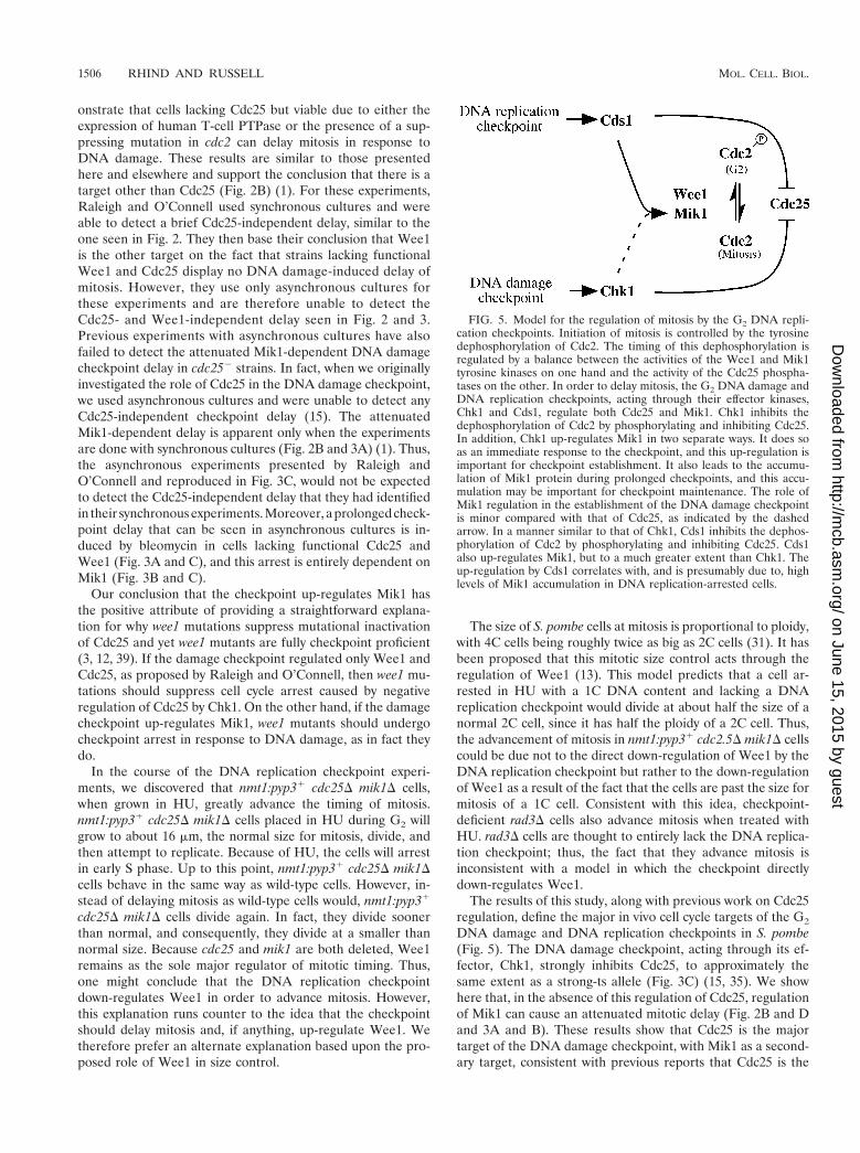
The results of this study, along with previous work on Cdc25regulation, define the major in vivo cell cycle targets of the G2
DNA damage and DNA replication checkpoints in S. pombe(Fig. 5). The DNA damage checkpoint, acting through its ef-fector, Chk1, strongly inhibits Cdc25, to approximately thesame extent as a strong-ts allele (Fig. 3C) (15, 35). We showhere that, in the absence of this regulation of Cdc25, regulationof Mik1 can cause an attenuated mitotic delay (Fig. 2B and Dand 3A and B). These results show that Cdc25 is the majortarget of the DNA damage checkpoint, with Mik1 as a second-ary target, consistent with previous reports that Cdc25 is the
FIG. 5. Model for the regulation of mitosis by the G2 DNA repli-cation checkpoints. Initiation of mitosis is controlled by the tyrosinedephosphorylation of Cdc2. The timing of this dephosphorylation isregulated by a balance between the activities of the Wee1 and Mik1tyrosine kinases on one hand and the activity of the Cdc25 phospha-tases on the other. In order to delay mitosis, the G2 DNA damage andDNA replication checkpoints, acting through their effector kinases,Chk1 and Cds1, regulate both Cdc25 and Mik1. Chk1 inhibits thedephosphorylation of Cdc2 by phosphorylating and inhibiting Cdc25.In addition, Chk1 up-regulates Mik1 in two separate ways. It does soas an immediate response to the checkpoint, and this up-regulation isimportant for checkpoint establishment. It also leads to the accumu-lation of Mik1 protein during prolonged checkpoints, and this accu-mulation may be important for checkpoint maintenance. The role ofMik1 regulation in the establishment of the DNA damage checkpointis minor compared with that of Cdc25, as indicated by the dashedarrow. In a manner similar to that of Chk1, Cds1 inhibits the dephos-phorylation of Cdc2 by phosphorylating and inhibiting Cdc25. Cds1also up-regulates Mik1, but to a much greater extent than Chk1. Theup-regulation by Cds1 correlates with, and is presumably due to, highlevels of Mik1 accumulation in DNA replication-arrested cells.
1506 RHIND AND RUSSELL MOL. CELL. BIOL.
on June 15, 2015 by guesthttp://m
cb.asm.org/
Dow
nloaded from

major target of the DNA damage checkpoint (1, 15). In con-trast, both Cdc25 and Mik1 are major targets of the DNAreplication checkpoint. The ability of Cdc25 to dephosphory-late Cdc2 in vivo is inhibited by the DNA replication check-point, and this inhibition is sufficient to delay mitosis in theabsence of Mik1 (26, 38). Conversely, Mik1 up-regulation inresponse to the DNA replication checkpoint is also sufficient todelay mitosis in most cells (Fig. 4B and D). The fact that cellslacking both Cdc25 and Mik1 are unable to delay mitosis inresponse to either DNA damage or a replication block dem-onstrates that they are the only major cell cycle targets of thetwo checkpoints (Fig. 2D, 3B, and 4D).
Fission yeast cells have provided an excellent model forframing checkpoint studies in more complex multicellular or-ganisms. Indeed, Cdc25 was first identified as a checkpointtarget through investigations of fission yeast cells, and it is nowevident that regulation of Cdc25 by Chk1 is substantially con-served among fission yeast cells, mammalian cells, and Xenopusoocytes (15, 21, 33, 35, 40). These facts raise the question ofwhether Wee1/Mik1 homologs are checkpoint targets in mul-ticellular organisms. As experiments aiming to answer thisquestion go forward, it will be important to keep in mind thatmammalian and Xenopus Wee1 protein sequences are equallyrelated to Wee1 and Mik1 in fission yeast cells. Thus, it ispossible that human Wee1 may be functionally more similar toMik1 than Wee1 in fission yeast cells. As we learn more abouthow Mik1 is regulated by checkpoints in fission yeast cells andare able to compare this knowledge to studies of Wee1/Mik1-related genes in other species, a better understanding of thefunctional distinctions between Wee1 and Mik1 will emerge.
ACKNOWLEDGMENTS
We are grateful to Kathy Gould for providing the nmt1:PTPaseconstruct with which preliminary experiments were done, MichaelBoddy for providing the glutathione S-transferase Wee1, and BethBaber-Furnari for providing the chk11:9Myc strain. We also thank themembers of the TSRI Cell Cycle Group for many interesting discus-sions and useful suggestions, in particular Jean-Marc Brondello forsuggesting the use of a conditional mik1 allele.
N.R. was supported by an NIH postdoctoral fellowship and a specialfellowship from the Leukemia and Lymphoma Society. This work wassupported by an NIH grant awarded to P.R.
REFERENCES
1. Baber-Furnari, B. A., N. Rhind, M. N. Boddy, P. Shanahan, A. Lopez-Girona, and P. Russell. 2000. Regulation of mitotic inhibitor mik1 helps toenforce the DNA damage checkpoint. Mol. Biol. Cell 11:1–11.
2. Bahler, J., J. Q. Wu, M. S. Longtine, N. G. Shah, A. McKenzie III, A. B.Steever, A. Wach, P. Philippsen, and J. R. Pringle. 1998. Heterologousmodules for efficient and versatile PCR-based gene targeting in Schizosac-charomyces pombe. Yeast 14:943–951.
3. Barbet, N. C., and A. M. Carr. 1993. Fission yeast wee1 protein kinase is notrequired for DNA damage-dependent mitotic arrest. Nature 364:824–827.
4. Blasina, A., I. V. de Weyer, M. C. Laus, W. H. Luyten, A. E. Parker, and C. H.McGowan. 1999. A human homologue of the checkpoint kinase Cds1 directlyinhibits Cdc25 phosphatase. Curr. Biol. 9:1–10.
5. Blasina, A., E. S. Paegle, and C. H. McGowan. 1997. The role of inhibitoryphosphorylation of CDC2 following DNA replication block and radiation-induced damage in human cells. Mol. Biol. Cell 8:1013–1023.
6. Boddy, M. N., B. Furnari, O. Mondesert, and P. Russell. 1998. Replicationcheckpoint enforced by kinases Cds1 and Chk1. Science 280:909–912.
7. Christensen, P. U., N. J. Bentley, R. G. Martinho, O. Nielsen, and A. M.Carr. 2000. Mik1 levels accumulate in S phase and may mediate an intrinsiclink between S phase and mitosis. Proc. Natl. Acad. Sci. USA 97:2579–2584.
8. Coleman, T. R., and W. G. Dunphy. 1994. Cdc2 regulatory factors. Curr.Opin. Cell Biol. 6:877–882.
9. Dalal, S. N., C. M. Schweitzer, J. Gan, and J. A. DeCaprio. 1999. Cytoplas-
mic localization of human cdc25C during interphase requires an intact 14-3-3binding site. Mol. Cell. Biol. 19:4465–4479.
10. Elledge, S. J. 1996. Cell cycle checkpoints: preventing an identity crisis.Science 274:1664–1672.
11. Enoch, T., A. M. Carr, and P. Nurse. 1992. Fission yeast genes involved incoupling mitosis to completion of DNA replication. Genes Dev. 6:2035–2046.
12. Fantes, P. 1979. Epistatic gene interactions in the control of division infission yeast. Nature 279:428–430.
13. Fantes, P. A., and P. Nurse. 1978. Control of the timing of cell division infission yeast. Cell size mutants reveal a second control pathway. Exp. CellRes. 115:317–329.
14. Furnari, B., A. Blasina, M. N. Boddy, C. H. McGowan, and P. Russell. 1999.Cdc25 inhibited in vivo and in vitro by checkpoint kinases Cds1 and Chk1.Mol. Biol. Cell 10:833–845.
15. Furnari, B., N. Rhind, and P. Russell. 1997. Cdc25 mitotic inducer targetedby Chk1 DNA damage checkpoint kinase. Science 277:1495–1497.
16. Gould, K. L., S. Moreno, N. K. Tonks, and P. Nurse. 1990. Complementationof the mitotic activator, p80cdc25, by a human protein-tyrosine phosphatase.Science 250:1573–1576.
17. Gould, K. L., and P. Nurse. 1989. Tyrosine phosphorylation of the fissionyeast cdc21 protein kinase regulates entry into mitosis. Nature 342:39–45.
18. Hartwell, L. H., and T. A. Weinert. 1989. Checkpoints: controls that ensurethe order of cell cycle events. Science 246:629–634.
19. Jin, P., Y. Gu, and D. O. Morgan. 1996. Role of inhibitory CDC2 phosphor-ylation in radiation-induced G2 arrest in human cells. J. Cell Biol. 134:963–970.
20. Krek, W., and E. A. Nigg. 1991. Mutations of p34cdc2 phosphorylation sitesinduce premature mitotic events in HeLa cells: evidence for a double blockto p34cdc2 kinase activation in vertebrates. EMBO J. 10:3331–3341.
21. Kumagai, A., Z. Guo, K. H. Emami, S. X. Wang, and W. G. Dunphy. 1998.The Xenopus Chk1 protein kinase mediates a caffeine-sensitive pathway ofcheckpoint control in cell-free extracts. J. Cell Biol. 142:1559–1569.
22. Kumagai, A., P. S. Yakowec, and W. G. Dunphy. 1998. 14-3-3 proteins act asnegative regulators of the mitotic inducer Cdc25 in Xenopus egg extracts.Mol. Biol. Cell 9:345–354.
23. Lindsay, H. D., D. J. Griffiths, R. J. Edwards, P. U. Christensen, J. M.Murray, F. Osman, N. Walworth, and A. M. Carr. 1998. S-phase-specificactivation of Cds1 kinase defines a subpathway of the checkpoint response inSchizosaccharomyces pombe. Genes Dev. 12:382–395.
24. Lopez-Girona, A., B. Furnari, O. Mondesert, and P. Russell. 1999. Nuclearlocalization of Cdc25 is regulated by DNA damage and a 14-3-3 protein.Nature 397:172–175.
25. Lopez-Girona, A., J. Kanoh, and P. Russell. Regulated localization of Cdc25is not required for DNA damage checkpoint control. Curr. Biol., in press.
26. Lundgren, K., N. Walworth, R. Booher, M. Dembski, M. Kirschner, and D.Beach. 1991. Mik1 and Wee1 cooperate in the inhibitory tyrosine phosphor-ylation of Cdc2. Cell 64:1111–1122.
27. Michael, W. M., and J. Newport. 1998. Coupling of mitosis to the completionof S phase through Cdc34-mediated degradation of Wee1. Science 282:1886–1889.
28. Millar, J. B. A., G. Lenaers, and P. Russell. 1992. Pyp3 PTPase acts as amitotic inducer in fission yeast. EMBO J. 11:4933–4941.
29. Moreno, S., A. Klar, and P. Nurse. 1991. Molecular genetic analysis of thefission yeast Schizosaccharomyces pombe. Methods Enzymol. 194:795–823.
30. Nurse, P. 1975. Genetic control of cell size at cell division in yeast. Nature256:547–551.
31. Nurse, P., and P. Thuriaux. 1980. Regulatory genes controlling mitosis in thefission yeast Schizosaccharomyces pombe. Genetics 96:627–637.
32. O’Connell, M. J., J. M. Raleigh, H. M. Verkade, and P. Nurse. 1997. Chk1is a wee1 kinase in the G2 DNA damage checkpoint inhibiting cdc2 by Y15phosphorylation. EMBO J. 16:545–554.
33. Peng, C. Y., P. R. Graves, R. S. Thoma, Z. Wu, A. S. Shaw, and H. Piwnica-Worms. 1997. Mitotic and G2 checkpoint control: regulation of 14-3-3 pro-tein binding by phosphorylation of Cdc25C on serine-216. Science 277:1501–1505.
34. Raleigh, J. M., and M. J. O’Connell. 2000. The G2 DNA damage checkpointtargets both Wee1 and Cdc25. J. Cell Sci. 113:1727–1736.
35. Rhind, N., B. Furnari, and P. Russell. 1997. Cdc2 tyrosine phosphorylationis required for the DNA damage checkpoint in fission yeast. Genes Dev. 11:504–511.
36. Rhind, N., and P. Russell. 2000. Chk1 and Cds1: linchpins of the DNAdamage and replication checkpoint pathways. J. Cell Sci. 113:3889–3896.
37. Rhind, N., and P. Russell. 1998. Mitotic DNA damage and replicationcheckpoints in yeast. Curr. Opin. Cell Biol. 10:749–758.
38. Rhind, N., and P. Russell. 1998. Tyrosine phosphorylation of Cdc2 is re-quired for the replication checkpoint in Schizosaccharomyces pombe. Mol.Cell. Biol. 18:3782–3787.
39. Russell, P., and P. Nurse. 1987. Negative regulation of mitosis by wee11, agene encoding a protein kinase homolog. Cell 49:559–567.
40. Sanchez, Y., C. Wong, R. S. Thoma, R. Richman, Z. Wu, H. Piwnica-Worms,and S. J. Elledge. 1997. Conservation of the Chk1 checkpoint pathway in
VOL. 21, 2001 Wee1 AND Mik1 IN G2 CHECKPOINTS 1507
on June 15, 2015 by guesthttp://m
cb.asm.org/
Dow
nloaded from

mammals: linkage of DNA damage to Cdk regulation through Cdc25. Sci-ence 277:1497–1501.
41. Toczyski, D. P., D. J. Galgoczy, and L. H. Hartwell. 1997. CDC5 and CKIIcontrol adaptation to the yeast DNA damage checkpoint. Cell 90:1097–1106.
42. Walworth, N. C., and R. Bernards. 1996. rad-dependent response of thechk1-encoded protein kinase at the DNA damage checkpoint. Science 271:353–356.
43. Yang, J., K. Winkler, M. Yoshida, and S. Kornbluth. 1999. Maintenance of
G2 arrest in the Xenopus oocyte: a role for 14-3-3-mediated inhibition ofCdc25 nuclear import. EMBO J. 18:2174–2183.
44. Zeng, Y., K. C. Forbes, Z. Wu, S. Moreno, H. Piwnica-Worms, and T. Enoch.1998. Replication checkpoint requires phosphorylation of the phosphataseCdc25 by Cds1 or Chk1. Nature 395:507–510.
45. Zeng, Y., and H. Piwnica-Worms. 1999. DNA damage and replication check-points in fission yeast require nuclear exclusion of the Cdc25 phosphatase via14-3-3 binding. Mol. Cell. Biol. 19:7410–7419.
1508 RHIND AND RUSSELL MOL. CELL. BIOL.
on June 15, 2015 by guesthttp://m
cb.asm.org/
Dow
nloaded from
Related Documents