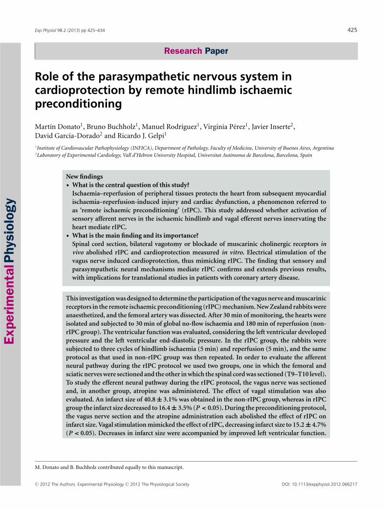
Experimental Physiology Exp Physiol 98.2 (2013) pp 425–434 425 Research Paper Research Paper Role of the parasympathetic nervous system in cardioprotection by remote hindlimb ischaemic preconditioning Mart´ ın Donato 1 , Bruno Buchholz 1 , Manuel Rodr´ ıguez 1 , Virginia P´ erez 1 , Javier Inserte 2 , David Garc´ ıa-Dorado 2 and Ricardo J. Gelpi 1 1 Institute of Cardiovascular Pathophysiology (INFICA), Department of Pathology, Faculty of Medicine, University of Buenos Aires, Argentina 2 Laboratory of Experimental Cardiology, Vall d’Hebron University Hospital, Universitat Aut` onoma de Barcelona, Barcelona, Spain New findings What is the central question of this study? Ischaemia–reperfusion of peripheral tissues protects the heart from subsequent myocardial ischaemia–reperfusion-induced injury and cardiac dysfunction, a phenomenon referred to as ‘remote ischaemic preconditioning’ (rIPC). This study addressed whether activation of sensory afferent nerves in the ischaemic hindlimb and vagal efferent nerves innervating the heart mediate rIPC. What is the main finding and its importance? Spinal cord section, bilateral vagotomy or blockade of muscarinic cholinergic receptors in vivo abolished rIPC and cardioprotection measured in vitro. Electrical stimulation of the vagus nerve induced cardioprotection, thus mimicking rIPC. The finding that sensory and parasympathetic neural mechanisms mediate rIPC confirms and extends previous results, with implications for translational studies in patients with coronary artery disease. This investigation was designed to determine the participation of the vagus nerve and muscarinic receptors in the remote ischaemic preconditioning (rIPC) mechanism. New Zealand rabbits were anaesthetized, and the femoral artery was dissected. After 30 min of monitoring, the hearts were isolated and subjected to 30 min of global no-flow ischaemia and 180 min of reperfusion (non- rIPC group). The ventricular function was evaluated, considering the left ventricular developed pressure and the left ventricular end-diastolic pressure. In the rIPC group, the rabbits were subjected to three cycles of hindlimb ischaemia (5 min) and reperfusion (5 min), and the same protocol as that used in non-rIPC group was then repeated. In order to evaluate the afferent neural pathway during the rIPC protocol we used two groups, one in which the femoral and sciatic nerves were sectioned and the other in which the spinal cord was sectioned (T9–T10 level). To study the efferent neural pathway during the rIPC protocol, the vagus nerve was sectioned and, in another group, atropine was administered. The effect of vagal stimulation was also evaluated. An infarct size of 40.8 ± 3.1% was obtained in the non-rIPC group, whereas in rIPC group the infarct size decreased to 16.4 ± 3.5% (P < 0.05). During the preconditioning protocol, the vagus nerve section and the atropine administration each abolished the effect of rIPC on infarct size. Vagal stimulation mimicked the effect of rIPC, decreasing infarct size to 15.2 ± 4.7% (P < 0.05). Decreases in infarct size were accompanied by improved left ventricular function. M. Donato and B. Buchholz contributed equally to this manuscript. C 2012 The Authors. Experimental Physiology C 2012 The Physiological Society DOI: 10.1113/expphysiol.2012.066217

Welcome message from author
This document is posted to help you gain knowledge. Please leave a comment to let me know what you think about it! Share it to your friends and learn new things together.
Transcript
Expe
rim
enta
lPhy
siol
ogy
Exp Physiol 98.2 (2013) pp 425–434 425
Research PaperResearch Paper
Role of the parasympathetic nervous system incardioprotection by remote hindlimb ischaemicpreconditioning
Martın Donato1, Bruno Buchholz1, Manuel Rodrıguez1, Virginia Perez1, Javier Inserte2,David Garcıa-Dorado2 and Ricardo J. Gelpi1
1Institute of Cardiovascular Pathophysiology (INFICA), Department of Pathology, Faculty of Medicine, University of Buenos Aires, Argentina2Laboratory of Experimental Cardiology, Vall d’Hebron University Hospital, Universitat Autonoma de Barcelona, Barcelona, Spain
New findings� What is the central question of this study?
Ischaemia–reperfusion of peripheral tissues protects the heart from subsequent myocardialischaemia–reperfusion-induced injury and cardiac dysfunction, a phenomenon referred toas ‘remote ischaemic preconditioning’ (rIPC). This study addressed whether activation ofsensory afferent nerves in the ischaemic hindlimb and vagal efferent nerves innervating theheart mediate rIPC.
� What is the main finding and its importance?Spinal cord section, bilateral vagotomy or blockade of muscarinic cholinergic receptors invivo abolished rIPC and cardioprotection measured in vitro. Electrical stimulation of thevagus nerve induced cardioprotection, thus mimicking rIPC. The finding that sensory andparasympathetic neural mechanisms mediate rIPC confirms and extends previous results,with implications for translational studies in patients with coronary artery disease.
This investigation was designed to determine the participation of the vagus nerve and muscarinicreceptors in the remote ischaemic preconditioning (rIPC) mechanism. New Zealand rabbits wereanaesthetized, and the femoral artery was dissected. After 30 min of monitoring, the hearts wereisolated and subjected to 30 min of global no-flow ischaemia and 180 min of reperfusion (non-rIPC group). The ventricular function was evaluated, considering the left ventricular developedpressure and the left ventricular end-diastolic pressure. In the rIPC group, the rabbits weresubjected to three cycles of hindlimb ischaemia (5 min) and reperfusion (5 min), and the sameprotocol as that used in non-rIPC group was then repeated. In order to evaluate the afferentneural pathway during the rIPC protocol we used two groups, one in which the femoral andsciatic nerves were sectioned and the other in which the spinal cord was sectioned (T9–T10 level).To study the efferent neural pathway during the rIPC protocol, the vagus nerve was sectionedand, in another group, atropine was administered. The effect of vagal stimulation was alsoevaluated. An infarct size of 40.8 ± 3.1% was obtained in the non-rIPC group, whereas in rIPCgroup the infarct size decreased to 16.4 ± 3.5% (P < 0.05). During the preconditioning protocol,the vagus nerve section and the atropine administration each abolished the effect of rIPC oninfarct size. Vagal stimulation mimicked the effect of rIPC, decreasing infarct size to 15.2 ± 4.7%(P < 0.05). Decreases in infarct size were accompanied by improved left ventricular function.
M. Donato and B. Buchholz contributed equally to this manuscript.
C© 2012 The Authors. Experimental Physiology C© 2012 The Physiological Society DOI: 10.1113/expphysiol.2012.066217
426 M. Donato and others Exp Physiol 98.2 (2013) pp 425–434
We demonstrated the presence of a neural afferent pathway, because the spinal cord sectioncompletely abolished the effect of rIPC on infarct size. In conclusion, rIPC activates a neuralafferent pathway, the cardioprotective signal reaches the heart through the vagus nerve (efferentpathway), and acetylcholine activates the ischaemic preconditioning phenomenon when actingon the muscarinic receptors.
(Received 23 March 2012; accepted after revision 31 July 2012; first published online 7 August 2012)Corresponding author R. J. Gelpi: Institute of Cardiovascular Pathophysiology, Faculty of Medicine, University ofBuenos Aires, J. E. Uriburu 950, 2nd floor, Buenos Aires, Argentina, C1114AAD. Email: [email protected]
Ischaemic preconditioning (IPC) is a cardioprotectivestrategy whereby prior brief episodes of ischaemiainduce a state of protection against subsequentprolonged ischaemia–reperfusion (I/R; Murry et al.1986). As IPC is not applicable in a clinical setting, avariant preconditioning stimulus called remote ischaemicpreconditioning (rIPC) has received increasing attention.rIPC describes the phenomenon whereby transientregional ischaemia of a tissue at a distance from the targetorgan (heart) affords protection with the same efficacyas local IPC. Thus, brief episodes of I/R applied to thesmall intestine, kidney, liver or even hindlimb have beenreported to reduce subsequent myocardial infarct size(Takaoka et al. 1999; Huda et al. 2005; Li et al. 2010).
The mechanisms underlying rIPC have not beenfully elucidated. Previous studies have attributed thecardioprotective signal from the preconditioned remoteorgan to either a neural pathway (Lim et al. 2010) or ahumoral pathway (Konstantinov et al. 2005), althoughboth are not mutually exclusive. Gho et al. (1996)demonstrated that a reduction in myocardial infarctsize, induced by brief I/R of the anterior mesentericartery, could be reversed by hexamethonium (a ganglionblocker), supporting the neural pathway hypothesis. Otherauthors (Lim et al. 2010), in a model of hindlimbischaemia, showed that rIPC protection requires acomplete innervation of the lower limb. The hypothesison a neural pathway was further developed, makingallusion to the endogenous substances that are releasedby the remote preconditioned organ, such as adenosine(Steensrud et al. 2010), bradykinin (Schoemaker & vanHeijningen, 2000) and calcitonin gene-related peptide(Wolfrum et al. 2005), that stimulate afferent nerve fibres.However, few studies (Jones et al. 2009; Basalay et al.2012) describe the possible efferent neural pathway bywhich the signal reaches the heart from the remoteorgan and only one of them studied the possible roleof the parasympathetic nervous system (Basalay et al.2012).
The parasympathetic nervous system, through thevagus nerve, has been implicated in myocardial protection.Kawada et al. (2009) demonstrated that pre-ischaemicefferent vagal stimulation increases acetylcholine releaseand protects the heart from I/R injury.
We hypothesized that rIPC activates a neural pathwayand that the cardioprotective signal reaches the heart viathe vagus nerve. We also hypothesized that acetylcholinereleased by the nervous terminal of the vagus nervecould precondition the heart through the activation ofmuscarinic receptors. Thus, the objective of this work wasto determine the possible participation of the vagus nerveand muscarinic receptors in the rIPC cardioprotectivemechanism.
Methods
Ethical approval
The experiments were performed on 54 male NewZealand rabbits (1.8–2.5 kg). The procedures used in thisstudy were approved by the Animal Care and ResearchCommittee of the University of Buenos Aires and werein compliance with the Guide for the Care and Useof Laboratory Animals published by the US NationalInstitutes of Health (NIH publication, Eight edition;2010).
Surgical procedure
First, the rabbits were anaesthetized with pentobarbital(35 mg kg−1, I.V.) and intubated for mechanical ventilationwith a mixture of ambient air and oxygen. Second, afluid-filled catheter was placed into the right commoncarotid artery and connected to a pressure transducer(Deltram II; Utah Medical Products, Midvale, UT, USA)to record the arterial pressure and calculate the heart rate.Third, the femoral artery was dissected and the animalswere randomized into different experimental groups.Anaesthesia was maintained using 5–10 mg kg−1 h−1 I.V.pentobarbital when required.
After completion of the in vivo protocols describedin the following paragraphs, the animals were killedby administration of pentobarbital (150 mg kg−1 I.V.)and each heart was rapidly excised and mounted on aLangendorff apparatus by the aortic root in less than1 min. Each heart was perfused with Krebs–Henseleitbuffer containing (mM): NaCl, 118.5; KCl, 4.7; NaHCO3,24.8; KH2PO4, 1.2; MgSO4, 1.2; CaCl2, 2.5; and glucose,
C© 2012 The Authors. Experimental Physiology C© 2012 The Physiological Society
Exp Physiol 98.2 (2013) pp 425–434 Remote ischaemic preconditioning and parasympathetic nervous system 427
10; the pH was maintained at 7.2–7.4 by aerating thesolution with a gas mixture 95% O2–5% CO2 at 37◦C.Two electrodes were sutured and connected to a pacemakerwith a constant heart rate of 200 beats min−1.
A saline-filled latex balloon, connected via a catheter to apressure transducer (Deltram II; Utah Medical Products),was inserted into the left ventricle (LV). The volume ofthe balloon was adjusted to an end-diastolic pressure of 8–10 mmHg. Coronary perfusion pressure was also recordedthrough a pressure transducer connected to the perfusionline. All hearts were perfused with constant flow.
Coronary flow was adjusted to obtain a coronaryperfusion pressure of 70.5 ± 4.2 mmHg during theinitial stabilization period. This flow was held constantthroughout the experiment. The left ventricular developedpressure (LVDP) was calculated from the differencebetween peak systolic pressure and left ventricular end-diastolic pressure (LVEDP). Ventricular function wasassessed at baseline during the first 30 min of reperfusion,although the hearts were reperfused for 180 min in orderto measure infarct size.
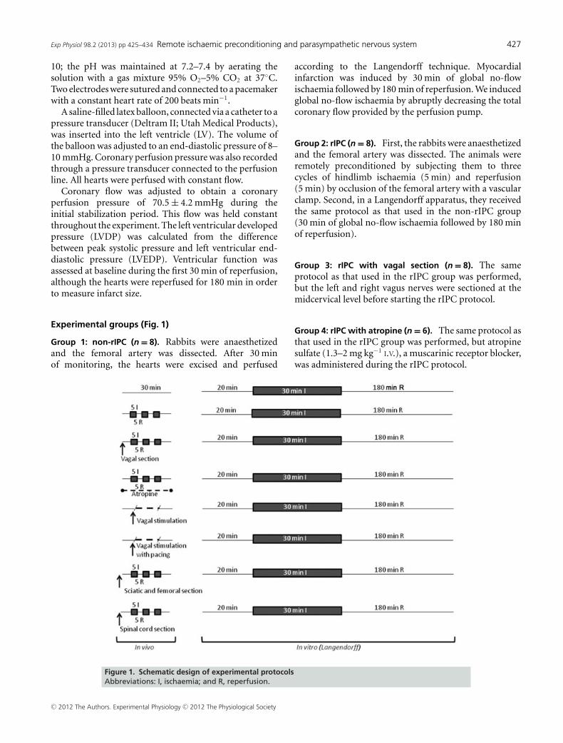
Experimental groups (Fig. 1)
Group 1: non-rIPC (n = 8). Rabbits were anaesthetizedand the femoral artery was dissected. After 30 minof monitoring, the hearts were excised and perfused
according to the Langendorff technique. Myocardialinfarction was induced by 30 min of global no-flowischaemia followed by 180 min of reperfusion. We inducedglobal no-flow ischaemia by abruptly decreasing the totalcoronary flow provided by the perfusion pump.
Group 2: rIPC (n = 8). First, the rabbits were anaesthetizedand the femoral artery was dissected. The animals wereremotely preconditioned by subjecting them to threecycles of hindlimb ischaemia (5 min) and reperfusion(5 min) by occlusion of the femoral artery with a vascularclamp. Second, in a Langendorff apparatus, they receivedthe same protocol as that used in the non-rIPC group(30 min of global no-flow ischaemia followed by 180 minof reperfusion).
Group 3: rIPC with vagal section (n = 8). The sameprotocol as that used in the rIPC group was performed,but the left and right vagus nerves were sectioned at themidcervical level before starting the rIPC protocol.
Group 4: rIPC with atropine (n = 6). The same protocol asthat used in the rIPC group was performed, but atropinesulfate (1.3–2 mg kg−1 I.V.), a muscarinic receptor blocker,was administered during the rIPC protocol.
Figure 1. Schematic design of experimental protocolsAbbreviations: I, ischaemia; and R, reperfusion.
C© 2012 The Authors. Experimental Physiology C© 2012 The Physiological Society
428 M. Donato and others Exp Physiol 98.2 (2013) pp 425–434
Group 5: vagal stimulation (n = 5). First, the rabbitswere anaesthetized. The right vagus nerve was sectionedat the midcervical level and its distal end electricallystimulated for 10 min followed by 5 min of recoverywithout stimulation. Second, the hearts were excisedand perfused according to the Langendorff technique.Myocardial infarction was induced by 30 min of globalno-flow ischaemia followed by 180 min of reperfusion.
Group 6: vagal stimulation with pacing (n = 4). The sameprotocol as that used in the vagal stimulation group wasperformed, but during the electrical stimulation of thevagus nerve the heart was paced at a constant rate equal to10% above baseline.
Group 7: rIPC with section of the femoral (F) and sciatic(S) nerves (n = 10). The same protocol as that used in therIPC group was performed, but the femoral and sciaticnerves were sectioned before starting the rIPC protocol.The hearts were then excised and perfused accordingto the Langendorff technique. Myocardial infarction wasinduced by 30 min of global no-flow ischaemia followedby 180 min of reperfusion.
Group 8: rIPC with spinal cord section (n = 5). The sameprotocol as that used in the rIPC group was performed,but the spinal cord was sectioned between T9 and T10before starting the rIPC protocol (Greenaway et al. 2001;Jones et al. 2009). In detail, rabbits were anaesthetizedand bone markers identified at the thoracic vertebrae T9–T10 levels. Under optical magnification, the spinal cordbetween T9 and T10 was transected by a sharp knife,with limited bleeding. After that, the hearts were excisedand perfused according to the Langendorff technique.Myocardial infarction was induced by 30 min of globalno-flow ischaemia followed by 180 min of reperfusion.
It is important to mention that the mean time betweenthe cardioprotective stimulus and the onset of the periodof global ischaemia was no longer than 25 min.
Infarct size measurement
After 3 h of reperfusion, the hearts were frozen and cutinto 4 mm transverse slices from apex to base. Sectionswere incubated for 20 min in 1% triphenyltetrazoliumchloride (pH 7.4, 37◦C) and then immersed in 10%formalin. Applying this technique, viable sections werestained red and the infarct area remained unstained.Sections were scanned and measured (Image Pro Plus,version 4.5, MediaCybernetics, Rockville, MD, USA). Theinfarct size was expressed as a percentage of the leftventricular area. The demarcation of the infarct zone bytetrazolium staining is dependent on the loss of NADHand of dehydrogenases from the irreversibly damaged
myocytes. In this context, Birnbaum et al. (1997) describedthat at least 3 h of reperfusion is needed to delineate infarctsize by tetrazolium staining following 30 min of ischaemiain the rabbit.
Vagal stimulation
The right vagus nerve was sectioned at the midcervicallevel and its distal end was stimulated, using a bipolarsilver electrode connected to a neurostimulator (HugoSachs Elektronik D7801, March-Hugstetten, Germany).The animals were subjected to rectangular electrical pulsesof 0.1 ms, 10 Hz (the intensity of electrical stimulation wasadjusted for each animal) in order to obtain a heart ratereduction between 10 and 20%. This stimulation protocolhas been widely used by other authors (Kawada et al. 2001;Uemura et al. 2007).
Statistical analysis
Data are expressed as means ± SEM. Intergroupcomparisons were carried out using one-way ANOVAfollowed by Student’s unpaired t tests with the P valueadjusted for multiple comparisons using the Bonferronitest. The data comparisons were not significantunless the corresponding P value was less than 0.05/k,where k represents the number of comparisons.The intragroup comparisons were analysed by two-factor repeated-measures ANOVA. Comparisons betweencardiac function measurements postmyocardial ischaemiaversus baseline were performed by Student’s paired t test.
Results
Table 1 shows in vivo measurement of heart rate andmean arterial pressure during application of the remoteischaemic preconditioning stimulus. There were nosignificant differences in the baseline values during thedifferent interventions among groups. As expected, in thevagal stimulation group, there was a significant reductionof heart rate from 267 ± 15 to 223 ± 8 beats min−1
(P < 0.05) and there were no changes in mean arterialpressure. When the pre-ischaemic vagal stimulation(10 min) was performed at constant heart rate we did notobserve changes in blood pressure (data not shown).
Interestingly, the rIPC also significantly reduced theheart rate, an effect that was reversed with the vagal section(Table 1). These results provide evidence for an increase invagal tone in the group subjected to rIPC. It is importantto mention that this effect was detected in the presenceof pentobarbital as an anaesthetic agent, even though thisdrug has the ability to inhibit parasympathetic efferentactivity.
C© 2012 The Authors. Experimental Physiology C© 2012 The Physiological Society
Exp Physiol 98.2 (2013) pp 425–434 Remote ischaemic preconditioning and parasympathetic nervous system 429
Table 1. Changes in heart rate and mean arterial pressure during remote hindlimb ischaemic preconditioning protocol
Parameter Group Baseline 5 Isch. (1) 5 Rep. (1) 5 Isch. (2) 5 Rep. (2) 5 Isch. (3) 5 Rep. (3)
Heart rate Non-rIPC 275 ± 24 277 ± 25 276 ± 25 271 ± 23 273 ± 22 273 ± 21 266 ± 20(beats min−1) rIPC 298 ± 10 294 ± 14 281 ± 12 281 ± 7 267 ± 13 272 ± 10 264 ± 10∗
rIPC with vagalsection
310 ± 17 304 ± 19 305 ± 18 305 ± 20 302 ± 19 298 ± 21 300 ± 20
rIPC withatropine
290 ± 19 290 ± 18 271 ± 21 271 ± 19 273 ± 15 278 ± 15 279 ± 16
rIPC with F and Ssection
284 ± 10 283 ± 10 284 ± 10 285 ± 10 283 ± 11 284 ± 10 284 ± 10
rIPC with spinalcord section
312 ± 26 316 ± 24 320 ± 22 319 ± 23 317 ± 24 319 ± 24 319 ± 23
Mean arterial Non-rIPC 88.7 ± 5.3 87.3 ± 5.3 85.2 ± 4.5 83.7 ± 5.7 87.5 ± 3.7 88.5 ± 5.1 85.1 ± 4.2pressure rIPC 84.9 ± 5.4 82.1 ± 6.5 81.6 ± 2.7 82.9 ± 3.4 83.5 ± 4.9 81.1 ± 3.5 78.6 ± 1.9(mmHg) rIPC with vagal
section99.1 ± 1.5 95.7 ± 3.2 97.1 ± 2.5 94.9 ± 4.9 97.4 ± 1.7 97.1 ± 2.4 95.6 ± 1.7
rIPC withatropine
83.6 ± 2.1 82.7 ± 8.6 82.7 ± 5.4 79.3 ± 0.1 81.1 ± 3.9 80.1 ± 3.7 79.6 ± 3.7
rIPC with F and Ssection
92.1 ± 3.3 92.1 ± 2.8 90.6 ± 2.3 90.7 ± 3.5 91.7 ± 3.2 89.6 ± 2.9 89.6 ± 3.1
rIPC with spinalcord section
85.9 ± 8.5 84.5 ± 8.4 85.1 ± 9.1 85.1 ± 9.4 84.5 ± 8.8 83.2 ± 8.6 80.7 ± 9.6
Abbreviations: F, femoral nerve; Isch., ischaemia; Rep., reperfusion; rIPC, remote ischaemic preconditioning; and S, sciatic nerve. Thenumbers in parentheses indicate the number of preconditioning cycles. ∗P < 0.05 versus baseline.
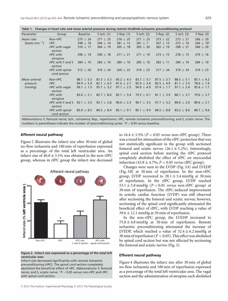
Afferent neural pathway
Figure 2 illustrates the infarct size after 30 min of globalno-flow ischaemia and 180 min of reperfusion expressedas a percentage of the total left ventricular area. Aninfarct size of 40.8 ± 3.1% was obtained in the non-rIPCgroup, whereas in rIPC group the infarct size decreased
Figure 2. Infarct size expressed as a percentage of the total leftventricular areaInfarct size decreased significantly with remote ischaemicpreconditioning (rIPC). The spinal cord section completelyabolished the beneficial effect of rIPC. Abbreviations: F, femoralnerve; and S, sciatic nerve. ∗P < 0.05 versus non-rIPC and rIPCwith spinal cord section.
to 16.4 ± 3.5% (P < 0.05 versus non-rIPC group). Therewas a trend for attenuation of the rIPC protection that wasnot statistically significant in the group with sectionedfemoral and sciatic nerves (26.1 ± 5.2%). Interestingly,spinal cord section before starting the rIPC protocolcompletely abolished the effect of rIPC on myocardialinfarction (43.8 ± 4.7%; P < 0.05 versus rIPC group).
Changes were seen in the LVDP (Fig. 3A) and LVEDP(Fig. 3B) at 30 min of reperfusion. In the non-rIPCgroup, LVDP recovered to 28.1 ± 3.4 mmHg at 30 minof reperfusion. In the rIPC group, LVDP reached53.1 ± 5.8 mmHg (P < 0.05 versus non-rIPC group) at30 min of reperfusion. The rIPC-induced improvementin systolic cardiac function (LVDP) was still observedafter sectioning the femoral and sciatic nerves; however,sectioning of the spinal cord significantly attenuated thebeneficial effect of rIPC, with LVDP reaching a value of39.6 ± 12.1 mmHg at 30 min of reperfusion.
In the non-rIPC group, the LVEDP increased to75.8 ± 4.8 mmHg at 30 min of reperfusion. Remoteischaemic preconditioning attenuated the increase ofLVEDP, which reached a value of 32.4 ± 6.2 mmHg at30 min of reperfusion (P < 0.05). This effect was abolishedby spinal cord section but was not affected by sectioningthe femoral and sciatic nerves (Fig. 3).
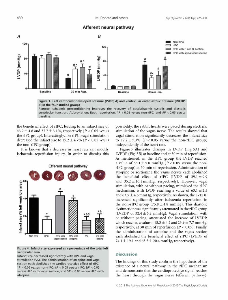
Efferent neural pathway
Figure 4 illustrates the infarct size after 30 min of globalno-flow ischaemia and 180 min of reperfusion expressedas a percentage of the total left ventricular area. The vagalsection and the administration of atropine each abolished
C© 2012 The Authors. Experimental Physiology C© 2012 The Physiological Society
430 M. Donato and others Exp Physiol 98.2 (2013) pp 425–434
Figure 3. Left ventricular developed pressure (LVDP; A) and ventricular end-diastolic pressure (LVEDP;B) in the four studied groupsRemote ischaemic preconditioning improves the recovery of postischaemic systolic and diastolicventricular function. Abbreviation: Rep., reperfusion. ∗P < 0.05 versus non-rIPC; and #P < 0.05 versusbaseline.
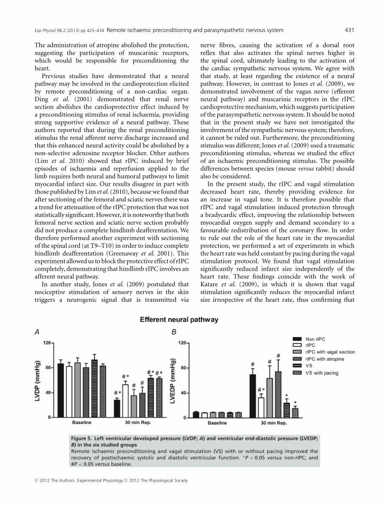
the beneficial effect of rIPC, leading to an infarct size of43.2 ± 4.8 and 37.7 ± 3.1%, respectively (P < 0.05 versusthe rIPC group). Interestingly, like rIPC, vagal stimulationdecreased the infarct size to 15.2 ± 4.7% (P < 0.05 versusthe non-rIPC group).
It is known that a decrease in heart rate can modifyischaemia–reperfusion injury. In order to dismiss this
Figure 4. Infarct size expressed as a percentage of the total leftventricular areaInfarct size decreased significantly with rIPC and vagalstimulation (VS). The administration of atropine and vagalsection each abolished the cardioprotective effect of rIPC.∗P < 0.05 versus non-rIPC; #P < 0.05 versus rIPC; &P < 0.05versus rIPC with vagal section; and $P < 0.05 versus rIPC withatropine.
possibility, the rabbit hearts were paced during electricalstimulation of the vagus nerve. The results showed thatvagal stimulation significantly decreases the infarct sizeto 17.2 ± 5.3% (P < 0.05 versus the non-rIPC group)independently of the heart rate.
Figure 5 illustrates changes in LVDP (Fig. 5A) andLVEDP (Fig. 5B) at baseline and at 30 min of reperfusion.As mentioned, in the rIPC group the LVDP reacheda value of 53.1 ± 5.8 mmHg (P < 0.05 versus the non-rIPC group) at 30 min of reperfusion. Administration ofatropine or sectioning the vagus nerves each abolishedthe beneficial effect of rIPC (LVDP of 39.1 ± 9.9and 35.2 ± 10.1 mmHg, respectively). However, vagalstimulation, with or without pacing, mimicked the rIPCmechanism, with LVDP reaching a value of 63.1 ± 2.5and 63.5 ± 4.6 mmHg, respectively. As shown, the LVEDPincreased significantly after ischaemia–reperfusion inthe non-rIPC group (75.8 ± 4.8 mmHg). This diastolicdysfunction was significantly attenuated in the rIPC group(LVEDP of 32.4 ± 6.2 mmHg). Vagal stimulation, withor without pacing, attenuated the increase of LVEDP,which reached a value of 15.3 ± 4.2 and 23.9 ± 7.7 mmHg,respectively, at 30 min of reperfusion (P < 0.05). Finally,the administration of atropine and the vagus sectioneach abolished the beneficial effect of rIPC (LVEDP of74.1 ± 19.1 and 63.5 ± 20.4 mmHg, respectively).
Discussion
The findings of this study confirm the hypothesis of theexistence of a neural pathway in the rIPC mechanismand demonstrate that the cardioprotective signal reachesthe heart through the vagus nerve (efferent pathway).
C© 2012 The Authors. Experimental Physiology C© 2012 The Physiological Society
Exp Physiol 98.2 (2013) pp 425–434 Remote ischaemic preconditioning and parasympathetic nervous system 431
The administration of atropine abolished the protection,suggesting the participation of muscarinic receptors,which would be responsible for preconditioning theheart.
Previous studies have demonstrated that a neuralpathway may be involved in the cardioprotection elicitedby remote preconditioning of a non-cardiac organ.Ding et al. (2001) demonstrated that renal nervesection abolishes the cardioprotective effect induced bya preconditioning stimulus of renal ischaemia, providingstrong supportive evidence of a neural pathway. Theseauthors reported that during the renal preconditioningstimulus the renal afferent nerve discharge increased andthat this enhanced neural activity could be abolished by anon-selective adenosine receptor blocker. Other authors(Lim et al. 2010) showed that rIPC induced by briefepisodes of ischaemia and reperfusion applied to thelimb requires both neural and humoral pathways to limitmyocardial infarct size. Our results disagree in part withthose published by Lim et al. (2010), because we found thatafter sectioning of the femoral and sciatic nerves there wasa trend for attenuation of the rIPC protection that was notstatistically significant. However, it is noteworthy that bothfemoral nerve section and sciatic nerve section probablydid not produce a complete hindlimb deafferentation. Wetherefore performed another experiment with sectioningof the spinal cord (at T9–T10) in order to induce completehindlimb deafferentation (Greenaway et al. 2001). Thisexperiment allowed us to block the protective effect of rIPCcompletely, demonstrating that hindlimb rIPC involves anafferent neural pathway.
In another study, Jones et al. (2009) postulated thatnociceptive stimulation of sensory nerves in the skintriggers a neurogenic signal that is transmitted via
nerve fibres, causing the activation of a dorsal rootreflex that also activates the spinal nerves higher inthe spinal cord, ultimately leading to the activation ofthe cardiac sympathetic nervous system. We agree withthat study, at least regarding the existence of a neuralpathway. However, in contrast to Jones et al. (2009), wedemonstrated involvement of the vagus nerve (efferentneural pathway) and muscarinic receptors in the rIPCcardioprotective mechanism, which suggests participationof the parasympathetic nervous system. It should be notedthat in the present study we have not investigated theinvolvement of the sympathetic nervous system; therefore,it cannot be ruled out. Furthermore, the preconditioningstimulus was different; Jones et al. (2009) used a traumaticpreconditioning stimulus, whereas we studied the effectof an ischaemic preconditioning stimulus. The possibledifferences between species (mouse versus rabbit) shouldalso be considered.
In the present study, the rIPC and vagal stimulationdecreased heart rate, thereby providing evidence foran increase in vagal tone. It is therefore possible thatrIPC and vagal stimulation induced protection througha bradycardic effect, improving the relationship betweenmyocardial oxygen supply and demand secondary to afavourable redistribution of the coronary flow. In orderto rule out the role of the heart rate in the myocardialprotection, we performed a set of experiments in whichthe heart rate was held constant by pacing during the vagalstimulation protocol. We found that vagal stimulationsignificantly reduced infarct size independently of theheart rate. These findings coincide with the work ofKatare et al. (2009), in which it is shown that vagalstimulation significantly reduces the myocardial infarctsize irrespective of the heart rate, thus confirming that
Figure 5. Left ventricular developed pressure (LVDP; A) and ventricular end-diastolic pressure (LVEDP;B) in the six studied groupsRemote ischaemic preconditioning and vagal stimulation (VS) with or without pacing improved therecovery of postischaemic systolic and diastolic ventricular function. ∗P < 0.05 versus non-rIPC; and#P < 0.05 versus baseline.
C© 2012 The Authors. Experimental Physiology C© 2012 The Physiological Society
432 M. Donato and others Exp Physiol 98.2 (2013) pp 425–434
the protective effects induced by vagal stimulation areindependent of its bradycardic effect. However, otherauthors have shown that an increase in the metabolicdemand of the myocardium induced by tachycardia, in theabsence of ischaemia, decreases the infarct size (Domenechet al. 1998).
On the contrary, it is known that the autonomicnervous system participates in the physiological andpathophysiological regulation of the mammalian heart.This regulation is established by sympathetic andparasympathetic cardiac nerves (Janes et al. 1986). Inaddition, there is evidence that neurons in the nucleusambiguous and dorsal vagal nucleus innervate differentsubpopulations of intrinsic cardiac postganglionicneurons (Cheng et al. 2004). In particular, the autonomicnervous system, via the vagus nerve, has been implicatedin phenomena of myocardial protection.
In this context we have recently described (Buchholzet al. 2012a,b) that vagal efferent stimulation, incertain conditions, coactivates the sympathetic nervoussystem together with an increase in plasma levels ofcatecholamines and myocardial consumption of oxygen,which, in vivo, increase the infarct size. However, when thevagus nerve is stimulated and the heart is then isolated, thebeneficial effects of vagal stimulation are evidenced. In thissense, it is important to mention that in the present studymyocardial ischaemia–reperfusion was induced in vitro,because our main objective was to study the myocardialinfarction and ventricular function in the absence ofextracardiac regulatory systems. Additionally, this modelallowed us to exclude any effects of changes in heart rateand loading conditions.
As mentioned above, efferent vagal stimulation inducesan increased release of acetylcholine, which could be oneof the beneficial mechanisms of vagal stimulation in thecontext of myocardial ischaemia (Kawada et al. 2001). Inthe heart, acetylcholine is the principal neurotransmitterof the postganglionic fibres of the parasympatheticefferent pathways, and binding to the muscarinic receptorsgenerates intracellular signals mediated by the G-protein-coupled receptor. Thus, in this work we demonstratedthat vagal stimulation, performed before ischaemia,mimics the beneficial effect of rIPC on myocardialinfarct size. Also, the bilateral vagotomy, which leadsto the loss of the cardioprotection provided by rIPC,allows us to assert that rIPC could activate a neuralpathway whereby the cardioprotective signal reaches theheart via the vagus nerve (efferent neural pathway).As increased vagal efferent nerve activity may lead toactivation of nicotinic as well as muscarinic cholinergicreceptors and release neuromodulators such as vasoactiveintestinal polypeptide, we administered atropine duringthe rIPC protocol. The results show that the acetylcholinereleased by the vagus nerve endings induces therIPC phenomenon by activating muscarinic receptors.
Although the involvement of muscarinic receptors hasbeen demonstrated in classic preconditioning (Oldenburget al. 2002), the possible role of muscarinic receptorsin rIPC has not been evaluated. Surprisingly, theadministration of atropine did not increase the heartrate, as expected, perhaps due to interaction with thepentobarbital. However, it is clear that the dose usedcaused complete abolition of the protective effect of therIPC.
In contrast, different studies have suggested thatthe reperfusion period should wash out a substanceor humoral factor generated by the preconditioningischaemia, which is then transported to the heart (Ghoet al. 1996). Thus, recently Redington et al. (2012)showed that direct femoral nerve stimulation and C fibrestimulation by capsaicin are associated with the release ofa blood-borne cardioprotective factor(s) similar to thatassociated with rIPC induced by transient limb ischaemia.Different experimental studies have attempted to identifythe humoral factor involved in the rIPC mechanismand have suggested that endogenous substances such asadenosine (Pell et al. 1998), bradykinin (Schoemaker &van Heijningen, 2000), opioids (Patel et al. 2002) andendocannabinoids (Hajrasouliha et al. 2008) are releasedfrom the remote organ during the preconditioningischaemia and are carried to the heart via the blood-stream, where they then activate intracellular pathwaysof cardioprotection. Despite this evidence, our data donot support this hypothesis. Although we cannot excludethe possible involvement of a humoral factor, it is clearthat sectioning of the vagus nerve abolishes the rIPC-induced cardioprotection. In this sense, our results arein agreement with those obtained by Basalay et al.(2012), who demonstrated the involvement of the vagusnerve in the rIPC mechanism. In the present study, wehave extended this knowledge because the administrationof atropine abolished the protection, suggesting thatacetylcholine released from vagal efferent nerves bindsto muscarinic receptors on myocytes, which wouldbe responsible for preconditioning the heart. Anotherdifference from the study of Basalay et al. (2012) is thatwe detected myocardial protection secondary to rIPC,not only on infarct size but also on the postischaemicventricular function.
The first description of rIPC (Przyklenk et al. 1993)shows that the transient ischaemia of one coronary arteryterritory reduces the effects of subsequent potentiallylethal ischaemia in the territory of another coronaryartery. It would be interesting to investigate whether theparasympathetic nervous system and the vagus nerve(efferent neural pathway) are involved in this intracardiacremote preconditioning, because this has not been studiedin myocardial infarction, but only suggested in models ofgastric injury (Brzozowski et al. 2004) to the best of ourknowledge.
C© 2012 The Authors. Experimental Physiology C© 2012 The Physiological Society
Exp Physiol 98.2 (2013) pp 425–434 Remote ischaemic preconditioning and parasympathetic nervous system 433
In conclusion, the present study demonstrates that thecardioprotection induced by rIPC involves an afferentneural signal, because the protection was lost afterthe spinal cord was sectioned. In addition, activationof efferent parasympathetic nerves and muscariniccholinergic receptors is involved in rIPC, given that thecardioprotection was lost after sectioning of the vagusnerves and after administration of atropine. As rIPC couldbe an accessible procedure in clinical practice to renderthe heart resistant to ischaemia, further investigation isrequired to study cellular mechanisms in detail.
References
Basalay M, Barsukevich V, Mastitskaya S, Mrochek A, PernowJ, Sjoquist PO, Ackland GL, Gourine AV & Gourine A(2012). Remote ischaemic pre- and delayedpostconditioning – similar degree of cardioprotection butdistinct mechanisms. Exp Physiol 97, 908–917.
Birnbaum Y, Hale SL & Kloner RA (1997). Differences inreperfusion length following 30 minutes of ischemia in therabbit influence infarct size, as measured bytriphenyltetrazolium chloride staining. J Mol Cell Cardiol 29,657–666.
Brzozowski T, Konturek PC, Pajdo R, Kwiecien S, Sliwowski Z,Drozdowicz D, Ptak-Belowska A, Pawlik M, Konturek SJ,Pawlik WW & Hahn GG (2004). Importance of brain-gutaxis in the gastroprotection induced by gastric and remotepreconditioning. J Physiol Pharmacol 55, 165–177.
Buchholz B, Donato M, Perez V, Ivalde FC, Hocht C, BuitragoE, Rodrıguez M & Gelpi RJ (2012a). Preischemic efferentvagal stimulation increases the size of myocardial infarctionin rabbits. Role of the sympathetic nervous system. Int JCardiol 155, 490–491.
Buchholz B, Siachoque N, Rodrıguez M, Ivalde FC, AlvarezYuseff MF & Gelpi RJ (2012b). Efferent vagal stimulationincreases infarct size in rabbits. Rev Argent Cardiol 80, 7–13.
Cheng Z, Zhang H, Guo SZ, Wurster R & Gozal D (2004).Differential control over postganglionic neurons in ratcardiac ganglia by NA and DmnX neurons: anatomicalevidence. Am J Physiol Regul Integr Comp Physiol 286,R625–R633.
Ding YF, Zhang MM & He RR (2001). Role of renal nerve incardioprotection provided by renal ischemic preconditioningin anesthetized rabbits. Sheng Li Xue Bao 53, 7–12.
Domenech RJ, Macho P, Velez D, Sanchez G, Liu X & Dhalla N(1998). Tachycardia preconditions infarct size in dogs: role ofadenosine and protein kinase C. Circulation 97, 786–794.
Gho BC, Schoemaker RG, van den Doel MA, Duncker DJ &Verdouw PD (1996). Myocardial protection by brief ischemiain noncardiac tissue. Circulation 94, 2193–2200.
Greenaway JB, Partlow GD, Gonsholt NL & Fisher KR (2001).Anatomy of the lumbosacral spinal cord in rabbits. J AmAnim Hosp Assoc 37, 27–34.
Hajrasouliha AR, Tavakoli S, Ghasemi M, Jabehdar-Maralani P,Sadeghipour H, Ebrahimi F & Dehpour AR (2008).Endogenous cannabinoids contribute to remote ischemicpreconditioning via cannabinoid CB2 receptors in the ratheart. Eur J Pharmacol 579, 246–252.
Huda R, Chung DH & Mathru M (2005). Ischemicpreconditioning at a distance: altered gene expression inmouse heart and other organs following briefocclusion of the mesenteric artery. Heart Lung Circ 14,36–43.
Janes RD, Brandys JC, Hopkins DA, Johnstone DE, Murphy DA& Armour JA (1986). Anatomy of human extrinsic cardiacnerves and ganglia. Am J Cardiol 57, 299–309.
Jones WK, Fan GC, Liao S, Zhang JM, Wang Y, Weintraub NL,Kranias EG, Schultz JE, Lorenz J & Ren X (2009).Peripheral nociception associated with surgical incisionelicits remote nonischemic cardioprotection via neurogenicactivation of protein kinase C signaling. Circulation 120,S1–S9.
Katare RG, Ando M, Kakinuma Y, Arikawa M, Handa T,Yamasaki F & Sato T (2009). Vagal nerve stimulationprevents reperfusion injury through inhibition of opening ofmitochondrial permeability transition pore independent ofthe bradycardiac effect. J Thorac Cardiovasc Surg 137,223–231.
Kawada T, Akiyama T, Shimizu S, Kamiya A, Uemura K, Li M,Shirai M & Sugimachi M (2009). Detection of endogenousacetylcholine release during brief ischemia in the rabbitventricle: a possible trigger for ischemic preconditioning.Life Sci 85, 597–601.
Kawada T, Yamazaki T, Akiyama T, Shishido T, Inagaki M,Uemura K, Miyamoto T, Sugimachi M, Takaki H &Sunagawa K (2001). In vivo assessment ofacetylcholine-releasing function at cardiac vagal nerveterminals. Am J Physiol Heart Circ Physiol 281,H139–H145.
Konstantinov IE, Li J, Cheung MM, Shimizu M, Stokoe J,Kharbanda RK & Redington AN (2005). Remote ischemicpreconditioning of the recipient reduces myocardialischemia-reperfusion injury of the denervated donor heartvia a Katp channel-dependent mechanism. Transplantation79, 1691–1695.
Li SJ, Wu YN, Kang Y, Yin YQ, Gao WZ, Liu YX & Lou JS(2010). Noninvasive limb ischemic preconditioning protectsagainst myocardial I/R injury in rats. J Surg Res 164,162–168.
Lim SY, Yellon DM & Hausenloy DJ (2010). The neural andhumoral pathways in remote limb ischemic preconditioning.Basic Res Cardiol 105, 651–655.
Murry CE, Jennings RB & Reimer KA (1986). Preconditioningwith ischemia: a delay of lethal cell injury in ischemicmyocardium. Circulation 74, 1124–1136.
Oldenburg O, Qin Q, Sharma AR, Cohen MV, Downey JM &Benoit JN (2002). Acetylcholine leads to free radicalproduction dependent on KATP channels, Gi proteins,phosphatidylinositol 3-kinase and tyrosine kinase.Cardiovasc Res 55, 544–552.
Patel HH, Moore J, Hsu AK & Gross GJ (2002).Cardioprotection at a distance: mesenteric artery occlusionprotects the myocardium via an opioid sensitive mechanism.J Mol Cell Cardiol 34, 1317–1323.
Pell TJ, Baxter GF, Yellon DM & Drew GM (1998). Renalischemia preconditions myocardium: role of adenosinereceptors and ATP-sensitive potassium channels. Am JPhysiol 275, H1542–H1547.
C© 2012 The Authors. Experimental Physiology C© 2012 The Physiological Society
434 M. Donato and others Exp Physiol 98.2 (2013) pp 425–434
Przyklenk K, Bauer B, Ovize M, Kloner RA & Whittaker P(1993). Regional ischemic ‘preconditioning’ protects remotevirgin myocardium from subsequent sustained coronaryocclusion. Circulation 87, 893–899.
Redington KL, Disenhouse T, Strantzas SC, Gladstone R, WeiC, Tropak MB, Dai X, Manlhiot C, Li J & Redington AN(2012). Remote cardioprotection by direct peripheral nervestimulation and topical capsaicin is mediated by circulatinghumoral factors. Basic Res Cardiol 107, 241.
Schoemaker RG & van Heijningen CL (2000). Bradykininmediates cardiac preconditioning at a distance. Am J PhysiolHeart Circ Physiol 278, H1571–H1576.
Steensrud T, Li J, Dai X, Manlhiot C, Kharbanda RK, Tropak M& Redington A (2010). Pretreatment with the nitric oxidedonor SNAP or nerve transection blocks humoralpreconditioning by remote limb ischemia or intra-arterialadenosine. Am J Physiol Heart Circ Physiol 299,H1598–H1603.
Takaoka A, Nakae I, Mitsunami K, Yabe T, Morikawa S,Inubushi T & Kinoshita M (1999). Renalischemia/reperfusion remotely improves myocardial energymetabolism during myocardial ischemia via adenosinereceptors in rabbits: effects of “remote preconditioning”.J Am Coll Cardiol 33, 556–564.
Uemura K, Li M, Tsutsumi T, Yamazaki T, Kawada T, KamiyaA, Inagaki M, Sunagawa K & Sugimachi M (2007). Efferentvagal nerve stimulation induces tissue inhibitor ofmetalloproteinase-1 in myocardial ischemia-reperfusioninjury in rabbit. Am J Physiol Heart Circ Physiol 293,H2254–H2261.
Wolfrum S, Nienstedt J, Heidbreder M, Schneider K, DominiakP & Dendorfer A (2005). Calcitonin gene related peptidemediates cardioprotection by remote preconditioning. RegulPept 127, 217–224.
Acknowledgements
This work was supported by a research grant from the NationalAgency for Scientific and Technological Promotion (ANPCyT,PICT 01071), the University of Buenos Aires (UBACyT M032),the National Council of Scientific and Technological Research(CONICET, PIP0197/2011) and Spanish Ministry of Sciencegrant SAF 2008/03736. M. Donato and R. J. Gelpi are membersof the National Council of Scientific and Technological Research(CONICET).
C© 2012 The Authors. Experimental Physiology C© 2012 The Physiological Society
Related Documents