Role of the oocyte nucleus in determination of the dorsoventtal polarity of Drosophila as revealed by molecular analysis of the KIO gene Edouard Prost ^'* Francois Deryckere/ Christophe Roos,^ Marc Haenlin,^ Veronique Pantesco, and Eliane Mohier^ Laboratoire de Genetique Moleculaire des Eucaryotes du CNRS, Unite 184 de Biologic Moleculaire et de Genie Genetique de riNSERM, Faculte de Medecine, 67085 Strasbourg Cedex, France In Drosophila, the establishment of dorsoventral polarity of the developing embryo depends on the expression of at least 11 maternally acting genes. Mutant females that lack any of these gene activities produce normally shaped eggs that develop into dorsalized embryos. The female sterile KIO mutation differs from these mutants, because in addition to the dorsalized development of the embryo, it causes a dorsalization of the egg shape. During oogenesis, the KIO gene is specifically expressed in the oocyte. Antibodies raised against a p- galactosidase-XlO fusion protein were used to visualize the KIO product in ovaries by indirect immunofluorescence. The protein, which contains a putative DNA recognition helix, accumulates in the nucleus of the oocyte, where it is assumed to have a regulatory function. Our results thus indicate that the controlled expression of some of the genes of the oocyte nucleus is essential for the determination of the dorsoventral polarity of the oocyte and possibly of the developing embryo. [Key Words: Drosophila; dorsoventral; oocyte; KIO gene] Received March 14, 1988; revised version accepted May 12, 1988. One of the goals of developmental biology is to under- stand the mechanisms underlying embryonic pattern formation. In Drosophila, the identification and genetic analysis of mutations affecting embryonic development have shown that the first step in the formation of the spatial pattern of the embryo is the definition of the two primary axes: the anterior-posterior axis and the dorso- ventral axis (Niisslein-Volhard 1979). Dorsoventral po- larity is already apparent in the shape of the wild-type Qgg at the time it is laid. The tgg has a curved ventral side and a flattened dorsal side, with two chorionic ap- pendages implanted dorsally (Fig. 1). The second visible manifestation of dorsoventral polarity arises at gastrula- tion, when the ventrally located cells of the cellular blastoderm invaginate to form the ventral furrow. So far, 11 maternally expressed loci have been identi- fied, the products of which are required for the estab- lishment of the dorsoventral pattern (Anderson and Niisslein-Volhard 1984a,b, 1986). The loss of any of these gene products by mutation leads to a common re- cessive phenotype initially described for the prototype of Present addresses: ^Institut Pasteur, 75015 Paris, France; ^Department of Genetics and Recombinant DNA Laboratory, University of Helsinki, Finland; ^Institut fiir Entwicklungsphysiologie der Universitat, 5000 Co- logne 41, Federal Republic of Germany. ^E.P. and F.D. are to be considered as equal first authors. ^Corresponding author. these mutations: dorsal (dl). Females homozygous for a dorsal-like mutation lay normally shaped eggs that de- velop in such a way that dorsal pattern elements are formed at the expense of lateral-ventral pattern ele- ments. It has been proposed that each of these dorsal- group gene products is necessary for generating a gra- dient of positional information along the dorsoventral axis of the cellular blastoderm (Anderson et al. 1985a,b). However attractive and credible this model may be, it does not predict what the initial localized cue required for triggering the later processes may be, nor does it ex- plain how this gradient is generated relative to the po- larity of the egg. To trace the origin of this egg polarity and to investi- gate how it might further influence the polarity of the developing embryo, it was of interest to analyze muta- tions affecting the polarity of both the egg and the em- bryo. The female sterility mutation KIO \fs(l)K10] satis- fies both of these criteria (Wieschaus et al. 1978; Wies- chaus 1979, 1980). Females homozygous for KIO lay eggs that are almost cylindrical. The dorsal appendages are fused by material encircling the tgg, as if its entire cir- cumference had become dorsalized. The dorsalization of KIO oocytes can already be detected during oogenesis, at a time when normal ovaries show a clear dorsoventral polarity (stage 11 of King 1970). The wild-type oocyte GENES & DEVELOPMENT 2:891-900 © 1988 by Cold Spring Harbor Laboratory ISSN 0890-9369/88 $1.00 891 Cold Spring Harbor Laboratory Press on June 12, 2018 - Published by genesdev.cshlp.org Downloaded from

Welcome message from author
This document is posted to help you gain knowledge. Please leave a comment to let me know what you think about it! Share it to your friends and learn new things together.
Transcript
Role of the oocyte nucleus in determination of the dorsoventtal polarity of Drosophila as revealed by molecular analysis of the KIO gene Edouard Prost '* Francois Deryckere/ Christophe Roos,^ Marc Haenlin,^ Veronique Pantesco, and Eliane Mohier^ Laboratoire de Genetique Moleculaire des Eucaryotes du CNRS, Unite 184 de Biologic Moleculaire et de Genie Genetique de riNSERM, Faculte de Medecine, 67085 Strasbourg Cedex, France
In Drosophila, the establishment of dorsoventral polarity of the developing embryo depends on the expression of at least 11 maternally acting genes. Mutant females that lack any of these gene activities produce normally shaped eggs that develop into dorsalized embryos. The female sterile KIO mutation differs from these mutants, because in addition to the dorsalized development of the embryo, it causes a dorsalization of the egg shape. During oogenesis, the KIO gene is specifically expressed in the oocyte. Antibodies raised against a p-galactosidase-XlO fusion protein were used to visualize the KIO product in ovaries by indirect immunofluorescence. The protein, which contains a putative DNA recognition helix, accumulates in the nucleus of the oocyte, where it is assumed to have a regulatory function. Our results thus indicate that the controlled expression of some of the genes of the oocyte nucleus is essential for the determination of the dorsoventral polarity of the oocyte and possibly of the developing embryo.
[Key Words: Drosophila; dorsoventral; oocyte; KIO gene] Received March 14, 1988; revised version accepted May 12, 1988.
One of the goals of developmental biology is to under-stand the mechanisms underlying embryonic pattern formation. In Drosophila, the identification and genetic analysis of mutations affecting embryonic development have shown that the first step in the formation of the spatial pattern of the embryo is the definition of the two primary axes: the anterior-posterior axis and the dorso-ventral axis (Niisslein-Volhard 1979). Dorsoventral po-larity is already apparent in the shape of the wild-type Qgg at the time it is laid. The tgg has a curved ventral side and a flattened dorsal side, with two chorionic ap-pendages implanted dorsally (Fig. 1). The second visible manifestation of dorsoventral polarity arises at gastrula-tion, when the ventrally located cells of the cellular blastoderm invaginate to form the ventral furrow.
So far, 11 maternally expressed loci have been identi-fied, the products of which are required for the estab-lishment of the dorsoventral pattern (Anderson and Niisslein-Volhard 1984a,b, 1986). The loss of any of these gene products by mutation leads to a common re-cessive phenotype initially described for the prototype of Present addresses: ^Institut Pasteur, 75015 Paris, France; ^Department of Genetics and Recombinant DNA Laboratory, University of Helsinki, Finland; Institut fiir Entwicklungsphysiologie der Universitat, 5000 Co-logne 41, Federal Republic of Germany. ^E.P. and F.D. are to be considered as equal first authors. ^Corresponding author.
these mutations: dorsal (dl). Females homozygous for a dorsal-like mutation lay normally shaped eggs that de-velop in such a way that dorsal pattern elements are formed at the expense of lateral-ventral pattern ele-ments. It has been proposed that each of these dorsal-group gene products is necessary for generating a gra-dient of positional information along the dorsoventral axis of the cellular blastoderm (Anderson et al. 1985a,b). However attractive and credible this model may be, it does not predict what the initial localized cue required for triggering the later processes may be, nor does it ex-plain how this gradient is generated relative to the po-larity of the egg.
To trace the origin of this egg polarity and to investi-gate how it might further influence the polarity of the developing embryo, it was of interest to analyze muta-tions affecting the polarity of both the egg and the em-bryo. The female sterility mutation KIO \fs(l)K10] satis-fies both of these criteria (Wieschaus et al. 1978; Wies-chaus 1979, 1980). Females homozygous for KIO lay eggs that are almost cylindrical. The dorsal appendages are fused by material encircling the tgg, as if its entire cir-cumference had become dorsalized. The dorsalization of KIO oocytes can already be detected during oogenesis, at a time when normal ovaries show a clear dorsoventral polarity (stage 11 of King 1970). The wild-type oocyte
GENES & DEVELOPMENT 2:891-900 © 1988 by Cold Spring Harbor Laboratory ISSN 0890-9369/88 $1.00 891
Cold Spring Harbor Laboratory Press on June 12, 2018 - Published by genesdev.cshlp.orgDownloaded from
Prost and Deryckete et al.
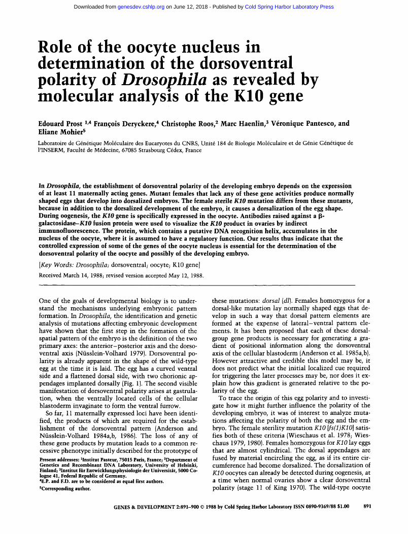
nucleus is acentric and lies near the surface at the ante-rior side of the oocyte (Fig. 1). Due to an asymmetric migration, the overlaying follicle cells become much thicker in this region than those on the opposite side of the oocyte. This region, where the dorsal appendages will be formed, corresponds to the prospective dorsal side of the embryo. Although the oocyte nucleus is still acentric in KIO egg chambers, all of the follicle cells sur-rounding the anterior side of the oocyte show the thick-ened appearance normally found at the dorsal side.
In spite of this effect on the follicle cells and on the chorion they secrete, Wieschaus et al. (1978) showed that the KIO mutation is strictly germ-line dependent, suggesting that the follicle cells are most likely in-structed by positional cues provided by the developing oocyte or by the nurse cells. When fertilized (1 or 2%), KIO eggs show the dorsalized development that is common to the dorsal-like mutations.
The fact that the KIO mutation, as well as two others recently described by Schupbach (1987), affects both the polarity of the tgg shape and the polarity of the devel-oping embryo demonstrates that there is a t ime during development when both polarities are coupled.
The KIO gene has been cloned and its function as-signed to a 5-kb DNA fragment by P-element transfor-mation. This fragment encodes a major transcript of 3.1 kb, which is specifically transcribed in the oocyte (Haenlin et al. 1985, 1987).
To understand the role of the KIO product in the es-tablishment of dorsoventral polarity, we have raised po-lyclonal antibodies specific for p-galactosidase-XiO fu-sion protein, which permitted the visualization of the KIO product on ovary sections by indirect immunofluo-rescence. We show that the product accumulates in the oocyte nucleus. We also report the nucleotide sequence and the putative protein sequence of KIO. Although it does not contain a homeo box, the sequence encodes a putative DNA recognition helix. Together, the nuclear localization of the protein and the putative DNA-binding motif suggest that KIO might possess a regula-tory function.
Results
Molecular structure of KIO mRNAs
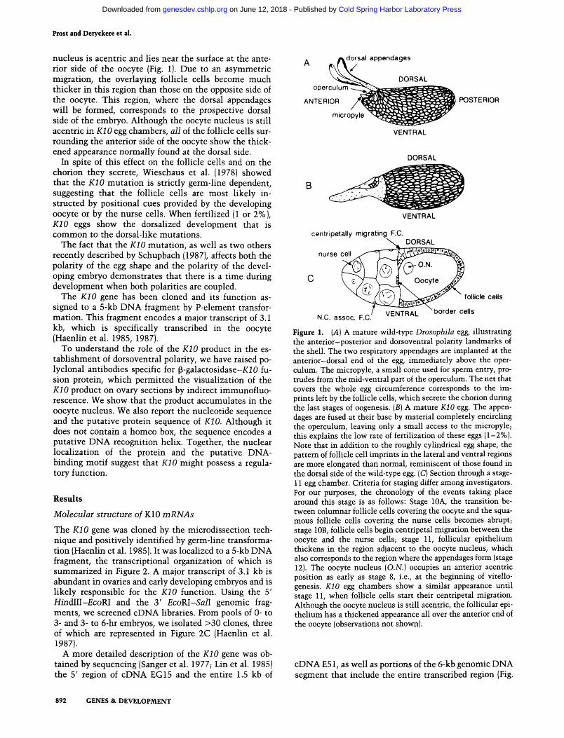
The KIO gene was cloned by the microdissection tech-nique and positively identified by germ-line transforma-tion (Haenlin et al. 1985). It was localized to a 5-kb DNA fragment, the transcriptional organization of which is summarized in Figure 2. A major transcript of 3.1 kb is abundant in ovaries and early developing embryos and is likely responsible for the KIO function. Using the 5' HmdIII-£coRI and the 3 ' EcoRl-Sall genomic frag-ments, we screened cDNA libraries. From pools of 0- to 3- and 3- to 6-hr embryos, we isolated >30 clones, three of which are represented in Figure 2C (Haenlin et al. 1987).
A more detailed description of the KIO gene was ob-tained by sequencing (Sanger et al. 1977; Lin et al. 1985) the 5' region of cDNA EG 15 and the entire 1.5 kb of
^dorsal appendages
DORSAL operculum
ANTERIOR
micropyle
POSTERIOR
VENTRAL
DORSAL
B
VENTRAL
centripetally migrating F.C. DORSAL
nurse cell
N.C. assoc. F.C.
Oocyte J^
follicle cells
VENTRAL ^border cells
Figure 1. (A) A mature wild-type Drosophila egg, illustrating the anterior-posterior and dorsoventral polarity landmarks of the shell. The two respiratory appendages are implanted at the anterior-dorsal end of the egg, immediately above the oper-culum. The micropyle, a small cone used for sperm entry, pro-trudes from the mid-ventral part of the operculum. The net that covers the whole egg circumference corresponds to the im-prints left by the follicle cells, which secrete the chorion during the last stages of oogenesis. [B] A mature KIO egg. The appen-dages are fused at their base by material completely encircling the operculum, leaving only a small access to the micropyle; this explains the low rate of fertilization of these eggs (1-2%). Note that in addition to the roughly cylindrical egg shape, the pattern of follicle cell imprints in the lateral and ventral regions are more elongated than normal, reminiscent of those found in the dorsal side of the wild-type egg. (C) Section through a stage-11 egg chamber. Criteria for staging differ among investigators. For our purposes, the chronology of the events taking place around this stage is as follows: Stage lOA, the transition be-tween columnar follicle cells covering the oocyte and the squa-mous follicle cells covering the nurse cells becomes abrupt; stage lOB, follicle cells begin centripetal migration between the oocyte and the nurse cells; stage 11, follicular epithelium thickens in the region adjacent to the oocyte nucleus, which also corresponds to the region where the appendages form (stage 12). The oocyte nucleus {O.N.] occupies an anterior acentric position as early as stage 8, i.e., at the beginning of vitello-genesis. KIO egg chambers show a similar appearance until stage 11, when follicle cells start their centripetal migration. Although the oocyte nucleus is still acentric, the follicular epi-thelium has a thickened appearance all over the anterior end of the oocyte (observations not shown).
cDNA E51, as well as portions of the 6-kb genomic DNA segment that include the entire transcribed region (Fig.
892 GENES & DEVELOPMENT
Cold Spring Harbor Laboratory Press on June 12, 2018 - Published by genesdev.cshlp.orgDownloaded from

Dorsoventral polarity in Drosopbila
H Ps Pv CI CI Ps Pv I—LT L-1 iC
/^-
Ps PvPsPsPv HPs —I Sir r :j
2.ikb
- 7 / K 1 0 -
- 7 / K10*
Figure 2. Transcriptional organization of the KIO gene. (A) Restriction map of the KIO locus. (H) Hindlll, (Ps) Pstl, (Pv) PyuII, (CI) CM, (E) £coRI, (S) 5 71. [B] Lo-calization of the transcripts, as defined by Northern blot analysis. The 4.2- and the 2.1-kb RNAs that map on either side of KIO correspond to the kz and crn loci, respectively. The open reading frame for the 3.1- and the 2.8-kb transcripts is shown by filled boxes. (C) Structures of the cDNA clones used in this analysis. cDNA libraries were kindly provided by L. Kauwar, S. Artavanis-Tsakonas, and H. Jackie. Hatched boxes rep-resent exonic regions. (D) Positioning of the KIO func-tion by P-element-mediated transformation [for details, see Haenlin et al. 1987]. The construction starting at the CM site is still capable of restoring the KIO func-tion.
3). Comparison of the genomic and cDNA sequences confirms that a single intron, 854 bp long, is spliced out of the mature transcript. The 5' end of the major tran-script, not present on either of the cDNA clones, is in-ferred from SI mapping and primer extension experi-ments (data not shown) and is found ~40 bp upstream from the 5' end of cDNA EG15. This promoter region is likely to be shared by all transcripts, as suggested by hy-bridization of poly(A)' RNA Northem blots, w ith small probes originating from the 5' end of the transcribed re-gion (Haenlin et al. 1987). This low^ abundance of the minor transcripts did not permit confirmation of their common 5' end, either by SI mapping or by primer ex-tension experiments. The region immediately preceding the transcriptional start site contains no canonical TATA box sequence. A consensus polyadenylation signal (AATAAA) occurs about 20 bp upstream of the mapped 3' end of the poly(A) tail of cDNA EF43.
The predicted KIO protein
The sequence analysis reveals a long open reading frame encoding a putative protein of 463 amino acids, 51.5 kD, with a pHi of 11.2 and an unusually high proline content (15%). It can be divided into roughly three domains: a hydrophilic amino-terminal region (up to residue 225) containing 21% Gin (encoded by CAG), a second region of ~112 amino acids that consists essentially of apolar residues, among which proline represents 37%, and a carboxy-terminal region with a more 'typical' amino acid composition containing a 'helix-tum-helix' motif.
The genomic DNA sequence corresponding to the amino-terminal region of the protein, with its high per-centage of Gin, is reminiscent of the M repeat in the Ubx and Antp genes (McGinnis et al. 1984b; Schneuwly et al. 1986), of the opa sequence in the Notch gene (Wharton et al. 1985), and of similar stretches in the en-grailed gene (Poole et al. 1985). A corresponding cDNA fragment from KIO has been used to isolate the murine opa sequence counterpart (Duboule et al. 1987). How-ever, in contrast to the apparently randomly arranged CAG triplets interspersed with other codons that nor-
mally constitute the M or opa repeats so far described, the structure of the op^-like area [opla] in the KIO gene is more organized. The opla region extends from 1373 to 1539 nucleotides and is composed of seven tandemly re-peated units of 24 nucleotides each. These repeated units encode the motif Gin, Gin, Gin, His, Pro, Ser, Pro, Asn, with some intemal variation (Fig. 4).
Secondary structure analysis (Gamier et al. 1978) of the KIO protein has shown the presence of a helix-turn-helix motif located between amino acids 390 and 418 (data not shown). Crystallographic analyses have shown the presence of such conserved helical structures in pro-karyotic regulatory proteins, such as the cro and cl re-pressor proteins of bacteriophage \ . Computer modeling studies and crystallographic data show that the con-served helical structures are the DNA-binding sites for these proteins (Pabo and Sauer 1984; Ptashne 1986; An-derson et al. 1987). Most of the amino acid residues im-portant for the conformation of the potential DNA-binding domain structure are conserved in KIO, as shown by a comparison of KlO's putative helix-tum-helix region with other potential DNA-binding domains in prokaryotes and yeast (Fig. 5). In particular, Ala, Gly, and Val at the respective consensus positions 5, 9, and 15, which are known to be important for conferring the helix-tum-helix configuration necessary for interaction with the major groove of DNA, are found in KIO.
It should be noted that this motif is highly divergent from the recognition helix that is found in the homeo domain (Gehring and Hiromi 1986) (Fig. 5). This result could be anticipated from the absence of a homeo box in the KIO gene, as determined by cross-hybridization, even under reduced stringency conditions (McGinnis et al. 1984a,b).
Construction and expression of the KIO fusion protein and production of antibodies To generate antibodies directed against the KIO protein, cDNA EF43 was inserted into the 3' end of the lacZ gene in the expression vector pUR288 (Riither and Miiller-Hill 1983) to produce the protein in Escherichia coli.
GENES & DEVELOPMENT 893
Cold Spring Harbor Laboratory Press on June 12, 2018 - Published by genesdev.cshlp.orgDownloaded from

Piost and Deryckete et al.
CAGCTGAGCGCGCTAAATTAGAAAAGATTTCAATTAAATATCAGTTTTATATTAGCACTGAATAAAAACCTACACCCTCTTTCTTTTTCTTCTTCTCCACAATAGCCTGGAGGTGCTTGC 120 GCTGCTTCTTGGAAAGGATCTTCACGTGTTGCACTTGTCCACCTTAATCTTGGTGGCCCGCTTCTCGGAGGGAGAACCAGAGCGTGGCCTCATCGTAGCCAACCTGGTTGCCGCCGCCAC 240 AGCATCCACATCGATCTGAATCTGTAGTGTATTGGATTTGGAAAGGGTAAGAAAGCGGATTCGAGTGTGTGGTCCGACGGATGACCTACCTTCTTAGGCCGAGTGCCACAATCGTTTGTG 360 GATTCTGCCGCGCTTGGATTGTGCACCTTCTTGCCCATGATTATTTATTTACCAATATAGTTAATAAAATAAACTAACAGAATTAACTTTGGGCAATAATATAAACATTGATTTAAATCA 480
C l a l CACGTCGAAGTACGCTTATCGATAACAGCGCAGGCGATAACAAAAGTTGAGATACTCTAAAATGTGCTGTGATGTTACATTTGTGTTAGTGGAAACATACATTAGTGGGAAAAACTGATT 600
H^^START cDNA EG15 r* Clal GTTATAGTTGTTCACACTCCTTCGACAGCACTGGTATCGAAACGTTATCGCAATCGATTCATCGTAATATTTTCAATATAAGTTTTTGTGACAAATAGGCGTGCAACATTTTAGATCCAT 720 CTGCTCCCCGCCTGTCGTTTCGGCTTGGCTCCTCTGCTCGAGTCCTGAACCCCCGTTTTCAGTGAAATCCAGCTACCTGCAATGGTGTCCAAGAACCAATTCTATCAGAACTGGACCATG 840
MetVa lSerLysAsnGlnPheTyrGlnAsnTrpThrMet 10
CAGTCGCAGCAGCAGCACCCGCACCAAATGCAGCAACAGTTTCAGCAACAGCAGCAGCCAAATCTGCAGCACCGCAACAACCAGAGCAACAATAATAATTGCAACAATAATCCCCGCGCA 960 GlnSerGlnGlnGlnHisProHisGlnMetGlnGlnGlnPheGlnGlnGlnGlnGlnProAsnLeuGlnHisArgAsnAsnGlnSerAsnAsnAsnAsnCysAsnAsnAsnProArgAl^
20 30 40 50 GCTGCGGCTCCGTATAGAAAACCGTTTCGTTCGGGCAAAATTAACTCCGGACCAGGAGGAAATGGGAATGGTAACAGGGTCAACGGTAACAATCAGATGATGTTTTCCTCCAGCCAAATG 1080 A l a A l a A l a P r o T y r A r g L y s P r o P h e A r g S e r G l y L y s I l e A s n S e r G l y P r o G l y G l y A s n G l y A s n G l y A s n A r g V a l A s n G l y A s n A s n G l n M e t M e t P h e S e r S e r S e r G l n M e t
6 0 70 80 90 CCGAGCGATCCCCTGTACATTGACTTCAGCAGCCCGCCACCAGGATTCAAACACAATCAGGTAGGCAGTCCCAAGAAGAAGTCTATGAAGGGCATCAAACAGCAGCAGCATCCCAGTCCG 1200 P r o S e r A s p P r o L e u T y r I l e A s p P h e S e r S e r P r o P r o P r o G l y P h e L y s H i s A s n G l n V a l G l y S e r P r o L y s L y s L y s S e r M e t L y s G l y l l e L v s C l n G l n G l n H i s P r o S e r P r o
100 110 120 130 AACtAGCAGCAGCCTCCCAGTCCGAAC|CAGCAGCAGCATCCCAGTCCGAAC|CAGCAGCAGCATCCCAGTCCGAACtAGCAGCAGCATCCCAATTCGAACtAGCAGCAGCATCTCAGTCCG 1320 Asr^GlnGinGlnProProSerProAsr^GlnGlnGlnHisProSerProAsrTGlnGlnGlnHisProSerProAsrijinGlnGlnHisProAsnSerAsrTGlnGln
140 150 160 170 AAdCAGCAGCAGGGCAAGATGAACAACCAGAACAACAATCATATGAACCAGTCGCAGCAGCCCTTCAACAATCAGATGAACGGAAGCGATTGGCAGCGCCATCCGGGCAACAATCCCAAT 1440 Asrfc t nG1nG(nGIyLysMetAsnAsnfc InAsnAsnAsnH i sMetAsnGInSerGInGInProPheAsnAsnGInMetAsnGIySerAs pT rpGInArgH i sP roGIyAsnAsnP roAsn
180 190 200 210 CAGATTCGCGGTGGCTTTAATGGATTCCAACGCGGGCCGCCTCCGAACCGTCCTCCTCCACGCCTCATGATGGGTCCGCCAATGGGTCCGATGGGCCCCGGACCACGGGGACCCGGACCG 1560 G l n l l e A r g G l y G l y P h e A s n G l y P h e G l n A r g G l y P r o P r o P r o A s n A r g P r o P r o P r o A r g L e u M e t M e t G l y P r o P r o M e t G l y P r o M e t G l y P r o G l y P r o A r g G l y P r o G l y P r o
220 230 240 250 ATGGGCCCTGGAGGACCTTACCCGCAAATGCCTTTCCCGCCACCGGTGCCTGGTATGCGCGGTCCCGGACACCATGGGCCCATGGTGGACCACCACCGCCCGCCGCCACCGCTCTTTATG 1680 M e t G l y P r o G l y G l y P r o T y r P r o G l n M e t P r o P h e P r o P r o P r o V a l P r o G l y M e t A r g G l y P r o G l y H i s H i s G l y P r o M e t V a l A s p H i s H i s A r g P r o P r o P r o P r o L e u P h e M e t
260 270 280 EcoRI p cDNA EF43 p cDNA E51 CGTCGCAACGGACCCGGTCCAGGTCCTATGATGGGTGTCCCACCGCCCATGCACATGATGGGTCCACGGATGCCGCCGCGGGGAATTCCACCGGTCGGACCTTATGfGACCAATGAACATG 1800 A r g A r g A s n G l y P r o G l y P r o G l y P r o M e t M e t G l y V a l P r o P r o P r o M e t H i s M e t M e t G l y P r o A r g M e t P r o P r o A r g G l y l l e P r o P r o V a l G l y P r o T y r G l y P r o M e t A s r ^ c t
3 0 0 3 1 0 320 330 AATGGCGGCCGGATAATGAAGCCCAATCCCAAGCTAATCAAGCAGGTGGTGAAAGGCAAGAGCAGCATAAAGACACTAAAGAACCTGATCAATCAGTATCCCATCGAAAAGCCCTGGGTA 1920 A s n G l y G l y A r g I l e M e t L y s P r o A s n P r o L y s L e u I l e L y s G l n V a l V a l L y s G l y L y s S e r S e r l l e L y s T h r L e u L y s A s n L e u I l e A s n G l n T y r P r o I l e G l u L y s P r o T r p V a l
3 4 0 350 360 370 ACGGACGAGATACGCAGCGAGCACGACAAGAAGGTGGACATCGAGAACAGATTGAAGGGCCACAAAGACGACGAGCTCTTCGCTCAGTACAAGGGCCAGCGGGACAAGTTCGTTAGTCTC 2040 T h r A s p G l u U e A r g S e r G l u H i s A s p L y s L y s V a l A s p I l e G l u A s n A r g L e u L y s G l y H \ s L y s A s c » k s p G l u L e u P h e A l a G l n T y r L v s G l y G l n A r g A s p L y s P h e V a l S e r L e u
3 8 0 3 9 0 4 0 0 410 TACGAAGCGGCGCGCGAAGAGTATCTCAAGCAGGAGGCAGCCACTGTCAAGGCCAAGGTGAGCATATAGAGACCCCCCAACATATCCTTATTCCCTTGGAACAGGTGTCCCATCCGAAGC 2160 T y r G l u A l a i A l a A r q G l u G l u T v r L e u L v s G l n G l u A l a A l a T h r V a l L v s A l a L y s K I n t r o n s t a r t
4 2 0 4 3 0 TCTATTCAGGGAGGCACACCGATTCCGGGTACAGTTTCGGCTTAATCCGGAATCACTTTGCACCCCTGATTTGATCCGTAACCCCCACCCGTCCCTCTTTGTTGCGTTTGCCGGGCGGTA 2280 GGAAAAAAATAAGGGAGCAGAGAAAGTGAAAAAGTAAGTCTCAACCTGATGCACTCTCTCGGCATTATCCTGCAGTCTGCGGAAGGTTGTTGTCATATCTATTTTCACCTTCGGTTGTTC 2400 CCTTGGCACTGATATATATCCGCAAAAGAGAGAAACTATTGGCATATATTTACAACCATAGATTTATTAGGAACTGAACAAAATTGTGTAACTATTTAGAACTGCAATACAATGTGTTTA 2520 CTGCCACCTTGTATACAATTTATAAAACTAATAGAATTTTGTACTGTTCCGATCGTGGCAGGCGTAGTAGATTCATAATGCACTCTCAGTAACTTCAATATTCTTAATTGAACGAGCCTC 2640 AGTACTGAATCCTATGGCTGTTTTCATTTTCCAATTTGTTGGCATGTGAAAAGCGAACTTAATGAATTTTAAACTGCCAGAACATCAATTTAGTTACTTAATTAGCAGATATAACCCAAC 2760 TTATGAAACTCATTGCATATGAAATGATAATAGATTTGAGTGTTAATGTTCGTGGGCGTAGTTATCTGGAAATAGCCCAAGAGAAGTTCGCAGAATGCATTTAATAGCGAAAACAGATCT 2880 GAGAGCTCTTTTTACACACTCAACAACCTTGCTATTGAACACCGTTGGCAATTCTCGTATTTCTTTTGCAGGATGCCAAGTCAGACAAAGACAAAAACGCAATTTCAAGTCAGAGCGCAG 3000
I n t r o n end ^ A s p A l a L y s S e r A s p L y s A s p L y s A s n A l a l l e S e r S e r G l n S e r A l a A 4 4 0
CCCCTAAGGCCGGAAGCGCTAAAGATGCAACAATTCCAAATCCCTAGGCAGCCAATGCAACCGAATCCGAATACCGAATCAATCGAATCGAACCGACTAAAGAAGAGGTCGGTCGACTCA 3120 l a P r o L y s A l a G l y S e r A l a L y s A s p A l a T h r l l e P r o A s n P r o
^50 ^63 CAGAACTAAATGTAAACTTAAACTTTAAACCAATGAGCAATTCAAAACTAAAACTAACAGCGAGCAACAGCAAGAGTCTGACAATTGAAGACGAATATAACTCAAAACCCCGACTCCTAT 3240
— I cDNA EG15 ATCGGATATACGTATACACAATCGTAATCCGAGGAGCGCCGCTTTGTCACACGTTGCTTTTGCGACTGAGAACTGAGTCTTAGAACGATAATGTAGCAACAAATACAAACCCAGCGATAA 3360 TAATTGTGGGCGAAACTCTAGTAATAGCGATAAAAATTTAGATTGCGAATCGATTCACTTGTCTAACGTAACGTACCAGTTTCCTATTCGAAAACTATTTGTTGTTCAATTCTAAGTTGC 3480 AGCCATTTCAAGTAT6TGTACACAATTTATGACACGCCCTTACCTATATATGTATGCATATATATTGATATATATATATATATAAATTATTTTCCTATTCGTTGCCCGAGTGCGTGGGCG 3600 CGACATAATTGAAACAGAGTGGCCCGGATCCTAGACCTGCATAATCACACTGTAGGCCTAGATACACACTGATGTATTTAATATCTAAAACTACAACTTAGATCACTCTGTGAACGTGTG 3720 CTCGATGGTGTGCATCTACGGATTTGTTTTTTGTGTTCTTTTTCCCCACCCAACCCCGTCCCTGACCCAATTCCGCTGTTCTCTTTACCGACTTGTAGATTAATCACGCTCTTGATCTTC 3840 ATGACCGGATCCACCTGACAAGTAACGCCAAAGCAGCCCCACTTAGCGGAATAGTAGAGCCTAGGGGACCCAACGTAAAAACTCTAGTATAGCACCAATCTCCCAACCAGTTAACATTAT 3960 ACCTAAACCCATGGTCAAGAGTAAACATTTCTGCCTTTGAAGTTGAGAACACAATTAAGCATCCCCTGGTTAAACCTGACACTTCATACTTGTTAATAGCGCCATAAACATAGCACCAAT 4080
cDNA E51 - 1 TTCGAAGAAATCAGTTAAAAGCAATTAGCAATTAGCAATTAGCAATAACTCTGCTGACTTCAAAACGAGAAGAGTTGCAAGTATTTGTAAGGCACAGTTTATAGACCACCGACGGCTCAT 4200 TAGGGCTCGTCATGTAACTAAGCGCGGTGAAACCCAATTGAACAGTAGTAGGTGGAATTAGTTAGTTAGTCAATGGGGAAGATTTAACCCTCAGGTAGCAAAGTAATTGAACTGCGGGCA 4320 GAGAGTCCTAAGACTAAATAATATATTTAAAAATCTGGCCCTTTGACCTTGCTTGTCAGGTGCATTTGGGTTCAATCGTAAGTTGCTTCTATATAAACACTTTCCCCATCCCCGCAATAA 4440
cONA EF43 —1 TGAAGAATACCGCAGAAIAAAGAGAGATTTGCAACAAAAAATAAAGGCATTGCGAAAACTTTTTATGGGGGATCATTACACTCGGGCCTACGGTTACAATTCCCAGCCACTTAAGCGACA 4560 AGTTTGGCCAACAATCCATCTAATAGCTAATAGCGCAATCACTGGTAATCGCAAGAGTATATAGGGCAATAGAACCCATGGATTTGACCAAAGGTAACCGAGACAATGGAGAAGCAAGAG 4680 GATTTCAAACTGAACACCCACAGTGCTGTGTACTACCACTGGCGCGTTTGGAGCTCACTGGCCTGATGCGCTCCTCCGGGCGTTTCAAGCCTGCTTTACGTGGTATACTCCATTACGGTC 4800 AAGTTGGTGGTCACCGTGCTGTTTCCCTTGAGCTTGCTGGCCAGGCTGCTGTTCACCACCAACATGGCCGGATTGTGCGAGAACCTGACCATAAGTATTACCGATATTGTGGCCAATTTG 4920 AAGTTTGCGAATGTGTACATGGTGAGGAAGCAGCTCCATGAGATTCGCTCTCTCCTAAGGCTCATGGACGCTAGAGCCCGGCTGGTGGGCGATCCCGAGGAGATTTCTGCCTTGAGGAAG 5040 GAAGTGAATATCGGCACAGGGCACTTTCCAGCACCTTTGGCCAGTATTTTCGTATTTGGCACTACTTTGAGTTCGCGTCCGCGTGGTCGTTCGCCCGGATCGAGAGCTCCTGTATCCGGC 5160 CTGGTTCGGCGTTGACTGGATTCACTCCACCAGAAACTATGTGCTCATCAATATCTACCAGCTCTTCGGCTTGATAGTGCAGGCTATACAGAACTGCGCTAGTGACTCCTATCCGCCTGC 5280 GTTTCTCTGCCTGCTCACGGGTCATATGCGTGCTTTGGAGCTGAGGGTGCGGCGGATTGGCTGCAGACATATGAAGCCTGGCGGYGAGGAGGTGTACCAGGAACTCAGCGAGTGCATCCG 5400 CGATCTGGCGCGGTCCATCGGGCTGAGGGAGACCAGCAGGCGGGCCTTTCAGTGCCCCCTTTCAGTGCCCTGCATGGCCCAGTTCGTCTGCTCCGCCGCCGTCCAGTGTACCGTCGCCAT 5520
Figure 3. (See facing page for legend.)
894 GENES & DEVELOPMENT
Cold Spring Harbor Laboratory Press on June 12, 2018 - Published by genesdev.cshlp.orgDownloaded from
Dorsoventral polarity in Drosopbila
CAG CAG CAG CAT CCC AGT CCG AAC 1 Gin Gin Gin His Pro Ser Pro Asn
CAG CAG CAG CCT CCC AGT CCG AAC 2 Gin Gin Gin Pro Pro Ser Pro Asn
CAG CAG CAG CAT CCC AGT CCG AAC 3 Gin Gin Gin His Pro Ser Pro Asn
CAG CAG CAG CAT CCC AGT CCG AAC 4 Gin Gin Gin His Pro Ser Pro Asn
CAG CAG CAG CAT CCC AAT TCG AAC 5 Gin Gin Gin His Pro Asn Ser Asn *** ««« CAG CAG CAG CAT CTC AGT CCG AAC 6 Gin Gin Gin His Leu Ser Pro Asn
CAG CAG CAG GGC AAG ATG AAC AAC 7 Gin Gin Gin Gly Lys Met Asn Asn
•«* ••* ••* »••
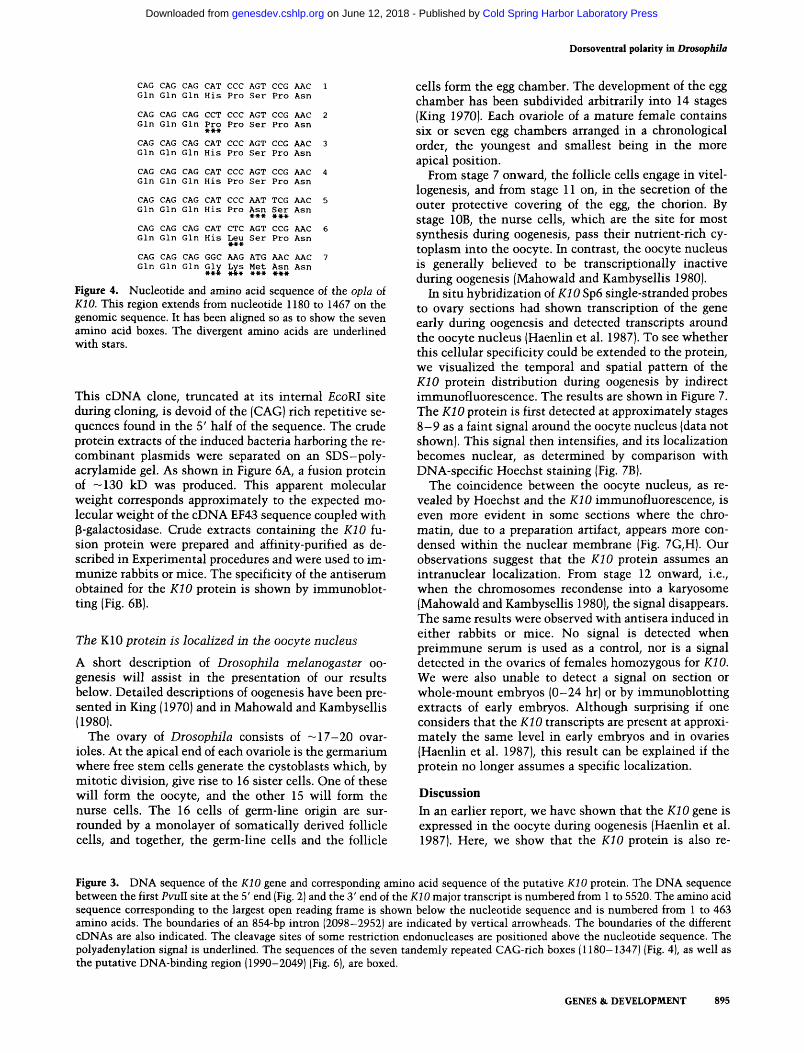
Figure 4. Nucleotide and amino acid sequence of the opla of KIO. This region extends from nucleotide 1180 to 1467 on the genomic sequence. It has been aligned so as to show the seven amino acid boxes. The divergent amino acids are underlined with stars.
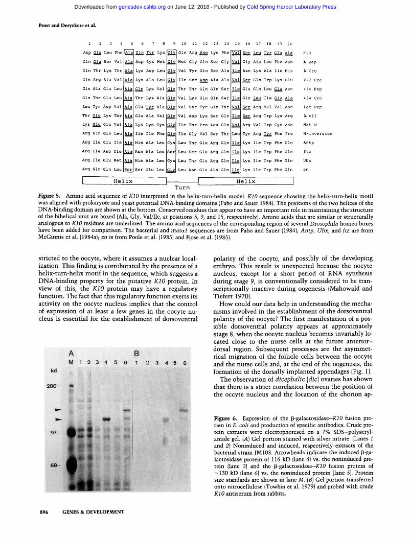
This cDNA clone, truncated at its internal EcoRl site during cloning, is devoid of the (CAG) rich repetitive se-quences found in the 5' half of the sequence. The crude protein extracts of the induced bacteria harboring the re-combinant plasmids were separated on an SDS-poly-acrylamide gel. As shown in Figure 6A, a fusion protein of —130 kD was produced. This apparent molecular weight corresponds approximately to the expected mo-lecular weight of the cDNA EF43 sequence coupled with p-galactosidase. Crude extracts containing the KIO fu-sion protein were prepared and affinity-purified as de-scribed in Experimental procedures and were used to im-munize rabbits or mice. The specificity of the antiserum obtained for the KIO protein is shown by immunoblot-ting (Fig. 6B).
The KIO protein is localized in the oocyte nucleus A short description of Drosophila melanogaster oo-genesis will assist in the presentation of our results below. Detailed descriptions of oogenesis have been pre-sented in King (1970) and in Mahowald and Kambysellis (1980).
The ovary of Drosophila consists of -17 -20 ovar-ioles. At the apical end of each ovariole is the germarium where free stem cells generate the cystoblasts which, by mitotic division, give rise to 16 sister cells. One of these will form the oocyte, and the other 15 will form the nurse cells. The 16 cells of germ-line origin are sur-rounded by a monolayer of somatically derived follicle cells, and together, the germ-line cells and the follicle
cells form the egg chamber. The development of the tgg chamber has been subdivided arbitrarily into 14 stages (King 1970). Each ovariole of a mature female contains six or seven egg chambers arranged in a chronological order, the youngest and smallest being in the more apical position.
From stage 7 onward, the follicle cells engage in vitel-logenesis, and from stage 11 on, in the secretion of the outer protective covering of the egg, the chorion. By stage lOB, the nurse cells, which are the site for most synthesis during oogenesis, pass their nutrient-rich cy-toplasm into the oocyte. In contrast, the oocyte nucleus is generally believed to be transcriptionally inactive during oogenesis (Mahowald and Kambysellis 1980).
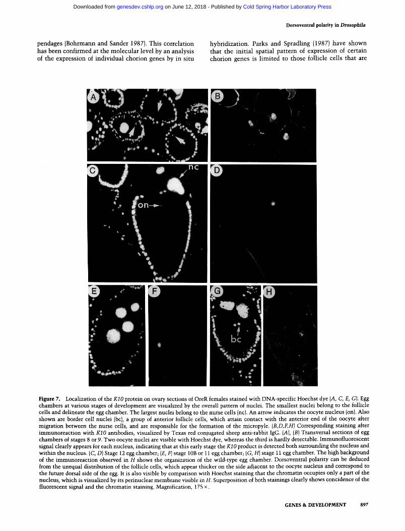
In situ hybridization of KIO Sp6 single-stranded probes to ovary sections had shown transcription of the gene early during oogenesis and detected transcripts around the oocyte nucleus (Haenlin et al. 1987). To see whether this cellular specificity could be extended to the protein, we visualized the temporal and spatial pattern of the KIO protein distribution during oogenesis by indirect immunofluorescence. The results are shown in Figure 7. The KIO protein is first detected at approximately stages 8-9 as a faint signal around the oocyte nucleus (data not shown). This signal then intensifies, and its localization becomes nuclear, as determined by comparison with DNA-specific Hoechst staining (Fig. 7B).
The coincidence between the oocyte nucleus, as re-vealed by Hoechst and the KIO immunofluorescence, is even more evident in some sections where the chro-matin, due to a preparation artifact, appears more con-densed within the nuclear membrane (Fig. 7G,H). Our observations suggest that the KIO protein assumes an intranuclear localization. From stage 12 onward, i.e., when the chromosomes recondense into a karyosome (Mahowald and Kambysellis 1980), the signal disappears. The same results were observed with antisera induced in either rabbits or mice. No signal is detected when preimmune serum is used as a control, nor is a signal detected in the ovaries of females homozygous for KIO. We were also unable to detect a signal on section or whole-mount embryos (0-24 hr) or by immunoblotting extracts of early embryos. Although surprising if one considers that the KIO transcripts are present at approxi-mately the same level in early embryos and in ovaries (Haenlin et al. 1987), this result can be explained if the protein no longer assumes a specific localization.
Discussion In an earlier report, we have shown that the KIO gene is expressed in the oocyte during oogenesis (Haenlin et al. 1987). Here, we show that the KIO protein is also re-
Figure 3. DNA sequence of the KIO gene and corresponding amino acid sequence of the putative KIO protein. The DNA sequence between the first Pvull site at the 5' end (Fig. 2) and the 3' end of the KIO ma]oi transcript is numbered from 1 to 5520. The amino acid sequence corresponding to the largest open reading frame is shown below the nucleotide sequence and is numbered from 1 to 463 amino acids. The boundaries of an 854-bp intron (2098-2952) are indicated by vertical arrowheads. The boundaries of the different cDNAs are also indicated. The cleavage sites of some restriction endonucleases are positioned above the nucleotide sequence. The polyadenylation signal is underlined. The sequences of the seven tandemly repeated CAG-rich boxes (1180-1347) (Fig. 4), as well as the putative DNA-binding region (1990-2049) (Fig. 6), are boxed.
GENES & DEVELOPMENT 895
Cold Spring Harbor Laboratory Press on June 12, 2018 - Published by genesdev.cshlp.orgDownloaded from
Prost and Deryckere et al.
1 2 3 4
Asp Glu Leu Phe
Gin Glu Ser Val
Gin Thr Lys Thr
Gin Arg Ala Val
Gin Ala Glu Leu
Gin Thr Glu Leu
Leu Tyr Asp Val
Thr Glu Lys Thr
Lys Glu Glu Val
Arg Gin Gin Leu
Arg H e Glu H e
Arg H e Asp H e
Arg H e Glu Met
Arg Gin Gin Leu
Ala
Ala
Ala
Ala!
Ala
Ala
Ala
Ala
Ala
Ala
Ala
Ala
Ala
Ser
5 6 7 8
Gin Tyr Lys
Asp Lys Met
Lys Asp Leu
Lys Ala Leu
Gin Lys Val
Thr Lys Ala
Glu Tyr Ala
Glu Ala Val
Lys Lys Cys
H e H e Phe
His Ala Leu
Asn Ala Leu
His Ala Leu
Ser Glu Leu
Si. 'Glyl
|Gly|
Glyl
iGly
Val
Val
Ilel
Val
H e
iGly
Gly
Gly
Cys|
Ser
Cys
Glyl
9 10 11 12 13 14 15 16
Gin Arg Asp Lys Phe
Met Gly Gin Ser Gly
Val Tyr Gin Ser Ala
H e Ser Asp Ala Ala
Thr Thr Gin Gin Ser
Val Lys Gin Gin Ser
Val Ser Tyr Gin Thr
Val Asp Lys Ser Gin
H e Thr Pro Leu Gin
H e Gly Val Ser Thr
Leu Thr Glu Arg Gin
Leu Ser Glu Arg Gin
Leu Thr Glu Arg Gin lllel
Leu Asn Glu Ala Gin |lle|
H e
Val
Ilel
val
Leu
H e
H e
17 18 19 20
Ser Leu Tyr Glu Ala
Gly Ala Leu Phe Asn
Asn Lys Ala H e His
Ser Gin Trp Lys Glu
Glu Gin Leu Glu Asn
Gin Leu H e Glu Ala
Ser Arg Val Val Asn
Ser Arg Trp Lys Arg
Arg Val Trp Cys Asn
Tyr Arg Tyr Phe Pro
Lys H e Trp Phe Gin
Lys H e Trp Phe Gin
Lys H e Trp Phe Gin
Lys H e Trp Phe Gin
Helix Helix Turn
KlO
X Rep
X Cro
P22 Cro
434 Rep
434 Cro
Lac Rep
X oil
Mat a
H-inversion
Antp
ftz
Ubx
Figure 5. Amino acid sequence of KlO interpreted in the helix-turn-helix model. KlO sequence showing the helix-turn-helix motif was aligned with prokaryote and yeast potential DNA-binding domains (Pabo and Sauer 1984). The positions of the two helices of the DNA-binding domain are shown at the bottom. Conserved residues that appear to have an important role in maintaining the structure of the bihelical unit are boxed (Ala, Gly, Val/Ile, at positions 5, 9, and 15, respectively). Amino acids that are similar or structurally analogous to KlO residues are underlined. The amino acid sequences of the corresponding region of several Diosophila homeo boxes have been added for comparison. The bacterial and matal sequences are from Pabo and Sauer (1984); Antp, Ubx, and ftz are from McGinnis et al. (1984a); en is from Poole et al. (1985) and Fjose et al. (1985).
stricted to the oocyte, where it assumes a nuclear local-ization. This finding is corroborated by the presence of a helix-tum-helix motif in the sequence, which suggests a DNA-binding property for the putative KlO protein. In view of this, the KlO protein may have a regulatory function. The fact that this regulatory function exerts its activity on the oocyte nucleus implies that the control of expression of at least a few genes in the oocyte nu-cleus is essential for the establishment of dorsoventral
M 1 2 3 4 5 6 1 2 3 4 5 6
fp
polarity of the oocyte, and possibly of the developing embryo. This result is unexpected because the oocyte nucleus, except for a short period of RNA synthesis during stage 9, is conventionally considered to be tran-scriptionally inactive during oogenesis (Mahowald and Tiefert 1970).
How could our data help in understanding the mecha-nisms involved in the establishment of the dorsoventral polarity of the oocyte? The first manifestation of a pos-sible dorsoventral polarity appears at approximately stage 8, when the oocyte nucleus becomes invariably lo-cated close to the nurse cells at the future anter ior-dorsal region. Subsequent processes are the asymmet-rical migration of the follicle cells between the oocyte and the nurse cells and, at the end of the oogenesis, the formation of the dorsally implanted appendages (Fig. 1).
The observation of dicephalic [die] ovaries has shown that there is a strict correlation between the position of the occyte nucleus and the location of the chorion ap-
Figure 6. Expression of the p-galactosidase-XiO fusion pro-tien in E. coli and production of specific antibodies. Crude pro-tein extracts were electrophoresed on a 7% SDS-polyacryl-amide gel. [A] Gel portion stained with silver nitrate. (Lanes 1 and 2) Noninduced and induced, respectively extracts of the bacterial strain JM103. Arrowheads indicate the induced p-ga-lactosidase protein of 116 kD (lane 4] vs. the noninduced pro-tein (lane 3) and the p-galactosidase-XlO fusion protein of ---ISO kD (lane 6) vs. the noninduced protein (lane 5). Protein size standards are shown in lane M. [B] Gel portion transferred onto nitrocellulose (Towbin et al. 1979) and probed with crude KlO antiserum from rabbits.
896 GENES & DEVELOPMENT
Cold Spring Harbor Laboratory Press on June 12, 2018 - Published by genesdev.cshlp.orgDownloaded from
Dorsoventral polarity in Drosopbila
pendages (Bohrmann and Sander 1987). This correlation has been confirmed at the molecular level by an analysis of the expression of individual chorion genes by in situ
hybridization. Parks and SpradHng (1987) have shown that the initial spatial pattern of expression of certain chorion genes is limited to those follicle cells that are
Figure 7. Localization of the KIO protein on ovary sections of OreR females stained with DNA-specific Hoechst dye {A, C, E, G). Egg chambers at various stages of development are visualized by the overall pattern of nuclei. The smallest nuclei belong to the follicle cells and delineate the Ggg chamber. The largest nuclei belong to the nurse cells (nc). An arrow indicates the oocyte nucleus (on). Also shown are border cell nuclei (be), a group of anterior follicle cells, which attain contact with the anterior end of the oocyte after migration between the nurse ceils, and are responsible for the formation of the micropyle. {B,D,F,H] Corresponding staining after immunoreaction with KIO antibodies, visualized by Texas red conjugated sheep anti-rabbit IgG. [A], {B) Transversal sections of Ggg chambers of stages 8 or 9. Two oocyte nuclei are visible with Hoechst dye, whereas the third is hardly detectable. Immunofluorescent signal clearly appears for each nucleus, indicating that at this early stage the iCiO product is detected both surrounding the nucleus and within the nucleus. (C, D) Stage 12 egg chamber; (£, F] stage lOB or 11 egg chamber; (G, H) stage 11 egg chamber. The high background of the immunoreaction observed in H shows the organization of the wild-type egg chamber. Dorsoventral polarity can be deduced from the unequal distribution of the follicle cells, which appear thicker on the side adjacent to the oocyte nucleus and correspond to the future dorsal side of the egg. It is also visible by comparison with Hoechst staining that the chromatin occupies only a part of the nucleus, which is visualized by its perinuclear membrane visible in H. Superposition of both stainings clearly shows concidence of the fluorescent signal and the chromatin staining. Magnification, 175 x.
GENES & DEVELOPMENT 897
Cold Spring Harbor Laboratory Press on June 12, 2018 - Published by genesdev.cshlp.orgDownloaded from
Prost and Deryckere et al.
close to the oocyte nucleus and will be subjected to rapid centripetal migration later on.
These observations emphasize the importance of the location of the oocyte nucleus in the morphogenetic processes that accompany the maturation of the oocyte. Together with the data obtained for KIO, they lead us to propose the following model to explain the establish-ment of the dorsoventral polarity of the oocyte and the role that KIO plays in the process.
The acentric position of the oocyte nucleus appears as a prerequisite for generating the subsequent asymmetry. Microtubules, or elements of the cytoskeleton, may be required to establish and retain the nucleus in this posi-tion (Gutzeit 1986). We exclude a role for the KIO gene in the process, because the oocyte nucleus still has an acentric position in KIO ovaries (Wieschaus 1980; our own observations).
To exert its influence, the oocyte nucleus, transcrip-tionally inactive thus far, starts expressing genes, the products of some of which may be diffusible and capable of conveying information to the follicle cells. We as-sume that transcripts originating from the oocyte nu-cleus remain in its close vicinity and that, consequently, the product appears initially centered on the oocyte nu-cleus before starting to diffuse. If its diffusion is random, the follicle cells will react in accordance with their posi-tion relative to the acentric position of the oocyte nu-cleus, and the follicle cells closest to the oocyte nucleus will receive proportionally more of the diffusible product. This would explain the thickening of the follic-ular epithelium in the region close to the oocyte nu-cleus, where the chorionic appendages would form later on and due to a similar process. This product could function at the oocyte surface to guide follicle cell mi-gration.
The results that we present here indicate that KIO is unlikely to be such a product. Instead, its putative DNA-binding properties make it a good candidate for being one of the regulatory factors that control the ex-pression of such a product. In accordance with our hy-pothesis, the alteration of the KIO regulatory function by mutat ion would therefore result in the symmetrical migration of the anteriorly located follicle cells. This is indeed the first defect observed in the KIO tgg chamber (Wieschaus 1980; our own observations and legend to Fig. 1). As a consequence, all the anterior follicle cells would have the same 'dorsalized' information and would synthesize enlarged appendages encircling the anterior end of the oocyte.
What is the influence of the oocyte polarity on the subsequent development of the embryo? It can be specu-lated that under normal conditions, the polarity of the oocyte provides the initial trigger required for the posi-tion-dependent activation of an evenly distributed mor-phogen. A possible source for such a morphogen has been proposed by Anderson et al. (1985a,b) to be the Toll gene product. The activation of the Toll product, from its precursor form, is regulated in a position-dependent manner relative to the dorsoventral axis. It is possible that this activation is initiated by the inherent polarity of the oocyte.
Experimental procedures Indirect immunofluoiescent staining of ovary sections
Ovary sections were treated as described by Mitchison and Sedat (1983) and by White and Wilcox (1984). Ovaries of 2- to 3-day-old Oregon ROT KIO homozygous females were dissected in Ringer's buffer and included in O.C.T. (Miles Laboratories). Frozen sections were dried and stored at - 80°C. Sections were fixed in 3% paraformaldehyde in phosphate-buffered saline (PBS) for 10 min at room temperature, followed by extensive rinsing in PBS. After transfer in TBS [50 mM Tris (pH 7.4), 150 EQM NaCl], the slides were incubated in TBS* [150 mM NaCl, 50 mM Tris (pH 7.4), 0.2% Nonidet P-40, 5% sheep serum). All successive antibody incubations and rinses were done in this buffer. Prefixed sections were then incubated with a crude anti-KIO serum diluted 1/50, for 3 hr at room temperature. After several washes, the sections were further incubated in a 1/50 dilution of sheep anti-rabbit IgG (Amersham). After washes in TBS and staining with DNA-specific Hoechst dye 33258 (1 jjLg/ml), the sections were mounted in 78% glycerol, 20 mM Tris (pH 7.4), and 2% propylgallate (Sigma); sealed with rubber ce-ment to prevent dehydration, and viewed under a Zeiss micro-scope using epifluorescence. Control reactions with preim-mune sera did not show any specific signal (data not shown).
Gene fusion The 2.4-kb KIO cDNA EF43 had been subcloned initially into the EcoKL site of each of the three open reading frames of the pTG vectors (Courtney et al. 1984) to select the functional open reading frame on the basis of its length. The choice of the clone giving rise to the longest protein was then confirmed by in vitro expression analysis. A 1.0-kb BamHl fragment corresponding to the open reading frame of cDNA EF43 in the correct phase was subsequently isolated from pTG-KlO and inserted into the BamHl site of the pUR288 vector (Riither and MuUer-Hill 1983) to produce a p-galactosidase-iCiO fusion protein in E. coli (JM103).
Expression and purification of the fusion protein
Bacterial cultures (700 ml) were grown in the presence of 3 mM IPTG for 8 hr. Cells were pelleted and resuspended in 50 ml of buffer A [20 mM Tris (pH 7.5), 10 mM MgCli, 0.1 M NaCl). The cells were lysed by sonication and spun at 12,000g for 20 min. The supernatant was adjusted to 1.6 M NaCl, and 20-30 ml was applied to a 1-mlp-amino phenyl-p-D-thiogalactosidase affinity column. Purification of the fusion protein was performed ac-cording to the technique described by Ullman (1984).
Immunization of rabbits Rabbits were immunized by intravascular injection of a 4-ml solution containing NaCl 0.9%, 400 ng of double-stranded poly(A)-poly(U) and 15 |xg of purified p-galactosidase-iCiO fu-sion protein. They were boosted four times every 8-10 days and were bled 10 days after the last injection. Blood was ob-tained from the ear. Similar results were obtained with antisera induced in mice.
Nucleotide sequencing The sequences of genomic DNA and cDNAs E51 and EF43 were determined on both strands by the dideoxynucleotide se-quencing procedure (Sanger et al. 1977). Restriction fragments
898 GENES & DEVELOPMENT
Cold Spring Harbor Laboratory Press on June 12, 2018 - Published by genesdev.cshlp.orgDownloaded from

Doisoventral polarity in Drosopbila
of cDNAs were inserted in M13 vectors (Messing 1983). Ge-nomic subclones were obtained either by using known restric-tion sites or by generating overlapping deletions with DNase I in the presence of Mn^+ (Lin et al. 1985). The length of the open reading frames, as deduced from the sequencing data, was checked by expression in E. coli of cDNA EG 15 subcloned in pUR278, pUR288, and pUR289 at the appropriate restriction sites (Riither and Miiller-Hill 1983).
Acknowledgments We are indebted to P. Chambon, in whose laboratory this work was done, for his interest and helpful discussions. We are grateful to G. Richards, P. Simpson, and A. Nicholson for criti-cally reading the manuscript and helping with the English. We thank the computer team, A. Eichwald, R. Fritz, D. Guinier, and J.L. Prato, for their advice during computer searches. We are grateful to M.T. Bocquel for her assistance with the immuniza-tions. We especially thank C. Werle for her fine graphic work, B. Boulay for the pictures, and H. Martina for patiently typing the manuscript. C.R. was the recipient of a predoctoral scholar-ship from the French government and a scholarship (29/269) from the Finnish Academy of Sciences. F.D. and M.H. were both funded by the Ministere de la Recherche et de FEnseigne-ment Superieur. This work was supported by grants from the INSERM and CNRS.
References Anderson, K. and C. Niisslein-Volhard. 1984a. Genetic analysis
of dorsal-ventral embryonic pattern in Drosophila. In Phmeis in developmental biology (ed. G. Malacinski and S. Bryant), pp. 269-289. Macmillan, New York.
. 1984b. Information for the dorsal-ventral pattern of the Drosophila embryo is stored as maternal mRNA. Nature 311:223-227.
-. 1986. Dorsal-group genes of Drosophila in gameto-genesis and the early embryo, pp. 177-194. Alan R. Liss, New York.
Anderson, K.V., G. Jiirgens, and C. Niisslein-Volhard. 1985a. Establishment of dorsal-ventral polarity in the Drosophila embryo: Genetic studies on the role of the Toll gene product. Ceii 42: 779-789.
Anderson, K.V., L. Bokla, and C. Niisslein-Volhard. 1985b. Es-tablishment of dorsal-ventral polarity in the Drosophila em-bryo: The induction of polarity by the Toll gene product. Ceii 42: 791-798.
Anderson, J.E., M. Ptashne, and J.C. Harrison. 1987. Structure of the repressor-operator complex of bacteriophage 434. Na-ture 326: 846-852.
Bohrmann, J. and K. Sander. 1987. Aberrant oogenesis in the patterning mutant dicephalic of Drosophila melanogaster: Time-lapse recordings and volumetry in vitro. Wilhelm Roux's Arch. Dev. Biol. 196: 279-285.
Courtney, M., A. Buchwalder, L. Tessier, M. Jaye, A. Benavente, A. Balland, V. Kohl, R. Lathe, P. Tolstoshev, and J.P. Lecocq. 1984. High level production of biologically active human a 1-antitrypsin in E. coli. Proc. Natl. Acad. Sci. 81: 669-673.
Duboule, D., M. Haenlin, B. GaUiot, and E. Mohier. 1987. DNA sequences homologous to the Drosophila opa repeat are present in murine mRNAs that are differentially expressed in fetuses and adult tissues. Mol. Cell. Biol. 7: 2003-2006.
Fjose, A., W.J. McGinnis, and W.J. Gehring. 1985. Isolation of a homeobox-containing gene from the engrailed region of Drosophila and the spatial distribution of its transcripts. Nature 313: 284-289.
Gamier, J., D.J. Osguthorpe, and B. Robson. 1978. Description of the method implemented in program Gamier. /. Mol. Biol. 120:97-120.
Gehring, W.J. and Y. Hiromi. 1986. Homeotic genes and the homeobox. Annu. Rev. Genet. 20: 147-173.
Gutzeit, H. 1986. The role of microtubules in the differentia-tion of ovarian follicles during vitellogenesis in Drosophila. Wilhelm Roux's Arch. Dev. Biol. 195: 173-181.
Haenlin, M., C. Roos, A. Cassab, and E. Mohier. 1987. Oocyte-specific transcription of fs(l)K10: A Drosophila gene af-fecting dorsal-ventral developmental polarity. EMBO f. 6: 801-807.
Haenlin, M., H. Steller, V. Pirrotta, and E. Mohier. 1985. A 43 kilobase cosmid P transposon rescues the fs(l)K10 morpho-genetic locus and three adjacent Drosophila developmental mutants. Cell 40: S17-S37.
King, R.C. 1970. Ovarian development in Drosophila melano-gaster. Academic Press, New York.
Lin, H., S. Lei, and G. Wilcox. 1985. An improved DNA se-quencing strategy. Anal. Biochem. 147: 114-119.
Mahowald, A.P. and M.P. Kambysellis. 1980. Oogenesis. In The genetics and biology of Drosophila. (ed. M. Ashbumer and T.R.F. Wright) vol. 2D, pp. 141-224. Academic Press, London.
Mahowald, A.P. and M. Tiefert. 1970. Fine stmctural changes in the Drosophila oocyte nucleus during a short period of RNA synthesis. Wilhelm Roux's Arch. Dev. Biol. 165: 8 -25.
McGinnis, W., R.L. Garber, J. Wirz, A. Kuroiwa, and W.J. Gehring. 1984a. A homologous protein-coding sequence in Drosophila homeotic genes and its conservation in other metazoans. Cell 37: 403-408.
McGinnis, W., M.S. Levine, E. Hafen, A. Kuroiwa, and W.J. Gehring. 1984b. A conserved DNA sequence in homeotic genes of the Drosophila Antennapedia and Bithorax com-plexes. Nature 308: 428-433.
Messing, J. 1983. New M13 vectors for cloning. Methods En-zymol. 101:20-78.
Mitchison, T.J. and J. Sedat. 1983. Localization of antigenic de-terminants in whole Drosophila embryos. Dev. Biol. 99:261-264.
Niisslein-Volhard, C. 1979. Matemal effect mutations that alter the spatial coordinates of the embryo of Drosophila melano-gaster. hi Determinants of spatial organisation (ed. T.S. Subtelny and I.R. Koingsberg), pp.185-211. Academic Press, New York.
Pabo, CO. and R.T. Sauer. 1984. Protein-DNA recognition. Annu. Rev. Biochem. 53: 293-321.
Parks, S. and A. Spradling. 1987. Spatially regulated expression of chorion genes during Drosophila oogenesis. Genes Dev. 1: 497-509.
Poole, S.J., L.M. Kauvar, B. Drees, and T. Kornberg. 1985. The engrailed locus of Drosophila'. Structural analysis of an em-bryonic transcript. Cell 40: 37-43.
Ptashne, M. 1986. Protein-DNA interactions and gene control. In A genetic switch, pp. 33-47. Cell Press and Blackwell, Cambridge, Massachussets.
Riither, V. and B. Miiller-Hill. 1983. Easy identification of cDNA clones. EMBO J. 2: 1791-1794.
Sanger, F., S. Nicklen, and A.R. Coulson. 1977. DNA se-quencing with chain-terminating inhibitors. Proc. Natl. Acad. Sci. 74: 5463-5467.
Schneuwly, S., A. Kuroiwa, P. Baumgartner, and W.J. Gehring. 1986. Stmctural organization and sequence of the homeotic gene Antennapedia of Drosophila melanogaster. EMBO f. 5: 733-739.
Schupbach, T. 1987. Germ line and soma cooperate during oo-
GENES & DEVELOPMENT 899
Cold Spring Harbor Laboratory Press on June 12, 2018 - Published by genesdev.cshlp.orgDownloaded from

Prost and Deryckere et al.
genesis to establish the dorsoventral pattern of egg shell and embryo in Drosophila melanogastei. Cell 49: 699-707.
Towbin, H., T. Staehelin, and J. Gordon. 1979. Electrophorectic transfer of proteins from polyacrylamide gels to nitrocellu-lose sheets: Procedure and some applications. Proc. Natl. Acad. Sci. 76: 4350-4354.
Ullman, A. 1984. One step purification of hybrid proteins which have p-galactosidase activity. Gene 29: 27-31.
Wharton, K.A., B. Yedvobnick, V.G. Finnerty, and S. Artavanis-Tsakonas. 1985. opa: A novel family of transcribed repeats shared by the Notch locus and other developmentally regu-lated loci in D. melanogastei. Cell 40: 55-62.
White, R.A. and M. Wilcox. 1984. Protein products of the Bith-orax complex in Drosophila. Cell 39: 163-171.
Wieschaus, E. 1979. fs(l)K10, a female sterile mutation altering the pattern of both the egg coverings and the resultant em-bryos in Drosophila. In Cell lineage, stem cell and cell dif-ferentiation (ed. N. le Douarin), pp. 291-302. Elsevier/ North-Holland Biomedical Press, New York.
. 1980. A combined genetic and mosaic approach to the study of oogenesis in Drosophila. In Development and neu-robiology of Drosophila (ed. O. Siddiqi, P. Babu, L.M. Hall, and J.C. Hall), pp. 85-94. Plenum Press, New York.
Wieschaus, E., J.L. Marsh, and W. Gehring. 1978. fs(l)K10, a germline-dependent female sterile mutation causing ab-normal chorion morphology in Drosophila melanogaster. Wilhelm Roux's Arch. Dev. Biol. 184: 75-82.
900 GENES & DEVELOPMENT
Cold Spring Harbor Laboratory Press on June 12, 2018 - Published by genesdev.cshlp.orgDownloaded from

10.1101/gad.2.7.891Access the most recent version at doi: 2:1988, Genes Dev.
Edouard Prost, François Deryckere, Christophe Roos, et al. gene
as revealed by molecular analysis of the K10Drosophilapolarity of Role of the oocyte nucleus in determination of the dorsoventral
License
ServiceEmail Alerting
click here.right corner of the article or
Receive free email alerts when new articles cite this article - sign up in the box at the top
Copyright © 1988 by Cold Spring Harbor Laboratory Press
Cold Spring Harbor Laboratory Press on June 12, 2018 - Published by genesdev.cshlp.orgDownloaded from
Related Documents