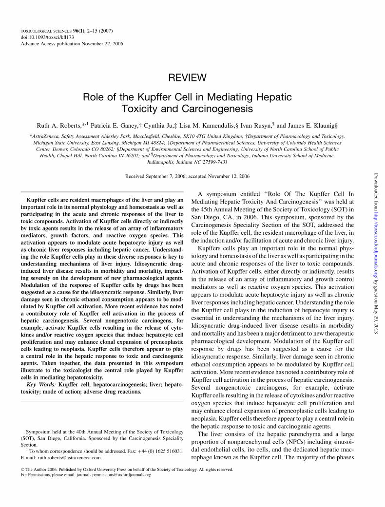
REVIEW Role of the Kupffer Cell in Mediating Hepatic Toxicity and Carcinogenesis Ruth A. Roberts,* ,1 Patricia E. Ganey,† Cynthia Ju,‡ Lisa M. Kamendulis,§ Ivan Rusyn, ¶ and James E. Klaunig§ *AstraZeneca, Safety Assessment Alderley Park, Macclesfield, Cheshire, SK10 4TG United Kingdom; †Department of Pharmacology and Toxicology, Michigan State University, East Lansing, Michigan MI 48824; ‡Department of Pharmaceutical Sciences, University of Colorado Health Sciences Center, Denver, Colorado CO 80262; §Department of Environmental Sciences and Engineering, University of North Carolina School of Public Health, Chapel Hill, North Carolina IN 46202; and ¶ Department of Pharmacology and Toxicology, Indiana University School of Medicine, Indianapolis, Indiana NC 27599-7431 Received September 7, 2006; accepted November 12, 2006 Kupffer cells are resident macrophages of the liver and play an important role in its normal physiology and homeostasis as well as participating in the acute and chronic responses of the liver to toxic compounds. Activation of Kupffer cells directly or indirectly by toxic agents results in the release of an array of inflammatory mediators, growth factors, and reactive oxygen species. This activation appears to modulate acute hepatocyte injury as well as chronic liver responses including hepatic cancer. Understand- ing the role Kupffer cells play in these diverse responses is key to understanding mechanisms of liver injury. Idiosyncratic drug- induced liver disease results in morbidity and mortality, impact- ing severely on the development of new pharmacological agents. Modulation of the response of Kupffer cells by drugs has been suggested as a cause for the idiosyncratic response. Similarly, liver damage seen in chronic ethanol consumption appears to be mod- ulated by Kupffer cell activation. More recent evidence has noted a contributory role of Kupffer cell activation in the process of hepatic carcinogenesis. Several nongenotoxic carcinogens, for example, activate Kupffer cells resulting in the release of cyto- kines and/or reactive oxygen species that induce hepatocyte cell proliferation and may enhance clonal expansion of preneoplastic cells leading to neoplasia. Kupffer cells therefore appear to play a central role in the hepatic response to toxic and carcinogenic agents. Taken together, the data presented in this symposium illustrate to the toxicologist the central role played by Kupffer cells in mediating hepatotoxicity. Key Words: Kupffer cell; hepatocarcinogenesis; liver; hepato- toxicity; mode of action; adverse drug reactions. A symposium entitled ‘‘Role Of The Kupffer Cell In Mediating Hepatic Toxicity And Carcinogenesis’’ was held at the 45th Annual Meeting of the Society of Toxicology (SOT) in San Diego, CA, in 2006. This symposium, sponsored by the Carcinogenesis Speciality Section of the SOT, addressed the role of the Kupffer cell, the resident macrophage of the liver, in the induction and/or facilitation of acute and chronic liver injury. Kupffers cells play an important role in the normal phys- iology and homeostasis of the liver as well as participating in the acute and chronic responses of the liver to toxic compounds. Activation of Kupffer cells, either directly or indirectly, results in the release of an array of inflammatory and growth control mediators as well as reactive oxygen species. This activation appears to modulate acute hepatocyte injury as well as chronic liver responses including hepatic cancer. Understanding the role the Kupffer cell plays in the induction of hepatocyte injury is essential in understanding the mechanisms of the liver injury. Idiosyncratic drug-induced liver disease results in morbidity and mortality and has been a major detriment to new therapeutic pharmacological development. Modulation of the Kupffer cell response by drugs has been suggested as a cause for the idiosyncratic response. Similarly, liver damage seen in chronic ethanol consumption appears to be modulated by Kupffer cell activation. More recent evidence has noted a contributory role of Kupffer cell activation in the process of hepatic carcinogenesis. Several nongenotoxic carcinogens, for example, activate Kupffer cells resulting in the release of cytokines and/or reactive oxygen species that induce hepatocyte cell proliferation and may enhance clonal expansion of preneoplastic cells leading to neoplasia. Kupffer cells therefore appear to play a central role in the hepatic response to toxic and carcinogenic agents. The liver consists of the hepatic parenchyma and a large proportion of nonparenchymal cells (NPCs) including sinusoi- dal endothelial cells, ito cells, and the dedicated hepatic mac- rophage known as the Kupffer cell. The majority of the phases Symposium held at the 40th Annual Meeting of the Society of Toxicology (SOT), San Diego, California. Sponsored by the Carcinogenesis Speciality Section. 1 To whom correspondence should be addressed. Fax: þ44 (0) 1625 516031. E-mail: [email protected]. Ó The Author 2006. Published by Oxford University Press on behalf of the Society of Toxicology. All rights reserved. For Permissions, please email: [email protected] TOXICOLOGICAL SCIENCES 96(1), 2–15 (2007) doi:10.1093/toxsci/kfl173 Advance Access publication November 22, 2006 by guest on May 29, 2013 http://toxsci.oxfordjournals.org/ Downloaded from

Welcome message from author
This document is posted to help you gain knowledge. Please leave a comment to let me know what you think about it! Share it to your friends and learn new things together.
Transcript
REVIEW
Role of the Kupffer Cell in Mediating HepaticToxicity and Carcinogenesis
Ruth A. Roberts,*,1 Patricia E. Ganey,† Cynthia Ju,‡ Lisa M. Kamendulis,§ Ivan Rusyn,¶ and James E. Klaunig§
*AstraZeneca, Safety Assessment Alderley Park, Macclesfield, Cheshire, SK10 4TG United Kingdom; †Department of Pharmacology and Toxicology,
Michigan State University, East Lansing, Michigan MI 48824; ‡Department of Pharmaceutical Sciences, University of Colorado Health Sciences
Center, Denver, Colorado CO 80262; §Department of Environmental Sciences and Engineering, University of North Carolina School of Public
Health, Chapel Hill, North Carolina IN 46202; and ¶Department of Pharmacology and Toxicology, Indiana University School of Medicine,
Indianapolis, Indiana NC 27599-7431
Received September 7, 2006; accepted November 12, 2006
Kupffer cells are resident macrophages of the liver and play an
important role in its normal physiology and homeostasis as well as
participating in the acute and chronic responses of the liver to
toxic compounds. Activation of Kupffer cells directly or indirectly
by toxic agents results in the release of an array of inflammatory
mediators, growth factors, and reactive oxygen species. This
activation appears to modulate acute hepatocyte injury as well
as chronic liver responses including hepatic cancer. Understand-
ing the role Kupffer cells play in these diverse responses is key to
understanding mechanisms of liver injury. Idiosyncratic drug-
induced liver disease results in morbidity and mortality, impact-
ing severely on the development of new pharmacological agents.
Modulation of the response of Kupffer cells by drugs has been
suggested as a cause for the idiosyncratic response. Similarly, liver
damage seen in chronic ethanol consumption appears to be mod-
ulated by Kupffer cell activation. More recent evidence has noted
a contributory role of Kupffer cell activation in the process of
hepatic carcinogenesis. Several nongenotoxic carcinogens, for
example, activate Kupffer cells resulting in the release of cyto-
kines and/or reactive oxygen species that induce hepatocyte cell
proliferation and may enhance clonal expansion of preneoplastic
cells leading to neoplasia. Kupffer cells therefore appear to play
a central role in the hepatic response to toxic and carcinogenic
agents. Taken together, the data presented in this symposium
illustrate to the toxicologist the central role played by Kupffer
cells in mediating hepatotoxicity.
Key Words: Kupffer cell; hepatocarcinogenesis; liver; hepato-
toxicity; mode of action; adverse drug reactions.
A symposium entitled ‘‘Role Of The Kupffer Cell InMediating Hepatic Toxicity And Carcinogenesis’’ was held atthe 45th Annual Meeting of the Society of Toxicology (SOT) inSan Diego, CA, in 2006. This symposium, sponsored by theCarcinogenesis Speciality Section of the SOT, addressed therole of the Kupffer cell, the resident macrophage of the liver, inthe induction and/or facilitation of acute and chronic liver injury.
Kupffers cells play an important role in the normal phys-iology and homeostasis of the liver as well as participating in theacute and chronic responses of the liver to toxic compounds.Activation of Kupffer cells, either directly or indirectly, resultsin the release of an array of inflammatory and growth controlmediators as well as reactive oxygen species. This activationappears to modulate acute hepatocyte injury as well as chronicliver responses including hepatic cancer. Understanding the rolethe Kupffer cell plays in the induction of hepatocyte injury isessential in understanding the mechanisms of the liver injury.Idiosyncratic drug-induced liver disease results in morbidityand mortality and has been a major detriment to new therapeuticpharmacological development. Modulation of the Kupffer cellresponse by drugs has been suggested as a cause for theidiosyncratic response. Similarly, liver damage seen in chronicethanol consumption appears to be modulated by Kupffer cellactivation. More recent evidence has noted a contributory role ofKupffer cell activation in the process of hepatic carcinogenesis.Several nongenotoxic carcinogens, for example, activateKupffer cells resulting in the release of cytokines and/or reactiveoxygen species that induce hepatocyte cell proliferation andmay enhance clonal expansion of preneoplastic cells leading toneoplasia. Kupffer cells therefore appear to play a central role inthe hepatic response to toxic and carcinogenic agents.
The liver consists of the hepatic parenchyma and a largeproportion of nonparenchymal cells (NPCs) including sinusoi-dal endothelial cells, ito cells, and the dedicated hepatic mac-rophage known as the Kupffer cell. The majority of the phases
Symposium held at the 40th Annual Meeting of the Society of Toxicology
(SOT), San Diego, California. Sponsored by the Carcinogenesis Speciality
Section.1 To whom correspondence should be addressed. Fax: þ44 (0) 1625 516031.
E-mail: [email protected].
� The Author 2006. Published by Oxford University Press on behalf of the Society of Toxicology. All rights reserved.For Permissions, please email: [email protected]
TOXICOLOGICAL SCIENCES 96(1), 2–15 (2007)
doi:10.1093/toxsci/kfl173
Advance Access publication November 22, 2006
by guest on May 29, 2013
http://toxsci.oxfordjournals.org/D
ownloaded from
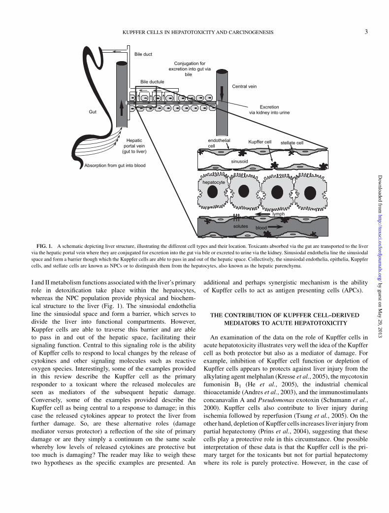
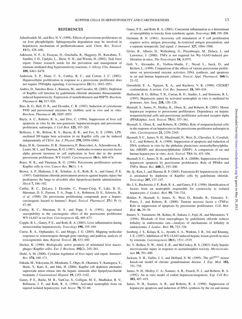
I and II metabolism functions associated with the liver’s primaryrole in detoxification take place within the hepatocytes,whereas the NPC population provide physical and biochem-ical structure to the liver (Fig. 1). The sinusiodal endothelialine the sinusiodal space and form a barrier, which serves todivide the liver into functional compartments. However,Kuppfer cells are able to traverse this barrier and are ableto pass in and out of the hepatic space, facilitating theirsignaling function. Central to this signaling role is the abilityof Kupffer cells to respond to local changes by the release ofcytokines and other signaling molecules such as reactiveoxygen species. Interestingly, some of the examples providedin this review describe the Kupffer cell as the primaryresponder to a toxicant where the released molecules areseen as mediators of the subsequent hepatic damage.Conversely, some of the examples provided describe theKupffer cell as being central to a response to damage; in thiscase the released cytokines appear to protect the liver fromfurther damage. So, are these alternative roles (damagemediator versus protector) a reflection of the site of primarydamage or are they simply a continuum on the same scalewhereby low levels of released cytokines are protective buttoo much is damaging? The reader may like to weigh thesetwo hypotheses as the specific examples are presented. An
additional and perhaps synergistic mechanism is the abilityof Kupffer cells to act as antigen presenting cells (APCs).
THE CONTRIBUTION OF KUPFFER CELL–DERIVED
MEDIATORS TO ACUTE HEPATOTOXICITY
An examination of the data on the role of Kupffer cells inacute hepatotoxicity illustrates very well the idea of the Kupffercell as both protector but also as a mediator of damage. Forexample, inhibition of Kupffer cell function or depletion ofKupffer cells appears to protects against liver injury from thealkylating agent melphalan (Kresse et al., 2005), the mycotoxinfumonisin B1 (He et al., 2005), the industrial chemicalthioacetamide (Andres et al., 2003), and the immunostimulantsconcanavalin A and Pseudomonas exotoxin (Schumann et al.,2000). Kupffer cells also contribute to liver injury duringischemia followed by reperfusion (Tsung et al., 2005). On theother hand, depletion of Kupffer cells increases liver injury frompartial hepatectomy (Prins et al., 2004), suggesting that thesecells play a protective role in this circumstance. One possibleinterpretation of these data is that the Kupffer cell is the pri-mary target for the toxicants but not for partial hepatectomywhere its role is purely protective. However, in the case of
Gut
Absorption from gut into blood
Hepaticportal vein
(gut to liver)
Bile duct
Bile ductuleCentral vein
Conjugation forexcretion into gut via
bile
Excretionvia kidney into urine
sinusoid
blood
stellate cell
solutes
endothelialcell
Kupffer cell
hepatocyte
lymph
FIG. 1. A schematic depicting liver structure, illustrating the different cell types and their location. Toxicants absorbed via the gut are transported to the liver
via the hepatic portal vein where they are conjugated for excretion into the gut via bile or excreted to urine via the kidney. Sinusiodal endothelia line the sinusiodal
space and form a barrier though which the Kuppfer cells are able to pass in and out of the hepatic space. Collectively, the sinusiodal endothelia, epithelia, Kuppfer
cells, and stellate cells are known as NPCs or to distinguish them from the hepatocytes, also known as the hepatic parenchyma.
KUPFFER CELLS IN HEPATOTOXICITY AND CARCINOGENESIS 3
by guest on May 29, 2013
http://toxsci.oxfordjournals.org/D
ownloaded from
acetaminophen-induced hepatotoxicity, Kupffer cells have beenreported to contribute to injury (Ito et al., 2003; Laskin et al.,1986; Michael et al., 1999) as well as to protect againsthepatocellular damage (Ju et al., 2002). In this instance,the Kupffer cell may be the primary site of an initially pro-tective response that develops to cause damage with furtherstimulation.
As with the other roles of Kupffer cells in chronic injury andcarcinogenesis, the mechanisms by which Kupffer cells con-tribute to acute liver injury are varied. In general, the mecha-nisms involve release by Kupffer cells of mediators includingcytokines such as tumor necrosis factor alpha (TNF-a) andinterleukins. Reactive oxygen, nitrogen species, proteases, andlipid metabolites such as prostaglandins and thromboxane arealso released. These mediators can act directly on hepatocytesto cause cell death or indirectly through activation of othercells. For example, activation of hepatic stellate cells by someof these mediators leads to contraction of sinusoids (Kharbandaet al., 2004), resulting in arrest of neutrophils. This is a preludeto transmigration of neutrophils into the parenchyma, which ispromoted by expression of adhesion molecules on sinusoidalendothelial cells activated by Kupffer cell–derived mediators.These mediators, as well as activated sinusiodal endothelialcells, contribute to activation of neutrophils, which in turn candamage hepatocytes through release of proteases and otherfactors (Ganey et al., 1994; Harbrecht et al., 1993; Ho et al.,1996; Mavier et al., 1988). In addition, activation of sinusiodalendothelial cells leads to a procoagulant state in liver andactivation of platelets. A consequence of increased coagulationis deposition of fibrin and hepatic hypoxia, which can also havedeleterious effects on hepatocytes. Thus, the release of media-tors from Kupffer cells can initiate a variety of downstreamevents that may initially stimulate survival and protection butwith continued or higher dose exposure, ultimately contribute tohepatic injury during chemical exposure.
This release of mediators by Kupffer cells and the ensuingevents described above are critical, early features of inflam-matory responses. Indeed, Kupffer cells are activated rapidly inresponse to the inflammatory agent, bacterial lipopolysaccha-ride (LPS) (Bellezzo et al., 1996; Portoles et al., 1994; Yaoet al., 2004). At relatively large doses, LPS damages liver, andthis injury is dependent on Kupffer cells (Brown et al., 1997;Fukuda et al., 2004; Iimuro et al., 1994; Vollmar et al., 1996).Treatment with small doses of LPS activates Kupffer cells inthe absence of liver injury perhaps because levels or duration ofthe release of cytokines and other molecules are below athreshold for injury and may even be protective This hypothesisin supported by the observation that cotreatment with non-injurious doses of LPS increases sensitivity of liver to a varietyof other chemicals including monocrotaline (MCT), cocaine,allyl alcohol, aflatoxin B1, chlorpromazine, halothane, andranitidine (RAN) (reviewed in Ganey and Roth, 2001). Formany of these chemicals, Kupffer cells and Kupffer cell–derived mediators contribute to liver damage from cotreatment
with LPS (Labib et al., 2003; Sneed et al., 1997; Yee et al.,2003a).
The data presented above clearly support the idea ofa threshold of Kupffer activation and release above which liverdamage is induced. This mechanism if illustrated by studies ofMCT and RAN, where small doses of LPS enhance hepato-toxicity in a Kupffer cell–dependent manner. MCT is a pyrro-lizidine alkaloid known to induce hepatotoxicity in people andanimals when they are exposed through consumption of con-taminated foods or pastures, respectively (Mattocks, 1986). Inrats, relatively large doses of MCT cause acute liver damage bya mechanism that appears to be independent of inflammation(Copple et al., 2003). However, smaller doses of MCT thatalone are not injurious are rendered hepatotoxic by coadmin-istration of a small, nontoxic dose of LPS (Yee et al., 2000).This combination damages not only hepatocytes but also sinu-soidal endothelial cells. Pretreatment with gadolinium chloride(GdCl), which inhibits Kupffer cell function, reduces bothhepatocyte and sinusoidal epithelial cell injury, demonstratingthe Kupffer cell dependence of hepatotoxicity in this model ofchemical-inflammation interaction.
One of the key components of inflammation important tohepatotoxicity from coadministration of small doses of MCTand LPS is the Kupffer cell–derived cytokine TNF-a. Theconcentration of TNF-a in plasma peaks about 90 min afterexposure to LPS and rapidly returns to baseline (Mastronardiet al., 2001; Noel et al., 1990; Xuan et al., 2001). Cotreatmentwith MCT prolonged the increase in plasma TNF-a in LPS-treated rats (Yee et al., 2003a). Neutralization of TNF-areduced damage and also reduced the accumulation of neutro-phils in the liver (Yee et al., 2003b). Taken together, theseresults suggest that Kupffer cells participate in MCT/LPS-induced liver injury through release of TNF-a, promotesaccumulation of neutrophils in liver leading to hepatic damage.
RAN is a histamine 2 receptor antagonist that causesidiosyncratic liver injury in a very small percentage of peopletaking the drug (Ribiero et al., 2000; Vial et al., 1991).Similarly to MCT, RAN is not hepatotoxic in laboratory ani-mals when given alone; however, pretreatment with LPS ren-ders RAN hepatotoxic in rats (Luyendyk et al., 2003). In thismodel, RAN was administered 2 h after LPS, and at this time,the concentration of TNF-a in plasma was increased comparedto animals not treated with LPS (Tukov et al., unpublished).The concentration of TNF-a decreased rapidly in animals thatwere not cotreated with RAN, whereas in animals that receivedthe drug, TNF-a concentration remained elevated for severalhours. Similarly, RAN increased the LPS-stimulated release ofTNF-a in cocultures of Kupffer cells and hepatocytes, sug-gesting that Kupffer cells contribute to circulating TNF-a inLPS/RAN-treated rats. The potential contribution of TNF-a toliver injury in this model was evaluated by pretreating LPS/RAN-cotreated animals either with pentoxifylline to reduce synthesisof TNF-a or with etanercept to interfere with the ability ofTNF-a to activate its cellular receptors. Prior administration of
4 ROBERTS ET AL.
by guest on May 29, 2013
http://toxsci.oxfordjournals.org/D
ownloaded from

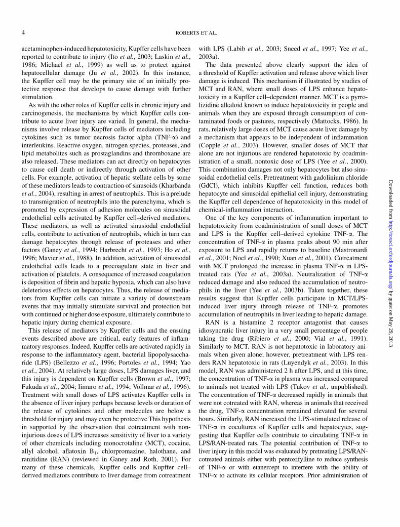
either agent reduced TNF-a activity in plasma as well as liverinjury assessed biochemically and histologically. Interference withTNF-a also attenuated the effects of LSP/RAN to activatecoagulation and to increase the plasma concentration of plas-minogen activator inhibitor-1 (PAI-1), a major inhibitor offibrinolysis. Moreover, interference with TNF-a also reducedhepatic fibrin deposition that occurs as a consequence ofdisregulation of the hemostatic system. Thus, TNF-a is impor-tant in the changes in the hemostatic system that contribute to liverinjury from LPS/RAN. Finally, pretreatment with either pentox-ifylline or etanercept decreased the LPS/RAN-induced increase inthe plasma concentration of the chemokine, macrophage in-flammatory protein-2 (MIP2) (Tukov et al., unpublished results).This finding is of significance because neutrophils are critical forliver injury in LPS/RAN-treated rats (Luyendyk et al., 2005), andMIP2 might contribute to hepatic neutrophil accumulation in thismodel. Drawing all of this together, it appears that Kupffer cellscontribute to liver injury induced by LPS/RAN by releasing TNF-athat promotes activation of the hemostatic system and deposition offibrin in the liver. As with MCN alone, RAN is not hepatotoxic inlaboratory animals but addition of RAN to a system where theKupffer cells are already activated leads to liver damage. Fibrindeposition leads to tissue hypoxia that probably contributes tohepatic cell death (Fig. 2). In addition, Kupffer cell–derived TNF-a plays a role in the increase in MIP2 after treatment with LPS/RAN, which might affect the accumulation of neutrophils that arenecessary for hepatotoxicity.
THE ROLE OF KUPFFER CELLS INIMMUNE-MEDIATED
ADVERSE DRUG REACTIONS
These data derived from investigation of known hepatotox-icants may help in understanding unpredictable and severeimmune-mediated adverse drug reactions (IADR). TheseIADRs pose a significant problem for clinical practice andthe pharmaceutical industry and are the subject of extensiveresearch. One possible explanation for the relatively lowincidence of IADR is that most individuals develop toleranceto immunogenic drug-protein adducts. Previous studies exam-ining this hypothesis, using a model hapten, demonstrated thatimmune tolerance was induced by a protein adduct of thehapten, and that Kupffer cells play an important role in suchtolerance. The mechanism of this tolerance is unclear but couldrely on Kupffer cells, acting as APCs downregulating antigen-specific T-cell responses.
With < 10% of all adverse drug reactions reported, themagnitude of the problem is significant, with estimates of costs> $US30 billion annually in the United States (1995 value).In addition, the costs of not determining the potential of a drugto produce hypersensitivity in the preclinical phase of drugdevelopment can be substantial. It has been estimated that thepreclinical phase and clinical phase I, phase II, and phase IIIcosts are approximately $US6 million, $US12 million, $US12million, and $US100 million per drug, respectively (1999values). It is important that investigational drugs with thepotential to produce hypersensitivity reactions be identified asearly in the development process as possible. Some adversereactions to drugs can be avoided if drug-drug interactions areknown or if there is a structure-activity relationship estab-lished. However, these methods are inadequate. Appropriateanimal models of drug-induced hypersensitivity are needed,especially because hypersensitivity has been cited as the lead-ing reason for taking drugs off the market.
IADRs, including allergic hepatitis, lupus, cutaneous reac-tions, and blood dyscrasias, account for approximately 6–10%of all adverse drug reactions. IADR caused 137,000–230,000hospital admissions per year in the United States (1998values), with approximate annual costs of $275 to $600 million(Adkinson et al., 2002; Lazarou et al., 1998; Ratajczak, 2004).One puzzling aspect of IADR is that among all patients whotake a certain drug, only a small percentage will develop IADR.One possible explanation for the low occurrence of IADR isthat most individuals develop immunological tolerance, ratherthan an adverse immune response, to drug-protein adducts.Thus, understanding the molecular and cellular mechanismsof immune tolerance to these adducts will lead to the iden-tification of risk factors that determine patients’ susceptibilityto IADR.
Previous studies using a model hapten, 2,4-dinitrochlorobenzene(DNCB), have demonstrated that pretreatment of mice with a bo-vine serum albumin (BSA) conjugate of DNCB (DNP-BSA)induced tolerance against a T-cell–mediated delayed-type
FIG. 2. Working hypothesis of the role of Kupffer cells in liver injury from
LPS/RAN interaction. RAN increases LPS-stimulated release of TNF-a from
Kupffer cells. TNF-a promotes activation of sinusoidal endothelial cells, leading
to activation of coagulation and deposition of fibrin in livers. TNF also
contributes to increased circulating concentrations of PAI-1 that inhibit
fibrinolysis. The consequence is enhanced fibrin deposition, ischemia, and
hypoxia. The hypoxia probably leads to hepatocellular death. Additionally, TNF-
a contributes to increased chemokine concentration, including MIP2, which
facilitates accumulation of neutrophils and their activation in the liver. Activated
neutrophils play a role in hepatotoxicity from LPS/RAN. Dotted lines represent
those effects for which there is not direct experimental evidence.
KUPFFER CELLS IN HEPATOTOXICITY AND CARCINOGENESIS 5
by guest on May 29, 2013
http://toxsci.oxfordjournals.org/D
ownloaded from
hypersensitivity reaction caused by DNCB sensitization (Juet al., 2003). The data revealed that the tolerance could beinhibited when Kupffer cells were depleted with liposome-entrapped clodronate prior to DNP-BSA pretreatment. Inaddition, the tolerance could be induced in naı̈ve mice byadoptive transfer with a Kupffer cell-enriched fraction of liverNPCs obtained from mice tolerized by DNP-BSA pretreatment.In contrast, a Kupffer cell–depleted fraction of liver NPC couldnot transfer tolerance. These findings suggest that Kupffer cellsplay an important role in downregulating T-cell–mediated re-actions against drug-protein adducts. As suggested above, thiscould occur by Kupffer cells acting as APCs to down regulateantigen-specific T-cell responses.
Kupffer cells, as the largest population of tissue residentmacrophages, not only play an important role in first-linedefense against invading pathogens, but may also act as APCsto activate and regulate T-cell responses. It is known that T-cellactivation requires two signals. The recognition of a specificmajor histocompatibility complex (MHC)/peptide complex bya T-cell receptor (TCR) gives signal 1 (Paul, 1999). Signal 2 isprovided by the binding of costimulatory molecules expressedon APCs to their ligands on T cells (Germain, 1981). Forexample, CD40 and B7 molecules (B7-1 and B7-2) on APCsinteract with CD40 ligand and CD28 on T cells, respectively(Greenfield et al., 1998; Schoenberger et al., 1998). It has beendemonstrated that T cells are anergized rather than activated ifthey receive only signal 1 but not signal 2, or insufficientamount of signal 2, during antigen stimulation (Lafferty andCunningham, 1975; Matzinger, 1994). Therefore, one possiblemechanism by which Kupffer cells may induce T-cell toleranceis that although they can act as APCs, they express inadequatelevels of costimulatory molecules and are thus only partiallycompetent leading to T-cell anergy rather than activation.
This hypothesis was examined by the evaluation of Kupffercell expression of various APC-function related molecules,such as MHC II, B7-1, B7-2, and CD40. The data demonstratedthat, compared with potent APCs such as dendritic cells,Kupffer cells express inadequate levels of costimulatory mole-cules. Furthermore, the abilities of Kupffer cells and dendriticcells to activate naı̈ve T cells were determined and comparedusing an in vitro system, in which ovalbumin (OVA)-TCRtransgenic T cells were cocultured with Kupffer cells or den-dritic cells in the presence of the antigen (an OVA peptide). Thedata revealed that dendritic cells significantly stimulated T-cellproliferation in response to antigen exposure, whereas Kupffercells could not induce T-cell activation. Interestingly, whenKupffer cells were included in the cocultures of dendritic cellsand T cells, T-cell proliferation was inhibited. This inhibitoryeffect of Kupffer cells was independent of cell-cell contact,suggesting that Kupffer cells could produce and release solubleimmuno-suppressive mediators. These findings suggest thatKupffer cells have some ability to act as APCs but are onlypartially competent due to their insufficient expression ofcostimulatory molecules. If this hypothesis is correct, Kupffer
cells could actively suppress T-cell activation induced by otherAPCs. Taken to its logical conclusion, this hypothesis suggeststhat individuals with a higher propensity to develop IADRsmay have more costimulatory molecules in their Kupffer cells.
In summary, the data indicate that Kupffer cells may playa role as primary inducers of immune tolerance against ahapten-induced delayed-type hypersensitivity reaction. Be-cause drug-protein adducts are predominantly formed in theliver, or they may circulate to the liver through blood, thesecells may have a similar regulatory effect on immune responsesto drug-protein adducts. The mechanism by which Kupffercells cause T-cell tolerance could involve inadequate expres-sion of costimulatory molecules and the production of immuno-suppressive mediators. These findings suggest that geneticand/or environmental factors that cause impairment of thetolerogenic functions of Kupffer cells may lead to increasedrisk of developing IADR in certain individuals.
ROLE OF THE KUPFFER CELL IN HEPATIC
PRENEOPLASTIC LESION GROWTH
So far, this review has explored the role that Kupffer cellsplay in acute liver responses to damage. However, it is clearthat activated Kupffer cells are also key to chronic liverresponses, including neoplasia. Chemically induced neoplasiais a multistep process involving two key mechanisms: DNAdamage and alteration in cell growth control that allow thisdamage to persist and be promoted to foci and tumors.Genotoxic agents induce tumors by increasing rates of DNAdamage, whereas nongenotoxic carcinogens act to promote thespontaneous or accumulated DNA damage present in all tis-sues. While much less is known about the exact mode of actionof nongenotoxic carcinogens they are known to modulate cellgrowth and cell death, with changes in gene expression and cellgrowth being paramount in their mechanism(s) of action. Theseagents frequently function at the promotion stage of the cancerprocess. The stage of hepatic tumor promotion involves theselective clonal expansion of preneoplastic focal cells. Chem-icals that function at this stage of the cancer process (tumorpromoters) encompass a structurally diverse group of pharma-ceutical and environmental agents, all of which result in in-creased DNA synthesis during the first few weeks of exposurethat typically returns to baseline levels within 2–4 weeks oftreatment despite continual chemical exposure in normal liver(Marsman et al., 1988; Schulte-Hermann et al., 1990). Incontrast, some tumor promoters produce a sustained increase inthe growth of chemically- and spontaneously induced lesionsin the liver (Cattley et al., 1991; Isenberg et al., 1997; Kolajaet al., 1995; Marsman et al., 1988). The increased cellreplication may enhance the rate of fixation of DNA damageinto the genome leading to changes in gene expression, changesthat facilitate the clonal expansion of initiated cells, and lead tothe formation of hepatic focal lesions (Cattley et al., 1998).
6 ROBERTS ET AL.
by guest on May 29, 2013
http://toxsci.oxfordjournals.org/D
ownloaded from

Historically, research has focused on the hepatocyte as thetarget cell of chemical carcinogens, however, recent studieshave emerged that suggest a role for NPCs, specifically Kupffercells, as important mediators of cell proliferation by tumorpromoters (Hasmall et al., 2000a,b; Rose et al., 1997b; Rusynet al., 1998, 2000).
As described already, activated Kupffer cells release a widearray of biologically active products including reactive oxygenspecies, interleukins, and cytokines (Decker, 1990; Winwoodand Arthur, 1993), all of which may be capable of modulatinghepatocellular growth. In particular TNF-a has been linked tothe stimulation of hepatocellular growth by tumor promotingcompounds (Hasmall et al., 2000b; West et al., 1999). Studiesin our group are directed at further defining the role of theKupffer cell in hepatocarcinogenesis. Our working hypothesisis that activation of Kupffer cells results in the release ofcellular growth regulatory signaling molecules that result in anincrease in the proliferation of hepatocytes; this is expected tobe transient in normal hepatocytes but sustained in preneo-plastic, initiated hepatocytes, ultimately resulting in selectiveclonal expansion of the preneoplastic hepatocytes (hepatictumor promotion).
Experimental models in which preneoplastic focal lesionsare induced in rodents using the DNA-damaging compound,diethylnitrosamine as a surrogate for liver tumor promotion(Isenberg et al., 1997; Klaunig and Kamendulis, 1999; Kolajaet al., 1995, 1996a,b; Schulte-Hermann et al., 1981, 1990) havebeen used to dissect, in a relatively defined experimental sys-tem, agents that influence lesion growth and death. The effectsof several tumor promoting agents on growth of preneoplasticlesions in B6C3F1 mice have been evaluated using stereologicmethodology (Kolaja et al., 1995; Xu et al., 1998) and im-munohistochemistry for quantitation of DNA synthesis. Fol-lowing exposure to phenobarbital (0, 10, 100, and 500 ppm) for30 or 60 days, an increase in the number, and relative volume offocal lesions was apparent after exposure to carcinogenic dosesof phenobarbital (100 and 500 ppm), while the noncarcino-genic dose did not increase the number or volume of hepaticlesions (Kolaja et al., 1995, 1996a,b). In addition, increases inDNA synthesis remained increased within preneoplastic le-sions at both 30 and 60 days while DNA synthetic activity innormal surrounding tissue returned to baseline (Kolaja et al.,1995, 1996a). Similar results on the growth of preneoplasticlesions in mouse liver were observed following exposure toWyeth 14,643 as well as dieldrin (Isenberg et al., 1997; Kolajaet al., 1995, 1996b). These results illustrate that tumor pro-moter exposure results in a sustained increase in growth but justin preneoplastic cells. To evaluate the potential role of Kupffercells on modulation of hepatocyte growth, a number of agentsare available that are reported to either affect Kupffer cellactivation or result in depletion of Kupffer cells. Althoughthese agents have a marked impact on acute responses to liverinjury as already described in the previous section, these pro-tocols have yielded mixed results in experiments on hepatocyte
growth. For example, while glycine and methyl palmitate(nonselective agents that inactivate but do not eliminateKupffer cells) have been shown to decrease hepatocellulargrowth (Rose et al., 1997a; Watanabe et al., 2000), depletion ofKupffer cells with GdCl has been shown to increase liver cellproliferation in rodent liver (Rai et al., 1997; Rose et al., 2001).These apparently conflicting data could be resolved by theproposal that GdCl inactivation of Kupffer cells initially pro-vokes a burst of growth mediators from the Kupffer cells that inturn cause an increase in DNA synthesis. Recognizing the po-tential limitations of these approaches, we have adopted aprotocol that utilizes clodronate-encapsulated liposomes todeplete Kupffer cells in vivo (vanRoojen et al., 1996). De-pletion of Kupffer cells by clodronate liposomes occurs througha mechanism involving the phagocytosis of liposomes byKupffer cells, resulting in the release of clodronate, whichleads to adenosine 5#-triphosphate depletion and triggers apo-ptotic cell death (Lehenkari et al., 2002; Van Rooijen andvan Kesteren-Hendrikx, 2003).
To assess the effect of Kupffer cell activation on induction ofDNA synthesis in hepatocytes as well as to assess the efficacyof inhibition of Kupffer cells (via clodronate liposomes) onhepatocellular growth, male B6C3F1 mice were given LPS(0.25 mg/kg, i.p.) in the presence or absence of clodronateliposomes (by tail vein injection of clodronate liposomes; 200llof 2.0 mg/ml clordronate in PBS, 3 times/week). After 7 daysof treatment, the number of Kupffer cells was determinedusing the F4/80 mouse macrophage antibody. The number ofKupffer cells identifiable immunohistologically was signifi-cantly decreased by clodronate liposome treatment in control aswell as in the LPS treatment groups (data not shown). DNAsynthesis was assessed in the liver of these same animals usingBrdU immunohistochemistry. LPS produced a significant in-crease in DNA synthesis (approximately 80%) compared withcontrols, whereas the induction of DNA synthesis by LPS wasdecreased by approximately 80% by Kupffer cell depletion(data not shown). In addition, Kupffer cell depletion resulted ina 50% reduction in the basal level of DNA synthesis seen incontrol liver. These results demonstrate that the clodronateliposome protocol is effective for depleting Kupffer cells andprovides evidence in support of the growth permissive role ofthe Kupffer cell in the induction of cell proliferation.
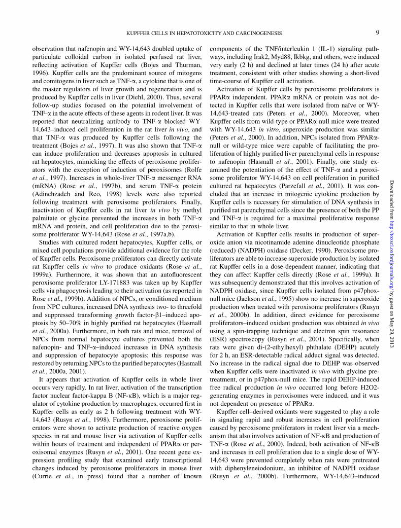
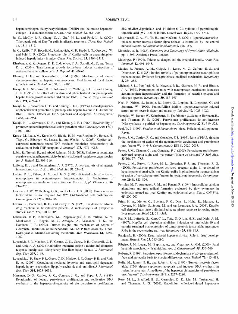
In a separate study, the role of Kupffer cells in the modulationof preneoplastic lesion growth was evaluated in hepatic focallesions produced in B6C3F1 mice using diethylnitrosamine.Following lesion development, LPS was given (0.25 mg/kg i.p.)for 7 or 30 days. LPS increased the relative volume of hepaticfocal lesions after 30 days (~4-fold increase over control) whileKupffer cell depletion with clodronate liposomes significantlyreduced the LPS-induced increase in focal lesion volume(Fig. 3). In addition, LPS increased DNA synthesis withinfocal lesions (~3-fold increase over control) after 30 days butthis was inhibited by depletion of Kupffer cells (Fig. 3). Innonfocal liver, LPS produced an early increase in DNA
KUPFFER CELLS IN HEPATOTOXICITY AND CARCINOGENESIS 7
by guest on May 29, 2013
http://toxsci.oxfordjournals.org/D
ownloaded from

synthesis (after 7 days) which was also prevented by Kupffercell depletion (Fig. 3). Similarly, an inhibition of phenobarbital-induced preneoplastic lesion growth was also seen followingKupffer cell depletion. These data collectively provide supportfor the involvement of the Kupffer cell in hepatic carcinogen-esis, and suggest that activation of this cell type may function atthe promotion stage of the cancer process. The mechanisms ofnongenotoxic carcinogenesis and the role of Kupffer cells willbe explored further in the next two sections.
KUPFFER CELL ACTIVATION BY NONGENOTOXIC
CHEMICALS: DOES IT PLAY A ROLE IN
HEPATOCARCINOGENESIS?
The previous two sections have discussed the role of Kupffercells in acute and chronic liver responses to hepatotoxicantssuch as MCP, RAN, and LPS. In these examples, evidencesuggests that the Kupffer cell is the primary target of chemicaldamage. However, the effects of many nongenotoxic livertoxicants are thought to be mediated through activation of nu-clear hormone receptors. Peroxisome proliferators, a class ofrodent liver carcinogens that are also relevant for human health,are one classic example. The current body of knowledge onthese compounds shows an important role for peroxisomeproliferator activated receptor alpha (PPARa)–dependent mo-lecular events (Peters et al., 2005). Importantly, PPARa isrequired for liver carcinogenesis in animals fed peroxisomeproliferators chronically (Peters et al., 1997). At the same time,several groups reported that PPARa-independent events, thatinclude an increase in liver oxidants and mitogenic cytokines,also take place. A large body of evidence exists in the scientificliterature to demonstrate that a number of acute pleiotropiceffects of peroxisome proliferators in rodent liver are mediatedby Kupffer cells (Rose et al., 1999b). At the same time, it is not
presently known whether Kupffer cell–specific events playa role in long-term effects of peroxisome proliferators, and ifthese mechanisms are operational in species other than rats andmice. Based on evidence presented already in this review, itseems probable that the pleiotropic effects of peroxisomeproliferators are dependent on PPARa but in parallel peroxi-some proliferators activate Kupffer cells as a direct target asalready described for MCP, RAN, and LPS. Thus, deletion ofPPARa leaves the Kupffer cell–mediated component of theresponse which by itself is unable to drive proliferation andcarcinogenesis for this class of compounds.
Peroxisome proliferators increase proliferation of rodentliver parenchymal cells both in vivo and in vitro; however, theeffect on isolated hepatocytes from rats and mice is much lessrobust and persistent. For example, 8- to 10-fold increases incell proliferation were reported in vivo (Marsman et al., 1988),while only up to twofold increases were demonstrated in vitro,regardless of the dose of the compound used (Goll et al., 1999;Hasmall et al., 1999). Most interestingly, in highly purifiedrodent hepatocytes, peroxisome proliferators fail to initiate anincrease in DNA synthesis (Hasmall et al., 2000a, 2001;Parzefall et al., 2001). It has been hypothesized that onepossible explanation for these differences is the involvement ofNPCs in whole liver that are lost during purification and cultureof liver parenchymal cells. Several laboratories in the pastdecade have demonstrated that Kupffer cell activation byperoxisome proliferators (1) is independent of PPARa, (2)involves generation of reactive oxygen species, and (3) leads toproduction of mitogenic cytokines (reviewed in Rusyn et al.,2000a). This hypothesis fits with the suggestion of a two-partresponse of the liver to peroxisome proliferators: one mediatedby PPARa and a second mediated by Kupffer cells.
One of the first experimental facts that suggested activationof Kupffer cells by peroxisome proliferators came from an
#
7 Days 30 Days
Focal Lesion VolumeVo
lum
e of
Foc
al L
esio
ns (%
of L
iver
)
0
2
4
6
8
10
#
*
7 Days 30 Days
DNA Synthesis(Non-Focal Liver)
Labe
ling
Inde
x (%
)
0
2
4
6
8
10
DNA Synthesis(Focal Lesion)
Labe
ling
Inde
x (%
)
0
10
20
30
40
#
*
7 Days 30 Days
*
FIG. 3. Effect of Kupffer cell activation and Kupffer cell depletion on preneoplastic hepatic focal lesion growth in B6C3F1 mouse liver. *Statistically different
from control (p < 0.05), #statistically different from LPS treatment group (p < 0.05).
8 ROBERTS ET AL.
by guest on May 29, 2013
http://toxsci.oxfordjournals.org/D
ownloaded from
observation that nafenopin and WY-14,643 doubled uptake ofparticulate colloidal carbon in isolated perfused rat liver,reflecting activation of Kupffer cells (Bojes and Thurman,1996). Kupffer cells are the predominant source of mitogensand comitogens in liver such as TNF-a, a cytokine that is one ofthe master regulators of liver growth and regeneration and isproduced by Kupffer cells in liver (Diehl, 2000). Thus, severalfollow-up studies focused on the potential involvement ofTNF-a in the acute effects of these agents in rodent liver. It wasreported that neutralizing antibody to TNF-a blocked WY-14,643–induced cell proliferation in the rat liver in vivo, andthat TNF-a was produced by Kupffer cells following thetreatment (Bojes et al., 1997). It was also shown that TNF-acan induce proliferation and decreases apoptosis in culturedrat hepatocytes, mimicking the effects of peroxisome prolifer-ators with the exception of induction of peroxisomes (Rolfeet al., 1997). Increases in whole-liver TNF-a messenger RNA(mRNA) (Rose et al., 1997b), and serum TNF-a protein(Adinehzadeh and Reo, 1998) levels were also reportedfollowing treatment with peroxisome proliferators. Finally,inactivation of Kupffer cells in rat liver in vivo by methylpalmitate or glycine prevented the increases in both TNF-amRNA and protein, and cell proliferation due to the peroxi-some proliferator WY-14,643 (Rose et al., 1997a,b).
Studies with cultured rodent hepatocytes, Kupffer cells, ormixed cell populations provide additional evidence for the roleof Kupffer cells. Peroxisome proliferators can directly activaterat Kupffer cells in vitro to produce oxidants (Rose et al.,1999a). Furthermore, it was shown that an autofluorescentperoxisome proliferator LY-171883 was taken up by Kupffercells via phagocytosis leading to their activation (as reported inRose et al., 1999b). Addition of NPCs, or conditioned mediumfrom NPC cultures, increased DNA synthesis two- to threefoldand suppressed transforming growth factor-b1–induced apo-ptosis by 50–70% in highly purified rat hepatocytes (Hasmallet al., 2000a). Furthermore, in both rats and mice, removal ofNPCs from normal hepatocyte cultures prevented both thenafenopin- and TNF-a–induced increases in DNA synthesisand suppression of hepatocyte apoptosis; this response wasrestored by returning NPCs to the purified hepatocytes (Hasmallet al., 2000a, 2001).
It appears that activation of Kupffer cells in whole liveroccurs very rapidly. In rat liver, activation of the transcriptionfactor nuclear factor-kappa B (NF-jB), which is a major reg-ulator of cytokine production by macrophages, occurred first inKupffer cells as early as 2 h following treatment with WY-14,643 (Rusyn et al., 1998). Furthermore, peroxisome prolif-erators were shown to activate production of reactive oxygenspecies in rat and mouse liver via activation of Kupffer cellswithin hours of treatment and independent of PPARa or per-oxisomal enzymes (Rusyn et al., 2001). One recent gene ex-pression profiling study that examined early transcriptionalchanges induced by peroxisome proliferators in mouse liver(Currie et al., in press) found that a number of known
components of the TNF/interleukin 1 (IL-1) signaling path-ways, including Irak2, Myd88, Ikbkg, and others, were inducedvery early (2 h) and declined at later times (24 h) after acutetreatment, consistent with other studies showing a short-livedtime-course of Kupffer cell activation.
Activation of Kupffer cells by peroxisome proliferators isPPARa independent. PPARa mRNA or protein was not de-tected in Kupffer cells that were isolated from naı̈ve or WY-14,643-treated rats (Peters et al., 2000). Moreover, whenKupffer cells from wild-type or PPARa-null mice were treatedwith WY-14,643 in vitro, superoxide production was similar(Peters et al., 2000). In addition, NPCs isolated from PPARa-null or wild-type mice were capable of facilitating the pro-liferation of highly purified liver parenchymal cells in responseto nafenopin (Hasmall et al., 2001). Finally, one study ex-amined the potentiation of the effect of TNF-a and a peroxi-some proliferator WY-14,643 on cell proliferation in purifiedcultured rat hepatocytes (Parzefall et al., 2001). It was con-cluded that an increase in mitogenic cytokine production byKupffer cells is necessary for stimulation of DNA synthesis inpurified rat parenchymal cells since the presence of both the PPand TNF-a is required for a maximal proliferative responsesimilar to that in whole liver.
Activation of Kupffer cells results in production of super-oxide anion via nicotinamide adenine dinucleotide phosphate(reduced) (NADPH) oxidase (Decker, 1990). Peroxisome pro-liferators are able to increase superoxide production by isolatedrat Kupffer cells in a dose-dependent manner, indicating thatthey can affect Kupffer cells directly (Rose et al., 1999a). Itwas subsequently demonstrated that this involves activation ofNADPH oxidase, since Kupffer cells isolated from p47phox-null mice (Jackson et al., 1995) show no increase in superoxideproduction when treated with peroxisome proliferators (Rusynet al., 2000b). In addition, direct evidence for peroxisomeproliferators–induced oxidant production was obtained in vivousing a spin-trapping technique and electron spin resonance(ESR) spectroscopy (Rusyn et al., 2001). Specifically, whenrats were given di-(2-ethylhexyl) phthalate (DEHP) acutelyfor 2 h, an ESR-detectable radical adduct signal was detected.No increase in the radical signal due to DEHP was observedwhen Kupffer cells were inactivated in vivo with glycine pre-treatment, or in p47phox-null mice. The rapid DEHP-inducedfree radical production in vivo occurred long before H2O2-generating enzymes in peroxisomes were induced, and it wasnot dependent on presence of PPARa.
Kupffer cell–derived oxidants were suggested to play a rolein signaling rapid and robust increases in cell proliferationcaused by peroxisome proliferators in rodent liver via a mech-anism that also involves activation of NF-jB and production ofTNF-a (Rose et al., 2000). Indeed, both activation of NF-jBand increases in cell proliferation due to a single dose of WY-14,643 were prevented completely when rats were pretreatedwith diphenyleneiodonium, an inhibitor of NADPH oxidase(Rusyn et al., 2000b). Furthermore, WY-14,643–induced
KUPFFER CELLS IN HEPATOTOXICITY AND CARCINOGENESIS 9
by guest on May 29, 2013
http://toxsci.oxfordjournals.org/D
ownloaded from

activation of NF-jB, increase in TNF-a mRNA, and acuteincreases in liver weight and cell proliferation did not occur inp47phox-null mice. Combined, these results provide strongevidence that NADPH oxidase in Kupffer cells is a source ofoxidants that is activated very early after treatment with per-oxisome proliferators. Since PPARa is not involved in acti-vation of Kupffer cells by peroxisome proliferators, it is not yetclear how exactly such activation occurs upstream of theNADPH oxidase.
A causal link between Kupffer cell–mediated production ofcytokines and long-term (effects of peroxisome proliferators inliver has not been established yet. However, it has been shownas described in a previous section that Kupffer cells play a rolein the sustained proliferation key to the development of earlypreneoplastic foci. One report suggests that Kupffer cell ac-tivation may persist for longer than a few days with WY-14,643but not DEHP (Rose et al., 1999a). Both agents are known tocause robust increase in hepatocellular proliferation in rodentliver during the first few days of treatment; however, only WY-14,643 sustains rates of proliferation with long-term treatment(as reported in Marsman et al., 1988). It was found that Kupffercells isolated from rats fed WY-14,643 generated superoxide atrates significantly greater than cells from controls for up to3 weeks of treatment; however, superoxide production was notstimulated by feeding DEHP for the same period of time (Roseet al., 1999a). Several published reports cast doubt on thesustainability of the Kupffer cell–mediated events under con-ditions of continuous exposure to peroxisome proliferators. Noincrease in hepatic proliferation was detected in PPARa-nullmice fed the Wy-14,643 diet for 1 or 5 weeks which suggeststhat subchronic effects of peroxisome proliferators on cell pro-liferation in mouse liver are mediated exclusively by PPARabut not Kupffer cells (Peters et al., 1997). However, this isperhaps explained by more recent data described earlier in thisreview where Kupffer cells were key to the sustained pro-liferation seen in foci but not in surrounding normal liver.
Several published reports have questioned the role for TNF-a in peroxisome proliferator–induced cell proliferation inrodent liver. Several groups have attempted to define the roleof TNF-a signaling in hepatocellular growth induced by per-oxisome proliferators by administering the potent peroxisomeproliferator, WY-14,643 (for up to 4 days), to mice nullizygousfor TNF-receptor 1 (TNFR1), TNFR2, both receptors, orTNF-a protein (Anderson et al., 2001; Lawrence et al., 2001).Neither study found evidence of abrogated peroxisome prolif-erator–induced proliferative response in mouse liver as a resultof a null genotype. It should be noted, however, that sincecytokine-induced signaling is highly redundant, the knockoutmodels used in these studies may have compensated for the lackof a particular signaling molecule. Furthermore, when cytokineexpression was examined in nontumorous liver tissue oradenomas in wild-type mice fed with a carcinogenic dose ofWY-14,643 for 52 weeks, no difference in expression of TNF-a,IL-6, or TNFR1 &2 was found (Anderson et al., 2001).
However, IL-1b mRNA was significantly elevated and it waspostulated that cytokines other than TNF-a may be importantliver comitogens in peroxisome proliferator–treated rodents.
The potential for the Kupffer cell–derived oxidants to con-tribute to oxidative DNA damage following exposure to peroxi-some proliferators has also been questioned. The analysis ofexpression of base excision DNA repair genes was used toassess whether this sensitive in vivo biomarker of oxidativestress to DNA can be used to determine the source of DNA-damaging oxidants following treatment with Peroxisome pro-liferators. Using PPARa- and p47phox-null mice treated withWY-14,643 for 4 weeks, the report concluded that DNA-damaging oxidants are generated by enzymes that are inducedafter activation of PPARa, such as those involved in lipidmetabolism in peroxisomes, and are not the result of activationof NADPH oxidase in Kupffer cells (Rusyn et al., 2004).
In conclusion, it is apparent that while PPARa is required forthe sustained activation of peroxisome proliferator–inducedmolecular processes that ultimately result in liver tumors andthat Kupffer cell–mediated events play a role only in the earlystages of the response (Fig. 4). Peroxisome proliferators doactivate Kupffer cells to generate oxidants and increase mito-genic cytokines independent of activation of PPARa; however,the Kupffer cell–mediated events likely require activation ofPPARa in parenchymal cells to achieve the overall pathophys-iological outcome. Peroxisome proliferators are a class ofnongenotoxic chemicals that exert both nuclear receptor–dependent and –independent modes of action in rodent liver,and thus offer a unique opportunity to understand the respectiverole of each pathway in the overall mechanism of their carcin-ogenic effects that is likely to be important for understandingthe carcinogenicity of other nuclear receptor activators. Thedata presented in this section underline the potential forKupffer cell–dependent and independent pathways in theresponse to PPARa ligands: the following section will explorethis hypothesis in more detail.
ROLE OF THE KUPFFER CELL IN MEDIATING THE
RESPONSE OF THE LIVER TO LIGANDS FOR PPARa
PPs are rodent nongenotoxic hepatocarcinogens that act viaPPARa to cause peroxisome proliferation, induce DNA syn-thesis and suppress apoptosis in rodent hepatocytes (Roberts,1999). Humans are exposed environmentally and therapeuti-cally to PPs such as plasticizers, pharmaceuticals, and herbi-cides. Hence, it is key to understand the mechanisms of therodent tumors in order to evaluate any potential risk to humans.
The in vivo response of the rodent liver to PPs can bemodeled in vitro using isolated rat or mouse hepatocytes (Barset al., 1993; Bayly et al., 1994; James et al., 1992, 1998;Roberts, 1999). As described previously, PPs added to themedium of these cultures cause an induction in DNA synthesisand a suppression of apoptosis (Roberts, 1999). In order to
10 ROBERTS ET AL.
by guest on May 29, 2013
http://toxsci.oxfordjournals.org/D
ownloaded from

evaluate the role of hepatic NPCs, particularly Kupffer cells inthis response, murine liver cells were isolated and the NPCswere removed prior to plating the hepatocytes (Hasmall et al.,2001). In these cultures, the response to PPs was lost but couldbe recovered by adding back the removed NPC cell fraction tothe hepatocytes (Hasmall et al., 2001). These experimentsdemonstrate that NPC are required for the hepatic response toperoxisome proliferators. Indeed, there are multiple papersfrom many different laboratories that confirm this observationboth in vivo and in vitro as described in more detail in theprevious section (Bojes and Thurman, 1996; Rose et al.,1997a,b, 1999a).
Since NPCs and particularly the Kupffer cells are a source ofgrowth modulating cytokines such as TNF-a, it seems reason-able to propose that the Kupffer cells are required for theresponse since they release cytokines in response to PPARaligands. However, we were unable to detect TNF-a activity inNPC conditioned medium although others have suggested itcan be detected (preceeding section and Lisa Kamendulis,personal communication). Interestingly, contact between NPCand hepatocytes is required since their separation using adiffusion chamber prevented the hepatocyte response.
Experiments with PPARa-null mice plus other supportingdata provide extensive evidence of a pivotal role for PPARa inthe hepatic growth response to PPs. So, what role does PPARaplay in the Kupffer cell response to peroxisome proliferators?To address this, the NPC separation and recombination experi-ments described for wild-type mouse liver were repeated butwith cells isolated from the PPARa-null transgenic mouse(Hasmall et al., 2001). As expected, hepatocytes from thePPARa-null mouse were unable to respond to peroxisome pro-liferators and this response could not be conferred using wild-type (PPARa þ/þ) NPCs (Hasmall et al., 2001). However,NPCs from PPARa-null mice were able to confer a response toperoxisome proliferators on PPARa wild-type hepatocytes(Hasmall et al., 2001). These data demonstrate that PPARa is
required in the hepatocyte but not in the Kupffer cells fora growth response to peroxisome proliferators and are in accordwith papers demonstrating that PPARa is not expressed in theNPCs (Peters et al., 2000).
If PPARa is not involved in the response of Kupffer cells toperoxisome proliferators, what are the mechanisms of thisresponse? Peroxisome proliferators appear to directly activateKupffer cells through mechanisms involving oxygen radicals,protein kinase C and the transcription factor, NF-jB (Roseet al., 1999b). In this, it would seem that the response ofKupffer cells to peroxisome proliferators is comparable to theresponse to other liver toxicants such as MCP, acetaminophen,RAN, and LPS as described earlier. NF-jB activation andbinding was seen shortly after Wy-14,643 administration butpretreatment with allopurinol, a xanthine oxidase inhibitor andfree radical scavenger, suppressed NF-jB activation by Wy-14,643. It is concluded that NF-jB is activated by reactiveoxygen species and plays a central role in the mechanism ofaction of peroxisome proliferators (Rusyn et al., 1998, 2001).Wy-14,643 was also shown to produce a rapid oxidant-dependent activation of NF-jB in Kupffer cells in vivo andactivated superoxide production by isolated Kupffer cells.Although it is clear that hepatic NPCs are required for theproliferative response to PPs, the mechanisms of this depen-dency are unclear. Activated NPCs, particularly Kupffer cells,are implicated in producing oxygen radicals and cytokines suchas TNF-a and IL-1. In support of this, TNF-a is able to increasehepatocyte proliferation and suppress apoptosis in culturedrodent hepatocytes (Holden et al., 2000; Rolfe et al., 1997).Furthermore, the hepatocyte growth response to PPs can beprevented by antibodies to either TNF-a or TNFR1 (Bojeset al., 1997; West et al., 1999). Treatment with peroxisomeproliferators may not mediate necessarily induced de novoTNF-a gene expression, suggesting that the response to thisclass of compounds may be mediated by bioactivation or re-lease of preexisting TNF-a protein from Kupffer cells. Kupffer
FIG. 4. A schematic depicting Kupffer cell-hepatocyte interactions in peroxisome proliferator-induced effects in rodent liver. Kupffer cells are rapidly
activated by peroxisome proliferators, generate oxidants, and release mitogenic cytokines that stimulate proliferation of parenchymal cells. Activation of PPARa-
mediated events in parenchymal cells leads to long-term effects on the induction of cell proliferation and production of oxidants by peroxisomal enzymes.
KUPFFER CELLS IN HEPATOTOXICITY AND CARCINOGENESIS 11
by guest on May 29, 2013
http://toxsci.oxfordjournals.org/D
ownloaded from

cells are thought to produce free radicals via NADPH oxidaseleading to activation of NF-jB and production of TNF-aleading to the induction of hepatocyte S-phase. In contrast tothese data arguing for a role for TNF-a, the proliferativeresponse of hepatocytes to PPs is intact both in TNF-a and inTNF-a receptor null mice (Givler et al., 2000). These dataappear to refute any specific role for this cytokine in theresponse to peroxisome proliferators but, as described in aprevious section, such knockout experiments with cytokinegenes can be confounded by redundancy (in other words, thetransgenic mouse embryo adapts its signaling pathways tocompensate for the absence of the absent gene). Overall, cur-rent data suggest that nonparenchymal Kupffer cells are re-quired but not sufficient for the response to PPs (Fig. 5).
In summary, hepatic nonparenchymal Kupffer cells contrib-ute to the cell proliferation response of the liver to PP. How-ever, the activation of NPCs occurs under many circumstancesand is not specific to PP-induced hepatocarcinogenesis.
Overall, these data support a role for Kupffer cells infacilitating a response of hepatocytes to PPs via a mechanismthat remains to be determined but is ultimately dependent onthe presence of PPARa in the hepatocyte but not the non-parenchymal liver cell population. In addition, the role thatKupffer cells play in the response of the liver to PP shares manysimilarities to the response to other toxicants and is not specificto PP-induced hepatocarcinogenesis.
CONCLUSIONS AND FUTURE PERSPECTIVES
Overall, it is clear that the Kupffer cell plays a pivotal role inliver cell biology. The data on mechanisms of the Kupffer cell-hepatocyte interplay described in this review fall into two
broad categories; the Kupffer cell as the primary target of toxicsignals and the Kupffer cell as an accessory in the overallresponse of the liver to a toxic signal received by the hepa-tocyte. When the Kupffer cell forms the primary target, manyof the mechanisms outlined above involve release of inflam-matory mediators that impact on hepatocyte survival, pro-liferation, and function. As discussed in the introduction, theproposed mechanisms of Kupffer cell mediation are at timesconflicting regarding the outcome of this release of cytokines.In some cases, Kupffer cell activation is associated with dam-age (MCP, RAN), whereas in others there may be no impact(acetaminophen) or even protection (surgery). Perhaps theseapparently conflicting data sets can be largely reconciled bya threshold hypothesis; excessive or prolonged release ofKupffer cell mediators can switch a protective mechanism toa damaging inflammatory response. One experiment that isvery informative in this context is that TNF-a is a survivalsignal to hepatocytes (West et al., 1999) but becomes a potentdeath signal if protein synthesis is inhibited.
In contrast to when the Kupffer cell is the primary target fortoxicants, when the hepatocyte is the primary target as appearsto be the case for peroxisome proliferators, the Kupffer cellseems to play more of a benign supporting role in the overallresponse to toxicant. This may also be true for surgical damageto the liver. Evidence suggests that low levels of cytokinesrelease from Kupffer cells constitute a survival signal, protect-ing hepatocytes from cell death and in some cases, stimulatingproliferation.
The detailed mechanisms of the Kupffer cell-hepatocyteinteraction and its consequences for both normal and toxicant-driven liver responses remain to be determined. New researchopenings often arise from cross-fertilization and learning be-tween studies on different classes of toxicant. For example,what impact does the hypothesis on the role of Kupffer cells inimmune-mediated drug reactions have on our understanding ofthe mechanisms of preneoplastic focal lesions caused bydieldrin or phenobarbitone? Evidence from investigation ofIADRs provide evidence that Kupffer cells may act as partiallycompetent APCs; could this mechanism be operating duringthe response to peroxisome proliferators or phenobarbitone?Similarly, data on LPS-induced sensitization of the liver tosubsequent toxicant exposure may provide new avenues tounderstanding how repeated administration of nongenotoxiccarcinogens can lead to sustained proliferation. This sympo-sium provided an exciting opportunity to review the state of ourknowledge and also facilitated such cross-fertilization givingnew ideas and strategic direction.
ACKNOWLEDGMENTS
Financial support for these studies was provided, in part, by grants from the
(1) National Institutes of Health (NIH): R01-ES12686, P30-ES10126, K22-
ES11660, and U19-ES11391 (I.R.); (2) NIH: RO1-CA100908-01 (J.E.K.); and
(3) NIH: RO1-ES012914 (C.J.).
FIG. 5. A schematic depicting the interplay between Kupffer cells, hepato-
cytes, and PPARa in mediating the growth response of hepatocytes to PPARaligands.
12 ROBERTS ET AL.
by guest on May 29, 2013
http://toxsci.oxfordjournals.org/D
ownloaded from
REFERENCES
Adinehzadeh, M., and Reo, N. V. (1998). Effects of peroxisome proliferators on
rat liver phospholipids: Sphingomyelin degradation may be involved in
hepatotoxic mechanism of perfluorodecanoic acid. Chem. Res. Toxicol.
11(5), 428–440.
Adkinson, N. F., Jr, Essayan, D., Gruchalla, R., Haggerty, H., Kawabata, T.,
Sandler, J. D., Updyke, L., Shear, N. H., and Wierda, D. (2002). Task force
report: Future research needs for the prevention and management of
immune-mediated drug hypersensitivity reactions. J. Allergy Clin. Immunol.
109, S461–S478.
Anderson, S. P., Dunn, C. S., Cattley, R. C., and Corton, J. C. (2001).
Hepatocellular proliferation in response to a peroxisome proliferator does
not require TNFalpha signaling. Carcinogenesis 22(11), 1843–1851.
Andres, D., Sanchez-Reus, I., Bautista, M., and Cascales, M. (2003). Depletion
of Kupffer cell function by gadolinium chloride attenuates thioacetamide-
induced hepatotoxicity. Expression of metallothionein and HSP70. Biochem.
Pharmacol. 66, 917–926.
Bars, R. G., Bell, D. R., and Elcombe, C. R. (1993). Induction of cytochrome
P450 and peroxisomal enzymes by clofibric acid in vivo and in vitro.
Biochem. Pharmacol. 45, 2045–2053.
Bayly, A. C., Roberts, R. A., and Dive, C. (1994). Suppression of liver cell
apoptosis in vitro by the non-genotoxic hepatocarcinogen and peroxisome
proliferator, nafenopin. J. Cell Biol. 125, 197–203.
Bellezzo, J. M., Britton, R. S., Bacon, B. R., and Fox, E. S. (1996). LPS-
mediated NF-kappa beta activation in rat Kupffer cells can be induced
independently of CD14. Am. J. Physiol. 270, G956–G961.
Bojes, H. K., Germolec, D. R., Simeonova, P., Bruccoleri, A., Schoonhoven, R.,
Luster, M. I., and Thurman, R. G. (1997). Antibodies to tumor necrosis factor
alpha prevent increases in cell replication in liver due to the potent
peroxisome proliferator, WY-14,643. Carcinogenesis 18(4), 669–674.
Bojes, H. K., and Thurman, R. G. (1996). Peroxisome proliferators activate
Kupffer cells in vivo. Cancer Res. 56(1), 1–4.
Brown, A. P., Harkema, J. R., Schultze, A. E., Roth, R. A., and Ganey, P. E.
(1997). Gadolinium chloride pretreatment protects against hepatic injury but
predisposes the lungs to alveolitis after lipopolysaccharide administration.
Shock 7, 186–192.
Cattley, R. C., DeLuca, J. Elcombe, C., Fenner-Crisp, P., Lake, B. G.,
Marsman, D. S., Pastoor, T. A., Popp, J. A., Robinson, D. E., Schwetz, B.,
et al. (1998). Do peroxisome proliferating compounds pose a hepato-
carcinogenic hazard to humans?. Regul. Toxicol. Pharmacol. 27(1 Pt 1):
47–60.
Cattley, R. C., Marsman, D. S., and Popp, J. A. (1991). Age-related
susceptibility to the carcinogenic effect of the peroxisome proliferator
WY-14,643 in rat liver. Carcinogenesis 12, 469–473.
Copple, B. L., Ganey, P. E., and Roth, R. A. (2003). Liver inflammation during
monocrotaline hepatotoxicity. Toxicology 190, 155–169.
Currie, R. A., Orphanides, G., and Moggs, J. G. (2005). Mapping molecular
responses to xenoestrogens through gene ontology and pathway analysis of
toxicogenomic data. Reprod. Toxicol. 20, 433–440.
Decker, K. (1990). Biologically active products of stimulated liver macro-
phages (Kupffer cells). Eur. J. Biochem. 192(2), 245–261.
Diehl, A. M. (2000). Cytokine regulation of liver injury and repair. Immunol.
Rev. 174, 160–171.
Fukuda, M., Yokoyama, H., Mizukami, T., Ohgo, H., Okamura, Y., Kamegaya, Y.,
Horie, Y., Kato, S., and Ishii, H. (2004). Kupffer cell depletion attenuates
superoxide anion release into the hepatic sinusoids after lipopolysaccharide
treatment. J. Gastroenterol. Hepatol. 19, 1155–1162.
Ganey, P. E., Bailie, M. B., VanCise, S., Colligan, M. E., Madhukar, B. V.,
Robinson, J. P., and Roth, R. A. (1994). Activated neutrophils from rat
injured isolated hepatocytes. Lab. Invest. 70, 53–60.
Ganey, P. E., and Roth, R. A. (2001). Concurrent inflammation as a determinant
of susceptibility to toxicity from xenobiotic agents. Toxicology 169, 195–208.
Germain, R. N. (1981). Accessory cell stimulation of T cell proliferation
requires active antigen processing, Ia-restricted antigen presentation, and
a separate nonspecific 2nd signal. J. Immunol. 127, 1964–1966.
Givler, B., Alberts, D., Wollenberg, G., Pitzenberger, M., Deluca, J., and
Lawrence, J. (2000). TNFa is not required for Wy-14,643-induced pro-
liferation in mice. The Toxicologist 54, A1975.
Goll, V., Alexandre, E., Viollon-Abadie, C., Nicod, L., Jaeck, D., and
Richert, L. (1999). Comparison of the effects of various peroxisome prolife-
rators on peroxisomal enzyme activities, DNA synthesis, and apoptosis
in rat and human hepatocyte cultures. Toxicol. Appl. Pharmacol. 160(1),
21–32.
Greenfield, E. A., Nguyen, K. A., and Kuchroo, V. K. (1998). CD28/B7
costimulation: A review. Crit. Rev. Immunol. 18, 389–418.
Harbrecht, B. G., Billiar, T. R., Curran, R. D., Stadler, J., and Simmons, R. L.
(1993). Hepatocyte injury by activated neutrophils in vitro is mediated by
proteases. Ann. Surg. 218, 120–128.
Hasmall, S., James, N., Hedley, K., Olsen, K., and Roberts, R. (2001). Mouse
hepatocyte response to peroxisome proliferators: Dependency on hepatic
nonparenchymal cells and peroxisome proliferator activated receptor alpha
(PPARalpha). Arch. Toxicol. 75(6), 357–361.
Hasmall, S., Olsen, K., and Roberts, R. (2000a). Role of nonparenchymal cells
in the response of rat hepatocytes to the peroxisome proliferator nafenopin in
vitro. Carcinogenesis 21, 2159–2165.
Hasmall, S. C., James, N. H., Macdonald, N., West, D., Chevalier, S., Cosulich,
S. C., and Roberts, R. A. (1999). Suppression of apoptosis and induction of
DNA synthesis in vitro by the phthalate plasticizers monoethylhexylphtha-
late (MEHP) and diisononylphthalate (DINP): A comparison of rat and
human hepatocytes in vitro. Arch. Toxicol. 73(8–9), 451–456.
Hasmall, S. C., James, N. H., and Roberts, R. A. (2000b). Suppression of mouse
hepatocyte apoptosis by peroxisome proliferators: Role of PPARa and
TNFa. Mutat. Res. 448(2), 193–200.
He, Q., Kim, J., and Sharma, R. P. (2005). Fumonisin B1 hepatotoxicity in mice
is attenuated by depletion of Kupffer cells by gadolinium chloride.
Toxicology 207, 137–147.
Ho, J. S., Buchweitz, J. P., Roth, R. A., and Ganey, P. E. (1996). Identification of
factors from rat neutrophils responsible for cytotoxicity to isolated
hepatocytes. J. Leukoc. Biol. 59, 716–724.
Holden, P., Hasmall, S., James, N., West, D., Brindle, R., Gonzalez, F.,
Peters, J., and Roberts, R. (2000). Tumour necrosis factor a (TNFa):
Role in suppression of apoptosis by peroxisome proliferators. Cell. Mol.
Biol. 46, 29–39.
Iimuro, Y., Yamamoto, M., Kohno, H., Itakura, J., Fujii, H., and Matsumoto, Y.
(1994). Blockade of liver macrophages by gadolinium chloride reduces
lethality in endotoxemic rats—Analysis of mechanisms of lethality in
endotoxemia. J. Leukoc. Biol. 55, 723–728.
Isenberg, J. S., Kolaja, K. L., Ayoubi, S. A., Watkins, J. B., 3rd, and Klaunig,
J. E. (1997). Inhibition of WY-14,643 induced hepatic lesion growth in mice
by rotenone. Carcinogenesis 18(8), 1511–1519.
Ito, Y., Bethea, N. W., Abril, E. R., and McCuskey, R. S. (2003). Early hepatic
microvascular injury in response to acetaminophen toxicity. Microcircula-
tion 10, 391–400.
Jackson, S. H., Gallin, J. I., and Holland, S. M. (1995). The p47phox mouse
knock-out model of chronic granulomatous disease. J. Exp. Med. 182,
751–758.
James, N. H., Molloy, C. A., Soames, A. R., French, N. J., and Roberts, R. A.
(1992). An in vitro model of rodent hepatocarcinogenesis. Exp. Cell Res.
203, 407–419.
James, N. H., Soames, A. R., and Roberts, R. A. (1998). Suppression of
hepatocyte apoptosis and induction of DNA synthesis by the rat and mouse
KUPFFER CELLS IN HEPATOTOXICITY AND CARCINOGENESIS 13
by guest on May 29, 2013
http://toxsci.oxfordjournals.org/D
ownloaded from
hepatocarcinogen diethylhexylphlathate (DEHP) and the mouse hepatocar-
cinogen 1,4-dichlorobenzene (DCB). Arch. Toxicol. 72, 784–790.
Ju, C., McCoy, J. P., Chung, C. J., Graf, M. L., and Pohl, L. R. (2003).
Tolerogenic role of Kupffer cells in allergic reactions. Chem. Res. Toxicol.
16, 1514–1519.
Ju, C., Reilly, T. P., Bourdi, M., Radonovich, M. F., Brady, J. N., George, J. W.,
and Pohl, L. R. (2002). Protective role of Kupffer cells in acetaminophen-
induced hepatic injury in mice. Chem. Res. Toxicol. 15, 1504–1513.
Kharbanda, K. K., Rogers, D. D. 2nd, Wyatt, T. A., Sorrell, M.. F., and Tuma,
D. J. (2004). Transforming growth factor-beta induces contraction of
activated hepatic stellate cells. J. Hepatol. 41, 60–66.
Klaunig, J. E., and Kamendulis, L. M. (1999). Mechanisms of cancer
chemoprevention in hepatic carcinogenesis: Modulation of focal lesion
growth in mice. Toxicol. Sci. 52, 101–106.
Kolaja, K. L., Stevenson, D. E., Johnson, J. T., Walborg, E. F., Jr, and Klaunig,
J. E. (1995). The effect of dieldrin and phenobarbital on preneoplastic
hepatic lesion growth in male F344 rat and B6C3F1 mouse. Prog. Clin. Biol.
Res. 391, 409–423.
Kolaja, K. L., Stevenson, D. E., and Klaunig, J. E. L. (1996a). Dose dependence
of phenobarbital promotion of preneoplastic hepatic lesions in F344 rats and
B6C3F1 mice: Effects on DNA synthesis and apoptosis. Carcinogenesis
17(5), 947–954.
Kolaja, K. L., Stevenson, D. E., and Klaunig, J. E. (1996b). Reversibility of
promoter induced hepatic focal lesion growth in mice. Carcinogenesis 17(7),
1403–1409.
Kresse, M., Latta, M., Kunstle, G., Riehle, H. M., van Rooijen, N., Hentze, H.,
Tiegs, G., Biburger, M., Lucas, R., and Wendel, A. (2005). Kupffer cell-
expressed membrane-bound TNF mediates melphalan hepatotoxicity via
activation of both TNF receptors. J. Immunol. 175, 4076–4083.
Labib, R., Turkall, R., and Abdel-Rahman, M. S. (2003). Endotoxin potentiates
cocaine-mediated hepatotoxicity by nitric oxide and reactive oxygen species.
Int. J. Toxicol. 22, 305–316.
Lafferty, K. J., and Cunningham, A. J. (1975). A new analysis of allogeneic
interactions. Aust. J. Exp. Biol. Med. Sci. 53, 27–42.
Laskin, D. L., Pilaro, A. M., and Ji, S. (1986). Potential role of activated
macrophages in acetaminophen hepatotoxicity. II. Mechanism of
macrophage accumulation and activation. Toxicol. Appl. Pharmacol. 86,
216–226.
Lawrence, J. W., Wollenberg, G. K., and DeLuca, J. G. (2001). Tumor necrosis
factor alpha is not required for WY14,643-induced cell proliferation.
Carcinogenesis 22(3), 381–386.
Lazarou, J., Pomeranz, B. H., and Corey, P. N. (1998). Incidence of adverse
drug reactions in hospitalized patients: A meta-analysis of prospective
studies. JAMA 279, 1200–1205.
Lehenkari, P. P., Kellinsalmi, M.., Napankangas, J. P., Ylitalo, K. V.,
Monkkonen, J., Rogers, M. J., Azhayev, A., Vaananen, H. K., and
Hassinen, I. E. (2002). Further insight into mechanism of action of
clodronate: Inhibition of mitochondrial ADP/ATP translocase by a non-
hydrolyzable, adenine-containing metabolite. Mol. Pharmacol. 61, 1255–
1262.
Luyendyk, J. P., Maddox, J. F., Cosma, G. N., Ganey, P. E., Cockerell, G. L.,
and Roth, R. A. (2003). Ranitidine treatment during a modest inflammatory
response precipitates idiosyncrasy-like liver injury in rats. J. Pharmacol.
Exp. Ther. 307, 9–16.
Luyendyk, J. P., Shaw, P. J., Green, C. D., Maddox, J. F., Ganey, P. E., and Roth,
R. A. (2005). Coagulation-mediated hypoxia and neutrophil-dependent
hepatic injury in rats given lipopolysaccharide and ranitidine. J. Pharmacol.
Exp. Ther. 314, 1023–1031.
Marsman, D. S., Cattley, R. C., Conway, J. G., and Popp, J. A. (1988).
Relationship of hepatic peroxisome proliferation and replicative DNA
synthesis to the hepatocarcinogenicity of the peroxisome proliferators
di(2-ethylhexyl)phthalate and [4-chloro-6-(2,3-xylidino)-2-pyrimidinylth-
io]acetic acid (Wy-14,643) in rats. Cancer Res. 48(23), 6739–6744.
Mastronardi, C. A., Yu, W. H., and McCann, S. (2001). Lipopolysaccharide-
induced tumor necrosis factor-alpha release is controlled by the central
nervous system. Neuroimmunomodulation 9, 148–156.
Mattocks, A. R. (1986). Chemistry and Toxicology of Pyrrolizidine Alkaloids,
pp. 1–393. Academic Press, London.
Matzinger, P. (1994). Tolerance, danger, and the extended family. Annu. Rev.
Immunol. 12, 991–1045.
Mavier, P., Preaux, A. M., Guigui, B., Lescs, M. C., Zafrani, E. S., and
Dhumeaux, D. (1988). In vitro toxicity of polymorphonuclear neutrophils to
rat hepatocytes: Evidence for a proteinase-mediated mechanism. Hepatology
8, 254–258.
Michael, S. L., Pumford, N. R., Mayeux, P. R., Niesman, M. R., and Hinson,
J. A. (1999). Pretreatment of mice with macrophage inactivators decreases
acetaminophen hepatotoxicity and the formation of reactive oxygen and
nitrogen species. Hepatology. 30, 186–195.
Noel, P., Nelson, S., Bokulic, R., Bagby, G., Lippton, H., Lipscomb, G., and
Summer, W. (1990). Pentoxifylline inhibits lipopolysaccharide-induced
serum tumor necrosis factor and mortality. Life Sci. 47, 1023–1029.
Parzefall, W., Berger, W., Kainzbauer, E., Teufelhofer, O., Schulte-Hermann, R.,
and Thurman, R. G. (2001). Peroxisome proliferators do not increase
DNA synthesis in purified rat hepatocytes. Carcinogenesis 22(3), 519–523.
Paul, W. E. (1999). Fundamental Immunology, 4th ed. Philadelphia: Lippincott-
Raven.
Peters, J. M., Cattley, R. C., and Gonzalez, F. J. (1997). Role of PPAR alpha in
the mechanism of action of the nongenotoxic carcinogen and peroxisome
proliferator Wy-14,643. Carcinogenesis 18(11), 2029–2033.
Peters, J. M., Cheung, C., and Gonzalez, F. J. (2005). Peroxisome proliferator-
activated receptor-alpha and liver cancer: Where do we stand? J. Mol. Med.
83(10), 774–785.
Peters, J. M., Rusyn, I., Rose, M. L., Gonzalez, F. J., and Thurman, R. G.
(2000). Peroxisome proliferator-activated receptor alpha is restricted to
hepatic parenchymal cells, not Kupffer cells: Implications for the mechanism
of action of peroxisome proliferators in hepatocarcinogenesis. Carcinogen-
esis 21(4), 823–826.
Portoles, M. T., Arahuetes, R. M., and Pagani, R. (1994). Intracellular calcium
alterations and free radical formation evaluated by flow cytometry in
endotoxin-treated rat liver Kupffer and endothelial cells. Eur. J. Cell Biol.
65, 200–205.
Prins, H. A., Meijer, C., Boelens, P. G., Diks, J., Holtz, R., Masson, S.,
Daveau, M., Meijer, S., Scotte, M., and van Leeuwen, P. A. (2004). Kupffer
cell-depleted rats have a diminished acute-phase response following major
liver resection. Shock 21, 561–565.
Rai, R. M., Loffreda, S., Karp, C. L., Yang, S. Q. Lin, H. Z., and Diehl, A. M.
(1997). Kupffer cell depletion abolishes induction of interleukin-10 and
permits sustained overexpression of tumor necrosis factor alpha messenger
RNA in the regenerating rat liver. Hepatology 25, 889–895.
Ratajczak, H. (2004). Drug-induced hypersensitivity: Role in drug develop-
ment. Toxicol. Rev. 23, 265–280.
Ribeiro, J. M., Lucas, M., Baptista, A., and Victorino, R.-MM. (2000). Fatal
hepatitis associated with ranitidine. Am. J. Gastroenterol. 95, 559–560.
Roberts, R. (1999). Peroxisome proliferators: Mechanisms of adverse rodents ef-
fects and molecular basis for species differences. Arch. Toxicol. 73, 413–418.
Rolfe, M., James, N. H., and Roberts, R. A. (1997). Tumour necrosis factor
alpha (TNF alpha) suppresses apoptosis and induces DNA synthesis in
rodent hepatocytes: A mediator of the hepatocarcinogenicity of peroxisome
proliferators? Carcinogenesis 18(11), 2277–2280.
Rose, M. L., Bradford, B. U., Germolec, D. R., Lin, M., Tsukamoto, H.
and Thurman, R. G. (2001). Gadolinium chloride-induced hepatocyte
14 ROBERTS ET AL.
by guest on May 29, 2013
http://toxsci.oxfordjournals.org/D
ownloaded from
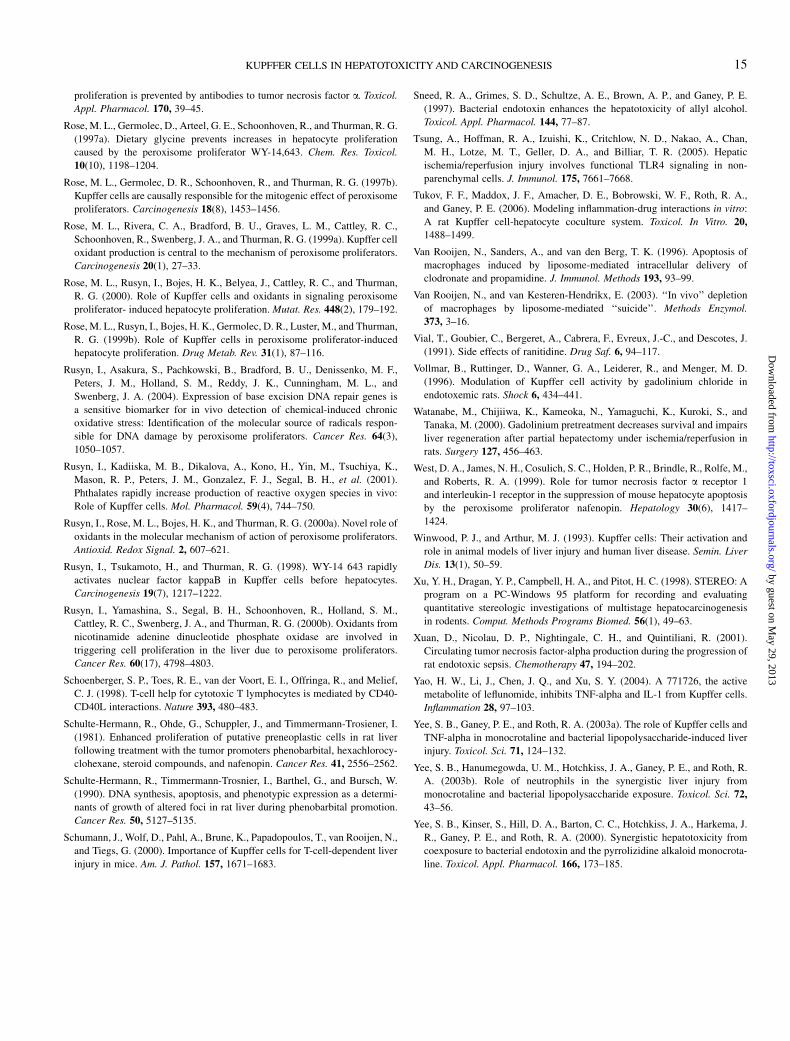
proliferation is prevented by antibodies to tumor necrosis factor a. Toxicol.
Appl. Pharmacol. 170, 39–45.
Rose, M. L., Germolec, D., Arteel, G. E., Schoonhoven, R., and Thurman, R. G.
(1997a). Dietary glycine prevents increases in hepatocyte proliferation
caused by the peroxisome proliferator WY-14,643. Chem. Res. Toxicol.
10(10), 1198–1204.
Rose, M. L., Germolec, D. R., Schoonhoven, R., and Thurman, R. G. (1997b).
Kupffer cells are causally responsible for the mitogenic effect of peroxisome
proliferators. Carcinogenesis 18(8), 1453–1456.
Rose, M. L., Rivera, C. A., Bradford, B. U., Graves, L. M., Cattley, R. C.,
Schoonhoven, R., Swenberg, J. A., and Thurman, R. G. (1999a). Kupffer cell
oxidant production is central to the mechanism of peroxisome proliferators.
Carcinogenesis 20(1), 27–33.
Rose, M. L., Rusyn, I., Bojes, H. K., Belyea, J., Cattley, R. C., and Thurman,
R. G. (2000). Role of Kupffer cells and oxidants in signaling peroxisome
proliferator- induced hepatocyte proliferation. Mutat. Res. 448(2), 179–192.
Rose, M. L., Rusyn, I., Bojes, H. K., Germolec, D. R., Luster, M., and Thurman,
R. G. (1999b). Role of Kupffer cells in peroxisome proliferator-induced
hepatocyte proliferation. Drug Metab. Rev. 31(1), 87–116.
Rusyn, I., Asakura, S., Pachkowski, B., Bradford, B. U., Denissenko, M. F.,
Peters, J. M., Holland, S. M., Reddy, J. K., Cunningham, M. L., and
Swenberg, J. A. (2004). Expression of base excision DNA repair genes is
a sensitive biomarker for in vivo detection of chemical-induced chronic
oxidative stress: Identification of the molecular source of radicals respon-
sible for DNA damage by peroxisome proliferators. Cancer Res. 64(3),
1050–1057.
Rusyn, I., Kadiiska, M. B., Dikalova, A., Kono, H., Yin, M., Tsuchiya, K.,
Mason, R. P., Peters, J. M., Gonzalez, F. J., Segal, B. H., et al. (2001).
Phthalates rapidly increase production of reactive oxygen species in vivo:
Role of Kupffer cells. Mol. Pharmacol. 59(4), 744–750.
Rusyn, I., Rose, M. L., Bojes, H. K., and Thurman, R. G. (2000a). Novel role of
oxidants in the molecular mechanism of action of peroxisome proliferators.
Antioxid. Redox Signal. 2, 607–621.
Rusyn, I., Tsukamoto, H., and Thurman, R. G. (1998). WY-14 643 rapidly
activates nuclear factor kappaB in Kupffer cells before hepatocytes.
Carcinogenesis 19(7), 1217–1222.
Rusyn, I., Yamashina, S., Segal, B. H., Schoonhoven, R., Holland, S. M.,
Cattley, R. C., Swenberg, J. A., and Thurman, R. G. (2000b). Oxidants from
nicotinamide adenine dinucleotide phosphate oxidase are involved in
triggering cell proliferation in the liver due to peroxisome proliferators.
Cancer Res. 60(17), 4798–4803.
Schoenberger, S. P., Toes, R. E., van der Voort, E. I., Offringa, R., and Melief,
C. J. (1998). T-cell help for cytotoxic T lymphocytes is mediated by CD40-
CD40L interactions. Nature 393, 480–483.
Schulte-Hermann, R., Ohde, G., Schuppler, J., and Timmermann-Trosiener, I.
(1981). Enhanced proliferation of putative preneoplastic cells in rat liver
following treatment with the tumor promoters phenobarbital, hexachlorocy-
clohexane, steroid compounds, and nafenopin. Cancer Res. 41, 2556–2562.
Schulte-Hermann, R., Timmermann-Trosnier, I., Barthel, G., and Bursch, W.
(1990). DNA synthesis, apoptosis, and phenotypic expression as a determi-
nants of growth of altered foci in rat liver during phenobarbital promotion.
Cancer Res. 50, 5127–5135.
Schumann, J., Wolf, D., Pahl, A., Brune, K., Papadopoulos, T., van Rooijen, N.,
and Tiegs, G. (2000). Importance of Kupffer cells for T-cell-dependent liver
injury in mice. Am. J. Pathol. 157, 1671–1683.
Sneed, R. A., Grimes, S. D., Schultze, A. E., Brown, A. P., and Ganey, P. E.
(1997). Bacterial endotoxin enhances the hepatotoxicity of allyl alcohol.
Toxicol. Appl. Pharmacol. 144, 77–87.
Tsung, A., Hoffman, R. A., Izuishi, K., Critchlow, N. D., Nakao, A., Chan,
M. H., Lotze, M. T., Geller, D. A., and Billiar, T. R. (2005). Hepatic
ischemia/reperfusion injury involves functional TLR4 signaling in non-
parenchymal cells. J. Immunol. 175, 7661–7668.
Tukov, F. F., Maddox, J. F., Amacher, D. E., Bobrowski, W. F., Roth, R. A.,
and Ganey, P. E. (2006). Modeling inflammation-drug interactions in vitro:
A rat Kupffer cell-hepatocyte coculture system. Toxicol. In Vitro. 20,
1488–1499.
Van Rooijen, N., Sanders, A., and van den Berg, T. K. (1996). Apoptosis of
macrophages induced by liposome-mediated intracellular delivery of
clodronate and propamidine. J. Immunol. Methods 193, 93–99.
Van Rooijen, N., and van Kesteren-Hendrikx, E. (2003). ‘‘In vivo’’ depletion
of macrophages by liposome-mediated ‘‘suicide’’. Methods Enzymol.
373, 3–16.
Vial, T., Goubier, C., Bergeret, A., Cabrera, F., Evreux, J.-C., and Descotes, J.
(1991). Side effects of ranitidine. Drug Saf. 6, 94–117.
Vollmar, B., Ruttinger, D., Wanner, G. A., Leiderer, R., and Menger, M. D.
(1996). Modulation of Kupffer cell activity by gadolinium chloride in
endotoxemic rats. Shock 6, 434–441.
Watanabe, M., Chijiiwa, K., Kameoka, N., Yamaguchi, K., Kuroki, S., and
Tanaka, M. (2000). Gadolinium pretreatment decreases survival and impairs
liver regeneration after partial hepatectomy under ischemia/reperfusion in
rats. Surgery 127, 456–463.
West, D. A., James, N. H., Cosulich, S. C., Holden, P. R., Brindle, R., Rolfe, M.,
and Roberts, R. A. (1999). Role for tumor necrosis factor a receptor 1
and interleukin-1 receptor in the suppression of mouse hepatocyte apoptosis
by the peroxisome proliferator nafenopin. Hepatology 30(6), 1417–
1424.
Winwood, P. J., and Arthur, M. J. (1993). Kupffer cells: Their activation and
role in animal models of liver injury and human liver disease. Semin. Liver
Dis. 13(1), 50–59.
Xu, Y. H., Dragan, Y. P., Campbell, H. A., and Pitot, H. C. (1998). STEREO: A
program on a PC-Windows 95 platform for recording and evaluating
quantitative stereologic investigations of multistage hepatocarcinogenesis
in rodents. Comput. Methods Programs Biomed. 56(1), 49–63.
Xuan, D., Nicolau, D. P., Nightingale, C. H., and Quintiliani, R. (2001).
Circulating tumor necrosis factor-alpha production during the progression of
rat endotoxic sepsis. Chemotherapy 47, 194–202.
Yao, H. W., Li, J., Chen, J. Q., and Xu, S. Y. (2004). A 771726, the active
metabolite of leflunomide, inhibits TNF-alpha and IL-1 from Kupffer cells.
Inflammation 28, 97–103.
Yee, S. B., Ganey, P. E., and Roth, R. A. (2003a). The role of Kupffer cells and
TNF-alpha in monocrotaline and bacterial lipopolysaccharide-induced liver
injury. Toxicol. Sci. 71, 124–132.
Yee, S. B., Hanumegowda, U. M., Hotchkiss, J. A., Ganey, P. E., and Roth, R.
A. (2003b). Role of neutrophils in the synergistic liver injury from
monocrotaline and bacterial lipopolysaccharide exposure. Toxicol. Sci. 72,
43–56.
Yee, S. B., Kinser, S., Hill, D. A., Barton, C. C., Hotchkiss, J. A., Harkema, J.
R., Ganey, P. E., and Roth, R. A. (2000). Synergistic hepatotoxicity from
coexposure to bacterial endotoxin and the pyrrolizidine alkaloid monocrota-
line. Toxicol. Appl. Pharmacol. 166, 173–185.
KUPFFER CELLS IN HEPATOTOXICITY AND CARCINOGENESIS 15
by guest on May 29, 2013
http://toxsci.oxfordjournals.org/D
ownloaded from
Related Documents