UNIVERSITÀ DEGLI STUDI DI MILANO Dipartimento di Bioscienze SCUOLA DI DOTTORATO TERRA, AMBIENTE E BIODIVERSITÀ Dottorato di Ricerca in Biologia Animale Ciclo XXVI Role of steroid hormones in echinoid reproductive biology PhD thesis Silvia Mercurio R08972 PhD tutor: Prof.ssa M. D. Candia Carnevali PhD coordinator: Prof. M. Ferraguti Academic Year 2012-2013

Welcome message from author
This document is posted to help you gain knowledge. Please leave a comment to let me know what you think about it! Share it to your friends and learn new things together.
Transcript
UNIVERSITÀ DEGLI STUDI DI MILANO
Dipartimento di Bioscienze
SCUOLA DI DOTTORATO
TERRA, AMBIENTE E BIODIVERSITÀ
Dottorato di Ricerca in Biologia Animale
Ciclo XXVI
Role of steroid hormones in echinoid
reproductive biology PhD thesis
Silvia Mercurio
R08972
PhD tutor: Prof.ssa M. D. Candia Carnevali
PhD coordinator: Prof. M. Ferraguti
Academic Year
2012-2013
To my twin sister
for the best advice I have ever received
TABLE OF CONTENTS
Chapter I – Abstract & thesis synopsis ............................................................................................ 6
1. Abstract ............................................................................................................................................ 7
2. List of abbreviations. ....................................................................................................................... 9
3. Aims and thesis synopsis ............................................................................................................... 10
Chapter II – General introduction ................................................................................................. 12
1. The experimental model: Paracentrotus lividus ............................................................................ 13
2. Steroid hormones: 17β-estradiol and testosterone ......................................................................... 20
3. E2 and T involvement in echinoderm reproduction ...................................................................... 22
4. Primary cell cultures from marine invertebrates ............................................................................ 29
Chapter III – Development of primary cell cultures from sea urchin ovaries ........................... 32
1. Abstract .......................................................................................................................................... 33
2. Introduction .................................................................................................................................... 33
3. Materials and Methods ................................................................................................................... 35
3.1. Animals ................................................................................................................................... 35
3.2. Cell cultures ............................................................................................................................. 35
3.3. Medium and supplement evaluation........................................................................................ 35
3.4. Microscopic and ultramicroscopic analyses ............................................................................ 36
3.5. Scanning electron microscopy................................................................................................. 37
3.6. Electrophoresis ........................................................................................................................ 37
3.7. Statistical analysis ................................................................................................................... 37
4. Results ............................................................................................................................................ 38
4.1. Development of primary cell cultures ..................................................................................... 38
4.2. Medium evaluation .................................................................................................................. 39
4.3. Substrates ................................................................................................................................ 41
4.4. Effects of medium supplements .............................................................................................. 41
4.5. Protein analysis........................................................................................................................ 43
5. Discussion ...................................................................................................................................... 43
Chapter IV – Sex-steroids in echinoid reproductin: an in vivo & in vitro approach ................. 46
1. Abstract .......................................................................................................................................... 47
2. Introduction .................................................................................................................................... 48
3. Materials and Methods ................................................................................................................... 50
3.1. In vivo experiment................................................................................................................... 50
3.1.1. Experimental animals and maintenance ........................................................................... 50
3.1.2. Experimental design ......................................................................................................... 51
3.1.3. Hormonal dietary administration ...................................................................................... 52
3.2. In vitro experiments ................................................................................................................. 52
3.2.1. Experimental design and animal maintenance ................................................................. 52
3.2.2. Cell cultures ...................................................................................................................... 53
3.2.3. Chemicals and solution preparation ................................................................................. 54
3.3. Determination of reproductive stages ...................................................................................... 54
3.3.1. Microscopic analysis ........................................................................................................ 54
3.3.2. Maturity Index and Gonad Index ...................................................................................... 55
3.4. Electrophoresis ........................................................................................................................ 55
3.4.1. Sample preparation ........................................................................................................... 55
3.4.2. SDS-PAGE ....................................................................................................................... 55
3.5. Statistical analysis ................................................................................................................... 56
4. Results ............................................................................................................................................ 56
4.1. Animal health conditions......................................................................................................... 56
4.2. In vivo experiment................................................................................................................... 56
4.2.1. Sex ratio ............................................................................................................................ 56
4.2.2. Gonad Index (GI) .............................................................................................................. 57
4.2.3. Maturity Index (MI) .......................................................................................................... 58
4.2.4. Reproductive stages .......................................................................................................... 58
4.3. In vitro experiments ................................................................................................................. 60
4.3.1. Cell cultures ...................................................................................................................... 60
4.3.2. Electrophoresis ................................................................................................................. 61
5. Discussion ...................................................................................................................................... 62

Chapter V – General Discussion & future prospectives ............................................................... 68
1. Development of primary cell cultures ............................................................................................ 69
2. Sex-steroid involvement in echinoid reproduction ........................................................................ 72
REFERENCES ................................................................................................................................. 77
APPENDIX 1 .................................................................................................................................... 89
APPENDIX 2 .................................................................................................................................... 96
Chapter I
ABSTRACT & THESIS SYNOPSIS
Chapter I
7
1. Abstract
Echinoid reproductive cycle has been extensively studied in several species but the mechanisms
regulating gametogenesis processes are still scarcely understood. Apart from environmental factors,
different research have suggested a steroid role in gonad maturation and growth. Particularly, in
echinoderms steroid involvement in reproduction has been suggested by both studies on seasonal
changes of steroid levels during the gonadal cycle and experiments of hormone administration.
Nevertheless, the steroid function in echinoid reproductive processes has not been clearly identified,
probably due to the low number of studies and the big variability of results reported. Thus, the main
aim of this research project was to shed light on echinoid endocrinology and, in particular, to clarify
the involvement of sex-steroid hormones in sea urchin reproductive biology. This was achieved
employing both in vivo and in vitro approaches.
First of all, considering the lack of studies on the development of effective cell cultures from
echinoderm gonads, primary cell cultures from ovaries of the edible sea urchin Paracentrotus
lividus were developed. Ovary cell phenotypes, present in culture, were identified and characterized
by different microscopic techniques. Although cell cultures could be produced from ovaries at all
stages of maturation, the cells appeared healthier and viable, displaying a higher survival rate, when
ovaries at early stages of gametogenesis were used. In terms of culture medium, ovarian cells were
successfully cultured in modified Leibovitz-15 medium, whereas poor results were obtained in
Minimum Essential Medium Eagle and Medium 199. Different substrates were tested but ovarian
cells completely adhered only on poly-L-lysine. To improve in vitro conditions and stimulate cell
proliferation different serum-supplements were tested. Fetal Calf Serum and an originally
developed Pluteus Extract resulted to be detrimental to cell survival, apparently accelerating
processes of cell death. In contrast, cells cultured with sea urchin Egg Extract appeared larger and
healthier, displaying an increased longevity that allowed to maintain them for up to 1 month.
Overall this study provides new experimental bases and procedures for producing successfully long-
term primary cell cultures from sea urchin ovaries, providing a simple and versatile experimental
tool for research in echinoderm reproductive biology.
Subsequently, in vivo and in vitro experiments, specifically addressed to determine possible 17β-
estradiol (E2) and testosterone (T) involvement in echinoid reproduction, were performed. An in
vivo long-term experiment of steroid dietary administration was performed in adult specimens of P.
lividus. The experimental plan was specifically designed in order to reduce individual variability
and synchronize the experimental animals at the same starting maturative condition. We analysed
and compared different reproductive parameters (Gonad Index, Maturative Index and maturative

Chapter I
8
stages distribution) in 4 experimental groups: control group (CTL), E2 and T groups fed with
pellets containing respectively 17β-estradiol and testosterone, and E2-4 weeks group fed with
control pellets for the first 4 weeks and then treated with 17β-estradiol. This latter was chosen in
order to verify the existence of a specific E2-sensitive gametogenic stage, as proposed in different
asteroid species.
Possible steroid effects on P. lividus female reproduction was also investigated with an in vitro
approach. Cells, isolated by ovaries in the same maturative conditions considered in the in vivo
experiments, were cultured in presence of E2 and T physiological concentrations for 2 weeks.
Effects on ovarian cell morphology and behaviour were investigated. In addition, steroid regulation
of the Major Yolk Protein (MYP) expression was analyzed 24 and 48 hours after E2 and T
exposure. According to our results, E2 and T do not markedly influence echinoid gonad maturation
and, particularly, they do not promote gamete maturation. Hormonal dietary administration did not
induce striking variations in the considered reproductive parameters and no effect was observed
also when males and females were analyzed separately. In addition, no specific maturative stage
sensitive to E2 was found, suggesting the existence of different hormonal mechanisms in asteroids
and echinoids. Similar considerations could be reported taking into account the in vitro
experiments. E2 and T exposure did not affect ovarian cell size and behaviour nor MYP expression.
The obtained results suggest that these hormones are not directly involved in either gamete
maturation, as demonstrated for vertebrates, or in vitellogenesis processes, as reported for several
asteroid species. However a possible involvement of steroids in echinoid physiology cannot be
completely excluded and their role in the regulation of lipid metabolism and protein synthesis
during the different reproductive stages should be strongly considered as suggested by several
authors.
Further specific research on steroid hormone mode of action, physiological function and
metabolism are therefore needed to completely understand echinoid reproduction and
endocrinology.
Chapter I
9
2. List of abbreviations
2-DE Two-dimensional electrophoresis
AR Androgen Receptor
CFMYP MYP isoform found in sea urchin Coelomic Fluid
CMFSW Ca2+ Mg2+ Free Sea Water
CTL Control (experimental group)
E1 Estrone
E2 17β-estradiol
EDC Endocrine Disrupting Compound
EE Egg Extract
ECM Extra Cellular Matrix
EGF Epidermal Growth Factor
EGMYP MYP isoform found in sea urchin Eggs
ER Estrogen Receptor
ERR Estrogen Receptor-related Receptor
FCS Fetal Calf Serum
GI Gonad Index
GC-MS Gas Chromatography Mass Spectrometry
HDMS Hexamethyldisilazane
L-15 Leibovitz L-15 medium
LLTP Large Lipid Transfer Protein
M199 Medium 199
MEM Minimum Essential Medium Eagle
MI Maturity Index
MS Mass Spectrometry
MS/MS Tandem Mass Spectrometry
MYP Major Yolk Protein
T Testosterone
SpSHR2 Strongylocentrotus purpuratus orphan steroid receptor 2
vtg Vitellogenin
Chapter I
10
3. Aims and thesis synopsis
The general aim of this research project was to shed light on echinoid endocrinology and, in
particular, to explore the involvement of steroid hormones in sea urchin reproductive biology. In
fact, the current knowledge about these hormones on echinoid reproduction is still fragmentary and
most of the studies have reported different and, sometimes, even contrasting results. Considering
the ecological and, in some cases, commercial importance of this marine invertebrates, further
investigations are certainly needed. This research was therefore addressed to investigate the role of
sex-steroid hormones, 17β-estradiol (E2) and testosterone (T), in the reproductive biology of the
regular sea urchin Paracentrotus lividus, applying both in vivo and in vitro approaches. In fact, the
employment of these different and complementary approaches should provide a wider view of sea
urchin endocrinology and help to finally unravel steroid role in echinoids.
In Chapter II, a review of the current knowledge regarding steroid hormone involvement in
echinoderm reproduction is presented. After an accurate description of the experimental model, the
common sea urchin Paracentrotus lividus, a detailed summary of previous studies on sex-steroid
role in different asteroid and echinoid species is provided. Particular attention is given to E2 and T,
whose function in echinoid reproductive processes was investigated in this research. Finally a
general overview of available data on primary cell cultures from marine invertebrates is also
proposed.
In Chapter III, the development of primary cell cultures from sea urchin ovaries is described.
Indeed, in echinoids there was no study reporting the development of effective cell cultures from
sea urchin gonads. In this work the first attempt to obtain cultures of P. lividus ovarian cells was
successfully carried out. After an accurate characterization of the cell phenotypes present in culture,
our priorities were to optimize the culture conditions, i.e. to define the suitable medium, substrate
and possible serum supplements. The obtained results, which are provided in details throughout this
chapter, have been already published in international scientific journal.
In Chapter IV, investigations on possible E2 and T functions on echinoid reproduction are
extensively described. Sex-steroid involvement in P. lividus reproductive processes were
investigated employing both in vivo and in vitro experiments. In particular, a long-term experiment
of E2 and T dietary administration was performed. The experimental plan was specifically designed
in order to obtained reliable results and different reproductive parameters were analyzed. The
observed results were then confirmed and deepened with in vitro steroid exposure experiments.
Chapter I
11
Chapter V presents a general and integrated discussion of all the obtained results; new interesting
fields and techniques for future investigations are suggested.
Overall, this project was addressed to provide further information on the scarcely known
endocrinology of echinoderms and, in particular, of echinoids. Our specific targets were: 1) to
analyze thoroughly echinoid reproductive processes and their regulatory mechanisms by focussing
on the possible role of 17β-estradiol and testosterone and 2) to provide new information on
possible control mechanisms of gonad development in P. lividus, an edible and commercially
relevant species.
Chapter II
GENERAL INTRODUCTION
Chapter II
Paracentro
Echinoderm
of intertida
phylogenet
vertebrates
phylogenet
since it m
basic mech
as processe
2007).
At present
echinoderm
Asteroids
feather sta
dollars),
Ophiuroids
apparent
morpholog
the pe
develop
charact
the me
and em
formin
the wa
numero
locomo
Paracentro
pointed sp
plates of s
organic com
1.
otus lividus
ms are excl
al zones to
tically clos
s) (Brusca
tic position
may suggest
hanisms bet
es of hormo
there are a
ms, tradition
(starfishes)
ars), Echino
Holoturoid
s (brittle sta
diversity t
gical feature
entamerous
pment as b
teristic pent
esodermal e
mbedded in
g the test, o
ater-vascula
ous project
otion, respir
otus lividus
pines. The t
skeletal tiss
mponent in
. The expe
is a regular
lusively ma
the deep ab
se to chor
and Brus
n is particu
t the existe
tween the t
onal regulat
about 7000
nally divide
), Crinoids
oids (sea u
ds (sea c
ars) (Fig. 1
that charac
es. The mos
radial sym
bilateral em
tamerous sy
endoskeleto
the derma
or differentia
ar system: t
tions (tube
ration and p
s (Fig. 2) h
test enclose
sue, whose
ncludes ECM
erimentall model: PParacentrrotus lividdus
r sea urchin, belonging to the phyllum Echinoddermata.
arine organi
byssal plain
rdates (incl
ca, 1990).
ularly inter
ence of com
two groups,
tion (Sugni
isms, wides
ns. They are
luding
This
resting
mmon
, such
et al.,
spread in all
e deuterosto
l the ocean
ome inverteb
s from shal
brates, bein
llow waters
ng therefore
s
e
extant spec
ed in five cl
(sea lilies
urchins and
cucumbers)
). In spite
cterizes the
t typical are
mmetry (in
mbryos and
ymmetry;
on: echinode
l layer of t
ally scattere
this is a co
feet) are
possibly exc
has a hemi
es and prote
inorganic c
M, collagen
cies of
lasses:
s and
d sand
and
of the
e phylum,
e:
n adults):
d larvae bu
erm endosk
the body w
ed and distr
omplex sys
involved in
cretion (Bru
spherical b
ects the inte
component
fibres and s
echinoder
all the e
ut during m
keleton con
wall. These
ributed in th
stem of flu
n a variety
usca and Bru
body, dense
ernal organ
consists of
sclerocytes
Fig.
rms displa
chinoderms
metamorpho
nsists of ske
plates can
he connectiv
uid-filled co
y of functio
usca, 1990).
ely covered
ns and is co
f calcium c
(Chia and H
. 1. Echinorde
y several
s indeed b
osis they a
eletal plate
be closely
ve tissue (os
oelomic can
ons, such a
.
d by long a
omposed of
carbonate, w
Harrison, 19
erm classes.
distinctivee
begin their
acquire the
r
e
s produced
connected,
ssicles);
d
,
nals whose
as feeding,
e
,
and sharply
f connected
whereas the
994).
y
d
e
133
Chapter II
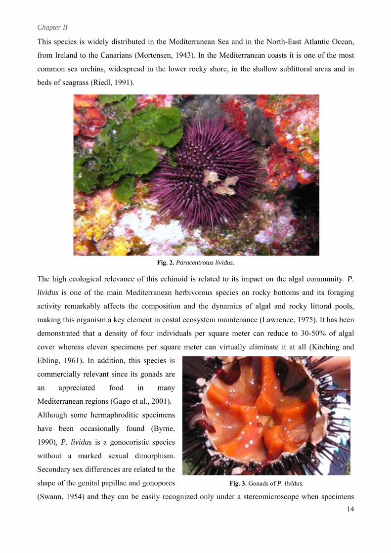
This species is widely distributed in the Mediterranean Sea and in the North-East Atlantic Ocean,
from Ireland to the Canarians (Mortensen, 1943). In the Mediterranean coasts it is one of the most
common sea urchins, widespread in the lower rocky shore, in the shallow sublittoral areas and in
beds of seagrass (Riedl, 1991).
Fig. 2. Paracentrotus lividus.
14
meter can virtually eliminate it at all (Kitching and
(Swann, 1954) and they can be easily recognized only und hen specimens
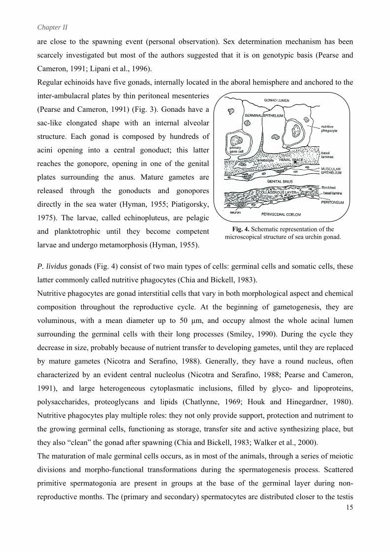
The high ecological relevance of this echinoid is related to its impact on the algal community. P.
lividus is one of the main Mediterranean herbivorous species on rocky bottoms and its foraging
activity remarkably affects the composition and the dynamics of algal and rocky littoral pools,
making this organism a key element in costal ecosystem maintenance (Lawrence, 1975). It has been
demonstrated that a density of four individuals per square meter can reduce to 30-50% of algal
cover whereas eleven specimens per square
Ebling, 1961). In addition, this species is
commercially relevant since its gonads are
an appreciated food in many
Mediterranean regions (Gago et al., 2001).
Although some hermaphroditic specimens
have been occasionally found (Byrne,
1990), P. lividus is a gonocoristic species
without a marked sexual dimorphism.
Secondary sex differences are related to the
shape of the genital papillae and gonopores Fig. 3. Gonads of P. lividus.
er a stereomicroscope w
Chapter II
are close t
scarcely in
Cameron,
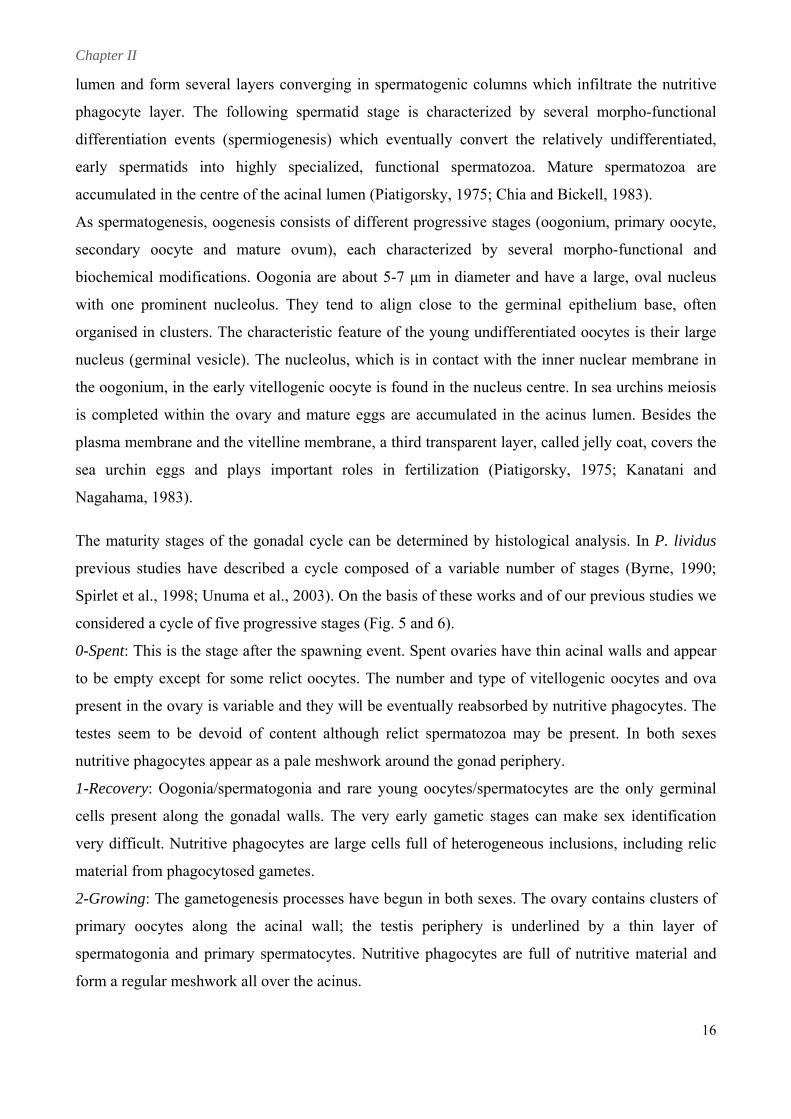
Regular ec
to the spaw
nvestigated
1991; Lipan
chinoids hav
wning event
but most o
ni et al., 199
ve five gona
t (personal
of the autho
96).
ads, internal
observation
ors suggeste
lly located i
n). Sex det
ed that it is
in the abora
termination
s on genoty
al hemis
mechanism
ypic basis (
m has been
(Pearse and
n
d
pheere and anchhored to thee
inter-ambu
(Pearse an
sac-like el
structure.
acini open
reaches th
plates sur
released t
directly in
1975). The
and plank
larvae and
P. lividus
ulacral plate
nd Cameron
longated sh
Each gonad
ning into a
e gonopore
rounding t
through th
the sea wa
e larvae, ca
ktotrophic
undergo m
es by thin p
n, 1991) (Fi
hape with
d is compo
a central g
e, opening i
the anus. M
he gonodu
ater (Hyma
alled echin
until they
etamorphos
peritoneal m
ig. 3). Gona
an interna
osed by hu
gonoduct;
in one of t
Mature gam
ucts and
an, 1955; Pi
nopluteus, a
become
sis (Hyman,
mesenteries
ads have a
al alveolar
undreds of
this latter
the genital
metes are
gonopores
iatigorsky,
are pelagic
competent
, 1955).
Fig. 4microsco
4. Schematic r
g
latter comm
Nutritive p
compositio
voluminou
surroundin
decrease in
by mature
characteriz
1991), and
polysaccha
Nutritive p
the growin
they also “
The matura
divisions a
primitive
reproductiv
gonads (Fig
monly calle
phagocytes a
on througho
us, with a m
ng the germ
n size, proba
gametes (
zed by an e
d large he
arides, prot
phagocytes p
ng germinal
“clean” the g
ation of ma
and morpho
spermatogo
ve months.
g. 4) consist
d nutritive p
are gonad in
out the rep
mean diam
minal cells w
ably becaus
(Nicotra an
evident cen
eterogeneou
teoglycans
play multip
l cells, func
gonad after
ale germinal
o-functiona
onia are pr
The (prima
t of two ma
phagocytes
nterstitial ce
productive
meter up to
with their
se of nutrien
nd Serafino,
tral nucleol
us cytoplas
and lipids
ple roles: the
ctioning as
spawning (
l cells occur
al transform
esent in gr
ary and seco
ain types of
(Chia and B
ells that var
cycle. At
50 μm, an
long proces
nt transfer to
, 1988). Ge
lus (Nicotra
matic inclu
s (Chatlynn
ey not only
storage, tra
(Chia and B
rs, as in mo
mations duri
roups at th
ondary) spe
cells: germ
Bickell, 198
ry in both m
the beginn
nd occupy
sses (Smile
o developin
enerally, th
a and Seraf
usions, fill
ne, 1969;
provide sup
ansfer site a
Bickell, 1983
ost of the an
ing the spe
he base of
rmatocytes
minal cells an
83).
morphologic
ning of gam
almost the
ey, 1990). D
ng gametes,
hey have a
fino, 1988;
ed by gly
Houk and
pport, prote
and active s
3; Walker e
nimals, throu
ermatogene
the germin
are distribu
opical structurrepresentation of the re of sea urchiin gonad.
nd somatic cells, thesee
al aspect annd chemicall
metogenesiss, they aree
e whole ac
During the
until they a
round nuc
Pearse and
yco- and li
Hinegardn
ection and n
synthesizing
t al., 2000).
ugh a series
esis process
cinal lumen
cycle they
are replaced
cleus, often
d Cameron,
ipoproteins,
ner, 1980).
nutriment to
g place, but
.
s of meiotic
s.
n
y
d
n
,
,
.
o
t
c
Scatteredd
nal layer d
uted closer t
during non-
to the testis
-
s 155
Chapter II
16
nal and
be determined by histological analysis. In P. lividus
previous studies have described a cycle composed of a variable number of stages (Byrne, 1990;
d type of vitellogenic oocytes and ova
ake sex identification
all; the testis periphery is underlined by a thin layer of
lumen and form several layers converging in spermatogenic columns which infiltrate the nutritive
phagocyte layer. The following spermatid stage is characterized by several morpho-functional
differentiation events (spermiogenesis) which eventually convert the relatively undifferentiated,
early spermatids into highly specialized, functional spermatozoa. Mature spermatozoa are
accumulated in the centre of the acinal lumen (Piatigorsky, 1975; Chia and Bickell, 1983).
As spermatogenesis, oogenesis consists of different progressive stages (oogonium, primary oocyte,
secondary oocyte and mature ovum), each characterized by several morpho-functio
biochemical modifications. Oogonia are about 5-7 μm in diameter and have a large, oval nucleus
with one prominent nucleolus. They tend to align close to the germinal epithelium base, often
organised in clusters. The characteristic feature of the young undifferentiated oocytes is their large
nucleus (germinal vesicle). The nucleolus, which is in contact with the inner nuclear membrane in
the oogonium, in the early vitellogenic oocyte is found in the nucleus centre. In sea urchins meiosis
is completed within the ovary and mature eggs are accumulated in the acinus lumen. Besides the
plasma membrane and the vitelline membrane, a third transparent layer, called jelly coat, covers the
sea urchin eggs and plays important roles in fertilization (Piatigorsky, 1975; Kanatani and
Nagahama, 1983).
The maturity stages of the gonadal cycle can
Spirlet et al., 1998; Unuma et al., 2003). On the basis of these works and of our previous studies we
considered a cycle of five progressive stages (Fig. 5 and 6).
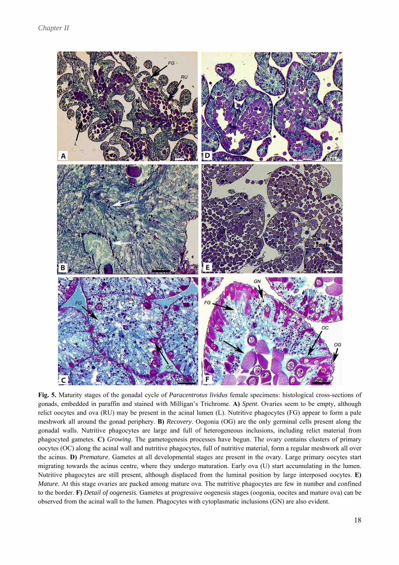
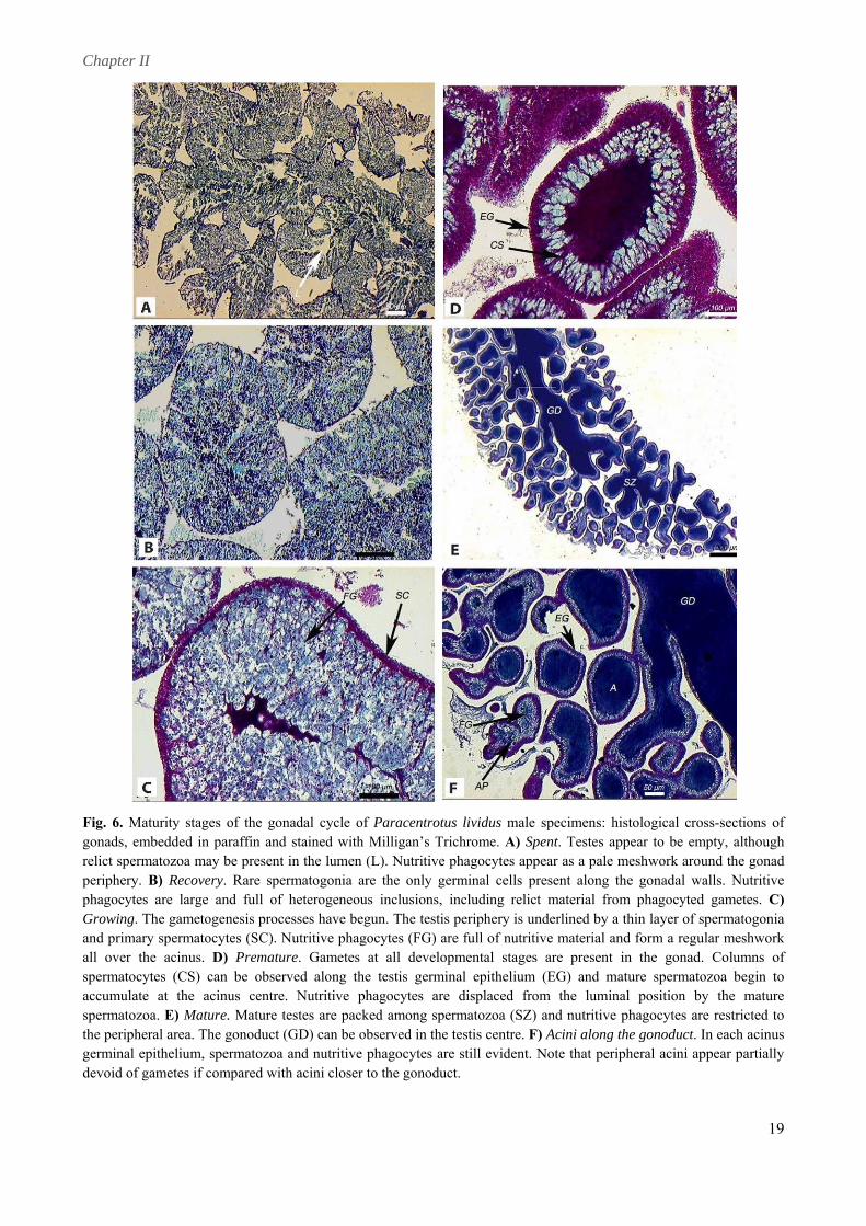
0-Spent: This is the stage after the spawning event. Spent ovaries have thin acinal walls and appear
to be empty except for some relict oocytes. The number an
present in the ovary is variable and they will be eventually reabsorbed by nutritive phagocytes. The
testes seem to be devoid of content although relict spermatozoa may be present. In both sexes
nutritive phagocytes appear as a pale meshwork around the gonad periphery.
1-Recovery: Oogonia/spermatogonia and rare young oocytes/spermatocytes are the only germinal
cells present along the gonadal walls. The very early gametic stages can m
very difficult. Nutritive phagocytes are large cells full of heterogeneous inclusions, including relic
material from phagocytosed gametes.
2-Growing: The gametogenesis processes have begun in both sexes. The ovary contains clusters of
primary oocytes along the acinal w
spermatogonia and primary spermatocytes. Nutritive phagocytes are full of nutritive material and
form a regular meshwork all over the acinus.
Chapter II
17
cinus centre and when they reach maximus size, they
Mature testes are filled as well with spermatozoa whereas
parameters can be used. One of the most common is the Gonad Index (GI) (Spirlet et al., 1998;
3-Premature: Gametes at all developmental stages are present in the gonads. In the ovary, large
primary oocytes start migrating towards the a
undergo maturation and early ova can start accumulating in the lumen. Nutritive phagocytes are still
present, although displaced from the luminal position by large interposed oocytes. In the testis,
columns of spermatocytes can be observed along the acinal wall and mature spermatozoa begin to
accumulate in the acinus centre. As in ovaries, nutritive phagocytes are displaced from the luminal
position by the mature spermatozoa.
4-Mature: At this stage ovaries are filled with mature ova. The nutritive phagocytes are few in
number and confined to the border.
nutritive phagocytes are restricted to the peripheral area. The spawning event occurs at this stage.
To quantitatively describe the seasonal trend of the reproductive cycle different numerical
Shpigel et al., 2004). In the present study the GI is defined as: GI = (GW / TW) 100. GW is the
wet weight of the five gonads and TW is the wet weight of the whole animal. This index provides
information on the different allocation of nutrients to somatic and gonadal production. The GI
values tend to increase during the stages before the spawning event and suddenly fall after it.
Another important parameter is the Maturity Index (MI), a numeric value associated to the
maturative stage of the gonads. In a population, the mean value of MI numerically describes its
in the present research usually displays a first main
reproductive event at the end of the spring, and a further minor and facultative spawning period in
el et al., 2004), food availability (Leoni et al., 2001), environmental
reproductive state.
The Tyrrhenian population considered
early autumn (Fenaux, 1968).
Several exogenous factors can influence gametogenesis: water temperature and photoperiod (Byrne,
1990; Spirlet et al., 1998; Shpig
hydrodynamics (Fenaux, 1968). Water temperature between 18 and 22 °C seems to enhance growth
and gonadal development (Shpigel et al., 2004), whereas higher (24 °C) temperatures appear to
inhibit spawning (Spirlet et al., 1998). Photoperiod may affect gonad maturation and the first
spawning event appears to be triggered by 15 h-long day (Spirlet et al., 1998). Food appears to play
an important role in the regulation of the reproductive cycle too; gametogenesis cannot be initiated
until a “critical level” of nutriments is available within the storage tissues (nutritive phagocytes) to
ensure gametes growth (Pearse and Cameron, 1991; Spirlet et al., 1998). Apart from these
exogenous factors, several endogenous factors, notably hormones, probably play an important role
in synchronizing the gonads individually (Spirlet et al., 1998).
Chapter II
18
Fig. 5. Maturity stages of the gonadal cycle of Paracentrotus lividus female specimens: histological cross-sections of gonads, embedded in paraffin and stained with Milligan’s Trichrome. A) Spent. Ovaries seem to be empty, al ough relict oocytes and ova (RU) may be present in the acinal lumen (L). Nutritive phagocytes (FG) appear to form pale
th a
meshwork all around the gonad periphery. B) Recovery. Oogonia (OG) are the only germinal cells present along the gonadal walls. Nutritive phagocytes are large and full of heterogeneous inclusions, including relict material from phagocyted gametes. C) Growing. The gametogenesis processes have begun. The ovary contains clusters of primary oocytes (OC) along the acinal wall and nutritive phagocytes, full of nutritive material, form a regular meshwork all over the acinus. D) Premature. Gametes at all developmental stages are present in the ovary. Large primary oocytes start migrating towards the acinus centre, where they undergo maturation. Early ova (U) start accumulating in the lumen. Nutritive phagocytes are still present, although displaced from the luminal position by large interposed oocytes. E) Mature. At this stage ovaries are packed among mature ova. The nutritive phagocytes are few in number and confined to the border. F) Detail of oogenesis. Gametes at progressive oogenesis stages (oogonia, oocites and mature ova) can be observed from the acinal wall to the lumen. Phagocytes with cytoplasmatic inclusions (GN) are also evident.
Chapter II
19
Fig. 6. Maturity stages of the gonadal cycle of Paracentrotus lividus male specimens: histological cross-sec ons of gonads, embedded in paraffin and stained with Milligan’s Trichrome. A) Spent. Testes appear to be empty, hough
tialt
relict spermatozoa may be present in the lumen (L). Nutritive phagocytes appear as a pale meshwork around the gonad periphery. B) Recovery. Rare spermatogonia are the only germinal cells present along the gonadal walls. Nutritive phagocytes are large and full of heterogeneous inclusions, including relict material from phagocyted gametes. C) Growing. The gametogenesis processes have begun. The testis periphery is underlined by a thin layer of spermatogonia and primary spermatocytes (SC). Nutritive phagocytes (FG) are full of nutritive material and form a regular meshwork all over the acinus. D) Premature. Gametes at all developmental stages are present in the gonad. Columns of spermatocytes (CS) can be observed along the testis germinal epithelium (EG) and mature spermatozoa begin to accumulate at the acinus centre. Nutritive phagocytes are displaced from the luminal position by the mature spermatozoa. E) Mature. Mature testes are packed among spermatozoa (SZ) and nutritive phagocytes are restricted to the peripheral area. The gonoduct (GD) can be observed in the testis centre. F) Acini along the gonoduct. In each acinus germinal epithelium, spermatozoa and nutritive phagocytes are still evident. Note that peripheral acini appear partially devoid of gametes if compared with acini closer to the gonoduct.
Chapter II
20
2. Steroid hormones: 17β-estradiol
Hormones are substances usually secreted into the ci
of the organism. Hormones transduce environmental infor
processes, particularly reproductive and developm
spawning, growth and metamorphosis (Hau, 2007).
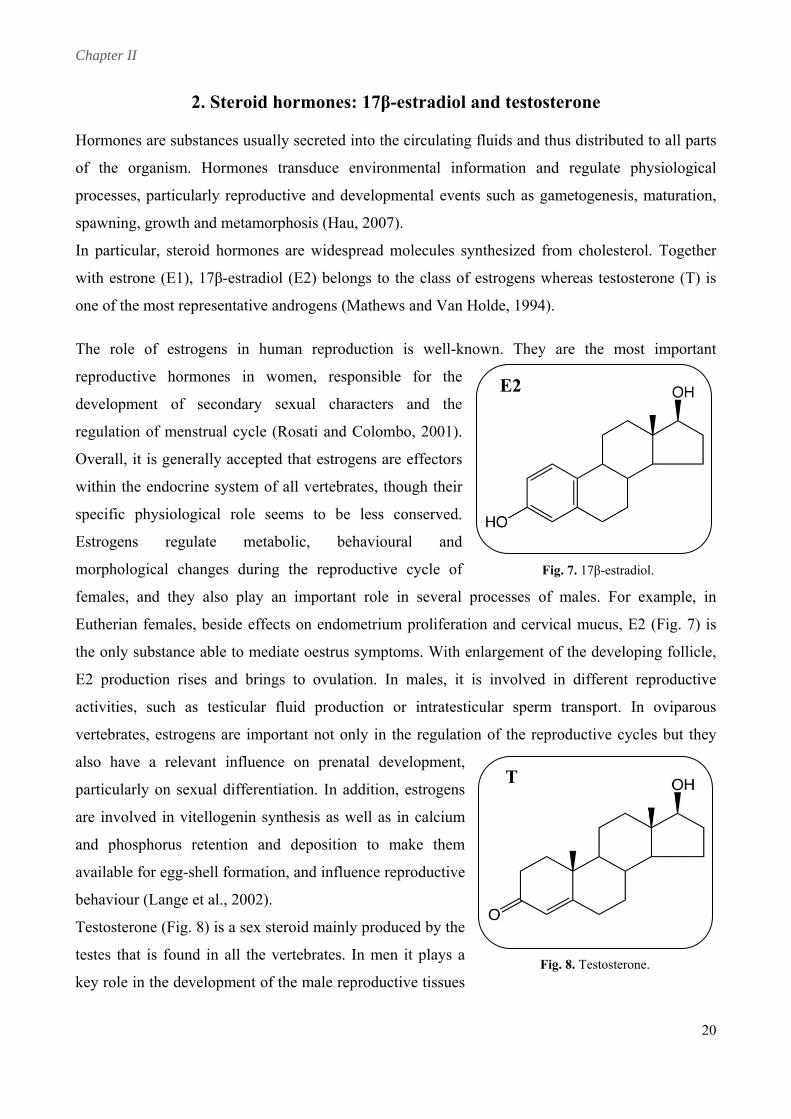
In particular, steroid hormones are widespread molecules s
with estrone (E1), 17β-estradiol (E2) belongs to the class of
one of the most representative androgens (Mathews and Van Holde, 1994).
The role of estrogens in human reproduction is well-known. They are the most important
reproductive hormones in women, responsible for the
development of secondary sexual characters and the
regulation of menstrual cycle (Rosati and Colombo, 2001).
Overall, it is generally accepted that estrogens are effectors
within the endocrine system of all vertebrates, though their
specific physiological role seems to be less conserved.
Estrogens regulate metabolic, behavioural and
morphological changes during the reproductive cycle of
females, and they also play an important role in several processes of males. For example, in
Eutherian females, beside effects on endometrium proliferation and cervical mucus, E2 (Fig. 7) is
the only substance able to mediate oestrus symptoms. With enlargement of the developing follicle,
E2 production rises and brings to ovulation. In males, it is involved in different reproductive
activities, such as testicular fluid production or intratesticular sperm transport. In oviparous
vertebrates, estrogens are important not only in the regulation of the reproductive cycles but they
also have a relevant influence on prenatal development,
particularly on sexual differentiation. In addition, estrogens
are involved in vitellogenin synthesis as well as in calcium
and phosphorus retention and deposition to make them
available for egg-shell formation, and influence reproductive
behaviour (Lange et al., 2002).
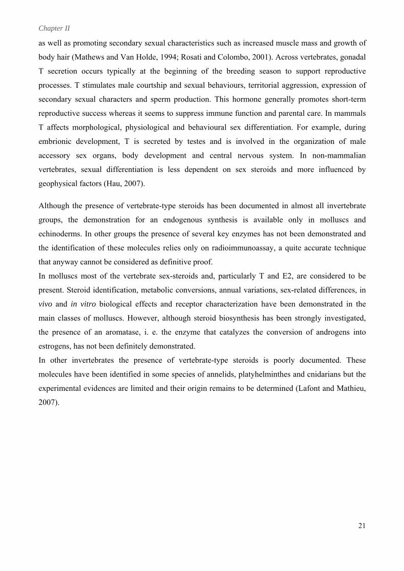
Testosterone (Fig. 8) is a sex steroid mainly produced by the
testes that is found in all the vertebrates. In men it plays a
key role in the development of the male reproductive tissues
and testosterone
rculating fluids and thus distributed to all parts
mation and regulate physiological
ental events such as gametogenesis, maturation,
ynthesized from cholesterol. Together
estrogens whereas testosterone (T) is
Fig. 8. Testosterone.
Fig. 7. 17β-estradiol.
E2
T
Chapter II
21
as well as promoting secondary sexual characteristics such as increased muscle mass and growth of
body hair (Mathe tebrates, gonadal
mune function and parental care. In mammals
. In non-mammalian
vertebrates, sexual differentiation is less dependent on sex steroids and more influenced by
ocumented in almost all invertebrate
is available only in molluscs and
ymes has not been demonstrated and
unoassay, a quite accurate technique
arly T and E2, are considered to be
variations, sex-related differences, in
be d termined (Lafont and Mathieu,
ws and Van Holde, 1994; Rosati and Colombo, 2001). Across ver
T secretion occurs typically at the beginning of the breeding season to support reproductive
processes. T stimulates male courtship and sexual behaviours, territorial aggression, expression of
secondary sexual characters and sperm production. This hormone generally promotes short-term
reproductive success whereas it seems to suppress im
T affects morphological, physiological and behavioural sex differentiation. For example, during
embrionic development, T is secreted by testes and is involved in the organization of male
accessory sex organs, body development and central nervous system
geophysical factors (Hau, 2007).
Although the presence of vertebrate-type steroids has been d
groups, the demonstration for an endogenous synthesis
echinoderms. In other groups the presence of several key enz
the identification of these molecules relies only on radioimm
that anyway cannot be considered as definitive proof.
In molluscs most of the vertebrate sex-steroids and, particul
present. Steroid identification, metabolic conversions, annual
vivo and in vitro biological effects and receptor characterization have been demonstrated in the
main classes of molluscs. However, although steroid biosynthesis has been strongly investigated,
the presence of an aromatase, i. e. the enzyme that catalyzes the conversion of androgens into
estrogens, has not been definitely demonstrated.
In other invertebrates the presence of vertebrate-type steroids is poorly documented. These
molecules have been identified in some species of annelids, platyhelminthes and cnidarians but the
experimental evidences are limited and their origin remains to
2007).
e
Chapter II
The presen
been docum
to asteroid
bioassay, r
Schoenmak
3. E
nce of verte
mented in s
s and echin
radioimmun
kers, 1979;
E2 and T i
brate-type s
several echi
noids, where
noassay and
Hines et al
involveme
steroids and
inoderm spe
e these mole
d gas chrom
., 1992b; Vo
ent in ech
d, particular
ecies (Lafon
ecules have
matography-
oogt et al.,
hinoderm
rly, of testos
nt and Math
been detect
mass spectr
1992).
reproduc
sterone (T)
hieu, 2007).
ted by seve
rometry (GC
ction
and estradi
. Most stud
eral techniqu
C-MS) (Die
iol (E2) has
ies referred
ues, such as
eleman and
s
d
s
d
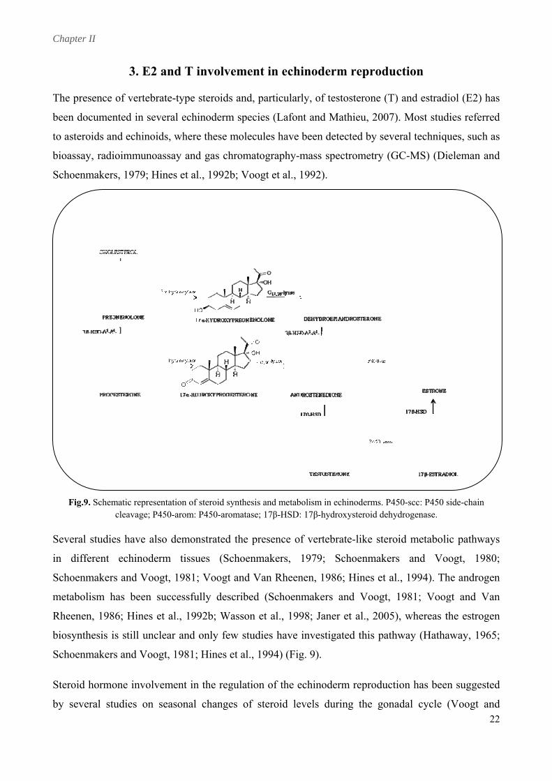
Fig.9. Sc
Several stu
in differ
chematic reprecleavage;
udies have a
esentation of s P450-arom: P
also demon
steroid syntheP450-aromata
nstrated the
esis and metabase; 17β-HSD
presence o
bolism in echin: 17β-hydroxy
of vertebrate
noderms. P45ysteroid dehyd
e-like steroi
50-scc: P450 sdrogenase.
id metaboli
side-chain
c pathwayss
eent echinodderm tissuues (Schoeenmakers, 1979; Schhoenmakerss and Vooogt, 1980;;
Schoenmakkers and Vooogt, 1981; Voogt andd Van Rheennen, 1986; HHines et al.
d Voogt,
, 1994). Thhe androgenn
metabolismm has beenn successfully describeed (Schoennmakers an 11981; Vooggt and Vann
Rheenen, 11986; Hiness et al., 19992b; Wasson et al., 19998; Janer ett al., 2005),, whereas thhe estrogenn
biosynthessis is still unnclear and oonly few sttudies have investigated this pathwway (Hathaaway, 1965;;
Schoenmakkers and Vooogt, 1981; Hines et al.., 1994) (Figg. 9).
Steroid horrmone invoolvement in the regulattion of the eechinodermm reproduction has beenn suggestedd
by severall studies onn seasonal changes off steroid leevels duringg the gonaadal cycle ((Voogt andd 222
Chapter II
23
Dieleman, 1984; Xu and Barker, 1990; Hines et al., 1992a; Wasson et al., 2000a; Barbaglio et al.,
2007). Most dat ccording to the
ulgaris where E2 increased in the fall
in parallel with oogonia/spermatogonia proliferation (Hines et al., 1992a). As far as T is concerned,
it seems to be involved in gamete maturation and gonad growth (Voogt and Dieleman, 1984; Xu
and Barker, 1990; Hines et al., 1992a). In A. vulgaris transient increases in the T levels coincided
with spermatogenic column formation and, in the ovaries, T concentrations were high at the onset
of oogenesis and during early maturation of oocytes (Hines et al., 1992a), suggesting its
involvement in the regulation of early stages of gonad maturation.
Moving to echinoids, the role of steroid hormones in the reproductive processes is still unclear. In
both ovaries and testes of Lytechinus variegatus, T and E2 concentrations were higher during the
period of early gonadal growth. These levels were much lower than those measured in asteroids,
probably due to different regulation mechanisms of gamete nutrition in the two echinoderm groups
(Wasson et al., 2000a).
Focussing on P. lividus, the experimental model employed in this research project, our previous
studies did not allow us to derive a clear correlation between T levels and the distribution of
reproductive stages through the year. Nevertheless, a relationship between T levels and
reproduction was strongly suggested. In testes, T concentrations were significantly lower during
spermatogenesis processes than at the end of gametogenesis, suggesting a possible role of T in late
spe e,
suggesting a ids
mporally” anticipated in the coelomic
fluid, i.e. they were found in the stages immediately before. These findings lead to the hypothesis
a refer to asteroids, where the hormone levels appear to vary a
reproductive cycle and in a sex-specific manner (Voogt and Dieleman, 1984; Xu and Barker, 1990;
Hines et al., 1992a). Maximum estrogen levels were registered at the beginning of vitellogenesis in
both Sclerasterias mollis (Xu and Barker, 1990) and Asterias rubens (Schoenmakers and Dieleman,
1981), suggesting that these hormones may affect protein biosynthesis, transport or incorporation
into oocytes. Slightly different results were found in Asterias v
rm maturation and spawning. In ovaries, T levels resulted higher during growing stag
T involvement in vitellogenesis (Barbaglio et al., 2007), as also reported in astero
(Hines et al., 1992a). As far as E2 is concerned, it was clear that E2 concentrations are lower than T
levels in both sexes. Furthermore, mean E2 concentration appeared to be lower in testes than in
ovaries, possibly reflecting a more important role for this hormone in female individuals. In the
ovaries, E2 levels were higher in early maturative stages, indicating a possible E2 involvement in
the regulation of nutritive phagocyte activity and/or oogonium proliferation. On the contrary, in the
testes, higher levels of E2 were measured in advanced maturative stages, suggesting a role in sperm
maturation (Barbaglio et al., 2007). These E2 peaks were “te
Chapter II
that E2, after being synthesizing in digestive tube, can be released in coelomic fluid, through which
it reaches gonads, the putative target organs (unpublished data).
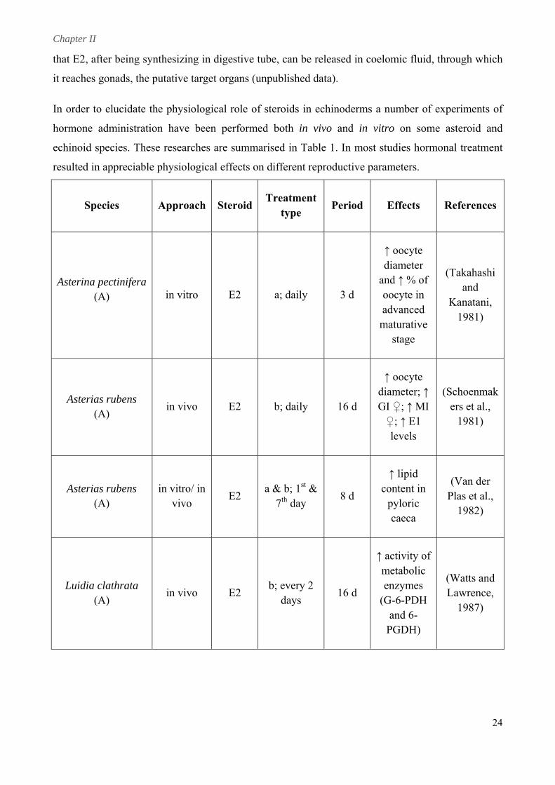
In order to elucidate the physiological role of steroids in echinoderms a number of experiments of
hormone administration have been performed both in vivo and in vitro on some asteroid and
echinoid species. These researches are summarised in Table 1. In most studies hormonal treatment
resulted in appreciable physiological effects on different reproductive parameters.
Species Approach SteroidTreatment
type Period Effects References
Asterina pectinifera (A) in vitro E2 a; daily 3 d
↑ oocyte diameter
and ↑ % of (Takahashi
oocyte in and
advanced maturative
stage
Kanatani, 1981)
↑ oocyte
Asterias rubens (A)
in vivo E2 b; daily 16 d GI ♀; ↑ MI ♀; ↑ E1
levels
ers et al., 1981)
diameter; ↑ (Schoenmak
Asterias rubens (A)
in vitro/ in vivo
E2 a & b; 1st &
7th day 8 d
↑ lipid content in
pyloric caeca
(Van der Plas et al.,
1982)
Luidia clathrata (A)
in vivo E2 b; every 2
days 16 d
↑ activity of metabolic enzymes
(G-6-PDH and 6-
PGDH)
(Watts and Lawrence,
1987)
24
Chapter II
25
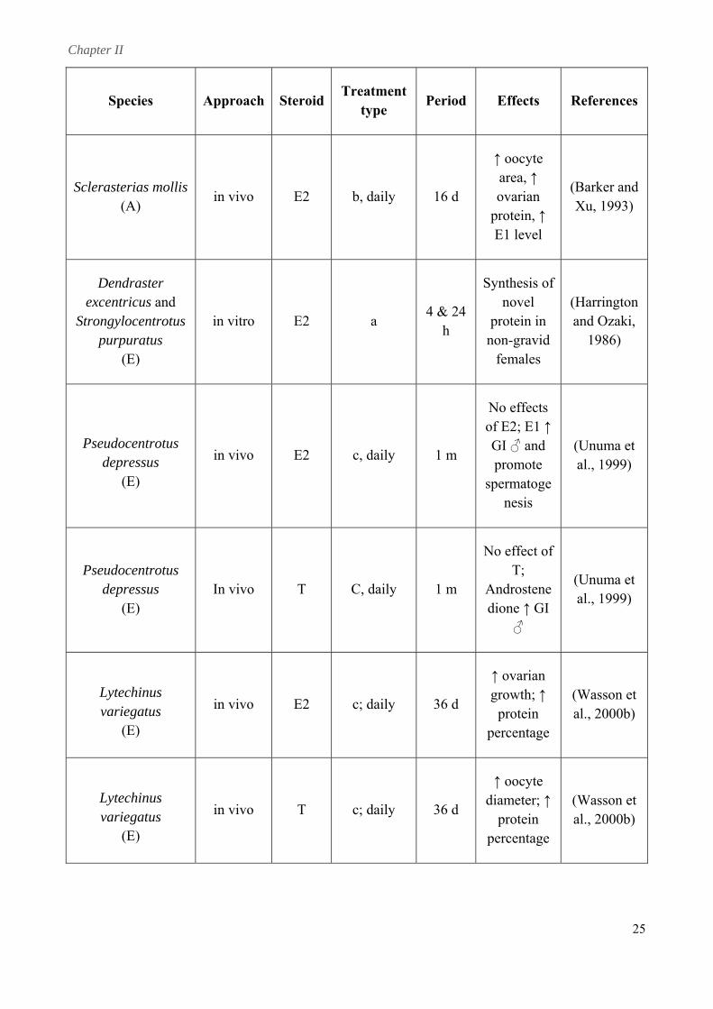
iod Effects References Species Approach SteroidTreatment
type Per
Sclerasterias mollis (A)
in vivo E2 b, daily 16 d
↑ oocyte area, ↑ ovarian
protein, ↑
(Barker and
E1 level
Xu, 1993)
Dendraster excentricus and
Strongylocentrotus purpuratus
in vitro E2 a 4 & 24
h
Synthesis of novel
non-gravid
(Harrington and Ozaki,
(E)
protein in
females 1986)
Pseudocentrotus depressus
(E)
in vivo E2 c, daily 1 m G promote
s
(Unuma et al., 1999)
No effects of E2; E1 ↑
I ♂ and
permatogenesis
Pseudocentrotus depressus
(E) In vivo T C, daily 1 m
No effect of
Androstenedi I
(Unuma et al., 1999)
T;
one ↑ G♂
Lytechinus variegatus
(E)
in vivo E2 c; daily 36 d
↑ ovarian growth; ↑ (Wasson et
al., 2000b) protein percentage
Lytechinus variegatus
(E)
in vivo T c; daily 36 d d
percentage
(Wasson et al., 2000b)
↑ oocyte iameter; ↑protein
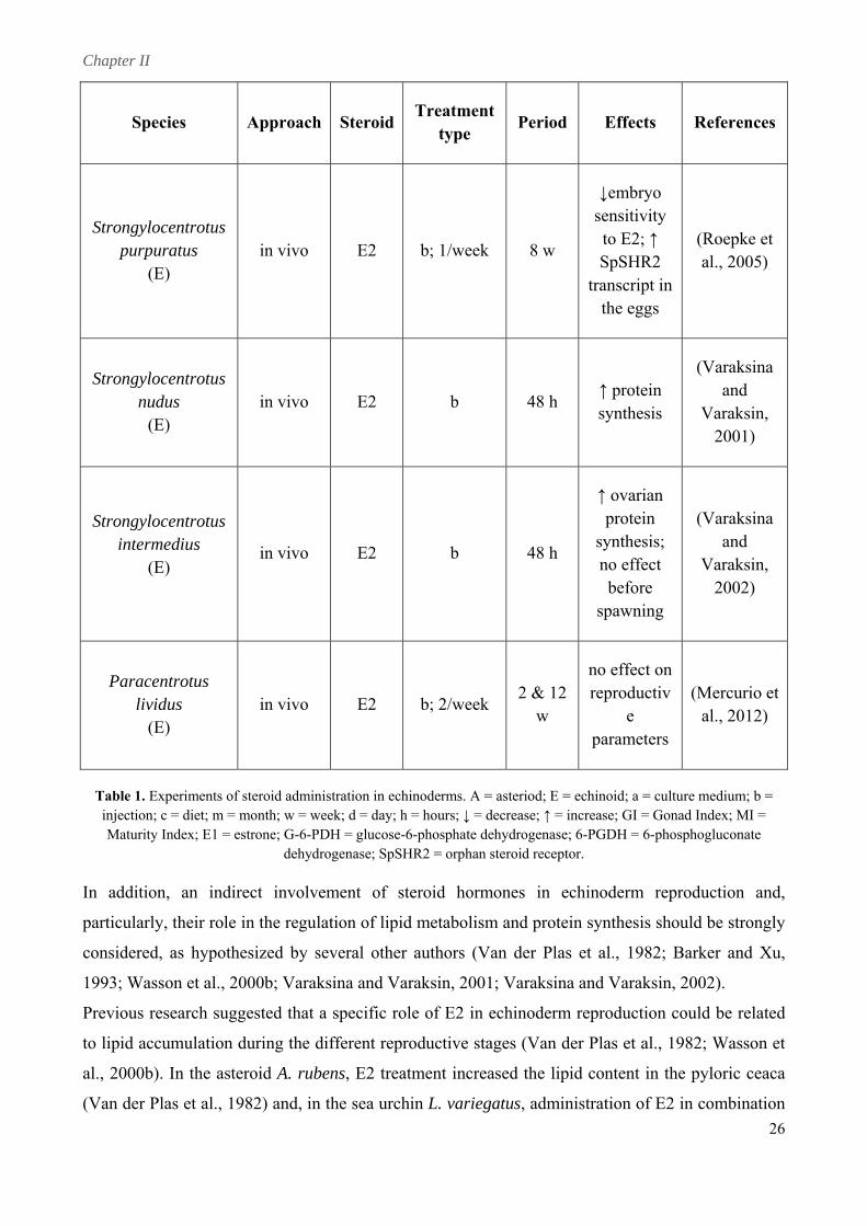
Chapter II
Species Approach SteroidTreatment
type Period Effects References
Strongylocentrotus purpuratus
(E) b; 1/week
sensitivity
tr the eggs
in vivo E2 8 w
↓embryo
to E2; ↑ SpSHR2 anscript in
(Roepke et al., 2005)
Strongylocentrotus nudus
(E) in vivo E2 b 48 h
Varaksin, 2001)
↑ protein synthesis
(Varaksina and
Strongylocentrotus
spawning
(Varaksina
2002)
intermedius (E)
in vivo E2 b 48 h
↑ ovarian protein
synthesis; no effect before
and Varaksin,
Paracentrotus lividus
(E) in vivo E2 b; 2/week
2 & 12 reproductiv ( w
no effect on
e parameters
Mercurio etal., 2012)
Table 1. Experiments of steroid administration in echinoderms. A = asteriod; E = echinoid; a = culture medium; b = injection; c = diet; m = month; w = week; d = day; h = hours; ↓ = decrease; ↑ = incre nad Index; MI = M 1 = estrone; G-6-PDH = glucose-6-phosphate dehydrogenase; 6-P sph
ogen pSHR2 steroi tor.
In addition, an indirect involvement of steroid hormones in echi roduction and,
particularly, their role in the regulation of lipid metabolism and protein synthesis should be strongly
considered, as hypothesized by several other authors (Van der Plas et Barker and Xu,
1993 l., 200 ksina Varak ; Va a a 20
Previous research suggested that a specific role of E2 in echinoderm reproduction could be related
to lipid accumulation during the different reproductive stages (Van der Plas et al., 1982; Wasson et
al., 2000b). In the asteroid A. rubens, E2 treatment increased the lipid content in the pyloric ceaca
(Van der Plas et al., 1982) and, in the sea urchin L. variegatus, administration of E2 in combination
ase; GI = GoGDH = 6-pho
noderm rep
aturity Index; E ogluconate dehydr ase; S = orphan d recep
al., 1982;
nd Varaksin, ; Wasson et a 0b; Vara and sin, 2001 raksin 02).
26
Chapter II
27
with progesterone similarly increased lipid percentage in the gonads (Wasson et al., 2000b).
Although species-specific differences can be certainly found, these results indicate a hormonal
control of lipid incorporation. This hypothesis is further supported by our previous studies reporting
that in ovaries of P. lividus, under physiological conditions, higher E ere found right
during those reproductive stages characterized by nutrient accu nd processing
(unpublished data).
Studies on the biochemical composition of echinoid gonads have rev me s
typically characterized by increased protein levels; in particular, in P. li tein levels were
found significantly correlated to the Gonad Index (Fernandez, 1998). As suggested by several
studies, steroids could be involved also in protein synthesis: E2 and T administration was
demonstrated to enhance the rate of protein synthesis in both asteroid ( Xu, 1 and
echinoid gonads (Varaksina and Varaksin, 2001; Varaksina and Va 02) and an E2
induction of protein synthesis was also observed in Strongylocentrotus purpuratus and Dendraster
excentricus coelomocytes (Harrington and Ozaki, 1986). In addition, est d be involved in
the expression of the sea urchin Major Yolk Protein (MYP) (Harrington ki, 1 t
al., 1
Echinoid MYP is a metal-binding glycoprotein of 170-180 kDa, belonging to the transferrin
superfamily (Brooks and Wessel, 2002). It was originally identified as th ponent of yolk
granules in sea urchin eggs and exchanged for a vitellogenin-like protein (Cervello et al., 1994;
Un 1). At present, it is well-known that M o s
vitellogenins: the sequen YP A fr entr depressu Unuma et al., 2001)
and other species (Brooks and Wessel, 2002; Noll et al., 2007) has revealed that it has about 25%
homology to vertebrate transferrin family, i. e. iron-binding glycoproteins that control the level of
fr t
mo 80
kDa MYP (CFMYP). A de firmed that these isoforms
in the
2 levels w
mulation a
ealed that ga
vidus, pro
togenesis i
Barker and
raksin, 20
993)
rogen coul
and Oza
e main com
986; Shyu e
987).
uma et al., 201 YP is n
otus
t homologou
s (
to vertebrate
cing of M cDN om Pseudoc
ee iron in biological fluid (Unuma et al., 2001). MYP has two isoforms with slightly differen
lecular masses: eggs contain the 170 kDa MYP (EGMYP) and coelomic fluid is rich in the 1
tailed analysis of S. purpuratus genome con
are products of the same gene, being only one gene encoding for MYP in sea urchin genome (Song
et al., 2006).
Unlike other oviparous animals, sea urchin yolk protein is not female-specific but MYP is
synthesized in both sexes (Shyu et al., 1986). Before gametogenesis, it is produced mainly
inner epithelium of the digestive tract and in the nutritive phagocytes of ovary and testis (Unuma et
al., 1998; Unuma et al., 2009; Unuma et al., 2010). MYP is accumulated in large quantities in the
nutritive phagocytes of agametogenic gonads (Unuma et al., 2011) and, as gametogenesis proceeds,
the stored protein is degraded to amino acids for the synthesis of new proteins and other nitrogen-
Chapter II
28
P may play the role of zinc carrier protein: MYP synthesized in the digestive
es, contain a measurable amount of CFMYP, which is discharged under stress
conditions, probably inducing the clotting processes due to the protein adhesive activity (Cervello
containing substances that constitute eggs and sperms (Unuma et al., 2003). Furthermore, MYP has
a zinc-binding capacity which is greater in CFMYP than in EGMYP (Unuma et al., 2007).
Although the geometry of MYP iron-binding site differs from other transferrins, it was
demonstrated that MYP binds iron, calcium, magnesium, barium, cadmium and manganese,
showing a higher affinity for zinc (Brooks and Wessel, 2002; Unuma et al., 2011). It has been
proposed that CFMY
tract can bind zinc derived from ingested food and transport it through the coelomic fluid to gonads.
Here, it can be partially deposited in nutritive phagocyte granules as protein and zinc storage and
partially modified to EGMYP with the loss of zinc-binding sites (Unuma et al., 2007; Unuma et al.,
2011). MYP seems to play an essential role not only in echinoid reproduction (Unuma et al., 1998;
Unuma et al., 2003) but also in embryonic development and immune response. In embryos, MYP
serves as a cell adhesion molecule: it is present both in yolk granules and at the surface of plasma
membranes and it is involved in cell-to-cell adhesion by mechanisms of calcium binding (Matranga
et al., 1986; McClay and Matranga, 1986; Noll et al., 2007). In the coelomic fluid MYP seems to be
involved in the clotting phenomenon. Colourless spherule cells, a specific subpopulation of
coelomocyt
and Matranga, 1989; Cervello et al., 1994).
Estrogen control of MYP expression has been suggested by several studies (Harrington and Ozaki,
1986; Shyu et al., 1987; Kiyomoto et al., 2008). In vertebrates, estrogens regulate the expression of
both vitellogenin and transferrin genes. The hormone first binds the estrogen receptor (ER) and then
the resulting complex attaches to short DNA sequences known as estrogen responsive elements
(EREs) and located upstream of the modulated genes (Prowse and Byrne, 2012). A palindromic
sequence, present in vertebrate EREs and essential for estrogen control, has been found upstream
MYP gene, strongly suggesting an estrogen involvement in the protein expression (Shyu et al.,
1987).
Chapter II
29
ltures were derived from the prawn Penaeus monodon
ecies, above all molluscs and crustaceans, where cells from several tissues were cultured
under different conditions and the most effective medium and supplements were evaluated. In most
cases, Leibovitz L-15, modified with salts, resulted the best culture medium and cell survival and
growth seemed to be improved by the addition of 5-10% heat-inactivated FCS (Moss et al., 1998;
Mulford and Austin, 1998; Walton and Smith, 1999; Cao et al., 2003; Rinkevich, 2005). However,
all the developed cell cultures could be maintained at least for some months and the proliferation
rate was reported to be very low or absent (Mulford and Austin, 1998; Maeda et al., 2003;
Odintsova et al., 2005; Rinkevich, 2011).
This failure has been explained in view of the in vitro low speed of cell proliferation (Cao et al.,
4. Primary cell cultures from marine invertebrates
Cells under in vitro conditions are used in a variety of fields and in many scientific studies and
related applications as extremely important experimental tools (Rinkevich, 2005). With respect to
marine invertebrates, despite the diversity of species and their potential as in vitro models for
numerous applications, almost all the efforts to develop proliferative and permanent cell cultures
have been unsuccessful (Rinkevich, 2011). At present, several short- and long-term cell cultures
from a variety of tissues in an increasing number of species have been developed (Mulford and
Austin, 1998; Walton and Smith, 1999; Cao et al., 2003; Odintsova et al., 2005; de Caralt et al.,
2007; Sharlaimova et al., 2010; Di Benedetto, 2011) but there are still few established examples of
proliferative cell lines from marine invertebrates (Frank et al., 1994; Fraser and Hall, 1999;
Rinkevich, 2005; Shashikumar and Desai, 2011).
Continuous cell cultures have been developed from 10 different species of sessile colonial marine
cnidarians: primary cultures of various cell types and sizes were obtained from both colony
fragments and planula larvae, culturing them in modified Leibovitz L-15 medium with Fetal Calf
Serum (FCS). Cell proliferation was observed within 7-20 days after dissociation and cultures were
maintained and subcloned for approximately 1 year (Frank et al., 1994).
In crustaceans, proliferative primary cell cu
ovaries at different maturative stages. Cells were maintained for up to 17 months, being subcultured
3 times (Fraser and Hall, 1999). Best results were obtained from the testicular tissues of the crab
Scylla serrata. Primary cell cultures from both explants and segregated tissues of S. serrata testes
were shown to be able to proliferate and grow in L-15 crab saline medium supplemented with
epidermal growth factors and glucose. These testicular cells were subcultured every 4-6 days and
remained healthy for 5 months (Shashikumar and Desai, 2011).
Apart from these few successful research, extensive studies had been performed on many other
edible sp
Chapter II
30
2003) and the lack of vital information regarding cell physiology and biology and their specific
requirements. In a ells enter, 24–72
evidence of increase in cell number and nerve
different stages of gut
iding suitable material for cell
fragments were reported. In order to analyze E2 effect on A. pectinifera oocyte growth, fragments
ddition, it has been recently suggested that marine invertebrate c
hours after their isolation, into a quiescent in vitro state (Rinkevich, 2011). Thus, cell cultures were
mostly developed from tissues with high growth potential, like embryonic, neoplastic, or
regenerating tissues (Odintsova et al., 2005).
Echinoderms are well known for their regenerative capabilities (Candia Carnevali, 2006; Candia
Carnevali and Burighel, 2010) and, thus, the studies present in literature, related to cell cultures,
were mostly performed using cells from tissues involved in the regenerating processes (Odintsova
et al., 2005; Sharlaimova et al., 2010; Di Benedetto, 2011). Neurons and neural tissue explants from
the starfish A. rubens and the brittle star Ophiura ophiura were cultured for up to 6 weeks in
modified L-15 medium. However, there was limited
outgrowth, probably due to the lack of specific growth factors. The addition of coelomic fluid,
neural tissue extracts and nerve growth factor did not enhance cell conditions, suggesting that
neurons require some other specific native conditioning factors (Moss et al., 1998).
On the contrary, long-term cell cultures were successfully developed from regenerating intestine of
the sea cucumber Apostichopus japonicus, showing that cells from
regeneration display different in vitro proliferation rates and behaviors (Odintsova et al., 2005). In
particular, only primary intestinal cultures, performed 14-16 days after evisceration, were involved
in active proliferation and their cell number increased more than twofold by the 20th day of culture.
Cultured cells seemed to be capable of mitotic division in suspension as well as in substrate-
attached conditions. Although the intensity of cell proliferation depends on both species and
regeneration type, this study strongly suggests that regenerating tissues can represent a promising
source of cells for long-term cell cultures.
Similar results were obtained in A. rubens, where coelomocytes and coelomic epithelium cells were
maintained under in vitro condition for at least 2 months (Sharlaimova et al., 2010). Coelomocytes
isolated 5 hours after injury, displayed a higher functional activity than cells derived from control
group: cells from injured animals tended to form large aggregates and network structures in which
spread cells were in contact to each other whereas roundish cells were located at the network
surfaces or among them. Coelomic epithelium cells formed colony-like aggregates too; in addition,
they showed a higher proliferation activity, leading to consider them the most encouraging object
for in vitro studies on asteroid regeneration processes.
The potential of other tissues and, particularly, gonad tissues, in prov
cultures has been less explored. In echinoderms, only few examples of cultures of ovary and testis
Chapter II
of ovary w
at the end
alone, deg
testicular t
germ-line c
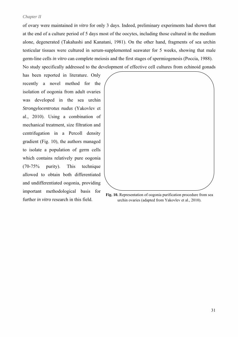
No study s
has been
recently
isolation o
was deve
Strongyloc
al., 2010)
mechanica
centrifugat
gradient (F
to isolate
which con
(70-75%
allowed to
and undiff
important
further in v
were maintai
of a culture
generated (T
tissues were
cells in vitro
ined in vitro
e period of 5
Takahashi a
e cultured i
o can comp
o for only 3
5 days most
and Kanatan
in serum-su
plete meiosis
3 days. Inde
t of the ooc
ni, 1981). O
upplemente
s a
eed, prelimi
cytes, includ
On the othe
d seawater
inary experi
ding those c
er hand, fra
for 5 week
iments had
cultured in t
agments of
ks, showing
shown that
the medium
f sea urchin
g that male
t
m
n
e
nd the firrst stages off spermiogeenesis (Pocccia, 1988).
specifically addressed tto the deve
reported in
a novel
of oogonia f
eloped in
centrotus nu
). Using a
al treatment,
tion in a
Fig. 10), the
a populati
ntains relativ
purity).
o obtain b
ferentiated o
methodolo
vitro researc
n literature
method f
from adult
the sea
udus (Yako
a combinat
, size filtrat
Percoll
e authors m
ion of germ
vely pure o
This tec
both differe
oogonia, pr
ogical bas
ch in this fie
llopment of effective ceell cultures from echinnoid gonadss
e. Only
for the
ovaries
urchin
ovlev et
tion of
tion and
density
managed
m cells
oogonia
chnique
entiated
oviding
sis for
eld. FFig. 10. Repres
urchin osentation of oovaries (adapt
ogonia purificted from Yako
cation proceduovlev et al., 20
ure from sea 010).
31
Chapter III
DEVELOPMENT OF PRIMARY CELL
CULTURES FROM SEA URCHIN OVARIES
Publications containing experimental data presented
in this chapter:
Mercurio S., Di Benedetto C., Sugni M., Candia Carnevali M.
D. (2013a). Development of primary cell cultures from sea
urchin gonads. In Springer Netherlands, Proceedings of the
symposium “Marine Invertebrate Cell Culture”,
Cytotechnology, 65, 5, 673-689. (Appendix 1)
Mercurio S., Di Benedetto C., Sugni M. & Candia Carnevali
M. D. (2013b).Primary cell cultures from sea urchin ovaries:
a new experimental tool. In Vitro Cell. Dev. Biol. – Animal. In
press. DOI: 10.1007/s11626-013-9686-1. (Appendix 2)
Chapter III
33
1. Abstract
In this chapter the development of primary cell cultures from ovaries of the edible sea urchin
Paracentrotus lividus is described in order to provide a simple and versatile experimental tool for
research in echinoderm reproductive biology.
The ovary cell phenotypes, present in culture, were identified and characterized by different
microscopic techniques. Although cell cultures could be produced from ovaries at all stages of
maturation, the cells appeared healthier and viable, displaying a higher survival rate, when ovaries
at early stages of gametogenesis were used. In terms of culture medium, ovarian cells were
successfully cultured in modified Leibovitz-15 medium, whereas poor results were obtained in
Minimum Essential Medium Eagle and Medium 199. Different substrates were tested, but ovarian
cells completely adhered only on poly-L-lysine. To improve in vitro conditions and stimulate cell
proliferation different serum-supplements were tested. Fetal Calf Serum and an originally
developed Pluteus Extract appeared to be detrimental to cell survival, apparently accelerating
processes of cell death. In contrast, cells cultured with sea urchin Egg Extract appeared larger and
healthier, displaying an increased longevity that allowed to maintain them for up to 1 month.
Overall our study provides new experimental bases and procedures for producing successfully long-
term primary cell cultures from sea urchin ovaries offering a good potential to study echinoid
oogenesis in a controlled system and to investigate different aspects of echinoderm endocrinology
and reproductive biology.
2. Introduction
Despite the traditional use of sea urchin as a favourite model in embryology and developmental
biology, the specific mechanisms regulating reproductive processes are still scarcely known in all
echinoderms. In echinoids, gametogenesis was demonstrated to be influenced by several
environmental factors, such as water temperature and photoperiod (Byrne, 1990; Spirlet et al., 2000;
Shpigel et al., 2004; McCarron et al., 2010), food availability (Leoni et al., 2001) and environmental
hydrodynamics (Fenaux, 1968). Apart from these exogenous factors, several endogenous
molecules, notably hormones and neuropeptides, likely play an important role in regulating
reproductive processes (Spirlet et al., 1998; Mita, 2013). Although several studies have been
performed in order to identify and to understand the roles of these molecules, their mechanisms of
action are still far to be clearly understood. In order to elucidate their complete physiological
significance, a simple and adequate model system, such as an appropriate in vitro approach, can be
certainly helpful, allowing to perform studies under controlled experimental conditions (Odintsova
et al., 2005).
Chapter III
34
The establishment of primary cell cultures from marine invertebrates and, particularly, from
echinoderms has been the objective of many previous attempts encountering uncounted obstacles
(Rinkevich, 1999). At present, short and long-term cell cultures from a variety of tissues and from
numerous species have been developed (Mulford and Austin, 1998; Walton and Smith, 1999; de
Caralt et al., 2007; Sharlaimova et al., 2010; Di Benedetto, 2011): however, there are only few rare
examples of establishment of proliferative cell lines from marine invertebrates (Rinkevich, 2011;
Shashikumar and Desai, 2011). The reasons of these failures have been mostly identified in the in
vitro low speed of cell proliferation and the lack of vital information regarding cell physiology and
biology (Rinkevich, 1999; Cao et al., 2003). Considering all these difficulties, cell cultures were
mostly developed from tissues with high growth potential (Odintsova et al., 2005).
In echinoderms, regenerating tissues display high proliferate rates (Candia Carnevali, 2006; Candia
Carnevali and Burighel, 2010) and, thus, they represent an optimal source of cells to successfully
develop long-term primary cell cultures (Odintsova et al., 2005; Sharlaimova et al., 2010).
The potential of other tissues, such as gonads, in providing an appropriate material for in vitro
studies have been less investigated. In literature there are only a few examples of cultures of
echinoderm ovary and testis fragments. In Asterias pectinifera, fragments of ovary were cultured
for only 3 days (Takahashi and Kanatani, 1981); on the other hand fragments of sea urchin
testicular tissues were cultured in serum-supplemented seawater for 5 weeks, showing that male
germ-line cells can complete their maturative processes in in vitro conditions (Poccia, 1988). In
addition, a novel method for the isolation of oogonia from adult ovaries was developed in the sea
urchin Strongylocentrotus nudus (Yakovlev et al., 2010). The authors obtained cell populations
which contain relatively pure oogonia (70-75% purity), providing the first methodological basis for
further in vitro research in this field. Nevertheless, no study specifically addressed to the
development of effective cell cultures from gonads was previously reported in literature. Taking
into account the advantages and the possible applications of the in vitro studies, we carried out the
first attempt to develop primary cell cultures from ovaries of the common Mediterranean sea urchin
Paracentrotus lividus. After an accurate characterization of the cell phenotypes present in culture,
we focused on culture condition optimization, i.e. to define the suitable medium, substrate and
possible serum supplements. Overall, the final aim of this investigation was to set up the
experimental basis for producing primary cell cultures from ovaries of this edible and ecologically
relevant species. Our results could be useful for improving and expanding the potential employment
of echinoderms in experimental research, in particular providing an important tool for in vitro
studies on echinoid reproductive biology and providing a simple and versatile method for multi-
disciplinary applications, such as ecotoxicological and aquaculture applied research.
Chapter III
35
3. Materials and Methods
3.1. Animals
P. lividus adult specimens were monthly collected in the Protected Marine Areas of Bergeggi and
Portofino, on the Ligurian coast of Italy, and immediately transported to the laboratory in cool
boxes filled with natural sea water. Animals were kept in aquaria under constant aeration in
circulating artificial sea water (Instant Ocean; salinity about 37‰, as in the Mediterranean Sea).
Animal conditions as well as all water physical and chemical parameters were daily monitored.
3.2. Cell cultures
P. lividus ovaries were removed from the internal side of the tests: for each specimen one gonad
was used for histological analysis and processed for standard methods of light microscopy, whereas
the remaining 4 gonads were used to develop primary cell cultures according to the following
protocol. Ovaries were washed several times in sterile Ca2+ Mg2+ Free Sea Water (CMFSW) with
antibiotics (40µg/l gentamycin and 100 units/mL penicillin, 100 µg/mL streptomycin) and dissected
into small pieces (2-5 mm) using fine-tipped tweezers. Ovary pieces were incubated in 0.5 mg/mL
collagenase dissolved in sterile CMFSW and stirred for 1 hour. The resulting cell suspension was
filtered through 50 µm nylon gauze (to remove mature oocytes), centrifuged at 300 × g for 6’ at 15
°C and the cell pellet was resuspended in modified culture medium. Cells were seeded at a
concentration of 3-4 × 10⁵ cells/mL in 24-well culture plates, without coating (medium evaluation,
see below) or coated with rat collagen (I type, Sigma), gelatin (Merck Millipore) or poly-L-lysine
(70-150 kDa, 0.01% solution, Sigma). Three culture media were compared: Leibovitz-15 medium
(L-15), Minimum Essential Medium Eagle (MEM) and Medium 199 (M199), all of them modified
by the addition of 20.2 g/L NaCl, 0.54 g/L KCl, 0.60 g/L CaCl2, 1 g/L Na2SO4, 3.9 g/L MgCl2, 1
g/L glucose, 25 mg/L Taurine, 5 mL/L Insulin (10 mg/mL in solution, SIGMA), 100 mg/L L-
Glutamine, 1.75 mg/L vitamin E (1000 mOsm). The pH was adjusted at 7.6. The media were then
sterilized by filtration (0.22 µm, Corning-Sigma) and 40 µg/L gentamycin, 100 units/mL penicillin
and 100 µg/mL streptomycin were added. All cultures were incubated at 15 °C for at least 20 days.
Cell behavior was observed daily for the first two weeks of culture and, then, at each medium
replacement using an inverted phase contrast microscope. Replacement of 50% of the medium was
carried out every two days.
3.3. Medium and supplement evaluation
As previously anticipated, three different modified culture media were tested: L-15, M199 and
MEM. The most effective medium was determined by a detailed analysis based on cell morphology,
Chapter III
36
using an inverted phase contrast microscope, and on cell viability during a 2 week culture period.
Cell viability was estimated in duplicates by direct cell counting, using a “Burker chamber” coupled
with Tripan blue exclusion test at 0 (T0), 2, 7 and 14 (Tn) days. The percentage of viable cells (Vx)
at the different time points was calculated as follows:
Vx= (viable cell mean n° at Tn/ viable cell n° at T0) x 100.
Once the most effective culture medium was determined, the effects of 2% inactivated Fetal Calf
Serum (FCS) and of several concentrations of P. lividus “Pluteus Estract” (PE) and “Egg Extract”
(EE) were tested. PE was obtained from 48h P. lividus larvae. When sea urchin larvae reached the
pluteus stage, they were collected, sonicated and centrifuged at 800 × g for 10’ at 4 °C. The
supernatant was then centrifuged at 17000 × g for 1 h at 4 °C. The same procedure was performed
to obtain EE from P. lividus mature ovaries. The resulting extracts were protein assayed (BCA
protein assay kit, Sigma), sterile filtered, heat-inactivated and stored at -20 °C. Different PE and EE
concentrations were tested: 34 ng/mL, 100 ng/mL, 200 ng/mL, 500 ng/mL, 1 µg/mL, 10 µg/mL and
50 µg/mL. Supplement effects on cell morphology and viability was observed and compared during
a 2 week culture period. Cell viability was determined at different time points by staining with
fluorescent Calcein AM (viable cell marker, Sigma) or by direct cell counting, using a “Burker
chamber” coupled with Tripan blue exclusion test (see before).
In order to analyze the cell proliferation activity, the incorporation method employing the thymidine
analog 5-bromo-deoxyuridine (BrdU) was used. Cells were exposed to 30 µM BrdU and FdU (10:1)
in culture medium for 12 hours. After this period they were fixed in 4% paraformaldehyde for 1
hour, treated with 1.5 N HCl and 0.2% Triton X-100 and incubated with antibodies against BrdU
(Cell Proliferation Kit: Amersham, GE Healthcare). Staining with a secondary antibody FITCH
conjugated (Anti-mouse IgG FITCH conjugate, SIGMA) was performed for 40 minutes. To
determine the BrdU nuclear localization, cells were also stained with DAPI. Samples were
examined using a fluorescence phase contrast microscope. The experiments on proliferation activity
evaluation were carried out on fresh, one week and two week cell cultures.
3.4. Microscopic and ultramicroscopic analyses
In order to determine ovary reproductive stages and characterize the cellular phenotypes present in
fresh cultures, standard methods of light and electron microscopy were employed, as previously
described by Barbaglio et al. (2007). Briefly, gonads or cell pellets were fixed with glutaraldeyde
2% in cacodylate buffer 0.1 M and NaCl 1.4%, washed with cacodylate buffer 0.1 M and post fixed
with a solution of OsO4 1% in cacodylate buffer 0.1 M. Then the samples were prestained with
uranyl acetate in ethanol 25%, dehydrated through the ethanol series, and finally, after washing in
Chapter III
37
propylene oxide, embedded in Epon 812–Araldite resin. Semithin (1 µm) and ultrathin (70 nm)
sections, were cut with a Reichert–Jung ULTRACUT E using glass knives. Semithin sections,
stained with crystal violet and basic fucsin, were observed under a Jenaval light microscope.
Ultrathin sections for electron microscopy were mounted on copper grids and stained with uranyl
acetate and lead citrate, then observed and photographed in a Jeol 100SX electron microscope.
Five ovary reproductive stages were considered: Spent (phase immediately following the spawning
event), Recovery (phase characterized by phagocytosis and nutrient accumulation), Growing,
Premature and Mature (all phases characterized by gametogenesis in progress) (Barbaglio et al.,
2007).
3.5. Scanning electron microscopy
Cells adhesion to the substrates was investigated by scanning electron microscopy. 48h cell cultures
were fixed in glutaraldeyde 2% in artificial sea water (ASW) for 2 hours at 4 °C. After overnight
wash in filtered ASW at 4 °C, samples were post fixed with a solution of OsO4 1% in ASW and
glucose for 2 hours, washed in distilled water and dehydrated through the ethanol series. Absolute
ethanol was gradually substituted with Hexamethyldisilazane (HMDS; Sigma). Samples were left to
dry, mounted on stabs, covered by thin pure gold layer (Sputter Coater Nanotech) and observed
with a scanning electron microscope (LEO-1430).
3.6. Electrophoresis
Sodium dodecylsulfate-polyacrilamide gel electrophoresis (SDS-PAGE) was performed using 6%
slab gel (Laemmli, 1970) in order to verify and compare the protein content in PE and EE
supplements. Prior to electrophoresis samples were dialyzed, diluted with sample buffer (SDS
reducing buffer) and boiled for 5 minutes. Gels were run at 100 V at room temperature. Protein
bands were visualized with Coomassie brilliant blue R-250. SDS-PAGE standards (StoS Protein
Marker, Genespin s.r.l.) were also run for molecular weight calibration.
3.7. Statistical analysis
Data are presented as mean values±SEM. Statistical significance was assessed using Paired t test
and one-way ANOVA (Tukey’s post-hoc test). A p-value of less than 0.05 was considered
statistically significant. Statistical analysis was performed by the computer program GraphPad
Prism 4.
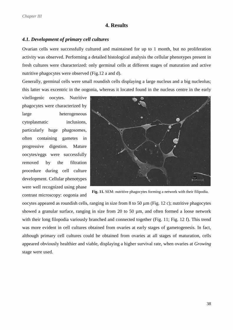
Chapter III
38
4. Results
4.1. Development of primary cell cultures
Ovarian cells were successfully cultured and maintained for up to 1 month, but no proliferation
activity was observed. Performing a detailed histological analysis the cellular phenotypes present in
fresh cultures were characterized: only germinal cells at different stages of maturation and active
nutritive phagocytes were observed (Fig.12 a and d).
Generally, germinal cells were small roundish cells displaying a large nucleus and a big nucleolus;
this latter was excentric in the oogonia, whereas it located found in the nucleus centre in the early
vitellogenic oocytes. Nutritive
phagocytes were characterized by
large heterogeneous
cytoplasmatic inclusions,
particularly huge phagosomes,
often containing gametes in
progressive digestion. Mature
oocytes/eggs were successfully
removed by the filtration
procedure during cell culture
development. Cellular phenotypes
were well recognized using phase
contrast microscopy: oogonia and
oocytes appeared as roundish cells, ranging in size from 8 to 50 µm (Fig. 12 c); nutritive phagocytes
showed a granular surface, ranging in size from 20 to 50 µm, and often formed a loose network
with their long filopodia variously branched and connected together (Fig. 11; Fig. 12 f). This trend
was more evident in cell cultures obtained from ovaries at early stages of gametogenesis. In fact,
although primary cell cultures could be obtained from ovaries at all stages of maturation, cells
appeared obviously healthier and viable, displaying a higher survival rate, when ovaries at Growing
stage were used.
Fig. 11. SEM: nutritive phagocytes forming a network with their filipodia.
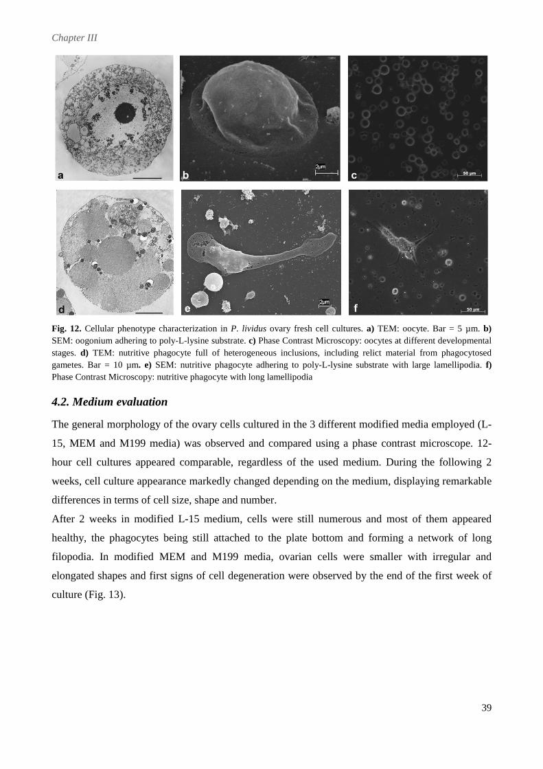
Chapter III
39
Fig. 12. Cellular phenotype characterization in P. lividus ovary fresh cell cultures. a) TEM: oocyte. Bar = 5 µm. b) SEM: oogonium adhering to poly-L-lysine substrate. c) Phase Contrast Microscopy: oocytes at different developmental stages. d) TEM: nutritive phagocyte full of heterogeneous inclusions, including relict material from phagocytosed gametes. Bar = 10 µm. e) SEM: nutritive phagocyte adhering to poly-L-lysine substrate with large lamellipodia. f) Phase Contrast Microscopy: nutritive phagocyte with long lamellipodia
4.2. Medium evaluation
The general morphology of the ovary cells cultured in the 3 different modified media employed (L-
15, MEM and M199 media) was observed and compared using a phase contrast microscope. 12-
hour cell cultures appeared comparable, regardless of the used medium. During the following 2
weeks, cell culture appearance markedly changed depending on the medium, displaying remarkable
differences in terms of cell size, shape and number.
After 2 weeks in modified L-15 medium, cells were still numerous and most of them appeared
healthy, the phagocytes being still attached to the plate bottom and forming a network of long
filopodia. In modified MEM and M199 media, ovarian cells were smaller with irregular and
elongated shapes and first signs of cell degeneration were observed by the end of the first week of
culture (Fig. 13).
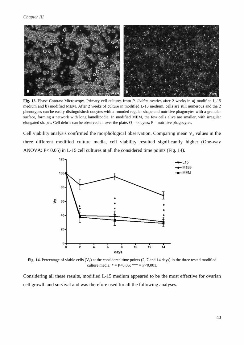
Chapter III
40
Fig. 13. Phase Contrast Microscopy. Primary cell cultures from P. lividus ovaries after 2 weeks in a) modified L-15 medium and b) modified MEM. After 2 weeks of culture in modified L-15 medium, cells are still numerous and the 2 phenotypes can be easily distinguished: oocytes with a rounded regular shape and nutritive phagocytes with a granular surface, forming a network with long lamellipodia. In modified MEM, the few cells alive are smaller, with irregular elongated shapes. Cell debris can be observed all over the plate. O = oocytes; P = nutritive phagocytes.
Cell viability analysis confirmed the morphological observation. Comparing mean Vx values in the
three different modified culture media, cell viability resulted significantly higher (One-way
ANOVA: P< 0.05) in L-15 cell cultures at all the considered time points (Fig. 14).
Fig. 14. Percentage of viable cells (Vx) at the considered time points (2, 7 and 14 days) in the three tested modified
culture media. * = P<0.05; *** = P<0.001.
Considering all these results, modified L-15 medium appeared to be the most effective for ovarian
cell growth and survival and was therefore used for all the following analyses.
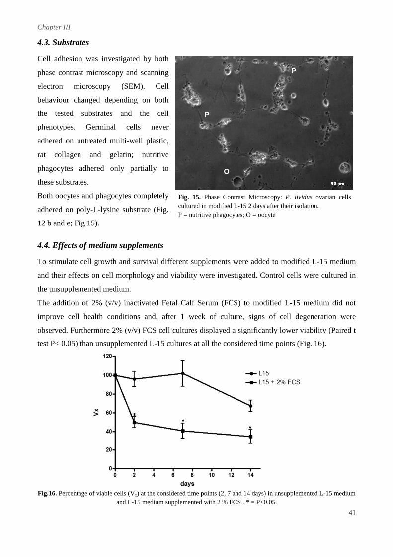
Chapter III
41
4.3. Substrates
Cell adhesion was investigated by both
phase contrast microscopy and scanning
electron microscopy (SEM). Cell
behaviour changed depending on both
the tested substrates and the cell
phenotypes. Germinal cells never
adhered on untreated multi-well plastic,
rat collagen and gelatin; nutritive
phagocytes adhered only partially to
these substrates.
Both oocytes and phagocytes completely
adhered on poly-L-lysine substrate (Fig.
12 b and e; Fig 15).
4.4. Effects of medium supplements
To stimulate cell growth and survival different supplements were added to modified L-15 medium
and their effects on cell morphology and viability were investigated. Control cells were cultured in
the unsupplemented medium.
The addition of 2% (v/v) inactivated Fetal Calf Serum (FCS) to modified L-15 medium did not
improve cell health conditions and, after 1 week of culture, signs of cell degeneration were
observed. Furthermore 2% (v/v) FCS cell cultures displayed a significantly lower viability (Paired t
test P< 0.05) than unsupplemented L-15 cultures at all the considered time points (Fig. 16).
Fig.16. Percentage of viable cells (Vx) at the considered time points (2, 7 and 14 days) in unsupplemented L-15 medium
and L-15 medium supplemented with 2 % FCS . * = P<0.05.
Fig. 15. Phase Contrast Microscopy: P. lividus ovarian cells cultured in modified L-15 2 days after their isolation. P = nutritive phagocytes; O = oocyte
P
O
P
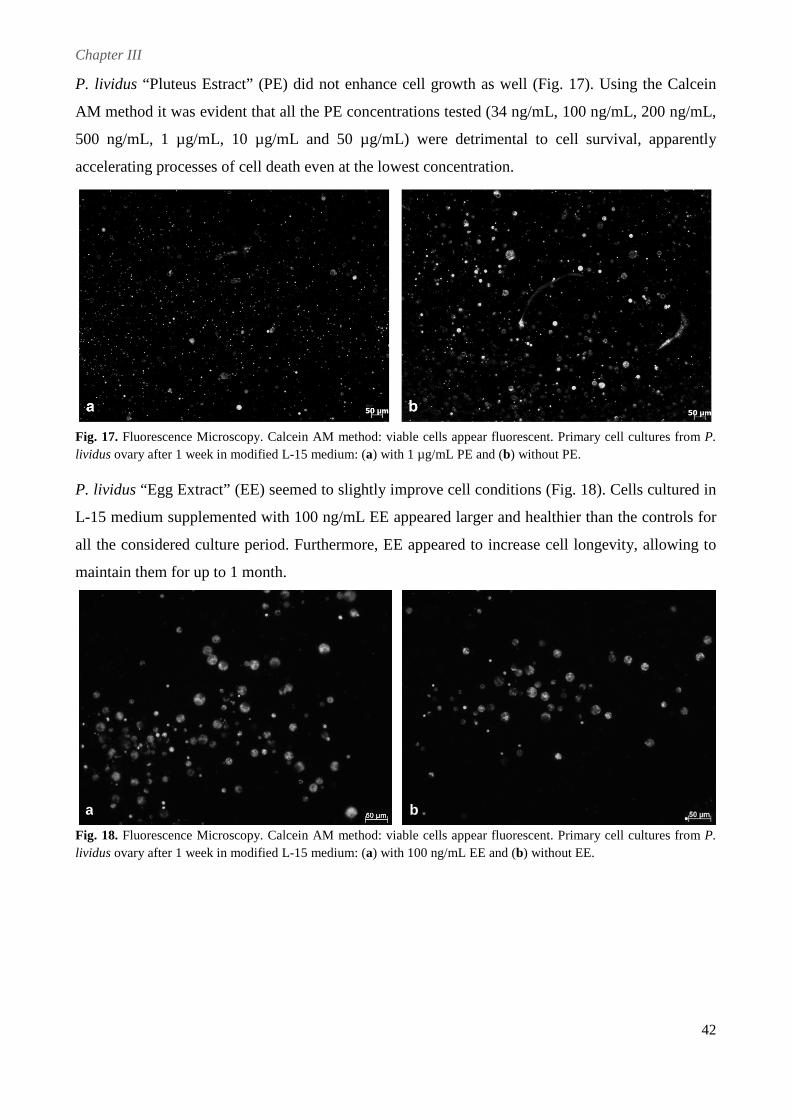
Chapter III
42
P. lividus “Pluteus Estract” (PE) did not enhance cell growth as well (Fig. 17). Using the Calcein
AM method it was evident that all the PE concentrations tested (34 ng/mL, 100 ng/mL, 200 ng/mL,
500 ng/mL, 1 µg/mL, 10 µg/mL and 50 µg/mL) were detrimental to cell survival, apparently
accelerating processes of cell death even at the lowest concentration.
Fig. 17. Fluorescence Microscopy. Calcein AM method: viable cells appear fluorescent. Primary cell cultures from P. lividus ovary after 1 week in modified L-15 medium: (a) with 1 µg/mL PE and (b) without PE.
P. lividus “Egg Extract” (EE) seemed to slightly improve cell conditions (Fig. 18). Cells cultured in
L-15 medium supplemented with 100 ng/mL EE appeared larger and healthier than the controls for
all the considered culture period. Furthermore, EE appeared to increase cell longevity, allowing to
maintain them for up to 1 month.
Fig. 18. Fluorescence Microscopy. Calcein AM method: viable cells appear fluorescent. Primary cell cultures from P. lividus ovary after 1 week in modified L-15 medium: (a) with 100 ng/mL EE and (b) without EE.
a b
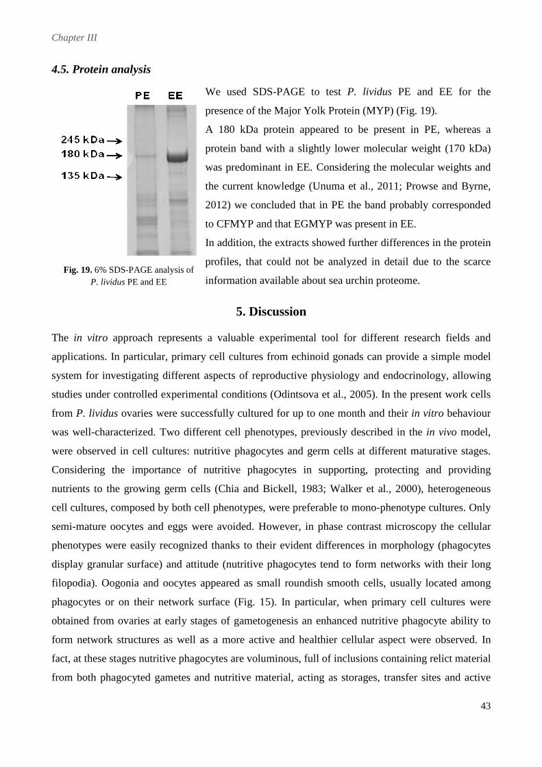
Chapter III
43
4.5. Protein analysis
We used SDS-PAGE to test P. lividus PE and EE for the
presence of the Major Yolk Protein (MYP) (Fig. 19).
A 180 kDa protein appeared to be present in PE, whereas a
protein band with a slightly lower molecular weight (170 kDa)
was predominant in EE. Considering the molecular weights and
the current knowledge (Unuma et al., 2011; Prowse and Byrne,
2012) we concluded that in PE the band probably corresponded
to CFMYP and that EGMYP was present in EE.
In addition, the extracts showed further differences in the protein
profiles, that could not be analyzed in detail due to the scarce
information available about sea urchin proteome.
5. Discussion
The in vitro approach represents a valuable experimental tool for different research fields and
applications. In particular, primary cell cultures from echinoid gonads can provide a simple model
system for investigating different aspects of reproductive physiology and endocrinology, allowing
studies under controlled experimental conditions (Odintsova et al., 2005). In the present work cells
from P. lividus ovaries were successfully cultured for up to one month and their in vitro behaviour
was well-characterized. Two different cell phenotypes, previously described in the in vivo model,
were observed in cell cultures: nutritive phagocytes and germ cells at different maturative stages.
Considering the importance of nutritive phagocytes in supporting, protecting and providing
nutrients to the growing germ cells (Chia and Bickell, 1983; Walker et al., 2000), heterogeneous
cell cultures, composed by both cell phenotypes, were preferable to mono-phenotype cultures. Only
semi-mature oocytes and eggs were avoided. However, in phase contrast microscopy the cellular
phenotypes were easily recognized thanks to their evident differences in morphology (phagocytes
display granular surface) and attitude (nutritive phagocytes tend to form networks with their long
filopodia). Oogonia and oocytes appeared as small roundish smooth cells, usually located among
phagocytes or on their network surface (Fig. 15). In particular, when primary cell cultures were
obtained from ovaries at early stages of gametogenesis an enhanced nutritive phagocyte ability to
form network structures as well as a more active and healthier cellular aspect were observed. In
fact, at these stages nutritive phagocytes are voluminous, full of inclusions containing relict material
from both phagocyted gametes and nutritive material, acting as storages, transfer sites and active
Fig. 19. 6% SDS-PAGE analysis of P. lividus PE and EE
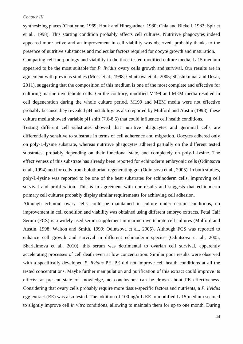
Chapter III
44
synthesizing places (Chatlynne, 1969; Houk and Hinegardner, 1980; Chia and Bickell, 1983; Spirlet
et al., 1998). This starting condition probably affects cell cultures. Nutritive phagocytes indeed
appeared more active and an improvement in cell viability was observed, probably thanks to the
presence of nutritive substances and molecular factors required for oocyte growth and maturation.
Comparing cell morphology and viability in the three tested modified culture media, L-15 medium
appeared to be the most suitable for P. lividus ovary cells growth and survival. Our results are in
agreement with previous studies (Moss et al., 1998; Odintsova et al., 2005; Shashikumar and Desai,
2011), suggesting that the composition of this medium is one of the most complete and effective for
culturing marine invertebrate cells. On the contrary, modified M199 and MEM media resulted in
cell degeneration during the whole culture period. M199 and MEM media were not effective
probably because they revealed pH instability: as also reported by Mulford and Austin (1998), these
culture media showed variable pH shift (7.6-8.5) that could influence cell health conditions.
Testing different cell substrates showed that nutritive phagocytes and germinal cells are
differentially sensitive to substrate in terms of cell adherence and migration. Oocytes adhered only
on poly-L-lysine substrate, whereas nutritive phagocytes adhered partially on the different tested
substrates, probably depending on their functional state, and completely on poly-L-lysine. The
effectiveness of this substrate has already been reported for echinoderm embryonic cells (Odintsova
et al., 1994) and for cells from holothurian regenerating gut (Odintsova et al., 2005). In both studies,
poly-L-lysine was reported to be one of the best substrates for echinoderm cells, improving cell
survival and proliferation. This is in agreement with our results and suggests that echinoderm
primary cell cultures probably display similar requirements for achieving cell adhesion.
Although echinoid ovary cells could be maintained in culture under certain conditions, no
improvement in cell condition and viability was obtained using different embryo extracts. Fetal Calf
Serum (FCS) is a widely used serum-supplement in marine invertebrate cell cultures (Mulford and
Austin, 1998; Walton and Smith, 1999; Odintsova et al., 2005). Although FCS was reported to
enhance cell growth and survival in different echinoderm species (Odintsova et al., 2005;
Sharlaimova et al., 2010), this serum was detrimental to ovarian cell survival, apparently
accelerating processes of cell death even at low concentration. Similar poor results were observed
with a specifically developed P. lividus PE. PE did not improve cell health conditions at all the
tested concentrations. Maybe further manipulation and purification of this extract could improve its
effects: at present state of knowledge, no conclusions can be drawn about PE effectiveness.
Considering that ovary cells probably require more tissue-specific factors and nutrients, a P. lividus
egg extract (EE) was also tested. The addition of 100 ng/mL EE to modified L-15 medium seemed
to slightly improve cell in vitro conditions, allowing to maintain them for up to one month. During
Chapter III
45
oogenesis several substances are accumulated in eggs (Kanatani, 1983) in order to allow embryo
development, and it is likely that similar nutrients and/or molecules are required for growth and
maintenance of different types of cells. Furthermore, SDS-PAGE analysis demonstrated the
presence of EGMYP, the most abundant ovarian protein (Unuma et al., 1998; Brooks and Wessel,
2002), in EE. Although its functions are still not completely understood (Cervello and Matranga,
1989; Unuma et al., 2009), there is no doubt about its importance in sea urchin biology and
physiology. The different results obtained with the addition of PE and EE could be also related to
the MYP isoforms contained in the extracts. In fact, EGMYP was not found in PE that seems to
contain the coelomic fluid isoform, CFMYP, as already suggested by previous authors (Noll et al.,
2007; Unuma et al., 2009). It could be speculated that PE detrimental effects on ovarian cells were
induced by the presence of CFMYP that is usually abundant in the coelomic fluid and is modified in
EGMYP before being accumulated in nutritive phagocytes (Unuma et al., 2007). However, SDS-
PAGE analysis demonstrated the existence of other important differences in extract protein profiles,
suggesting that the results observed could be also related to the different PE and EE protein
compositions.
In conclusion, we developed primary cell cultures from sea urchin ovaries, providing a new useful
approach for successful investigations of echinoid reproductive biology, especially, for focusing on
events occurring at the beginning and during oogenesis. Furthermore, our model system can
represent a new simple and versatile experimental tool for a wide range of applied researches (in
both ecotoxicology and aquaculture field), finally allowing to unravel the mechanisms regulating
sea urchin gametogenesis.
Chapter IV
SEX-STEROIDS IN ECHINOID
REPRODUCTION: AN IN VIVO & IN VITRO
APPROACH
Chapter IV
47
1. Abstract
In this chapter we deeply described in vivo and in vitro experiments specifically addressed to
determine possible sex-steroid involvement in echinoid reproduction. Indeed, echinoid reproductive
cycle has been studied in several species but the mechanisms regulating gametogenesis processes
are still scarcely understood. Apart from environmental factors, different studies have suggested a
steroid role in gonad maturation and growth. To finally clarify possible E2 and T involvement in
echinoid reproductive biology, first of all, a long-term experiment of steroid dietary administration
was performed in adult specimens of the common Mediterranean sea urchin Paracentrotus lividus.
We analysed and compared different reproductive parameters (Gonad Index, Maturative Index and
maturative stage distribution) in 4 experimental groups: control group (CTL), E2 and T groups fed
with pellets containing respectively 17β-estradiol and testosterone, and E2-4 weeks group fed with
control pellets for the first 4 weeks and then treated with 17β-estradiol. In particular, this latter was
chosen in order to verify the existence of a specific E2-sensitive gametogenic stage, as proposed in
different asteroid species. Subsequently, possible steroid effects on P. lividus female reproduction
was investigated with an in vitro approach. Cells, isolated by ovaries in the same maturative
conditions considered in the in vivo experiment, were cultured in presence of E2 and T
physiological concentrations for 2 weeks. Possible hormone effects on ovarian cell morphology and
behaviour were investigated. In addition, steroid regulation of the sea urchin Major Yolk Protein
(MYP) expression was analyzed 24 and 48 hours after E2 and T exposure.
According to our results, E2 and T do not markedly influence echinoid gonad maturation,
particularly not promoting gamete maturation. Hormonal dietary administration did not induce
striking variations in the considered reproductive parameters and no effect was observed also when
males and females were analyzed separately. In addition, no specific maturative stage sensitive to
E2 was found, suggesting the existence of different hormonal mechanisms in asteroids and
echinoids. Similar considerations could be inferred taking into account the in vitro experiments. E2
and T exposure did not affect ovarian cell size and behaviour nor MYP expression.
Overall, the present research provided new information on sex-steroid implications in echinoid
reproductive processes. Indeed, the obtained results suggest that these hormones are not directly
involved in either gamete maturation, as occurs in vertebrates, or in vitellogenesis processes, as
reported for several asteroid species. However a steroid involvement in echinoid physiology can not
be completely excluded and their role in the regulation of lipid metabolism and protein synthesis
during the different reproductive stages should be strongly considered as a future interesting field of
investigation.
Chapter IV
48
2. Introduction
Although echinoid reproductive cycle has been extensively described in different species
(Himmelman, 1978; Byrne, 1990; Spirlet et al., 1998; Kelly, 2000) the mechanisms regulating their
reproductive processes are still scarcely known. Environmental factors, such as temperature,
photoperiod and food availability, surely play an important role (Byrne, 1990; Pearse and Cameron,
1991; Spirlet et al., 2000; Leoni et al., 2001; Shpigel et al., 2004). In addition, further endogenous
factors, notably hormones, are probably involved in regulating and synchronizing gonad maturation
individually (Spirlet et al., 1998).
The involvement of sex-steroid hormones, in particular 17β-estradiol (E2) and testosterone (T), in
echinoderm reproduction has been suggested by several studies on seasonal changes of steroid
levels during the gonadal cycle (Voogt and Dieleman, 1984; Xu and Barker, 1990; Hines et al.,
1992a; Wasson et al., 2000a; Barbaglio et al., 2007).
In asteroids, the hormone levels appear to vary according to the reproductive cycle and in a sex-
specific manner (Voogt and Dieleman, 1984; Xu and Barker, 1990; Hines et al., 1992a). Estrogens
seem to affect vitellogenesis processes and, particularly, protein biosynthesis, transport and/or
incorporation into oocytes (Schoenmakers and Dieleman, 1981; Xu and Barker, 1990).
Furthermore, the existence of a specific window (in terms of reproductive stages) of E2 sensitivity
has been hypothesized (Schoenmakers et al., 1981; Takahashi and Kanatani, 1981). According to
Schoenmakers et al. (1981), in Asterias rubens there is a threshold oocyte size for E2 effectiveness,
as only already developed oocytes appeared to be positively affected by the hormone whereas no
effect was observed on small oocytes. Similar results were also reported in Asterina pectinifera in
which E2 was found to promote oocyte growth only during vitellogenesis stages (Takahashi and
Kanatani, 1981). As far as T is concerned, this hormone seemed to be involved in gamete
maturation and gonad growth in both females and males (Xu and Barker, 1990; Hines et al., 1992a).
On the other hand, in echinoids a relationship between steroid levels and reproduction has not been
clearly proved yet, probably due to the lower number of studies performed and the big variability of
results obtained (Wasson et al., 2000a; Barbaglio et al., 2007). During the annual reproductive cycle
of Lytechinus variegatus T and E2 concentrations were found to be higher during the period of early
gonadal growth in both males and females (Wasson et al., 2000a), suggesting a steroid role in
gamete nutrition. In Paracentrotus lividus, the experimental model used in this research, T levels
appeared to vary according to the reproductive stages, but no significant correlation was found
along the year. Concerning E2, higher hormone levels were observed in ovary at early maturative
stages, supporting a possible E2 involvement in the regulation of nutritive fagocytes activity and/or
Chapter IV
49
oogonia proliferation. On the contrary, in the testis, higher levels of E2 were measured in advanced
maturative stages, suggesting a role in sperm maturation. Furthermore, mean E2 concentrations
appeared to be lower in testes than in ovaries, possibly reflecting a more important role for this
hormone in female individuals (Barbaglio et al., 2007).
In order to elucidate the physiological significance of sex-steroid in echinoids several experiments
of hormone administration have been performed; however there were really too variables in terms
of species, life stage, administration period and type and therefore results were not comparable. In
juveniles of Pseudocentrotus depressus, estrone administration induced testis growth and promoted
spermatogenesis whereas E2 treatment did not result in any significant effects (Unuma et al., 1999).
Also in P. lividus E2 direct administration did not affect any of the considered reproductive
parameters (Mercurio et al., 2012). On the contrary, in L. variegatus, E2 and T dietary
administration resulted in appreciable effects on several reproductive parameters: E2 appeared to
enhance ovarian growth but no effect was observed on oocytes diameter, which was instead
increased by T treatment. In addition, both the hormones seemed to increase protein concentration
in the gonads, suggesting a steroid influence in protein accumulation (Wasson et al., 2000b).
Similar results were reported in Strongylocentrotus nudus (Varaksina and Varaksin, 2001) and in
Strongylocentrotus intermedius (Varaksina and Varaksin, 2002). In these species estradiol
dipropionate injections stimulated protein synthesis in both testes and ovaries, leading to the
hypothesis of a steroid control of gonad protein expression. This idea has been further suggested by
studies concerning sea urchin Major Yolk Protein (MYP) (Shyu et al., 1987; Prowse and Byrne,
2012). MYP is a glycoprotein belonging to the transferrin-like superfamily, which is present in
almost all echinoid tissues and represents the most abundant protein of sea urchin eggs (Shyu et al.,
1986; Unuma et al., 1998; Unuma et al., 2001; Unuma et al., 2003). This protein seems to play
different roles and its importance in echinoid reproduction is well known (Cervello et al., 1994;
Noll et al., 2007; Unuma et al., 2007). The sequencing of MYP cDNA has revealed the presence of
a putative estrogen responsive element (ERE) upstream the gene. In particular, a palindromic
sequence, similar to that present in vertebrate EREs and essential for estrogen control, has been
found, strongly suggesting an estrogen involvement in the regulation of MYP expression (Shyu et
al., 1987).
Considering this background of knowledge, it is clear that the role of steroid hormones in echinoid
reproduction is far to be well understood. To overcome this gap of knowledge, in this work, we
investigated T and E2 involvement in the reproductive biology of the common Mediterranean sea
urchin Paracentrotus lividus, applying both an in vivo and in vitro approaches. Firstly, a long-term
experiment of T and E2 dietary administration in adult specimens was performed and different
Chapter IV
50
reproductive parameters were analysed. Then ovarian cells, isolated from ovaries at the same
reproductive stages considered in the in vivo study, were cultured and exposed to the same steroids.
The results, obtained from the two different approaches, were compared and analysed to figure out
final general considerations.
3. Materials and Methods
3.1. In vivo experiment
3.1.1. Experimental animals and maintenance
P. lividus adult specimens were collected in the Protected Marine Area of Bergeggi (44°14'N;
8°26'E), on the Ligurian coast of Italy (Tyrrhenian Sea), at 3-5 meters of depth. 84 animals were
collected at the end of July and immediately transferred to the laboratory, at the University of
Milan, in cool boxes filled with natural sea water.
After their arrival in the laboratory, animals were randomly distributed in 9 aquaria (10 animals in
each aquarium, except for an aquarium in which 4 animals were placed). The 50 l glass aquaria
were filled with artificial sea water (Instant Ocean; salinity about 37‰, as in the Mediterranean
Sea) and provided with circulation system as well as mechanical, chemical and biological filters.
Animal conditions as well as all physical and chemical parameters were daily (temperature and
salinity) or weekly (pH, KH, Ca2+, Mg2+, PO4, NO2, NO3) monitored throughout the experimental
period (including the pre-administration phase, see below). At the beginning of each week filters
were cleaned and 10-20% of the sea water was renewed.
Before the hormonal treatment, animals were starved for six weeks (Fig. 20). According to Spirlet
et al. (2000) an appropriate starvation period should reset the reproductive cycle to a resting phase
in which gonads are almost devoid of sexual cells. In this way all the experimental animals are
supposed to be synchronized in the same starting maturative condition. The achievement of this
condition was helped by the real maturative state of the collected field specimens, most of which
were naturally in resting phase in July (Barbaglio et al., 2007). Throughout the starvation period
temperature was set at 16±1 °C and photoperiod was fixed at 16h:8h (dark:light), thus simulating
winter condition. This condition should prevent mortality during the starvation period, as the animal
metabolism is supposed to slow down in winter. At the beginning of the feeding period these
parameters were gradually increased up to 20 °C and 10h:14h (dark:light) respectively, as in
summer time, and remained fixed for the whole hormonal administration period. Indeed, it is
reported that the changes in water temperature and daylight length (winter→summer) experienced
by field animals stimulate the onset of gametogenesis (Pearse and Cameron, 1991; Kelly, 2001;
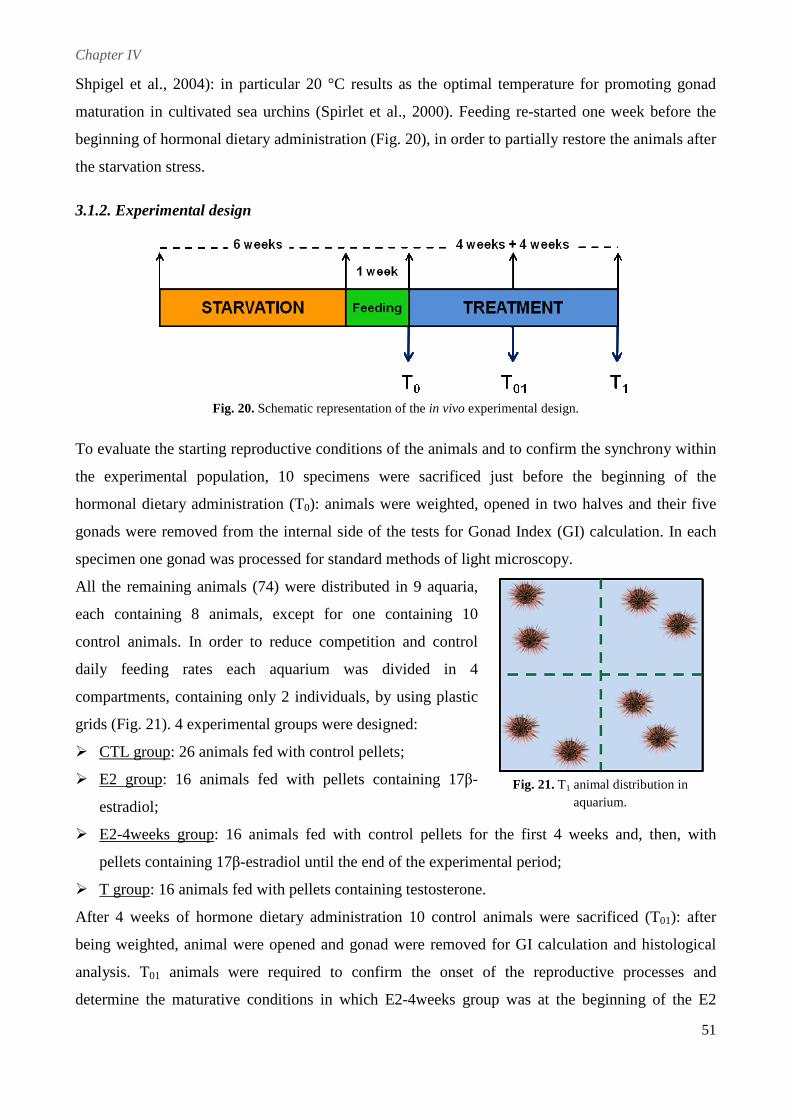
Chapter IV
51
Shpigel et al., 2004): in particular 20 °C results as the optimal temperature for promoting gonad
maturation in cultivated sea urchins (Spirlet et al., 2000). Feeding re-started one week before the
beginning of hormonal dietary administration (Fig. 20), in order to partially restore the animals after
the starvation stress.
3.1.2. Experimental design
Fig. 20. Schematic representation of the in vivo experimental design.
To evaluate the starting reproductive conditions of the animals and to confirm the synchrony within
the experimental population, 10 specimens were sacrificed just before the beginning of the
hormonal dietary administration (T0): animals were weighted, opened in two halves and their five
gonads were removed from the internal side of the tests for Gonad Index (GI) calculation. In each
specimen one gonad was processed for standard methods of light microscopy.
All the remaining animals (74) were distributed in 9 aquaria,
each containing 8 animals, except for one containing 10
control animals. In order to reduce competition and control
daily feeding rates each aquarium was divided in 4
compartments, containing only 2 individuals, by using plastic
grids (Fig. 21). 4 experimental groups were designed:
� CTL group: 26 animals fed with control pellets;
� E2 group: 16 animals fed with pellets containing 17β-
estradiol;
� E2-4weeks group: 16 animals fed with control pellets for the first 4 weeks and, then, with
pellets containing 17β-estradiol until the end of the experimental period;
� T group: 16 animals fed with pellets containing testosterone.
After 4 weeks of hormone dietary administration 10 control animals were sacrificed (T01): after
being weighted, animal were opened and gonad were removed for GI calculation and histological
analysis. T01 animals were required to confirm the onset of the reproductive processes and
determine the maturative conditions in which E2-4weeks group was at the beginning of the E2
Fig. 21. T1 animal distribution in aquarium.
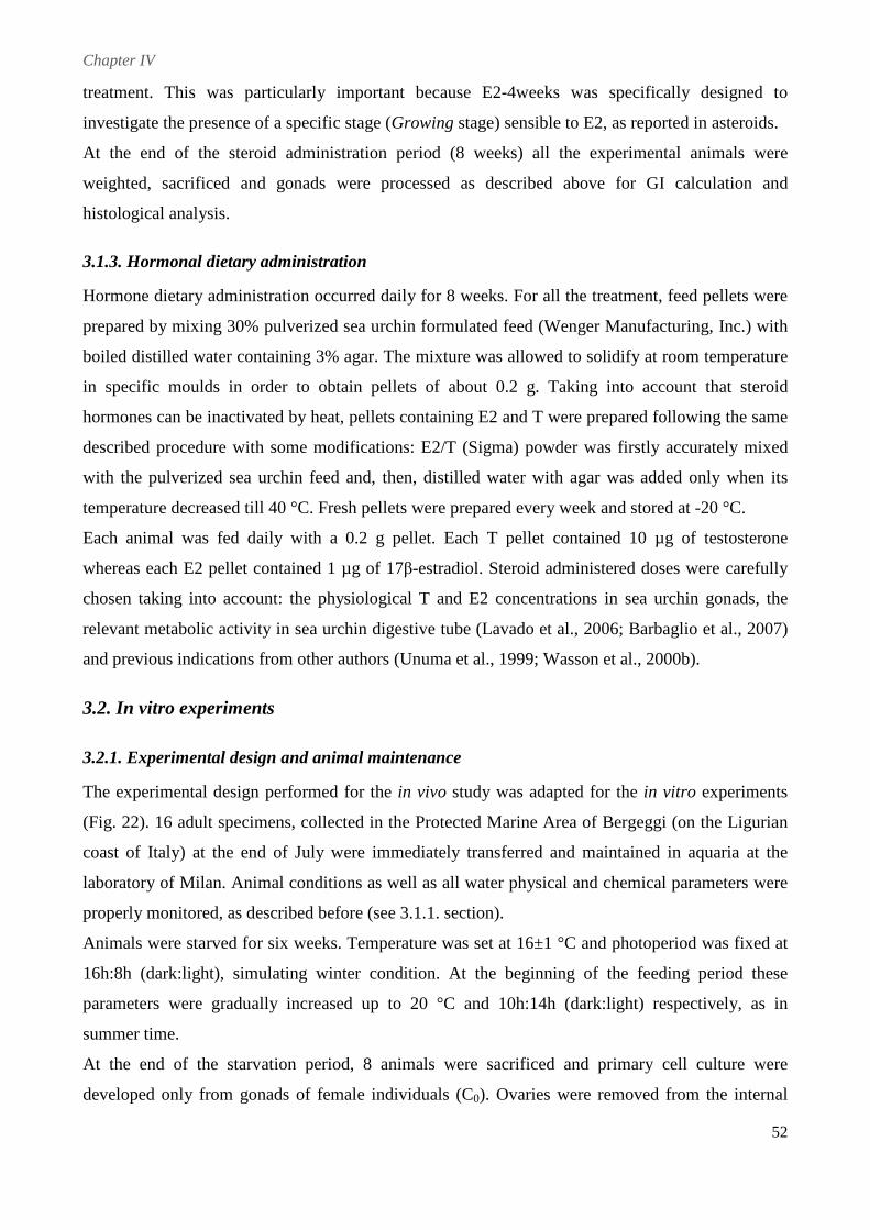
Chapter IV
52
treatment. This was particularly important because E2-4weeks was specifically designed to
investigate the presence of a specific stage (Growing stage) sensible to E2, as reported in asteroids.
At the end of the steroid administration period (8 weeks) all the experimental animals were
weighted, sacrificed and gonads were processed as described above for GI calculation and
histological analysis.
3.1.3. Hormonal dietary administration
Hormone dietary administration occurred daily for 8 weeks. For all the treatment, feed pellets were
prepared by mixing 30% pulverized sea urchin formulated feed (Wenger Manufacturing, Inc.) with
boiled distilled water containing 3% agar. The mixture was allowed to solidify at room temperature
in specific moulds in order to obtain pellets of about 0.2 g. Taking into account that steroid
hormones can be inactivated by heat, pellets containing E2 and T were prepared following the same
described procedure with some modifications: E2/T (Sigma) powder was firstly accurately mixed
with the pulverized sea urchin feed and, then, distilled water with agar was added only when its
temperature decreased till 40 °C. Fresh pellets were prepared every week and stored at -20 °C.
Each animal was fed daily with a 0.2 g pellet. Each T pellet contained 10 µg of testosterone
whereas each E2 pellet contained 1 µg of 17β-estradiol. Steroid administered doses were carefully
chosen taking into account: the physiological T and E2 concentrations in sea urchin gonads, the
relevant metabolic activity in sea urchin digestive tube (Lavado et al., 2006; Barbaglio et al., 2007)
and previous indications from other authors (Unuma et al., 1999; Wasson et al., 2000b).
3.2. In vitro experiments
3.2.1. Experimental design and animal maintenance
The experimental design performed for the in vivo study was adapted for the in vitro experiments
(Fig. 22). 16 adult specimens, collected in the Protected Marine Area of Bergeggi (on the Ligurian
coast of Italy) at the end of July were immediately transferred and maintained in aquaria at the
laboratory of Milan. Animal conditions as well as all water physical and chemical parameters were
properly monitored, as described before (see 3.1.1. section).
Animals were starved for six weeks. Temperature was set at 16±1 °C and photoperiod was fixed at
16h:8h (dark:light), simulating winter condition. At the beginning of the feeding period these
parameters were gradually increased up to 20 °C and 10h:14h (dark:light) respectively, as in
summer time.
At the end of the starvation period, 8 animals were sacrificed and primary cell culture were
developed only from gonads of female individuals (C0). Ovaries were removed from the internal
Chapter IV
53
side of the test: one gonad was processed for standard methods of light microscopy, whereas the
remaining 4 gonads were used to obtain primary cell cultures and test steroid exposure.
Fig. 22. Schematic representation of the in vitro experimental design.
The remaining 8 animals were arranged in an aquarium as described for T1 animals (Fig. 21) and
daily fed with control pellets. After 4 weeks of feeding (C01) the animals were sacrificed and
ovaries were used for cell culture development and histological analysis, as performed for C0
animals.
3.2.2. Cell cultures
Primary cell cultures from P. lividus ovaries were developed, as previously described in Chapter 3
with some modifications. Briefly, ovaries were washed in sterile Ca2+ Mg2+ Free Sea Water
(CMFSW) with antibiotics (40 µg/L gentamycin, 100 units/mL penicillin and 100 µg/mL
streptomycin). Ovary pieces were incubated in 0.5 mg/mL collagenase dissolved in sterile CMFSW
and stirred for 1 hour. The resulting cell suspension was centrifuged at 300 × g for 6’ at 15 °C and
the cell pellet was resuspended in modified Leibovitz-15 medium without supplement addition.
Ovarian cells, isolated from each female, were divided in 3 different experimental groups:
� CTL group: cells maintained in culture medium without hormones;
� E2 group: cells cultured in presence of 20 pg/mL 17β-estradiol;
� T group: cells cultured in presence of 200 pg/mL testosterone.
In order to analyze the E2 and T effects on cell morphology and viability, cells were seeded at a
concentration of 3-4 × 10⁵ cells/mL in 24-well culture plates, coated with poly-L-lysine (70-150
kDa, 0.01% solution, Sigma). Cultures were developed in replicates and incubated at 15 °C.
Replacement of 50% of the medium was carried out every two days. Cell behavior was observed
daily using an inverted phase contrast microscope . For the biochemical analyses, cells were seeded
at a concentration of 3 × 10⁶ cells/mL in 6-well culture plates, coated with poly-L-lysine, and
maintained at 15 °C for 24 h and 48 h.
Chapter IV
54
3.2.3. Chemicals and solution preparation
All chemicals were of reagent grade. 17-β estradiol and testosterone were purchased from Sigma.
E2 and T final exposure concentrations were determined on the basis of our previous original
results about sex-steroid physiological levels (unpublished data). Culture medium containing E2
was prepared as follows: 2 mg E2 were dissolved in 10 mL acetone (Merck), then 10 µL of this
solution was dissolved in 10 mL autoclaved artificial sea water. 10µL of this latter solution were
mixed with 10 mL of modified Leibovitz-15 medium obtaining a stock solution which was
subdivided in aliquots and maintained at -20 °C in the dark, in order to prevent steroid degradation.
Finally, to reach the final concentration of 20 pg/mL, 500 µL of the stock solution were dissolved in
4.5 mL of medium. Final acetone concentration was considered negligible (0.00001%). To prepare
the culture medium containing 200 pg/mL T, the same procedure was followed but 20 mg T were
used. Fresh culture medium with hormones was prepared every time. Stock solutions were prepared
every week.
3.3. Determination of reproductive stages
Reproductive stages were determined by histological analysis. Standard methods for light
microscopy (paraffin and/or resin) were employed.
3.3.1. Microscopic analysis
Gonads were fixed with Bouin (picric acid, formaldehyde, acetic acid, 75:25:5) for at least 24 h and
washed several times in tap water until all the fixative solution was completely removed. Samples
were then dehydrated with an ethanol series (70%, 90%, 95% and 100%), washed with xilene and
left overnight in a solution of xilene and paraffin 56–58 °C (1:1). Gonads were then washed three
times in paraffin and finally embedded. Longitudinal sections (5-7 µm) were cut with a Reichert
OmE sledge microtome and stained with Milligan’s Trichrome. Before staining, the sections were
washed in xilene and ethanol (100% and 95%) and placed into a solution of potassium dichromate
and hydrochloric acid. They were first stained with acid fuchsin, fixed with phosphomolybdic acid
(1%), stained with orange G and fast green FCF, and then treated with a solution of acetic acid 1%.
Finally, sections were cleared in xilene and dehydrated with ethanol (95% and 100%) before
mounting.
Both T0 and C0 samples were embedded in resin and processed for semithin sections (which provide
better quality and higher resolution) because difficulties in determining animal sex were expected
after the starvation period. Briefly, gonads were prefixed with 2% glutaraldehyde in 0.1 M
cacodylate buffer and NaCl 1.4% for 2 h and, after overnight washing in the same buffer, postfixed
with 1% solution of OsO4 in 0.1 M cacodylate buffer (2 h). After standard dehydration in an ethanol
Chapter IV
55
series (25%, 70%, 90% and 100%), the samples were washed in propylene oxide, left in propylene
oxide:resin (1:1) for 2 h. After overnight washing in resin they were embedded in Epon-Araldite
812 resin. The semithin (about 1 µm) sections were cut with a Reichert-Jung ULTRACUT E using
glass knives and stained with crystal violet and basic fuchsin.
Both resin and paraffin sections were observed and photographed under a Jenaval light microscope
to determine the gonad maturative stage. Five stages were considered: Spent (immediately after the
spawning event), Recovery (phagocitosis and nutrient accumulation phase), Growing, Premature
and Mature (progressive stages of gametogenesis).
3.3.2. Maturity Index and Gonad Index
Maturity Index (MI) was calculated as the mean value of the numerical maturative stages of each
experimental group based on histological analysis (Spent=0, Recovery=1, Growing=2,
Premature=3, Mature=4). The numerical maturative stage of each animal was corrected with +0,25
or -0,25 when in advanced or precocious conditions, respectively.
Gonad Index (GI) was determined as a percentage of the ratio between gonads wet weight (GW)
and total wet weight (TW):
GI = (GW/TW) 100
3.4. Electrophoresis
3.4.1. Sample preparation
The ovarian cells, cultured in 6-well culture plates, were processed for electrophoresis analysis after
24 and 48 hours of in vitro steroid exposure.
To prepared samples for running on gel, cells were gently scraped off the dish, using an ice-cold
plastic cell scraper. Cell suspension was transferred into a pre-cooled tube and centrifuged at 300 ×
g for 6’ at 4 °C; the resulting cell pellet was resuspended in 100 µL lysis buffer. Cells were lysed in
ice-cold 20 mM Tris-HCl (pH 7.5) with protease inhibitors (5 mM EDTA, 0.2 mM PMSF and 4
mM NEM), maintaining them in constant agitation for 30 minutes. Then, samples were centrifuge
at 12000 × g for 20’ at 4 °C and the supernatant was transferred into a fresh tube, kept on ice.
Finally, samples were dialyzed, protein assayed (BCA protein assay kit, Sigma) and stored at -20
°C until electrophoresis was performed.
3.4.2. SDS-PAGE
Sodium dodecylsulfate-polyacrilamide gel electrophoresis (SDS-PAGE) was performed using 6%
slab gel (Laemmli, 1970) in order to verify and compare the sample protein content in C0 and C01
Chapter IV
56
experimental groups (CTL, E2 and T). Samples containing 10 µg of total proteins were diluted with
sample buffer (SDS reducing buffer), boiled for 5 minutes and, then, applied to each lane. Gel were
run at 100 V at room temperature. Protein bands were visualized with Coomassie brilliant blue R-
250. SDS-PAGE standards (StoS Protein Marker, Genespin s.r.l.) were also run for molecular
weight calibration.
3.5. Statistical analysis
Results are presented as mean values±SEM. Statistical significance was assessed using one-way
ANOVA (Tukey’s test) and χ2 test. A p-value of less than 0.05 was considered statistically
significant. Statistical analysis was performed by the computer program GraphPad Prism 4.
4. Results
4.1. Animal health conditions
During both the in vivo and the in vitro experimental periods, aquaria physical and chemical
parameters were properly monitored and promptly adjusted, when necessary. All the animals
appeared healthy and no mortality event was observed throughout the experiments. As far as the
steroid dietary administration is concerned, animal daily feeding rate, calculated as mean percentage
of the eaten pellets, was close to 100% for all the experimental groups (CTL: 99.4%; E2: 99.6%; T:
99.7%; E2-4weeks: 100%), ensuring that daily E2 and T administration correctly occurred.
4.2. In vivo experiment
4.2.1. Sex ratio
Comparing sex ratio values (Fig. 23), no statistically significant difference was found among all the
experimental groups (χ2 test: P>0.05). T0, T01 and all the hormonally treated groups displayed
similar frequencies: males were ∽40% (T0: 50%; T01: 40%; E2: 37.5%; T: 43.75%; E2-4weeks:
31.25%) and females were ∽60% (T0: 50%; T01: 60%; E2: 62.5%; T: 56.25%; E2-4weeks: 68.75%)
in each group. Only in CTL group, the relative frequencies of males and females were quite
different from the others, showing 68.75% males and 31.25% females.
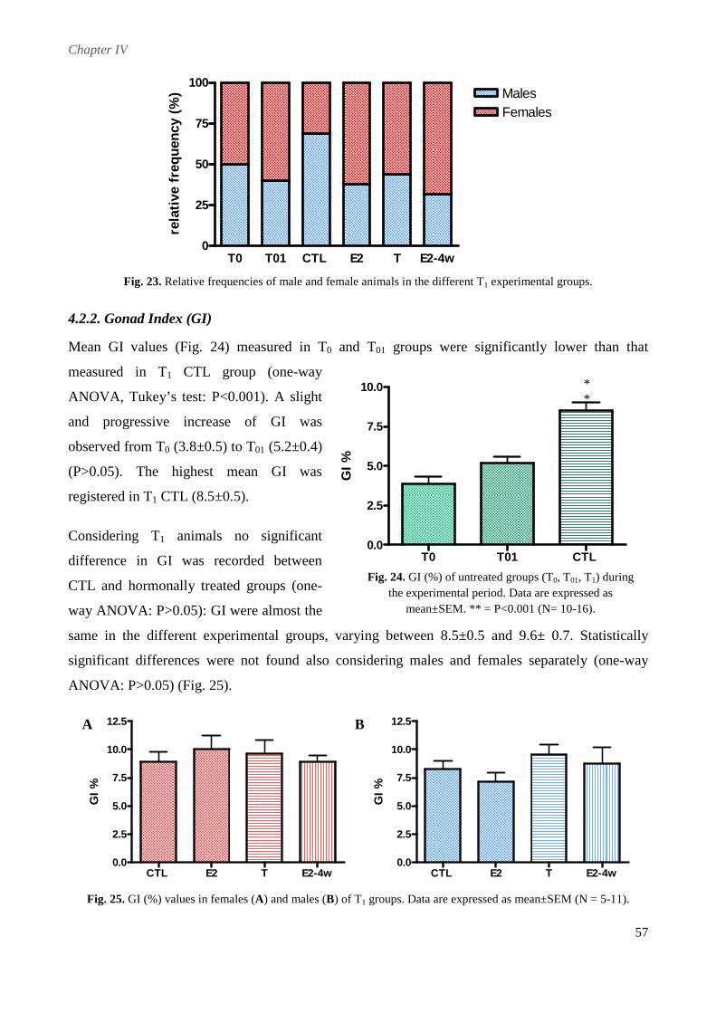
Chapter IV
57
T0 T01 CTL E2 T E2-4w0
25
50
75
100MalesFemales
rela
tive
fre
que
ncy
(%)
Fig. 23. Relative frequencies of male and female animals in the different T1 experimental groups.
4.2.2. Gonad Index (GI)
Mean GI values (Fig. 24) measured in T0 and T01 groups were significantly lower than that
measured in T1 CTL group (one-way
ANOVA, Tukey’s test: P<0.001). A slight
and progressive increase of GI was
observed from T0 (3.8±0.5) to T01 (5.2±0.4)
(P>0.05). The highest mean GI was
registered in T1 CTL (8.5±0.5).
Considering T1 animals no significant
difference in GI was recorded between
CTL and hormonally treated groups (one-
way ANOVA: P>0.05): GI were almost the
same in the different experimental groups, varying between 8.5±0.5 and 9.6± 0.7. Statistically
significant differences were not found also considering males and females separately (one-way
ANOVA: P>0.05) (Fig. 25).
CTL E2 T E2-4w0.0
2.5
5.0
7.5
10.0
12.5
GI
%
CTL E2 T E2-4w
0.0
2.5
5.0
7.5
10.0
12.5
GI
%
Fig. 25. GI (%) values in females (A) and males (B) of T1 groups. Data are expressed as mean±SEM (N = 5-11).
T0 T01 CTL 0.0
2.5
5.0
7.5
10.0
GI
%
* *
Fig. 24. GI (%) of untreated groups (T0, T01, T1) during the experimental period. Data are expressed as
mean±SEM. ** = P<0.001 (N= 10-16).
A B
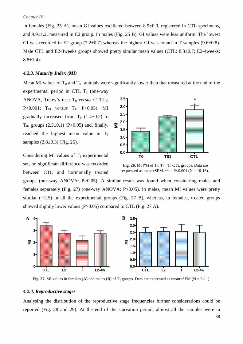
Chapter IV
58
In females (Fig. 25 A), mean GI values oscillated between 8.9±0.9, registered in CTL specimens,
and 9.9±1.2, measured in E2 group. In males (Fig. 25 B), GI values were less uniform. The lowest
GI was recorded in E2 group (7.2±0.7) whereas the highest GI was found in T samples (9.6±0.8).
Male CTL and E2-4weeks groups showed pretty similar mean values (CTL: 8.3±0.7; E2-4weeks:
8.8±1.4).
4.2.3. Maturity Index (MI)
Mean MI values of T0 and T01 animals were significantly lower than that measured at the end of the
experimental period in CTL T1 (one-way
ANOVA, Tukey’s test: T0 versus CTLT1:
P<0.001; T01 versus T1: P<0.05). MI
gradually increased from T0 (1.4±0.2) to
T01 groups (2.3±0.1) (P>0.05) and, finally,
reached the highest mean value in T1
samples (2.8±0.3) (Fig. 26).
Considering MI values of T1 experimental
set, no significant difference was recorded
between CTL and hormonally treated
groups (one-way ANOVA: P>0.05). A similar result was found when considering males and
females separately (Fig. 27) (one-way ANOVA: P>0.05). In males, mean MI values were pretty
similar (∽2.5) in all the experimental groups (Fig. 27 B), whereas, in females, treated groups
showed slightly lower values (P>0.05) compared to CTL (Fig. 27 A).
CTL E2 T E2-4w0
1
2
3
4
MI
CTL E2 T E2-4w0.0
0.5
1.0
1.5
2.0
2.5
3.0
3.5
MI
Fig. 27. MI values in females (A) and males (B) of T1 groups. Data are expressed as mean±SEM (N = 5-11).
4.2.4. Reproductive stages
Analysing the distribution of the reproductive stage frequencies further considerations could be
reported (Fig. 28 and 29). At the end of the starvation period, almost all the samples were in
T0 T01 CTL 0.0
0.5
1.0
1.5
2.0
2.5
3.0
3.5
MI
* *
Fig. 26. MI (%) of T0, T01, T1 CTL groups. Data are expressed as mean±SEM. ** = P<0.001 (N = 10-16).
A B
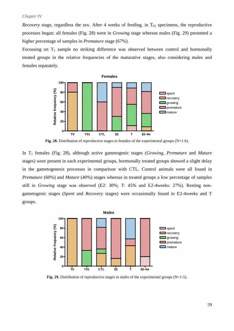
Chapter IV
59
Recovery stage, regardless the sex. After 4 weeks of feeding, in T01 specimens, the reproductive
processes began: all females (Fig. 28) were in Growing stage whereas males (Fig. 29) presented a
higher percentage of samples in Premature stage (67%).
Focussing on T1 sample no striking difference was observed between control and hormonally
treated groups in the relative frequencies of the maturative stages, also considering males and
females separately.
Females
T0 T01 CTL E2 T E2-4w0
20
40
60
80
100
spentrecoverygrowingprematuremature
Rel
ativ
e fr
eque
ncy
(%
)
Fig. 28. Distribution of reproductive stages in females of the experimental groups (N=1-6).
In T1 females (Fig. 28), although active gametogenic stages (Growing, Premature and Mature
stages) were present in each experimental groups, hormonally treated groups showed a slight delay
in the gametogenesis processes in comparison with CTL. Control animals were all found in
Premature (60%) and Mature (40%) stages whereas in treated groups a low percentage of samples
still in Growing stage was observed (E2: 30%; T: 45% and E2-4weeks: 27%). Resting non-
gametogenic stages (Spent and Recovery stages) were occasionally found in E2-4weeks and T
groups.
Males
T0 T01 CTL E2 T E2-4w0
20
40
60
80
100
spentrecoverygrowingprematuremature
Rel
ativ
e fr
equ
ency
(%
)
Fig. 29. Distribution of reproductive stages in males of the experimental groups (N=1-5).
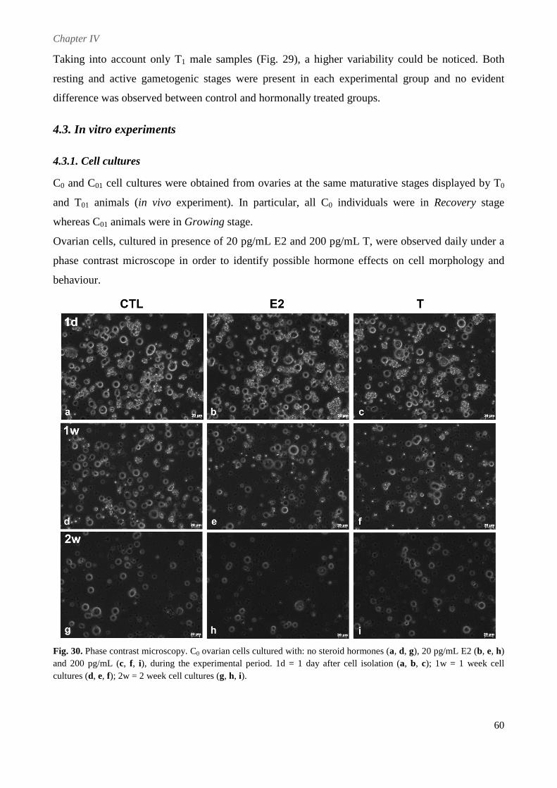
Chapter IV
60
Taking into account only T1 male samples (Fig. 29), a higher variability could be noticed. Both
resting and active gametogenic stages were present in each experimental group and no evident
difference was observed between control and hormonally treated groups.
4.3. In vitro experiments
4.3.1. Cell cultures
C0 and C01 cell cultures were obtained from ovaries at the same maturative stages displayed by T0
and T01 animals (in vivo experiment). In particular, all C0 individuals were in Recovery stage
whereas C01 animals were in Growing stage.
Ovarian cells, cultured in presence of 20 pg/mL E2 and 200 pg/mL T, were observed daily under a
phase contrast microscope in order to identify possible hormone effects on cell morphology and
behaviour.
Fig. 30. Phase contrast microscopy. C0 ovarian cells cultured with: no steroid hormones (a, d, g), 20 pg/mL E2 (b, e, h) and 200 pg/mL (c, f, i), during the experimental period. 1d = 1 day after cell isolation (a, b, c); 1w = 1 week cell cultures (d, e, f); 2w = 2 week cell cultures (g, h, i).
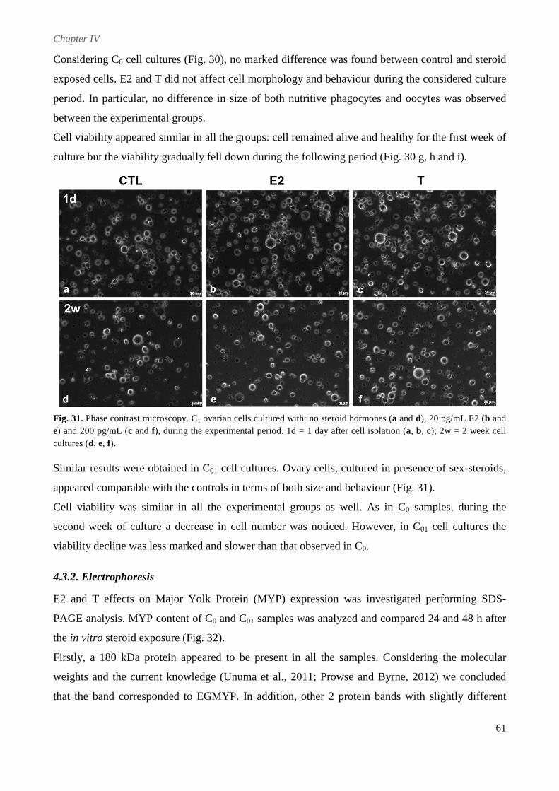
Chapter IV
61
Considering C0 cell cultures (Fig. 30), no marked difference was found between control and steroid
exposed cells. E2 and T did not affect cell morphology and behaviour during the considered culture
period. In particular, no difference in size of both nutritive phagocytes and oocytes was observed
between the experimental groups.
Cell viability appeared similar in all the groups: cell remained alive and healthy for the first week of
culture but the viability gradually fell down during the following period (Fig. 30 g, h and i).
Fig. 31. Phase contrast microscopy. C1 ovarian cells cultured with: no steroid hormones (a and d), 20 pg/mL E2 (b and e) and 200 pg/mL (c and f), during the experimental period. 1d = 1 day after cell isolation (a, b, c); 2w = 2 week cell cultures (d, e, f).
Similar results were obtained in C01 cell cultures. Ovary cells, cultured in presence of sex-steroids,
appeared comparable with the controls in terms of both size and behaviour (Fig. 31).
Cell viability was similar in all the experimental groups as well. As in C0 samples, during the
second week of culture a decrease in cell number was noticed. However, in C01 cell cultures the
viability decline was less marked and slower than that observed in C0.
4.3.2. Electrophoresis
E2 and T effects on Major Yolk Protein (MYP) expression was investigated performing SDS-
PAGE analysis. MYP content of C0 and C01 samples was analyzed and compared 24 and 48 h after
the in vitro steroid exposure (Fig. 32).
Firstly, a 180 kDa protein appeared to be present in all the samples. Considering the molecular
weights and the current knowledge (Unuma et al., 2011; Prowse and Byrne, 2012) we concluded
that the band corresponded to EGMYP. In addition, other 2 protein bands with slightly different
Chapter IV
62
molecular weights were found. The 170 kDa protein band could correspond to CFMYP (Unuma et
al., 2011).
Fig. 32. 6% SDS-PAGE analysis of C0 and C01 cell extracts.
Comparing C0 experimental groups, no difference in EGMYP expression was observed in cells
exposed for both 24 and 48 hours. Steroids did not affect EGMYP content in C01 ovary cells as
well: in E2 and T samples the intensity of MYP band was comparable to the control (Fig. 32).
5. Discussion
In this study E2 and T involvement in echinoid reproduction was explored applying both an in vivo
and in vitro approach.
Concerning the in vivo study, a long-term experiment of steroid dietary administration was
performed in adult specimens of P. lividus and different reproductive parameters were investigated.
The experiment was successfully designed in order to synchronize the starting maturative
conditions of the animals and reduce as much as possible the variability present in field population.
Animals were divided in 4 groups: CTL, E2, T and E2-4 weeks. In particular, the latter was chosen
in order to verify the existence of a specific maturative stage sensitive to E2, as proposed in
different asteroid species. In fact, in both A. rubens and A. pectnifera E2 seemed to enhance oocyte
growth only during vitellogenesis, suggesting the presence of a threshold oocyte size for E2
effectiveness (Schoenmakers et al., 1981; Takahashi and Kanatani, 1981). To investigate the
existence of similar mechanisms in echinoids, E2-4weeks specimens were fed with control pellets
Chapter IV
63
for 4 weeks and then treated with E2, i.e. after they reached the Growing stage during which
gametogenesis processes began.
The nutritional conditions of the animals resulted to be optimal: in fact a progressive increase of GI
mean values was observed from starved animals (T00) to sea urchins well-fed for 4 (T01) and 8
weeks (T1). It is well documented that quantity and quality of food influence gonad size and the
amount of gametes produced, thus playing a very important role in the regulation of the
reproductive cycle (Pearse and Cameron, 1991; Spirlet et al., 1998). MI values of control groups
(T0, T01 and CTL) displayed a marked and progressive increase throughout the experimental period
as well. Therefore, despite the stress caused by the maintenance conditions, after starvation
gametogenesis processes restarted and sea urchins progressively reached higher maturative stages.
These results underlined the success of the experimental design and confirmed the presence of
active gametogenesis processes during the hormonal treatment.
E2 and T dietary administration did not affect animal health conditions and the tested steroid
concentrations did not cause acute toxicity effects. No mortality event occurred and all sea urchins
appeared healthy throughout the experimental period.
The sex ratio was close to 1.5:1 (females:males) in most of the experimental groups. This value,
although slightly different from data reported for field specimens (Pearse and Cameron, 1991), was
completely in agreement with our previous observations in the population of P. lividus considered
in this work (unpublished data). Focussing on E2 and T treatment, steroid dietary administration did
not influence sex ratio: males and females frequencies found in controls and treated groups were
pretty similar with the only exception being representing by the CTL animals that displayed a
higher percentage of male individuals. This result was probably due to natural variability and did
not seem to be related to hormonal treatment. Indeed, in all the other control groups (T0 and T01)
sex ratio values were similar to those observed in T1 treated groups.
In vertebrates, exposure/administration of E2 and T induces sex-reversal phenomena during early
stages (i.e. during sexual differentiation), even in those classes (such as fish, amphibians, birds, etc.)
with a genetic sex determination mechanism. Sex-reversal of adult specimens is very rare and it is
reported only for those species, mainly fishes, which are naturally “programmed” to change sex
during their life (hermaphroditic species) (Gutzke and Bull, 1986; Baroiller et al., 1999; Nakamura,
2010). The lack of T and E2 influence on sex ratio on our experimental animals was therefore quite
expected (although not excluded a priori) since in the present research we employed adult sea
urchins which had already undergone sex differentiation. As almost all sea urchins, P. lividus is a
gonochoristic species without marked sexual dimorphism (Swann, 1954). Sex-determination
Chapter IV
64
mechanisms are scarcely understood, although some authors suggest a genotypic basis (Delavault,
1966; Lipani et al., 1996).
Considering the analyzed reproductive parameters, E2 and T administration did not affect gonad
growth, expressed as GI, in both males and females. Sex-steroid exogenous administration led to
different and contrasting results on this parameter in both asteroids and echinoids (Schoenmakers et
al., 1981; Van der Plas et al., 1982; Barker and Xu, 1993; Unuma et al., 1999; Wasson et al.,
2000b). Particularly, E2 seemed to stimulate ovarian growth in the starfish A. rubens and in the sea
urchin L. variegatus whereas in the other studied species no effect was reported. In asteroids,
estrogens appeared to be more involved in oocyte maturation and growth: an E2 induced increase of
oocyte diameter in several species and a hormone involvement in vitellogenesis processes was
strongly suggested (Schoenmakers et al., 1981; Takahashi and Kanatani, 1981; Barker and Xu,
1993). As far as T is concerned, in echinoids no effect on GI was observed in several species. In P.
depresuss an increase of testis size was obtained only after the administration of another androgen,
androstenedione, whereas T was found to promote oocyte growth in L. variegatus (Unuma et al.,
1999; Wasson et al., 2000b). These heterogeneous results could be explained in the light of the
existence of species-specific hormonal mechanisms; on the other hand the different experimental
conditions (treatment length, steroid concentrations, etc.) could have affected the experimental
results and therefore have to be carefully considered.
Analysing the mean maturative state, i.e. the Maturity Index (MI) reached by the experimental
groups of the present study, neither E2 nor T treatment affected gonad maturation, even when
considering males and females separately. To better investigate this reproductive aspect, the relative
frequencies of male and female reproductive stages were analyzed. Also in this case, no striking
variations were observed, although all hormonally treated female animals showed a slight delay in
the gametogenesis processes, that, however, seemed to be less affected by hormonal treatment than
by the low number of samples per group (P. lividus specimens have no evident sexual dimorphism
and so it is not possible to equally distribute males and females in each experimental group). Thus,
E2 did not apparently induce oocytes maturation and no temporal window of E2 sensitivity was
found, as, on the contrary, was observed in starfishes (Schoenmakers et al., 1981; Takahashi and
Kanatani, 1981; Barker and Xu, 1993). It has been proposed that this different results could be due
to class-specific mechanisms: echinoids and asteroids could simply have a different hormonal
regulation of oocytes development and growth. This hypothesis was supported by Wasson et al.
(2000b) who reported an inhibited growth of individual oocytes after dietary administration of E2 in
the echinoid L. variegatus. This findings however disagreed with our results: neither E2 nor T
affected reproductive parameters. Furthermore, contrasting results were reported for other echinoid
Chapter IV
65
species. In S. intermedius and S. nudus the administration of estradiol dipropionate stimulated
gonad development and gamete growth (Varaksina and Varaksin, 2001; Varaksina and Varaksin,
2002), whereas no response to E2 and T was observed in P. depressus yearling females (Unuma et
al., 1999). The high variability of responses to steroid treatment observed in echinoids could be
reasonably related to the different experimental conditions. In particular, steroid hormone
administration (dietary or injections), treatment period (days or weeks), hormone doses and
individual differences (age, species, reproductive state of gonads) could in fact strongly influenced
the results. Our experiment was designed in order to reduce as much as possible individual
variability and, most of all, to synchronize animal reproductive conditions. In this way the results
should be less affected by the heterogeneity present in field population. The administered steroid
concentrations were carefully chosen taking into account the physiological T and E2 concentrations
in sea urchin gonads, the high metabolic activity present in sea urchin digestive tube (Lavado et al.,
2006; Barbaglio et al., 2007) and also previous results obtained by other authors (Unuma et al.,
1999; Wasson et al., 2000b). In view of these considerations, our results can be considered realistic
and reliable.
However, in order to further confirm these findings and deepen the possible E2 and T involvement
in echinoid reproduction we additionally performed in vitro exposure experiments. Although the in
vitro conditions are obviously different and far from the in vivo model, cell cultures allow studies
under controlled experimental conditions without most of the metabolic interferences that can occur
in animals. Ovarian cells were cultured for 2 weeks in presence of physiological levels of E2 and T
in order to investigate possible morpho-functional effects. Primary cell cultures were developed
from gonads at two different starting maturative conditions (C0: Recovery stage; C01: Growing
stages) in order to make the results comparable with those obtained in the experiments of steroid
dietary administration.
Analysing cell morphology and behaviour no variation was observed between controls and
hormonally exposed groups in either C0 or C01 cultures. The size of nutritive phagocytes and
oocytes was not affected by E2 and T exposure throughout the experimental period as, on the
contrary, was reported in many asteroid and echinoid species (Schoenmakers et al., 1981; Takahashi
and Kanatani, 1981; Wasson et al., 2000b). Nevertheless, most of the studies were performed with
an in vivo approach and only one study based on in vitro E2 exposure work was attempted. In A.
pectinifera, fragments of ovary were cultured for 3 days in medium containing 10-6 M E2 and a
significant increase of mean oocyte diameter was observed (Takahashi and Kanatani, 1981). These
contrasting results could be due to the different type of culture performed (organ cultures vs and
Chapter IV
66
primary cell cultures) or, as previously proposed, to the existence of specie-specific differences, the
employment of different steroid concentrations and diverse experimental designs.
To better understand steroid possible involvement in sea urchin physiology and reproductive
biology further investigations were performed in vitro concerning E2 and T role in the regulation of
the Major Yolk Protein (MYP) expression. Indeed, estrogen control of MYP expression was firstly
proposed by Shyu et al., (1987), who found a palindromic sequence similar to the estrogen-
responsive element upstream MYP gene. MYP is a metal-binding glycoprotein belonging to the
transferrin superfamily (Noll et al., 2007; Unuma et al., 2011) and in vertebrates transferrin genes
are regulated by estrogens (Prowse and Byrne, 2012). Considering echinoderm phylogenetic
position (close to chordates, including vertebrates) the existence of common hormonal mechanisms
can reasonably be proposed.
SDS-PAGE analysis of cell extracts was performed in control and hormonally exposed samples but
no difference was observed between the experimental groups. Our results were in agreement with
those reported in S. nudus larvae and juveniles: E2 did not affect MYP expression in all the
considered embryo stages. On the contrary, estrone (E1) seemed to be more involved in the protein
expression, playing a suppressive role during specific developmental stages (Kiyomoto et al., 2008).
Nevertheless, these results were obtained in larvae and post-metamorphosis juveniles, i. e. stages
very different from the adult condition in which sexual development and reproductive activities are
not yet established. Furthermore, our data could be influenced by the in vitro condition and no
analysis was performed on either MYP mRNA levels or culture medium protein content. In the sea
cucumber Apostichopus japonicus, although the intensity of MYP band in SDS-PAGE gel remained
stable throughout ovarian development, a clear increase of MYP transcription was observed. The
inconsistency of the levels of MYP mRNA and MYP protein was proposed to be due to MYP
delivery from the ovary to other animal regions, such as coelomic fluids, or to its rapid consumption
(Fujiwara et al., 2010). Taking into account all these considerations further research are surely
needed to confirm our results: steroid involvement in protein expression should be deeply
investigated expanding the target organs (MYP is mainly synthesized in both gonads and digestive
tube) and employing different technical and methodological approaches. This is further supported
by previous studies, reporting steroid involvement in protein synthesis: in fact E2 and T
administration was demonstrated to enhance the rate of protein synthesis in both asteroid (Barker
and Xu, 1993) and echinoid gonads (Varaksina and Varaksin, 2001; Varaksina and Varaksin, 2002)
and E2 stimulation on protein synthesis was also observed in Strongylocentrotus purpuratus and
Dendraster excentricus coelomocytes (Harrington and Ozaki, 1986).
Chapter IV
67
In conclusion, E2 and T did appear to not markedly affect P. lividus reproductive biology either in
vivo or in vitro. Hormonal dietary administration did not induce striking variations in the considered
reproductive parameters and no effect was observed in ovarian cell cultures exposed to hormone
physiological concentrations.
Overall, the present research provided new information on sex-steroid implications in echinoderm
reproductive processes. Indeed, according to the obtained results, E2 and T do not markedly
influence echinoid gonad maturation and, particularly, they do not promote gamete maturation, as
on the contrary reported in vertebrates (Lange et al., 2002). In addition, no specific maturative stage
sensitive to E2 was found, suggesting the existence of different hormonal mechanisms controlling
these processes in asteroids and echinoids. However, an indirect involvement of steroids and, in
particular, of E2 in echinoderm reproduction can not be completely excluded and its role in the
regulation of lipid metabolism and protein synthesis during the different reproductive stages should
be strongly considered, as suggested by several other authors (Wasson et al., 2000b; Varaksina and
Varaksin, 2001; Varaksina and Varaksin, 2002).

Chapter V
GENERAL DISCUSSION
&
FUTURE PROSPECTIVES
Chapter V
69
The present research project was designed in order to explore different aspects of sex-steroid
involvement in echinoid reproductive biology by performing both in vivo and in vitro experiments.
The employment of these different and, at the same time, complementary approaches allowed us to
provide a better understanding of sea urchin endocrinology and help to elucidate E2 and T role in
echinoderm reproduction.
Furthermore, we set up new and important experimental tools. Indeed, at our knowledge, we were
the first to develop effective primary cell cultures from sea urchin ovaries and apply them for in
vitro exposure tests. On the other hand, in the in vivo model, we optimized an innovative
experimental design that gave the opportunity to study the effects induced by exogenous
administration without risking that individual variability affected our final results.
Thus, in this research, we provided not only new information on echinoid reproduction but also new
and successful methodological approaches, that could be useful for expanding the potential
employment of echinoderms in experimental research and, in particular, for investigating further
endogenous factors involved in the regulation of echinoderm reproductive processes.
1. Development of primary cell cultures
Considering the importance and possible applications of the in vitro studies, we developed primary
cell cultures from sea urchin ovaries. Heterogeneous cultures, composed by both nutritive
phagocytes and germ cells were preferred as nutritive phagocytes play important and multiple roles,
providing support, protection and nutriments to the growing germinal cells (Chia and Bickell, 1983;
Walker et al., 2000). Considering the lack of knowledge about germ cell requirements and the need
to maintain cells in healthy conditions, heterogeneous cultures appeared more suitable, at least in
this starting phase of the research. However, the two cellular phenotypes present in culture were
well recognizable thanks to their overall morphology and behaviour and, thus, this heterogeneity
did not cause difficulties in the in vitro analyses.
Contamination is a central aspect of the in vitro studies and it has often represented an important
obstacle in the development of effective cell cultures and even cell lines from a variety of marine
organisms (Rinkevich, 1999). In the present research, contamination occurred at a very low rate
and, in most of the cases, bacteria and moulds were the main responsibles. This was probably due to
the combination of two main aspects: the addition of antibiotics in culture medium and the presence
of natural antibacterial compounds in sea urchin gonads/gametes. In fact, in different echinoderm
species both seminal plasma and eggs were found to exert an antibacterial activity and, particularly,
a lysozyme-like activity seemed to be involved (Stabili and Canicattì, 1994; Stabili and Pagliara,
1994). This specific gonad feature, together with antibiotic and antimycotic (gentamycin,
Chapter V
70
streptomycin and penicillin) addition, surely helped to maintain bacterial contaminations at a very
low level and to drastically reduce mould occurrence.
Ovarian cell culture were produced from ovaries at all stages of maturation but a higher survival
rate and a more lively behaviour was observed when ovaries at early stages of gametogenesis
(Growing and Premature stages) were used. This attitude was noticed also during the sex-steroid
exposure experiments (Chapter IV). C0 cell cultures, obtained from ovary in resting non-
gametogenic stages, displayed a cell viability decrease during the second week of culture that was
less evident and slower in C01 cells, isolated from ovaries at Growing stage. It has to be underlined
that in the in vitro E2 and T exposure experiments no supplements were added to the culture
medium in order not to affect biochemical analyses. Since the exposure period was only 2 weeks
the lack of supplements was not considered determinant and a general decrease in cell viability,
during the second week, was expected. What is really interesting is the different viability showed by
C0 and C01 cell cultures, probably due to the maturative stage of the ovaries from which they were
derived. In fact, during gametogenic stages nutritive phagocytes appear voluminous and full of
inclusions, containing nutritive substances and, probably, molecular factors required for oocyte
growth and maturation (Chatlynne, 1969; Houk and Hinegardner, 1980; Chia and Bickell, 1983;
Spirlet et al., 1998). Thus, gonad starting conditions affected primary cell cultures in terms of both
viability and behaviour and gametogenic ovaries appeared to be more suitable, containing probably
specific growth factors and nutrients required for cell health.
Investigations on the proper culture medium showed that modified Leibovitz-15 medium is the
most suitable for sea urchin ovarian cells. The effectiveness of this medium has been previously
reported for other echinoderm and marine invertebrate species (Moss et al., 1998; Odintsova et al.,
2005; Shashikumar and Desai, 2011), suggesting that its composition is particularly appropriate for
marine invertebrate cells since it probably provides most of the nutrients and molecules required for
cell growth and survival.
Moving to cell-substrate interactions, nutritive phagocytes and germinal cells appeared
differentially sensitive to the tested substrates. Nutritive phagocytes partially adhered on different
substrates and even on uncoated culture plates, tending to produce long filopodia and forming
networks. Their attitude in migrating and contacting each other was probably related to their
functional state: it appeared more evident when cultures were obtained from gonads in advanced
maturative stages. Oocytes and oogonia did not adhered on any tested substrates with the only
exception of poly-L-lysine. In particular, poly-L-lysine was found to be the only analyzed substrate
Chapter V
71
allowing complete adherence of both nutritive phagocytes and oocytes. Indeed, poly-L-lysine was
reported to be one of the best substrates for echinoderm embryonic cells (Odintsova et al., 1994)
and for cells isolated from holothurian gut (Odintsova et al., 2005). Although poly-L-lysine
effectiveness was determined and previously suggested for echinoderm cells, no improvement in
cell survival and growth was observed. To enhance cell migration and viability other possible
substrates could be investigated: laminin and fibronectin seem to be the most valid candidates
(Spiegel et al., 1983; Venkatasubramanian and Solursh, 1984; Odintsova et al., 1994; Moss et al.,
1998). In addition, echinoderm fibrillar collagen, directly extracted from sea urchin tissues, could
represent another promising solution (work in progress).
To improve cell conditions and stimulate proliferation different embryo extracts were tested.
Considering the poor results obtained with the addition of FCS, a widely used serum-supplement in
primary cell cultures from other echinoderms (Odintsova et al., 2005; Sharlaimova et al., 2010), we
moved to more species-specific extracts. P. lividus PE and EE were specifically developed and their
addition to the culture medium was deeply investigated at different concentrations. PE did not
enhance cell growth and survival at all the tested concentrations and it even seemed to accelerate
processes of cell death. Although poor results were observed with this extract, it is possible that
further manipulations and purifications may be necessary before conclusions can be drawn about its
effectiveness. On the other hand, EE appeared to slightly improve cell health conditions, finally
allowing to maintain ovarian cultures for up to one month. This marked difference between PE and
EE effectiveness could be related to the different MYP isoforms contained in the extracts: EGMYP
was present in EE whereas CFMYP was found in PE. The egg isoform was reported to play a main
role in reproduction functioning as a nutritive supply for both gametogenesis and developing
embryos (Unuma et al., 2007; Unuma et al., 2010). On the contrary, CFMYP was observed to play
multiple functions and, particularly, PE detrimental effects on ovarian cells could be due to its
involvement in sea urchin immune response (Cervello et al., 1994). However, SDS-PAGE analyses
showed that PE and EE deeply differed in their protein profiles, suggesting that their different
effects could be simply due to their protein compositions.
Although we set up the basis for the successful development of primary cell cultures from sea
urchin ovaries, other research are strongly recommended to optimize cell growth conditions and
promote cell proliferation. In particular, other supplements and growth factors should be tested. The
testicular cells isolated from the crab Scylla serrata were successfully cultured in L-15 but the
addition of epidermal growth factor (EGF) was required to promote good proliferation and extend
cell survivability (Shashikumar and Desai, 2011). The results exhibited by this study further
underlined the necessity to test even factors known to be active in vertebrate systems, as EGF or
Chapter V
72
other specific hormones and neuropeptides. However, the lack of effect by this kind of factors is not
uncommon in invertebrate studies (Moss et al., 1998) and the employment of species-specific or
even tissue-specific extracts could represent a promising alternative. In echinoderms, different
studies have demonstrated the higher mitotic activity displayed by cell cultures produced from
regenerating tissues (Odintsova et al., 2005; Sharlaimova et al., 2010), suggesting the presence of
molecules and factors specifically involved in proliferation induction. The regenerating conditions
could be used to improve other kinds of primary cell cultures. For examples, in our case, ovarian
cell proliferation could be stimulated by the addition of inactivated cell-free coelomic fluid,
previously collected from regenerating individuals.
Overall, primary cell cultures from sea urchin ovaries were successfully developed and maintained
for up to one month. However, further studies are surely necessary and strongly suggested. Future
research should be firstly addressed to identify supplements and growth factors able to optimize cell
culture conditions, improving cell growth and stimulating cell proliferation. In addition, to achieve
the optimal in vitro conditions other substrates could be investigated and, particularly, fibrillin and
echinoderm collagen should be taken in consideration. Finally, mono-phenotype cell cultures from
sea urchin ovaries could be developed, employing both size filtration and a density gradient
techniques, as already described for oogonia isolation in the sea urchin Strongylocentrotus nudus
(Yakovlev et al., 2010).
2. Sex-steroid involvement in echinoid reproduction
Sex-steroid involvement in echinoderm reproductive biology has been proposed in several species.
Most of the studies were performed in asteroids, where these hormones seemed to control
vitellogenesis and gamete maturation (Schoenmakers et al., 1981; Xu and Barker, 1990; Hines et
al., 1992a). In echinoids, only few studies have been performed, reporting different and, sometimes,
even contrasting results (Unuma et al., 1999; Wasson et al., 2000b; Barbaglio et al., 2007).
Considering the poor information about the mechanisms regulating sea urchin reproductive
processes, the role of E2 and T in echinoid reproduction was firstly investigated performing a long-
term experiment of sex-hormone dietary administration in the common sea urchin P. lividus.
Analyzing our experimental results, neither E2 nor T administration induced marked variations in
the considered reproductive parameters, suggesting that these hormones are not involved in sea
urchin gametogenesis processes. Similar results were also obtained in the in vitro exposure
experiments: P. lividus ovarian cells were exposed to physiological concentrations of E2 and T for 2
weeks but no effect on cell morphology or behavior was observed. These findings strongly contrast
with what has been reported in several asteroid species (Schoenmakers et al., 1981; Takahashi and
Chapter V
73
Kanatani, 1981; Voogt et al., 1991; Hines et al., 1992b; Voogt et al., 1992; Barker and Xu, 1993)
and suggest the existence of different class-specific hormonal mechanisms in these marine
organisms. This idea is further supported by studies on echinoderm yolk protein characterization
and evolution.
Within the phylum of Echinodermata, MYP was demonstrated to be present in the Echinozoa
(echinoids and holoturoids) (Shyu et al., 1986; Brooks and Wessel, 2002; Noll et al., 2007;
Fujiwara et al., 2010; Unuma et al., 2010) whereas only few data have suggested its presence in the
Asterozoa (asteroids and ophiuroids) (Reimer and Crawford, 1995; Reunov et al., 2010). MYP is
the most abundant protein present in sea urchin eggs and for this reason it was originally exchanged
for a vitellogenin-like protein. At the present several investigations have revealed that MYP is a
metal-binding glycoprotein from the transferrin superfamily (Brooks and Wessel, 2002; Noll et al.,
2007; Unuma et al., 2007), completely different from vitellogenin (vtg) that belongs to the large
lipid transfer protein (LLTP) superfamily. Recently, asteroid yolk protein has been characterized
and it was demonstrated to be a vtg. Vitelligenin was reported to be produced and cleaved to form
abundant yolk protein in eggs of two asteroid species with completely different developmental
models, indicating that the protein function is evolutionary conserved within this group. On the
contrary, in echinoids no vitellogenin-like molecule has been found until now and, although sea
urchin genome contains a predicted vtg, this is probably a pseudogene as its several atypical
features have suggested (Prowse and Byrne, 2012). Taking into account that gender and tissue
expression of sea urchin MYP and starfish vtg are strikingly similar, it has been proposed that
during evolution MYP progressively assumed vtg reproductive functions in Echinozoa, leading to
the current differences between these sister clades. All these considerations strongly underline the
diversity present between the two classes, suggesting that not only yolk proteins but probably
several other aspects, including hormonal mechanisms, in sea urchin reproductive biology could be
differentially evolved. Indeed, although in vertebrates estrogens modulates the expression of both
vtg and transferrin genes (Prowse and Byrne, 2012), this might not occur in echinoderms.
According to our in vitro experiments, in P. lividus, MYP expression was not influenced by E2
exposure, whereas estrogen treatment affected vitellogenesis and protein incorporation into oocytes
in different asteroid species (Schoenmakers et al., 1981; Takahashi and Kanatani, 1981; Van der
Plas et al., 1982). However, further research are certainly needed to clarify these aspects: at the
moment, these interpretations are still highly speculative, because are based on a fragmentary
knowledge and not on a comprehensive view of the complex integrated biological system. Studies
on the nature of yolk protein genes in other echinoderm classes are surely necessary to verify the
interesting yolk protein evolution hypothesis proposed by Prowse and Byrne (2012). In addition, the
Chapter V
74
mechanisms regulating vtg expression in asteroids should be deeply investigated and, in particular,
the estrogen involvement in this protein expression and synthesis has to be seriously taken into
consideration.
Nevertheless, sex-steroid involvement in sea urchin physiology and, particularly, a possible E2
function in the regulation of lipid metabolism and protein synthesis should be strongly considered,
as suggested by several previous studies (Van der Plas et al., 1982; Barker and Xu, 1993; Wasson et
al., 2000b; Varaksina and Varaksin, 2001; Varaksina and Varaksin, 2002).
Indeed, in echinoderm reproduction, a specific role of estrogens in lipid accumulation during the
different reproductive stages has been suggested in both asteroids and echinoids (Van der Plas et al.,
1982; Wasson et al., 2000b). In A. rubens, E2 treatment increased the lipid content in the pyloric
ceaca (Van der Plas et al., 1982) and, in the sea urchin L. variegatus, E2 administration in
combination with progesterone similarly increased lipid percentage in the gonads (Wasson et al.,
2000b). Apart from class-specific differences, these results suggest a hormonal control of lipid
incorporation. This hypothesis is further supported by our previous studies reporting that in ovaries
of P. lividus, under physiological conditions, higher E2 levels were found right during those
reproductive stages characterized by nutrient accumulation and processing (unpublished data).
Studies on the biochemical composition of echinoid gonads have revealed that gametogenesis is
typically characterized by increased protein concentrations: particularly, in P. lividus, protein levels
were found significantly correlated to the Gonad Index (Fernandez, 1998). Furthermore, several
studies have reported a possible steroids involvement in protein synthesis: E2 and T administration
was demonstrated to enhance the rate of protein synthesis in both asteroid (Barker and Xu, 1993)
and echinoid gonads (Varaksina and Varaksin, 2001; Varaksina and Varaksin, 2002) and an E2
induction of protein synthesis was also observed in Strongylocentrotus purpuratus and Dendraster
excentricus coelomocytes (Harrington and Ozaki, 1986). However, the actual nature and identity of
these proteins are still unknown. Previous research have suggested a possible steroid involvement in
MYP expression (Shyu et al., 1987; Prowse and Byrne, 2012) but, in the light of our results and
also considering the importance and the ubiquity of this glycoprotein in echinoid biology and
physiology, this hypothesis does not seem persuasive.
To elucidate sex-steroid control of protein synthesis and expression and to finally clarify their
involvement in echinoid biology and physiology, proteomic-mass spectrometry based technique
appears to be the most exhaustive approach. Nowadays, proteomic techniques, from the two-
dimensional SDS electrophoresis (2-DE) to the mass spectrometry analysis, are widespread and
applied in a variety of different scientific research. During my PhD, I had the opportunity to learn
this technique and to personally experience the potential of these analyses (LLP - Erasmus Student
Chapter V
75
Placements, 2012-2013). In particular, functional proteomic, that allows the quantification and
identification of the differentially expressed proteins among distinct conditions, might represent the
successful approach to finally solve the present question. 2-DE allows to separate, visualize,
quantify several thousands of proteins in a single gel from a complex biological sample, providing a
large-scale analysis of protein expression differences (Depagne and Chevalier, 2012). Then, the
resulted protein compositions can be compared between different biological situations, just
calculating the ratio between the spot intensities at the same spot position. Mass Spectrometry (MS)
is a set of powerful analytical techniques that finally allows the identification of the proteins, giving
the mass of a molecule if the charge is known. Further information about the studied samples can
also be obtained performing the tandem mass spectrometry (MS/MS). Taking into account the
described potential of proteomic analysis and the complexity of the scientific problem faced in the
present research, the proteomic-mass spectrometry based approach seems to be an optimal and
practical solution, also considering that protein expression pattern of sea urchin mature ovary has
been already successfully identified (Sewell et al., 2008).
Furthermore, in echinoderms steroid mode of action is still unknown. In vertebrates, sex-steroids act
at a genomic level, generally binding an intracellular receptor and forming the typical hormone-
receptor complex that contacts the hormone responsive element and promotes specific gene
transcription (Guerriero, 2009). Recent research have also shown that steroids can also exert non-
genomic effects via membrane receptors or novel interactions with classical steroid receptors (Hau,
2007). In echinoderms, there are evidences for a receptor-mediated signal transduction (Köhler et
al., 2007). In A. rubens the presence of a specific E2 binding protein in pyloric caeca of the female
starfish was demonstrated (Waal et al., 1982) and, more recently, the existence of specific cytosolic
androgen and estrogen binding sites in the echinoid P. lividus and the crinoid Antedon mediterranea
has been determined by radioreceptor assay (Köhler et al., 2007). Studies on endocrine disrupting
compounds (EDCs) have provided further interesting information. E2 and other xenoestrogen
compounds were found to cause developmental toxicity in S. purpuratus and Lythechinus anamesus
by a mechanism sensitive to tamoxifen, i. e. a partial Estrogen Receptor (ER) antagonist, but
insensitive to complete ER antagonist (Roepke et al., 2005). In addition, in S. purpuratus maternal
exposure to E2 and other EDCs altered the sensitivity of the developing sea urchin embryos to the
same compounds. Maternal exposure to E2, regardless of concentration, was shown to have the
most significant effect on embryo sensitivity and to alter the orphan steroid receptor SpSHR2
expression, inducing a dose-independent up regulation (Roepke et al., 2006).
However no definitive evidence was obtained on the existence of classical steroid receptors in
echinoids. In particular, no typical vertebrate Estrogen Receptor (ER) has been found on the sea
Chapter V
76
urchin genome, where only an Estrogen Receptor-related Receptor (ERR) is present (Goldstone et
al., 2006) and three orphan members of the steroid nuclear receptor superfamily have been
characterized (Kontrogianni-Konstantopoulos et al., 1996; Kontrogianni-Konstantopoulos et al.,
1998; Kontrogianni-Konstantopoulos and Flytzanis, 2001). Summarizing the current knowledge,
there is probably a sex-steroid receptor-mediated signalling cascade in echinoderms, but it remains
to be demonstrated whether this mechanism involves a nuclear-receptor, a membrane-associated
receptor, or a completely different way of signalling (Roepke et al., 2005).
In conclusion, although the present research gives a significant contribution to a better knowledge
of basic echinoderm endocrinology. However, specific research on steroid hormone mode of action,
physiological function and metabolism are certainly needed. In particular, functional proteomic
appears to be one of the most suitable approaches to finally clarify steroid involvement in echinoid
biology and physiology. In addition, many efforts should be directed towards the identification of
the specific target organs/tissues of these hormones. This can be reliably obtained only by localizing
the specific receptors. Preliminary screening of the Sea Urchin Genome (S. purpuratus), released by
The Sea Urchin Genome Sequencing Consortium (Consortium et al., 2006), could not reveal the
presence of classical estrogen and androgen receptors (ERs and ARs). Nevertheless, since sex-
steroids are present in echinoderms and they are endogenously synthesized, some kind of
receptor/transducer is likely to be present, although this is probably structurally different from that
of vertebrates, thus preventing the application of classical genomic methodologies.
Finally, investigations on sex-steroid role in reproduction of other echinoderm classes (at first,
holoturoids) might help to understand the different results obtained in asteroids and echinoids,
providing a more complete picture of the echinoderm reproductive endocrinology.
REFERENCES
References
78
Barbaglio A, Sugni M, Di Benedetto C, Bonasoro F, Schnell S, Lavado R, Porte C, Candia
Carnevali DM (2007). Gametogenesis correlated with steroid levels during the gonadal
cycle of the sea urchin Paracentrotus lividus (Echinodermata: Echinoidea). Comparative
biochemistry and physiology Part A, Molecular & Integrative Physiology 147: 466-474.
Barker MF, Xu RA (1993). Effects of estrogens on gametogenesis and steroid levels in the ovaries
and pyloric caeca of Sclerasterias mollis (Echinodermata: Asteroidea). Invertebrate
Reproduction & Development 24: 53-58.
Baroiller JF, Guiguen Y, Fostier A (1999). Endocrine and environmental aspects of sex
differentiation in fish. CMLS, Cellular and Molecular Life Sciences 55: 910-931.
Brooks JM, Wessel GM (2002). The major yolk protein in sea urchins is a transferrin-like, iron
binding protein. Developmental Biology 245: 1-12.
Brusca RC, Brusca GJ (1990). Phylum Echinodermata. In: Brusca and Brusca GJ (eds),
Invertebrates, Sinauer Associates, Sunderland, MA: 801-839.
Byrne M (1990). Annual reproductive cycles of the commercial sea urchin Paracentrotus lividus
from an exposed intertidal and a sheltered subtidal habitat on the west coast of Ireland.
Marine Biology 104: 275-289.
Candia Carnevali DM (2006). Regeneration in Echinoderms: repair, regrowth, cloning. Invertebrate
Survival Journal 3: 64-76.
Candia Carnevali, DM and Burighel, P (2010) Regeneration in Echinoderms and Ascidians. In:
eLS. John Wiley & Sons Ltd, Chichester.
Cao A, Mercado L, Ramos-Martinez JI, Barcia R (2003). Primary cultures of hemocytes from
Mytilus galloprovincialis Lmk.: expression of IL-2Rα subunit. Aquaculture 216: 1-8.
Cervello M, Arizza V, Lattuca G, Parrinello N, Matranga V (1994) Detection of vitellogenin in a
subpopulation of sea urchin coelomocytes. European Journal of Cell Biology 64: 314-319
Cervello M, Matranga V (1989). Evidence of a precursor-product relationship between vitellogenin
and toposome, a glycoprotein complex mediating cell adhesion. Cell differentiation and
Development: the official journal of the International Society of Developmental Biologists
26: 67-76.
Chatlynne LG (1969). A histochemical study of oogenesis in the sea urchin, Strongylocentrotus
purpuratus. The Biological Bulletin 136: 167-184.
Chia FS, Bickell LR (1983). Echinodermata. In: K. G. Adiyodi and R. G. Adiyodi (eds),
Reproductive Biology of Invertebrates. Spermatogenesis and sperm function. John Wiley
and Sons, New York, pp 545-620
References
79
Chia FS, Harrison FW (1994). Introduction to the Echinodermata. In: Harrison F.W. and Chia F.S.
(eds), Microscopic Anatomy of Invertebrates. Wiley-Liss, New York: 1-8.
Consortium SUGS, Sodergren E, Weinstock GM, Davidson EH, Cameron RA, Gibbs RA, Angerer
RC, Angerer LM, Arnone MI, Burgess DR, Burke RD, Coffman JA, Dean M, Elphick MR,
Ettensohn CA, Foltz KR, Hamdoun A, Hynes RO, Klein WH, Marzluff W, McClay DR,
Morris RL, Mushegian A, Rast JP, Smith LC, Thorndyke MC, Vacquier VD, Wessel GM,
Wray G, Zhang L, Elsik CG, Ermolaeva O, Hlavina W, Hofmann G, Kitts P, Landrum MJ,
Mackey AJ, Maglott D, Panopoulou G, Poustka AJ, Pruitt K, Sapojnikov V, Song X,
Souvorov A, Solovyev V, Wei Z, Whittaker CA, Worley K, Durbin KJ, Shen Y, Fedrigo O,
Garfield D, Haygood R, Primus A, Satija R, Severson T, Gonzalez-Garay ML, Jackson AR,
Milosavljevic A, Tong M, Killian CE, Livingston BT, Wilt FH, Adams N, Bellé R,
Carbonneau S, Cheung R, Cormier P, Cosson B, Croce J, Fernandez-Guerra A, Genevière
A-M, Goel M, Kelkar H, Morales J, Mulner-Lorillon O, Robertson AJ, Goldstone JV, Cole
B, Epel D, Gold B, Hahn ME, Howard-Ashby M, Scally M, Stegeman JJ, Allgood EL, Cool
J, Judkins KM, McCafferty SS, Musante AM, Obar RA, Rawson AP, Rossetti BJ, Gibbons
IR, Hoffman MP, Leone A, Istrail S, Materna SC, Samanta MP, Stolc V, Tongprasit W, Tu
Q, Bergeron K-F, Brandhorst BP, Whittle J, Berney K, Bottjer DJ, Calestani C, Peterson K,
Chow E, Yuan QA, Elhaik E, Graur D, Reese JT, Bosdet I, Heesun S, Marra MA, Schein J,
Anderson MK, Brockton V, Buckley KM, Cohen AH, Fugmann SD, Hibino T, Loza-Coll
M, Majeske AJ, Messier C, Nair SV, Pancer Z, Terwilliger DP, Agca C, Arboleda E, Chen
N, Churcher AM, Hallböök F, Humphrey GW, Idris MM, Kiyama T, Liang S, Mellott D,
Mu X, Murray G, Olinski RP, Raible F, Rowe M, Taylor JS, Tessmar-Raible K, Wang D,
Wilson KH, Yaguchi S, Gaasterland T, Galindo BE, Gunaratne HJ, Juliano C, Kinukawa M,
Moy GW, Neill AT, Nomura M, Raisch M, Reade A, Roux MM, Song JL, Su Y-H,
Townley IK, Voronina E, Wong JL, Amore G, Branno M, Brown ER, Cavalieri V, Duboc
V, Duloquin L, Flytzanis C, Gache C, Lapraz F, Lepage T, Locascio A, Martinez P, Matassi
G, Matranga V, Range R, Rizzo F, Röttinger E, Beane W, Bradham C, Byrum C, Glenn T,
Hussain S, Manning G, Miranda E, Thomason R, Walton K, Wikramanayke A, Wu S-Y, Xu
R, Brown CT, Chen L, Gray RF, Lee PY, Nam J, Oliveri P, Smith J, Muzny D, Bell S,
Chacko J, Cree A, Curry S, Davis C, Dinh H, Dugan-Rocha S, Fowler J, Gill R, Hamilton
C, Hernandez J, Hines S, Hume J, Jackson L, Jolivet A, Kovar C, Lee S, Lewis L, Miner G,
Morgan M, Nazareth LV, Okwuonu G, Parker D, Pu L-L, Thorn R, Wright R (2006). The
Genome of the Sea Urchin Strongylocentrotus purpuratus. Science 314: 941-952.
References
80
De Caralt S, Uriz MJ, Wijffels RH (2007). Cell culture from sponges: pluripotency and
immortality. Trends in Biotechnology 25: 467-471.
Delavault R (1966). Determinism of sex. In: Boolootian (eds), Physiology of Echinodemata.
Interscience Publischer, New York, London, Sydney, pp 615-638
Depagne J, Chevalier F (2012). Technical updates to basic proteins focalization using IPG strips.
Proteome Science 10: 54.
Di Benedettto C (2011). Progenitor cells and regenerative potential in echinoderms: an in vivo and
in vitro approach. PhD Thesis. Saarbrucken: LAP, Lambert Academic Publishing, ISBN:
9783844311051.
Dieleman SJ, Schoenmakers HJN (1979). Radioimmunoassays to determine the presence of
progesterone and estrone in the starfish Asterias rubens. General and Comparative
Endocrinology 39: 534-542.
Fenaux L (1968). Maturation des gonads et cycle saisonnier des larves chez A.lixula, P.lividus et
P.microtuberculatus (Echinides) a’ Villefranche-sur-mer. Vie Milieu: 1-52.
Fernandez C (1998). Seasonal Changes in the Biochemical Composition of the Edible Sea Urchin
Paracentrotus lividus Echinodermata: Echinoidea in a Lagoonal Environment. Marine
Ecology 19: 1-11.
Frank U, Rabinowitz C, Rinkevich B (1994). In vitro establishment of continuous cell cultures and
cell lines from ten colonial cnidarians. Marine Biology 120: 491-499.
Fraser CA, Hall MR (1999). Studies on primary cell cultures derived from ovarian tissue of
Penaeus monodon. Methods of Cell Science 21: 213-218.
Fujiwara A, Unuma T, Ohno K, Yamano K (2010). Molecular characterization of the major yolk
protein of the Japanese common sea cucumber (Apostichopus japonicus) and its expression
profile during ovarian development. Comparative Biochemistry and Physiology Part A:
Molecular & Integrative Physiology 155: 34-40
Gago J, Range P, Luis O (2001). Growth, reproductive biology and habitat selection of the sea
urchin Paracentrotus lividus in the costal waters of Cascais, Portugal. In: Echinoderm
Research 2001. Fèral, David (Eds.) Balkema, Rotterdam, pp 269-276.
Goldstone JV, Hamdoun A, Cole BJ, Howard-Ashby M, Nebert DW, Scally M, Dean M, Epel D,
Hahn ME, Stegeman JJ (2006). The chemical defensome: Environmental sensing and
response genes in the Strongylocentrotus purpuratus genome. Developmental Biology 300:
366-384.
Guerriero G (2009). Vertebrate sex steroid receptors: evolution, ligands, and neurodistribution.
Annals of the New York Academy of Sciences 1163: 154-168.
References
81
Gutzke WHN, Bull JJ (1986). Steroid hormones reverse sex in turtles. General and Comparative
Endocrinology 64: 368-372.
Harrington FE, Ozaki H (1986). The effect of estrogen on protein synthesis in echinoid
coelomocytes. Comparative Biochemistry and Physiology Part B: Comparative
Biochemistry 84: 417-421.
Hathaway RR (1965). Conversion of estradiol-17β by sperm preparations of sea urchins and
oysters. General and Comparative Endocrinology 5: 504-508.
Hau M (2007). Regulation of male traits by testosterone: implications for the evolution of vertebrate
life histories. BioEssays 29: 133-144.
Himmelman JH (1978). Reproductive cycle of the green sea urchin, Strongylocentrotus
droebachiensis. Canadian Journal of Zoology 56: 1828-1836.
Hines GA, Watts SA, McClintock JB (1994). Biosynthesis of estrogen derivatives in the echinoid
Lytechinus variegatus Lamarck. In: David G, Féral and Roux eds. (ed), Echinoderms
through Time. Balkema, Rotterdam: 711-715
Hines GA, Watts SA, Sower SA, Walker CW (1992a). Sex steroid levels in the testes, ovaries, and
pyloric caeca during gametogenesis in the sea star Asterias vulgaris. General and
Comparative Endocrinology 87: 451-460
Hines GA, Watts SA, Walker CW, Voogt PA (1992b). Androgen metabolism in somatic and
germinal tissues of the sea star Asterias vulgaris. Comparative Biochemistry and Physiology
Part B: Comparative Biochemistry 102: 521-526.
Houk MS, Hinegardner RT (1980). The formation and early differentiation of sea urchin gonads.
Biological Bulletin 159: 280-294.
Hyman LH (1955). Echinodermata. In: Mc Graw Hill Book Company Inc. New York (ed), The
invertebrates. Toronto, London: 34-131
Janer G, LeBlanc GA, Porte C (2005). A comparative study on androgen metabolism in three
invertebrate species. General and Comparative Endocrinology 143: 211-221.
Kanatani H, Nagahama Y (1983). Echinodermata. In K.G. Adiyodi & R.G. Adiyodi (eds),
Reproductive Biology of Invertebrates Spermatogenesis, Ooogenesis, Oviposition, and
Oosorption John Wiley & Sons, New York, pp 545-620.
Kelly MS (2000). The reproductive cycle of the sea urchin Psammechinus miliaris (Echinodermata:
Echinoidea) in a Scottish sea loch. Journal of the Marine Biological Association of the
United Kingdom 80: 909-919.
Kelly MS (2001). Environmental parameters controlling gametogenesis in the echinoid
Psammechinus miliaris. Journal of Experimental Marine Biology and Ecology 266: 67-80.
References
82
Kitching JA, Ebling FJ (1961). The Ecology of Lough Ine. Journal of Animal Ecology 30: 373-383.
Kiyomoto M, Kikuchi A, Morinaga S, Unuma T, Yokota Y (2008). Exogastrulation and
interference with the expression of major yolk protein by estrogens administered to sea
urchins. Cell biology and toxicology 24: 611-620.
Köhler H-R, Kloas W, Schirling M, Lutz I, Reye A, Langen J-S, Triebskorn R, Nagel R,
Schönfelder G (2007). Sex steroid receptor evolution and signalling in aquatic invertebrates.
Ecotoxicology 16: 131-143.
Kontrogianni-Konstantopoulos A, Flytzanis CN (2001). Differential cellular compartmentalization
of the nuclear receptor SpSHR2 splicing variants in early sea urchin embryos. Molecular
Reproduction and Development 60: 147-157.
Kontrogianni-Konstantopoulos A, Leahy PS, Flytzanis CN (1998). Embryonic and post-embryonic
utilization and subcellular localization of the nuclear receptor SpSHR2 in the sea urchin.
Journal of Cell Science 111: 2159-2169.
Kontrogianni-Konstantopoulos A, Vlahou A, Vu D, Flytzanis CN (1996). A novel sea urchin
nuclear receptor encoded by alternatively spliced maternal RNAs. Developmental Biology
177: 371-382.
Laemmli UK (1970). Cleavage of Structural Proteins during the Assembly of the Head of
Bacteriophage T4. Nature 227: 680-685.
Lafont R, Mathieu M (2007). Steroids in aquatic invertebrates. Ecotoxicology 16: 109-130.
Lange IG, Hartel A, Meyer HHD (2002). Evolution of oestrogen functions in vertebrates. The
Journal of Steroid Biochemistry and Molecular Biology 83: 219-226.
Lavado R, Sugni M, Candia Carnevali MD, Porte C (2006). Triphenyltin alters androgen
metabolism in the sea urchin Paracentrotus lividus. Aquatic Toxicology 79: 247-256.
Lawrence JM (1975). On the relationships between marine plants and sea urchins. Oceanography
and Marine Biology 13: 213-286.
Leoni V, Fernandez C, Johnson M, Ferrat L, Pergent-Martini C (2001). Preliminary study on
spawning periods in the sea urchin Paracentrotus lividus from lagoon and marine
environments. Echinoderm research: 277-280.
Lipani C, Vitturi R, Sconzo G, Barbata G (1996). Karyotype analysis of the sea urchinParacentrotus
lividus (Echinodermata): evidence for a heteromorphic chromosome sex mechanism. Marine
Biology 127: 67-72.
Maeda M, Mizuki E, Itami T, Ohba M (2003). Ovarian primary tissue culture of the kuruma shrimp
Marsupenaeus japonicus. In Vitro Cellular and Developmental Biology-Animal 39: 208-
212.
References
83
Mathews CK, Van Holde KE (1994). Biochimica. Casa editrice ambrosiana, Milano.
Matranga V, Kuwasaki B, Noll H (1986). Functional characterization of toposomes from sea urchin
blastula embryos by a morphogenetic cell aggregation assay. The EMBO journal 5: 3125-
3132.
McCarron E, Burnell G, Kerry J, Mouzakitis G (2010). An experimental assessment on the effects
of photoperiod treatments on the somatic and gonadal growth of the juvenile European
purple sea urchin Paracentrotus lividus. Aquaculture Research 41: 1072-1081.
McClay DR, Matranga V (1986). On the role of calcium in the adhesion of embryonic sea urchin
cells. Experimental Cell Research 165: 152-164.
Mercurio S, Sugni M, Fernandes D, Porte C, Candia Carnevali MD (2012). Estrogen administration
to the edible sea urchin Paracentrotus lividus (Lamarck, 1816). In: Kroh, A. & Reich M.
(Eds.), Echinoderm Research 2010: Proceedings of the Seventh European Conference on
Echinoderms, Göttingen, Germany, 2-9 October 2010, Zoosymposia 7, pp 247-254.
Mita M (2013). Relaxin-like gonad-stimulating substance in an echinoderm, the starfish: A novel
relaxin system in reproduction of invertebrates. General and Comparative Endocrinology
181: 241-245.
Mortensen T (1943). A monograph of the Echinoidea C. A. Reitzel (ed), Copenhagen.
Moss C, Beesley PW, Thorndyke MC, Bollner T (1998). Preliminary observations on ascidian and
echinoderm neurons and neural explants in vitro. Tissue & Cell 30: 517-524.
Mulford AL, Austin B (1998) Development of primary cell cultures from Nephrops norvegicus.
Methods of Cell Science 19: 269-275.
Nakamura M (2010) The mechanism of sex determination in vertebrates—are sex steroids the key-
factor? Journal of Experimental Zoology Part A: Ecological Genetics and Physiology 313A:
381-398.
Nicotra A, Serafino A (1988) Ultrastructural observation on the interstitial cells of the testis of
Paracentrotus lividus. International Journal of Invertebrate Reproductive Development 13:
239-250.
Noll H, Alcedo J, Daube M, Frei E, Schiltz E, Hunt J, Humphries T, Matranga V, Hochstrasser M,
Aebersold R, Lee H, Noll M (2007). The toposome, essential for sea urchin cell adhesion
and development, is a modified iron-less calcium-binding transferrin. Developmental
Biology 310: 54-70.
Odintsova NA, Dolmatov IY, Mashanov VS (2005). Regenerating holothurian tissues as a source of
cells for long-term cell cultures. Marine Biology 146: 915-921.
References
84
Odintsova NA, Ermak AV, Tsal LG (1994). Substrate selection for long-term cultivation of marine
invertebrate cells. Comparative Biochemistry and Physiology Part A: Physiology 107: 613-
619.
Pearse JS, Cameron AC (1991). Echinodermata: Echinoidea. In: Giese Pearse and Pearse (eds),
Reproduction of Marine Invertebrates: Lophophorates and Echinoderms. Boxwood press,
Pacific Groove, California, pp 513-661.
Piatigorsky J (1975). Gametogenesis. In: Czihak G Springer (ed), The sea urchin embryo:
biochemistry and morphogenesis, Berlin: 42-98.
Poccia D (1988). In vitro differentiation of male germ line cells from sea urchin testis. Journal of
Experimental Zoology 246: 57-64.
Prowse TA, Byrne M (2012). Evolution of yolk protein genes in the Echinodermata. Evolution &
Development 14: 139-151.
Reimer CL, Crawford BJ (1995). Identification and partial characterization of yolk and cortical
granule proteins in eggs and embryos of the starfish, Pisaster ochraceus. Developmental
Biology 167: 439-457.
Reunov AA, Crawford BJ, Reunova YA (2010). An investigation of yolk-protein localization in the
testes of the starfish Pisaster ochraceus. Canadian Journal of Zoology 88: 914-921.
Riedl R (1991). Phylum Echinodermata. In: Franco Muzio Editore (ed), Flora e Fauna del
Mediterraneo.
Rinkevich B (1999). Cell cultures from marine invertebrates: obstacles, new approaches and recent
improvements. Progress in Industrial Microbiology 35:133-153.
Rinkevich B (2005). Marine Invertebrate Cell Cultures: New Millennium Trends. Marine
Biotechnology 7: 429-439.
Rinkevich B (2011). Cell cultures from marine invertebrates: new insights for capturing endless
stemness. Marine Biotechnology 13: 345-354.
Roepke TA, Chang ES, Cherr GN (2006). Maternal exposure to estradiol and endocrine disrupting
compounds alters the sensitivity of sea urchin embryos and the expression of an orphan
steroid receptor. Journal of Experimental Zoology Part A: Comparative Experimental
Biology 305A: 830-841.
Roepke TA, Snyder MJ, Cherr GN (2005). Estradiol and endocrine disrupting compounds adversely
affect development of sea urchin embryos at environmentally relevant concentrations.
Aquatic Toxicology 71: 155-173.
Rosati P, Colombo R (2001) I tessuti. Edi -ermes, Milano.
References
85
Schoenmakers HJN (1979). In Vitro biosynthesis of steroids from cholesterol by the ovaries and
pyloric caeca of the starfish Asterias rubens. Comparative Biochemistry and Physiology
Part B: Comparative Biochemistry 63: 179-184.
Schoenmakers HJN, Dieleman SJ (1981). Progesterone and estrone levels in the ovaries, pyloric
ceca, and perivisceral fluid during the annual reproductive cycle of starfish, Asterias rubens.
General and Comparative Endocrinology 43: 63-70.
Schoenmakers HJN, Van Bohemen CG, Dieleman SJ (1981). Effects of oestradiol-17 β on the
ovaries of the starfish Asterias rubens. Development, Growth & Differentiation 23: 125-
135.
Schoenmakers HJN, Voogt PA (1980). In vitro biosynthesis of steroids from progesterone by the
ovaries and pyloric ceca of the starfish Asterias rubens. General and Comparative
Endocrinology 41: 408-416.
Schoenmakers HJN, Voogt PA (1981). In vitro biosynthesis of steroids from androstenedione by
the ovaries and pyloric caeca of the starfish Asterias rubens. General and Comparative
Endocrinology 45: 242-248.
Sewell MA, Eriksen S, Middleditch MJ (2008). Identification of protein components from the
mature ovary of the sea urchin Evechinus chloroticus (Echinodermata: Echinoidea).
PROTEOMICS 8: 2531-2542.
Sharlaimova NS, Pinaev GP, Petukhova OA (2010). Comparative analysis of behavior and
proliferative activity in culture of cells of coelomic fluid and of cells of various tissues of the
sea star Asterias rubens L. isolated from normal and injured animals. Cell and Tissue
Biology 4: 280-288.
Shashikumar A, Desai PV (2011). Development of cell line from the testicular tissues of crab Scylla
serrata. Cytotechnology 63: 473-480 doi 10.1007/s10616-011-9365-6
Shpigel M, McBride SC, Marciano S, Lupatsch I (2004) The effect of photoperiod and temperature
on the reproduction of European sea urchin Paracentrotus lividus. Aquaculture 232: 343-
355.
Shyu A-B, Blumenthal T, Raff RA (1987) A single gene encoding vitellogenin in the sea urchin
Strongylocentrotus purpuratus: sequence at the 5′ end. Nucleic Acids Research 15: 10405-
10417.
Shyu AB, Raff RA, Blumenthal T (1986). Expression of the vitellogenin gene in female and male
sea urchin. Proceedings of the National Academy of Sciences 83: 3865-3869.
Smiley S (1990). A review of echinoderm oogenesis. Journal of Electron Microscopy Technique
16: 93-114.
References
86
Song JL, Wong JL, Wessel GM (2006) Oogenesis: single cell development and differentiation.
Developmental Biology 300: 385-405.
Spiegel E, Burger MM, Spiegel M (1983). Fibronectin and laminin in the extracellular matrix and
basement membrane of sea urchin embryos. Experimental Cell Research 144: 47-55.
Spirlet C, Grosjean P, Jangoux M (1998) Reproductive cycle of the echinoid Paracentrotus lividus:
analysis by means of the maturity index. Invertebrate Reproduction & Development 34: 69-
81.
Spirlet C, Grosjean P, Jangoux M (2000). Optimization of gonad growth by manipulation of
temperature and photoperiod in cultivated sea urchins, Paracentrotus lividus (Lamarck)
(Echinodermata). Aquaculture 185: 85-99.
Stabili L, Canicattì C (1994). Antibacterial activity of the seminal plasma of Paracentrotus lividus.
Canadian Journal of Zoology 72: 1211-1216.
Stabili L, Pagliara P (1994). Antibacterial protection in Marthasterias glacialis eggs:
characterization of lysozyme-like activity. Comparative Biochemistry and Physiology Part
B: Comparative Biochemistry 109: 709-713.
Sugni M, Mozzi D, Barbaglio A, Bonasoro F, Carnevali MDC (2007). Endocrine disrupting
compounds and echinoderms: new ecotoxicological sentinels for the marine ecosystem.
Ecotoxicology 16: 95-108.
Swann MM (1954). Secondary sex differences in five Europeans species of sea-urchin.
Pubblicazioni della Stazione Zoologica di Napoli 25: 198-199.
Takahashi N, Kanatani H (1981). Effect of 17β-Estradiol on growth of oocytes in cultured ovarian
fragments of the starfish, Asterina Pectinifera. Development, Growth & Differentiation 23:
565-569.
Unuma T, Ikeda K, Yamano K, Moriyama A, Ohta H (2007). Zinc-binding property of the major
yolk protein in the sea urchin - implications of its role as a zinc transporter for
gametogenesis. FEBS Journal 274: 4985-4998.
Unuma T, Konishi K, Kiyomoto M, Matranga V, Yamano K, Ohta H, Yokota Y (2009). The major
yolk protein is synthesized in the digestive tract and secreted into the body cavities in sea
urchin larvae. Molecular Reproduction and Development 76: 142-150.
Unuma T, Nakamura A, Yamano K, Yokota Y (2010). The sea urchin major yolk protein is
synthesized mainly in the gut inner epithelium and the gonadal nutritive phagocytes before
and during gametogenesis. Molecular Reproduction and Development 77: 59-68.
References
87
Unuma T, Okamoto H, Konishi K, Ohta H, Mori K (2001) Cloning of cDNA encoding vitellogenin
and its expression in red sea urchin, Pseudocentrotus depressus. Zoological Science 18:
559-565.
Unuma T, Sawaguchi S, Yamano K, Ohta H (2011). Accumulation of the major yolk protein and
zinc in the agametogenic sea urchin gonad. The Biological Bulletin 221: 227-237.
Unuma T, Suzuki T, Kurokawa T, Yamamoto T, Akiyama T (1998). A protein identical to the yolk
protein is stored in the testis in male red sea urchin, Pseudocentrotus depressus. The
Biological Bulletin 194: 92-97.
Unuma T, Yamamoto T, Akiyama T (1999) Effect of steroids on gonadal growth and
gametogenesis in the juvenile red sea urchin Pseudocentrotus depressus. The Biological
Bulletin 196: 199-204.
Unuma T, Yamamoto T, Akiyama T, Shiraishi M, Ohta H (2003). Quantitative changes in yolk
protein and other components in the ovary and testis of the sea urchin Pseudocentrotus
depressus. Journal of Experimental Biology 206: 365-372.
Van der Plas AJ, Koenderman AHL, Deibel-van Schijndel GJ, Voogt PA (1982). Effects of
oestradiol-17β on the synthesis of RNA, proteins and lipids in the pyloric caeca of the
female starfish Asterias rubens. Comparative Biochemistry and Physiology Part B:
Comparative Biochemistry 73: 965-970.
Varaksina GS, Varaksin AA (2001). The effect of estradiol dipropionate on the rate of protein
synthesis in the testicle of the sea urchin Strongylocentrotus nudus. Russian Journal of
Marine Biology 27: 94-97.
Varaksina GS, Varaksin AA (2002). Effect of estradiol dipropionate on protein synthesis in oocytes
and ovary of the sea urchin Strongylocentrotus intermedius at different stages of the
reproductive cycle. Biology Bulletin 29: 496-500.
Venkatasubramanian K, Solursh M (1984). Adhesive and migratory behavior of normal and sulfate-
deficient sea urchin cells in vitro. Experimental cell research 154: 421-431.
Voogt PA, den Besten PJ, Jansen M (1991)- Steroid metabolism in relation to the reproductive
cycle in Asterias rubens L. Comparative Biochemistry and Physiology Part B: Comparative
Biochemistry 99: 77-82.
Voogt PA, Dieleman SJ (1984). Progesterone and oestrone levels in the gonads and pyloric caeca of
the male sea star Asterias rubens: A comparison with the corresponding levels in the female
sea star. Comparative Biochemistry and Physiology Part A: Physiology 79: 635-639.
References
88
Voogt PA, Lambert JGD, Granneman JCM, Jansen M (1992). Confirmation of the presence of
oestradiol-17β in sea star Asterias rubens by GC-MS. Comparative Biochemistry and
Physiology Part B: Comparative Biochemistry 101: 13-16.
Voogt PA, Van Rheenen JWA (1986). Androstenedione metabolism in the sea star Asterias rubens
L. Studied in homogenates and intact tissue: Biosynthesis of the novel steroid fatty-acyl
testosterone. Comparative Biochemistry and Physiology Part B: Comparative Biochemistry
85: 497-501.
Waal DM, Portman J, Voogt PA (1982). Steroid receptors in invertebrates. A specific 17β-estradiol
binding protein in a sea star. Marine Biology Letters 3: 317-323.
Walker CW, McGinn NA, Harrington LM, Lesser MP (2000). Reproduction in Sea Urchins. In:
Lawrence J (ed) The Biology and Ecology of Edible Sea Urchins. El Sevier Press,
Amsterdam.
Walton A, Smith VJ (1999). Primary culture of the hyaline haemocytes from marine decapods. Fish
& Shellfish Immunology 9: 181-194.
Wasson KM, Gower BA, Hines GA, Watts SA (2000a). Levels of progesterone, testosterone, and
estradiol, and androstenedione metabolism in the gonads of Lytechinus variegatus
(Echinodermata:Echinoidea). Comparative Biochemistry and Physiology Part C:
Pharmacology, Toxicology and Endocrinology 126: 153-165.
Wasson KM, Gower BA, Watts SA (2000b). Responses of ovaries and testes of Lytechinus
variegatus (Echinodermata: Echinoidea) to dietary administration of estradiol, progesterone
and testosterone. Marine Biology 137: 245-255.
Wasson KM, Hines GA, Watts SA (1998). Synthesis of testosterone and 5α-androstanediols during
nutritionally stimulated gonadal growth in Lytechinus variegatus Lamarck
(Echinodermata:Echinoidea). General and Comparative Endocrinology 111: 197-206.
Watts SA, Lawrence JM (1987). The effect of 17 β-estradiol and estrone on intermediary
metabolism of the pyloric caeca of the asteroid Luidia clathrata (say) maintained under
different nutritional regimes. Development, Growth & Differentiation 29: 153-160.
Xu RA, Barker MF (1990) Annual changes in the steroid levels in the ovaries and the pyloric caeca
of Sclerasterias mollis (Echinodermata: Asteroidea) during the reproductive cycle.
Comparative Biochemistry and Physiology Part A: Physiology 95: 127-133.
Yakovlev KV, Battulin NR, Serov OL, Odintsova NA (2010). Isolation of oogonia from ovaries of
the sea urchin Strongylocentrotus nudus. Cell and Tissue Research 342: 479-490.
APPENDIX 1
Mercurio S., Di Benedetto C., Sugni M., Candia Carnevali M. D. (2013a).
Development of primary cell cultures from sea urchin gonads.
In Springer Netherlands, Proceedings of the symposium “Marine Invertebrate Cell
Culture”, Cytotechnology, 65, 5, 673-689.
SI: MARINE INVERTEBRATE CELL CULTURE
Proceedings of the symposium ‘marine invertebrate cellculture’
Concarneau, France, August 30–31 2012
Published online: 12 July 2013� Springer Science+Business Media Dordrecht 2013
Concarneau, France
August 30–31 2012
Guest Editors:Isabelle Domart-Coulon and Stephanie Auzoux-Bordenave
Museum national d’Histoire naturelle
123
Cytotechnology (2013) 65:673–689
DOI 10.1007/s10616-013-9610-2
Preface
The symposium ‘Marine Invertebrate Cell Culture’ was held
at the ‘Marine Biology Station of the French Museum national
d’Histoire naturelle, in Concarneau (France) on August 30 and
31 2012. In the context of the expanding field of marine
biotechnology, this symposium aimed at promoting scientific
exchanges and transfer of knowledge on marine invertebrate cell
cultures. In the past 50 years, the progress in vertebrate cell
culture models demonstrated the value of in vitro systems as
biological models targeting specific organs or cell types. These
models enabled research on fundamental biological questions
such as cell lineage differentiation processes, symbiosis,
biomineralization, as well as the development of in vitro models
of human or other vertebrate diseases and assays for pathology
and ecotoxicology. However, in contrast to vertebrate cells, the
development of marine invertebrate cell cultures has progressed
slowly, and in the last five decades, all attempts to produce
continuously proliferating cell cultures have failed. To date no
marine invertebrate cell line exists and we are still using primary
cell cultures which cannot be propagated more than a few times
after their establishment from the organism.
Twenty years ago, a symposium entitled ‘Marine inverte-
brate cell culture: breaking the barriers’ was held in Anaheim,
California, by the National Oceanographic and Atmospheric
Administration. Since then, there have been a few isolated
communications reporting technological improvements or
applications of marine invertebrate cell cultures, mostly in
meetings of the Society for In Vitro Biology and Marine
Biotechnology Conferences. This research field is very frag-
mented, both in space (involving a handful of scientists in
academic institutions from different countries) and in time
(as many scientist stop exploring this technology when they
face persistent difficulties and failure). Marine invertebrate cell
culture is a field requiring substantial funding (for cell culture
equipment, consumables and reagents) for a slow return on
investment, in terms of success and publications. Most failures
are not reported, so the same mistakes are often repeated.
The present MICC symposium gathered 52 participants,
senior and early-stage researchers, from 12 countries, to
exchange their experiences and discuss recent advances and
current challenges in the field. The main topics covered in this
symposium included cell lineages and proliferation, 3D culture
and tissue regeneration, cell-microorganisms interactions and
various applications of primary cell cultures. Communications
were distributed within five sessions of oral presentations, each
introduced by a keynote presentation, and two poster sessions.
The communications highlighted the great diversity of the
cellular models originated from sponges, cnidarians, molluscs,
crustaceans and echinoderms, including commercially impor-
tant species. The symposium concluded with two round table
discussions on ‘Technical issues’ and ‘Stem cell cultures’.
This Special Issue of the journal Cytotechnology contains
the abstracts of all the contributions presented at the sympo-
sium and 7 manuscripts peer-reviewed by external referees
according to the review procedure of the journal. We wish to
thank all authors who submitted abstracts and/or manuscripts
for publication and referees for their care in the review process.
The symposium committee acknowledges the Museum
national d’Histoire naturelle (MNHN), the Centre National de
la Recherche Scientifique (CNRS), the GIS Europole Mer and
the City of Concarneau for their support. The committee also
wishes to thank the director and staff of the Station de Biologie
Marine of the MNHN in Concarneau who hosted the sympo-
sium, for their help in organizing convivial and friendly breaks
between sessions, thus contributing to successful informal
exchanges between participants.
A consensus was reached between participants to organize
regular gathering of the scientific community involved in
marine invertebrate cell cultures, at few year intervals, to
strengthen the development of this research field.
The symposium committee:
Dr. Stephanie Auzoux-Bordenave (MNHN-UPMC, Concar-
neau, France)
Dr. Isabelle Domart-Coulon (MNHN, Paris, France)
Pr. Dominique Doumenc (MNHN, Paris, France)
Pr. Yves LeGal (MNHN, Concarneau, France)
Pr. Werner Muller (Univ Mainz, Germany)
Dr. Christine Paillard (LEMAR, Brest, France)
Pr. Shirley Pomponi (Florida Univ., USA)
Pr. Baruch Rinkevich (NIO, Haifa, Israel)
674 Cytotechnology (2013) 65:673–689
123
Contents
Session 1: Cell lineages and proliferation
Keynote lecture 1: Cell cultures from marine invertebrates:past failures and future promises.Baruch Rinkevich
Spicule formation and pigment cell differentiation inprimary cell cultures of sea urchin embryos. Cryopreser-vation of the culturesNelly Odintsova, Natalia Ageenko, Andrey Boroda, Yulia
Kiprushina
Extracellular matrix is required for muscle differentiationin primary cell culture of larval Mytilus trossulus (Mol-lusca: Bivalvia)Vyacheslav Dyachuk
Germinal niche of the oyster Crassostrea gigas: cellular andmolecular characterizationSebastien Chong, Clothilde Heude Berthelin, Alban Franco,
Ramdane Khider, Kristell Kellner, Didier Goux, Christophe Lelong
Subpopulation of coelomic epithelium cells of starfishAsterias rubens L. able to long-term proliferation in cultureOlga Petukhova, Sergey Shabelnikov, Natalia Sharlaimova
Session 2: 3D culture and tissue organization
Keynote lecture 2: Hydra: a simple in vivo model allowingcellular analysis at in vitro depthAlexander Klimovich
New perspectives in cnidarian cell culture: the model ofoctocoralCarla Huete-Stauffer, Laura Stagnaro, Laura Valisano, Carlo
Cerrano
Coral cell proliferation in situ (polyp) and in primarycultures (3D aggregates)Agathe Lecointe, Marc Geze, Shakib Djediat, Isabelle Domart-
Coulon
Developing coral explants and micropropagates for bio-technological usesEsti Kramarsky-Winter
Session 3: Cellular models for biomineralisation andnanomaterial studies
Keynote lecture 3: Genetic: cell biological—structuralaspects of biomineralization:Sponge biosilica formation an exceptional modelWerner E. G. Muller
The relationship among cell, organic matrix and biomin-eralization in freshwater pearl oysterYufei Ma and Qingling Feng
Expression of biomineralisation genes in tissues andcultured cells of the abalone, Haliotis tuberculataMatthew O’Neill, Beatrice Gaume, Francoise Denis, Stephanie
Auzoux-Bordenave
Bio-silica and bio-polyphosphate: applications in biomed-icine (bone formation)Xiaohong Wang
Silicatein interactors and structure-guided biosilicaformationHeinz C. Schroder
Session 4: Cell–microorganisms interactions, cellular mod-els for pathology
Keynote lecture 4: Hematopoietic stem cell culture incrayfish as a model for studies of hemocyte differentiationand immunityIrene Soderhall
Development of two reproducible haemocyte culture sys-tems for application in crustacean immunity studiesJoao Dantas-Lima, Mathias Corteel, Dang Oanh, Peter Bossier,
Patrick Sorgeloos, Hans Nauwynck
Expression of an NFjB homologue in cultured haemocytesfrom the South African abalone Haliotis midaeRoslyn M. Ray and Vernon E. Coyne
Cell tracking and velocimetric parameters analysis as anapproach to assess activity of mussel hemocytes in vitroDamien Rioult, Jean-Marc Lebel, Frank Le Foll
Session 5: Cell–microorganisms interactions, cellular mod-els for symbiosis
Keynote lecture 5: Sponge Cell culture state-of-the-art:what’s worked, what hasn’t, what’s nextShirley A. Pomponi
A 3D-sponge cell culture to study the pre-requisites of theinstallation of a bacterial symbiosisGael Le Pennec
The use of cultivated tissue fragments for in vitroexperimentsMiralda Blinova, Marija Daugavet, Danila Bobkov, George
Pinaev
Characterization of heterotrophs from Icelandic watersHjorleifur Einarsson, Kristinn P. Magnusson, Arnheiður
Eythorsdottir
Session: Posters
Primary cell cultures from Sepia officinalis embryos:preliminary resultsYann Bassaglia, Sebastien Baratte
Anemonia viridis primary cell culture: a new tool forcnidarian studiesStephanie Barnay-Verdier, Diane Dall’osso, Nathalie Joli,
Juliette Olivre, Fabrice Priouzeau, Thamilla Zamoum, Pierre-
Laurent Merle and Paola Furla
Primary cell culture of mantle of the black-lip pearl oysterPinctada margaritiferaNelly Schmitt and Marina Demoy-Schneider
Diversity of bacteria associated with the carnivoroussponge Asbestopluma hypogeaSamuel Dupont, Alyssa Carre-Mlouka, Yan-yan Li, Jean
Vacelet, Marie-Lise Bourguet-Kondracki
Cytotoxic effects of in vitro exposure to triclosan on hemarine gastropod Haliotis tuberculataBeatrice Gaume, Nathalie Bourgougnon, Stephanie Auzoux-
Bordenave, Benoit Roig, Barbara Le Bot, Gilles Bedoux
Early assessment of the quality of cryopreserved Pinctadamargaritifera spermatozoaNeıla Guelaı, Nelly Schmitt, Gael Le Pennec, Marina Demoy-
Schneider
Cytotechnology (2013) 65:673–689 675
123
Haemocyte primary-culture from three Mollusc speciesand its application in ecotoxicologyCharles Le Pabic, Antoine Mottier, Katherine Costil, Noussithe
Koueta, Jean-Marc Lebel, Antoine Serpentini
Development of primary cell cultures from sea urchingonadsSilvia Mercurio, Cristiano Di Benedetto, Michela Sugni,
M. Daniela Candia Carnevali
Characterization of abalone Haliotis tuberculata–Vibrioharveyi interactions in gill primary culturesDelphine Pichon, Benoit Cudennec, Sylvain Huchette, Chris-
tine Paillard, Stephanie Auzoux-Bordenave
Establishment of a primary cell culture from Crangoncrangon and their characteristicsFrauke Symanowski, Bianka Grunow
Session 1: Cell lineages and proliferation
Cell cultures from marine invertebrates: past failuresand future promises
Baruch Rinkevich
Israel Oceanographic and Limnological Research, National
Institute of Oceanography, Tel- Shikmona, P.O. Box 8030,
Haifa 31080, Israel
Corresponding author: Baruch Rinvevich, [email protected]
Despite decades of extensive research efforts, and notwithstand-
ing all attempts, immortalized cell lines from marine inver-
tebrates are not yet available, in part, because of the wide variety
of cell types in marine invertebrates from different phyla, limited
knowledge on the nutritional needs, growth factors and other
unique conditions that support proliferation of marine inverte-
brate cells in vitro. While the biological reasons of these failures
are still elusive, it is customary to document that marine
invertebrate cells stop dividing in vitro within 24–72 h after their
isolation, starting cellular quiescence. The limited achievement
in marine invertebrate cell cultures is also associated with the
fact that scientific journals usually avoid publishing failed
experiments, so much of the unsuccessful attempts are not
presented to the scientific community. Summarising past failures
in the development of cell cultures from marine invertebrates,
novel biological methodologies, recently developed, bring new
approaches and new hopes for successful development of cell
cultures from these organisms. For example, evaluating the list of
cell lines developed from insects and mammals elucidates that a
significant portion of these new cell lines represents transformed
cells, immortal cells acquired from naturally developed tumors,
hybridomas, induced mutagenesis or plasmid transfected cells,
use of adult stem cells, employment of induced pluripotent stem
(iPS) cells, and additional new approaches. Also, the fast
application of genomic and proteomic methodologies in marine
biology, may enable researchers to survey globally the altera-
tions at messenger RNA and protein levels for advancing the
knowledge on in vitro cellular quiescence versus cell prolifer-
ation. The present talk will summary current trends and scientific
approaches in the research discipline of invertebrate cell cultures
and will focus on novel promising avenues in the research. It is
claimed that recapturing cellular immortality that has failed
when using the traditional approaches for cell cultures can be
successfully achieved by employing modern molecular and
cellular tools, developed for mammalian systems.
Keywords: Marine invertebrates, Cell division, Quiescence,
Stem cells, Immortalization
Spicule formation and pigment cell differentiationin primary cell cultures of sea urchin embryos.Cryopreservation of the cultures
Nelly Odintsova1,2, Natalia Ageenko2, Andrey Boroda1,2,Yulia Kiprushina1,2
1Far Eastern Federal University, 690950, Sukhanova Str. 8,
Vladivostok, Russia; 2A.V. Zhirmunsky Institute of Marine
Biology, Far Eastern Branch of the Russian Academy of
Sciences, 690059, Palchevsky Str. 17, Vladivostok, Russia.
Corresponding author: Nelly Odintsova, nelodin54@
yahoo.com
Marine organisms passed through the long path of evolution and
adaptations; this is duly reflected in the peculiarities of their
biosynthesis and metabolism. The purpose of the study is to
reveal exogenous factors that influence the implementation of
the spicule- and pigment-formation program in a culture of sea
urchin embryonic cells and to estimate the effect of these factors
on cell differentiation. As shown by Okazaki (1975), isolated sea
urchin micromeres can under certain conditions differentiate
into cells capable of forming spicules. We have found that the
process of spicule formation depends on the substrate type and
the medium composition. The maximal number of spicules was
detected in cells cultivated on fibronectin. For the first time, we
have shown that the serum required for spicule formation in vitro
can be replaced by a complex of factors, including insulin,
transferrin, and lectins. Recently, we have characterized the
expression of a new gene, Si-VEGF2, which is a member of the
vascular endothelial growth factor family in the sea urchin
Strongylocentrotus intermedius. Based on the RT-PCR and
in situ hybridization results, we assume that Si-VEGF2 can play
an essential role in skeleton formation. In normal development,
the nonskeletogenic mesoderm gives rise to several differenti-
ated cell types, one of which is the pigment cell type. Pigment
cells could provide a source of pharmacologically important
quinone pigments that would help to reduce the impact on the
adult sea urchin population. We have previously shown that
some foreign genes, such as the yeast transcriptional activator
gal4 gene, can be incorporated into the genome of sea urchin
embryos inducing abnormal embryo development. After
20 days of cultivation of the transformed embryos, they disso-
ciated into single pigment cells. Here we continued the studies of
the pigment differentiation of sea urchin cells in culture and
developed conditions for committed differentiation of pigment
cells without transfection of sea urchin embryos with foreign
genes. After 2–3 days of cultivation, the cells of a blastula-
derived culture were transferred into new dishes with fibronectin
coated coverslips. Shikimic acid, the precursor of naphthoqui-
none pigments, has been found to affect the expression of some
pigment cell-specific genes in the cell culture. The clearest effect
was detected with sea water medium and the coelomic fluid of
injured sea urchins. We failed to develop a potential permanent
676 Cytotechnology (2013) 65:673–689
123
cultured black pearl industry. Sperm freezing requires the
control of different steps: preparation of breeders, sperm
collection, evaluation of sperm quality and the freezing process
itself.
The objective of this study is to estimate the quality of
cryopreserved spermatozoa immediately after thawing. There-
fore, different criteria need to be evaluated such as the
ultrastructure, concentration, movement characteristics of the
sperm before and after cryopreservation. Sperm was manually
collected after natural ‘‘shedding’’ from the gonopore. After
appropriate dilution in swimming media, spermatozoa move-
ment characteristics were estimated under light microscopy
using CASA image analysis. Ultrathin sections were prepared
for TEM examination. The presence of parvalbumin-like
protein (indicator of spermatozoa maturity) was immunode-
tected after electrophoresis.
Concerning the freezing process, sperm was diluted in a
cryoprotectant then drawn into semen straws. After equilibra-
tion at room temperature then in liquid nitrogen steam, the
straws were immersed in the liquid nitrogen at least for 2 h and
finally thawed at room temperature.
Our results showed that, spermatozoa can be reactivated
in alkaline media and are able to restore motility after
cryopreservation.
Keywords: Black-lip pearl oyster, Pinctada margaritifera,
Cryopreservation, sperm quality
Haemocyte primary-culture from three mollusc speciesand its application in ecotoxicology
Charles Le Pabic*, Antoine Mottier*, Katherine Costil,Noussithe Koueta, Jean-Marc Lebel, Antoine Serpentini
CNRS-INEE FRE 3484 BioMEA (Biologie de Mollusques
Marins et des Ecosystemes Associes), University of Caen
Basse-Normandie, Esplanade de la Paix, 14032 Caen Cedex,
France. * These authors contributed equally to the work.
Corresponding author: Charles Le Pabic,
Haemocytes play a fundamental role in invertebrate immune
system and are responsible for different types of immune
responses such as phagocytosis, pathogen hydrolysis or phenol-
oxydase cascade. These cells could potentially be affected by
contaminants such as pesticides and metals. To better under-
stand impacts of contaminants on mollusc immune system,
in vitro primary-culture of haemocytes could be a useful tool
(Mottin et al. 2010; Latire et al. 2012). The aims of this work
were to improve culture parameters for haemocytes from three
species of marine molluscs (Crassostrea gigas, Sepia officinalis
and Haliotis tuberculata) in order to use those cells for
ecotoxicity assays.
To assess our cellular culture conditions, MTT reduction assay
or Water Soluble Tetrazolium salts (WST-1) assays were
performed after different times of culture. After the validation of
culture parameters, viability assays and other biomarkers were
conducted to assess the effects of different types of contaminants.
Experiments on Crassostrea gigas haemocytes showed
difficulty to maintain these cells in culture. Different cell
concentrations and different kinds of culture media were tested
but a high decrease of cell viability was observed on the first
48 h of culture. In Sepia officinalis, haemocyte primary-culture
is poorly documented and different parameters were thus
tested. Combination of modified L-15, Hank’s 199 and DMEM
media, and two temperatures were assessed. Results showed
that the combination of L-15 medium and the temperature of
15 �C was the best one for cuttlefish haemocyte primary-
culture. First experiments carried out on cuttlefish haemocytes
under zinc contamination showed a negative effect on lyso-
somal system after 48 h of exposure.
Parameters for Haliotis tuberculata haemocyte primary-
culture are well known and those cells could be maintained for
10 days in modified Hank’s 199 medium without any decrease
of viability. Effects of an herbicide (glyphosate) and a metal
(zinc) were tested after 10 days of exposures. Glyphosate
appeared to have no effect on haemocyte viability even at
very high doses (e.g. 100,000 lg L-1) whereas an EC50 of
6,300 lg L-1 could be calculated for zinc.
This approach will allow us to compare haemocyte
responses from three species of molluscs which differ phylo-
genetically and ecologically.
Keywords: Haemocytes, primary culture, Molluscs,
ecotoxicology
Development of primary cell cultures from sea urchingonads
Silvia Mercurio, Cristiano Di Benedetto, Michela Sugni,M. Daniela Candia Carnevali
University of Milan, Department of Life Sciences,
Via Celoria, 26 - 20133 Milano, Italy.
Corresponding author: Silvia Mercurio:
The Mediterranean sea urchin Paracentrotus lividus is one of
the favorite and most used experimental model in develop-
mental biology. Taking into account the possible advantages
and applied implications of in vitro studies (Rinkevich 1999),
first attempts to develop primary cell cultures from gonads of
this species were carried out. Gonads were collected, dissected
into small pieces and incubated in sterile Ca2+ Mg2+ Free Sea
Water with 0.5 mg/mL collagenase for 1 h. The resulting cell
suspensions were filtered through 50 lm nylon gauze, then
centrifuged (330 g 9 6 min) and the cell pellets were resus-
pended in culture medium. The obtained cell phenotypes (germ
cells and nutritive phagocytes) were determined by detailed
histological analysis. Three different modified culture media
were tested: Leibovitz-15 (L-15), Medium 199 (M199) and
Minimum Essential Medium Eagle (MEM). According to cell
morphology and viability tests (direct cell counting using
‘‘Burker chamber’’ coupled with Trypan blue exclusion test),
L-15 appeared to be the most suitable medium for cell growth
and survival. Particularly, cell viability resulted significantly
higher in L-15 cell cultures (One-way ANOVA: P \ 0.001) at
all considered time points (2 days, 1 and 2 weeks). M199
and MEM media were not effective probably because they
revealed pH instability, as also reported by Mulford and Austin
(1998). Various substrates were tested. Gonad cells adhered
only on poly-L-lysine substrate, whereas we did not find any
688 Cytotechnology (2013) 65:673–689
123
improvements in terms of cell adhesion using mammalian
collagen, gelatin and sea urchin insoluble collagen substrates.
To stimulate cell growth and survival L-15 medium was
supplemented with: (1) inactivated standard Fetal Calf Serum
(FCS) or (2) a ‘‘sea urchin Pluteus Estract’’ (PE) specifically
developed in our lab. In FCS-supplemented cell cultures signs
of degeneration on were observed early and cell viability was
significantly lower than in unsupplemented cultures (One-way
ANOVA: P \ 0.05), suggesting that for sea urchin gonad cells
this extract does not improve cell conditions, as, on the
contrary, was reported for other marine invertebrates (Mulford
and Austin 1998; Walton and Smith 1999; Odintsova et al.
2005). Different concentrations (up to 50 lg/mL) of PE were
tested but no improvement in cell conditions was obtained as
well. PE apparently increased and accelerated processes of cell
death even at low concentration. This preliminary study
suggests that it is possible to develop primary cell cultures
from sea urchin gonads and maintain these cells under in vitro
conditions for more than 1 month. Overall, our findings
represent an important starting point for the establishment of
proliferative primary cell culture from P. lividus gonads.
Keywords: Sea urchin, Gonads, Primary cell culture, Medium,
Viability
References
Mulford AL, Austin B (1998). Development of primary cell
cultures from Nephrops norvegicus. Methods in Cell Sci-
ence, 19: 269–275
Odintsova NA, Dolmatov I Yu., Mashanov VS (2005).
Regenerating holothurian tissues as a source of cells for
long-term cultures. Marine Biology, 146: 915–921
Rinkevich B (1999). Cell cultures from marine invertebrates:
obstacles, new approaches and recent improvements. Journal
of Biotechnology, 70: 133–153
Walton A, Smith VJ (1999). Primary cell culture of the hyaline
haemocytes from marine decapods. Fish and Shellfish
Immunology, 9: 181–194
Characterization of abalone Haliotis tuberculata–Vibrioharveyi interactions in gill primary cultures
Delphine Pichon1, Benoit Cudennec2, Sylvain Huchette3,Christine Paillard4, Stephanie Auzoux-Bordenave1,5
1Station de Biologie Marine, Museum National d’Histoire
Naturelle, DMPA, UMR BOREA 7208 CNRS/MNHN/IRD/
UPMC, 29900 Concarneau;2 Laboratoire ProBioGEM,
Universite Lille1, Villeneuve d’Asq, France, 3 France-
Haliotis 29880 Plouguerneau, 4 LEMAR Universite de
Bretagne occidentale, Brest, France, 5 Universite Pierre et
Marie Curie Paris VI, 4 place Jussieu, 75005 Paris, France
Corresponding author: Delphine Pichon, [email protected]
The decline of European abalone Haliotis tuberculata popu-
lations have been associated with various parasites among
them the bacteria of the genus Vibrio. Following the summer
mortalities of 1998 and 2000 in France, Vibrio harveyi strains
were isolated from both farmed and wild abalones, allowing
in vivo and in vitro studies on the interactions between abalone
H. tuberculata and Vibrio harveyi.
This work reports the development of primary cell culture
from abalone gill tissue, a target tissue for bacterial infection,
and their use for in vitro study of host cell—Vibrio harveyi
interactions. Gill cells originated from 4-day-old explant
primary cultures were successfully sub-cultured in multi-well
plates and maintained in vitro for up to 24 days. Cytological
parameters, cell morphology and viability were monitored over
time using flow cytometry analysis and semi-quantitative assay
(XTT). Then, gill cell cultures were used to investigate in vitro
the mode of action of V. harveyi. The effects of two bacterial
strains were evaluated on gill cells: a pathogen bacterial strain
ORM4 which is responsible of abalone mortalities and
LMG7890 a non-pathogenic strain. Cellular responses of gill
cells exposed to increasing concentrations of bacteria were
evaluated by measuring mitochondrial activity (XTT assay)
and phenoloxydase activity, an enzyme which is strongly
involved in immune response. The abiltity of gill cells to
phagocyte V. harveyi was studied by flow cytometry and gill
cells-Vibrio harveyi interactions were characterized using
fluorescent microscopy and transmission electron microscopy.
During phagocytosis process we evidenced that Vibrio
harveyi bacteria induced significant changes in gill cells
metabolism and immune response. Together, the results showed
that primary cell cultures from abalone gills are suitable for
in vitro study of host-pathogen interactions, providing comple-
mentary assays to in vivo experiments.
Keywords: Haliotis tuberculata, Vibrio harveyi, Gills cell
culture, Pathogenicity
Establishment of a primary cell culture from Crangoncrangon and their characteristics
Frauke Symanowski, Bianka Grunow
Fraunhofer Research Institution for Marine Biotechnology,
Paul-Ehrlich-Straße 1-3, 23562 Lubeck, Germany
Corresponding author:
The present study reports a culture technique for hepatopan-
creatal tissues of brown shrimp (Crangon crangon). This
commercially important species of shrimp is widely distributed
along the European coast from the White Sea to Morocco within
the Atlantic and North Sea as well as throughout the Mediter-
ranean and Black Sea. Hepatopancreatal tissues were isolated
and digested by a trypsin–EDTA treatment and a mechanical
dissection with scissor, resulting in a slowly but continuously
proliferating cell culture. The cells were stable in the process of
long-term cultivation over 6 months. Light and electron
microscopically studies as well as red oil staining were carried
out in order to characterize the cells and the isolated tissue of
the brown shrimp. Two types of cells were recognized: dark
pigmented cells with highly different shape and colorless
spherule cells with vacuoles containing lipid droplets.
These results contribute to the development of invertebrate
cell culture and provide an important tool for research of
Crangon crangon populations regarding to virus infection and
other immunological studies.
Keywords: Cell culture, Crangon, Shrimp, Marine invertebrates
Cytotechnology (2013) 65:673–689 689
123
APPENDIX 2
Mercurio S., Di Benedetto C., Sugni M. & Candia Carnevali M. D. (2013b).
Primary cell cultures from sea urchin ovaries: a new experimental tool.
In Vitro Cell. Dev. Biol. – Animal. In press. DOI: 10.1007/s11626-013-9686-1.
Primary cell cultures from sea urchin ovaries: a newexperimental tool
Silvia Mercurio & Cristiano Di Benedetto &
Michela Sugni & M. Daniela Candia Carnevali
Received: 3 July 2013 /Accepted: 22 August 2013 / Editor: T. Okamoto# The Society for In Vitro Biology 2013
Abstract In the present work, primary cell cultures fromovaries of the edible sea urchin Paracentrotus lividus weredeveloped in order to provide a simple and versatile experi-mental tool for researches in echinoderm reproductive biolo-gy. Ovary cell phenotypes were identified and characterizedby different microscopic techniques. Although cell culturescould be produced from ovaries at all stages of maturation, thecells appeared healthier and viable, displaying a higher sur-vival rate, when ovaries at early stages of gametogenesis wereused. In terms of culture medium, ovarian cells were success-fully cultured in modified Leibovitz-15 medium, whereaspoor results were obtained in minimum essential mediumEagle and medium 199. Different substrates were tested, butovarian cells completely adhered only on poly-L-lysine. Toimprove in vitro conditions and stimulate cell proliferation,different serum-supplements were tested. Fetal calf serum andan originally developed pluteus extract were detrimental tocell survival, apparently accelerating processes of cell death.In contrast, cells cultured with sea urchin egg extract appearedlarger and healthier, displaying an increased longevity thatallowed maintaining them for up to 1 month. Overall, ourstudy provides new experimental bases and procedures forproducing successfully long-term primary cell cultures fromsea urchin ovaries offering a good potential to study echinoidoogenesis in a controlled system and to investigate differentaspects of echinoderm endocrinology and reproductivebiology.
Keywords Primary cell culture . Ovary . Sea urchin .
Microscopic characterization
Introduction
Despite the traditional use of sea urchin as favorite model inembryology and developmental biology, the specific mecha-nisms regulating echinoderm reproductive processes are stillscarcely known. In echinoids, gametogenesis was demonstrat-ed to be influenced by several environmental factors, such aswater temperature and photoperiod (Byrne 1990; Spirlet et al.2000; Shpigel et al. 2004; McCarron et al. 2010), food avail-ability (Leoni et al. 2001), and environmental hydrodynamics(Fenaux 1968). Apart from these exogenous factors, severalendogenous molecules, notably hormones and neuropeptides,likely play an important role in regulating reproductive pro-cesses (Spirlet et al. 1998; Mita 2013). Although severalstudies have been performed in order to identify and to un-derstand the roles of these molecules, their mechanisms ofaction are still far to be clearly understood. In order to eluci-date their complete physiological significance, a simple andadequate model system, as an appropriate in vitro approach,can be certainly helpful, allowing studies under controlledexperimental conditions (Odintsova et al. 2005).
The establishment of primary cell cultures from marineinvertebrates and, particularly, from echinoderms has beenthe objective of many previous attempts encountering un-counted obstacles (Rinkevich 1999). At present, short- andlong-term cell cultures from a variety of tissues and from anincreasing number of species have been developed (Mulfordand Austin 1998; Walton and Smith 1999; Cao et al. 2003;Odintsova et al. 2005; de Caralt et al. 2007; Sharlaimova et al.2010); however, there are only few rare examples of estab-lishment of proliferative cell lines from marine invertebrates(Rinkevich 2011; Shashikumar and Desai 2011). This failurehas been explained in view of the in vitro low speed of cellproliferation and the lack of vital information regarding cellphysiology and biology (Rinkevich 1999; Cao et al. 2003). Inaddition, it has been suggested that marine invertebrate cells
S. Mercurio (*) : C. Di Benedetto :M. Sugni :M. D. Candia CarnevaliDepartment of Biosciences, University of Milan, via Celoria,26-20133 Milan, Italye-mail: [email protected]
In Vitro Cell.Dev.Biol.—AnimalDOI 10.1007/s11626-013-9686-1
enter into a quiescent state and stop cell cycle activities24–72 h after their isolation (Rinkevich 2011). Consideringall these difficulties, cell cultures were mostly developedfrom tissues with high growth potential, like embryonic,neoplastic, or regenerating tissues (Odintsova et al.2005).
Echinoderms are well known for their regenerative capa-bilities (Candia Carnevali 2006), and, thus, the studies presentin literature, related to cell cultures, were mostly performedusing cells from tissues involved in the regenerating processes(Odintsova et al. 2005; Sharlaimova et al. 2010).
The potential of other tissues, particularly of gonad tissues,in providing suitable material for cell cultures have been lessexplored. In echinoderms, only a few examples of cultures ofovary and testis fragments were reported. In Asteriaspectinifera , fragments of ovary were cultured for only 3 d(Takahashi and Kanatani 1981); on the other hand, fragmentsof sea urchin testicular tissues were cultured in serum-supplemented seawater for 5 wk, showing that malegerm line cells can complete meiosis and the first stagesof spermiogenesis in in vitro conditions (Poccia 1988).Nevertheless, no study specifically addressed to thedevelopment of effective cell cultures from gonads waspreviously reported in literature. Only recently, a novelmethod for the isolation of oogonia from adult ovarieswas developed in the sea urchin Strongylocentrotusnudus (Yakovlev et al. 2010). The authors obtained cellpopulations which contain relatively pure oogonia (70–75% purity), providing the first methodological basis forfurther in vitro research in this field.
Taking into account the advantages and the high potentialof possible applications of the in vitro studies, we carried out afirst attempt to develop primary cell cultures from ovaries ofthe common Mediterranean sea urchin Paracentrotus lividus .After an accurate characterization of the cell phenotypes pres-ent in culture, our priorities were to optimize the cultureconditions, i.e., to define the suitable medium, substrate, andpossible serum supplements. Overall, the final aim of thisinvestigation was to set up the experimental basis for produc-ing primary cell cultures from ovaries of this edible andecologically relevant species. Our results could be useful forimproving and expanding the potential employment ofechinoderms in experimental research, providing in par-ticular an important tool for in vitro studies on echinoidreproductive biology specifically addressed to (1) theelucidation of the mechanisms involved in gonad matu-ration and (2) the identification of the role of endoge-nous factors (such as hormones and neuropeptides) in theregulation of echinoderm reproductive processes.Furthermore, the in vitro approach might represent avaluable tool for ecotoxicological and aquaculture ap-plied studies, being a simple and versatile method formultidisciplinary applications.
Materials and Methods
Animals . P. lividus adult specimens were monthly collectedin the Protected Marine Areas of Bergeggi and Portofino, onthe Ligurian coast of Italy, and immediately transported to thelaboratory in cool boxes filled with natural seawater. Animalswere kept in aquaria under constant aeration in circulatingartificial seawater (Instant Ocean; salinity about 37‰, as intheMediterranean Sea). Animal conditions as well as all waterphysical and chemical parameters were properly monitored.
Cell cultures. P. lividus ovaries were removed from the in-ternal side of the tests; one gonad was used for histologicalanalysis and processed for standard methods of light micros-copy, whereas the remaining gonads were used to developprimary cell cultures according to the following protocol.
Ovaries were washed several times in sterile Ca2+ Mg2+-free seawater (CMFSW) with antibiotics (40 μg/l gentamicin,100 U/mL penicillin, and 100 μg/mL streptomycin). Thecoelomic epitheliumwas removed, and ovaries were dissectedinto small pieces (2–5 mm) using fine-tipped tweezers. Ovarypieces were incubated in 0.5 mg/mL collagenase dissolved insterile CMFSWand stirred for 1 h. The resulting cell suspen-sion was filtered through 50 μm nylon gauze (to removemature oocytes), centrifuged at 300×g for 6' at 15°C, andthe cell pellet was resuspended in modified culture medium.Cells were seeded at a concentration of 3–4×10⁵ cells/mL in24-well culture plates, without coating (medium evaluation,see below) or coated with rat collagen (I type, Sigma, St.Louis, MO), gelatin (Merck Millipore, San Diego, CA), orpoly-L-lysine (70–150 kDa, 0.01% solution, Sigma). Threeculture media were compared as follows: Leibovitz-15 medi-um (L-15), minimum essential medium Eagle (MEM), andmedium 199 (M199), all of them modified by the addition of20.2 g/L NaCl, 0.54 g/L KCl, 0.60 g/L CaCl2, 1 g/L Na2SO4,3.9 g/L MgCl2, 1 g/L glucose, 25 mg/L taurine, 5 mL/Linsulin (10 mg/mL in solution, Sigma), 100 mg/L L-glutamine, and 1.75 mg/L vitamin E (1,000 mOsm). The pHwas adjusted at 7.6. The media were then sterilized by filtra-tion (0.22 μm, Corning-Sigma), and 40 μg/L gentamicin,100 U/mL penicillin, and 100 μg/mL streptomycin wereadded. All cultures were incubated at 15°C for at least 20 d.Cell behavior was observed daily for the first 2 wk of cultureand, then, at each medium replacement using an invertedphase contrast microscope. Replacement of 50% of the medi-um was carried out every 2 d.
Medium and supplement evaluation . As described above,three different modified culture media were tested: L-15,M199, and MEM. The most effective medium was deter-mined by a detailed analysis based on cell morphology,using an inverted-phase contrast microscope, and on cellviability during a 2-wk culture period. Cell viability was
MERCURIO ETAL.
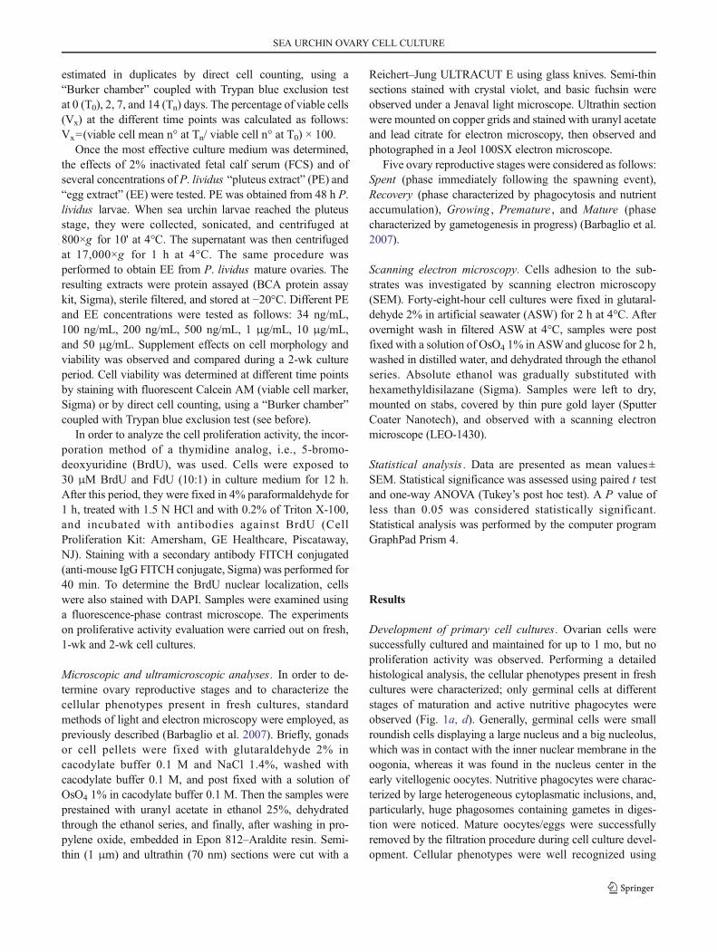
estimated in duplicates by direct cell counting, using a“Burker chamber” coupled with Trypan blue exclusion testat 0 (T0), 2, 7, and 14 (Tn) days. The percentage of viable cells(Vx) at the different time points was calculated as follows:Vx=(viable cell mean n° at Tn/ viable cell n° at T0) × 100.
Once the most effective culture medium was determined,the effects of 2% inactivated fetal calf serum (FCS) and ofseveral concentrations of P. lividus “pluteus extract” (PE) and“egg extract” (EE) were tested. PE was obtained from 48 h P.lividus larvae. When sea urchin larvae reached the pluteusstage, they were collected, sonicated, and centrifuged at800×g for 10' at 4°C. The supernatant was then centrifugedat 17,000×g for 1 h at 4°C. The same procedure wasperformed to obtain EE from P. lividus mature ovaries. Theresulting extracts were protein assayed (BCA protein assaykit, Sigma), sterile filtered, and stored at −20°C. Different PEand EE concentrations were tested as follows: 34 ng/mL,100 ng/mL, 200 ng/mL, 500 ng/mL, 1 μg/mL, 10 μg/mL,and 50 μg/mL. Supplement effects on cell morphology andviability was observed and compared during a 2-wk cultureperiod. Cell viability was determined at different time pointsby staining with fluorescent Calcein AM (viable cell marker,Sigma) or by direct cell counting, using a “Burker chamber”coupled with Trypan blue exclusion test (see before).
In order to analyze the cell proliferation activity, the incor-poration method of a thymidine analog, i.e., 5-bromo-deoxyuridine (BrdU), was used. Cells were exposed to30 μM BrdU and FdU (10:1) in culture medium for 12 h.After this period, they were fixed in 4% paraformaldehyde for1 h, treated with 1.5 N HCl and with 0.2% of Triton X-100,and incubated with antibodies against BrdU (CellProliferation Kit: Amersham, GE Healthcare, Piscataway,NJ). Staining with a secondary antibody FITCH conjugated(anti-mouse IgG FITCH conjugate, Sigma) was performed for40 min. To determine the BrdU nuclear localization, cellswere also stained with DAPI. Samples were examined usinga fluorescence-phase contrast microscope. The experimentson proliferative activity evaluation were carried out on fresh,1-wk and 2-wk cell cultures.
Microscopic and ultramicroscopic analyses. In order to de-termine ovary reproductive stages and to characterize thecellular phenotypes present in fresh cultures, standardmethods of light and electron microscopy were employed, aspreviously described (Barbaglio et al. 2007). Briefly, gonadsor cell pellets were fixed with glutaraldehyde 2% incacodylate buffer 0.1 M and NaCl 1.4%, washed withcacodylate buffer 0.1 M, and post fixed with a solution ofOsO4 1% in cacodylate buffer 0.1 M. Then the samples wereprestained with uranyl acetate in ethanol 25%, dehydratedthrough the ethanol series, and finally, after washing in pro-pylene oxide, embedded in Epon 812–Araldite resin. Semi-thin (1 μm) and ultrathin (70 nm) sections were cut with a
Reichert–Jung ULTRACUT E using glass knives. Semi-thinsections stained with crystal violet, and basic fuchsin wereobserved under a Jenaval light microscope. Ultrathin sectionwere mounted on copper grids and stained with uranyl acetateand lead citrate for electron microscopy, then observed andphotographed in a Jeol 100SX electron microscope.
Five ovary reproductive stages were considered as follows:Spent (phase immediately following the spawning event),Recovery (phase characterized by phagocytosis and nutrientaccumulation), Growing , Premature , and Mature (phasecharacterized by gametogenesis in progress) (Barbaglio et al.2007).
Scanning electron microscopy. Cells adhesion to the sub-strates was investigated by scanning electron microscopy(SEM). Forty-eight-hour cell cultures were fixed in glutaral-dehyde 2% in artificial seawater (ASW) for 2 h at 4°C. Afterovernight wash in filtered ASW at 4°C, samples were postfixed with a solution of OsO4 1% in ASWand glucose for 2 h,washed in distilled water, and dehydrated through the ethanolseries. Absolute ethanol was gradually substituted withhexamethyldisilazane (Sigma). Samples were left to dry,mounted on stabs, covered by thin pure gold layer (SputterCoater Nanotech), and observed with a scanning electronmicroscope (LEO-1430).
Statistical analysis . Data are presented as mean values±SEM. Statistical significance was assessed using paired t testand one-way ANOVA (Tukey’s post hoc test). A P value ofless than 0.05 was considered statistically significant.Statistical analysis was performed by the computer programGraphPad Prism 4.
Results
Development of primary cell cultures . Ovarian cells weresuccessfully cultured and maintained for up to 1 mo, but noproliferation activity was observed. Performing a detailedhistological analysis, the cellular phenotypes present in freshcultures were characterized; only germinal cells at differentstages of maturation and active nutritive phagocytes wereobserved (Fig. 1a, d). Generally, germinal cells were smallroundish cells displaying a large nucleus and a big nucleolus,which was in contact with the inner nuclear membrane in theoogonia, whereas it was found in the nucleus center in theearly vitellogenic oocytes. Nutritive phagocytes were charac-terized by large heterogeneous cytoplasmatic inclusions, and,particularly, huge phagosomes containing gametes in diges-tion were noticed. Mature oocytes/eggs were successfullyremoved by the filtration procedure during cell culture devel-opment. Cellular phenotypes were well recognized using
SEA URCHIN OVARY CELL CULTURE
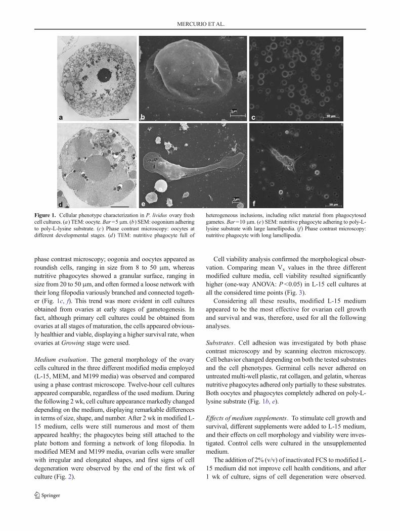
phase contrast microscopy; oogonia and oocytes appeared asroundish cells, ranging in size from 8 to 50 μm, whereasnutritive phagocytes showed a granular surface, ranging insize from 20 to 50 μm, and often formed a loose network withtheir long filopodia variously branched and connected togeth-er (Fig. 1c, f). This trend was more evident in cell culturesobtained from ovaries at early stages of gametogenesis. Infact, although primary cell cultures could be obtained fromovaries at all stages of maturation, the cells appeared obvious-ly healthier and viable, displaying a higher survival rate, whenovaries at Growing stage were used.
Medium evaluation. The general morphology of the ovarycells cultured in the three different modified media employed(L-15, MEM, and M199 media) was observed and comparedusing a phase contrast microscope. Twelve-hour cell culturesappeared comparable, regardless of the used medium. Duringthe following 2 wk, cell culture appearance markedly changeddepending on the medium, displaying remarkable differencesin terms of size, shape, and number. After 2 wk in modified L-15 medium, cells were still numerous and most of themappeared healthy; the phagocytes being still attached to theplate bottom and forming a network of long filopodia. Inmodified MEM and M199 media, ovarian cells were smallerwith irregular and elongated shapes, and first signs of celldegeneration were observed by the end of the first wk ofculture (Fig. 2).
Cell viability analysis confirmed the morphological obser-vation. Comparing mean Vx values in the three differentmodified culture media, cell viability resulted significantlyhigher (one-way ANOVA: P <0.05) in L-15 cell cultures atall the considered time points (Fig. 3).
Considering all these results, modified L-15 mediumappeared to be the most effective for ovarian cell growthand survival and was, therefore, used for all the followinganalyses.
Substrates . Cell adhesion was investigated by both phasecontrast microscopy and by scanning electron microscopy.Cell behavior changed depending on both the tested substratesand the cell phenotypes. Germinal cells never adhered onuntreated multi-well plastic, rat collagen, and gelatin, whereasnutritive phagocytes adhered only partially to these substrates.Both oocytes and phagocytes completely adhered on poly-L-lysine substrate (Fig. 1b, e).
Effects of medium supplements. To stimulate cell growth andsurvival, different supplements were added to L-15 medium,and their effects on cell morphology and viability were inves-tigated. Control cells were cultured in the unsupplementedmedium.
The addition of 2% (v/v) of inactivated FCS to modified L-15 medium did not improve cell health conditions, and after1 wk of culture, signs of cell degeneration were observed.
Figure 1. Cellular phenotype characterization in P. lividus ovary freshcell cultures. (a) TEM: oocyte. Bar=5 μm. (b) SEM: oogonium adheringto poly-L-lysine substrate. (c) Phase contrast microscopy: oocytes atdifferent developmental stages. (d) TEM: nutritive phagocyte full of
heterogeneous inclusions, including relict material from phagocytosedgametes. Bar=10 μm. (e) SEM: nutritive phagocyte adhering to poly-L-lysine substrate with large lamellipodia. (f) Phase contrast microscopy:nutritive phagocyte with long lamellipodia.
MERCURIO ETAL.
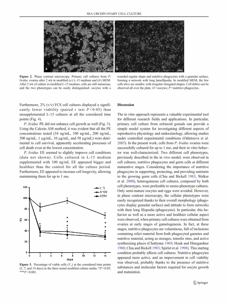
Furthermore, 2% (v/v) FCS cell cultures displayed a signifi-cantly lower viability (paired t test P <0.05) thanunsupplemented L-15 cultures at all the considered timepoints (Fig. 4).
P. lividus PE did not enhance cell growth as well (Fig. 5).Using the Calcein AM method, it was evident that all the PEconcentrations tested (34 ng/mL, 100 ng/mL, 200 ng/mL,500 ng/mL, 1 μg/mL, 10 μg/mL, and 50 μg/mL) were detri-mental to cell survival, apparently accelerating processes ofcell death even at the lowest concentration.
P. lividus EE seemed to slightly improve cell conditions(data not shown). Cells cultured in L-15 mediumsupplemented with 100 ng/mL EE appeared bigger andhealthier than the control for all the culture period.Furthermore, EE appeared to increase cell longevity, allowingmaintaining them for up to 1 mo.
Discussion
The in vitro approach represents a valuable experimental toolfor different research fields and applications. In particular,primary cell culture from echinoid gonads can provide asimple model system for investigating different aspects ofreproductive physiology and endocrinology, allowing studiesunder controlled experimental conditions (Odintsova et al.2005). In the present work, cells from P. lividus ovaries weresuccessfully cultured for up to 1 mo, and their in vitro behav-ior was well-characterized. Two different cell phenotypes,previously described in the in vivo model, were observed incell cultures; nutritive phagocytes and germ cells at differentmaturative stages. Considering the importance of nutritivephagocytes in supporting, protecting, and providing nutrientsto the growing germ cells (Chia and Bickell 1983; Walkeret al. 2000), heterogeneous cell cultures, composed by bothcell phenotypes, were preferable to mono-phenotype cultures.Only semi-mature oocytes and eggs were avoided. However,in phase contrast microscopy, the cellular phenotypes wereeasily recognized thanks to their overall morphology (phago-cytes display granular surface) and attitude to form networkswith their long filopodia (phagocytes). In particular, this be-havior as well as a more active and healthier cellular aspectwere observed, when primary cell cultures were obtained fromovaries at early stages of gametogenesis. In fact, at thesestages, nutritive phagocytes are voluminous, full of inclusionscontaining relict material from both phagocyted gametes andnutritive material, acting as storages, transfer sites, and activesynthesizing places (Chatlynne 1969; Houk and Hinegardner1980; Chia and Bickell 1983; Spirlet et al. 1998). This startingcondition probably affects cell cultures. Nutritive phagocytesappeared more active, and an improvement in cell viabilitywas observed, probably thanks to the presence of nutritivesubstances and molecular factors required for oocyte growthand maturation.
Figure 2. Phase contrast microscopy. Primary cell cultures from P.lividus ovaries after 2 wk in modified (a) L-15 medium and (b) MEM.After 2 wk of culture in modified L-15 medium, cells are still numerous,and the two phenotypes can be easily distinguished: oocytes with a
rounded regular shape and nutritive phagocytes with a granular surface,forming a network with long lamellipodia. In modified MEM, the fewcells alive are smaller, with irregular elongated shapes. Cell debris can beobserved all over the plate. O =oocytes; P=nutritive phagocytes.
Figure 3. Percentage of viable cells (Vx) at the considered time points(2, 7, and 14 days) in the three tested modified culture media. *P<0.05;***P<0.001.
SEA URCHIN OVARY CELL CULTURE
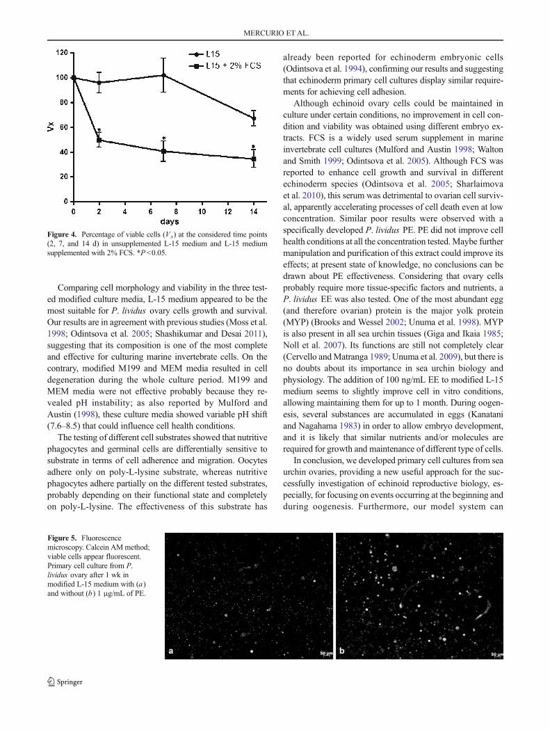
Comparing cell morphology and viability in the three test-ed modified culture media, L-15 medium appeared to be themost suitable for P. lividus ovary cells growth and survival.Our results are in agreement with previous studies (Moss et al.1998; Odintsova et al. 2005; Shashikumar and Desai 2011),suggesting that its composition is one of the most completeand effective for culturing marine invertebrate cells. On thecontrary, modified M199 and MEM media resulted in celldegeneration during the whole culture period. M199 andMEM media were not effective probably because they re-vealed pH instability; as also reported by Mulford andAustin (1998), these culture media showed variable pH shift(7.6–8.5) that could influence cell health conditions.
The testing of different cell substrates showed that nutritivephagocytes and germinal cells are differentially sensitive tosubstrate in terms of cell adherence and migration. Oocytesadhere only on poly-L-lysine substrate, whereas nutritivephagocytes adhere partially on the different tested substrates,probably depending on their functional state and completelyon poly-L-lysine. The effectiveness of this substrate has
already been reported for echinoderm embryonic cells(Odintsova et al. 1994), confirming our results and suggestingthat echinoderm primary cell cultures display similar require-ments for achieving cell adhesion.
Although echinoid ovary cells could be maintained inculture under certain conditions, no improvement in cell con-dition and viability was obtained using different embryo ex-tracts. FCS is a widely used serum supplement in marineinvertebrate cell cultures (Mulford and Austin 1998; Waltonand Smith 1999; Odintsova et al. 2005). Although FCS wasreported to enhance cell growth and survival in differentechinoderm species (Odintsova et al. 2005; Sharlaimovaet al. 2010), this serum was detrimental to ovarian cell surviv-al, apparently accelerating processes of cell death even at lowconcentration. Similar poor results were observed with aspecifically developed P. lividus PE. PE did not improve cellhealth conditions at all the concentration tested. Maybe furthermanipulation and purification of this extract could improve itseffects; at present state of knowledge, no conclusions can bedrawn about PE effectiveness. Considering that ovary cellsprobably require more tissue-specific factors and nutrients, aP. lividus EE was also tested. One of the most abundant egg(and therefore ovarian) protein is the major yolk protein(MYP) (Brooks and Wessel 2002; Unuma et al. 1998). MYPis also present in all sea urchin tissues (Giga and Ikaia 1985;Noll et al. 2007). Its functions are still not completely clear(Cervello andMatranga 1989; Unuma et al. 2009), but there isno doubts about its importance in sea urchin biology andphysiology. The addition of 100 ng/mL EE to modified L-15medium seems to slightly improve cell in vitro conditions,allowing maintaining them for up to 1 month. During oogen-esis, several substances are accumulated in eggs (Kanataniand Nagahama 1983) in order to allow embryo development,and it is likely that similar nutrients and/or molecules arerequired for growth and maintenance of different type of cells.
In conclusion, we developed primary cell cultures from seaurchin ovaries, providing a new useful approach for the suc-cessfully investigation of echinoid reproductive biology, es-pecially, for focusing on events occurring at the beginning andduring oogenesis. Furthermore, our model system can
Figure 4. Percentage of viable cells (Vx) at the considered time points(2, 7, and 14 d) in unsupplemented L-15 medium and L-15 mediumsupplemented with 2% FCS. *P <0.05.
Figure 5. Fluorescencemicroscopy. Calcein AMmethod;viable cells appear fluorescent.Primary cell culture from P.lividus ovary after 1 wk inmodified L-15 medium with (a)and without (b) 1 μg/mL of PE.
MERCURIO ETAL.
represent a new simple and versatile experimental tool for awide range of applied researches (in both ecotoxicology andaquaculture field), finally allowing to unravel the mechanismsregulating sea urchin gametogenesis.
Acknowledgment We are grateful to the “Area Marina” of Bergeggi(SV) and Portofino (GE) for giving their permission to collect experi-mental animals.
References
Barbaglio A.; Sugni M.; Di Benedetto C.; Bonasoro F.; Schnell S.;Lavado R.; Porte C.; Candia Carnevali D. M. Gametogenesis cor-related with steroid levels during the gonadal cycle of the sea urchinParacentrotus lividus (Echinodermata: Echinoidea). Comp.Biochem. Physiol. A 147(2): 466–474; 2007.
Brooks J. M.; Wessel G. M. The major yolk protein in sea urchins is atransferrin-like, iron binding protein.Dev. Biol. 245(1): 1–12; 2002.
Byrne M. Annual reproductive cycles of the commercial sea-urchinParacentrotus lividus from an exposed intertidal and a shelteredsubtidal habitat on the west-coast of Ireland.Mar. Biol. 104(2): 275–289; 1990.
Candia Carnevali D. M. Regeneration in Echinoderms: repair, regrowth,cloning. Invertebr. Surviv. J. 3: 64–76; 2006.
Cao A.; Mercado L.; Ramos-Martinez J. I.; Barcia R. Primary cultures ofhemocytes from Mytilus galloprovincialis Lmk.: expression of IL-2Rα subunit. Aquaculture 216(1–4): 1–8; 2003.
Cervello M.; Matranga V. Evidence of a precursor-product relationshipbetween vitellogenin and toposome, a glycoprotein complex medi-ating cell adhesion. Cell Differ. Dev. 26(1): 67–76; 1989.
Chatlynne L. G. A histochemical study of oogenesis in the sea urchin,Strongylocentrotus purpuratus . Biol. Bull. 136(2): 167–184; 1969.
Chia FS, Bickell, L. R. (1983) Echinodermata. In: Adiyod KGAaRG (ed)Reproductive biology of invertebrates. spermatogenesis and spermfunction, vol 2. K.G. Adiyodi & R.G. Adiyodi New York, pp 545–620
de Caralt S.; Uriz M. J.; Wijffels R. H. Cell culture from sponges:pluripotency and immortality. Trends Biotechnol. 25(10): 467–471; 2007.
Fenaux L. Maturation des gonads et cycle saisonnier des larves chez A.lixula ,P. lividus etP. microtuberculatus (Echinides) a’Villefranche-sur-mer. Vie Milieu 19: 1–52; 1968.
Giga Y.; Ikaia A. Purification of the most abundant protein in the coelo-mic fluid of a sea urchin which immunologically cross reacts with23S glycoprotein in the sea urchin eggs. J. Biochem. 98(1): 19–26;1985.
HoukM. S.; Hinegardner R. T. The formation and early differentiation ofsea urchin gonads. Biol. Bull. 159(2): 280–294; 1980.
Kanatani H.; Nagahama Y. Echinodermata. In: Adiyodi K. G. A. R. G.(ed) Reproductive biology of invertebrates. spermatogenesis,ooogenesis, oviposition, and absorption. 1st ed. JohnWiley & Sons,New York, pp 545–620; 1983.
Leoni V, Fernandez C, Johnson M, Ferrat L, Pergent-Martini C (2001)Preliminary study on spawning periods in the sea urchinParacentrotus lividus from lagoon and marine environments. Echi-noderm Research:277–280
McCarron E.; Burnell G.; Kerry J.; Mouzakitis G. An experimentalassessment on the effects of photoperiod treatments on the somaticand gonadal growth of the juvenile European purple sea urchinParacentrotus lividus . Aquac. Res. 41(7): 1072–1081; 2010.
Mita M. Relaxin-like gonad-stimulating substance in an echinoderm, thestarfish: A novel relaxin system in reproduction of invertebrates.Gen. Comp. Endocr. 181: 241–245; 2013.
Moss C.; Beesley P. W.; Thorndyke M. C.; Bollner T. Preliminaryobservations on ascidian and echinoderm neurons and neural ex-plants in vitro. Tissue Cell 30(5): 517–524; 1998.
Mulford A. L.; Austin B. Development of primary cell cultures fromNephrops norvegicus . Method Cell Sci. 19(4): 269–275; 1998.
Noll H.; Alcedo J.; Daube M.; Frei E.; Schiltz E.; Hunt J.; Humphries T.;Matranga V.; Hochstrasser M.; Aebersold R.; Lee H.; Noll M. Thetoposome, essential for sea urchin cell adhesion and development, isa modified iron-less calcium-binding transferrin. Dev. Biol. 310(1):54–70; 2007.
Odintsova N. A.; Dolmatov I. Y.; Mashanov V. S. Regenerating holothu-rian tissues as a source of cells for long-term cell cultures.Mar. Biol.146(5): 915–921; 2005.
Odintsova N. A.; Ermak A. V.; Tsal L. G. Substrate selection for long-term cultivation of marine invertebrate cells. Comp. Biochem. Phys-iol. A 107(4): 613–619; 1994.
Poccia D. In vitro differentiation of male germ line cells from sea urchintestis. J. Exp. Zool. 246(1): 57–64; 1988.
Rinkevich B. Cell cultures from marine invertebrates: obstacles, newapproaches and recent improvements. In: R. Osinga JTJGB,WijffelsRH (eds) Progress in Industrial Microbiology, vol 35. Elsevier, pp133–153; 1999
Rinkevich B. Cell cultures from marine invertebrates: new insights forcapturing endless stemness.Mar. Biotechnol. 13(3): 345–354; 2011.
Sharlaimova N. S.; Pinaev G. P.; Petukhova O. A. Comparative analysisof behavior and proliferative activity in culture of cells of coelomicfluid and of cells of various tissues of the sea star Asterias rubens L.isolated from normal and injured animals. Cell Tissue Biol. 4(3):280–288; 2010.
Shashikumar A.; Desai P. V. Development of cell line from the testiculartissues of crab Scylla serrata . Cytotechnology 63(5): 473–480;2011.
Shpigel M.; McBride S. C.; Marciano S.; Lupatsch I. The effect ofphotoperiod and temperature on the reproduction of European seaurchin Paracentrotus lividus . Aquaculture 232(1–4): 343–355;2004.
Spirlet C.; Grosjean P.; Jangoux M. Reproductive cycle of the echinoidParacentrotus lividus: analysis by means of the maturity index.Invertebr. Reprod. Dev. 34(1): 69–81; 1998.
Spirlet C.; Grosjean P.; Jangoux M. Optimization of gonad growth bymanipulation of temperature and photoperiod in cultivated sea ur-chins, Paracentrotus lividus (Lamarck) (Echinodermata). Aquacul-ture 185(1–2): 85–99; 2000.
Takahashi N.; Kanatani H. Effect of 17β-estradiol on growth of oocytesin cultured ovarian fragments of the starfish, Asterina pectinifera .Dev. Growth Differ. 23(6): 565–569; 1981.
Unuma T.; Konishi K.; Kiyomoto M.; Matranga V.; Yamano K.; Ohta H.;Yokota Y. Themajor yolk protein is synthesized in the digestive tractand secreted into the body cavities in sea urchin larvae.Mol. Reprod.Dev. 76(2): 142–150; 2009.
Unuma T.; Suzuki T.; Kurokawa T.; Yamamoto T.; Akiyama T. A proteinidentical to the yolk protein is stored in the testis in male red seaurchin,Pseudocentrotus depressus . Biol. Bull. 194(1): 92–97; 1998.
Walker C. W.; McGinn N. A.; Harrington L. M.; Lesser M. P. Reproduc-tion in sea urchins. In: Lawrence J. (ed) The Biology and Ecology ofEdible Sea Urchins. El Sevier Press, Amsterdam; 2000.
Walton A.; Smith V. J. Primary culture of the hyaline hemocytes frommarine decapods. Fish Shellfish Immunol. 9(3): 181–194; 1999.
Yakovlev K. V.; Battulin N. R.; Serov O. L.; Odintsova N. A. Isolation ofoogonia from ovaries of the sea urchin Strongylocentrotus nudus .Cell Tissue Res. 342(3): 479–490; 2010.
SEA URCHIN OVARY CELL CULTURE
Related Documents