© 2015 Wang et al. This work is published by Dove Medical Press Limited, and licensed under Creative Commons Attribution – Non Commercial (unported, v3.0) License. The full terms of the License are available at http://creativecommons.org/licenses/by-nc/3.0/. Non-commercial uses of the work are permitted without any further permission from Dove Medical Press Limited, provided the work is properly attributed. Permissions beyond the scope of the License are administered by Dove Medical Press Limited. Information on how to request permission may be found at: http://www.dovepress.com/permissions.php Drug Design, Development and Therapy 2015:9 2553–2563 Drug Design, Development and erapy Dovepress submit your manuscript | www.dovepress.com Dovepress 2553 ORIGINAL RESEARCH open access to scientific and medical research Open Access Full Text Article http://dx.doi.org/10.2147/DDDT.S81539 Aging-related rotenone-induced neurochemical and behavioral deficits: role of SIRT2 and redox imbalance, and neuroprotection by AK-7 Xijin Wang 1 Qiang Guan 2 Meihua Wang 1 Liu Yang 1 Jie Bai 1 Zhiqiang Yan 3 Yuhong Zhang 4 Zhenguo Liu 1 1 Department of Neurology, Xinhua Hospital Affiliated to Shanghai Jiao Tong University School of Medicine, 2 Department of Neurology, Tongji Hospital, Tongji University, 3 Shanghai Laboratory Animal Center, Chinese Academy of Sciences, 4 Department of Neurology, Shanghai Tenth People’s Hospital, Tongji University, Shanghai, People’s Republic of China Abstract: Aging is one of the strongest risk factors for Parkinson’s disease (PD). SIRT2 has been implicated in the aging process. It is pertinent to investigate the role of SIRT2 in aging- related dopaminergic neurotoxicity and to develop effective therapeutic strategies for PD through the use of aging animals. In this study, we observed that rotenone induced significant behavior abnormality and striatal dopamine depletion in aging rats, while it did not do so in young rats. No significant change in striatal serotonin level was observed in the aging rats after rotenone administration. There was also aging-related rotenone-induced increase in substantia nigra (SN) SIRT2 expression in the rats. In addition, there was aging-related rotenone-induced SN malondialdehyde (MDA) increase and glutathione (GSH) decrease in the rats. No signifi- cant changes in cerebellar SIRT2, MDA, or GSH levels were observed in the aging rats after rotenone administration. Striatal dopamine content was significantly inversely correlated with SN SIRT2 expression in the rats. AK-7 significantly diminished striatal dopamine depletion and improved behavior abnormality in the rotenone-treated aging rats. Furthermore, AK-7 significantly decreased MDA content and increased GSH content in the SN of rotenone-treated aging rats. Finally, the effect of AK-7 on dopaminergic neurons and redox imbalance was sup- ported by the results from primary mesencephalic cultures. Our study helps to elucidate the mechanism for the participation of aging in PD and suggests that SN SIRT2 may be involved in PD neurodegeneration, that AK-7 may be neuroprotective in PD, and that maintaining redox balance may be one of the mechanisms underlying neuroprotection by AK-7. Keywords: Parkinson’s disease, environmental toxin, dopamine, oxidative stress, sirtuin Introduction Parkinson’s disease (PD) is a common and progressive neurodegenerative disease that is characterized by motor dysfunction due to decreased dopamine (DA) content in the striatum, resulting from dopaminergic neurodegeneration in the substantia nigra (SN). 1–3 Accumulating evidence indicates that the cause of PD is multifactorial, involving genetic predisposition, innate characteristics of the nigrostriatal dopaminergic system in the brain, exposure of environmental toxins and immune/inflammatory factors, and aging. 3–12 Aging appears to be one of the prominent and unifying risk factors for idiopathic PD. 13–16 Epidemiological studies reveal that the incidence and prevalence of PD increase with advancing age, occurring in approximately 1% of people over age 65. With the development of molecular biology and further understanding of PD, increasing importance is being attached to the search for aging-related molecules involved in PD neurodegeneration and to develop effective therapeutic strategies for PD through the use of aging animals. 1–3 Correspondence: Xijin Wang; Zhenguo Liu Department of Neurology, Xinhua Hospital Affiliated to Shanghai Jiao Tong University School of Medicine, 1665 Kongjiang Road, Shanghai 200092, People’s Republic of China Tel +86 137 7426 1539 Email [email protected]; [email protected]

Welcome message from author
This document is posted to help you gain knowledge. Please leave a comment to let me know what you think about it! Share it to your friends and learn new things together.
Transcript
© 2015 Wang et al. This work is published by Dove Medical Press Limited, and licensed under Creative Commons Attribution – Non Commercial (unported, v3.0) License. The full terms of the License are available at http://creativecommons.org/licenses/by-nc/3.0/. Non-commercial uses of the work are permitted without any further
permission from Dove Medical Press Limited, provided the work is properly attributed. Permissions beyond the scope of the License are administered by Dove Medical Press Limited. Information on how to request permission may be found at: http://www.dovepress.com/permissions.php
Drug Design, Development and Therapy 2015:9 2553–2563
Drug Design, Development and Therapy Dovepress
submit your manuscript | www.dovepress.com
Dovepress 2553
O r i g i n a l r e s e a r c h
open access to scientific and medical research
Open access Full Text article
http://dx.doi.org/10.2147/DDDT.S81539
aging-related rotenone-induced neurochemical and behavioral deficits: role of SIRT2 and redox imbalance, and neuroprotection by aK-7
Xijin Wang1
Qiang guan2
Meihua Wang1
liu Yang1
Jie Bai1
Zhiqiang Yan3
Yuhong Zhang4
Zhenguo liu1
1Department of Neurology, Xinhua Hospital Affiliated to Shanghai Jiao Tong University School of Medicine, 2Department of Neurology, Tongji hospital, Tongji University, 3shanghai laboratory animal center, chinese Academy of Sciences, 4Department of Neurology, Shanghai Tenth People’s hospital, Tongji University, shanghai, People’s Republic of China
Abstract: Aging is one of the strongest risk factors for Parkinson’s disease (PD). SIRT2 has
been implicated in the aging process. It is pertinent to investigate the role of SIRT2 in aging-
related dopaminergic neurotoxicity and to develop effective therapeutic strategies for PD
through the use of aging animals. In this study, we observed that rotenone induced significant
behavior abnormality and striatal dopamine depletion in aging rats, while it did not do so in
young rats. No significant change in striatal serotonin level was observed in the aging rats after
rotenone administration. There was also aging-related rotenone-induced increase in substantia
nigra (SN) SIRT2 expression in the rats. In addition, there was aging-related rotenone-induced
SN malondialdehyde (MDA) increase and glutathione (GSH) decrease in the rats. No signifi-
cant changes in cerebellar SIRT2, MDA, or GSH levels were observed in the aging rats after
rotenone administration. Striatal dopamine content was significantly inversely correlated with
SN SIRT2 expression in the rats. AK-7 significantly diminished striatal dopamine depletion
and improved behavior abnormality in the rotenone-treated aging rats. Furthermore, AK-7
significantly decreased MDA content and increased GSH content in the SN of rotenone-treated
aging rats. Finally, the effect of AK-7 on dopaminergic neurons and redox imbalance was sup-
ported by the results from primary mesencephalic cultures. Our study helps to elucidate the
mechanism for the participation of aging in PD and suggests that SN SIRT2 may be involved
in PD neurodegeneration, that AK-7 may be neuroprotective in PD, and that maintaining redox
balance may be one of the mechanisms underlying neuroprotection by AK-7.
Keywords: Parkinson’s disease, environmental toxin, dopamine, oxidative stress, sirtuin
IntroductionParkinson’s disease (PD) is a common and progressive neurodegenerative disease that
is characterized by motor dysfunction due to decreased dopamine (DA) content in the
striatum, resulting from dopaminergic neurodegeneration in the substantia nigra (SN).1–3
Accumulating evidence indicates that the cause of PD is multifactorial, involving
genetic predisposition, innate characteristics of the nigrostriatal dopaminergic system
in the brain, exposure of environmental toxins and immune/inflammatory factors,
and aging.3–12 Aging appears to be one of the prominent and unifying risk factors for
idiopathic PD.13–16 Epidemiological studies reveal that the incidence and prevalence
of PD increase with advancing age, occurring in approximately 1% of people over
age 65. With the development of molecular biology and further understanding of PD,
increasing importance is being attached to the search for aging-related molecules
involved in PD neurodegeneration and to develop effective therapeutic strategies for
PD through the use of aging animals.1–3
correspondence: Xijin Wang; Zhenguo liuDepartment of Neurology, Xinhua Hospital Affiliated to Shanghai Jiao Tong University School of Medicine, 1665 Kongjiang road, shanghai 200092, People’s Republic of ChinaTel +86 137 7426 1539email [email protected]; [email protected]
Journal name: Drug Design, Development and TherapyArticle Designation: Original ResearchYear: 2015Volume: 9Running head verso: Wang et alRunning head recto: Role of SIRT2 and AK-7 in Parkinson’s diseaseDOI: http://dx.doi.org/10.2147/DDDT.S81539
Drug Design, Development and Therapy 2015:9submit your manuscript | www.dovepress.com
Dovepress
Dovepress
2554
Wang et al
Among the various environmental factors suspected
to play a role in the etiopathogenesis of PD, agrochemical
exposure has been most intensely investigated in recent
years.1,17–24 The discovery of the neurotoxin rotenone has pro-
vided a valuable tool into PD research.17–24 Rotenone exposure
has been observed to induce parkinsonism in rodents.17–21
At present, rotenone is widely being employed to create in
vitro and in vivo models of PD in rodents.1,17–24 Rotenone was
also found to induce aging-related SN dopaminergic neurode-
generation in rats.22 However, little is known about the effect
of rotenone administration on striatal DA content and motor
behavior in aging animals. In the present study, rotenone
was observed to induce significant behavior abnormality
and striatal DA depletion in aging rats, while it did not do so
in young rats. SIRT2, a mammalian sirtuin, is a cytoplasmic
NAD+-dependent deacetylase.25 It is mainly expressed in the
brain.25,26 SIRT2 is an abundant neuronal protein that accu-
mulates in the aging central nervous system (CNS) and is
indicated to be associated with the aging process.25,27–30
Interestingly, in the present study, we observed that
there was aging-related rotenone-induced increase in SIRT2
expression in the SN of rats. Furthermore, striatal DA content
was significantly correlated with SN SIRT2 expression in
the rats. Importantly, we investigated the role of SN SIRT2
in aging-related rotenone-induced behavior abnormality
and striatal DA depletion, and the potential neuroprotec-
tion and its underlying mechanism of SIRT2 modulation in
PD. Because it is not easy to obtain aging animals, young
animals are often used in PD research. However, PD is an
aging-related neurodegenerative disease. Aging is one of the
strongest risk factors for idiopathic PD.13–15 So, aging animals
are more significant for PD research than are young ones.
Therefore, aging rats were employed in our study.
Materials and methodsanimals and treatmentAll animals were from Sino-British SIPPR/BK Lab Animal
LTD (Shanghai, People’s Republic of China). Male Sprague–
Dawley rats of two different age groups were used in the
present study: a young group (3 months of age) and an aging
group (18 months of age). Rotenone, emulsified in sunflower
oil at 0.5 mg/mL, was given intraperitoneally, at 1 mL/kg
once a day for 35 days, to the rats. At the first and eighth
day of rotenone treatment, some aging rats received the
selective SIRT2 inhibitor AK-7 (3-(1-azepanylsulfonyl)-N-
(3-bromophenyl)benzamide) by intranigral injection on both
sides (1 µg/day/side or 5 µg/day/side). Before rotenone treat-
ment, the rats were anesthetized with ketamine and xylazine
(60 mg/kg and 3 mg/kg, respectively, by intramuscular injec-
tion) and positioned in a stereotaxic apparatus. Then, AK-7
(2 µg or 10 µg) dissolved in dimethyl sulfoxide (DMSO)
(4 µL) or vehicle (4 µL of DMSO) was injected into the
SN at a flow rate of 1 µL/min, using a 10 µL Hamilton
microsyringe, with 2 µL of volume for intranigral injection
of each side. The following coordinates were used: -5.4 mm
anterior-posterior; ±2.1 mm medial-lateral; and -7.8 mm
dorsal-ventral. The needle was left in place for 5 minutes,
to avoid reflux along the injection track, prior to being
withdrawn. All experiments were conducted according to
the Guideline for Animal Experimentation of Shanghai Jiao
Tong University School of Medicine and the Guide for the
Care and Use of Laboratory Animals published by the US
National Institutes of Health (NIH]) (publication number
85-23, revised 1996). All attempts were made to minimize
the number of animals used and their suffering.
Behavior testsRotarod and open-field tests were performed to evaluate rat
behavior during the light period. The following three parts
were required for the rotarod test: (1) a roller; (2) a power
source used to turn the roller; and (3) four separators divid-
ing the roller into equal-sized compartments. Animals were
placed on the rod and sequentially tested at 5, 10, 15 rpm.
After being trained, the rats were tested three times at each
rotarod speed. The latency time to fall was measured for
each test. For locomotor activity, each rat was placed in an
open-field chamber made of wood covered with imperme-
able formica. The chamber had a white floor (100×100 cm)
divided into 25 squares of 20×20 cm, and 50 cm-high walls.
Before testing, each rat was placed in the center of the
open field and habituated for 10 minutes. Rat behavior was
recorded for 30 minutes. The following parameters were
determined: (1) crossing number, defined as entering of
another square with all four paws; and (2) rearing number,
defined as rearing with and without wall contact (standing
only on hind legs).
neurochemical analysisBiochemical analysis of neurotransmitter in the rat striata was
performed using high-performance liquid chromatography
(HPLC) with electrochemical detection (ECD) (HPLC-
ECD).31 Rat striata were removed, held on ice, and weighed.
Then striata were homogenized (10% wt/vol) through sonica-
tion, in ice-cold homogenization buffer containing perchloric
acid (0.1 mol/L) with 3,4-dihydroxybenzylamine as internal
standard. Obtained samples were centrifuged at 25,000 g
Drug Design, Development and Therapy 2015:9 submit your manuscript | www.dovepress.com
Dovepress
Dovepress
2555
Role of SIRT2 and AK-7 in Parkinson’s disease
for 10 minutes at 4°C, and supernatants were collected. DA
and serotonin (5-HT) content were assayed by HPLC-ECD,
equipped with a column of 5 µm spherical C18 particles. The
mobile phase consisted of 0.1 M phosphate buffer (pH 2.6)
containing 0.2 mM octane sulfonic acid, 2.5% methanol, and
4.5% acetonitrile. The content of each neurotransmitter was
expressed as ng/g equivalent striatal tissue.
Western blottingWestern blotting was performed to evaluate SIRT2 expres-
sion, as previously described.8 The SN and cerebellum
tissue were homogenized on ice. After centrifugation, the
supernatant protein content was assayed using a commer-
cially available protein assay kit (BioRad Laboratories,
Hercules, CA, USA). Sample proteins were separated
by sodium dodecyl sulfate polyacrylamide gel electro-
phoresis. After being blocked for 1 hour with blocking
solution, membranes were incubated overnight at 4°C
with rabbit polyclonal SIRT2 antibody (1:200) (Santa
Cruz Biotechnology, Dallas, TX, USA). Subsequently, the
membranes were washed and incubated with horseradish
peroxidase–conjugated secondary antibodies for 1 hour at
room temperature. The signals were finally detected using
an enhanced chemiluminescence (ECL) assay kit (EMD
Millipore, Billerica, MA, USA). Results were expressed
as densitometric relative units, representing the ratio of
density of the target protein to β-actin.
Primary mesencephalic neuron/glia culturesPrimary rat ventral mesencephalic neuron/glia cultures were
prepared, as previously described.24 Briefly, ventral mesen-
cephalic tissues were dissected from Sprague–Dawley rats
(embryonic day 14) and dissociated with a mild mechanical
trituration. Depending on the experimental design, dissoci-
ated cells were seeded at 5×10⁵, 2.5×10⁵, or 1×10⁵/well in
poly-D-lysine-coated 24-, 48-, and 96-well cell culture plates,
respectively. The culture medium was minimum essential
medium (MEM) supplemented with 10% heat-inactivated
fetal bovine serum (FBS) and 10% heat-inactivated horse
serum (HS), 2 mM L-glutamine, 1 g/L glucose, 100 µM
nonessential amino acids, 1 mM sodium pyruvate, 50 U/mL
penicillin, and 50 µg/mL streptomycin. Cultures were
kept at 37°C in a humidified atmosphere of 5% CO2 and
95% air. For treatment, the cultures were kept in treatment
medium (MEM containing 2% FBS, 2% HS, 1 mM sodium
pyruvate, 2 mM L-glutamine, 50 U/mL penicillin, and
50 µg/mL streptomycin).
immunostaining and cell countingThe cell cultures were processed for immunostaining detec-
tion, as previously described.24 After being blocked, they
were incubated with the primary antibody, anti-tyrosine
hydroxylase (TH, 1:1000) (Sigma-Aldrich Corp, St Louis,
MO, USA), at 4°C overnight. Then, they were detected
using a secondary antibody (Molecular Probes; Life
Technologies Corp, Carlsbad, CA) for 1.5 hours at room
temperature. Cell counting was performed in duplicate and
blindly. Ten representative areas per well were used for
cell quantification.
Assay of malondialdehyde (MDA) and glutathione (GSH) and superoxideThe level of MDA and GSH was determined by using a com-
mercial assay kit (Cayman Chemical Co, Ann Arbor, MI,
USA). The operation followed the manufacturer’s protocol.
The MDA and GSH content was determined in compari-
son with the standards and normalized to protein content.
Superoxide production was determined by measuring the
SOD-inhibitable reduction of cytochrome c, as previously
described.24
Determination of protein concentrationProtein content in the brain homogenate was measured
by using the dye-binding method described by Bradford
wherein bovine serum albumin (BSA) was used as a protein
standard.32
statistical analysisData were expressed as the mean ± standard error of the
mean (SEM). Differences were determined using two-
tailed Student’s t-test for comparison between two groups
and using an analysis of variance (ANOVA) and Bonfer-
roni post hoc test for comparison between more than two
groups. Normality of sample distribution and homogeneity
of variances were tested before each ANOVA. When cor-
relation analysis was performed, the Pearson’s value was
reported. Values of P,0.05 were accepted as statistically
significant.
ResultsGeneral assessment of animal healthIn the present study, none of the rats developed morbid health
necessitating special treatment throughout the experimental
period, and the survival rate was 100% in both the young and
aging rats after rotenone treatment. As shown in Figure 1A,
there was no significant difference in body weight between the
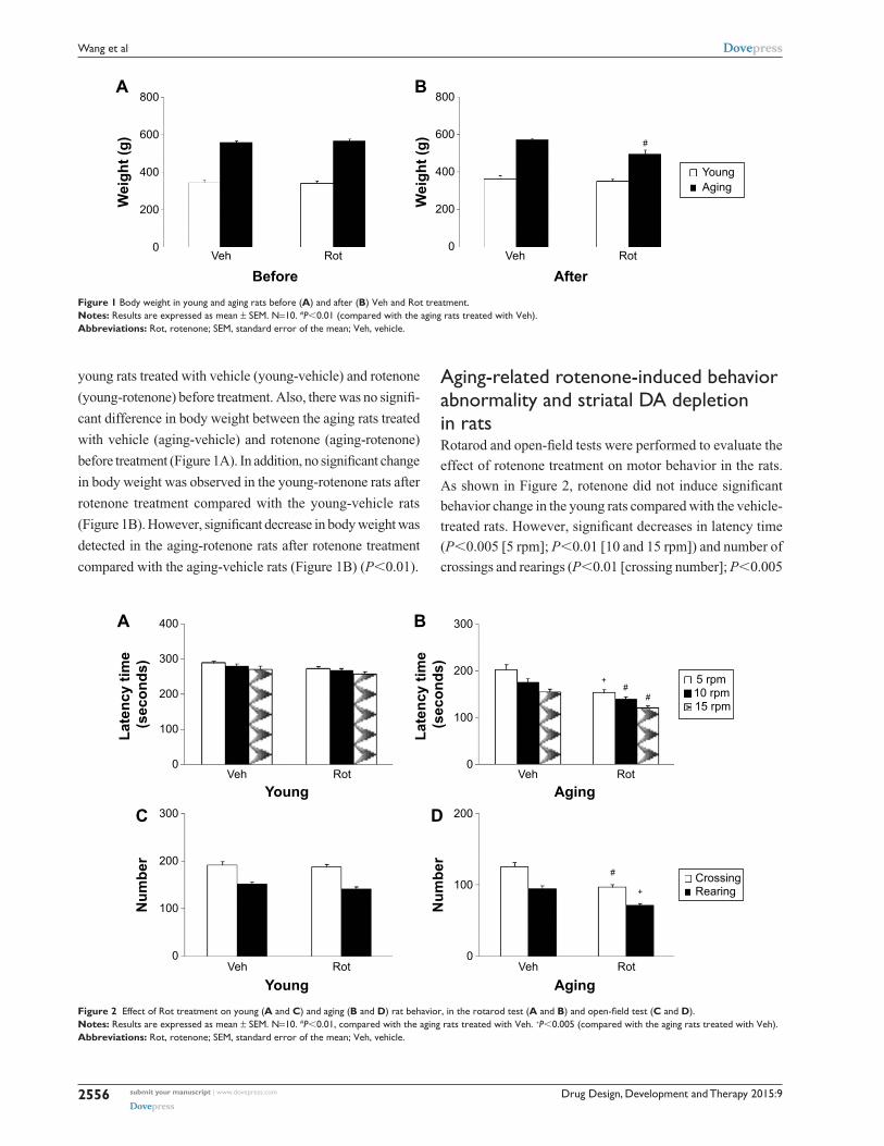
Drug Design, Development and Therapy 2015:9submit your manuscript | www.dovepress.com
Dovepress
Dovepress
2556
Wang et al
young rats treated with vehicle (young-vehicle) and rotenone
(young-rotenone) before treatment. Also, there was no signifi-
cant difference in body weight between the aging rats treated
with vehicle (aging-vehicle) and rotenone (aging-rotenone)
before treatment (Figure 1A). In addition, no significant change
in body weight was observed in the young-rotenone rats after
rotenone treatment compared with the young-vehicle rats
(Figure 1B). However, significant decrease in body weight was
detected in the aging-rotenone rats after rotenone treatment
compared with the aging-vehicle rats (Figure 1B) (P,0.01).
aging-related rotenone-induced behavior abnormality and striatal Da depletion in ratsRotarod and open-field tests were performed to evaluate the
effect of rotenone treatment on motor behavior in the rats.
As shown in Figure 2, rotenone did not induce significant
behavior change in the young rats compared with the vehicle-
treated rats. However, significant decreases in latency time
(P,0.005 [5 rpm]; P,0.01 [10 and 15 rpm]) and number of
crossings and rearings (P,0.01 [crossing number]; P,0.005
Figure 1 Body weight in young and aging rats before (A) and after (B) Veh and Rot treatment.Notes: results are expressed as mean ± seM. n=10. #P,0.01 (compared with the aging rats treated with Veh).Abbreviations: Rot, rotenone; SEM, standard error of the mean; Veh, vehicle.
Figure 2 Effect of Rot treatment on young (A and C) and aging (B and D) rat behavior, in the rotarod test (A and B) and open-field test (C and D). Notes: results are expressed as mean ± seM. n=10. #P,0.01, compared with the aging rats treated with Veh. +P,0.005 (compared with the aging rats treated with Veh). Abbreviations: Rot, rotenone; SEM, standard error of the mean; Veh, vehicle.

Drug Design, Development and Therapy 2015:9 submit your manuscript | www.dovepress.com
Dovepress
Dovepress
2557
Role of SIRT2 and AK-7 in Parkinson’s disease
[rearing number]) were observed in the aging rats after rote-
none treatment in comparison with the vehicle-treated rats
(Figure 2). In agreement with behavior tests, rotenone did
not cause significant striatal DA depletion in the young rats
compared with the vehicle-treated rats (Figure 3A). However,
significant decrease in striatal DA content was observed in
the aging rats after rotenone treatment in comparison with
the vehicle-treated rats (Figure 3A) (P,0.005). Although
rotenone significantly decreased striatal DA content in the
aging rats compared with the rats treated with vehicle, no
significant change in striatal 5-HT level was observed in the
aging rats after rotenone administration in comparison with
the vehicle-treated rats (Figure 3B).
aging-related rotenone-induced sirT2 expression increase in the SN of ratsWestern blotting was conducted to evaluate the effect of
rotenone treatment on SIRT2 expression in the SN of rats.
As shown in Figure 4A and C, rotenone did not significantly
induce SIRT2 expression change in the SN of young rats
compared with the vehicle-treated rats. However, significant
increase in SN SIRT2 expression was observed in the aging
rats after rotenone treatment in comparison with the vehicle-
treated rats (Figure 4B and C) (P,0.005). In our preliminary
study, rotenone (0.1 mg/kg) did not induce significant SIRT2
expression increase in the SN of aging rats in comparison
with the rats treated with vehicle. Correlation analysis indi-
cated that there was significant positive correlation between
SN SIRT2 protein expression and the dosage of rotenone
in the aging rats (R=0.8012, P,0.005). Although rotenone
significantly increased SN SIRT2 expression in the aging rats
compared with the rats treated with vehicle, no significant
change in cerebellar SIRT2 expression was observed in the
aging rats after rotenone administration in comparison with
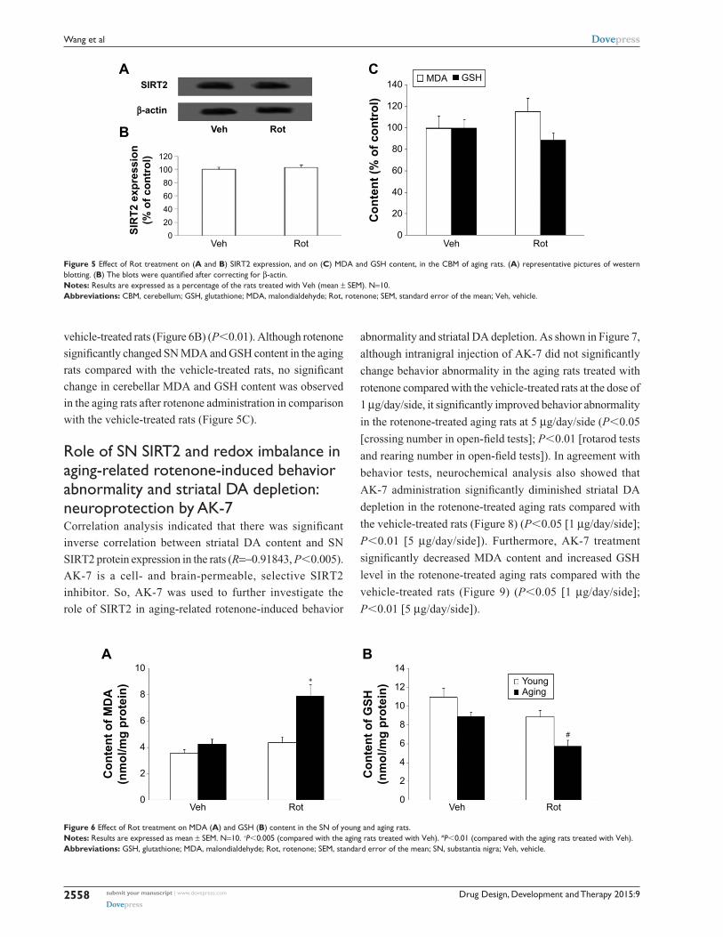
the vehicle-treated rats (Figure 5A and B).
aging-related rotenone-induced MDa increase and gsh decrease in the sn of ratsWe further investigated the effect of rotenone treatment
on MDA and GSH content in the SN of rats. As shown in
Figure 6A, rotenone did not significantly induce MDA
content change in the SN of young rats compared with the
vehicle-treated rats. However, significant increase in SN
MDA content was observed in the aging rats after rote-
none treatment in comparison with the vehicle-treated rats
(Figure 6A) (P,0.005). In addition, rotenone did not sig-
nificantly induce GSH content change in the SN of young
rats compared with the vehicle-treated rats. However, sig-
nificant decrease in SN GSH content was observed in the
aging rats after rotenone treatment in comparison with the
Figure 3 Effect of Rot treatment on rat striatal DA and 5-HT content in young and aging rats. (A) DA; (B) DA and 5-HT.Notes: results are expressed as mean ± seM. n=10. +P,0.005 (compared with the aging rats treated with Veh).Abbreviations: 5-HT, serotonin; DA, dopamine; Rot, rotenone; SEM, standard error of the mean; Veh, vehicle.
β β
Figure 4 aging-related rot-induced sirT2 expression increase in rat sn. Representative pictures of western blotting in (A) young rats and (B) aging rats. (C) The blots were quantified after correcting for β-actin.Notes: Results are expressed as a percentage of the young rats treated with Veh (mean ± SEM). N=10. +P,0.005 (compared with the aging rats treated with Veh).Abbreviations: Rot, rotenone; SEM, standard error of the mean; SN, substantia nigra; Veh, vehicle.
Drug Design, Development and Therapy 2015:9submit your manuscript | www.dovepress.com
Dovepress
Dovepress
2558
Wang et al
vehicle-treated rats (Figure 6B) (P,0.01). Although rotenone
significantly changed SN MDA and GSH content in the aging
rats compared with the vehicle-treated rats, no significant
change in cerebellar MDA and GSH content was observed
in the aging rats after rotenone administration in comparison
with the vehicle-treated rats (Figure 5C).
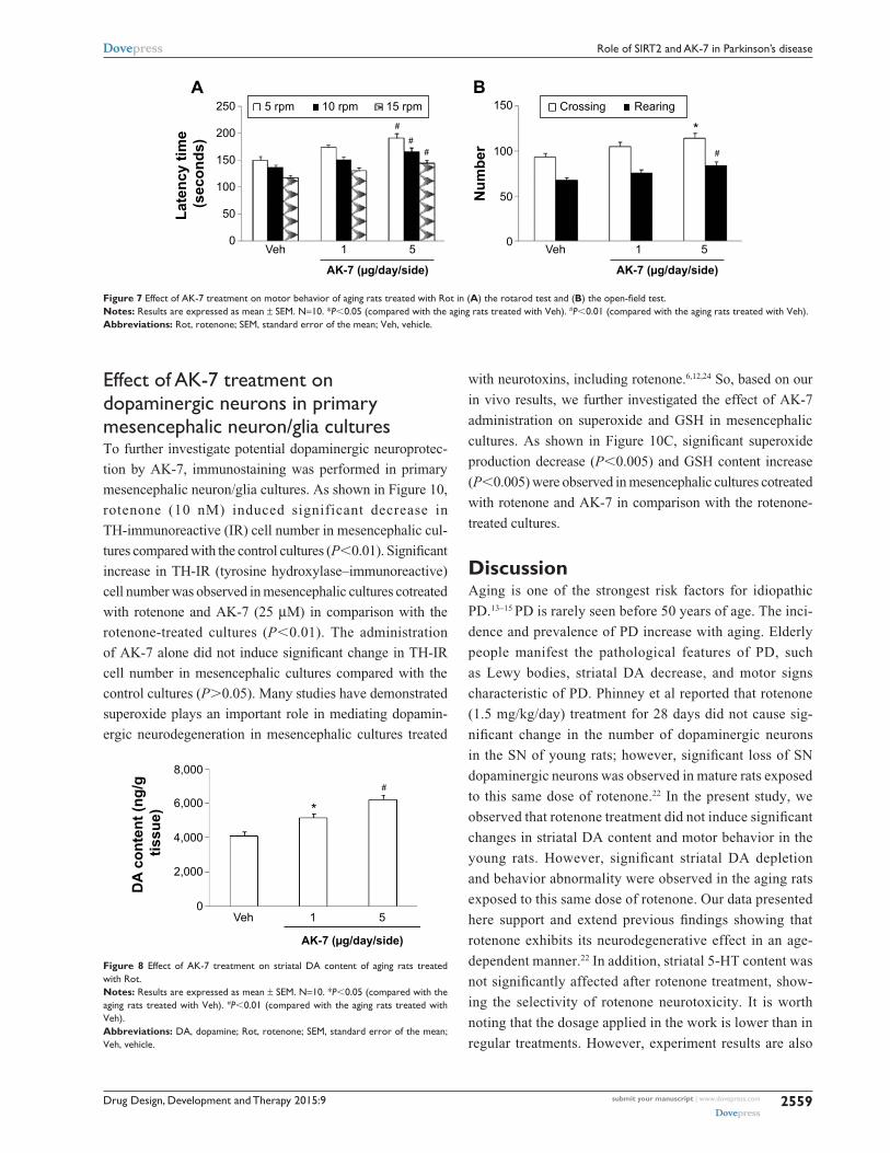
Role of SN SIRT2 and redox imbalance in aging-related rotenone-induced behavior abnormality and striatal Da depletion: neuroprotection by aK-7Correlation analysis indicated that there was significant
inverse correlation between striatal DA content and SN
SIRT2 protein expression in the rats (R=-0.91843, P,0.005).
AK-7 is a cell- and brain-permeable, selective SIRT2
inhibitor. So, AK-7 was used to further investigate the
role of SIRT2 in aging-related rotenone-induced behavior
abnormality and striatal DA depletion. As shown in Figure 7,
although intranigral injection of AK-7 did not significantly
change behavior abnormality in the aging rats treated with
rotenone compared with the vehicle-treated rats at the dose of
1 µg/day/side, it significantly improved behavior abnormality
in the rotenone-treated aging rats at 5 µg/day/side (P,0.05
[crossing number in open-field tests]; P,0.01 [rotarod tests
and rearing number in open-field tests]). In agreement with
behavior tests, neurochemical analysis also showed that
AK-7 administration significantly diminished striatal DA
depletion in the rotenone-treated aging rats compared with
the vehicle-treated rats (Figure 8) (P,0.05 [1 µg/day/side];
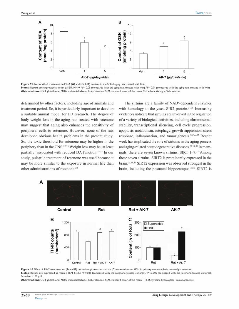
P,0.01 [5 µg/day/side]). Furthermore, AK-7 treatment
significantly decreased MDA content and increased GSH
level in the rotenone-treated aging rats compared with the
vehicle-treated rats (Figure 9) (P,0.05 [1 µg/day/side];
P,0.01 [5 µg/day/side]).
Figure 6 Effect of Rot treatment on MDA (A) and GSH (B) content in the SN of young and aging rats.Notes: results are expressed as mean ± seM. n=10. +P,0.005 (compared with the aging rats treated with Veh). #P,0.01 (compared with the aging rats treated with Veh).Abbreviations: GSH, glutathione; MDA, malondialdehyde; Rot, rotenone; SEM, standard error of the mean; SN, substantia nigra; Veh, vehicle.
β
Figure 5 Effect of Rot treatment on (A and B) SIRT2 expression, and on (C) MDA and GSH content, in the CBM of aging rats. (A) representative pictures of western blotting. (B) The blots were quantified after correcting for β-actin.Notes: Results are expressed as a percentage of the rats treated with Veh (mean ± SEM). N=10.Abbreviations: CBM, cerebellum; GSH, glutathione; MDA, malondialdehyde; Rot, rotenone; SEM, standard error of the mean; Veh, vehicle.
Drug Design, Development and Therapy 2015:9 submit your manuscript | www.dovepress.com
Dovepress
Dovepress
2559
Role of SIRT2 and AK-7 in Parkinson’s disease
Effect of AK-7 treatment on dopaminergic neurons in primary mesencephalic neuron/glia culturesTo further investigate potential dopaminergic neuroprotec-
tion by AK-7, immunostaining was performed in primary
mesencephalic neuron/glia cultures. As shown in Figure 10,
rotenone (10 nM) induced significant decrease in
TH-immunoreactive (IR) cell number in mesencephalic cul-
tures compared with the control cultures (P,0.01). Significant
increase in TH-IR (tyrosine hydroxylase–immunoreactive)
cell number was observed in mesencephalic cultures cotreated
with rotenone and AK-7 (25 µM) in comparison with the
rotenone-treated cultures (P,0.01). The administration
of AK-7 alone did not induce significant change in TH-IR
cell number in mesencephalic cultures compared with the
control cultures (P.0.05). Many studies have demonstrated
superoxide plays an important role in mediating dopamin-
ergic neurodegeneration in mesencephalic cultures treated
with neurotoxins, including rotenone.6,12,24 So, based on our
in vivo results, we further investigated the effect of AK-7
administration on superoxide and GSH in mesencephalic
cultures. As shown in Figure 10C, significant superoxide
production decrease (P,0.005) and GSH content increase
(P,0.005) were observed in mesencephalic cultures cotreated
with rotenone and AK-7 in comparison with the rotenone-
treated cultures.
DiscussionAging is one of the strongest risk factors for idiopathic
PD.13–15 PD is rarely seen before 50 years of age. The inci-
dence and prevalence of PD increase with aging. Elderly
people manifest the pathological features of PD, such
as Lewy bodies, striatal DA decrease, and motor signs
characteristic of PD. Phinney et al reported that rotenone
(1.5 mg/kg/day) treatment for 28 days did not cause sig-
nificant change in the number of dopaminergic neurons
in the SN of young rats; however, significant loss of SN
dopaminergic neurons was observed in mature rats exposed
to this same dose of rotenone.22 In the present study, we
observed that rotenone treatment did not induce significant
changes in striatal DA content and motor behavior in the
young rats. However, significant striatal DA depletion
and behavior abnormality were observed in the aging rats
exposed to this same dose of rotenone. Our data presented
here support and extend previous findings showing that
rotenone exhibits its neurodegenerative effect in an age-
dependent manner.22 In addition, striatal 5-HT content was
not significantly affected after rotenone treatment, show-
ing the selectivity of rotenone neurotoxicity. It is worth
noting that the dosage applied in the work is lower than in
regular treatments. However, experiment results are also
Figure 7 Effect of AK-7 treatment on motor behavior of aging rats treated with Rot in (A) the rotarod test and (B) the open-field test.Notes: results are expressed as mean ± seM. n=10. *P,0.05 (compared with the aging rats treated with Veh). #P,0.01 (compared with the aging rats treated with Veh).Abbreviations: Rot, rotenone; SEM, standard error of the mean; Veh, vehicle.
Figure 8 Effect of AK-7 treatment on striatal DA content of aging rats treated with rot.Notes: results are expressed as mean ± seM. n=10. *P,0.05 (compared with the aging rats treated with Veh). #P,0.01 (compared with the aging rats treated with Veh).Abbreviations: DA, dopamine; Rot, rotenone; SEM, standard error of the mean; Veh, vehicle.
Drug Design, Development and Therapy 2015:9submit your manuscript | www.dovepress.com
Dovepress
Dovepress
2560
Wang et al
Figure 9 Effect of AK-7 treatment on MDA (A) and GSH (B) content in the SN of aging rats treated with Rot.Notes: results are expressed as mean ± seM. n=10. *P,0.05 (compared with the aging rats treated with Veh). #P,0.01 (compared with the aging rats treated with Veh).Abbreviations: GSH, glutathione; MDA, malondialdehyde; Rot, rotenone; SEM, standard error of the mean; SN, substantia nigra; Veh, vehicle.
determined by other factors, including age of animals and
treatment period. So, it is particularly important to develop
a suitable animal model for PD research. The degree of
body weight loss in the aging rats treated with rotenone
may suggest that aging also enhances the sensitivity of
peripheral cells to rotenone. However, none of the rats
developed obvious health problems in the present study.
So, the toxic threshold for rotenone may be higher in the
periphery than in the CNS.22,33 Weight loss may be, at least
partially, associated with reduced DA function.22,33 In our
study, pulsatile treatment of rotenone was used because it
may be more similar to the exposure in normal life than
other administrations of rotenone.20
The sirtuins are a family of NAD+-dependent enzymes
with homology to the yeast SIR2 protein.34,35 Increasing
evidences indicate that sirtuins are involved in the regulation
of a variety of biological activities, including chromosomal
stability, transcriptional silencing, cell cycle progression,
apoptosis, metabolism, autophagy, growth suppression, stress
response, inflammation, and tumorigenesis.28,34–37 Recent
work has implicated the role of sirtuins in the aging process
and aging-related neurodegenerative diseases.25,38–41 In mam-
mals, there are seven known sirtuins, SIRT 1–7.35 Among
these seven sirtuins, SIRT2 is prominently expressed in the
brain.25,38,39 SIRT2 expression was observed strongest in the
brain, including the postnatal hippocampus.42,43 SIRT2 is
Figure 10 Effect of AK-7 treatment on (A and B) dopaminergic neurons and on (C) superoxide and GSH in primary mesencephalic neuron/glia cultures.Notes: results are expressed as mean ± seM. n=12. #P,0.01 (compared with the rotenone-treated cultures). +P,0.005 (compared with the rotenone-treated cultures). scale bar =100 µM.Abbreviations: GSH, glutathione; MDA, malondialdehyde; Rot, rotenone; SEM, standard error of the mean; TH-IR, tyrosine hydroxylase–immunoreactive.
Drug Design, Development and Therapy 2015:9 submit your manuscript | www.dovepress.com
Dovepress
Dovepress
2561
Role of SIRT2 and AK-7 in Parkinson’s disease
rather uniformly expressed in all neurites and their growth
cones in neurons.43 In neurons and oligodendrocytes, SIRT2
is confined to the cytoplasm and not the nucleus of these
postmitotic cells.42–45 Outeiro et al reported that decreasing
SIRT2 activity with small-molecule inhibitors, or decreasing
its expression by RNA interference, ameliorated α-synuclein-
induced toxicity in three different models (in vitro and
in vivo): transfected human H4 neuroglioma cells with
expression of wild-type α-synuclein; transfected rat midbrain
primary neurons expressing a variant of α-synuclein that
manifests enhanced proteotoxicity; and a Drosophila model
of PD.46 The degree of rescue from α-synuclein toxicity in
H4 cells was comparable with that detected when Hsp70
is overexpressed. Hsp70 plays important roles in protein
folding and monitors aggregation. Data from the latter two
models are particularly exciting because they show decreased
neuronal loss in response to reduced SIRT2 activity. Recent
studies identify SIRT2 inhibition as a promising avenue
for Huntington’s disease therapy.47–49 In addition, SIRT2
overexpression was shown to reduce the survival of healthy
neurons.50 However, the relationship of SIRT2 with PD
neurodegeneration has remained unknown. Here, our results
showed that there was an aging-related rotenone-induced
SIRT2 expression increase in the SN of rats. SN SIRT2 pro-
tein expression was significantly positively correlated with
the dosage of rotenone in the aging rats. Striatal DA content
was significantly correlated with SN SIRT2 expression in the
rats. It is worth noting that no significant change in cerebel-
lar SIRT2 expression was observed in the aging rats after
rotenone administration, being in best agreement with the
distribution of the PD injury region, which suggests regional
selectivity of the aging-related rotenone-induced change in
SIRT2 expression. These results strongly indicate that there
may be increased SN SIRT2 expression that is involved in
PD dopaminergic neurodegeneration.
AK-7 is a selective SIRT2 inhibitor with cell and brain
permeability.48 It ameliorates neuronal cell death induced
by mutant huntingtin fragments. AK-7 administration was
shown to result in reduced brain atrophy, extended survival,
and improved motor function in two genetic mouse models
of Huntington’s disease.49 So, AK-7 is a good chemical probe
to study the roles of SIRT2 in metabolic diseases, cancer,
aging-related disorders, and neurodegenerative diseases.
Here, by using AK-7, we showed that selective SIRT2 inhi-
bition significantly diminished striatal DA depletion and
improved behavior abnormality in the rotenone-treated aging
rats. In addition, AK-7’s dopaminergic neuroprotection in PD
was further supported by the results from immunostaining
in primary mesencephalic cultures. Our results suggest the
role of SN SIRT2 in aging-related rotenone-induced behav-
ior abnormality and striatal DA depletion, and the potential
dopaminergic neuroprotection of AK-7 in PD.
Oxidative stress is increasingly prominent in the process
of aging and neuronal cell damage.51–54 Redox imbalance may
play a key role in many neurodegenerative diseases.55 Evi-
dence of the increased oxidative stress in PD includes elevated
levels of MDA, lipids, and cholesterol hydroperoxide.56,57
MDA is the most cytotoxic aldehyde produced in the process
of lipid peroxidation. It reflects oxidative damage to lipids
and has been found to be significantly increased in the SN of
PD patients compared with other brain regions and control
tissue.55,58,59 GSH is an essential ubiquitous thiol tripeptide.
It is an antioxidant found in all animal cells. GSH exerts
cell protection by reacting with free radicals and reducing
superoxide radicals, hydroxyl radicals, and peroxynitrites.
GSH has also been implicated in many biological processes,
including DNA synthesis and repair, protein synthesis, cel-
lular immunity, and enzymatic reaction.52,55 GSH reduction
has been observed in the SN of PD patients and in the animal
models of PD.55,60 In our study, rotenone was observed to
induce aging-related SN MDA increase and GSH decrease
in the rats. No significant changes in cerebellar MDA and
GSH level were observed in the aging rats after rotenone
administration. Based on the observations of MDA and GSH,
we hypothesized that redox imbalance may be involved in
aging-related rotenone-induced dopaminergic neurotoxicity.
AK-7 could decrease the content of MDA and increase the
content of GSH in the rotenone-treated aging rats. The role of
recovering redox balance in AK-7’s dopaminergic neuropro-
tection was further supported by the results from the primary
mesencephalic cultures. Therefore, the recovering balance of
redox in the tissue microenvironment may be one of the most
likely mechanisms by which AK-7 exerted neuroprotection
effects in the aging rats treated with rotenone.
ConclusionIn summary, our study helps to elucidate the mechanisms for
the participation of aging in PD, and suggests that SN SIRT2
may be involved in PD neurodegeneration and that AK-7 may
be neuroprotective in PD. Maintaining redox balance may be
one of the mechanisms underlying neuroprotection by AK-7.
Of course, we could not rule out the possibility of other fac-
tors involved in PD and aging-related neurodegeneration.
Further studies, including immunostaining and cell death
analysis, will bring advances to better understand the poten-
tial role of SIRT2 in aging process and PD, and to develop
Drug Design, Development and Therapy 2015:9submit your manuscript | www.dovepress.com
Dovepress
Dovepress
2562
Wang et al
effective therapeutic strategies to slow the progression of
aging and PD neurodegeneration.29,61,62
AcknowledgmentsThis work was supported by the Projects of National Science
Foundation of China (No. 81171204, 81171203, 30772280,
81200871, and 81200921), the Project of Shanghai Municipal
Education Commission of China (No. 14YZ046), the Project
of Shanghai Municipal Health and Family Planning Com-
mission of China (No. 20134049), the Project of Shanghai
Jiao Tong University of China (No. YG2013MS22), and the
Projects of Shanghai Committee of Science and Technology
of China (No. 11nm0503300 and 12XD1403800).
DisclosureThe authors report no conflicts of interest in this work.
References 1. Dauer W, Przedborski S. Parkinson’s disease: mechanisms and models.
Neuron. 2003;39(6):889–909. 2. Qian L, Flood PM, Hong JS. Neuroinflammation is a key player in
Parkinson’s disease and a prime target for therapy. J Neural Transm. 2010; 117(8):971–979.
3. Connolly BS, Lang AE. Pharmacological treatment of Parkinson dis-ease: a review. JAMA. 2014;311(16):1670–1683.
4. Olanow CW, Tatton WG. Etiology and pathogenesis of Parkinson’s disease. Annu Rev Neurosci. 1999;22:123–144.
5. Kidd PM. Parkinson’s disease as multifactorial oxidative neurode-generation: implications for integrative management. Altern Med Rev. 2000;5(6):502–529.
6. Gao HM, Hong JS, Zhang W, Liu B. Synergistic dopaminergic neuro-toxicity of the pesticide rotenone and inflammogen lipopolysaccharide: relevance to the etiology of Parkinson’s disease. J Neurosci. 2003;23(4): 1228–1236.
7. Nelson M, Huggins T, Licorish R, Carroll MA, Catapane EJ. Effects of p-Aminosalicylic acid on the neurotoxicity of manganese on the dopaminergic innervation of the cilia of the lateral cells of the gill of the bivalve mollusc, Crassostrea virginica. Comp Biochem Physiol C Toxicol Pharmacol. 2010;151(2):264–270.
8. Wang X, Chen S, Ma G, Ye M, Lu G. Involvement of proinflamma-tory factors, apoptosis, caspase-3 activation and Ca2+ disturbance in microglia activation-mediated dopaminergic cell degeneration. Mech Ageing Dev. 2005;126(12):1241–1254.
9. Wang X, Chen S, Ma G, Ye M, Lu G. Genistein protects dopaminergic neurons by inhibiting microglial activation. Neuroreport. 2005;16(3): 267–270.
10. Wang XJ, Yan ZQ, Lu GQ, Stuart S, Chen SD. Parkinson disease IgG and C5a-induced synergistic dopaminergic neurotoxicity: role of microglia. Neurochem Int. 2007;50(1):39–50.
11. Wang XJ, Liu WG, Zhang YH, Lu GQ, Chen SD. Effect of transplanta-tion of c17.2 cells transfected with interleukin-10 gene on intracerebral immune response in rat model of Parkinson’s disease. Neurosci Lett. 2007;423(2):95–99.
12. Wang XJ, Zhang S, Yan ZQ, et al. Impaired CD200-CD200R-mediated microglia silencing enhances midbrain dopaminergic neurodegenera-tion: roles of aging, superoxide, NADPH oxidase, and p38 MAPK. Free Radic Biol Med. 2011;50(9):1094–1106.
13. Yankner BA, Lu T, Loerch P. The aging brain. Annu Rev Pathol. 2008;3: 41–66.
14. Gureviciene I, Gurevicius K, Tanila H. Aging and alpha-synuclein affect synaptic plasticity in the dentate gyrus. J Neural Transm. 2009;116(1): 13–22.
15. Hindle JV. Ageing, neurodegeneration and Parkinson’s disease. Age Ageing. 2010;39(2):156–161.
16. Ma L, Wei L, Wu F, Hu Z, Liu Z, Yuan W. Advances with microR-NAs in Parkinson’s disease research. Drug Des Devel Ther. 2013;7: 1103–1113.
17. Betarbet R, Sherer TB, MacKenzie G, Garcia-Osuna M, Panov AV, Greenamyre JT. Chronic systemic pesticide exposure repro-duces features of Parkinson’s disease. Nat Neurosci. 2000;3(12): 1301–1306.
18. Cannon JR, Tapias V, Na HM, Honick AS, Drolet RE, Greenamyre JT. A highly reproducible rotenone model of Parkinson’s disease. Neurobiol Dis. 2009;34(2):279–290.
19. Fleming SM, Zhu C, Fernagut PO, et al. Behavioral and immunohis-tochemical effects of chronic intravenous and subcutaneous infusions of varying doses of rotenone. Exp Neurol. 2004;187(2):418–429.
20. Alam M, Schmidt WJ. Rotenone destroys dopaminergic neurons and induces parkinsonian symptoms in rats. Behav Brain Res. 2002;136(1): 317–324.
21. Sherer TB, Kim JH, Betarbet R, Greenamyre JT. Subcutaneous rote-none exposure causes highly selective dopaminergic degeneration and alpha-synuclein aggregation. Exp Neurol. 2003;179(1):9–16.
22. Phinney AL, Andringa G, Bol JG, et al. Enhanced sensitivity of dop-aminergic neurons to rotenone-induced toxicity with aging. Parkin-sonism Relat Disord. 2006;12(4):228–238.
23. Moldzio R, Radad K, Krewenka C, et al. Effects of epigallocatechin gallate on rotenone-injured murine brain cultures. J Neural Transm. 2010;117(1):5–12.
24. Gao HM, Hong JS, Zhang W, Liu B. Distinct role for microglia in rotenone-induced degeneration of dopaminergic neurons. J Neurosci. 2002;22(3):782–790.
25. Dillin A, Kelly JW. Medicine. The yin-yang of sirtuins. Science. 2007; 317(5837):461–462.
26. Harting K, Knöll B. SIRT2-mediated protein deacetylation: An emerging key regulator in brain physiology and pathology. Eur J Cell Biol. 2010; 89(2–3):262–269.
27. Maxwell MM, Tomkinson EM, Nobles J, et al. The Sirtuin 2 microtu-bule deacetylase is an abundant neuronal protein that accumulates in the aging CNS. Hum Mol Genet. 2011;20(20):3986–3996.
28. Milne JC, Denu JM. The Sirtuin family: therapeutic targets to treat diseases of aging. Curr Opin Chem Biol. 2008;12(1):11–17.
29. Donmez G, Outeiro TF. SIRT1 and SIRT2: emerging targets in neuro-degeneration. EMBO Mol Med. 2013;5(3):344–352.
30. Yoo DY, Kim DW, Kim MJ, et al. Sodium butyrate, a histone deacety-lase Inhibitor, ameliorates SIRT2-induced memory impairment, reduc-tion of cell proliferation, and neuroblast differentiation in the dentate gyrus. Neurol Res. 2015;37(1):69–76.
31. McNaught KS, Perl DP, Brownell AL, Olanow CW. Systemic exposure to proteasome inhibitors causes a progressive model of Parkinson’s disease. Ann Neurol. 2004;56(1):149–162.
32. Bradford MM. A rapid and sensitive method for the quantitation of microgram quantities of protein utilizing the principle of protein-dye binding. Anal Biochem. 1976;72:248–254.
33. Baldo BA, Sadeghian K, Basso AM, Kelley AE. Effects of selective dopamine D1 or D2 receptor blockade within nucleus accumbens subregions on ingestive behavior and associated motor activity. Behav Brain Res. 2002;137(1–2):165–177.
34. Outeiro TF, Marques O, Kazantsev A. Therapeutic role of sirtuins in neurodegenerative disease. Biochim Biophys Acta. 2008;1782(6): 363–369.
35. Finkel T, Deng CX, Mostoslavsky R. Recent progress in the biology and physiology of sirtuins. Nature. 2009;460(7255):587–591.
36. Saunders LR, Verdin E. Sirtuins: critical regulators at the crossroads between cancer and aging. Oncogene. 2007;26(37):5489–5504.
Drug Design, Development and Therapy
Publish your work in this journal
Submit your manuscript here: http://www.dovepress.com/drug-design-development-and-therapy-journal
Drug Design, Development and Therapy is an international, peer-reviewed open-access journal that spans the spectrum of drug design and development through to clinical applications. Clinical outcomes, patient safety, and programs for the development and effective, safe, and sustained use of medicines are a feature of the journal, which
has also been accepted for indexing on PubMed Central. The manu-script management system is completely online and includes a very quick and fair peer-review system, which is all easy to use. Visit http://www.dovepress.com/testimonials.php to read real quotes from published authors.
Drug Design, Development and Therapy 2015:9 submit your manuscript | www.dovepress.com
Dovepress
Dovepress
Dovepress
2563
Role of SIRT2 and AK-7 in Parkinson’s disease
37. Kalle AM, Mallika A, Badiger J, Alinakhi , Talukdar P, Sachchidanand. Inhibition of SIRT1 by a small molecule induces apoptosis in breast cancer cells. Biochem Biophys Res Commun. 2010;401(1):13–19.
38. Longo VD, Kennedy BK. Sirtuins in aging and age-related disease. Cell. 2006;126(2):257–268.
39. Gan L, Mucke L. 2008. Paths of convergence: sirtuins in aging and neurodegeneration. Neuron. 2008;58(1):10–14.
40. de Oliveira RM, Pais TF, Outeiro TF. Sirtuins: common targets in aging and in neurodegeneration. Curr Drug Targets. 2010;11(10): 1270–1280.
41. Orozco H, Matallana E, Aranda A. Wine yeast sirtuins and Gcn5p con-trol aging and metabolism in a natural growth medium. Mech Ageing Dev. 2012;133(5):348–358.
42. Southwood CM, Peppi M, Dryden S, Tainsky MA, Gow A. Microtubule deacetylases, SirT2 and HDAC6, in the nervous system. Neurochem Res. 2007;32(2):187–195.
43. Pandithage R, Lilischkis R, Harting K, et al. The regulation of SIRT2 function by cyclin-dependent kinases affects cell motility. J Cell Biol. 2008;180(5):915–929.
44. Li W, Zhang B, Tang J, et al. Sirtuin 2, a mammalian homolog of yeast silent information regulator-2 longevity regulator, is an oligodendroglial protein that decelerates cell differentiation through deacetylating alpha-tubulin. J Neurosci. 2007;27(10):2606–2616.
45. Werner HB, Kuhlmann K, Shen S, et al. Proteolipid protein is required for transport of sirtuin 2 into CNS myelin. J Neurosci. 2007;27(29): 7717–7730.
46. Outeiro TF, Kontopoulos E, Altmann SM, et al. Sirtuin 2 inhibitors rescue alpha-synuclein-mediated toxicity in models of Parkinson’s disease. Science. 2007;317(5837):516–519.
47. Luthi-Carter R, Taylor DM, Pallos J, et al. SIRT2 inhibition achieves neuroprotection by decreasing sterol biosynthesis. Proc Natl Acad Sci U S A. 2010;107(17):7927–7932.
48. Taylor DM, Balabadra U, Xiang Z, et al. A brain-permeable small molecule reduces neuronal cholesterol by inhibiting activity of sirtuin 2 deacetylase. ACS Chem Biol. 2011;6(6):540–546.
49. Chopra V, Quinti L, Kim J, et al. The sirtuin 2 inhibitor AK-7 is neuro-protective in Huntington’s disease mouse models. Cell Rep. 2012;2(6): 1492–1497.
50. Pfister JA, Ma C, Morrison BE, D’Mello SR. Opposing effects of sirtuins on neuronal survival: SIRT1-mediated neuroprotection is independent of its deacetylase activity. PLoS One. 2008;3(12):e4090.
51. Argüelles S, Cano M, Machado A, Ayala A. Effect of aging and oxi-dative stress on elongation factor-2 in hypothalamus and hypophysis. Mech Ageing Dev. 2011;132(1–2):55–64.
52. Cui H, Kong Y, Zhang H. Oxidative stress, mitochondrial dysfunction, and aging. J Signal Transduct. 2012;2012:646354.
53. Sykora P, Wilson DM, Bohr VA. Base excision repair in the mammalian brain: implication for age related neurodegeneration. Mech Ageing Dev. 2013;134(10):440–448.
54. Kamarudin MN, Mohd Raflee NA, Hussein SS, Lo JY, Supriady H, Abdul Kadir H. (R)-(+)-α-lipoic acid protected NG108-15 cells against H
2O
2-induced cell death through PI3K-Akt/GSK-3β pathway
and suppression of NF-κβ-cytokines. Drug Des Devel Ther. 2014;8: 1765–1780.
55. Navarro-Yepes J, Zavala-Flores L, Anandhan A, et al. Antioxidant gene therapy against neuronal cell death. Pharmacol Ther. 2014;142(2): 206–230.
56. Dexter DT, Carter CJ, Wells FR, et al. Basal lipid peroxidation in sub-stantia nigra is increased in Parkinson’s disease. J Neurochem. 1989; 52(2):381–389.
57. Dexter DT, Holley AE, Flitter WD, et al. Increased levels of lipid hydroperoxides in the parkinsonian substantia nigra: an HPLC and ESR study. Mov Disord. 1994;9(1):92–97.
58. Esterbauer H, Schaur RJ, Zollner H. Chemistry and biochemistry of 4-hydroxynonenal, malonaldehyde and related aldehydes. Free Radic Biol Med. 1991;11(1):81–128.
59. Ross BM, Moszczynska A, Erlich J, Kish SJ. Low activity of key phospholipid catabolic and anabolic enzymes in human substantia nigra: possible implications for Parkinson’s disease. Neuroscience. 1998; 83(3):791–798.
60. Ferraro TN, Golden GT, DeMattei M, Hare TA, Fariello RG. Effect of 1-methyl-4-phenyl-1,2,3,6-tetrahydropyridine (MPTP) on levels of glutathione in the extrapyramidal system of the mouse. Neuropharma-cology. 1986;25(9):1071–1074.
61. Han SH. Potential role of sirtuin as a therapeutic target for neurode-generative diseases. J Clin Neurol. 2009;5(3):120–125.
62. Lavu S, Boss O, Elliott PJ, Lambert PD. Sirtuins – novel therapeutic tar-gets to treat age-associated diseases. Nat Rev Drug Discov. 2008;7(10): 841–853.
Related Documents