doi:10.1182/blood-2004-07-2881 Prepublished online December 21, 2004; and Leslie V Parise Meghan M Murphy, Mohamed A Zayed, Allyson Evans, Carol E Parker, Kenneth I Ataga, Marilyn J Telen BCAM/LU Role of Rap1 in promoting sickle red blood cell adhesion to laminin via (1930 articles) Signal Transduction (1174 articles) Red Cells (790 articles) Cell Adhesion and Motility Articles on similar topics can be found in the following Blood collections http://bloodjournal.hematologylibrary.org/site/misc/rights.xhtml#repub_requests Information about reproducing this article in parts or in its entirety may be found online at: http://bloodjournal.hematologylibrary.org/site/misc/rights.xhtml#reprints Information about ordering reprints may be found online at: http://bloodjournal.hematologylibrary.org/site/subscriptions/index.xhtml Information about subscriptions and ASH membership may be found online at: digital object identifier (DOIs) and date of initial publication. the indexed by PubMed from initial publication. Citations to Advance online articles must include final publication). Advance online articles are citable and establish publication priority; they are appeared in the paper journal (edited, typeset versions may be posted when available prior to Advance online articles have been peer reviewed and accepted for publication but have not yet Copyright 2011 by The American Society of Hematology; all rights reserved. 20036. the American Society of Hematology, 2021 L St, NW, Suite 900, Washington DC Blood (print ISSN 0006-4971, online ISSN 1528-0020), is published weekly by For personal use only. by guest on December 9, 2013. bloodjournal.hematologylibrary.org From For personal use only. by guest on December 9, 2013. bloodjournal.hematologylibrary.org From

Welcome message from author
This document is posted to help you gain knowledge. Please leave a comment to let me know what you think about it! Share it to your friends and learn new things together.
Transcript
doi:10.1182/blood-2004-07-2881Prepublished online December 21, 2004;
and Leslie V PariseMeghan M Murphy, Mohamed A Zayed, Allyson Evans, Carol E Parker, Kenneth I Ataga, Marilyn J Telen BCAM/LURole of Rap1 in promoting sickle red blood cell adhesion to laminin via
(1930 articles)Signal Transduction � (1174 articles)Red Cells �
(790 articles)Cell Adhesion and Motility �Articles on similar topics can be found in the following Blood collections
http://bloodjournal.hematologylibrary.org/site/misc/rights.xhtml#repub_requestsInformation about reproducing this article in parts or in its entirety may be found online at:
http://bloodjournal.hematologylibrary.org/site/misc/rights.xhtml#reprintsInformation about ordering reprints may be found online at:
http://bloodjournal.hematologylibrary.org/site/subscriptions/index.xhtmlInformation about subscriptions and ASH membership may be found online at:
digital object identifier (DOIs) and date of initial publication. theindexed by PubMed from initial publication. Citations to Advance online articles must include
final publication). Advance online articles are citable and establish publication priority; they areappeared in the paper journal (edited, typeset versions may be posted when available prior to Advance online articles have been peer reviewed and accepted for publication but have not yet
Copyright 2011 by The American Society of Hematology; all rights reserved.20036.the American Society of Hematology, 2021 L St, NW, Suite 900, Washington DC Blood (print ISSN 0006-4971, online ISSN 1528-0020), is published weekly by
For personal use only. by guest on December 9, 2013. bloodjournal.hematologylibrary.orgFrom For personal use only. by guest on December 9, 2013. bloodjournal.hematologylibrary.orgFrom
Role of Rap1 in Promoting Sickle Red Blood Cell Adhesion to Laminin via BCAM/LU
Meghan M. Murphy, Mohamed A. Zayed1, Allyson Evans1, Carol E. Parker2, Kenneth I. Ataga3,
Marilyn J. Telen4, and Leslie V. Parise1 
Departments of Pharmacology1, Biochemistry2, and Medicine3 , University of North Carolina-
Chapel Hill, Chapel Hill, NC, and Medicine4, Duke University, Durham, NC
¶ Address correspondence to:
Dr. Leslie V. Parise, Department of Pharmacology, The University of North Carolina at Chapel
Hill, CB#7365, Chapel Hill NC 27599, USA. Tel. 919-966-2238, Fax 919-966-5640.
E-mail: [email protected]
Short Title: Rap1 induces BCAM/LU-mediated sickle red blood cell adhesion
Scientific Heading: Red Cells
Financial Support: This work was supported by the National Institutes of Health 1-RO1-
HL67440-01 (LVP), HL58939 (MJT and LVP), and HL63409 (MJT). MMM is supported by 5-
T32-GM07040-29. This work was previously reported in abstract form in Blood 2003, 102
abstract #276.
Word Count: 5,786
Blood First Edition Paper, prepublished online December 21, 2004; DOI 10.1182/blood-2004-07-2881
Copyright © 2004 American Society of Hematology
For personal use only. by guest on December 9, 2013. bloodjournal.hematologylibrary.orgFrom
2
Abstract
Vasoocclusion is a hallmark of sickle cell disease. Agonist-induced activation of sickle red
blood cells (SS RBCs) promotes their adhesion to vascular proteins, potentially contributing to
vasoocclusion. Previously, we described a cAMP-dependent increase in SS RBC adhesion to
laminin. Here, we investigated whether Rap1, a small GTPase known to promote integrin-
mediated adhesion in other cells, was involved in this signaling pathway. We found that agonists
known to induce cAMP signaling promoted the GTP-bound, active state of Rap1 in SS RBCs.
The cAMP-dependent exchange factor Epac is a likely upstream activator of Rap1, since Epac is
present in these cells and the Epac-specific cAMP analog, 8CPT-2-Me, activated Rap1 and
promoted SS RBC adhesion to laminin. This 8CPT-2-Me-stimulated adhesion was integrin-
independent, since it was insensitive to RGD peptide or antibodies against the only known
integrin on SS RBCs, α4β1. However, this adhesion was completely inhibited by either a
soluble version of BCAM/LU or a BCAM/LU adhesion-blocking antibody. Surprisingly, 8CPT-
2-Me-activated Rap1 did not promote SS RBC adhesion to a known α4β1 ligand, VCAM-1.
These results demonstrate that Epac-induced Rap1 activation in SS RBCs promotes BCAM/LU-
mediated adhesion to laminin. Thus, Epac-mediated Rap1 activation may represent an important
signaling pathway for promoting SS RBC adhesion.
For personal use only. by guest on December 9, 2013. bloodjournal.hematologylibrary.orgFrom
3
Introduction
Recurrent, painful vasoocclusive crises are a hallmark of sickle cell disease. A likely
contributor to vasoocclusion is the propensity of sickle red blood cells (SS RBCs) to adhere to
proteins in the vasculature. We recently determined that SS RBC adhesiveness is promoted by
intracellular signaling events leading to receptor-mediated adhesion via either the integrin, α4β1
or the Ig superfamily adhesion receptor, BCAM/LU1,2. However, the signaling pathways leading
to adhesion of SS RBCs are not well understood. One signaling molecule that may be
responsible for promoting adhesion in SS RBCs is the small GTPase Rap1.
Rap1 is a close relative of Ras. There are two known isoforms of Rap1, Rap1a and Rap1b,
which are 95% identical in amino acid sequence 3. Like other small G proteins, Rap1 is active
when GTP-bound and inactive when GDP-bound. Guanine nucleotide exchange factors (GEFs)
promote the exchange of GDP for GTP, thus activating Rap1. GTPase activating proteins
(GAPs) activate the intrinsic GTPase activity of Rap1, resulting in the hydrolysis of bound GTP
to GDP, thus inactivating Rap1. Several GEFs can activate Rap1 and are themselves activated
by a diverse array of signaling pathways. One class of GEFs is exchange proteins activated by
cAMP (Epacs), which activate Rap1 upon binding cAMP. Epacs are widely expressed, and are
emerging as an important class of cAMP effectors 4. The downstream effectors of Rap1 are
largely unknown. However, it has been demonstrated that Rap1 promotes the activation of
integrin adhesion receptors, leading to cellular adhesion. Integrins known to be activated by
Rap1 include: αIIbβ3, α5β1, αLβ2, αMβ2 and α4β1 3. A role for Rap1 in activating other
classes of adhesion receptors is not well characterized.
Rap1 is present in a number of cell types, including hematopoietic cells. Previous studies
have shown that Rap1 is abundant in both platelets and white blood cells (WBCs) and can
For personal use only. by guest on December 9, 2013. bloodjournal.hematologylibrary.orgFrom
4
contribute to integrin-mediated signaling in both cell types 5,6. However, neither the presence of
Rap1 nor its signaling capacity in RBCs has been examined.
Sickle red blood cells (SS RBCs) represent a unique system for the study of signal
transduction-mediated adhesion. Circulating SS RBCs are less mature relative to normal RBCs,
and are consequently highly reactive signaling cells. Moreover, the youngest population of red
blood cells found in patients with sickle cell disease (SCD) is known to express only one
integrin, α4β1, and adhesion can be mediated through this receptor, as well as through the non-
integrin, Ig superfamily receptor BCAM/LU 1,7-9. Signaling leading to enhanced SS RBC
adhesion in the vasculature may contribute to the vasooclusive crises experienced by patients
with sickle cell disease. Recent studies in our laboratory have demonstrated that SS RBCs
exhibit agonist-induced increases in cell adhesion to the extracellular matrix proteins
thrombospondin and laminin via intracellular signaling events1,10. Signaling in SS RBCs can
occur via a variety of pathways, one major pathway being mediated by cAMP 1,10. A recent
study confirmed that an immature population of red blood cells that was intermediate in maturity
between reticulocyte and fully mature SS RBCs was responsible for cAMP-dependent,
BCAM/LU-mediated SS RBC adhesion to laminin.1 Although SS RBCs are activated by
signaling pathways that, in other cells, are also known to activate Rap1, neither the presence, nor
the potential role of Rap1 in promoting SS RBC adhesion has ever been examined.
In this study, we find that Rap1 is present and can be activated in SS RBCs by cAMP, most
likely via the Rap1 GEF, Epac. Although Rap1 can activate adhesion in a diverse array of cells
via a variety of integrins, very few studies address Rap1 signaling leading to adhesion via non-
integrin receptors11. We further demonstrate that SS RBC adhesion to the extracellular matrix
protein laminin is promoted by Rap1 via the Ig superfamily receptor BCAM/LU, providing
For personal use only. by guest on December 9, 2013. bloodjournal.hematologylibrary.orgFrom

5
evidence for a new paradigm in Rap1 adhesive signaling as well as elucidating a novel signaling
pathway that promotes SS RBC adhesion.
For personal use only. by guest on December 9, 2013. bloodjournal.hematologylibrary.orgFrom
6
Materials and Methods
Materials
Isoproterenol, forskolin, Zwittergent 3-16, and 3-isobutyl-1-methylxanthine (IBMX) were
obtained from Calbiochem (La Jolla, CA). Dibutyryl cAMP was obtained from Sigma Aldrich
(St. Louis, MO). 8-(4-Cholorophenylthio)-2’-O-methyl-cAMP (8CPT-2-Me) was obtained from
Axxora LLC USA (San Diego, CA). RGD and RGE peptides (RGDW) and (RGEW) were
synthesized and purified via high performance liquid chromatography at the University of North
Carolina Protein Chemistry Laboratory (Chapel Hill, NC). The LDV and control peptides
(EILDV and EILEVPST), as well as the 4N1K peptide (kRFYVVMWKk) were obtained from
SynPep Corporation (Dublin, CA). Adhesion blocking antibodies for the α4 (mouse anti-human
clone 6S6) and β1(mouse anti-human clone 46) integrin subunits were obtained from Chemicon
International (Temecula, CA). The BCAM/LU adhesion-blocking antibody (goat anti-human
NSO derived rh BCAM extracellular domain epitope) was obtained from R&D Systems
(Minneapolis, MN).
Red Blood Cell Preparation
This study was conducted with the approval of The University of North Carolina-Chapel Hill
institutional review board, and informed consent from each patient was obtained in accordance
with the Declaration of Helsinki. SS RBCs were obtained from patients with sickle cell anemia
(SS) at the UNC Comprehensive Sickle Cell Center during clinic visits. AA RBCs were
obtained from normal donors. Blood was obtained by venipuncture into 0.13 M sodium citrate
and subjected to centrifugation at 150 g for 15 minutes at room temperature to isolate the RBCs
from plasma and platelets. The plasma, buffy coat, and top layer of RBCs were removed by
For personal use only. by guest on December 9, 2013. bloodjournal.hematologylibrary.orgFrom
7
aspiration to minimize WBC and platelet contamination. RBCs were then washed three times in
CGS buffer (1.29 mM sodium citrate, 3.33 mM glucose, 124 mM sodium chloride, pH 7.2).
Cells were resuspended in PBS and subjected to centrifugation at 400 g for 10 minutes. For all
pharmacological assays, a 10% hematocrit was prepared by suspending 200 µL of packed cells
per mL of perfusion media (Hank’s balanced salt solution (HBSS) (1.25 mM CaCl2.2 H20, 0.811
mM MgSO4, 5.37 mM KCl, 0.441 mM KH2PO4, 137 mM NaCl, 0.34 mM Na2PO4, and 5.5
mM D-Glucose) (Sigma Aldrich, St. Louis, MO) supplemented with 0.3% bovine serum
albumin, phenol red, and 20 mM HEPES, pH 7.4). The cells were then counted in a coulter
counter and the RBC concentration was adjusted to 1 x 10⁹ cells/mL. The diluted cells were
treated with 25 µL/mL of anti-CD45 antibody conjugated magnetic beads (Dynal, Lake Success,
NY) for 5 minutes with shaking to remove any remnant WBCs. The cell suspension was placed
against a magnet, and the cell suspension was separated from the beads for use in the assays. For
the flow adhesion assay, a 1% hematocrit was prepared by suspending 30 µL of cells in 1.5 mL
of perfusion media and, if necessary, this suspension was diluted 1:1 in HBSS to form a 0.5%
hematocrit.
White Blood Cell Preparation
White blood cells (WBCs) were prepared by obtaining buffy coats from separated whole
blood and layering them on top of a 1.0770-1.0800 Ficoll-sodium diatrizoate solution (ICN
Biomedicals Inc., Aurora, OH). The layered suspension was centrifuged for 25 minutes at 400 g.
The WBCs were removed, washed in PBS and then suspended in perfusion media. The cells
were counted on a hemacytometer and adjusted to a concentration of 2 x 10⁷ cells/mL.
For personal use only. by guest on December 9, 2013. bloodjournal.hematologylibrary.orgFrom
8
GST-RalGDS-RBD Precipitation of GTP-bound Rap1
GST-RalGDS-RBD beads were generated and GTP-bound Rap was precipitated from RBC or
WBC lysates as previously described 12. Briefly, suspensions at a concentration of 1x 10⁹
RBCs/mL were treated with various agonists. At each time point, a 500 µL aliquot of cells was
added to 500 µL of cold 2x lysis buffer (50 mM HEPES, 150 mM NaCl, 100 mM NaF, 20 mM
β-Glycerophosphate, 1% deoxycholate, 1:100 protease inhibitor cocktail III (Calbiochem, La
Jolla, CA)) mixed, and placed immediately on ice. WBCs were lysed in 2x lysis buffer on ice
for 20 minutes. For loading controls, an aliquot of lysate was removed prior to bead addition and
blotted for protein phosphatase 2A (PP2A) a with PP2A specific antibody (mouse anti-human,
clone 46) (BD Biosciences Pharmingen, San Jose, CA). A bead volume of 15 µL was added to
the lysates and incubated for 1 hour at 4°C. The beads were separated from the lysate via
centrifugation, protein was separated on a 10% SDS-PAGE gel, and active Rap1 was detected by
immunoblotting with a Rap1 specific antibody (mouse anti-human, clone 3)(BD Biosciences
Pharmingen, San Jose, CA).
Western Blotting for αIIb in Platelet and RBC Lysate
RBCs were prepared and lysed as described above. Platelets were obtained by centrifugation
of platelet-rich plasma at 800 g. The platelet pellet was then lysed in 3mL of the 2x lysis buffer
used to lyse RBCs described above. Lysate from RBCs and platelets, respectively was then
separated on a 6% SDS-PAGE gel, and αIIb was detected by blotting with a rabbit anti-human
αIIb-specific antibody.
For personal use only. by guest on December 9, 2013. bloodjournal.hematologylibrary.orgFrom
9
Detection of Epac in SS RBCs
SS RBC lysate was prepared as described in the previous procedure. The lysate was
combined with a corresponding amount of 3x Laemmli sample buffer under reducing conditions
and boiled 5 minutes at 95ºC13. The lysate was then quickly centrifuged to remove any debris.
The samples were separated via a 6% SDS-PAGE gel at 120 V for 2 hours and the proteins were
subsequently transferred to a PVDF membrane for 1 hour at 5V. The membrane was blocked in
TBST with 5% BSA and then probed with an anti-Epac1 antibody (rabbit anti-human
polyclonal) (Upstate, Lake Placid, NY), or a rabbit anti-human Rap1 antibody (Santa Cruz
Biotechnology, Santa Cruz, CA) which served as a control. The blot was developed with
Western Lightening chemiluminescent reagent (PerkinElmer Life Sciences, Boston, MA). The
molecular weight of the protein was calculated from the Western blot by measuring the
migration distance of the molecular weight markers, graphing the log MW vs. migration distance
and then interpolating based on the migration distance of the Epac protein band.
Flow Adhesion Assay
RBC adhesion to extracellular matrix proteins was measured in a parallel plate flow chamber
as previously described 1. Briefly, 0.75 µg of purified laminin (Sigma Aldrich, St. Louis, MO,
Chemicon, Temecula, CA, and Gibco BRL, Grand Island, NY) in PBS or 3µg of recombinant
human VCAM-1 (R&D Systems, Minneapolis, MN) was immobilized in identical wells formed
by a silicon gasket pressed into a 35 mm polystyrene tissue culture dish via incubation overnight
at 4°C. Laminin from Sigma was used in two of the four RGD experiments comprising Figure
5A, and one of the five α4 and β1 adhesion blocking antibody experiments comprising Figure
5B, due to a temporary lack of availability of anti-α5 chain 4C7 antibody-purified laminin from
For personal use only. by guest on December 9, 2013. bloodjournal.hematologylibrary.orgFrom
10
other sources. Laminin from Chemicon and Gibco BRL provided the most consistent results,
most likely because they are enriched for laminins 10 and 11 via purification with the 4C7
antibody, and laminin from these suppliers was used for all other experiments14,15. It has been
previously shown that SS RBCs adhere only to α5 chain containing laminins, which include
laminins 10 and 1116. A 0.5% or 1% hematocrit of RBCs (1 mL) in perfusion media with the
reagents used for each specific treatment condition was flowed across the laminin or VCAM-1
coated wells at a flow rate of 1.0 mL per minute and a constant shear stress of 1 dyne/cm2. The
indicated antibody, peptide, inhibitor, and 8CPT-2-Me concentrations were maintained
throughout the experiment. The adherent cells were washed for 3 min in perfusion media and
were counted directly by light microscopy from four different representative areas, averaged, and
expressed as adherent cells/mm2. Due to patient to patient variability in the level of baseline
adhesion which could be due to such factors as hydroxyurea treatment of some patients17,
baseline adhesion was normalized to 1 and the treatment conditions were compared to the
baseline value as a fold increase in adhesion. Any occasional adherent WBCs were excluded
from the analysis.
Preparation of Soluble BCAM/LU Protein
The extracellular domain of the BCAM/LU receptor was cloned, expressed in 293 cells, and
purified as previously described 1. Briefly, a cDNA construct was generated from the
extracellular domain of full-length Lutheran by PCR, subcloned into the pcDNA3.1/V5-His-
TOPO expression vector (Invitrogen, Carlsbad, CA), and transfected into 293 cells with
Lipofectin (Life technologies, Inc.). The 293 cells were adapted to 293 serum-free medium
For personal use only. by guest on December 9, 2013. bloodjournal.hematologylibrary.orgFrom
11
(SFM) that contained 0.5 mg/mL of Geneticin and the secreted recombinant Lutheran protein
was purified with the XpressTM protein purification system (Invitrogen, Carlsbad, CA).
Preparation of Protein Sample for Mass Spectometry
Rap protein was obtained by precipitation with a 60 µL bead volume of GST-RalGDS-RBD
beads in 1 mL RBC lysate made from 500 µL of packed RBCs in 2x lysis buffer (50 mM
HEPES, 150 mM NaCl, 1% Zwittergent 3-16), for 1 hour at 4oC. The sample was then washed
six times in 0.1% phosphate buffered saline (PBS), reconstituted in 120 µL 0.1% PBS, and Rap
protein was cleaved from the GST-RalGDS-RBD beads by incubation of the suspension with
14U thrombin for 2 hours at room temperature. The beads were collected with a quick
centrifugation and the supernatant was digested in 100 mM ammonium bicarbonate. Trypsin
was prepared by adding a 20 µL aliquot of Promega buffer (Promega, Madison, WI) (50 mM
acetic acid) to 20 µg of trypsin. A 2 µL aliquot of this solution, (containing 2 µg of trypsin) was
added to each sample tube. Samples were digested overnight at 37oC, with slow agitation,
lyophilized and stored at –80o C. Immediately prior to analysis, the lyophilized samples were
reconstituted with 20 µL 5:95 acetonitrile:water (0.1% formic acid).
Liquid Chromatography-Tandem Mass Spectrometry of Rap Peptides
A 6.4 µL aliquot of each digest was analyzed by LC/MS/MS on a Waters/Micromass API US
Q-tof mass spectrometer, interfaced to Waters CapLC. The HPLC system was equipped with a 5
mm x 800 Å id C18 P3 trapping column, and a 75 µ id C18 PepMap analytical column (Dionex
Corporation, Sunnyvale, CA). Spectra were acquired in the “survey” mode, where an MS survey
scan is acquired first, followed by MS/MS scans on parent ions meeting a preselected intensity
For personal use only. by guest on December 9, 2013. bloodjournal.hematologylibrary.orgFrom
12
threshold. For these experiments, the intensity threshold was set to “1” (the minimum
allowable). MS spectra were acquired over the mass range 400 to 1900, and MS/MS spectra
were acquired over the mass range 50 to1900, at a scan rate of 1 second/scan. The
Waters/Micromass ProteinLynx software (version 1.1) was used to create tabulated MS/MS
spectra (peak lists) from the raw data. These peak lists were input into a Mascot database
searching program, which matches the observed spectrum with those from a theoretical digest of
all of the proteins in the database 18. The MS/MS data matched fragmentation data from a
theoretical tryptic digest of Rap1A and Rap1B with a confidence level of p< 0.05.
Results
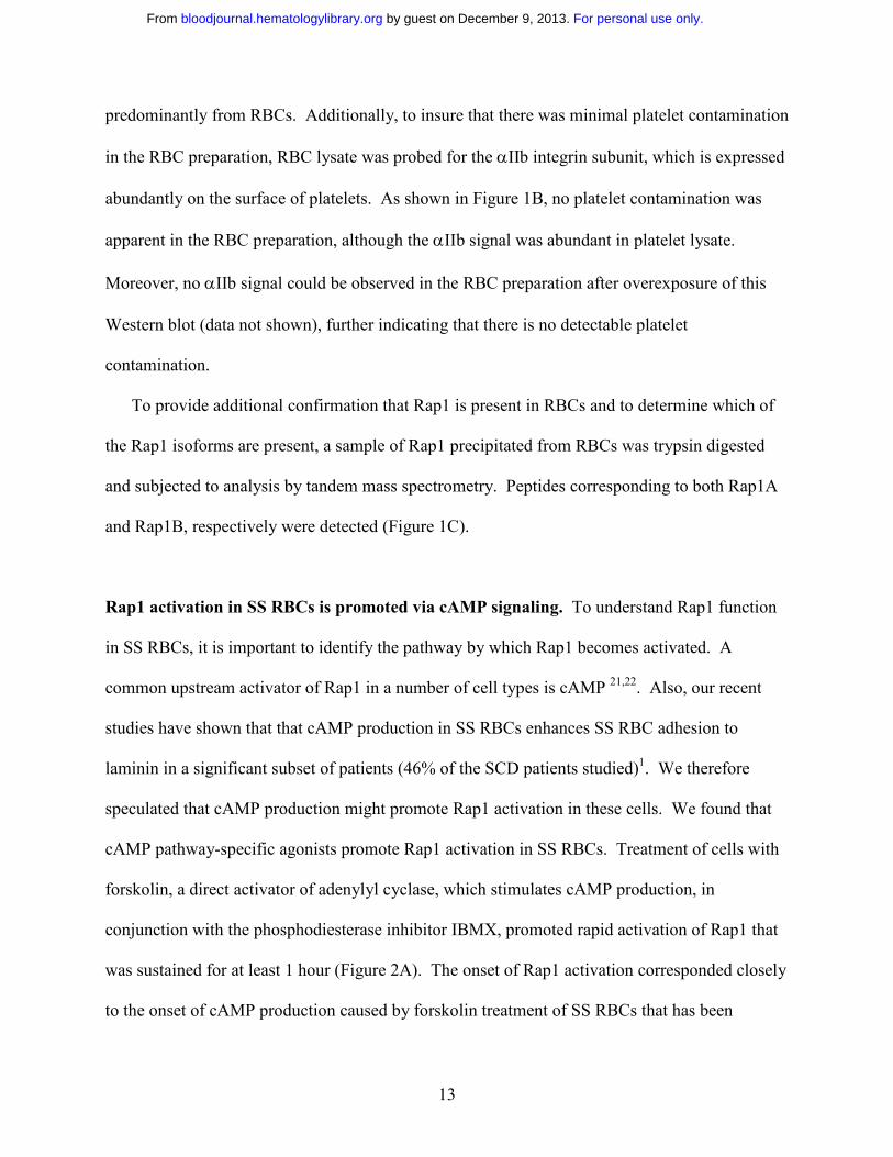
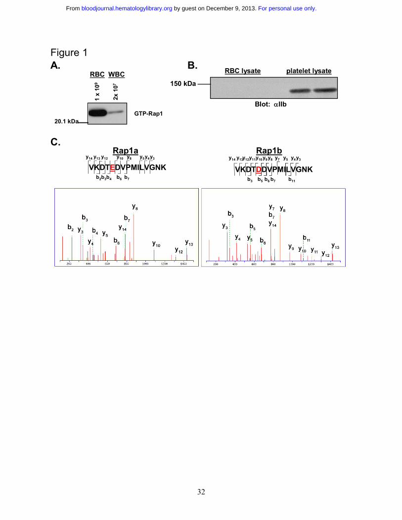
Determination of the presence of Rap1 in RBCs. Although Rap1 is abundant in platelets and
WBCs, its existence in RBCs has never been examined. To confirm that Rap1 exists in RBCs,
active Rap1 was precipitated with GST-RalGDS-RBD conjugated beads, which bind selectively
to the GTP-associated form of Rap119. Rap1 was then detected on Western blots with a Rap1-
specific antibody. White blood cells contain predominantly Rap1A, which is readily detected by
the antibody used for the Western blot20. As shown in figure 1A, RBCs exhibited a robust basal
Rap1 activation signal. While contamination of our RBC preparations by platelets was generally
undetectable, a low level of contamination by WBCs was possible. We therefore examined the
preparation microscopically and found the WBC contamination levels to be less than 0.01%. To
determine whether Rap1 from contaminating WBCs contributes to the observed Rap1 signal, a
concentration of WBCs corresponding to 2% contamination, an approximately 200 fold
overestimate of the contamination level, was examined for active Rap1. However, a minimal
signal was observed (Figure 1A), making it apparent that the observed signal is derived
For personal use only. by guest on December 9, 2013. bloodjournal.hematologylibrary.orgFrom
13
predominantly from RBCs. Additionally, to insure that there was minimal platelet contamination
in the RBC preparation, RBC lysate was probed for the αIIb integrin subunit, which is expressed
abundantly on the surface of platelets. As shown in Figure 1B, no platelet contamination was
apparent in the RBC preparation, although the αIIb signal was abundant in platelet lysate.
Moreover, no αIIb signal could be observed in the RBC preparation after overexposure of this
Western blot (data not shown), further indicating that there is no detectable platelet
contamination.
To provide additional confirmation that Rap1 is present in RBCs and to determine which of
the Rap1 isoforms are present, a sample of Rap1 precipitated from RBCs was trypsin digested
and subjected to analysis by tandem mass spectrometry. Peptides corresponding to both Rap1A
and Rap1B, respectively were detected (Figure 1C).
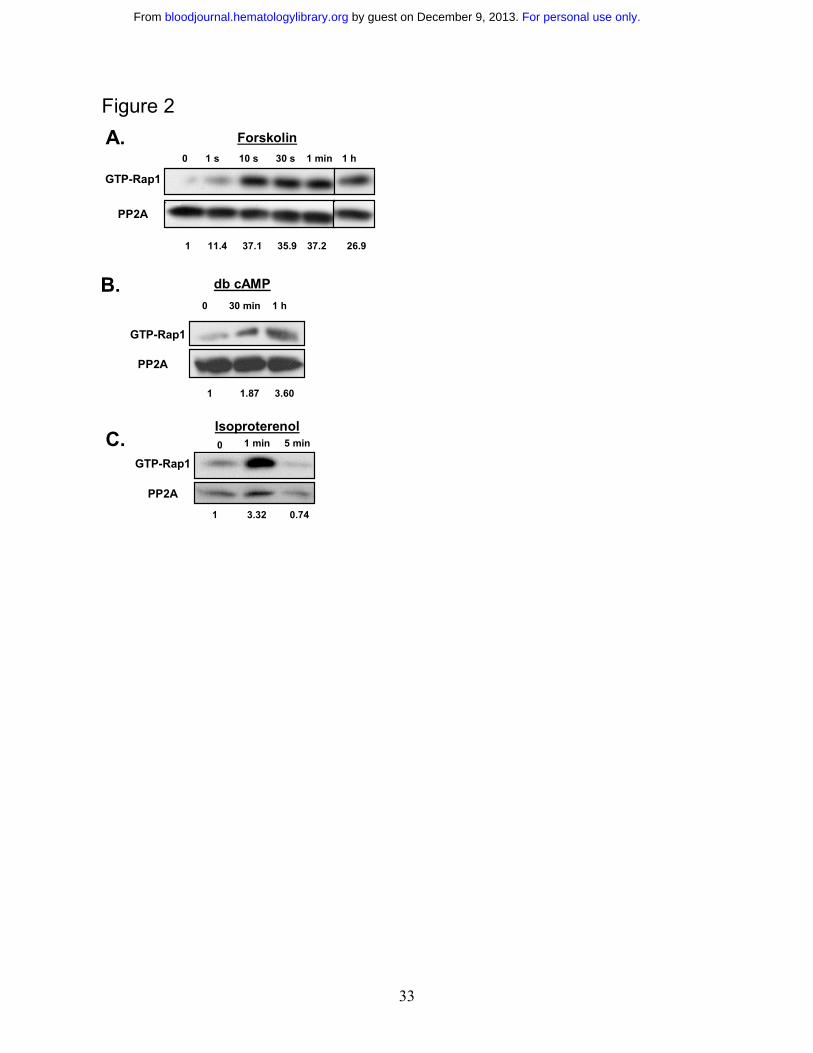
Rap1 activation in SS RBCs is promoted via cAMP signaling. To understand Rap1 function
in SS RBCs, it is important to identify the pathway by which Rap1 becomes activated. A
common upstream activator of Rap1 in a number of cell types is cAMP 21,22. Also, our recent
studies have shown that that cAMP production in SS RBCs enhances SS RBC adhesion to
laminin in a significant subset of patients (46% of the SCD patients studied)1. We therefore
speculated that cAMP production might promote Rap1 activation in these cells. We found that
cAMP pathway-specific agonists promote Rap1 activation in SS RBCs. Treatment of cells with
forskolin, a direct activator of adenylyl cyclase, which stimulates cAMP production, in
conjunction with the phosphodiesterase inhibitor IBMX, promoted rapid activation of Rap1 that
was sustained for at least 1 hour (Figure 2A). The onset of Rap1 activation corresponded closely
to the onset of cAMP production caused by forskolin treatment of SS RBCs that has been
For personal use only. by guest on December 9, 2013. bloodjournal.hematologylibrary.orgFrom
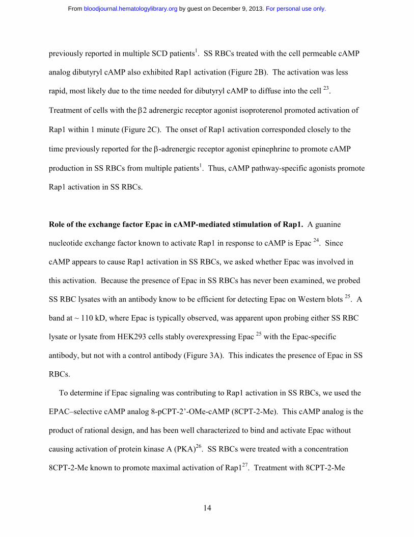
14
previously reported in multiple SCD patients1. SS RBCs treated with the cell permeable cAMP
analog dibutyryl cAMP also exhibited Rap1 activation (Figure 2B). The activation was less
rapid, most likely due to the time needed for dibutyryl cAMP to diffuse into the cell 23.
Treatment of cells with the β2 adrenergic receptor agonist isoproterenol promoted activation of
Rap1 within 1 minute (Figure 2C). The onset of Rap1 activation corresponded closely to the
time previously reported for the β-adrenergic receptor agonist epinephrine to promote cAMP
production in SS RBCs from multiple patients1. Thus, cAMP pathway-specific agonists promote
Rap1 activation in SS RBCs.
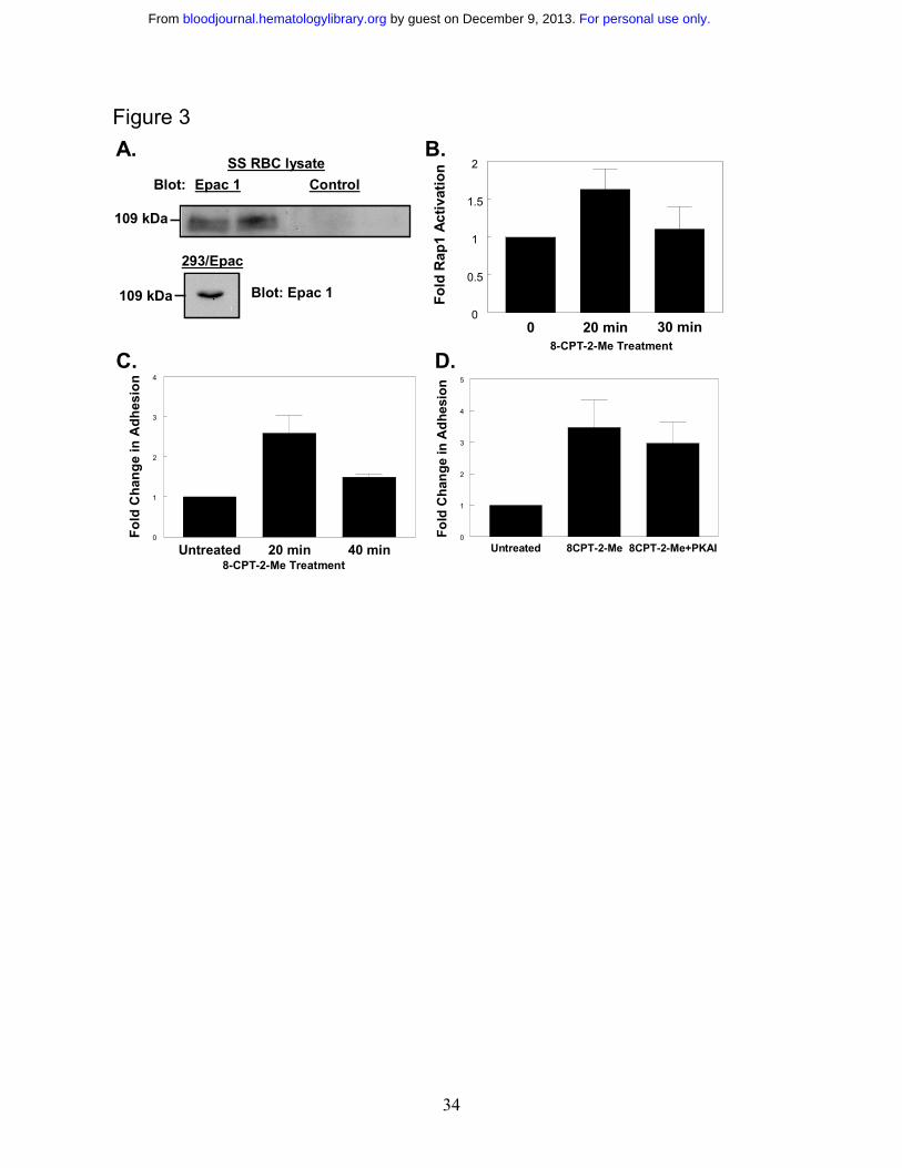
Role of the exchange factor Epac in cAMP-mediated stimulation of Rap1. A guanine
nucleotide exchange factor known to activate Rap1 in response to cAMP is Epac 24. Since
cAMP appears to cause Rap1 activation in SS RBCs, we asked whether Epac was involved in
this activation. Because the presence of Epac in SS RBCs has never been examined, we probed
SS RBC lysates with an antibody know to be efficient for detecting Epac on Western blots 25. A
band at ~ 110 kD, where Epac is typically observed, was apparent upon probing either SS RBC
lysate or lysate from HEK293 cells stably overexpressing Epac 25 with the Epac-specific
antibody, but not with a control antibody (Figure 3A). This indicates the presence of Epac in SS
RBCs.
To determine if Epac signaling was contributing to Rap1 activation in SS RBCs, we used the
EPAC–selective cAMP analog 8-pCPT-2’-OMe-cAMP (8CPT-2-Me). This cAMP analog is the
product of rational design, and has been well characterized to bind and activate Epac without
causing activation of protein kinase A (PKA)26. SS RBCs were treated with a concentration
8CPT-2-Me known to promote maximal activation of Rap127. Treatment with 8CPT-2-Me
For personal use only. by guest on December 9, 2013. bloodjournal.hematologylibrary.orgFrom
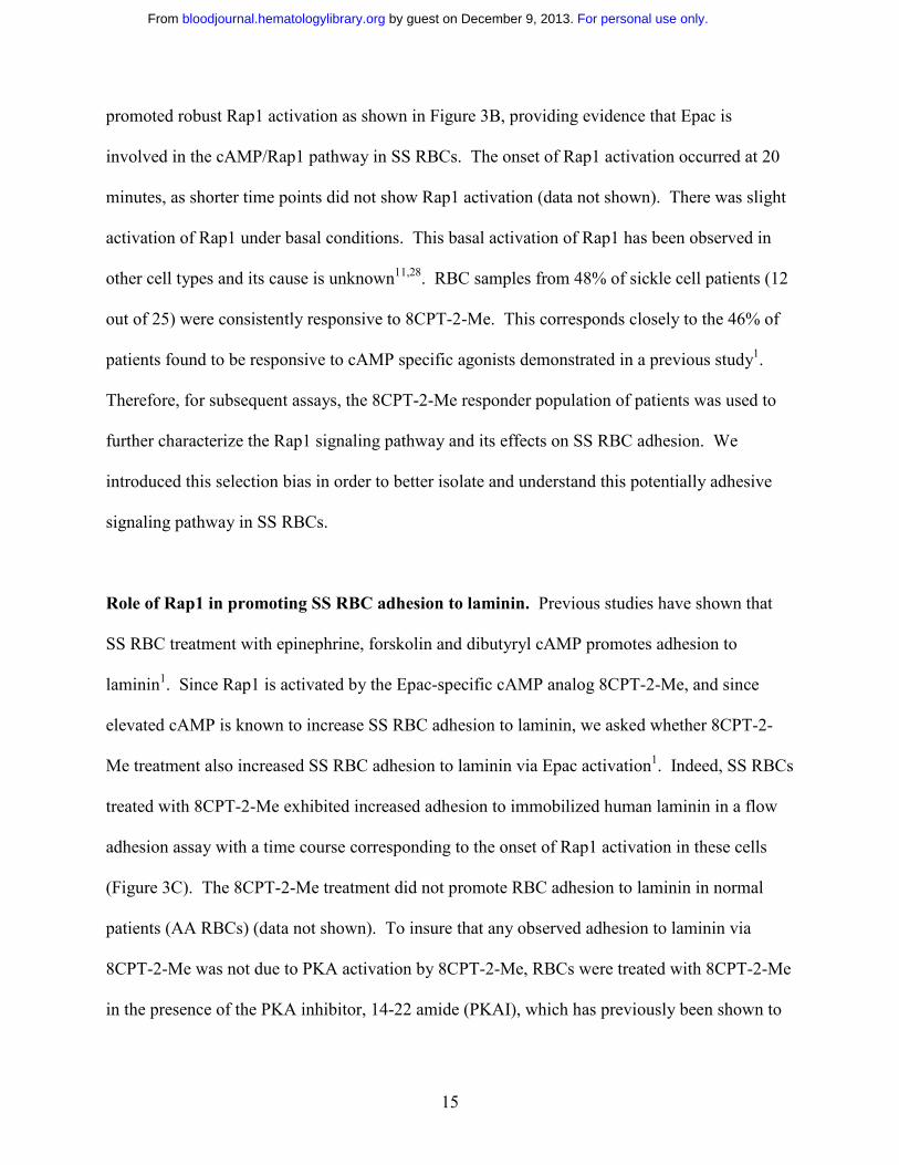
15
promoted robust Rap1 activation as shown in Figure 3B, providing evidence that Epac is
involved in the cAMP/Rap1 pathway in SS RBCs. The onset of Rap1 activation occurred at 20
minutes, as shorter time points did not show Rap1 activation (data not shown). There was slight
activation of Rap1 under basal conditions. This basal activation of Rap1 has been observed in
other cell types and its cause is unknown11,28. RBC samples from 48% of sickle cell patients (12
out of 25) were consistently responsive to 8CPT-2-Me. This corresponds closely to the 46% of
patients found to be responsive to cAMP specific agonists demonstrated in a previous study1.
Therefore, for subsequent assays, the 8CPT-2-Me responder population of patients was used to
further characterize the Rap1 signaling pathway and its effects on SS RBC adhesion. We
introduced this selection bias in order to better isolate and understand this potentially adhesive
signaling pathway in SS RBCs.
Role of Rap1 in promoting SS RBC adhesion to laminin. Previous studies have shown that
SS RBC treatment with epinephrine, forskolin and dibutyryl cAMP promotes adhesion to
laminin1. Since Rap1 is activated by the Epac-specific cAMP analog 8CPT-2-Me, and since
elevated cAMP is known to increase SS RBC adhesion to laminin, we asked whether 8CPT-2-
Me treatment also increased SS RBC adhesion to laminin via Epac activation1. Indeed, SS RBCs
treated with 8CPT-2-Me exhibited increased adhesion to immobilized human laminin in a flow
adhesion assay with a time course corresponding to the onset of Rap1 activation in these cells
(Figure 3C). The 8CPT-2-Me treatment did not promote RBC adhesion to laminin in normal
patients (AA RBCs) (data not shown). To insure that any observed adhesion to laminin via
8CPT-2-Me was not due to PKA activation by 8CPT-2-Me, RBCs were treated with 8CPT-2-Me
in the presence of the PKA inhibitor, 14-22 amide (PKAI), which has previously been shown to
For personal use only. by guest on December 9, 2013. bloodjournal.hematologylibrary.orgFrom
16
inhibit PKA-dependent SS RBC adhesion to laminin.1 As shown in Figure 3D, PKAI treatment
induced a small, but statistically insignificant decrease in SS RBC adhesion to laminin (p>0.05).
Thus, the adhesion is dependent on Epac/Rap1 signaling and not on PKA signaling.
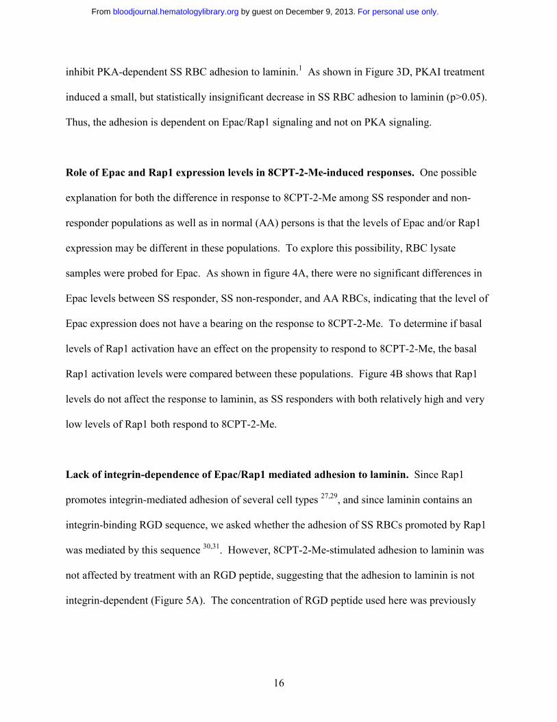
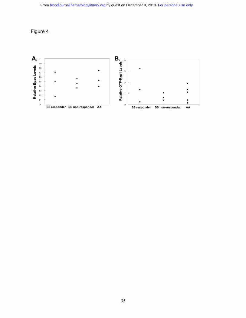
Role of Epac and Rap1 expression levels in 8CPT-2-Me-induced responses. One possible
explanation for both the difference in response to 8CPT-2-Me among SS responder and non-
responder populations as well as in normal (AA) persons is that the levels of Epac and/or Rap1
expression may be different in these populations. To explore this possibility, RBC lysate
samples were probed for Epac. As shown in figure 4A, there were no significant differences in
Epac levels between SS responder, SS non-responder, and AA RBCs, indicating that the level of
Epac expression does not have a bearing on the response to 8CPT-2-Me. To determine if basal
levels of Rap1 activation have an effect on the propensity to respond to 8CPT-2-Me, the basal
Rap1 activation levels were compared between these populations. Figure 4B shows that Rap1
levels do not affect the response to laminin, as SS responders with both relatively high and very
low levels of Rap1 both respond to 8CPT-2-Me.
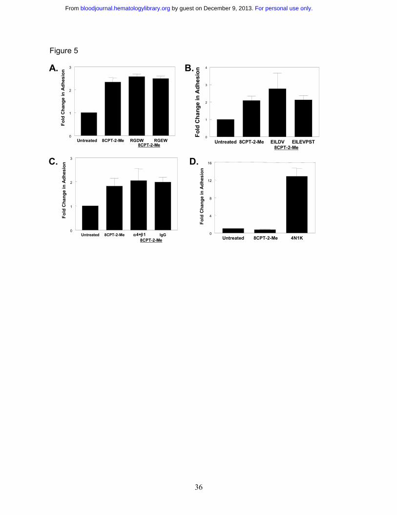
Lack of integrin-dependence of Epac/Rap1 mediated adhesion to laminin. Since Rap1
promotes integrin-mediated adhesion of several cell types 27,29, and since laminin contains an
integrin-binding RGD sequence, we asked whether the adhesion of SS RBCs promoted by Rap1
was mediated by this sequence 30,31. However, 8CPT-2-Me-stimulated adhesion to laminin was
not affected by treatment with an RGD peptide, suggesting that the adhesion to laminin is not
integrin-dependent (Figure 5A). The concentration of RGD peptide used here was previously
For personal use only. by guest on December 9, 2013. bloodjournal.hematologylibrary.orgFrom
17
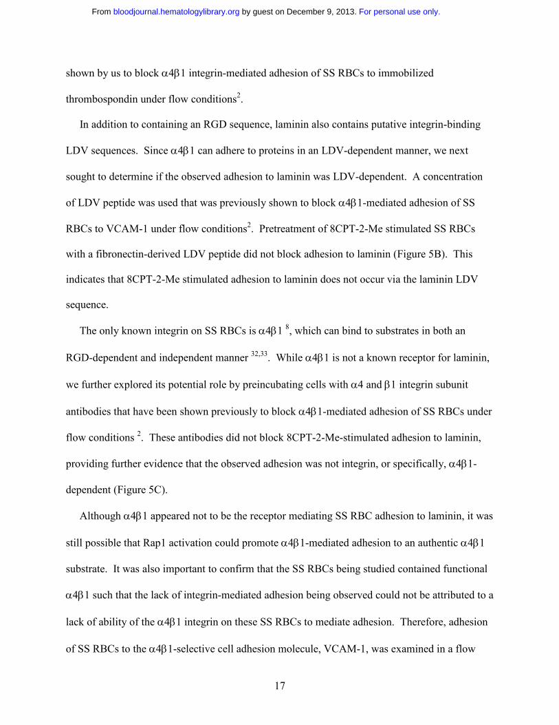
shown by us to block α4β1 integrin-mediated adhesion of SS RBCs to immobilized
thrombospondin under flow conditions2.
In addition to containing an RGD sequence, laminin also contains putative integrin-binding
LDV sequences. Since α4β1 can adhere to proteins in an LDV-dependent manner, we next
sought to determine if the observed adhesion to laminin was LDV-dependent. A concentration
of LDV peptide was used that was previously shown to block α4β1-mediated adhesion of SS
RBCs to VCAM-1 under flow conditions2. Pretreatment of 8CPT-2-Me stimulated SS RBCs
with a fibronectin-derived LDV peptide did not block adhesion to laminin (Figure 5B). This
indicates that 8CPT-2-Me stimulated adhesion to laminin does not occur via the laminin LDV
sequence.
The only known integrin on SS RBCs is α4β1 8, which can bind to substrates in both an
RGD-dependent and independent manner 32,33. While α4β1 is not a known receptor for laminin,
we further explored its potential role by preincubating cells with α4 and β1 integrin subunit
antibodies that have been shown previously to block α4β1-mediated adhesion of SS RBCs under
flow conditions 2. These antibodies did not block 8CPT-2-Me-stimulated adhesion to laminin,
providing further evidence that the observed adhesion was not integrin, or specifically, α4β1-
dependent (Figure 5C).
Although α4β1 appeared not to be the receptor mediating SS RBC adhesion to laminin, it was
still possible that Rap1 activation could promote α4β1-mediated adhesion to an authentic α4β1
substrate. It was also important to confirm that the SS RBCs being studied contained functional
α4β1 such that the lack of integrin-mediated adhesion being observed could not be attributed to a
lack of ability of the α4β1 integrin on these SS RBCs to mediate adhesion. Therefore, adhesion
of SS RBCs to the α4β1-selective cell adhesion molecule, VCAM-1, was examined in a flow
For personal use only. by guest on December 9, 2013. bloodjournal.hematologylibrary.orgFrom
18
adhesion assay2. Treatment with 8CPT-2-Me did not promote adhesion to an immobilized,
soluble version of VCAM-1. However, these cells did adhere to VCAM-1 in response to a
positive control, the 4N1K peptide derived from thrombospondin, which is known to promote
α4β1-mediated SS RBC adhesion to VCAM-1 via activation of CD47 on these cells (Figure
5D)2.
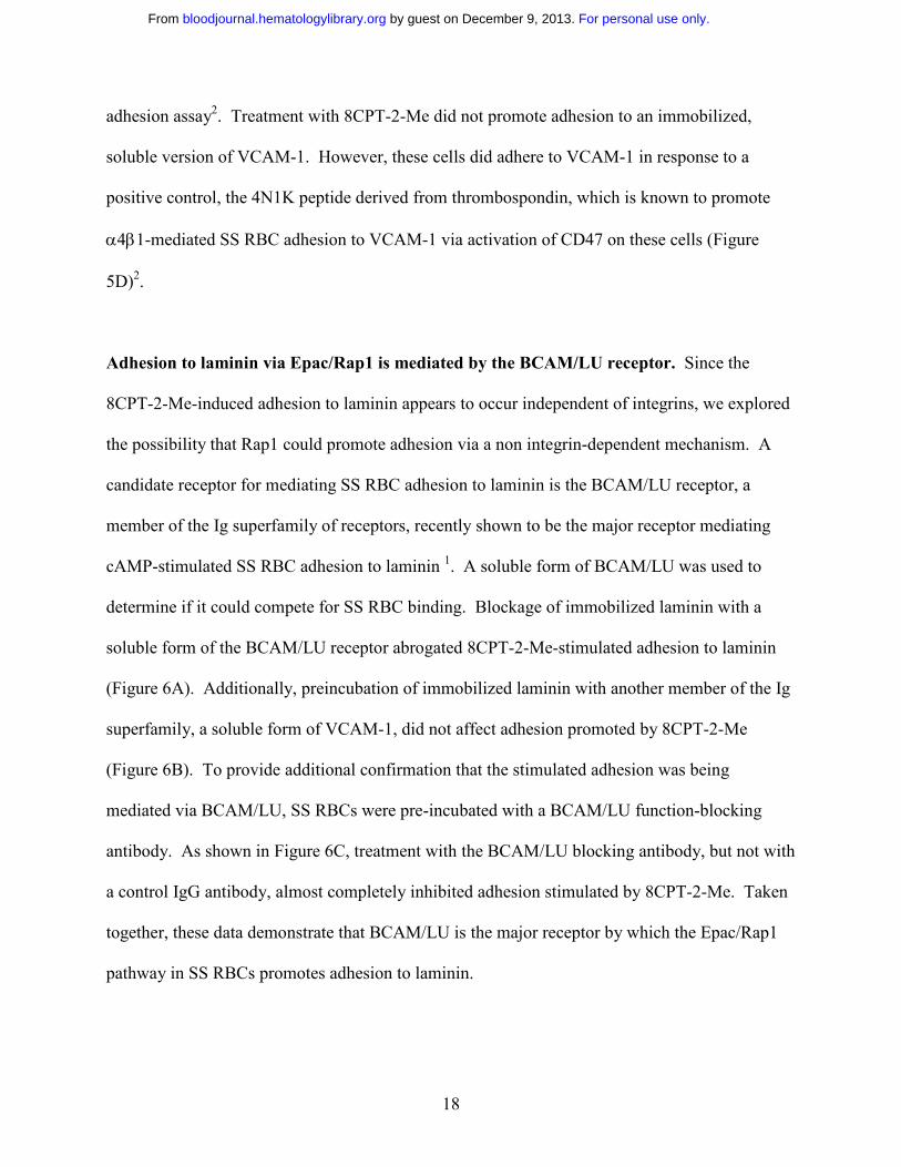
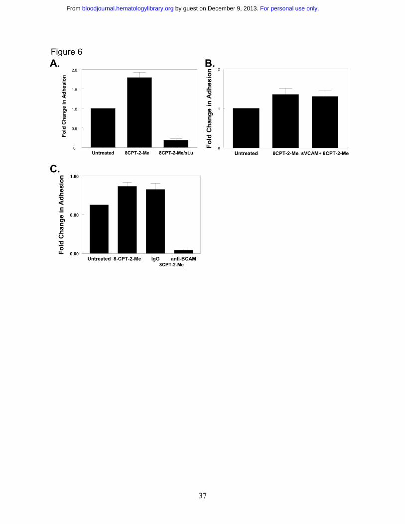
Adhesion to laminin via Epac/Rap1 is mediated by the BCAM/LU receptor. Since the
8CPT-2-Me-induced adhesion to laminin appears to occur independent of integrins, we explored
the possibility that Rap1 could promote adhesion via a non integrin-dependent mechanism. A
candidate receptor for mediating SS RBC adhesion to laminin is the BCAM/LU receptor, a
member of the Ig superfamily of receptors, recently shown to be the major receptor mediating
cAMP-stimulated SS RBC adhesion to laminin 1. A soluble form of BCAM/LU was used to
determine if it could compete for SS RBC binding. Blockage of immobilized laminin with a
soluble form of the BCAM/LU receptor abrogated 8CPT-2-Me-stimulated adhesion to laminin
(Figure 6A). Additionally, preincubation of immobilized laminin with another member of the Ig
superfamily, a soluble form of VCAM-1, did not affect adhesion promoted by 8CPT-2-Me
(Figure 6B). To provide additional confirmation that the stimulated adhesion was being
mediated via BCAM/LU, SS RBCs were pre-incubated with a BCAM/LU function-blocking
antibody. As shown in Figure 6C, treatment with the BCAM/LU blocking antibody, but not with
a control IgG antibody, almost completely inhibited adhesion stimulated by 8CPT-2-Me. Taken
together, these data demonstrate that BCAM/LU is the major receptor by which the Epac/Rap1
pathway in SS RBCs promotes adhesion to laminin.
For personal use only. by guest on December 9, 2013. bloodjournal.hematologylibrary.orgFrom
19
Discussion
Our results demonstrate that Rap1 is present and activated in response to agonists in SS
RBCs, and contributes to their adhesion to laminin, a protein present in both the blood vessel
wall and in SS RBC plasma34. Hence, Rap1 may be an important mediator of signaling leading
to vasoocclusion. Tandem mass spectromtetry data reveal that peptides corresponding to both
Rap1a and Rap1b are present, indicating that both Rap1 isoforms exist in RBCs. Rap1 in SS
RBCs can be activated by the agonists isoproterenol, forskolin and db cAMP, implicating a
cAMP-dependent mechanism of Rap1 activation in these cells. The role of cAMP in Rap1
activation has gained increased attention with the discovery of Epac, a cAMP-dependent Rap1
GEF, and development of the pharmacological tool 8-pCPT-2’OMe-cAMP (8CPT-2-Me), a
cAMP analog that has been well characterized to specifically bind and activate Epac, but not
PKA 26. Western blotting of SS RBC lysate confirmed the presence of Epac in these cells. This
is the first evidence that Epac is present in RBCs. By use of the analog 8CPT-2-Me, we found
that Epac contributes to cAMP-induced activation of Rap1 in SS RBCs. Additionally, 8CPT-2-
Me-induced Rap1 activation promotes SS RBC adhesion to the extracellular matrix protein
laminin that was not reduced by PKA inhibition. Since SS RBCs are anucleate, making
transfection impossible, treatment with 8CPT-2-Me is the only readily available means of
studying Epac function in these cells.
Only a subset of patients (48%) respond to 8CPT-2-Me. Patient variability, which manifests
as “responder” and “non-responder” SCD patient populations to particular cAMP specific
agonists, has been previously described1. We demonstrate that the levels of Epac and/or Rap1 in
RBCs have no bearing on whether or not there is a response to 8CPT-2-Me. Thus, the factors
that cause a certain population of SS RBCs to respond to this treatment are likely complex and
For personal use only. by guest on December 9, 2013. bloodjournal.hematologylibrary.orgFrom
20
multifactoral. As is the case with many other chronic diseases, this may be a reflection of the
broad range of clinical manifestations and severity observed with SCD, which may be due to
such issues as complex genetic backgrounds and/or other clinical factors.
Another possibility may be that the levels of downstream effectors between Rap1 and the
BCAM/LU receptor are different in these two populations. Thus, the SCD patients who respond
to 8CPT-2-Me have all of the signaling components in place to mount an adhesive response via
the BCAM/LU receptor. In the non-responder populations, some or all of these signaling
components may be missing, making an adhesive response impossible.
There has been some conjecture that the vaso-occlusive manifestations of SCD are due to the
high number of circulating reticulocytes found in SCD patients. However, previous studies have
demonstrated that the reticulocytes are not the RBC population involved in cAMP-dependent SS
RBC adhesion to laminin1. In fact, when the fold increase in adhesion to laminin in response to
epinephrine, via cAMP signaling, was plotted against the reticulocyte count of each
corresponding patient, no correlation was observed1. Further, an increase in the number of
reticulocytes within individual patients did not cause an increased epinephrine-induced adhesive
response to laminin1. In addition, persons with other hematologic disorders that cause high
reticulocyte counts do not necessarily experience vasooclusive events like those observed in
SCD. From the above findings, it was concluded that the responsive, adhesive SS RBC
population was at an intermediate stage of developmental relative to the immature reticulocytes
and more mature erythrocytes1. As RBCs mature, they lose their signaling capacity, with fully
mature RBCs having very little signaling capability. While reticulocytes per se do not appear to
cause the increased in epinephrine or cAMP-stimulated adhesion, the overall population of RBCs
in patients with SCD is younger. Thus, SS RBCs still retain more signaling capacity than a
For personal use only. by guest on December 9, 2013. bloodjournal.hematologylibrary.orgFrom
21
population of more mature AA RBCs and this makes the cells more likely to be responsive to
agonists.
In addition, the vasculature of persons with SCD tends to be extensively damaged. This
damage exposes extracellular matrix proteins, including laminin, to flowing blood, providing a
substrate for the adhesive SS RBCs. This, in conjunction with the hyper-coagulant and pro-
immflamatory state in which SCD patients typically exist, provides an environment that is far
more amenable to cellular adhesion and vasooclusion than would be typically found in normal
human physiology.
An additional reason that SS RBCs exhibit more adhesion to laminin compared to AA RBCs
may be due to differences in the expression of the BCAM/LU protein on the cell surface. A
previous study revealed that SS RBCs contain 67% more BCAM/LU than AA RBCs35. This
substantial increase in the amount of BCAM/LU could contribute to the increased adhesive
potential of SS RBCs.
SS RBCs provide a unique system for studying Rap1-promoted cellular adhesion, as they
contain only one integrin, α4β1 8,9. Since laminin contains an RGD sequence 31, the potential
existed for Rap1-stimulated adhesion to laminin to be RGD- and integrin-dependent. Although
α4β1-mediated adhesion is not necessarily RGD-dependent, it is in some instances. For
example, we have shown that α4β1-mediated adhesion of SS RBCs to thrombospondin is RGD-
dependent and another study has demonstrated that α4β1-mediated adhesion of B cells to
fibronectin is RGD-dependent2,33. However, SS RBC adhesion to laminin via Rap1 was not
inhibited by an RGD peptide. Also, incubation of SS RBCs with the LDV peptide, another
sequence in laminin that is potentially recognized by integrins, had no effect on Rap1-promoted
adhesion to laminin. In addition, incubation of SS RBCs with α4 and β1 integrin subunit
For personal use only. by guest on December 9, 2013. bloodjournal.hematologylibrary.orgFrom
22
adhesion-blocking antibodies did not affect the stimulated adhesion, confirming that α4β1 was
not the Rap1-stimulated receptor responsible for mediating the adhesion to laminin.
Upon elimination of α4β1 as the Rap1-stimulated receptor, we next considered the Ig
superfamily member, BCAM/LU, as it has been shown to mediate cAMP-dependent SS RBC
adhesion to laminin 1. Since both a soluble form of BCAM/LU and a BCAM/LU function-
blocking antibody abrogated the adhesion to laminin promoted via the Epac/Rap1 pathway, it
appears that Rap1 in these cells promotes adhesion via a non-integrin adhesion receptor. This is
the first time that Rap1 has been shown to promote adhesion via an Ig superfamily member.
However, it is not the first time that Rap1 has been linked to an integrin-independent adhesive
process. There is evidence that Rap1 may be involved in cell-cell contact by modulating
adherens junctions. In a recent study, it was reported that Rap1 is localized in adherens junctions
and is involved in the localization of adherens junctions within cells36. Another study
demonstrated that Rap1 may be involved in cell-cell adhesion mediated by E-cadherin11.
Surprisingly, we found that Epac-mediated Rap1 activation did not induce α4β1-mediated
adhesion to a known α4β1 substrate, VCAM-1, even though adhesion via this integrin could be
activated by another signaling pathway. This finding spawns the question of why a non-integrin
adhesion receptor is activated by Rap1 to the exclusion of an integrin, when integrins, including
α4β1, are known to be activated by Rap1 signaling37. The answer to this question may well exist
in the least well-understood aspect of Rap1 signaling—the downstream pathways by which Rap1
signals to adhesion receptors. It is reasonable to assume that the signaling molecules
downstream of Rap1 responsible for activating integrins would be different from the signaling
molecules responsible for activating other classes of adhesion receptors.
For personal use only. by guest on December 9, 2013. bloodjournal.hematologylibrary.orgFrom

23
Because Rap1 has been studied intensively in the integrin field, less attention has been paid to
Rap1-mediated activation of adhesion via other adhesion receptor families. Since BCAM/LU is
a member of the Ig superfamily of adhesion receptors, it is possible that Rap1 may also promote
adhesion via other Ig superfamily members.
A previous study in our laboratory demonstrated that the cAMP-dependent adhesion of SS
RBCs to laminin via BCAM/LU was PKA-dependent1. In this study, we demonstrate that
BCAM/LU mediated adhesion to laminin promoted by Epac/Rap1 signaling occurs independent
of PKA. Thus, it appears that cAMP signaling can promote adhesion to laminin via BCAM/Lu
through two divergent signaling pathways. The observation that adhesion promoted by
epinephrine, which can signal via both PKA and Epac, can be blocked via PKA inhibition
suggests that these pathways may synergize. Thus, the potential exists for both pathways to act
individually or in concert to promote an adhesive, vaso-occlusive pathology. The relative
contributions of these signaling pathways to promoting SS RBC adhesion and vasoocclusion
could be a subject of future investigation. Obtaining a better understanding of adhesive signaling
in SS RBCs with regard to Rap1 as well as other pathways may lead to more targeted approaches
for preventing and treating vasoocclusion.
Acknowledgements The authors would like to thank Dr. Eugene Orringer, Dell Strayhorn and
Susan Jones at the UNC Comprehensive Sickle Cell center and Shantres Clark in the Parise
laboratory for their assistance with obtaining patient blood samples. We thank Julia Brittian for
the 4N1K peptide and technical advice. The protein identification work was performed at the
UNC Michael Hooker Proteomics Core facility. We thank Christine Eyler at Duke University
for purified BCAM/LU. We would also like to thank Xiaodong Cheng at University of Texas,
Galveston for the HEK293/Epac lysate.
For personal use only. by guest on December 9, 2013. bloodjournal.hematologylibrary.orgFrom
24
Reference List
(1) Hines PC, Zen Q, Burney SN et al. Novel epinephrine and cyclic AMP-mediated activation of BCAM/Lu-dependent sickle (SS) RBC adhesion. Blood. 2003;101:3281-3287.
(2) Brittain JE, Han J, Ataga KI, Orringer EP, Parise LV. Mechanism of CD47-induced α4β1 Integrin Activation and Adhesion in Sickle Reticulocytes. J Biol Chem. 2004;279:42393-42402.
(3) Caron E. Cellular functions of the Rap1 GTP-binding protein: a pattern emerges. J Cell Sci. 2003;116:435-440.
(4) Bos JL. Epac: a new cAMP target and new avenues in cAMP research. Nat Rev Mol Cell Biol. 2003;4:733-738.
(5) de Bruyn KM, Zwartkruis FJ, de Rooij J, Akkerman JW, Bos JL. The small GTPase Rap1 is activated by turbulence and is involved in integrin αIIbβ3-mediated cell adhesion in human megakaryocytes. J Biol Chem. 2003;278:22412-22417.
(6) Reedquist KA, Ross E, Koop EA et al. The small GTPase, Rap1, mediates CD31-induced integrin adhesion. J Cell Biol. 2000;148:1151-1158.
(7) Udani M, Zen Q, Cottman M et al. Basal cell adhesion molecule/lutheran protein. The receptor critical for sickle cell adhesion to laminin. J Clin Invest. 1998;101:2550-2558.
(8) Joneckis CC, Ackley RL, Orringer EP, Wayner EA, Parise LV. Integrin α4β1 and glycoprotein IV (CD36) are expressed on circulating reticulocytes in sickle cell anemia. Blood. 1993;82:3548-3555.
(9) Swerlick RA, Eckman JR, Kumar A, Jeitler M, Wick TM. α4β1-integrin expression on sickle reticulocytes: vascular cell adhesion molecule-1-dependent binding to endothelium. Blood. 1993;82:1891-1899.
(10) Brittain JE, Mlinar KJ, Anderson CS, Orringer EP, Parise LV. Activation of sickle red blood cell adhesion via integrin-associated protein/CD47-induced signal transduction. J Clin Invest. 2001;107:1555-1562.
(11) Price LS, Hajdo-Milasinovic A, Zhao J et al. Rap1 regulates E-cadherin-mediated cell-cell adhesion. J Biol Chem. 2004.
(12) Larson MK, Chen H, Kahn ML et al. Identification of P2Y12-dependent and -independent mechanisms of glycoprotein VI-mediated Rap1 activation in platelets. Blood. 2003;101:1409-1415.
(13) Laemmli UK. Cleavage of structural proteins during the assembly of the head of bacteriophage T4. Nature. 1970;227:680-685.
For personal use only. by guest on December 9, 2013. bloodjournal.hematologylibrary.orgFrom
25
(14) Ferletta M, Ekblom P. Identification of laminin-10/11 as a strong cell adhesive complex for a normal and a malignant human epithelial cell line. J Cell Sci. 1999;112 ( Pt 1):1-10.
(15) Tiger CF, Champliaud MF, Pedrosa-Domellof F et al. Presence of laminin α5 chain and lack of laminin α1 chain during human muscle development and in muscular dystrophies. J Biol Chem. 1997;272:28590-28595.
(16) Lee SP, Cunningham ML, Hines PC et al. Sickle cell adhesion to laminin: potential role for the α5 chain. Blood. 1998;92:2951-2958.
(17) Hillery CA, Du MC, Wang WC, Scott JP. Hydroxyurea therapy decreases the in vitro adhesion of sickle erythrocytes to thrombospondin and laminin. Br J Haematol. 2000;109:322-327.
(18) Perkins DN, Pappin DJ, Creasy DM, Cottrell JS. Probability-based protein identification by searching sequence databases using mass spectrometry data. Electrophoresis. 1999;20:3551-3567.
(19) Franke B, Akkerman JW, Bos JL. Rapid Ca2+-mediated activation of Rap1 in human platelets. EMBO J. 1997;16:252-259.
(20) Quilliam LA, Mueller H, Bohl BP et al. Rap1A is a substrate for cyclic AMP-dependent protein kinase in human neutrophils. J Immunol. 1991;147:1628-1635.
(21) Dremier S, Vandeput F, Zwartkruis FJ et al. Activation of the small G protein Rap1 in dog thyroid cells by both cAMP-dependent and -independent pathways. Biochem Biophys Res Commun. 2000;267:7-11.
(22) Vossler MR, Yao H, York RD et al. cAMP activates MAP kinase and Elk-1 through a B-Raf- and Rap1-dependent pathway. Cell. 1997;89:73-82.
(23) Bartsch M, Zorn-Kruppa M, Kuhl N et al. Bioactivatable, membrane-permeant analogs of cyclic nucleotides as biological tools for growth control of C6 glioma cells. Biol Chem. 2003;384:1321-1326.
(24) de Rooij J, Zwartkruis FJ, Verheijen MH et al. Epac is a Rap1 guanine-nucleotide-exchange factor directly activated by cyclic AMP. Nature. 1998;396:474-477.
(25) Mei FC, Qiao J, Tsygankova OM et al. Differential signaling of cyclic AMP: opposing effects of exchange protein directly activated by cyclic AMP and cAMP-dependent protein kinase on protein kinase B activation. J Biol Chem. 2002;277:11497-11504.
(26) Enserink JM, Christensen AE, de Rooij J et al. A novel Epac-specific cAMP analogue demonstrates independent regulation of Rap1 and ERK. Nat Cell Biol. 2002;4:901-906.
(27) Rangarajan S, Enserink JM, Kuiperij HB et al. Cyclic AMP induces integrin-mediated cell adhesion through Epac and Rap1 upon stimulation of the β2-adrenergic receptor. J Cell Biol. 2003;160:487-493.
For personal use only. by guest on December 9, 2013. bloodjournal.hematologylibrary.orgFrom
26
(28) Enserink JM, Price LS, Methi T et al. The cAMP-Epac-Rap1 Pathway Regulates Cell Spreading and Cell Adhesion to Laminin-5 through the α3β1 Integrin but Not the α6β4Integrin. J Biol Chem. 2004;279:44889-44896.
(29) Liu L, Schwartz BR, Tupper J et al. The GTPase Rap1 regulates phorbol 12-myristate 13-acetate-stimulated but not ligand-induced β1 integrin-dependent leukocyte adhesion. J Biol Chem. 2002;277:40893-40900.
(30) Colucci S, Grano M, Zigrino P et al. Human osteoclast-like cells recognize laminin via an RGD independent mechanism. Boll Soc Ital Biol Sper. 1993;69:295-300.
(31) Sasaki T, Timpl R. Domain IVa of laminin alpha5 chain is cell-adhesive and binds β1and αVβ3 integrins through Arg-Gly-Asp. FEBS Lett. 2001;509:181-185.
(32) Decker S, van Valen F, Vischer P. Adhesion of osteosarcoma cells to the 70-kDa core region of thrombospondin-1 is mediated by the α4β1 integrin. Biochem Biophys Res Commun. 2002;293:86-92.
(33) Sanchez-Aparicio P, Dominguez-Jimenez C, Garcia-Pardo A. Activation of the α4β1integrin through the β1 subunit induces recognition of the RGDS sequence in fibronectin. J Cell Biol. 1994;126:271-279.
(34) Bolarin DM, Swerdlow P, Wallace AM, Littsey L. Serum concentrations of laminin P1 and aminoterminal propeptide of type III procollagen in sickle cell disease. Haematologia (Budap ). 1998;29:51-58.
(35) Zen Q, Cottman M, Truskey G, Fraser R, Telen MJ. Critical factors in basal cell adhesion molecule/lutheran-mediated adhesion to laminin. J Biol Chem. 1999;274:728-734.
(36) Knox AL, Brown NH. Rap1 GTPase regulation of adherens junction positioning and cell adhesion. Science. 2002;295:1285-1288.
(37) de Bruyn KM, Rangarajan S, Reedquist KA, Figdor CG, Bos JL. The small GTPase Rap1 is required for Mn(2+)- and antibody-induced LFA-1- and VLA-4-mediated cell adhesion. J Biol Chem. 2002;277:29468-29476.
For personal use only. by guest on December 9, 2013. bloodjournal.hematologylibrary.orgFrom
27
Figure Legends
Figure 1 Rap1 is present in a pure fraction of red blood cells. A. White blood cell
contamination does not contribute to the observed Rap1 signal. RBCs and WBCs were prepared
as described in the Materials and Methods section. RBCs were counted with a Coulter cell
counter and diluted to 1x10⁹ cells/mL. WBCs were counted microscopically on a
hemacytometer and adjusted to 2x10⁷ cells/mL (representing a 2% contamination level). The
cells were lysed and subjected to a GST-RalGDS-RBD pulldown assay. Rap1 was detected by
Western blotting with a Rap1 specific antibody. B. Platelet contamination is not detectable in
the RBC preparation. RBC and platelet lysates were probed by Western blotting with an αIIb-
specific antibody as described in the Materials and Methods section. C. Both Rap1a and Rap1b
are present in RBCs. Shown are MS/MS spectra corresponding to peptides from Rap1a and
Rap1b obtained from tryptic digestion of Rap protein from a GST-RalGDS-RBD pulldown of
RBCs. Spectra were acquired as described in the Materials and Methods section. Peptides
corresponding to each ion peak are labeled.
Figure 2 Rap1 is activated by the cAMP pathway in SS RBCs. A. Forskolin stimulates
Rap1 in SS RBCs. Cells were pretreated with 200 µM IBMX for 30 minutes to inhibit
phosphodiesterase activity. The cells were subsequently treated with 80 µM forskolin for the
time points shown. After lysis, GTP-bound Rap1 was precipitated with GST-RalGDS-RBD
beads and detected with a Rap1 specific antibody by Western blotting. The numbers shown
below the blot are relative densitometry values obtained by taking a ratio of the densitometry
value obtained from the Rap1 band to its corresponding PP2A loading control band. Values
For personal use only. by guest on December 9, 2013. bloodjournal.hematologylibrary.orgFrom
28
were then normalized to the zero time point. Densitometry for parts B and C were also analyzed
in this manner. The one hour time point is from the same Western blot as the other time points.
Data are representative of experiments from three different patient samples. B. Dibutyryl (db)
cAMP activates Rap1. SS RBCs were treated with 200 µM db cAMP for the indicated time
points. GTP bound Rap1 was detected as in Figure 2A. Data are representative of experiments
from five different patient samples. C. Rap1 is activated by isoproterenol in SS RBCs. Cells
were treated with 100 µM isoproterenol for the time points indicated. After lysis, GTP-bound
Rap1 was detected as in Fig 2A. Data are representative of experiments from five different
patient samples.
Figure 3 Epac contributes to Rap1 activation in SS RBCs. A. Epac is present in SS RBCs.
SS RBC lysate or HEK293/Epac lysate was separated by SDS-PAGE. The proteins were then
transferred to PVDF membrane and Western blotted with a rabbit anti-human Epac1 antibody
(left and below), or a rabbit, anti-human Rap1 control antibody (right). Duplicate lanes are
shown. B. Treatment with an Epac-specific cAMP analog stimulates Rap1. SS RBCs were
treated with 100 µM 8CPT-2-Me at the indicated time points. The cells were lysed, and GTP-
bound Rap1 was detected as in Figure 1A. Densitometry values were calculated as described in
Figure 2A. Results are expressed as mean +/- SE from four separate experiments. C
Stimulation of Rap1 via Epac promotes cellular adhesion to laminin. SS RBCs were treated with
100 µM 8CPT-2-Me at the times indicated. While still in the presence of 100 µM 8CPT-2-Me,
the cells were then flowed over chambers coated with 0.75 µg laminin in a flow adhesion assay.
Adhesion was quantified as described in the Materials and Methods section. Results are
expressed as mean +/- SE from one of three similar experiments. D. Inhibition of PKA has no
For personal use only. by guest on December 9, 2013. bloodjournal.hematologylibrary.orgFrom
29
effect on 8CPT-stimulated adhesion. SS RBCs were untreated or pretreated with 87nM PKAI
for 1 hr and then 100 µM 8CPT-2-Me was added in with the PKAI for 20 minutes. The cells,
while still in the presence of these pharmacological agents, were flowed across chambers coated
with 0.75 µg laminin in a flow adhesion assay. Adhesion was quantified as described in the
Materials and Methods section. Results are expressed as mean +/-SE from four separate
experiments.
Figure 4. Response to 8CPT-2-Me is not dependent on Epac or Rap1 protein levels in SS
RBCs. A. Response to 8CPT-2-Me is not dependent on the levels of Epac protein. An
equivalent concentration of RBC lysate was derived from nine previously characterized donors
relative to 8CPT-2-Me-induced RBC adhesion to laminin (3 SS responder, 3 SS non-responder,
and 3 AA). The lysate was separated on a 6% SDS polyacrylamide gel under reducing
conditions, transferred to a PVDF membrane, and blotted for Epac1 with an Epac1 monoclonal
antibody. PP2A was used as a loading control. Relative Epac protein levels were determined by
taking a ratio of the densitometry value obtained from the Epac sample to the densitometry value
from the corresponding PP2A loading control and plotted for each patient. B. Response to
8CPT-2-Me is independent of basal GTP-Rap1 levels. RBC lysate from eleven patients (3 SS
responder, 3 SS non-responder, and 5 AA), of equivalent RBC concentration were assayed for
GTP-Rap1 with GST-RalGDS-RBD beads as described in the Materials and Methods section.
Rap1 was then detected on a Western blot with a Rap1-specific antibody, with PP2A as a loading
control. Relative Rap1 values were obtained by densitometry as described in Figure 2A. The
relative GTP-Rap1 value obtained for each patient is represented as a dot.
For personal use only. by guest on December 9, 2013. bloodjournal.hematologylibrary.orgFrom
30
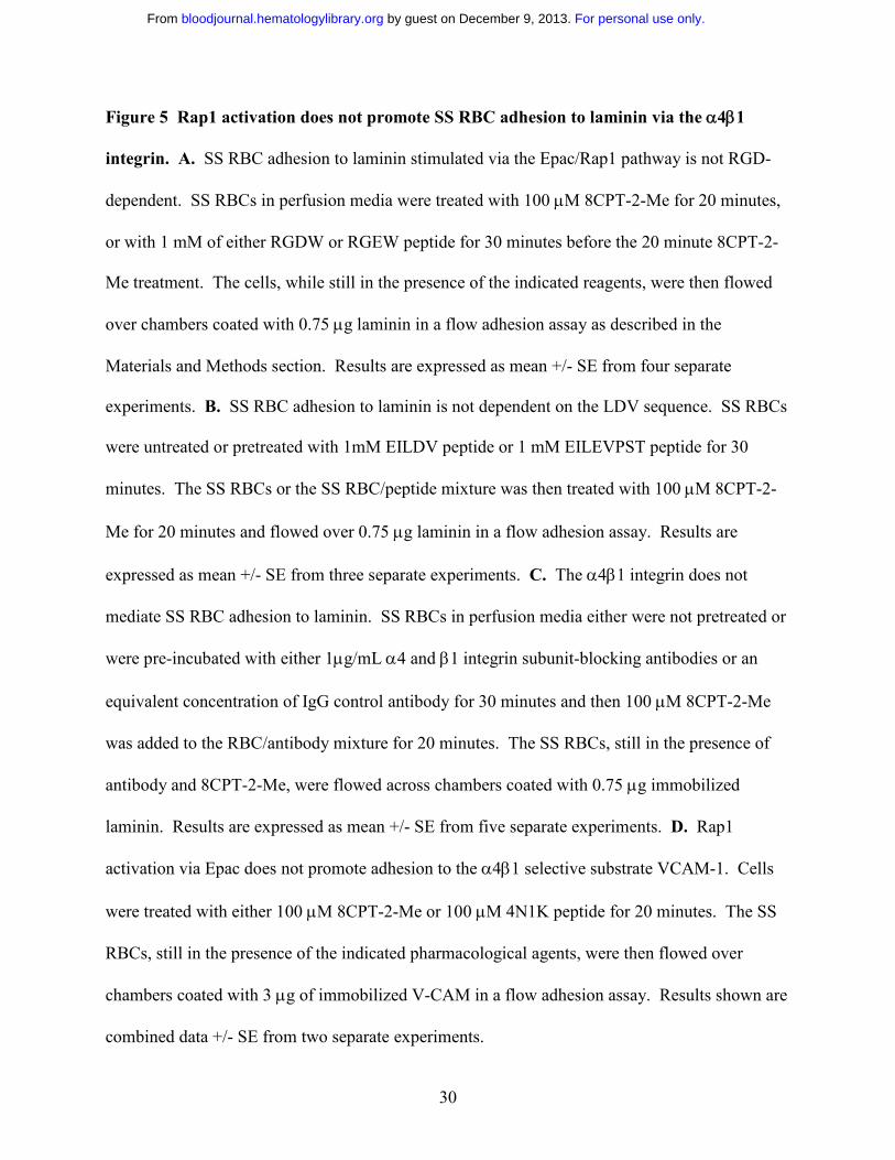
Figure 5 Rap1 activation does not promote SS RBC adhesion to laminin via the α4β1
integrin. A. SS RBC adhesion to laminin stimulated via the Epac/Rap1 pathway is not RGD-
dependent. SS RBCs in perfusion media were treated with 100 µM 8CPT-2-Me for 20 minutes,
or with 1 mM of either RGDW or RGEW peptide for 30 minutes before the 20 minute 8CPT-2-
Me treatment. The cells, while still in the presence of the indicated reagents, were then flowed
over chambers coated with 0.75 µg laminin in a flow adhesion assay as described in the
Materials and Methods section. Results are expressed as mean +/- SE from four separate
experiments. B. SS RBC adhesion to laminin is not dependent on the LDV sequence. SS RBCs
were untreated or pretreated with 1mM EILDV peptide or 1 mM EILEVPST peptide for 30
minutes. The SS RBCs or the SS RBC/peptide mixture was then treated with 100 µM 8CPT-2-
Me for 20 minutes and flowed over 0.75 µg laminin in a flow adhesion assay. Results are
expressed as mean +/- SE from three separate experiments. C. The α4β1 integrin does not
mediate SS RBC adhesion to laminin. SS RBCs in perfusion media either were not pretreated or
were pre-incubated with either 1µg/mL α4 and β1 integrin subunit-blocking antibodies or an
equivalent concentration of IgG control antibody for 30 minutes and then 100 µM 8CPT-2-Me
was added to the RBC/antibody mixture for 20 minutes. The SS RBCs, still in the presence of
antibody and 8CPT-2-Me, were flowed across chambers coated with 0.75 µg immobilized
laminin. Results are expressed as mean +/- SE from five separate experiments. D. Rap1
activation via Epac does not promote adhesion to the α4β1 selective substrate VCAM-1. Cells
were treated with either 100 µM 8CPT-2-Me or 100 µM 4N1K peptide for 20 minutes. The SS
RBCs, still in the presence of the indicated pharmacological agents, were then flowed over
chambers coated with 3 µg of immobilized V-CAM in a flow adhesion assay. Results shown are
combined data +/- SE from two separate experiments.
For personal use only. by guest on December 9, 2013. bloodjournal.hematologylibrary.orgFrom
31
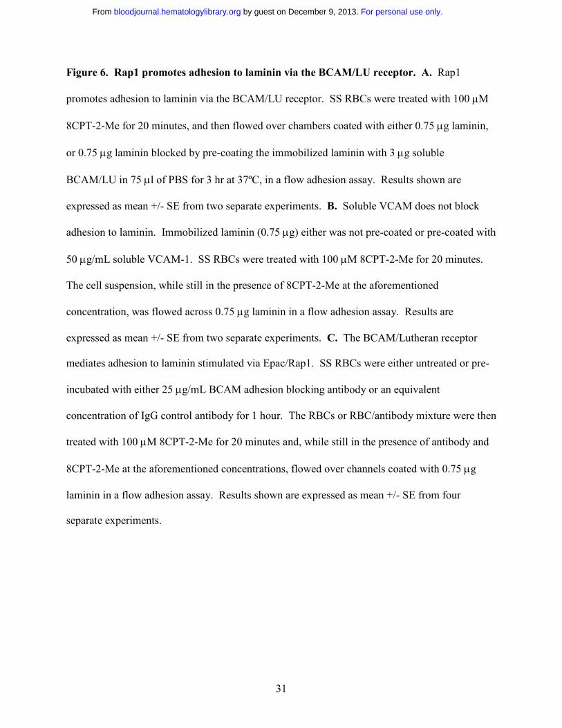
Figure 6. Rap1 promotes adhesion to laminin via the BCAM/LU receptor. A. Rap1
promotes adhesion to laminin via the BCAM/LU receptor. SS RBCs were treated with 100 µM
8CPT-2-Me for 20 minutes, and then flowed over chambers coated with either 0.75 µg laminin,
or 0.75 µg laminin blocked by pre-coating the immobilized laminin with 3 µg soluble
BCAM/LU in 75 µl of PBS for 3 hr at 37ºC, in a flow adhesion assay. Results shown are
expressed as mean +/- SE from two separate experiments. B. Soluble VCAM does not block
adhesion to laminin. Immobilized laminin (0.75 µg) either was not pre-coated or pre-coated with
50 µg/mL soluble VCAM-1. SS RBCs were treated with 100 µM 8CPT-2-Me for 20 minutes.
The cell suspension, while still in the presence of 8CPT-2-Me at the aforementioned
concentration, was flowed across 0.75 µg laminin in a flow adhesion assay. Results are
expressed as mean +/- SE from two separate experiments. C. The BCAM/Lutheran receptor
mediates adhesion to laminin stimulated via Epac/Rap1. SS RBCs were either untreated or pre-
incubated with either 25 µg/mL BCAM adhesion blocking antibody or an equivalent
concentration of IgG control antibody for 1 hour. The RBCs or RBC/antibody mixture were then
treated with 100 µM 8CPT-2-Me for 20 minutes and, while still in the presence of antibody and
8CPT-2-Me at the aforementioned concentrations, flowed over channels coated with 0.75 µg
laminin in a flow adhesion assay. Results shown are expressed as mean +/- SE from four
separate experiments.
For personal use only. by guest on December 9, 2013. bloodjournal.hematologylibrary.orgFrom
32
A.Figure 1
RBC WBC
1x
109
2x10
7GTP-Rap1
C.Rap1a Rap1b
VKDTEDVPMILVGNKb2b3b4 b7b6
y3y4y8 y5y13y14 y12 y10
VKDTDDVPMILVGNKb3 b5 b7b6 b11
y3y5 y4y8y9y10y11y12y13y14 y7
B. RBC lysate platelet lysate
150 kDa
Blot: αIIb
20.1 kDa
For personal use only. by guest on December 9, 2013. bloodjournal.hematologylibrary.orgFrom
33
B. db cAMP
GTP-Rap1
PP2A
0 30 min 1 h
1 1.87 3.60
A.
GTP-Rap1
PP2A
Forskolin0 1 s 10 s 30 s 1 min 1 h
1 11.4 37.1 35.9 37.2 26.9
Figure 2
C.Isoproterenol0 5 min1 min
GTP-Rap1
PP2A
1 3.32 0.74
For personal use only. by guest on December 9, 2013. bloodjournal.hematologylibrary.orgFrom
34
Figure 3B.A.
Epac 1 ControlBlot:
109 kDa
D.C.
Untreated 20 min 40 min0
1
2
3
4
Fold
Cha
nge
inA
dhes
ion
0 20 min 30 min8-CPT-2-Me Treatment
0
0.5
1
1.5
2
Fold
Rap
1Ac
tivat
ion
8-CPT-2-Me TreatmentUntreated 8CPT-2-Me 8CPT-2-Me+PKAI
0
1
2
3
4
5
Fold
Cha
nge
inA
dhes
ion
293/Epac
Blot: Epac 1109 kDa
SS RBC lysate
For personal use only. by guest on December 9, 2013. bloodjournal.hematologylibrary.orgFrom
35
Figure 4
A. B.
SS responder SS non-responder AA0
1
2
3
4
Rel
ativ
eG
TP-R
ap1
Leve
ls
SS responder SS non-responder AA0
0.1
0.2
0.3
0.4
0.5
0.6
0.7
0.8
0.9
1
Rel
ativ
eEp
acLe
vels
For personal use only. by guest on December 9, 2013. bloodjournal.hematologylibrary.orgFrom
36
Figure 5
A. B.
8CPT-2-MeUntreated 8CPT-2-Me RGDW RGEW
0
1
2
3Fo
ldC
hang
ein
Adh
esio
n
C.
α4+β18CPT-2-Me
Untreated 8CPT-2-Me IgG0
1
2
3
Fold
Cha
nge
inA
dhes
ion
Untreated 8CPT-2-Me 4N1K0
4
8
12
16
Fold
Cha
nge
inA
dhes
ion
D.
Untreated 8CPT-2-Me EILDV EILEVPST0
1
2
3
4
Fold
Cha
nge
inA
dhes
ion
8CPT-2-Me
For personal use only. by guest on December 9, 2013. bloodjournal.hematologylibrary.orgFrom
37
8CPT-2-Me
A.
Untreated 8CPT-2-Me 8CPT-2-Me/sLu0
0.5
1.0
1.5
2.0Fo
ldC
hang
ein
Adh
esio
nB.
Figure 6
Untreated 8-CPT-2-Me IgG anti-BCAM0.00
0.80
1.60
Fold
Cha
nge
inA
dhes
ion
Untreated 8CPT-2-Me sVCAM+ 8CPT-2-Me0
1
2
Fold
Cha
nge
inA
dhes
ion
C.
For personal use only. by guest on December 9, 2013. bloodjournal.hematologylibrary.orgFrom
Related Documents


![Dynamic changes in Rap1 activity are required for cell ... · Rap1 controls adhesion in mitosis 2997 Rap1[Q63E] mutant or negative regulators of Rap1 (such as Rap1GAP or Spa-1) respectively](https://static.cupdf.com/doc/110x72/5f2879addd4b6650a03cdafd/dynamic-changes-in-rap1-activity-are-required-for-cell-rap1-controls-adhesion.jpg)


