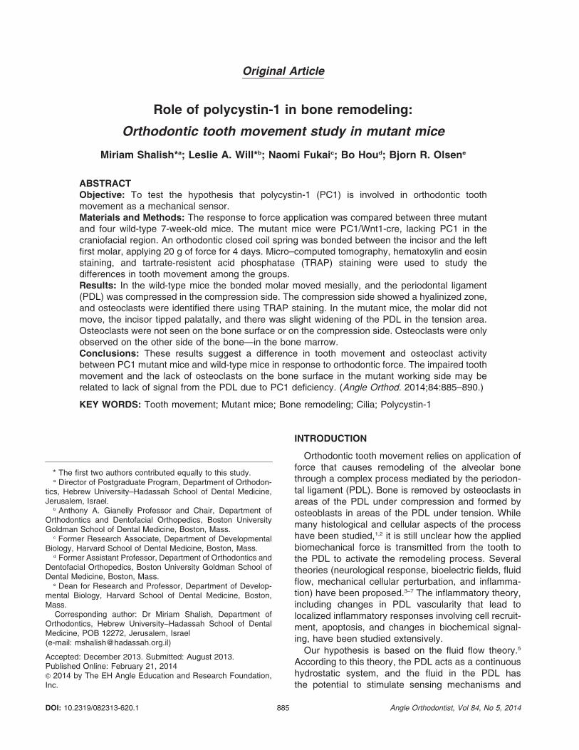
Original Article Role of polycystin-1 in bone remodeling: Orthodontic tooth movement study in mutant mice Miriam Shalish* a ; Leslie A. Will* b ; Naomi Fukai c ; Bo Hou d ; Bjorn R. Olsen e ABSTRACT Objective: To test the hypothesis that polycystin-1 (PC1) is involved in orthodontic tooth movement as a mechanical sensor. Materials and Methods: The response to force application was compared between three mutant and four wild-type 7-week-old mice. The mutant mice were PC1/Wnt1-cre, lacking PC1 in the craniofacial region. An orthodontic closed coil spring was bonded between the incisor and the left first molar, applying 20 g of force for 4 days. Micro–computed tomography, hematoxylin and eosin staining, and tartrate-resistent acid phosphatase (TRAP) staining were used to study the differences in tooth movement among the groups. Results: In the wild-type mice the bonded molar moved mesially, and the periodontal ligament (PDL) was compressed in the compression side. The compression side showed a hyalinized zone, and osteoclasts were identified there using TRAP staining. In the mutant mice, the molar did not move, the incisor tipped palatally, and there was slight widening of the PDL in the tension area. Osteoclasts were not seen on the bone surface or on the compression side. Osteoclasts were only observed on the other side of the bone—in the bone marrow. Conclusions: These results suggest a difference in tooth movement and osteoclast activity between PC1 mutant mice and wild-type mice in response to orthodontic force. The impaired tooth movement and the lack of osteoclasts on the bone surface in the mutant working side may be related to lack of signal from the PDL due to PC1 deficiency. (Angle Orthod. 2014;84:885–890.) KEY WORDS: Tooth movement; Mutant mice; Bone remodeling; Cilia; Polycystin-1 INTRODUCTION Orthodontic tooth movement relies on application of force that causes remodeling of the alveolar bone through a complex process mediated by the periodon- tal ligament (PDL). Bone is removed by osteoclasts in areas of the PDL under compression and formed by osteoblasts in areas of the PDL under tension. While many histological and cellular aspects of the process have been studied, 1,2 it is still unclear how the applied biomechanical force is transmitted from the tooth to the PDL to activate the remodeling process. Several theories (neurological response, bioelectric fields, fluid flow, mechanical cellular perturbation, and inflamma- tion) have been proposed. 3–7 The inflammatory theory, including changes in PDL vascularity that lead to localized inflammatory responses involving cell recruit- ment, apoptosis, and changes in biochemical signal- ing, have been studied extensively. Our hypothesis is based on the fluid flow theory. 5 According to this theory, the PDL acts as a continuous hydrostatic system, and the fluid in the PDL has the potential to stimulate sensing mechanisms and * The first two authors contributed equally to this study. a Director of Postgraduate Program, Department of Orthodon- tics, Hebrew University–Hadassah School of Dental Medicine, Jerusalem, Israel. b Anthony A. Gianelly Professor and Chair, Department of Orthodontics and Dentofacial Orthopedics, Boston University Goldman School of Dental Medicine, Boston, Mass. c Former Research Associate, Department of Developmental Biology, Harvard School of Dental Medicine, Boston, Mass. d Former Assistant Professor, Department of Orthodontics and Dentofacial Orthopedics, Boston University Goldman School of Dental Medicine, Boston, Mass. e Dean for Research and Professor, Department of Develop- mental Biology, Harvard School of Dental Medicine, Boston, Mass. Corresponding author: Dr Miriam Shalish, Department of Orthodontics, Hebrew University–Hadassah School of Dental Medicine, POB 12272, Jerusalem, Israel (e-mail: [email protected]) Accepted: December 2013. Submitted: August 2013. Published Online: February 21, 2014 G 2014 by The EH Angle Education and Research Foundation, Inc. DOI: 10.2319/082313-620.1 885 Angle Orthodontist, Vol 84, No 5, 2014

Role of polycystin 1 in bone remodeling- orthodontic tooth movement study in mutant mice
Jan 24, 2017
Welcome message from author
This document is posted to help you gain knowledge. Please leave a comment to let me know what you think about it! Share it to your friends and learn new things together.
Transcript
Original Article
Role of polycystin-1 in bone remodeling:
Orthodontic tooth movement study in mutant mice
Miriam Shalish*a; Leslie A. Will*b; Naomi Fukaic; Bo Houd; Bjorn R. Olsene
ABSTRACTObjective: To test the hypothesis that polycystin-1 (PC1) is involved in orthodontic toothmovement as a mechanical sensor.Materials and Methods: The response to force application was compared between three mutantand four wild-type 7-week-old mice. The mutant mice were PC1/Wnt1-cre, lacking PC1 in thecraniofacial region. An orthodontic closed coil spring was bonded between the incisor and the leftfirst molar, applying 20 g of force for 4 days. Micro–computed tomography, hematoxylin and eosinstaining, and tartrate-resistent acid phosphatase (TRAP) staining were used to study thedifferences in tooth movement among the groups.Results: In the wild-type mice the bonded molar moved mesially, and the periodontal ligament(PDL) was compressed in the compression side. The compression side showed a hyalinized zone,and osteoclasts were identified there using TRAP staining. In the mutant mice, the molar did notmove, the incisor tipped palatally, and there was slight widening of the PDL in the tension area.Osteoclasts were not seen on the bone surface or on the compression side. Osteoclasts were onlyobserved on the other side of the bone—in the bone marrow.Conclusions: These results suggest a difference in tooth movement and osteoclast activitybetween PC1 mutant mice and wild-type mice in response to orthodontic force. The impaired toothmovement and the lack of osteoclasts on the bone surface in the mutant working side may berelated to lack of signal from the PDL due to PC1 deficiency. (Angle Orthod. 2014;84:885–890.)
KEY WORDS: Tooth movement; Mutant mice; Bone remodeling; Cilia; Polycystin-1
INTRODUCTION
Orthodontic tooth movement relies on application offorce that causes remodeling of the alveolar bonethrough a complex process mediated by the periodon-tal ligament (PDL). Bone is removed by osteoclasts inareas of the PDL under compression and formed byosteoblasts in areas of the PDL under tension. Whilemany histological and cellular aspects of the processhave been studied,1,2 it is still unclear how the appliedbiomechanical force is transmitted from the tooth tothe PDL to activate the remodeling process. Severaltheories (neurological response, bioelectric fields, fluidflow, mechanical cellular perturbation, and inflamma-tion) have been proposed.3–7 The inflammatory theory,including changes in PDL vascularity that lead tolocalized inflammatory responses involving cell recruit-ment, apoptosis, and changes in biochemical signal-ing, have been studied extensively.
Our hypothesis is based on the fluid flow theory.5
According to this theory, the PDL acts as a continuoushydrostatic system, and the fluid in the PDL hasthe potential to stimulate sensing mechanisms and
* The first two authors contributed equally to this study.a Director of Postgraduate Program, Department of Orthodon-
tics, Hebrew University–Hadassah School of Dental Medicine,Jerusalem, Israel.
b Anthony A. Gianelly Professor and Chair, Department ofOrthodontics and Dentofacial Orthopedics, Boston UniversityGoldman School of Dental Medicine, Boston, Mass.
c Former Research Associate, Department of DevelopmentalBiology, Harvard School of Dental Medicine, Boston, Mass.
d Former Assistant Professor, Department of Orthodontics andDentofacial Orthopedics, Boston University Goldman School ofDental Medicine, Boston, Mass.
e Dean for Research and Professor, Department of Develop-mental Biology, Harvard School of Dental Medicine, Boston,Mass.
Corresponding author: Dr Miriam Shalish, Department ofOrthodontics, Hebrew University–Hadassah School of DentalMedicine, POB 12272, Jerusalem, Israel(e-mail: [email protected])
Accepted: December 2013. Submitted: August 2013.Published Online: February 21, 2014G 2014 by The EH Angle Education and Research Foundation,Inc.
DOI: 10.2319/082313-620.1 885 Angle Orthodontist, Vol 84, No 5, 2014
mediate a cellular response. The bony wall of the dentalsocket contains fenestrations that allow compressedfluid in the PDL to be expressed into the marrowspaces. Thus, changes in fluid flow are not only felt byPDL cells but also by osteocytes located in the adjacentbone, and cells in the marrow spaces could mediate acellular response. Fluid flow dynamics have also beenimplicated in the stimulation of a number of cell types viaprimary cilium.8 This raises the possibility that the linkbetween the extracellular matrix, fluid flow, and cellularmechanotransduction may be the primary cilium.
A previous study9 suggested that primary cilia, withtheir associated ion channels and signaling molecules,play an important role in fluid detection by kidneyepithelial cells. A subsequent study10 demonstrated thata Ca2+ channel complex, composed of the transmem-brane proteins polycystin-1 (PC1) and polycystin-2,located at the base of the primary cilium, mediatesthe sensing of cilia bending. This discovery offered adetailed insight into the way cells sense dynamic fluidmovements and transmit signals by means of calciuminflux into the cells. Based on these findings, primarycilia have been proposed to act as mechanosensors inosteocytes8 and chondrocytes,11 capable of regulatingbone remodeling. Numerous studies of kidney cellsdescribe a direct mechanical stimulation of primary ciliatriggered by fluid flow dynamics. Recent reports12,13
suggest that cultured osteoblastic cells respond to fluidflow by altering gene expression. As such, fluid flow isone of the most potent mechanical stimulators for bonecells.14 It has also been suggested8 that bone andkidney cells might share certain similarities in flowdetection mechanisms. Current studies15–19 specific tothe effect of primary cilia on skeletal growth andmechanical stress indeed showed promising results insupport of the PC1 sensor role hypothesis.
If PC1 is indeed essential to bone remodeling, then onemay expect a major effect on orthodontic tooth movementin PC1-deficient mice. In this study we compare toothmovement in wild-type and PC1-deficient mice to testthe hypothesis that PC1 partakes in orthodontic toothmovement as a mechanical force sensor.
MATERIALS AND METHODS
Seven-week-old female mice, 20–23g in weight,were subjected to orthodontic force for 4 days. Afteranesthesia with intraperitoneal injection of ketamine(87 mg/kg) and xylazine (13 mg/kg), the mice weresubjected to orthodontic tooth movement according tothe method described by Pavlin and Gluhak-Heinrich.20
Our study was carried out in accordance with therecommendations in the Guide for the Care and Use ofLaboratory Animals of the National Institutes of Health.The protocol was approved by the Harvard Medical
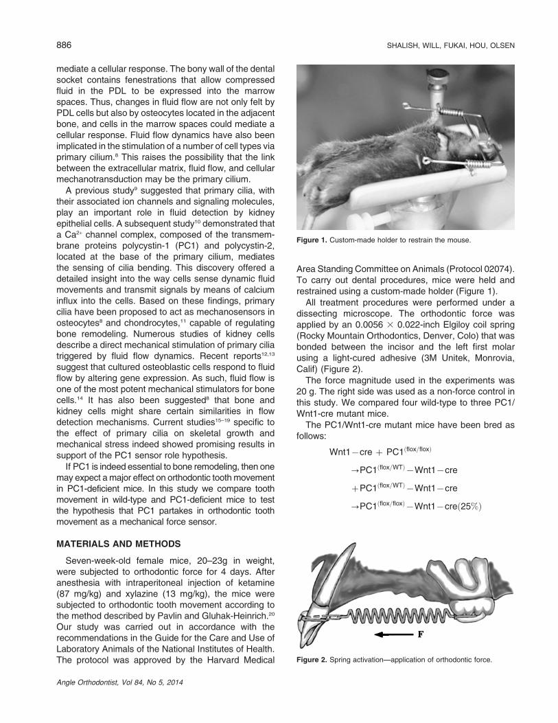
Area Standing Committee on Animals (Protocol 02074).To carry out dental procedures, mice were held andrestrained using a custom-made holder (Figure 1).
All treatment procedures were performed under adissecting microscope. The orthodontic force wasapplied by an 0.0056 3 0.022-inch Elgiloy coil spring(Rocky Mountain Orthodontics, Denver, Colo) that wasbonded between the incisor and the left first molarusing a light-cured adhesive (3M Unitek, Monrovia,Calif) (Figure 2).
The force magnitude used in the experiments was20 g. The right side was used as a non-force control inthis study. We compared four wild-type to three PC1/Wnt1-cre mutant mice.
The PC1/Wnt1-cre mutant mice have been bred asfollows:
Wnt1{cre z PC1 flox=floxð Þ
?PC1 flox=WTð Þ{Wnt1{cre
zPC1 flox=WTð Þ{Wnt1{cre
?PC1 flox=floxð Þ{Wnt1{cre 25%ð Þ
Figure 1. Custom-made holder to restrain the mouse.
Figure 2. Spring activation—application of orthodontic force.
886 SHALISH, WILL, FUKAI, HOU, OLSEN
Angle Orthodontist, Vol 84, No 5, 2014
The conditional knockout mice lack the transmembraneprotein PC1 in the craniofacial region. The Pkd1 gene isfloxed. Since the Wnt1 promoter is expressed in cranialneural crest cells, cre recombinase removes the Pkd1gene in neural crest–derived cells. Mice carrying floxedalleles of Pkd1 were described previously.21 The micewere fed a sugar diet for 4 days and then sacrificedusing carbon dioxide. The maxillae were dissected outfrom both the control and experimental mice immedi-ately after the animals were euthanatized.
Micro–Computed Tomography
A desktop micro–computed tomography (mCT) 40system (Scanco Medical AG, Bruttisellen, Switzerland)was used for scanning. The intact maxillae wereplaced in a holder and scanned with the palateperpendicular to the image plane producing a three-dimensional stack of images. Scans were taken every16 mm.
Histology
Samples were fixed in 4% (w/v) paraformaldehydeovernight and demineralized in 0.5 M ethylenediaminetetraacetic acid (EDTA) for 14 days at 4uC before theywere embedded in paraffin. The maxillae were then
hemisected along the midpalatal raphe, embedded inparaffin, and sectioned into 6-mm sections parallel tothe mesiodistal plane of the tooth. For histologicalanalysis, paraffin-embedded sections were stainedwith hematoxylin and eosin and then microphoto-graphs were taken. Tartrate-resistent acid phospha-tase (TRAP) staining to identify osteoclasts wascarried out as follows: Samples were fixed in 4%paraformaldehyde, decalcified in EDTA, and thensectioned after paraffin embedding. After deparaffini-zation, TRAP staining was carried out using a Sigma(St Louis, Mo) Diagnostic kit, images were obtained,and the TRAP-positive cells were identified.16
In all experiments, we compared the following foursamples:
1. Wild-type mice, left side (with orthodontic force);
2. Wild-type mice, right side (without orthodontic force);
3. Mutant mice, left side (with orthodontic force); and
4. Mutant mice, right side (without orthodontic force).
Comparison between samples 1 and 2 and betweensamples 3 and 4 was made in order to isolate the effectof the applied force, while comparison betweensamples 1 and 3 and between samples 2 and 4 wasmade to isolate the effect of PC1.
RESULTS
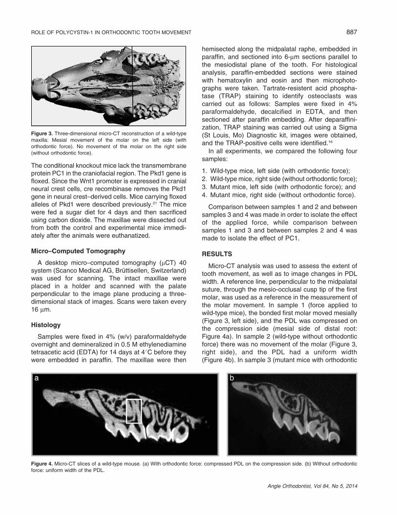
Micro-CT analysis was used to assess the extent oftooth movement, as well as to image changes in PDLwidth. A reference line, perpendicular to the midpalatalsuture, through the mesio-occlusal cusp tip of the firstmolar, was used as a reference in the measurement ofthe molar movement. In sample 1 (force applied towild-type mice), the bonded first molar moved mesially(Figure 3, left side), and the PDL was compressed onthe compression side (mesial side of distal root:Figure 4a). In sample 2 (wild-type without orthodonticforce) there was no movement of the molar (Figure 3,right side), and the PDL had a uniform width(Figure 4b). In sample 3 (mutant mice with orthodontic
Figure 3. Three-dimensional micro-CT reconstruction of a wild-type
maxilla: Mesial movement of the molar on the left side (with
orthodontic force). No movement of the molar on the right side
(without orthodontic force).
Figure 4. Micro-CT slices of a wild-type mouse. (a) With orthodontic force: compressed PDL on the compression side. (b) Without orthodontic
force: uniform width of the PDL.
ROLE OF POLYCYSTIN-1 IN ORTHODONTIC TOOTH MOVEMENT 887
Angle Orthodontist, Vol 84, No 5, 2014
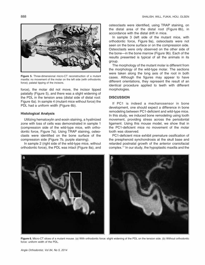
force), the molar did not move, the incisor tippedpalatally (Figure 5), and there was a slight widening ofthe PDL in the tension area (distal side of distal root:Figure 6a). In sample 4 (mutant mice without force) thePDL had a uniform width (Figure 6b).
Histological Analysis
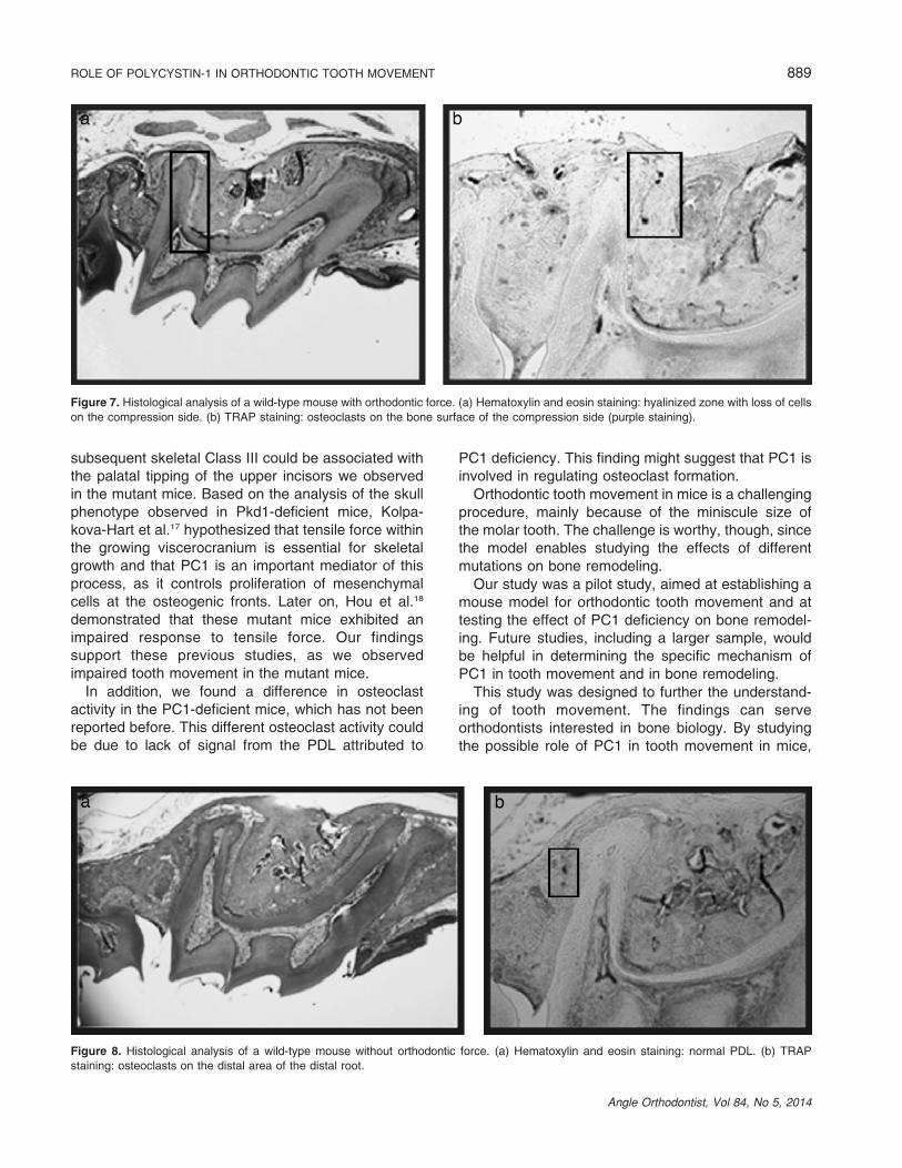
Utilizing hematoxylin and eosin staining, a hyalinizedzone with loss of cells was demonstrated in sample 1(compression side of the wild-type mice, with ortho-dontic force, Figure 7a). Using TRAP staining, osteo-clasts were identified on the bone surface of thecompression side (Figure 7b, purple staining).
In sample 2 (right side of the wild-type mice, withoutorthodontic force), the PDL was intact (Figure 8a), and
osteoclasts were identified, using TRAP staining, onthe distal area of the distal root (Figure 8b), inaccordance with the distal drift in mice.
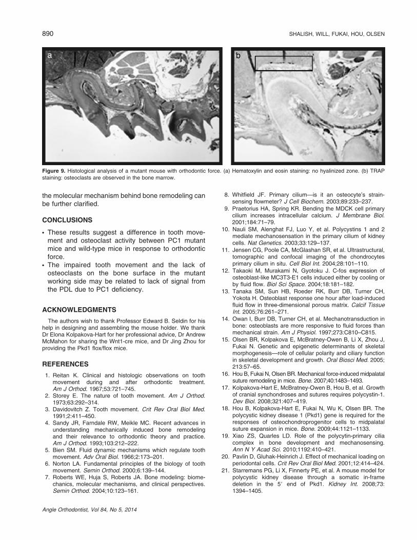
In sample 3 (left side of the mutant mice, withorthodontic force, Figure 9a), osteoclasts were notseen on the bone surface or on the compression side.Osteoclasts were only observed on the other side ofthe bone—in the bone marrow (Figure 9b). Each of theresults presented is typical of all the animals in itsgroup.
The morphology of the mutant molar is different fromthe morphology of the wild-type molar. The sectionswere taken along the long axis of the root in bothcases. Although the figures may appear to havedifferent orientations, they represent the result of anidentical procedure applied to teeth with differentmorphologies.
DISCUSSION
If PC1 is indeed a mechanosensor in bonedevelopment, one should expect a difference in boneremodeling between PC1-deficient and wild-type mice.In this study, we induced bone remodeling using toothmovement, providing stress across the periodontalligament. Using this mouse model, we show that inthe PC1-deficient mice no movement of the molartooth was observed.
PC1-deficient mice exhibit premature ossification ofthe presphenoid synchondrosis at the skull base andretarded postnatal growth of the anterior craniofacialcomplex.17 In our study, the hypoplastic maxilla and the
Figure 5. Three-dimensional micro-CT reconstruction of a mutant
maxilla: no movement of the molar on the left side (with orthodontic
force); palatal tipping of the incisors.
Figure 6. Micro-CT slices of a mutant mouse. (a) With orthodontic force: slight widening of the PDL on the tension side. (b) Without orthodontic
force: uniform width of the PDL.
888 SHALISH, WILL, FUKAI, HOU, OLSEN
Angle Orthodontist, Vol 84, No 5, 2014
subsequent skeletal Class III could be associated withthe palatal tipping of the upper incisors we observedin the mutant mice. Based on the analysis of the skullphenotype observed in Pkd1-deficient mice, Kolpa-kova-Hart et al.17 hypothesized that tensile force withinthe growing viscerocranium is essential for skeletalgrowth and that PC1 is an important mediator of thisprocess, as it controls proliferation of mesenchymalcells at the osteogenic fronts. Later on, Hou et al.18
demonstrated that these mutant mice exhibited animpaired response to tensile force. Our findingssupport these previous studies, as we observedimpaired tooth movement in the mutant mice.
In addition, we found a difference in osteoclastactivity in the PC1-deficient mice, which has not beenreported before. This different osteoclast activity couldbe due to lack of signal from the PDL attributed to
PC1 deficiency. This finding might suggest that PC1 isinvolved in regulating osteoclast formation.
Orthodontic tooth movement in mice is a challengingprocedure, mainly because of the miniscule size ofthe molar tooth. The challenge is worthy, though, sincethe model enables studying the effects of differentmutations on bone remodeling.
Our study was a pilot study, aimed at establishing amouse model for orthodontic tooth movement and attesting the effect of PC1 deficiency on bone remodel-ing. Future studies, including a larger sample, wouldbe helpful in determining the specific mechanism ofPC1 in tooth movement and in bone remodeling.
This study was designed to further the understand-ing of tooth movement. The findings can serveorthodontists interested in bone biology. By studyingthe possible role of PC1 in tooth movement in mice,
Figure 7. Histological analysis of a wild-type mouse with orthodontic force. (a) Hematoxylin and eosin staining: hyalinized zone with loss of cells
on the compression side. (b) TRAP staining: osteoclasts on the bone surface of the compression side (purple staining).
Figure 8. Histological analysis of a wild-type mouse without orthodontic force. (a) Hematoxylin and eosin staining: normal PDL. (b) TRAP
staining: osteoclasts on the distal area of the distal root.
ROLE OF POLYCYSTIN-1 IN ORTHODONTIC TOOTH MOVEMENT 889
Angle Orthodontist, Vol 84, No 5, 2014
the molecular mechanism behind bone remodeling canbe further clarified.
CONCLUSIONS
N These results suggest a difference in tooth move-ment and osteoclast activity between PC1 mutantmice and wild-type mice in response to orthodonticforce.
N The impaired tooth movement and the lack ofosteoclasts on the bone surface in the mutantworking side may be related to lack of signal fromthe PDL due to PC1 deficiency.
ACKNOWLEDGMENTS
The authors wish to thank Professor Edward B. Seldin for hishelp in designing and assembling the mouse holder. We thankDr Elona Kolpakova-Hart for her professional advice, Dr AndrewMcMahon for sharing the Wnt1-cre mice, and Dr Jing Zhou forproviding the Pkd1 flox/flox mice.
REFERENCES
1. Reitan K. Clinical and histologic observations on toothmovement during and after orthodontic treatment.Am J Orthod. 1967;53:721–745.
2. Storey E. The nature of tooth movement. Am J Orthod.1973;63:292–314.
3. Davidovitch Z. Tooth movement. Crit Rev Oral Biol Med.1991;2:411–450.
4. Sandy JR, Farndale RW, Meikle MC. Recent advances inunderstanding mechanically induced bone remodelingand their relevance to orthodontic theory and practice.Am J Orthod. 1993;103:212–222.
5. Bien SM. Fluid dynamic mechanisms which regulate toothmovement. Adv Oral Biol. 1966;2:173–201.
6. Norton LA. Fundamental principles of the biology of toothmovement. Semin Orthod. 2000;6:139–144.
7. Roberts WE, Huja S, Roberts JA. Bone modeling: biome-chanics, molecular mechanisms, and clinical perspectives.Semin Orthod. 2004;10:123–161.
8. Whitfield JF. Primary cilium—is it an osteocyte’s strain-sensing flowmeter? J Cell Biochem. 2003;89:233–237.
9. Praetorius HA, Spring KR. Bending the MDCK cell primarycilium increases intracellular calcium. J Membrane Biol.2001;184:71–79.
10. Nauli SM, Alenghat FJ, Luo Y, et al. Polycystins 1 and 2mediate mechanosensation in the primary cilium of kidneycells. Nat Genetics. 2003;33:129–137.
11. Jensen CG, Poole CA, McGlashan SR, et al. Ultrastructural,tomographic and confocal imaging of the chondrocytesprimary cilium in situ. Cell Biol Int. 2004;28:101–110.
12. Takaoki M, Murakami N, Gyotoku J. C-fos expression ofosteoblast-like MC3T3-E1 cells induced either by cooling orby fluid flow. Biol Sci Space. 2004;18:181–182.
13. Tanaka SM, Sun HB, Roeder RK, Burr DB, Turner CH,Yokota H. Osteoblast response one hour after load-inducedfluid flow in three-dimensional porous matrix. Calcif TissueInt. 2005;76:261–271.
14. Owan I, Burr DB, Turner CH, et al. Mechanotransduction inbone: osteoblasts are more responsive to fluid forces thanmechanical strain. Am J Physiol. 1997;273:C810–C815.
15. Olsen BR, Kolpakova E, McBratney-Owen B, Li X, Zhou J,Fukai N. Genetic and epigenetic determinants of skeletalmorphogenesis—role of cellular polarity and ciliary functionin skeletal development and growth. Oral Biosci Med. 2005;213:57–65.
16. Hou B, Fukai N, Olsen BR. Mechanical force-induced midpalatalsuture remodeling in mice. Bone. 2007;40:1483–1493.
17. Kolpakova-Hart E, McBratney-Owen B, Hou B, et al. Growthof cranial synchondroses and sutures requires polycystin-1.Dev Biol. 2008;321:407–419.
18. Hou B, Kolpakova-Hart E, Fukai N, Wu K, Olsen BR. Thepolycystic kidney disease 1 (Pkd1) gene is required for theresponses of osteochondroprogenitor cells to midpalatalsuture expansion in mice. Bone. 2009;44:1121–1133.
19. Xiao ZS, Quarles LD. Role of the polycytin-primary ciliacomplex in bone development and mechanosensing.Ann N Y Acad Sci. 2010;1192:410–421.
20. Pavlin D, Gluhak-Heinrich J. Effect of mechanical loading onperiodontal cells. Crit Rev Oral Biol Med. 2001;12:414–424.
21. Starremans PG, Li X, Finnerty PE, et al. A mouse model forpolycystic kidney disease through a somatic in-framedeletion in the 59 end of Pkd1. Kidney Int. 2008;73:1394–1405.
Figure 9. Histological analysis of a mutant mouse with orthodontic force. (a) Hematoxylin and eosin staining: no hyalinized zone. (b) TRAP
staining: osteoclasts are observed in the bone marrow.
890 SHALISH, WILL, FUKAI, HOU, OLSEN
Angle Orthodontist, Vol 84, No 5, 2014
Related Documents